Award Number: DAMD17-02-1-0691 TITLE: Center for the ... · PEB algorithm, for Parametric Empirical...
93
p AD Award Number: DAMD17-02-1-0691 TITLE: Center for the Evaluation of Biomarkers for the Early Detection of Breast Cancer PRINCIPAL INVESTIGATOR: Nicole D. Urban, Sc.D. CONTRACTING ORGANIZATION: Fred Hutchinson Cancer Research Center Seattle, WA 98109-1024 REPORT DATE: October 2004 TYPE OF REPORT: Annual PREPARED FOR: U.S. Army Medical Research and Materiel Command Fort Detrick, Maryland 21702-5012 DISTRIBUTION STATEMENT: Approved for Public Release; Distribution Unlimited The views, opinions and/or findings contained in this report are those of the author(s) and should not be construed as an official Department of the Army position, policy or decision unless so designated by other documentation. 20050603 105
Transcript of Award Number: DAMD17-02-1-0691 TITLE: Center for the ... · PEB algorithm, for Parametric Empirical...

p
AD
Award Number: DAMD17-02-1-0691
TITLE: Center for the Evaluation of Biomarkers for the EarlyDetection of Breast Cancer
PRINCIPAL INVESTIGATOR: Nicole D. Urban, Sc.D.
CONTRACTING ORGANIZATION: Fred Hutchinson Cancer Research CenterSeattle, WA 98109-1024
REPORT DATE: October 2004
TYPE OF REPORT: Annual
PREPARED FOR: U.S. Army Medical Research and Materiel CommandFort Detrick, Maryland 21702-5012
DISTRIBUTION STATEMENT: Approved for Public Release;Distribution Unlimited
The views, opinions and/or findings contained in this report arethose of the author(s) and should not be construed as an officialDepartment of the Army position, policy or decision unless sodesignated by other documentation.
20050603 105

O Form ApprovedREPORT DOCUMENTATION PAGE [OMB No. 074-0188
Public reporting burden for this collection of information is estimated to average 1 hour per response, including the time for reviewing instructions, searching existing data sources, gathering and maintainingthe data needed, and completing and reviewing this collection of information. Send comments regarding this burden estimate or any other aspect of this collection of information, including suggestions forreducing this burden to Washington Headquarters Services, Directorate for Information Operations and Reports, 1215 Jefferson Davis Highway, Suite 1204, Arlington, VA 22202-4302, and to the Office ofManagement and Budget, Paperwork Reduction Project (0704-0188), Washington, DC 205031. AGENCY USE ONLY 2. REPORT DATE 3. REPORT TYPE AND DATES COVERED
(Leave blank) October 2004 Annual (23 Sep 2003 - 22 Sep 2004)
4. TITLE AND SUBTITLE 5. FUNDING NUMBERSCenter for the Evaluation of Biomarkers for the Early DAMD17-02-1-0691Detection of Breast Cancer
6. AUTHOR(S)
Nicole D. Urban, Sc.D.
7. PERFORMING ORGANIZATION NAME(S) AND ADDRESS(ES) 8. PERFORMING ORGANIZATIONFred Hutchinson Cancer Research Center REPORT NUMBERSeattle, WA 98109-1024
E-Mail: nurban@fhcrc .org
9. SPONSORING / MONITORING 10. SPONSORING / MONITORING
AGENCY NAME(S) AND ADDRESS(ES) AGENCY REPORT NUMBER
U.S. Army Medical Research and Materiel CommandFort Detrick, Maryland 21702-5012
11. SUPPLEMENTARY NOTES
12a. DISTRIBUTION / AVAILABILITY STATEMENT 12b. DISTRIBUTION CODEApproved for Public Release; Distribution Unlimited
13. ABSTRACT (Maximum 200 Words)
Breast Cancer remains a leading cause of death for women in the U.S. despite the popularity ofmammography as a preventive tool. At diagnosis, many breast cancers are at an advanced stage ofdisease, even for women undergoing yearly screening, resulting in costly and painful follow-upprocedures. Based on the ongoing studies in many research institutions, it has been shown thatmolecular markers can increase our ability to diagnose early stages tumors. This has beendemonstrated by current clinical practices using the CA-125 marker and PSA for the detection ofovarian and prostate cancer, respectively. The purpose of this study is to search for breastcancer biomarkers and evaluate their effectiveness in detecting early stage carcinoma. Bycombining molecular diagnosis with current imaging analysis of breast tissue, we may furtherreduce the number of deaths as well as the number of women undergoing surgery and chemotherapydue to breast cancer. To date, we have created the infrastructure necessary for ourinterdisciplinary team of investigators to obtain study samples, characterize candidatebiomarkers, and efficiently communicate research findings. We have also increased the number ofpotential biomarkers and looked into more efficient and sensitive biotechnology that may betterassist our study investigators.
14. SUBJECT TERMS 15. NUMBER OF PAGESEarly detection, serum markers, screening, mammography, antibody 93tests, molecular profiling, marker panels, mammography biomarker 16. PRICE CODE
17. SECURITY CLASSIFICATION 18. SECURITY CLASSIFICATION 19. SECURITY CLASSIFICATION 20. LIMITATION OF ABSTRACTOF REPORT OF THIS PAGE OF ABSTRACT
Unclassified Unclassified Unclassified UnlimitedNSN 7540-01-280-5500 Standard Form 298 (Rev. 2-89)
Prescribed by ANSI Std. Z39-18298-102

Table of Contents
Cover ........................................................................................... I
SF 298 ............................................................................................ 2
Table of Contents .......................................................................... 3
Introduction .................................................................................. 4
Body ................................................................................................. 4-13
Key Research Accomplishments ....................................................... 14
Reportable Outcomes ...................................................................... 14
Conclusions .................................................................................... 14
References .................................................................................... 14
Appendices ................................................................................... 15
3

Introduction
Although mammography significantly reduces its toll, breast cancer remains a leading cause ofcancer mortality in the U.S. Many breast cancers are advanced at the time of diagnosis, evenamong women participating in screening. The discovery of molecular markers associated withbreast cancer potentially increases our ability to diagnose early stage tumors. We are proposingthat molecular diagnosis be combined with imaging to enhance our ability to identify breastcancer when it is most treatable, i.e. still localized to the breast. This study will test thehypothesis that use of a breast cancer biomarker panel can improve the performance ofmammography in early detection of breast cancer. The primary aims of this study are: 1) tovalidate and refine the ability of candidate biomarkers to predict disease status; 2) to evaluatepanels of biomarkers for use as an adjunct to mammography, to detect all breast cancer at ahighly curable stage; and 3) to identify the molecular signatures of benign, pre-invasive andinvasive breast cancers and explore their associations with biomarkers in the panel.
We are focusing on markers that can be measured in serum, as they are generally inexpensive andnot subjective in their interpretation. To avoid over-diagnosis, we will perform molecularprofiling to identify aggressive subsets of breast cancer that are most likely to be missed bymammography and in need of early detection. Our current list of candidate markers includescirculating antigens3, growth factors associated with angiogenesis, DNA methylation, andantibodies to oncogenic proteins known to be associated with aggressive disease such asHer2/neu, p53, IGFBP-2, Cyclin D1, Cathepsin, and Topoisomerase IIA.
We will use screening history as suggested by McIntosh & Urban1 to generate what is called aPEB algorithm, for Parametric Empirical Bayes. The PEB approach can be combined withmethods developed by McIntosh & Pepe2 to permit multiple marker algorithms over time.Decisions on screening are made by observing the deviation of a marker from its expected normalbehavior, but where PEB methods are used to determine this normal behavior.
At the end of this Center of Excellence study, the expected result is a panel of markers anddecision rules for its use clinically to improve the performance of mammography.
Body
Throughout the second year of this study, investigators continued to focus efforts on developing arobust infrastructure to support the scientific objectives of the study and to meet study goals asoutlined in the statement of work. By the end of our first year, the Mammography TumorRegistry (MTR) portion of this study had received approval by the Office of ResearchProtections; however, investigators and staff continued to work closely with Peter Marshall, DoDHuman Protections Specialist, throughout the second year to receive human subjects approval onthe study's clinical/recruitment protocols. This study has two clinical and recruitment protocols.The first one covers clinical/recruitment activities in Seattle where women receiving annualmammograms, a pre-scheduled stereotactic biopsy, or breast related surgery may be approachedfor a blood only donation. The Seattle spurgical population can also be approached for a bloodand tissue donation. The second protocol covers specific recruitment activities at Cedars SinaiMedical Center in Los Angeles where women undergoing a biopsy or surgery may be approachedfor blood and tissue donation. These protocols have been standardized between recruitment sitesas much as possible. In May 2004, the DoD's Office of Research Protections approved theclinical/recruitment protocol that will be followed in Seattle. Investigators continue to await DoDhuman subjects approval on the Cedars Sinai protocol.
4

In addition to working with Peter Marshall and the Office of Research Protections towardreceiving human subjects approval on the clinical/recruitment protocols, investigators and studystaff have concentrated their efforts in the following areas over the last year: 1) refinement anduse of a breast cancer micro simulation model to investigate the impact of DCIS diagnosis onbreast cancer mortality and associated over treatment of this disease; 2) integration of patientadvocates into the overall research program; 3) implementation of the Mammography TumorRegistry components of the study; 4)organization of investigator meetings/conference calls torefine the study protocol, to develop standardized specimen collection and processing procedures,and to discuss progress on breast cancer biomarker assays that are being developed or refined incollaborating laboratories; 5) continued development of relationships with clinicians who willprovide access to patients for the study; 6) collaboration among investigators to completedevelopment of data collections instruments; 7) continued development of a web-basedinformatics system and adaptation of an existing specimen inventory and tracking system toaccommodate breast specimens; and 8) development of a web-based knowledge managementsystem to support communication among investigators.
Investigators have refined a previously developed micro simulation model to explore the impactof DCIS diagnosis on breast cancer mortality and associated overdiagnosis. Clinical use of amarker panel is a complex area of study that requires integration of all of the information frommarker analyses and molecular profiling as well as economic and health systems considerations.It is critical to understand what we want our biomarker panel to detect. The latter considerationsare being studied through a micro simulation model4 that was developed through a previouslyfunded DoD grant (DAMD 17-94-J-4237). We are currently using the model to investigate theimpact of DCIS detection and treatment on breast cancer mortality and associated over diagnosis.Specifically, the model is being used to generate disease histories, including disease onset,progression to diagnosis, and mortality, for a cohort of women in the United States.Mammography screening schedules are superimposed on these disease histories, allowinginvestigation of the efficacy of early detection of breast cancer, including the in situ stage.Cancer incidence data are combined with data from autopsy studies to estimate the prevalence ofbreast cancer, including DCIS, in the population. Model parameters are selected to replicatediagnosis patterns reported in published studies.
Using available data for breast cancer growth rates, mammography performance, and stage-specific survival, our analyses suggest that mammography use, including detection of DCIS atcurrent rates, yields a 25% reduction in breast cancer mortality. We estimate that detection ofDCIS accounts for over 20% of this reduction (5.6%), that 64% of screen-detected DCIS wouldremain latent until death due to other causes (over-diagnosis), and that mammography detectsonly one fifth of the prevalent DCIS. These results are reported in a manuscript titledQuantifying Risks of Breast Cancer Mortality and Overdiagnosis due to Mammography-diagnosed DCJS that will be submitted to JNCI.
COE patient advocates continue to influence the direction of the research study. We aredeveloping a well-integrated patient advocacy program that is focused on addressing the mostcritical issues in the Center of Excellence study. Ms. Joan McAree was hired to support this effortlast year, but has left the study to devote time to an organization that she co-founded with hersister named the Ovarian and Breast Cancer Alliance of Washington State. Ms. Shannon Marsh,who is an attorney by trade and has personal experience with participating in research studies, hasreplaced Ms. McAree. Since joining the study, Ms. Marsh has had many training opportunities tolearn more about patient advocacy in support of research. For example, she attended the NCIsponsored SPORE Investigator Workshop last July where she participated in patient advocacy
5

training sessions led by Ms. Deborah Collyar. In addition, she has been working closely with Ms.Sheryl Eisenbarth who leads the Pacific Ovarian Cancer Research Consortium (POCRC) PatientAdvocacy group at the Fred Hutchinson Center. Under Ms. Eisenbarth's leadership, the POCRCadvocacy group has become nationally recognized and is used as a model for many developingpatient advocacy groups.
Recent experiences with our ovarian work have shown that there are many intellectual propertyrights issues that must be addressed before multiple markers that are "owned" by different entitiescan be combined in a marker panel. In addition, it is imperative that agreements are establishedwith commercial partners to expedite the development and movement of the panel throughclinical testing. With the support of Center lawyers and technology transfer specialists, Dr. Urbanhas been working through these issues. Recently, she has asked Ms. Marsh to assist her with thiswork knowing that her legal expertise would be valuable. In addition, learning from thisexperience will help Ms.Marsh work with COE investigators to address intellectual propertyrights with the breast cancer marker panel and future identification of appropriate clinicalapplications.
In addition to working with Dr. Urban on intellectual property rights, Ms. Marsh is also focusingefforts on facilitating communication between scientists and consumers, and increasing minorityparticipation. Specifically, Ms. Marsh has attended many community events including the SusanG. Koman Race for the Cure in June. At this event, she spoke to over 100 women and discussedin detail the goals and requirements for becoming involved in the COE study. In addition, shedistributed a variety of educational materials on detection and prevention of breast cancer.Generally, women are very excited and interested in learning more about participation in researchstudies, particularly early detection biomarker studies in breast cancer.
Another effort that will support greater communication between scientists and the community isdevelopment of a quarterly newsletter. This newsletter will be sent to all women participating inthe Women's Cancer Prevention and Detection Network and those specifically participating inthe Center of Excellence study and will feature articles that focus on women's cancers and cancerresearch. We will encourage women to write or telephone in any questions they may have so thata dialogue develops between the research community and its study participants; thus, thenewsletter will provide a mechanism to reach out to the community facilitating communicationamong scientists and patients.
Ms. Marsh has also taken the lead on developing study materials including approach letters,consent forms, and the Women's Cancer Prevention and Detection Network brochure. A draftcopy of the brochure is included as Appendix A. While developing this brochure, Ms. Marshpaid particular attention to issues important to the patient, such as privacy issues and access tomedical records. The brochure was also designed to encompass many different minority groups.
As recruitment efforts ramp up this year, Ms. Marsh will focus attention on minority recruitmentworking closely with collaborating physicians toward increasing minority enrollment in allpopulations. We will concentrate efforts on specific minority groups (ex. Asian and Hispanic)that are most prevalent at participating facilities, and will explore the feasibility of translating ourstudy materials into appropriate languages in order to reach a wider minority base.
Mammographv Tumor Registry protocol has been implemented. The first step towardimplementing this protocol was hiring a systems analyst programmer/data coordinator dedicatedto this work. At the start of the second year, the MTR programmer developed methods to receiveregular downloads of electronic mammography data from the Swedish Breast Care Center's
6

Mammography Reporting System (MRS). In aggregate, this data can be used to report canceroutcomes to participating radiologists through a feedback report that summarizes false positiveand false negative rates within a given time frame. Using "old" data, the MTR programmerpiloted methods and a linking algorithm to link the MRS aggregate data to the Washington StateCancer Registry. This linkage work is done in collaboration with the Fred Hutchinson Center'sCancer Surveillance System (CSS). Because CSS recently changed its data structures, it wasnecessary to update existing CSS import algorithms and study data structures (particularly cancerstaging structures) to accommodate to this change. To test for quality control, the MTRprogrammer is currently in the process of comparing current linkage results using the newalgorithm with previous linkage results. Once this step is complete, investigators will work ondeveloping a feedback report with new outcomes data that will be provided to participatingradiologists at the Swedish Breast Care Center.
In accordance with the approved protocol, individual mammography data will be used only aftera woman has signed a HIPAA authorization form giving us permission to review hermammography records. This mammography results release form will be mailed to potential studyparticipants with a one-page screening questionnaire. If a mammography results release form ison file, mammogram findings (assessment codes, follow up recommendation, breast density) willbe used in addition to the information from the screening questionnaire to determine risk statusfor an individual woman5'6 . If a release form is not on file, then we will use information obtainedthrough the screening questionnaire only to determine initial risk status. These instruments allowus to determine study eligibility and help us to prioritize women for invitation into the stddy.
COE investigators have been meeting regularly to standardize procedures between recruitmentsites and to discuss marker assays. We have organized quarterly investigator conference calls torefine protocols and data collection instruments that will be used in this study. Specific effortswere made to work with Dr. Scott Karlan, lead surgeon/investigator at Cedars Sinai MedicalCenter, and his staff toward the development of similar clinical recruitment protocols usingstandardized consent forms and data collection instruments. The data collection instrumentsinclude a baseline questionnaire, health status questionnaire, and a health status update. Both theFHCRC and Cedars Sinai clinical/recruitment protocols are included as Appendices B and C. Inaddition, investigators are working closely to implement standardized tissue collection andprocessing protocols, as well as a centralized tissue review and characterization process that willbe completed by Dr. Nancy Kiviat (pathologist) in Seattle. Investigators have also used thisopportunity to discuss candidate biomarkers and the progress the laboratory investigators aremaking toward refining their specific marker assays.
COE annual investigator meeting was held in January 2004 to discuss candidate and novel earlydetection markers. COE investigators presented recent work on their markers at the first COEannual investigator meeting. Dr. Gordon Mills was scheduled to present his lysophospholipidmarkers; however, due to a scheduling conflict he could not attend. In his place, Dr. NicoleUrban presented his work suggesting that certain lysophospholipids represent a novel class ofbioactive factors for breast and ovarian cancer that can potentially be measured through SELDI-TOF mass spectrometry 7. Dr. Nathalie Scholler, senior staff scientist at the Fred HutchinsonCenter, presented the concepts behind the use of a yeast display library of single chain Fragmentvariable (scFv) antibodies for rapid ELISA development. Dr. Scholler also presented thepossibilities of using a multiplexing system that would enable simultaneous measurement ofseveral markers on the same sample set. This technique can be done in Seattle, using a BioRadProtein Array machine that was purchased by the Fred Hutchinson Center and the Pacific OvarianCancer Research Consortium/SPORE in ovarian cancer. The machine is currently located in Dr.George McDonald's cytokine shared resource laboratory. With the support of Center leadership,
7

Drs. McDonald and Urban are working to obtain CLIA approval for this lab, which willeventually allow investigators to act clinically on marker results as appropriate.
To close the workshop, Dr. Nancy Kiviat from the Univ. of Washington presented her work withMammaglobin and discussed its potential for assessing clinical stage, nodal status, and recurrenceof breast cancer8. In addition, Dr. Kiviat discussed her work in DNA methylation and its abilityto detect early stage cancer. We have started to plan the next workshop, which will be held inSeattle in February 2005.
Investinators will begin testing markers using samples from collaborators. We have received pre-validation samples from various collaborators and will begin testing our candidate markers forinclusion in the marker panel. In addition, we are using some of these pre-validation samples toanalyze markers that are currently not on our list of candidates. For example, Dr. Scholler at theFred Hutchinson Cancer Center is exploring use of a cytokine panel and an assay for CD24. Ifone of the candidate markers is ineffective, we will be poised to add a new marker to the panelthat could increase its ability to detect specific forms of breast cancer.
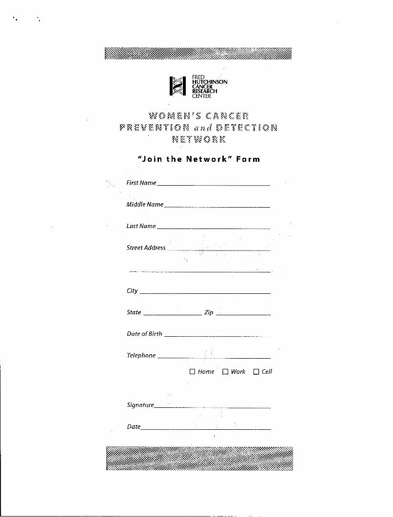
Recruitment of women without breast cancer. While awaiting DoD human subjects approval, wehave been working to identify potentially eligible women to invite to participate in the study.Funded through another IRB-approved resource, we are building a network of women who arereceiving screening mammograms at the Swedish Breast Care Center (SBCC) and who indicate aninterest in participating in future research studies. This network is titled the Women's CancerPrevention and Detection Network. All women who undergo mammograms at SBCC are invited toread a brochure describing the Network, and to complete a "Join the Network" form. The "Join theNetwork" form gives permission for researchers to maintain a woman's name indefinitely in aconfidential "registry" and invite her to participate in future research studies. This process assurescompliance with HIPAA for all of our studies. Women are also sent a one-page screeningquestionnaire that asks about family history of breast or ovarian cancer, and a mammographyresults release form. Currently, the network contains about 1,800 women. We anticipate that thisnetwork will grow substantially over the next couple of years with the immediate possibility ofadding a few more radiology facilities.
Some of these women, approximately 740, have already been invited to participate in a breastcancer research study funded by a NCI/Avon Progress for Patients award (PI: Nicole Urban). Werefer to this study of high-risk women as the "Avon study." Of the women invited, 341 haveenrolled in the Avon study and most are approaching study completion. These women have provento be a motivated group with many indicating that they would like to continue some type ofparticipation in research. Towards that end, we have developed an approach letter and have revisedthe study protocol to enable us to invite these women to participate in the Center of Excellencestudy. This modification is currently under review by Peter Marshall, Human Protections Specialist.Once approved, study invitation letters will be mailed to all women in the Avon study. Weanticipate that the enrollment rate for this population will be over 90%.
Since obtaining human subjects approval to begin patient recruitment, we have worked on refmingour procedures to classify the remaining network population by risk status based on family historyinformation. Women at high-risk will be over-sampled to increase the expected number of womenwith serial blood samples obtained prior to a breast cancer diagnosis. Study invitation packets arebeing mailed each week in batches of 25. Based on our recent experiences with the Avon study, weexpect an enrollment rate of 60% or more in this population.
8

Recruitment of women with breast cancer. Under the leadership and guidance of Dr. DavidBeatty, Director of the Breast Cancer Program at Swedish Medical Center, we have organizedseveral meetings with the Swedish breast surgeons and pathologists to develop detailed surgicalrecruitment and specimen collection plans that are feasible within their specific clinicalenvironments. The Swedish group remains enthusiastic and committed to making thiscollaboration and the study a success. Meeting minutes are included as Appendix D. With thesurgeons' guidance it was determined that the ideal surgical candidate who could potentiallydonate both tissue and blood would be a woman who presents with disease that is T2 or greater,DCIS or invasive carcinoma where the patient is undergoing total mastectomy, or suspectedlesions >2 cm. With the pathologists' guidance, we have been able to develop a detailed bloodand tissue collection and processing protocol that is included as Appendix E.
In August 2004, we implemented specific recruitment procedures in the surgeons' offices on theFirst Hill and Providence campuses of Swedish Medical Center. We anticipate a learning curveas physicians and clinic staff familiarize themselves with specific study guidelines, but onceprocedures become routine then enrollment and collection rates should increase dramatically. Todate, we have enrolled 12 women into the surgical cohort with 11 providing a pre-surgical blooddonation and 1 declining this option. Five women have provided a blood donation at the time ofsurgery. In addition, we have successfully obtained a pre-surgical blood donation, as well as ablood and tissue donation from 1 of these 12 women at the time of her surgery. A detailedenrollment report based on specimen donation and clinical diagnosis is currently beingdeveloped.
Taking things in a stepwise fashion, we will begin to work with the clinicians and their staff toimplement biopsy enrollment procedures, once the surgical enrollment procedures are runningsmoothly. Towards that end, in order to increase study efficiency in the surgeons' offices, wehave organized in-services with the clinic staff, as past experiences have shown that clinic staffplay a key role in successful recruitment efforts. Shirley Gough, Research Nurse, and other studystaff met with clinic nurses, assistants, and schedulers to present the study and to discussrecruitment procedures that could fit into normal clinic flow. These meetings have been veryinformative and have resulted in several ideas that can be easily implemented toward increasingeffective communication among the surgeons, their staff, and the research staff.
In addition to study specific meetings with the physicians, we are planning monthlyInterdisciplinary Working Group meetings to facilitate regular scientific exchange betweenscientists and community physicians. Although these meetings have not been scheduled yet, wehave identified meeting chairs, Drs. David Beatty and Kristine Rinn. Supported by study staff,Drs. Beatty and Rinn are working together to find a convenient meeting time, and are developingoverall meeting objectives, list of attendees, and an initial list of presenters.
Pathology data collection instruments have been developed. A "Patient Level Clinical Diagnosis"form has been developed and will provide appropriate information to characterize a woman basedon TNM staging and grade of disease at the time of her diagnosis. A study staff member willcomplete this form with the research nurse conducting quality assurance. Working closely withDr. Kiviat, lead pathologist, we have also developed a histopathology (tissue review) form thatwill allow us to characterize all tissue samples. Once it is implemented, Dr. Kiviat will completethis form. We are now working on a clinical follow-up form that will capture specific informationon treatment. The pathology data collection instruments are included as Appendix F.

Programmers have developed a web-based App Server
interface to manage study data. We have Client (Web Browser) PHSITOR]
prepared much of the informatics Network * • ninfrastructure required to support COE Mp-rTtP
study. Infrastructure now in place include:web server hardware, web service software, L.............----access security, data entry form templates,and referential integrity between database I Iobjects. In addition, template reports, data Data Baee seredictionary documentation, and a high-riskalgorithm have been developed. Figure 1 SOL MOshows the current structure of the design inplace.
Security has been implemented in a layeredapproach. Only persons authenticating to -
the server that hosts the web service can Figure 1. Infrastructure of the Seattle Informatics Management
access the web service. However, the server (SIM) System
is set up to accept authentication from alimited set of IP addresses, and from a limited set of client machine operating systems. Onceusers have authenticated to the host server, they must login to the application. Additionally, usersdo not have access to the directories where data are stored. Access is limited to the web service,which acts as the proxy to communicate with the database retrieving data for web based requests.Authentication and application login screens are illustrated below in Figure 2.
'Connecbngto tor-app Fcrc fr
>: ______ Reee
tJsose tP41 Lnostiais
Figre 2. Srver Aut.hent.cat.o a Applc TOR aiM onsLom
+ , + + + ...... ... .... .. [ ............................................... -.............. J
Figure 2. Server Authentication and Application Login
Web based screens for questionnaire data entry, similar to those needed by the COE project, havebeen developed and are currently in use for the Avon study by FHCRC staff and collaborators atMD Anderson Cancer Center, the University of Alabama Cancer Center and Fox Chase CancerCenter. During the development of these questionnaires stumbling blocks related to, remote siteaccess, security, and data validation issues have been removed. Routines for data validation witheach submission of data to the server have been implemented. Every value entered is checked forvalidity. Any outliers are returned to the data entry specialist for verification before the data arecommitted to the database. In addition, attempts to re-enter data that have previously beencollected, are preempted via referential integrity. Figure 3 minimally illustrates the kinds ofvalidation that data entry specialists see prior to the acceptance of data by the database.
10
To Si W OeSWmUa
............. . • ' . , . .... . ..........................................................................................
oata E.nteyTyp
Z. Yhtt iS ytour dote of biriti?
•. Whoal was yfOur |ast" menstralls peule{d?
Figure 3. Data Entry Validation
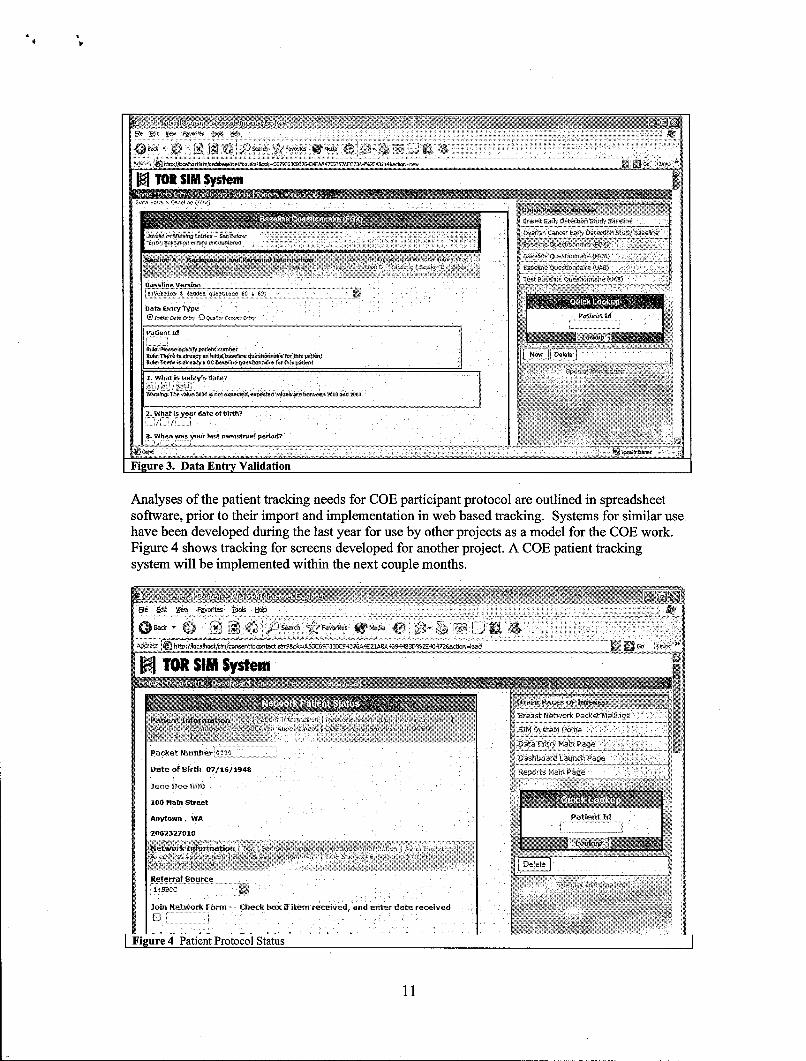
Analyses of the patient tracking needs for COE participant protocol are outlined in spreadsheetsoftware, prior to their import and implementation in web based tracking. Systems for similar usehave been developed during the last year for use by other projects as a model for the COE work.Figure 4 shows tracking for screens developed for another project. A COE patient trackingsystem will be implemented within the next couple months.
300 Main Street
Anytawn, WA2062327010 , -0
Referral Source",,
Join Network Form .- Check b ifitem received, and enter dte received pe
I Figure 3. Patient Protocol Status
11

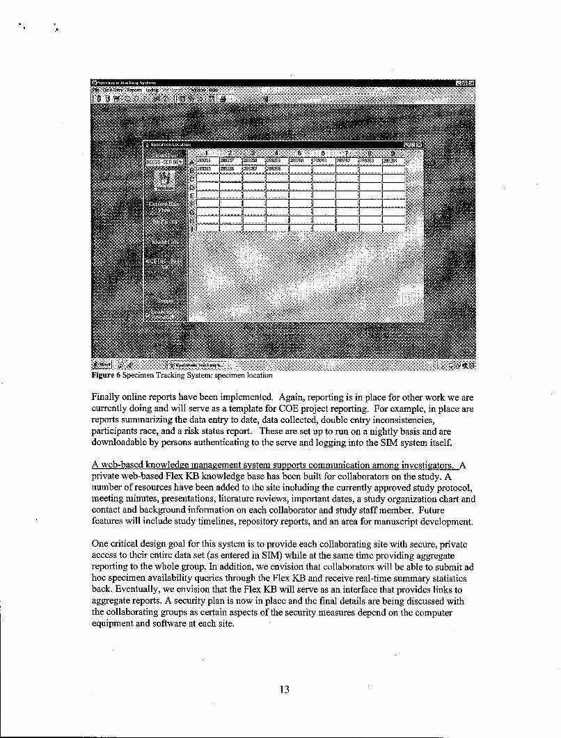
Specimens are tracked utilizing existing specimen tracking software developed for work withovarian specimens. Currently tracked for COE specimens are date of blood and/or tissuedonation, specimen processing, amount of specimen collected, types of specimen storage, andstorage location of specimen aliquot or tissue block. Current POCRC specimen tracking systemsinclude modules for characterization of patient and tissues with respect to ovarian disease.Additional modules for tracking with respect to breast disease are currently under development.Figure 5 illustrates the tracking of Patient specimen donation, while Figure 6 illustrates thetracking of Specimen location.
N, Data~ntry &kmrks -,rjj lainteane Wido tiel
od: zpl .'4, .,.0, ,1l 1ji ,.~ nrn rere 6 r e t~
lUsable Freezer
269254 ACD Bufly C N/A 1.8 ml Yes Useable Freezer
289255 ACD Plasma N/A 5 ml Yes Useable Freezer289269 Plasma N/A 11 ml Yes Useable Freezer
I 2MWfI Pla.ýmA NINA 1 Ii ml Iyre IIszAhle lFrpnPAr
1 Frozen NIA Primary beni Useable Freezer Right Breast N/A: 268682 OCT N/A Primary cand Useable Freezer I Right Breast N/A
2268683 OCT N/A Primary beni Useable Freezer Right Breast N/A
sr:z 4 nrT IN, Prira h-rr I I I-rahl. I R-ePa I Rwl Ftr-rtl W/A
Figure 5 Specimen Tracking System: patient specimen donation
12
.4"
::.:,:,::~ ~ J ................................. .............................. ... > .::::...•.. •••:,
Figure 6 Specimen Tracking System: specimen location
Finally online reports have been implemented. Again, reporting is in place for other work we arecurrently doing and will serve as a template for COE project reporting. For example, in place arereports summarizing the data entry to date, data collected, double entry inconsistencies,participants race, and a risk status report. These are set up to run on a nightly basis and aredownloadable by persons authenticating to the serve and logging into the SIM system itself.
A web-based knowledge management system supports communication among investigators. Aprivate web-based Flex KB knowledge base has been built for collaborators on the study. Anumber of resources have been added to the site including the currently approved study protocol,meeting minutes, presentations, literature reviews, important dates, a study organization chart andcontact and background information on each collaborator and study staff member. Futurefeatures will include study timelines, repository reports, and an area for manuscript development.
One critical design goal for this system is to provide each collaborating site with secure, privateaccess to their entire data set (as entered in SIM) while at the same time providing aggregatereporting to the whole group. In addition, we envision that collaborators will be able to submit adhoc specimen availability queries through the Flex KB and receive real-time summary statisticsback. Eventually, we envision that the Flex KB will serve as an interface that provides links toaggregate reports. A security plan is now in place and the final details are being discussed withthe collaborating groups as certain aspects of the security measures depend on the computerequipment and software at each site.
13

Key Research Accomplishments
Year two of this study has continued to focus on infrastructure development and start-up efforts.No research results are available at this time.
Reportable Outcomes
No manuscripts, presentations or publications have yet resulted from this study. A web-based
database to support informatics is being developed. Screen shots are shown above.
Conclusions
No research conclusions are available at this time.
References
1. Generating Longitudinal Screening Algorithms Using Novel Biomarkers for Disease;McIntosh M, Urban N, Karlan B. Cancer Epidemiology, Biomarkers, and Prevention.2002;11:159-166.
2. Combining several screening tests: optimality of the risk score; McIntosh MW and Pepe MS;Biometrics; 58(3): 657-64; September 2002.
3. Circulating Tumor Markers in Breast Cancer: Accepted utilities and novel prospects;Steams, Vered, Yamauchi, Hideko, Hayes, Daniel; Breast Cancer Research and Treatment;52: 239-259. 1998.
4. A health priorities model: application to mammography screening; Meischke H, AndersenMR, Bowen D, Kuniyuki A, Urban N. Health Education and Behavior 1998; 25: 383-395.
5. Effect of age and breast density on screening mammograms with false-positive findings;Lehman CD, White E, Peacock S, Drucker MJ, and Urban N; Am J Roentgenol; 173(6):1651-5; December 1999.
6. Performance of screening mammography among women with and without a first-degreerelative with breast cancer; Kerlikoske K, Carney P, Geller B, Mandelson M, Taplin S, MalvinK, Ernster V, Urban N, Cutter G, Rosenberg R, and Ballard-Barbash R; Annals of InternalMedicine, Vol. 133, No. 11; December 5, 2000.
7. Lysophosphatidic acid production and action: validated targets in cancer? Umezu-Goto M,Tanyi J, Lahad J, Liu S, Yu S, Lapushin R, Hasegawa Y, Lu Y, Trost R, Bevers T, Jonasch E,Aldape K, Liu J, James RD, Ferguson CG, Xu Y, Prestwich GD, Mills GB; J Cell Biochem;15; 92(6): 1115-40; August 2004.
8. Mammaglobin as a Novel Breast Cancer Biomarker: Multigene Reverse Transcription-PCRAssay and Sandwich ELISA; Zehentner BK, Deme A, Toure P, Hawes SE, Brooks L,Feng Q, Hayes DC, Persing DH, Critichlow CW, Houghton RL, Kiviat NB.; ClinChem.Sept. 2004.
14

Appendices
Appendix A Women's Cancer Prevention and Detection draft brochureAppendix B FHCRC clinical/recruitment protocolAppendix C Cedars-Sinai clinical/recruitment protocolAppendix D Surgeon and Pathologist meeting minutesAppendix E Blood and tissue collection and processing proceduresAppendix F Pathology data collection instruments
15

* )
Appendix A
Women's Cancer Prevention and DetectionDraft Brochure
16


715,
VA
INI I
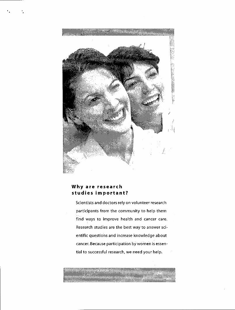
Why are researchstudies important?
Scientists and doctors rely on volunteer research
participants from the community to help them
find ways to improve health and cancer care.
Research studies are the best way to answer sci-
entific questions and increase knowledge about
cancer. Because participation by women is essen-
tial to successful research, we need your help.
<.. . .... ... ...........
What will happen if I mail in
the "Join the Network" form?
You will be sent a one-page questionnaire,
which asks about your personal and family med-
ical history, and a medical records release form
that will give researchers permission to look at
your mammography results. Later, if you are
eligible for one of the studies, you will be sent a
description of the study along with a telephone
number so that you can ask questions. Partici-
pation in a study is optional. If you choose not
to participate in a study, your contact informa-
tion remains in the network so that you can be
told about future research opportunities. Join-
ing the network does not require that you par-
ticipate in a research study, or guarantee that
you will be invited to participate in a research
study. All network members will receive a quar-
terly newsletter that provides updates on our
research studies and other news about cancers
that affect women.

4ý7

Is the information I provide
kept private?
Yes, we take extreme care and follow strict proce-
dures to keep all network and study records pri-
vate. Only authorized members of the research
team will see the information you provide. Your
information will be entered into a database that
is accessible only to study staff. All paper files
are stored in our research office, located in a
secured area of the Fred Hutchinson Cancer Re-
search Center. Network infotmation is used only
to inform women about research opportunities;
it will not be sold or distributed.
Every research activity conducted at the
Hutchinson Center is carefully reviewed and ap-
proved by the Institutional Review Board, which
protects the rights of people who participate in
research studies.
S... . :: :• •i • i• :''• •• • ... . i j • .... .

If I join, how long will I be in
the network?
Your name will remain in the network indefinitely,
even if you decide not to participate in certain
studies, unless you ask that it be removed.
Can I change my mind about
participating in the network?
Yes, you can change your mind. If you decide
that you prefer no further contact about re-
search opportunities, please call the study office
and request that your name be removed from
the network.
Are there risks or benefitsassociated with being inthe network?
Joining the network does not involve any risk or
direct benefit to you. Participation in research
studies, however, may benefit women in the fu-
ture who are at risk for cancer or who are living
with cancer.
::::::9::::: :: :: ::: :::: :::::::: :

What kinds of studies will
I learn about?
Researchers conduct many different kinds of
studies on cancers affecting women. Currently,
women from the network are being recruited for
the following studies:
VThe Breast Cancer Eadrly DiUcvery Stu,
The purpose of this study is to identify sub-
stances in blood that can be used to detect
breast cancer early in the disease process.
The goal is to develop a simple blood test that
can be used along with mammography to im-
prove the early detection of breast cancer. Eli-
gibility for this study depends on your risk for
breast cancer.
~ Oen~i Caeu• Eadly Detection St-dy.
The aim of this study is to determine if CA125
blood tests and ovarian ultrasounds are use-
ful for the early detection of ovarian cancer.
Women whose family history indicates they
may be at increased risk for developing ovar-
ian cancer are eligible for this study.
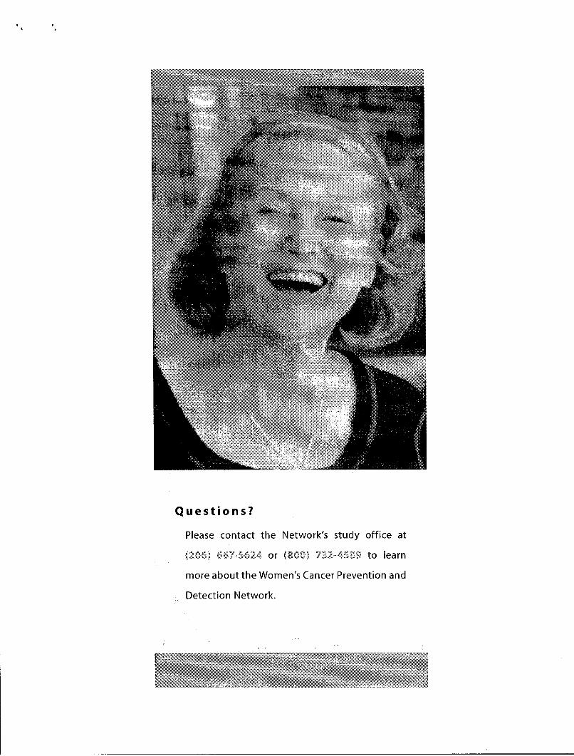
Questions?
Please contact the Network's study office at
{206} 6T or C8 0) 7:260 to learn
more about the Women's Cancer Prevention and
Detection Network.
Id FREDHUTCHINSONCANCERRESEARCHCENTER
Advancrisg Kn.ow!edgo, Sa-ing Lgei
H. FREDHUTCHINSONCANCERRESEARCHCENTER
WO•EWS CAF•C :•
PREV JT• • and DKTEI•T•ON
"Join the Network" Form
First Name
Middle Name
Last Name
Street Address _ __
City
State Zip
Date of Birth
Telephone
El Home El Work El Cell
Signature
Date ___
:: ::::: • • .. • • ••: •- . . .. 4 ' i,1 g; ... . .
E '-
•4 I0
0
z0
U
do
a'iv0
NIC
ON
E cz, :j C0 p
0cc
Appendix BFHCRC Clinical/Recruitment Protocol

Center for the Evaluation of Biomarkers for Early Detection of BreastCancer
Funded by the Department of Defense as a Breast Cancer Center of ExcellenceAward Number: DAMD 17-02-1-0691
proposal Number: BC013002
Recruitment and Specimen Collection Protocol (IR#5317)
Section Section Title Page(s)Number
1 Protocol Title 22 Phase 23 Principal Investigator, Other Investigators, and 2-3
Medical Monitor4 Locations of Study 3-45 Expected Start and Completion Dates 46 Purpose and Objectives 4-67 Study Population 6-78 Protocol Design 7
8a Eligibility Criteria 78b Subject Identification 78c Participant Approach, Enrollment, Informed Consent 7
and Specimen Collection8c.1 Consent to Contact (Mammography Cohort) 7-88c.2 Women From Other Research Study Protocols 88c.3 Invitation Letter and Consent for Annual Blood 8
Donation (Mammography Cohort)8c.4 Telephone and In-Person Approach (Women 8
Undergoing Biopsy)8c.5 Approach via Breast Surgeons (Surgical Cohort) 8-98c.6 Specimen Collection and Processing 9
8c.6.1 Blood Collection 98c.6.2 Tissue Collection 9-108c6.3 Specimen characterization 108c.6.4 Specimen Storage 108c.6.5 Repository Quality Control 108c.6.6 Repository Use 10
8d. Subject Assignment (randomization) 108e. Evaluations Prior to Entry 10-118f. Evaluations to be made during the conduct of the 11
study.8g. Clinical Assessments 118h. Research Intervention/Activity that the Participant will 11
experience9 Risk Benefit Assessment 11-1210 Reporting of Serious or Unexpected Adverse Events 1211 Description of Protocol Drugs or Devices 1212 Disposition of Data 12-1313 Modification of the Protocol 1314 Departure from the Protocol 1315 Roles and Responsibilities of Study Personnel 13-22

1. Protocol Title: Breast Cancer Early Discovery Study
2. Phase. Not applicable
3. Principal Investigator: Nicole Urban, ScDFred Hutchinson Cancer Research Center (FHCRC)1100 Fairview Avenue North, M2-B230P.O. Box 19024Seattle, WA 98109(206) 667-6771
Other Investigators:
Martin McIntosh, PhD Garnet Anderson, PhD Nancy Kiviat, MDFHCRC FHCRC Harborview Medical Center1100 Fairview Avenue North, 1100 Fairview Avenue North, Dept. of PathologyM2-B230 M3-A410 Box 359791P.O. Box 19024 P.O. Box 19024 325 Ninth Ave.Seattle, WA 98109 Seattle, WA 98109 Seattle, WA 98104206-667-4612 206-667-4699 206-731-4277
Scott Ramsey, MD, PhD Meg Mandelson, PhDFHCRC Group Health Cooperative1100 Fairview Avenue North, Center for Health StudiesM2-B230P.O. Box 19024 1730 Minor AvenueSeattle, WA 98109 Suite 1600206-667-7846 Seattle, WA 98101
206-287-2900
Allen Gown, MD Hadi Yaziji, MD Brad Nelson, PhDPhenopath Laboratories Phenopath Laboratories Deely Research Centre3000 First Avenue #1 3000 First Avenue #1 B.C. Cancer AgencySeattle, WA 98121 Seattle, WA 98121 2410 Lee Avenue206-374-9000 206-374-9000 Victoria, BC V8R 6V5 Canada
250-519-5705
Michel Schummer, PhD Ingegerd Hellstrom, MD, PhD Gordon Mills, MD, PhDMacroGenics Pacific NW Research Institute MD Anderson Cancer Center1441 N 34th St 720 Broadway 1515 Holcombe Blvd.Seattle, WA 98103 Seattle, WA 98122 Houston, TX 77030-4095206-732-1430 206-726-1209 713-792-2121
10/22/04 2BCEDS Protocol version 2.0
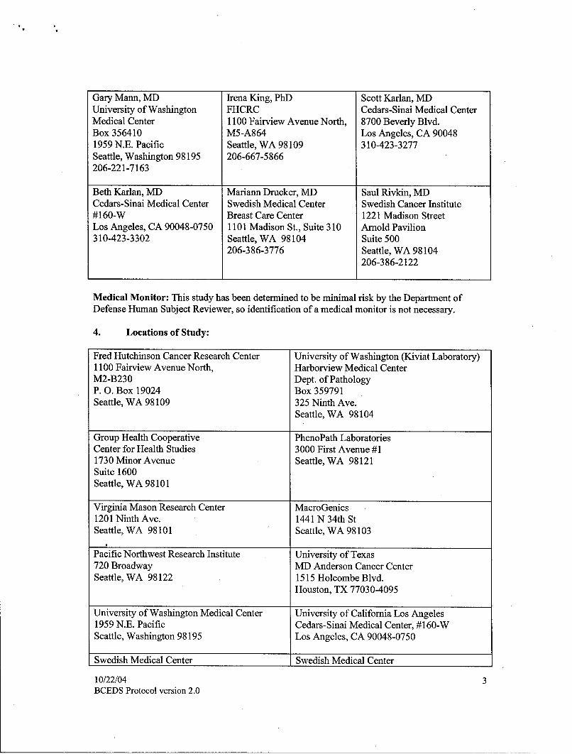
Gary Mann, MD Irena King, PhD Scott Karlan, MDUniversity of Washington FHCRC Cedars-Sinai Medical CenterMedical Center 1100 Fairview Avenue North, 8700 Beverly Blvd.Box 356410 M5-A864 Los Angeles, CA 900481959 N.E. Pacific Seattle, WA 98109 310-423-3277Seattle, Washington 98195 206-667-5866206-221-7163
Beth Karlan, MD Mariann Drucker, MD Saul Rivkin, MDCedars-Sinai Medical Center Swedish Medical Center Swedish Cancer Institute#160-W Breast Care Center 1221 Madison StreetLos Angeles, CA 90048-0750 1101 Madison St., Suite 310 Arnold Pavilion310-423-3302 Seattle, WA 98104 Suite 500
206-386-3776 Seattle, WA 98104206-386-2122
Medical Monitor: This study has been determined to be minimal risk by the Department of
Defense Human Subject Reviewer, so identification of a medical monitor is not necessary.
4. Locations of Study:
Fred Hutchinson Cancer Research Center University of Washington (Kiviat Laboratory)1100 Fairview Avenue North, Harborview Medical CenterM2-B230 Dept. of PathologyP. 0. Box 19024 Box 359791Seattle, WA 98109 325 Ninth Ave.
Seattle, WA 98104
Group Health Cooperative PhenoPath LaboratoriesCenter for Health Studies 3000 First Avenue #11730 Minor Avenue Seattle, WA 98121Suite 1600Seattle, WA 98101
Virginia Mason Research Center MacroGenics1201 Ninth Ave. 1441 N 34th StSeattle, WA 98101 Seattle, WA 98103
Pacific Northwest Research Institute University of Texas720 Broadway MD Anderson Cancer CenterSeattle, WA 98122 1515 Holcombe Blvd.
Houston, TX 77030-4095
University of Washington Medical Center University of California Los Angeles1959 N.E. Pacific Cedars-Sinai Medical Center, #160-WSeattle, Washington 98195 Los Angeles, CA 90048-0750
Swedish Medical Center Swedish Medical Center
10/22/04 3BCEDS Protocol version 2.0

Breast Care Center Drs. Beatty, Hart, Florence and Horton (Breast(Radiologist) Surgeons)
Dr. Stracener (Radiologist) Arnold Pavilion1101 Madison St., Suite 310 1221 Madison, Suite 1411Seattle, WA 98104 Seattle, WA 98104-1360PacMed ClinicsDr. Needle (Breast Surgeon)1200 12th Avenue SouthSeattle, WA 98144
5. Expected Start and Completion Dates: 10/1/2002-9/30/2006
6. Purpose and Objectives:
Purpose. The purpose of the Center is to assemble the team and the infrastructure needed toaccelerate progress in breast cancer early detection biomarker research. We will develop a uniqueresource for inter-institutional collaborative breast biomarker research, and evaluate the potentialof biomarkers detectable in serum or plasma to improve existing breast cancer early detectionstrategies. We will use the resource to evaluate the performance of candidate breast cancerbiomarkers in a cohort of women participating in mammography.
Our vision is that a simple blood test could be used in conjunction with mammography to detectall breast cancer early in the disease process. For example, one clinical scenario might work asfollows: at the time of her annual physician visit, a woman could have her blood drawn and testedfor a panel of breast cancer markers. If the mammogram were clearly positive, she would bereferred for biopsy. If the mammogram were equivocal, she would be referred to biopsy only ifthe marker panel suggested a malignancy. If the mammogram was negative, but the marker panelsuggested malignancy, she would be referred for additional imaging such as MRI. Otherscenarios may be preferable, and indeed one of the purposes of our study is to explore otherpossibilities. For example, use of the marker panel between annual mammograms might identifywomen with fast-growing tumors.
The expected result of the proposed Center is a panel of markers and decision rules for its use.The panel will be useful clinically, to improve the performance of mammography. Ourcomprehensive approach and access to an appropriate specimen repository increases theprobability of our success. The systematic evaluation of biomarkers for early detection requiresaccess to large numbers of high-quality blood samples. Cases and disease-free women are neededto evaluate the markers' ability to distinguish malignant from healthy individuals. In cases, it iscritical that blood samples are obtained prior to treatment of any kind, including surgery, becausetreatment is likely to affect marker levels. Serial specimens obtained from healthy women areneeded to evaluate the variance in marker levels within individual women over time (i.e.intraclass correlation), to establish criteria for marker positivity. Specimens from womenrepresenting the range of diagnoses (histology, grade, stage) are needed to assure the markerpanel is sensitive to all disease types. Blood samples must be collected, processed and storedidentically, as some assays are sensitive to these parameters. Because high-quality specimens arefrequently not available, investigators often rely on samples that were obtained from differentsources for cases and healthy women. They may have been obtained during remission or evenduring treatment, or processed and stored differently in ways that affect marker levels. As aresult, many markers that appear initially to be very promising prove later not to be useful, andthe development process is characterized by false starts and missed opportunities. The scientific
10/22/04 4BCEDS Protocol version 2.0

community is often skeptical about the claims made for new markers, in part because manyinitially promising results cannot be replicated in different laboratories.
Another barrier is the clinical challenge we will face if we are successful. Markers could beordered before, at the same time as, or after a mammogram, by the primary care physician, aradiologist, or the specialist who evaluates suspicious findings. Each strategy will affect thesensitivity and specificity of the combination of tests. If markers identify cancers that cannot beseen on a mammogram, clinical work up to identify the location of the tumor will be required.Cost-effective strategies will be needed. As we will obtain serum for women in several stages ofevaluation-prior to screening, just prior to biopsy, and just prior to surgery-we will be in aunique position to evaluate the clinical utility of markers at all these potential stages of screening.
Objectives. Our goal is to evaluate breast cancer biomarkers for their contribution to earlydetection of breast cancer. Subsets of breast cancer that are missed by mammography, or thatgrow too quickly to be detected in early stage by annual mammography, are of particular interest.Our aims are:
1. To validate and refine the ability of candidate biomarkers to predict disease status;
2. To evaluate panels of biomarkers for use as an adjunct to mammography, to detect allbreast cancer at a highly curable stage; and
3. To identify the molecular signatures of subsets of in situ and invasive breast cancers andexplore their associations with biomarkers in the panel.
To support the research goals we will build a unique resource for multidisciplinary, inter-institutional research on breast cancer biomarkers including
1. Blood samples obtained annually and processed identically in women with and withoutbreast cancer,
2. Fresh tissue matched to blood samples on a subset of the women with breast cancer,
3. Epidemiological, clinical and follow-up information for women who donate specimens,and
4. A system to facilitate use of the specimens, including state-of-the-art informationsystems.
We will develop a specimen resource from a well-characterized population, with associated riskfactor information, mammography findings and follow-up data on cancer outcomes. It willinclude blood samples obtained from selected women who have mammograms, biopsy or breastcancer surgery over the 4-year period of the grant. We will evaluate candidate biomarkers fortheir ability to distinguish among women with healthy breasts and women with various breastconditions, including invasive ductal and lobular carcinoma, lobular carcinoma in situ, comedo-and noncomedo-type DCIS, hyperplasia and other potentially premalignant conditions, andbenign conditions. We will evaluate the role of biomarkers detectable in serum or plasma inimproving our current breast cancer early detection strategies.
For selected women, we will explore the feasibility of collection and analysis of fresh-frozentissue in order to characterize malignant conditions at the molecular level. This will allow us tocorrelate biomarkers included in the panel with subsets of breast tumors identified throughmolecular profiling.
Our hypotheses are:
(a) Used alone, biomarkers detectable in blood products can detect subsets of, but not all,breast cancer;
10/22/04 5BCEDS Protocol version 2.0
(b) Use of a breast cancer biomarker panel can improve the performance of mammography inearly detection of breast cancer.
7. Study Population.
The study population includes three cohorts defined by recruitment source. The first is theapproximately 8,600 women who obtain mammograms and undergo biopsies at the SwedishMedical Center Breast Care Center each year. All women seen for mammography at SwedishBreast Care Center (SBCC) will be asked to join the Women's Cancer Prevention and DetectionNetwork by signing a Consent to be Contacted for Future Research Studies (Join the NetworkForm) for potential participation in women's cancer research. The Consent to be Contacted formprovides permission for the research study to maintain the woman's name and contactinformation indefmitely so that she may be invited to future studies. Women who sign and returnthe consent to contact at the SBCC or other participating facilities will be mailed the Women'sCancer Prevention and Detection Network Questionnaire, the Women's Cancer Preventionand Detection Network Release Form, and a Women's Cancer Prevention and DetectionNetwork Cover Letter asking them to complete the questionnaire and/or release form and returnthem to the study office in the stamped, self-addressed envelope provided. Women who return thecompleted questionnaire and/or the Release Form will be sent a Thank You Letter. Women whoare recruited at the SBCC or any other Swedish facilities will not be approached for futureparticipation in other research studies unless those studies have first been approved by theSwedish Cancer Institute Research Steering Committee.
Every 3 months, data describing mammogram findings will be submitted to FHCRC inaccordance with Mammography Tumor Registry procedures (FHCRC IR#3636). For womenundergoing mammograms, data from the Network Questionnaire will be used to classify womenwith respect to risk status. If a Release Form is on file, mammogram findings (assessment codes,follow up recommendation, breast density) will also be used to determine risk status for anindividual woman.
A stratified random sample of women who participate in the Network will be identified and askedto provide blood samples annually at the time of subsequent mammograms. Women at high riskwill be over-sampled to increase the expected number of women with sequential blood samplesobtained prior to a breast cancer diagnosis. As shown in Table 1, 500 (375 high-risk and 125average-risk) women will be enrolled in Year 1, and 100 additional women will be enrolled insubsequent years.
Women who have completed other research study protocols and have given permission to becontacted about future research opportunities will also be invited to participate in the COE byproviding annual blood samples at the time of their mammograms. These women will beapproached about potential COE study participation in person or by mail.(see section 8c.2)
In addition, all women undergoing biopsies will be invited to provide a blood specimen prior tobiopsy and annually at the time of subsequent mammograms. 100 women undergoing biopsieswill be enrolled annually. Women identified and enrolled through the SBCC, and women invitedto participate who were identified from other research study protocols, are referred to as theMammography Cohort (MC).
The second cohort includes women undergoing breast surgery at Swedish Medical Center. Thesecond recruitment source is the approximately 650 women who have surgery for breast cancer atSMC each year. Collaborating surgeons will identify those most likely to have tumors of size > 2cm and therefore to be candidates for tissue collection. In 1999 there were 124 women who hadsurgery for breast tumors over 2 cm at SMC. These women will be sampled with probability 1.
10/22/04 6BCEDS Protocol version 2.0

Remaining women will be sampled with lower probability to yield desired numbers in the cohort.Based on our experience in the ovarian SPORE, we expect that tissue collection will occur inabout 50% of the women sampled. As shown in Table 1, we expect to enroll 25 women in Year 1and 50 women annually thereafter for donation of both blood and tissue. Fresh tissue specimenswill be obtained when it is logistically and ethically appropriate. In addition we expect to enroll100 women annually beginning in year 2 for donation of blood only. Blood samples will beobtained just prior to or at the time of the surgical procedure. Women identified and enrolledthrough our collaborations with SMC surgeons are referred to as the Surgical Cohort (SC).Following surgery and treatment, women in the SC will be invited to participate in the MC,providing blood samples at the time of their annual mammograms.
The third recruitment source is women undergoing biopsy by Mammotome® at Cedars SinaiHospital. Approximately 2,500 women undergo this procedure annually. Of these we will enroll50 per year, including women with benign lesions, hyperplasia, and in situ disease as well aswomen with invasive carcinoma. As at SMC, we expect about 20% to be malignant. Both fresh-frozen tissue and blood samples will be donated by women in the Cedars biopsy cohort.Procedures at Cedars Sinai are described in a separate document, which is currently beingfinalized and reviewed by the local IRB and will be forwarded to DOD for review once local IRBapproval is in place. We will forward the updated Cedars documentation as soon as possible.
Table 1. Unique ParticipantsYr 01 Yr 02 Yr 03 Yr 04 Total
Mammography Cohort - High Risk 375 75 75 75 600Mammography Cohort - Average Risk 125 25 25 25 200Mammography Cohort - Biopsy 100 100 100 100 400Subtoal: Mammography Cohort .600 200 200 200 1200SMC Surgical Cohort: Blood and Tissue 25 50 50 50 175SMC Surgical Cohort: Blood Only+ 100 100 100 300Subtotal: Surgical Cohort 25 150 150 150 475Cedars Biopsy Cohort: Blood and Tissue 50 50 50 50 200Total 675 400 400 400 1875
8. Protocol Design. This protocol outlines the methods for a study to collect blood andtissue specimens from women undergoing mammography and breast-related biopsy and surgeryat Swedish Medical Center. Patient approach procedures to be used at Cedars Sinai MedicalCenter are addressed in a separate document. The study uses the already-establishedinfrastructure in place for the Mammography Tumor Registry Study (MTR). The purpose of thestudy is to provide a specimen resource to support both breast and ovarian cancer biomarkerevaluation. Investigators propose to build a unique resource for multidisciplinary, inter-institutional research on cancer biomarkers including blood samples obtained annually andprocessed identically in women with and without breast cancer, fresh tissue matched to bloodsamples on a subset of the women with breast cancer, epidemiological, clinical and follow-upinformation for women who donate specimens, and a system to facilitate use of the specimens,including state-of-the-art information systems. Collaboration with mammography facilities andsurgeons offices is planned to recruit women undergoing screening, biopsy and surgery.
8a. Eligibility Criteria. All women 18+ years of age undergoing mammography or biopsy atthe SBCC are potential participants for this research. Women identified and enrolled through theSBCC, including women having both mammograms and biopsies, are referred to as theMammography Cohort (MC). All women 18+ years of age scheduled for breast surgery by
10/22/04 7BCEDS Protocol version 2.0
participating surgeons are potential participants for this research. These women are referred to asthe surgical cohort (SC).
Mammography Cohort Risk Stratification. Women having screening mammograms will beinvited to complete the Cancer Research Registry Questionnaire and the Cancer ResearchRegistry Mammography Results Release form. Of those who respond, study investigators willselect a stratified random sample and invite those women to donate blood. Risk stratification willbe based on epidemiological data provided on the questionnaire, and on mammogram findings (ifthe woman gave permission to have these reviewed), as follows:
0 A woman will be considered average risk if her mammogram result was assessmentcode 1 or 2.
a If mammogram results are unknown, a woman will be considered average risk if shedoes not meet any of the high risk criteria specified below
0 Women will be classified as high risk if any of the following apply:1. She is referred to biopsy (assessment code 4 or 5); or
2. The family contains at least two ovarian or breast cancers among the subjectand first and second degree relatives of the subject within the same lineage.This condition is satisfied by multiple primary cancers in the same person,where breast cancer is required to meet this criterion, at least one breastcancer must be pre-menopausal (age at diagnosis less than or equal to 50 ifage at menopause is unknown).
3. She is of Ashkenazi Jewish ethnicity with one first-degree or two second-degree relatives with breast or ovarian cancer, or subject is of Ashkenaziancestry and has had breast cancer. Where breast cancer is required to meetthis criterion, at least one breast cancer must be pre-menopausal (age atdiagnosis less than or equal to 50 if age at menopause is unknown).
4. She presents with suspicious mammogram findings (assessment code 3), orhas experienced symptoms.
8b. Subject Identification. Potential participants in this study will be identified at theSwedish Medical Center Breast Care Center (SBCC) or through their participation in otherresearch study protocols. The SBCC mammography practice already participates in theMammography Tumor Registry (IR File #3636), which means they provide regular downloads ofmammography data that are routinely linked to the Cancer Surveillance System. The registry isused to support breast cancer research and to provide regular performance reports to participatingradiologists. All aspects of the linkage process, including extensive confidentiality procedures,are covered in IR file #3636 and are not discussed here.
8c. Participant Approach, Enrollment, Informed Consent and Specimen Collection.Approach processes are tailored to participant type. All women receiving screeningmammograms are asked to sign a consent for future contact. Women receiving biopsiesare approached prior to the biopsy procedure. Women from other study protocols areeither approached in person at a study appointment or by mail. Women in the surgicalcohort are approached at the pre-surgery appointment. All women who consent toparticipate in the initial blood collection are invited to donate blood at their subsequentmammogram appointment (regardless of cohort).
10/22/04 8BCEDS Protocol version 2.0

8c.1 Consent to Contact (Mammography Cohort). All women seen for mammography atSBCC will be asked to join the Women's Cancer Prevention and Detection Network by signing aConsent to be Contacted for Future Research Studies for potential participation in women'scancer research. Additional risk factor information will be collected using the Women's CancerPrevention and Detection Network Questionnaire, which will be mailed to the participant aftershe has completed the Consent to be Contacted form. This mailing will also include a HIPAAcompliant medical records release (Women's Cancer Prevention and Detection Network Release),which will allow access only to her mammography records. A broader medical records releasewill be obtained from women donating specimens, as described in Section 8c2. Within 3 monthsfollowing the mammogram, data describing mammogram findings will be submitted to FHCRCin accordance with MTR procedures. For women undergoing mammograms, data from theNetwork Questionnaire in combination with mammogram findings (assessment codes, follow-uprecommendation, breast density) will be used to classify women with respect to risk status. Astratified random sample of these women will be invited to donate blood. In cases wheremammography data are not available, women will be selected based only on data provided on theWomen's Cancer Prevention and Detection Network Questionnaire.
8c.2 Women From Other Research Study Protocols
Women who have completed other research protocols, and who have given permission to becontacted about future research opportunities will be approached to participate in the COE. Thesewomen may be approached one of two ways. One, they may be approached in person by studystaff during their final study appointment and given the COE invitation packet (containinginvitation letter, study brochure, COE consent form and medical records release) plus a smallincentive item (decorative magnet, Athena Water coupon, etc.). Women will be asked to take thepacket home where materials can be reviewed at their convenience. A self addressed stampedenvelope will be included in the packet, so that signed consent forms can be returned to the studyoffice. Two, women who have already had their final study appointment will be approached bymail, as described below in section 8c.3.
This group of women will receive an invitation letter appropriately tailored for their population.Several studies conducted by Dr. Urban's group use the same data collection instruments (ex.Baseline questionnaire, Health Status Questionnaire). Accordingly, women who are currentlyparticipating in our research studies and represent a potential recruitment source for the COE willnot be asked to complete the same instruments twice.
Women who return their signed consent form will receive a COE Invitation Thank You Letter(currently under development). The thank you letter will inform study participants thatstudy staffwill call them 1-2 months prior to their mammogram to schedule their first COE blood drawappointment.
8c.3 Invitation Letter and Consent for Annual Blood Donation (Mammography Cohort)Women selected for approach will be contacted by mail several months prior to the nextmammogram appointment, using the Study Brochure, and the Invitation Letter. The letter willdescribe the study and invite interested women to return a signed Consent to Participate in theBreast Cancer Early Discovery Study for Women Receiving Mammograms and a MedicalRecords Release Form. This medical records release is broader than the one previouslyobtained, and allows general access to a woman's medical records so that the study may obtainhistory of and treatment of cancer. Upon receipt of the signed consent form and medical records10/22/04 9BCEDS Protocol version 2.0

release form, the study office will call women to schedule their first blood draw appointment. Ifwe are unable to reach a woman by phone, she will be sent the Not Able to Contact By PhoneLetter, which requests that she call the study office if she would like to participate. If we do notreceive any materials or hear from a potential participant within 3 weeks of sending the invitationpacket, study staff will call the woman once to follow up with her regarding the study.
Once an appointment has been made, the woman will be sent a Blood Draw AppointmentConfirmation Letter with directions (map) to the blood draw site. All women will have theirblood drawn at the Swedish Breast Care Center on the Swedish Medical Center campus unlessthey are patients at the Swedish Cancer Institute (SCI) and request to have their blood drawn atSCI. If this is the case, study staff will arrange to meet the participant at SCI for her appointmentand administer the study questionnaires. At her first blood draw appointment, the participant willcomplete a Health Status Questionnaire. This questionnaire asks about current healthinformation at the time of the draw (ex. Medications). She will also be given a BaselineQuestionnaire to either complete at her appointment or take home. If she chooses to completethe Baseline Questionnaire at home, she will be given a stamped, self-addressed envelope toreturn it to the study office. Information from the Baseline Questionnaire will be used to confirminitial risk status and to determine if a woman is at high risk for breast or ovarian cancer based onfamily history. If a participant does not return the Baseline Questionnaire within 3 weeks, she willbe sent a Reminder Card and another copy of the questionnaire. If the study office does notreceive the questionnaire or hear from the participant 2 weeks after the card was sent, she willreceive one follow up call from study staff.
For subsequent blood draws, the study office will send the Annual Blood Draw Reminder Letterone month prior to the next draw and schedule an appointment over the phone. If we cannotreach a woman by phone to schedule her annual appointments, we will send her the AnnualDraw Not Able to Contact by Phone Letter, which requests that she call the study office withher updated contact information. Once the appointment has been made, the woman will be sent aBlood Draw Confirmation Letter. At each draw she will receive a Health Status Updatequestionnaire to complete during her appointment. This questionnaire asks for updated healthinformation. If possible, one draw each year will occur at the time of subsequent mammograms tomake it more convenient for participants who receive mammograms at the SBCC. We willprovide parking validation for each visit including the blood draw visit that coincides with theannual mammogram.
8c.4 Telephone and In-Person Approach (Women Undergoing Biopsy) All womenundergoing biopsies will be invited to provide a blood specimen prior to biopsy and annually atthe time of subsequent mammograms. Consent for specimen donation will take place during thebiopsy appointment, prior to the biopsy. The SBCC Scheduling Nurse routinely contacts bytelephone all women undergoing stereotactic or in some cases ultrasound-guided biopsy 2-3 daysprior to the appointment. During this phone call, the scheduling nurse will obtain verbal consentfor study staff to contact the woman by phone to discuss study participation. If a woman providesverbal consent, the SBCC nurse will fax the study office the Biopsy Flowsheet including thewoman's name and the date and time of the biopsy appointment to the confidential fax at thestudy office. The Scheduling Nurse will check a box that indicates that the woman has givenverbal consent to be contacted about study participation. Study staff will then telephone theparticipant to describe the study and go through the consent materials. Study staff will carefullygo over the Consent to Participate in the Breast Cancer Early Discovery Study for WomenHaving Breast Biopsies, Medical Records Release Form, and study questionnaires with thewoman. Study staff will also inform the woman that the attending SBCC biopsy nurse will draw
10/22/04 10BCEDS Protocol version 2.0

their blood and obtain written consent. Again, only women undergoing pre-scheduled stereotacticor ultrasound guided biopsies will be approached in this manner, women undergoing biopsies onthe same day as a suspicious mammogram will not be approached for this study. This process isdescribed in the Biopsy Consent Procedure Telephone Script.
The SBCC nurse who attends the biopsy appointment will encourage the woman to ask anyquestions she may have about participation, and to discuss participation with any family memberswho are present. The consent process will take place in a private consultation or examinationroom in the breast care center. Clinical staff at SBCC will serve as witnesses to the informedconsent process. If the woman asks a question which the attending biopsy nurse cannot answer,study staff will be available to go to the SBCC and address these questions. If the woman decidesto participate, the attending biopsy nurse will obtain written informed consent for the blooddonation using the Consent for Data Collection and Blood Donation prior to Biopsy form anddraw blood from the participant prior to the procedure. A copy of the consent form will beprovided to the participant. To allow the study to characterize the donated blood specimens byobtaining the pathology results, women will be asked to complete a Medical Records ReleaseForm. If possible, to obtain information about the woman's health at the time of her blood draw,she will be asked to complete a short Health Status Questionnaire before her draw. If this is notfeasible, she will be asked to complete the questionnaire as soon as possible after her draw. Allconsented participants will be provided the Baseline Questionnaire, which they may completeimmediately or return by mail.
If a participant does not return the Baseline Questionnaire within 3 weeks after her blood draw,she will be sent a Reminder Card and another copy of the questionnaire. If the study office doesnot receive the questionnaire or hear from the participant 2 weeks after the card was sent, studystaff will conduct one follow-up call.
Women undergoing biopsy will also be asked to provide blood specimens annually with theirsubsequent mammograms, regardless of the outcome of their biopsy, using the Reminder Letteras described in Section 8c3. At each subsequent draw they will also be asked to complete theHealth Status Update Questionnaire.
8c.5 Approach via Breast Surgeons (Surgical Cohort). Several breast surgeons at SwedishMedical Center have agreed to identify patients that are likely candidates for surgical specimencollection. Women selected for approach based on the surgical schedule will be approached at thepre-surgical appointment for consent to obtain specimens. The informed consent will beconducted by a study Specimen Collection Specialist (SCS) or the Research Nurse. Women willbe asked to sign a Consent for Data Collection and Specimen Donation At Surgery allowingpermission to obtain a blood sample prior to surgery, and to donation of breast cancer tissueshould there be excess tissue available. Women will also be asked to sign a Medical RecordsRelease Form, which allows researchers to access their surgical pathology report and otherrecords related to their cancer diagnosis. This is the procedure that we have used successfully toobtain blood samples and tissue from women undergoing surgery for suspected ovarian cancer atSMC. We will obtain also consent to obtain tissue blocks, which are stored for 10 years at SMCand made available routinely to investigators at the FHCRC.
8c.6 Specimen Collection and Processing
8c.6.1 Blood Collection. In all blood collections, the Specimen Collection Specialist will collectup to 50 ml of whole blood in one 10ml yellow-top tube, two 10 ml red-top tubes and twopotassium EDTA 10 ml (purple-top) tubes. The volume of blood drawn may be adjusted
10/22/04 11BCEDS Protocol version 2.0
downward as necessary. Tubes will be stored in the refrigerator and processed within 48 hours ofreceipt. Standard protocols will be followed to process specimens into sera and plasma and toaliquot into cryovials uniquely labeled with study specimen ids. Blood specimens will be storedin very small quantities to avoid freeze-thaw cycles and re-aliquotting. Aliquoted specimens willbe transported to the study repository for long-term storage or delivered to laboratoryinvestigators. Times of blood draw, processing and freezing will be recorded.
8c.6.2 Tissue Collection A log of scheduled surgeries for potential tissue donors will bemaintained so that the SCS can be present at the surgery. Immediately after the surgeon hasremoved the necessary tissue and the pathologist has taken what is required for pathologicdiagnosis, the SCS will be permitted to collect specimens from the removed tissue for thepurposes of the study. When clinically appropriate, a minimum of two and a maximum of ten1.Oml cryovials, each holding 1 gram, 2cc, or 20mm of breast tissue, will be collected for thestudy. When feasible, the optimal tissue quantity per case is 6 grams. Tissue collected willinclude malignant tissue and, if possible, adjacent normal tissue.
During the entire surgical procedure, the treating surgeon and the surgical team will beresponsible for monitoring the patient's condition. The surgeon will ultimately remove a surgicalspecimen and give this to the pathologist. The pathologist will then identify tissue that will notbe needed for pathologic diagnosis. All or part of the un-needed tissue will be collected andplaced in the labeled cryovials and processed for storage. The pathologist may refuse to provideany specimen for research if they believe that the entire specimen is needed for the subject'sclinical care.
The tissue will be snap-frozen, placed into cryovial(s), and stored at -80 degrees C.
8c6.3 Specimen characterization. For all cases with fresh tissue, Dr. Kiviat will performhistology review.
8c.6.4 Specimen Storage_ Each specimen will be labeled with a unique 6-digit numeric label. Aduplicate of this label is affixed to a specimen transmittal form identifying the specimen type andparticipant number, and logged into the database. Examples of these specimen transmittal formsare presented as Instruments 9 and 10. Blood specimens will be stored in a -70' C freezer, andtissue specimens will be stored in a liquid nitrogen freezer.
8c.6.5 Repository Quality Control. A rigorous quality assurance plan is already in place thatincludes monitoring and maintenance checks of equipment, an eight-hour C02 back-up systemand a temperature-sensitive alarm system that alerts the building maintenance staff when theinterior temperature reaches a designated temperature.
8c.6.6 Repository Use. A Specimen Review Committee described in the Communication sectionwill oversee use of specimens in the resource. Advocates will participate in this committee.
8d. Subject Assignment (randomization): Not applicable.
8e. Evaluations Prior to Entry. No clinical evaluations are made prior to entry into thestudy. If a woman has given us permission to access her mammography records and/or completedand signed the Network Questionnaire, mammogram findings (assessment codes, follow-uprecommendation, breast density) and/or family history information from the questionnaire areused to classify women with respect to risk. The mammogram findings are obtained throughprocedures outlined in the Mammography Tumor Registry (IR#3636). A stratified randomsample of women in the mammography cohort are invited to donate blood annually at subsequentmammograms, as described in Section 7.10/22/04 12BCEDS Protocol version 2.0
8f. Evaluations to be made during the conduct.of the study. Breast cancer case status willbe ascertained on all women using the MTR system of linking to CSS/SEER and WashingtonState Cancer Registry Data, which include date of diagnosis, stage and grade of disease,histology, age, and race/ethnicity, in addition to individual identifiers. Linkage with high-qualitycancer registry data provides passive follow-up for cancer outcomes, including survival. ACertificate of Confidentiality was obtained to protect the linked data from subpoena. These 2procedures are outlined in detail in IR#3636.
8g. Clinical Assessments. Pathology reports will be obtained routinely for womendiagnosed with cancer at SMC to obtain day of diagnosis (CSS records only the month and year)for women in the SC and the subset of women in the MC who have undergone biopsy. Thisinformation will be collected using the Patient Pathology at Diagnosis Form (currently underdevelopment) Additional clinical data will be collected using the Clinical Status Form (currentlyunder development). Other data collection instruments include the Cancer Research RegistryQuestionnaire, the Baseline Questionnaire, the Health Status Questionnaire, the HealthStatus Update which are attached to this protocol.
8g.1 Baseline Questionnaire Reliability and Validity
In an effort to ensure high quality data and to maintain common data elements, investigators havedecided to use a Baseline Questionnaire that they are already using in collaborative work in thenational Risk of Ovarian Cancer (ROCA) screening trial. Although significant statistical andclinical expertise were involved in questionnaire development, the CGN questionnaire has notbeen formally tested for validity and reliability. Investigators agree that it is not feasible to assessthe validity of the questionnaire because of the substantial personnel effort that would be requiredto review source documentation such as medical records.
Investigators do plan to plan to assess the reliability of selected measures on the questionnaire, bycomparing the responses to variables that are captured on both the Baseline Questionnaire(completed at baseline) to the Health Status Questionnaire (completed at each blood draw). Dr.Garnet Anderson will oversee the design and conduct of a reliability assessment for the BaselineQuestionnaire. Dr. Anderson is an investigator at the Women's Health Initiative (WHI) statisticalcoordinating center, and has significant expertise in this area. WHI investigators have recentlycompleted a study assessing the reliability of selected baseline measures in the WHIObservational Study (Ann Epidemiol 2003; 13:1-15, in press). This study observed that mostdemographic factors, reproductive variables and family medical history were reliably reported,with kappa or weighted kappa above 0.8. Most of the self-reported medical conditions yieldedkappa above 0.75. Investigators anticipate that the Breast Cancer Early Discovery study willobserve similar reliability using the Baseline Questionnaire, since many of the measures are thesimilar to those used in the WHI baseline questionnaires.
8h. Research Intervention/Activity that the Participant will experience
Consent for Contact. Women undergoing mammography will be asked to sign a Consent to beContacted for Future Research Studies (Join the Network Form), which means that they maybe contacted in the future to donate blood for this study, or they may be contacted about otherstudies for which they are potential candidates. An example of another study that may contact10/22/04 13BCEDS Protocol version 2.0

these participants is the Ovarian Cancer Early Detection Study, which is recruiting high-riskwomen to participate in an ovarian cancer screening protocol (IR file #5159).
Screening Questionnaire. To ensure consistency in data collection across sites, the Women'sCancer Prevention and Detection Network Questionnaire will be used at all locations,including the SBCC. This one-page screening questionnaire is used to collect preliminary medicaland family history data. A copy of the questionnaire, along with a HIPAA compliant MedicalRecords Release (Network Release) will be mailed to women after they return the Consent to beContacted for Future Research Studies.
Annual Blood Donation. Selected women among those who have undergone screeningmammography and consented for future contact will approached and requested to donate blood atthe time of their next annual mammogram and annually thereafter. Women will be approachedand consented by mail, as described in section 8c.2. Women who donate blood during biopsy,and well as those in the surgical cohort will also be invited to donate blood annually at the time ofsubsequent mammograms. All women who donate blood will be asked to complete a HealthStatus Questionnaire at the time of their first blood draw, and a Health Status Update at eachsubsequent draw.
Blood Donation at the Time of Biopsy. Women undergoing biopsy at SBCC will be asked todonate blood specimens at the time of the biopsy. They will also donate blood annuallythereafter, at the time of subsequent mammograms.
Blood and Tissue Donation at the Time of Surgery. Women undergoing breast surgery will beinvited to donate blood and tissue during surgery. Fresh tissue specimens will be obtained when itis logistically and ethically appropriate. Blood samples will be obtained just prior to or at the timeof the surgical procedure. They will also donate blood annually thereafter, at the time ofsubsequent mammograms.
9. Risk/Benefit Assessment
The results of this research will be used to develop ways to find breast cancer early. Specifically,the study aims to develop a panel of biomarkers than can be used in conjunction withmammography to improve its performance. Although there is no direct benefit to womenparticipating in this study, there may be benefits for other women in the future who are at risk forbreast cancer or who are living with breast cancer.
Risks to participants including the potential of feeling uncomfortable due to answering personalquestions on study questionnaires. Risks associated with blood draw include the possibility oftemporary discomfort or a bruise at the site of the needle puncture. Donation of blood and tissueduring surgery will not present any additional risks.
10. Reporting of Serious or Unexpected Adverse Events
Adverse events are expected to be rare on this study. Study personnel will have direct exposureto participants only for a short period of time (eg during the consent and specimen collectionprocess), and will not be monitoring participant's health as they would for a clinical treatmentprotocol. Therefore, study participants will be asked to report any adverse events that theyexperience that appear to be related to study participation. This will be done by calling theResearch Nurse at: 206-215-6209 or 1-800-328-1124.
An adverse event is any event that occurs during the study, which results in the subjectexperiencing a new symptom or worsening of an existing symptom. For example, fainting or lossof cognitive function after a blood draw would be considered an adverse event.
10/22/04 14BCEDS Protocol version 2.0

A serious adverse event is an event that results in any of the following outcomes:1. Death2. Life threatening event3. Inpatient hospitalization*4. Persistent or significant disability or incapacity
*Patients undergoing surgery for breast cancer will be approached to donate blood and tissuefor the study. Hospitalization associated with this already-planned surgery will not beconsidered an adverse event.
Adverse events will be reported to the FHCRC Institutional Review Office (IRO) at the time ofannual review for the study. Adverse events that are both serious and unexpected will beimmediately (upon the study office notified) reported by telephone to the FHCRC IRO and bytelephone to the USAMRMC, Deputy for Regulatory Compliance and Quality (301-619-2165;FAX 301-619-7803). A written report will follow the initial telephone call within 3 workingdays. The written report will be addressed to:
U.S. Army Medical Research and Materiel CommandATrN: MCMR-RCQ504 Scott StreetFort Detrick, Maryland 21702-5012
11. Description of Protocol Drugs or Devices: Not applicable.
12. Disposition of Data. All participant materials such as survey responses will be kept inlocked cabinets and accessed only by study personnel who have signed a confidentialitypledge. All data will be reported in aggregate format, and will not identify specificparticipants in any way.
All study materials are stored in locked filing cabinets accessible to studypersonnel who have signed a confidentiality pledge. All data is stored on asecure network server. Data entry systems are password protected. Access todata files is limited to those study personnel who manage data as part of their jobresponsibilities and who have signed the confidentiality pledge.
It should be noted that representatives of the U.S. Army Medical Research andMateriel Command are eligible to review research records as a part of theirresponsibility to protect human subjects in research.
13. Modification of the Protocol. Any modification to the protocol will follow establishedprotocol modification procedure of the FHCRC Internal Review Office and the HSRRBfor review and approval, which include detailed submission of any changes or additions.
14. Departure from the Protocol. If the study investigators or staff become aware of anydeparture from the protocol, the FHCRC Internal Review office will be notified inwriting as well as HSRRB office.
15. Roles and Responsibilities of Study Personnel.
Nicole Urban, ScD, Principal Investigator (20% FTE) Dr. Urban will be responsible for overallcoordination of logistics, facilitation of progress of the scientific teams, and communication atboth the investigator and staff level, overall design and conduct of all research activities, andinterdisciplinary leadership for the project. She is currently the Principal Investigator for several10/22/04 15BCEDS Protocol version 2.0
interdisciplinary projects involving scientists from molecular biotechnology and clinicalimmunology disciplines. She will provide the scientific leadership and framework for theevaluation and application of biomarkers for breast cancer screening. Dr. Urban is responsiblefor coordination of inter-institutional activities, as well as all activities conducted by investigatorsand staff at FHCRC, including statistical work on development and validation of the markerpanel, and coordination of all recruitment, tracking and specimen collection activities. She willwork closely with the statisticians and laboratory scientists in carrying out the proposed workplanand ensuring that study objectives are met. Dr. Urban is a recognized leader in interdisciplinarycancer screening research in the Seattle area with the ability to successfully integrate clinical,basic and public health investigators into a unified research team. She will supervise the ProjectManager and meet regularly with all Center Investigators to monitor progress and ensure thatinterim tasks are completed in a timely manner. Dr. Urban will chair the All- Investigatormeetings, co-chair the Interdisciplinary Working Group and participate in the Specimen ReviewCommittee.
Lisa Isozaki, Project Manager (100% FTE) Ms. Isozaki will coordinate all activities of theCenter at the staff level and supervise the support staff. She will work closely with projectinvestigators to provide overall coordination for the study, and specifically be responsible formanaging the relationship between the clinical, laboratory, and statistical components of thestudy to ensure interim and final deadlines are met. Ms. Isozakiwill develop timelines with cleardeliverables that integrate the activities of the laboratory scientists with the statistical scientistsand project leadership. She will coordinate communication between study staff and investigatorsby soliciting regular written updates from the Informatics Manager and Research Nurse whomshe will directly supervise. Ms. Isozaki will develop agendas and facilitate staff meetings andinvestigator meetings. She will provide leadership at the staff level for all study staff, and willdirectly supervise Kate Watabayashi , Program Assistant. With the assistance of key staff, Ms.Isozaki will compile complete documentation of detailed protocols for participant recruitment andretention, data management, and specimen repository management. Ms. Isozakiwill oversee allthe administrative activities of the study. Ms. Isozaki will be responsible for all projectmanagement activities such as budget preparation and monitoring, managing logistics ofsubcontractor relationships, human subjects applications including the necessary cooperative filesat subcontractors institutions, facilitating investigator meetings and incorporating investigatordecisions into the staff workplan.
Kate Watabayashi, Program Assistant (100% FTE) will assist the Project Manager in the overallcoordination of research activities. She will arrange staff and investigator meetings, prepareminutes, perform literature searches, conduct word processing/desktop publishing of scientificpresentations and manuscripts arising from the study, coordinate investigator travel, and provideoverall staff support to the project. Ms. Watabayashi will assist with budget monitoring andsecuring subcontracts in a timely manner. She will be responsible for all purchasing for thestudy, including office supplies, laboratory supplies and equipment. Ms. Watabayashi willcoordinate all human subjects applications and modifications including oversight of allnecessary cooperative files at subcontractors institutions. Ms. Watabayashi will assist theClinical and Informatics Core staff with participant mailings and development of dataentry and study tracking systemsas needed.
AdvocatesShannon Marsh, Advocate (25% FTE) Ms. Marsh will coordinate advocate involvement in theresearch program. She will chair patient advocacy meetings and facilitate ongoingcommunication among advocates and between the advocate group and study investigators. Shewill also play a central policy role in evaluating the risks and potential benefits of this research,
10/22/04 16BCEDS Protocol version 2.0

and in representing her assessment to the Institutional Review Board (IRB). She will attendInstitutional Review Board (IRB) meetings when the study is discussed to educate the IRB aboutthe overall goals of the study, and to provide information about patient approach procedures andthe confidentiality and privacy protections in place. In addition, Ms. Marsh will work closelywith Drs. Ramsey, andDrucker and Ms. Gough to address consideration of the clinical utility ofthe marker panel. Ms. Marsh will attend regular scientific meetings including All-Investigatormeetings, Specimen Review Committee meetings, and Interdisciplinary Working GroupMeetings. She will represent the research study at local and national advocacy meetings.
Julia Cafias. Julia Cafias will be responsible for coordinating minority recruitment. Ms. Cafiaswill work through established contacts to disseminate study information and will collaborate withShin-Ping Tu, a UW investigator responsible for minority recruitment for the ovarian SPORE, todevelop culturally appropriate approach protocols for minority populations.
Mona Bailey. Mona Bailey will be responsible for educational outreach. Ms. Bailey will workwith Ms. Cafias to develop a study brochure specifically targeting minority groups. She will alsocoordinate the development and implementation of educational outreach programs geared towardthe African American, Asian, and Hispanic communities.
Barbara Bridge. Barbara Bridge will be responsible for development of patient retentionmaterials, and programs to empower breast cancer survivors to be active participants in a researchprogram. Ms. Bridge will work with study investigators and clinical staff on creating survivor-to-survivor letters and reminder cards. She will also coordinate efforts to develop a study brochureand an advocacy newsletter.
ScientistsMartin McIntosh, PhD, Statistician (20% FTE) will be responsible for the data that describes thelaboratory analyses and for the data analysis relating to the validation of markers that may beadded to a breast biomarker panel. He will work closely with Ms. Guay to develop analysis setsfor use by the investigators. He will work with the other investigators to solve methodologicalproblems associated with collected variables and data analysis. Dr. McIntosh will collaboratewith Drs. Urban and Anderson in this methodological work. He will also participate inmanuscript and report preparation.
Garnet Anderson, PhD, Statistician (10% FTE) Dr. Anderson is responsible for the PatientRegistry, including epidemiological, clinical and follow-up information about the women. Shewill collaborate with Drs. Mandelson and Clarfeld to develop risk assessment and samplingmeasures that incorporate breast density, mammography outcome, pedigree and other risk factordata, and participate in the development of study questionnaires and other data collectioninstruments. Dr. Anderson will provide scientific leadership to Shirley Gough in the managementof patient contact, data collection and the patient tracking system. She will develop statisticalmethods for measuring risk using data available from consenting women and associatedmammography data. She will work closely with Dr. McIntosh and will coordinate with otherinvestigators in the development and evaluation of potential marker panels resulting from theassays performed on the Panel Validation Set. Together with Dr. Kiviat, Dr. Anderson with co-chair the Specimen Review Committee. Dr. Anderson will participate in manuscript and reportpreparation.
Nancy Kiviat, MD. Pathologist (10% FTE) Dr. Kiviat will serve as co-PI, responsible for theSpecimen Repository. She will be supported by Ms. O'Briant who will manage all activitiessurrounding specimens from the time of collection to analysis. Dr. Kiviat and Ms. O'Briant willbe responsible for specimen inventory control, the specimen tracking system and the SpecimenReview Committee. Dr. Kiviat will be responsible for ensuring that the common data elements
10/22/04 17BCEDS Protocol version 2.0

used to describe stored specimens that are currently being developed by other research networksincluding SPORE's and the EDRN will be used in this study. She will also be responsible forblood processing protocols and characterization of specimens in the repository. Dr. Kiviat willmeasure mammaglobin using PCR in DNA from cells obtained from blood. She will workclosely with Drs. Schummer and Karlan on methods for extraction, amplification andpreservation of mRNA from tissue for molecular profiling work. Dr. Kiviat will also collaboratewith Drs. Hellstrom, Mills and Schummer to investigate gene expression in breast tissues. Dr.Kiviat will be responsible for all activities conducted by investigators and staff at the UW. TheUW contributes laboratory-based clinical scientists with an understanding of the biology of thedisease as well as the needs of the patients. Dr. Kiviat will facilitate communication among theUW laboratory scientists and provide leadership and guidance for the assay development andbiomarker measurement activities.
Scott Ramsey, MD, PhD, Health Economist (5% FTE) Dr. Ramsey is an internist and healtheconomist who oversees the Mammography Tumor Registry at FHCRC. He will lead thescientific team evaluating the clinical utility of study results and the feasibility of integrating amarker panel routinely into clinical care. He will work with the team to develop standards toevaluate markers. Dr. Ramsey will participate in the development of data collection instruments,to ensure that adequate data are collected for future cost-effectiveness analyses that would beconducted as part of translating study results into clinical practice. In addition, he will assist withgeneral analysis and participate in manuscript and report preparation. Because Dr. Ramsey is arecipient of a National Cancer Institute career development award he will donate his salary untilFebruary 1, 2003.
Meg Mandelson, PhD, Ep~idemiologist, (5% FTE) Dr. Mandelson has investigated thecharacteristics, including breast density, of breast cancers that are not detected by mammography,and will collaborate with Dr. McIntosh in investigating candidate biomarkers that maycomplement mammography and improve screening performance. She will collaborate with Drs.Anderson and Clarfeld to develop risk assessment and sampling measures that incorporate breastdensity, mammography outcome, pedigree and other risk factor data, and participate in thedevelopment of study questionnaires and other data collection instruments.
Allen Gown, MD, Breast Pathologist (5% FTE) Dr. Gown is an internationally recognized expertin breast pathology and in the diagnostic and research applications of immunohistochemistry. Hewill provide expertise in expression at the tissue level of angiogenesis and other markers. He willwork closely with Drs. Yaziji, Rivkin and Schummer to identify breast cancer subtypes and "badactors". He will interpret results from IHC studies of biomarkers on breast tissue samples andprovide input on which biomarkers might have utility as diagnostic and prognostic indicators.
Hadi Yaziii, MD, Breast Pathologist (5% FTE) Dr. Yaziji is a breast pathologist with extensiveexperience in immunohistochemistry (IHC) and fluorescence in situ hybridization (FISH). Hewill be responsible for activities in the PhenoPath laboratory. Dr. Yaziji will evaluate breastbiopsies from study participants and work with Dr. Gown to interpret results from IHC studies ofbiomarkers on breast tissue samples. He will work closely with Drs. Gown, Rivkin andSchummer to identify breast cancer subtypes and "bad actors". Dr. Yaziji will evaluate tissuedonated by participants for histologic prognosticators, as well as selecting foci of interest on thetissue sections for potential ancillary studies. He will work with Dr. Gown to interpret resultsfrom IHC studies of biomarkers on breast tissue samples and provide input on which biomarkersmight have utility as prognostic indicators.
Brad Nelson, PhD, Immunologist (Contributed Consultant). Dr. Nelson has identified a panel of12 tumor antigens that together are recognized by serum antibodies from 18/30 ovarian cancer10/22/04 18BCEDS Protocol version 2.0
patients compared to 0/20 normal controls. Preliminary results using a small number of early-stage breast cancer sera (n- 12) indicate that several of these antigens are also immunogenic inbreast cancer. Dr. Nelson is currently developing assays for these markers. Dr. Nelson willcollaborate with Dr. Mann to explore markers that continue to look promising for potentialinclusion in the biomarker panel.
Michel Schummer, PhD, Molecular Biotechnologist (Consultant 20% FTE Years 01-02: 20%FTE Years 03-04) Dr. Schummer is a Senior Research Scientist at the Institute for SystemsBiology, and has experience conducting laboratory procedures to measure gene expression. Dr.Schummer will collaborate with Dr. Kiviat to ensure integration of the newest molecularapproaches to genomics and proteomics in the laboratory work. Dr. Schummer will maintainongoing communication with Dr. Kiviat regarding additional PCR markers that become availableover the period of the grant, so that they can be incorporated into this study. He and will workclosely with Drs. Kiviat and Karlan on methods for extraction, amplification and preservation ofmRNA from tissue for molecular profiling work in Years 1-2 and will be responsible formolecular profiling of breast cancer tissues in Years 3-4. Dr. Schummer will also collaboratewith Drs. Hellstrom, Mills and Kiviat to investigate gene expression in breast tissues. He willwork closely with Drs. Gown, Yaziji and Rivkin to identify breast cancer subtypes and "badactors". Dr. Schummer will help ensure that the protocols for processing and handling ofmacrodissected tissues will preserve the integrity of the molecules that will later be extractedfrom tissue specimens, including RNA, DNA and protein.
Ingegerd Hellstrom, PhD, Immunologist (5% FTE) Dr. Hellstrom is an expert in the developmentof antibody-based tests to detect shed and secreted proteins expressed by tumor, and organ-specific antigens that are amplified by tumor growth. She will work closely Drs. Mills and Kiviatto develop assays for genes identified in breast cancer pathways. Dr. Hellstrom will test an assayto measure mesothelin using the Assay Refinement/Triage set of specimens. If it appears usefulfor the panel, she will then measure mesothelin and other proteins that meet the criteria forinitially promising results in the Panel Definition Set. She will collaborate with Dr. Mills todevelop ELISA assays necessary to facilitate his work in lipid markers, and with Dr. Kiviat on anantibody for mammaglobin.
Gordon Mills, MD, PhD, Molecular Oncologist (5%) Dr. Mills will be responsible for laboratoryscience conducted at MD Anderson Cancer Center. His overall responsibility will be to studylipid markers that may complement mammography. He will use serum samples from the AssayRefinement/Triage Set to refine the assay to measure LPA and other lipid markers. In addition,he will measure the lipid markers in the panel development set of serum samples. Dr. Mills willprovide leadership and guidance for the development of detailed specimen processing protocolsto ensure consistent measurement of lipid markers in serum samples. He will also collaboratewith Drs. Schummer, Hellstrom, and Kiviat to investigate gene expression in breast tissues.
Garv Mann, MD, Immunologist (5% FTE) Dr. Mann will be responsible for activities in theTumor Immunology Laboratory at the UW. He will use serum samples from the AssayRefinement/Triage Set and Panel Development Set to refine the assays to measure for antibodiesto HER2, p53, and IGFBP-2. He will participate in the evaluation of laboratory results and thedevelopment and validation of the marker panel.
Irena King, PhD, Lab Director (5% FTE) Dr. King will be responsible for activities in the PHSCore Lab. She will measure VEGF and HGF in both the Assay Refinement/Triage Set and PanelDevelopment set of plasma samples. In addition, Dr. King will oversee all scientific and qualitycontrol activities of the project as they relate to blood processing, storage and analysis. Dr. Kingwill supervise Kathy O'Briant, the Research Technician who will provide assistance in all of thestudy laboratories in Years 1-3, and who will conduct the assays for the Validation Panel Set in
10/22/04 19BCEDS Protocol version 2.0
Dr. King's laboratory in Years 3 and 4. Dr. King will be responsible for ensuring identicalprocessing and storage of all specimens as well as coordinating the completion of serum-assaysperformed in off-site laboratories. Dr. King will also manage and coordinate the transfer of serumspecimens between study sites when necessary. She will work with Dr. Urban and Ms. O'Briantto develop a protocol for the transfer and tracking of specimens from designated study freezers toother laboratories for their initial and on-going assay analysis. She will also lend her expertise tothe evaluation of serum assays and will participate in manuscript preparation.
CliniciansScott Karlan, MD, Cedars-Sinai Breast Surgeon (Consultant 20% FTE) Dr. Karlan is a leadingbreast surgeon who has participated in research activities at Cedars-Sinai involving tissuecollection and biomarker development. Dr. Karlan will oversee recruitment, enrollment andspecimen collection from women undergoing biopsy for suspected breast cancer at Cedars SinaiMedical Center. He will travel to Seattle once in each year for an All-Investigator Meeting andwill participate in additional All-Investigator meetings via conference call or web interface. SeeCedars-Sinai subcontract.
Beth Karlan, MD, Gynecologic Oncologist (5%FTE: 1% compensated, 4% donated) Dr. Karlanhas extensive experience in cancer screening and has collaborated with Dr. McIntosh in the paston the evaluation of markers for ovarian cancer screening. She will participate in the evaluationof laboratory results and the development and validation of the marker panel. Dr. Karlan willwork closely with Drs. Schummer and Kiviat on methods for extraction, amplification andpreservation of mRNA from tissue for molecular profiling work. Dr. Karlan will participate inconference calls and be available via email to collaborate with investigators in Seattle. She willtravel to Seattle annually to participate in an All-Investigator Meeting and will participate inadditional Investigator meetings via conference call or web interface. She will participate inmanuscript and report preparation. See Cedars-Sinai subcontract.
Mariann Drucker, MD, Swedish Medical Center Radiologist (5% FTE). Dr. Drucker is a highlyexperienced mammographer in the Swedish Breast Care Center. She will provide clinicalguidance and expertise to the project about breast cancer screening and will help interpretmammography data. She has been an investigator on the Mammography Tumor Registry (MTR)since 1994 and has served as the liaison to the medical community in Washington State and as theprimary contact for radiologists participating in the MTR project. She will supervise interactionsbetween project staff and radiologists and their staff. Dr. Drucker, her associates and their staff,will administer the consent to contact form and Screening Questionnaire to patients and willinvite women undergoing stereotactic biopsies to participate. She will work with Drs. Ramseyand Kessler and Ms. McAree to address the clinical utility of the panel of biomarkers. She willattend Investigator and Interdisciplinary Working Group meetings and provide leadership ininterpreting the clinical relevance of mammography findings.
Saul Rivkin, MD, Oncologist (3% FTE) Dr. Rivkin is a well-known breast cancer oncologist inthe Seattle community, practicing at Swedish Medical Center. Dr. Rivkin will serve as a liaisonbetween study investigators and the clinical community, especially clinicians providing breastcancer care. In this role, Dr. Rivkin will facilitate and encourage participation by clinicians in theInterdisciplinary Working Group for which he will serve as Co-Chair. Dr. Rivkin has a similarrole for an ovarian cancer research program in Seattle, and has been successful in bringing theclinical perspective to scientific studies. He will work closely with Drs. Gown, Yaziji, andSchummer to identify breast cancer subtypes and "bad actors". Dr. Rivkin will also providescientific leadership for the evaluation of VEGF as a candidate for inclusion in the biomarkerpanel.
10/22/04 20BCEDS Protocol version 2.0

David Beatty, MD, Swedish Medical Center Breast Surgeon (Contributed Consultant) Dr. Beattyis a leading Seattle breast surgeon who has been an investigator on other FHCRC breast studiesinvolving tissue collection. He will notify the study of patients scheduled for breast biopsy orsurgery so the SCS can obtain informed consent pre-operatively, draw blood and attend surgeriesto collect tissue on patients with large tumors that could potentially yield tissue for studypurposes. He will collaborate with Drs. Anderson and Mandelson to develop risk assessment andsampling measures that incorporate breast density, mammography outcome, pedigree and otherrisk factor data, and participate in the development of study questionnaires and other datacollection instruments. He will be a member of the Interdisciplinary Working Group.
Marc Horton, MD, Swedish Medical Center Breast Surgeon (Contributed Consultant) Dr. Hortonis an experienced breast surgeon. His surgical practice is located on the same floor as the bloodcollection clinic for the Marsha Rivkin Ovarian Cancer Research Center, which can be used forthis Center's blood draws. Dr. Horton and his partners will notify the study of patients scheduledfor breast biopsy or surgery so a SCS can obtain informed consent pre-operatively, draw bloodand attend surgeries to collect tissue on patients with large tumors that could potentially yieldtissue for study purposes. He will be a member of the Interdisciplinary Working Group.
David Needle, MD, Swedish Medical Center Breast Surgeon (Contributed Consultant) Dr.Needle is an experienced breast surgeon and member of a large surgical practice. Surgery officestaff will work closely with the SCS and will notify the study of patients scheduled for breastbiopsy or surgery so the SCS can obtain informed consent pre-operatively, draw blood and attendsurgeries to collect tissue on patients in their practice with large tumors that could potentiallyyield tissue for study purposes.
Janice Stracener, MD, Swedish Medical Center Radiologist (Contributed Consultant). Dr.Stracener is a highly experienced mammographer in the Swedish Breast Care Center and iscurrently the SBC liaison to the Mammography Tumor Registry, participating in the RadiologistAdvisory Committee. Dr. Stracener, her associates and their staff, will administer the consent tocontact form and Screening Questionnaire to patients and will invite women undergoingstereotactic biopsies to participate. She will attend Investigator and Interdisciplinary WorkingGroup meetings and assist in interpreting the clinical relevance of mammography findings.
Patient Registry and Specimen Repository StaffShirley Gough, RN, Research Nurse, (50% FTE) Ms. Gough has extensive experience in thebreast cancer care clinical setting, and will manage the Patient Registry, responsible for allactivities that involve contact with women or their physicians or the data that describes them,including patient contact, data collection activities and the patient tracking system. She will beresponsible for implementing the components of this study involving physician and participantinteraction, including participant recruitment, retention, and compliance. Ms. Gough willfacilitate relationships with the breast cancer surgeons at SMC whose patients will be approached,and work with these physicians to develop efficient enrollment procedures to minimize impact onpatient flow in the clinics. She will work with the Breast Care Center to coordinate enrollment ofwomen undergoing mammograms and biopsies. She will work closely with Ms. McAree andDrs. Ramsey, Kessler and Drucker to address the clinical utility of the panel of biomarkers. Shewill oversee and monitor the recruitment process, and work with Ms. Shaw to developstandardized recruitment and compliance reports accessible to all investigators via the Flex KBcollaborative site. Ms. Gough will attend biopsy appointments of women who have providedpermission, conduct informed consent, and perform blood draw. Ms. Gough will oversee weeklytransfer of serum specimens from Dynacare laboratories to study freezers. She will conductquality assurance checks on specimen collection forms, pathology data forms, and freezer10/22/04 21BCEDS Protocol version 2.0
inventory. Ms. Gough will also review the pathology reports for patients who develop cancer tocollect information on estrogen and progesterone receptor assays, HER2, p53 and other markers.She will review the reports of in situ cancers to ascertain the histologic subtypes of DCIS becauseCSS and WSCR coding guidelines for histology lead to a broad grouping of DCIS and lose someof the more specific information on subtypes which are of interest to investigators. Ms. Goughwill supervise the Specimen Collection Specialists and Alisa Larson, Program Assistant.
Kathy O'Briant, Research Technician (100% FTE) Ms. O'Briant is very experienced in all of thelaboratory procedures required for the proposed work. She will manage the SpecimenRepository, including all activities surrounding specimens from the time of collection to analysisand will be the contact person for all requests and communication. Under the direction of Dr.Kiviat, she will be responsible for specimen inventory control, the specimen tracking system andthe Specimen Review Committee. To promote good communication between laboratories, KathyO'Briant will serve as liaison between the laboratory scientists and statisticians and will supervisethe TBN Research Technician who will work in the various laboratories. She will provideassistance with assay development, refinement, and validation in all of the collaboratinglaboratories. She will compile complete documentation of laboratory procedures and protocols.She will work with Dr. Kiviat to develop lab procedures to extract RNA from small amounts ofbreast tissue. She will work closely with the laboratory scientists on the development of assaysfor genes identified in breast cancer pathways and the identification of breast cancer subtypes and"bad actors". Ms. O'Briant will conduct the assays on the Panel Validation Set in Dr. King'slaboratory (Years 3 and 4). She will work closely with Ms. Gough to develop and monitorcollection procedures. She will also work closely with the Informatics staff on the redesign andmaintenance of the specimen tracking system (STS) and the implementation of the FlexKB toensure both systems meet the needs of the laboratory scientists. Ms. O'Briant will supervise theResearch Technician hired in Year 03.
Meghan Crawford, Research Technician (100% FTE in Years 03 and 04) The LaboratoryResearch Technician will provide assistance in all of the study laboratories and, along with Ms.O'Briant, will conduct assays on the Panel Validation Set in Dr. King's laboratory. Ms.Crawford will also be responsible for facilitating communication among laboratories anddocumenting lab procedures. All activities will be coordinated by Ms. O'Briant.
Heather Aiosa, Specimen Collection Specialist (50% FTE Year 01, 100% FTE Year 02-04) Ms.Aisoa is a trained phlebotomist and experienced Specimen Collection Specialist, She will beassigned to Dr.Beatty's surgical practice and be responsible for conducting informed consent fordonation of blood and tissue specimens for patients enrolled at that practice. To facilitate thisprocess, work space for the SCS will be provided in Dr. Beatty's practice. Ms. Aiosa willperform blood draw on consenting patients for whom tissue will not be collected. She willmonitor the surgical schedule, and attend surgeries of enrolled women when the surgeon hasindicated that excess tissue may be available. In these cases, she will collect blood and tissue inthe operating room. Although her principal assignment is in Dr. Beatty's practice, his office iswithin walking distance of the offices of Dr. Horton and the SBC and she will be available forcollections at other locations. When time allows, Ms. Aiosa will assist with the substantial dataentry volume that will be required to enter the 1-page screening questionnaire completed bywomen undergoing mammograms at the Breast Care Center. Ms. Aiosawill also assist with dataentry of the epidemiologic Baseline Questionnaires, and the annual health status forms fromwomen donating specimens. For each specimen collection, Ms. Aiosa will complete a detailedspecimen collection form which indicates the specimen type and tracking number, data and timeof collection, patient UPN, and associated data. She will use this form to enter collectedspecimens into the computerized inventory system.
10/22/04 22BCEDS Protocol version 2.0

(TBN?)Josh Sallin, Specimen Collection Specialist (50%FTE Year 01, 100% FTE Years 02-04)Mr. Sallin is a trained phlebotomist and experienced Specimen Collection Specialist, currentlyperforming these duties for the Ovarian SPORE. He will be assigned to Dr. Needle's surgicalpractice and be responsible for conducting informed consent for donation of blood and tissuespecimens for patients enrolled at that practice. To facilitate this process, work space for the SCSwill be provided in Dr. Needle's practice. Mr. Sallin will perform blood draw on consentingpatients for whom tissue will not be collected. He will monitor the surgical schedule, and attendsurgeries of enrolled women when the surgeon has indicated that excess tissue may be available.In these cases, he will collect blood and tissue in the operating room. Although his principalassignment is at the PacMed Clinic, the clinic is located within .3 miles of SMC and the offices ofDr. Clarfeld, Dr. Horton and the SBC and Mr. Sallin will be available for collections at otherlocations. When time allows, Mr. Sallin will assist with the substantial data entry volume thatwill be required to enter the 1-page screening questionnaire completed by women undergoingmammograms at the Breast Care Center. Mr. Sallin will also assist with data entry of theepidemiologic Baseline Questionnaires, and the annual health status forms from women donatingspecimens. For each specimen collection, Mr. Sallin will complete a detailed specimen collectionform which indicates the specimen type and tracking number, data and time of collection, patientUPN, and associated data. He will use this form to enter collected specimens into thecomputerized inventory system.
Leah Sabacan, Program Assistant (50% FTE Year 01: 75% FTE Years 02-04) Ms. Sabacanwillprovide support to all recruitment and intervention activities, including assembly and distributionof recruitment and enrollment materials, data entry of participant questionnaires, assembly anddistribution of blood collection kits, assisting with specimen transfers and freezer inventory,conducting follow-up and reminder calls for all participants who are overdue for annual bloodcollections. 8,600 women will be asked to complete the Consent to Contact and 1-page screeningquestionnaire each year, and Ms. Sabacan will be responsible for data entry of thosequestionnaires from women who return them. She will be responsible for preparingenrollment/blood collection packets for 675 participants in year 1, and 400 participants in eachsubsequent year. She will conduct follow-up calls with participants who fail to complete theirblood draws within the expected time frame. Ms. Sabacan will provide overall support to thestaff leadership, coordinating special projects as needed. In particular, she will be available toassist Ms. Gough with special projects related to recruitment, and to assist Ms. O'Briant withSpecimen Repository management tasks.
Informatics StaffSteve Zeliadt. Informatics Manager, (50% FTE) Mr. Zeliadt has extensive experience in databasedevelopment. He manages the Mammography Tumor Registry (MTR). Mr. Zeliadt will beresponsible for managing informatics including the Specimen Tracking System (STS), access toand analysis of data, report generation and web-based communication tools (FlexKB). The broadscope of his responsibility will include database design and development, maintenance andupdates of the linked mammography tumor registry, design and development of data collectioninstruments, coordinating investigator requests for analysis datasets, development of standardizedreports describing the Specimen Repository, and management of the STS. Mr. Zeliadt willcoordinate quarterly data transfers from radiology facilities to the study office and identificationof high risk patients based on mammography and questionnaire data, and linkage ofmammography data to the SEER/WSCR registries to identify cancers in the screening population.Mr. Zeliadt will develop task lists and timelines for the database development work, and workwith Carole Shaw, the Database Manager, and programmers to ensure that study requirements forinformatics support are met. Mr. Zeliadt will serve as liaison with Lauren Clarke, the consultantdeveloping the FlexKB system to support real time inter-institutional communication and project
10/22/04 23BCEDS Protocol version 2.0

updates between investigators. He will work with Ms. O'Briant to support the Specimen ReviewCommittee, and will be responsible for compiling detailed specimen inventory information forthe committee's reference in making specimen allocation decisions. Mr. Zeliadt will work withCarole Shaw to identify specimens to be transferred to project investigators, and will ensure thatall appropriate scientific approvals are in place. Mr. Zeliadt will supervise the Database Managerand Informatics Specialist.
Michael O'Donnell, Statistical Research Associate (50% FTE) Mr. O'Donnell will beresponsible for statistical procedures including data quality control, dataset cleaning and analysisfor all study data. He will work closely with Drs. McIntosh and Anderson to evaluate the resultsof surveys and questionnaires, and will provide statistical analysis support to study investigators.He will also work closely with Mr. Zeliadt to manage the mammography data obtained from theSwedish Breast Care Center following approved Mammography Tumor Registry procedures.
Carole Shaw, Database Manager (50% FTE) Ms. Shaw currently has responsibility for twodatabases which will be used for the study: the linked mammography tumor registry, and the STSwhich is used to track inventory and laboratory results for biological specimens for the ovarianSPORE. Ms. Shaw will continue to manage and oversee all database activities. She will developa study database to identify all subjects who have agreed to participate and routines to select andidentify eligible participants. Other features of the database will include reports documenting thestatus of participants and generation of follow-up letters for serial blood draws. She will analyzethe current specimen tracking system and develop revised software design specificationsnecessary for this study and a plan for porting existing data in the legacy system to the new datastructure. She will be responsible for ongoing database design and management, including datacollection, data validation, and quality assurance checks on the linkage process as the databasebecomes more complex. She will be responsible for resolving disparity among linked data items,and for ensuring confidentiality. Ms. Shaw will make any adaptations to the STS to ensure that itmeets the needs of this study, and for development of reports documenting the status ofspecimens collected for the study, including inventory, tracking, and laboratory value reporting.Ms. Shaw will supervise the Programmer, Shelly Hager, and Data Coordinator, Caroline Smith,and will be the primary contact for data management personnel for the exchange of data betweenparticipating laboratories and mammography facilities.
10/22/04 24BCEDS Protocol version 2.0

Appendix CCedars Clinical/Recruitment Protocol
Center for the Evaluation of Biomarkers for Early Detection of BreastCancer
Funded by the Department of Defense as a Breast Cancer Center of ExcellenceAward Number: DAMD 17-02-1-0691
proposal Number: BC013002
Main Grant Recipient: Fred Hutchinson Cancer Research CenterSubcontractor: Cedars-Sinai Medical Center
Recruitment and Specimen Collection Protocol for Cedars Sinai MedicalCenter (CSMC IR#4321-01)
Section Section Title Page(s)Number
1 Protocol Title 22 Phase 23 Principal Investigator, Other Investigators, and 2-3
Medical Monitor4 Locations of Study 3-45 Expected Start and Completion Dates 46 Purpose and Objectives 5-77 Study Population 7-88 Protocol Design 88a Eligibility Criteria 88b Recruitment (Subject Identification) 8-98c Participant Approach, Enrollment, Informed 9-10
Consent and Procedures8c.1 Participant Approach, Enrollment and Informed 9-10
Consent8c.2 Research Procedures/Activities that the 9-10
Participant will Experience8d. Subject Assignment (randomization) 118e. Evaluations Prior to Entry 118f. Evaluations to be made during the conduct of the 11
study.8g. Clinical Assessments 118h. Research Intervention/Activity that the Participant 11
will experience9 Risk Benefit Assessment 11
10 Reporting of Serious or Unexpected Adverse 11-12Events
11 Description of Protocol Drugs or Devices 1212 Disposition of Data 1213 Modification of the Protocol 1214 Departure from the Protocol 1215 Roles and Responsibilities of Study Personnel 12-13
1. Protocol Title: Biomarkers for the Early Detection of Breast Cancer.
2. Phase. Not applicable
3. Principal Investigator: Nicole Urban, ScDFred Hutchinson Cancer Research Center (FHCRC)1100 Fairview Avenue North, MP-900P.O. Box 19024Seattle, WA 98109(206) 667-6771
Co-Investigators at Cedars Sinai Medical Center:
Scott Karlan, MDCedars-Sinai Medical Center310 N. San Vicente Blvd., #314Los Angeles, CA 90048-1810310-423-9331
Beth Karlan, MDCedars-Sinai Medical Center8700 Beverly Blvd., #160-WLos Angeles, CA 90048-1804310-423-3302
Other Investigators:
Martin McIntosh, PhD Garnet Anderson, PhD Nancy Kiviat, MDFHCRC FHCRC Harborview Medical Center1100 Fairview Avenue 1100 Fairview Avenue Dept. of PathologyNorth, MP-900 North, MP-900 Box 359791P.O. Box 19024 P.O. Box 19024 325 Ninth Ave.Seattle, WA 98109 Seattle, WA 98109 Seattle, WA 98104206-667-4612 206-667-4699 206-731-4277
Scott Ramsey, MD, PhD Meg Mandelson, PhD Larry Kessler ScDFHCRC Group Health Cooperative FHCRC1100 Fairview Avenue Center for Health Studies 1100 Fairview AvenueNorth, MP-900 1730 Minor Avenue North, MP-900P.O. Box 19024 Suite 1600 P.O. Box 19024Seattle, WA 98109 Seattle, WA 98101 Seattle, WA 98109206-667-7846 206-287-2900 206-667-6808
Allen Gown, MD Hadi Yaziji, MD Brad Nelson, PhDPhenopath Laboratories Phenopath Laboratories Virginia Mason Res. Center3000 First Avenue #1 3000 First Avenue #1 1201 Ninth Ave.10/22/04 2Cedars Protocol 11_20_2003
Seattle, WA 98121 Seattle, WA 98121 Seattle, WA 98101206-374-9000 206-374-9000 206-223-6347
Michel Schummer, PhD Ingegerd Hellstrom, MD, Gordon Mills, MD, PhDMacroGenics PhD MD Anderson Cancer1441 N 34th St Pacific NW Research CenterSeattle, WA 98103 Institute 1515 Holcombe Blvd.206-732-1430 720 Broadway Houston, TX 77030-4095
Seattle, WA 98122 713-792-2121206-726-1209
Gary Mann, MD Irena King, PhD Mariann Drucker, MDUniversity of Washington FHCRC Swedish Medical CenterMedical Center 1100 Fairview Avenue Breast Care CenterBox 356410 North, DE-320 1101 Madison St., Suite 3101959 N.E. Pacific Seattle, WA 98109 Seattle, WA 98104Seattle, Washington 98195 206-667-5866 206-386-3776206-221-7163
Saul Rivkin, MDSwedish Cancer Institute1221 Madison StreetArnold PavilionSuite 500Seattle, WA 98104206-386-2122
Medical Monitor: This study has been determined to be minimal risk by the Departmentof Defense Human Subject Reviewer, so identification of a medical monitor is notnecessary.
4. Locations of Study:
Cedars-Sinai Medical Center, Fred Hutchinson Cancer Research Center8700 Beverly Blvd. 1100 Fairview Avenue North, MP-900Los Angeles, CA 90048-1804 P. 0. Box 19024
Seattle, WA 98109
Group Health Cooperative PhenoPath LaboratoriesCenter for Health Studies 3000 First Avenue #11730 Minor Avenue Seattle, WA 98121Suite 1600Seattle, WA 98101
Virginia Mason Research Center MacroGenics1201 Ninth Ave. 1441 N 34th St
10/22/04 3Cedars Protocol 11_20_2003

Seattle, WA 98101 Seattle, WA 98103
Pacific Northwest Research Institute University of Texas720 Broadway MD Anderson Cancer CenterSeattle, WA 98122 1515 Holcombe Blvd.
Houston, TX 77030-4095
University of Washington Medical Center1959 N.E. PacificSeattle, Washington 98195
Swedish Medical Center Swedish Medical CenterBreast Care Center Drs. Hart, Florence and Horton (BreastDr. Dwyer (Radiologist) Surgeons)Dr. Stracener (Radiologist) Arnold Pavilion1101 Madison St., Suite 310 1221 Madison, Suite 1411Seattle, WA 98104 Seattle, WA 98104-1360PacMed Clinics University of Washington (KiviatDr. Needle (Breast Surgeon) Laboratory)1200 12th Avenue South Harborview Medical CenterSeattle, WA 98144 Dept. of Pathology
Box 359791325 Ninth Ave.Seattle, WA 98104
5. Expected Start and Completion Dates: 10/1/2002-9/30/2006
10/22/04 4Cedars Protocol 11_20_2003
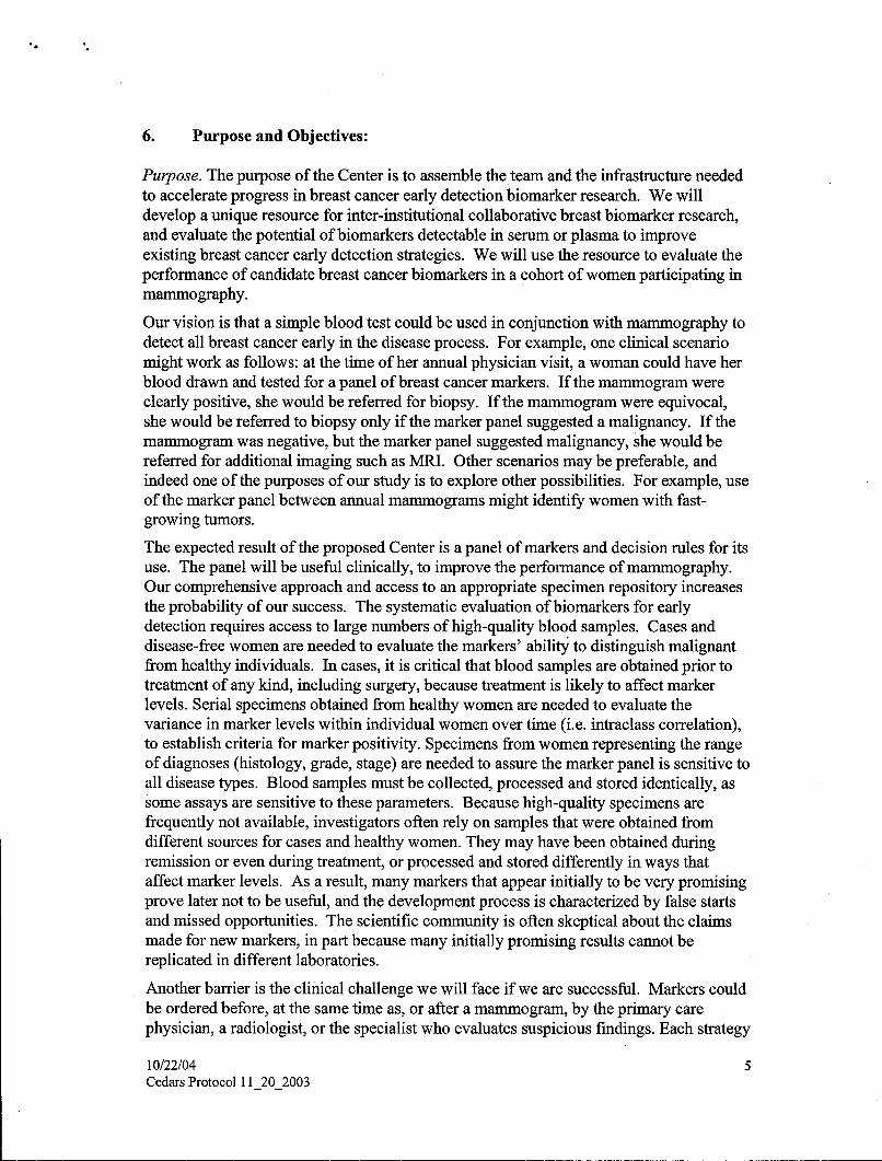
6. Purpose and Objectives:
Purpose. The purpose of the Center is to assemble the team and the infrastructure neededto accelerate progress in breast cancer early detection biomarker research. We willdevelop a unique resource for inter-institutional collaborative breast biomarker research,and evaluate the potential of biomarkers detectable in serum or plasma to improveexisting breast cancer early detection strategies. We will use the resource to evaluate theperformance of candidate breast cancer biomarkers in a cohort of women participating inmammography.
Our vision is that a simple blood test could be used in conjunction with mammography todetect all breast cancer early in the disease process. For example, one clinical scenariomight work as follows: at the time of her annual physician visit, a woman could have herblood drawn and tested for a panel of breast cancer markers. If the mammogram wereclearly positive, she would be referred for biopsy. If the mammogram were equivocal,she would be referred to biopsy only if the marker panel suggested a malignancy. If themammogram was negative, but the marker panel suggested malignancy, she would bereferred for additional imaging such as MRI. Other scenarios may be preferable, andindeed one of the purposes of our study is to explore other possibilities. For example, useof the marker panel between annual mammograms might identify women with fast-growing tumors.
The expected result of the proposed Center is a panel of markers and decision rules for itsuse. The panel will be useful clinically, to improve the performance of mammography.Our comprehensive approach and access to an appropriate specimen repository increasesthe probability of our success. The systematic evaluation of biomarkers for earlydetection requires access to large numbers of high-quality blood samples. Cases anddisease-free women are needed to evaluate the markers' ability to distinguish malignantfrom healthy individuals. In cases, it is critical that blood samples are obtained prior totreatment of any kind, including surgery, because treatment is likely to affect markerlevels. Serial specimens obtained from healthy women are needed to evaluate thevariance in marker levels within individual women over time (i.e. intraclass correlation),to establish criteria for marker positivity. Specimens from women representing the rangeof diagnoses (histology, grade, stage) are needed to assure the marker panel is sensitive toall disease types. Blood samples must be collected, processed and stored identically, assome assays are sensitive to these parameters. Because high-quality specimens arefrequently not available, investigators often rely on samples that were obtained fromdifferent sources for cases and healthy women. They may have been obtained duringremission or even during treatment, or processed and stored differently in ways thataffect marker levels. As a result, many markers that appear initially to be very promisingprove later not to be useful, and the development process is characterized by false startsand missed opportunities. The scientific community is often skeptical about the claimsmade for new markers, in part because many initially promising results cannot bereplicated in different laboratories.
Another barrier is the clinical challenge we will face if we are successful. Markers couldbe ordered before, at the same time as, or after a mammogram, by the primary carephysician, a radiologist, or the specialist who evaluates suspicious findings. Each strategy
10/22/04 5Cedars Protocol 11_20_2003

will affect the sensitivity and specificity of the combination of tests. If markers identifycancers that cannot be seen on a mammogram, clinical work up to identify the location ofthe tumor will be required. Cost-effective strategies will be needed. As we will obtainserum for women in several stages of evaluation-prior to screening, just prior to biopsy,and just prior to surgery-we will be in a unique position to evaluate the clinical utilityof markers at all these potential stages of screening.
Objectives. Our goal is to evaluate breast cancer biomarkers for their contribution toearly detection of breast cancer. Subsets of breast cancer that are missed bymammography, or that grow too quickly to be detected in early stage by annualmammography, are of particular interest. Our aims are:
1. To validate and refine the ability of candidate biomarkers to predict diseasestatus;
2. To evaluate panels of biomarkers for use as an adjunct to mammography, todetect all breast cancer at a highly curable stage; and
3. To identify the molecular signatures of subsets of in situ and invasive breastcancers and explore their associations with biomarkers in the panel.
To support the research goals we will build a unique resource for multidisciplinary, inter-institutional research on breast cancer biomarkers including
1. Blood samples obtained annually and processed identically in women with andwithout breast cancer,
2. Fresh tissue matched to blood samples on a subset of the women with and withoutbreast cancer,
3. Epidemiological, clinical and follow-up information for women who donatespecimens, and
4. A system to facilitate use of the specimens, including state-of-the-art informationsystems.
We will develop a specimen resource from a well-characterized population, withassociated risk factor information, mammography findings and follow-up data on canceroutcomes. It will include blood samples obtained from selected women who havemammograms, biopsy or breast cancer surgery over the 4-year period of the grant. Wewill evaluate candidate biomarkers for their ability to distinguish among women withhealthy breasts and women with various breast conditions, including both invasive andin-situ forms of breast cancer, premalignant breast diseases, and benign lesions. We willevaluate the role of biomarkers detectable in serum or plasma in improving our currentbreast cancer early detection strategies.
For selected women, we will explore the feasibility of collection and analysis of fresh-frozen tissue in order to characterize malignant conditions at the molecular level. Thiswill allow us to correlate biomarkers included in the panel with subsets of breast tumorsidentified through molecular profiling.
Our hypotheses are:
10/22/04 6Cedars Protocol 11_20_2003

(a) Used alone, biomarkers detectable in blood products can detect subsets of, but notall, breast cancer;
(b) Use of a breast cancer biomarker panel can improve the performance ofmammography in early detection of breast cancer.
7. Study Population.
Recruitment for this study will take place in Seattle, at Swedish Medical Center, and inLos Angeles, at Cedars Sinai Medical Center. The overall study population is describedbelow.
The study population includes three cohorts defined by recruitment source. The firstrecruitment source is women undergoing biopsy or surgery for breast cancer at CedarsSinai Medical Center. Approximately 1,500 women undergo this procedure annually. Ofthese we will enroll 50 per year, including women with benign lesions and premalignantbreast diseases, as well as women with in-situ and invasive carcinoma (see table 1). Bothfresh-frozen tissue and blood samples will be donated by women in the Cedars Sinaicohort (CSC). Blood and tissue donors are recruited from patients previously scheduledfor breast surgeries that involve the removal of some or all of their breast tissue. Thepatient's surgery will have been scheduled previously as a result of either a benign ormalignant condition. Patients are not scheduled for surgical procedures for the purposeof this study alone.
The second source is the approximately 8,600 women who obtain mammograms andundergo biopsies at the Swedish Medical Center Breast Care Center in Seattle each year.As shown in Table 1, 500 (375 high-risk and 125 average-risk) women will be enrolled inYear 1, and 100 additional women will be enrolled in subsequent years. In addition, allwomen undergoing biopsies will be invited to provide a blood specimen prior to biopsyand annually at the time of subsequent mammograms. 100 women undergoing biopsieswill be enrolled annually. Women identified and enrolled through the breast care centerincluding women having both mammograms and biopsies, are referred to as theMammography Cohort (MC).
The third cohort includes women undergoing breast surgery at Swedish Medical Center.The second recruitment source is the approximately 650 women who have surgery forbreast cancer at SMC each year. As shown in Table 1, we expect to enroll 25 women inYear 1 and 50 women annually thereafter for donation of both blood and tissue. Freshtissue specimens will be obtained when it is logistically and ethically appropriate. Inaddition we expect to enroll 100 women annually beginning in year 2 for donation ofblood only. Blood samples will be obtained just prior to or at the time of the surgicalprocedure. Women identified and enrolled through our collaborations with SMCsurgeons are referred to as the Surgical Cohort (SC). Following surgery and treatment,women in the SC will be invited to participate in the MC, providing blood samples at thetime of their annual mammograms.
10/22/04 7Cedars Protocol 11 20 2003
Table 1. Unique ParticipantsYr 01 Yr 02 Yr 03 Yr 04 Total
Mammography Cohort- High Risk 375 75 75 75 600Mammography Cohort - Average Risk 125 25 25 25 200Mammography Cohort - Biopsy 100 100 100 100 400Subtotal: Mammography Cohort 600 200 '200 200 1200SMC Surgical Cohort: Blood and Tissue 25 50 50 50 175SMC Surgical Cohort: Blood Only+ 100 100 100 300Subtotal: Surgical Cohort 25 150 150 150 475Cedars Surgical Cohort: Blood and 50 50 50 50 200Tissue (CSC)Total 675 400 400 400 1875
8. Protocol Design. This document outlines the methods for collection of blood andtissue from women undergoing surgical biopsy procedures at Cedars-Sinai MedicalCenter. Methods for recruitment and enrollment of the Seattle study population aredescribed in a separate document.
8a. Eligibility Criteria.
Inclusion CriteriaWomen 18+ years of ageScheduled for breast surgical procedure for a benign or malignant condition
Exclusion CriteriaBreast abnormalities less than 15mm (for procedures using a mammotome)Breast abnormalities less than 20mm (when using an open surgical technique)
Women enrolled at Cedars-Sinai are referred to as the Cedars Sinai Cohort (CSC).Surgical procedures will performed in the Cedars-Sinai Medical Center hospitaloperating suites, the Saul and Joyce Brandman Breast Center, or the Cedars-SinaiOutpatient Surgery Center.
8b. Recruitment (Subject Identification)After approval from the IRB, we intend to approach physicians who attend Breast Centerconferences, to educate them about available research protocols for interested patients.Recruitment flyers will also be posted and made available to raise patient awareness, andwe expect some patients to self-refer. This study will also be listed on the Cedars-Sinaiweb site.
Blood and tissue donors will be recruited from patients previously scheduled for breastsurgical procedures that involve the removal of some or all of their breast tissue. Thepatient's surgery will have previously been scheduled as a result of either a benign ormalignant condition. Patients will not be scheduled for surgical procedures for thepurpose of this study alone. The Principal Investigator, co-investigators, or treatingphysicians (usually a breast surgeon, occassionaly a radiologist or a medical oncologist)will identify potential subjects. The treating physician will make initial contact withpotential subjects.
10/22/04 8Cedars Protocol 11_20_2003

8c. Participant Approach, Enrollment, Informed Consent and Procedures
8cl. Participant Approach, Enrollment and Informed ConsentPhysicians will identify patients who meet eligibility criteria at the time their patient isscheduled for breast surgery. The treating physician (or a member of their staff) willascertain whether the patient is interested in learning more about the research protocol.The treating physician may be the principal investigator, a co-investigator or a non-investigator. Patients who express an interest will be referred to and ultimately contactedby a member of the research team, usually the study coordinator. The study will bedescribed in detail, including the risks and benefits of study participation. The patientwill be given an informed consent form to take home and review. The patient will haveample time to consider the study. Coercion and conflict of interest will be avoided byclearly explaning that this study plays no role in their clinical care, that participation inthis study is strictly voluntary, and that declining to participate will not affect theirtreatment in any way. The investigator or co-investigator will obtain a signed informedconsent prior to the collection of data, blood and tissue.
8c2 Research Procedures /Activity that the Participant will experienceWomen undergoing breast surgery will be invited to donate blood and tissue duringsurgery and to complete two questionnaires. Tissue specimens and blood specimens willbe obtained when it is logistically and ethically appropriate.
Questionnaire and dataAfter signed consent is obtained, study participants will be given the "BaselineQuestionnaire" and "Health Status Questionnaire" and a stamped self-addressed returnenvelope. Study participants will be asked to return the Baseline Questionnaire withinfour weeks of their procedure, and asked to complete the Health Status Questionnaire justprior to their procedure. Participants who do not return the Baseline Questionnaire willbe contacted by phone six weeks after their procedure.
Patient demographic data and pathologic classification will be acquired from the patient'smedical records and anatomic pathology reports respectively. This information will bestored in the database and identified by the use of the assigned numeric identifier.Clinical assessments of the donors for the purpose of this study will be required.
Blood Collection:Prior to each surgery, patient consent will be obtained to collect blood. The researchcollection team will notify and work with the operating room staff prior to the beginningof surgery that a patient is participating in the research study. Up to 50cc of blood willbe obtained prior to or at the time of surgery, or at a time when blood is drawn for otherlaboratory tests. Priority of blood collection is: 1) four 10 ml red top tubes for serumspecimens, 2) one 10 ml lavender top EDTA or yellow top tube for plasma.The volume of blood drawn may be adjusted downward as necessary if a clinical draw isbeing conducted on the same day.
Blood Storage:10/22/04 9Cedars Protocol 11_20_2003

Standard protocols will be followed to process specimens into sera and plasma and toaliquot into cryovials uniquely labeled with study specimen ids. Tubes will be stored inthe refrigerator and processed within 48 hours of receipt. After processing, bloodspecimens will be aliquotted into 1.8 mL pre-labeled cryovials or very small quantities toavoid freeze-thaw cycles. Aliquoted specimens will be transported to the Core TissueBank for long-term storage or delivered to laboratory investigators. Times of blood draw,processing and freezing will be recorded.
Tissue Collection:The amount of tissue collected for the purposes of the research study will vary. Tissuewill be collected only if it is not needed for pathologic diagnosis. The clinicalpathologist involved in the participant's case will work with the treating surgeon toidentify the location and the amount of tissue available for research purposes. When.clinically appropriate, a minimum of two and a maximum of ten 1.0mL cryovials (eachholding 1 gram of breast tissue) will be collected for the study. When feasible, theoptimal tissue quantity per case is 6 grams.
Tissue collected will include malignant tissue and, if possible, adjacent normal tissue.
For each tissue collection, a packet containing a "Dr. Karlan's Lab - SpecimenPickup/Documentation" form, the original and a copy of the signed informed consentform, a Cedars-Sinai Department of Pathology "Request for Tissue for Research" form,and labeled cryovials will be assembled. The study coordinator or designee will (1.) takethis packet to the surgical suite for tissue collection, (2.) ensure that the original signedinformed consent for study participation is placed in the subject's medical record prior tothe surgical procedure, and (3.) work with the surgeon and operating room personnel toassure that they are aware of the patient's participation in this research project prior to thebeginning of the patient's surgery. During the entire surgical procedure, the treatingsurgeon and the surgical team will be responsible for monitoring the patient's condition.The surgeon will ultimately remove a surgical specimen and give this to the pathologist.The pathologist will then identify tissue that will not be needed for pathologic diagnosis.All or part of the un-needed tissue will be collected and placed in the labeled cryovialsand processed for storage. The pathologist may refuse to provide any specimen forresearch if they believe that the entire specimen is needed for the subject's clinical care.
Tissue Storage:It is anticipated that only a small amount of tissue will be available per patient After thistissue has been placed in cryovials, the study coordinator or designee will transfer thecryovials to the histobath located in the 3rd floor OR (for procedures performed in thehospital operating room suites) or the C-S Outpatient Surgery Center 1 st floor OR (forprocedures performed in the C-S Outpatient Surgery Center or the Joyce and SaulBrandman Breast Center).The tissue will be snap-frozen in the histobath. Once a week, cryovials will betransported to the Core Tissue Bank. Specimens will be stored at -80 degrees C.
8d. Subject Assignment (randomization): Not applicable.
10/22/04 10Cedars Protocol 11_20_2003

8e. Evaluations Prior to Entry.No clinical evaluations are made prior to entry into the study.
8f. Evaluations to be made during the conduct of the study. Breast cancer casestatus will be ascertained on all women by abstracting information from participantmedical records.
8g. Clinical Assessments. Pathology reports will be obtained and abstracted usingthe Patient Pathology at Diagnosis Form. Additional clinical data will abstracted frommedical records using the Patient Clinical Status at Dx Form and the Clinical FollowUp Form (for women diagnosed with breast cancer as a result of their biopsy).Mammography results will be abstracted from the medical records using theMammography Results Form. All of these forms are currently under development andwill be submitted to IRB at a later date.
9. Risk/Benefit Assessment
This research is intended to improve our ability to diagnose breast cancer at an earlystage. Specifically, this study aims to develop a panel of biomarkers than can be used inconjunction with mammography to improve the clinical detection rate (true sensitivity) ofmammography. Although there is no direct benefit to women participating in this study,there may be benefits for other women in the future who are at risk for developing breastcancer or who are living with breast cancer.
Risks to participants including the potential of feeling uncomfortable as a result ofanswering personal questions on study questionnaires. Participants may or may not beconcerned about not knowing results of studies conducted on their blood and tissue.Risks associated with blood drawing include the possibility of temporary discomfort or abruise at the site of the needle puncture. Donation of blood and tissue during surgery willnot present any additional risks.
10. Reporting of Serious or Unexpected Adverse Events
Adverse events are expected to be rare in this study. Study personnel will have directexposure to participants only for a short period of time (e.g., during the consent andspecimen collection process), and will not be monitoring participant's health as theywould for a clinical treatment protocol. Therefore, study participants will be asked toreport any adverse events that they experience that appear to be related to studyparticipation. This will be done by calling the Study Coordinator / Research Nurse.
An adverse event is any event that occurs during the study, which results in the subjectexperiencing a new symptom or worsening of an existing symptom. For example,fainting or loss of cognitive function after a blood draw would be considered an adverseevent.
A serious adverse event is an event that results in any of the following outcomes:1. Death2. Life threatening event3. Inpatient hospitalization*4. Persistent or significant disability or incapacity10/22/04 11Cedars Protocol 11_20_2003

*Patients undergoing surgery for breast cancer will be approached to donate blood andtissue for the study. Hospitalization associated with this already-planned surgery willnot be considered an adverse event.
Adverse events will be reported to the local Institutional Review Board (IRB) as perinstitutional policy.
Adverse events that are both serious and unexpected will be immediately (upon the studyoffice notified) reported by telephone to the local IRO and by telephone to Nicole Urban,FHCRC Principal Investigator. Dr. Urban will be responsible for reporting these eventsto the USAMRMC, Deputy for Regulatory Compliance and Quality (301-619-2165; FAX301-619-7803). A written report will follow the initial telephone call within 3 workingdays. The written report will be addressed to:
U.S. Army Medical Research and Materiel CommandATTN: MCMR-RCQ504 Scott StreetFort Detrick, Maryland 21702-5012
11. Description of Protocol Drugs or Devices: Not applicable.
12. Disposition of Data.
To maintain human subject anonymity, specimens and data are retrieved through the useof the numeric identifier only. Patient demographic data and pathologic classificationwill be acquired from the patient's medical records and anatomic pathology reportsrespectively. This information will be stored in the database and identified by the use ofthe assigned numeric identifier. Access to information linking the donors' real nameswith the numeric identifier will be restricted to study staff members.
13. Modification of the Protocol.
Any modification to the protocol will follow established protocol modification procedureof the local Internal Review Office and the HSRRB for review and approval, whichinclude detailed submission of any changes or additions.
14. Departure from the Protocol. If the study investigators or staff become awareof any departure from the protocol, the local Internal Review office will be notified inwriting as well as HSRRB office.
15. Roles and Responsibilities of Study Personnel at Cedars Sinai MedicalCenter
Scott Karlan, MD, Cedars-Sinai Breast Surgeon (Consultant 20% FTE) Dr. S. Karlan isa leading breast surgeon who has participated in research activities at Cedars-Sinaiinvolving tissue collection and biomarker development. Dr. S. Karlan will overseerecruitment, enrollment and specimen collection from women undergoing biopsy orsurgery for breast cancer at Cedars Sinai Medical Center. He will travel to Seattleannually for an All-Investigator Meeting and will participate in additional Investigatormeetings via conference call or web interface.10/22/04 12Cedars Protocol 11_20_2003

Beth Karlan, MD, Gynecologic Oncologist (5%FTE; 1% compensated, 4% donated) Dr.B. Karlan has extensive experience in cancer screening and has collaborated with Dr.McIntosh in the past on the evaluation of markers for ovarian cancer screening. She willparticipate in the evaluation of laboratory results and the development and validation ofthe marker panel. Dr. B. Karlan will work closely with Drs. Schummer and Kiviat onmethods for extraction, amplification and preservation of mRNA from tissue formolecular profiling work. Dr. B. Karlan will participate in conference calls and beavailable via email to collaborate with investigators in Seattle. She will travel to Seattleannually for an All-Investigator Meeting and will participate in additional Investigatormeetings via conference call or web interface. She will participate in manuscript andreport preparation.
The Study Coordinator / Research Nurse will work closely with project investigators toprovide overall coordination for the study. (S)he will oversee administrative and projectmanagement activities, which include preparing and monitoring budgets, submitting andmodifying IRB and Human Subjects applications, facilitating investigator meetings andincorporating investigator decisions into staff workplans. The StudyCoordinator/Research Nurse will be responsible for implementing the components of thisstudy that involve physician and participant interaction, participant recruitment,retention, and compliance. The Study Coordinator/Research Nurse will also reviewsubject medical records to obtain data needed as described in the protocol.
10/22/04 13Cedars Protocol 11_20_2003

Appendix DSurgeon and Pathologist meeting minutes

Breast Cancer Early Discovery Meeting SummaryJune 14, 2004
5:00pm-6:30pmA-West level A & B conference rooms
Swedish Medical Center
Distribution List: J. David Beatty, Nicole Urban, Patricia Dawson, Christine Lee,Charles Drescher, Toni Oien, Patra Grevstad, John Dawson, Ron Tickman, NanPing Wang, Lisa Isozaki, Ksenia Koon, Shirley Gough, Kate Watabayashi, KathyO'Briant
Consenting patients:
Consenting patients would be most practical at the pre-surgical visit and it was suggestedby some that the surgeon introduce the study to their patients. By doing this, the patientmay be more willing to consent if it appears her surgeon approves of the study. Then astudy staff can proceed by providing detailed information about what the study willrequire from a participant. It was agreed that women should be consented for both bloodand tissue donation, but that it would be decided later by the surgeons and pathologistswhether tissue would be collected for research.
Blood draw:
Dr. Urban prefers that conditions at the time of blood draw mimic those of a woman whogoes for screening, to avoid conditions such as fasting. Collections should be done at thepre-surgical visit rather than the OR
Dr. Lee believes that this is feasible, because if a woman consents to the study, she mostlikely will allow blood to be drawn at the pre-surgical visit.
Tissue:
All were concerned by the amount of tissue requested for the study. Dr. Tickman believesthat the study will not receive grams of tissue, but rather millimeter tissue slices from thecenter of the tumor which, implying that the tumor tissue requirement must be reduced to1 or 1.5 cm diameter. Dr. Urban is comfortable with this if the tissue is OCT-embeddedfor the purposes of RNA extraction by laser capture microdisection (LCM).
Tissue collection at surgical procedures:
There was much discussion regarding the challenges of obtaining breast cancer tissuefrom surgical procedures.
The major challenges include:
1. The current practice is to place all breast tissue in formalin at the time of surgery,and, at a later time, cut, section, and evaluate the tissue. To provide fresh tissue,pathology would need to add a step in their procedure whereby a pathologist orthe gross PA, would cut the fresh breast tissue and give samples to study staffimmediately following surgery.
2. The current practice does not support obtaining tissue from lumpectomyspecimens in a timely manner. In lumpectomy cases, surgically removed tissue issent to the breast center to determine if the excision is complete. Until aradiologist reviews the tissue radiograph to determine that the breast lesion andlocalizing wires are removed, no tissue can be collected for research. Thisprocedure can take up to 40 minutes and would need to be expedited for us toobtain lumpectomy tissues for our study.
3. It is critical that tissue collected for research does not compromise the ultimatediagnosis of a patient. The Center of Excellence study protocol describes thecollection of 1 to 10 grams of breast tissue with an optimal amount of 6 grams.Dr. Beatty believes that this may be feasible from total mastectomies, whichaccount for about 30% of the breast cancer patients seen at SMC. In some cases,however, the majority of the malignant tissue may have already been excised atbiopsy.
To begin, Dr. Beatty suggests tissue be collected from women scheduled for atotal mastectomy and, once surgeons and pathologists are comfortable with theprocedure and the donation of tissue to the study, that tissue from a lumpectomyprocedure and other types of surgery be considered. The protocol criteria statethat we will obtain tissue from women who have a 2 cm or greater sized tumor.
Dr. Wang stated he agrees with the study criteria, which restricts collection oftissue on lesions smaller than 2 cm. Dr. Lee suggests that surgeons andpathologists begin with tissue collection on 2cm-sized tumors and, once everyoneis comfortable with the procedure, smaller tumor sizes be considered.
4. Breast tissue collection via mammotome device was briefly discussed. Dr. Beattyis interested in the possibility of using a hand held mammotome device to biopsysolid masses and to potentially collect samples for research. All agreed obtainingstereotactic mammotome samples should be discussed at another time withradiologists in attendance.
Tissue to pathologists:
In order to expedite the transfer of tissue to the pathologist and subsequent donation tothe study, Drs. Tickman and Wang made several suggestions:

1. Request that a dedicated pathologist at SMC, 1st Hill and Providence campus,work with the study and assist with development of an acceptable procedure.
2. Alter the pathology procedure for study participants by implementing freshsectioning of breast tissue by a pathologist or Gross PA, including tissue samplesprovided directly to study staff
3. Research staff to provide advanced notice to pathology that a study participant isscheduled for surgery
4. Have a Specimen Collection Specialist present at the surgery and transfer tissuequickly to the participating pathologist or gross PA.
5. Study staff to provide transport of lumpectomy tissue from surgery to SBCC andback again in an effort to expedite the process.
Dr. Urban is particularly interested in obtaining DCIS tissue in order to characterize thedifferent DCIS histology types using biomarkers and to perhaps learn which DCIS maybecome invasive and which may not. However, it was said that the tissue removed mightnot be determined as DCIS because some cases of DCIS do not have distinctive grossfindings.
Action items:
1. Distribute protocol and consent form for surgical cohort to surgeons andpathologist.Updated, June 18, 2004: Materials distributed.
2. Provide study staff contact list to participating surgeons.Updated, June 21, 2004: Heather Aiosa will be the main contact and specimencollection person. She will begin by spending Monday afternoons in ChristineLee's clinic beginning in mid-July. We anticipate that other surgeons may haveslightly different procedures for their clinics and we will work with them todevelop an approach procedure.
3. Identify a dedicated pathologist at SMC and Providence4. Request space for a Specimen Collection Specialist at Providence.
In Attendance: Heather Aiosa, Sarika Bhatia, Meghan Crawford, Dr. John Dawson, Dr. ChuckDrescher, Marcia Gaul, Shirley Gough, Samantha Hoyt-Schmid, Lisa Isozaki,Ksenia Koon, Jennifer Lange, Dr. Christine Lee, Elizabeth Murphy, KathyO'Briant, Anamaria Prosser, Leah Sabacan, Dr. Ron Tickman
Distribution: SPORE Staff, Dr. John Dawson, Dr. Chuck Drescher, Dr. Christine Lee, Dr. PamPaley, Dr. Ron Tickman, Nicole Urban
BCEDS surgery specimen collectionCurrently we are working in conjunction with Dr. Christine Lee to enroll patients into BCEDS from herbreast surgery practice at the Providence Comprehensive Breast Center on Monday afternoons. We hopeto expand the enrollment population to include other physicians at Providence as well as at Swedish. Dr.John Dawson is interested in participating and has asked for the required enrollment materials for review.
Deb Forman, Dr. Lee's nurse, is an integral part of the enrollment process at Providence. Each Friday, sheidentifies and informs Heather or Elizabeth of potential study participants who will be coming to theirpre-surgical clinic visit the following Monday. This gives Heather or Elizabeth ample time to be atProvidence to meet with the potential participant, immediately following her clinic visit, in hopes ofenrolling her. To date, one woman has been consented and two others have been approached.
Dr. Dawson has indicated an interest in approaching and possibly enrolling the patients himself. In suchcases, patients can go to Dynacare to have their blood drawn so study staff will supply Dr. Dawson withblood draw packets. Alternatively, Heather and Elizabeth will be available to come for the blood drawportion only. Heather and Elizabeth will also be available to consent those women that Dr. Dawsondoesn't enroll, but who are interested in the study. Since Dr. Dawson's practice is at Swedish MedicalCenter, Heather and Elizabeth will be on-call. That is, they will, go over to Dr. Dawson's office, whencontacted, to enroll women who are interested. Dr. Dawson and his nurse will be given the study cellphone number to call in such situations.
We are asking women to donate blood pre-surgery, during surgery, and also to donate a sample of theirbreast tissue when clinically appropriate, as determined by the pathologist. The study calls for a draw atthe pre-surgical appointment so that the blood will be drawn under similar circumstances to that of ablood draw on a healthy woman. In terms of the draw that happens during (or right around the time ofsurgery), we discussed the option of having the draw immediately prior to surgery. Due to the new SMCprotocol, which requires nursing staff to place an antibiotic IV and administer a beta-blocker beforesurgery, it was decided to draw the surgery blood when the woman is in the operating room instead of inthe transfer area. This will be done when the woman is under general anesthesia, but just before the timethat the blue dye is administered.
Initially, women undergoing mastectomies, new patients that are undergoing neoadjuvant therapy, andthose patients that are undergoing surgery who are T2 will be approached for the tissue collection portionof the protocol, in addition to the standard blood collection portion of the protocol.
We discussed how much tissue will be collected from each participant. At least a core biopsy must beobtained, but we are looking for a half-gram of tissue or more. It is feasible that women undergoing

neoadjuvant therapy may have a tissue sample removed at the same time as the T-clip instead of duringsurgery.
There are still barriers that need to be worked out as far as collecting tissue and being able to fresh freezeit within the twenty-minute time frame. We will look at the first five surgical patients or so to determinewhat is feasible.
Drs. Dawson, Lee, and Tickman will attend the informal SPORE staff meetings, held every other month,beginning September 27, 2004.

Appendix EBlood and tissue
collection and processing procedures

Breast Cancer Early Discovery Study
Blood and tissue collection and processing procedures
Blood draw:
Women will be asked to donate blood prior to and during surgery. The pre-surgicalblood draw will be obtained preferably at the pre-surgical visit, although a woman canhave this sample drawn any time before surgery (for example, if a woman has ascheduled blood draw for other reasons on another day before surgery, the research staffcan be available to capture blood for the purposes of the study at that time). The blooddraw during surgery will be performed in the operating room. Because of a new SMCprotocol, nursing staff is required to place an IV to administer a beta-blocker andantibiotic prior to surgery. Therefore, the anesthesiologist or research staff will performthis blood draw in the operating room after the insertion of the IV and beta-blocker. Atthis time, the woman will be under general anesthesia and no blue dye will have yet beenadministered.
For each draw, 50 mL of blood will be taken:
3 10 ml Serum Separator Tubes (SST) or red-top tubes1 10 ml EDTA Tube (Lavender-top) tube1 10 ml ACD Tube (Yellow top-tube)
Tissue collection from surgery:
In addition to the blood draws from women undergoing surgery, women will also beasked to consent to donate tissue, but only if clinically feasible or deemed appropriate bythe pathologist. Initially, patients undergoing full and partial mastectomies, patients whohave been diagnosed with stage T2 disease, and new patients undergoing neo-adjuvanttherapy will be approached for the tissue collection portion of the protocol along with theblood collection portion of the protocol. A minimum of 0.1 g of tumor tissue will becollected for the study. In addition, 0.1 g to 1 g of normal breast tissue will be collectedwhen it is available. For women undergoing neo-adjuvant therapy, it may be feasible fora tissue sample to be removed at the time the surgical clip is inserted instead of duringsurgery.
Summary:Condition Tissue Note
requirementFull Mastectomy - 0.1 g tumor
i 0.1 g normalPartial Mastectomy > 0.1 g tumor
0 0.1 g normalNeoadjuvant therapy k 0.1 g tumor Tissue sample may be removed at the time
_0- 0.1 g normal the surgical clip is inserted.

Stage T2 disease 0.1 g tumorS0.1 g normal
Tissue processing:
Once the pathologist has determined that tissue will be available for the study, theresearch staff will process the tissue as follows:
If< 0.5 g tissue:0 Embed the entire tissue section in OCT
If0.5 g - 1 g tissue:* Divide the tissue into 2 sections and embed both in OCT
If> 1 g tissue:"* Divide into section -0.5 g each"* If 3 sections, embed and freeze 2 sections in OCT and snap freeze the
other by placing in a cryovial and dropping in liquid nitrogen" If > 3 sections, embed 3 sections in OCT and snap freeze all remaining
sections by placing in a cryovial and dropping in liquid nitrogen
Tissue should be placed in liquid nitrogen within a 20-minute time frame. In order tostay within the time allowed, the study staff will expedite transport of tissue to thepathologist or gross pathology assistant who will provide tissue to the research staff.

Appendix FPathology data collection instruments

UPN
BREAST CANCER EARLY DISCOVERY STUDYPatient Level Clinical Diagnosis
Date: / / Form completed by:_
UPN: Form QC by:
SECTION A: Patient Reports Obtained
Reports Received:
Date report completed Date report received
O Pathology Report (from biopsy)
"o Chemistry Report (from biopsy)
"o Pathology Report (from surgery)
El Chemistry Report (from surgery)
O Operative Notes
o Progress Reports
0 Oncology/ Treatment Notes
O Other Reports, specify
o Other Reports, specify
P:\DOD breast\Admin\Annual Progress Reports to DOD\Year Two Progress Report\appendices\Patient Clinical Dx version 1 .doc Vers. 3

UPN
SECTION B: Specimen Collection Information
B1. Specimen collected from the following procedure if only one type of procedure performed:
01 Fine needle aspiration 04 Surgical biopsy/Lumpectomy02 Core biopsy 05 Mastectomy03 Vacuum assisted/ Mammotome 06 Other, specify
B2. Histologic cell types that have been identified in this patient to date
01 Benign02 Atypia03 In-situ104 Invasive
SECTION C: Tissue Characteristics - Assessment of Benign
C1. Please indicate laterality of the procedure:
01E Left breast 01 Upper Outer (UO) 02 Right breast 01 Upper Outer (UO)02 Upper Inner (UI) 02 Upper Inner (Ul)03 Lower Outer (LO) 03 Lower Outer (LO)04 Lower Inner (LI) 04 Lower Inner (LI)0F5 Breast, NOS 05 Breast, NOS06 Other, specify _ 06 Other, specify_090 Unknown 090 Unknown
C2. Cores/Specimen collected and described:
C2a. Core/Specimen description:
C2b. Core/Specimen size:
C3. Histology type: (Check all that apply)
01 No specific pathologic change 09 Fibrocystic changes, NOS02 Ductal hyperplasia (w/o atypia), NOS 010 Fibrocystic changes, specified03 Ductal hyperplasia (w/o atypia), specified _1_ _ Radial scar04 Sclerosing adenosis 012 Columnar cell change (without atypia)05 Fibroadenoma 013 Cyst06 Cellular fibroadenoma 014 Phylloides07 Intraductal papilloma 015 Other, specify08 Micropapilloma 090 Indeterminate/Unknown
C4. Presence of microcalcification:
01 No02 Yes090 Indeterminate/Unknown
-99 Not assessed
C5. Additional information for benign histology
P:\DOD breast\Admin\Annual Progress Reports to DOD\Year Two Progress Report\appendices\Patient Clinical Dx version 1.doc Vers. 3 2
UPN
SECTION D: TISSUE CHARACTERISTICS - Assessment of AtypiaPlease duplicate section D for each atypical histologic cell type present.
D1. Please indicate laterality of the procedure:
01 Left breast 01 Upper Outer (UO) 02 Right breast 01 Upper Outer (UO)02 Upper Inner (UI) 02 Upper Inner (UI)03 Lower Outer (LO) 03 Lower Outer (LO)04 Lower Inner (LI) 04 Lower Inner (LI)05 Breast, NOS 05 Breast, NOS06 Other, specify_ _ 06 Other, specify_090 Unknown 090 Unknown
D2. Cores/Specimen collected and described:
D2a. Core/Specimen description:
D2b. Core/Specimen size:
D3. Histology Type: (Check all that apply)
015 Ductal hyperplasia with atypia (ADH)016 Lobular hyperplasia with atypia (ALH)017 Intraductal papilloma with atypia018 Columnar cell change/hyperplasia with atypia019 Other, specify090 Indeterminate/Unknown
D4. Presence of microcalcification:
01 No02 Yes, NOS03 Yes, involving atypia090 Indeterminate/Unknown099 Not assessed
D5. Additional information for atypia
P:\DOD breast\AdminKAnnual Progress Reports to DOD\Year Two Progress Report\appendices\Patient Clinical Dx version 1.doc Vers. 3 3

UPN
SECTION E: Tissue Characteristics - Assessment of In-Situ Carcinoma
Please duplicate section E for each in-situ histologic cell type present.
El. Please indicate laterality of the procedure:
01 Left breast 01 Upper Outer (UO) 02 Right breast 01 Upper Outer (UO)02 Upper Inner (UI) 02 Upper Inner (Ul)03 Lower Outer (LO) 03 Lower Outer (LO)04 Lower Inner (LI) 04 Lower Inner (LI)05 Breast, NOS 05 Breast, NOS06 Other, specify_ _ 06 Other, specify_090 Unknown 090 Unknown
E2. Cores/Specimen collected and described:
E2a. Core/Specimen description:
E2b. Core/Specimen size:
E3. Tumor size estimated by:
01 Palpation02 Ultrasound03 Mammography04 MRI05 Measurement of gross specimen06 Measurement of specimen on pathology report099 Assessment not performed
E4. Histology type:
-I Ductal
020 Comedo 024 Apocrine021 Solid 025 Intracystic022 Cribriform 026 Papillary023 Micropapillary 0127 Clinging
028 Lobular029 Other, specify_090 Indeterminate/Unknown
E5. Margin involvement (closest margin to in-situ component):
01 < than 5mm 05 Involved, NOS02 From 5mm to 10mm 06 Not involved, NOS03 > 10mm 090 Indeterminate/Unknown04 Actual margin, if known: mm 099 Not assessed
E6. Nuclear Grade:
01 Low nuclear grade02 Intermediate nuclear grade03 High nuclear grade090 Indeterminate/Unknown099 Not assessed
P:\DOD breast\Admin\Annual Progress Reports to DOD\Year Two Progress Report\appendices\Patient Clinical Dx version 1.doc Vers. 3 4

UPN
SECTION E: Assessment of In-Situ Carcinoma (Continued)
Please duplicate section E for each in-situ histologic cell type present.
E7. Presence of necrosis:
01 No02 Yes090 Indeterminate/Unknown099 Not assessed
E8. Presence of microinvasion:
01 No02 Yes090 Indeterminate/Unknown099 Not assessed
E9. Presence of microcalcifications:
01 No02 Yes, NOS03 Yes, involving benign ducts04 Yes, involving atypia05 Yes, involving both benign ducts and DCIS06 Yes, involving DCIS07 Other, specify090 Indeterminate/Unknown099 Not assessed
E10. Presence of Paget's disease of the nipple:
01 No02 Yes090 Indeterminate/UnknownD99 Not assessed
Ell. Additional information in-situ carcinoma
P:\DOD breast\Admin\Annual Progress Reports to DOD\Year Two Progress Report\appendices\Patient Clinical Dx version 1 .doc Vers. 3 5

UPN
SECTION F: Tissue Characteristics - Assessment of Invasive Carcinoma
Please duplicate section F for each invasive histologic cell type present.
Fl. Please indicate laterality of the procedure:
01 Left breast 01 Upper Outer (UO) 02 Right breast 01 Upper Outer (UO)02 Upper Inner (UI) 02 Upper Inner (UI)03 Lower Outer (LO) 03 Lower Outer (LO)04 Lower Inner (LI) 04 Lower Inner (LI)05 Breast, NOS 05 Breast, NOS06 Other, specify_ _ 06 Other, specify_090 Unknown 090 Unknown
F2. CoreslSpecimen collected and described:
F2a. Core/Specimen description:
F2b. Core/Specimen size:
F3. Tumor size estimated by:
0, Palpation02 Ultrasound03 Mammography04 MRI05 Measurement of gross specimen06 Measurement of specimen on pathology report099 Assessment not performed
F4. Histology type:
0 Epithelial
030 Ductal031 Lobular032 Mixed lobular/ductal033 Medullary034 Atypical Medullary035 Tubular036 Mixed tubular/lobular037 Mucinous038 Papillary039 Adenoid cystic040 Metaplastic041 Squamous042 Inflammatory (IBC)
0 Stromal
043 Phylloides044 Sarcoma, specify type
045 Other, specify090 Indeterminate/Unknown
P:\DOD breast\Admin\Annual Progress Reports to DOD\Year Two Progress Report\appendices\Patient Clinical Dx version 1 .doc Vers. 3 6

UPN
SECTION F: Assessment of Invasive Carcinoma (Continued)
Please duplicate section F for each invasive histologic cell type present.
F5. Nuclear Grade:
01 Low nuclear grade02 Intermediate nuclear grade03 High nuclear grade090 Indeterminate/Unknown099 Not assessed
F6. Nuclear pleomorphism:
01 Small variation in size and shape02 Moderate variation in size and shape03 Marked variation in size and shape090 Indeterminate/Unknown099 Not assessed
F7. Mitotic Rate:
01 Low02 Intermediate03 High04 Mitotic rate, specifiedDgo Indeterminate/Unknown099 Not assessed
F8. Tubule differentiation:
01 Majority of tumor, > 75% of tumor02 Moderate, > 10 - 75% of tumor03 Minimal, < 10% of tumor090 Indeterminate/Unknown099 Not assessed
F9. Histologic grade: Specify scheme
01 Low02 Intermediate03 High090 Indeterminate/Unknown099 Not Assessed04 Other, specify_
F10. Presence of lymphatic or vascular invasion:
01 No02 Yes090 Indeterminate/Unknown099 Not assessed
F1l. Tumor Necrosis:
01 Scant 04 None02 Moderate 090 Indeterminate/Unknown03 Extensive 099 Not assessed
P:\DOD breast\Admin\Annual Progress Reports to DOD\Year Two Progress Report\appendices\Patient Clinical Dx version 1 .doc Vers. 3 7
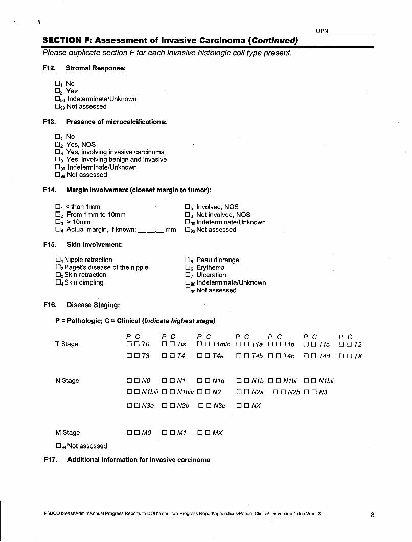
UPN
SECTION F: Assessment of Invasive Carcinoma (Continued)
Please duplicate section F for each invasive histologic cell type present.
F12. Stromal Response:
01 No02 Yes090 Indeterminate/Unknown099 Not assessed
F13. Presence of microcalcifications:
01 No02 Yes, NOS03 Yes, involving invasive carcinoma09 Yes, involving benign and invasive090 Indeterminate/Unknown099 Not assessed
F14. Margin involvement (closest margin to tumor):
01 <than 1mm 05 Involved, NOS02 From 1mm to 10mm 06 Not involved, NOS03 > 10mm -9o Indeterminate/Unknown04 Actual margin, if known: mm 099 Not assessed
F15. Skin involvement:
01 Nipple retraction 05 Peau d'orange02 Paget's disease of the nipple 06 Erythema03 Skin retraction 07 Ulceration04 Skin dimpling 090 Indeterminate/Unknown
099 Not assessed
F16. Disease Staging:
P = Pathologic; C = Clinical (Indicate highest stage)
PC PC PC PC PC PC PCTStage 0Q0OTO O O Tis 0 11 Tlmic O O Tla 0 T1b 0 01 TIc 0 0 T2
0 O T3 0 0 T4 O O T4a O O T4b O0O T4c OO T4d O 0 TX
NStage QO0 NO ON1 O 0 Nla O O Nlb O O Nlbi 0 0 Nlbfi
O O Nlbiii O Nlbiv O 0 N2 OO N2a OON2b 0ON3
0ON3a OO N3b DON3c O O NX
M Stage DOMO [ MI DO WMX
099 Not assessed
F17. Additional information for invasive carcinoma
P:\DOD breast\Admin\Annual Progress Reports to DOD\Year Two Progress Report\appendices\Patient Clinical Dx version 1 .doc Vers. 3 8

UPN
SECTION G: Prognostic Information: Assessment of Nodes
G1. Was sentinel node sampling performed?
01 No02 Yes Gla. Sentinel Node Site:
01 Axillary02 Internal Mammary03 Supraclavicular090 Unknown
090 Indeterminate/Unknown
G2. Total number of sentinel nodes examined (removed):
G3. Total number of positive sentinel nodes:
G4. Size of largest metastasis:
Mm
G5. Number of positive sentinel nodes examined by:
_____ Hemotoxylin & Eosin (H&E)
_____ Immunohistochemistry (IHC)
______ Other, specify
G6. Was axillary dissection performed?
01 No02 Yes G~a. Lymph Node Involvement:
01 Axillary012 Internal Mammary013 Supraclavicular03 Infraclavicular090 Unknown
090 Indeterminate/Unknown
P:\DOD breast\Admin\Annual Progress Reports to DOD\Year Two Progress Report\appendices\Patient Clinical Dx version 1.doc Vers. 3 9
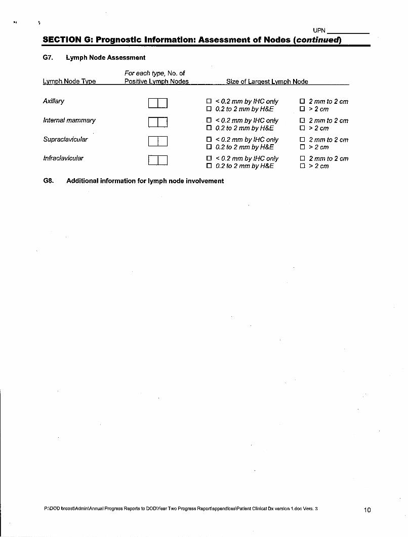
UPN
SECTION G: Prognostic Information: Assessment of Nodes (continued)
G7. Lymph Node Assessment
For each type, No. ofLymph Node Type Positive Lymph Nodes Size of Largest Lymph Node
Axillary [77 El < 0.2 mm by IHC only l 2 mm to 2 cmEl 0.2 to 2 mm by H&E l > 2 cm
Internal mammary ED E < 0.2 mm by IHC only l 2 mm to 2 cm0 0.2 to 2 mm by H&E l > 2 cm
Supraclavicular [7D El < 0.2 mm by IHC only l 2 mm to 2 cmEl 0.2 to 2 mm by H&E 0 > 2 cm
Infraclavicular ED E < 0.2 mm by lHC only l 2 mm to 2 cmEl 0.2 to 2 mm by H&E l > 2 cm
G8. Additional information for lymph node involvement
P:\DOD breast\Admin\Annual Progress Reports to DOD\Year Two Progress Report\appendices\Patient Clinical Dx version 1.doc Vers. 3 10

UPN
SECTION H: Marker StatusPlease duplicate section H for each tissue assayed.
HI. Please indicate laterality of the procedure:
01 Left breast 01 Upper Outer (UO) D2 Right breast 01 Upper Outer (UO)02 Upper Inner (Ul) 02 Upper Inner (UI)03 Lower Outer (LO) 03 Lower Outer (LO)04 Lower Inner (LI) 04 Lower Inner (LI)05 Breast, NOS 05 Breast, NOS06 Other, specify _ 06 Other, specify_090 Unknown 090 Unknown
H2. Cores/Specimen collected and described:
H2a. Core/Specimen description:
H2b. Core/Specimen size:
H3. Estrogen receptor status:
Specify antibody clone and vendor for Estrogen test
Method used
"% of nuclei staining (Invasive tumor)"% of nuclei staining (DCIS)
01 Negative02 Positive09o Indeterminate/Unknown099 Test Not Performed
H4. Progesterone receptor status:
Specify antibody clone and vendor for Progesterone test
Method used
"% of nuclei staining (Invasive tumor)"% of nuclei staining (DCIS)
01 Negative02 Positive090 Indeterminate/Unknown099 Test Not Performed
H5. Her2lneu test(s) performed: (Check all that apply)
Specify antibody clone and vendor for HER2 test
01 Immunohistochemistry (IHC)02 Fluorescent In-situ Hybridization (FISH)03 Enzyme-Linked Immunosorbent Assay (ELISA)099 Test not performed
P:\DOD breast\Admin\Annual Progress Reports to DOD\Year Two Progress Report\appendices\Patient Clinical Dx version 1.doc Vers. 3 11

UPN
SECTION H: Marker Status
Please duplicate section H for each tissue assayed.
H5a. Her2lneu status as determined by IHC:
01 Negative (0,1+)02 Indeterminate (2+)03 Positive (3+)
H5b. Her21neu status as determined by FISH:
01 Positive for amplification (2-5 fold)02 Positive for high amplification (>5 fold)03 Negative for amplification090 Indeterminate/Unknown
H5c. Her21neu status as determined by ELISA:
01 Positive for amplification (2-5 fold)02 Positive for high amplification (>5 fold)03 Negative for amplification090 Indeterminate/Unknown
H6. KI-67 status:
01 Low; <10%02 High; > 20%03 Indeterminate; <20%, but >10%04 Actual number, if known05 Unknown/ Indeterminate099 Test not Performed
H7. Other Markers at diagnosis:
CA 27-29 01 099
CA 15-3 E0109
P53 0] E99
S phase Ell E199
CarcinoembryonicAntigen (CEA) 01 D99
D1NA Ploldy 0IU99
Other, specify_ _ _
H8. Additional information for marker status
P:\DOD breast\Admin\Annual Progress Reports to DOD\Year Two Progress Report\appendices\Patient Clinical Dx version 1.doc Vers. 3 12

UPN
SECTION 1: Additional Comments or Information
PADOD breastXAdmin\Annual Progress Reports to DOD\Year Two Progress Report\appendices\Patient Clinical Dx version 1.doc Vers. 3 13

z 0
0
C')
0 C)~
0u o0
U
In a00C.
0,
00
P-4-0-
L. a
C6o
00
U, ~ 0.
- 40
CO)
(M (1
0 ).. .. .I -

C)7
.- .W 0. la
U) Co - . CO -~ 75 Lm ~ :3 = M CL -
>U cac 0 a) t E l0~~D ) = ~ -0 0= (D -C L
0o CU (U
CL - W 5, 0-0
i2 0 E
0 L
(U (U 2 0-2(LV
000C-0.0000000000
00
M :3
0 .(U> 4 E m
N I .Cý c.-.(x EC13 c 2
IL :Jw 0 >.
00000000
CoCL
U)/7jj ~ ~ CL ) CL
.;> CL2 m
0. cc2 c' Ev CT~~Cc 0. MC(C)>
CC)
CL =
2 ca a) .x- -t5 ~. '0 U > C
0O o m L" = ~ d
*Cu C U CE z0. 1. L) ~ 00 0 0