
Attenuation of Ischemic Liver Injury by Prostaglandin El...
11
Attenuation of Ischemic Liver Injury by Prostaglandin El Analogue, Misoprostol, and Prostaglandin 12 Analogue,OP-41483 Eishi Totsuka, MD, Satoru Todo, MD, FACS, Yue Zhu, MD, Naoki Ishizaki, MD, Yoshiyuki Kawashima, MD, Maeng Bong Jin, MD, Atsushi Urakami, MD, Tsuyoshi Shimamura, MD, and Thomas E Starzl, MD, PhD, FACS Background: Prostaglandin has been reported to have protective effects against liver injury. Use of this agent in clinical settings, however, is limited because of drug- related side effects. This study investigated whether mi- soprostol, prostaglandin El analogue, and OP-41483, prostaglandin 12 analogue, which have fewer adverse ef- fects with a longer half-life, attenuate ischemic liver damage. Study Design: Thirty beagle dogs underwent 2 hours of hepatic vascular exclusion using venovenous bypass. Mi- soprostol was administered intravenously for 30 min- utes before ischemia and for 3 hours after reperfusion. o P -41483 was administered intraportally for 30 min- utes before ischemia (2#Lg/kg/min) and for 3 hours after reperfusion (0.5 #Lg/kg/min). Animals were divided into five groups: untreated control group (n = 10); high-dose misoprostol (total 100 #Lg/kg) group (MP-H, n = 5); middle-dose misoprostol (50 #Lg/kg) group (MP-M, n = 5); low-dose misoprostol (25 #Lg/kg) group (MP-L, n = 5); and OP-41483 group (OP, n = 5). Animal survival, hepatic tissue blood flow (HTBF), liver func- tion, and histology were analyzed. Results: Two-week animal survival rates were 30% in control, 60% in MP-H, 100% in MP-M, 80% in MP-L, and 100% in OP. The treatments with prostaglandin analogues improved HTBF, and attenuated liver enzyme release, adenine nucleotrides degradation, and histo- logic abnormalities. In contrast to the MP-H animals that exhibited unstable cardiovascular systems, the Aided by Research Grants from the Veterans Administration and Project Grant No. DK-29961 from the Narional Institutes of Health, Bethesda, MD. Received Mav 4. 1998; Accepted May 25, 1998. From the Thomas E Starzl Transplantation Institute, University of Pittsburgh, Pittsburgh, PA. Correspondence address: Eishi Totsuka, MD, University of Pittsburgh, School of Medicine, TE Starzl Transplantation Institute, Biomedical Science Tower, E1555, 200 Lothrop St, Pittsburgh, PA 15213-2582. © 1998 by the American College of Surgeons Published by Elsevier Science Inc. 276 MP-M, MP-L, and OP animals experienced only tran- sient hypotension. Conclusions: These results indicate that misoprostol and 0 P -41483 prevent ischemic liver damage, although careful dose adjustment of misoprostol is required to obtain the best protection with minimal side effects. (J Am ColI Surg 1998;187:276-286. © 1998 by the American College of Surgeons) Since the protective effects of 16, 16-dimethyl pros- taglandin E2 on carbon tetrachloride-induced liver necrosis were reported by Stachura and colleagues in 1981,1 many investigators have demonstrated the potency of prostaglandins, such as prostaglandin E1 (PGE 1 ) and prostaglandin 12 (PGI 2 ), against liver injury induced by ischemia,2-7 preservation,8.9 and various toxinslO,11 in experimental studies. On the other hand, since Abecassis and coworkers showed the beneficial effects of PGE 1 on acute or subacute fulminant viral hepatitis in clinical studies for the first time in 1987,12 PGE 1 has been used clinically in cases of liver transplantation, and its potency has been reported by some investigators. 13 ,14 However, the use of prostaglandin in clinical set- tings has been limited because of drug-related side effects, such as diarrhea, emesis, and hypoten- sion, 15,16 and its short duration of action. 17 ,IS Specif- ically, an unstable blood pressure caused by prosta- glandin could lead to a critical situation at reperfusion after ischemia in liver transplantation. Therefore, it is difficult to administer enough pros- taglandin to exert its effects. Indeed, some clinical reports showed that PGE I did not have any benefi- cial effects on the incidence of primary graft nonfunc- tion,19 rejection,20 or renal dysfunction 20 . 2 ! in recipi- ents after liver transplantation. Misoprostol, which is a structurally changed an- ISSN 1072-7515/98/$19.00 PIT S1072-7515(98)00179-3
Transcript of Attenuation of Ischemic Liver Injury by Prostaglandin El...

Attenuation of Ischemic Liver Injury by Prostaglandin El Analogue, Misoprostol, and Prostaglandin 12 Analogue,OP-41483
Eishi Totsuka, MD, Satoru Todo, MD, FACS, Yue Zhu, MD, Naoki Ishizaki, MD, Yoshiyuki Kawashima, MD,
Maeng Bong Jin, MD, Atsushi Urakami, MD, Tsuyoshi Shimamura, MD, and Thomas E Starzl, MD, PhD, FACS
Background: Prostaglandin has been reported to have protective effects against liver injury. Use of this agent in clinical settings, however, is limited because of drugrelated side effects. This study investigated whether misoprostol, prostaglandin El analogue, and OP-41483, prostaglandin 12 analogue, which have fewer adverse effects with a longer half-life, attenuate ischemic liver damage.
Study Design: Thirty beagle dogs underwent 2 hours of hepatic vascular exclusion using venovenous bypass. Misoprostol was administered intravenously for 30 minutes before ischemia and for 3 hours after reperfusion. o P -41483 was administered intraportally for 30 minutes before ischemia (2#Lg/kg/min) and for 3 hours after reperfusion (0.5 #Lg/kg/min). Animals were divided into five groups: untreated control group (n = 10); high-dose misoprostol (total 100 #Lg/kg) group (MP-H, n = 5); middle-dose misoprostol (50 #Lg/kg) group (MP-M, n = 5); low-dose misoprostol (25 #Lg/kg) group (MP-L, n = 5); and OP-41483 group (OP, n = 5). Animal survival, hepatic tissue blood flow (HTBF), liver function, and histology were analyzed.
Results: Two-week animal survival rates were 30% in control, 60% in MP-H, 100% in MP-M, 80% in MP-L, and 100% in OP. The treatments with prostaglandin analogues improved HTBF, and attenuated liver enzyme release, adenine nucleotrides degradation, and histologic abnormalities. In contrast to the MP-H animals that exhibited unstable cardiovascular systems, the
Aided by Research Grants from the Veterans Administration and Project Grant No. DK-29961 from the Narional Institutes of Health, Bethesda, MD.
Received Mav 4. 1998; Accepted May 25, 1998. From the Thomas E Starzl Transplantation Institute, University of Pittsburgh, Pittsburgh, PA. Correspondence address: Eishi Totsuka, MD, University of Pittsburgh, School of Medicine, TE Starzl Transplantation Institute, Biomedical Science Tower, E1555, 200 Lothrop St, Pittsburgh, PA 15213-2582.
© 1998 by the American College of Surgeons Published by Elsevier Science Inc. 276
MP-M, MP-L, and OP animals experienced only transient hypotension.
Conclusions: These results indicate that misoprostol and 0 P -41483 prevent ischemic liver damage, although careful dose adjustment of misoprostol is required to obtain the best protection with minimal side effects. (J Am ColI Surg 1998;187:276-286. © 1998 by the American College of Surgeons)
Since the protective effects of 16, 16-dimethyl prostaglandin E2 on carbon tetrachloride-induced liver necrosis were reported by Stachura and colleagues in 1981,1 many investigators have demonstrated the potency of prostaglandins, such as prostaglandin E1 (PGE1) and prostaglandin 12 (PGI2), against liver injury induced by ischemia,2-7 preservation,8.9 and various toxinslO,11 in experimental studies. On the other hand, since Abecassis and coworkers showed the beneficial effects of PGE1 on acute or subacute fulminant viral hepatitis in clinical studies for the first time in 1987,12 PGE1 has been used clinically in cases of liver transplantation, and its potency has been reported by some investigators. 13,14
However, the use of prostaglandin in clinical settings has been limited because of drug-related side effects, such as diarrhea, emesis, and hypotension, 15,16 and its short duration of action. 17,IS Specifically, an unstable blood pressure caused by prostaglandin could lead to a critical situation at reperfusion after ischemia in liver transplantation. Therefore, it is difficult to administer enough prostaglandin to exert its effects. Indeed, some clinical reports showed that PGE I did not have any beneficial effects on the incidence of primary graft nonfunction,19 rejection,20 or renal dysfunction20.2 ! in recipients after liver transplantation.
Misoprostol, which is a structurally changed an-
ISSN 1072-7515/98/$19.00 PIT S1072-7515(98)00179-3
Vol. 187, No.3, September 1998 Totsuka et a1 Attenuation of Ischemic Liver Injury 277
alogue from natural occurring PGEl' enables us to overcome these problems. In this compound, typical prostaglandin side-effects, such as rhinorrhea, emesis, and diarrhea, are diminished, and undesirable actions on the cardiovascular system are also minimized.22 Besides, the active duration of misoprostol, whose half life is more than 30 minutes,23 is longer than that of natural PGE1• Also, the effects of OP-41483, a PGI2 analogue, on anti-platelet aggregation and vasodilation are half and a tenth as potent as natural occurring PGI2, respectively.24 The aim of this study is to determine whether these two kinds of prostaglandin analogues, misoprostol and OP-41483, attenuate hepatic warm ischemia and reperfusion injury, and to investigate usefulness of these drugs in clinical liver transplantation.
METHODS
Animals Thirty adult female beagle dogs, weighing 8 to
12 kg, were used for this study. The experiment was performed with the approval of the Animal Care and Use Committee of the University of Pittsburgh, and was managed in accordance with guidelines issued by the National Institutes of Health and the Public Health Service Policy on the humane use and care of laboratory animals. The animals were fasted overnight but had free access to water before the experiments. After intravenous administration of thiopentalsodium (25 mg/kg; Abbott Laboratories, North Chicago, IL), dogs were intubated and maintained with a mixture of oxygen (2 Llmin), nitrous oxide (2 Llmin) and isoflurane (1.0%) by positive pressure mechanical ventilation. The right carotid artery and the right jugular vein were cannulated for monitoring of arterial blood pressure and for serial blood sampling. An electrolyte solution (Plasmalyte, Baxter, Deerfield, IL) was continuously infused into the animals at a rate of 20 mLlkg/h. Electrocardiogram, arterial blood gas, electrolytes, and esophageal temperature were monitored throughout the operation.
Operative procedure After midline laparotomy, the liver was com
pletely skeletonized by dissecting all of the suspensory ligaments and retrohepatic vena cava. Total hepatic vascular exclusion was initiated by crossclamping the portal vein, hepatic artery, and infraand supra-hepatic inferior vena cava. Pump-driven venovenous bypass (Biomedicus 520D, Minetonka, MN) was used to decompress the splanchnic venous bed and infrahepatic inferior vena cava.25,26 The by-
pass circuit connected the left femoral vein and splenic vein to the left jugular vein. The animals were given heparin (50 U/kg) 5 minutes before the initiation of ischemia. At the end of the two-hour ischemic period, the clamps were removed, bypass was stopped, and a splenectomy was performed. Mter ensuring adequate hemostasis, the abdomen was rinsed with warm saline and closed. Cefamandole (Mandol 1 g; Lilly, Firenze, Italy) was given intraoperatively and continued daily for 3 postoperative days. Animals were allowed to eat and drink the next morning, and followed for 2 weeks postoperatively.
Experimental groups Misoprostol, a PGE1 analogue, and OP-41483, a
PGI2 analogue, were supplied by Searle (Skokie, IL) and Ono Pharmaceutical Co. Ltd. (Osaka, Japan), respectively. Misoprostol was administered by continuous intravenous infusion for 30 minutes before ischemia onset (pre) and from 15 minutes before reperfusion for 3 hours (post). Animals were divided into three groups based on the dosage of misoprostol; the high-dose group (MP-H group, n = 5) receiving 30 JLg/kg (pre) and 70 JLg/kg (post) of misoprostol; the middle-dose group (MP-M group, n = 5) receiving 15 JLg/kg (pre) and 35 JLg/kg (post); and the low-dose group (MP-L group, n = 5) receiving 7.5 JLg/kg (pre) and 17.5 JLg/kg (post). In an OP-41483 administered group (OP group; n = 5), animals received 2 JLg/kg/min OP-41483 for 30 minutes before ischemia and from 30 minutes before reperfusion 0.5 JLg/kg/min for 3 hours intraportally. Animals not receiving any study drugs were used as controls (control group, n = 10).
Determinations Two-week animal survival, hepatic tissue blood
flow (HTBF), liver function tests, tissue biochemistry, and histologic assessments were used to evaluate the efficacy of the treatment in this study.
Serial measurements of HTBF were performed intraoperatively using a laser Doppler flowmeter (Advance Laser Flowmeter, ALF21 , Advance Company, Ltd., Tokyo, Japan). The probe was placed on three lobes of the liver until a stable flow value was obtained. To avoid a misreading in HTBF measurements related to respiratory motion, the ventilation was always stopped briefly. The mean of three values was used as representative value and was expressed as a percentage of the value of the preischemic level.
Peripheral venous blood samples were collected serially for determination ofliver enzymes, including aspartate aminotransferase (AST) , alanine amino-
278 Totsuka et al Attenuation of Ischemic Liver Injury J Am Call Surg
transferase (ALT) and lactate dehydrogenase (LDH), by a Technicon RA500 autoanalyzer (Bayer, Tarrytown, NY). Serum total bile acid (TBA) was measured by the enzymatic fluorimetric method using the Sterongnost-alpha Flu Kit (Nyegaad, Oslo, Norway).27
Wedge biopsies from the liver were repeated before ischemia, 1 hour and 2 hours after the onset of ischemia, and 15 and 60 minutes after reperfusion. The liver tissue was immediately immersed in liquid nitrogen. Adenine nucleotides (adenosine triphosphate, [ATP]; adenosine diphosphate, [ADP]; and adenosine monophosphate, [AMP]), and purine catabolites (adenosine; inosine; hypoxanthine, [HX]; and xanthine, [XA]) were assayed with a highperformance liquid chromatography system (Waters Chromatography Division, Millipore Corp., Milford, MA; Model 51 0 pumps, Model 484 absorbance module, and Model 717 WISP system), equipped with a tunable absorbance detector (Water Model 484).28 The hepatic energy charge (EC), originally described by Atkinson
ATP + ~ADP EC = ATP + ADP + AMP
was calculated.29
A small portion of each tissue sample taken for tissue biochemistry was fixed in buffered formalin, paraffin-embedded and stained with hematoxylineosin. The specimens were examined by a single pathologist without knowledge of the groups and the timing of tissue sampling. Histologic assessment was semiquantitatively performed based on the degree of sinusoidal congestion, ischemic hepatocyte injury, and hepatocyte necrosis, which was expressed as none = 0, mild = 1, moderate = 2, and severe = 3. The number of neutrophils that had infiltrated into the tissue was counted after Leder's staining,30 and expressed as that number per 1,000 hepatocyte nuclei.
Statistical analysis The values were expressed as means ± SEM. An
imal survival was determined using the Fisher's exact probability test. The comparison of statistical significance was performed according to analysis of variance (ANOYA) analysis for unpaired data; differences at p < 0.05 were considered to be statistically significant.
RESULTS
Clinical observation Preischemic administration of high dose miso
prostol triggered severe hypotension and bradycardia, in contrast to the treatments with middle and low dose of misoprostal and OP-41483, which caused only transient and mild hypotension (Fig. 1). These hemodynamics in the MP-H group continued even after introduction of liver ischemia; systolic arterial pressure and heart rate remained below 75 and 70%, respectively, of the initial level during ischemia. After reperfusion, blood pressure markedly decreased in the control and MP-H groups, while hypotension in the MP-M, MP-L, and OP groups was slight. Heart rate was stable after reperfusion in the control, MP-M, MP-L, and OP groups, but in the MP-H group it remained bradycardic even after reperfusion. No vasopressor was administered, and atropine was used for bradycardia of 50 beat/min or less.
Neither complications related to bypass, such as systemic thrombosis and intestinal congestion, nor bleeding tendency caused by misoprostol and OP-41483 could be found during and after ischemia.
Immediately after reperfusion, all livers in the control group and one out of five livers in the MP-L group developed swelling, congestion, and an extensive hypo perfused area, which lasted for 30 to 60 minutes. The livers in the MP-H group, at first, showed fair revascularization without swelling after reperfusion, but notable congestion appeared after 30 minutes. The livers in all MP-M, four MP-L, and all OP animals, by contrast, showed smooth revascularization and a remarkably limited hypoperfused area.
After operation, a dry or moist rale was ausculated on the lung field of the MP-H dogs. Postoperative arterial blood gas analysis showed pulmonary failure only in the high dose misoprostol administered animals (Table 1). At 6 hours after reperfusion, pressure of arterial carbon dioxide (PaC02) in the MP-H group was significantly higher than that in the other four groups. Likewise, pulmonary oxygenation was suppressed only in the MP-H group.
Animal survival Survival after surgery is summarized in Fig. 2.
Two hours of warm liver ischemia was poorly tolerated by the untreated control group, in which six dogs died within 24 hours, and one on postoperative day 10 from liver failure. Two dogs in the MP-H group and one dog in the MP-L group died within 24 hours of reperfusion. In the MP-M and OP

Vol. 187, No.3, September 1998 Totsuka et al Attenuation of Ischemic Liver Injury 279
-...- Control
110 'a~ ~ 100 v
---0-- MP-fJ ---[}-- 11,11'-\1 ---r:r-- \11'-1 ---<)-- 01'
~ C/o <)11 Cf. u .... 0- RO
"" ';::: 70 8 ....
"" ,~ 60
0 tl 50 >.
Vl 40
110
100
.--" c" ~
90
8 "" so .... t
"" u 70 :c
hO
50 c ,S 'E E
c c c c c c
E E E E 'E E 0 0 r"\
0 0 0 0 0 0 r"\ '-D 0-. r1 r"\ '-D
... ~ .. Pre-ischemia
• .. Ischemia Post-reperfusion
Figure 1. Systolic arterial pressure (upper) and heart rate (lower) before, during, and after ischemia (mean ± SEM). Values are expressed as a percentage of the level at 30 min bd()re ischemia. *p < 0.05 versus control value. **p < 0.05 versus control, MP-M, MP-L, and OP values, #p < 0.05 verSllS MP-M, MP-L, and OP values. ##p < 0.05 versus control. MP-M, and OP values. MP-H, high-dose misoprostal; MP-M, medium-dose misoprostal; MP-L, low-dose misoprostal; Ol~ prostaglandin I2 analogue OP-41483.
groups, each five animals survived more than 14 days (p < 0.05 versus control group).
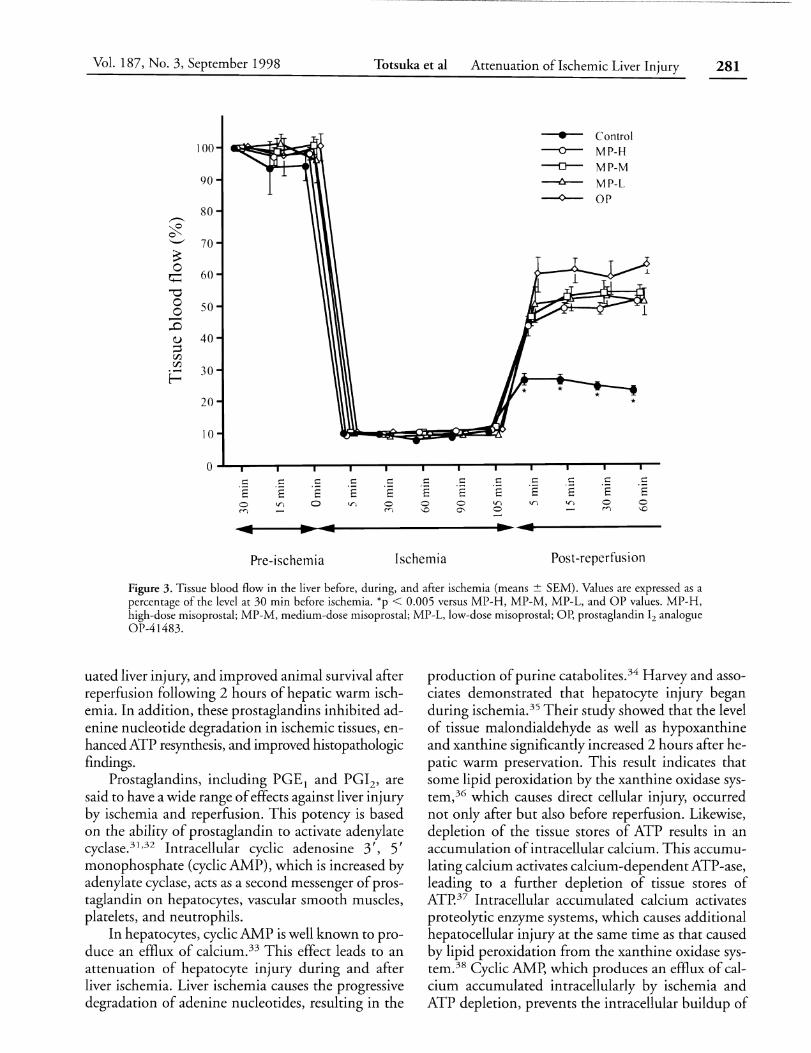
Hepatic tissue blood flow The levels of HTBF after reperfusion were less
than 25% of initial value in the control group, around 50% in the misoprostol-treated groups, and around 60% in the OP-41483-treated group (Fig.
Table 1. Arterial Blood Gas Analysis 6 Hours After Reperfusion (Mean ± SEM)
------~------------
Group
Control MP-H MP-M MP-L OP
Pa02 (mmHg) PaC02 (mmHg) --~------------~----~
92.7 ± 3.56 32.1 ± 1.96 69.2 ± 8.77* 43.6 ± 4.03* 92.2 ± 3.59 33.6 ± 1.51 87.1 ± 4.94 33.6 ± 3.19 94.3 ± 2.20 31.4 ± 1.1 0
*p < 0.05 versus control, MP-M. MP-L, and OP values, Pa02 , pressure of arterial oxygen; PaC0 2 • presure of arterial carbon dioxide,
MP-H. high-dose misoprostal; MP-M. medium-dose misoprostal; MP-L, lowdose misoprostal; Or, prostaglandin 12 analogue OP-41483.
3). There were significant differences between the control and study groups in all postischemic measurements.
Liver function test Serum AST, ALT and LDH increased sharply
after reperfusion in all the groups, while the elevations of liver enzymes in the low dose misoprostol group were significantly suppressed during the early phase of reperfusion (Fig. 4, Table 2). These values in the control and MP-H groups progressively increased and reached peak levels at 12 hours after reperfusion. By contrast, treatment with middle and low dose misoprostol, and OP-41483 significantly inhibited these elevations, although one MP-L animal that died within 24 hours after surgery showed high levels of liver enzymes.
Serum TBA values increased in all animals after reperfusion (Table 2). In the control group, the value

280 Totsuka et a1 Attenuation ofIschemic Liver Injury ] Am Coll Surg
100
90 MP-L
80
- 70
~ MP-H '-' <l) 60 ..... ell .... ~ 50 .;; !3 40
rfJ Control
30
20
10
0 0 2 3 4 5 6 7 8 9 10 II 12 13 14
Days after operation
Figure 2. Survival of dogs after 2 hours of total hepatic vascular exclusion. *p < 0.05 versus control group. MP-M, medium-dose misoprostal; MP-L, low-dose misoprostal; MP-H, high-dose misoprostal.
progressively increased, while those in the MP-H, MP-M, MP-L, and op groups began to decrease 3 hours after reperfusion. There were significant differences between the control group and the treated groups at 6, 12, and 24 hours after reperfusion.
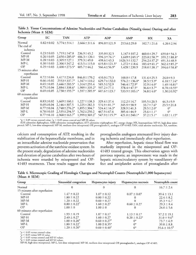
Liver tissue biochemistry Changes in adenine nucleotides and purine ca
tabolites in liver tissue at the end of ischemia and after reperfusion are summarized in Table 3. Two hours of hepatic vascular exclusion in the control animals induced a 54.5% decrease in total adenine nucleotide (TAN) and an 840% increase in purine catabolites, compared with preischemic values. In particular, the ischemia caused >90% reduction in ATp, and> 1,000% production ofHX and XA. Misoprostol and OP-41483 treatments retarded the degradation of adenine nucleotides to purine catabolites during ischemia, especially in the MP-H and OP groups. While resynthesis of ATP was suppressed after reperfusion in control livers, prompt increases in ATP levels were seen with treated livers, especially in the MP-L and OP groups. Likewise, the levels of tissue purine catabolites in the treated livers were significantly lower than those in the control livers after reperfusion. At 60 minutes after reperfusion, however, ATP levels in the MP-H livers were not
different from those in control livers. Concerning changes in hepatic energy charge (EC), no significant differences were found among all the groups.
Histopathology Histopathologic examination showed no differ
ences between control and misoprostol-treated groups at the end of ischemia. After reperfusion, the control livers developed sinusoidal congestion and derangement, ballooning, bridging hepatocyte necrosis, and endothelial cell detachment. According to the histologic score, it was demonstrated that treatment with middle and low dose misoprostol, and OP-41483 significantly alleviated these structural abnormalities (Table 4). The high dose-treated liver showed almost the same sinusoidal congestion and parenchymal injury as those of the control group, even though parenchymal necrosis was rarely seen in the MP-H livers. The numbers of neutrophils infiltrated into postischemic livers 60 minutes after reperfusion in the MP-H and OP groups were significantly less than that in control group. (Table 4).
DISCUSSION In this study, we demonstrated that two kinds of prostaglandin analogues, misoprostol and OP-41483, lessened microcirculatory disturbance, atten-
Vol. 187, No.3, September 1998 Totsuka et al Attenuation of Ischemic Liver Injury 281
,S E o 0'
,S E V) 0
...
• --0--
---0--
~
--0--
s: s: 's 's V) V)
:4
Control MP-H MP-M MP-L OP
s: s: 'S 'S 0 0 M 'D
Ischemia Post-reperfusion
Figure 3. Tissue blood flow in the liver before, during, and after ischemia (means:!: SEM). Values are expressed as a percentage of the level at 30 min before ischemia. *p < 0.005 versus MP-H, MP-M, MP-L, and OP values. MP-H, high-dose misoprostal; MP-M, medium-dose misoprostal; MP-L, low-dose misoprostal; Or, prostaglandin 12 analogue OP-41483.
uated liver injury, and improved animal survival after reperfusion following 2 hours of hepatic warm ischemia. In addition, these prostaglandins inhibited adenine nucleotide degradation in ischemic tissues, enhanced ATP resynthesis, and improved histopathologic findings.
Prostaglandins, including PGEl and PGlz, are said to have a wide range of effects against liver injury by ischemia and reperfusion. This potency is based on the ability of prostaglandin to activate adenylate cyclase.31 ,32 Intracellular cyclic adenosine 3', 5' monophosphate (cyclic AMP), which is increased by adenylate cyclase, acts as a second messenger of prostaglandin on hepatocytes, vascular smooth muscles, platelets, and neutrophils.
In hepatocytes, cyclic AMP is well known to produce an efflux of calcium.33 This effect leads to an attenuation of hepatocyte injury during and after liver ischemia. Liver ischemia causes the progressive degradation of adenine nucleotides, resulting in the
production of purine catabolites.34 Harvey and associates demonstrated that hepatocyte injury began during ischemia.35 Their study showed that the level of tissue malondialdehyde as well as hypoxanthine and xanthine significantly increased 2 hours after hepatic warm preservation. This result indicates that some lipid peroxidation by the xanthine oxidase system,36 which causes direct cellular injury, occurred not only after but also before reperfusion. Likewise, depletion of the tissue stores of ATP results in an accumulation of intracellular calcium. This accumulating calcium activates calcium-dependent ATP-ase, leading to a further depletion of tissue stores of ATp'37 Intracellular accumulated calcium activates proteolytic enzyme systems, which causes additional hepatocellular injury at the same time as that caused by lipid peroxidation from the xanthine oxidase system.38 Cyclic AMp, which produces an efflux of calcium accumulated intracellularly by ischemia and ATP depletion, prevents the intracellular buildup of

282 Totsuka et a1 Attenuation ofIschemic Liver Injury J Am Coli Surg
Figure 4. Changes in serum aspartate aminotransferase (AST) levels after ischemia (means ± SEM). *p < 0.05 versus control value, #p < 0.05 versus control and MP-H values. MP-H, high-dose misoprostal; MP-M, medium-dose misoprostal; MP-L, low-dose misoprostal; Or, prostaglandin 12 analogue OP-41483.
Table 2. Alanine Aminotransferase, Lactate Dehydrogenase, and Total Bile Acid Levels after Ischemia (Mean ± SEM)
Preischemia 60 minutes 3 hours 6 hours 12 hours 24 hours ~----- --- ----
Alanine aminotransferase (U/L)
Control 32.2±4.2 4,961±798 6,576± 1,007 9,332± 1,128 13,563± 1,379 12,681 ± 1,451 MP-H 26.2±4.0 3,527±686 4,057±736 6,248±696* 9,006± 1,629* 4,414±448* MP-M 26.6±5.5 2,302±1,183* 2,745±1,300* 1,644±465t 1,668±655t 1,315±361 1
MP-L 23.0±2.8 1,444±508* 2,087±749* 2,580±842t 2,544± 1,005t 2,185±1,028t
OP 18.2±3.0 2,274±540* 2,873±5lO* 2,082±3331 1,443±272t 723±241 t Lactate dehydrogenase
(U/L) Control 80.6±15.7 3,385±474 3,128±179 2,906±559 3,046±634 1,441 ±216 MP-H 40.6±6.3 2,966±480 2,784±291 2,662±505 2,919±532 1,099±491 MP-M 35.2±8.8 1,199±541 t 934±358"1 521±153t 358±77.8t 195±53.3t
MP-L 42.0±4.9 1,364±527t 1,165±419t 1,290±401* 813±228t 415± 173 t
OP 36.4±4.6 1,207±4581 1,109±404t 354±9l.9t 244±36.5t 190±52.1t Total bile acid
(f.LmollL) Control 0.3±0.3 48.5±12.4 100.2± 11.6 133.8± 12.5 124.3± 14.0 175.4±42.2 MP-H 0 66.0±17.6 90.0± 14.2 8l.9±22.5* 50.8±35.5* 11.9± 14.5* MP-M 0 13.7±6.3 18.0±8.5t 10.2±lO.Ot 0.74±0.83t 0* MP-L 0 16.3± 11.2 43.9±23.4* 43.9±26.9* 14.6±8.5* 5.4±3.7* OP 0 32.9±30.6 66.2±61.5 45.5±27.0* 23.6±9.1* 16.0± 18.2*
*p < 0.05 versus control value. t p < 0.05 versus control and MP-H values. MP-H, high-dose misoprostal; MP-M, medium-dose misoprostal; MP-L, low-dose misoprostal; OP, prostaglandin I2 analogue 01'-41483.
Vol. 187, No.3, September 1998 Totsuka et al Attenuation ofIschemic Liver Injury 283
Table 3. Tissue Concentrations of Adenine Nucleotides and Purine Catabolites (Nmol/g tissue) During and after Ischemia (Mean ± SEM)
Group EC TAN ATP ADP AMP HX XA Normal 0.82±0.02 3,774±314.1 2,666±311.6 The end of
il94.0± 121.9 213.6±29.0 102.7±2l.6 4.20±2.94
ischemia Control 0.23±0.03 1,719±147.8 236.9±45.2 335.0±32.5 1,147± 107.2 460.0±29.7 659.0±54.3 MP-H 0.26±0.05 2,650±285.0* 406.3±120.1 594.3±76.1* 1,649±249.1 * 223.8±58.7* 359.2±88.8* MP-M 0.28±0.03 2,305±127.1 379.3±49.0 498.6±43.3 1426.5± 132.7 254.2±37.2* 491.3±48.9 MP-L 0.30±0.03 2,306±347.2 413.5± 115.0 619.8±12l.5* 1,272± 118.6 183.4±41.1* 362.4± 85.2* OP 0.29±0.O3 2,400±273.5* 405.7±44.2 566.4±56.9* 1,428±230.9 218.0±38.1 * 394.6± 58.8*
15 minutes after reperfusion
Control 0.72±0.04 1,417±234.0 846.8±170.2 410.0±73.3 160.0±17.8 13l.6±25.5 24.0±9.3 MP-H 0.66±0.02 2553±337.7* 1,347± 110.2 629.7± 132.0 576.2± 136.0* 38.5±5.9* 6.18±7.14* MP-M 0.65±0.05 2,446±203.5* 1,314±255.4 585.5±52.9 546.4±72.9* 32.9±4.0* l.49±0.81 * MP-L 0.75±0.04 2,884±338.6* 1,909±335.2* 597.2±77.1 378.4±47.5* 36.6±3.7* 0.78±0.55* OP 0.69±0.05 2,738± 193.7* 1,597±205.9* 607.6± 123.7 533.9± 103.1 * 34.8±4.0* l.30±0.92*
60 minutes after reperfusion
Control 0.83±0.02 1,669±160.1 1,227±138.3 329.4±37.4 112.2±14.7 105.5±20.5 44.3±9.9 MP-H 0.69±0.04 2,146±307.5 1,225 ± 282.3 574.9±81.7* 345.9±58.9 33.7±7.6* 23.5±21.8 MP-M 0.77±0.04 2,748±276.2* 1,895±304.7 524.4± 16.3* 328.9±41.3 29.5±2.0* O' MP-L 0.78±O.03 2,535 ±294.5* 1,762±250.2 467.9±67.6 305.4±44.9 25.6± 1.1 * 0* OP 0.77±0.10 2,968± 163.7t 1,999 ± 369.2 t 547.9±1l5.7* 42l.0±240.1* 37.2±5.1* 1.03± 1.15*
'p < 0.05 versus control value; tp < 0.05 verSllS control and MI'-H values. ADP, adenosine diphosphate; AMP, adenosine monophosphate; ATI~ adenosine triphosphate; Ee, energy charge; HX, hypoxanthine; MP-H, high-dose miso
prostal; MP-L, low-dose misoprostal; MP-M, medium-dose misoprostal; Or, proS{aglandin 12 analogue 01'-41483; TAN, total adenine nucleotide; XA, xanthine.
calcium and consumption of ATp, resulting in the stabilization of the hepatocellular membrane, and in an intracellular adenine nucleotide preservation that prevents activation of the xanthine oxidase system. In the present study, degradation of adenine nucleotides and elevation of purine cat abo lites after two hours of ischemia were retarded by misoprostol and op-41483 treatments. These results suggest that these
prostaglandin analogues attenuated liver injury during ischemia and immediately after reperfusion.
After reperfusion, hepatic tissue blood flow was markedly improved in the misoprostol and op-41483 treated groups. This observation agrees with previous reports; an improvement was made in the hepatic microcirculatory system by vasodilatory effect and anti platelet action of prostaglandin after
Table 4. Microscopic Grading of Histologic Changes and Neutrophil Counts (Neutrophils/l,OOO hepatocytes) (Mean ± SEM)
Group Sinusoidal congestion
Normal 15 minutes after reperfusion
Control MP-H MP-M MP-L OP
60 minutes after reperfusion Control MP-H MP-M MP-L OP
*p < 0.05 versus control value. tp < 0.05 versus MP-H value. "p < 0.05 versus control and MP-H values. §p < 0.05 versus control and MP-H values.
0
1.47 ± 0.22 1.80 ± 0.22 1.20 ± 0.22 0.80 ± O.22T 1.00 ± a
1.93±0.19 2.40 ± 0.27 1.00 ± 0.20t l.80 ± O.22t 1.20 ± 0.20+
Hepatocytes injury Hepatocytes necrosis Neutrophils count ---- ---------
0 0 16.7 ± 2.4
1.07 ± 0.12 0.07 ± 0.07 39.4 ± 13.1 0.80 ± 0.22 0 22.4 ± 8.2 0.60 ± 0.27 0 35.3 ± 6.7 1.60 ± 0.27 0.60 ± 0.27 29.3 ± 8.4 1.00 ± 0 0 24.0 ± 5.6
1.87 ± 0.17 1.13±0.17 57.2 ± 19.1 1.60 ± 0.27 0.20 ± 0.22* 3l.0 ± 9.6§ 0.60 ± 0.27+ 0* 73.7 ± 6.9 1.00 ± 0.35* 0* 39.0 ± 8.8 0.60 ± 0.40+ 0* 35.6 ± 1O.5§
MP-H, high-dose misoprostal; MP-L, low-dose misoprostal; MP-M, medium-dose misoprostal; Or, prostaglandin 12 analogue 01'-41483.

284 Totsuka et al Attenuation of Ischemic Liver Injury ] Am Coll Surg
reperfusion.3 '),40 Cyclic AMP was reported to produce an efflux of calcium in vascular smooth muscles through the activation of the Ca + + -activated K+ channel. 41 The decline of in tracellular calci urn causes vasodilation as a consequence of vasorelaxation. In platelets, cyclic AMP suppresses the inositol phospholipid metabolic turnover that generates both inositol I,4,5-triphosphate (IP 3) and I,2-diacylglycerol (DAG).42,41 This function prevents the mobilization of intracellular calcium by IP 3 and the activation of protein kinase C by DAG. In this mechanism, cyclic AMP inhibits the process of platelet aggregation, such as ADP release and activation of phospholipase A2 , which produces thromboxan A2 from arachidonic acid. These functions produced by prostaglandin contribute to an increase in organ blood flow.
Many investigators demonstrated that prostaglandin has antiinflammatory potency. At the initial phase of reperfusion after liver ischemia, activated Kupffer cells release the inflammatory mediators such as tumor necrosis factor or interleukin-I.44 Neutrophils, which are activated by these mediators or activated endothelial cells,45 infiltrate the liver tissue and produce reactive oxygen species by means of the NADPH oxidase system, resulting in the progression of the liver injury.46 This injury urges platelets into an activation, causing aggregation and further activationY As a result, microcirculatory failure of the liver has occurred throughout these processes. On the other hand, cyclic AMp, which is increased by prostaglandin in neutrophils, inhibits intracellular calcium elevation and activation of protein kinase C, resulting in direct suppression of neutrophil adhesion, lysosomal enzyme release, and NADPH oxidase system.48 Since prostaglandin E2 was reported to inhibit the release of inflammatory mediators from activated Kupffer cells by elevating intracellular cyclic AMp,49 prostaglandin may block the activation of neutrophils induced by the mediators indirectly. In this study, high doses of misoprostol and OP-41483 significantly inhibited the infiltration of neutrophils 60 minutes after reperfusion. Although the numbers of neutrophils in the MP-M and MP-L livers were not different from those in control livers, the elevation ofliver enzyme release was markedly suppressed in both the MP-M and MP-L groups as well as in the OP group compared with the control group for 3 hours after reperfusion. The values of TBA, which reflect hepatic circulation and hepatocyte function more sensitively than the standard liver function tests, 50 were sustained at low levels in these three
groups. These results suggest that the misoprostol and OP-41483 treatments could protect the liver from progressive damage by neutrophils after reperfusion.
In this study, we made three misoprostol-treated groups depending on the drug dosage, because the potency and optimal dose of misoprostol against liver injury has not yet been determined. Dosages of misoprostolused were based on a previous report. Using a model of 90 minutes partial liver ischemia in rats, Lim and associates51 showed that both two-dose treatments of 25 mg/kg misoprostal before ischemia and before reperfusion, and a single dose treatment of 25 mg/kg misoprostol before ischemia were effective in attenuating ischemia and reperfusion injury. We tried three dosages: total IOO, 50, and 25 mg/kg misoprostol, and we divided the drug administration between before and after hepatic ischemia in the ratio of seven to three. To avoid sudden changes in cardiovascular system caused by a bolus injection, the drug was administered by continuous intravenous infusion. We found that the high dose misoprostol treatment partially protected the liver against ischemic injury, but produced an unstable cardiovascular system and pulmonary dysfunction that would have an adverse influence on liver function. We considered that this adverse effect was due to not only vasodilation but also vagal reflexes caused by high-dose misoprostol administration. Previously, two clinical cases were reported in which cardiac arrest occurred after severe hypotension and bradycardia induced by E-type prostaglandin treatment. 52, 51 In experimental study, it was demonstrated that PGI2 stimulated vagal cardiac c fibers, causing vagal reflexes, such as a decrease in cardiac output and bradycardia. 54,55
Therefore, an excessive dose of misoprostol as well as PGI2 may produce the vagal reflex, although this hypothesis has not been proved. In the present study, severe hypotension and bradycardia were exhibited in the MP-H animals, and this cardiovascular instability must have an adverse influence on the postischemic condition. In this group, sinusoidal congestion and hepatocyte injury developed, probably from low cardiac function after reperfusion, resulting in suppression of ATP resynthesis, even though the inhibition of adenine nucleotide degradation during ischemia and the improvement ofHTBF after reperfusion were induced by treatment with high dose misoprostol. In addition to the liver findings, the high dose misoprostol treatment led to pulmonary failure after the operation. Although histologic examination of the lung was not performed, we conjec-

Vol. 187, No.3, September 1998 Totsuka et al Attenuation of Ischemic Liver Injury 285
tured that pulmonary edema occurred in the MP-H animals by misoprostol-induced heart failure and dilatation of the pulmonary artery.
Regarding two lower dose misoprostol groups, 2-week animal survival and amelioration of histologic abnormalities in the MP-L group were poorer than those in the MP-M group, although inhibitions of liver enzyme release and bile acid elevation were significant in both groups. This result suggests that the low dose misoprostol treatment had a less desirable effect than the middle dose treatment.
On the other hand, the dose of OP-41483 was based on our previous study. 56 When PGI2 was administered at a rate of 1 or 2 /Lg/kg/min for 60 minutes via the portal vein before 3 hours warm ischemia of the canine liver, 2 /Lg/kg/min-PGI2 treatment significantly improved the survival rate and remarkably suppressed the serum liver enzyme elevation, and 1 /Lg/kg/min-PGI2 showed low effects. In addition to the OP-41483 administration at a rate of2/Lg/g/min for 30 minutes before ischemia, 3 hours infusion of 0.5 /Lg/kg/min OP-41483 from 30 minutes before reperfusion was performed in the present study, because it was considered that the halflife ofOP-41483 is too short to inhibit the actions of platelets and neutrophils after reperfusion. OP-41483 was infused via the portal vein to alleviate the cardiovascular side effects of this drug, because PGI2 including OP-41483 is not metabolized by the lung but by the liver. 57 In this method of administration, OP-41483 exerted its effects on our hepatic ischemia and reperfusion model with minimal side effects.
In conclusion, treatments with misoprostol, a PGE1 analogue, and with OP-41483, a PGI2 analogue, successfully prevented liver damage after 2-hour warm ischemia. Attenuation of adenine nucleotide degradation during ischemia, improvement of liver microcirculation at the early phase of reperfusion, and inhibition of neutrophil activation played an important role in liver protection by these prostaglandin analogues. These prostaglandin analogues should be positive applications to clinical liver transplantation.
References
1. Stachira], TarnawskiA, lvey K], et a1. Prostaglandin protection of carbon tetrachloride-induced liver cell necrosis in the rat. Gastroenterology 1981;81:211-217.
2. Crafa E Gugenheim], Saint-Paul MC, et a1. Protective effects of prostaglandin E] on normothermic liver ischemia. Eur Surg Res 1991 ;23:278-284.
3. Helling TS, Wogahn BM, Olson SA, et a1. The effect of prostaglandin E] on liver adenine nucleotides and cytoplasmic enzymes in porcine model of normothermic hepatic ischemia. Hepatology 1995;22: 1554-1559.
4. Sikujara 0, Monden M, Toyoshima K, et al. Cytoprotcctive effect of prostaglandin 12 on ischemia-induced hepatic cell injury. Trans-plantation 1983;36:238-243. .
5. Steininger R, Muhlbacher F, Rauhs R, et al. Protective effect of PGI 2 and diltiazem on liver ischemia and reperfusion in pigs. Transplant Proc 1988;20:999-1002.
6. Lambotte L, Hamptinne B, Alvarez-Lopez A, Besse T. Effects of calcium blocking agents and prostaglandin 12 or E, on the tolerance of the rat liver to ischemia. Transplant Proc 1988;20:986.
7. Besse T, Gustin T, Claeys, P, et al. Effect ofPGI2 and thromboxane antagonist on liver ischemic injury. Eur Surg Res 1989;21 :213-217.
8. OlthoffKM, WasefE, Seu P, et al. PGE I reduces injury in hepatic allografts following preservation. J Surg Res 1991 ;50:595-60 1.
9. Okabe K, Malchesky PS, Nose Y. Protective effect of prostaglandin 12 on hepatic mitochondrial function of the preserved rat liver. TohokuJ Exp Med 1986;150:373-379.
10. Mizoguchi Y, Ysutsui H, Miyajima K, et al. The protective effects of prostaglandin E] in an experimental massive hepatic cell necrosis model. Hepatology 1987;7: 1184-1188.
11. Nakano H, Monden M, Umeshita K, et a1. Cytoprotective effect of prostaglandin 12 analogue on superoxide-induced hepatocyte injury. Surgery 1994;116:883-889.
12. Abecassis M, Falk R, Blendis L, et al. Treatment of fulminant hepatic failure with a continuous infusion of prostin VR (PGE]). Heparology 1987;7:1104.
13. Greig PD, Woolf GM, Sinclair SB, et al. Treatment of primary liver graft nonfunction with prostaglandin E]. Transplantation 1989;48:447-453.
14. Takaya S, Doyle H, Todo S, et al. Reduction of primary nonfunction with prostaglandin E] after clinical liver transplantation. Transplant Proc 1995;27:1862-1867.
15. Cattral MS, AltraifI, Grig PD, et al. Toxic effects of intravenous and oral prostaglandin E therapy in patients with liver disease. Am J Med 1994;97:369-373.
16. Pickles H, O'Grady]. Side effects occurring during administration of epoprostenol (prostacyclin, PGI2 ) in man. Br J Clin PharmacoI1982;14:177-185.
17. Golub M, Zia P, Matsuno M, Horton R. Metabolism of prostaglandin Al and E1 in man. J Clin Invest 1975;56: 1404-141 O.
18. Cho MJ, Allen MA. Chemical stability of prostacyclin (PGI2 ) in aqueous solutions. Prostaglandins 1978; 15:943-954.
19. Henley KS, Lucey MR, Normolle Dr, et al. A double-blind, randomized, placebo-controlled trial of prostaglandin E I in liver transplantation. Hepatology 1995;21 :366-372.
20. Ismail T, Ayres R, KiffP, et al. Enisoprost in liver transplantation. 'Iransplantation 1995;59: 1298-130 1.
21. ManasiaAR, LeibowitzAB, Miller CM, et al. Postoperative intravenous infusion of alprostadil (PGE1) does not improve renal function in hepatic transplant recipients. J Am Coli Surg 1996;182:347-352.
22. Bauer RF. Misoprostol preclinical pharmacology. Dig Dis Sci 1985;30:118S-125S.
23. Foote EF, Lee DR, Karim A, et al. Disposition of misoprosrol and its active metabolite in patients with normal and impaired renal function.] Clin Pharmacol 1995;35:384-389.
24. Yui Y, Takatsu Y, Hattori R, et al. A new stable analogue (15-cyclopentyl-omega-pentanor-5(E)-carbacyclin). Jpn Circ J 1985; 49:571-575.
25. Todo S, Zhu Y, Zhang S, et al. Attenuation of ischemic liver injury by augmentation of endogenous adenosine. Transplantation 1997;63:217-223.
26. Urakami A, Todo S, Zhu Y, et al. Attenuation of ischemic liver injury by monoclonal anti-endothelin antibody, AwETN40.] Am Coll Surg 1997; 185:358-364.
27. Mashige F, Imai K, Osuga T. A simple and sensitive assay of total serum bile acids. Clinica Chimica Acta 1976;70:79-86.
28. Hamamoto I, Takay S, Todo S, et al. Can adenine nucleotides predict primary nonfunction of the human liver homograft? Transplant Int 1994;7:89-95.
29. Atkinson DE. The energy charge of the adenylate pool as a regulatory parameter. Interaction with feedback modifiers. Biochemistry 1968;7:4030-4034.

286 Totsuka et at Attenuation ofIschemic Liver Injury ] Am Coll Surg
30. Stevens A. Enzyme histochemistry: Diagnostic applications. In: Bancroft JD, and Stevens A, eds. Theory and practice ofhistological tC'chniques. New York: Churchill Livingstone; 1996:40 1-411.
31. Sweat FW, Wincek TJ. The stimulation of hepatic adenylate cyclase by prostaglandin E t . Biochem Biophys Res Commun 1973;55:522-529.
32. Wise H, Jones RL. Characterization of prostanoid receptors on rat neurrophils. Br J PharmacoI1994;113:581-587.
33. Levine RA. The role of cyclic AMP in hepatic and gastrointestinal function. Gastroenterology 1970;59:280-300.
34. Marubayashi S, Takenaka M, Dohi K, et a!. Adenine nucleotide metabolism during hepatic ischemia and subsequent blood reflow periods and its relation to organ viability. Transplantation 1980;30:294-296.
35. Harvey PRC, Iu S, McKeown CMB, et a!. Adenine nucleotide tissue concentrations and liver allograft viability after cold preservation and warm ischemia. Transplantation 1988;45: 1016-1020.
36. McCord ]M. Oxygen-derived free radicals in postischemic tissue injury. N Engl] Med 1985;17:159-163.
37. Friedmann NK, Carafoli E, Biber ], Murer H. ATP-dependent Ca2 t uptake in isolated hepatic plasma membrane vesicles. Ann NY Acad Sci 1982;402:440--442.
38. Nicotera P, Hartzell P, Baldi C. Cystamine induces toxicity in hepatocytes through the elevation of cytosolic Ca2 + and the stimulation of a non lysosomal proteolytic system. ] Bioi Chem 1986;201: 14628-14635.
39. Lim Sp, Andrews FJ, Christophi C, O'Brien PE. Microvascular changes in liver after ischemia-reperfusion injury. Protection with misoprostol. Dig Dis Sci 1994;39:1683-1690.
40. Quiroga], Prieto J. Liver cytoprotection by prostaglandins. Pharmacol Ther 1993;58:67-92.
41. Minami K, Fukuzawa K, Nakaya Y, et a!. Mechanism of activation of the Ca2 + -activated K+ channel by cyclic AMP in cultured porcine coronary artery smooth muscle cells. Life Sci 1993;53: 1129-1135.
42. Yun ]CH, Ohman KP, Gill Jr ]R, Keiser H. Effects of prostaglandins, cAMp, and changes in cytosolic calcium on platelet aggregation induced by a thromboxane A2 mimic (U46619). Can J Physiol PharmacoI1991;69:599-604.
43. Geiger J, Nolte C. Walter U. Regulation of calcium mobilization and entry in human platelets by endothelium-derived factors. Am J Physiol 1 994;267:C236-C244.
44. Colletti LM, Remick DG, Burtch GD, et a!. Role of tumor necrosis factor-a in the pathophysiologic alterations after hepatic
ischemialreperfusion injury in the rat. J Clin Invest 1990; 85: 1936-1943.
45. Suzuki S, Toledo-Pereyra LH, Rodriguez P, Lopez F. Role of Kupffer cells in neutrophil activation and infiltration following total hepatic ischemia and reperfusion. Circ Shock 1994; 42:204-209.
46. Wclbourn CRB, Goldman G, Paterson IS, et al. Pathophysiology of ischemia reperfusion injury: central role of the neutrophil. Br J Surg 1991;78:651-655.
47. Russell RC, Roth AC, Kucan JO, Zook EG. Rcpcrfusion injury and oxygen free radicals: a review. J Reconst Microsurg 1989;5: 79-84.
48. Mitsuyama T, Takeshige K, Minakami S. Cyclic AMP inhibits the respiratory burst of c1ectropermeabilized human neutrophils at a downstream site of protein kinase C. Biochemica Biophysica Acta 1993; 1177: 167-173.
49. Karck U, Peters T, Decker K. The release of tumor necrosis factor from endotoxin-stimulated rat Kupffer cells is regulated by prostaglandin E2 and dexamethasone. J Hepatol1988; 7:352-361.
50. Visser n, Noorloos AAB, Meijer S, Hoitsma HFW Serum total bile acids monitoring after experimental orthotopic liver transplantation. J Surg Res 1984;36:147-155.
51. Lim Sp, Andrews FJ, Christophi C, O'Brien PE. Misoprostol hepatoprotection against ischemia-reperfusion-induced liver injury in the rat. Dig Dis Sci 1992;:)7:1275-1281.
52. Kalra PA, Litherland D, Sallomi DF, et a!. Cardiac standstill induced by prostaglandin pessaries. Lancet 1989;24: 1460-1461.
53. Meyer WJ, Benton SL, Hoon TJ, et al. Acute myocardial infarction associated with prostaglandin E2 . Am] Obstet Gynecol1991; 165:359-360.
54. Chapple DJ, Dusting GJ, Hughes R, Vane JR. Some direct and reflex cardiovascular actions of prostacyclin (PGI2 ) and prostaglandin E2 in anaesthetized dogs. Br] Pharmacol1980; 68:437-447.
55. Hintze TH. Reflex regulation of the circulation after stimulation of cardiac receptors by prostaglandins. Federation Proc 1987; 46: 73-80.
56. Todo S, Yokoi H, Podesta L, et a!. Amelioration of normothermic canine liver ischemia with prostacyclin. Transplant Proc 1988; 20:965-968.
57. Dusting GJ, Moncada S, Vane JR. Recirculation of prostacyclin (PGlz) in the dog. Br J Pharmacal 1978;64:315-320.


![OBE022, an Oral and Selective Prostaglandin F Receptor Antagonist · specific prostaglandin synthases], and metabolism via pros-taglandin dehydrogenase enzymes. Prostaglandin E 2](https://static.fdocuments.in/doc/165x107/612431e6b1d2d8488c3d852e/obe022-an-oral-and-selective-prostaglandin-f-receptor-antagonist-specific-prostaglandin.jpg)