(Fluorescent) ELISA Kit Prostaglandin E2 ab136949
20
Version 4 Last Updated 15 January 2019 Instructions for Use For quantitative detection of Prostaglandin E2 (PGE 2 ) in buffers and culture media. This product is for research use only and is not intended for diagnostic use. ab136949 – Prostaglandin E2 (Fluorescent) ELISA Kit
Transcript of (Fluorescent) ELISA Kit Prostaglandin E2 ab136949

Version 4 Last Updated 15 January 2019
Instructions for Use
For quantitative detection of Prostaglandin E2 (PGE2) in buffers and culture media.
This product is for research use only and is not intended for diagnostic use.
ab136949 – Prostaglandin E2 (Fluorescent) ELISA Kit

Discover more at www.abcam.com 1
Table of ContentsINTRODUCTION1. BACKGROUND 22. ASSAY SUMMARY 3
GENERAL INFORMATION3. PRECAUTIONS 44. STORAGE AND STABILITY 45. MATERIALS SUPPLIED 46. MATERIALS REQUIRED, NOT SUPPLIED 57. LIMITATIONS 58. TECHNICAL HINTS 5
ASSAY PREPARATION9. REAGENT PREPARATION 610. STANDARD PREPARATIONS 711. SAMPLE COLLECTION AND STORAGE 9
ASSAY PROCEDURE12. ASSAY PROCEDURE 10
DATA ANALYSIS13. CALCULATIONS 1114. TYPICAL DATA 1215. TYPICAL SAMPLE VALUES 1316. ASSAY SPECIFICITY 14
RESOURCES17. TROUBLESHOOTING 1718. NOTES 18
Discover more at www.abcam.com 2
INTRODUCTION
1. BACKGROUND
Abcam’s Fluorescent Prostaglandin E2 (PGE2) in vitro competitive ELISA (Enzyme-Linked Immunosorbent Assay) kit is designed for the accurate quantitative measurement of Prostaglandin E2 in buffers and culture media. A solution of PGE2 conjugated to fluorescein is added to uncoated wells containing standards or samples. A solution of monoclonal antibody to PGE2 is then added. This binds PGE2 in a competitive manner. This is a Fluorescence polarization immunoassay (FPIA), where the underlying principle is “fluorescence anisotropy” or “fluorescence polarization”. The rate of the rotation of a molecule in motion is indicative of its size. Low amount of analyte results in more antibody molecules bound to the fluorophore probe, read-out: high signal. High amount of analyte results in less antibody molecules bound to the fluorophore probe, read-out: low signal. Hence, the signal is inversely proportional to the concentration of PGE2 in the standards or samples. The assay should be performed in a black uncoated low-binding microtiter plate (not supplied) and is analyzed using a Fluorescence polarization detector, capable of reading emissions at 520 - 535 nm, with excitation at 485 nm.Prostaglandin E2 is formed in a variety of cells from PGH2, which itself is synthesized from arachidonic acid by the enzyme prostaglandin synthetase. Prostaglandin E2 has been shown to have a number of biological actions, including vasodilation, both anti- and proinflammatory action, modulation of sleep/wake cycles, and facilitation of the replication of human immunodeficiency virus. It elevates cAMP levels, stimulates bone resorption, and has thermoregulatory effects. It has been shown to be a regulator of sodium excretion and renal hemodynamics.

Discover more at www.abcam.com 3
INTRODUCTION
2. ASSAY SUMMARY
Prepare all reagents and samples as instructed. Add standards and samples to uncoated wells.
Add prepared PGE2 FPIA conjugate to appropriate wells.
Add PGE2 antibody to appropriate wells. Incubate at room temperature for 30 minutes.
Read immediately. The amount of signal is inversely proportional to the concentration of PGE2 in the standards or samples.
Steps 1-3 are carried out one after another.

Discover more at www.abcam.com 4
GENERAL INFORMATION
3. PRECAUTIONSPlease read these instructions carefully prior to beginning the assay. Some kit components contain azide, which may react with lead or
copper plumbing. When disposing of reagents always flush with large volumes of water to prevent azide build-up
We test the performance of this kit with a variety of samples, however it is possible that high levels of interfering substances may cause variation in assay results
The Prostaglandin E2 Standard provided, is supplied in ethanolic buffer at a pH optimized to maintain PGE2 integrity. Care should be taken handling this material because of the known and unknown effects of prostaglandin.
4. STORAGE AND STABILITYStore kit at 4°C immediately upon receipt, apart from the PGE2 FPIA concentrate and PGE2 Standard, which should be stored at -20°C. Avoid multiple freeze-thaw cycles.
5. MATERIALS SUPPLIED
Item AmountStorage
Condition(Before
Preparation)PGE2 FPIA Antibody 8 mL 4ºCAssay Buffer 1 Concentrate 125 mL 4ºCPGE2 FPIA Conjugate Concentrate 0.1 mL -20ºCProstaglandin E2 Standard (1,000,000 pg/mL) 500 µL -20ºC

Discover more at www.abcam.com 5
GENERAL INFORMATION
6. MATERIALS REQUIRED, NOT SUPPLIEDThese materials are not included in the kit, but will be required to successfully utilize this assay:
Solid black uncoated low-binding microtiter plate.
Foil microtiter plate sealer, if desired.
Fluorescence polarization detector, capable of reading emissions at 520 - 535 nm, with excitation at 485 nm.
7. LIMITATIONS Assay kit intended for research use only. Not for use in diagnostic
procedures Do not mix or substitute reagents or materials from other kit lots or
vendors. Kits are QC tested as a set of components and performance cannot be guaranteed if utilized separately or substituted
8. TECHNICAL HINTS Glass tubes must be used for standard preparation.
Pre-rinse the pipette tip with the reagent, use fresh pipette tips for each sample, standard and reagent.
Pipette standards and samples to the bottom of the wells.
Add the reagents to the side of the well to avoid contamination.
This kit is sold based on number of tests. A ‘test’ simply refers to a single assay well. The number of wells that contain sample, control or standard will vary by product. Review the protocol completely to confirm this kit meets your requirements. Please contact our Technical Support staff with any questions

Discover more at www.abcam.com 6
ASSAY PREPARATION
9. REAGENT PREPARATIONEquilibrate all reagents and samples to room temperature (18 - 25°C) prior to use.
9.1 Assay BufferPrepare the assay buffer by diluting 100 mL of the supplied Assay Buffer 1 Concentrate with 900 mL of deionized water. This can be stored at room temperature until the kit’s expiration, or for 3 months, whichever is earlier. Use the diluted assay buffer for the entire assay.
9.2 PGE2 ConjugateCount the total number of wells that will receive conjugate. Use the following formula to calculate the volume of PGE2 Conjugate Concentrate and the assay buffer needed to prepare PGE2 conjugate.9.2.1 (Number of wells + 1) x 0.05 mL / well = Volume of assay buffer needed. Increase the calculated volume to the next whole milliliter.9.2.2 (Volume from part A) x 10 μL / mL = Volume of PGE2
Conjugate Concentrate needed.
Pipet the volume of assay buffer from part A into an amber container. From this volume, remove the volume calculated in part B. Add the calculated PGE2 Conjugate Concentrate to the assay buffer. Vortex thoroughly and use.
Discover more at www.abcam.com 7
ASSAY PREPARATION
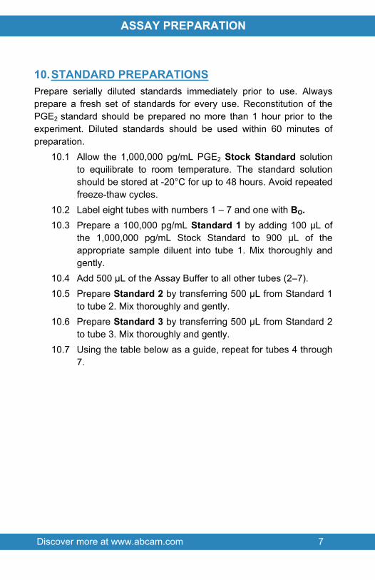
10.STANDARD PREPARATIONSPrepare serially diluted standards immediately prior to use. Always prepare a fresh set of standards for every use. Reconstitution of the PGE2 standard should be prepared no more than 1 hour prior to the experiment. Diluted standards should be used within 60 minutes of preparation.
10.1 Allow the 1,000,000 pg/mL PGE2 Stock Standard solution to equilibrate to room temperature. The standard solution should be stored at -20°C for up to 48 hours. Avoid repeated freeze-thaw cycles.
10.2 Label eight tubes with numbers 1 – 7 and one with BO.10.3 Prepare a 100,000 pg/mL Standard 1 by adding 100 µL of
the 1,000,000 pg/mL Stock Standard to 900 µL of the appropriate sample diluent into tube 1. Mix thoroughly and gently.
10.4 Add 500 μL of the Assay Buffer to all other tubes (2–7).10.5 Prepare Standard 2 by transferring 500 μL from Standard 1
to tube 2. Mix thoroughly and gently. 10.6 Prepare Standard 3 by transferring 500 μL from Standard 2
to tube 3. Mix thoroughly and gently. 10.7 Using the table below as a guide, repeat for tubes 4 through
7.

Discover more at www.abcam.com 8
ASSAY PREPARATION
Standard#
Sample toDilute
Volume to Dilute
(µL)
Volume of
Diluent (µL)
StartingConc.
(pg/mL)
Final Conc.
(pg/mL)
1 Standard 100 900 1,000,000 100,0002 Standard 1 500 500 100,000 50,0003 Standard 2 500 500 50,000 25,0004 Standard 3 500 500 25,000 12,5005 Standard 4 500 500 12,500 6,2506 Standard 5 500 500 6,250 3,1257 Standard 6 500 500 3,125 1,562
BO None - 500 - 0
Discover more at www.abcam.com 9
ASSAY PREPARATION
11.SAMPLE COLLECTION AND STORAGE This kit is compatible with PGE2 samples in a wide range of
matrices. Samples diluted sufficiently into Assay Buffer can be read directly from the standard curve. Samples containing organic solvents or inherently fluorescing materials may interfere with the assay.
Samples in the majority of tissue culture media, including those containing fetal bovine serum, can also be read in the assay, provided the standards have been diluted into the tissue culture media instead of Assay Buffer. There will be a small change in binding associated with running the standards and samples in media.
Store samples to be assayed within 24 hours at 2 - 8°C. For long-term storage, aliquot and freeze samples at -20°C. Avoid repeated freeze-thaw cycles.

Discover more at www.abcam.com 10
ASSAY PROCEDURE
12.ASSAY PROCEDURE Equilibrate all materials and prepared reagents to room
temperature prior to use It is recommended to assay all standards, controls and
samples in duplicate12.1 Prepare all reagents, working standards, and samples as
directed in the previous sections.12.1 Pipet 150 μL of assay buffer into the Total Fluorescence
(TF) wells.12.1 Pipet 100 μL of assay buffer into the Bo (0 pg/mL) wells.12.1 Pipet 100 μL of Standards 1 through 7 to the bottom of the
appropriate wells.12.1 Pipet 100 μL of the samples to the bottom of the
appropriate wells.12.1 Add 50 μL of the conjugate into each well.12.1 Add 50 μL of antibody into each well, except the TF wells.12.1 Seal the plate with a foil plate sealer. Incubate the plate for
at least 30 minutes at room temperature. The FP signal is stable for at least 20 hours.
12.1 Read plate on a suitable fluorescence polarization detector at 520 - 535 nm emission, with excitation at 485 nm, using the appropriate settings for that instrument.

Discover more at www.abcam.com 11
DATA ANALYSIS
13.CALCULATIONSSeveral options are available for the calculation of the concentration of PGE2 in samples. We recommend that the data be handled by an immunoassay software package utilizing a 4 parameter logistic curve fitting program. If data reduction software is not readily available, the concentrations can be calculated as follows:
Calculate the binding for each standard and sample as a percentage of the maximum binding (Bo), using the following formula:
Percent Bound = Average mP x 100 Average Bo
Plot the Percent Bound for each standard versus PGE2 concentration in each standard. Approximate a straight line through the points. The concentration of the unknowns can be determined by interpolation.
Samples with concentrations outside of the standard curve range will need to be re-analyzed using a higher dilution.
Discover more at www.abcam.com 12
DATA ANALYSIS
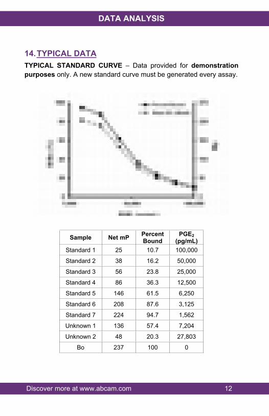
14.TYPICAL DATATYPICAL STANDARD CURVE – Data provided for demonstration purposes only. A new standard curve must be generated every assay.
Sample Net mP Percent Bound
PGE2 (pg/mL)
Standard 1 25 10.7 100,000
Standard 2 38 16.2 50,000
Standard 3 56 23.8 25,000
Standard 4 86 36.3 12,500
Standard 5 146 61.5 6,250
Standard 6 208 87.6 3,125
Standard 7 224 94.7 1,562
Unknown 1 136 57.4 7,204
Unknown 2 48 20.3 27,803
Bo 237 100 0

Discover more at www.abcam.com 13
DATA ANALYSIS
15.TYPICAL SAMPLE VALUESSENSITIVITY –The sensitivity of the assay, defined as the concentration of PGE2 measured at 2 standard deviations from the mean of 24 zeros along the standard curve, was determined to be 684 pg/mL.
SAMPLE RECOVERY –PGE2 standard was spiked into the following buffers, which were already diluted with the assay buffer, and measured in the kit. The results are shown below. These are the minimum dilutions required to remove the matrix interference of these solutions.
Sample Average % Recovery
Recommended Dilution
Hanks Buffered Saline Solution 108 ≥ neatRPMI-1640 without phenol red 95 ≥ 1:4RPMI-1640 with phenol red, sodium pyruvate and essential amino acids 100 ≥1:16
RPMI-1640 with phenol red, 10% FBS and antibiotics 105 ≥1:32
LINEARITY OF DILUTION –A buffer sample containing PGE2 was serially diluted 1:2 in the kit assay buffer and measured in the assay. The results are shown in the table below.
Dilution Expected (pg/mL) Observed (pg/mL) Recovery (%)Neat - 76,035 -1:2 38,017 31,610 831:4 19,009 19,701 1041:8 9,504 11,713 123
1:16 4,752 4,228 891:32 2,376 2,329 98
Discover more at www.abcam.com 14
DATA ANALYSIS
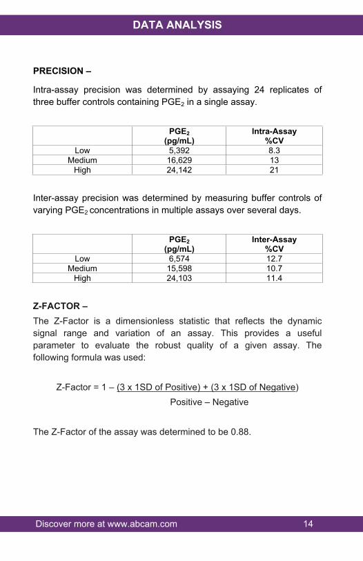
PRECISION –
Intra-assay precision was determined by assaying 24 replicates of three buffer controls containing PGE2 in a single assay.
PGE2(pg/mL)
Intra-Assay%CV
Low 5,392 8.3Medium 16,629 13
High 24,142 21
Inter-assay precision was determined by measuring buffer controls of varying PGE2 concentrations in multiple assays over several days.
PGE2(pg/mL)
Inter-Assay%CV
Low 6,574 12.7Medium 15,598 10.7
High 24,103 11.4
Z-FACTOR –The Z-Factor is a dimensionless statistic that reflects the dynamic signal range and variation of an assay. This provides a useful parameter to evaluate the robust quality of a given assay. The following formula was used:
Z-Factor = 1 – (3 x 1SD of Positive) + (3 x 1SD of Negative) Positive – Negative
The Z-Factor of the assay was determined to be 0.88.
Discover more at www.abcam.com 15
DATA ANALYSIS
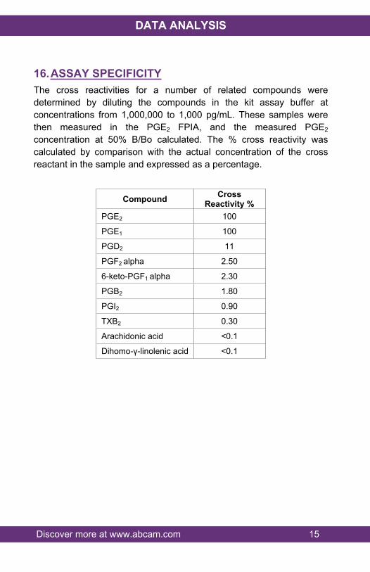
16.ASSAY SPECIFICITYThe cross reactivities for a number of related compounds were determined by diluting the compounds in the kit assay buffer at concentrations from 1,000,000 to 1,000 pg/mL. These samples were then measured in the PGE2 FPIA, and the measured PGE2 concentration at 50% B/Bo calculated. The % cross reactivity was calculated by comparison with the actual concentration of the cross reactant in the sample and expressed as a percentage.
Compound Cross Reactivity %
PGE2 100
PGE1 100
PGD2 11
PGF2 alpha 2.50
6-keto-PGF1 alpha 2.30
PGB2 1.80
PGI2 0.90
TXB2 0.30
Arachidonic acid <0.1
Dihomo-γ-linolenic acid <0.1

Discover more at www.abcam.com 16
DATA ANALYSIS
INTEREFERENCES – PGE2 standard was spiked into the following solutions, which were already diluted with the assay buffer, and measured in the kit. The results were as follows:
Sample Recovery(%)
Recommended Percent(%)
Methanol 107 ≤10Acetonitrile 109 ≤52-propanol 98 ≤2.5
Ethanol 16 ≤2.5DMF 104 ≤1.3
DMSO 102 ≤1.3
Discover more at www.abcam.com 17
RESOURCES
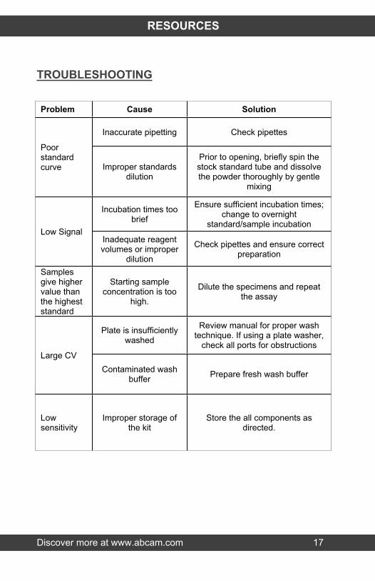
TROUBLESHOOTING
Problem Cause Solution
Inaccurate pipetting Check pipettes
Poor standard curve Improper standards
dilution
Prior to opening, briefly spin the stock standard tube and dissolve the powder thoroughly by gentle
mixing
Incubation times too brief
Ensure sufficient incubation times; change to overnight
standard/sample incubationLow Signal
Inadequate reagent volumes or improper
dilution
Check pipettes and ensure correct preparation
Samples give higher value than the highest standard
Starting sample concentration is too
high.
Dilute the specimens and repeat the assay
Plate is insufficiently washed
Review manual for proper wash technique. If using a plate washer,
check all ports for obstructionsLarge CV
Contaminated wash buffer Prepare fresh wash buffer
Low sensitivity
Improper storage of the kit
Store the all components as directed.
Discover more at www.abcam.com 18
RESOURCES
17.NOTES

RESOURCES 19
UK, EU and ROWEmail: [email protected] | Tel: +44-(0)1223-696000
AustriaEmail: [email protected] | Tel: 019-288-259
FranceEmail: [email protected] | Tel: 01-46-94-62-96 GermanyEmail: [email protected] | Tel: 030-896-779-154 SpainEmail: [email protected] | Tel: 911-146-554 SwitzerlandEmail: [email protected] Tel (Deutsch): 0435-016-424 | Tel (Français): 0615-000-530
US and Latin AmericaEmail: [email protected] | Tel: 888-77-ABCAM (22226)
CanadaEmail: [email protected] | Tel: 877-749-8807
China and Asia Pacific Email: [email protected] | Tel: 400 921 0189 / +86 21 2070 0500
JapanEmail: [email protected] | Tel: +81-(0)3-6231-0940 www.abcam.com | www.abcam.cn | www.abcam.co.jp
Copyright © 2018 Abcam, All Rights Reserved. The Abcam logo is a registered trademark.
All information / detail is correct at time of going to print.