
Atomic structure of a rhinovirus C, a virus species linked ...
6
Atomic structure of a rhinovirus C, a virus species linked to severe childhood asthma Yue Liu a , Marchel G. Hill b , Thomas Klose a , Zhenguo Chen a , Kelly Watters b , Yury A. Bochkov c , Wen Jiang a , Ann C. Palmenberg b,1 , and Michael G. Rossmann a,1 a Department of Biological Sciences, Purdue University, West Lafayette, IN 47907; b Institute for Molecular Virology, University of Wisconsin, Madison, WI 53706; and c Department of Pediatrics, School of Medicine and Public Health, University of Wisconsin, Madison, WI 53706 Edited by Peter Palese, Icahn School of Medicine at Mount Sinai, New York, NY, and approved June 7, 2016 (received for review April 25, 2016) Isolates of rhinovirus C (RV-C), a recently identified Enterovirus (EV) species, are the causative agents of severe respiratory infec- tions among children and are linked to childhood asthma exacer- bations. The RV-C have been refractory to structure determination because they are difficult to propagate in vitro. Here, we report the cryo-EM atomic structures of the full virion and native empty particle (NEP) of RV-C15a. The virus has 60 “fingers” on the virus outer surface that probably function as dominant immunogens. Because the NEPs also display these fingers, they may have utility as vaccine candidates. A sequence-conserved surface depression adjacent to each finger forms a likely binding site for the sialic acid on its receptor. The RV-C, unlike other EVs, are resistant to capsid-binding antiviral compounds because the hydrophobic pocket in VP1 is filled with multiple bulky residues. These results define potential molecular determinants for designing antiviral therapeu- tics and vaccines. rhinovirus C | cryoelectron microscopy | atomic structure | asthma T he Picornaviridae family includes a variety of small, non- enveloped, icosahedral viruses with positive-strand RNA ge- nomes (1). Many picornaviruses (e.g., rhinoviruses, polioviruses, coxsackieviruses, enterovirus A71, and enterovirus D68) that infect humans and cause high morbidity belong to the Enterovirus genus (EV) (1). A number of these viruses have been structurally char- acterized by X-ray crystallography (2–5), establishing the general mechanisms for virus infection and for the development of effective anti-EV therapeutics. Nevertheless, rhinovirus C (RV-C), a newly discovered species among the EVs, remains enigmatic. RV-C viruses (55 types), together with RV-A and RV-B viruses (∼100 types), are the leading cause of common colds. However, the RV-C might be associated with more severe respiratory infections among children than any other known rhinoviruses (6, 7). In con- trast to other RVs, RV-Cs use cadherin-related family member 3 (CDHR3) as a cellular receptor (8). This childhood asthma susceptibility gene product is expressed in the human lower re- spiratory tract (9). In line with this etiology, RV-Cs cause a significantly higher rate of lower respiratory tract infections in children than in adults (10) and are directly associated with childhood asthma exacerbations (7). Similar to influenza, RV-C infections peak in winter months. Currently, there are no vac- cines or effective antiviral treatments available. RV-C isolates have been refractory to structural character- ization since their discovery in 2006 (11) because of an inability to infect standard tissue culture (e.g., HeLa) (12). Only modeled structures, based on amino acid sequence comparisons, have been available to aid biological investigations (12–15) However, with recent advances in direct electron detection (16) and image processing approaches (17, 18), single-particle cryoelectron mi- croscopy (cryo-EM) has now emerged as a powerful method for determining near atomic resolution (better than 4 Å) structures of macromolecular assemblies (19). Cryo-EM requires only a limited amount of sample without intensive purification, offering advantages over X-ray crystallography in structural studies of samples that are difficult to produce. Picornavirus capsids are assembled from 60 copies of biolog- ical protomers, each composed of four proteins: VP1, VP2, VP3, and VP4 (2). The three large surface polypeptides, VP1, VP2, and VP3, are folded into eight-stranded antiparallel “jelly rolls.” During the assembly process, autocatalytic cleavage of precursor VP0 into VP2 and VP4 in the presence of viral RNA results in the formation of full infectious virions (20). The arrangement of jelly rolls in the virions exhibits pseudo T = 3 icosahedral sym- metry with an outer diameter of ∼300 Å (2, 3). The internal surface of the capsid is lined by the 60 copies of VP4. A surface depression or canyon (2), encircling each fivefold axis, is fre- quently the receptor binding site for many EVs (21). Amino acid residues located on the outer surface of the virus but not spe- cifically within this canyon are typically involved in forming im- munogenic sites recognized by neutralizing antibodies. The canyon allows only limited access to these antibodies (22). In many EVs, a hydrophobic pocket within the VP1 jelly roll and situated underneath the canyon floor is occupied by a fatty-acid like molecule, or “pocket factor” (23, 24), that regulates the conformational states of the virus during cell entry (25). Capsid- binding reagents that replace the pocket factor within VP1 are effective antiviral therapeutics against many EVs (26), but not RV-Cs (14). Significance The recently identified rhinovirus C (RV-C) species of picorna- viruses might be associated with more severe respiratory in- fections than other rhinoviruses in children. The RV-C have been linked to 50–85% of hospital-level childhood asthma ex- acerbations that can lead to significant adult respiratory problems. However, there are currently no effective antiviral treatments or vaccines available. Using cryoelectron microscopy, we have de- termined the atomic structures of the full virion and native empty particle (NEP) of a cell-adapted RV-C strain. The structures high- light novel immunogenic sites on the virus surface, a probable binding site for the RV-C receptor, molecular determinants of antiviral resistance, and the rationale of utilizing NEPs as vaccine candidates. These results provide the basis for designing antiviral therapeutics and vaccines. Author contributions: A.C.P. and M.G.R. designed research; Y.L., M.G.H., T.K., Z.C., and K.W. performed research; Y.A.B. and W.J. contributed new reagents/analytic tools; Y.L., A.C.P., and M.G.R. analyzed data; and Y.L., A.C.P., and M.G.R. wrote the paper. The authors declare no conflict of interest. This article is a PNAS Direct Submission. Data deposition: The atomic coordinates of the RV-C15a full particle and of the RV-C15a empty particle have been deposited in the Protein Data Bank, www.pdb.org (PDB ID codes 5K0U and 5JZG). The cryo-EM maps for the RV-C15a full particle and empty particles have been deposited in the Electron Microscopy Data Bank (EMDB ID codes EMD-8189 and EMD-8184). 1 To whom correspondence may be addressed. Email: [email protected] or acpalmen@wisc. edu. This article contains supporting information online at www.pnas.org/lookup/suppl/doi:10. 1073/pnas.1606595113/-/DCSupplemental. www.pnas.org/cgi/doi/10.1073/pnas.1606595113 PNAS Early Edition | 1 of 6 BIOPHYSICS AND COMPUTATIONAL BIOLOGY
Transcript of Atomic structure of a rhinovirus C, a virus species linked ...
Atomic structure of a rhinovirus C, a virus specieslinked to severe childhood asthmaYue Liua, Marchel G. Hillb, Thomas Klosea, Zhenguo Chena, Kelly Wattersb, Yury A. Bochkovc, Wen Jianga,Ann C. Palmenbergb,1, and Michael G. Rossmanna,1
aDepartment of Biological Sciences, Purdue University, West Lafayette, IN 47907; bInstitute for Molecular Virology, University of Wisconsin, Madison,WI 53706; and cDepartment of Pediatrics, School of Medicine and Public Health, University of Wisconsin, Madison, WI 53706
Edited by Peter Palese, Icahn School of Medicine at Mount Sinai, New York, NY, and approved June 7, 2016 (received for review April 25, 2016)
Isolates of rhinovirus C (RV-C), a recently identified Enterovirus(EV) species, are the causative agents of severe respiratory infec-tions among children and are linked to childhood asthma exacer-bations. The RV-C have been refractory to structure determinationbecause they are difficult to propagate in vitro. Here, we reportthe cryo-EM atomic structures of the full virion and native emptyparticle (NEP) of RV-C15a. The virus has 60 “fingers” on the virusouter surface that probably function as dominant immunogens.Because the NEPs also display these fingers, they may have utilityas vaccine candidates. A sequence-conserved surface depressionadjacent to each finger forms a likely binding site for the sialicacid on its receptor. The RV-C, unlike other EVs, are resistant tocapsid-binding antiviral compounds because the hydrophobic pocketin VP1 is filled with multiple bulky residues. These results definepotential molecular determinants for designing antiviral therapeu-tics and vaccines.
rhinovirus C | cryoelectron microscopy | atomic structure | asthma
The Picornaviridae family includes a variety of small, non-enveloped, icosahedral viruses with positive-strand RNA ge-
nomes (1). Many picornaviruses (e.g., rhinoviruses, polioviruses,coxsackieviruses, enterovirus A71, and enterovirus D68) that infecthumans and cause high morbidity belong to the Enterovirus genus(EV) (1). A number of these viruses have been structurally char-acterized by X-ray crystallography (2–5), establishing the generalmechanisms for virus infection and for the development of effectiveanti-EV therapeutics. Nevertheless, rhinovirus C (RV-C), a newlydiscovered species among the EVs, remains enigmatic.RV-C viruses (55 types), together with RV-A and RV-B viruses
(∼100 types), are the leading cause of common colds. However, theRV-C might be associated with more severe respiratory infectionsamong children than any other known rhinoviruses (6, 7). In con-trast to other RVs, RV-Cs use cadherin-related family member3 (CDHR3) as a cellular receptor (8). This childhood asthmasusceptibility gene product is expressed in the human lower re-spiratory tract (9). In line with this etiology, RV-Cs cause asignificantly higher rate of lower respiratory tract infections inchildren than in adults (10) and are directly associated withchildhood asthma exacerbations (7). Similar to influenza, RV-Cinfections peak in winter months. Currently, there are no vac-cines or effective antiviral treatments available.RV-C isolates have been refractory to structural character-
ization since their discovery in 2006 (11) because of an inabilityto infect standard tissue culture (e.g., HeLa) (12). Only modeledstructures, based on amino acid sequence comparisons, havebeen available to aid biological investigations (12–15) However,with recent advances in direct electron detection (16) and imageprocessing approaches (17, 18), single-particle cryoelectron mi-croscopy (cryo-EM) has now emerged as a powerful method fordetermining near atomic resolution (better than 4 Å) structuresof macromolecular assemblies (19). Cryo-EM requires only alimited amount of sample without intensive purification, offeringadvantages over X-ray crystallography in structural studies ofsamples that are difficult to produce.
Picornavirus capsids are assembled from 60 copies of biolog-ical protomers, each composed of four proteins: VP1, VP2, VP3,and VP4 (2). The three large surface polypeptides, VP1, VP2,and VP3, are folded into eight-stranded antiparallel “jelly rolls.”During the assembly process, autocatalytic cleavage of precursorVP0 into VP2 and VP4 in the presence of viral RNA results inthe formation of full infectious virions (20). The arrangement ofjelly rolls in the virions exhibits pseudo T = 3 icosahedral sym-metry with an outer diameter of ∼300 Å (2, 3). The internalsurface of the capsid is lined by the 60 copies of VP4. A surfacedepression or canyon (2), encircling each fivefold axis, is fre-quently the receptor binding site for many EVs (21). Amino acidresidues located on the outer surface of the virus but not spe-cifically within this canyon are typically involved in forming im-munogenic sites recognized by neutralizing antibodies. Thecanyon allows only limited access to these antibodies (22). Inmany EVs, a hydrophobic pocket within the VP1 jelly roll andsituated underneath the canyon floor is occupied by a fatty-acidlike molecule, or “pocket factor” (23, 24), that regulates theconformational states of the virus during cell entry (25). Capsid-binding reagents that replace the pocket factor within VP1 areeffective antiviral therapeutics against many EVs (26), but notRV-Cs (14).
Significance
The recently identified rhinovirus C (RV-C) species of picorna-viruses might be associated with more severe respiratory in-fections than other rhinoviruses in children. The RV-C havebeen linked to 50–85% of hospital-level childhood asthma ex-acerbations that can lead to significant adult respiratory problems.However, there are currently no effective antiviral treatments orvaccines available. Using cryoelectron microscopy, we have de-termined the atomic structures of the full virion and native emptyparticle (NEP) of a cell-adapted RV-C strain. The structures high-light novel immunogenic sites on the virus surface, a probablebinding site for the RV-C receptor, molecular determinants ofantiviral resistance, and the rationale of utilizing NEPs as vaccinecandidates. These results provide the basis for designing antiviraltherapeutics and vaccines.
Author contributions: A.C.P. and M.G.R. designed research; Y.L., M.G.H., T.K., Z.C., andK.W. performed research; Y.A.B. and W.J. contributed new reagents/analytic tools; Y.L.,A.C.P., and M.G.R. analyzed data; and Y.L., A.C.P., and M.G.R. wrote the paper.
The authors declare no conflict of interest.
This article is a PNAS Direct Submission.
Data deposition: The atomic coordinates of the RV-C15a full particle and of the RV-C15aempty particle have been deposited in the Protein Data Bank, www.pdb.org (PDB IDcodes 5K0U and 5JZG). The cryo-EMmaps for the RV-C15a full particle and empty particleshave been deposited in the Electron Microscopy Data Bank (EMDB ID codes EMD-8189and EMD-8184).1To whom correspondence may be addressed. Email: [email protected] or [email protected].
This article contains supporting information online at www.pnas.org/lookup/suppl/doi:10.1073/pnas.1606595113/-/DCSupplemental.
www.pnas.org/cgi/doi/10.1073/pnas.1606595113 PNAS Early Edition | 1 of 6
BIOPH
YSICSAND
COMPU
TATIONALBIOLO
GY

Here we report atomic resolution cryo-EM structures of thefull and native empty particles (NEP) of the cell-adapted RV-C15astrain. These structures highlight novel immunogenic surfaces, aprobable binding site for the glycosylated CDHR3 receptormolecule, and the requirements for antiviral compound resistance.The external surface of both types of particles is almost iden-tical. Thus, the RV-C NEPs represent potential vaccine can-didates. The present findings also identify targets for designinganti–RV-C therapeutics.
ResultsProduction of RV-C15a Viruses. Recently, a recombinant RV-C15virus (12), adapted for tissue culture growth by serial passagein HeLa-E8 cells (8) (a transduced HeLa cell line expressingCDHR3) led to new protocols for enhanced virus yields. Thederivative, RV-C15a, represents a cell-adapted, uncloned pop-ulation. The consensus sequence of this population differs in thecapsid region from that of RV-C15, primarily by a single, high-frequency, nucleotide polymorphism. The substitution convertsresidue 1125 from Thr to Lys. (Numbering convention adds 1,000to VP1 residues, 2,000 to VP2 residues, 3,000 to VP3 residues, and4,000 to VP4 residues.) In this study, an RV-C15a sample, purifiedonly by sucrose cushion sedimentation, was used for cryo-EMstructure analysis. To obtain a minimum of 5–6 particles per mi-crograph, given the high dilution of the sample, data collectionwas carried out at a low magnification. Specifically, movies offrozen RV-C15a particles within a thin layer of vitreous ice wererecorded at a nominal magnification of 14,000× using a Gatan K2Summit direct electron detector. However, the tradeoff was a lowsignal-to-noise ratio and a high anisotropic magnification distor-tion compared with what would be the case were high magnifi-cations used for data collection. The primary data were collectedin less than 1 wk.
Biochemical Characterization of Two Forms of Particles. Cryo-EMmicrographs of RV-C15a showed the presence of two majorforms of particles. One form lacked density at their centers andanother form had density at their centers (Fig. S1). When frac-tionated on sucrose gradients, these types of particles sepa-rated from each other. One form was full, infectious virionsthat contained VP1, VP2, VP3, and VP4, whereas the otherform (∼30% of all particles) was NEP that had VP1, VP3, anduncleaved VP0, as shown by Western blot analyses using an
antibody against VP2 (Fig. S2A). Unlike the full virions, NEPswere devoid of viral RNA and had no infectivity to HeLa-E8cells (Fig. S2B).
Cryo-EM Structure Determination. Images of full and empty parti-cles were separated by reference-free 2D classification using theprogram Relion (17). A “truly independent” procedure of 3Dreconstruction was used to avoid overfitting to noise (18). Es-sentially, initial model calculations, low-resolution refinementsand high-resolution refinements were performed independentlyfor each of the two half-data subsets. Parameters of anisotropicmagnification distortion, a major resolution limiting factor forlarge assemblies (e.g., viruses), were estimated using powderdiffraction patterns of polycrystalline gold particles (27). Theresultant parameters were used in the program jspr (18) forcorrecting anisotropic magnification distortion on individualparticles. Refinements of particle center, orientation, defocus,astigmatism, scale, and beam tilt resulted in icosahedral recon-structions of 8,973 full particles and 3,614 empty particles at 2.8 Åand 3.2 Å resolution, respectively (Fig. S3 and Table S1). Theresolution of the maps was estimated by calculating the Fouriershell correlation between the two half maps, using 0.143 as a cutoff(28) (Fig. S4).
RV-C15a Has a Spiky Structure. The structure of the RV-C15a fullparticle has 60 dominant spike-like protrusions, or “fingers,” onthe outer surface of the virion (Fig. 1). In contrast, all other EVstructures have smoother, spherical surfaces (Fig. 1). Each RV-C15a finger, located at the juncture between VP1, VP2, and VP3that form a protomer, is formed by the VP1 C-terminal residues1252–1265 as well as residues 2136–2138 and 2160–2165 thatform part of the VP2 EF loop (the loop that connects β strand Eand β strand F in the jelly roll; Fig. 1). The quality of the densityfor the finger is not as good as is typical for the rest of the virus,presumably because this density is on the periphery of the virus.It is noteworthy that residues 2160–2165 are highly variableamong alignments of RV-C sequences (13). This segment cor-responds to the neutralizing immunogenic site NIm-II on theRV-B14 structure (2, 13). The VP1 contribution to the finger,residues 1252–1265, is an RV-C-specific insertion. This region isalso conserved in length but not in sequence among all membersof the RV-C (Fig. S5).
Fig. 1. The spiky structure of RV-C15a. A 10-Å resolution density map of RV-B14 (PDB ID code 4RHV) (A) and RV-C15a (B) calculated based on the respectivecoordinates is colored by radial distance (Å) to the virus center. A black triangle indicates an icosahedral asymmetric unit on each of the two viruses. Arectangle (black dashed line) outlines the limit of a close up view of a “finger” in C. Residues that form the finger region, which are fitted into the EM mapdensities (gray), are shown as Cα backbones and colored blue (VP1 residues) and green (VP2 residues).
2 of 6 | www.pnas.org/cgi/doi/10.1073/pnas.1606595113 Liu et al.

Because of relatively large deletions (21–35 residues) in partsof the VP1 BC, DE, and HI loops, the RV-C15a structure lacks aprotruding “plateau” around each of the fivefold vertices, a char-acteristic feature of other EV (Fig. 1 A and B). Thus, the RV-C donot have the analogous surface mass near the fivefold vertices toform immunogenic sites equivalent to NIm-IA (VP1 BC loop) andNIm-IB (VP1 DE loop) on RV-B14 (2). Instead, the finger regions,as mentioned above, probably function as the dominant antigenicsites (13). As another consequence of these finger regions, the RV-C15a particles have narrow, noncontinuous canyons, much like thesurface of EV-D68, a virus that also causes respiratory illnesses (5).In each icosahedral asymmetric unit, the C-proximal, RV-C15a VP1insertion helps create a wall-like feature blocking the eastern end ofthe canyon (defined with respect to the usual orientation of picor-naviruses used in most figures; Fig. 1 A and B).
A Sequence-Conserved Depression Could Bind Glycosylated CDHR3.Sialic acid is the glycan moiety recognized by EV-D68 when itinteracts with its cellular receptor (25). Superposition of EV-D68structure complexed with sialic acid (Fig. 2A) onto the structureof RV-C15a showed that the region near the eastern end of theRV-C15a canyon has a similar surface electrostatic potential asthe sialic acid binding site on EV-D68 (Fig. 2 B and C). In theEV-D68, sialic acid can be bound mainly by the Pro3231 carbonylgroup and by the Arg3104 guanidinium group (Fig. 2D). In RV-C15a, potentially those interactions would be replaced by thestructurally equivalent carbonyl group of Pro3226 and by the sidechain amino group of Lys1271, respectively (Fig. 2E). Some of thenearby surface residues contributing to this region are conservedamong all RV-C (Fig. S6), and it is clear that the overall topographycould readily accommodate a sialic acid ligand. Therefore, this re-gion, close to the base of each finger in the RV-C15a structure, is alikely binding site for a CDHR3 glycan. Consistent with this pre-diction, mutation of Asn186, a key glycosylation site on CDHR3,impairs RV-C15 binding to receptor-expressing cells (8). Therefore,glycans must play an important role in RV-C receptor interactions,as they do also for EV-D68.
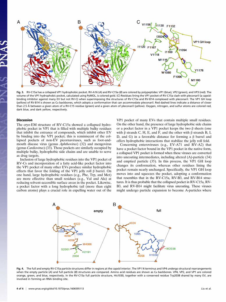
The VP1 Hydrophobic Pocket Is Unsuitable for Antiviral CapsidBinding Agents. Unlike many EV structures, the hydrophobicpocket within the VP1 jelly roll fold, where a pocket factor is
typically bound (4, 5, 23), is collapsed in RV-C15a (Fig. 3 A andB). The collapsed structure is similar to the empty pockets foundin purified RV-B14 (2) and RV-B3 (29). None of these threestructures have sufficient space to accommodate a fatty-acidpocket factor, because for each, the VP1 GH loop, located at theboundary between the canyon and the entrance to the VP1pocket, is in a conformation that squeezes the pocket. Never-theless, in RV-B3 and RV-B14, the flexibility of the VP1 GHloop allows enlargement of the pocket that then can bind anti-viral reagents. The RV-B14 pocket is lined with multiple smallresidues (e.g., Ala, Ser, Val, etc.) that can accommodate suchcompounds. In contrast, the collapsed RV-C15a VP1 pocket isfilled with bulky, hydrophobic residues (in particular, Trp1080,Phe1096, Met1116, and Met1180; Fig. 3C and Table S2). Theseamino acids are conserved in almost all RV-Cs (14). Addition-ally, Ile1198 and Tyr1246 partially block the entrance to the VP1pocket. Therefore, as has been observed experimentally (14), noRV-Cs are likely to be responsive to antiviral therapies based onpocket-binding compounds.
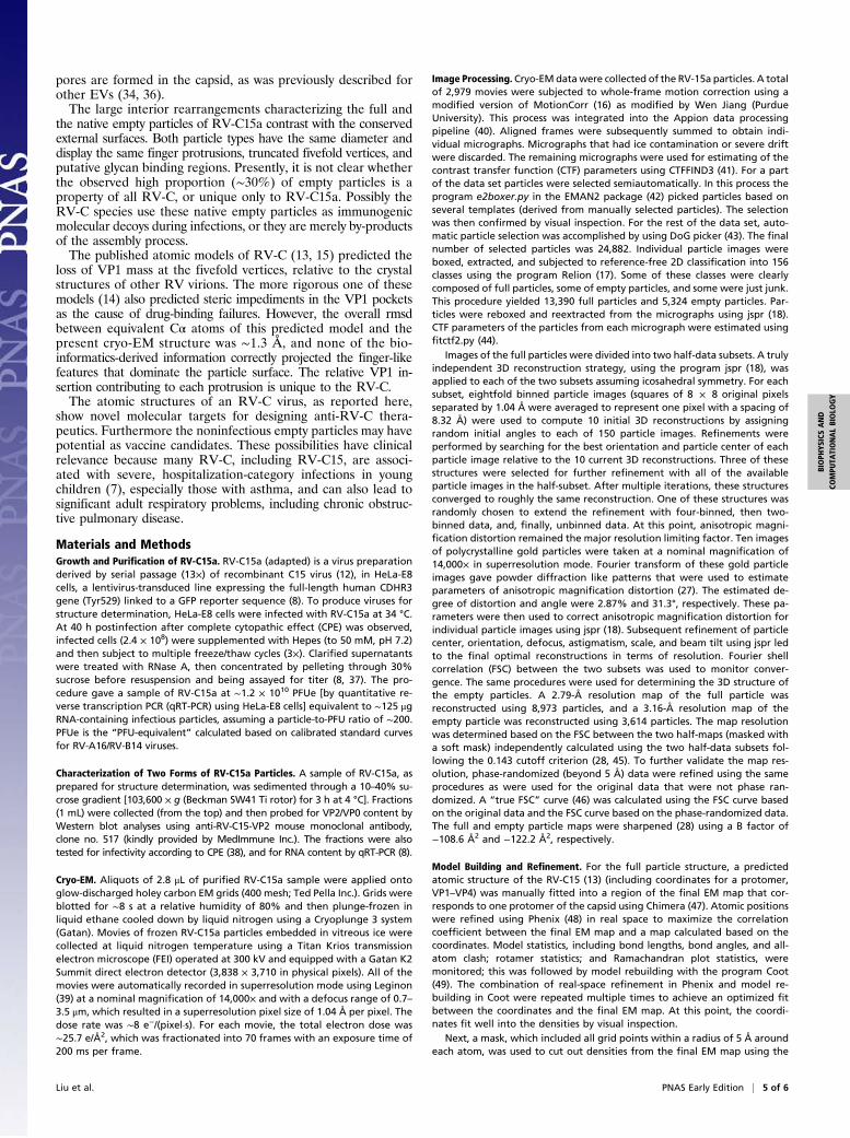
Comparison of the Full and Empty Particle Structures. RV-C15a fulland empty particles differed mainly in regions on the innersurfaces of their capsid shells (Fig. 4). In particular, the VP1N-terminal residues 1017–1053 are well-ordered in the full par-ticle map, but disordered in the empty particle map; this isconsistent with other EV structures where the VP1 N terminus isinvolved in binding to viral RNA (23) and is externalized beforeejecting the genome during infection (30). Thus, the specificconfiguration of this internal region is RNA-dependent and islikely to exert strong influence on VP0 cleavage when the RNAis packaged. In the empty particles, VP0 residues 4024–4050form a hairpin loop positioning the VP0 cleavage site in closeproximity to His2191 (Fig. 4A), a crucial residue in the cleavagemechanism (31). Nearby VP1 residues 1054–1064 interact withthe VP0 hairpin within the same protomer, presumably helpingto set up the pending cleavage reaction. However, in the fullparticle structure, the VP1 N-terminal residues 1027–1053 (dis-ordered in the empty particles) interact with the C terminus ofVP4 within the same protomer and participates in viral RNAbinding (Fig. 4B).
Fig. 2. A potential binding site for glycans on the RV-C receptor. (A) A triangle indicates an icosahedral asymmetric unit. A red rectangle (dashed line)outlines the limit of the sialic acid binding site shown in B and C. Surface electrostatic potential of EV-D68 (PDB ID code 5BNO) (B) and RV-C15a (C) is rep-resented with a scale of −8kT/e (red) to 8kT/e (blue). (D and E) The sialic acid (yellow) interacts with surrounding residues on EV-D68 (green) and as anticipatedon RV-C15a (cyan). Red dashed lines indicate (potential) polar interactions. Oxygen and nitrogen atoms are colored red and blue, respectively.
Liu et al. PNAS Early Edition | 3 of 6
BIOPH
YSICSAND
COMPU
TATIONALBIOLO
GY
DiscussionThe cryo-EM structure of RV-C15a showed a collapsed hydro-phobic pocket in VP1 that is filled with multiple bulky residuesthat inhibit the entrance of compounds, which inhibit other EVby binding into the VP1 pocket; this is reminiscent of the col-lapsed pockets of non-EV picornaviruses, such as foot-and-mouth disease virus (genus Aphthovirus) (32) and mengovirus(genus Cardiovirus) (33). Those pockets are similarly occupied bymultiple bulky, hydrophobic side chains and are unable to serveas drug targets.Inclusion of large hydrophobic residues into the VP1 pocket of
RV-Cs and incorporation of a fatty acid-like pocket factor intothe VP1 pocket of many other EVs produce similar hydrophobiceffects that favor the folding of the VP1 jelly roll β barrel. Onone hand, large hydrophobic residues (e.g., Phe, Trp, and Met)are more effective than small residues (e.g., Val and Ala) atreducing solvent-accessible surface areas in the pocket. Likewise,a pocket factor with a long hydrophobic tail (more than eightcarbon atoms) plays a crucial role in expelling water out of the
VP1 pocket of many EVs that contain multiple small residues.On the other hand, the presence of large hydrophobic side chainsor a pocket factor in a VP1 pocket keeps the two β sheets (onewith β strands C, H, E, and F, and the other with β strands B, I,D, and G) in a favorable distance for forming a β barrel andoffers hydrophobic interactions that stabilize the jelly roll fold.Concerning enteroviruses (e.g., EV-A71 and RV-A2) that
have a pocket factor bound in the VP1 pocket in the native form,a collapsed VP1 pocket is formed when these viruses are convertedinto uncoating intermediates, including altered (A)-particle (34)and emptied particle (35). In this process, the VP1 GH loopchanges its conformation, whereas other residues lining thepocket remain nearly unchanged. Specifically, the VP1 GH loopmoves into and squeezes the pocket, adopting a conformationthat resembles that in the RV-C15a, RV-B3, and RV-B14 struc-tures. It is thus probable that the collapsed pocket in RV-C15a, RV-B3, and RV-B14 might facilitate virus uncoating. These virusesmight undergo particle expansion to become A-particles where
Fig. 4. The full and empty RV-C15a particle structures differ in regions at the capsid interior. The VP1 N terminus and VP4 undergo structural rearrangementswhen the empty particle (A) and full particle (B) structures are compared. Amino acid residues are shown as Cα backbones. VP4, VP2, and VP1 are coloredorange, green, and blue, respectively. In the RV-C15a full particle structure, His1030, together with a conserved residue Trp2038 shared by many EV, areinvolved in forming an RNA binding site.
Fig. 3. RV-C15a has a collapsed VP1 hydrophobic pocket. RV-A16 (A) and RV-C15a (B) are colored by polypeptides: VP1 (blue), VP2 (green), and VP3 (red). Thevolume of the VP1 hydrophobic pocket, calculated using PyMOL, is colored gold. (C) Residues lining the VP1 pocket of RV-C15a clash with pleconaril (a capsid-binding inhibitor against many EV but not RV-C) when superimposing the structures of RV-C15a and RV-B14 complexed with pleconaril. The VP1 GH loop(yellow) of RV-B14 is shown as Cα backbones, which adopts a conformation that can accommodate pleconaril. Red dashed lines indicate a distance of closerthan 2.5 Å between a given atom of a RV-C15 residue (green) and a given atom of pleconaril (yellow). Oxygen, nitrogen, and sulfur atoms are colored red,dark blue, and dark yellow, respectively.
4 of 6 | www.pnas.org/cgi/doi/10.1073/pnas.1606595113 Liu et al.
pores are formed in the capsid, as was previously described forother EVs (34, 36).The large interior rearrangements characterizing the full and
the native empty particles of RV-C15a contrast with the conservedexternal surfaces. Both particle types have the same diameter anddisplay the same finger protrusions, truncated fivefold vertices, andputative glycan binding regions. Presently, it is not clear whetherthe observed high proportion (∼30%) of empty particles is aproperty of all RV-C, or unique only to RV-C15a. Possibly theRV-C species use these native empty particles as immunogenicmolecular decoys during infections, or they are merely by-productsof the assembly process.The published atomic models of RV-C (13, 15) predicted the
loss of VP1 mass at the fivefold vertices, relative to the crystalstructures of other RV virions. The more rigorous one of thesemodels (14) also predicted steric impediments in the VP1 pocketsas the cause of drug-binding failures. However, the overall rmsdbetween equivalent Cα atoms of this predicted model and thepresent cryo-EM structure was ∼1.3 Å, and none of the bio-informatics-derived information correctly projected the finger-likefeatures that dominate the particle surface. The relative VP1 in-sertion contributing to each protrusion is unique to the RV-C.The atomic structures of an RV-C virus, as reported here,
show novel molecular targets for designing anti-RV-C thera-peutics. Furthermore the noninfectious empty particles may havepotential as vaccine candidates. These possibilities have clinicalrelevance because many RV-C, including RV-C15, are associ-ated with severe, hospitalization-category infections in youngchildren (7), especially those with asthma, and can also lead tosignificant adult respiratory problems, including chronic obstruc-tive pulmonary disease.
Materials and MethodsGrowth and Purification of RV-C15a. RV-C15a (adapted) is a virus preparationderived by serial passage (13×) of recombinant C15 virus (12), in HeLa-E8cells, a lentivirus-transduced line expressing the full-length human CDHR3gene (Tyr529) linked to a GFP reporter sequence (8). To produce viruses forstructure determination, HeLa-E8 cells were infected with RV-C15a at 34 °C.At 40 h postinfection after complete cytopathic effect (CPE) was observed,infected cells (2.4 × 10⁸) were supplemented with Hepes (to 50 mM, pH 7.2)and then subject to multiple freeze/thaw cycles (3×). Clarified supernatantswere treated with RNase A, then concentrated by pelleting through 30%sucrose before resuspension and being assayed for titer (8, 37). The pro-cedure gave a sample of RV-C15a at ∼1.2 × 10¹⁰ PFUe [by quantitative re-verse transcription PCR (qRT-PCR) using HeLa-E8 cells] equivalent to ∼125 μgRNA-containing infectious particles, assuming a particle-to-PFU ratio of ∼200.PFUe is the “PFU-equivalent” calculated based on calibrated standard curvesfor RV-A16/RV-B14 viruses.
Characterization of Two Forms of RV-C15a Particles. A sample of RV-C15a, asprepared for structure determination, was sedimented through a 10–40% su-crose gradient [103,600 × g (Beckman SW41 Ti rotor) for 3 h at 4 °C]. Fractions(1 mL) were collected (from the top) and then probed for VP2/VP0 content byWestern blot analyses using anti-RV-C15-VP2 mouse monoclonal antibody,clone no. 517 (kindly provided by MedImmune Inc.). The fractions were alsotested for infectivity according to CPE (38), and for RNA content by qRT-PCR (8).
Cryo-EM. Aliquots of 2.8 μL of purified RV-C15a sample were applied ontoglow-discharged holey carbon EM grids (400 mesh; Ted Pella Inc.). Grids wereblotted for ∼8 s at a relative humidity of 80% and then plunge-frozen inliquid ethane cooled down by liquid nitrogen using a Cryoplunge 3 system(Gatan). Movies of frozen RV-C15a particles embedded in vitreous ice werecollected at liquid nitrogen temperature using a Titan Krios transmissionelectron microscope (FEI) operated at 300 kV and equipped with a Gatan K2Summit direct electron detector (3,838 × 3,710 in physical pixels). All of themovies were automatically recorded in superresolution mode using Leginon(39) at a nominal magnification of 14,000× and with a defocus range of 0.7–3.5 μm, which resulted in a superresolution pixel size of 1.04 Å per pixel. Thedose rate was ∼8 e−/(pixel·s). For each movie, the total electron dose was∼25.7 e/Å2, which was fractionated into 70 frames with an exposure time of200 ms per frame.
Image Processing. Cryo-EM datawere collected of the RV-15a particles. A totalof 2,979 movies were subjected to whole-frame motion correction using amodified version of MotionCorr (16) as modified by Wen Jiang (PurdueUniversity). This process was integrated into the Appion data processingpipeline (40). Aligned frames were subsequently summed to obtain indi-vidual micrographs. Micrographs that had ice contamination or severe driftwere discarded. The remaining micrographs were used for estimating of thecontrast transfer function (CTF) parameters using CTFFIND3 (41). For a partof the data set particles were selected semiautomatically. In this process theprogram e2boxer.py in the EMAN2 package (42) picked particles based onseveral templates (derived from manually selected particles). The selectionwas then confirmed by visual inspection. For the rest of the data set, auto-matic particle selection was accomplished by using DoG picker (43). The finalnumber of selected particles was 24,882. Individual particle images wereboxed, extracted, and subjected to reference-free 2D classification into 156classes using the program Relion (17). Some of these classes were clearlycomposed of full particles, some of empty particles, and some were just junk.This procedure yielded 13,390 full particles and 5,324 empty particles. Par-ticles were reboxed and reextracted from the micrographs using jspr (18).CTF parameters of the particles from each micrograph were estimated usingfitctf2.py (44).
Images of the full particles were divided into two half-data subsets. A trulyindependent 3D reconstruction strategy, using the program jspr (18), wasapplied to each of the two subsets assuming icosahedral symmetry. For eachsubset, eightfold binned particle images (squares of 8 × 8 original pixelsseparated by 1.04 Å were averaged to represent one pixel with a spacing of8.32 Å) were used to compute 10 initial 3D reconstructions by assigningrandom initial angles to each of 150 particle images. Refinements wereperformed by searching for the best orientation and particle center of eachparticle image relative to the 10 current 3D reconstructions. Three of thesestructures were selected for further refinement with all of the availableparticle images in the half-subset. After multiple iterations, these structuresconverged to roughly the same reconstruction. One of these structures wasrandomly chosen to extend the refinement with four-binned, then two-binned data, and, finally, unbinned data. At this point, anisotropic magni-fication distortion remained the major resolution limiting factor. Ten imagesof polycrystalline gold particles were taken at a nominal magnification of14,000× in superresolution mode. Fourier transform of these gold particleimages gave powder diffraction like patterns that were used to estimateparameters of anisotropic magnification distortion (27). The estimated de-gree of distortion and angle were 2.87% and 31.3°, respectively. These pa-rameters were then used to correct anisotropic magnification distortion forindividual particle images using jspr (18). Subsequent refinement of particlecenter, orientation, defocus, astigmatism, scale, and beam tilt using jspr ledto the final optimal reconstructions in terms of resolution. Fourier shellcorrelation (FSC) between the two subsets was used to monitor conver-gence. The same procedures were used for determining the 3D structure ofthe empty particles. A 2.79-Å resolution map of the full particle wasreconstructed using 8,973 particles, and a 3.16-Å resolution map of theempty particle was reconstructed using 3,614 particles. The map resolutionwas determined based on the FSC between the two half-maps (masked witha soft mask) independently calculated using the two half-data subsets fol-lowing the 0.143 cutoff criterion (28, 45). To further validate the map res-olution, phase-randomized (beyond 5 Å) data were refined using the sameprocedures as were used for the original data that were not phase ran-domized. A “true FSC” curve (46) was calculated using the FSC curve basedon the original data and the FSC curve based on the phase-randomized data.The full and empty particle maps were sharpened (28) using a B factor of−108.6 Å2 and −122.2 Å2, respectively.
Model Building and Refinement. For the full particle structure, a predictedatomic structure of the RV-C15 (13) (including coordinates for a protomer,VP1–VP4) was manually fitted into a region of the final EM map that cor-responds to one protomer of the capsid using Chimera (47). Atomic positionswere refined using Phenix (48) in real space to maximize the correlationcoefficient between the final EM map and a map calculated based on thecoordinates. Model statistics, including bond lengths, bond angles, and all-atom clash; rotamer statistics; and Ramachandran plot statistics, weremonitored; this was followed by model rebuilding with the program Coot(49). The combination of real-space refinement in Phenix and model re-building in Coot were repeated multiple times to achieve an optimized fitbetween the coordinates and the final EM map. At this point, the coordi-nates fit well into the densities by visual inspection.
Next, a mask, which included all grid points within a radius of 5 Å aroundeach atom, was used to cut out densities from the final EM map using the
Liu et al. PNAS Early Edition | 5 of 6
BIOPH
YSICSAND
COMPU
TATIONALBIOLO
GY
CCP4 (50) program suite. The resultant segment of the final EM map wasplaced into a pseudo crystallographic unit cell (P1 space group) and wasback-transformed into pseudo structure factors (including both amplitudesand phases). The coordinates were then subjected to refinement of indi-vidual B factors, atom positions, and occupancy against the pseudo structurefactors using standard reciprocal space refinement procedures in Phenix (48).R factors were monitored during the refinement cycles. Only the coordinateswere refined, whereas the map was kept constant. Subsequently, the co-ordinates were refined in real space against the final EM map by applyingnoncrystallographic symmetry (60-fold) constraints using Phenix (48). Vali-dation of the final coordinates was based on the criteria of MolProbity (51).The full particle atomic model (excluding VP4 and the VP1 N-terminal resi-dues 1101–1160) was used as a starting atomic model for model building andrefinement of the empty particle structure.
As a further validation of the EM map resolution and the fitting betweenthe atomic model and the final EM map, a density map was calculated basedon the atomic model specifying a resolution of 2.79 Å for the full particlestructure and 3.16 Å for the empty particle structure. An FSC was computedbetween the resultant density map and the final EM map. The resolutiondetermined using 0.5 FSC as a cutoff was 2.85 Å (full particle) and 3.24 Å(empty particle). Oligomers were generated using VIPERdb (52). Figureswere made using Chimera (47) and PyMOL (https://www.pymol.org).
ACKNOWLEDGMENTS. We thank Roland Rueckert for critically reading themanuscript, Guimei Yu for helpful discussions, Valorie Bowman of thePurdue University Cryo-EM Facility and Ken Hibler of the FEI Company fortechnical assistance, and Sheryl Kelly for help preparing this manuscript. Thiswork was supported by NIH Grants R37 AI011219 (to M.G.R.) and U19AI104317 (to A.C.P.).
1. Knowles NJ, et al. (2012) Picornaviridae. Virus Taxonomy: Classification andNomenclature of Viruses: Ninth Report of the International Committee on Taxonomy ofViruses, eds King AMQ, Adams MJ, Carstens EB, Lefkowitz EJ (Elsevier, San Diego), pp855–880.
2. Rossmann MG, et al. (1985) Structure of a human common cold virus and functionalrelationship to other picornaviruses. Nature 317(6033):145–153.
3. Hogle JM, Chow M, Filman DJ (1985) Three-dimensional structure of poliovirus at 2.9 Aresolution. Science 229(4720):1358–1365.
4. Hogle JM (2012) A 3D framework for understanding enterovirus 71. Nat Struct MolBiol 19(4):367–368.
5. Liu Y, et al. (2015) Structure and inhibition of EV-D68, a virus that causes respiratoryillness in children. Science 347(6217):71–74.
6. Miller EK, et al. (2009) Human rhinovirus C associated with wheezing in hospitalisedchildren in the Middle East. J Clin Virol 46(1):85–89.
7. Bizzintino J, et al. (2011) Association between human rhinovirus C and severity ofacute asthma in children. Eur Respir J 37(5):1037–1042.
8. Bochkov YA, et al. (2015) Cadherin-related family member 3, a childhood asthmasusceptibility gene product, mediates rhinovirus C binding and replication. Proc NatlAcad Sci USA 112(17):5485–5490.
9. Bønnelykke K, et al. (2014) A genome-wide association study identifies CDHR3 as asusceptibility locus for early childhood asthma with severe exacerbations. Nat Genet46(1):51–55.
10. Piralla A, et al. (2009) Clinical severity and molecular typing of human rhinovirus Cstrains during a fall outbreak affecting hospitalized patients. J Clin Virol 45(4):311–317.
11. Arden KE, McErlean P, Nissen MD, Sloots TP, Mackay IM (2006) Frequent detection ofhuman rhinoviruses, paramyxoviruses, coronaviruses, and bocavirus during acute re-spiratory tract infections. J Med Virol 78(9):1232–1240.
12. Bochkov YA, et al. (2011) Molecular modeling, organ culture and reverse genetics fora newly identified human rhinovirus C. Nat Med 17(5):627–632.
13. Basta HA, Sgro JY, Palmenberg AC (2014) Modeling of the human rhinovirus C capsidsuggests a novel topography with insights on receptor preference and immunoge-nicity. Virology 448:176–184.
14. Basta HA, et al. (2014) Modeling of the human rhinovirus C capsid suggests possiblecauses for antiviral drug resistance. Virology 448:82–90.
15. McErlean P, et al. (2008) Distinguishing molecular features and clinical characteristicsof a putative new rhinovirus species, human rhinovirus C (HRV C). PLoS One 3(4):e1847.
16. Li X, et al. (2013) Electron counting and beam-induced motion correction enable near-atomic-resolution single-particle cryo-EM. Nat Methods 10(6):584–590.
17. Scheres SH (2012) RELION: Implementation of a Bayesian approach to cryo-EMstructure determination. J Struct Biol 180(3):519–530.
18. Guo F, Jiang W (2014) Single particle cryo-electron microscopy and 3-D reconstructionof viruses. Methods Mol Biol 1117:401–443.
19. Bartesaghi A, et al. (2015) 2.2 Å resolution cryo-EM structure of β-galactosidase incomplex with a cell-permeant inhibitor. Science 348(6239):1147–1151.
20. Basavappa R, et al. (1994) Role and mechanism of the maturation cleavage of VP0 inpoliovirus assembly: Structure of the empty capsid assembly intermediate at 2.9 Aresolution. Protein Sci 3(10):1651–1669.
21. Rossmann MG, He Y, Kuhn RJ (2002) Picornavirus-receptor interactions. TrendsMicrobiol 10(7):324–331.
22. Rossmann MG (1989) The canyon hypothesis. Hiding the host cell receptor attach-ment site on a viral surface from immune surveillance. J Biol Chem 264(25):14587–14590.
23. Filman DJ, et al. (1989) Structural factors that control conformational transitions andserotype specificity in type 3 poliovirus. EMBO J 8(5):1567–1579.
24. Smyth M, Pettitt T, Symonds A, Martin J (2003) Identification of the pocket factors in apicornavirus. Arch Virol 148(6):1225–1233.
25. Liu Y, et al. (2015) Sialic acid-dependent cell entry of human enterovirus D68. NatCommun 6:8865.
26. Rogers JM, Diana GD, McKinlay MA (1999) Pleconaril. A broad spectrum anti-picornaviral agent. Adv Exp Med Biol 458:69–76.
27. Grant T, Grigorieff N (2015) Automatic estimation and correction of anisotropicmagnification distortion in electron microscopes. J Struct Biol 192(2):204–208.
28. Rosenthal PB, Henderson R (2003) Optimal determination of particle orientation,absolute hand, and contrast loss in single-particle electron cryomicroscopy. J Mol Biol333(4):721–745.
29. Zhao R, et al. (1996) Human rhinovirus 3 at 3.0 A resolution. Structure 4(10):1205–1220.
30. Fricks CE, Hogle JM (1990) Cell-induced conformational change in poliovirus: Exter-nalization of the amino terminus of VP1 is responsible for liposome binding. J Virol64(5):1934–1945.
31. Hindiyeh M, Li QH, Basavappa R, Hogle JM, Chow M (1999) Poliovirus mutants athistidine 195 of VP2 do not cleave VP0 into VP2 and VP4. J Virol 73(11):9072–9079.
32. Acharya R, et al. (1989) The three-dimensional structure of foot-and-mouth diseasevirus at 2.9 A resolution. Nature 337(6209):709–716.
33. Luo M, et al. (1987) The atomic structure of Mengo virus at 3.0 A resolution. Science235(4785):182–191.
34. Ren J, et al. (2013) Picornavirus uncoating intermediate captured in atomic detail. NatCommun 4:1929.
35. Garriga D, et al. (2012) Insights into minor group rhinovirus uncoating: The X-raystructure of the HRV2 empty capsid. PLoS Pathog 8(1):e1002473.
36. Butan C, Filman DJ, Hogle JM (2014) Cryo-electron microscopy reconstruction showspoliovirus 135S particles poised for membrane interaction and RNA release. J Virol88(3):1758–1770.
37. Griggs TF, Bochkov YA, Nakagome K, Palmenberg AC, Gern JE (2015) Production,purification, and capsid stability of rhinovirus C types. J Virol Methods 217:18–23.
38. Ledford RM, Collett MS, Pevear DC (2005) Insights into the genetic basis for naturalphenotypic resistance of human rhinoviruses to pleconaril. Antiviral Res 68(3):135–138.
39. Suloway C, et al. (2005) Automated molecular microscopy: The new Leginon system.J Struct Biol 151(1):41–60.
40. Lander GC, et al. (2009) Appion: An integrated, database-driven pipeline to facilitateEM image processing. J Struct Biol 166(1):95–102.
41. Mindell JA, Grigorieff N (2003) Accurate determination of local defocus and specimentilt in electron microscopy. J Struct Biol 142(3):334–347.
42. Tang G, et al. (2007) EMAN2: An extensible image processing suite for electron mi-croscopy. J Struct Biol 157(1):38–46.
43. Voss NR, Yoshioka CK, Radermacher M, Potter CS, Carragher B (2009) DoG Picker andTiltPicker: Software tools to facilitate particle selection in single particle electronmicroscopy. J Struct Biol 166(2):205–213.
44. Jiang W, Guo F, Liu Z (2012) A graph theory method for determination of cryo-EMimage focuses. J Struct Biol 180(2):343–351.
45. Scheres SH, Chen S (2012) Prevention of overfitting in cryo-EM structure de-termination. Nat Methods 9(9):853–854.
46. Chen S, et al. (2013) High-resolution noise substitution to measure overfitting andvalidate resolution in 3D structure determination by single particle electron cry-omicroscopy. Ultramicroscopy 135:24–35.
47. Pettersen EF, et al. (2004) UCSF Chimera–a visualization system for exploratory re-search and analysis. J Comput Chem 25(13):1605–1612.
48. Adams PD, et al. (2010) PHENIX: A comprehensive Python-based system for macro-molecular structure solution. Acta Crystallogr D Biol Crystallogr 66(Pt 2):213–221.
49. Emsley P, Lohkamp B, Scott WG, Cowtan K (2010) Features and development of Coot.Acta Crystallogr D Biol Crystallogr 66(Pt 4):486–501.
50. Collaborative Computational Project, Number 4 (1994) The CCP4 suite: Programs forprotein crystallography. Acta Crystallogr D Biol Crystallogr 50(Pt 5):760–763.
51. Chen VB, et al. (2010) MolProbity: All-atom structure validation for macromolecularcrystallography. Acta Crystallogr D Biol Crystallogr 66(Pt 1):12–21.
52. Carrillo-Tripp M, et al. (2009) VIPERdb2: An enhanced and web API enabled relationaldatabase for structural virology. Nucleic Acids Res 37(Database issue):D436–D442.
6 of 6 | www.pnas.org/cgi/doi/10.1073/pnas.1606595113 Liu et al.


![Cybersecurity Strategy in Japan kawaguchi.pdf2014.1 [Japan Atomic Energy Agency (JAEA)] Found possibility of information leakage by virus infection [Threats to government’s organizations]](https://static.fdocuments.in/doc/165x107/5acf97e37f8b9a56098d4be9/cybersecurity-strategy-in-kawaguchipdf20141-japan-atomic-energy-agency-jaea.jpg)


