
ATOMIC STRUCTURE AND X RAY PRODUCTION · ATOMIC STRUCTURE AND X RAY PRODUCTION ... Atomic Structure...
19
© Barbara Lamb 2013 ATOMIC STRUCTURE AND X RAY PRODUCTION X-rays were discovered by Roentgen in 1895. The nature was unknown and so they were called x-rays They are high energy electromagnetic radiation part of the electromagnetic spectrum ELECTROMAGNETIC RADIATION Electromagnetic radiation consists of waves most are invisible (e.g. radiowaves, microwaves, ultraviolet light, x- rays) only a very small part is visible to humans they are all part of the electromagnetic spectrum X-rays consists of ‘wave packets’ of energy called photons X-rays are short wavelength with high power A photon = one quantum of energy X-ray beam is made up from millions of individual x-ray photons
Transcript of ATOMIC STRUCTURE AND X RAY PRODUCTION · ATOMIC STRUCTURE AND X RAY PRODUCTION ... Atomic Structure...

© Barbara Lamb 2013
ATOMIC STRUCTURE AND
X RAY PRODUCTION
X-rays were discovered by Roentgen in 1895.
The nature was unknown and so they were called x-rays
They are high energy electromagnetic radiation part of the
electromagnetic spectrum
ELECTROMAGNETIC RADIATION
Electromagnetic radiation consists of waves
most are invisible (e.g. radiowaves, microwaves, ultraviolet light, x-
rays)
only a very small part is visible to humans
they are all part of the electromagnetic spectrum
X-rays consists of ‘wave packets’ of energy called photons
X-rays are short wavelength with high power
A photon = one quantum of energy
X-ray beam is made up from millions of individual x-ray photons
© Barbara Lamb 2013
Energy = the ability to do work
Radiation = the transfer of energy in the form of waves or particles
SOURCES OF RADIATION
1.ARTIFICIAL BACKGROUND RADIATION
Fallout from nuclear explosions
Radioactive waste discharge from nuclear establishments
Average background radiation is approximately 2.7 mSv per year
2. MEDICAL AND DENTAL DIAGNOSTIC RADIATION X RAYS
3.RADIATION FROM OCCUPATIONAL EXPOSURE
4.NATURAL BACKGROUND RADIATION
Cosmic rays from the atmosphere
Gamma radiation from the rocks and soil in the earth’s crust
Radiation from ingested isotopes in certain food
Radon and its decay products. Radon is a gaseous decay product of
uranium naturally found in granite. As the gas it diffuses readily
from rocks through soil and can be trapped in poorly ventilated
houses. Cornwall and Scotland have house is built on granite
Average background radiation is approximately 2.7 mSv per year
MAN MADE X RAYS
They are produced when fast moving electrons collide with a metal
target and are suddenly brought to rest.The electrons kinetic
energy is transformed into
electromagnetic energy (x-ray photons)
X-rays for medical procedures are produced inside an x-ray tube

© Barbara Lamb 2013
the primary function of an X-ray tube is to convert electrical energy
into X-rays
ATOMIC STRUCTURE
Atomic Structure - the building blocks of matter
Nucleus-protons and neutrons forming a dense centre of particles
Electrons- orbiting around nucleus in specific orbits
Atomic no is the no of protons in an atom
Atoms are minute particles held by nuclear or electric forces
The unit of energy in atomic structure is the electron volt
Neutral Atom-no of electrons=no of protons
Atoms at ground state are electrically neutral
positive protons = no of negative electrons
NUCLEUS
A typical nucleus is made up from particles known as protons and
neutrons, and carries a positive charge
Protons and neutrons are almost equal in mass (size)
1.66 x10⁻²⁴ g

© Barbara Lamb 2013
PROTONS AND NEUTRONS
Protons are positively-charged They will tend to repel each other because
each proton carries the same charge (i.e. like charges repel each other,
whereas unlike charges attract each other)
Neutrons have no charge therefore are electrically neutral They act as
a ‘glue’ to bind the nucleus together by counteracting the repulsive
forces between the protons
The charge of a nucleus is therefore POSITIVE
ELECTRONS
Electrons are negatively-charged
They are 1840 times smaller than protons and neutrons
The orbiting electrons are arranged in energy levels or shells
around the nucleus
These have increasing energy levels from the nucleus outwards
Electrons can move up and down the orbiting shells around the
nucleus
There are different energy levels labelled KLMNO outwards from
the nucleus
There is a maximum no of electrons allowed in each shell
K-2 L-8 M-18 N-32 O-50
The electrons must first fill the lowest available energy level/shell
available nearest the nucleus
When the level is full the next electron goes into the next highest
level(shell) available
Electron removed we now have a positive charged “positive ion”
This process is called “ionisation ”

© Barbara Lamb 2013
Electron displaced to outer shell (higher energy) “excitation”
Atomic Number (Z) – the number of protons in the nucleus of an
atom
Neutron number(N)-the number of neutrons in the nucleus of an
atom
Atomic mass number(A)-sum of the numbers of protons and
number of neutrons in an atom
(A=Z+N )
Isotopes –atoms with the same atomic number (Z)but with
different atomic mass numbers (A) and hence different number of
neutrons(N)
Radioisotopes-isotopes with unstable nuclei which undergo
radioactive disintegration
Atoms with different Z numbers are known as Elements
Elements form the basis of the periodic table
No two elements have the same atomic number
The number of electrons in an atom determines its chemical
properties
In a neutral atom the number of orbiting electrons is equal to the
number of protons in the nucleus, ie the number of positive and
negative charges are balanced If an electron is removed the atom is no
longer electrically neutral but becomes positively charged, i.e. it
becomes a positive ion and is said to be ionised
Ionisation is the process whereby an electron is removed from an
atom to overcome the binding energy of attraction that keeps
electrons within their shells
The unit of energy in the atomic system is the electron
volt (eV)

© Barbara Lamb 2013
X-RAY PRODUCTION
X RAYS ARE PRODUCED WHEN HIGH SPEED
ELECTRONS HIT A TARGET AND STOP!!!
ELECTRONS BOMBARD THE TARGET AND
LOSE ENERGY 99% HEAT AND 1% XRAYS
High voltage (potential difference between the cathode and anode )
accelerates electrons Kv

© Barbara Lamb 2013
Current flows between the cathode and anode mA
Lead casing absorbs unwanted x rays
Oil casing removes the heat
Electrons are brought to rest at the target
Energy lost is HEAT AND X RAYS
Heat is dissipated
X rays are emitted through the lead window BEAM
RELATIONSHIP OF FOCAL SPOT ON IMAGE
BLURRING
PRINCIPLE OF LINE FOCUS
Lead casing absorbs the unwanted x rays only allowing the x rays
to pass out of the x ray window
The oil in the x ray tube removes heat
The copper block surrounding target removes the heat
X rays are emitted through the lead window
X RAY PRODUCTION
© Barbara Lamb 2013
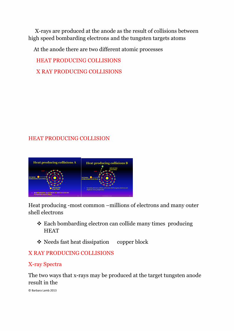
X-rays are produced at the anode as the result of collisions between
high speed bombarding electrons and the tungsten targets atoms
At the anode there are two different atomic processes
HEAT PRODUCING COLLISIONS
X RAY PRODUCING COLLISIONS
HEAT PRODUCING COLLISION
BOMBARDING ELECTRON IS DEFLECTED BY TUNGSTON ELECTRON
INCOMING
BOMBARDING ELECTRON
DEFLECTED
ELECTRON
HEAT
© 2012 Barbara H Lamb 54
Incoming electron collides with outer shell tungsten electron and displaces more peripherally
INCOMING
BOMBARDING ELECTRON
DEFLECTED
ELECTRON
HEAT
© 2012 Barbara H Lamb 55
Heat producing -most common –millions of electrons and many outer
shell electrons
Each bombarding electron can collide many times producing
HEAT
Needs fast heat dissipation copper block
X RAY PRODUCING COLLISIONS
X-ray Spectra
The two ways that x-rays may be produced at the target tungsten anode
result in the

© Barbara Lamb 2013
spectra:
1 Continuous spectrum
2 Characteristic spectrum
CONTINUOUS SPECTRUM
PROCESS 1
The incoming electron passes close to the nucleus it is pulled
towards the nucleus (positive and negative charges attract)
the electron slows down and change direction towards the nucleus
the electron loses a large amount of its energy, which is then
emitted as multiple x-ray photons
original electron is slowed down or stopped
Incoming electron passes close to the nucleus
Electron-dramatically slowed down-deflected by the nucleus-large loss of energy emitted as X Rays
INCOMING
BOMBARDING ELECTRON
X RAY
PHOTONS
EMISSION
© 2012 Barbara H Lamb 60
PROCESS 1
X-rays produced in this way are referred to as Bremsstrahlung (or
Braking) Radiation
During this process many x-ray photons of different energies are
produced. These x-rays form the continuous spectrum
This type of interaction accounts for 85% of the x-rays produced by an
x-ray tube

© Barbara Lamb 2013
Small deflections are most common, resulting in many useless low
energy photons (most will not leave the x-ray tube and those that do will
be filtered out)
ALUMINIUM FILTRATION are used at the x ray window
TO FILTER X RAYS
There are many fewer large deflections, therefore few high-energy
photons are produced
The maximum photon energy (Emax) is directly related to the kV
across the x-ray tube
This is formed from the range of x-rays produced by
Bremsstrahlung(process 1)
This is termed the continuous spectrum. The amount of deceleration
and degree of deflection of the incoming electron determines the
amount of energy lost. Therefore a wide range(spectrum) of x-ray
photons of differing energies can be produced
X RAY PRODUCING COLLISIONS
CHARACTERISTIC SPECTRUM
NK
L
M
© Barbara Lamb 2012
X ray producing collisions B STAGE 1

© Barbara Lamb 2013
N
66
BOMBARDING ELECTRON
INCOMING BOMBARDING ELECTRON
DEFLECTED ELECTRON
© 2012 Barbara H Lamb 69
Incoming electron collides with inner shell electron-displaces it to an
outer excitation or displaces it from the atom ionisation large loss of
energy and subsequent X rays
X ray producing collisions B STAGE 2
OUTER SHELL ELECTRONS DROP INTO INNER SHELLS -X RAY EMISSION
69
OUTER SHELL ELECTRONSCASCADE TO FILL SPACE
ENERGY IS GIVEN OFF AS X RAYS
Remember that atoms have their electrons arranged in"shells" of
different energies
The K-shell (closest to the nucleus) is the lowest energy state of an
atom the incoming electron collides with a tungsten K-shell electron
and gives it enough energy to jump out of its shell ionisation
Following this ionisation, one of the atoms electrons of higher energy
(from an outer shell) falls into the K-shell

© Barbara Lamb 2013
During this there is a specific loss of energy, which produces a x-ray
photon
characteristic of tungsten atoms
These x-rays form the
characteristic spectrum
CHARACTERISTIC SPECTRUM....recap
This is formed by the x ray photons characteristic of tungsten
atoms (process 2)
The photon lines are named K or L dependant on the shell from
which they have been emitted
Only K lines are of use as L lines are too low energy
The accelerated electron must have enough energy to displace the
K-shell tungsten electron out of its shell
Line spectra emitted from another element would plot onto the
figure at different photon energies
PROPERTIES AND CHARACTERISTICS OF X RAYS
X-rays are wave packets of energy of electromagnetic radiation that
originate at atomic level
© Barbara Lamb 2013
Each wave packet is equivalent to a quantum of energy and is
called a Photon
An x-ray beam is made up of millions of photons of differing
energies
The x-ray beam can vary in its
a) INTENSITY (the quantity/number of photons in the beam) and
its
b) QUALITY (the energy carried by the photons which is a
measure of their penetrating power)
INTENSITY
The x-ray beam can vary in its intensity (the quantity/number of
photons in the beam)
QUALITY
The energy carried by the photons which is a measure of their
penetrating power)
FACTORS THAT CAN AFFECT THE INTENSITY
AND/OR THE QUALITY OF THE X-RAY BEAM ARE:
– Tube kilovoltage (kV)
– Tube current (mA)

© Barbara Lamb 2013
– Distance from the target (d)
– Time/length if exposure (t)
– Filtration-usually aluminium filtration to remove harmful low energy
( soft) x-rays
– Target material
X RAYS
X-rays travel in straight lines in free space (e.g air)
In free space, x-rays obey the inverse square law
This means that doubling the distance from the x-ray source will
reduce the intensity to ¼ (* radiation protection)
DOUBLE THE DISTANCE FROM THE SOURCEQUARTER THE DOSE TO THE PATIENT
© 2012 Barbara H Lamb 78
Shorter-wave x-rays have greater energy and can penetrate a
greater distance
Longer-wave x-rays (soft) have less energy and little penetrating
power
The energy x-rays carry can be attenuated by matter

© Barbara Lamb 2013
X-rays are capable of producing ionisation (and therefore can
cause biological damage in living tissue)
X-rays are undetectable to the human senses
X-rays can affect film emulsion producing a visual image (the
radiograph) and can cause certain salts to fluoresce and emit light
– this is the principle behind the use of intensifying screens in
extra oral cassettes and digital sensors
INTERACTION OF XRAYS AND MATTER
Complete scatter-no loss of energy
Absorbed –total loss of energy
Scattered-some absorption and loss of energy
Transmitted unchanged
X Ray Photon
Scattered with no energy loss
X RAY PHOTON INTERACTION WITH TISSUES
Transmitted unchanged
Scattered withsome energy loss
Complete absorption
83
DEFINITION OF TERMS USED IN X RAY
INTERACTIONS

© Barbara Lamb 2013
Scattering-change in direction of a photon with or without loss of
energy
Absorption-removal of energy from the beam
Attenuation -reduction in intensity of the beam caused by
absorption and scattering
Ionisation -removal of an electron from a neutral atom producing
a negative ion(the electron) and a positive ion(remaining atom)
Interactions important in X ray production
PHOTOELECTRIC EFFECT
COMPTON EFFECT
PHOTOELECTRIC EFFECT
N
89
INCOMING X RAY PHOTON interacts with inner shell electron N
90
INNER SHELL ELECTRON IS EJECTED
X RAY PHOTON ISTOTALLY ABSORBED

© Barbara Lamb 2013
N
91
OUTER SHELL ELECTRONSCASCADE TO FILL SPACE
ENERGY IS GIVEN OFF AS LIGHT
Outer shell electron replaces inner shell------then ongoing
replacement of shell electrons
Electrons replacing inner shells release excess energy----LIGHT
AND HEAT
A free electron is captured and returns to NEUTRAL STATE
The photoelectron that was ejected behaves like the original x ray
photon
Many interactions—many ejected electrons as it goes through
tissues
The interaction is usually low energy photons almost equal to the
electron it displaces
These low energy electrons are responsible for the majority of ionising
interactions in tissues----possibly resulting in damage that can be caused
by X rays
LOW Kv EQUIPMENT results in HIGH ABSORPTION and HIGH
DOSE TO TISSUES
HIGH DOSE TO TISSUES =Good contrast Films
LOW Kv = HIGH CONTRAST

© Barbara Lamb 2013
COMPTON EFFECT
Absorption and scattering process predominating with high
energy photons.
Incoming photon interacts with free or loosely bound outer shell
electron
Outer shell electron is ejected COMPTON RECOIL ELECTRON
With some of the energy of the photon (some absorption)
The ejected electron has more interactions
The remainder of the incoming photon energy is deflected (or
scattered) from its original path as a scattered photo
N
100
INCOMING X RAYPHOTON
X RAY PHOTON INTERACTS WITHOUTER SHELL ELECTRON
N
101
COMPTON RECOIL ELECTRON
OUTER SHELLELECTRON ISEJECTED AND THE INCOMING PHOTONSCATTERED
SCATTEREDPHOTON
The scattered photon may then
Undergo further Compton interactions in the tissues
Undergo photoelectric interaction with the tissues.
Escape from the tissues as scattered radiation.
The atomic stability is achieved by capturing another free electron.
© Barbara Lamb 2013
Important points to note
Energy of the incoming photon is greater than the binding energy
of the outer shell electron
Incoming photon cannot distinguish between one free electron and
another– interaction is not dependent on an atomic number(Z)
therefore this interaction provides very little diagnostic
information as there is little discrimination between different
tissues on the final radiograph
This interaction predominates with high x-ray photon energies,
this explains by high-voltage x ray sets result in poor radiographic
contrast
The energy of the scattered photon(Es) is always less than the
energy of the incoming photon(E) depending on the energy given
to the recoil electron(e) Es =E-e
Scattered photons can be deflected in any direction but the angle
of scatter depends on their energy.
High energy scattered photons produce forward scatter
Low energy scattered photons produce back scatter
Forward scatter may reach the film and degrade the image but can
be removed by an anti-scatter grid
Overall result of the interaction is ionisation of the tissuesThis
interaction predominates with high x ray photon energies,which
explains why high voltage x ray sets result in radiographs with poor
contrast