Assessment of sa node and av node dr.i tammi raju
115
NONINVASIVE AND INVASIVE ASSESMENT OF SA NODE AND AV NODE AND ITS CLINICAL RELEVANCE I.TAMMI RAJU
-
Upload
tammiraju-iragavarapu -
Category
Education
-
view
45 -
download
0
Transcript of Assessment of sa node and av node dr.i tammi raju

NONINVASIVE AND INVASIVE ASSESMENT OF SA NODE AND AV NODE AND ITS CLINICAL RELEVANCE
I.TAMMI RAJU

OVERVIEW OF TOPIC SA NODE INTRODUCTION SYMPTOMS DIAGNOSTIC ALGORYTHM
NONINVASIVE ASSESMENT
ECG
EXCERSICE
DRUGS
INVASIVE ASSESMENT
SNRT
SACT TREATMENT
AV NODE INTRODUCTION
CLINICAL FEATURESNON-INVASIVEINVASIVE
ATRIAL EXTRA STIMULI
TREATMENT

INTRODUCTION OF SA NODE

SA NODE:- Located laterally in the epicardial grove of the sulcus terminalis, near the junction of the right atrium and the superior vena cava
ANATOMY


Blood Supply
Right coronary artery in 59 percent,
Left circumflex artery in 38 percent,
Dual blood supply in 3 percent.
Kyrialikdis MK, Kouraouklis CB, Papaioannou JT, et al.. Am J Cardiol 1983;51:749–750

INNERVATION
Both the parasympathetic and sympathetic.
The sinus node is richly innervated with postganglionic adrenergic and cholinergic nerve terminals.
Vagal stimulation-- slows the sinus node discharge rate and increases the intranodal conduction time.
Adrenergic stimulation increases the sinus node discharge rate

SINUS NODE DYSFUNCTIONDisorders of automaticity, conduction, or both.
Abnormal automaticity, / sinus arrest--failure of sinus impulse
generation.
Abnormal conduction, or sinoatrial delay or block- failure of impulse transmission.

SYMPTOMSStokes-Adams attacks, which is a case of
fainting due to insufficient blood to the brain. It is caused by improper contraction of the ventricles.
Dizziness or feeling light headed.Angina or chest pain Fatigue Headache Nausea Palpitations Shortness of Breath.

DIAGNOSTIC ALGORITHM
symptoms of SA NODE dysfunction
Surface ECG
Excersice testing
Drugs-atropine+/-propronolol
Long Term ECG Recording
EPS (INVASIVE)

ECGA routine ECG may provide information in
such patients.
However, the symptoms are nonspecific
and the ECG changes may not be diagnostic.

variants of sinus node dysfunction(sss)
Asystole
Sinustachycardia (>100 beats per minute)
Sinusbradycardia (<60 beats per minute)
Sinus arrest or pause
Sino-atrial exit block
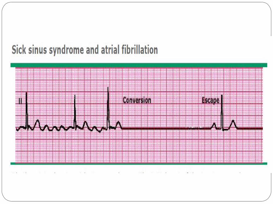
Atrial fibrillation with slow ventricular
response

Atrial frequency < 60 bpm
Ventricular frequency same
Regularityregular
Originsinus node
P-wavenormal
INAPPROPRIATE SINUS BRADYCARDIA
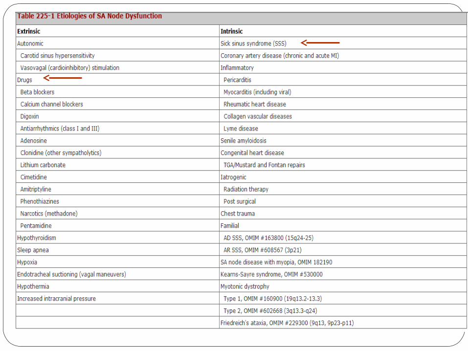
INAPPROPRIATE SINUS TACHYCARDIA
Atrial frequency 100-180 bpm
Ventricular frequency Same
Regularity regular
Originsinus node
P-wave positive in II, AVF

BRADYCARDIA-TACHYCARDIA SYN

SINUS ARREST
The sinus node stops firing resulting in a pause in heart beat
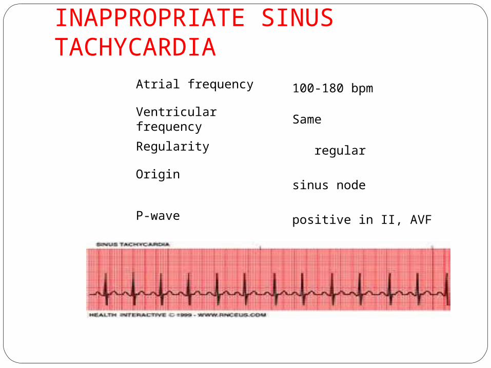
SINO-ATRIAL EXIT BLOCK
The depolarizations that occur in the sinus node cannot leave the node towards the atria. They are blocked.
On the ECG this is expressed as a pause.
SA exit block can be destinguished from sinusarrest because the pause in SA exit block is a multiple of the P-P interval that preceded the pause.

Three subtypes can be distinguished
Type I second degree (Wenkebach) SA exit block:
the P-P interval progressively shortens prior to the
pause
Type II second degree SA exit block:
the pause equals approximately 2-4 times the
preceding PP interval
Third degree SA exit block:
absence of P waves (diagnosed with an sinus node
electrode, during electrophysiological evaluation)
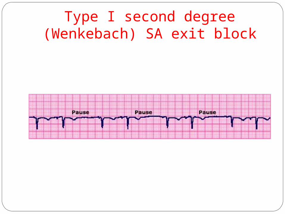
Type I second degree (Wenkebach) SA exit block

SA BLOCK TYPE 2

Exercise testingCHRONOTROPIC INCOMPETENCE
Inability of the sinus node to achieve at least 80 percent of the
age predicted heart rate. Astrand's formula (220- age) at
peak exercise.
Seen in 20 to 60 percent of patients with sinus node
dysfunction.
Although the resting heart rates may be normal,
may have inability to increase their heart rate during
exerciseor
have unpredictable fluctuations in heart rate during activity.
-Gwynn N, R, Kratz, et al. Chronotropic incompetence. Am Heart J 1992;123:1216.

Specific sinus node chronotropic incompetence is the inability of the sinus node to accelerate in response to metabolic demands secondary to intrinsic disease or negative chronotropic drugs.
Functional chronotropic incompetence manifests itself either as atrial tachyarrhythmias or as retrograde ventriculoatrial conduction

DRUGS
Atropine/isoproterenoll— Atropine (1 or 2 mg) / isoproterenol (2 to 3
μg/min)
Abnormal response --- increase in the sinus rate of <25 %, or to a
rate below 90 beats/min.
Potential problems.
There has been no standardization of the pharmacologic testing,
No dose ranging has been reported,
The specificity and sensitivity of the tests are uncertain,
SSS may exist even if the response is normal.
Isoproterenol is risky in patients with ischemic and other types of heart
disease.

DRUGS Beta blockers —
Propranolol has been used to assess sinus node function on the assumption that the chronotropic response may differ between patients with a normal and a sick sinus node.
This approach, however, has been disappointing

DRUGS cont…
Adenosine directly inhibits sinus node activity
Due to increased potassium conductance -hyperpolarization of the resting membrane potential.
Adenosine should be considered as an alternative to invasive testing in patients with suspected SSS.
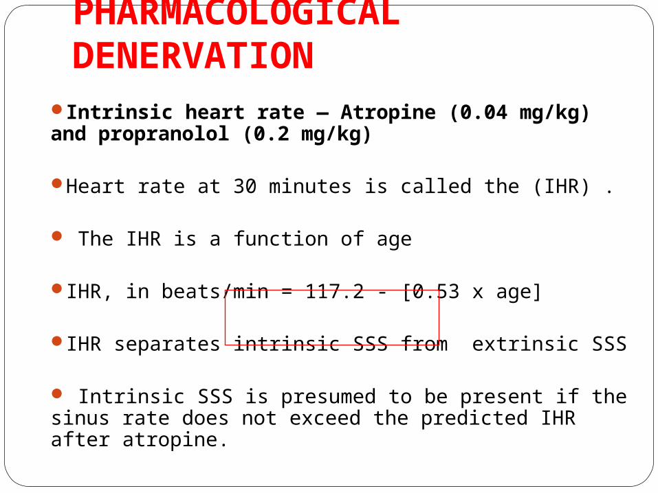
PHARMACOLOGICAL DENERVATION
Intrinsic heart rate — Atropine (0.04 mg/kg) and propranolol (0.2 mg/kg)
Heart rate at 30 minutes is called the (IHR) .
The IHR is a function of age
IHR, in beats/min = 117.2 - [0.53 x age]
IHR separates intrinsic SSS from extrinsic SSS
Intrinsic SSS is presumed to be present if the sinus rate does not exceed the predicted IHR after atropine.

Long-Term Electrocardiographic
Recording
In patients engaged in normal daily activities
document and quantitate the frequency and complexity of an
arrhythmia
correlate the arrhythmia with the patient's symptoms, and
evaluate the effect of antiarrhythmic therapy on spontaneous
arrhythmia.
For example, recording normal sinus rhythm during the
patient's typical symptomatic episode effectively excludes
cardiac arrhythmia as a cause
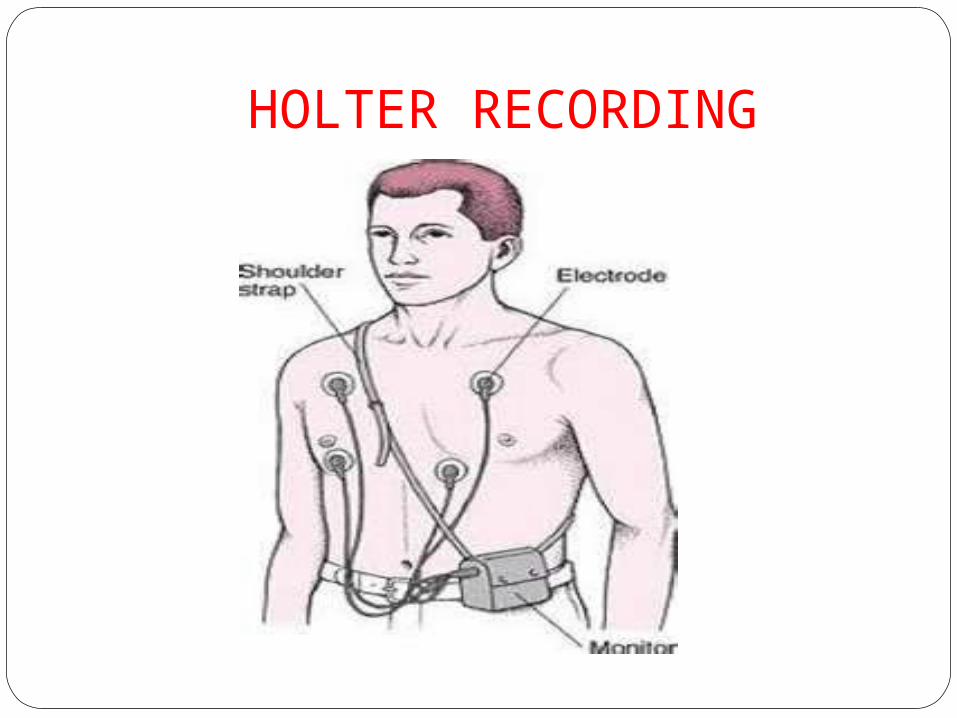
HOLTER RECORDING

HOLTER RECORDINGIf symptoms are frequent, 24- or 48-hour
ambulatory Holter monitoring can be useful. Documentation of symptoms in a diary by
the patient. Often the sinus pauses recorded are not
associated with symptoms. Several Holter monitor studies demonstrated
the futility of treating asymptomatic pauses, even if they were 3 seconds or longer.
The length of the pause correlated poorly with symptoms and prognosis

EVENT RECORDING.
In many patients, the 24-hour snapshot provided by the Holter recording is incapable of documenting the cause of the patient's symptoms.
These devices are about the size of a pager and are kept by the patient for 30 days.

IMPLANTABLE LOOP RECORDER
For patients with infrequent and transient symptoms, neither Holter recorders nor 30-day event recorders may yield diagnostic information.
In such patients, implantable loop recorders may be used.
This device (about the size of a pack of chewing gum) is inserted under the skin at about the second rib on the left front of the chest and is activated by passing a special magnet over the device.

LOOP RECORDERS

Signal-averaged P wave
fractionated endocardial electrograms in the SSS:-
Long, low amplitude signals early during the signal averaged P wave were found to be characteristic of SSS.
sensitivity-76% specificity-91%In paroxysmal atrial fibrillation the incidence of
SSS was higher in those with low amplitude atrial early potentials on a signal-averaged P wave
- Yamada, et al. J Am Coll Cardiol 1996; 28:738.

EPS

ELECTROPHYSIOLOGICAL TESTING
IndicationsThe symptomatic patient who has no ECG findings suggestive of SSS.
The symptomatic patient in whom ECG fail to correlate with symptoms.
The patient who develops dysfunction of the SA node on usual doses of drugs.

EPS indications cont…
The patient with syncope or near syncope who has bundle branch or multifascicular block may require electrophysiologic evaluation of the SA node, the AV node, and the infranodal His-bundle branch-Purkinje system.
Electrophysiologic testing that shows SA nodal dysfunction allows the selection of appropriate therapy in up to 50 percent of these patients.

CONTRAINDICATIONS
Unstable angina
Bacteremia or septicemia
Acute decompensated congestive heart
failure not caused by the arrhythmia
Major bleeding diathesis
Acute lower extremity venous thrombosis if
femoral vein cannulation is desired.

SINOATRIAL NODE RECOVERY TIME — The SNRT is perhaps the most
useful test of overall sinus nodal automaticity.
The atria are driven rapidly: a normal SA node will have a recovery
time within certain limits,
while recovery will be delayed in a depressed or sick sinus node.

SNRT cont….It is performed by placing a catheter near the SN.
Overdrive stimulation is performed at a rate higher
than that of the SA node for about one minute (the
range in most published reports is 30 to 180 sec).
Pacing is then stopped, and the time from the last
paced atrial beat to the first spontaneous electrical
beat with a sinus morphology is measured. Pacing
is increased 10 to 20 beats/min up to 200
beats/min, if tolerated.

SNRT cont…. SNRTc corrected by subtracting the sinus
cycle length from the SNRT
SNRTn a function of the cycle length-,
SNRT/sinus cycle length.
Total recovery time (TRT) which is the time required to return to
thebasal sinus rate..

SNRT

Normal values have generally been estimated as follows :SNRT/SCL <150 percentCSNRT < 550 millisecondsTRT less than five seconds.
Josephson, ME. Sinus Node Dysfunction. In: Josephson ME, ed. Clinical Cardiac Electrophysiology. Techniques and Interpretations. Third ed. Philadelphia: Lippincott; 2002:68.

The SNRT or SNRTc is abnormal in more than 50 percent of patients with suspected SSS.
J Am Coll Cardiol 1995; 26:555
Overdrive suppression, used in conjunction with intravenous disopyramide ,may increase the sensitivity of the test and its potential to diagnose a SSS .
Ishikawa, et al.. Sinus node recovery. Europace 2000; 2:54
Limitations of SNRTChanges in autonomic tone due to the effects of pacing
Changes in P-wave morphology suggesting a pacemaker shift
Sinoatrial entrance block
The hemodynamic effects of atrial pacing
Secondary pauses.
In fact, direct SN recordings have demonstrated that the majority of these pauses are due to sinoatrial exit block rather than impaired automaticity.
- Tracy, CM, Akhtar,
M, DiMarco, JP, et al

THE USE OF TRANSESOPHAGEAL ATRIAL PACING
Transesophageal pacing has been used to assess sinus node dysfunction, primarily by assessing SNRT, and has been recommended in the evaluation of patients with syncope.
Brembilla-Perrot, B, Beurrier, D, Houriez, P, et al. Utility oftransesophageal atrial pacing in the diagnostic evaluation of patientswith unexplained syncope associated or not with palpitations. Int JCardiol 2004; 96:347.

SACT
Sino Atrial Conduction Time
Can be measured in two ways
Indirect
Direct

SACT cont…Indirect method; The usual methodThis approach involves the placement of a
catheter near the sinus node. Strauss method
progressively premature atrial extrastimuli (A2) are introduced after every eighth to tenth beat of stable sinus rhythm.
Narula method Brief periods of atrial pacing at rates above sinus.
.

SACTA1-A1 The interval between each of the normal sinus
beat or the stable sequence of paced beats.A2-paced impulseA3-The first spontaneous sinus beat that occurs
after termination of the pacing.A2-A3 The interval between the final paced impulse
(A2) and the first spontaneous beat (A3) is referred toas the return interval
Four zones after APD

No reset/collision/Interferance
APD IN LAST 20-30% OF SCL

RESET(MAJOR PART)
ie, less than compensatory-impulse in earliest third of zone 2(40-50% OF
SCL)

INTERPOLATION

SA ECHO’S/RE-ENTRY


SACTThe A1-A1 interval Time required to generate a sinus impulse.,
conduction time does not contribute to A1-A1.
A2-A3 (Because A2 resets the SN, the return cycle length) =
generation of the next sinus beat (reflecting SNautomaticity) +
conduction of the impulse into and out of SN tissue.
(A2-A3) –(A1-A1) = Total time it takes to enter and exit the
SN tissue. This number represents twice the SACT SACT = [(A2-A3) – (A1-A1)] / 2
Normal SACT times generally range from 40 to 150 milliseconds

Limitations of SACT The assumption that entrance into and exit out of the SN takes the same
amount of time is not necessarily valid
Distance of the stimulation site from the SN is another source of error. The
farther the site is from the SN, the greater the potential for overestimation of
the SACT due to conduction delays in both atrial and perinodal tissues.
Reproducibility of results
sinus arrhythmia
shift in intrinsic pacemaker site
Depression of automaticity, and
sinus entrance block

Direct recording of the sinoatrial conduction time (sinus node electrogram)
Endocardial recordings demonstrate diastolic phase 4 activity followed by a slow upstroke culminating in a rapid atrial EGM.
The directly measured SACT was defined as the interval between the local EGM and the rapid atrial deflection.
Sinoatrial block occurs when the local EGM was seen in the absence of an atrial deflection


SACT-DIRECT METHOD


SENSITIVITY OF SNRT AND SACT70%
SPECIFICITY OF SNRT AND SACT90%

CORRELATION OF SNRT AND SACT WITH ECG ABNORMALITIES
Patients with Symptomatic sinus bradycardia – longer SNRT and SACT
Patients with SA block -longer SACT; and
Patients with the tachycardia-bradycardia syndrome had a longer SNRT
Breithardt, G, Seipel, L, Loogen, F. Sinus node recovery time and calculated sinoatrialconduction time in normal subjects and patients with sinus node dysfunction. Circulation 1977; 56:43.

SINUS NODE AND ATRIAL REFRACTORY PERIOD
SA nodal refractoriness has been determined using both the extrastimulus and pacing train techniques.
Normal subjects having a SNERP of 250 to 350 msec as compared to a value of 500 to 550 msec in patients with the SSS
Kerr, CR, Strauss, HC. The measurement of sinus node refractoriness inman. Circulation 1983; 68:1231.
.
.

COMPLICATIONS DURING EP STUDY
Hematoma at the puncture site in the groin and or neck
Hemorrhage
Infection caused by manipulation of catheters (theoretical risk)
Perforation upon catheter manipulation inside the heart of small patients (most commonly involving the right atrial appendage and the right ventricular outflow tract)


TREATMENT OF SSSMainly directed at symptoms
Search for remidial causes - Drugs
-Ischemia-Autonomic imbalance
Mostly by pace makers.
Drugs-Theopylline
Ablation for AF


A number of additional pacemaker features may be useful in selected patients:
Rate responsive programming
SSS and chronotropic insufficiency and exertional symptoms.
Mode switching
SSS who have paroxysmal atrial tachycardia.


Class I indications
Sinus bradycardia in which symptoms are clearly related to the bradycardia’.
Symptomatic chronotropic incompetence

Class II
Sinus bradycardia (heart rate <40 beats/min) with symptoms suggestive of
bradycardia, but without a clearly demonstrated association.
Sinus node dysfunction in a patient with unexplained syncope.
Chronic heart rates <40 beats/min while awake in a minimally
symptomatic patient.

DRUG-INDUCED SINUS NODE DYSFUNCTION
Some patients will meet the above indications
for pacemaker implantation due to the effects
of medications that are necessary for the
treatment of arrhythmias or other medical
conditions.
In such cases, patients should be considered to
have the same indication for pacemaker
implantation as those with intrinsic sinus node
dysfunction

FOLLOW-UPThree major issues that need to be considered;
1. Ventricular pacing can induce dyssynchrony observe for evidence of heart failure.
2. In patients with an AAI pacemaker, monitoring for progression to high degrees of
AV block is essential as the reported rate varies from 0.6 to as high as 3 percent.
3. Due to the thromboembolic risks associated with unrecognized AF, patients should
be observed for the development of atrial arrhythmias.

AV NODE
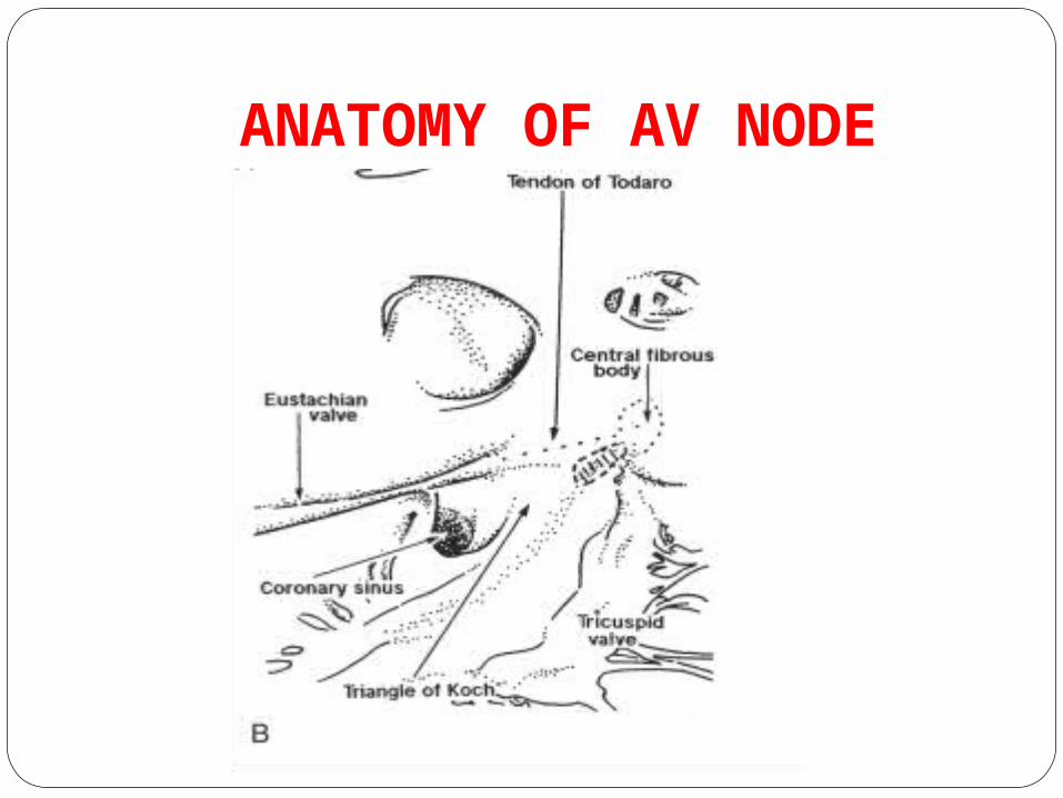
ANATOMY OF AV NODE

BLOOD SUPPLY The AV node AV nodal artery ---right coronary artery in 90 percent left circumflex artery in 10 percent .The bundle of His –both the AV nodal artery and branches of the left anterior descending artery.
The left bundle has a rich blood supply AV nodal artery, Posterior descending artery, and Branches of the left anterior descending artery.
Frink RJ, James TN.. Circulation 1973;43:491–502

CLINICAL FEATURES First-degree AV block -long a-c wave
diminished intensity of S1.
type I second-degree AV block- the heart rate may increase
imperceptibly with
gradually diminishing intensity of s1,
widening of the a-c interval
terminated by a pause, and a wave not followed
by a v wave.
type II AV block --Intermittent ventricular pauses and
a waves in the neck not followed by v
waves s1 maintains a constant intensity.

In complete AV block
-variable intensity of s1
-a waves in the jugular venous pulse --lacking a consistent relationship to ventricular contraction.
-Intermittent large (cannon) a waves
may be seen in the jugular venous pulse when atrial and ventricular contractions occur simultaneously.
The second heart sound can split normally or
paradoxically, depending on the manner of ventricular activation.

First-Degree AV Block
During first-degree AV block, every atrial impulse conducts to the ventricles and a regular ventricular rate is produced, but the PR interval exceeds 0.20 second in adults.
PR intervals as long as 1.0 second have been
noted and can at times exceed the P-P interval, a phenomenon known as “skipped” P waves.
can result from a conduction delay in the AV node (A-H interval), in the His-Purkinje system (H-V interval), or at both sites.


First-degree and type I second-degree AV block can occur in normal healthy children, and a Wenckebach AV block can be a normal phenomenon in well-trained athletes, as noted earlier, probably related to an increase in resting vagal tone

Third-Degree (Complete) AV Block No atrial activity is conducted to the
ventricles Atria and ventricles are controlled by
independent pacemakers. A type of complete AV dissociation. The atrial pacemaker can be sinus or
ectopicThe ventricular focus is usually located just
below the region of the block, which can be above or below the His bundle bifurcation.

ventricular pacemaker activitycloser to the His bundle appear to be more stable and a faster escape rate than can those located more distally in the ventricular conduction system.
The ventricular rate in acquired complete heart block is less than 40 beats/min but can be faster in congenital complete AV block

COMPLETE HEART BLOCK
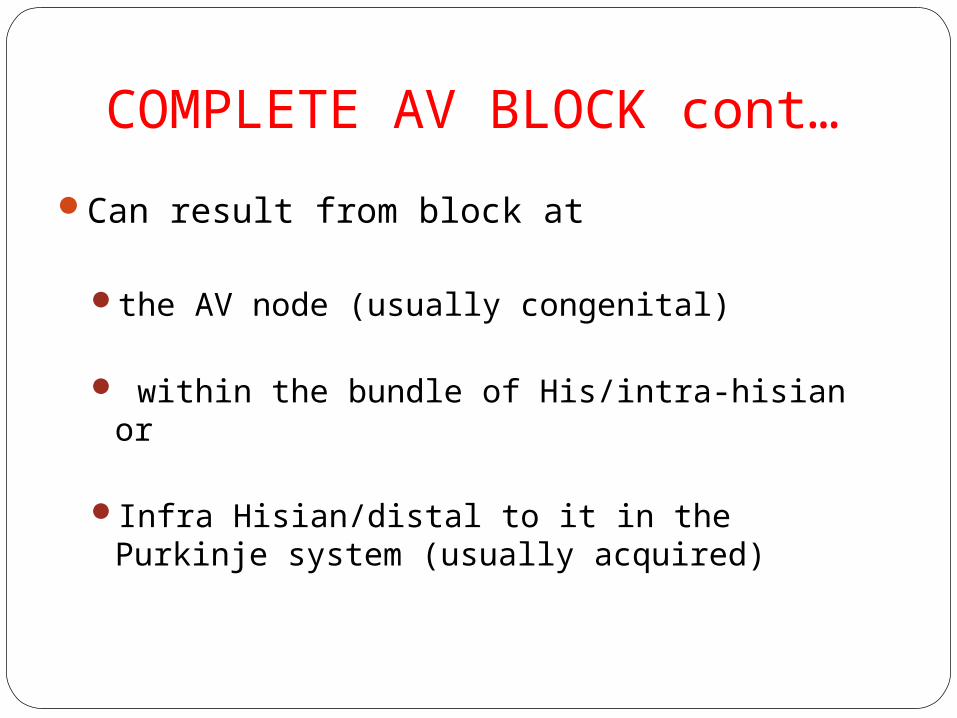
COMPLETE AV BLOCK cont…
Can result from block at
the AV node (usually congenital)
within the bundle of His/intra-hisian or
Infra Hisian/distal to it in the Purkinje system (usually acquired)

Block proximal to the His bundle –escape focus in or near the His bundle.
normal QRS rates of 40 to 60 beats/min
His bundle recording –differentiate AV nodal from
intrahisian block.

High-Grade Atrioventricular Block
When two or more consecutive atrial impulses do not
conduct to the ventricle, it is defined as high-grade
AV block.
It may be associated with a junctional or ventricular
escape rhythm. .
Unless a clearly defined reversible etiology is
identified, permanent pacing is indicated.

EPS IN AV NODE DYSFUNCTIONPatients with AV block without clear
symptom association.
Patients with symptoms suggestive of bradyarrhythmias, but in whom significant AV block has not been documented.
When the site of AV block cannot be determined by surface tracings.( 2: 1 AV block ).

BASE LINE RECORDINGSThe first intracardiac tracing is a recording from
the high right atrium (HRA) close to the sinus node.
Pacing at this position allows evaluation of sinoatrial node function and atrioventricular conduction;
Recordings from this site also help determine the direction of atrial activation (eg, high-low versus low-high, and right-left versus left-right).


PA INTERVAL
Atrial electrogra
m
HBE-D

The reported normal range is 20 to 60 msec.
Represent -total atria conduction, reflection of internodal
conduction (sinoatrial to atrioventricular node)

AH INTERVAL
AH INTERVAL
Atrial electrogra
m
HBE-D

This interval is taken to represent AV nodal conduction.
50 to 120 msecmarkedly influenced by the autonomic
nervous system Long AH intervals –drugs,increased vagal
tone,AV node disease

HV INTERVAL
Atrial electrogra
m
HBE-D

conduction time through the distal His-Purkinje . less influenced by the autonomic nervoussystem, Normal, 35 to 55 msecA prolonged HV interval is consistent with diseased
distal conduction in all fascicles .

HBE-D

first-degree AV block
most common EPS finding in patients with P-R prolongation is a long A-H interval because intra- and infra-Hisian conduction compromise only a minor portion of the P-R interval

Second-degree AV block — Type IWenckebach almost always occurs in
the AV node. During EPS, this process is most evident on a
His bundle EGM.
progressively longer A-H interval and a stable H-V interval
until the final beat of the series in which there is an atrial EGM with no ventricular EGM.

True type II second-degree block Mobitz II
almost always, if not always, occurs in the HPS.
His bundle EGM follows the atrial EGM prior to the dropped ventricular EGM confirming the site of block.

Third-degree (complete) AV blockMost patients with complete AV block are
treated with permanent pacemakers EPS is not generally indicated. Among patients with complete AV block,
EPS may be indicatedwhen symptoms are not present,the site of block is not apparent, orthe block is potentially reversible

Findings suggestive of high risk
alternating left bundle branch block (LBBB) and right bundle branch block (RBBB), or those with
RBBB with alternating left anterior and left posterior fascicular block.
Symptomic- prolonged H-V interval (>55 milliseconds)
Asymptomatic -H-Vinterval >100 milliseconds
Highest progression to complete heart block and should receive pacemakers

ATRIAL EXTRASTIMULI

ATRIAL EXTRASTIMULI
Long coupling interval – conduct through the AV node with a constant velocity.
As the coupling intervel becomes shorter the conduction velocity decreases –increased A-H iterval.
Decremental conduction is a property of AV node


AVN REFRACTORY PERIODERP- longest copling intervel between the
basic drive and the premature impulse that fails to propogate through the AVN.
NORMAL-- <450 milliseconds.
Increasing refractory period with shorter cycle lengths indicates abnormal HPS conduction.

PHARMACOLOGIC CHALLENGE
Procainamide normally prolongs the H-V interval by 10 to 20 percent.
After administering procainamide-Doubling of the H-V interval,-an H-V interval >100 milliseconds or
the-development of infra-Hisian block
represent poor HPS reserve. Mandates permanent pacing

TREATMENT OF AV DYSFUNCTION

INDICATIONS OF PACING
Class I
Complete (third-degree) AV blockAdvanced second-degree AV block (block of
two or more consecutive P-waves)Symptomatic Mobitz I or Mobitz II second-
degree AV blockMobitz II second-degree AV block with a
widened QRS or chronic bifascicular block,regardless of symptoms
Exercise-induced second or third degree AV block (in the absence of myocardial ischemia)

Class IIAsymptomatic Mobitz II second-degree AV
block with a narrow QRS intervalFirst-degree AV block when there is
hemodynamic compromise because of effective AV dissociation secondary to a very long PR interval.
Bifascicular or trifascicular block associated with syncope that can be attributed to transient complete heart block

PACING IN CONGENITAL CHBcongestive heart failure
average heart rate of less than 50
beats/min in the awake infant
history of syncope or presyncope
significant ventricular ectopy or
exercise intolerance

THANK YOU