The Knee (Tibiofemoral Joint) By Todd Piraino Tammi Wells Holly Cox.
65
The Knee (Tibiofemoral Joint) By Todd Piraino Tammi Wells Holly Cox
-
Upload
robert-patterson -
Category
Documents
-
view
213 -
download
0
Transcript of The Knee (Tibiofemoral Joint) By Todd Piraino Tammi Wells Holly Cox.

The Knee (Tibiofemoral Joint)
By Todd PirainoTammi Wells
Holly Cox
Quadriceps femoris tendon
Connects the quadriceps femoris muscles to the superior aspects of the patella. Controls knee flexion and extension

Patellar ligament
Connects Patella to the tuberosity of the Tibia.

Oblique popliteal ligament
A strong broad flat fibrous ligament that pass obliquely across and strengthens the posterior part of the knee.

Arcuate Popliteal Ligament
A triangular band in the posterior part of the knee that passes medially downward from the lateral condyle of the femur to the area between the condyles of the tibia and to the head of the fibula

Tibial collateral ligament and Fibular collateral ligament
Tibial Collateral ligament also known as Medial Collateral ligament or MCL.
Fibular Collateral ligament is located on the lateral side of the knee.
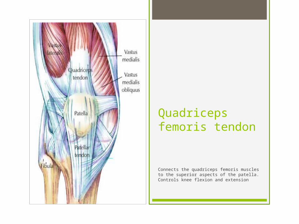
Anterior Cruciate Ligament and Posterior Cruciate Ligament
The function of the ACL is to provide stability to the knee. It limits rotational movements of the knee and restrains excessive extension of the lower leg.
PCL prevents the Tibia from sliding to far backwards in relation to the Femur. Stronger of the two cruciate ligaments.
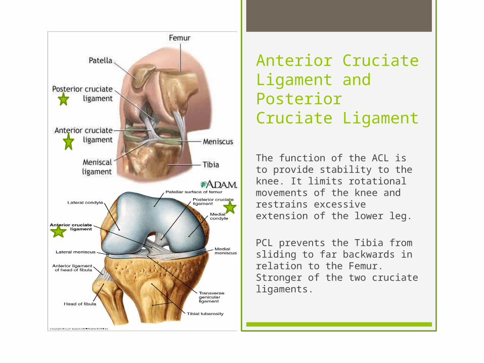
Transverse Ligament
Connects the Anterior margin of the lateral meniscus to the anterior end of the medial meniscus.
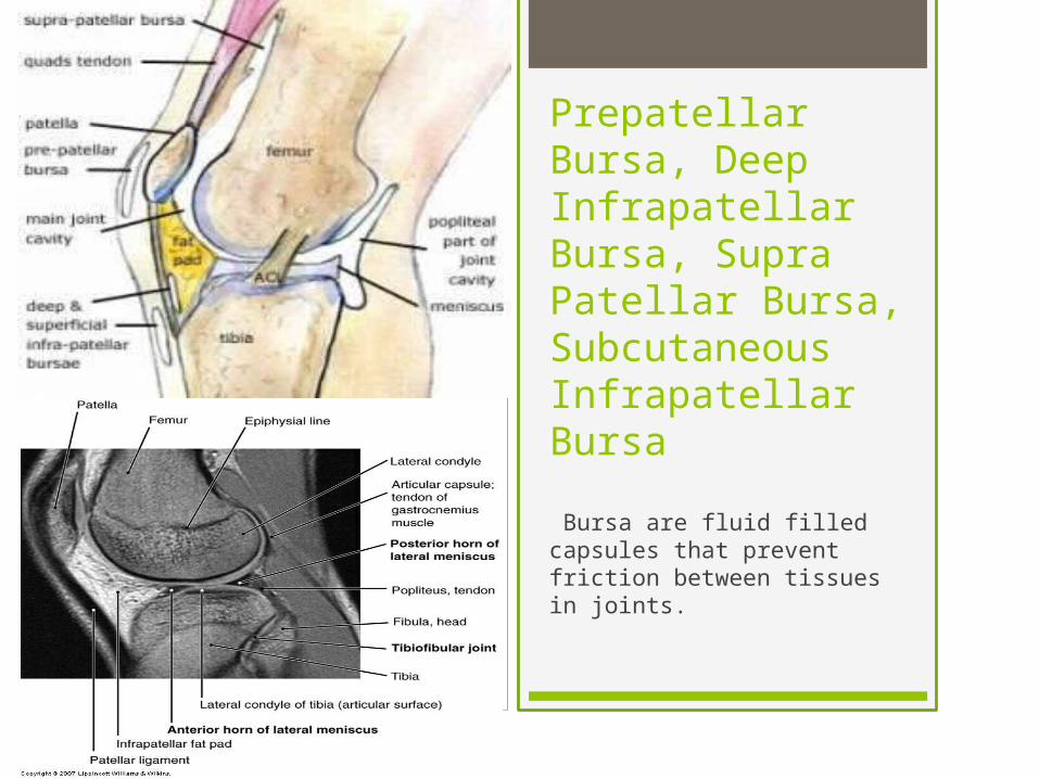
Prepatellar Bursa, Deep Infrapatellar Bursa, Supra Patellar Bursa, Subcutaneous Infrapatellar Bursa Bursa are fluid filled capsules that prevent friction between tissues in joints.

Lateral Meniscus and Medial Meniscus
The Meniscus are fibrocartilage that separate the Tibia and Femur to decrease contact area between the bone. Acts as a shock absorber and reduces friction between the two bones.

Femur: Lateral Condyle, Medial Condyle, Intercondylar Fossa.
Condyles of the Femur articulate with the TibiaLateral
Condyle MedialCondyle
Intercondylar Fossa

Femur: Lateral Epicondyle, Medial Epicondyle
Epicondyles are not part of the articulating surface. Main purpose is attachment points for ligaments and tendons.
MedialEpicondyle
LateralEpicondyle

Tibia: Lateral Condyle, Medial Condyle, Intercondylar Eminence.
Articulate with the Femur.
Intercondylar Eminence

Tibial Tuberosity and Head of Fibula

Buckwheat says, “Otay, that’s gonna leave a mark.”

This guy is probably hearing the sound of popcorn coming from his knee.

More sound of popcorn.
Most likely a torn ACL

ACL Injury
Most common type of Knee injury. Hyperextension is the main cause. Torn ACL’s are most often related to high impact sports or when the knee is forced to make sharp changes in movement during sudden stops from high speed. Very prevalent in Alpine skiing, Soccer, Rugby, Hockey, and martial arts.

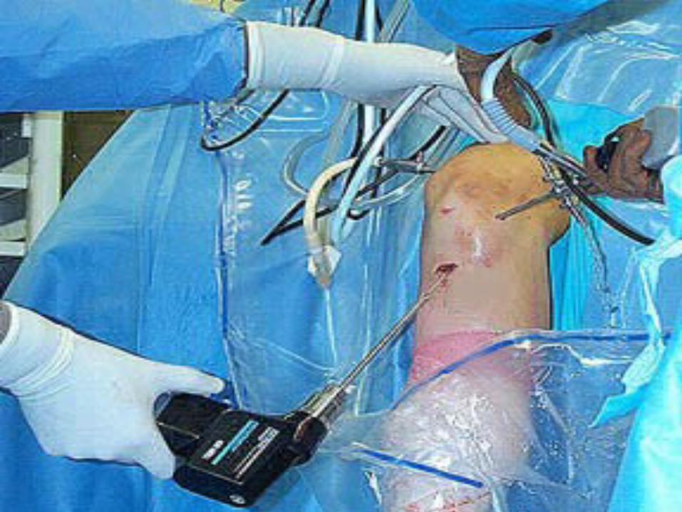
Repair of ACL
It has become an outpatient operation with many people returning home the day of the surgery and bearing weight within a week.

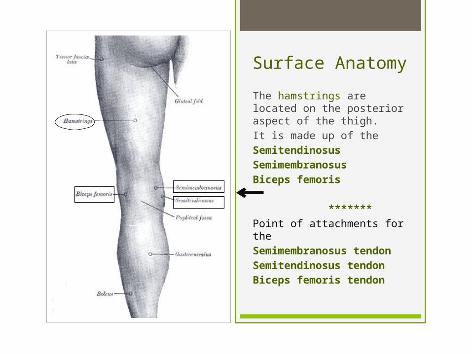
Surface Anatomy
Popliteal fossa- a mostly fat-filled diamond shaped space posterior to the knee. All the important nerves and vessels from the thigh to the leg pass through this fossa.
Patella- (knee cap) is a small, triangular bone located anterior to the knee joint. It articulates with the patellar surface of the femur.

*The soleus is a long flat muscle that runs down the posterior of the lower leg, underneath the gastrocnemius.
*The gastrocnemius creates the distinctive shape of the calf and is an important muscle for overall leg development and symmetry. Gastrocnemius is Greek means ‘The belly of the calf’.The medial head is the inner head of the two gastrocnemius heads that are located at the rear of the lower leg. The lateral head is the outer head of the two gastrocnemius heads.
Medial head of
Lateral head of
Surface Anatomy
Surface Anatomy
The hamstrings are located on the posterior aspect of the thigh. It is made up of the SemitendinosusSemimembranosus Biceps femoris
*******Point of attachments for the Semimembranosus tendonSemitendinosus tendonBiceps femoris tendon

Surface Anatomy
Vastus lateralis is the largest component of the quadriceps, located on the lateral aspect of the thigh.
Vastus medialis covers the medial aspect of the thigh
The Quadriceps femoris consists of four muscles:
Rectus femoris Vastus lateralisVastus intermediusVastus medialis

Surface Anatomy
Soleus
Popliteal fossa
Lateral head of Gastrocnemius
Medial head of Gastronemius

Popliteal Fossa
The popliteal fossa is a fat-filled diamond shaped shallow depression which is posterior to the knee.
All the important nerves and vessels from the thigh to the leg pass through the popliteal fossa.

Arteries of knee
The major blood vessels around the knee travel with the popliteal nerve down the back of the leg.
The popliteal artery and popliteal vein are the largest blood supply to the leg and foot.
If the popliteal artery is damaged beyond repair, it is very likely the leg will not be able to survive

Arteries( and muscles they
innervate)
• Femoral artery • Popliteal artery • (Popliteus, Gastrocnemius)
• Posterior tibial artery• Dorsalis pedis• Fibular (peroneal)• Anterior tibial artery• Lateral circumflex femoral
artery• (Rectus femoris, Vastus intermedialis,• Vastus lateralis, Vastus medialis)
• Medial circumflex femoral artery
• Inferior gluteal artery (Biceps femoris, Semimembranosus)
• Deep femoral artery (profunda)
• (Semitendinosus)

VeinsAnterior tibial veinPosterior tibial veinFibular (peroneal vein)Popliteal veinFemoral veinSmall saphenous veinGreat saphenous vein

Femoral artery
Great Saphenous vein
Femoral vein
Popliteal artery
Anterior tibial artery
Fibular (peroneal) artery
Posterior tibial artery
Dorsalis pedis
Small saphenous vein
Fibular (peroneal) vein
Anterior tibial vein
Posterior tibial vein
Popliteal vein
Wire man

Flat Man …………NERVES
*Femoral nerve
*Sciatic nerve
*Superficicial fibular nerve (peroneal)
*Deep fibular nerve (peroneal)
*Tibial nerve
Semitendinosus, SemimembranousBiceps femoris (long head)
Gastrocnemius, Popliteus
Rectus FemorisVastus intermedialisVastus lateralisVastus medialis
Common fibular nerveBiceps femoris
(long head)

Classroom ModelsExternal iliac artery
Femoral nerve
Femoral nerve
Femoral artery
Inguinal ligament

Classroom models
Anterior tibial artery
Deep fibular nerve (peroneal)Dorsalis
pedis artery
Femoral artery
Femoral vein
Femoral nerve
External iliac artery
Inguinal ligament

Classroom ModelsSciatic nerve
Tibial nerve
Common fibular(peroneal) nerve
Tibial nerve
Popliteal vein
Popliteal artery
Posterior tibial artery

Classroom Models
Sciatic nerve
Tibial nerve
MUSCLES
KNEE

ANTERIOR KNEE MUSCLES
Function as extensors

Quadriceps Femoris: *largest muscle in the body, consists of 4 muscles

Rectus Femoris:
• Most anterior of quadriceps
• Origin: Anterior Inferior Iliac Spine
• Insertion: Tibial Tuberosity
• Innervation: Femoral Nerve
• Vascular Supply: Lateral Circumflex Femoral Artery

Action:
• Hip flexion• Knee extension

Vastus Medialis
Origin:• Linea Aspera
Insertion:• Tibial Tuberosity via Patellar Tendon
Innervation:• Femoral Nerve
Vascular Supply:• Lateral Circumflex Femoral Artery

Action:
• Knee extension

Vastus Intermedialis
Origin: Anterior Femur
Insertion: Tibial Tuberosity via Patellar Tendon
Innervation: Femoral Nerve
Vascular Supply: Lateral Circumflex Femoral Artery

Action:
• Knee extension

Vastus lateralis:
Origin:• Linea Aspera
Insertion:• Tibial tuberosity via
Patellar Tendon
Innervation:• Femoral Nerve
Vascular Supply:• Lateral Circumflex Femoral
Artery
VastusLateralis

Action:
• Knee Extension

POSTERIOR KNEE MUSCLES

HAMSTRINGS: composed of Biceps Femoris, Semimembranous and Semitendinosus Function as flexors
(named due to tendons being long and stringlike in popliteal area)

Ah, those hamstrings…
See how they work? Or not??
**This is the torn version that we don’t need to know.

Biceps Femoris-long head:
Origin:• Ischial Tuberosity
Insertion:• Fibular Head
Innervation:• Sciatic Nerve
Vascular Supply:• Inferior Gluteal Artery
Action:
• Hip extension • Knee flexion

Biceps Femoris – Short Head:
Origin:• Lateral Lip of Linea Aspera
Insertion:• Fibular Head
Innervation:• Common Peroneal Nerve
Vascular Supply:• Inferior Gluteal Artery

Action:• Knee flexion

Semimembranosus:
Origin:• Ischial Tuberosity
Insertion:• Posterior surface of Medial
Condyle of Tibia
Innervation:• Sciatic Nerve
Vascular Supply:• Inferior Gluteal Artery
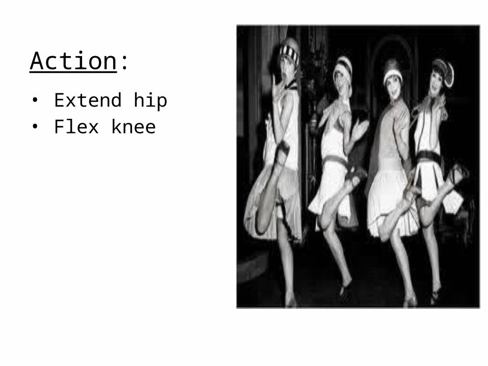
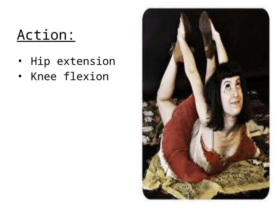
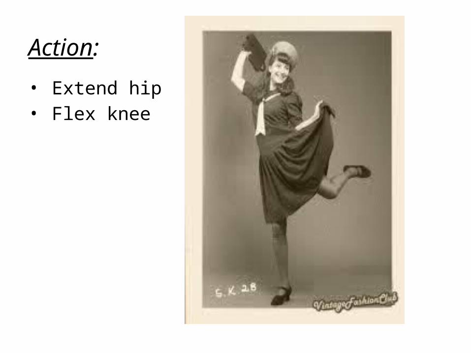
Action:• Extend hip • Flex knee

Semitendinosus:
Origin:• Ischial Tuberosity
Insertion:• Anteromedial surface
of proximal Tibia
Innervation:• Sciatic Nerve
Vascular Supply:• Deep Femoral Artery
Action:
• Extend hip • Flex knee
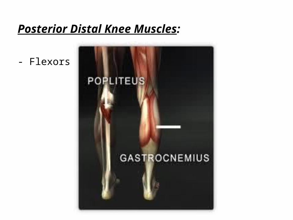
Posterior Distal Knee Muscles:
- Flexors

Popliteus:
Origin:• Lateral Condyle of
Femur
Insertion:• Posteriorly on Medial
Condyle of Tibia
Innervation:• Tibial Nerve
Vascular Supply:• Popliteal Artery

Action:• Initiates knee
flexion by unlocking the knee when fully extended

Gastrocnemius:
Origin:• Medial and
Lateral Condyles of Femur
Insertion:• Posterior
Calcaneus
Innervation:• Tibial Nerve
Vascular Supply:• Popliteal
Artery
The reason for high heels?

Action:• Ankle plantar flexion when knee extended• Raises heel while walking with knee extended
OR…
(Sorry, couldn’t resist!)

Action:• Knee
flexion
(Basketball players learning Charleston)

ByeFromBob
BFB:
Referenceswww.getfittogolf.com.auphysiomed.patientsite.comwww.anatomy.tvwww.freebase.comwww.chiropractic-help.comwww.orthobullets.comwww.msdlatinamerica.comwww.netteranatomy.com