Assessment of Heart and Great Vessels Christine M. Wilson Viterbo University.
29
Assessment of Assessment of Heart and Great Heart and Great Vessels Vessels Christine M. Wilson Christine M. Wilson Viterbo University Viterbo University
-
Upload
blaze-wilcox -
Category
Documents
-
view
224 -
download
0
Transcript of Assessment of Heart and Great Vessels Christine M. Wilson Viterbo University.
Assessment of Heart Assessment of Heart and Great Vesselsand Great Vessels
Christine M. WilsonChristine M. WilsonViterbo UniversityViterbo University

ObjectivesObjectives
LandmarksLandmarks StructuresStructures Cardiac cycleCardiac cycle Developmental/transcultural Developmental/transcultural Risk factorsRisk factors Subjective dataSubjective data Objective dataObjective data

Position in the ChestPosition in the Chest
Beneath precordium—area on Beneath precordium—area on anterior chest overlying the anterior chest overlying the heart and great vesselsheart and great vessels
Located in mediastinum—Located in mediastinum—middle third of chest—middle third of chest—between the lungsbetween the lungs

Heart is an Heart is an upside down upside down triangle in the triangle in the chestchest
Top of heart isTop of heart is BaseBase, bottom , bottom is is ApexApex

Extends from Extends from 2nd to 5th ICS 2nd to 5th ICS and from Rt and from Rt sternal border sternal border to Lt MCLto Lt MCL
Great vessels Great vessels above base of above base of heartheart

Blood FlowBlood Flow
Inferior vena cavaInferior vena cava Right atriumRight atrium Right ventricleRight ventricle Pulmonary arteryPulmonary artery Pulmonary veinPulmonary vein Left atriumLeft atrium Left ventricleLeft ventricle AortaAorta
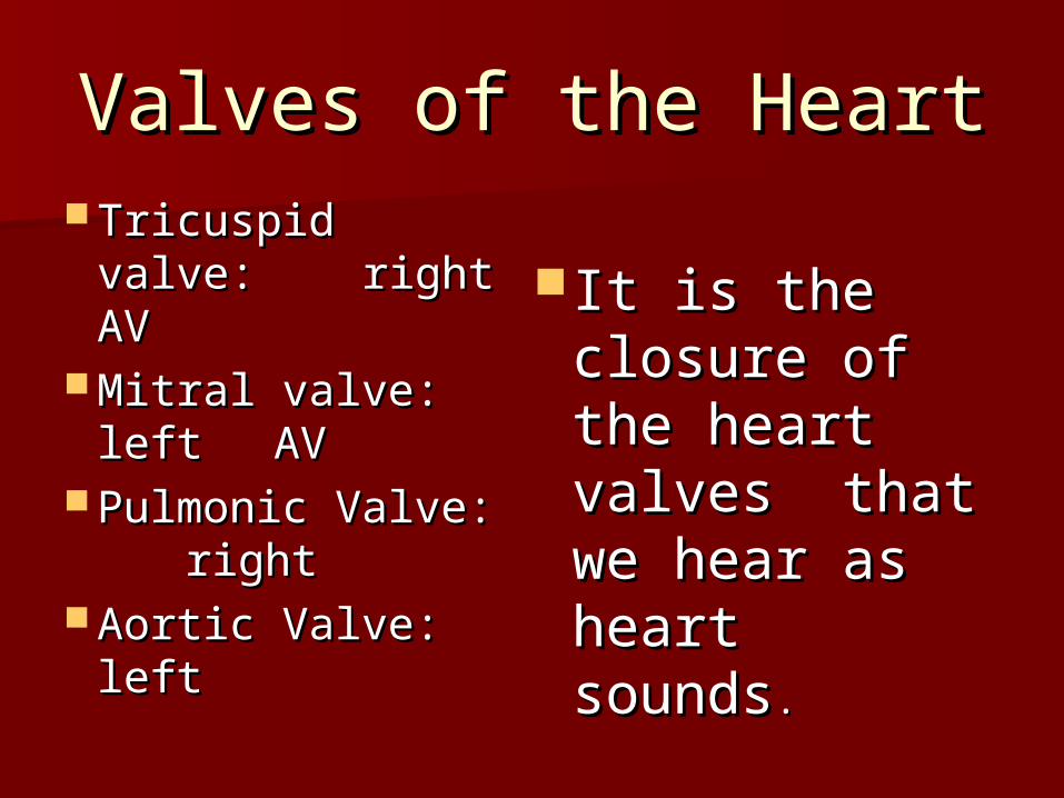
Valves of the HeartValves of the Heart Tricuspid valve: Tricuspid valve:
right AVright AV Mitral valve: Mitral valve:
left left AVAV Pulmonic Valve: Pulmonic Valve:
rightright Aortic Valve: Aortic Valve:
leftleft
It is the It is the closure of the closure of the heart valves heart valves that we hear that we hear as heart as heart soundssounds..

Cardiac CycleCardiac CycleDiastoleDiastole Tricuspid/mitral Tricuspid/mitral
valves openvalves open Ventricles relax and Ventricles relax and
fill with bloodfill with blood Ventricular Ventricular
pressures increasespressures increases Tricuspid/mitral Tricuspid/mitral
valves close causing valves close causing first heart sound first heart sound S1S1
LUBLUB dub dub
SystoleSystole Ventricular Ventricular
contraction increases contraction increases pressurepressure
Pulmonic/aortic valves Pulmonic/aortic valves open; blood ejectsopen; blood ejects
Ventricular pressure Ventricular pressure dropsdrops
Pulmonic/aortic valves Pulmonic/aortic valves close causing second close causing second heart sound heart sound S2S2
lub lub DUBDUB

More heart soundsMore heart sounds Events of right heart occur slightly laterEvents of right heart occur slightly later
– S1 Mitral valve closes then tricuspid S1 Mitral valve closes then tricuspid – S2 Aortic valve closes then pulmonicS2 Aortic valve closes then pulmonic
Sound radiates with direction of blood Sound radiates with direction of blood flowflow– S1 heard loudest at apexS1 heard loudest at apex– S2 heard loudest at baseS2 heard loudest at base
Murmurs—turbulent flow through Murmurs—turbulent flow through chambers and valveschambers and valves– Swooshing, blowing soundSwooshing, blowing sound

Pumping abilityPumping ability
Right side pumps blood to lungsRight side pumps blood to lungs Left side pumps blood to bodyLeft side pumps blood to body Cardiac output—volume of blood Cardiac output—volume of blood
pumped per minutepumped per minute– dependent upon volume ejected per dependent upon volume ejected per
stroke and heart rate (CO=SV x rate)stroke and heart rate (CO=SV x rate)– Normal cardiac output 4-6 L per minuteNormal cardiac output 4-6 L per minute
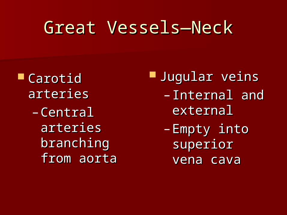
Great Vessels—Neck Great Vessels—Neck
Carotid arteriesCarotid arteries– Central Central
arteries arteries branching branching from aortafrom aorta
Jugular veinsJugular veins– Internal and Internal and
externalexternal– Empty into Empty into
superior vena superior vena cavacava

Developmental Developmental ConsiderationsConsiderations
FetalFetal Fetal heart begins to beat at 3 weeksFetal heart begins to beat at 3 weeks Oxygenation takes place through the Oxygenation takes place through the
placentaplacenta Blood returned to the Right side of the Blood returned to the Right side of the
heart and bypasses lungsheart and bypasses lungs– Foramen ovale-opening between atriumForamen ovale-opening between atrium– Ductus arteriosus-opening b/tw PA and Ductus arteriosus-opening b/tw PA and
Aorta Aorta

Fetal CirculationFetal Circulation

Changes take place at Changes take place at birth!birth!
Blood is oxygenated through lungsBlood is oxygenated through lungs Foramen ovale closes in one hourForamen ovale closes in one hour Ductus Arteriosus closes in Ductus Arteriosus closes in
10-15 hours10-15 hours Left ventricle pumps blood to Left ventricle pumps blood to
entire body; by one year, left entire body; by one year, left ventricle twice as large as the ventricle twice as large as the rightright

Pregnant FemalePregnant Female
Blood volume increases by 30-Blood volume increases by 30-40%, mostly during the 2nd 40%, mostly during the 2nd trimestertrimester
Increases Stroke Volume and COIncreases Stroke Volume and CO Rate increases by 10-15 BPMRate increases by 10-15 BPM
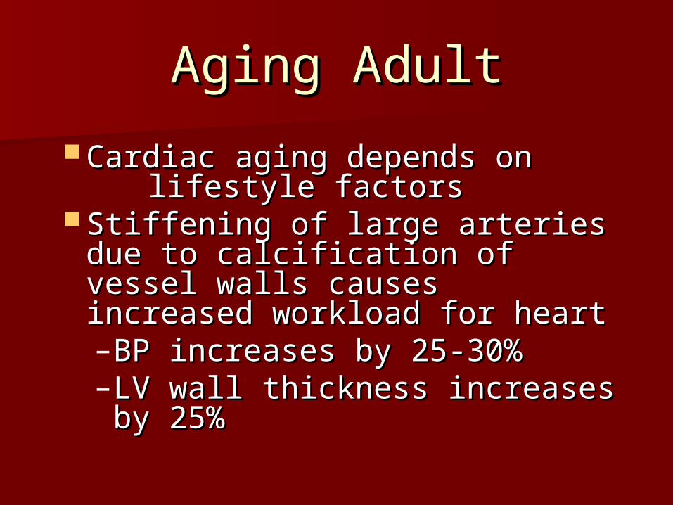
Aging AdultAging Adult
Cardiac aging depends on Cardiac aging depends on lifestyle factors lifestyle factors
Stiffening of large arteries due Stiffening of large arteries due to calcification of vessel walls to calcification of vessel walls causes increased workload for causes increased workload for heartheart– BP increases by 25-30%BP increases by 25-30%– LV wall thickness increases by LV wall thickness increases by
25%25%
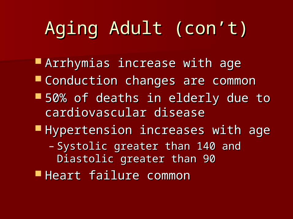
Aging Adult (con’t)Aging Adult (con’t)
Arrhymias increase with ageArrhymias increase with age Conduction changes are commonConduction changes are common 50% of deaths in elderly due to 50% of deaths in elderly due to
cardiovascular diseasecardiovascular disease Hypertension increases with ageHypertension increases with age
– Systolic greater than 140 and Diastolic Systolic greater than 140 and Diastolic greater than 90greater than 90
Heart failure commonHeart failure common
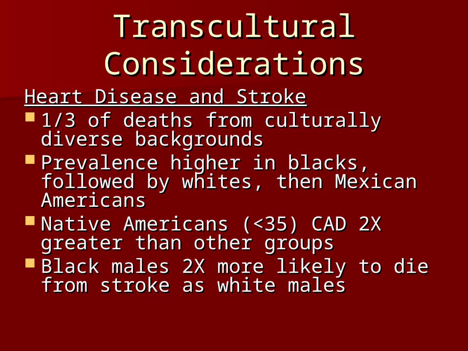
Transcultural Transcultural ConsiderationsConsiderations
Heart Disease and StrokeHeart Disease and Stroke 1/3 of deaths from culturally diverse 1/3 of deaths from culturally diverse
backgroundsbackgrounds Prevalence higher in blacks, followed Prevalence higher in blacks, followed
by whites, then Mexican Americansby whites, then Mexican Americans Native Americans (<35) CAD 2X Native Americans (<35) CAD 2X
greater than other groupsgreater than other groups Black males 2X more likely to die Black males 2X more likely to die
from stroke as white malesfrom stroke as white males

Transcultural Transcultural ConsiderationsConsiderations
Heart Disease and Stroke (con’t)Heart Disease and Stroke (con’t) Blacks 20-40 yearsBlacks 20-40 years
– Increased # of deaths from CVD Increased # of deaths from CVD compared to whitescompared to whites
– Increased mortality in black Increased mortality in black females over black malesfemales over black males
Black and Mexican American females Black and Mexican American females higher CVD risk factorshigher CVD risk factors
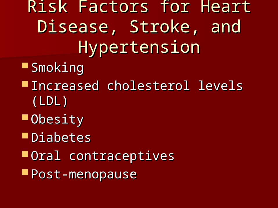
Risk Factors for Heart Risk Factors for Heart Disease, Stroke, and Disease, Stroke, and
HypertensionHypertension SmokingSmoking Increased cholesterol levels Increased cholesterol levels
(LDL)(LDL) ObesityObesity DiabetesDiabetes Oral contraceptivesOral contraceptives Post-menopausePost-menopause

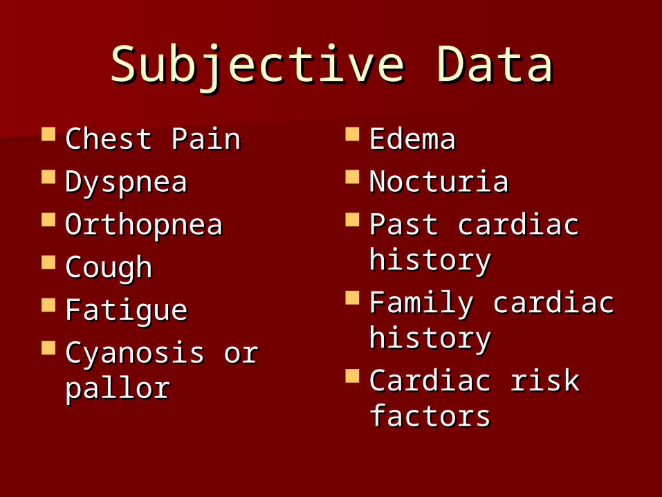
Subjective DataSubjective Data Chest PainChest Pain DyspneaDyspnea OrthopneaOrthopnea CoughCough FatigueFatigue Cyanosis or Cyanosis or
pallorpallor
EdemaEdema NocturiaNocturia Past cardiac Past cardiac
historyhistory Family cardiac Family cardiac
historyhistory Cardiac risk Cardiac risk
factorsfactors

Objective AssessmentObjective Assessment
Order of the examOrder of the exam Pulse and blood pressurePulse and blood pressure Extremities:Peripheral Vascular Extremities:Peripheral Vascular
SystemSystem Neck VesselsNeck Vessels PrecordiumPrecordium

The Neck VesselsThe Neck Vessels
Carotid arteriesCarotid arteries PalpatePalpate
– Individually, with gentle touchIndividually, with gentle touch AuscultateAuscultate
– Angle of jaw, mid-cervical, base Angle of jaw, mid-cervical, base of neckof neck
– Patient to exhale and hold breath Patient to exhale and hold breath

The Precordium The Precordium Inspect for pulsationsInspect for pulsations Palpate the apical pulsePalpate the apical pulse
– 55thth ICS MCL ICS MCL– ““Bump” of the left ventricle Bump” of the left ventricle
against chest wall during systoleagainst chest wall during systole Palpate apex, Lt sternal border, Palpate apex, Lt sternal border,
basebase Percussion not usually donePercussion not usually done

AuscultationAuscultation
Areas to listenAreas to listen Aortic valve area: 2Aortic valve area: 2ndnd Rt. intercostal Rt. intercostal
spacespace Pulmonic area: 2Pulmonic area: 2ndnd Lt. intercostal space Lt. intercostal space Erb’s Point: 3rd Lt. intercostal spaceErb’s Point: 3rd Lt. intercostal space Tricuspid area: 5Tricuspid area: 5thth Lt. intercostal space Lt. intercostal space Mitral area: 5th intercostal space at MCLMitral area: 5th intercostal space at MCL

Heart Ascultation AreasHeart Ascultation Areas

Ascultation tipsAscultation tips
ConcentrateConcentrate Inch diaphragm in Z pattern, base to Inch diaphragm in Z pattern, base to
apexapex– Aortic—pulmonic—Erb’s—tricuspid—mitral Aortic—pulmonic—Erb’s—tricuspid—mitral
Listen to one sound at a timeListen to one sound at a time– RateRate– RhythmRhythm– Identify and assess S1 and S2 separatelyIdentify and assess S1 and S2 separately
Listen for extra soundsListen for extra sounds
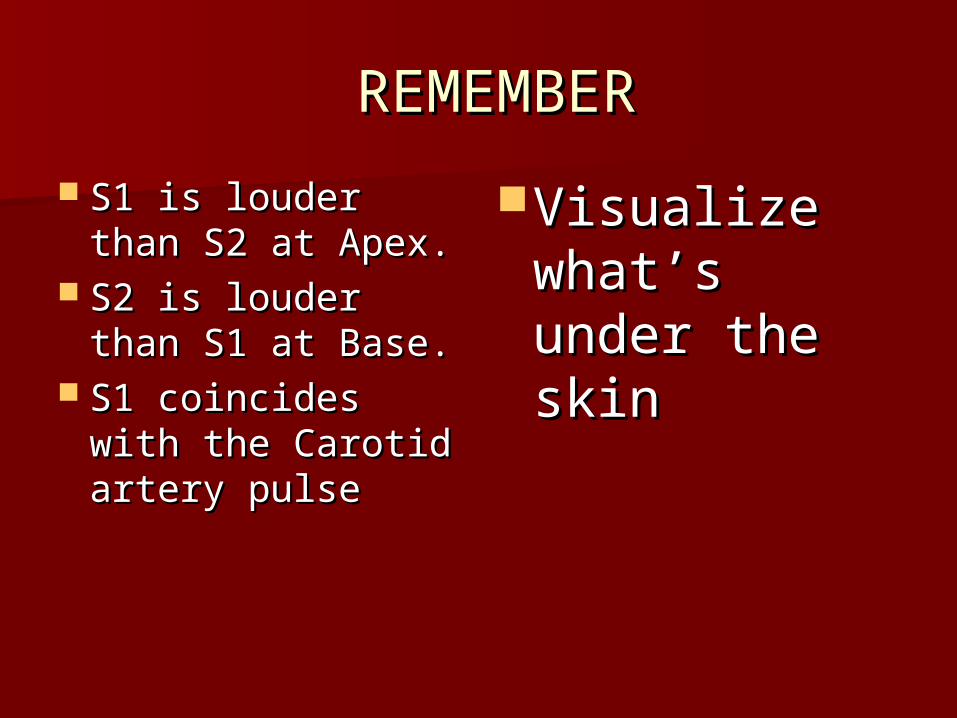
REMEMBERREMEMBER
S1 is louder than S1 is louder than S2 at Apex.S2 at Apex.
S2 is louder than S2 is louder than S1 at Base.S1 at Base.
S1 coincides S1 coincides with the Carotid with the Carotid artery pulseartery pulse
Visualize Visualize what’s under what’s under the skinthe skin