Ascites and Renal Dysfunction in Liver Disease 2nd ed - P. Gines, et al., (Blackwell, 2005) WW
463
second edition A scites & R enal D y sfu nction Pathogenesis Diagnosis &Treatment edited by Pere Ginès, Vicente Arroyo Juan Rodés, Robert W.Schrier inLiverDisease
Transcript of Ascites and Renal Dysfunction in Liver Disease 2nd ed - P. Gines, et al., (Blackwell, 2005) WW

second edition
Ascites&RenalDysfunction
PathogenesisDiagnosis&Treatment
edited by Pere Ginès, Vicente ArroyoJuan Rodés, Robert W.Schrier
inLiverDisease

Ascites and Renal Dysfunction in Liver DiseasePathogenesis, Diagnosis, and Treatment

Dedicated to our wives, Nuria, Joana, Paula, and Barbara, in recognition of their contribution to our scientifi c careers.

Ascites and Renal Dysfunction in Liver DiseasePathogenesis, Diagnosis, and Treatment
PERE GINÈSConsultant in Hepatology, Associate Professor of MedicineLiver Unit, University of Barcelona School of Medicine, Hospital Cliníc Villarroel170 8036 Barcelona, Spain
VICENTE ARROYOProfessor of MedicineLiver Unit, University of Barcelona School of Medicine, Hospital Cliníc Villarroel170 8036 Barcelona, Spain
JUAN RODÉSProfessor of MedicineLiver Unit, University of Barcelona School of Medicine, Hospital Cliníc Villarroel170 8036 Barcelona, Spain
ROBERT SCHRIERProfessor of MedicineDivision of Renal Diseases and Hypertension, University of Colorado School of Medicine4200 East 9th Avenue, Denver, CO 80262, USA
SECOND EDITION

© 1999, 2005 by Blackwell Publishing LtdBlackwell Publishing, Inc., 350 Main Street, Malden, Massachusetts 02148–5020, USABlackwell Publishing Ltd, 9600 Garsington Road, Oxford OX4 2DQ, UKBlackwell Publishing Asia Pty Ltd, 550 Swanston Street, Carlton, Victoria 3053, Australia
The right of the Authors to be identifi ed as the Authors of this Work has been asserted in accordance with the Copyright, Designs and Pat-ents Act 1988.
All rights reserved. No part of this publication may be reproduced, stored in a retrieval system, or transmitted, in any form or by any means, electronic, mechanical, photocopying, recording or otherwise, except as permitted by the UK Copyright, Designs and Patents Act 1988, with-out the prior permission of the publisher.
First published 1999Second edition 2005
Library of Congress Cataloging-in-Publication DataAscites and renal dysfunction in liver disease / edited by Pere Ginès ... [et al.].-- 2nd ed. p. ; cm.Includes bibliographical references and index.ISBN-13: 978-1-4051-1804-0 (alk. paper)ISBN-10: 1-4051-1804-0 (alk. paper)
1. Liver--Diseases--Complications. 2. Ascites. 3. Kidneys--Diseases.[DNLM: 1. Liver Diseases--complications. 2. Ascites--etiology. 3. Ascites--therapy. 4. Kidney Diseases--etiology. 5. Liver Diseases--physi-opathology. WI 700 A814 2005] I. Ginès, Pere.
RC846.A83 2005616.3’62--dc22
2004026925
ISBN-13: 978-1-4051-1804-0 ISBN-10: 1-4051-1804-0
A catalogue record for this title is available from the British Library
Set in Palatino 9.5/12pt by Sparks, Oxford – www.sparks.co.ukPrinted and bound by Gopsons Papers, Noida, India
Commissioning Editor: Alison BrownDevelopment Editor: Rebecca HuxleyProduction Controller: Kate Charman
For further information on Blackwell Publishing, visit our website:www.blackwellpublishing.com
The publisher’s policy is to use permanent paper from mills that operate a sustainable forestry policy, and which has been manufactured from pulp processed using acid-free and elementary chlorine-free practices. Furthermore, the publisher ensures that the text paper and cover board used have met acceptable environmental accreditation standards.

v
Contents
Contributors, viiPreface to the Second Edition, xiii
Part 1 Regulation of Extracellular Fluid Volume and Renal and Splanchnic Circulation
1 Extracellular Fluid Volume Homeostasis, 3Brian D. Poole, William T. Abraham, and Robert W. Schrier
2 Physiology of the Renal Circulation, 15 Roland C. Blantz and Francis B. Gabbai
3 Physiology of the Gastrointestinal Circulation, 29Thomas Petnehazy, Thorsten Vowinkel, and D. Neil Granger
Part 2 Factors Involved in the Pathogenesis of Renal Dysfunction and Ascites in Cirrhosis
4 The Renin–Angiotensin–Aldosterone System in Cirrhosis, 43Mauro Bernardi and Marco Domenicali
5 The Sympathetic Nervous System in Cirrhosis, 54Francis J. Dudley and Murray D. Esler
6 Atrial Natriuretic Peptide and other Natriuretic Factors in Cirrhosis, 73Giorgio La Villa and Giacomo Laffi
7 Arachidonic Acid Metabolites and the Kidney in Cirrhosis, 84Silvia Ippolito and Kevin P. Moore
8 Nitric Oxide and Systemic and Renal Hemodynamic Disturbances in Cirrhosis, 105Manuel Morales-Ruiz and Wladimiro Jiménez
9 Endothelin and Systemic, Renal, and Hepatic Hemodynamic Disturbances in Cirrhosis, 115Veit Gülberg and Alexander L. Gerbes
10 Carbon Monoxide and the Heme Oxygenase System in Cirrhosis, 125Richard W. Lambrecht, Mercedes Fernández, Ying Shan, and Herbert L. Bonkovsky
Part 3 Systemic and Splanchnic Hemo-dynamic Abnormalities in Cirrhosis
11 The Systemic Circulation in Cirrhosis, 139 Søren Møller and Jens Henriksen
12 The Splanchnic Circulation in Cirrhosis, 156Jaime Bosch and Juan Carlos García-Pagán
13 Physiology of Hepatic Circulation in Cirrhosis, 164Roberto J. Groszmann and Mauricio R. Loureiro-Silva
14 Alterations of Hepatic and Splanchnic Microvascular Exchange in Cirrhosis: Local Factors in the Formation of Ascites, 174Jens H. Henriksen and Søren Møller
15 The Heart in Cirrhosis, 186Hongqun Liu and Samuel S. Lee
Part 4 Ascites and Sodium Retention in Cirrhosis
16 Pathogenesis of Sodium Retention in Cirrhosis: the Arterial Vasodilation Hypothesis of Ascites Formation, 201Patricia Fernández de la Llama, Pere Ginès, and Robert W. Schrier
17 Experimental Models of Cirrhosis and Ascites, 215Joan Clària and Wladimiro Jiménez

vi Contents
18 Medical Treatment of Ascites in Cirrhosis, 227Paolo Angeli and Angelo Gatta
19 Paracentesis for Cirrhotic Ascites, 241Rosa María Morillas, Justiniano Santos, Silvia Montoliu, and Ramon Planas
20 Transjugular Intrahepatic Portosystemic Shunt (TIPS) for the Management of Refractory Ascites in Cirrhosis, 251Guadalupe Garcia-Tsao
21 Prognosis of Patients with Cirrhosis and Ascites, 260Mónica Guevara, Andrés Cárdenas, Juan Uríz, and Pere Ginès
22 Liver Transplantation for Patients with Cirrhosis and Ascites, 271Antoni Rimola, Miguel Navasa, Luis Grande, and Juan-Carlos García-Valdecasas
23 A Practical Approach to Treatment of Patients with Cirrhosis and Ascites, 286Andrés Cárdenas and Pere Ginès
24 Etiology, Diagnosis, and Management of Non-cirrhotic Ascites, 294Egbert Frick and Jürgen Schölmerich
Part 5 Hyponatremia and Water Retention in Cirrhosis
25 Pathogenesis of Hyponatremia: the Role of Arginine Vasopressin, 305San-e Ishikawa and Robert W. Schrier
26 Management of Hyponatremia in Cirrhosis, 315Andrés Cárdenas and Pere Ginès
Part 6 Renal Failure and Hepatorenal Syndrome in Liver Disease
27 Pathogenesis of Renal Vasoconstriction in Cirrhosis, 329Mónica Guevara, Rolando Ortega, Pere Ginès, and Juan Rodés
28 Hepatorenal Syndrome in Cirrhosis: Clinical Features, Diagnosis, and Management, 341Vicente Arroyo, Carlos Terra, Aldo Torre, and Pere Ginès
29 Glomerular Disease in Cirrhosis, 360Brian D. Poole, Robert W. Schrier, and Alkesh Jani
30 Drug-induced Renal Failure in Cirrhosis, 372Francesco Salerno and Salvatore Badalamenti
31 Clinical Disorders of Renal Function in Acute Liver Failure, 383John G. O’Grady
32 Renal Dysfunction in Obstructive Jaundice, 394Antonio Sitges-Serra and Javier Padillo
Part 7 Spontaneous Bacterial Peritonitis in Cirrhosis
33 Experimental Models of Spontaneous Bacterial Peritonitis, 411Agustín Albillos, Antonio de la Hera, and Melchor Alvarez-Mon
34 Pathogenesis and Clinical Features of Spontaneous Bacterial Peritonitis, 422José Such, Carlos Guarner, and Bruce Runyon
35 Treatment and Prophylaxis of Spontaneous Bacterial Peritonitis, 434Alejandro Blasco Pelicano and Miguel Navasa
Index, 441

vii
Contributors
William T. AbrahamProfessor of MedicineChief, Division of Cardiovascular MedicineAssociate Director,
Davis Heart & Lung Research InstituteOhio State UniversityColumbus, Ohio, USA
Agustín AlbillosAssociate Professor of MedicineResearch and Development Unit Associated to the
Spanish National Research Council (CSIC)Department of MedicineUniversity of Alcalá School of MedicineGastroenterology DepartmentHospital Universitario Ramón y CajalMadrid, Spain
Melchor Alvarez-Mon Professor of MedicineResearch and Development Unit Associated to the
Spanish National Research Council (CSIC)Department of MedicineUniversity of Alcalá School of MedicineImmune System and Oncology Diseases DepartmentHospital Universitario Príncipe de AsturiasMadrid, Spain
Paolo AngeliDepartment of Clinical and Experimental MedicineUniversity of PaduaPadua, Italy
Vicente ArroyoProfessor of MedicineDirector, Institute for Digestive DiseasesHospital ClinicUniversity of Barcelona School of MedicineBarcelona, Spain
Salvatore BadalamentiChief, Department of Emergency and NephrologyIstituto Clinico HumanitasMilan, Italy
Mauro BernardiProfessor of Internal MedicineDirector, Department of Internal Medicine,
Cardioangiology, HepatologyAlma Mater Studiorum – University of BolognaItaly
Roland C. BlantzUniversity of California, San DiegoLa Jolla, California, USAVA San Diego Healthcare SystemSan Diego, California, USA
Alejandro Blasco PelícanoResearch Fellow Hospital ClinicBarcelona, Spain
Herbert L. BonkovskyProfessor of Medicine and Molecular,
Microbial and Structural Biology Director, General Clinical Research Center Director, The Liver-Biliary-Pancreatic Center MC-1111, University of Connecticut Health Center Farmington, Connecticut, USA
Jaime BoschProfessor of MedicineSenior Consultant Hepatologist and Chief,
Hepatic Hemodynamic Laboratory Liver Unit, IMDM, Hospital Clinic and IDIBAPSUniversity of BarcelonaBarcelona, Spain

viii Contributors
Andrés CárdenasLiver UnitInstitute of Digestive Diseases, Hospital Clinic and
IDIBAPSBarcelona, Spain
Joan ClàriaAssociate Professor University of Barcelona School of MedicineBarcelona, SpainSenior SpecialistHospital ClinicBarcelona, Spain
Antonio de la Hera Scientifi c Member,
Spanish National Research Council (CSIC)Associate Profesor of ImmunologyResearch and Development Unit Associated to the
Spanish National Research Council (CSIC)Department of MedicineUniversity of Alcalá School of MedicineMadrid, Spain
Marco DomenicaliResearch FellowDepartment of Internal Medicine,
Cardioangiology, Hepatology Alma Mater Studiorum – University of BolognaItaly
Francis J. DudleyGastroenterology DepartmentThe Alfred HospitalMelbourne, Victoria, Australia
Murray D. EslerGastroenterology DepartmentThe Baker Medical Research InstitutePrahran, Victoria, Australia
Mercedes FernandezLiver Unit, IDIBAPSHospital Clinic, University of BarcelonaBarcelona, Spain
Patricia Fernández-Llama Renal Unit and Hypertension Fundacio Puigvert Cartagena, Barcelona, Spain
Egbert FrickChair of Internal MedicineHospital RodingRoding, Germany
Francis B. GabbaiUniversity of California, San DiegoLa Jolla, California, USAVA San Diego Healthcare SystemSan Diego, California, USA
Juan Carlos García-PagánConsultant HepatologistLiver Unit, IMDM, Hospital Clinic and IDIBAPSUniversity of BarcelonaBarcelona, Spain
Guadalupe Garcia-TsaoProfessor of MedicineYale University School of MedicineDepartment of Internal MedicineNew Haven, Connecticut, USA
Juan-Carlos García-ValdecasasDepartment of SurgeryHospital ClinicBarcelona, Spain
Angelo GattaDepartment of Clinical and Experimental MedicineUniversity of PaduaPadua, Italy
Alexander L. GerbesDepartment of Medicine IIKlinikum of the University – GrosshadernMunich, Germany
Pere GinèsChief, Liver UnitAssociate Professor of MedicineInstitute of Digestive Diseases Hospital ClinicUniversity of Barcelona School of MedicineBarcelona, Spain
Luis GrandeLiver Unit and Department of SurgeryHospital ClinicBarcelona, Spain

ixContributors
D. Neil GrangerDepartment of Molecular and Cellular PhysiologyLouisiana State University Health Sciences CenterShreveport, Louisiana, USA
Roberto J. GroszmannProfessor of MedicineYale University School of MedicineDepartment of Internal MedicineNew Haven, Connecticut, USAChief, Digestive Diseases SectionDirector, Hepatic Hemodynamic LaboratoryVA Connecticut Healthcare SystemWest Haven, Connecticut, USA
Carlos GuarnerChief of Liver UnitAssociate Professor of MedicineHospital de la Santa Creu i Sant PauAutonomous UniversityBarcelona, Spain
Mónica GuevaraAssociate InvestigatorHospital Clinic and IDIBAPSUniversity of BarcelonaBarcelona, Spain
Veit GülbergDepartment of Medicine IIKlinikum of the University – GrosshadernMunich, Germany
Jens H. HenriksenProfessor of Clinical PhysiologyUniversity of CopenhagenCopenhagen, DenmarkHead of DepartmentDepartment of Clinical Physiology
and Nuclear MedicineHvidovre HospitalHvidovre, Denmark
Silvia IppolitoResearch FellowCentre for HepatologyRoyal Free & University College Medical SchoolUniversity College of LondonLondon, UK
San-e IshikawaProfessor of MedicineDepartment of MedicineJichi Medical School, Omiya Medical CenterSaitama, Japan
Alkesh JaniAssistant Professor of MedicineUniversity of Colorado Health Sciences CenterDenver, Colorado, USA
Wladimiro JiménezHormonal LaboratoryHospital Clinic, IDIBAPS, IRSINUniversity of BarcelonaBarcelona, Spain
Giorgio La VillaFull Professor of MedicineUniversity of Florence School of MedicineFlorence, Italy
Giacomo Laffi Full Professor of MedicineUniversity of Florence School of MedicineFlorence, Italy
Richard W. LambrechtAssistant Professor of PharmacologyMC-1111, University of Connecticut Health CenterFarmington, Connecticut, USA
Samuel S. LeeProfessor, Department of MedicineLiver UnitUniversity of CalgaryCalgary, Canada
Hongqun LiuAdjunct Assistant ProfessorDepartment of MedicineLiver UnitUniversity of CalgaryCalgary, Canada
Mauricio R. Loureiro-SilvaAssociate Research ScientistYale University School of MedicineDepartment of Internal MedicineNew Haven, Connecticut, USAVA Connecticut Healthcare SystemHepatic Hemodynamic LaboratoryWest Haven, Connecticut, USA

x Contributors
Søren MøllerChief PhysicianUniversity of CopenhagenCopenhagen, DenmarkDepartment of Clinical Physiology and Nuclear
MedicineHvidovre HospitalHvidovre, Denmark
Silvia MontoliuLiver UnitGastroenterology DepartmentHospital Universitari Germans Trias I PujolBadalona, Spain
Kevin P. MooreProfessor of HepatologyCentre for HepatologyRoyal Free & University College Medical SchoolUniversity College of LondonLondon, UK
Manuel Morales-RuizHormonal LaboratoryHospital Clinic, IDIBAPS, IRSINUniversity of BarcelonaBarcelona, Spain
Rosa María MorillasLiver UnitGastroenterology DepartmentHospital Universitari Germans Trias I PujolBadalona, Spain
Miguel NavasaConsultant of the Liver UnitHospital ClinicBarcelona, SpainAssociate Professor of MedicineUniversity of BarcelonaBarcelona, Spain
John O’GradyInstitute of Liver StudiesKing’s College HospitalLondon, UK
Rolando OrtegaCoordinador Unidad de HepatologíaHospital BocagrandeDepartamento de InvestigacionesFacultad de BacteriologíaUniversidad San BuenaventuraCartagena de Indias, Colombia
Javier PadilloAttending SurgeonDepartment of SurgeryHospital Reina SofíaCórdoba, Spain
Thomas Petnehazy Department of Molecular and Cellular PhysiologyLouisiana State University Health Sciences CenterShreveport, Louisiana, USA
Ramon PlanasLiver UnitGastroenterology DepartmentHospital Universitari Germans Trias I PujolBadalona, Spain
Brian PooleFellowUniversity of Colorado Health Sciences CenterDenver, Colorado, USA
Antoni RimolaLiver UnitHospital Clinic Barcelona, Spain
Juan RodésProfessor of MedicineHospital ClinicUniversity of BarcelonaBarcelona, Spain
Bruce A. Runyon Professor of Medicine Chief, Liver Service Loma Linda University Medical Center Loma Linda, California, USA
Francesco SalernoChief, Department of Internal MedicinePoliclinico San DonatoUniversity of MilanMilan, Italy
Justiniano SantosLiver UnitGastroenterology DepartmentHospital Universitari Germans Trias I PujolBadalona, Spain

xiContributors
Jürgen SchölmerichProfessor of MedicineUniversity of RegensburgChair of Department of Internal Medicine IUniversity HospitalRegensburg, Germany
Robert W. SchrierProfessor of MedicineDepartment of MedicineUniversity of Colorado Health Sciences CenterDenver, Colorado, USA
Ying ShanAssistant Professor of MedicineMC-1111, University of Connecticut Health CenterFarmington, Connecticut, USA
Antonio Sitges-SerraProfessor of SurgeryHead, Department of SurgeryHospital del MarBarcelona, Spain
José SuchLiver UnitHospital General UniversitarioAlicante, Spain
Carlos TerraResearch FellowUniversity of Barcelona Medical School
and Institute of Digestive DiseasesHospital ClinicBarcelona, Spain
Aldo TorreResearch FellowUniversity of Barcelona Medical School
and Institute of Digestive DiseasesHospital ClinicBarcelona, Spain
Juan UrízUnidad Aparato de DigestivoServicio Medicina InternaHospital Virgen del CaminoPamplona, Navarra, Spain
Thorsten Vowinkel Department of Molecular and Cellular PhysiologyLouisiana State University Health Sciences CenterShreveport, Louisiana, USADepartment of General SurgeryUniversity Hospital MünsterMünster, Germany

xiii
Preface to the Second Edition
It has been six years since we published the fi rst edition of Ascites and Renal Dysfunction in Liver Disease. Since then, signifi cant advances have been made in the pathogen-esis of circulatory and renal dysfunction that occur in the setting of chronic liver diseases, particularly cirrhosis. Specifi cally, the role of vasodilatory factors, particularly nitric oxide, has been investigated extensively. Moreover, there is increased recognition of the mechanistic role of impaired heart function on the circulatory dysfunction of liver failure. In this second edition of Ascites and Renal Dysfunction in Liver Disease, these advances in patho-genetics are described in specifi c chapters.
Besides this increased knowledge on pathophysiology, major advances have been made in the clinical manage-ment of renal dysfunction in liver disease. A new thera-peutic method, transjugular intravenous portosystemic shunts, has emerged for patients with ascites refractory to diuretic therapy. A large number of nonrandomized studies (as well as several randomized trials) have been published concerning the effects of this therapeutic ap-proach. For the fi rst time ever, an effective treatment has been described to treat hepatorenal syndrome in patients with cirrhosis, namely administration of vasoconstrictor drugs. Moreover, there are studies showing how hepato-renal syndrome can be effectively prevented in specifi c settings such as spontaneous bacterial peritonitis and alcoholic hepatitis. Finally, specifi c antagonists of the V2 vasopressin receptor are in advanced stages of clini-cal development. These drugs might prove to be useful in the management and prevention of dilutional hypo-natremia, a complication for which there is currently no
effective therapy. All these new topics, as well as other topics on the management of liver disease, are covered in this second edition.
The layout and look of the book have changed from the previous edition. The book has been divided into two sections: the fi rst (Parts 1, 2 and 3) describes the patho-physiology of circulatory and renal abnormalities, whilst the second (Parts 4–7) relates to clinical management of patients. We hope this will make the book easier to read when looking for either pathogenic factors or answers to clinical questions.
Finally, we would like to acknowledge the work of the authors of the chapters, who are internationally recog-nised specialists in their fi elds and have done a tremen-dous job in summarizing the different topics inside the page limits. We thank both Nicki van Berckel and Janet Darling for their administrative assistance, and Black-well Publishing for making the book appealing to the readers.
We hope that this second edition of Ascites and Renal Dysfunction in Liver Disease will be helpful not only to clinical researchers interested in complications of cirrho-sis, but also to those clinicians – whether they be gastro-enterologists, transplant hepatologists, nephrologists, or internists – caring for patients with liver diseases.
P. GinèsV. Arroyo
J. RodésR.W. Schrier
2005

Part 1Regulation of Extracellular Fluid Volume and Renal and Splanchnic Circulation


3
Chapter 1Extracellular Fluid Volume Homeostasis
Brian D. Poole, William T. Abraham, and Robert W. Schrier
of water into the extracellular space. Sodium balance is determined by the equilibrium between sodium intake, extrarenal sodium loss, and renal sodium excretion. Prac-tically, renal sodium excretion is the major determinant of sodium balance, given the ability of the kidney to excrete large amounts of sodium in response to a sodium load. In addition, sodium loading, by increasing serum osmo-lality, stimulates the hypothalamic thirst center leading to increased fl uid intake as well as the osmotic release of arginine vasopressin (AVP). The release of AVP from the posterior pituitary decreases water excretion by increas-ing the permeability of the collecting duct epithelium to water. If the increase in ECF volume is suffi cient to alter the Starling forces governing the transfer of fl uid from the vas-cular to the interstitial compartment then edema results.
One of the major regulators of sodium excretion is the mineralocorticoid hormone aldosterone. Aldosterone is produced in the zona glomerulosa of the adrenal gland and acts to increase sodium reabsorption by increasing the number of epithelial sodium channels in the cortical col-lecting duct. In states of volume depletion, the renin–angi-otensin–aldosterone system (RAAS) is stimulated causing an increase in sodium reabsorption that leads to expansion of the ECF. With expansion, the stimulus for aldosterone secretion is removed and sodium reabsorption is dimin-ished, thereby stabilizing volume status. In states of min-eralocorticoid excess such as primary hyperaldosteronism there is unregulated secretion of aldosterone leading to an increase in sodium reabsorption with resultant volume expansion and hypertension. The effect of aldosterone to cause renal sodium retention can be overridden, how-ever, by the phenomenon of aldosterone escape. In this circumstance the ECF reaches a new, higher steady state, but does not continue to expand despite increased levels of aldosterone. This has been postulated to be mediated by hemodynamic mechanisms whereby an increase in renal artery pressure secondary to expansion of the ECF causes a pressure natriuresis. The increase in renal artery pressure subsequently increases the glomerular fi ltration rate (GFR) and the fractional excretion of sodium (FeNa). Recently it was reported that the chief molecular target of the escape phenomenon is the thiazide-sensitive NaCl
The development of ascites is the most common compli-cation in patients with compensated cirrhosis, occurring in 58% of patients within 10 years of diagnosis. Ascites develops in the context of an increase in the extracellular fl uid volume (ECF) and therefore it is essential to under-stand the regulation of body fl uid volume to appreciate its pathogenesis. Knowledge of the intrarenal and extra-renal factors governing renal sodium excretion is crucial to understanding because the sodium ion is the principal determinant of ECF volume. In normal individuals, if the ECF is expanded by the administration of isotonic saline the kidney will excrete the excess sodium and water in the urine and return the ECF to normal values. However, in pathogenic disease states such as congestive heart failure (CHF) and cirrhosis the kidneys continue to retain sodi-um and water even in the presence of an increased ECF volume. In these edematous disorders the integrity of the kidney as the primary organ controlling ECF volume re-mains intact because transplantation of the kidney from an edematous, cirrhotic patient to a subject with normal liver function totally reverses the renal sodium and water retention (1). Moreover, transplantation of a normal liver into a cirrhotic patient with ascites and edema has been shown to abolish the renal sodium and water retention (2). Thus, the kidney must be responding to extrarenal signals from the afferent limb of a volume regulatory system in these edematous disorders. The study of these edematous states has led to a unifying hypothesis of body fl uid vol-ume regulation (3–8). This chapter will review the afferent and efferent mechanisms that contribute to extracellular fl uid volume homeostasis in health and disease.
Regulation of sodium excretion
Due to active transport processes, the sodium ion is pri-marily located in the ECF and, along with its major ani-ons chloride and bicarbonate, constitutes more than 90% of the extracellular solute. Therefore, because sodium and its anions are the major osmotically active substances in the ECF, they are the major determinants of the ECF vol-ume. With a positive sodium balance, the ECF volume will increase secondary to osmotically driven movement

4 Chapter 1
cotransporter. In a rat model of aldosterone infusion cou-pled with a high sodium diet, it was found that levels of the epithelial sodium channel were unchanged during the escape phenomenon, but the amount of the thiazide-sen-sitive NaCl cotransporter was signifi cantly diminished. Therefore it appears that the so-called pressure natriuresis is mediated at least in part by downregulation of the thi-azide-sensitive NaCl cotransporter.
Homeostasis of the ECF is also mediated by the hor-mones atrial natriuretic peptide (ANP) and, as mentioned previously, AVP. ANP has been shown to be released from the myocardium in response to volume expansion and it has two major actions contributing to maintenance of volume status. It is a direct vasodilator that can lower systemic blood pressure and it also increases the urinary excretion of sodium and water. The natriuresis appears to be mediated by an increase in GFR secondary to afferent arteriole vasodilation coupled with efferent arteriole va-soconstriction. Furthermore, ANP has been shown to di-rectly decrease tubular sodium reabsorption. In another rat model of hyperaldosteronism, it has been shown that the level of ANP increases coinciding with an increase in sodium excretion. Therefore the authors postulate that ANP may also mediate aldosterone escape. It is not known whether ANP has an effect on the thiazide-sensi-tive NaCl cotransporter.
AVP is the chief regulator of renal water excretion and as such can be expected to have a major role in ECF vol-ume regulation. It is known that in edematous disorders like CHF and cirrhosis there are inappropriate levels of AVP relative to plasma osmolality that results in water re-tention and hyponatremia. However, it has been shown that there is also counterregulation of this system. In rats administered AVP plus a water load it was shown that after an initial period of water retention there was subse-quently an increase in urine volume that corresponded to a downregulation of the renal water channel aquaporin 2 despite continued elevated levels of AVP. Therefore, the authors conclude there is a vasopressin-independent downregulation of aquaporin 2 and therefore a limit on water reabsorption in this model.
Unlike in conditions such as primary hyperaldos-teronism where there is an escape from continued sodium reabsorption despite elevated levels of aldosterone, in the edematous disorders there is impaired escape and contin-uous sodium and water reabsorption. It seems that the dif-ference in these disorders is in how the kidney is respond-ing to the afferent limb of the volume regulatory system.
Afferent mechanisms governing extracellular fl uid volume homeostasis
An increase in sodium and water intake is associated with an expansion of the extracellular fl uid volume. This includes expansion of the interstitial fl uid and plasma
components of total body fl uid volume. Under normal circumstances, this expansion of total body fl uid volume results in an increase in renal sodium and water excretion followed by restoration of the normal extracellular fl uid volume. However, in patients with edematous disorders, avid sodium and water retention persists despite expan-sion of total extracellular fl uid and blood volume. Thus, the afferent volume receptors governing extracellular fl uid volume must not primarily sense total extracellular fl uid or blood volume. In such instances, there must be some body fl uid compartment that is still inadequately fi lled even in the presence of expansion of these body fl uid compartments.
Effective blood volume and the concept of arterial underfi lling
John Peters fi rst coined the term effective blood volume in al-lusion to the component of blood or body fl uid volume to which the volume regulatory system responds by altering the renal excretion of sodium and water (9). Peters sug-gested that extrarenal signals that enhance tubular sodium and water reabsorption by the otherwise normal kidney are initiated by this decrease in effective blood volume in the setting of cardiac failure or cirrhosis. In support of this claim is the observation that renal sodium and water re-tention can occur in patients with cardiac or liver failure before any decrease in glomerular fi ltration rate.
Borst and deVries (10) fi rst suggested cardiac output as the primary regulator of renal sodium and water excre-tion, thus constituting effective blood volume. While this notion is attractive, there exist several states of sodium and water retention that are associated with an augment-ed rather than a decreased cardiac output. For example, a signifi cant increase in cardiac output may occur in the presence of avid renal sodium and water retention and expansion of extracellular fl uid volume in association with cirrhosis, high-output cardiac failure, pregnancy, and large arteriovenous fi stulae. Hence, cardiac output must not constitute the sole or primary determinant of effective blood volume.
The unifying hypothesis of body fl uid volume regula-tion suggests that the relative integrity or fullness of the arterial circulation constitutes the primary afferent sig-nal through which the kidneys either increase or decrease their excretion of sodium and water (3–8). This theory explains how an increase in the volume of blood on the venous side of the circulation may cause a rise in total blood volume, whereas a decrease in the relative volume of blood in the arterial circulation may promote contin-ued renal sodium and water retention. A reduction in cardiac output is one way in which a decrease in arterial circulatory integrity may occur. However, as mentioned above, diminished cardiac output cannot be the only af-ferent signal for underfi lling of the arterial circulation.

5Extracellular Fluid Volume Homeostasis
The unifying hypothesis of body fl uid volume regula-tion proposes peripheral arterial vascular resistance and the compliance of the arterial vasculature as the second major determinant of the fullness of the arterial circula-tion (3–8). Thus, peripheral arterial vasodilation may provide another afferent signal for arterial underfi lling, which causes renal sodium and water retention.
In summary, either a decrease in cardiac output or pe-ripheral arterial vasodilation may constitute the afferent signal for arterial underfi lling with resultant renal so-dium and water retention that leads to expansion of the
total blood volume. The afferent receptors or sensors of arterial underfi lling must be responsive to small changes in effective arterial blood volume since the steady-state arterial blood pressure is not a sensitive index of the pres-ence of arterial underfi lling. For example, the rapidity of the compensatory response to arterial underfi lling may obscure a fall in blood pressure until this efferent response becomes inadequate to maintain effective arterial blood volume. The mechanisms involved in this volume regu-latory system are summarized in Figs 1.1 and 1.2, and the sensors of arterial underfi lling are discussed next.
Extracellular fluidvolume
Low output cardiac failure,pericardial tamponade,constrictive pericarditis
Intravascular volume2 to diminishedoncotic pressure
orincreasedcapillary
permeabilityCARDIAC OUTPUT
Activation of ventricularand
arterial receptors
Stimulation ofsympathetic nervous
system
PERIPHERAL AND RENALARTERIAL VASCULAR RESISTANCE
MAINTENANCE OF EFFECTIVEARTERIAL BLOOD VOLUME
RENAL WATERRETENTION
RENAL SODIUMRETENTION
Non-osmoticvasopressinstimulation
Activation of therenin-angiotensin-
aldosterone system
High-outputcardiac failure
Sepsis Cirrhosis Arteriovenousfistula
Pregnancy Arterialvasodilators
PERIPHERALARTERIAL VASODILATION
Activation ofarterial
baroreceptors
Stimulation ofsympathetic
nervous system
Non-osmoticvasopressinstimulation
CARDIACOUTPUT
WATERRETENTION
PERIPHERAL ARTERIALVASCULAR AND RENAL
RESISTANCE
SODIUMRETENTION
MAINTENANCE OF EFFECTIVEARTERIAL BLOOD VOLUME
Activation of therenin-angiotensin-
aldosterone systemFigure 1.2 ·Mechanism by which peripheral arterial vasodilation results in renal sodium and water retention, increased cardiac output, and peripheral and renal vasoconstriction. (Reproduced with permission from Schrier R, Niederberger M. Paradoxes of body fl uid volume regulation in health and disease: a unifying hypothesis. West J Med 1994; 16:393–407.)
Figure 1.1 Mechanism by which decreased cardiac output results in renal sodium and water retention and peripheral and renal vasoconstriction. (Reproduced with permission from Schrier R, Niederberger M. Paradoxes of body fl uid volume regulation in health and disease: a unifying hypothesis. West J Med 1994; 16:393–407.)

6 Chapter 1
Sensors of arterial underfi lling
High-pressure baroreceptors
Afferent receptors for this volume regulatory system must reside in the arterial vascular compartment. In this regard, high-pressure baroreceptors in the left ventricle, carotid sinus, aortic arch, and juxtaglomerular apparatus have been implicated as the primary afferent receptors in-volved in the regulation of renal sodium and water excre-tion and extracellular fl uid volume homeostasis (11–19). The presence of volume-sensitive receptors in the arterial circulation in humans was initially suggested by obser-vations made in patients with traumatic arterio venous fi stulae (20). In such patients, closure of the fi stulae re-sults in a decrease in the rate of emptying of the arterial blood into the venous circulation, as demonstrated by closure-induced increases in diastolic arterial pressure and decreases in cardiac output. This increase in arterial fullness produces an immediate increase in renal sodium excretion without changes in either glomerular fi ltration rate or renal blood fl ow (20).
Various denervation experiments also implicate high-pressure volume receptors, and thus the integrity of the arterial circulation, as primary afferent receptors in modu-lating renal sodium and water excretion. In these studies, pharmacological or surgical interruption of sympathetic afferent neural pathways arising from high-pressure areas inhibited the natriuretic response to volume expansion (21–27). In addition, reduction of pressure or stretch at the carotid sinus has been shown to activate the sympathetic nervous system and to cause renal sodium and water re-tention (28,29). High-pressure baroreceptors also appear to be important factors in regulating the non-osmotic release of vasopressin and thus renal water excretion (30,31).
The juxtaglomerular apparatus is a high-pressure re-ceptor located in the afferent arterioles within the kidney. It responds to decreased stretch or increased renal sympa-thetic activity with enhanced secretion of renin (28). Thus, this renal baroreceptor is an important factor in the control of angiotensin II formation and aldosterone secretion and ultimately in the regulation of renal sodium excretion.
Low-pressure baroreceptors
The low-pressure baroreceptors of the thorax, including the atria, right ventricle, and pulmonary vessels, may also contribute to extracellular fl uid volume homeosta-sis. Loading of these volume-sensitive receptors results in enhanced cardiac release of natriuretic peptides (32) and suppression of non-osmotic vasopressin release from the neurohypophysis (33). Since patients with ad-vanced cardiac failure exhibit avid sodium and water re-tention and activation of neurohormonal vasoconstrictor systems—including enhanced non-osmotic vasopressin
release—despite elevated atrial pressures and increased circulating concentrations of the natriuretic peptides, high-pressure baroreceptors must predominate over these low-pressure ones. This observation also supports the primacy of the arterial circulation as the determinant of extracellular fl uid volume homeostasis.
Cardiac and pulmonary chemoreceptors
In the heart and lungs, both vagal and sympathetic affer-ent nerve endings respond to a variety of exogenous and endogenous chemical substances, including capsaicin, phenyldiguanidine, bradykinin, substance P, and prostag-landins (34–36). Since substances such as bradykinin and prostaglandins may circulate at increased concentrations in subjects with edematous disorders (37), it is possible that altered central nervous system input from chemically sensitive cardiac and/or pulmonary afferents contributes to the sodium and water retention characteristic of these disease states. This possibility may have important impli-cations for the treatment of some sodium-retaining disor-ders. For example, in heart failure, commonly prescribed medications such as angio tensin-converting enzyme in-hibitors may alter circulating bradykinin and prostaglan-din levels, thus potentially infl uencing cardiopulmonary chemoreceptor activity. At the present time, however, the exact role of these cardiac and pulmonary chemoreceptors in body fl uid volume regulation remains unknown.
Hepatic receptors
Conceptually, the liver should be in an ideal position to monitor dietary sodium intake and thus adjust urinary sodium excretion. In support of this notion, infusion of sa-line into the portal circulation has been reported to result in a greater natriuresis when compared with peripheral venous saline administration (38,39). Similarly, the in-crease in urinary sodium excretion has been shown to be greater when the sodium load is given orally than when it is given intravenously (40–42). Moreover, the patho-physiological retention of sodium in patients with severe liver disease is also consistent with an important role for the liver in the control of sodium excretion. However, the experimental evidence in favor of hepatic sodium or vol-ume receptors remains controversial since some investi-gators have been unable to confi rm the above observa-tions of increased sodium excretion in response to portal vein or gastric sodium loading (43–45).
In summary, the afferent mechanisms for sodium and water retention appear to be preferentially localized to the arterial or high-pressure side of the circulation, where arterial fullness may serve as the primary determinant of the renal response. Refl exes emanating from low-pres-sure cardiopulmonary receptors may also be altered so as to infl uence renal sodium and water handling in

7Extracellular Fluid Volume Homeostasis
heart failure. In this regard, increases in atrial pressure also stimulate the release of the natriuretic peptides and inhibit vasopressin release, which may be important at-tenuating factors in renal sodium and water retention. At the present time, the role of cardiac and pulmonary chemoreceptors and possibly hepatic volume receptors and osmoreceptors remains unclear.
Efferent mechanisms involved in extracellular fl uid volume homeostasis
The kidney alters the amount of dietary sodium excreted in response to signals from high-pressure and low-pres-sure volume receptors in the circulation. These receptors may affect renal function by altering renal sympathetic nerve activity and by altering levels of circulating hor-mones with vasoactive (renal hemodynamic) and nonva-soactive (direct sodium- and/or water-retaining) effects on the kidney. In addition to the sympathetic neurotrans-mitter norepinephrine, angiotensin II, aldosterone, ar-ginine vasopressin, and other vasoconstrictor hormones may contribute to renal sodium and water retention. Nitric oxide, vasodilating prostaglandins, bradykinin, and the natriuretic peptides may play important coun-terregulatory roles attenuating both the renal vasocon-striction and antinatriuresis caused by norepinephrine, angiotensin II, and other vasoconstrictor hormones.
Renal hemodynamics
The glomerular fi ltration rate is usually normal early in the course of arterial underfi lling and is reduced only as the disease state becomes more advanced. Renal vascular resistance, however, is often increased early, with a con-comitant decrease in renal blood fl ow (46,47). Thus, the ratio of glomerular fi ltration rate to renal blood fl ow, or the fi ltration fraction, is often increased in such patients. This increased fi ltration fraction is a consequence of pre-dominant constriction of the efferent arterioles within the kidney. These changes in renal hemodynamics alter the hydrostatic and oncotic forces in the peritubular capillar-ies to favor increased proximal tubular reabsorption of sodium and water. These renal hemodynamic changes are primarily mediated by the neurohormonal response to arterial underfi lling.
The neurohormonal response to arterial underfi lling
Arterial underfi lling secondary to a diminished cardiac output or to peripheral arterial vasodilation elicits a se-ries of initially compensatory neuroendocrine responses in order to maintain the integrity of the arterial circula-tion by promoting increased cardiac inotropy, periph-eral vasoconstriction, and expansion of the extracellular
fl uid volume through renal vasoconstriction and renal sodium and water retention (Figs 1.1 and 1.2). The three major neurohormonal vasoconstrictor responses to arte-rial underfi lling are activation of the sympathetic nerv-ous system and the RAAS, and the non-osmotic release of vasopressin.
Baroreceptor activation of the sympathetic nervous system appears to be the primary integrator of the hor-monal vasoconstrictor systems involved in renal sodium and water retention, since the non-osmotic release of vasopressin involves sympathetic stimulation of the su-praoptic and paraventricular nuclei of the hypothalamus (48), and activation of the RAAS involves renal β-adren-ergic stimulation (49). In addition, the renin-angiotensin system may provide positive feedback stimulation of the sympathetic nervous system and non-osmotic vaso-pressin release (50), thus indicating that these vasocon-strictor systems may be co-regulated in various patho-physiological states. The effects of these neurohormonal systems on renal hemodynamics and tubular sodium and water handling are discussed below.
The sympathetic nervous system
The sympathetic nervous system is unquestionably acti-vated in patients with arterial underfi lling. In edematous states such as heart failure and cirrhosis, this sympathetic activity has been documented by both indirect (51–61) and direct (62,63) measures. For example, Leimbach et al. (62) in the case of heart failure and Floras et al. (63) in the case of cirrhosis have demonstrated increased central sympathetic outfl ow to skeletal muscle using direct in-traneuronal recordings of the peroneal nerve. Similarly, employing continuous infusion of tritiated norepineph-rine in patients with mild to moderate heart failure or cir-rhosis, whole-body norepinephrine kinetics studies have shown increased norepinephrine secretion rates and nor-mal norepinephrine clearance rates, compatible with ac-tivation of the sympathetic nervous system (55,61). Final-ly, using similar techniques, renal sympathetic activation has been demonstrated in patients with such edematous disorders as heart failure (51). Signifi cantly, the degree of activation of the sympathetic nervous system strongly correlates with disease severity and poor prognosis in both heart failure and cirrhosis (64,65).
Through renal vasoconstriction, stimulation of the RAAS, and direct effects on the proximal convoluted tu-bule, enhanced renal sympathetic activity may contrib-ute to the avid sodium and water retention associated with arterial underfi lling. Indeed, intrarenal adrenergic blockade has been shown to cause a natriuresis in ex-perimental animals and humans with heart failure or cir-rhosis (21,66,67). In the rat, renal nerve stimulation has been demonstrated to produce an approximately 25% reduction in sodium excretion and urine volume (68).

8 Chapter 1
The diminished renal sodium excretion that accompa-nies renal nerve stimulation may be mediated by at least two mechanisms. Studies performed in rats have dem-onstrated that norepinephrine-induced efferent arteri-olar constriction alters peritubular hemodynamic forces in favor of increased tubular sodium reabsorption (69). As previously mentioned, the increase in fi ltration frac-tion with a normal or only slightly reduced glomerular fi ltration rate that is often seen in edematous patients is due to efferent arteriolar constriction. Constriction of the efferent arterioles in such states has been confi rmed by renal micropuncture studies performed in rats (70) and is at least partially mediated by increased renal sympa-thetic activity and also by angiotensin II. Thus, efferent arteriolar constriction in states of arterial underfi lling shifts the balance of hemodynamic forces in the peritu-bular capillaries in favor of enhanced proximal tubular sodium reabsorption.
In addition, renal nerves have been shown to exert a direct infl uence on sodium reabsorption in the proximal convoluted tubule (66,68). Bello-Reuss et al. (68) demon-strated this direct effect of renal nerve activation to en-hance proximal tubular sodium reabsorption in whole-kidney and individual nephron studies in the rat. In these animals, renal nerve stimulation produced an increase in the tubular fl uid to plasma inulin concentration ratio in the late proximal tubule, an outcome of increased frac-tional sodium and water reabsorption in this segment of the nephron (68). Hence, increased renal nerve activity may promote sodium retention by a mechanism inde-pendent of changes in renal hemodynamics.
The renin–angiotensin–aldosterone system
The RAAS is also activated in response to arterial under-fi lling, as assessed by plasma renin activity and plasma aldosterone concentration (71–73). Moreover, activation of the RAAS is associated with hyponatremia and an unfavorable prognosis in edematous disorders (74,75). Angiotensin II may contribute to sodium and water re-tention through direct and indirect effects on proximal tubular sodium reabsorption and by stimulating the re-lease of aldosterone from the adrenal gland. Angiotensin II causes renal efferent vasoconstriction, resulting in decreased renal blood fl ow and an increased fi ltration fraction. As with renal nerve stimulation, this results in increased peritubular capillary oncotic pressure and re-duced peritubular capillary hydrostatic pressure, which favor the reabsorption of sodium and water in the proxi-mal tubule (70,76). In addition, angiotensin II has been shown to have a direct effect of enhancing sodium reab-sorption in the proximal tubule (77). Finally, angiotensin II enhances aldosterone secretion by the adrenal gland, which promotes tubular sodium reabsorption in the cor-tical and medullary ducts.
A role for aldosterone in the renal sodium retention of human heart failure has been demonstrated (78). The ef-fect of spironolactone on urinary sodium excretion was examined in patients with mild to moderate heart failure, who were withdrawn from all medications prior to study. Sodium was retained in all subjects throughout the period prior to aldosterone antagonism (Fig. 1.3). On an average sodium intake of 97 ± 8 mmol/day, the average sodium excretion before spironolactone was 76 ± 8 mmol/day. During therapy with spironolactone, all heart failure pa-tients demonstrated a signifi cant increase in urinary sodi-um excretion to 131 ± 13 mmol/day. Moreover, the urine sodium concentration to potassium concentration ratio signifi cantly increased during spironolactone administra-tion, consistent with a decrease in aldosterone action in
Net Na+
balancebefore
spironolactone
Net Na+
balanceafter
spironolactone
(mmol)
(mmol)
60
40
20
20
–20
–40
–60
0
0
1 2 3
1 2 3 4
Day
Figure 1.3 Reversal of sodium retention in heart failure patients during aldosterone antagonism. (Top) Net cumulative positive sodium balance, by day, for the period before spironolactone administration. (Bottom) Net cumulative negative sodium balance with spironolactone 400 mg/day. P < 0.01 for increase in sodium excretion with aldosterone antagonism. (Reproduced with permission from Hensen J, Abraham WT, Durr JA et al. Aldosterone in congestive heart failure: analysis of determinants and role in sodium retention. Am J Nephrol 1991; 11:441.)

9Extracellular Fluid Volume Homeostasis
the distal nephron. Similarly, there also have been reports of natriuresis occurring in cirrhosis after the administra-tion of spironolactone (79). The near-uniform response to spironolactone in cirrhosis suggests that the high plasma levels of aldosterone frequently seen in these subjects con-tribute to the increased distal sodium reabsorption.
The non-osmotic release of vasopressin
Elevated plasma vasopressin levels have been demon-strated in patients with heart failure and cirrhosis and correlate with the clinical and hemodynamic sever-ity of disease and with the serum sodium concentration (80–89). Through the use of a single intravenous bolus technique, we determined vasopressin clearance to be normal in six patients with mild to moderate heart fail-ure (unpublished observations). Moreover, plasma va-sopressin concentrations are inappropriately elevated in hyponatremic patients with heart failure or cirrhosis, and these levels fail to suppress normally with acute water loading (82,84,85), suggesting that the enhanced release of vasopressin in these settings is due to non-osmotic stimulation. As already suggested, baroreceptor activa-tion of the sympathetic nervous system probably medi-ates this non-osmotic release of vasopressin in states of arterial underfi lling.
Arginine vasopressin, via stimulation of its renal or V 2 receptor, enhances water reabsorption in the cortical and medullary collecting ducts. Two lines of evidence impli-cate non-osmotic vasopressin release in the abnormal water retention seen in the edematous disorders. First, in animal models of heart failure, the absence of a pituitary source of vasopressin is associated with normal or near-normal water excretion (17,90). This observation was fi rst made by Anderson and colleagues in the dog dur-ing acute thoracic vena caval constriction (17). In these animals, acute removal of the pituitary source of vaso-pressin by surgical hypophysectomy virtually abolished the defect in water excretion. Abnormal water excretion occurring in the rat with high-output cardiac failure due to aortocaval fi stula also appears to be the result of abnor-mal vasopressin release, since the defect is not demon-strable in rats with central diabetes insipidus (90). The second line of evidence supporting a role for vasopressin in the water retention of heart failure and cirrhosis may be found in studies of selective V2 receptor antagonists. These agents have been shown to reverse the impairment in water excretion in animal models of cardiac failure and cirrhosis and in human heart failure (91–95). Thus, while diminished fl uid delivery to the distal diluting segment may also contribute to the abnormal water excretion seen in states of arterial underfi lling, increased vasopressin appears to exert the predominant effect.
In summary, baroreceptor activation of the three major neurohormonal vasoconstrictor systems is involved in
the avid renal sodium and water retention characteristic of the edematous disorders. Increased adrenergic nerv-ous system activity in response to arterial underfi lling ap-pears to orchestrate this neurohormonal response. Renal nerves, angiotensin II, aldosterone, and vasopressin all may play a role as important effector mechanisms in the abnormal retention of sodium and water.
While the aforementioned neuroendocrine systems conspire to promote sodium and water retention in states of arterial underfi lling, counterregulatory vasodilatory or natriuretic substances may attenuate, to some degree, this neurohormonal vasoconstrictor activation. Chief among these are the natriuretic peptides and vasodilat-ing prostaglandins.
The natriuretic peptides
The natriuretic peptides, including atrial natriuretic pep-tide (ANP) and brain natriuretic peptide (BNP), circulate at increased concentrations in patients with heart fail-ure (96–98) and in some patients with cirrhosis (99,100). These peptide hormones possess natriuretic, vasorelax-ant, and renin-, aldosterone-, and possibly vasopressin- and sympathoinhibiting properties (101–106). In normal humans, ANP and BNP increase glomerular fi ltration rate and urinary sodium excretion with no change or only a slight fall in renal blood fl ow (107,108). The changes in renal hemodynamics are probably mediated by afferent arteriolar vasodilation with constriction of the efferent arterioles, as discerned by micropuncture studies in the rat (109,110). In addition to increasing glomerular fi ltra-tion rate and fi ltered sodium load as a mechanism of their natriuretic effect, ANP and BNP are specifi c inhibitors of sodium reabsorption in the collecting tubule (111–113).
Despite the above observations, the natriuretic ef-fects of these peptide hormones are blunted in states of arterial underfi lling such as heart failure and cirrhosis (107,114–116). Possible mechanisms for natriuretic pep-tide resistance in heart failure and cirrhosis include: (i) downregulation of renal natriuretic peptide receptors; (ii) secretion of biologically inactive, immunoreactive ANP or BNP; (iii) enhanced renal neutral endopeptidase activity that degrades natriuretic peptides, thus limiting the delivery of ANP and BNP to distal nephron recep-tor sites; (iv) hyperaldosteronism causing an increased sodium reabsorption in the distal renal tubule; (v) intra-cellular mechanisms, including increased phosphodi-esterase activity; and (vi) diminished delivery of sodium to the distal renal tubule site of natriuretic peptide ac-tion. According to the unifying hypothesis of body fl uid volume regulation, arterial underfi lling results in renal vasoconstriction, decreased renal perfusion pressure, and activation of the sympathetic and renin–angiotensin systems. These renal hemodynamic and neurohormonal changes then decrease the glomerular fi ltration rate and

10 Chapter 1
increase proximal tubular sodium reabsorption, thereby resulting in diminished distal tubular sodium delivery that may explain the blunted natriuretic response to ANP and BNP (3–8). This notion is supported by several obser-vations. In sodium-retaining patients with heart failure, a strong positive correlation between levels of plasma ANP and urinary cyclic guanosine monophosphate [the second messenger for the natriuretic effect of ANP in vivo (117)] has been reported, supporting the active biologi-cal responsiveness of renal ANP receptors in heart failure (118). Further, in cirrhosis, maneuvers that increase dis-tal tubular sodium delivery have been shown to reverse ANP resistance (119). Finally, distal tubular sodium de-livery has been reported to be the most potent predictor of renal responsiveness to BNP in heart failure patients (Fig. 1.4) (115).
Renal prostaglandins
In normal subjects and in intact animals, renal prostag-landins do not regulate renal sodium excretion or renal hemodynamics to any signifi cant extent (120,121). In pa-
tients with heart failure or cirrhosis, vasodilating pros-taglandins appear to play an important role in the main-tenance of renal blood fl ow and glomerular fi ltration. For example, inhibition of prostaglandin synthesis in decompensated cirrhotic patients decreases renal blood fl ow, glomerular fi ltration rate, sodium excretion, and solute-free water excretion and impairs the natri uretic response to furosemide or spironolactone (122,123). In-fusion of prostaglandin E1 has been shown to reverse these decreases in renal hemodynamics observed after prostaglandin inhibition (123). Similar observations have been made in patients with chronic heart failure (124). These fi ndings support a counterregulatory role for va-sodilating prostaglandins in the regulation of body fl uid volume in patients with heart failure and cirrhosis.
Summary
The various neurohormonal systems activated in re-sponse to diminished effective arterial blood volume infl uence changes in renal hemodynamics and directly affect tubular sodium and water handling, resulting in an avid sodium- and water-retaining state in an attempt to restore the integrity of the arterial circulation. Activa-tion of neurohormonal vasoconstrictor systems appears to be mediated primarily by high-pressure baroreceptor stimulation of the sympathetic nervous system, leading to activation of the RAAS and the non-osmotic release of vasopressin, in response to arterial underfi lling. Coun-terregulatory vasodilator and natriuretic hormones, such as the natriuretic peptides and vasodilating prostaglan-dins, are also activated in edematous states such as heart failure and cirrhosis. These hormones may serve to atten-uate to some degree the antinatriuretic and antidiuretic effects of vasoconstrictor hormone activation.
References 1· Koppel MH, Coburn JW, Mims MM et al. Transplantation
of cadaveric kidneys from patients with hepatorenal syndrome: evidence for the functional nature of renal failure in advanced liver disease. N Engl J Med 1969; 280:1367 .
2· Iwatsuki S, Popovtzer MM, Corman JL et al. Recovery from hepatorenal syndrome after orthotopic liver transplantation. N Engl J Med 1973; 289:1155.
3· Schrier RW. Pathogenesis of sodium and water retention in high-output and low-output cardiac failure, nephrotic syndrome, cirrhosis, and pregnancy. N Engl J Med 1988; 319:1065.
4· Schrier RW. Body fl uid volume regulation in health and disease: a unifying hypothesis. Ann Intern Med 1990; 113:155.
5· Schrier RW. A unifying hypothesis of body fl uid volume regulation: the Lilly lecture 1992. J Royal Coll Physicians Lond 1992; 26:295.
6· Schrier RW. An odyssey into the milieu interieur: pondering
300r = 0.98p = 0.0001
200
100
–100–10
Change in urinary lithium clearance(ml/min)
0 10 20 30 40
0
Changein
UNa
(mmol/min)
V•
Figure 1.4 Correlation between the natriuretic response to infused brain natriuretic peptide and the change in distal tubular sodium delivery in heart failure patients. UNaV, urinary sodium excretion. (Reproduced with permission from Abraham WT, Lowes BD, Ferguson DA et al. Systemic hemodynamic, neurohormonal, and renal excretory effects of a steady-state infusion of human brain natriuretic peptide in patients with decompensated chronic heart failure. J Card Fail 1998; 4:1.)

11Extracellular Fluid Volume Homeostasis
the enigmas. J Am Soc Nephrol 1992; 2:1549. 7· Schrier RW, Arroyo V, Bernardi M et al. Peripheral arterial
vasodilation hypothesis: a proposal for the initiation of renal sodium and water retention in cirrhosis. Hepatology 1988; 8:1151.
8· Abraham WT, Schrier RW. Body fl uid regulation in health and disease. In: Schrier RW, Abboud FM, Baxter JD, Fauci AS, eds. Advances in Internal Medicine, Vol. 39. Chicago: Mosby Year Book, 1994; 23.
9· Peters JP. The role of sodium in the production of edema. N Engl J Med 1948; 239:353.
10· Borst JG, deVries LA. Three types of ‘natural’ diuresis. Lancet 1950; 2:1.
11· Goetz KL, Bond GC, Bloxham DD. Atrial receptors and renal function. Physiol Rev 1975; 55:157.
12 Zucker IH, Earle AM, Gilmore JP. The mechanism of adaptation of left atrial stretch receptors in dogs with chronic congestive heart failure. J Clin Invest 1977; 60:323.
13· Schrier RW, Lieberman RA, Ufferman RC. Mechanism of antidiuretic effect of beta adrenergic stimulation. J Clin Invest 1972; 51:97.
14· Schrier RW, Berl T. Mechanism of effect of alpha-adrenergic stimulation with norepinephrine on renal water excretion. J Clin Invest 1973; 52:502.
15· Berl T, Cadnapaphornchai P, Harbottle JA et al. Mechanism of suppression of vasopressin during alpha-adrenergic stimulation with norepinephrine. J Clin Invest 1974; 53:219.
16· Berl T, Cadnapaphornchai P, Harbottle JA et al. Mechanism of stimulation of vasopressin release during beta adrenergic stimulation with isoproterenol. J Clin Invest 1974; 53:857.
17· Anderson RJ, Cadnapaphornchai P, Harbottle JA et al. Mechanism of effect of thoracic inferior vena cava constriction on renal water excretion. J Clin Invest 1974; 54:1473.
18· Anderson RJ, Pluss RG, Berns AS et al. Mechanism of effect of hypoxia on renal water excretion. J Clin Invest 1978; 62:769.
19· Schrier RW, Berl T. Mechanism of antidiuretic effect of interruption of parasympathetic pathways. J Clin Invest 1972; 51:2613.
20· Epstein FH, Post RS, McDowell M. Effects of an arteriovenous fi stula on renal hemodynamics and electrolyte excretion. J Clin Invest 1953; 32:233.
21· Schrier RW, Humphreys MH. Factors involved in the antinatriuretic effects of acute constriction of the thoracic inferior and abdominal vena cava. Circ Res 1971; 29:479.
22· Schrier RW, Humphreys MH, Ufferman RC. Role of cardiac output and the autonomic nervous system in the antinatriuretic response to acute constriction of the thoracic superior vena cava. Circ Res 1971; 29:490.
23· Gilmore JP. Contribution of baroreceptors to the control of renal function. Circ Res 1964; 14:301.
24· Gilmore JP, Daggett WM. Response of chronic cardiac denervated dog to acute volume expansion. Am J Physiol 1966; 210:509.
25· Knox FG, Davis BB, Berliner RW. Effect of chronic cardiac denervation on renal response to saline infusion. Am J Physiol 1967; 213:174.
26· Pearce JW, Sonnenberg H. Effects of spinal section and renal denervation on the renal response to blood volume expansion. Can J Physiol Pharmacol 1965; 43:211.
27· Schedl HP, Bartter FC. An explanation for and experimental correction of the abnormal water diuresis in cirrhosis. J Clin Invest 1967; 46:1297.
28· Davis JO. The control of renin release. Am J Med 1973; 55:333.
29· Guyton A, Scanlon CJ, Armstrong GG. Effects of pressoreceptor refl ex and Cushing’s refl ex on urinary output. Fed Proc 1952; 11:61.
30· Schrier RW, Berl T, Anderson RJ. Osmotic and nonosmotic control of vasopressin release. Am J Physiol 1979; 236:F321.
31· Schrier RW, Berl T, Anderson RJ et al. Nonosmolar control of renal water excretion. In: Andreoli T, Grantham J, Rector F, eds. Disturbances in Body Fluid Osmolality. Bethesda, MD: American Physiological Society, 1977; 149–178.
32· Bichet DG, Schrier RW. Cardiac failure, liver disease and nephrotic syndrome. In: Schrier RW, Gottschalk CW, eds. Diseases of the Kidney, 4th edn. Boston: Little Brown & Co, 1988; 2703.
33· de Torrente A, Robertson GL, McDonald KM et al. Mechanism of diuretic response to increased left atrial pressure in the anesthetized dog. Kidney Int 1975; 8:355.
34· Baker DG, Coleridge HM, Coleridge JCG et al. Search for a cardiac nociceptor: stimulation by bradykinin of sympathetic afferent nerve endings in the heart of the cat. J Physiol 1980; 306: 519.
35· Panzenbeck MJ, Tan W, Hajdu MA et al. PGE2 and arachidonate inhibit the barorefl ex in conscious dogs via cardiac receptors. Am J Physiol 1989; 256:H999.
36· Zucker IH, Panzenbeck MJ, Barker S et al. PGI2 attenuates the barorefl ex control of renal nerve activity by an afferent vagal mechanism. Am J Physiol 1988; 254:R424.
37· Dzau VJ, Packer M, Lilly LS et al. Prostaglandins in severe congestive heart failure: relation to activation of the renin–angiotensin system and hyponatremia. N Engl J Med 1984; 310:347.
38· Daly JJ, Roe JW, Horrocks PA. Comparison of sodium excretion following the infusion of saline into systemic and portal veins in the dog: evidence for hepatic role in the control of sodium excretion. Clin Sci 1967; 33:481.
39· Passo SS, Thornborough JR, Rothballer AB. Hepatic receptors in control of sodium excretion in anesthetized cats. Am J Physiol 1975; 224:373.
40· Carey RM, Smith JR, Ortt EM. Gastrointestinal control of sodium excretion in sodium-depleted conscious rabbits. Am J Physiol 1976; 230:1504.
41· Carey RM. Evidence for a splanchnic sodium input monitor regulating renal sodium excretion in man: lack of dependence upon aldosterone. Circ Res 1978; 43:19.
42· Lennane RJ, Peart WS, Carey RW et al. A comparison of natriuresis after oral and intravenous sodium loading in sodium depleted rabbits: evidence for a gastrointestinal or portal monitor of sodium intake. Clin Sci Mol Med 1975; 49:433.
43· Potkay S, Gilmore JP. Renal response to vena caval and portal venous infusions of sodium chloride in unanesthetized dogs. Clin Sci Mol Med 1970; 39:13.
44· Schneider EG, Davis JO, Robb CA et al. Lack of evidence for a hepatic osmoreceptor in conscious dogs. Am J Physiol 1970; 218:42.
45· Obika LFO, Fitzgerald EM, Gleason SD et al. Lack of evidence for gastrointestinal control of sodium excretion

12 Chapter 1
in unanesthetized rabbits. Am J Physiol 1981; 240:F94. 46· Merrill AJ. Mechanism of salt and water retention in heart
failure. Am J Med 1949; 6:357. 47· Epstein M, Pins DS, Schneider N et al. Determinants of
deranged sodium and water homeostasis in decompensated cirrhosis. J Lab Clin Med 1976; 87:822.
48· Sklar AH, Schrier RW. Central nervous system mediators of vasopressin release. Physiol Rev 1983; 63:1243.
49· Berl T, Henrich WL, Erickson AL et al. Prostaglandins in the beta adrenergic and baroreceptor-mediated secretion of renin. Am J Physiol 1979; 235:F472.
50· Bristow MR, Abraham WT. Anti-adrenergic effects of angiotensin converting enzyme inhibitors. Eur Heart J 1995; 16 (Suppl. K):37.
51· Hasking GJ, Esler MD, Jennings GL et al. Norepinephrine spillover to plasma in patients with congestive heart failure: evidence of increased overall and cardiorenal sympathetic nervous activity. Circulation 1986; 73:615.
52· Thomas JA, Marks BH. Plasma norepinephrine in congestive heart failure. Am J Cardiol 1978; 41:233.
53· Levine TB, Francis GS, Goldsmith SR et al. Activity of the sympathetic nervous system and renin–angiotensin system assessed by plasma hormone levels and their relation to hemodynamic abnormalities in congestive heart failure. Am J Cardiol 1982; 49:1659.
54· Davis D, Baily R, Zelis R. Abnormalities in systemic norepinephrine kinetics in human congestive heart failure. Am J Physiol 1988; 254:E760.
55· Abraham WT, Hensen J, Schrier RW. Elevated plasma noradrenaline concentrations in patients with low-output cardiac failure: dependence on increased noradrenaline secretion rates. Clin Sci 1990; 79:429.
56· Henriksen JH, Christensen JJ, Ring-Larsen H. Noradrenaline and adrenaline concentrations in various vascular beds in patients with cirrhosis: relation to hemodynamics. Clin Physiol 1981; 1:293.
57· Bichet DG, van Putten VJ, Schrier RW. Potential role of increased sympathetic activity in impaired sodium and water excretion in cirrhosis. N Engl J Med 1982; 307:1552.
58· Pérez-Ayuso RM, Arroyo V, Camps J et al. Evidence that renal prostaglandins are involved in renal water metabolism in cirrhosis. Kidney Int 1984; 26:72.
59· Arroyo V, Planas R, Gaya J et al. Sympathetic nervous activity, renin–angiotensin system and renal excretion of prostaglandin E2 in cirrhosis. Relationship to functional renal failure and sodium and water excretion. Eur J Clin Invest 1983; 13:271.
60· Ring-Larsen H, Hesse B, Henriksen JH et al. Sympathetic nervous activity and renal and systemic hemodynamics in cirrhosis: plasma norepinephrine concentration, hepatic extraction and renal release. Hepatology 1982; 2:304.
61· Nicholls KM, Shapiro MD, Van Putten VJ et al. Elevated plasma norepinephrine concentration in decompensated cirrhosis: association with increased secretion rates, normal clearance rates, and suppressibility by central blood volume expansion. Circ Res 1985; 56:457.
62· Leimbach WN, Wallin BG, Victor RG et al. Direct evidence from intraneural recordings for increased central sympathetic outfl ow in patients with heart failure. Circulation 1986; 73:913.
63· Floras JS, Legault L, Morali GA et al. Increased sympathetic outfl ow in cirrhosis and ascites: direct evidence from
intraneural recordings. Ann Intern Med 1991; 114:373. 64· Cohn JN, Levine BT, Olivari MT et al. Plasma norepinephrine
as a guide to prognosis in patients with chronic congestive heart failure. N Engl J Med 1984; 311:819.
65· Bichet DG, van Putten VJ, Schrier RW. Potential role of increased sympathetic activity in impaired sodium and water excretion in cirrhosis. N Engl J Med 1982; 307:1552.
66· DiBona GF. Neurogenic regulation of renal tubular sodium reabsorption. Am J Physiol 1977; 233:F73.
67· Gill JR, Mason DT, Bartter GC. Adrenergic nervous system in sodium metabolism: effects of guanethidine and sodium-retaining steroids in normal man. J Clin Invest 1964; 43:177.
68· Bello-Reuss E, Trevino DL, Gottschalk CW. Effect of renal sympathetic nerve stimulation on proximal water and sodium reabsorption. J Clin Invest 1976; 57:1104.
69· Meyers BD, Deen WM, Brenner BM. Effects of norepinephrine and angiotensin II on the determinants of glomerular ultrafi ltration and proximal tubule fl uid reabsorption in the rat. Circ Res 1975; 37:101.
70· Ichikawa I, Pfeffer JM, Pfeffer MA et al. Role of angiotensin II in the altered renal function in congestive heart failure. Circ Res 1984; 55:669.
71· Francis GS, Benedict C, Johnstone EE et al. Comparison of neuroendocrine activation in patients with left ventricular dysfunction with and without congestive heart failure. A substudy of the studies of left ventricular dysfunction (SOLVD). Circulation 1990; 82:1724.
72· Merrill AJ, Morrison JL, Brannon ES. Concentration of renin in renal venous blood in patients with chronic heart failure. Am J Med 1946; 1:468.
73· Watkins L, Burton JA, Haber E et al. The renin–angiotensin–aldosterone system in congestive heart failure in conscious dogs. J Clin Invest 1976; 57:1606.
74· Dzau VJ, Packer M, Lilly LS et al. Prostaglandins in severe congestive heart failure: relation to activation of the renin–angiotensin system and hyponatremia. N Engl J Med 1984; 310:347.
75· Lee WH, Packer M. Prognostic importance of serum sodium concentration and its modifi cation by converting-enzyme inhibition in patients with severe chronic heart failure. Circulation 1986; 73:257.
76· Ichikawa I, Brenner BM. Importance of efferent arteriolar vascular tone in regulation of proximal tubule fl uid reabsorption and glomerulotubular balance in the rat. J Clin Invest 1980; 65:1192.
77· Liu F-Y, Cogan MG. Angiotensin II: a potent regulator of acidifi cation in the rat early proximal convoluted tubule. J Clin Invest 1987; 80: 272.
78· Hensen J, Abraham WT, Durr JA et al. Aldosterone in congestive heart failure: analysis of determinants and role in sodium retention. Am J Nephrol 1991; 11:441.
79· Eggert RC. Spironolactone diuresis in patients with cirrhosis and ascites. Br Med J 1970; 4:401.
80· Szatalowicz VL, Arnold PE, Chaimovitz C et al. Radioimmunoassay of plasma arginine vasopressin in hyponatremic patients with congestive heart failure. N Engl J Med 1981; 305:263.
81· Bichet DG, Kortas C, Mettauer B et al. Modulation of plasma and platelet vasopressin by cardiac function in patients with heart failure. Kidney Int 1986; 29:1188.
82· Riegger GAJ, Liebau G, Koschiek K. Antidiuretic hormone

13Extracellular Fluid Volume Homeostasis
in congestive heart failure. Am J Med 1982; 72:49. 83· Pruszczynski W, Vahanian A, Ardailou R et al. Role of
antidiuretic hormone in impaired water excretion of patients with congestive heart failure. J Clin Endocrinol Metab 1984; 58: 599.
84· Goldsmith SR, Francis GS, Cowley AW Jr. Arginine vasopressin and the renal response to water loading in congestive heart failure. Am J Cardiol 1986; 58:295.
85· Bichet D, Szatalowicz VL, Chaimovitz C et al. Role of vasopressin in abnormal water excretion in cirrhotic patients. Ann Intern Med 1982; 96:413.
86· Arroyo V, Rodés J, Guitierrez-Lizarraga MA et al. Prog-nostic value of spontaneous hyponatremia in cirrhosis with ascites. Dig Dis 1976; 21:249.
87· Ralli EP, Leslie SH, Stuek GH et al. Studies of the serum and urine constituents in patients with cirrhosis of the liver during water tolerance tests. Am J Med 1951; 11:157.
88· Reznick RK, Langer B, Taylor BR et al. Hyponatremia and arginine vasopressin secretion in patients with refractory hepatic ascites undergoing peritoneovenous shunting. Gastroenterology 1983; 84:713.
89· Salerno F, DelBo A, Maggi A et al. Vasopressin release and water metabolism in patients with cirrhosis. J Hepatol 1994; 21:822.
90· Handelman W, Lum G, Schrier RW. Impaired water excretion in high output cardiac failure in the rat. Clin Res 1979; 27:173A.
91· Ishikawa S, Saito T, Okada K et al. Effect of vasopressin antagonist on water excretion in inferior vena cava constriction. Kidney Int 1986; 30:49.
92· Yared A, Kon V, Brenner BM et al. Role for vasopressin in rats with congestive heart failure. Kidney Int 1985; 27:337.
93· Claria J, Jiménez W, Arroyo V et al. Blockade of the hydroosmotic effect of vasopressin normalizes water excretion in cirrhotic rats. Gastroenterology 1989; 97:1294.
94· Tsuboi Y, Ishikawa SE, Fujisawa G et al. Therapeutic effi cacy of the nonpeptide AVP antagonist OPC-31260 in cirrhotic rats. Kidney Int 1994; 46:237.
95· Abraham WT, Oren RM, Robertson AD et al. Effects of an oral, nonpeptide, selective V2 receptor AVP antagonist in human heart failure. Nephrology 1997; 3 (Suppl. 1):S15.
96· Burnett JC Jr, Kao PC, Hu C et al. Atrial natriuretic peptide elevation in congestive heart failure in the human. Science 1986; 231:1145.
97· Nakaoka H, Imataka K, Amano M et al. Plasma levels of atrial natriuretic factor in patients with congestive heart failure. N Engl J Med 1985; 313:892.
98· Raine AEG, Erne P, Bürgisser E et al. Atrial natriuretic peptide and atrial pressure in patients with congestive heart failure. N Engl J Med 1986; 315:533.
99· Ginès P, Jiménez W, Arroyo V et al. Atrial natriuretic factor in cirrhosis with ascites: plasma levels, cardiac release and splanchnic extraction. Hepatology 1998; 8:636.
100· Panos MZ, Anderson JV, Payne N et al. Plasma atrial natriuretic peptide and reninaldosterone in patients with cirrhosis and ascites: basal levels, changes during daily activity and nocturnal diuresis. Hepatology 1992; 16:82.
101· Atlas SA, Kleinert HD, Camargo MJ et al. Purifi cation, sequencing, and synthesis of natriuretic and vasoactive rat atrial peptide. Nature 1984; 309:717.
102· Currie MG, Geller DM, Cole BR et al. Bioactive cardiac substances: potent vasorelaxant activity in mammalian
atria. Science 1983; 221:71.103· Molina CR, Fowler MB, McCrory S et al. Hemodynamic,
renal, and endocrine effects of atrial natriuretic peptide in severe heart failure. J Am Coll Cardiol 1988; 12:175.
104· Atarashi K, Mulrow PJ, Franco-Saenz R et al. Inhibition of aldosterone production by an atrial extract. Science 1984; 224:992.
105· Samson WK. Atrial natriuretic factor inhibits dehydration and hemorrhage-induced vasopressin release. Neuroendo-crinology 1985; 40:277.
106· Floras JS. Sympathoinhibitory effects of atrial natriuretic factor in normal humans. Circulation 1990; 81:1860.
107· Cody RJ, Atlas SA, Laragh JH et al. Atrial natriuretic factor in normal subjects and heart failure patients: plasma levels and renal, hormonal, and hemodynamic responses to peptide infusion. J Clin Invest 1986; 78:1362.
108· Biollaz J, Nussberger J, Porchet M et al. Four-hour infusion of synthetic atrial natriuretic peptide in normal volunteers. Hypertension 1986; 8:II96.
109· Borenstein HB, Cupples WA, Sonnenberg H et al. The effect of natriuretic atrial extract on renal hemodynamics and urinary excretion in anesthetized rats. J Physiol 1983; 334:133.
110· Dunn BR, Ichikawa I, Pfeffer JM et al. Renal and systemic hemodynamic effects of synthetic atrial natriuretic peptide in the anesthetized rat. Circ Res 1986; 58:237.
111· Kim JK, Summer SN, Dürr J et al. Enzymatic and binding effects of atrial natriuretic factor in glomeruli and nephrons. Kidney Int 1989; 35:799.
112· Koseki C, Hayashi Y, Torikai S et al. Localization of binding sites for alpha-rat atrial natriuretic polypeptide in rat kidney. Am J Physiol 1986; 250:F210.
113· Healy DP, Fanestil DD. Localization of atrial natriuretic peptide binding sites within the rat kidney. Am J Physiol 1986; 250:F573.
114· Hoffman A, Grossman E, Keiser HR. Increased plasma levels and blunted effects of brain natriuretic peptide in rats with congestive heart failure. Am J Hypertens 1991; 4:597–601.
115· Abraham WT, Lowes BD, Ferguson DA et al. Systemic hemodynamic, neurohormonal, and renal excretory effects of a steady-state infusion of human brain natriuretic peptide in patients with decompensated chronic heart failure. J Card Fail 1988; 4:1.
116· Salerno F, Badalamenti S, Incerti P et al. Renal response to atrial natriuretic peptide in patients with advanced liver cirrhosis. Hepatology 1988; 8:21.
117· Huang C-L, Ives HE, Cogan MG. In vivo evidence that cGMP is the second messenger for atrial natriuretic factor. Proc Natl Acad Sci USA 1986; 83:8015.
118· Abraham WT, Hensen J, Kim JD et al. Atrial natriuretic peptide and urinary cyclic guanosine monophosphate in patients with congestive heart failure. J Am Soc Nephrol 1992; 2:697.
119· Abraham WT, Lauwaars ME, Kim JK et al. Reversal of atrial natriuretic peptide resistance by increasing distal tubular sodium delivery in patients with decompensated cirrhosis. Hepatology 1995; 22:737.
120· Swain JA, Heyndrickx GR, Boettcher DH et al. Prostaglandin control of renal circulation in the unanesthetized dog and baboon. Am J Physiol 1975; 229:826.
121· Walker RM, Brown RS, Stoff JS. Role of renal prostaglandins

14 Chapter 1
during antidiuresis and water diuresis in man. Kidney Int 1981; 21:365.
122· Arroyo V, Planas R, Gaya J et al. Sympathetic nervous activity, renin–angiotensin system and renal excretion of prostaglandin E2 in cirrhosis. Relationship to functional renal failure and sodium and water excretion. Eur J Clin Invest 1983; 13:271.
123· Boyer TD, Zia P, Reynolds TB. Effect of indomethacin and prostaglandin A1 on renal function and plasma renin activity in alcoholic liver disease. Gastroenterology 1979; 77:215.
124· Riegger GA, Kahles HW, Elsner D et al. Effects of acetylsalicylic acid on renal function in patients with chronic heart failure. Am J Med 1991; 90:571.

15
Chapter 2Physiology of the Renal Circulation
Roland C. Blantz and Francis B. Gabbai
Introduction
The kidneys receive approximately 20% of cardiac out-put in the normal human or mammal while at rest. This is a surprisingly large percentage of cardiac output when one considers that the two kidneys only consti-tute 0.5% of total body mass, implying that the blood fl ow per gram of tissue is about 40-fold higher in the kidney than the average of the rest of the body (1). This means that the total renal blood fl ow in the human is at least 1·l/min with a plasma fl ow rate of approximately 600·ml/min and a glomerular fi ltration rate (GFR) of about 100·ml/min. These quantitative realities imply at least two things: since all organs observe the same mean arterial pressure in essence, renal vascular resist-ance must be extraordinarily low. Second, since such a large volume of ultrafi ltrate is formed, 150·liters of protein-free ultrafi ltrate leaving the plasma volume each day, the process of glomerular fi ltration must be highly regulated. The latter conclusion is implied from the fact that total body water in such an individual is probably no more than 40–50·liters and impairments in glomerular ultrafi ltration regulation or wide swings in glomerular fi ltration rate might overload the capacity of the tubules to reabsorb this large volume of ultrafi ltrate since 98–99% is absorbed on a normal day (2). In fact, total body volume status is very tightly regulated with swings much less than half a liter each day noted in the normal human on a reasonably fi xed diet.
First, let us add to the question about “why does the kidney receive such a large percentage of cardiac out-put?” Low vascular resistance can be the consequence of a more dilated resistance vessel, usually at the precapil-lary or afferent arteriolar level. However, electron photo-micrographs and standard light microscopy would sug-gest that the range of diameters for the afferent arteriole, the major preglomerular resistance, is very similar to the diameters of precapillary resistance vessels in skeletal muscle, mesentery, and other organs. Therefore, the rea-son for the low vascular resistance in the kidney derives from the very large number of afferent arterioles closely packed into the kidney. In fact, there are probably at least
a million afferent arteriolar-glomerular-tubular units per kidney. The reason for the low vascular resistance, there-fore, is that all renal blood fl ow is via the glomerulus and there are a large number of nephron units so that the low vascular resistance is a consequence of a large number of resistances in parallel intensely packed in the organ.
The preglomerular vasculature in the kidney is reason-ably complex with the renal artery branching into inter-lobular vessels, and arcuate vessels which connect these interlobular arteries, which then branch into intralobular vessels and later afferent arteriolar units, each of which is connected to a glomerular capillary. Therefore, the total vascular resistance of an individual vascular nephron unit is the summation of all resistances, preglomerular, the efferent arteriole which separates glomerular capil-lary from the peritubular network, and the resistances supplied by the peritubular and venular capillaries prior to the renal vein. The distribution of resistances in a ne-phron vascular unit is similar to that of other organs in the sense that approximately 50% of vascular resistance between aorta and the glomerular capillary resides be-tween the aorta and the glomerular capillary. Presum-ably, a fairly high percentage of this preglomerular re-sistance is proximal to the afferent arteriole although, as we will see, the afferent arteriole functions as the major regulator of vascular resistance in most circumstances. The kidney vasculature is unique in one other aspect. The kidney vasculature is divided into two distinct cap-illary beds, the fi rst being the glomerulus, the series of structures between the afferent arteriole and the efferent arteriole which are constituted by up to 20 or more paral-lel capillary units that freely communicate with one an-other and contain no measurable smooth muscle in the capillary system. This unit is dominantly a unidirectional fi ltering unit (3). These capillaries are supported by me-sangial cells which supply physical support for the cap-illary units in the glomerulus and also exhibit contrac-tile properties which may modify the architecture of the capillary units but probably do not signifi cantly increase resistance to blood fl ow. In the secondary peritubular capillary network, fl uid fl ux is also unidirectional with reabsorption of solutes and water.

16 Chapter 2
The species in which glomerular capillary pressure has been directly and consistently monitored is the rat. Based upon directly measured assessments, the mean glomerular capillary pressure in the rat under anesthetic conditions is approximately 45–50·mmHg, while aortic mean arterial pressure is approximately 100–110·mmHg. Experimental data would suggest that there is no sig-nifi cant pressure drop along the glomerular capillary, implying (3,4) that the glomerular capillary vasculature supplies no signifi cant percentage of total vascular re-sistance. The efferent arteriole contributes substantially to total vascular resistance of the nephron vascular unit, approximately 30–35%. Peritubular capillary pressure is usually approximately equal to tubular pressure or 10–15·mmHg. The renal vein pressure is usually in the order of 3–4·mmHg. Therefore, the major regulation of renal vascular resistance logically must take place in the preglomerular vasculature. Experimental studies would suggest that regulation of blood fl ow in response to changes in blood pressure occurs primarily at or near the afferent arteriole (5,6). Vascular resistances proximal to the afferent arteriole can change but usually in response to other neurohumoral factors. The contribution of these larger vessels to autoregulation, although important, is probably of lesser primary signifi cance than is the affer-ent arteriole.
Autoregulation of renal blood fl ow
Excellent studies demonstrating highly effi cient autoreg-ulation of renal blood fl ow date back at least 30–40·years (5–8). Experimental studies in the anesthetized and unanesthetized dog and rat have suggested nearly 100% effi ciency of autoregulation of blood fl ow in response to changes in blood pressure between mean pressures of 50 mmHg and 140–150 mmHg. Investigators have con-cluded that the variation in vascular resistance which oc-curs in response to changes in systemic and renal artery pressure occurs predominantly in preglomerular vessels (5,6). There are a variety of conditions in which autoregu-lation of renal blood fl ow can be impaired which include massive volume expansion, partial ureteral obstruction with elevation of intratubular and intrarenal pressures, administration of certain potent diuretics such as furo-semide or bumetanide, vasodilators, and in certain dis-ease conditions such as acute renal failure (9,10). Studies in the rat have also evaluated autoregulation of GFR and renal blood fl ow while glomerular pressure was moni-tored by direct pressure measurements (6). Results veri-fi ed the effi ciency of autoregulation of renal blood fl ow but also attested to the fact that GFR is almost equally well autoregulated. These phenomena are accomplished with a relative constancy of glomerular capillary pres-sure, as a result of appropriate vasodilation of the afferent arteriole in response to lowering of systemic and renal
artery pressure, but also to some extent due to the behav-ior of the efferent arteriole which, at the lower limits of systemic blood pressure, increases resistance slightly to sustain or maintain glomerular capillary pressure. The rapid component of autoregulation at the afferent arteri-ole appears to be dominantly myogenic in character (11). However, there appear to be slower components which are unique to the kidney and they involve participation of the tubuloglomerular feedback (TGF) system, a sys-tem that will be described in greater detail later in this chapter. Most organs in the body exhibit highly effi cient myogenic autoregulation of blood fl ow, but the addition-al impact of TGF appears to permit the kidney to exhibit the most effi cient autoregulatory processes (12). Since the major reason for oxygen utilization in the kidney is tubu-lar reabsorption of NaCl and other solutes and secretion of other molecules, it is logical that mechanisms contrib-uting to the autoregulation of renal blood fl ow should be linked in some way to the tubular reabsorptive process. This linkage is probably mediated via the activity of the TGF system.
The arteriovenous oxygen difference in the kidney is exceedingly small, in part because of the high blood fl ow rate and the relatively small oxygen utilization relative to blood fl ow rate. The high renal blood fl ow rate is re-quired teleologically for the high rate of glomerular ul-trafi ltration. In the nonfi ltering, nonreabsorbing kidney the nutrient requirements necessary to sustain viable cell function are no more than 5–10% of normal renal blood fl ow (13). However, with normal fi ltration rates oxygen utilization is somewhat higher. These initial observa-tions would suggest that the kidney is living in a condi-tion of oxygen excess whereby more oxygen is supplied than is needed for both active transport and sustaining nutrient activity of the cells. However, at the same time studies from clinical experience suggest that the kid-ney is in fact not protected from hypoxia or ischemia, and that acute renal failure does occur as a result of a variety of hemodynamic insults. Recent studies utiliz-ing pO2 electrodes have also demonstrated that there is a signifi cant compartmentalization of oxygen within the kidney cortex and medulla which is relatively unaffected by large variations in systemic pO2. Studies by Schurek and co workers have demonstrated the normal pO2 of the kidney cortex is much lower than systemic pO2, in the range of 50–65 mmHg (14). Medullary pO2 is much lower in the range of 25–40 mmHg, suggesting signifi cant com-partmental heterogeneity within both the cortex and the medulla (15). These relatively low values for oxygen ten-sion remain relatively constant even when systemic pO2 is raised to 550 mmHg, suggesting signifi cant compart-mentalization of gas mixtures within the kidney (14). It should be recalled that the pCO2 in the kidney cortex is also elevated above systemic CO2 at approximately 60–65 mmHg (16). This latter fi nding is a refl ection of the

17Physiology of the Renal Circulation
high rate of proton secretion by renal epithelial cells and the acidifi cation of the urine resulting in high rates of CO2 production.
This relatively low cortical and medullary pO2 is ac-complished by a signifi cant preglomerular arteriolar–ve-nous diffusion shunt pathway in soluble oxygen (14). The density of the arteriolar networks in the kidney as previ-ously described is quite high since there are normally a million nephrons within the normal human kidney. It is presumed that as pO2 is elevated in the systemic circu-lation and in the renal artery, arteriolar oxygen diffuses into the exiting blood prior to the glomerulus and prior to the cortical fi ltration process such that, regardless of pO2 in the systemic blood, oxygen tension remains rela-tively low in both the cortex and the medulla. This fi nd-ing makes some sense since the site for erythropoietin production is located within the kidney and the genera-tion of this hormone which regulates red cell production should be sensitive to variations in oxygenation (17). Since the pO2 is lower within the kidney than previously predicted, probably similar to the capillary pO2 in other organs, the cortex and particularly the medulla may be living on the edge of hypoxia and dependent upon the oxygen demands placed upon the tubule for reabsorp-tion of solutes and water.
Regulation of medullary pO2 would be quite impor-tant since the normal value is on the edge of aerobic/anaerobic metabolism. Recent studies have demon-strated that inhibitors of nitric oxide synthase (NOS), L-NMMA, when infused into the isolated perfused kidney, further decrease medullary pO2, suggesting either that nitric oxide (NO) is linked to the regulation of vascular resistance in the medulla or that NO somehow infl uences transport rates or NaCl delivery to the macula densa thick ascending limb (15). It is possible that there are also more complex interactions between NO and oxygen since both gases, oxygen and NO radical, bind to various ferropor-phryin enzyme systems and may regulate in some direct or indirect manner the vascular resistance in medullary blood vessels (18). Mechanisms whereby oxygen utili-zation and tubular reabsorption may contribute to the regulation of renal blood fl ow also relate to the role of the tubuloglomerular feedback system described in the next section.
Tubuloglomerular feedback system
The renal system which fi lters 150 l/day and reabsorbs 148 l of these solutes and water must be highly coordinat-ed. If there was no relationship between reabsorptive and ultrafi ltration processes and they were not coordinated, large swings in extracellular volume and total body water might occur. It has been known for several years that part of the coordination of the reabsorptive fi ltration processes is mediated by a process called glomerular tubular balance,
a term coined by Homer Smith (2), whereby increases or decreases in fl ow into each nephron segment, proximal tubule, loop of Henle, and distal tubule are accompanied by a near proportional increase or decrease in the rate of tubular reabsorption, maintaining fractional reabsorp-tion nearly constant. However, one can obviously deduce that such a system which guarantees a forward, positive feedback relationship resulting in constancy of fractional reabsorption will not produce full coordination between the processes of fi ltration and reabsorption. Glomerulotu-bular balance as the single regulatory mechanism would make excretion of solutes and water totally proportional to the fi ltration rate.
For the past three or four decades it has been recog-nized that another coordinating system exists called the TGF system, a mechanism which is intrinsic to the kidney and to single nephrons, which regulates the fi ltered load in relationship to the delivery or reabsorption of NaCl in the more distal segments of the nephron (12,19,20). The afferent or affecter signal of this system appears to re-side in and around the macula densa, a specialized distal tubular cell which is in close physical contiguity to the glomerulus to the vascular pole afferent and efferent ar-terioles of the same nephron (21).
Demonstration of this system was accomplished by microperfusion techniques in which purposely the dis-tal tubule was isolated from the proximal nephron by blockade of the nephron (19,20). When the distal or late proximal tubule is perfused at varying rates with NaCl-containing solutions, it can be demonstrated that the in-creased perfusion rate is associated with a reduction in fi ltration rate of that nephron unit. When the fl ow rate is reduced to the distal nephron below normal ambi-ent levels, a rise in fi ltration rate occurs presumably as a result of an intrinsic system transmitting information to the vascular pole and vascular resistances primarily the afferent arteriole. These initial studies, which might be considered open loop assessments of the TGF system, demonstrated that various factors modulated the activ-ity or the gain and the maximum response of this sys-tem. When the perfusion rates increased to at least twice normal, the reduction in nephron fi ltration was approxi-mately 30–50% in euvolemic condition under these open loop systems purposely separating the affecter signal from the effector response mechanism. It was clear that volume status on a chronic basis modulates the response of the system with volume expansion diminishing the magnitude of that response.
However, the importance or the physiological rel-evance of this system relates to its operation in and around the normal fl ow rate. Recent studies from our laboratory and others have attempted to estimate the gain and effi ciency of the system by examining the TGF system in a closed loop system in which the proximal and distal portions of each nephron are in constant

18 Chapter 2
communication (22). This system utilizes free-fl owing nephrons with videometric fl ow of velocitometry meas-uring the ambient fl ow rate in the tubule while fl ow rate distal to this measurement is purposely altered by ei-ther addition or subtraction of fl uid from the nephron. This technique measures the integrated effects of both glomerulotubular balance and TGF functions on the fl ow rate. Utilizing the open loop microperfusion ap-proach, the contribution of glomerulotubular balance is not assessed, but the maximum capacity of the system is quite accurately characterized. The current closed loop on-line technique permits evaluation of the behavior of the feedback system in and around its ambient fl ow rate. These studies suggest that the gain or the effi ciency around the normal fl ow rate of the single nephron is re-ally quite high, around 70–75%. Whole kidney integrat-ed operation of the TGF is probably even higher because of some cross-communication of information among nephrons via transmission along the afferent arteriole. This means that as distal fl uid delivery increased, there was approximately 70–75% compensation via reduction in fl ow rate proximal to the perturbation. This was me-diated by an alteration in nephron fi ltration rate. There was symmetrical effi ciency to either addition or with-drawal of fl uid from the nephron. Peak effi ciency was always in and around the normal fl ow. Utilizing these techniques and others, it was suggested that this feed-back system exhibits an oscillatory behavior with the rate of about 2–3 cycles/min, suggesting a system with a high gain or effi ciency and a defi ned time delay be-tween the regulated fl ow rates and the proximal tubule and the sensing segment located at the macula densa in the early distal tubule (23).
There have also been in vitro demonstrations of the existence of the TGF system utilizing perfusion of dis-sected glomeruli with their macula densa segments (24). Perfusion with high NaCl concentration fl uids results in afferent arteriolar constriction. When low NaCl so-lutions are utilized, the afferent preglomerular vessels vasodilate. This relationship as it is in vivo is inhibited by high concentrations of furosemide, a diuretic that in-hibits the Na+-Cl–-Cl–-K+ symporter in the distal tubular segments. This fi nding further suggests that a signal re-lated to the transport of NaCl is mediating the afferent signal, which eventually communicates to the vascular segments, resulting in changes in afferent arteriolar di-ameter. From a teleological standpoint, the TGF system functions to maintain distal fl ow rate relatively constant despite variations in proximal reabsorption and in GFR in an effort to prevent the capacity of the distal tubular reabsorption from being overwhelmed or exceeded, thereby avoiding inordinate extracellular volume losses into the urine. In addition, this system operates as an ad-junctive mechanism to the autoregulation of renal blood fl ow (11). Surges in renal blood fl ow related to increases
in systemic blood pressure will result in increased fl ow to the macula densa, thereby eliciting slightly time-delayed vasoconstriction at the afferent arteriole, augmenting ex-isting myogenic mechanisms operating to maintain renal blood fl ow constant.
Questions arise as to what are the practical day-to-day functions of the TGF system and how does this system in-fl uence renal function and the regulation of GFR (25,26). Certainly, all investigators who examined the issue have agreed that diabetes mellitus and hyperglycemia result in a dampening or diminished homeostatic effi ciency of the TGF system (27,28). From a teleological standpoint this would result in further or inordinate losses of salt and water during episodes of hyperglycemia because of the relative inability of the kidney to control the fi ltered load in spite of high distal NaCl and volume delivery.
Although NO and NOS activity will be discussed in a later portion of this chapter, it is also clear that NO is a modulator of TGF activity. Nonselective inhibition of NO generation resulted in signifi cant increases in the homeostatic effi ciency of the feedback system (29). There is evidence that the NOS which is participating in this process is the neuronal or brain NOS system located pri-marily in the macula densa cell segment and, in part, in the efferent arteriole (30). It is interesting to speculate that various pathophysiological conditions which have been characterized as relative NO activity defi ciency states could therefore be associated with heightened TGF ac-tivity. Certain forms of hereditary hypertension such as salt-sensitive hypertension and the spontaneously hyper tensive rat exhibit either heightened TGF activity or diminished capacity to suppress TGF activity when submitted to a exceedingly high NaCl intake (31,32).
In addition, angiotensin II (AII), possibly related to changes in volume status, exerts important modulator infl uences on the behavior of this system (33). The search for a mediator has been frustrated by complexity of the neurohumoral interactions. A fi rm conclusion cannot be stated at this juncture, but it is clear that adenosine plays a critical role and may serve as a major mediator of this system (34). It is also possible that other substances which are regulated by NO, such as the local cytochrome P450s which metabolize arachidonic acid to vasoactive prod-ucts, may modulate or possibly mediate TGF responses (35). Another important pathophysiological condition in which TGF systems appear to be importantly activated are forms of acute renal failure. In a normal animal inhi-bition of proximal tubular reabsorption elicits feedback responses by increasing distal delivery of NaCl and so-dium bicarbonate resulting in signifi cant reductions in nephron fi ltration rate (36). In a similar fashion, nephro-toxic insults to the kidney with heavy metals elicit TGF responses which appear to persist and contribute to the reduction in GFR observed following major proximal tu-bular injuries (37).

19Physiology of the Renal Circulation
With our present state of understanding, there is no doubt that the TGF system participates in the regulation of GFR and as an adjunct to autoregulation of renal blood fl ow. However, the role of this system may be adaptation or deactivation over time. Certainly, during such normal processes as growth, pregnancy, and chronic alterations in NaCl intake, adaptations must take place that allow the TGF system to operate effi ciently at the normal fl ow rate. Temporal adaptation is a normal phenomenon (38,39) and certain pathophysiological conditions may contribute to salt retention or hypertension due to asso-ciated abnormalities in the adaptation of normal intrin-sic feedback systems. These temporal adaptations may depend upon specifi c neurohumoral alterations within the kidney milieu. Further investigations are required to defi ne the specifi c mechanisms whereby adaptation does or does not take place.
The major intrarenal vasoconstrictor systems
Angiotensin II
AII is a small octapeptide of approximately 1000 molecu-lar weight. This is probably the fi rst hormone for which there was early evidence of a major role in the regulation of the renal circulation (4). Investigators have been aware for years that AII was generated in the kidney and that the hormone exerted signifi cant pressor effects when in-jected into the systemic circulation (40). However, over the years it has become appreciated that AII exerts mul-tiple renal responses which are both direct and indirect, and these effects are not confi ned to effects of AII on vas-cular smooth muscle cells.
AII increases proximal reabsorption (41,42) and AII receptor blockade reduces absolute and fractional proxi-mal reabsorption during chronic salt depletion (43). AII exerts rather complex effects on glomerular ultrafi ltra-tion by increasing both afferent and efferent arteriolar re-sistances and increasing glomerular hydrostatic pressure gradient, in part because of a somewhat greater effect on the efferent arteriole, and by decreasing the glomerular ultrafi ltration coeffi cient (LpA) (4). Because of the balance between increased glomerular pressure and the negative infl uences (decreased plasma fl ow and decreased LpA), modest infl uences of AII may result in maintenance of GFR in spite of reductions in plasma fl ow resulting in an increased fi ltration fraction. Initial studies were based upon exogenous infusions of AII (4). However, later studies demonstrated that during chronic salt depletion and in models of congestive heart failure, endogenous AII generation resulted in almost identical changes in glomerular hemodynamics (43,44). Studies on glomeru-lar hemodynamics utilizing AII infusion or chronic salt depletion suggested that there were target cells within
the glomerulus other than vascular smooth muscle which responded to AII. These include glomerular mesangial cells, which exhibit modest contractual properties, and probably the glomerular visceral epithelial cell acting in concert to mediate reductions in the glomerular ultrafi l-tration coeffi cient.
AII effects on tubular reabsorption are complex and both direct and indirect. Obviously, AII generates aldosterone from the adrenal cortex, which in turn infl uences collect-ing duct sodium reabsorption and potassium and proton secretion. In addition, AII exerts biphasic and contrasting effects on proximal tubular reabsorption and lower doses (high pM) stimulate proximal sodium reabsorption and high nM doses inhibit reabsorption, in all probability via differing signal transduction pathways (41). There are re-cent data suggesting modest direct effects of AII on tubu-lar reabsorption in such disparate segments as the thick ascending limb and the cortical distal tubule (45–47).
AII is generated locally within the kidney and recent reports suggest rather high proximal tubular luminal concentrations of this hormone (48). In fact, all of the components of the renin–angiotensin system are present within the kidney, generating rather remarkably large quantities of this peptide. However, the receptors within the kidney are even more numerous, such that much of the AII measured by radioimmunoassay may be bound to receptors and the actual free concentration within the interstitium is undoubtedly somewhat lower. The loca-tion of AII receptors appears also to be very important. Rather low levels of AII in blood (pM) appear to exert signifi cant biological effects within the circulation, while concentrations of AII within the kidney are up to 1000-fold higher (48).
Angiotensin interactions
The effects of AII on certain effector cells are modifi ed not only by the density and type of AII receptor but also by the capacity of local antagonistic hormonal systems to modify the effects of AII. For example, prostaglandins and NO are vasodilators, which function as naturally oc-curring antagonists of AII activity, especially on vascular smooth muscle cells. In fact, most of the initial confusion on whether AII acted at both the afferent and efferent ar-terioles is probably related to the capacity of these alter-nate hormonal systems to inhibit or modify the activity of AII as a vasoconstrictor. This interaction is further com-plicated by the fact that certain types of cyclooxygenase-derived prostaglandins stimulate renin as does NO (48), generating a rather consistent pattern in which certain hormones antagonize AII at effector cells, yet stimulate the generation of the AII via effects on the enzyme, renin. Acute inhibition of NO synthase (49) and of cyclooxyge-nase (50) magnifi es the effects of AII. However, chronic inhibition of these systems exerts more complex effects

20 Chapter 2
because of their capacity to decrease renin activity, and therefore AII generation (51).
Renal adrenergic activity
Renal nerve stimulation, increases in circulating nor-epinephrine or both result in renal vasoconstriction (52–54). However, analysis of norepinephrine effects on the kidney vasculature are complicated by the fact that multiple adrenergic receptor subtypes reside within the kidney. Each of the α1, α2, and β-adrenoreceptors have been subdivided into a variety of subtypes that exert particular functions with differing in situ localizations within the kidney. It is generally recognized that adren-ergic renal vasoconstriction is dominantly produced by the activity of α-adrenoreceptors. However, the concert-ed effect of multiple adrenoreceptor stimulation may be necessary for the full expression of norepinephrine ac-tivity. This is in part related to the fact that α1 receptor stimulation probably is less effective as a single vaso-constrictor agent in the kidney than it is in the systemic circulation (55). Renal nerve stimulation also results in β receptor activation, which in turn stimulates renin and generates AII.
It is diffi cult to analyze the effects of renal adrenergic activity without discussing interactions with other sys-tems. Studies have demonstrated that much of the vaso-constrictor effects of modest frequency renal nerve stim-ulation are mediated via the actions of AII (52). Blockade with AII receptor blockers or angiotensin-converting enzyme (ACE) inhibitors remove about 75% of the vaso-constrictor effects of 3 Hz frequency nerve stimulation. Alternatively, acute renal denervation does not produce a major increase in renal plasma fl ow unless angiotensin activity has been inhibited by ACE inhibitors or by AII receptor blockade (56). Subacute renal denervation does result in a modest increase in nephron plasma fl ow but also appears to be associated with heightened sensitiv-ity to AII, in part based upon increases in AII receptor number in glomeruli (57).
The effects of α2-adrenoreceptor stimulation are even more complex. In the innervated kidney administration of α2 agonists results in vasodilation, primarily as a re-sult of prejunctional α2-adrenoreceptor stimulation and decreased norepinephrine release (58). However, in the denervated kidney α2-adrenoreceptor stimulation re-duces nephron fi ltration rate primarily by producing a reduction in the glomerular ultrafi ltration coeffi cient. AII and α2-adrenoreceptors interact in a positive or synergis-tic fashion in that the effects of α2-adrenergic agonists are enhanced by high local AII activity in the kidney and are blocked by AII receptor blockers (59). In a similar fash-ion, low levels of α2-adrenergic agonists, which do not affect glomerular hemodynamics, appear to amplify the effects of AII on glomerular ultrafi ltration. In summary,
the signifi cant vasoconstrictor effects of norepinephrine infusion and renal nerve stimulation depend upon acti-vation of a variety of adrenergic receptors, α1, α2, and β, and may involve concurrent activities of the other major renal vasoconstrictor system such as AII.
Endothelin
The endothelin family includes three 21 amino acid pep-tides (ET1, ET2, ET3) (60,61). Endothelins are cleaved from proendothelins both in the intracellular and ex-tracellular compartment by endothelin-converting en-zymes (62,63). Among the three peptides, the major renal isoform is ET-1 (64–67). ET-1 is produced by endothelial cells, mesangial cells, and epithelial cells in the glomeru-lus as well as by tubular cells. Endothelin is recognized as one of the most potent vasoconstrictors known (60). Administration of ET-1 is associated with signifi cant re-ductions in GFR and renal plasma fl ow. Micropuncture studies have characterized the effects of endothelin at the single nephron level (68–70). These studies demonstrate that endothelin reduces nephron fi ltration rate due to reductions in nephron plasma fl ow and the ultrafi ltra-tion coeffi cient. The reduction in nephron plasma fl ow is secondary to an increase in both afferent and efferent arteriolar resistances. Using the hydronephrotic kidney preparation, endothelin was demonstrated to have simi-lar effects on preglomerular and efferent arteriolar ves-sels of cortical and juxtamedullary nephrons. Interest-ingly, the effects on preglomerular and efferent arterioles are mediated through a different receptor mechanism (ETA for the preglomerular vessels and ETB for efferent arterioles) (71).
The effects of endothelin-1 are mediated through two different receptors, ETA and ETB (72,73). One very im-portant and unusual characteristic of ET-1 is the fact that it remains associated with its receptor for a very long pe-riod of time (up to 2 h after endocytosis in the case of ET-1 and ETA) leading to a prolonged biological effect (74). In-creased sensitivity of the renal vasculature to ET-1 results from the increased density of receptors in the renal vas-culature (75,76). Human kidneys have a predominance of ETB receptor over ETA, ETA receptors being localized in the vasculature and ETB in renal tubules and medulla (77,78).
Three major categories of stimuli lead to increased renal production of ET-1: (i) vasoconstrictor/thrombo-genic agents, (ii) physical factors, and (iii) infl ammatory cytokines (79). AII, vasopressin, 8-epi-prostaglandin F2α and thrombin are among the vasoconstrictors/thrombo-genic agents (80). Mechanical strain, low levels of shear stress, and pressure without cell distortion constitute the physical factors (81–84). Tumor necrosis factor-α and in-terleukin-1 stimulate ET-1 production in mesangial cells (85,86). ET-1 production is inhibited by nitric oxide, pros-

21Physiology of the Renal Circulation
tacyclin, atrial natriuretic factor, prostaglandin E2, brady-kinin, and heparin (87–90).
ET-1-induced vasoconstriction is mediated by in-creased intracellular calcium concentration secondary to release from intracellular stores by the inositol triphos-phate pathway and by receptor-operated and voltage-gated channels in the plasma membrane (91).
There are signifi cant interactions between endothelin and the other vasoconstrictors and vasodilators which modulate effects in the renal vasculature. Of great inter-est, endothelin stimulates, through the ETB receptor, the production of NO in endothelial cells, which limits the effects of endothelin on vascular smooth muscle cells (92,93). The production of vasodilation and vasocon-striction through two different receptors (ETB and ETA) makes the design and interpretation of the effects of en-dothelin antagonists or receptor blockers a confusing but potentially very interesting fi eld of research (94).
Endothelin has important interactions with AII, nitric oxide, and the prostaglandin system (95). Some of these interactions are reciprocal, as in the case of ET-1 and AII. ET-1 modulates renin secretion, although important dif-ferences exist between in vitro and in vivo conditions (91). In in vivo situations, the vasoconstrictor effects of ET-1 seem to activate the renin–angiotensin system. While ET-1 modulates renin secretion, AII increases ET-1 release from endothelial cells (79).
ET-1 activates phospholipase A2 and prostaglandin endoperoxide synthase-2 leading to increases in prosta-glandin generation. In endothelial cells, ET-1 stimulates the production of PGI2, PGE2, and thromboxane A2 (TxA2) (96). In mesangial cells, ET-1 induces PGH2 leading to marked increases in PGE2 generation (97). Production of these vasodilatory prostaglandins blocks some of the vasoconstrictor effects of ET-1, as clearly demonstrated by the enhanced vasoconstriction observed after the ad-ministration of nonsteroidal anti-infl ammatory drugs (NSAIDs) (98,99).
As mentioned previously, ET-1 stimulates constitutive NOS to increase NO generation in endothelial cells and mesangial cells. Increased NO generation decreases va-soconstriction and reduces mesangial cell contraction.
Prostaglandins
Prostaglandins are derived from the metabolism of ara-chidonic acid (100–102). In the presence of phospholipase A2, arachidonic acid is freed from membrane phospholip-ids and can be converted to the various prostaglandins in the presence of the enzyme prostaglandin endoperoxide synthase or cyclooxygenase (COX). Two isoforms of COX have been demonstrated: COX-1 and COX-2 (101–103). COX-1 is expressed constitutively in most cells through-out the organism and is responsible for the generation of prostaglandins in response to various hormones. In
contrast, COX-2 is not detectable under normal condi-tions but can be induced in the presence of various cy-tokines and infl ammatory processes with the exception of the macula densa where neuronal NOS and COX-2 are constitutively expressed. Induction of COX-2 leads to in-creased production of prostaglandins over a long period of time. This increase in prostaglandin generation after immune injury may be critical to maintain renal function (RPF and GFR) in the various renal infl ammatory condi-tions (104,105). The individual characteristics of COX-1 and COX-2 suggest that COX-1 is actively involved in the minute-to-minute regulation of renal blood fl ow and sodium excretion while COX-2 is an unlikely participant in this acute regulatory process. However, COX-2 seems to play an important role in decreasing along with NO the increase in afferent arteriolar tone during increases in macula densa sodium chloride. Interestingly, the up-regulation of COX-2 during salt restriction suggests the possibility that this enzyme is also involved in the regu-lation of sodium and water homeostasis (106). COX-1 and COX-2 convert arachidonic acid into the unstable products endoperoxides PGG2 and PGH2, which are then converted in the presence of the various synthases and reductases into the different prostaglandins (PGI2, PGE2, TxA2, PGF2α) (100–102).
Prostacyclin (PGI2) is the most abundant prostagland-in in the renal cortex and is synthesized by glomeruli and arterioles (107,108). PGE2 is the most abundant prosta-glandin produced by the tubules but is also produced in the glomerulus. Systemic and intrarenal administration of PGI2 and PGE2 lead to renal vasodilation with reduc-tions in both afferent and efferent resistances (109,110). The increase in RPF is associated with variable changes in GFR, probably as a refl ection of the impact of prosta-glandin administration on blood pressure and other neu-rohumoral systems (111).
Both PGE2 and PGI2 bind to their specifi c receptor (112). PGE2 binds to the EP receptor that is the most abun-dant prostanoid receptor in the kidney. There are four different subtypes of EP receptors localized throughout the entire kidney, including epithelial cells, endothelial cells, mesangial cells, and vascular smooth muscle cells (112). Signaling pathways vary between the receptor types and include phosphatidylinositol hydrolysis with receptor-operated calcium mobilization, and pertus-sis toxin Gi and GS leading to activation or inhibition of adenylcyclase. PGI2 activates the PGI2 receptor IP found throughout the cortex and the medulla. IP receptor ac-tivation is coupled to generation of intracellular cyclic AMP.
In contrast with the vasodilatory effect of PGE2 and PGI2, the kidney also synthesizes TxA2, which is a potent vasoconstrictor (113–114). TxA2 is produced in very small quantities under normal conditions and its site of pro-duction is the glomerular mesangial cell and podocyte.

22 Chapter 2
TxA2 binds to its receptor (Tpa) present in intrarenal ar-teries and glomeruli leading to increases in intracellular inositol triphosphate (IP3) and mobilization of calcium from intracellular stores with afferent and efferent va-soconstriction. Increases in TxA2 levels have been found during nephritis, cyclosporin toxicity, and renal trans-plant rejection.
Prostaglandins are not major regulators of GFR or renal plasma fl ow under normal conditions. As demonstrated both in experimental animals and humans, acute and chronic blockade of prostaglandin synthesis with NSAIDs does not modify GFR or renal plasma fl ow in euvolemic animals or normal volunteers (115–123). In contrast, con-ditions associated with increased prostaglandin levels or activity demonstrate signifi cant reductions in renal plasma fl ow and GFR after administration of NSAIDs (124–126). Among the classical examples are the dramatic reductions in GFR, RPF, and sodium excretion observed after administration of NSAID in patients with cirrhosis and ascites (127–131). Increased prostaglandin levels can be primary or secondary (132). Decreased intravascular volume or decreased effective arterial blood volume con-ditions (i.e. congestive heart failure, cirrhosis, nephrosis, sepsis, hypotension) are secondary causes that stimulate prostaglandin generation via increased adrenergic activity and AII levels. Primary causes include various conditions such as chronic renal failure, obstructive nephropathy, cyclosporin, and glomerulonephritis. Primary causes are not associated with reductions in effective arterial blood volume or increased AII or adrenergic activity.
There has been great interest recently in the potential benefi cial effects of COX-2 inhibitors vs. the traditional nonspecifi c COX-1 and COX-2 inhibitors such as the NSAIDs. COX-2 inhibitors constitute ideal agents for the management of chronic infl ammatory diseases such as rheumatoid arthritis or osteoarthritis with reduced risk of gastrointestinal side-effects. However, these new agents offer no benefi t compared with traditional NSAIDs in terms of renal protection in individuals with volume de-pletion, high angiotensin II states, liver, heart, or renal disease. COX-2 inhibitors do not alter renal function in normal healthy individuals but reduce GFR and RPF in patients under ‘stress conditions’ (133–134).
There is a two-way interaction between prostagland-ins and the renin–angiotensin system. PGI2 plays a criti-cal role in renin secretion such that it is well established that administration of NSAIDs is associated with a re-duction in renin secretion (135–138). AII stimulates phos-pholipase A2, increasing free arachidonic acid and pros-taglandin generation. As mentioned earlier, endothelin is another important stimulus for phospholipase A2 and through this mechanism stimulates prostaglandin gen-eration. Prostaglandins also interact with the L-arginine NO system. Recent studies also suggest the presence of a two-way interaction between prostaglandin and NO
by which NO regulates prostaglandin production and prostaglandins can modulate the induction of NOS (139–143).
Other eicosanoids: CYP450 metabolites
Arachidonic acid can be metabolized by CYP450 mono-oxygenase to epoxyeicosatrienoic acids (EETs) that are hydrolyzed to dihydroxyeicosatrienoic acids (DHETs) and HETEs (144,145). CYP450 epoxygenase enzymes primarily form EETs and DHETs while HETEs are pri-marily formed via CYP450 hydroxylase enzymes. These enzymes are distributed throughout the kidney vascu-lature and tubules. Of interest, epoxygenase and hydro-lase enzyme activities are modulated by humoral factors including angiotensin II, endothelin, parathyroid hor-mone, and epidermal growth factor (144), low salt diet, and disease conditions such as hypertension and diabe-tes mellitus (145).
One of the major products of the CYP450 hydroxylase is 20 HETE, which constitutes a potent vasoconstrictor of preglomerular vessels and may contribute signifi cantly to the autoregulatory response of the afferent arteriole (146). No specifi c receptor has been identifi ed so far for 20 HETE, the response of which is associated with mem-brane depolarization and increases in intracellular cal-cium (147). Increases in AII and endothelin activity lead to increases in 20 HETE.
Reductions in 20 HETE activity constitute an impor-tant mechanism of action of NO in the renal vasculature. NO binds to the heme moiety of the CYP450 enzyme and inhibits the activity of this enzyme (148). Such inhibition decreases 20 HETE production, leading to vasodilation. Attempts to quantify the role of inhibition of CYP450 en-zymes as a mechanism to explain NO-induced vasodila-tion suggest that 75% of the vasodilatory effect of NO is mediated by inhibiting the generation of 20 HETE, the other 25% depending on increases in cGMP production.
CYP450 epoxygenase metabolites 11,12-EET and 14-15-EET vasodilate preglomerular arterioles, while 5,6-EET and 8,9-EET cause vasodilation or vasoconstriction dependent on where COX converts these products into prostaglandins or thromboxane-like compounds (147). These two CYP450 pathways, HETE and EET, seem to be closely linked since activators such as AII and endothelin stimulate both, thereby limiting important vasoconstric-tion.
L-Arginine nitric oxide system
NO is derived from the amino acid L-arginine in the pres-ence of the enzyme NOS and various cofactors (149–151). Two major families of NOS isoforms have been described, the constitutive type NOS and the inducible type NOS (iNOS). Endothelial NOS (eNOS) and neuronal NOS

23Physiology of the Renal Circulation
(nNOS) are considered the constitutive NOS, which re-quire intracellular Ca2+ mobilization for activation. Hor-monal activation of eNOS stimulates NO production, which is critical to maintain vessel tone. The kidney con-tains all three enzyme isoforms (eNOS, nNOS, and iNOS) (152). eNOS is found in glomeruli and renal vessels while nNOS is localized at the level of the macula densa and possibly efferent arteriole. Localization of eNOS and nNOS suggests that both enzymes play an important role in the regulation of renal blood fl ow and GFR. iNOS has been detected in almost all the structures, both vascular and tubular, within the kidney.
A large number of studies demonstrate an important role of NO in the regulation of renal blood fl ow and GFR (149,150). All these studies, however, have utilized vari-ous inhibitors of NOS to defi ne the role of NO in the regu-lation of renal function. These studies demonstrate that NOS blockade is associated with reduction in renal blood fl ow secondary to increases in both afferent and efferent arteriolar resistances (49,153–155). These studies also demonstrate that NOS inhibition reduces the ultrafi ltra-tion coeffi cient, which in combination with the reduction in blood fl ow leads to variable reductions in GFR. Inter-estingly, the effects of NOS blockade modify glomerular hemodynamics in a manner very similar to the infusion of AII. These fi ndings suggest that production of NO is important to negate the effect of major intrarenal vaso-constrictors (49,153,155,156). As mentioned previously, there are important interactions between NO, AII, and renal nerves. Interestingly, once again the interaction be-tween NO and AII is a two-way interaction by which NO is important in renin generation at the same time that NO negates the effects of AII on the renal vasculature.
Generation of large amounts of NO for a prolonged period of time by iNOS makes this enzyme an unlikely participant in the minute-to-minute regulation of renal function. However, increased activity of this enzyme can reduce renal blood fl ow by modifying the effects of eNOS as postulated in a rat model of sepsis (157). This study demonstrates that induction of iNOS after lipopolysac-charide injection produces renal vasoconstriction through NO autoinhibition and suppression of the normal eNOS response to hormonal stimuli. This provocative fi nding opens the possibility that iNOS may also infl uence renal blood fl ow under certain disease conditions.
AII/NO interaction
Following acute NOS blockade, systemic blood pressure increases in parallel with renal vascular resistance (49). However, the glomerular hemodynamic alterations are completely eliminated or reversed by concurrent admin-istration of AT1 AII receptor blockers while the systemic blood pressure is unaffected. These results suggest major differences in the interactions of AII and NO within the
kidney vs. the systemic vasculature. The correction of glomerular hemodynamics during NOS blockade by AT1 receptor blockers was not the result of restoration of NO generating capacity, suggesting that the net effects on glomerular hemodynamics result from a balance of NO and AII activity and that NO functions primarily in the kidney as a tonic antagonist of AII. This functional antagonism appears to occur at the level of the glomeru-lus in the vasculature and, in addition, at the level of the proximal tubule. Losartan, an AT1 receptor blocker, can reverse the effects of NOS blockade on proximal tubular reabsorption. In addition, NO modulates the generation of AII by a variety of mechanisms (48).
The effects of acute NOS blockade can also be modifi ed by renal adrenergic innervation. Subacute renal denerva-tion of the kidney eliminates the effects of nonselective NOS blockers on glomerular hemodynamics, although generation of both NO and AII within the kidney appears to be unaffected (158). The effects of denervation can be reversed by the concurrent administration of α2-adren-ergic agonists, thereby completely restoring the normal glomerular hemodynamic response to NOS blockade (renal vasoconstriction, reductions in plasma fl ow, fi ltra-tion rate and the glomerular ultrafi ltration coeffi cient) (159). In innervated kidneys the effects of denervation could also be duplicated by yohimbine, an α2-adrenergic agonist, resulting in elimination of the glomerular and tubular effects of acute NOS inhibition. Given the rapid-ity of the restoration of these responses, it seems unlikely that the quantity of NOS enzyme actually changed as a result of either removal or restoration of α2-adrenergic activity. These studies then suggest a complex three-way interaction between NO, AII, and α2-adrenergic activity. As we have previously stated, α2-adrenoreceptors can magnify the effects of AII at effector cells (59), but at the same time α2-adrenergic stimulation is purported to de-crease renin activity (160). This pattern is parallel to those of AII and NO, whereby NO plays an antagonistic role at the level of effector cells, yet NO promotes the activity of renin and the generation of AII.
AII can exert its effects within the kidney via receptors other than the AT1 receptor. Although the AT2 receptors are not readily demonstrable in the adult rat kidney, there is physiological and pharmacological evidence that the AT2 system does exert modulating infl uences. In chronic salt depletion it appears that AT2 receptors modulate the generation of prostaglandins in response to AII and may play a role in activating NOS and generating cGMP (161).
Renal kallikrein–kinin system
Kallikreins exist in two major types: plasma and glan-dular (162). Kallikreins, which are serine proteases, con-vert low-molecular-weight or high-molecular-weight

24 Chapter 2
kininogen to bradykinin, lysyl-bradykinin or methionyl-lysyl-bradykinin. Kinins are degraded by kininases (I and II). The kidney is rich in tissular kallikrein and all the different components of this system (kallikrein, kinino-gen, kinin binding sites, and kininases), although they are localized in the distal segment of the nephron (163–167). The proximal tubule and the vascular endothelial are rich in kininases.
Intrarenal administration of bradykinin and kallikrein produces renal vasodilation, suggesting a potential role for these substances in the regulation of renal blood fl ow (168,169). However, the presence of large amounts of ki-ninases both in the vascular endothelium and proximal tubule makes this possibility very unlikely (170,171). Lo-calization of the renal kallikrein–kinin system to the distal nephron added to the natriuretic effect of kinins, which suggests that this system is more involved in the regula-tion of sodium and water excretion than modulation of renal blood fl ow (172). In spite of its unlikely effect on renal blood fl ow, the renal kallikrein–kinin system inter-acts with the renin–angiotensin system, the prostaglan-din and NO systems. Studies suggest an important role for renin in the activity of the kallikrein–kinin system, since renin suppression is associated with reduction in urinary kallikrein and kinin excretion (173). Bradykinin activates phospholipase A2 leading to increased prosta-glandin generation, and bradykinin is also a stimulus for eNOS leading to increased NO generation (174).
References 1 Berliner RW. Urine formation. In: Best CH, Taylor NB, eds.
The Physiologic Basis of Medical Practice. Baltimore: Williams & Wilkins, 1961.
2 Smith HW. Effect of perfusion on the fl uxes of water, sodium, chloride and urea across the proximal convoluted tubule. Kidney Int 1977; 11:11–18.
3 Blantz RC. Glomerular fi ltration. In: Kurtzman NA, Martinez-Maldonado M, eds. Pathophysiology of the Kidney. Springfi eld: Charles Thomas, 1977; 56–87.
4 Blantz RC, Konnen KS, Tucker BJ. Angiotensin II effects upon the glomerular microcirculation and ultrafi ltration coeffi cient of the rat. J Clin Invest 1975; 57:819–29.
5 Belleau LJ, Earley LE. Autoregulation of renal blood fl ow in the presence of angiotensin infusion. Am J Physiol 1967; 213:1590–5.
6 Robertson CR, Deen WM, Troy JL, Brenner BM. Dynamics of glomerular ultrafi ltration in the rat 3. Hemodynamics and autoregulation. Am J Physiol 1972; 223:1191–200.
7 Herbaczynska-Cedro K, Vane JR. Contribution of intrarenal generation of prostaglandin to autoregulation of renal blood fl ow in the dog. Cir Res 1973; 33:428–36.
8 Fojas JE, Schmid HE. Renin release, renal autoregulation, and sodium excretion in the dog. Am J Physiol 1970; 219:464–8.
9 Kiil F, Iqekshus J, Loyning E. Renal autoregulation during infusion of noradrenaline, angiotensin and acetylcholine. Acta Physiol Scand 1969; 76:10–23.
10 Kiil F, Kekshus J, Loyning E. Role of autoregulation in maintaining glomerular fi ltration rate at large urine fl ow. Acta Physiol Scand 1969; 76:24–39.
11 Chon KH, Chen YM, Marmarelis VZ et al. Detection of interactions between myogenic and TGF mechanisms using non-linear analysis. Am J Physiol 1994; 267:FI60–FI73.
12 Thurau K, Schnermann. Die Natrium Konzetration an den Macula densa Zellen als regulierender faktor für das Glomerulumfi ltrat (Mikropunktionversuche). Klin Wochenschr 1965; 43:410–3.
13 Aukland K, Johannesen J, Kiil F. In vivo measurements of local metabolic rate in the dog kidney. Effect of mersalyl, chlorothiazide, ethacrynic acid and furosemide. Scand J Clin Lab Invest 1969; 23:317–30.
14 Schurek HJ, Lost V, Baumgarth H et al. Evi dence for a preglomerular oxygen diffusion shunt in rat renal cortex. Am J Physiol 1990; 259:F910–15.
15 Brezis M, Heyman SN, Dinour D et al. Role of nitric oxide in renal medullary oxygenation. Studies in isolated and intact rat kidneys. J Clin Invest 1991; 88:390–5.
16 Bidani A, Crandall ED, Dubose TD Jr. Analysis of the determinants of renal cortical pCO2. Am J Physiol 1984; 247:F466–F474.
17 Scholz H, Schurek HJ, Eckardt KV et al. Oxygen-dependent erythropoietin production by the isolated perfused kidney. Pfl ugers Arch 1991; 418:228–33.
18 Alonso-Galicia M, Drummond HA, Reddy KK et al. Inhibition of 20-HETE production contributes to the vascular responses to nitric oxide. Hypertension 1997; 29:320–5.
19 Schnermann J, Wahl M, Liebau G, Fischbach H. Balance between tubular fl ow rate and net fl uid reabsorption in the proximal convolution of the rat kidney. Pfl ugers Arch 1968; 304:90.
20 Blantz RC, Konnen KS. Relation of distal tubular delivery and reabsorptive rate to nephron fi ltration. Am J Physiol 1977; 233:F315-F324.
21 Goormaghtigh N. L’appareil neuro-myoarterial juxtaglomerulaire du rein: ses reactions en pathologie et ses rapports avec le tube urinefere. CR Seances Soc Biol 1937; 124:293.
22 Thomson SC, Blantz RC. Homeostatic effi ciency of tubuloglomerular feedback in hydropenia, euvolemia, and acute volume expansion. Am J Physiol 1993; 264:F930-F936.
23 Holstein-Rathlou NH, Marsha DJ. Oscillations of tubular pressure, fl ow, and distal chloride concentrations in rats. Am J Physiol 1989; 256:F1007–F1014.
24 Ito S, Carretero OA. An in vivo approach to the study of macula densa mediated glomerular hemodynamics. Kidney Int 1990; 38:1206–10.
25 Blantz RC, Thomson SC, Peterson OW, Gabbai FB. Physiologic adaptations of the tubuloglomerular feedback system. Kidney Int 1990; 38:577–83.
26 Braam B, Mitchell KD, Koomans HA, Navar LG. Relevance of the tubuloglomerular feedback mechanism in pathophysiology. J Am Soc Nephrol 1993; 4:1257–74.
27 Blantz RC, Peterson OW, Gushwa L, Tucker BJ. The effect of modest hyperglycemia upon tubuloglomerular feedback activity. Kidney Int 1982; 22:S206–S212.
28 Vallon V, Blantz RC, Thomson SC. Homeosta tic effi ciency

25Physiology of the Renal Circulation
of tubuloglomerular feedback is reduced in established diabetes mellitus in rats. Am J Physiol 1995; 38:F876–F883.
29 Vallon V, Thomson SC. Inhibition of local nitric oxide synthase increases the homeostatic effi ciency of tubuloglomerular feedback. Am J Physiol 1995; 269:F892–F899.
30 Bachmann S, Mundel P. Nitric oxide in the kidney. Synthesis, localization and function. Am J Kidney Dis 1996; 24:112–29.
31 Stec DE, Trolliet MR, Krieger JE et al. Renal cytochrome P4504A and salt sensitivity in spontaneously hypertensive rats. Hypertension 1996; 27:1329–36.
32 Zou A-P, Drummond HA, Roman RJ. Role of 20-HETE in elevating loop chloride reabsorption in Dahl SS/jr rats. Hypertension 1996; 27:631–5.
33 Persson AEG, Gushwa LC, Blantz RC. Feedback pressure-fl ow responses in normal and angiotensin-prostaglandin blocked rats. Am J Physiol 1984; 247:F925–F931.
34 Schnermann J, Weihprecht H, Briggs J. Inhibition of tubuloglomerular feedback during adenosine1 receptor blockade. Am J Physiol 1990; 258:F553–F561.
35 Zou A-P, Imig JD, Ortiz De Montellano PR et al. Effect of P-450 hydroxylase metabolites of arachidonic acid on tubuloglomerular feedback. Am J Physiol 1994; 266:F934– F941.
36 Tucker BJ, Steiner RW, Gushwa LC, Blantz RC. Studies on the tubulo-glomerular feedback system in the rat. The mechanism of reduction in fi ltration rate with benzolamide. J Clin Invest 1978; 62:993–1004.
37 Peterson OW, Gabbai FB, Myers R et al. A single nephron model of acute tubular injury: role of tubuloglomerular feedback. Kidney Int 1989; 36:1037–44.
38 Thomson SC, Blantz RC, Vallon V. Increased tubular fl ow induces resetting of tubuloglomerular feedback in euvolemic rats. Am J Physiol 1966; 270:F461–F468.
39 Thomson SC, Vallon V, Blantz RC. Reduced proximal reabsorption resets tubuloglomerular feedback in euvolemic rats. Am J Physiol 1997; 36:273–7.
40 Tigerstedt R, Bergman PG. Niere und Kreislauf. Scand Arch Physiol 1898; 8:223–71.
41 Harris PJ, Young JA. Dose-dependent stimulation and inhibition of proximal tubular sodium reabsorption by angiotensin II in the rat kidney. Pfl ugers Arch 1977; 367:295–7.
42 Schuster VL, Kokko JP, Jacobson HR. Angiotensin II directly stimulates sodium transport in rabbit proximal convoluted tubules. J Clin Invest 1984; 73:507–15.
43 Steiner RW, Tucker BJ, Blantz RC. Glomerular hemodynamics in rats with chronic sodium depletion: effect of saralasin. J Clin Invest 1979; 64:503–12.
44 Badr KF, Ichikawa I. Prerenal failure. A deleterious shift from renal compensation to decompensation. N Engl J Med 1988; 319:623–9.
45 Lu M, Zhu Y, Balazy M et al. Effect of angiotensin II on the apical K+ channel in the thick ascending limb of the rat kidney. J Gen Physiol 1996; 108:537–47.
46 Seikaly MG, Arant BS Jr, Seney FD Jr. Endogenous angiotensin concentrations in specifi c intrarenal fl uid compartments of the rat. J Clin Invest 1990; 86:1352–7.
47 Levine DZ, Lacovitti M, Buckmnan S, Burns KD. Role of angiotensin II in dietary modulation of rat late distal tubule bicarbonate fl ux in vivo. J Clin Invest 1996; 97:120–5.
48 Beierwaltes WH. Macula densa stimulation of renin is reversed by selective inhibition of neuronal nitric oxide synthase. Am J Physiol 1997; 272:RI359–RI364.
49 De Nicola L, Blantz RC, Gabbai FB. Nitric oxide and angiotensin II. Glomerular and tubular interaction in the rat. J Clin Invest 1992; 89:1248–56.
50 Baylis C, Brenner BM. Modulation by prostaglandin synthesis inhibitors of the action of exogenous angiotensin II on glomerular ultrafi ltration in the rat. Circ Res 1978; 43:889–98.
51 Tucker BJ, Blantz RC. Acute and subacute prostaglandin and angiotensin inhibition on glomerular and tubular dynamics in the rat. Am J Physiol 1990; 258:FIO26–FIO35.
52 Pelayo JC, Ziegler MG, Blantz RC. Angiotensin II in adrenergic induced alterations in glomerular hemodynamics. Am J Physiol 1984; 247:F799–F807.
53 DiBona GF, Sawin LL. Renal nerve activity in conscious rats during volume expansion and depletion. Am J Physiol 1985; 248:F15–F23.
54 Kon V, Yared A, Ichikawa I. Role of renal sympathetic nerves in mediating hypoperfusion of renal cortical microcirculation in experimental congestive heart failure and acute extracellular fl uid volume depletion. J Clin Invest 1985; 76:1913–20.
55 Pelayo JC, Tucker B, Blantz RC. Differential effects of methoxamine (MTX) and isoproterenol (ISO) on glomerular hemodynamics. Kidney Int 1983; 23:247 (Abstract).
56 Pelayo JC, Blantz RC. Analysis of renal denervation in the hydropenic rat: interactions with angiotensin II. Am J Physiol 1984; 246:F87–F95.
57 Pelayo JC, Blantz RC. Changes in glomerular hemodynamic response to angiotensin II after sub-acute renal denervation in rats. J Clin Invest 1986; 78:680–8.
58 Thomson SC, Tucker BJ, Gabbai FB, Blantz RC. Glomerular hemodynamics and alpha-2 adrenoreceptor stimulation: the role of renal nerves. Am J Physiol 1990; 258:F21–F27.
59 Thomson SC, Gabbai FB, Tucker BJ, Blantz RC. Interaction between α2-adrenergic and angiotensin II systems in the control of glomerular hemodynamics as assessed by renal micropuncture in the rat. J Clin Invest 1992; 90:604–11.
60 Yanagisawa M, Kurihara H, Kimura S et al. A novel potent vasoconstrictor peptide produced by vascular endothelial cells. Nature 1988; 332:411–15.
61 Inoue A, Yanagisawa M, Kimura S et al. The human endothelin family: three structurally and pharmacologically distinct isopeptides predicted by three separate genes. Proc Natl Acad Sci USA 1989; 86:2863–7.
62 Emoto N, Yanagisawa M. Endothelin converting enzyme-2 is a membrane-bound phosphoramidon-sensitive metalloprotease with acidic pH optimum. J Biol Chem 1995; 270:15262–8.
63 Xu D, Emoto N, Giaid A et al. ECE-l: A membrane-bound metalloprotease that catalyzes the proteolytic activation of big endothelin-1. Cell 1994; 78:473–85.
64 Kohan DE. Endothelin synthesis by renal tubule cells. Am J Physiol 1991; 261:F221 –F226.
65 Kohan DE. Production of endothelin-l by rat mesangial cells: regulation by tumor necrosis factor. J Lab Clin Med 1992; 119:477–84.
66 Kohn DE. Endothelin production by human inner medullary collecting ducts. J Am Soc Nephrol 1993; 3:1719–21.

26 Chapter 2
67 Kasinath BS, Fried TA, Davalath S, Marsden PA. Glomerular epithelial cells synthesize endothelin peptides. Am J Pathol 1992; 141:279–83.
68 Badr KF, Murray J, Breyer MD et al. Mesangial cell, glomerular and renal vascular responses to endothelin in the rat kidney. Elucidation of signal transduction pathways. J Clin Invest 1989; 83:336–42.
69 Kon V, Yoshioka T, Fogo A, Ichikawa I. Glomerular actions of endothelin in vivo. J Clin Invest 1989; 83:1762–7.
70 King AJ, Brenner BM, Anderson S. Endothelin: a potent renal and systemic vasoconstrictor peptide. Am J Physiol 1989; 256:F1051–F1058.
71 Endlich K, Hoffend J, Steinhausen M. Localization of endo-thelin ETA and ETB receptor mediated constriction in the renal microcirculation of rat. J Physiol 1996; 497:211–18.
72 Hosoda K, Nakao K, Tamura N et al. Organization, structure, chromosomal assignment, and expression of the gene encoding the human endothelin-A receptor. J Biol Chem 1992; 267:18797–804.
73 Sakamoto A, Yanagisawa M, Sakurai T et al. Cloning and functional expression of human cDNA for the ETB endothelin receptor. Biochem Biophys Res Commun 1991; 178:656–63.
74 Chun M, Lin HY, Henis VI, Lodish HF. Endothelin-induced endocytosis of cell surface ETA receptors. J Biol Chem 1995; 270:10855–60.
75 Pernow J, Franco-Cereceda A, Matran R, Lund berg JM. Effect of endothelin-l on regional vascular resistances in the pig. J Cardiovasc Pharmacol 1989; 13:S205–S206.
76 Madeddu P, Troffa C, Glorioso N et al. Effect of endothelin on regional hemodynamics and renal function in awake normotensive rats. J Cardiovasc Pharmacol 1989; 14:818–25.
77 Karet F, Kuc R, Davenport A. Novel ligands BQ123 and BQ3020j characterize endothelin receptor subtypes ETA and ETB in human kidney. Kidney Int 1993; 44:36–42.
78 Nambi P, Pullen M, Wu HL et al. Identifi cation of endothelin receptor subtypes in human renal cortex and medulla using subtype -selective ligands. Endocrinology 1992; 131:1081–6.
79 Tasaka K, Kitazumi K. The control of endothelin-l secretion. Gen Pharmacol 1994; 25:1059–69.
80 Fukunaga M, Yura T, Badr KF. Stimulatory effect of 8-epi-PGF2, an F2-isoprostane on endothelin-l release. J Cardiol Pharmacol 1995; 26:S51–S52.
81 Wang DL, Wung B, Peng Y, Wang LJ. Mechanical strain increases endothelin-l gene expression via protein kinase C pathway in human endothelial cells. J Cell Physiol 1995; 163:400– 6.
82 Hishikawa K, Nakaki T, Marumo T et al. Pressure enhances endothelin-l release from cultured human endothelial cells. Hypertension 1995; 25:449–52.
83 Kuchan MJ, Frangos JA. Shear stress regulates endothelin-l release via protein kinase C and cGMP in cultured endothelial cells. Am J Physiol 1993; 264:HI50–HI56.
84 Morita T, Kurihara H, Maemura K et al. Disruption of cytoskeletal structures mediates shear stress-induced endothelin-l gene expression in cultured porcine aortic endothelial cells. J Clin Invest 1993; 92:1706–12.
85 Lee M-U, Temizer DH, Clifford JA, Quertermous T. Cloning of the GATA-binding protein that regulates endothelin-l gene expression in endothelial cells. J Biol Chem 1991;
266:16188–92. 86 Inoue A, Yanagisawa M, Takuwa Y et al. The human
preproendothelin-l gene. J Biol Chem 1989; 264:14954–9. 87 Kohan DE. Endothelins in the kidney: physiology and
pathophysiology. Am J Kidney Dis 1993; 22:493–510. 88 Prins BA, Hu R-M, Nazario B et al. Prostaglandin E2
and prostacyclin inhibit the production and secretion of endothelin from cultured endothelial cells. J Biol Chem 1994; 269:11938–44.
89 Yokokawa K, Tahara H, Kohno M et al. Heparin regulates endothelin production through endothelium-derived nitric oxide in human endothelial cells. J Clin Invest 1993; 92:2080–5.
90 Momose N, Fukuo K, Morimoto S, Ogihara T. Captopril inhibits endothelin-1 secretion from endothelial cells through bradykinin. Hypertension 1993; 21:921–4.
91 Kohan DE. Endothelins in normal and diseased kidney. Am J Kidney Dis 1997; 29:2–26.
92 De Nucci G, Thomas R, D’Orleans Juste P et al. Pressor effects of circulating endothelin are limited by its removal in the pulmonary circulation and by the release of prostacyclin and endothelium-derived relaxing factor. Proc Natl Acad Sci USA 1988; 85:9797–800.
93 Fujitani Y, Oda K, Takimoto M et al. Autocrine receptors for endothelins in the primary culture of endothelial cells of human umbilical vein. FEBS Lett 1992; 298:79–83.
94 Rabelink TJ, Kaasjager KAM, Stroes ESG, Koomans HA. Endothelin in renal pathophysiology: from experimental to therapeutic application. Kidney Int 1996; 50:1827–33.
95 Schramek H, Coroneos E, Dunn MJ. Interactions of the vasoconstrictor peptides, angiotensin II and endothelin-l, with vasodilatory prostaglandins. Semin Nephrol 1995; 3:195–204.
96 Hollenberg SM, Tong W, Shelhamer H et al. Eicosanoid production by human aortic endothelial cells in response to endothelin. Am J Physiol 1994; 267:H2290–H2296.
97 Hughes AK, Padilla E, Kutchera WA et al. Endothelin-l induction of cyclooxygenase-2 expression in rat mesangial cells. Kidney Int 1995; 47:53–61.
98 Munger KA, Takahashi K, Awazu M et al. Maintenance of endothelin-induced renal arteriolar constriction in rats is cyclooxygenase dependent. Am J Physiol 1993; 264:F637–F644.
99 Trybulee M, Dude RR, Gryglewski RJ. Effects of endothelin-l and endothelin-3 on the release of prostanoids from isolated perfused rat kidney. J Cardiovasc Pharmacol 1991; 17:S229–S232.
100 Smith WL. Prostanoid biosynthesis and mechanisms of action. Am J Physiol 1992; 263:F181–F191.
101 Smith WL, DeWitt DL. Biochemistry of prostaglandin endoperoxide H synthase-l and synthase-2 and their differential susceptibility to nonsteroidal anti-infl ammatory drugs. Semin Nephrol 1995; 15:179–94.
102 Smith WL, Maniett LJ, DeWitt DL. Prostaglandin and thromboxane biosynthesis. Pharmacol Ther 1991; 49:153–79.
103 Obanion MK, Winn VD, Yong DA. cDNA cloning and functional activity of a glucocorticoid-regulated infl ammatory cyclooxygenase. Proc Natl Acad Sci USA 1992; 89:4888–92.
104 Chanmugam P, Feng LL, Liou SE et al. Radicicol, a protein tyrosine kinase inhibitor, suppresses the expression

27Physiology of the Renal Circulation
of mitogen-inducible cyclooxygenase in macrophages stimulated with lipopolysaccharide and in experimental glomerulonephritis. J Biol Chem 1995; 270: 5418–26.
105 Salvemini D, Seibert K, Masferrer JL et al. Endogenous nitric oxide enhances prostaglandin production in a model of infl ammation. J Clin Invest 1994; 93:1940–7.
106 Harris RC, McKanna JA, Akai Y et al. Cyclooxygenase-2 gene is associated with the macula densa of rat kidney and increases with salt restriction. J Clin Invest 1994; 94:2504–10.
107 Schlondorff D. Renal prostaglandin synthesis: sites of production and specifi c actions of prostaglandins. Am J Med 1986; 81 (Suppl. 2B):I–I1.
108 Whorton AR, Smigel M, Oates JA, Frolich JC. Regional differences in prostacyclin formation by the kidney: prostacyclin is a major prostaglandin of renal cortex. Biochim Biophys Acta 1978; 529:176–80.
109 Baylis C, Deen WM, Myers BD, Brenner BM. Effects of some vasodilator drugs on transcapillary fl uid exchange in renal cortex. Am J Physiol 1976; 230:1148–52.
110 Baer PG, Navar LG, Guyton AC. Renal autoregulation, fi ltration rate, and electrolyte excretion during vasodilation. Am J Physiol 1970; 219:619–25.
111 Schor N, Ichikawa I, Brenner BM. Mechanisms of action of various hormones and vasoactive substances on glomerular ultrafi ltration in the rat. Kidney Int 1981; 20:442–7.
112 Breyer MD, Jacobson HR, Breyer RM. Functional and molecular aspects of renal prostaglandin receptors. J Am Soc Nephrol 1996; 7:8–17.
113 Patrono C, Ciabattoni G, Pugliese F, Pierucci A, Blair IA, Fitzgerald GA. Estimated rate of thromboxane secretion into the peripheral circulation of normal humans. J Clin Invest 1986; 77:590–4.
114 Navar LG, Inscho EW, Majid DSA, Imig JD, Harrison-Bernard LM, Mitchell KD. Paracrine regulation of the renal microcirculation. Physiol Rev 1996; 76:425–536.
115 Berg KJ. Acute effects of acetylsalicylic acid on renal function in normal man. Eur J Clin Pharm 1977; 11:117–23.
116 Haylor J. Prostaglandin synthesis and renal function in man. J Physiol 1980; 298:371–81.
117 Muther RS, Bennett WM. Effects of aspirin on glomerular fi ltration rate in normal humans. Ann Intern Med 1980; 92:386–7.
118 Rumpf KW, Frenzel S, Lowitz HD. The effect of indomethacin on plasma renin activity in man under normal conditions and after stimulation of the renin–angiotensin system. Prostaglandins 1975; 10:641–8.
119 Donker AJM, Arisz L, Brentjens JRH et al. The effect of indomethacin on kidney function and plasma renin activity in man. Nephron 1976; 17:288–96.
120 Epstein M, Lifschitz MD, Hoffman DS, Stein JH. Relationship between renal prostaglandin E and renal sodium handling during water immersion in normal man. Circ Res 1979; 45:71–80.
121 Favre L, Glasson P, Vallotton MB. Reversible acute renal failure from combined triamterene and indomethacin. Ann Intern Med 1982; 96:317–20.
122 Gullner H-G, Gill JR, Bartter FC, Dusing R. The role of the prostaglandin system in the regulation of renal function in normal women. Am J Med 1980; 69:718–24.
123 Zambraski EJ, Dunn MJ. Renal prostaglandin E2 secretion and excretion in conscious dogs. Am J Physiol 1979; 236:
F552–F558.124 Swain JA, Heyndrickx GR, Boettcher DH, Vatner
SF. Prostaglandin control of renal circulation in the unanesthetized dog and baboon. Am J Physiol 1975; 299:826–30.
125 Henrich WL, Anderson RJ, Berns AS et al. The role of renal nerves and prostaglandins in control of renal hemodynamics and plasma renin activity during hypotensive hemorrhage in the dog. J Clin Invest 1978; 61:744–50.
126 Henrich WL, Berl T, McDonald KM et al. Angiotensin II, renal nerves, and prostaglandins in renal hemodynamics during hemorrhage. Am J Physiol 1978; 235:F46–F51.
127 Boyer TD, Zia P, Reynolds TB. Effect of indomethacin and prostaglandin Al on renal function and plasma renin activity in alcoholic liver disease. Gastroenterology 1979; 77:215–22.
128 Zipser RD, Kerlin P, Hoefs JC et al. Renal kallikrein excretion in alcoholic cirrhosis. Am J Gastroenterol 1981; 75:183–7.
129 Zipser RD, Hoefs JC, Speckart PF et al. Prostaglandins. Modulators of renal function and pressor resistance in chronic liver disease. J Clin Endocrinol 1979; 48:895–900.
130 Brater DC, Anderson SA, Brown-Cartwright D, Toto RD. Effects of nonsteroidal antiinfl ammatory drugs on renal function in patients with renal insuffi ciency and in cirrhotics. Am J Kidney Dis 1986; 8:351–5.
131 Zipser RD. Role of renal prostaglandins and the effects of nonsteroidal anti-infl ammatory drugs in patients with liver disease. Am J Med 1986; 81 (Suppl. 2B):95–103.
132 Palmer BF, Henrich WL. Clinical acute renal failure with nonsteroidal anti-infl ammatory drugs. Semin Nephrol 1995; 15:214–27.
133 Swan SK, Rudy DW, Lasseter KC et al. Effect of cyclooxygenase-2 inhibition on renal function in elderly persons receiving a low salt diet. A randomized, controlled trial. Ann Intern Med 2000; 133:1–9.
134 Dunn MJ. Are COX-2 selective inhibitors nephrotoxic? Am J Kidney Dis 2000; 35:976.
135 Whorton AR, Lazar JD, Smigel MD, Dates JA. Prostaglandin mediated renin release from renal cortical slices. Adv Prostaglandin Thromboxane Res 1980; 7:1123–6.
136 Franco-Saenz R, Suzuki S, Tan SY, Mulrow PJ. Prostaglandin stimulation of renin release: independence of beta-adrenergic receptor activity and possible mechanism of action. Endocrinology 1980; 106:1400–6.
137 Seymor AA, Zehr JE. Infl uence of renal prostaglandin synthesis on renin control mechanisms in the dog. Circ Res 1979; 45:13–18.
138 Bailie MD, Crosslank, Hook JB. Natriuretic effect of furosemide after inhibition of prostaglandin synthetase. J Pharmacol Exp Ther 1976; 199:469–75.
139 Salvemini D, Settle SL, Masferrer JL et al. Regulation of prostaglandin production by nitric oxide, an in vivo analysis. Br J Pharmacol 1995; 114:1171–8.
140 Davidge ST, Baker PN, McLaughlin MK, Roberts M. Nitric oxide produced by endothelial cells increases production of eicosanoids through activation of prostaglandin H synthase. Circ Res 1995; 77:274–83.
141 Aeberhard EE, Henderson SA, Arabolos NS et al. Nonsteroidal anti-infl ammatory drugs inhibit expression of the inducible nitric oxide synthase gene. Biochem Biophys Res Commun 1995; 208:1053–9.

28 Chapter 2
142 Salvemini D, Manning PT, Zweifel BS et al. Dual inhibition of nitric oxide and prostaglandin production contributes to the anti-infl ammatory properties of nitric oxide synthase inhibitors. J Clin Invest 1995; 96:301–8.
143 Tetsuka T, Daphna-Iken D, Srivastava SK et al. Cross-talk between cyclooxygenase and nitric oxide pathways: prostaglandin E2 negatively modulates induction of nitric oxide synthase by interleukin 1. Proc Natl Acad Sci USA 1994; 91:12168–72.
144 Kone BC. Nitric oxide in renal health and disease. Am J Kidney Dis 1997; 30:311–33.
145 Kone BC, Baylis C. Biosynthesis and homeostatic roles in the normal kidney. Am Physiol 1997; 272:F561–F578.
146 Imig JD, Kitiyakara C, Wilcox CS. Arachidonate metabolites. In: Seldin D, Giebish G, eds. The Kidney: Physiology and Pathophysiology. New York: Lippincott Williams & Wilkins, 2000; 875–89.
147 McGiff JC, Quilley J. 20-HETE and the kidney: resolution of old problems and new beginnings. Am J Physiol (Regulatory Integrative Comp Physiol) 1999; 277:R607–R623.
148 Rahman M, Wright JT, Douglas JG. The role of the cytochrome P450-dependent metabolites of arachidonic acid in blood pressure regulation and renal function. Am J Hypertens 1997; 10:356–65.
149 Imig JD. Eicosanoid regulation of the renal vasculature. Am J Physiol (Renal Physiol) 2000; 279:F965–981.
150 Sun CW, Alonso-Galicia M, Taheri R, Falck JR, Harder DR, Roman RJ. Nitric oxide-20-hydroxyeicosatetraenoic acid interaction in the regulation of the K channel activity and vascular tone in renal arterioles. Circ Res 1998; 83:1069–79.
151 Nathan C, Xie QW. Regulation of biosynthe sis of nitric oxide. Biol Chem 1994; 269:13725–8.
152 Bachmann S, Mundel P. Nitric oxide in the kidney: synthesis, localization, and function. Am J Kidney Dis 1994; 24:112–29.
153 Zatz R, DeNucci G. Effects of acute nitric oxide inhibition on rat glomerular microcirculation. Am J Physiol 1991; 261:F360–F363.
154 Baylis C, Harton P, Engels K. Endothelial derived relaxing factor (EDRF) controls renal hemodynamics in the normal rat kidney. J Am Soc Nephrol 1990; 1:875–81.
155 Gabbai FB, Peterson OW, Khang S, Blantz RC. Glomerular hemodynamics in the isolated perfused rat kidney in control and during administration of NG-monomethyl-L-arginine. J Am Soc Nephrol 1992; 3:561 (Abstract).
156 Sigmon DH, Beierwaltes WH. Angiotensin II nitric oxide interaction and the distribution of blood fl ow. Am J Physiol 1993; 265:R1276–R1283.
157 Schwartz D, Mendonca M, Schwartz I et al. Inhibition of constitutive nitric oxide synthase (NOS) by nitric oxide generated by inducible NOS after lipopolysaccharide administration provokes renal dysfunction in rats. J Clin Invest 1997; 100:439–48.
158 Gabbai FB, Thomson SC, Peterson O et al. Glomerular and tubular interactions between renal adrenergic activity and nitric oxide. Am J Physiol 1995; 37:F1004–F1008.
159 Thomson SC, Vallon V. Alpha-2 adrenoceptors determine the response to nitric oxide inhibition in the rat glomerulus and proximal tubule. J Am Soc Nephrol 1995; 6:1482–90.
160 Smyth DD, Umemura S, Yang E, Pettinger WA. Inhibition of renin release by alpha-adrenoceptor stimulation in the isolated perfused rat kidney. Eur J Pharmacol 1987; 140:33–8.
161 Siragy HM, Carey RM. The subtype-2 (AT2) angiotensin receptor regulates renal cyclic guanosine 3’,5’-mono-phosphate and ATI receptor-mediated prostaglandin E2 production in conscious rats. J Clin Invest 1996; 97:1978–82.
162 Sharma JN, Uma K, Noor AR, Rahman AR. Blood pressure regulation by the kallikrein kinin system. Gen Pharmacol 1996; 27:55–63.
163 Tomita K, Pisano LJ. Binding of bradykinin in isolated nephron segments of the rabbit. Am J Physiol 1984; 246:F732–F736.
164 Tomita K, Endou H, Sakai F. Localization of kallikrein-like activity along a single nephron in rabbits. Pfl ugers Arch 1981; 389:91–6.
165 Proud D, Knepper MA, Pisano JJ. Distribution of immunoreactive kallikrein along the rat nephron. Am J Physiol 1983; 244:F510–F514.
166 Figueroa CD, MacIver AG, Mackenzie JC, Bhoola KD. Localization of immunoreactive kininogen and tissue kallikrein in the human nephron. Histochemistry 1988; 89:437–42.
167 Ornata K, Carretero OA, Sachi AG, Jackson BA. Localization of active and inactive kallikrein in the isolated tubular segments of the rabbit nephron. Kidney Int 1982; 22:602–6.
168 Gill JR, Melmon KL, Gillespie L, Bartter FC. Bradykinin and renal function in normal man: effect of adrenergic blockade. Am J Physiol 1965; 209:844–50.
169 Webster ME, Gilmore JP. Infl uences of kallidin on renal function. Am J Physiol 1964; 206:714–8.
170 Ura N, Carretero OA, Erdos EG. Role of renal endopeptidase 24.11 in kinin metabolism in vitro and in vivo. Kidney Int 1987; 32:507 –11.
171 Marinkovic D, Ward PE, Erdos EG, Mills IH. Carboxypeptidase-type kininase of human kidney and urine. Proc Soc Exp Biol Med 1980; 165:6–11.
172 Siragy HM. Evidence that intrarenal bradykinin plays a role in regulation of renal function. Am J Physiol 1993; 265:E648–E654.
173 Siragy HM, Jaffa AA, Margolius HS, Carey RM. Renin–angiotensin system modulates renal bradykinin production. Am J Physiol 1996; 271:R1090–R1095.
174 Grenier FC, Rollins TE, Smith WL. Kinin induced prostaglandin synthesis by renal papillary collecting cells in culture. Am J Physiol 1981; 241:F94–F98.

29
Chapter 3Physiology of the Gastrointestinal Circulation
Thomas Petnehazy, Thorsten Vowinkel, and D. Neil Granger
Introduction
The gastrointestinal circulation is markedly affected dur-ing the onset and progression of liver disease. The main-tenance of adequate blood fl ow through this vascular bed is not only essential to ensure optimal function of the gastrointestinal tract, but it also contributes to the health and well-being of the liver, which receives over half of its blood supply from veins draining the gastrointestinal tract. The portal hypertension that often accompanies chronic liver disease is known to profoundly alter the mechanisms that regulate blood fl ow and transcapillary fl uid exchange within the gastrointestinal tract. The re-sulting changes in blood fl ow, intravascular pressures, and capillary fi ltration in the gastrointestinal tract serve only to worsen the cardiovascular dysfunction in chronic liver disease. This chapter is focused on the physiology of the gastrointestinal circulation, with special emphasis given to factors involved in the regulation of blood fl ow and transcapillary exchange.
Vascular anatomy, distribution of intestinal blood fl ow
The vascular supply to the gastrointestinal mucosa is particularly well suited for the absorptive and secretory functions of this tissue in that it can accommodate a high rate of blood fl ow, has a large exchange surface area, and permits easy permeation of nutrients and water, yet largely retains proteins within the plasma compartment. The blood and lymph circulations provide the conduits for transferring absorbed nutrients and water to the en-tire body (1).
The gastrointestinal system of mammals is supplied by three direct branches of the aorta: the celiac artery, and the superior and inferior mesenteric arteries (2). The celiac artery supplies blood fl ow to the stomach, liver, and spleen, while the superior mesenteric artery sup-plies the entire small intestine, proximal portions of the colon, and the pancreas. The inferior mesenteric artery
delivers blood fl ow to the distal colon and rectum ex-cept the distal rectum, which is supplied by rectal ar-teries arising from the internal iliac arteries. Total blood fl ow through these arteries of the gastrointestinal tract is typically 20–25% of cardiac output in the unfed state (3). Along the mesenteric border of the intestine, arterial and venous branches form multiple arcades, anastomose with one another, and provide a pathway for collateral blood fl ow. The arcade gives rise to vasa recta, which branch to encircle the intestine and ultimately pierce the circular muscle (1).
The intraorgan distribution of blood fl ow within the tissue layers of the intestine is not uniform, neither in hu-mans nor in different animal species (Table 3.1). Blood fl ow appears to correspond to the functional importance of the tissue layer with the highest perfusion of the mu-cosa and submucosa (4). The major arterial vessels of the intestinal wall are located principally as an arterial plex-us in the deep submucosa (5). From here vessels distrib-ute further into arterioles and capillaries which form an extensive network just beneath the mucosal epithelium. Of the mucosal blood fl ow, approximately 60% perfuses the vessels that terminate as end loops and that supply the epithelial cells in the intestinal villi. The remaining 40% supplies fl ow to the crypts and goblet cells (4). The mucosal capillaries drain into mucosal venules, which in turn drain into collecting venules which join to form main venules and fi nally drain into the portal vein except the distal rectum, which drains into the internal iliac veins. Lymph vessels are closely associated with the arteries
Organ blood fl ow Mucosa– Muscle–(ml/min/100 g) Total wall submucosa*† serosa†
Stomach 38–77Small intestine 8–77 7–103 5–38Large intestine 8–44 9–55 10–34
Table 3.1 Resting blood fl ow values and transmural distribution.
*H2 gas clearance. †Inert gas washout.

30 Chapter 3
supplying the gastrointestinal tract, ultimately draining into the cysterna chyli and then into the systemic circu-lation through the thoracic duct into the left subclavian vein.
Intrinsic vasoregulation
In response to various functional stimuli, blood fl ow in the gastrointestinal tract, which is normally maintained within narrow limits, is altered (Fig. 3.1). This ability to modulate blood perfusion to the demand of the tissue in specifi c situations has been attributed to intrinsic va-soregulatory systems. The myogenic, metabolic, and hormonal mechanisms are considered to be of the great-est physiological signifi cance in the intrinsic vasoregula-tion of gastrointestinal blood fl ow.
Reactive hyperemia
The term reactive hyperemia is used to describe the over-shoot in blood fl ow that occurs after the release of arte-rial occlusion. After brief periods of arterial occlusion all regions of the gastrointestinal tract exhibit a reactive hyperemia (1). The magnitude and duration of the reac-tive hyperemic response are related to the duration of occlusion. After a 60-s occlusion, blood fl ow increases to approximately twice control values. The cause for the vascular response is thought to be an accumulation of metabolites, a defi ciency of oxygen, or both. The ability of the small bowel to repay the oxygen debt incurred during vascular occlusions largely depends on which vessel is occluded, i.e. artery or vein. With arterial occlusions there is inadequate repayment of the oxygen debt, inasmuch as oxygen extraction is depressed during the postocclusion hyper emia. The magnitude of the oxygen defi cit is pro-
portional to the duration of arterial occlusion. In contrast, venous occlusions are associated with an overpayment of oxygen in the postocclusion period, the magnitude of which is related to the duration of occlusion. It is sug-gested that arterial occlusions depress, whereas venous occlusions enhance, oxidative metabolism in the small intestine (6). Increasing basal intestinal oxygen consump-tion by intraenteric placement of nutrients prolongs the reactive hyperemic response and increases the oxygen payback-to-debt ratio (7).
During the ischemic period that accompanies arterial occlusion, gastrointestinal motility increases, with a rise in intraluminal pressure that becomes more prominent with greater durations of occlusion (6). Relatively uni-form increases in intestinal blood fl ow occur only for oc-clusion periods less than 60 s. Increasing the duration of arterial occlusion results in a predominant hyperemia in the muscularis layer, which has been attributed to the enhanced motility observed with longer-lasting occlu-sion.
Postprandial hyperemia
The term postprandial hyperemia is used to describe the increase in blood fl ow that occurs after a meal. It can be regarded as a functional response that exists to maintain adequate blood fl ow for preservation of intestinal func-tion and integrity during digestion and absorption. This hyperemia is a complex phenomenon mediated by neu-ral, humoral, and paracrine elements. The ingestion of foodstuffs results in a dual hemodynamic change within the splanchnic circulation: (i) the initial response during the anticipation and ingestion of food and (ii) a subse-quent response during the digestion and absorption of chyme from the intestinal lumen.
GastrointestinalCirculation
Myogenic factors
Metabolic factors
Hormones
Osmolality
Intestinal motility
Intraenteric pressure
Perfusion pressure
Shear rate
Extraintestinal cardiovascular variables
Nutrients
Hydrolytic products
Bile acids
Neuropeptides
Neurotransmitters
Nitric oxide
Figure 3.1 Modulators of the gastrointestinal circulation.
Gastrointestinalcirculation

31Physiology of the Gastrointestinal Circulation
The anticipatory/ingestion phase of digestion is me-diated by the sympathetic nervous system and can be attenuated by adrenergic blocking agents (6). This phase is characterized by cardiovascular response such as in-creased cardiac output, elevated blood pressure and heart rate, and increased splanchnic and renal vascular resistance, with a variable response in skeletal and skin vascular resistance depending on the initial status of the individual (6,8). Within 5–30 min after meal inges-tion, the extraintestinal cardiovascular variable returns to preprandial baseline values, whereas in the diges-tion/absorption phase the gastrointestinal blood fl ow increases with peak hyperemias achieved 30–90 min postprandially. Blood fl ow to the stomach and proximal bowel increases 30–90 min after ingestion of a meal, to the ileum after 45–120 min, whereas colonic blood fl ow tends to decrease transiently 30 min after a meal, which is probably due to tonic contractions elicited by the gas-trocolic refl ex (1). Blood fl ow in the superior mesenteric artery typically increases between 25% and 130% post-prandially and may last 4–7 h depending on the nature of the meal (9). A smaller increase (10–60%) in blood fl ow is observed in isolated bowel segments in adult animals after intraluminal placement of digested food or nutri-ent solutions.
Considerable effort has been devoted to defi ning the luminal stimuli responsible for postprandial hyperemia. Mechanical stimulation of the mucosa, activation of in-trinsic nerves, metabolic factors, release of vasoactive gastrointestinal hormones, changes in luminal pH, and a rise in luminal osmolality have all been implicated in postprandial hyperemia (1,6). Bile also appears to play an important role in postprandial hyperemia by rendering glucose and long-chain fatty acids vasoactive (10), by en-hancing glucose-induced hyperemia, and by direct bile-induced hyperemia mediated by the bile acids, an asser-tion supported by the observation that cholestyramine abolishes the vasodilator effects of endogenous bile on ileal blood fl ow (11). The known hierarchy of nutrient in-ducers of postprandial hyperemia is led by lipids and fats in combination with bile salts. These are followed by glu-cose and other carbohydrates, with proteins, peptides, and amino acids as the least potent nutrient inducers.
Luminal placement of undigested food does not elicit a hyperemia, whereas digested food signifi cantly in-creases blood fl ow, indicating that hydrolytic products of food digestion initiate the hyperemia. These hydrolyt-ic products may be fragments, cleaved off protein mol-ecules, which possess amino acid sequences similar to those found in vasoactive regulatory peptides normally produced in the intestinal mucosa (12). Although lipids produce the largest postprandial hyperemia, the vascu-lar response elicited by protein and carbohydrate are not insignifi cant in that the three major dietary components
of food appear to act synergistically on blood fl ow when placed in the bowel lumen (1).
The selectivity of postprandial hyperemia extends to the various layers of the gut wall. Differences in blood fl ow distribution exist among the mucosa, submucosa, and muscularis. Using radioactive labeled microspheres, Chou et al. (4) showed the local distribution of micro-spheres within the jejunum favoured the mucosa, which was also reported by Gallavan et al. (13). Postprandial gastric and colonic hyperemias are also attributed to increased mucosal fl ow (13). Thus, it appears that post-prandial hyperemia is quite selective in its distribution to regions that are closely tied to digestive and absorptive processes, and that the mucosal region receives the larg-est share of the increase.
Pressure fl ow autoregulation
Pressure fl ow autoregulation is the intrinsic ability of an organ to maintain a relatively constant blood fl ow despite imposed changes in perfusion pressure. The intensity of pressure fl ow autoregulation, which has been demon-strated in stomach, small and large bowel, increases dur-ing periods of enhanced functional activity. The metabol-ically active mucosa shows the greatest autoregulatory ability of all layers of the gut wall (14). Although blood fl ow is not perfectly regulated with an arterial pressure reduction from 100 to 50 mmHg, oxygen uptake remains relatively stable due to an increased oxygen extraction to compensate for the reduced blood fl ow (15). Addition-ally, autoregulation is enhanced by lack of oxygen (hy-poxia, ischemia or increased metabolism). Pressure-fl ow autoregulation is absent in the neonatal intestine (16).
Venous pressure elevation
Venous pressure elevation has proved to be a useful per-turbation for determining whether metabolic or myo-genic mechanisms are involved in local vasoregulation. The metabolic hypothesis predicts that acute venous hy-pertension causes vasodilation and increased capillary density as a result of reduced blood fl ow and vasodilator accumulation. According to the myogenic hypothesis, vascular resistance increases and capillary density de-creases during venous pressure elevation because of a rise in intravascular pressure at the arteriolar and precapillary sphincter levels (1). In newborn animals, the intestinal vasculature dilates, rather than constricts, in response to venous hypertension, suggesting that metabolic factors are dominant in the hypermetabolic neonatal intestine (17). Findings consistent with a myogenic mechanism indicate rising vascular resistance in response to venous pressure elevation in the stomach, small and large bowel of adult animals (1).

32 Chapter 3
In general, metabolic factors seem to exert a greater infl uence on precapillary sphincters in the stomach and colon, whereas myogenic factors dominate in the small intestine. As venous pressure is elevated, the percentage of total blood fl ow directed to the mucosa and submu-cosa is reduced, whereas the muscularis receives a larger fraction of the total blood fl ow. These observations indi-cate that the constriction of arteriolar and precapillary sphincter smooth muscles elicited by acute venous hy-pertension takes place in the mucosal and submucosal layers, and that the vasculature of the muscularis dilates in response to this stimulus.
Mediators of intrinsic regulation
In accordance with the law of Laplace, resistance vessels should dilate when vascular transmural pressure is de-creased and constrict when it is increased. Hence, the my-ogenic theory has been invoked to explain both pressure fl ow autoregulation and venous pressure elevation in the gastrointestinal tract. Since the intensity of all intrinsic vasoregulatory phenomena in the gastrointestinal tract is signifi cantly infl uenced by the oxidative requirements of the tissue, metabolic factors have also received much at-tention (Table 3.2). In general, blood fl ow to the mucosa, submucosa, and muscular layers of the gastrointestinal tract is regulated by metabolic factors such as decreased pO2, pH or osmolarity and increased pCO2 or adenosine, which serve to maintain or increase blood fl ow to meet the tissue’s need for oxygen and nutrient delivery and waste removal (18).
Any condition that reduces oxygen delivery, increases oxygen demand, or both, will lead to a reduction in tissue oxygen tension and an accumulation of vasodilator metab-olites in the immediate perivascular space. These changes relax arteriolar and precapillary sphincter smooth mus-cles, thereby increasing blood fl ow and recruiting more perfused capillaries, which stabilize capillary oxygen ten-sion (pO2). These mechanisms increase the surface area for oxygen exchange and decrease the capillary-to-cell diffu-sion distance. An ultimate effect of these changes is that cell pO2 is maintained above the critical level at which oxy-gen availability limits energy metabolism (19). An inverse linear correlation exists between intestinal blood fl ow and the hematocrit, and a direct linear correlation between the arteriovenous oxygen difference and hematocrit in the stomach and intestine (1).
In addition to these metabolic factors, adenosine is a ubiquitous vasodilator produced in the gut that may con-tribute to nutrient-induced hyperemia (18). Most of the hormones and peptides produced in the gastrointestinal mucosa, such as cholecystokinin, secretin, gastrin, neuro-tensin, substance P, and vasoactive intestinal polypeptide (VIP), act as vasodilators when infused into the splanch-nic circulation (Table 3.2). There is evidence that at least some of these products exert their effect via paracrine mechanisms (1). A variety of endogenous autocoids, e.g. serotonin, histamine, and prostaglandin, have also been implicated as local paracrine mediators in the regulation of intestinal blood fl ow. Although these substances are diverse in regard to structure and overall biological ac-tivity, they share an ability to dilate the gastrointestinal vasculature (6).
Nitric oxide (NO) is a potent endothelial cell-derived vasodilator that is synthesized from L-arginine by the enzyme NO synthase. NO produced by endothelial cells diffuses to adjacent vascular smooth muscle, where it binds to soluble guanylate cyclase, which in turn increas-es the production of cGMP, with subsequent relaxation of smooth muscle (1). NO appears to be a key mediator of mucosal blood fl ow responses both under basal condi-tions and in response to irritants (20) as well as a key reg-ulator of intestinal motility, fl uid balance, and electrolyte absorption (18). While NO has been implicated in main-taining mucosal integrity and perfusion in some patho-logical conditions, excessive NO production appears to injure the mucosa directly in other conditions (21). The nature of the contribution of NO to intrinsic regulation of gastrointestinal blood fl ow remains poorly understood. NO has been demonstrated to contribute to basal vascu-lar tone inasmuch as inhibition of NO synthase decreases resting intestinal blood fl ow (1). There is a growing body of evidence that implicates NO as a mediator of the in-testinal hyperemia associated with conditions as diverse as portal hypertension and central vagal stimulation (1). Therefore, modulation of tissue NO levels may offer po-
Vasoconstriction Vasodilation
Metabolic mediators↑pO2 ↓pO2
↓pCO2 ↑pCO2
↑pH ↓pH↓Metabolites (K+, lactate, adenosine, etc.) ↑Metabolites
Circulating humoral and paracrine mediatorsCatecholamines (except liver and muscle) Catecholamines (only liver and muscle) Angiotensin II, vasopressin Cholecystokinin, secretin, gastrinSerotonin Histamine, bradykininPlatelet-activating factor (PAF) Nitric oxide (NO)Constrictor prostaglandins Dilator prostaglandinsEndothelin-1 (vascular smooth muscle) Endothelin-1 (endothelium)
Neural mediators↑Sympathetic tone (adrenergic) ↓Sympathetic tone↓Parasympathetic tone (cholinergic) ↑Parasympathetic toneNeuropeptid Y Substance P Vasoactive intestinal polypeptide (VIP)
Table 3.2 Potential vasoactive mediators of the gastrointestinal circulation.

33Physiology of the Gastrointestinal Circulation
tential therapeutic benefi ts in maintaining and/or restor-ing gastrointestinal blood fl ow.
Extrinsic vasoregulation
Blood fl ow within the gastrointestinal tract is also in-fl uenced by extrinsic neural and neurohumoral factors (Table 3.2). This control of the splanchnic vascular bed can be an important component in the overall refl ex con-trol of the circulation, especially during periods of stress. Blood vessels are innervated by both vasoconstrictor and vasodilator fi bers, considered sympathetic and parasym-pathetic, respectively. On the other hand, the myenteric and submucous nerve plexuses have continuous capil-lary networks, which functionally connect them. Sub-mucosal neurons are the primary vasomotor effectors and excitation of these neurons leads to vasodilation and therefore increased fl ow to the mucosa (22). The infl uence of sympathetic nerves on the gastrointestinal circulation overshadows that of the other determinants. Activation of additional receptors can either facilitate (e.g. nicotinic drugs) or reduce (e.g. muscarinic drugs) sympathetic neurotransmitter release (23). Norepinephrine causes redistribution of fl ow to and within the mucosa, with in-creased fl ow to the villi (hyperemia) and decreased fl ow to the crypts. The parasympathetic fi bers travel predomi-nantly with vagus nerves, all with acetylcholine as the primary transmitter (24).
The phenomenon in which stimulation of sympathetic nerves or intra-arterial infusion of norepinephrine pro-duces an initial intense vasoconstriction with a decrease in blood fl ow, followed by a escape-like return of blood fl ow toward baseline levels despite continued nerve stimulation or norepinehrine infusion, is called autoregu-latory escape (1). Three mechanisms commonly invoked to explain autoregulatory escape are redistribution of blood fl ow from the mucosa to submucosa, adaptation of adrenergic receptors to continued nerve stimulation, and accumulation of vasodilator substances. The latter is the most popular theory, suggesting that vasodilator sub-stance accumulation in the vasoconstrictor phase leads to arteriolar dilation and consequent restoration of blood fl ow to normal (1).
The afferent nerves which serve as luminal sensors regu-late local blood fl ow by releasing neuroendocrine substanc-es. They are type C fi bers that are activated by mechanical or thermal stimuli, ischemia, and hypoxia and bring about an increased local blood fl ow. This refl ex vasodilation ap-pears to be the result of activation of inhibitory synaptic nerves from sympathetic and myenteric ganglia and syn-aptic activity in submucosal neurons containing VIP (23). VIP has been proposed to be engaged as a neurotransmitter in the control of blood fl ow in all segments of the gastroin-testinal tract. VIP innervation of vascular smooth muscle is found in all vessels of the gastrointestinal tract, including
large conduit arteries in the mesentery with the arterial in-nervation being most dense in the mucosa.
Circulating vasoactive substances that affect gastroin-testinal blood fl ow include adrenergic agents, vasopressin and angiotensin II. Norepinephrine, a predominantly α-adrenergic receptor stimulant, causes intestinal vasocon-striction, a decrease in capillary density and a reduction in oxygen uptake, whereas epinephrine can cause either α-receptor-mediated vasoconstriction at high doses or β-receptor-mediated vasodilation at low doses, as well as a variable response in oxygen uptake (25). Both vasopressin and angiotensin II are potent physiological vasoconstric-tors that reduce blood fl ow and increase vascular resist-ance in all gastrointestinal organs. Vasopressin causes a decrease in capillary density and a reduction in oxygen uptake, in contrast to angiotensin II, which either reduces or does not affect splanchnic oxygen uptake (1). There ap-pears to be cooperation between angiotensin II and the sympathetic nervous system that takes place at several dif-ferent levels, namely that angiotensin II can induce pres-sure responses through (i) central activation of the sympa-thetic nervous system by catecholamine release from the adrenal medulla, (ii) nervous activation at the ganglionic level, and fi nally (iii) facilitation of the norepinephrine re-lease from peripheral sympathetic nerve endings (24).
Transcapillary fl uid exchange
Capillary structure
The fenestrated capillaries found in the mucosal layer of the gastrointestinal tract contain numerous fenestrae that often face and are in close proximity (2 µm) to the basal aspect of the mucosal epithelium. The fenestrae are circular openings of 200–300 Å radius in the capillary endothelium, with more than 60% of the fenestrae cov-ered with a diaphragm. Open fenestrae are permeated by tracer molecules ranging in size from 25 to 150 Å radius, whereas the diaphragms are considered to account for the observation that tracer molecules of > 50 Å radius exit through only a fraction of the fenestral population. The greatest numbers of fenestrae occur in the tips of the villi and in the crypts and their frequency increases from arte-rial to venous ends of the capillaries (26).
Lymphatics
The lymphatics originate as single elongate, blind-ended vessels with a diameter of approximately 20 µm and lying roughly 50 µm beneath the epithelial cells. These lymph vessels join a plexus of lymphatic capillaries in the glandular layer of the mucosa and fi nally drain in the submucosal network of collecting lymphatics. While these lymphatics largely contribute to the drainage of capillary fi ltrate throughout the gastrointestinal tract,

34 Chapter 3
intestinal lymphatics also play a role in the removal of fl uid entering the mucosal interstitium via electrolyte-coupled transport (27).
Interstitium
The hydrostatic and oncotic pressures within the inter-stitium are determinants of transcapillary and lymphatic fl uid fl uxes. Thus, the spatial orientation of the intersti-tium, blood and lymph capillaries relative to the trans-porting epithelium is of considerable importance (28). The mucosal interstitial space is not homogeneous and can be considered as two functionally distinct compart-ments. The hydrostatic and oncotic pressures in the im-mediate vicinity of the subepithelial capillaries are differ-ent from those near the central lacteal. The juxtacapillary fl uid compartment is a narrow space (2 µm) between the epithelial cell layer and the subepithelial capillaries and represents a small proportion of total mucosal fl uid vol-ume; hence, its protein concentration would tend to be very sensitive to changes in transepithelial fl uid move-ment (29). Consequently, there may be larger changes in interstitial oncotic and hydrostatic pressures near the subepithelial capillaries than detected at the lacteal.
It is also important to recognize that tissues are not sim-ply bathed in an aqueous solution but are surrounded and supported by a glycosaminoglycan gel that is the main component of the interstitial matrix. An important prop-erty of this glycosaminoglycan gel is that, although it can resist compression, it tends to fall apart when stretched. Since hyaluronic acid is anionic, there exist within the gel areas of negative charges. Dilution of the hyaluronic acid concentration by matrix hydration greatly increases the hydraulic conductivity, which is very low in the normally hydrated interstitium. For example, doubling of the inter-stitial volume would increase the hydraulic conductance more than 1000-fold. Matrix hydration can be increased as a result of either an enhanced capillary fi ltration rate or secondary net fl uid absorption from the lumen. In contrast, active fl uid secretion by gastrointestinal epithelium tends to dehydrate the interstitium. The compliance character-istics of the interstitium play a pivotal role in determining the rate of net fl uid movement across capillary and lym-phatic walls. At a normal or low interstitial fl uid volume (approximately 25 ml/100 g), the compliance is also low (0.4 ml/mmHg/100 g). Consequently, small increments in intestinal fl uid volume produce substantial increases in interstitial fl uid pressure. When the interstitial fl uid vol-ume exceeds 30 ml/100 g, an abrupt change in interstitial compliance takes place (4.0 ml/mmHg/100 g). The reason for this sudden change is a reversible structural alteration in the interstitial matrix (i.e. disentanglement of hyaluronic acid chains and rupture of cross-links) that occurs at the infl ection of the compliance curve. If the interstitial hydra-tion is the result of enhanced net fl uid absorption, the com-
pliance increase is greater than that produced by increases in venous pressure, possibly because the congested micro-vasculature contributes to the stiffness of the tissue (30).
Another important property of the interstitium is the ability of hyaluronic acid within the matrix to exclude large molecules and behave like a net with a defi ned pore size (31). For example, albumin is excluded from approximately 40% of the total water space of the normal hydrated interstitium. However, as the interstitial fl uid volume increases, the degree of albumin exclusion falls, matrix porosity increases (32), and the frictional resist-ance to the diffusion of macromolecules is reduced.
The Starling forces
The direction and rate of fl uid movement across capillaries in the gastrointestinal tract are directed by the hydrostatic and oncotic pressure gradients between the blood and in-terstitium. According to the Starling hypothesis, net capil-lary fi ltration rate (Jv,c) can be described by the formula
Jv,c = Kf,c [(Pc – Pt) – σd (πc – πt)]
where Jv,c is the net capillary fi ltration (or absorption) rate, Kf,c is the capillary fi ltration coeffi cient, Pc is the capillary hydrostatic pressure, Pt is the interstitial fl uid pressure, σd is the osmotic refl ection coeffi cient, πc is the oncotic pressure in the capillary, and πt is the interstitial oncotic pressure (Fig. 3.2).
Net capillary fi ltration rate
Total intestinal lymph fl ow provides an accurate estimate of net capillary fi ltration rate within a tissue. The major driving force for lymphatic fi lling and lymph fl ow is the interstitial lymphatic hydrostatic pressure gradient. Resting values for intestinal lymph fl ow range between 0.02 and 0.08 ml/min/100 g in cats and between 0.13 and 0.38 ml/min/100 g in rats. Presumably due to the inter-stitial compliance characteristics of intestinal intersti-tium, there is a positive, nonlinear relationship between interstitial hydrostatic pressure and lymph fl ow in the small intestine (30,33,34). Interstitial volume expansion is considered to be the origin of the increase in interstitial pressure required for enhanced lymphatic fi lling. Fur-thermore, lymph formation and fl ow are enhanced by intermittent decreases in lacteal pressure during contrac-tions of the lymphatic wall and/or villus (26).
Capillary fi ltration coeffi cient
The number of perfused capillaries as well as the size and number of pores in each capillary infl uence the capillary fi ltration coeffi cient, which is an expression of the hydrau-lic conductance of a capillary bed. A change in Kf,c refl ects

35Physiology of the Gastrointestinal Circulation
an alteration in either the number of perfused microvessels or their permeability. The basal fi ltration coeffi cients vary by a factor of 5 among the organs in the alimentary tract. Organs with a relatively large muscle mass, such as the stomach, have the lowest fi ltration coeffi cient, whereas the liver, with its highly permeable sinusoids, has the highest. Capillary pressure changes lead to an alteration of Kf,c due to capillary recruitment or derecruitment. In the small in-testine an increase in portal pressure is generally associated with reductions in blood fl ow and fi ltration coeffi cient with an increased vascular resistance. The myogenic reduction, caused by constriction of the arteriolar resistance vessels and precapillary sphincters, protects the small intestine against edema formation following sudden elevations in venous pressure. In contrast, increased venous pressure leads to an elevated fi ltration coeffi cient in the colon and pancreas, suggesting a weaker myogenic control of precap-illary sphincters in those organs. On the other hand, when blood fl ow is decreased by arterial pressure reduction an inverse relationship between Kf,c and blood fl ow has been observed in the small intestine. This inverse relationship can be explained by myogenic relaxation of precapil-
Conditions or agents increasing Kf,c
Acetylcholine HyperthermiaAminophylline HypoxiaArterial hypotension IsoproterenolBradykinin NeostigmineCholecystokinin NitroglycerinCholera toxin PhentolamineDenervation PropranololEpinephrine Prostaglandin E1
Glucagon SecretinGlucose absorption SerotoninHistamine Sodium nitroprussideHemorrhagic shock Sodium nitrite
Conditions or agents decreasing Kf,c
Acute arterial hypertension NorepinephrineAdenosine PentagastrinAngiotensin II PhenylephrineErgotamine Portal hypertensionHypothermia SerotoninLuminal distension Sympathetic nerve stimulation
Table 3.3 Effects of physiological, pathological, and pharmacological conditions on intestinal capillary fi ltration coeffi cient (Kf,c)
Figure 3.2 Starling forces and capillary membrane parameters in the small intestine under control (nontransporting) and during fl uid absorption. NFP, Net capillary fi ltration pressure; NAP, net capillary absorptive pressure.
Control Fluid absorption
Pc = 12.6 mmHg Pc = 13.7 mmHg
NFP = 0.3 mmHgJv,c = 0.04 ml x min-1 x 100g-1 NAP= 2.3 mmHg Jv,c = 0.61 ml x min-1 x 100g-1
Pt = 4.4 mmHg
c = 20 mmHgt = 9.2 mmHg
d = 0.92c = 20 mmHg t = 7.4 mmHg
d = 0.92
Net absorption rate 0.74 ml x min-1 x 100g-1
Lacteal
Capillary Capillary InterstitiumInterstitium
Pt = 2.4 mmHg
Lymph flow 0.04 ml x min-1 x 100g-1
Lymph flow 0.13 ml x min-1 x 100g-1
Kf,c = 0.13 ml x min-1 x mmHg-1 x 100g-1 Kf,c = 0.26 ml x min-1 x mm Hg-1 x 100g-1

36 Chapter 3
lary sphincter muscle as vascular transmural pressure is reduced through lowering arterial pressure or by reduc-tion of precapillary sphincter tone as a result of accumula-tion of metabolites as blood fl ow decreases. A variety of physiological conditions and pharmacological agents are known to alter the capillary fi ltration coeffi cient. As shown in Table 3.3, vasodilators generally increase while vaso-constrictors reduce Kf,c (33). For example, during isoprot-erenol-induced vasodilation a direct relationship between Kf,c and blood fl ow is observed in the small intestine result-ing from progressive relaxation of both resistance and pre-capillary sphincter smooth muscle. There is a large body of evidence for a true increase in Kf,c due to elevated capillary permeability produced by hemorrhagic shock (35), brady-kinin (36), histamine (37), glucagon (38), and arterial hy-poxemia (39). Even though small changes in capillary pore diameter should markedly infl uence fi ltration, since fl ow through cylindrical channels is proportional to the fourth power of the radius, intestinal fi ltration coeffi cient changes of more than 50% are rarely observed with agents or under conditions that dramatically alter the permeability of in-testinal capillaries to macromolecules, such as histamine, bradykinin or ischemia. These fi ndings may be explained by the fact that most of the hydraulic conductance across intestinal capillaries takes place through the ‘small pores’ (47 Å radius), whereas most of the permeability to macro-molecules is through the relatively few ‘large pores’ (250 Å radius) (40).
Capillary pressure
Microvascular pressure has been estimated in the small intestine of dogs, cats, and rats using venous occlusion and stop-fl ow isogravimetric techniques (26). As shown in micropuncture studies, the microvascular pressures in the villi are signifi cantly lower than the microvascular pres-sures in the intestinal muscle layers, probably because of the very low resistance in mucosal venules. In addition to these techniques, capillary pressures and relative fl ows in various layers of the intestine have been used to calculate a weighted average microvascular pressure for the whole small intestine. The overall resting value for this microvas-cular pressure is approximately 16 mmHg at normal portal pressure, which is greater than the values reported for the liver (6 mmHg) and pancreas (13 mmHg). The different values among these organs presumably refl ect variations in the resting precapillary (Ra)-to-postcapillary (Rv) resist-ance ratio and in venous pressure. The intestinal Ra : Rv ratio (15 : 1) is higher than that in skeletal muscles (5 : 1), causing a lower intestinal capillary pressure compared with that in skeletal muscles (> 20 mmHg). Similar to their infl uence on Kf,c, vasodilators increase while vasoconstrictors decrease microvascular pressure, presumably resulting from the fact that most vasoactive drugs primarily exert their effect on precapillary resistance vessels (26,27).
During an increase of arterial pressure in the small in-testine only 5–10% of the incremental change is transmit-ted to the capillaries. In contrast to this observation, an increase of venous pressure has more profound effects, as 60–70% of the incremental change is transmitted to the capillaries. Nearly all of an increment in venous pressure is transmitted back to the hepatic sinusoids and pancre-atic capillaries. A rise of Ra and a fall of Rv dampen the increase in microvascular pressure produced by venous hypertension in the small intestine. The increased Ra : Rv
ratio due to a venous pressure elevation reduces the change in capillary pressure by 6.5 mmHg for an increase in venous pressure from 0 to 30 mmHg (27,34).
Interstitial fl uid pressure
Values for interstitial pressure in the small intestine (Pt) have been determined using micropuncture techniques and calculated using the Starling equation. The calculat-ed values compare favorably with the indirect estimates obtained by Guyton’s capsule technique. At normal lev-els of arterial and portal pressures, Pt ranges from – 2.0 to 2.4 mmHg (34,41,42). When portal pressure exceeds 5 mmHg, intestinal interstitial fl uid pressure is consist-ently positive. The micropuncture technique allows measurements of Pt in a single layer of small intestine and yields a value of 0.5–1.0 mmHg for Pt (in the mucosa of the rat small intestine) that increases during intestinal absorption and decreases during cholera toxin-induced intestinal secretion. Pt increases in response to intra-ar-terial infusion of glucagon (38) or bradykinin (41) and decreases during local arterial hypotension (43). The value of Pt in the liver (~ 6.0 mmHg) clearly exceeds that of most other organs. The relationship between hepatic Pt and venous pressure is linear for venous pressures of 0–30 mmHg, so that approximately 65% of the elevation in hepatic venous pressure is transmitted to the intersti-tium (27).
Osmotic refl ection coeffi cient
The capillary osmotic refl ection coeffi cient (σd) describes the percentage of the total oncotic pressure generated across the microvascular endothelium. Various physi-ological and pharmacological interventions alter σd. For example, it is well known that intestinal lymph fl ow and lymphatic protein fl ux increase after a meal (28,44). While intestinal vascular permeability is not altered during the absorption of glucose or electrolytes, fat absorption is as-sociated with a pronounced reduction in σd (from 0.92 to 0.70) (45). This amount of reduction in σd could decrease the effective absorptive force due to the transcapillary oncotic pressure gradient by as much as 3.0 mmHg. A loss in the net absorptive force of this magnitude would dramatically reduce the effectiveness of capillaries in re-

37Physiology of the Gastrointestinal Circulation
moving absorbed fl uid. If one assumes that an increased vascular permeability leads to a rise in Kf,c, it is possible that the resulting small absorptive force remains suffi -cient to drive fl uid into the capillaries.
Transcapillary oncotic pressure gradient
Based on the assumption that lymph provides a reason-able refl ection of interstitial fl uid composition, the trans-capillary oncotic pressure gradient has been estimated from lymph and plasma using either an osmometer or equations that relate protein concentration to osmotic pressure. Several studies have shown that the basal on-cotic pressure difference is similar for stomach, small intestine, pancreas, and colon, with the liver exhibiting a negligible gradient. At normal capillary permeabil-ity, in all splanchnic organs except the liver, the oncotic pressure difference increases when capillary pressure increases. Of interest is the fi nding that the calculated osmotic pressure difference in the liver of cirrhotic pa-tients is 10 mmHg compared with 3 mmHg in normal subjects (46). This rise in the oncotic pressure gradient with chronic portal hypertension suggests that the liver sinusoids are converted from highly permeable vessels under normal conditions to vessels displaying selective solute restriction (σd > 0) when portal pressure is chroni-cally elevated. Capillarization (deposition of collagen fi bers in the space of Disse) has been invoked to explain this change in capillary permeability induced by portal hypertension.
Interactions of capillary and interstitial forces during water absorption
While the Starling forces often receive attention in the con-text of pathological processes such as edema formation, these factors are more frequently involved in the day-to-day assimilation of ingested water through the gut mucosa (Fig. 3.2). The fi rst compartment exposed to water absorbed in the small bowel is the mucosal interstitium. Accumula-tion of absorbed fl uid in the interstitial space of the lamina propria initiates a series of physical changes within the ma-trix that ultimately facilitate the removal of absorbed fl uid via lymph and blood microvessels. As much as a doubling of interstitial volume is predicted at water absorption rates in excess of 1.8 ml/min/100 g tissue. Several physiologi-cal consequences of this interstitial volume expansion occur during net fl uid absorption in the intestine, such as an increased hydraulic conductivity of the interstitial matrix, a reduction in the ability of the interstitium to re-tard the diffusive and convective migration of solutes, an elevated interstitial hydrostatic pressure, and a decrease of interstitial oncotic pressure. Since mucopolysaccharides tend to immobilize interstitial fl uid, the hydraulic conduc-tivity of the normally hydrated interstitium is normally
quite low. The interstitial hydraulic conductivity increases to approximately 200 times the nonabsorptive value at an absorption rate of 1.0 ml/min/100 g and rises 1000-fold at a rate of 2.0 ml/min/100 g. These profound changes in hydraulic conductivity allow small hydrostatic pressure gradients within the mucosal interstitium to move large amounts of fl uid between the epithelia and microvessels. The interstitial volume expansion combined with fl uid ab-sorption also lead to a reduction in the extent of albumin exclusion within the intestinal interstitium. The diffusion rate of albumin is reduced by at least one-third its velocity in water because the volume or effective surface area for diffusion is limited by the exclusion effect and even more reduced as a result of the steric interaction between the sol-ute and that portion of the matrix it is able to penetrate. A dramatic fall of albumin exclusion to < 10% occurs during high absorption rates, leading to a signifi cantly enlarged area available for diffusive exchange. The most important physiological consequences of interstitial volume expan-sion associated with intestinal fl uid absorption are the al-terations of interstitial hydrostatic and oncotic pressures (Fig. 3.3). Because the interstitial hydrostatic pressure increases and the interstitial oncotic pressure decreases, the removal of absorbed fl uid from the lamina propria is enhanced by the following mechanisms: opposing further capillary fi ltration and converting fi ltering capillaries to absorbing ones, and providing an increased hydrostatic pressure gradient for lymphatic fi lling (47). Even though lacteal pressure was measured over a narrow range of ab-sorption rates, using a micropuncture technique and cal-culations of interstitial pressure from Starling force meas-urements, it appears that interstitial hydrostatic pressure rises signifi cantly as absorption rate increases, presum-ably because of progressive interstitial volume expansion. Several studies provide evidence that interstitial (lymph) oncotic pressure is reduced by 2–7 mmHg (from a normal value of 10 mmHg) during fl uid absorption. In addition, there is clear evidence that the magnitude of the reduction in oncotic pressure is dependent on the net fl uid absorp-tion rate (28). Another important point is that the elevated interstitial hydrostatic pressure produced by net fl uid ab-sorption leads to an increased rate of intestinal lymph for-mation. During net fl uid absorption, lymph fl ow increases to as high as 0.45 ml/min/100 g (from normal values of 0.02–0.08 ml/min/100 g). Several factors contribute to the variability in the magnitude of increased lymph fl ow, including: tonicity of fl uid placed into the lumen, portal vein pressure, intraenteric pressure, motility, and the use of ‘contaminated’ lymph from other tissues.
At absorption rates higher than 0.20 ml/min/100 g, not the lymph but the capillaries are the major routes for removal of absorbed fl uid from the interstitium, with capillaries accounting for up to 85% of the total volume removed. The differential role of capillaries and lym-phatics in removing absorbed fl uid when the absorption

38 Chapter 3
rate is altered is consistent with the concept of a change in interstitial compliance at low absorption rates. The absorption-induced changes in microvascular and inter-stitial forces modify the balance of pressures across in-testinal capillaries to produce a net absorptive force of 2.3 mmHg. This force, linked to the increased capillary hydraulic conductance, drives 82% of the absorbed fl uid into the capillaries. Lymph fl ow, enhanced by the rise in interstitial hydrostatic pressure, removes the remain-ing 18% of absorbed fl uid from the mucosal interstitium (26,27).
Summary and conclusions
The past three decades have witnessed a dramatic increase in our knowledge of the gastrointestinal circulation. While many of the anatomical and physiological characteristics of this vascular bed resemble those of other regional cir-culations, there are several features of the gastrointestinal circulation that are unique to it. These unique features are primarily geared towards regulating blood fl ow and capil-lary exchange to meet the needs imposed on the gastroin-testinal tract during the assimilation of a meal. A number of potential mediators of the vascular responses to food and water ingestion have been identifi ed, but the precise mechanisms that underlie these responses remain unde-fi ned. Future research will provide more insights into the physiology of the gastrointestinal circulation and should facilitate our understanding of the vascular dysfunction that accompanies acute and chronic diseases of the gas-trointestinal tract and liver.
References
1 Crissinger KD, Granger DN. Gastrointestinal blood fl ow. In: Yamada T, Alpers DH, Laine L et al., eds. Textbook of Gastroenterology, 4th edn. Philadelphia: Lippincott Williams & Wilkins, 2003; 498–520.
2 Chou CC, Alemayehu A. Peptidergic regulation of gastrointestinal blood fl ow. In: Brown DR, ed. Handbook of Experimental Pharmacology, Vol. 106. Gastrointestinal Regulatory Peptides. Berlin: Springer Verlag, 1993; 325–42.
3 Chou CC. Contribution of splanchnic circulation to overall cardiovascular and metabolic homeostasis. Fed Proc 1983; 42:1656.
4 Chou CC. Intestinal blood fl ow regulation. In: Dulbecco R, ed. Encyclopedia of Human Biology. San Diego: Academic Press, 1992; 4:547–56.
5 Casley-Smith JR, Gannon BJ. Intestinal microcirculation: spatial organization and fi ne structure. In: Shepherd AP, Granger DN, eds. Physiology of the Intestinal Circulation. New York: Raven Press, 1984; 9–31.
6 Granger DN, Kvietys PR, Korthuis RJ et al. Microcirculation of the intestinal mucosa. In: Schultz SG, ed. Handbook of Physiology. Section 6: The Gastrointestinal System. Bethesda, MD: American Physiological Society, 1989; 1405–74.
7 Kvietys PR, Wilborn WH, Granger DN. Effect of atropine on bile-oleic acid-induced alterations in dog jejunal hemodynamics, oxygenation, and net transmucosal water movement. Gastroenterology 1981; 80:31–38.
8 Vatner SF, Franklin D, VanCritters RL. Coronal and visceral vasoactivity associated with eating and digestion in the conscious dog. Am J Physiol 1970; 219:1380–5.
9 Fara JW. Postprandial mesenteric hyperemia. In: Shepherd AP, Granger DN, eds. Physiology of the Intestinal Circulation. New York: Raven Press, 1984; 99–106.
10.0
8.0
6.0
4.0
Inte
rstit
ial o
ncot
ic p
ress
ure
(mm
Hg)
2.0
0 0.10 0.20 0.30 0.40 0.50 0.60 0.70 0.80 0.90 1.00
1.0
2.0
3.0
4.0 Interstitial hydrostatic pressure(m
mH
g)
5.0
TTt
Pt
Net transmucosal fluid absorption(ml min–1 100–1)
Figure 3.3 Steady-state relations between interstitial hydrostatic (Pt) and oncotic (πt) pressures and net fl uid absorption rate. (From ref. 28, with permission.)

39Physiology of the Gastrointestinal Circulation
10 Kvietys PR, Gallavan RH, Chou CC. Contribution of bile to postprandial intestinal hyperemia. Am J Physiol 1980; 238:G284–G288.
11 Kvietys PR, McLendon JM, Granger DN. Postprandial intestinal hyperemia: role of bile salts in the ileum. Am J Physiol 1981; 241:G469–G477.
12 Gallavan RH, Chou CC. Possible mechanisms for the initiation and maintenance of postprandial intestinal hyperemia. Am J Physiol 1985; 249:G301–G308.
13 Gallavan RH, Chou CC, Kvietys PR et al. Regional blood fl ow during digestion in the conscious dog. Am J Physiol 1980; 238:H220–H225.
14 Lundgren O, Svanvik J. Mucosal hemodynamics in the small intestine of the cat during reduced perfusion pressure. Acta Physiol Scand 1973; 88:551–63.
15 Kvietys PR, Granger DN. Regulation of colonic blood fl ow. Fed Proc 1982; 41:2106–10.
16 Nowicki PT, Miller CE. Autoregulation in the developing postnatal intestinal circulation. Am J Physiol 1988; 254:G189–G193.
17 Crissinger KD, Kvietys PR, Granger DN. Developmental intestinal vascular responses to venous pressure elevation. Am J Physiol 1988; 254:G658–663.
18 Matheson PJ, Wilson MA, Garrison RN. Regulation of intestinal blood fl ow. J Surg Res 2000; 93:182–96.
19 Shepherd AP, Granger DN. Metabolic regulation of the intestinal circulation. In: Shepherd AP, Granger DN, eds. Physiology of the Intestinal Circulation. New York: Raven Press, 1984; 33–47.
20 Elliott SN, Wallace JL. Nitric oxide: a regulator of mucosal defense and injury. J Gastroenterol 1998; 33:792–803.
21 Salzman AL. Nitric oxide in the gut. New Horiz 1995; 3:352–64.
22 Hansen MB, Dresner LS, Wait RB. Profi le of neurohumoral agents on mesenteric and intestinal blood fl ow in health and disease. Physiol Res 1998; 47:307–27.
23 Hansen MB. The enteric nervous system II: gastrointestinal functions. Pharmacol Toxicol 2003; 92:249–57.
24 Jodal M, Lundgren O. Neurohormonal control of gastrointestinal blood fl ow. In: Schultz SG, ed. Handbook of Physiology. Section 6: The Gastrointestinal System. Bethesda, MD: American Physiological Society, 1989; 1667–711.
25 Shepherd AP, Pawlik W, Mailman D, Burks TF, Jacobson ED. Effects of vasoconstrictors on intestinal vascular resistance and oxygen extraction. Am J Physiol 1976; 230:298–305.
26 Granger DN, Kvietys PR, Perry MA, Barrowman JA. The microcirculation and intestinal transport. In: Johnson LR, ed. Physiology of the Gastrointestinal Tract, 2nd edn. New York: Raven Press, 1987; 1671–97.
27 Granger DN, Barrowman JA. Gastrointestinal and liver edema. In: Staub NC, Taylor AE, eds. Edema. New York: Raven Press, 1984; 615–56.
28 Granger DN. Intestinal microcirculation and transmucosal fl uid transport. Am J Physiol 1981; 240:G343–349.
29 Yablonski ME, Lifson N. Mechanism of production of intestinal secretion by elevated venous pressure. J Clin Invest 1976; 57:904–15.
30 Granger DN, Mortillaro NA, Kvietys PR, Taylor AE.
Regulation of interstitial fl uid volume in the small bowel. In: Hargens AR, ed. Tissue Fluid Pressure and Composition. Baltimore: Williams & Wilkins, 1981; 171–83.
31 Granger HJ. Physicochemical properties of the extracellular matrix. In: Hargens AR, ed. Tissue Fluid Pressure and Composition. Baltimore: Williams & Wilkins, 1981; 51–61.
32 Granger DN, Mortillaro NA, Kvietys PR, Rutili G, Parker JC, Taylor AE. Role of the interstitial matrix during intestinal volume absorption. Am J Physiol 1980; 238:G183–189.
33 Granger DN, Barrowman JA. Microcirculation of the alimentary tract I. Physiology of transcapillary fl uid and solute exchange. Gastroenterology 1983; 84:846–68.
34 Mortillaro NA, Taylor AE. Interaction of capillary and tissue forces in the cat small intestine. Circ Res 1976; 39:348–58.
35 Granger DN, Rutili G, McCord JM. Superoxide radicals in feline intestinal ischemia. Gastroenterology 1981; 81:22–9.
36 Granger DN, Richardson PD, Taylor AE. The effects of isoprenaline and bradykinin on capillary fi ltration in the cat small intestine. Brit J Pharmacol 1979; 67:361–6.
37 Mortillaro NA, Granger DN, Kvietys PR, Rutili G, Taylor AE. Effects of histamine and histamine antagonists on intestinal capillary permeability. Am J Physiol 1981; 240:G381–386.
38 Granger DN, Kvietys PR, Wilborn WH, Mortillaro NA, Taylor AE. Mechanism of glucagon-induced intestinal secretion. Am J Physiol 1980; 239:G30–38.
39 Perry MA, Shepherd AP, Kvietys PR, Granger DN. Effect of hypoxia on feline intestinal capillary permeability. Am J Physiol 1985; 248:G272–276.
40 Taylor AE, Granger DN. Exchange of macromolecules across the circulation. In: Renkin EM, Michel CC, eds. Handbook of Physiology, Microcirculation. Washington DC: American Physiological Society, 1984; 467–520.
41 Barrowman JA, Perry MA, Kvietys PR, Ulrich M, Granger DN. Effects of bradykinin on intestinal transcapillary fl uid exchange. Can J Physiol Pharmacol 1981; 59:786–9.
42 Granger DN, Valleau JD, Parker RE, Lane RS, Taylor AE. Effects of adenosine on intestinal hemodynamics, oxygen delivery, and capillary fl uid exchange. Am J Physiol 1978; 235:H707–719.
43 Granger DN, Mortillaro NA, Perry MA, Kvietys PR. Is intestinal capillary fi ltration rate autoregulated? Physiologist 1981; 24:15.
44 Barrowman JA. Physiology of the gastro-intestinal lymphatic system. Monographs of the Physiological Society 1978: ix–xii.
45 Granger DN, Perry MA, Kvietys PR, Taylor AE. Permeability of intestinal capillaries: effects of fat absorption and gastrointestinal hormones. Am J Physiol 1982; 242:G194–201.
46 Witte MH, Witte CL, Dumont AE. Estimated net transcapillary water and protein fl ux in the liver and intestine of patients with portal hypertension from hepatic cirrhosis. Gastroenterology 1981; 80:265–72.
47 Granger DN, Ulrich M, Parks DA, Harper SL. Transcapillary exchange during intestinal fl uid absorption. In: Shepherd AP, Granger DN, eds. Physiology of the Intestinal Circulation. New York: Raven Press, 1984; 211–21.


Part 2Factors Involved in the Pathogenesis of Renal Dysfunction and Ascites in Cirrhosis


43
Essential physiology
The renin–angiotensin system (RAS) (Fig. 4.1) plays a fundamental role in the regulation of volume and com-position of the body’s extracellular fl uids, and cardiovas-cular function. The most important site for renin synthe-sis, storage, and release is the juxtaglomerular apparatus, located in close contact with the afferent glomerular ar-teriole and macula densa of the distal convoluted tubule within the kidney. The classical RAS pathway (1) initiates with the proteolysis of the α2-globulin angiotensinogen by renin to form the decapeptide angiotensin I, which is converted by angiotensin converting enzyme (ACE) to the octapeptide angiotensin II (AII), the fi nal effector of the system. AII is then cleaved by aminopeptidases to angiotensin III and, fi nally, IV. AII generation also results from alternative pathways, involving the tissue plas-minogen activator and chymostatin-sensitive AII-gener-ating enzyme (1).
Systemic RAS activation (1) follows renin cleavage of circulating angiotensinogen, mainly synthesised by the liver, and AII generation by ACE, mainly located in lung vasculature. The entire RAS pathway also resides in sev-eral tissues, including blood vessels, kidneys, heart, liver, and adrenal glands. Namely, within the kidney, proximal tubule cells express angiotensinogen, which is secreted in the tubular lumen, and present high ACE concentration on their brush border membrane. Tissue RAS activation leads to the local generation and effects of AII. Thus, RAS acts as both an endocrine and paracrine system (1).
Systemic RAS is mainly activated by reduced renal perfusion, as occurs in case of fl uid loss, sodium deple-tion, arterial hypotension, and postural changes from su-pine to erect through: (i) stretch of the afferent glomerular arteriole, (ii) β-adrenergic drive to the juxtaglomerular apparatus and the muscle layer of glomerular afferent arterioles, and (iii) reduced tubular sodium and chloride delivery to the macula densa. Many other circulating and
Figure 4.1 Schematic representation of the renin–angiotensin–aldosterone system and its main modulating factors and effects. The sites of action of drugs inhibiting the system activity and mentioned in the text are also shown. ACE, Angiotensin converting enzyme; ADH, anti-diuretic hormone; ANF, atrial natriuretic factor; AII-III-IV, angiotensin II-III-IV; AT1-2, angiotensin subtype 1-2 receptors; eNOS, endothelial constitutive nitric oxide synthase; PG, prostaglandins; TNF-α, tumor necrosis factor-alpha; +, stimulation; −, inhibition.
Chapter 4The Renin–Angiotensin–Aldosterone System in Cirrhosis
Mauro Bernardi and Marco Domenicali
Hypovolemia
Hypotension
Norepinephrine
Decreased renal perfusion
PG
TNF
Kallicrein
Calcitonin
Other
Nitric oxide
ANF
ANG II
ADH
Adenosine
Others
RENIN
Angiotensinogen
ANG I
ANG IV
ANG IIIACE
Bradykinin Inactivefragments
ANG II
AT1 receptor
AT2 receptor
ALDOSTERONE
Renal tubular sodium reabsorption
Vasoconstriction
Sympatheticnervous activity
Remodelingvascular smooth muscle cells
Angiotensinogen
Renin release
Baroceptorsensitivity
Liver fibrosis
Others
Losartan
Ibersartan
ACE inhibitors
+
+
+
+
+
+
+
+
+
-
-
-
-
-
-
-
+
+
+
+
+
+
+
-
-
-
--blockers
Saralasin
-
-
Renin release-
Vasodilation+
+eNOS
Others
β

44 Chapter 4
local factors infl uence renin release. Namely, AII, nitric oxide (NO), atrial natriuretic factor (ANF), vasopressin, and adenosine inhibit renin release, while prostagland-ins, kallikrein, calcitonin, and tumor-necrosis factor-α (TNF-α) stimulate renin release (2).
Several substances, the description of which is beyond the scope of this chapter, regulate tissue RAS. We just mention that intrarenal RAS is also mainly regulated by the extracellular fl uid volume (2).
Among others, the most important effects of AII are vasoconstriction, renal sodium retention, and adrenal gland cortex stimulation. Powerful vasoconstriction en-sues stimulation of smooth-muscle cells and facilitation of norepinephrine release by sympathetic nerves. Renal vasculature is extremely sensitive to AII, as a subpressor dose reduces renal blood fl ow and increases glomerular efferent arteriolar resistance. This preserves glomerular fi ltration at the lower end of renal blood fl ow autore-gulation. AII-stimulated sodium reabsorption occurs at the proximal convoluted tubule, by direct stimulation of the tubular epithelium and as a consequence of enhanced fi ltration fraction (3,4).
Two specifi c membrane receptors mediate AII actions (3,4): AT1 receptor, identifi ed in vessels, kidneys, adrenal glands, heart, and brain, and AT2 receptor, present in ves-sel wall and several organs, including heart and kidneys. AT1 receptor promotes, among other effects, vasocon-striction, aldosterone release, mesangial cell contraction, sodium reabsorption by the proximal tubule, sensitivity of the tubuloglomerular feedback mechanism, trophic effect on cardiomyocytes and vascular smooth-muscle cells, angiogenesis, and fi brogenesis. The effects medi-ated by AT2 receptor are less defi ned, but evidence exists which supports its role in promoting vasodilation, acti-vating endothelial NO synthase, tissue regeneration, and apoptosis. Both receptors regulate renin secretion.
Since AII represents the main stimulus for aldosterone synthesis and release by the glomerulosa cells (4,5), the factors regulating renin release also infl uence aldoster-one secretion. Moreover, direct stimulation to adrenal glomerulosa cells results from severe hyponatremia, hy-perkalemia, and NO, whereas ANF and dopamine cause inhibition.
The principal target site of aldosterone is the neph-ron cortical collecting duct and the adjacent convoluted tubule (5). Aldosterone binding to specifi c cytoplasmic receptors is followed by the translocation of hormone–re-ceptor complex to nuclear acceptors, RNA transcription, and synthesis of specifi c proteins with prolonged half-life. These enhance sodium fl ux from the tubular lumen to the cell cytosol, and then to the extracellular fl uid and peritubular capillaries. The latter step is achieved by in-creasing the Na+-K+ ATPase activity, and is coupled with potassium and hydrogen secretion in the tubular lumen.
As for RAS, tissue aldosterone production occurs in endothelial and vascular smooth-muscle cells, which ex-press the aldosterone-synthase gene (5). The physiologi-cal importance of locally produced aldosterone, which is also regulated by AII and modifi cations in dietary so-dium and potassium, is not clear.
Abnormalities in renal sodium handling and car-diovascular homeostasis commonly occur in cirrhosis: changes in the renin–angiotensin–aldosterone system (RAAS) and their consequences are therefore expected.
The renin–angiotensin–aldosterone system in cirrhosis
Abnormalities in systemic RAS activity and aldosterone secretion in cirrhosis have been defi ned over half a cen-tury, while studies on tissue, mainly renal, RAAS have appeared only recently and are devoted to patients in the pre-ascitic stage of the disease.
Pre-ascitic cirrhosis
With few exceptions, reduced plasma renin concentra-tion or activity (PRA) has been documented by several studies in supine patients (6) (Fig. 4.2). Despite a pre-served profi le of daily fl uctuations, both the mean daily level and afternoon surge were also depressed in recum-bent patients (7). Conversely, either mildly increased or reduced PRA has been reported in the upright position (8–11). Values in the normal range were also reported during moderate physical exercise (10). Such variability probably results from different experimental conditions and sodium intake.
Systemic RAAS activity, however, may not refl ect that of intrarenal RAAS. In fact, sodium retaining pre-ascitic rats with long-term bile duct ligation showed normal plasma renin concentration/activity, but increased renin mRNA and reduced angiotensinogen within the kidney (12). These fi ndings, suggesting that intrarenal RAAS is activated earlier than the systemic pathway, have been confi rmed in humans with pre-ascitic cirrhosis. In fact, lower-body negative pressure, which reduces central blood volume, pre-eminently enhanced renal renin and AII secretion rates (13). Moreover, despite baseline sup-pression of systemic RAAS, sodium overload induced by high sodium diet was reversed by the AT1 receptor an-tagonist losartan administered at a dose not perturbing either renal or systemic hemodynamics (14).
Plasma aldosterone of pre-ascitic patients is in the low to normal range in the supine position (6) (Fig. 4.2), and normal (10,11) or barely increased (8,9) in the upright posture. An increased individual variability character-izes the rhythmicity of aldosterone secretion. Since the plasma cortisol circadian rhythm is preserved, such a

45The Renin–Angiotensin–Aldosterone System in Cirrhosis
chronobiological alteration cannot be attributed to a de-regulated activity of the pituitary–adrenal axis (7).
Cirrhosis with ascites
RAAS activity in cirrhotic patients with ascites varies through the different disease stages (Fig. 4.2); further-more, it can be greatly affected by drugs such as diuret-ics. Up to 70% of patients developing ascites for the fi rst time show normal or even reduced PRA and aldoster-one while supine (6). Such patients show normal RAAS activity even when sitting and doing moderate physical exercise (10). As cirrhosis progresses, RAAS activation becomes more frequent and pronounced, so that it is the rule in severely decompensated patients (6), is not sup-pressed by central blood volume expansion (15,16), and is enhanced by upright posture (16).
In advanced cirrhosis, the circadian profi les of both PRA and plasma aldosterone are disrupted, with time-related values permanently increased throughout the day either in patients maintained supine for 24 h (7), or allowed to be mobile during daylight hours (17).
Regulation of the renin–angiotensin–aldosterone system
Pre-ascitic cirrhosis
PRA suppression in supine patients with pre-ascitic cir-rhosis cannot be attributed to a defective hepatic syn-thesis of angiotensinogen, which could affect the in vitro generation of AI in the PRA assay. In fact, even when angiotensinogen synthesis is actually depressed, as in advanced cirrhosis (18), striking elevations of PRA are found. Therefore, the “low-renin” status of early cirrho-sis refl ects a reduced renin release, as confi rmed by the determination of plasma renin concentration (6).
The main regulators of RAAS, such as arterial pres-sure, renal perfusion, plasma electrolyte concentrations, and adrenergic tone, are usually normal in supine pre-ascitic cirrhotic patients (7,9,10). Subtle changes in sym-pathoadrenergic activity are revealed by circadian evalu-ation (19), but should account for activation rather than a suppression of RAAS.
Studies on the effect of postural changes helped in clar-ifying the regulation of RAAS activity in pre-ascitic cir-rhosis, by showing that PRA is only reduced in the supine position, while it is in the normal range during standing (9) (Fig. 4.3a). The existence of a plasma volume expan-sion in pre-ascitic cirrhosis is undisputed (20). Therefore, we proposed that during standing there is blood pooling in the venous splanchnic area, whereas on lying down translocation of blood towards the central circulation oc-curs with suppression of RAAS (21).
In conclusion, RAAS activity changes in pre-ascitic cir-rhosis are largely determined by the volume status, and imply that the supine-induced redistribution of blood volume is hemodynamically effective, suppresses the RAAS, and leads to natriuresis (22,23). These fi ndings also suggest that both systemic and intrarenal RAAS are suppressed. In contrast, normal PRA in the upright pos-ture does not mean that intrarenal RAS is not activated. In fact, lower-body negative pressure, which promotes a blood volume shift away from the central circulatory area, as occurs with upright posture, leads to a pre-emi-nent activation of intrarenal RAS in pre-ascitic cirrhotic patients, but not in healthy subjects (13). Interestingly, both lower-body negative pressure and upright posture are associated with renal sodium retention (13,23).
How a slight reduction in central blood volume sec-ondary to venous splanchnic pooling activates intrarenal RAAS remains elusive. Subtle intrarenal hemodynamic abnormalities have been described in cirrhosis with as-cites and preserved renal blood fl ow (see below) (24), and
0
4
8
12
16
20P.R.A. (ng/ml/h)
0
300
600
900
1200
1500
NoAscites
HRSRecentAscites
Long-StandingAscites
Plasma aldosterone (pg/ml)
NoAscites
HRSRecentAscites
Long-StandingAscites
20
16
12
8
4
0
1500
1200
900
600
300
0
Figure 4.2 Plasma renin activity (PRA) and plasma aldosterone concentration in different stages of cirrhosis. The measurements were done in supine patients receiving a diet providing 40 mmol/day of sodium. Normal values are those included between the dashed lines. No ascites, patients with pre-ascitic cirrhosis; recent ascites, patient developing ascites for the fi rst time; HRS, hepatorenal syndrome.
No Recent Long- HRS No Recent Long- HRS ascites ascites standing ascites ascites standing ascites ascites
PRA (ng/ml/h) Plasma aldosterone (pg/ml)

46 Chapter 4
may also occur at an earlier stage of the disease. More re-cently, Doppler ultrasound examination of patients with pre-ascitic cirrhosis showed a slight increase in resistance indices of intrarenal arteries (25).
The chronobiological approach has clearly indicated that RAS activity is a major determinant of aldosterone secretion in pre-ascitic cirrhosis (7). In fact, the mean daily levels of PRA and plasma aldosterone were closely correlated. It should be emphasized that a normal plasma aldosterone can coexist with a depressed PRA. Accord-
ingly, by plotting these variables, the regression line slope in pre-ascitic cirrhotic patients was steeper than that in healthy subjects. This suggests that adrenal glomerulosa cells are sensitized to AII, as in sodium-depleted indi-viduals (26) and long-term bile duct ligated dogs (27). An impaired hepatic degradation of aldosterone (28) may also contribute. In addition, the increased NO pro-duction reported in pre-ascitic patients (29) could reset aldosterone secretion at a higher level, as NO stimulates steroidogenesis in a dose-dependent manner (5).
Figure 4.3 Effect of postural change from upright to supine position on renin–angiotensin–aldosterone system and renal sodium excretion in cirrhotic patients in the pre-ascitic stage and with ascites receiving a diet providing 120 mmol/day of sodium. (a) Plasma renin activity (continuous line) and plasma aldosterone concentration (dashed line) in patients with pre-ascitic cirrhosis ( ) and healthy control subjects ( ). Values in the upright posture: standing; values in the supine posture: 30, 60, 120 min from lying down. (b) Plasma renin activity and plasma aldosterone concentration in patients with cirrhosis and ascites ( ) and healthy control subjects. Other symbols and legends as in (a). (c) Correlations between plasma aldosterone concentration and hourly renal sodium excretion in patients with pre-ascitic cirrhosis in upright ( ; rs = − 0.79; P < 0.01) and supine position ( ; rs = − 0.49; P > 0.05). Supine values of plasma
aldosterone concentration were the average of levels measured 30, 60 and 120 min after the assumption of the supine position. The maximal value of plasma aldosterone concentration and the minimal value of renal sodium excretion disclosed by controls in the upright (dashed lines) and supine positions (dotted lines) are also reported. (d) Correlations between plasma aldosterone concentration in patients with ascites in upright ( ; rs = − 0.73) and supine position ( ; rs = − 0.89, P < 0.001). Supine values of plasma aldosterone concentration were determined as in (c). The maximal value of plasma aldosterone concentration and the minimal value of renal sodium excretion disclosed by controls are indicated as in (c). In both (c) and (d) the supine-induced changes in renal sodium excretion were largely predicted based on the changes in plasma aldosterone concentration. (Data from references 9,16,23,44.)
0
20
40
60
80
100
120
Standing 30 min 60 min 120 min0
0.5
1
1.5
2
2.5
0
4
8
12
16
20
0 100 200 300 400 500 600 700Plasma aldosterone concentration (pg/ml)
0
3
6
9
12
15
0 500 1000 1500 2000 2500Plasma aldosterone concentration (pg/ml)
Pla
sma
aldo
ster
one
conc
entr
atio
n (p
g/m
l)
Pla
sma
aldo
ster
one
conc
entr
atio
n (p
g/m
l)
PR
A (
ng/m
l/h)
PR
A (
ng7m
l7h)
Ren
al s
odiu
m e
xcre
tion
(mm
ol/h
)
Ren
al s
odiu
m e
xcre
tion
(mm
ol/h
)
0
200
400
600
800
1000
1200
Standing 30 min 60 min 120 min0
2
4
6
8
10
PR
A (
ng/m
l/h)
Standing 30 min 60 min 120 min Standing 30 min 60 min 120 min
120
100
80
60
40
20
0
1200
1000
800
600
400
200
0
15
12
9
6
3
0
20
16
12
8
4
0
2.5
2
1.5
1
0.5
0
10
8
6
4
2
0
0 100 200 300 400 500 600 700 0 500 1000 1500 2000 2500
(a) (b)
(c) (d)
A250
200
150
100
50
0

47The Renin–Angiotensin–Aldosterone System in Cirrhosis
Despite the putative adrenal cortex sensitization to AII, renin does not control the circadian rhythm of plasma al-dosterone in pre-ascitic cirrhosis as in healthy subjects (7).
Cirrhosis with ascites
Although the liver inactivates renin, a substantial reduc-tion of its hepatic extraction has not been found in de-compensated cirrhosis (6). Thus, RAS activation in this setting is attributable to enhanced renin secretion.
RAS activation in advanced cirrhosis results from dif-ferent pathways acting in concert. Arterial pressure and renal blood fl ow or glomerular fi ltration rate are closely and inversely correlated with PRA (6). Moreover, upright posture induces an increase in PRA which is propor-tional to the standing-induced reduction in glomerular fi ltration rate (30). In patients with preserved renal per-fusion, the blood fl ow through the outer cortical neph-rons, which are mainly responsible for renin release, is reduced because of a relative redistribution towards jux-tamedullary nephrons, and this is related to an increase in PRA (24). All these fi ndings imply that renin release is stimulated through a stretch receptor-like mechanism in the glomerular afferent arteriole. In addition, reduced renal perfusion also leads to enhanced proximal tubular sodium reabsorption and, hence, reduced sodium deliv-ery to the macula densa, with increased renin secretion from the juxtaglomerular apparatus. Direct stimulation also ensues from sympathoadrenergic activation, and β-blocking agents suppress PRA to an extent proportional to baseline PRA levels (24,31). Finally, abnormal produc-tions of glucagon, growth hormone, parathyroid hor-mone, prostaglandins, thromboxanes, and TNF-α may contribute to RAS activation in advanced cirrhosis (6).
The fundamental mechanism activating RAS in ad-vanced cirrhosis is represented by a reduced effective central blood volume with arterial vasodilation of the afferent arteriole (32). In fact, maneuvers improving ef-fective arterial blood volume, such as head-out water immersion (8,15) and the assumption of supine posture (16), reduce, albeit do not normalize, PRA (Fig. 4.3b). The further enhancement of effective arterial blood volume obtained by combining head-out water immersion with norepinephrine infusion suppresses RAAS to a greater extent (15).
Secondary hyperaldosteronism in advanced cirrhosis results from both enhanced release and reduced clear-ance, the former mechanism being far more important (28). Suppression of renin release by different means (β-blockade, head-out water immersion, changes in posture, and plasma volume expansion) consistently reduce plasma aldosterone (6) (Fig. 4.3b). In contrast to what occurs in pre-ascitic patients and healthy subjects, a longitudinal correlation between time-related PRA and plasma aldosterone has been reported in patients with
ascites (7). Therefore, in decompensated cirrhosis, RAS not only governs the degree, but also the circadian rhyth-micity, of aldosterone secretion. Such a pivotal role is fa-vored by adrenal sensitization to AII, which appears to be more pronounced in patients with ascites compared with pre-ascitic patients (7). Additional stimulating fac-tors for aldosterone secretion can be hyponatremia and hyperkalemia.
Renal sodium retention
Renal sodium retention is a cardinal manifestation of cir-rhosis, as it is responsible for ascites formation, which is the most frequent complication of the disease (33).
Pre-ascitic cirrhosis
It has been clearly demonstrated that renal sodium re-tention precedes ascites formation. In animal models of cirrhosis, a positive sodium balance develops 1–2 weeks before the development of ascites (34,35), leading to plas-ma volume expansion. The latter fi nding has been con-fi rmed in human cirrhosis (20), and indisputably means that renal sodium retention also occurs in pre-ascitic pa-tients. The demonstration of such an abnormality was somewhat hampered by the results of seminal studies performed in patients under prolonged (24 h) bed rest, which failed to reveal an increased renal sodium reab-sorption. Actually, hypernatriuresis exceeding by three- to fi ve-fold the dietary sodium intake was found (22,36). Considering that posture greatly infl uences renal sodium handling, it was fi nally shown that patients with pre-as-citic cirrhosis exhibited renal sodium retention while standing (23) (Fig. 4.3c).
Renal sodium retention in pre-ascitic cirrhosis may re-sult from many efferent mechanisms, including RAS and sympathetic nervous system activation, and renal hemo-dynamic abnormalities. Renal perfusion and glomeru-lar fi ltration rate are usually preserved in both experi-mental and human pre-ascitic cirrhosis (37), pointing to factors promoting an exaggerated tubular reabsorption. Although a subtle activation of the sympathoadrenergic system (19) may play a role, this does not appear to pre-vail. In fact, in patients with pre-ascitic cirrhosis, lower-body negative pressure produced renal sodium retention without increasing renal sympathoadrenergic activity (13), and AT1 receptor blockade by losartan resolved up-right posture-induced renal sodium retention, despite the rise in plasma norepinephrine being preserved (38).
Several fi ndings support the contention that RAS is a major determinant of pre-ascitic renal sodium retention. In dogs with dimethylnitrosamine-induced cirrhosis developing pre-ascitic positive sodium balance, proxi-mal tubular sodium reabsorption was unaltered (34), suggesting that sodium retention mainly occurs at the

48 Chapter 4
distal nephron, the site of action of aldosterone. How-ever, plasma aldosterone is not increased in this model. Contrariwise, pre-ascitic renal sodium retention in rats with CCl4-induced cirrhosis was temporally related with increasing renal aldosterone excretion, and was prevent-ed by the aldosterone antagonist spironolactone (35). In upright pre-ascitic cirrhotic patients, renal sodium reten-tion was associated with a borderline elevation in plasma aldosterone (Fig. 4.3a), reduced urine sodium/potas-sium ratio (23), and increased sodium reabsorption by the distal nephron (39). It should be noted that slight in-creases in plasma aldosterone can induce a substantial re-duction of sodium excretion, as the relationship between these variables is hyperbolic. Moreover, supine-induced natriuresis, a phenomenon largely linked to ANF release due to the heightened venous return (9), is also closely related to plasma aldosterone reduction (Fig. 4.3c) and an increase in urine sodium/potassium ratio (22,23). Further evidence for the importance of aldosterone in promoting renal sodium retention in pre-ascitic cirrho-sis is provided by the direct correlation between plasma aldosterone and blood volume (20), which is lowered by spironolactone (40).
Pre-ascitic renal sodium retention could also be in-duced by other mechanisms. In fact, low-dose losartan, which selectively antagonizes the effects of intrarenal AII without perturbing renal and systemic hemodynamics, blunts the standing-induced renal sodium retention (38). This suggests that intrarenal AII accumulation would play a role by acting at the proximal convoluted tubule. In fact, sodium retention occurs at both the proximal and distal nephron, the former being reverted by losartan.
The combined action of AII and aldosterone may partly account for the fi nding that, for any given value of plasma aldosterone concentration, patients with pre-ascitic cir-rhosis excreted about two-thirds of the amount of urinary sodium excreted by healthy subjects (20), a feature sug-gesting a tubular sensitization to the hormone (Fig. 4.4). In fact, the proximal action of AII reduces distal sodium delivery, so that the site of aldosterone action would be exposed to a decrease in delivery. This can explain why normal plasma aldosterone concentrations are correlated with reduced renal sodium excretion (23), plasma vol-ume is reduced by spironolactone (40), and selective AT1 receptor blockade also induces natriuresis (38).
Cirrhosis with ascites
Many studies in cirrhotic patients with ascites have shown increased renal excretion or plasma levels of aldosterone and a close inverse relationship between aldosterone and renal sodium excretion (6). This was fully confi rmed by circadian studies (36). Some reports, however, questioned the importance of aldosterone. It should be underlined that, in advanced cirrhosis, the role of aldosterone may
be disguised by factors, such as poor renal perfusion, enhanced AII production, and increased sympatho-adrenergic activity, which promote sodium reabsorption in the proximal tubule. As a result, both proximal and distal sodium reabsorption are increased. The relative contribution of sodium extraction by the distal nephron prevails in patients with preserved glomerular fi ltration rate, while proximal tubular reabsorption prevails when renal perfusion is impaired (41). This explains why the suppression of aldosterone synthesis by aminogluteth-imide (42) fails to resolve renal so dium retention. Similar-ly, ACE inhibitors (6) do not increase natriuresis, as their administration reduces the glomerular fi ltration rate by inducing arterial hypotension or, in any case, decreasing post-glomerular resistance (see below). Finally, an appar-ent discrepancy between changes in plasma aldosterone and renal sodium excretion can refl ect their hyperbolic relationship, so that, when hyperaldosteronism is severe, an even striking reduction of plasma aldosterone would barely increase sodium excretion (Fig. 4.3d). As a result, expansion of central blood volume by head-out water immersion (43) or the assumption of the supine posture (44) only provokes natriuresis if plasma aldosterone de-creases below a critical threshold value.
Plasma aldosterone inversely correlates with renal sodium excretion even in those patients accumulating ascites for the fi rst time and showing plasma aldoster-one within the normal range. Aldosterone is likely to maintain its pathogenic role due to tubular sensitization, which is even more marked in cirrhosis with ascites than
0.1
1
10
10 100 1000
PLASMA ALDOSTERONE CONCENTRATION (pg/ml)
RE
NA
L S
OD
IUM
EX
CR
ETI
ON
(mm
ol/h
)
50 500
0.5
5
Figure 4.4 Correlations between supine values of plasma aldosterone concentration and hourly renal sodium excretion in healthy subjects ( ; r = − 78; P < 0.001), patients with pre-ascitic cirrhosis ( ; r = − 59; P < 0.05), and patients with cirrhosis developing ascites for the fi rst time ( ; r = − 94; P < 0.001) receiving 40 mmol/day of sodium. The regression line is progressively and signifi cantly down-shifted in patients with cirrhosis (without and with ascites) compared with controls. (Data from reference 20.)
Plasma aldosterone concentration (pg/ml)
Ren
al s
odiu
m e
xcre
tion
(mm
ol/h
)

49The Renin–Angiotensin–Aldosterone System in Cirrhosis
in pre-ascitic cirrhosis. Namely, for any given value of plasma aldosterone, the amount of sodium excreted by ascitic patients with preserved renal perfusion is about one-quarter of that eliminated by healthy subjects (20) (Fig. 4.4). Chronobiological studies confi rmed this fi nd-ing by showing that aldosterone effectiveness in these patients was four-fold greater than in healthy subjects throughout the day (36). This abnormality still awaits explanation, but the simultaneous presence of other so-dium-retaining substances/mechanisms is probably in-volved. Among these, it is likely that AII is implicated, as supported by the inverse correlation between PRA and lithium clearance, a reliable marker of sodium delivery to the distal nephron. Interestingly, there was such a fi nd-ing in patients with ascites and increased PRA, but not in healthy subjects (45).
The crucial importance of aldosterone on the clinical ground is witnessed by the effectiveness of anti-miner-alocorticoid drugs (46,47). Interestingly, unopposed hy-peraldosteronism can even extinguish the effects of the potent loop diuretics (46). Adequate antagonism of al-dosterone requires the administration of spironolactone at dosages proportional to plasma concentration of the hormone (48). Obviously, when proximal sodium reab-sorption becomes prevalent, spironolactone alone cannot promote natriuresis (49). This is the rule when there is a severe reduction in renal perfusion.
Renal perfusion abnormalities
Reduced effective arterial blood volume frequently im-pairs renal perfusion and glomerular fi ltration rate in ad-vanced cirrhosis, eventually leading to renal failure.
As reported above, plasma renin concentration/activ-ity are elevated in advanced cirrhosis and inversely cor-relate with renal plasma fl ow or glomerular fi ltration rate (37). Given the potent vasoconstrictor activity of AII, to which renal vasculature is highly sensitive, and the in-trarenal AII generation is present, it is tempting to think that the RAS is involved in the pathogenesis of renal va-soconstriction. Abnormalities in blood fl ow distribution within the renal cortex, with relative ischemia of outer cortical nephrons, could also be related to RAS activa-tion, as PRA correlates inversely with outer cortical ne-phron blood fl ow (24).
These fi ndings, however, do not clarify whether RAS activation is the cause or effect of renal ischemia. In fact, poor perfusion pressure enhances renin release by reduc-ing the stretch of the afferent glomerular arteriole and sodium delivery to the macula densa. Infusion of AII and inhibition of the RAS by different means have contrib-uted to elucidate this item.
AII infusion to healthy subjects reduces renal per-fusion without affecting glomerular fi ltration rate. In cirrhotic patients, pressor doses of AII induce similar
changes, even though renal plasma fl ow is only moder-ately reduced (50). This suggests that AII predominantly increases post-glomerular resistance, and is therefore supportive of renal function in advanced cirrhosis. ACE inhibition by captopril in patients with ascites usually induced a substantial reduction in glomerular fi ltration rate (6), which is largely attributable to a concomitant re-duction in arterial pressure (see below). As this effect is associated with a decrease in renal vascular resistance, allowing renal plasma fl ow to remain steady or even increase, it appears that RAS inhibition greatly impairs post-glomerular resistance. Such a deleterious effect is also seen with a low dose of captopril, which does not alter arterial pressure (51). β-Blockers inhibit renin re-lease, but their administration to patients with advanced cirrhosis is not followed by untoward effects on renal function (24,31,52), as unopposed α-adrenergic drive ul-timately leads to an increase in peripheral vascular resist-ance. β-Blockade does not revert the altered intrarenal blood fl ow distribution reported above (24), suggesting that this abnormality is the cause, rather than the conse-quence of, RAS activation. As a whole, these data sug-gest that RAS activation in advanced cirrhosis is crucial to preserve arterial pressure and maintain glomerular fi ltration rate at the highest possible levels for the actual renal perfusion pressure.
The potential adverse effect of RAS activation on renal perfusion is probably prevented by an increased intrare-nal synthesis of prostaglandins and other vasodilators. In fact, patients with elevated PRA and fairly preserved renal perfusion showed an enhanced renal excretion of PGE 2, PGF2α, and kallikrein (52). In fact, nonsteroidal anti-infl ammatory agents, which inhibit prostaglandin synthesis, can precipitate functional renal failure in pa-tients with advanced cirrhosis (52).
An imbalance between vasodilator and vasoconstric-tor factors, including AII, is likely to be involved in the pathogenesis of hepatorenal syndrome (HRS), character-ized by a striking intrarenal arterial vasoconstriction (see Chapter 28). Indeed, cirrhotic patients with HRS have the highest levels of plasma renin concentration/activ-ity (6) (Fig. 4.2); moreover, elevated PRA independently predicts the development of HRS (53). These fi ndings are probably related to severe systemic hemodynamic abnormalities leading to a striking reduction in effective arterial blood volume. Interestingly, renal prostaglandin and kallikrein excretion in HRS are reduced compared with cirrhosis with ascites and preserved renal perfusion (52), suggesting that the exhaustion of prostaglandin and, possibly, the synthesis of other vasodilating factors precipitate the syndrome. In this context, RAS activation is likely to be pathogenetically rele vant.
In conclusion, RAS activation in advanced cirrhosis probably plays a compensatory role to preserve renal function through its effects on arterial pressure and

50 Chapter 4
post-glomerular resistance. However, a striking activation of the RAS, especially when not counterbalanced by local vasodilators, may favor intrarenal vasoconstriction.
Cardiovascular abnormalities
Patients with advanced cirrhosis present a hyperdynam-ic circulatory syndrome, char acterized by high cardiac output and low peripheral vascular resistance due to ar-terial vasodilation (see Chapter 11). Arterial vasodilation leads to a reduction in effective arterial blood volume, which activates vasoconstrictor systems aimed at coun-terbalancing the reduced vasomotor tone. These include RAS, as suggested by the inverse correlations between either arterial pressure or peripheral vascular resistance and PRA reported in several studies (6). Arterial pressure is often reduced to an extent that would be insuffi cient to stimulate renin release through a stretch receptor-like mechanism, but it should be considered that actual ar-terial pressure results from the combined effects of va-sodilating substances and compensatory vasoconstrictor systems.
A reduced vascular response to AII distinctively char-acterizes advanced cirrhosis in both experimental mod-els and humans. Seminal studies showed that AII ad-ministration to cirrhotic pa tients with ascites yielded a reduced pressor effect compared with that observed in healthy subjects, irrespective of the mode of adminis-tration (boluses, short-term or long-term infusions) (6). Consistent results were obtained in subsequent studies evaluating the pressor response (54) or vascular reactiv-ity in the forearm (55). In one study, however, changes in forearm blood fl ow under AII infusion did not differ be-tween cirrhotic patients and controls (56). As the patients enrolled had a less advanced disease with respect to the previously cited studies, vascular hyporeactivity to AII probably parallels the severity of cirrhosis.
Reduced vascular reactivity to AII has been confi rmed in experimental cirrhosis, both in vivo and in vitro. These studies can also help in elucidating the mechanisms un-derlying this abnormality. In rats with cirrhosis showing a reduced pressor response to AII, an increased number but a reduced affi nity of AII receptors in the mesenteric artery was documented, suggesting a post-receptor sign-aling defect (57). Vascular hyporeactivity to AII is largely mediated by the endothelium, as endothelium denuda-tion of aortic rings isolated from cirrhotic rats restores AII-induced contractility (58) (Fig. 4.5). Endothelial cells synthesize a number of vasodilating substances with paracrine action, which counteract AII effects (59). En-hanced NO production is likely to play a major role, as AII-induced vascular contractility is restored by NO synthase inhibition with Nω-nitro-L-arginine (58). The mesenteric bed shows the greatest impairment in AII-induced contractility (60), a fi nding coherent with the contention that vasodilation mostly occurs in this arte-rial district both in experimental models and cirrhotic patients (see Chapter 12).
ANF may represent another factor interfering with RAS activity, as it counteracts AII action and inhibits renin release, and its plasma levels are often increased in cirrhosis (see Chapter 6). In patients with ascites, maneu-vers provoking a sudden increase in cardiac output and plasma ANF, such as head-out water immersion (61) and the assumption of the supine position (16), lead to a signifi cant, albeit transient, arterial pressure reduction. This implies an exaggerate decline in peripheral vascu-lar resistance. Interestingly, plasma ANF rise is inversely correlated with both peripheral vascular resistance and PRA (16). In addition to the ANF vasodilating effect, arte-rial pressure reduction may refl ect the inhibition of renin release by ANF.
Despite impaired vascular responsiveness to AII, RAS is crucial to counteract vasorelaxation. In fact, ACE inhib-
0
50
100
150
200
250
300
-9 -8 -7 -6
Angiotensin (log M)
Incr
ease
inte
nsio
n (m
g)
0
50
100
150
200
250
300
350
-9 -8 -7 -6
Angiotensin (log M)
0
50
100
150
200
250
300
350
-9 -8 -7 -6
Angiotensin (log M)
A B C
Figure 4.5 Reduced vascular contractility to angiotensin II in rats with experimental cirrhosis ( ) is reverted by endothelial denudation and nitric oxide synthase inhibition. Control rats, . Curves of dose–response to angiotensin II in aortic rings: (a), intact; (b), endothelium-denuded; (c), intact pretreated with Nω-nitro-L-arginine. (Data from reference 59.)
Angiotensin (log M) Angiotensin (log M) Angiotensin (log M)
300
250
200
150
100
50
0
350
300
250
200
150
100
50
0
(a) (b) (c)
B
350
300
250
200
150
100
50
0–9 –8 –7 –6 –9 –8 –7 –6 –9 –8 –7 –6

51The Renin–Angiotensin–Aldosterone System in Cirrhosis
itors and AII antagonists, even at dosages which do not alter arterial pressure in healthy subjects, induce hypo-tension in patients with ascites (6,62), the magnitude of which is roughly proportional to the baseline PRA. These fi ndings caution against the administration of such drugs in patients with hyperdynamic circulatory syndrome and a remarkable activation of RAS, especially when given together with low sodium diet and diuretics.
Tissue RAS, in addition to controlling the vascular tone, is also involved in regulating infl ammatory and reparative processes by promoting cytokine production, infl ammatory cell adhesion, chemotaxis, and macro-phage activation. As a result, growth of fi broblasts, syn-thesis of types I and III fi brillar collagens, and angiogen-esis are stimulated (3). These effects become important in disease processes, such as heart failure. In fact, there is evidence that adverse vascular remodeling by fi brous tis-sue induced by coronary heart disease or hypertension is largely and independently promoted by tissue RAS (63).
Cardiac contractile dysfunction has been documented in cirrhosis, which has been termed “cirrhotic cardiomy-opathy” (see Chapter 15). Although major pathological changes are not usually evident, cardiomyocyte edema and mild fi brosis associated with ventricular hypertrophy have been reported. Whether chronic activation of tissue RAS is involved in cirrhotic cardiomyopathy is not known at present. However, it is interesting to note that the aldos-terone antagonist canrenone minimized the abnormal car-diac response to a postural change presented by pre-ascitic cirrhotic patients under baseline conditions (64).
Hepatic fi brosis, angiogenesis, and portal hypertension
Fibrogenesis is a key event leading to cirrhosis. It follows hepatic stellate cell (HSC) activation and transformation into a myofi broblastic phenotype, which releases various cytokines, such as transforming growth factor-β1 (TGF-β1), and matrix proteins favoring type I collagen formation (65). RAS activation is likely to be important in these processes.
In bile-duct ligated rats, liver expression of AT1 recep-tor and ACE mRNA is increased (66), and AII stimulated TGF-ß mRNA synthesis by rat HSC in vitro (67). Moreo-ver, ACE inhibition (67) and AT1 receptor blockade (68) signifi cantly reduced liver fi brosis in different rat mod-els. Chronic hyperaldosteronism may also be important, as aldosterone promotes heart fi brosis through still un-defi ned mechanism(s), but possibly favoring sodium entry into fi broblasts (63). Interestingly, the anti-miner-alocorticoid canrenone inhibits proliferation and motil-ity induced by platelet-derived growth factor in human HSC in vitro (69). However, it appears that these effects are independent of the drug aldosterone antagonism.
RAS can also contribute to portal hypertension by in-creasing intrahepatic vascular resistance, as AII induces
HSC contraction, thus increasing sinusoidal resistance (70). RAS inhibition, therefore, can be expected to lower portal pressure. Arroyo et al. (71) found that the infusion of the AII competitive analog saralasin to patients with ascites led to a ≈ 20% reduction in wedge hepatic venous pressure, which was proportional to a concomitant drop in arterial pressure. This may suggest that portal pres-sure decrease simply mirrored systemic hemodynamic changes. However, as hepatic blood fl ow remained steady, a reduction in post-sinusoidal vascular resistance was pro-posed. More recently, several studies (62,72) evaluated the effect of AT1 receptor antagonists losartan and irbesartan in cirrhotic patients. The results were not entirely consist-ent, as losartan induced a striking drop in hepatic venous pressure gradient (HVPG) by ≈ 45% in one study, while it failed to infl uence portal pressure signifi cantly in another. Irbesartan induced a mild, clinically nonsignifi cant, HVPG reduction by ≈ 12%. Moreover, the negative impact of RAS inhibition on arterial pressure and glomerular fi ltration rate (see above), which occurred in two studies, may pre-vent the use of these drugs in advanced cirrhosis.
Finally, RAS activation can increase portal pressure through aldosterone-dependent blood volume expan-sion (20), which contributes to the portal hypertension. In fact, spironolactone administration reduces portal pressure (40,73). Interestingly, the combined administra-tion of spironolactone and nadolol to cirrhotic patients without ascites reduced the incidence of variceal bleed-ing and ascites (74).
References 1 Lavoie JL, Sigmund CD. Minireview: overview of the renin-
angiotensin system—an endocrine and paracrine system. Endocrinology 2003; 144:2179–83.
2 Nicholls MG, Robertson JI. The renin–angiotensin system in the year 2000. J Hum Hypertens 2000; 14:649–66.
3 Matsusaka T, Ichikawa I. Biological functions of angiotensin and its receptors. Annu Rev Physiol 1997; 59:395–412.
4 Lumbers ER. Angiotensin and aldosterone. Regulatory Peptides 1999; 80:91–100.
5 Booth RE, Johnson JP, Stockand JD. Aldosterone. Adv Physiol Educ 2002; 26:8–20.
6 Bernardi M, Trevisani F, Gasbarrini G. The renin–angiotensin–aldosterone system in liver disease. In: Bonzon A, Blendis LM, eds. Cardiovascular Complications of Liver Disease. Boca Raton: CRC Press Inc., 1990; 29–62.
7 Bernardi M, De Palma R, Trevisani F et al. Chronobiological study on factors affecting plasma aldosterone concentration in cirrhosis. Gastroenterology 1986; 91:683–91.
8 Gerbes AL, Wernze H, Arendt RN et al. Atrial natriuretic factor and renin–aldosterone in volume regulation of patients with cirrhosis. Hepatology 1989; 9:417–22.
9 Bernardi M, Di Marco C, Trevisani F et al. The hemodynamic status of preascitic cirrhosis: an evaluation under steady-state condition and after postural change. Hepatology 1992; 15:341–6.
10 Salò J, Ginès A, Anibarrbo L et al. Effect of upright posture

52 Chapter 4
and physical exercise on endogenous neurohumoral systems in cirrhotic patients with sodium retention and normal supine plasma renin, aldosterone, and norepinephrine levels. Hepatology 1995; 22:479–87.
11 Trevisani F, Colantoni A, Sica G et al. High plasma levels of atrial natriuretic peptide in preascitic cirrhosis: indirect evidence of reduced natriuretic effectiveness of the peptide. Hepatology 1995; 22:132–7.
12 Ubeda M, Matzilevich MM, Atucha NM et al. Renin and angiotensin m RNA expression in the kidney of rats subjected to long-term bile duct ligation. Hepatology 1994; 19:1431–6.
13 Wong F, Sniderman K, Blendis L. The renal sympathetic and renin–angiotensin response to lower body negative pressure in well-compensated cirrhosis. Gastroenterology 1998; 115:397–405.
14 Girgrah N, Liu P, Collier J et al. Haemodynamic, renal sodium handling, and neurohormonal effects of acute administration of low dose losartan, an angiotensin II receptor antagonist, in preascitic cirrhosis. Gut 2000; 46:114–20.
15 Shapiro MD, Nicholls KM, Graves B et al. Interrelationship between cardiac output and vascular resistance as determinants of effective arterial blood volume in cirrhotic patients. Kidney Int 1985; 28:206–11.
16 Bernardi M, Fornalè L, Di Marco C et al. Hyperdynamic circulation of advanced cirrhosis: a re-appraisal based on posture-induced changes in hemodynamics. J Hepatol 1995; 22:310–18.
17 Panos MZ, Anderson JV, Payne N et al. Plasma atrial natriuretic peptide and renin–aldosterone in patients with cirrhosis and ascites: basal levels, changes during daily activity and nocturnal diuresis. Hepatology 1992; 16:82–8.
18 Ayers CR. Plasma renin activity and renin-substrate concentration in patients with liver disease. Circ Res 1967; 20:594–8.
19 Bernardi M, Trevisani F, De Palma R et al. Chronobiological evaluation of sympathoadrenergic function in cirrhosis. Relationship with arterial pressure and heart rate. Gastroenterology 1987; 93:1178–86.
20 Bernardi M, Trevisani F, Santini C, De Palma R, Gasbarrini G. Aldosterone related blood volume expansion in cirrhosis before and during the early phase of ascites formation. Gut 1983; 8:761–6.
21 Bernardi M. Renal sodium retention in pre-ascitic cirrhosis: expanding knowledge, enduring uncertainties. Hepatology 2002; 35:1544–7.
22 Trevisani F, Bernardi M, Gasbarrini A et al. Bed-rest-induced hypernatriuresis in cirrhotic patients without ascites: does it contribute to maintain “compensation”? J Hepatol 1992; 16:190–6.
23 Bernardi M, Di Marco C, Trevisani F et al. Renal sodium retention during upright posture in pre-ascitic cirrhosis. Gastroenterology 1993; 105:188–93.
24 Wilkinson SP, Bernardi M, Smith IK et al. Effect of β-blocking drugs on the renin–aldosterone system, sodium excretion and renal haemodynamics in cirrhosis with ascites. Gastroenterology 1977; 73:659–63.
25 Rivolta R, Maggi A, Cazzaniga M et al. Reduction of renal cortical blood fl ow assessed by Doppler in cirrhotic patients with refractory ascites. Hepatology 1998; 28:1235–40.
26 Oelkers W, Brown JJ, Fraser R et al. Sensitisation of the
adrenal cortex to angiotensin II in sodium-depleted man. Circ Res 1974; 34:69–77.
27 Chaimovitz C, Alon U, Better OS. Pathogenesis of salt retention in dogs with chronic bile-duct ligation. Clin Sci 1982; 62:65–70.
28 Bernardi M, Trevisani F, Gasbarrini A et al. Degradation of circulating cortical steroids: physiology and pathophysiology. In: Henriksen JH, ed. Degradation of Bioactive Substances: Physiology and Pathophysiology. Boca Raton: CRC Press Inc., 1991; 267–88.
29 Guarner C, Soriano G, Tomas A et al. Increased serum nitrite and nitrate levels in patients with cirrhosis: relationship to endotoxemia. Hepatology 1993; 18:1139–43.
30 Bernardi M, Santini C, Trevisani F et al. Renal function impairment induced by change in posture in patients with cirrhosis and ascites. Gut 1985; 26:629–35.
31 Bernardi M, De Palma R, Trevisani F et al. Renal function and effective β-blockade in cirrhosis with ascites. Relationship with baseline sympathoadrenergic tone. J Hepatol 1989; 8:279–86.
32 Schrier RW, Arroyo V, Bernardi M et al. Peripheral vasodilation hypothesis: a proposal for the initiation of renal sodium and water retention in cirrhosis. Hepatology 1988; 8:1151–7.
33 Ginès P, Quintero E, Arroyo V et al. Compensated cirrhosis: natural history and prognostic factors. Hepatology 1987; 7:122–8.
34 Levy M, Allotey JB. Temporal relationships between urinary salt retention and altered systemic hemodynamics in dogs with experimental cirrhosis. J Lab Clin Med 1978; 92:560–9.
35 Jiménez W, Martinez-Pardo A, Arroyo V et al. Temporal relationship between hyperaldosteronism, sodium retention and ascites formation in rats with experimental cirrhosis. Hepatology 1985; 5:245–50.
36 Trevisani F, Bernardi M, De Palma R et al. Circadian variation in renal sodium and potassium handling in cirrhosis. Gastroenterology 1989; 96:1187–98.
37 Bernardi M, Trevisani F, Gasbarrini A et al. Hepatorenal disorders: role of the renin–angiotensin–aldosterone system. Semin Liver Dis 1994; 14:23–34.
38 Wong F, Liu P, Blendis L. The mechanism of improved sodium homeostasis of low-dose Losartan in preascitic cirrhosis. Hepatology 2002; 35:1449–58.
39 Sansoè G, Ferrari A, Baraldi E et al. Renal distal tubular handling of sodium in central fl uid volume homoeostasis in preascitic cirrhosis. Gut 1999; 45:750–5.
40 García-Pagan JC, Salmerón JM, Feu F et al. Effects of low-sodium diet and spironolactone on portal pressure in patients with compensated cirrhosis. Hepatology 1994; 19:1095–9.
41 Arroyo V, Bernardi M, Epstein M et al. Pathophysiology of ascites and functional renal failure in cirrhosis. J Hepatol 1988; 6:239–57.
42 Rosoff L Jr, Zia P, Reynolds TB, Horton R. Studies of renin and aldosterone in cirrhotic patients with ascites. Gastroenterology 1975; 69:689–705.
43 Nicholls KM, Shapiro MD, Kluge R et al. Sodium excretion in advanced cirrhosis: effect of expansion of central blood volume and suppression of plasma aldosterone. Hepatology 1986; 6:235–8.
44 Bernardi M, Trevisani F, Fornalè L et al. Renal sodium handling

53The Renin–Angiotensin–Aldosterone System in Cirrhosis
in cirrhosis with ascites: mechanisms of impaired natriuretic response to reclining. J Hepatol 1994; 21:1116–22.
45 Angeli P, Gatta A, Caregaro L et al. Tubular site of renal sodium retention in ascitic liver cirrhosis evaluated by lithium clearance. Eur J Clin Inv 1990; 20:111–17.
46 Pérez-Ayuso RM, Arroyo V, Planas R et al. Randomized comparative study of effi cacy of furosemide versus spironolactone in patients with liver cirrhosis and ascites. Gastroenterology 1983; 84:961–8.
47 Bernardi M, Laffi G, Salvagnini M et al. Effi cacy and safety of the stepped care medical treatment of ascites in liver cirrhosis: a randomized controlled clinical trial comparing two diets with different sodium content. Liver 1993; 13:156–62.
48 Bernardi M, Servadei D, Trevisani F et al. Importance of plasma aldosterone concentration on the natriuretic effect of spironolactone in patients with liver cirrhosis and ascites. Digestion 1985; 31:189–93.
49 Gatta A, Angeli P, Caregaro L et al. A pathophysiological interpretation of unresponsiveness to spironolactone in a stepped-care approach to the diuretic treatment of ascites in nonazotemic cirrhotic patients. Hepatology 1991; 14:231–6.
50 Gutman RA, Forrey AW, Fleet WP et al. Vasopressor-induced natriuresis and altered hemodynamics in cirrhotic man. Clin Sci Mol Med 1973; 45:19–34.
51 Gentilini P, Romanelli RG, La Villa G et al. Effects of low-dose captopril on renal hemodynamics and function in patients with cirrhosis of the liver. Gastroenterology 1993; 104:588–94.
52 Arroyo V, Ginès P, Rimola A et al. Renal function abnormalities, prostaglandins, and effects of non-steroidal anti-infl ammatory drugs in cirrhosis with ascites. An overview with emphasis on pathogenesis. Am J Med 1986; 81 (Suppl. 2B):104–22.
53 Ginès A, Escorsell A, Ginès P et al. Incidence, predictive factors, and prognosis of the hepatorenal syndrome in patients with ascites. Gastroenterology 1993; 105:229–36.
54 MacGilchrist AJ, Sumner D, Reid JL. Impaired pressor reactivity in cirrhosis: evidence for a peripheral vascular defect. Hepatology 1991; 13:689–94.
55 Ryan J, Sudhir K, Jennings G et al. Impaired reactivity of the peripheral vasculature to pressor agents in alcoholic cirrhosis. Gastroenterology 1993; 105:1167–72.
56 Lunzer MR, Manghani KK, Newman SP et al. Impaired cardiovascular responsiveness in liver disease. Lancet 1975; 2:382–5.
57 Murray BM, Paller MS. Decreased pressor reactivity to angiotensin II in cirrhotic rats. Evidence for a post-receptor defect. Circ Res 1985; 57:424–31.
58 Castro A, Jiménez W, Claria J et al. Impaired responsiveness to angiotensin II in experimental cirrhosis: role of nitric oxide. Hepatology 1993; 18:367–72.
59 Moreau R, Lebrec D. Endogenous factors involved in the control of arterial tone in cirrhosis. J Hepatol 1995; 22:370–6.
60 Braillon A, Cailmail S, Gaudin C et al. Reduced splanchnic vasoconstriction to angiotensin II in conscious rats with biliary cirrhosis. J Hepatol 1993; 17:86–90.
61 Skorecki KL, Leung WM, Campbell P et al. Role of atrial natriuretic peptide in the natriuretic response to central volume expansion induced by head-out water immersion in sodium-retaining cirrhotic subjects. Am J Med 1988; 85:375–82.
62 Schepke M, Werner E, Biecker E et al. Hemodynamic effects of the angiotensin II receptor antagonist irbesartan in patients with cirrhosis and portal hypertension. Gastroenterology 2001; 121:389–95.
63 Weber KT. Aldosterone in congestive heart failure. N Engl J Med 2001; 345:1689–97.
64 La Villa G, Barletta G, Romanelli RG et al. Cardiovascular effects of canrenone in patients with preascitic cirrhosis. Hepatology 2002; 35:1441–8.
65 Brenner DA, Waterboer T, Choi SK et al. New aspects of hepatic fi brosis. J Hepatol 2000; 32 (1 Suppl.):32–8.
66 Paizis G, Cooper ME, Schembri JM et al. Up-regulation of components of the renin–angiotensin system in the bile duct-ligated rat liver. Gastroenterology 2002; 123:1667–76.
67 Yoshiji H, Kuriyama S, Yoshii J et al. Angiotensin-II type 1 receptor interaction is a major regulator for liver fi brosis development in rat. Hepatology 2001; 34:745–50.
68 Paizis G, Gilbert RE, Cooper ME et al. Effect of angiotensin II type 1 receptor blockade on experimental hepatic fi brogenesis. J Hepatol 2001; 35:376–85.
69 Caligiuri A, De Franco RM, Romanelli RG et al. Antifi brogenic effects of canrenone, an antialdosteronic drug, on human hepatic stellate cells. Gastroenterology 2003; 124:504–20.
70 Pinzani M, Gentilini P. Biology of hepatic stellate cells and its possible relevance in the pathogenesis of portal hypertension in cirhosis. Semin Liver Dis 1999; 19:397–410.
71 Arroyo V, Bosch J, Mauri M et al. Effect of angiotensin II blockade on systemic and hepatic hemodynamics and on the renin–angiotensin–aldosterone system, in cirrhosis with ascites. Eur J Clin Invest 1981; 11:221–9.
72 Bosch J, Abraldes JG, Groszmann R. Current management of portal hypertension. J Hepatol 2003; 38 (Suppl. 1):S54–68.
73 Nevens F, Lijnen P, VanBilloen H et al. The effect of long-term treatment with spironolactone on variceal pressure in patients with portal hypertension without ascites. Hepatology 1996; 23:1047–52.
74 Abecasis R, Kravetz D, Fassio E et al. Nadolol plus spironolactone in the prophylaxis of fi rst variceal bleed in nonascitic cirrhotic patients: a preliminary study. Hepatology 2003; 37:359–65.

54
Chapter 5The Sympathetic Nervous System in Cirrhosis
Francis J. Dudley and Murray D. Esler
Introduction
The sympathetic nervous system (SNS) occupies a po-sition of central importance in the maintenance of the body’s homeostasis. Cardiovascular, renal, gastrointesti-nal, reproductive, metabolic, and thermoregulatory con-trol are, at least in part, regulated through this division of the autonomic nervous system. The SNS is also a major determinant of the mammalian response to stress (1).
SNS dysfunction has been implicated in the patho-physiology of many disorders including the autonomic insuffi ciency syndromes, essential hypertension, cardiac failure, coronary artery spasm, mitral valve prolapse, and Raynaud’s syndrome (2). Studies performed over the last two decades have also indicated that, in cirrhosis, changes in SNS activity occur that attempt to maintain the integrity of the systemic circulation and contribute to the pathophysiology of two of the more devastating complications of chronic liver disease: portal hyperten-sion and ascites formation.
Assessment of sympathetic nervous system activity
A diverse range of tests are available for the assessment of SNS activity (Table 5.1) and are considered comple-mentary rather than competitive as they measure differ-ent aspects of SNS function (3). It is, however, important to recognize their limitations when evaluating reports of SNS activity under differing physiological and patho-logical conditions.
Total sympathetic nervous system activity
Plasma norepinephrine concentration
The recognition in 1957 that the spillover of norepi-nephrine (NE) into the venous drainage of an organ was proportional to the rate of electrical stimulation of its sympathetic innervation and the development of iso-
topic methods for determining the concentration of cat-echolamines in plasma led to the use of arterial NE meas-urements as a helpful guide to overall SNS activity (4–6), despite a number of well-recognized caveats (7–12).
Norepinephrine spillover rate
Interpretation of plasma NE levels must be tempered by the recognition that they are dependent not only on sym-pathetic tone and NE release but also on the rates of re-moval of the neurotransmitter from the plasma (Fig. 5.1) (7–12). Changes in plasma clearance can have a profound effect on blood levels and if ignored could adversely af-fect data interpretation (13). The problem of changes in plasma clearance can be overcome by using radiotracer techniques which allow one to adjust for clearance and thereby derive a more accurate measure of overall sym-pathetic activity (7).
Regional sympathetic nervous system activity
Regional differences may exist in the response of the SNS to different stimuli (3,14–19) with sympathetic outfl ow being activated in some vascular beds while it is un-changed or inhibited in others. As a result, arterial plas-ma NE measurements are of limited value as an index of regional sympathetic activity. Alternative methodology has been developed to address this problem.
Total SNS activity Plasma norepinephrine concentration (NE) Norepinephrine spillover rateRegional SNS activity Clinical microneurography Regional arteriovenous plasma (NE) Regional NE spillover rates Power spectral analysis of circulatory rhythmsPharmacological autonomic blockade
Table 5.1 Assessment of sympathetic nervous system (SNS) activity.

55The Sympathetic Nervous System in Cirrhosis
Clinical microneurographyThis technique measures sympathetic nerve fi ring rates to skeletal muscle and skin by electrophysiological re-cording. The technique involves the insertion of fi ne tungsten electrodes through the skin and the positioning of the electrode tip in sympathetic fi bers of, usually, the common peroneal or median nerves. Multifi ber record-ings of nerve activity are generated and are directly pro-portional to regional SNS activity (20,21).
In clinical research microneurographic methods for studying sympathetic nerve fi ring rates can only be ap-plied using easily accessible nerves to skeletal muscle and skin: access to the sympathetic nerve supply of in-ternal organs is limited. The use of this methodology is therefore effectively restricted to the upper and lower limbs.
Regional plasma norepinephrine levels and spillover rate
Regional differences in arteriovenous plasma NE levels provide an assessment of local SNS activity but interpre-tation may be hampered by variations in local clearance. The use of radiotracer techniques combined with plas-ma NE measurements in arterial blood and the venous drainage of a particular organ allows assessment of that organ’s release of NE into the circulation, eliminating any confounding effects that could arise as a result of changes in local NE clearance (19,22–24).
A possible limitation of the isotope dilution methodol-ogy for the determination of regional NE spillover is the release of isotopically labeled NE back into the plasma
(Table 5.2) after its neuronal uptake (tracer recycling). Recycling must be insignifi cant if the isotope dilution methodology is to refl ect regional SNS activity accurate-ly. This prerequisite appears to have been satisfactorily established (25).
It should be emphasized that these methods measure the arteriovenous gradient of plasma NE and its clear-ance and NE spillover is calculated and is inferred to refl ect NE release and sympathetic activity. A variety of other factors, however, infl uence the rate at which NE released into the interstitial space diffuses into plasma (16,26,27). Released NE is subject to several possible fates: re-uptake into sympathetic nerves, O-methyla-tion after uptake into extraneuronal cells, and diffusion into plasma. The rate of overfl ow is determined not only by the rate of NE release (sympathetic nerve activ-ity) but also by the activity of the competing clearance
Figure 5.1 Norepinephrine (NE) release and metabolism in man.
Neuronal release of NE Nerve fi ring rate Nerve density Organ massNeuronal NE uptake and metabolism Sympathetic cleft widthLocal extraneuronal uptake and metabolism Capacity for O-methylationDiffusion into circulation Capillary permeability to NE Blood fl ow
Table 5.2 Factors determining regional norepinephrine (NE) spillover to plasma.
Adapted from Esler et al. (16).
Venous plasma NE
Extraneronal metabolism e.g. liver
Arterial plasma NE
Neuron
Postganglionic
Sympatheticα receptor
Release, local metabolism and neuronal uptake
β receptor
NE
NE

56 Chapter 5
mechanisms of uptake, metabolism and diffusion into the circulation. Diffusion into plasma will also be de-pendent on regional blood fl ow (26) and capillary per-meability to NE (27).
A further limitation of both microneurography and plasma NE levels and spillover is the recognition that the effect of activation of the SNS is also dependent on receptor characteristics and tissue responsiveness to cat-echolamines.
Power spectral analysis of circulatory rhythms
Independent high-frequency and low-frequency rhyth-mic infl uences on heart rate (RR interval) and arterial pressure can be identifi ed by this technique and have been used as a non-invasive tool to obtain a quantitative assessment of autonomic activity which can differenti-ate between sympathetic and parasympathetic activity (28–30). However, low-frequency heart rate and arterial pressure variability are only partly determined by the in-trinsic centrally controlled oscillation of the activity of sympathetic nerves and, as such, changes in the power spectral analysis may not always be solely due to changes in sympathetic activity.
Pharmacological autonomic blockade
Pharmacological blockade of the SNS can provide semi-quantitative information on both systemic and regional SNS activity (31–33).
The measured changes of power spectral analysis and pharmacological blockade will be dependent not only on neural activity but also on changes in end organ respon-siveness.
The sympathetic nervous system and cardiovascular homeostasis
In health
The SNS has a central role in maintaining cardiovascular homeostasis (1). This control system consists of: periph-eral receptors responsive to changes in cardiovascular stability; afferent, central, and efferent integrated path-ways; and neurohumoral effectors that ultimately modu-late vascular tone (34).
Baroreceptors are an integral part of the regulatory sys-tem involved in the maintenance of circulatory stability (35). High-pressure arterial baroreceptors such as those found in the carotid sinus and aortic arch are present within the arterial vascular tree and will respond to changes in arterial pressure which, in turn, is dependent upon both cardiac output and peripheral vascular resist-ance (34). Low-pressure cardiopulmonary baroreceptors respond to changes in cardiac preload which is depend-
ent on venous return to the heart and central blood vol-ume (36).
The sensation by the low-pressure cardiopulmonary or high-pressure arterial baroreceptors of a decrease in cardiac preload (37) or arterial pressure (38) leads to a decrease in the tonic inhibitory traffi c in the afferent glos-sopharyngeal pathways to the central nervous system, leading to an increase in efferent sympathetic activity which will increase arterial vascular tone and activate other neurohumoral systems to help preserve cardio-vascular stability (38). The peripheral effects of this in-creased sympathetic activity include: constriction of the arterial resistance vessels, increasing peripheral vascular resistance; stimulation of renin release from the kidney, leading to the activation of the renin–angiotensin–aldos-terone system (RAAS); and positive chronotropic and inotropic effects on the heart. The resultant increase in angiotensin II production will further increase the tone of the arterial resistance vessels. SNS activity is also im-portant in controlling the non-osmotic release of arginine vasopressin (AVP) (38). Adrenergic pathways running from the glossopharyngeal baroreceptor nuclei to the su-praoptic and paraventricular nuclei in the hypothalamus terminate on neurons in these nuclei that synthesize AVP (39–41). Electrical activity of the supraoptic neurons cor-relates with AVP release and is altered by systemic blood pressure changes in the appropriate direction to also in-criminate AVP in blood pressure control (40).
Thus, a decrease in cardiac preload or arterial pressure inhibits baroreceptor activity and results in a simultane-ous activation of the vasoconstrictor systems: the SNS, the RAAS, and the non-osmotic release of AVP. The activation of the three systems is interdependent and has additive and synergistic effects on vascular smooth muscle cells (42). The immediate response to this inte-grated system involves the vascular effects of the three hormones, but maintenance of the response is assisted by AVP-induced water retention, aldosterone-mediated sodium retention, and an SNS and angiotensin II-medi-ated increase in the proximal renal tubular reabsorption of sodium (38).
In cirrhosis
Activation of the SNS is commonly present in patients with liver disease. The presence of increased sympa-thetic nerve fi ring rates in cirrhosis was inferred initially from the higher heart rate and cardiac output that are commonly present in these patients (43). Biochemical methods have subsequently demonstrated the presence of an elevated concentration of the sympathetic neuro-transmitter NE in plasma (9,44–46) and increased rates of NE spillover to plasma for the body as a whole (Fig. 5.2) (8,47–50) and from individual organs and vascular beds such as the heart (51), the kidneys (8,9,49), and the

57The Sympathetic Nervous System in Cirrhosis
hepatomesenteric circulation (Fig. 5.3) (47,51,52). The magnitude of SNS activity in different vascular beds in a patient with cirrhosis can vary, suggesting that there may not be a uniform increase in SNS activity (53). NE spill over from the pulmonary vasculature in cirrhotic pa-tients is similar to controls despite evidence of increased SNS activity in other vascular beds (Fig. 5.3) (54).
In general there is a direct relationship between SNS activity and the severity of the underlying liver disease (45,49,54,55). SNS activity is directly related to Child–Pugh score (Fig. 5.4) (54) and portal venous pressure (Fig. 5.5) (50,56) and is most pronounced in patients with ascites (9,45,48,57) and the hepatorenal syndrome (44,56–58). Studies of the diurnal changes in the urinary
Figure 5.2 Total plasma norepinephrine (NE) kinetics in healthy persons and patients with cirrhosis. The arterial plasma NE concentration (left) and the overall rate of spillover of NE to plasma (middle) were higher in patients with cirrhosis (P < 0.001) but NE plasma clearance (right) was similar in the two groups. (Reproduced with permission, Esler et al. (54).)
Figure 5.3 Rates of regional norepinephrine (NE) overfl ow to plasma. NE spillover into the hepatomesenteric circulation (P = 0.039) and from the kidneys (P = 0.001) and heart (P = 0.024) was higher in patients with cirrhosis than in healthy persons, in contrast to that from the lungs which was slightly lower in the patients (P = 0.065). (Reproduced with permission, Esler et al. (54).)
Healthy Cirrhosis Healthy CirrhosisHealthy Cirrhosis
Healthy Cirrhosis Healthy CirrhosisHealthy CirrhosisHealthy Cirrhosis
Pla
sma
conc
entr
atio
n, n
mol
/L
Spi
llove
r, nm
ol/m
in
Pla
sma
clea
ranc
e, L
/min
8.85 11.8
0
2.95
5.90
1.5
3.0
5.9
0 0
0.36
00 0 0
2.36
1.18
0.72
0.18
0.36
2.95
1.48
Nor
epin
ephr
ine
spill
over
to
plas
ma
nmol
/min
5.40.470.73
Kidneys HepatomesentericLungsHeart

58 Chapter 5
excretion of NE and its metabolites have indicated that, even in patients with well-compensated cirrhosis in whom overall SNS activity is similar to healthy controls, there are subtle changes in the sympathetic control of cardiovascular homeostasis (59). The normal diurnal rhythm of adrenergic activity is lost and the sympathetic control of heart rate and arterial pressure altered. The re-
lationship between the severity of liver disease and SNS activity is strongly supported by the fi nding that plasma NE is an independent predictor of survival in patients with cirrhosis (60,61).
SNS activity in cirrhosis is governed by the activity of both the high-pressure arterial and low-pressure cardio-pulmonary baroreceptors.
Arterial baroreceptor activity
A decrease in peripheral vascular resistance is clinically evident early in the course of chronic liver disease due to a decrease in the tone of arterial resistance vessels and the opening of arteriovenous shunts in almost all vascu-lar beds, including the skin, muscle, lungs, splanchnic circulation, and brain. These vascular changes contrib-ute to the development of hypoxia, portal hypertension, and cerebral edema (62). The decreased peripheral vas-cular resistance is manifest clinically by a hyperdynamic circulatory state with a resting tachycardia, a bounding pulse, wide pulse pressure, and a warm periphery with capillary pulsation (63). There is a tendency toward ar-terial hypotension despite coexisting increases in car-diac output (64,65) and plasma volume (66). The total peripheral vascular resistance decreases as the liver disease progresses and ascites develops (67) and is con-sistent with the presence of an endogenous vasodilator
1500
1000
500
Tota
l NE
spi
llove
r (n
g/m
in)
0A B C
CirrhosisControls
*
*
Figure 5.4 The relation of the severity of the liver disease as assessed by Child–Pugh grade to total norepinephrine (NE) spillover in cirrhotic patients: P = 0.012—Spearman rank correlation applied using the numerical scale of Pugh grading. (Adapted from Esler et al. (54).)
Figure 5.5 The relation between corrected wedged hepatic vein pressure (CWHVP) and total norepinephrine (NE) spillover in cirrhotic patients. Portal pressure correlated directly with overall sympathetic nervous activity (r = 0.811; p < 0.05).
cWHVP(mmHg)
20
n = 18r = 0.811 (p < 0.05)
Total norepinephrine spillover (ng/min)
0 200 400 600 800 1000 1200 1400 1600 1800 2000 22000
30
10

59The Sympathetic Nervous System in Cirrhosis
produced by, or not inactivated by, the diseased liver. The magnitude of the vasodilation that is evident in dif-ferent regional vascular beds varies as cirrhosis devel-ops and is complicated by the onset of ascites and the hepatorenal syndrome. Splanchnic vasodilation per-sists and increases while other vascular beds (renal, cer-ebral, peripheral) respond to activation of homeostatic neurohumoral control systems, such as the SNS, with an increase in vascular resistance (67–69). The resultant decrease in renal blood fl ow and glomerular fi ltration rate contributes to the development of ascites and the hepatorenal syndrome.
The SNS may be activated in cirrhosis as a compensato-ry response, initiated by changes in arterial baroreceptor activity, to maintain systemic hemodynamic homeostasis in the presence of peripheral vasodilation (38,70). Chronic SNS stimulation is well documented to accompany other forms of longstanding arteriolar vasodilation such as that produced by vasodilator antihypertensive drugs in-cluding hydralazine (71,72). There is also an inverse cor-relation between peripheral vascular resistance and SNS activity in cirrhosis (Fig. 5.6), implying that vasodilation may be responsible for the sympathetic activation (54). Finally, inhibition of sympathetic tone with clonidine re-sults in a signifi cant fall in mean arterial pressure due to a decrease in both cardiac output and peripheral vascu-lar resistance (54), again consistent with the concept that activation of the SNS in these patients is a compensatory mechanism to maintain circulatory homeo stasis particu-larly within the arterial vascular tree.
Cardiopulmonary baroreceptor activity
It is also possible that changes in cardiac preload will be recognized by low-pressure baroreceptors and modulate SNS activity in patients with cirrhosis, and some stud-ies have demonstrated an inverse relationship between measures of central blood volume and plasma NE in pa-tients with cirrhosis. Right and left atrial pressures and pulmonary arterial pressure are usually normal in pa-tients with cirrhosis, as are plasma concentrations of atri-al natriuretic factor (ANF), an indirect indicator of central fi lling (73–76). In fact, as ascites develops, plasma ANF levels tend to increase slightly, suggesting an expanded central blood volume. The activity of the low-pressure cardiopulmonary baroreceptors would therefore be ex-pected to be normal or increased in cirrhosis and lead to an increase in the tonic inhibitory neural activity to the controlling centers in the brain and result in decreased efferent sympathetic outfl ow to the periphery.
However, the low-pressure cardiopulmonary barore-ceptors present in the lungs, the walls of the atria and ventricles, and at the junctions of the vena cavae and pulmonary veins with the atria are responsible, at least in part, for the altered responsiveness of the systemic cir-culation to changes in posture (77–80) and to physical ex-
ercise (81,82) in patients with cirrhosis. In the presence of portal hypertension, patients with chronic liver disease develop an expanded plasma volume, which increases as the disease progresses (66). In the erect position this expanded plasma volume is largely sequestered in the splanchnic venous bed (77) and the lower limbs and re-sults in a decreased cardiac preload, a decrease in cardiac output, and an increase in sympathetic efferent activity. On assuming the supine position there is a dispropor-tionate increase in venous return and cardiac preload and this can result in an increased cardiac output, decreased efferent sympathetic activity, and a decrease in systemic vascular resistance and thereby contribute to the devel-opment of the hyperdynamic circulatory state that is clin-ically evident in many of these patients (77–80). Similar changes in cardiac preload are probably also responsible for the hemodynamic response to a number of therapeu-tic interventions that are occasionally used to manage patients with decompensated chronic liver disease. An increase in cardiac preload occurs as a result of head-out water immersion (83), following the insertion of trans-jugular intrahepatic portosystemic (84–86) or peritoneo-venous shunts (87–89) and occasionally after therapeutic paracentesis with intravascular volume replacement in patients with tense ascites (90,91). All these maneuvers can result in an increase in cardiac output, decreased ef-ferent SNS activity, and a decrease in systemic vascular resistance.
The sensitivity of both the low-pressure cardiopulmo-nary and high-pressure arterial baroreceptors to changes in cardiac preload and arterial pressure, respectively, ap-pears normal in patients with cirrhosis and the overall sympathetic activity in the individual patient will refl ect the balance of tonic inhibitory activity in the afferent neu-ral pathways to the central nervous system from both types of baroreceptor.
Figure 5.6 The relation between the total peripheral vascular resistance and overall sympathetic nervous activity. Total norepinephrine (NE) plasma spillover correlated inversely with calculated total peripheral vascular resistance (r = – 0.58; P = 0.031). (Reproduced with permission, Esler et al. (54).)
Total peripheral resistance (arb. units)
11.8
5 10 15 20
Tota
l nor
epin
ephr
ine
spill
over
, nm
ol/m
in
0
5.9

60 Chapter 5
The sympathetic nervous system and the liver
In health
The sympathetic nerve supply to the hepatobiliary sys-tem originates from the celiac ganglion and enters the liver via the porta hepatis and two intercommunicating plexuses. The anterior plexus surrounds the hepatic ar-tery and the posterior plexus encircles the portal vein and bile duct (92,93). Within the liver the nerve fi bers sup-ply the biliary system, the parenchymal cells, and the hepatic arterial and portal venous beds (92,93). The fi ber types are predominantly monoaminergic and peptider-gic, but a variety of putative transmitter substances have been identifi ed (94). Nerve terminals lie in close proxim-ity to smooth muscle cells, fi broblasts, biliary epithelial cells, sinusoidal endothelial cells, and parenchymal cells (92,93,95,96) and have the potential to modulate the tone of the intrahepatic portal vascular bed (97,98). Stimula-tion of hepatic sympathetic nerves causes hepatic arte-rial constriction and an increase in intrahepatic vascular resistance. Portal blood fl ow does not change and there is a rise in portal pressure (99,100). Stimulation of sym-pathetic nerves induces a graded constriction of terminal portal venules and sinusoids, presumably by contraction of perisinusoidal stellate cells (101). Hepatic sympathetic stimulation also has an effect on hepatic metabolism and bile formation. It increases glycogenolysis and glucose release from the liver but decreases urea formation, ke-tone body formation, xenobiotic metabolism and oxygen consumption (102–105). Sympathetic stimulation also results in a decrease in bile fl ow and bile acid secretion (102,105). Afferent sympathetic fi bers arising in the liver may be responsive to osmo, ionic, baro, metabolic, and nociceptive stimuli (106).
In cirrhosis
Chronic liver disease is associated with striking changes in the density and distribution of the sympathetic nerves. Nerve fi bers are increased in the portal areas and fi brous septa but are decreased in regenerative nodules (107).
The hepatic microcirculation also undergoes substantial change as a result of liver injury and the development of cirrhosis. Hepatic sinusoids become capillarized with the loss of fenestrae and the formation of a basement mem-brane (108) and hepatic stellate cells develop a contractile phenotype being transformed into myofi broblasts (109–113). Both the size of endothelial fenestrae and the tone of the myofi broblasts are sensitive to sympathetic tone (114) and other pressors (115,116) and an increase in SNS activ-ity will lead to an increase in intrahepatic vascular resist-ance and restrict access of plasma to the space of Disse. Hepatomesenteric SNS activity is increased in cirrhosis
and contributes to the pathogenesis of the increased int-rahepatic resistance and to the development of portal hy-pertension (50,54). It is possible that increased hepatic SNS activity could also limit fl ow-dependent clearance. The increase in intrahepatic vascular resistance in response to sympathetic stimulation would be expected to be greater than normal in patients with cirrhosis because of the in-creased density of sympathetic nerves and activated stel-late cells (111) and this is supported by the increased sensi-tivity of the hepatic microcirculation to (i) norepinephrine in animal models of cirrhosis (117) and (ii) the central in-hibition of the SNS by the α2 agonist clonidine in human subjects with cirrhosis (54).
Norepinephrine is also considered to possess a po-tent anti-apoptotic effect on hepatocytes and has been shown to enhance hepatocyte growth kinetics and pos-sibly promote the development of regenerating nodules (118).
The sympathetic nervous system and the kidney
In health
There is an extensive network of sympathetic nerve fi bers supplying the kidney with a high density of nerve termi-nals concentrated on the afferent and efferent glomerular arterioles, the thick ascending limb of the loop of Henle, the proximal and distal convoluted renal tubules, and the juxtaglomerular complex (119). At a low level of renal stimulation, which is subthreshold for renal vasoconstric-tion, sympathetic activity has both antinatriuretic and antidiuretic effects. It also leads to stimulation of renin secretion by the juxtaglomerular complex (120,121). In the normal situation basal renal sympathetic nerve ac-tivity is too low to infl uence renal hemodynamics but is suffi cient to alter renin secretion and the renal tubular reabsorption of sodium and water (120).
In cirrhosis
The SNS is thought to play an important role in the patho-genesis of ascites formation, and in the development of the hepatorenal syndrome in patients with chronic liver disease (56,58,122). Renal efferent sympathetic activity is often increased, particularly in the presence of decompen-sated disease (45,49,123–125), and results in an increase in both afferent and efferent arteriolar resistance and a decrease in the glomerular ultrafi ltration coeffi cient Kf (119). The latter is due in part to a decrease in glomerular capillary surface area. There is also a fall in the glomeru-lar hydrostatic pressure gradient which is largely respon-sible for the decrease in glomerular fi ltration rate (GFR) and indicates that the effect on renal vascular resistance is greater in the afferent than in the efferent arterioles (119).

61The Sympathetic Nervous System in Cirrhosis
At moderate levels of sympathetic activity the fall in renal plasma fl ow is greater than the fall in GFR, indicating that there has been vasoconstriction of both the afferent and efferent arterioles and the fi ltration fraction is increased. However, when there is a marked increase of sympathetic nervous system activity, as in the hepatorenal syndrome, GFR and renal plasma fl ow fall in parallel, indicating that afferent arteriolar constriction predominates. Inhibition of the increased sympathetic neural outfl ow to the kid-neys in patients with cirrhosis (54) leads to a fall in renal vascular resistance but renal blood fl ow is unchanged due to a concomitant fall in mean arterial pressure. GFR increases signifi cantly, again indicating that the vascular effects of the increased renal sympathetic activity in cir-rhosis are largely due to a preferential increase in afferent arteriolar tone.
The SNS also has an effect on the tubular handling of sodium,an increase in α receptor tone resulting in an in-creased proximal tubular reabsorption of sodium, and an increase in β receptor tone resulting in increased renin secretion (126). That this may be important in the patho-genesis of sodium retention in cirrhosis is supported by the fi ndings in animal studies which demonstrate that renal denervation improves the ability to excrete a saline load and improves the attenuated diuretic and natriuretic response to atrial natriuretic peptide (127,128). However, the role of an activated SNS in the pathogenesis of the avid renal sodium retention evident in cirrhosis remains unclear. Although there is a negative correlation between plasma norepinephrine and urinary sodium in advanced cirrhosis (45), suppression of plasma norepinephrine levels by head-out water immersion does not correlate with the associated increase in urinary sodium excretion (129,130). Additionally, a chronobiological study iden-tifi ed no correlation between urinary norepinephrine and urinary sodium excretion in cirrhotics with ascites (59). Finally, although inhibition of SNS activity by clo-nidine results in a signifi cant increase in urinary volume and free water clearance, urinary sodium excretion falls slightly (54).
There is considerable evidence supporting the presence of a hepatorenal baroreceptor refl ex (131–136) which is responsive to changes in sinusoidal pressure, an increase resulting in changes in hepatic afferent nerve activity and an increase in cardiopulmonary and renal sympathetic activity. This refl ex arc may be important in the genesis of the renal hemodynamic changes and the impaired renal tubular handling of sodium and water associated with cirrhosis.
In summary, current studies in patients with advanced liver disease indicate that the associated increase in SNS activity has a major role in the pathogenesis of the in-creased renal vascular resistance, acting preferentially at the level of the afferent arteriole and impairing both renal blood fl ow and GFR. The role of the activated SNS in the
pathogenesis of the associated renal tubular sodium re-tention remains unclear.
Pharmacological inhibition of sympathetic nervous system activity in cirrhosis
Nonselective β-adrenergic blockade
Nonselective β-adrenergic blockade partially reverses the hyperdynamic circulatory state in patients with por-tal hypertension. The changes in systemic hemodynam-ics are largely predictable, are dose related and refl ect plasma propranolol levels (137,138). There is a consist-ent 20–30% fall in cardiac output but minimal change in mean arterial pressure as the fall in cardiac output is balanced by a 20–30% increase is systemic vascular re-sistance (139,140). These changes are achieved at plasma levels that result in a decrease in heart rate by 15–20%.
The effects of nonselective β blockade on the splanchnic circulation are, however, largely unpredictable (139,141–143). Although the doses used to treat portal hyperten-sion result in a reproducible and consistent 30–35% fall in portal (144–148) and azygos blood fl ow (139,149), the decrease in portal venous pressure is much more variable and averages 10–15% (139,149). This is largely due to an increase in resistance to outfl ow from the portal venous bed (150) at the level of the portosystemic collaterals as there tends to be a greater fall in variceal pressure than in portal pressure (151). Overall hepatic perfusion is mini-mally affected (139,149).
In summary, the portal hypotensive effect of nonsel-ective β blockade depends on two competing drug-in-duced changes. A decrease in portal blood fl ow is con-tributed to by both β1 blockade decreasing cardiac output (139,149,152) and β2 blockade increasing splanchnic arte-rial resistance (144–146,153). This tends to lower portal pressure but the effect is at least partly balanced by an increase in portal vascular resistance, mainly at the level of the portosystemic collaterals. As a result, the portal hypotensive effect of nonselective β blockade is greater and more predictable in those patients without evidence of extensive portosystemic collaterals (154). The portal hypotensive effect of nonselective β blockade cannot be predicted by basal clinical, laboratory or hemodynamic criteria, the systemic hemodynamic response to block-ade or by plasma propranolol levels (137,155). The portal hypotensive effect has important therapeutic implica-tions, as the risk of bleeding is dependent on the change in portal pressure being signifi cantly less with a fall of > 20% (156–158) or to < 12 mmHg (144).
Nonselective β adrenergic blockade has also been shown to decrease gastric mucosal blood fl ow by approx-imately 10% (159,160), to prevent meal-induced increases in portal blood fl ow (161,162), and it has the therapeutic

62 Chapter 5
potential to limit the development of portal hypertension and portosystemic shunts (163–165).
Importantly, nonselective β blockade has been shown to have no adverse effects on renal (166–168) and cerebral perfusion and function (169,170).
Inhibition of efferent sympathetic neural activity
The effects of acute inhibition of sympathetic neural ac-tivity with the α2 agonist clonidine effectively reduces total sympathetic activity as well as that to regional vas-cular beds such as the kidneys and the hepatomesenteric circulation (Fig. 5.7) (54). The effects on the systemic cir-culation include a signifi cant decrease in mean arterial pressure due to both a decrease in cardiac output and systemic vascular resistance (Fig. 5.8) (54).
Sympathetic inhibition to the kidney results in a fall in renal vascular resistance but renal blood fl ow remains unchanged because of the concomitant fall in perfusion pressure (54). Renal vascular resistance largely depends on afferent and efferent arteriolar tone. The GFR rises signifi cantly after clonidine administration, implying that the decrease in renal vascular resistance must have been caused by a preferential decrease in afferent arte-riolar tone allowing glomerular fi ltration pressure and rate to increase. If proportional falls had occurred in af-ferent and efferent arteriolar tone, glomerular fi ltration pressure and rate would have fallen because of the lower mean arterial pressure. These fi ndings support the view that the increased renal vascular resistance and impaired GFR often evident in patients with cirrhosis are, at least
in part, due to the modulation of afferent arteriolar tone by the sympathetic nervous system.
Because of the increased sodium fi ltered load seen after inhibition of sympathetic activity by clonidine and experimental evidence supporting the direct neural fa-cilitation of renal tubular sodium reabsorption (171), an increase in urine volume and sodium excretion would be expected following clonidine. Substantial increases occur in urine volume and solute-free water clearance but urinary sodium excretion actually falls slightly, sug-gesting that the SNS is not a major factor contributing to the pathogenesis of sodium retention in cirrhosis.
Portal pressure falls after clonidine administration but total hepatic blood fl ow is not diminished despite a fall in cardiac output and blood pressure (Fig. 5.8) (50,54). This suggests that the fall in portal pressure must have been due, at least in part, to a reduction in neurally regulated post-sinusoidal vascular resistance. A special feature of the hepatomesenteric sympathetic inhibition with cloni-dine compared with such inhibition in other vascular beds is its extreme sensitivity (Fig. 5.9), being almost complete with an intravenous dose of 1 µg/kg (54). This fi nding im-plies that clonidine may have differential effects on differ-ent regional sympathetic outfl ows. Clonidine may act on the central autonomic neuronal pool projecting to the gut and the liver at a lower dose than is required for the drug to act on nuclei projecting to some other sites. In this context it may be relevant that certain hypothalamic nuclei exert a differentiated and to some extent specifi c excitatory infl u-ence on the sympathetic nervous regulation of the hepatic circulation, sparing the systemic circulation (172). Alter-natively, it is possible that an increased sensitivity of the
Figure 5.7 Effect of intravenous clonidine 2.5 µg/kg on norepinephrine (NE) spillover to plasma from the body as a whole (P < 0.001), the kidneys (P = 0.018), and the gut and liver (P = 0.009). (Reproduced with permission, Esler et al. (54).)
Total HepatomesentericKidneys
Untreated Clonidine Untreated ClonidineUntreated Clonidine
11.8
Nor
epin
ephr
ine
spill
over
to
plas
ma
nmol
/min
5.9
0
1.18
0.59
0
1.18
0.59
0

63The Sympathetic Nervous System in Cirrhosis
hepatic microcirculation to norepinephrine in cirrhotics (114,117), due to increased innervation or a higher density of activated stellate cells (111), could be responsible. The clinical relevance is that it may prove possible with further testing to fi nd a therapeutic window for clonidine dosage that would favorably modify portal hypertension in cir-rhosis without adversely affecting the refl ex systemic car-diovascular adjustments to hypotension that accompany blood loss from esophageal variceal hemorrhage.
α-Adrenergic receptor sensitivity in cirrhosis
Peripheral arterial microcirculation
Vascular smooth muscle tone is maintained by a complex interplay of both endothelial dependent and independ-ent factors (173). These factors have the potential to both contract and relax vascular smooth muscle and the re-sultant resistance to fl ow through the vessel is depend-ent on the overall balance of these competing stimuli. In cirrhosis there is an increased synthesis of the vasodilator nitric oxide by endothelial cells and it has been suggested that this synthesis has been activated by increased levels
of circulating substances such as endotoxin and tumor necrosis factor-α (174). In these circumstances it is likely that vascular tone would be maintained initially by the compensatory production of local vasoconstrictors such as endothelin and later by activation of systemic neu-rohumoral pressor control systems such as the SNS. A change in local vascular resistance would not become evident unless the compensatory mechanisms were un-able to counter the local effects of the postulated vasodi-lators. Both the vasodilator and the activation of local and systemic compensatory mechanisms would be expected to alter vascular smooth muscle cells at either a recep-tor or post-receptor level so that they will become more resistant to infused pressors such as norepinephrine (175–177).
Animal models of cirrhosis have shown normal or in-creased vascular sensitivity to infused vasoconstrictors (178), whereas the entire range of responsiveness to in-fused vasoconstrictors has been shown in whole body studies in cirrhotics (179–181). The differences in reac-tivity were often only apparent in cirrhotics with more severe disease.
Many in vivo studies assessing the sensitivity of the pe-ripheral circulation to norepinephrine in cirrhotics have
Figure 5.8 Effect of intravenous clonidine, 2.5 µg/kg on the systemic and hepatomesenteric circulation. Mean arterial pressure (P = 0.0001), corrected wedged hepatic vein pressure (P = 0.0003), and cardiac output (P = 0.002) were all reduced signifi cantly but hepatic blood fl ow was unchanged. (Reproduced with permission, Esler et al. (54).)
Untreated Clonidine
Mea
n ar
teria
l pre
ssur
em
mH
g
120
Untreated Clonidine Untreated Clonidine
Untreated Clonidine
Cor
rect
ed w
edge
epa
tic v
ein
pree
ssur
em
mH
gH
epat
ic b
lood
fl ow
L/m
in
Car
diac
out
put
L/m
in
0
12
24
36
0
40
80
7
14
1.0
2.0
0

64 Chapter 5
used the intravenous infusion of norepinephrine in doses suffi cient to alter systemic blood pressure (179–181). This has meant that it has been diffi cult to separate clearly pe-ripheral from central effects because of the stimulation of baroreceptor-mediated homeostatic refl exes, potential
cardiac inotropic effects of norepinephrine, suppression of endogenous neurohumoral pressor activity, and al-tered disposition and clearance of the infused pressors. This has made interpretation of the data diffi cult. By studying the isolated forearm circulation in vivo during the infusion of subpressor doses of norepinephrine many of these problems can be avoided. However, such stud-ies have also produced variable results, possibly due to methodological differences, reporting either reduced or unaltered vasoconstrictor responses to norepinephrine in cirrhosis (182,183).
It is possible that changes in the local control of vascu-lar tone in patients with liver disease could be responsible for the impaired response of the peripheral circulation to norepinephrine. Nitric oxide (NO) is one of the endothe-lial-derived vasodilators that has been implicated in the pathogenesis of the reduced vascular resistance in por-tal hypertension (184–186). Increased synthesis of NO is probably largely responsible for the observed impaired vascular responsiveness to pressors, including norepine-phrine, as the abnormality can be partially corrected by the inhibition of nitric oxide synthase activity (187–191).
Hepatic microcirculation
In contrast to the systemic circulation, the hepatic mi-crocirculation demonstrates an increased sensitivity to pressors, including norepinephrine (114,117,192). The in-creased hepatomesenteric sympathetic activity that is ev-ident in many patients with cirrhosis (54) may therefore contribute to the pathogenesis of the associated increase in intrahepatic resistance. An increase in the density of activated stellate cells may contribute to the increased sensitivity to NE (111). The development of cirrhosis is also associated with a decreased synthesis of NO by the endothelial cells within the liver (193–195) and it is likely that changes in the endothelial dependent and independ-ent control of vascular tone within the hepatic sinusoids is another important factor contributing to the documented increased sensitivity to α-adrenergic stimulation.
Mesenteric arterial microcirculation
A decrease in the resistance of the mesenteric arterial vas-cular bed is evident in all forms of portal hypertension and the resultant increase in fl ow into the portal venous bed contributes to the maintenance of the portal hypertensive state (196,197). Studies in cirrhotic and noncirrhotic ani-mal models of portal hypertension have demonstrated that the resistance vessels in the mesenteric arterial bed are hyporesponsive to pressors including α1-adrenergic agonists (187–190,198). The impaired responsiveness is greater in animals with decompensated liver disease and is largely corrected by inhibition of NO production or re-moval of the endothelium (189,190). It is likely that an
Figure 5.9 Effect of intravenous clonidine at cumulative doses of 1, 2, and 3 µg/kg on total and hepatomesenteric norepinephrine (NE) spillover rates and on arterial and corrected wedged hepatic vein pressures in patients with cirrhosis. Sympathetic nervous system (SNS) activity was more sensitive to inhibition by clonidine in the hepatomesenteric than in the systemic circulation (P < 0.001). Similarly, portal pressure was more sensitive to reduction by clonidine than was arterial pressure (P = 0.21). (Reproduced with permission, Esler et al. (54).)
Clonidine dose
Nor
epin
ephr
ine
spill
over
(% o
f ba
selin
e)
Clonidine dose
Pres
sure
(% o
f ba
selin
e)
TOTAL
H.MES
C.HVWP
MAP
1 2 3
1 2 3
100
100
50
0
50
0

65The Sympathetic Nervous System in Cirrhosis
increased synthesis of endothelial vasodilators, predom-inantly NO (191) and possibly prostaglandins (199,200), is responsible for the impaired response to α-adrenergic stimulation.
Cerebral microcirculation
Power spectral analysis of RR interval and arterial pres-sure allows a quantitative assessment of autonomic activity and differentiation between sympathetic and parasympathetic drive. This methodology has been used to demonstrate that the autonomic response to passive tilting is impaired in patients with cirrhosis and ascites (176) and that, despite adequate baroreceptor function, there is an impaired response of the cerebral resistance microcirculation to sympathetic activation (201).
β-Adrenergic sensitivity in cirrhosis
Cirrhotic cardiomyopathy refers to a syndrome where patients with cirrhosis exhibit impaired cardiac contractil-ity (202), particularly in response to exercise (203,204) or a change in posture (205). A number of factors have been implicated in contributing to the development of impaired cardiac function in these patients, including changes to the physiochemical properties of the cell membrane of cardiac muscle (206), neurohumoral changes associated with cir-rhosis (90,207–209), and ventricular overload induced by the hyperdynamic circulation (210). More importantly, patients with cirrhosis have been shown to have an at-tenuated cardiovascular response to catecholamines. The cardiac β-adrenergic receptor and its post-receptor signal transduction pathway are of critical importance in modu-lating myocardial contraction (211) and in vitro and in vivo studies in animal models of cirrhosis have demonstrated a decreased receptor density (177,206,212), possibly due to downregulation secondary to high plasma NE levels (213–215), without changes in binding affi nity (206,212) as well as an impaired β-adrenergic receptor signal transduc-tion pathway (216,217). It is likely that this insensitivity to β-adrenergic stimulation is a critical factor in the develop-ment of cirrhotic cardiomyopathy (202).
The functional role of β-adrenergic receptors in the control of vascular tone remains controversial, but ac-tivation of the receptors does stimulate the endogenous release of NO (218,219). Studies of β-adrenergic receptor function in the peripheral and splanchnic vasculature in cirrhosis or portal hypertension are, however, limited. β2-adrenergic relaxation of the mesenteric veins of animals with portal vein stenosis is impaired and is possibly the result of the defective neuronal release of NO (220). The fact that the tone of the mesenteric venous bed can be modulated by β-adrenergic activity is of obvious clini-cal relevance with the widespread use of nonselective β blockade in the treatment of portal hypertension. Any
resultant increase in portosystemic collateral venous re-sistance has the potential to limit the portal hypotensive effect of the medication.
Conclusions
The SNS is of central importance in the maintenance of cardiovascular homeostasis in patients with cirrhosis and can contribute to the pathogenesis of (i) the renal vascular and tubular changes that characterize patients with decompensated cirrhosis, ascites, and the hepato-renal syndrome and (ii) the increased hepatic vascular resistance that contributes to the development of portal hypertension.
A change in end-organ responsiveness to NE also contributes to the pathogenesis of the cardiomyopathy, peripheral vasodilation, and increased intrahepatic vas-cular resistance that are often evident in patients with cirrhosis.
Finally the pharmacological modulation of SNS activ-ity is of proven value in the management of portal hyper-tension and of potential value in the treatment of ascites and the hepatorenal syndrome.
References
1 Hjemdahl P. Physiology of the autonomic nervous system as related to cardiovascular function—implications for stress research. In: Byrne D, Rosenman R, eds. Anxiety and the Heart. New York: Hemisphere, 1990; 95–158.
2 Esler M, Jennings G, Lambert G, Meredith I, Horne M, Eisenhofer G. Overfl ow of catecholamine neurotransmitters to the circulation: source, fate and functions. Physiol Rev 1990; 70:963–85.
3 Esler M. Clinical application of noradrenaline spillover methodology: delineation of regional human sympathetic nervous responses. Pharmacol Toxicol 1993; 73:243–53.
4 Ziegler MG, Lake CR, Kopin IJ. The sympathetic nervous system defect in primary orthostatic hypotension. N Engl J Med 1977; 29:293–7.
5 Wing LMH, Reid JL, Hamilton CA, Sever P, Davies DS, Dollery CT. Effects of clonidine on biochemical indices of sympathetic function and plasma renin activity in normotensive man. Clin Sci 1977; 53:45–53.
6 Robertson D, Johnson GA, Robertson RM, Nies AS, Shand DG, Oates JA. Comparative assessment of stimuli that release neuronal and adrenomedullary catecholamines in man. Circulation 1979; 59:637–43.
7 Esler M, Jackman G, Bobik A et al. Determination of norepinephrine apparent release rate and clearance in humans. Life Sci 1979; 25:1461–70.
8 Esler M, Willett I, Leonard P et al. Plasma noradrenaline kinetics in humans. J Aut Nerv System 1984; 11:125–44.
9 Henriksen JH, Ring-Larsen H, Kanstrup IL, Christensen NJ. Splanchnic and renal elimination and release of catecholamines in cirrhosis. Evidence of enhanced sympathetic nervous activity in patients with

66 Chapter 5
decompensated cirrhosis. Gut 1984; 25:1034–43. 10 Veith RC, Best JD, Halter JB. Dose dependent suppression
of norepinephrine appearance rate in plasma by clonidine in man. J Clin Endocrinol Metab 1984; 59:151–5.
11 Hjemdahl P. Physiological aspects of catecholamine sampling. Life Sci 1987; 41:841–4.
12 Goldstein DS, Eisenhofer G. Plasma catecholamines—what do they mean? News Physiol Soc 1988; 3:138–44.
13 Meredith IT, Eisenhofer G, Lambert GW, Jennings GL, Esler MD. Plasma noradrenaline responses to head up tilt are misleading in autonomic failure. Hypertension 1992; 19:628–33.
14 Folkow B. Physiological aspects of primary hypertension. Physiol Rev 1982; 62:347–404.
15 Folkow B, DiBona GF, Hjemdahl P, Toren PH, Wallin BG. Measurement of plasma norepinephrine concentrations in human primary hypertension: a word of caution on their applicability for assessing neurogenic contributions. Hypertension 1983; 5:399–403.
16 Esler MD, Hasking GJ, Willett IR, Leonard PW, Jennings GL. Noradrenaline release and sympathetic nervous system activity. J Hypertension 1985; 3:117–29.
17 Floras J, Jones JV, Hassan MO, Osikowska BA, Sever PS, Sleight P. Failure of plasma norepinephrine to consistently refl ect sympathetic activity in humans. Hypertension 1986; 8:641–9.
18 Mancia G, Ferrari A, Gregorini L et al. Plasma catecholamines do not invariably refl ect sympathetically induced changes in blood pressure in man. Clin Sci 1983; 65:227–35.
19 Esler MD, Blombery P, Leonard P, Jennings G, Korner P. Radiotracer methodology for the simultaneous estimation of total, and renal, sympathetic nervous system activity in humans. Clin Sci 1982; 63:285s–287s.
20 Sundlof G, Wallin BG. The variability of muscle nerve sympathetic activity in resting recumbent man. J Physiol 1977; 272:383–97.
21 Vallbo AB, Hagbarth K-E, Torebjork HE, Wallin BG. Somatosensory, proprioceptive and sympathetic activity in human peripheral nerves. Physiol Rev 1979; 59:919–57.
22 Esler M, Jennings G, Korner P, Blombery P, Sacharias N, Leonard P. Measurement of total and organ specifi c norepinephrine kinetics in humans. Am J Physiol 1984; 247:E21–E28.
23 Goldstein DS, Brush JE, Eisenhofer G, Stull R, Esler M. In vivo measurement of neuronal uptake of norepinephrine in the human heart. Circulation 1988; 78:41–8.
24 Hjemdahl P, Fagius J, Freyschuss U et al. Muscle sympathetic activity and norepinephrine release during mental challenge in humans. Am J Physiol 1989; 257:E654–E664.
25 Eisenhofer G, Esler MD, Goldstein DS, Kopin IJ. Neuronal uptake, metabolism and release of tritium-labelled norepinephrine during assessment of its plasma kinetics. Am J Physiol 1991; 261:E505–E515.
26 Chang PG, Krogt JA, Van Brummelen P. Demonstration of neuronal and extraneuronal uptake of circulating norepinephrine in the forearm. Hypertension 1987; 9:647–53.
27 Cousineau D, Goresky CA, Bach GG, Rose CP. Effect of β adrenergic blockade on in vivo norepinephrine release in canine heart. Am J Physiol 1984; 246:H283–H292.
28 Akselrod S, Gordon D, Madwed JB, Snidman NC, Shannon DC, Cohen RJ. Hemodynamic regulation: investigation by spectral analysis. Am J Physiol 1985; 249:H867–H875.
29 Malliani A, Pagani M, Lombardi F. Methods for assessment of sympatho-vagal balance: power spectral analysis. In: Levy MN, Schwartz PJ, eds. Vagal Control of the Heart: Experimental Basis and Clinical Implications. New York: Future Publishing Co., 1994; 433–54.
30 Kingwell BA, Thompson JM, Kaye DM, McPherson GA, Jennings GL, Esler MD. Heart rate spectral analysis, cardiac norepinephrine spillover, and muscle sympathetic nerve activity during human sympathetic nervous activation and failure. Circulation 1994; 90:234–40.
31 Doyle AE, Smirk FH. The neurogenic component in hypertension. Circulation 1955; 12:543–52.
32 Egan B, Panis R, Hinderliter A, Schork N, Julius S. Mechanism of increased alpha-adrenergic vasoconstriction in human essential hypertension. J Clin Invest 1987; 80:182–6.
33 Julius S, Pascual A, London R. Role of parasympathetic inhibition in the hyperkinetic type of borderline hypertension. Circulation 1971; 44:413–18.
34 Abraham WT, Schrier RW. Body fl uid volume regulation in health and disease. Adv Intern Med 1994; 39:23–47.
35 Schrier RW. Body fl uid volume regulation in health and disease: a unifying hypothesis. Ann Intern Med 1990; 113:155–9.
36 Mohanty PK, Sowers JR, McNamara C, Thames MD. Refl ex effects of prolonged cardiopulmonary baroreceptor unloading in humans. Am J Physiol 1988; 254:R320–R324.
37 Stevens DM, Lambe LE. Effects of lower body negative pressure on the cardiovascular system. Am J Cardiol 1965; 16:506–16.
38 Schrier RW. Pathogenesis of sodium and water retention in high output and low output cardiac failure, nephrotic syndrome, cirrhosis and pregnancy. N Engl J Med 1988; 319:1063–72, 1127–34.
39 Brooks DP, Share L, Crofton JT. Central adrenergic control of vasopressin release. Neuroendocrinology 1986; 42:416–20.
40 Renoud LP, Day TA, Randle JCR, Bourque CW. In vivo and in vitro electrophysiological evidence that central nonadrenergic pathways enhance the activity of hypothalamic vasopressinergic neurosecretory cells. In: Schrier RW, ed. Vasopressin. New York: Raven Press, 1985; 385–93.
41 Sladek JR Jr. Central catecholamine pathways to vasopressin neurons. In: Schrier RW, ed. Vasopressin. New York: Raven Press, 1985; 343–52.
42 Ishikawa S, Goldberg JP, Schrier DM, Aisenbrey GA, Schrier RW. Inter-relationship between subpressor effects of vasopressin and other vasoactive hormones in the rat. Miner Electrolyte Metab 1984; 10:184–9.
43 Sherlock S. Diseases of the Liver and Biliary System. Oxford: Blackwell, 1975; 66–9.
44 Arroyo V, Planas R, Gaya J et al. Sympathetic nervous

67The Sympathetic Nervous System in Cirrhosis
activity, renin angiotensin system and renal excretion of prostaglandin E2 in cirrhosis: relationship to functional renal failure and sodium and water excretion. Eur J Clin Invest 1983; 13:271–8.
45 Bichet DG, Van Putten VJ, Schrier RW. Potential role of increased sympathetic activity in impaired sodium and water excretion in cirrhosis. N Engl J Med 1982; 307:1552–7.
46 Henriksen JH. Christensen NJ, Ring-Larsen H. Noradrenalin and adrenalin concentrations in various vascular beds in patients with cirrhosis: relation to hemodynamics. Clin Physiol 1981; 1:293–304.
47 Henriksen JH, Ring-Larsen H, Christensen NJ. Hepatic intestinal uptake and release of catecholamines in alcoholic cirrhosis. Evidence of enhanced hepatic intestinal sympathetic nervous activity. Gut 1987; 28:1637–42.
48 Nicholls KM, Shapiro MD, Van Putten VJ et al. Elevated plasma norepinephrine concentrations in decompensated cirrhosis: association with increased secretion rates, normal clearance rates and suppressibility by central blood volume expansion. Circ Res 1985; 56:457–61.
49 Willett I, Esler M, Burke F, Leonard P, Dudley F. Total and renal sympathetic nervous system activity in alcoholic cirrhosis. J Hepatol 1985; 1:639–48.
50 Willett IR, Esler M, Jennings G, Dudley FJ. Sympathetic tone modulates portal venous pressure in alcoholic cirrhosis. Lancet 1986; 2:939–43.
51 Esler M, Jennings G, Korner P et al. Assessment of human sympathetic nervous system activity from measurements of norepinephrine turnover. Hypertension 1988; 11:3–20.
52 Esler M, Jennings G, Lambert G. Regional patterns of sympathetic nervous system activation in cardiovascular disorders. In: Sandler M, Dahlstrom A, Belmaker RH, eds. Progress in Catecholamine Research, Part C, Clinical Aspects. Proceedings of the Sixth International Catecholamine Symposium, Jerusalem. New York: Alan R Liss, 1987; 95–100.
53 Pozzi M, Grassi G, Redaelli E et al. Patterns of regional sympathetic nerve traffi c in preascitic and ascitic cirrhosis. Hepatology 2001; 34:1113–18.
54 Esler M, Dudley F, Jennings G et al. Increased sympathetic nervous activity and the effects of its inhibition with clonidine in alcoholic cirrhosis. Ann Intern Med 1992; 116:446–55.
55 Floras JS, Legault L, Morali M, Hara K, Blendis LM. Increased sympathetic outfl ow in cirrhosis and ascites: direct evidence from intraneuronal recordings. Ann Intern Med 1991; 114:373–80.
56 Henriksen JH, Ring-Larsen H. Hepatorenal disorders: role of the sympathetic nervous system. Semin Liver Dis 1994; 14:35–43.
57 Burghardt W, Wernze H, Schaffrath I. Changes of circulating noradrenaline and adrenaline in hepatic cirrhosis—relation to stage of disease, liver and renal function. Acta Endocrinol 1982; 99 (Suppl. 246):100–1.
58 Laffi G, La Villa G, Gentilini P. Pathogenesis and management of the hepatorenal syndrome. Semin Liver Dis 1994; 14:71–81.
59 Bernardi M, Trevisani F, de Palma R et al. Chronobiological
evaluation of sympathoadrenergic function in cirrhosis. Relationship with arterial pressure and heart rate. Gastroenterology 1987; 93:1178–86.
60 Tage-Jensen V, Henriksen JH, Christensen E, Widding A, Ring Larson H, Christensen NJ. Plasma catecholamine level and portal venous pressure as guides to prognosis in cirrhosis. J Hepatol 1988; 6:350–8.
61 Llach J, Ginès P, Arroyo V. Prognostic value of arterial pressure, endogenous vasoactive systems and renal function in cirrhosis with ascites. Gastroenterology 1988; 94:482–7.
62 Sherlock S. Vasodilatation associated with hepatocellular disease: relation to functional organ failure. Gut 1990; 31:365–7.
63 Murray JF, Dawson AM, Sherlock S. Circulatory changes in chronic liver disease. Am J Med 1958; 24:358–67.
64 Kontos HA, Shapiro W, Mauck HP, Patterson JL. General and regional circulatory alterations in cirrhosis of the liver. Am J Med 1964; 37:526–35.
65 Kowalski HJ, Abelmann WH. Cardiac output at rest in Laennec’s cirrhosis. J Clin Invest 1953; 32:1025–33.
66 Liebermann FL, Reynolds TB. Plasma volume in cirrhosis of the liver: its relation to portal hypertension, ascites and renal failure. J Clin Invest 1967; 46:1297–308.
67 Fernández-Seara J, Prieto J, Quiroga J et al. Systemic and regional hemodynamics in patients with liver cirrhosis and ascites with and without functional renal failure. Gastroenterology 1989; 97:1304–12.
68 Guevara M, Bru C, Ginès P et al. Increased cerebrovascular resistance in cirrhotic patients with ascites. Hepatology 1998; 28:39–44.
69 Maroto A, Ginès P, Arroyo V et al. Brachial and femoral artery blood fl ow in cirrhosis: relationship to kidney dysfunction. Hepatology 1993; 17:788–93.
70 Schrier RW, Arroyo V, Bernardi M, Epstein M, Henriksen JH, Rodés J. Peripheral arterial vasodilation hypothesis: a proposal for the initiation of renal sodium and water retention in cirrhosis. Hepatology 1988; 8:1151–7.
71 Zoller RP, Mark AL, Abboud FM, Schmid PG, Heistad DD. The role of low pressure baroreceptors in refl ex vasoconstrictor responses in man. J Clin Invest 1972; 51:2967–72.
72 Maekawa K, Liang CS, Tsui A, Chen BT, Kawashima S. Vasodilative effect of hydralazine in awake dogs: the role of prostaglandins and the sympathetic nervous system. Circulation 1984; 70:908–16.
73 Epstein M, Preston R, Aceto R, Camargo MF, Loutzenhiser R, Atlas S. Disassociation of plasma irANF and renal sodium handling in cirrhotic humans undergoing water immersion. Kidney Int 1987; 31:269 (Abstract).
74 Fernández-Cruz A, Marco J, Cuadrado LM et al. Plasma levels of atrial natriuretic peptide in cirrhotic patients. Lancet 1985; 2:1439–40.
75 Kawasaki H, Uemasu J, Maeda N, Hirayama C, Kobayashi T, Sakurai H. Plasma levels of atrial natriuretic peptide in patients with chronic liver disease. Am J Gastroenterol 1987; 82:727–31.
76 Wood LJ, Massie D, Colman JC, Dudley FJ. Atrial natriuretic peptide in alcoholic cirrhosis. J Gastroenterol

68 Chapter 5
Hepatol 1988; 3:601–9. 77 Bernardi M, Di Marco C, Trevisani F et al. The hemodynamic
status of preascitic cirrhosis: an evaluation under steady-state conditions and after postural change. Hepatology 1992; 16:341–6.
78 Bernardi M, Fornale L, Di Marco C et al. Hyperdynamic circulation of advanced cirrhosis: a reappraisal based on posture-induced changes in hemodynamics. J Hepatol 1995; 22:309–18.
79 Bernardi M, Trevisani F. Systemic and regional hemodynamics in pre-ascitic cirrhosis. J Hepatol 1997; 27:588–91.
80 Iwao T, Toyonaga A, Sato M et al. Effect of posture-induced blood volume expansion on systemic and regional hemodynamics in patients with cirrhosis. J Hepatol 1997; 27:484–91.
81 Saló J, Guevara M, Fernández-Esparrach G et al. Impairment of renal function during moderate physical exercise in cirrhotic patients with ascites: relationship with the activity of neurohormonal systems. Hepatology 1997; 25:1338–42.
82 Garcia-Pagan JC, Santos C, Barberá JA et al. Physical exercise increases portal pressure in patients with cirrhosis and portal hypertension. Gastroenterology 1996; 111:1300–6.
83 Epstein M, Pins DS, Arrington R, De Nunzio AG, Engstrom R. Comparison of water immersion and saline infusion as a means of inducing volume expansion in man. J Appl Physiol 1975; 39:66–70.
84 Azoulay D, Castaing D, Dennison A, Martino W, Eyraud D, Bismuth H. Transjugular intrahepatic portosystemic shunt worsens the hyperdynamic circulatory state of the cirrhotic patient: preliminary report of a prospective study. Hepatology 1994; 19:129–32.
85 Quiroga J, Sangro B, Nunez M et al. Transjugular intrahepatic portal-systemic shunt in the treatment of refractory ascites: effect on clinical, renal, humoral, and hemodynamic parameters. Hepatology 1995; 21:986–94.
86 Wong F, Sniderman K, Liu P, Allidina Y, Sherman M, Blendis L. Transjugular intrahepatic portosystemic stent shunt: effects on hemodynamics and sodium homeostasis in cirrhosis and refractory ascites. Ann Intern Med 1995; 122:816–22.
87 Blendis LM, Greig PD, Langer B, Baigrie RS, Ruse J, Taylor BR. The renal and hemodynamic effects of the peritoneovenous shunt for intractable hepatic ascites. Gastroenterology 1979; 77:250–7.
88 Greig PD, Blendis LM, Langer B, Taylor BR, Colapinto RF. Renal and hemodynamic effects of the peritoneovenous shunt. II Long term effects. Gastroenterology 1981; 81:119–25.
89 Blendis LM, Sole MJ, Campbell P et al. The effect of peritoneovenous shunting on catecholamine metabolism in patients with hepatic ascites. Hepatology 1987; 143–8.
90 Pozzi M, Carugo S, Boari G et al. Evidence of functional and structural cardiac abnormalities in cirrhotic patients with and without ascites. Hepatology 1997; 26:1131–7.
91 Pozzi M, Grassi G, Pecci V et al. Early effects of total paracentesis and albumin infusion on muscle sympathetic
nerve activity in cirrhotic patients with tense ascites. J Hepatol 1999; 30:95–100.
92 Forssman WG, Ito S. Hepatocyte innervation of primates. J Cell Biol 1977; 74:299–313.
93 Lautt WW. Hepatic nerves: a review of their functions and effects. Can J Physiol Pharmacol 1980; 56:679–82.
94 El-Salhy M, Stenling R, Grimelius L. Peptidergic innervation and endocrine cells in the human liver. Scand J Gastroenterol 1993; 28:809–15.
95 Burt AD, Tiniakas D, MacSween RNM, Griffi ths MR, Wisse E, Polak JM. Localization of adrenergic and neuropeptide tyrosine-containing nerves in the mammalian liver. Hepatology 1989; 9:839–45.
96 Ueno T, Inuzuka S, Torimura T et al. Distribution of substance P and vasoactive intestinal peptide in the human liver: light and electron immunoperoxidase methods of observation. Am J Gastroenterol 1991; 86:1633–7.
97 Grossman HJ, Grossman VL, Bhathal PS. Effect of changes in vascular tone on the haemodynamic characteristics of the portal vascular bed of the isolated perfused rat liver. J Gastroenterol Hepatol 1995; 10:459–64.
98 Grossman HJ, Grossman VL, Bhathal PS. Hemodynamic characteristics of the intrahepatic portal vascular bed over an extended fl ow range: a study in the isolated perfused rat liver. Hepatology 1995; 21:162–8.
99 Lautt WW, Greenway CV. Conceptual review of the hepatic vascular bed. Hepatology 1987; 7:952–63.
100 Richardson PDI, Withrington PG. Liver blood fl ow. I. Intrinsic and nervous control of liver blood fl ow. Gastroenterology 1981; 81:159–73.
101 McCuskey RS, Reilly FD. Hepatic microvascular: dynamic structure and its regulation. Semin Liver Dis 1993; 13:1–12.
102 Gardemann A, Puschell GP, Jungermann K. Nervous control of liver metabolism and hemodynamics. Eur J Biochem 1992; 207:399–411.
103 Niijima A. Neural control of blood glucose level. Jap J Physiol 1986; 36:827–41.
104 Shimazu T. Neural regulation of hepatic glucose metabolism in mammals. Diabetes Metab Rev 1987; 3:185–206.
105 Weidenbach H, Nussler AK, Shu Z, Adler G, Beck H. Nitric oxide formation lowers norepinephrine-induced intrahepatic resistance without major effects on the metabolism in the perfused rat liver. Hepatology 1997; 26:147–54.
106 Sawchenko PE, Friedman MI. Sensory functions of the liver—a review. Am J Physiol 1979; 225:R5–R20.
107 Miyazawa Y, Fukuda Y, Imoto M, Koyama Y, Nagura H. Immunohistochemical studies on the distribution of nerve fi bers in chronic liver diseases. Am J Gastroenterol 1988; 83:1108–14.
108 Schaffner F, Popper H. Capillarization of hepatic sinusoids in man. Gastroenterology 1963; 44:239–42.
109 Bhathal PS. Presence of modifi ed fi broblasts in cirrhotic livers in man. Pathology 1972; 4:139–44.
110 Rudolph R, McClure WJ, Woodward M. Contractile fi broblasts in chronic alcoholic cirrhosis. Gastroenterology 1979; 76:704–9.
111 Bhathal PS, Grossman HJ. Reduction of the increased

69The Sympathetic Nervous System in Cirrhosis
portal vascular resistance of the isolated perfused cirrhotic rat liver by vasodilators. J Hepatol 1985; 1:325–37.
112 Rockey D. The cellular pathogenesis of portal hypertension: stellate cells contractility, endothelin and nitric oxide. Hepatology 1997; 25:2–5.
113 Friedman SL. The cellular basis of hepatic fi brosis. Mechanisms and treatment strategies. N Engl J Med 1993; 328:1828–35.
114 Gardemann A, Zimmerman T, Machnik G, Dargel R, Jungermann K. Normal increase in glucose output by portal noradrenaline and glucagon but not nerve stimulation with enhanced portal resistance, not only in micronodular but also in intermediate thioacetamide cirrhotic livers. Hepatology 1993; 18:1544.
115 Bauer M, Paquette NC, Zhang JX et al. Chronic ethanol consumption increases hepatic sinusoidal contractile response to endothelin-1 in the rat. Hepatology 1995; 22:1565–76.
116 Rockey DC, Weisiger R. Endothelin induced contractility of stellate cells from normal and cirrhotic rat liver: implications for regulation of portal pressure and resistance. Hepatology 1996; 24:233–40.
117 Grossman HJ, Grossman VL, Elliot AJ, Vo LT, Bhathal PS. Infl uence of adrenergic nerves on intrahepatic vasomotor tone in cirrhosis. Hepatology 1995; 22:259A.
118 Hamasaki K, Nakashima M, Naito S et al. The sympathetic nervous system promotes carbon tetrachloride induced liver cirrhosis in rats by suppressing apoptosis and enhancing the growth kinetics of regenerating hepatocytes. J Gastroenterol 2001; 36:111–20.
119 Kopp UC, DiBona GF. Effects of renal nerves and neurotransmitters on renal function. In: Brenner BM, ed. The Kidney, 5th edn. Philadelphia: WB Saunders, 1996; 789–814.
120 Osborn JL, Holdaas H, Thames MD, DiBona GF. Renal adrenoreceptor mediation of antinatriuretic and renin secretion responses to low frequency renal nerve stimulation in the dog. Circ Res 1983; 53:298–305.
121 LeVens NR, Peach MJ, Carey RM. Role of the intrarenal renin–angiotensin system in the control of renal function. Circ Res 1981; 48:157–67.
122 Dudley FJ. Pathophysiology of ascites formation. Gastroenterol Clin N Am 1992; 21:215–35.
123 Ring-Larsen J, Hesse B, Henriksen JH, Christensen NJ. Sympathetic nervous activity and renal and systemic haemodynamics in cirrhosis—plasma norepinephrine concentration, hepatic extraction and renal release. Hepatology 1982; 2:304–10.
124 DiBona GF, Sawin LL, Jones SY. Characteristics of renal sympathetic nerve activity in sodium-retaining disorders. Am J Physiol 1996; 271:R295–302.
125 DiBona GF, Sawin LL. Role of renal nerves in sodium retention of cirrhosis and congestive heart failure. Am J Physiol 1991; 260:R298–305.
126 DiBona GF. The functions of renal nerves. Rev Physiol Biochem Pharmacol 1982; 94:75–181.
127 Koepke JP, Jones S, DiBona GF. Renal nerves mediate blunted natriuresis to atrial natriuretic peptide in cirrhotic rats. Am J Physiol 1987; 252:R1019–23.
128 Solis-Herruzo JA, Duran A, Favela V et al. Effects of lumbar sympathetic block on kidney function in cirrhotic patients with hepatorenal syndrome. J Hepatol 1987; 5:167–73.
129 Epstein M, Larios O, Johnson G. Effects of water immersion on plasma catecholamines in decompensated cirrhosis: implications of deranged sodium and water homeostasis. Miner Electrolyte Metab 1985; 11:25–34.
130 Henriksen JH, Ring-Larsen H, Christensen NJ. Sympathetic nervous activity in cirrhosis: a survey of plasma catecholamine studies. J Hepatol 1984; 1:55–65.
131 Andrews WHH, Palmer JF. Afferent nervous discharge from the canine liver. Q J Exp Physiol Cog Med Sci 1967; 52:269–76.
132 Kostreva DR, Castaner A, Kampine JP. Refl ex effects of hepatic baroreceptors on renal and cardiac sympathetic nerve activity. Am J Physiol 1980; 238:R390–4.
133 Levy M, Wexler MJ. Sodium excretion in dogs with low-grade caval constriction: role of hepatic nerves. Am J Physiol 1987; 253:F672–8.
134 Koyama S, Kanai K, Aibiki M, Fujita T. Refl ex increase in renal nerve activity during acutely altered portal venous pressure. J Aut Nerv Syst 1988; 23:55–62.
135 DiBona GF, Sawin LL. Hepatorenal barorefl ex in cirrhotic rats. Am J Physiol 1995; 269:G29–33.
136 Murakami S, Ohnishi A, Matsuo A et al. Hepatic denervation ameliorates sodium and water retention in experimental cirrhosis in rats. Dig Dis Sci 1997; 42:2292–8.
137 Koshy A, Girod C, Lee SS, Hadengue A, Cerini R, Lebrec D. Discrepancy between portal pressure and systemic hemodynamic changes after incremental doses of propranolol in awake portal hypertensive rats. Hepatology 1989; 9:269–73.
138 Bercoff E, Bataille C, Pariente AE, Valla D, Delhotal B, Lebrec D. Assessment of β-adrenergic blockade with propranolol in patients with cirrhosis. Hepatology 1984; 4:451–3.
139 Bosch J, Masti R, Kravetz D et al. Effects of propranolol on azygos blood fl ow and hepatic and systemic hemodynamics in cirrhosis. Hepatology 1984; 4:1200–5.
140 Braillon A, Moreau R, Hadengue A, Roulot D, Sayegh R, Lebrec D. Hyperkinetic circulatory syndrome in patients with presinusoidal portal hypertension. Effect of propranolol. J Hepatol 1989; 9:312–18.
141 Garcia-Tsao G, Grace ND, Groszmann RJ et al. Short-term effects of propranolol on portal venous pressure. Hepatology 1986; 6:101–6.
142 Vorobioff J, Picabea E, Villavicencio R et al. Acute and chronic hemodynamic effects of propranolol in unselected cirrhotic patients. Hepatology 1987; 7:648–53.
143 Groszmann R, Bosch J, Grace N et al. Hemodynamic events in a prospective randomised trial of propranolol versus placebo in the prevention of a fi rst variceal hemorrhage. Gastroenterology 1990; 99:1401–7.
144 Hillon P, Blanchet L, Lebrec D. Effect of propranolol on hepatic blood fl ow in normal and portal hypertensive rats. Clin Sci 1982; 63:29–32.
145 Price HL, Coopermann LH, Warder JC. Control of the splanchnic circulation in man. Role of beta-adrenergic

70 Chapter 5
receptors. Circ Res 1967; 21:333–40.146 Kroeger RJ, Groszmann RJ. Effect of selective blockade of β2-
adrenergic receptors on portal and systemic hemodynamics in a portal hypertensive rat model. Gastroenterology 1985; 88:896–900.
147 Lebrec D, Poynard T, Hillon P, Benhamou JP. Propranolol for prevention of recurrent gastrointestinal bleeding in patients with cirrhosis. A controlled study. N Engl J Med 1981; 305:1371–4.
148 Lebrec D, Hillon P, Munoz C, Goldfarb G, Novel O, Benhamou JP. The effect of propranolol on portal hypertension in patients with cirrhosis: a hemodynamic study. Hepatology 1982; 2:523–7.
149 Cales P, Braillon A, Jiron MI, Lebrec D. Superior portosystemic collateral circulation estimated by azygos blood fl ow in patients with cirrhosis. Lack of correlation with oesophageal varices and gastrointestinal bleeding. Effect of propranolol. J Hepatol 1984; 1:37–46.
150 Kroeger RJ, Groszmann RJ. Increased portal venous resistance hinders portal pressure reduction during the administration of β-adrenergic blocking agents in a portal hypertensive model. Hepatology 1985; 5:97–101.
151 Feu F, Bordas JM, Luca A et al. Reduction of variceal pressure by propranolol: comparison of the effects on portal pressure and azygos blood fl ow in patients with cirrhosis. Hepatology 1993; 18:1082–9.
152 Hillon P, Lebrec D, Munoz C, Jungers M, Goldfarb G, Benhamou JP. Comparison of the effects of a cardioselective and a nonselective β-blocker on portal hypertension in patients with cirrhosis. Hepatology 1982; 2:528–31.
153 Jacob H, Brandt LJ, Farkas P, Frishman W. Beta-adrenergic blockade and the gastrointestinal system. Am J Med 1983; 74:1042–51.
154 Escorsell A, Ferayorni L, Bosch J et al. The portal pressure response to β-blockade is greater in cirrhotic patients without varices than in those with varices. Gastroenterology 1997; 112:2012–16.
155 Lebrec D. Pharmacological treatment of portal hypertension: hemodynamic effects and prevention of bleeding. Pharmac Ther 1994; 61:65–107.
156 Feu F, Garcia-Pagan JC, Bosch J et al. Relation between portal pressure response to pharmacotherapy and risk of recurrent variceal haemorrhage in patients with cirrhosis. Lancet 1995; 346:1056–9.
157 Villanueva C, Balanzo J, Novella MT et al. Nadolol plus isosorbide mononitrate compared with sclerotherapy for the prevention of variceal bleeding. N Engl J Med 1996; 334:1624–9.
158 Colman J, Cromie S, Jones P, Dudley E, Dudley F. Clinical relevance of sequential portal pressure measurements in alcoholic cirrhosis. Hepatology 1997; 26:207A.
159 Piqué JM, Pizcueta P, Pérez-Ayuso RM, Bosch J. Effects of propranolol on gastric microcirculation and acid secretion in portal hypertensive rats. Hepatology 1990; 12:476–80.
160 Panés J, Bordas JM, Piqué JM et al. Effects of propranolol on gastric mucosal perfusion in cirrhotic patients with portal hypertensive gastropathy. Hepatology 1993; 17:213–18.
161 Bendtsen F, Simonsen L, Henriksen JH. Effect on hemodynamics of a liquid meal alone and in combination
with propranolol in cirrhosis. Gastroenterology 1992; 102:1017–23.
162 Sabba C, Ferraioli G, Buonamico P et al. A randomized study of propranolol on postprandial portal hyperemia in cirrhotic patients. Gastroenterology 1992; 102:1009–16.
163 Sarin SK, Groszmann RJ, Mosca PG et al. Propranolol ameliorates the development of portal-systemic shunting in a chronic murine schistosomiasis model of portal hypertension. J Clin Invest 1991; 87:1032–6.
164 McLaren M, Braye S, Fleming J, Karran S, Taylor I. Changes in blood fl ow, portal pressure and shunting during the development of cirrhosis in response to beta-blockade. Gut 1987; 28:663–7.
165 Lin HC, Soubrane O, Cailmail S, Lebrec D. Early chronic administration of propranolol reduces the severity of portal hypertension and portal-systemic shunts in conscious portal vein stenosed rats. J Hepatol 1991; 13:213–19.
166 Wilkinson SP, Bernardi M, Smith I, Jowett T, Slater J, Williams R. Effect of β-adrenergic blocking drugs on the renin–aldosterone system, sodium excretion and renal hemodynamics in cirrhosis with ascites. Gastroenterology 1977; 73:659–63.
167 Vorobioff J, Picabea E, Gamen M et al. Propranolol compared with propranolol plus isosorbide dinitrate in portal hypertensive patients: long-term hemodynamic and renal effects. Hepatology 1993; 18:477–84.
168 Henriksen JH, Ring-Larsen H. Renal effects of drugs used in the treatment of portal hypertension. Hepatology 1993; 18:688–94.
169 Cales P, Pierre-Nicolas M, Guell A et al. Propranolol does not alter cerebral blood fl ow and functions in cirrhotic patients without previous hepatic encephalopathy. Hepatology 1989; 9:439–42.
170 Testa R, Rodriguez G, Dagnino F et al. Effect of beta-adrenoreceptor antagonists on cerebral blood fl ow of cirrhotic patients with portal hypertension. J Clin Pharmacol 1991; 31:136–9.
171 DiBona GF. Neurogenic regulation of renal tubular sodium reabsorption. Am J Physiol 1977; 233:F73–81.
172 Tysbenko VA, Yanchuk PJ. Central nervous control of hepatic circulation. J Aut Nerv Syst 1991; 33:255–66.
173 Brenner BM, Troy JL, Ballerman BJ. Endothelium-dependent vascular responses—mediators and mechanisms. J Clin Invest 1989; 84:1373–8.
174 Vallance P, Moncada S. Hyperdynamic circulation in cirrhosis: a role for nitric oxide? Lancet 1991; 337:776–8.
175 Tazi KA, Moreau, R, Heller J, Poirel O, Lebrec D. Changes in protein kinase C isoforms in association with vascular hyporeactivity in cirrhotic rat aortas. Gastroenterology 2000; 119:201–10.
176 Laffi G, Lagi A, Cipriani M et al. Impaired cardiovascular autonomic response to passive tilting in cirrhosis with ascites. Hepatology 1996; 24:1063–7.
177 Gerbes AL, Remien J, Jungst D, Sauerbruch T, Paumgartner G. Evidence for down-regulation of beta-2-adrenoceptors in cirrhotic patients with severe ascites. Lancet 1986; 1:1409–10.
178 Villamediana LM, Dieguez G, Santos JC, Garcia-Villalon AL, Caramelo C, López-Novoa JM. Vascular reactivity to

71The Sympathetic Nervous System in Cirrhosis
norepinephrine in rats with cirrhosis of the liver. Can J Physiol Pharmacol 1988; 66:567–72.
179 Lunzer MR, Newman SP, Bernard AG, Manghani KK, Sherlock SP, Ginsburg J. Impaired cardiovascular responsiveness in liver disease. Lancet 1975; 1:382–5.
180 Lenz K, Hortnagl H, Magometschnigg D, Kleinberger G, Druml W, Laggner A. Function of the autonomic nervous system in patients with hepatic encephalopathy. Hepatology 1985; 5:831–6.
181 MacGilchrist AJ, Sumner D, Reid JL. Impaired pressor reactivity in cirrhosis: evidence for a peripheral vascular defect. Hepatology 1991; 13:689–94.
182 Ryan J, Sudhir K, Jennings G, Esler M, Dudley F. Impaired reactivity of the peripheral vasculature to pressor agents in alcoholic cirrhosis. Gastroenterology 1993; 105:1167–72.
183 Helmy A, Jalan R, Newby DE, Hayes PC, Webb DJ. Role of angiotensin II in regulation of basal and sympathetically stimulated vascular tone in early and advanced cirrhosis. Gastroenterology 2000; 118:565–72.
184 Pizcuetta MP, Piqué JM, Bosch J, Whittle BJR, Moncada S. Effects of inhibiting nitric oxide biosynthesis on the systemic and splanchnic vascular circulation of rats with portal hypertension. Br J Pharmacol 1992; 105:184–90.
185 Garcia-Pagan JC, Fernández M, Bernadich PP, Piqué JM, Bosch J, Rodés J. Effects of continued NO inhibition on portal hypertensive syndrome after portal vein stenosis in rat. Am J Physiol 1994; 267:G984–90.
186 Hou M-C, Cahill PA, Zhang S, Redmond EM, Sitzmann JV. Enhanced G-protein-induced relaxation in portal hypertensive rats: role of nitric oxide. Hepatology 1997; 26:27–33.
187 Sieber CC, Groszmann RJ. In vitro hyporeactivity to methoxamine in portal hypertensive rats: reversal by nitric oxide blockade. Am J Physiol 1992; 262:G996–1001.
188 Sieber CC, Groszmann RJ. Nitric oxide mediates in vitro hyporeactivity to vasopressors in mesenteric vessels of portal hypertensive rats. Gastroenterology 1992; 103:235–9.
189 Sieber CC, López-Talavera JC, Groszmann RJ. Role of nitric oxide in the in vitro splanchnic vascular hyporeactivity in ascitic cirrhotic rats. Gastroenterology 1993; 104:1750–4.
190 Atucha NM, Shah V, Garcia-Cardena G, Sessa WE, Groszmann RJ. Role of endothelium in the abnormal response of mesenteric vessels in rats with portal hypertension and liver cirrhosis. Gastroenterology 1996; 111:1627–32.
191 Atucha NM, Ortiz MC, Fortepiani LA, Nadal FCA, Martinez-Prieto C, Garcia-Estan J. Mesenteric hyporesponsiveness in cirrhotic rats with ascites: role of cGMP and K+ channels. Clin Sci 2000; 99:455–60.
192 Grossman HJ, Grossman VL, Bhathal PS. Enhanced vasoconstrictor response of the isolated perfused cirrhotic rat liver to humoral vasoconstrictor substances found in portal venous blood. J Gastroenterol Hepatol 1992; 7:283–7.
193 Mittal MK, Gupta TK, Lee FY, Sieber CC, Groszmann RJ. Nitric oxide modulates hepatic vascular tone in normal rat liver. Am J Physiol 1994; 267:G416–22.
194 Gupta TK, Groszmann RJ. Administration of L-arginine,
the physiological precursor of nitric oxide, reduces portal perfusion pressure and ameliorates hepatic hyperreactivity in experimental cirrhosis. Hepatology 1994; 20:200A.
195 Rockey DC, Chung JJ. Regulation of inducible nitric oxide synthase during liver injury. Hepatology 1995; 22:249A.
196 Sikuler E, Kravetz D, Groszmann RJ. Evolution of portal hypertension and mechanisms involved in its maintenance in a rat model. Am J Physiol 1985; 248:G618–25.
197 Vorobioff J, Bredfeldt JE, Groszmann RJ. Increased blood fl ow through the portal system in cirrhotic rats. Gastroenterology 1984; 87:1120–6.
198 Castro A, Jiménez W, Clària J et al. Impaired responsiveness to angiotensin II in experimental cirrhosis: role of nitric oxide. Hepatology 1993; 18:367–72.
199 Sitzman JV, Li SS, Lin PW. Prostacyclin mediates splanchnic vascular response to norepinephrine in portal hypertension. J Surg Res 1989; 47:208–11.
200 Wu Y, Burns RC, Sitzmann JV. Effects of nitric oxide and cyclooxygenase inhibition on splanchnic hemodynamics in portal hypertension. Hepatology 1993; 18:1416–21.
201 Lagi A, Laffi G, Concetti S et al. Impaired sympathetic regulation of cerebral blood fl ow in patients with cirrhosis of the liver. Clin Sci 2002; 103:43–51.
202 Ma Z, Lee SS. Cirrhotic cardiomyopathy: getting to the heart of the matter. Hepatology 1996; 24:451–9.
203 Bernardi M, Rubboli A, Trevisani F et al. Reduced cardiovascular responsiveness to exercise-induced sympathoadrenergic stimulation in patients with cirrhosis. J Hepatol 1991; 12:207–16.
204 Grose RD, Nolan J, Dillon JF et al. Exercise-induced left ventricular dysfunction in alcoholic and non-alcoholic cirrhosis. J Hepatol 1995; 22:326–32.
205 Laffi G, Barletta G, La Villa G et al. Altered cardiovascular responsiveness to active tilting in nonalcoholic cirrhosis. Gastroenterology 1997; 113:891–8.
206 Ma Z, Meddings JB, Lee SS. Membrane physical properties determine β-adrenergic receptor function in cirrhotic rats. Am J Physiol 1994; 267:G87–93.
207 deMikulic LE, Auclair MC, Vernimmen C, Lebrec D, Mikulic E. Plasma from cirrhotic patients induces inotropic changes on cultured rat heart cells. Life Sci 1987; 41:2177–84.
208 Sosa G, Milstein JM, Bennett SH. Escherichia coli endotoxin depresses left ventricular contractility in neonatal lambs. Pediatr Res 1994; 35:62–7.
209 Binah OS, Bomzon A, Blendis LM, Mordechowitz D, Better OS. Obstructive jaundice blunts myocardial contractile response to isoprenaline in the dog: a clue to susceptibility of jaundiced patients to shock. Clin Sci 1985; 69:647–53.
210 Holtz J, Studer R, Reinecke H, Just H, Drexler H. Modulation of myocardial sarcoplasmic reticulum Ca++- ATPase in cardiac hypertrophy by angiotensin converting enzyme? Basic Res Cardiol 1992; 87:197–204.
211 Fleming JW, Wisler PL, Watanabe AM. Signal transduction by G proteins in cardiac tissue. Circulation 1992; 85:420–33.
212 Lee SS, Marty J, Mantz J, Samain E, Braillon A, Lebrec D. Desensitization of myocardial β-adrenergic receptors in cirrhotic rats. Hepatology 1990; 12:481–5.

72 Chapter 5
213 Hausdorff WP, Caron MG, Lefkowitz RJ. Turning off the signal: desensitization of β-adrenergic receptor function. FASEB J 1990; 4:2881–9.
214 Pitcher JA, Inglese J, Jiggins JB et al. Role of beta gamma subunits of G-proteins in targeting the beta-adrenergic receptor kinase to membrane bound receptors. Science 1992; 257:1264–7.
215 Premont TR, Inglese J, Leftkowitz RJ. Protein kinases that phosphorylate activated G-protein-coupled receptors. FASEB J 1995; 9:175–82.
216 Ma Z, Miyamoto A, Lee SS. Role of altered β-adrenoceptor signal transduction in the pathogenesis of cirrhotic cardio-myopathy in rats. Gastroenterology 1996; 110:1191–8.
217 Zavecz JH, Battarbee HD, O’Donnell JM. Myocardial β-
adrenergic receptor (BAR)-adenyl cyclase (AC) coupling in rats with portal vein stenosis (PVS) and CCl4-induced cirrhosis (CIC). FASEB J 1994; 8:870A.
218 Gray DW, Marshall I. Novel signal transduction pathway mediating endothelium dependent β-adrenoceptor vasorelaxation in rat thoracic aorta. Br J Pharmacol 1992; 107:684–90.
219 Graves J, Poston L. β-Adrenoceptor agonist mediated relaxation of isolated resistance arteries: a role for the endothelium and nitric oxide. Br J Pharmacol 1993; 108:631–7.
220 Martinez-Cuesta MA, Moreno L, Piqué JM, Bosch J, Rodrigo J, Esplugues JV. Nitric oxide-mediated B2-adrenoceptor relaxation is impaired in mesenteric veins from portal-hypertensive rats. Gastroenterology 1996; 111:727–35.

73
Chapter 6Atrial Natriuretic Peptide and other Natriuretic Factors in Cirrhosis
Giorgio La Villa and Giacomo Laffi
Natriuretic peptides
Natriuretic peptides (NPs) are structurally related but genetically distinct peptides that contribute to the regu-lation of circulatory and body fl uid homeostasis in health and disease (1–6). All NPs contain a 17-amino acid ring closed by a disulfi de bond between two cysteine resi-dues, that is critical for their biological activities.
Atrial natriuretic peptide (ANP) is a 28-amino acid peptide mainly synthesized by cardiac atria (1–3,6). ANP gene encodes for the 151-amino acid prepro-ANP. Following cleavage of a signal sequence, the 126-amino acid pro-ANP is stored in electron-dense secretory gran-ules. On release, pro-ANP is cleaved into a C-terminal mature peptide (ANP99–126) and a N-terminal (ANP1–98) portion, further processed to form ANP1–30 (long-acting natriuretic peptide, LANP), ANP31–67 (vessel dilator, VD) and ANP79–98 (kaliuretic peptide, KP), that circulate into the bloodstream and have renal and/or vasorelaxant ac-tions. The main stimulus leading to ANP release is atrial stretch, not atrial (transmural) pressure (6): plasma ANP is thus related to atrial volume, increases in response to high sodium diet, head-down tilt, head-out water immersion (HWI), and saline infusion, and decreases on standing, sodium restriction, or administration of diuretics. ANP is therefore a reliable index of the fullness of the intratho-racic vascular compartment. ANP release is also stimu-lated by tachycardia, glucocorticoids, thyroid hormones, angiotensin II, vasopressin (AVP), endothelin-1, and norepinephrine. In healthy subjects, plasma ANP ranges between 30 and 50 pg/ml, increases with age, and has a half-life of 2–4 min. Plasma ANP is elevated in renal and, even more, in congestive heart failure (CHF), correlates directly with the severity of the disease, and can exceed normal values by more than 100-fold. Within the kidney, a different pro-ANP post-translational process leads to the synthesis of urodilatin (ANP95–126) by cortical collecting tu-bule cells. Urodilatin is released into the tubular lumen, to be excreted into urine, and is also in the bloodstream where its concentration is 9–12 pg/ml. In healthy humans,
urinary urodilatin parallels sodium excretion (UNaV) and increases following saline administration (7).
Brain natriuretic peptide (BNP) is a 32-amino acid pep-tide produced by ventricular myocardium in response to myocyte stretch. BNP is synthesized in bursts and is constitutively released as the active hormone (BNP77–108) and an N-terminal, inactive fragment (NT-proBNP1–76). So, in contrast to ANP, which is regulated at the level of release from storage granules, BNP regulation occurs during gene expression (6). In healthy humans, plasma BNP concentration ranges between 20 and 30 pg/ml, is slightly higher in women, and increases with age. In patients with CHF, BNP markedly increases, exceeding ANP concentration in more severe cases. BNP and NT-proBNP correlate with functional status and outcome in patients with CHF, and are of major value for emergency diagnosis of CHF and rapid screening of left ventricular dysfunction. BNP and NT-proBNP also correlate with left ventricular dilation, remodeling, and dysfunction, as well as with outcome in patients with myocardial infarc-tion. Plasma BNP levels are also elevated, but to a lesser extent, in patients with diastolic dysfunction, left ven-tricular hypertrophy, and right ventricular disorders (6).
C-type natriuretic peptide (CNP) is a 22-amino acid peptide analogous to ANP and BNP. It has the 17-amino acid ring structure, but lacks the C-terminal tail. Outside the central nervous system, CNP is mainly located in the vascular endothelium, where it acts as a paracrine factor, reducing vascular tone and inhibiting smooth muscle cell proliferation in response to vascular injury. Production and/or release of CNP is stimulated by cytokines [trans-forming growth factor-β, tumor necrosis factor-α, inter-leukin (IL)-1α, IL-1β, and others]. CNP circulates at very low levels (3–5 pg/ml) and is excreted into urine.
Dendroaspis natriuretic peptide (DNP) is a 38-amino acid peptide isolated from the venom of the green mamba. DNP-like immunoreactivity is detectable in human plas-ma, and increases in CHF (8). However, whether DNP is indeed an endogenous hormone in humans remains to be established (9).

74 Chapter 6
Natriuretic peptides act by binding to specifi c, high-affi nity receptors (NPRs). Two functional classes of re-ceptors have been described: the biological receptors (NPR-A and -B), and NPR-C. NPR-A and B have differ-ent affi nities for NPs (NPR-A: ANP > BNP > CNP; NPR-B: CNP > ANP > BNP). They are linked to the guanyl-cyclase (GC)-dependent signaling cascade and mediate many of the cardiovascular and renal effects of NPs. So, any increase in plasma ANP and/or BNP is associated with increments in plasma and urinary guanylate 3’5’-cyclic monophosphate (cGMP). cGMP, however, is also the second messenger of nitric oxide, that is increased in cirrhosis (10). Indeed, in one study (11), no correla-tion was observed between ANP and cGMP in cirrhosis. NPR-A is most abundant in large blood vessels, while NPR-B predominates in the brain. Both receptor types are also present in the kidney.
NPR-C interacts with NPs in the order ANP > CNP > BNP and acts to remove NPs from circulation (12). NPs are also inactivated by cleavage by neutral endopep-tidase (NEP), located on the surface of endothelial and smooth muscle cells, myocytes, renal tubular cells, and fi broblasts. Elimination of endogenous NPs takes place mainly in the lungs, liver, and kidneys.
Following interaction with NPR-A, NPs elicit a spec-trum of renal, hemodynamic, and endocrine actions, all of which serve to reduce cardiac preload and afterload, thereby improving cardiac performance. In the kidney, NPs have marked diuretic and natriuretic effects, and increase urinary phosphate, calcium, magnesium, and chloride. The natriuretic effect of NPs is mainly due to the inhibition of sodium reabsorption in the inner medul-lary collecting duct, an effect that results from coordinat-ed inhibition of apical sodium channel and basolateral Na+,K+,ATPase. Other renal effects of NPs include: an increase in glomerular fi ltration rate (GFR), due to dila-tion of preglomerular and constriction of postglomerular arterioles; impairment of the glomerulotubular balance; inhibition of sodium reabsorption in the proximal tu-bule; and inhibition of AVP-mediated water reabsorp-tion. These latter effects, however, occur at pathophysi-ological or even pharmacological levels. NPs also inhibit the release of renin, the production of endothelin, and the synthesis and secretion of aldosterone and AVP, and counteract the sodium-retaining effects of angiotensin II and norepinephrine.
NPs relax vascular smooth muscle cells, causing va-sodilation of both arteries and veins, especially when precontracted by vasoconstricting agents, thus reducing ventricular preload and afterload and also arterial pres-sure, at least under some circumstances. Interestingly, ANP-induced hypotension usually does not preclude the natriuretic response. NPs also increase capillary perme-ability, with shifting of intravascular fl uid to the inter-stitial compartment, and reduce sympathetic tone. More
recently, NPs were found to have antimitogenic effect on endothelial and vascular smooth muscle cells, and myo-cytes.
The physiology and pathophysiology of ANP were fi rst investigated by evaluating the effects of exogenous ANP. The recent availability of NEP inhibitors (that en-hance the plasma levels of endogenous NPs), NPR an-tagonists (that block the biological effects of endogenous NPs), and transgenic animals with alterations in the synthesis of NPs, NPRs or their signaling mechanisms, has added new insight to our knowledge of the role of NPs in the regulation of cardiovascular and body fl uid homeostasis. HS-142-1, an NP antagonist, impairs the natriuretic response to an acute volume load in normal rats. In rats with CHF, it reduces UNaV, increases renal vascular resistance, plasma renin activity (PRA), plasma aldosterone (PAC), and catecholamines, and blunts the natriuretic response to exogenous ANP. Transgenic mice overexpressing the ANP gene manifest life-long hypo-tension, whereas ANP gene knockout mice have salt-sen-sitive hypertension (13). Mice with NPR-A gene deletion have an impaired natriuretic and vasorelaxant response to exogenous ANP and increments in arterial pressure proportional to the loss of NPR-A alleles. These data con-fi rm the major role of ANP in the overall regulation of fl uid balance and cardiovascular homeostasis.
Intravenous urodilatin elicits biological effects quite similar to those of ANP through interaction with NPR-A, and is inactivated by NPR-C and NEP. However, the most likely “physiological” function of urodilatin is that of an intrarenal, paracrine regulator of sodium and water homeostasis. This hypothesis is consistent with the ob-servation that ANP stimulates NPR-A and inhibits so-dium reabsorption when applied to the luminal surface of the distal tubule (7).
BNP has similar affi nity for NPR-A and exerts the same biological activities as ANP. In addition, it has direct lusi-tropic (relaxant) properties in the myocardium. The ef-fects of BNP seem to be dose-dependent. In our experi-ence (14), “physiological” levels of BNP affect only distal sodium reabsorption, whereas “pathophysiological” doses are required to modify systemic and renal hemo-dynamics (15). Transgenic mice overexpressing the BNP gene are hypotensive, whereas BNP gene knockout mice have high plasma ANP and normal arterial pressure, but show multifocal fi brotic lesions in the ventricles, suggest-ing that BNP has antifi brotic activity in the heart (16).
C-type natriuretic peptide interacts with NPR-B and perhaps with other, GC-independent receptor(s), since the vasorelaxant and hypotensive effects of CNP infu-sion are not abolished in GC-defi cient mice (17). In rats, CNP is less natriuretic than ANP, but has a more marked hypotensive effect. However, infusion of CNP to healthy subjects, raising its plasma levels up to 10-fold those ob-served in disease states, has no effects on plasma and

75Atrial Natriuretic Peptide and other Natriuretic Factors in Cirrhosis
urinary cGMP, renal sodium handling or cardiovascular homeostasis, suggesting that this compound is a para-crine, rather than an endocrine factor (18). CNP gene knockout mice show severe dwarfi sm and early death, suggesting that CNP may regulate endochondral ossifi -cation (19).
Natriuretic peptides in cirrhosis
Data on NPs in cirrhosis refer mainly to ANP, the most extensively studied compound. It should be pointed out that several data on ANP also apply to BNP, that has a similar behavior to ANP (see later) and exerts its biologi-cal activities by interacting with the same receptor/NPR-A).
Atrial natriuretic peptide
A defi cient production of endogenous natriuretic factor(s) has traditionally been considered as a possible mechanism for sodium retention in cirrhosis (20). After the discovery of ANP, several investigations were there-fore conducted to test this hypothesis. However, these studies clearly indicate that ascitic patients, with or with-out renal failure, have higher (usually two-to-four times) than normal plasma ANP levels (Fig. 6.1) (21–30). Sodi-um retention of cirrhosis cannot therefore be attributed to a defi ciency of ANP.
Data on compensated cirrhotics are somewhat con-fl icting, with studies reporting either normal (21,27–29) or increased (25,31–34) ANP. This discrepancy is not surprising. In fact, patients with compensated cirrhosis and portal hypertension are rather heterogeneous with respect to the presence and degree of alteration of sys-temic hemodynamics and sodium handling (35), the lat-
ter ranging from intolerance to a normal sodium diet to the ability to escape to mineralocorticoids (36).
Due to the substantial contribution of the liver to ANP clearance (1–3), an impaired removal of ANP might ac-count for the high plasma ANP levels in cirrhosis with ascites. To address this point, Ginès et al. (23) measured ANP in plasma samples from the coronary sinus, right atrium, pulmonary artery, hepatic vein, and femoral vein in decompensated cirrhotics and healthy subjects under-going right heart catheterization. Patients had signifi -cantly higher plasma ANP levels in every vascular terri-tory studied. The estimated cardiac release of ANP was eight times greater in patients than in controls (Fig. 6.2), while peripheral and splanchnic extraction of ANP was similar in both groups, indicating that the elevated levels of ANP are due to an enhanced cardiac release.
The main mechanism stimulating the release of ANP is atrial stretching (1–4). It is conceivable that the same mechanism could be operative also in cirrhosis. Patients with decompensated cirrhosis have high blood volume and cardiac output (CO) (35), suggesting that the volume of blood circulating through the cardiac chambers is in-creased. Left atrial volume is also elevated and correlates with plasma ANP (25). Further evidence supporting this contention comes from evaluation of the effect of volume expansion on plasma ANP and the relationship between ANP and systemic hemodynamics. The former investi-gations showed that ANP release in response to volume expansion is not impaired in cirrhosis. So, plasma ANP increases in response to high sodium intake (33,36), infu-sion of saline (37), administration of mineralocorticoids (38), HWI (39–41), and peritoneovenous shunting (24,42). The second group of investigations disclosed signifi cant correlations between plasma ANP and blood volume (25,29), mean pulmonary artery pressure (43), left atrial volume (25), and CO (29). Furthermore, changes in plas-ma ANP paralleled those of systemic hemodynamics dur-
50
40
30
20
10
0
fmol
/ml
HS CC CA HRS
Figure 6.1 Plasma levels (mean ± SE) of atrial natriuretic peptide in healthy subjects (HS) and patients with compensated cirrhosis (CC), cirrhosis with ascites (CA) and cirrhosis with ascites and hepatorenal syndrome (HRS). ANOVA F = 7.37, P < 0.001; HS vs. CA and HRS: P < 0.01; CC vs. CA and HRS : P < 0.01. Data from La Villa et al. Plasma levels of brain natriuretic peptide in patients with cirrhosis. Hepatology 1992; 16: 156–61.
Cardiac production Cardiac release
20
15
10
5
0
µmol
/min
p < 0.0001
p < 0.005
Figure 6.2 Estimated cardiac production and release of immunoreactive atrial natriuretic peptide (mean ± SE) in 11 healthy subjects (empty bars) and 11 cirrhotic patients with ascites. Data from Ginès et al. Atrial natriuretic factor in cirrhosis with ascites: plasma levels, cardiac release and splanchnic extraction. Hepatology 1988; 8: 636–42.

76 Chapter 6
ing postural changes (30,31). Henriksen et al. (44) showed that although cirrhotic patients have high CO, the mean transit time of blood in the central area is very short, so that the amount of blood residing in this compartment at a particular moment is actually reduced. However, mean transit time of blood appears to be reduced in right cham-ber-pulmonary artery and lung vascular beds, but not beyond cardiopulmonary circulation (45). Indeed, blood entering the left atrium at each cardiac cycle, the main determinant of atrial stretching and ANP release, must be equal to stroke volume, and stroke volume is increased in supine cirrhotics (46).
Other possibilities deserve consideration. Patients with ascites usually have elevated levels of angiotensin II and norepinephrine (35). However, the possibility that these factors stimulate ANP release is unlikely. In fact, volume expansion induces a dissociation between plasma ANP, which increases, and the circulating levels of renin and norepinephrine, that in contrast decrease (30,31,37,39–41). A more exciting hypothesis is that plas-ma ANP might be related to the so-called “cirrhotic cardi-omyopathy“ (47). The fi nding of an increased ventricular ANP gene expression in cirrhotic rats (48) is in keeping with this contention. However, in the only clinical study investigating this possibility, no relationship was found between the plasma levels of N-terminal ANP and either structural alterations of the heart or diastolic dysfunction (49).
The high plasma ANP levels of cirrhosis usually occur in the setting of sodium retention and ascites. In addi-tion, plasma volume expansion is usually associated with greater than normal increase in plasma ANP and reduced or absent natriuresis (35,38,39–41), suggesting that cirrhot-ic patients may have an abnormal natriuretic response to ANP (50). This contention appeared consistent with the results of studies evaluating the renal effects of exogenous ANP. Salerno et al. (51) administered an ANP bolus to 14 patients with variable degrees of sodium retention. In pa-tients without or with moderate sodium retention, ANP had similar effects as in healthy subjects. In contrast, the natriuretic response to ANP was attenuated or absent in pa-tients with avid sodium retention and high baseline PRA and PAC. Arterial pressure fell after ANP injection in both cirrhotics and controls, the drop being greater in cirrhotic patients. Laffi et al. (52) evaluated renal hemodynamics, diuresis, natriuresis, PRA, and PAC in 15 ascitic patients before, during, and after ANP administration. ANP in-duced similar increments in plasma and urinary cGMP in all patients, but the natriuretic response was quite differ-ent from one patient to another. Five patients (respond-ers) showed a remarkable increase in UNaV (> 200 µmol/min), six patients (nonresponders) had no changes in UNaV and four had an intermediate response. Nonrespon-ders had higher baseline PRA and plasma ANP and lower solute-free water clearan ce than responders. During ANP
infusion, changes in UNaV correlated with those of renal hemodynamics. Responders showed parallel increases in renal plasma fl ow (RPF), GFR and sodium excretion (UNaV), whereas nonresponders had reductions in RPF and GFR. Arterial pressure decreased to a similar extent in all patients, but nonresponders more marked increments in PRA and heart rate. This and similar investigations in-dicate that the natriuretic response to exogenous ANP is highly variable in cirrhosis, being reduced or absent in patients with avid sodium retention and marked activa-tion of the main sodium-retaining systems (41,43,53). In these investigations, ANP was usually given at pharma-cological doses, raising plasma ANP to 200–1000 pmol/l, a point that should be kept in mind when these data are applied to the pathophysiology of ANP in cirrhosis. In fact, pharmacological doses of ANP usually induce a reduction in blood pressure. In susceptible patients, ANP-induced hypotension may lead to further activation of the renin–angiotensin–aldosterone (RAAS) and sympathetic nerv-ous systems (SNS) and impair renal hemodynamics and GFR, contributing to the blunted natriuretic response. These adverse effects are more likely to occur in patients with more marked alterations of systemic and renal hemo-dynamics and activation of vasoconstricting and sodium-retaining factors (52).
Among the several mechanisms that might explain why the renal response to ANP is attenuated in cirrhotic patients with ascites (54), some can be ruled out with con-fi dence. ANP from cirrhotic patients has the same chro-matographic pattern and affi nity for NPR-A as synthetic human ANP99–126, indicating that there is no dysregulation in the process of synthesis and maturation of ANP in cir-rhosis (55). Correction of ANP-induced hypotension did not improve the blunted natriuretic response to ANP (53).
Cirrhotic patients with high plasma ANP levels have elevated plasma and/or urinary levels of cGMP (24,38,40,41). Furthermore, cGMP showed appropriate increments in response to exogenous ANP (42,53), in-dicating that the agonist-receptor signaling mechanism that involves cGMP as a second messenger is not im-paired in cirrhosis. Recently, however, Angeli et al. (56) found increased activity of cGMP phosphodiesterase and reduced content of cGMP in renal tissue of rats with experimental cirrhosis and ascites, a phenomenon that could contribute to the renal resistance to NPs. To our knowledge, the possibility that alterations in the signal transduction beyond cGMP generation may modify the response to ANP has never been investigated.
Patients with blunted natriuretic response to ANP usu-ally have higher PRA, PAC, and plasma norepinephrine compared with values in responders (39–41,52,53). In these patients, UNaV before and during HWI correlated directly with the ANP/PAC ratio (57), and inversely with sympathetic nerve activity (58). Furthermore, suppres-sion of antinatriuretic factors in experimental cirrhosis

77Atrial Natriuretic Peptide and other Natriuretic Factors in Cirrhosis
restores the natriuretic effectiveness of ANP (59). Thus, overactivity of antinatriuretic factors is of major impor-tance in determining the renal resistance to ANP in cir-rhosis. Antinatriuretic factors can act by either reducing distal sodium delivery (angiotensin II and renal sympa-thetic nerves) or increasing sodium reabsorption in the distal tubule (aldosterone). Abraham et al. (60) adminis-tered mannitol and ANP plus mannitol to 12 ascitic pa-tients who had renal resistance to ANP alone. Mannitol did not modify UNaV in any patient, but increased distal sodium delivery in six of them. In these latter patients, ANP plus mannitol induced a fi ve-fold increase in UNaV, providing evidence for the fi rst of the above contentions. On the other hand, a role for hyperaldosteronism in the renal resistance to ANP in cirrhosis is suggested by the observation that bolus injection of ANP increases urinary potassium in cirrhotic patients with blunted natriuresis but not in healthy subjects (51).
Several lines of evidence indicate that endogenous NPs activating NPR-A are involved in the regulation of sodium excretion in cirrhosis, despite their apparently re-duced natriuretic effectiveness. In patients with compen-sated cirrhosis, the increased cardiac release of ANP con-tributes, together with suppression of sodium-retaining factors, to re-establish sodium balance during long-term administration of a high sodium diet (61) or mineralocor-ticoids (38). Salerno et al. (26) measured plasma ANP and sodium excretion in 24 cirrhotic patients with tense as-cites who were not taking diuretics. Plasma ANP was el-evated in all patients and correlated with UNaV, despite avid sodium retention, suggesting that ANP stimulates sodium excretion in cirrhosis, although being unable to counterbalance sodium-retaining forces. Furthermore, in cirrhotic patients submitted to HWI both basal and stim-ulated natriuresis correlated with the ANP/PAC ratio (57). Finally, Angeli et al. (28) measured plasma ANP in 44 patients with ascites, classifi ed into three groups ac-cording to their response to a standardized stepped-care medical treatment. Group 1 patients underwent natriu-resis in response to bed rest and low sodium diet, group 2 responded to spironolactone, whereas group 3 patients did not respond. Patients, as a whole, had higher plasma ANP than control subjects. However, plasma ANP pro-gressively fell from group 1 to group 3 patients, whereas PRA progressively increased. Sodium excretion correlat-ed with the logarithm of ANP/PRA and ANP/PAC. All these data are consistent with the above contention.
Dussaule et al. (62) gave sinorphan, a NEP inhibitor, to 11 cirrhotic patients with ascites. Sinorphan induced a 1.8 times increase in plasma ANP, increments in plasma and urinary cGMP, a two-fold increase in UNaV, and a reduc-tion in PAC. Interpretation of these data as supporting a role for endogenous NPs in the regulation of UNaV in cirrhosis, however, requires caution, since several other hormones and autacoids involved in the regulation of
sodium handling are substrates for NEP, including angi-otensin II, bradykinin, and endothelin.
Angeli et al. (63) administered HS-142-1, a specifi c an-tagonist of GC-NPRs, to rats with experimental cirrhosis and ascites. Blockade of NPRs did not infl uence systemic hemodynamics, but markedly affected renal function, in-ducing signifi cant reductions in RPF, GFR, diuresis, and UNaV. HS-142-1 also induced a two-fold increase in PRA and PAC. These results provide the best evidence avail-able so far that endogenous NPs play a major role in the maintenance of renal function and sodium excretion in cirrhosis.
Warner et al. (54) raised the possibility that ANP could exacerbate arterial vasodilation in cirrhosis. As previously reported, ANP reduces blood pressure in cirrhotic patients when used in pharmacological amounts (51–53). Bernardi et al. (30,31) evaluated systemic hemodynamics and plas-ma ANP in cirrhotic patients in the standing and supine positions. Standing patients had the same hemodynamic pattern as healthy subjects; on assumption of the supine position, they developed a hyperdynamic circulation, with high CO, reduced peripheral vascular resistance (PVR), and an increase in plasma ANP, raising the possibility that this hormone might be involved in the decline of PVR ob-served during recumbence. Increments in plasma ANP to the levels usually observed in ascitic cirrhosis do not mod-ify blood pressure or PVR in healthy humans (1–4), but cirrhotics have marked vasomotor instability and may be more susceptible to ANP-induced vasodilation. Indeed, we found that administration of low-dose ANP to patients with compensated cirrhosis, to increase plasma ANP to the same range of ascitic patients, reduced left ventricular fi lling volume, stroke volume, CO, and arterial pressure, and increased PRA and plasma norepinephrine (64). On the other hand, no hemodynamic changes were observed in response to inhibition of NEP in cirrhotic humans (62) or blockade of NPRs in cirrhotic rats (63). Whether ANP contributes to the hemodynamic derangement of cirrhosis remains to be established.
In conclusion, patients with cirrhosis and portal hy-pertension eventually develop alterations of systemic he-modynamics, with high plasma volume and CO and low PVR, renal function, with sodium retention and ascites, ac-tivation of the RAAS and SNS, and non-osmotic release of AVP (35,65). Even if direct measurements of “central vol-ume“ indicates effective hypovolemia (43,45), the amount of blood that fi lls cardiac atria and ventricles appears to be increased in cirrhotic patients with high ANP levels. This phenomenon probably stimulates ANP release, leading to enhanced plasma levels of this NP. This fi nding, by itself, is consistent with both the overfl ow (66) and the peripheral arterial vasodilation (65) hypotheses of ascites formation, but only the latter explains the apparently paradoxical increment in both natriuretic and antinatriuretic factors. According to this hypothesis, arterial vasodilation, mainly

78 Chapter 6
occurring in the splanchnic circulation, is the initial event in the pathogenesis of sodium retention, since it leads to effective hypovolemia and baroreceptor-mediated activa-tion of the main sodium-retaining systems. The increased venous return that follows arterial vasodilation would stimulate the release of ANP. The high circulating levels of ANP (and BNP, the other NP acting through NPR-A) con-tribute to the maintenance of renal function and sodium excretion, which at any time appears to be determined by the balance between vasodilator/natriuretic and vaso-constrictor/antinatriuretic factors. The role of ANP in the hemodynamic derangement of cirrhosis, if any, remains to be established.
Other pro-ANP derivatives
Plasma N-terminal ANP concentration was normal in compensated and increased in decompensated cirrhosis (49). Circulating levels of LANP and KP, the other meas-ured pro-ANP derivatives (see above), were elevated in cirrhotic patients, and increased more in patients than in controls during HWI (67,68). After HWI, plasma ANP returned to baseline levels within 30 min, while LANP and KP were elevated for at least 1 h, suggesting different clearance mechanisms.
Urodilatin
The urinary excretion rate of urodilatin was within the normal range in both compensated and decompensated cirrhotic patients, suggesting that urodilatin has an inde-pendent regulation from that of ANP or BNP in cirrhosis (69). Infusion of urodilatin to compensated and ascitic cirrhotic patients improved UNaV by enhancing fl uid delivery to the distal tubule and inhibiting sodium reab-sorption in this segment of the nephron (70).
B type natriuretic peptide
Like ANP, plasma BNP is about four-fold higher in cir-rhotic patients with ascites, with or without renal failure compared with levels in healthy controls (Fig. 6.3) (27), and correlated with the degree of hepatic and renal impair-ment, PRA, PAC, and ANP, while it was normal (27,49) or elevated (34) in compensated cirrhotics. Wong et al. (49) evaluated the relationship between plasma BNP and “cir-rhotic cardiomyopathy” in 36 patients (21 with alcoholic cirrhosis). Both pre-ascitic and ascitic patients had sig-nifi cantly increased interventricular septal thickness and left atrial size, prolonged isovolumetric relaxation time, increased deceleration time, and a trend towards a reduc-tion in the E/A ratio, all signs of diastolic dysfunction. Systolic function was normal. Plasma BNP signifi cantly correlated with septal thickness, deceleration time, and left ventricular end-diastolic diameter (Fig. 6.4), suggest-
ing that the increased plasma BNP in cirrhosis could be related to cardiomyopathy. This contention received fur-ther support by a recent study by Henriksen et al. (71), who measured pro-BNP and BNP in 51 patients with alcoholic cirrhosis (21 with ascites). Both pro-BNP and BNP were higher in patients than in healthy controls, and were relat-ed to parameters refl ecting the severity of cirrhosis (Child score, serum albumin, coagulation factors II, VII, and X) and to markers of cardiac dysfunction (QT interval, heart rate, plasma volume), but not to indicators of the hyperdy-namic circulation. Hepatic disposal of pro-BNP and BNP was not impaired in cirrhosis. Taken together, these data indicate that the elevated plasma levels of BNP in cirrhosis are due to enhanced ventricular production of this NP and refl ect the presence of cardiac dysfunction. Further stud-ies, however, are needed to establish whether plasma BNP could be a reliable marker of the presence and degree of heart dysfunction in cirrhosis, also in patients with non-alcoholic cirrhosis.
In cirrhotic patients with ascites, administration of low-dose BNP induced increments in plasma and urine cGMP, but did not modify renal hemodynamic or UNaV (72).
C type natriuretic peptide
Gulberg et al. (73) measured plasma CNP in healthy sub-jects and cirrhotic patients with normal and impaired renal function and found signifi cantly lower levels in
40
30
20
10
HS CC AC FRF
Bra
in n
atriu
retic
pep
tide
(fm
ol/m
l)
Figure 6.3 Individual plasma levels of brain natriuretic peptide in healthy subjects (HS) and patients with compensated cirrhosis (CC), ascitic cirrhosis (AS), and functional renal failure (FRF). ANOVA: F = 15.04, P < 0.001; HS vs. AC and FRF: P < 0.01; CC vs. AC and FRF: P < 0.01. (Reproduced wit permission from La Villa et al. Plasma levels of brain natriuretic peptide in patients with cirrhosis. Hepatology 1992; 16: 156–61.)

79Atrial Natriuretic Peptide and other Natriuretic Factors in Cirrhosis
patients than in controls. In contrast, urinary CNP was two- to three-fold higher in patients with renal impair-ment than in the other groups. Finally, in patients with re-fractory ascites or the hepatorenal syndrome treated with ornipressin or insertion of a transjugular intrahepatic porto-systemic shunt (TIPS), urinary CNP decreased
three- to four-fold while UNaV increased. The same group of investigators later found an inverse relationship between plasma CNP and arterial compliance, which is increased in cirrhosis (74). According to these investiga-tors, a downregulation of CNP occurs in cirrhosis, which may compensate for the persistent vasodilation.
Administration of pharmacological doses of CNP to cirrhotic rats with ascites signifi cantly decreased portal pressure and PVR and increased CO, but had no effect on UNaV, probably because of activation of antinatriu-retic factors secondary to systemic vasodilation (75). In the same experimental model, administration of ANP4–23, a ring-deleted analog of ANP that selectively activates NPR-B, induced splanchnic vasoconstriction and re-duced portal pressure, without affecting UNaV, despite a concomitant increase in plasma ANP (76).
Dendroaspis natriuretic peptide
DNP-like immunoreactivity is signifi cantly elevated in ascitic patients and even more in those with refractory as-cites (77), and correlates directly with PRA, bilirubin and endothelin, and inversely with albumin, prothrombin time, and serum sodium, while no correlations are found between DNP and arterial pressure, renal function, UNaV, or cGMP.
Natriuretic hormone
Virtually all animal cells have high K+ and low Na+ rela-tive to the external medium. These ionic gradients are generated and maintained by Na+,K+-ATPase (the sodi-um pump), a membrane enzyme that catalyzes the active transport of Na+ and K+ across cell membranes. Within the kidney, Na+,K+-ATPase plays a major role in sodium reabsorption by actively pumping Na+, which enters tubular cells at the luminal surface, across basolateral membrane to the peritubular interstitium. In cardiac my-ocytes, Na+,K+-ATPase infl uences the availability of cy-tosolic Ca2+ and contraction. With a similar mechanism, Na+,K+-ATPase infl uences the tone of vascular smooth muscle cells. Na+,K+-ATPase consists of α, β, and γ sub-units. The former is a transmembrane protein that con-tains the binding site for ATP and a specifi c binding site for cardiac glycosides. The evolutionary persistence of an ouabain-binding site on Na+,K+-ATPase may be due to the requirement of a specifi c conformation of the enzyme for successful ion translocation. An alternative, more ex-citing hypothesis is that an endogenous compound(s) with digitalis-like properties would exist in mammalians that regulates Na+,K+-ATPase activity by binding to a specifi c receptor on the α subunit of the enzyme.
In 1961, De Wardener et al. (78) provided evidence of the existence of a “natriuretic hormone” (NH) by showing that volume expansion causes the appearance
Figure 6.4 Correlations between BNP levels and interventricular thickness (top), deceleration time (middle), and left ventricular mass (bottom) in ascitic patients and the subgroup of pre-ascitic patients with high BNP levels. (Reproduced with permission from Wong F et al. Brain natriuretic peptide: is it a predictor of cardiomyopathy in cirrhosis? Clin Sci 2001; 101: 621–8, © the Biochemical Society and the Medical Research Society.)
50
40
30
20
10
0
50
40
30
20
10
0
50
40
30
20
10
0
7 8 9 10 11 12 13 14Septal thickness (mm)
Deceleration time (msec)
Left ventricular mass (gm)
r = 0.44p < 0.01
r = 0.55p < 0.01
r = 0.55p < 0.01
100 150 200 250 300 350 400
50 100 150 200 250 300
Bra
in n
atriu
retic
pep
tide
leve
ls
in n
g/L
SEM
Bra
in n
atriu
retic
pep
tide
leve
lsin
ng/
L S
EMB
rain
nat
riure
tic p
eptid
e le
vels
in n
g/L
SEM

80 Chapter 6
1500
1000
500
0
200
150
100
50
0HS CC AC FRF
HS CC AC FRF
Dig
oxin
–lik
e im
mun
orea
ctiv
ity(n
g eq
uiv
digo
xin/
day)
Na–
K–A
TPas
e in
hibi
tion
(nm
ol e
quiv
oua
bain
/day
)
Figure 6.5 Individual values of Na-K-ATPase inhibition (top) and digoxin-like immunoreactivity (bottom) found in the urine of healthy subjects (HS) and patients with compensated cirrhosis (CC), ascitic cirrhosis (AC), and functional renal failure (FRF). ANOVA: Na-K-ATPase inhibition: F = 11.33, P < 0.001; HS vs. AC and FRF: P < 0.01; CC vs. AC and FRF: P < 0.01; digoxin-like immunoreactivity: F = 20.30, P < 0.001; HS vs. AC and FRF: P < 0.01; CC vs. AC and FRF: P < 0.01. (Reproduced with permission from La Villa et al. Natriuretic hormone activity in the urine of cirrhotic patients. Hepatology 1990; 12:467–75.)
in the blood of a factor that increases sodium excretion when transferred to a euvolemic animal. Further studies, although unsuccessful in characterizing the responsible compound(s), led to the demonstration that the postsalt fraction after Sephadex chromatography of plasma and urine from humans and animals contains a substance(s) of low molecular weight (< 1000 Da) that increases sodi-um excretion in experimental animals without affecting renal hemodynamics or GFR. The natriuretic response to this extract has an immediate onset, peaks after 40–60 min and lasts 2 h. Other activities of this fraction include inhi-bition of ouabain binding and Na+,K+-ATPase activity in vitro, inhibition of the short-circuit current in toad blad-der (“antinatriferic” activity), inhibition of sodium trans-port in isolated renal tubules, and cross-reactivity with antidigoxin antibodies. Finally, the natriuretic factor(s) contained in plasma and urinary extracts raises blood pressure when administered to experimental animals (79). All biological activities of these extracts increase during HWI, infusion of saline, high sodium intake, ad-ministration of mineralocorticoids, and renal failure (79). Although indirect, available evidence is therefore suffi -cient to support the contention that volume expansion stimulates the release into circulation of an endogenous digitalis-like substance(s) with natriuretic and vasocon-stricting activities, the so-called NH, which contributes to the maintenance of cardiovascular homeostasis and sodium balance by regulating the activity of renal and vascular Na+,K+-ATPase. NH is thought to play an im-portant role in the pathogenesis of hypertension and the mechanisms of adaptation to reduced renal mass in chronic renal failure (78,79).
Further investigations in this fi eld led to the more or less complete characterization of several mammalian Na+,K+-ATPase inhibitors, including the cardenolides, such as digoxin and a steroidal isomer of plant ouabain [ouabain-like factor or endogenous ouabain (EO)], the bufodienolides (bufalin and marinobufogenin) (79–81), and other unrelated substances, suggesting the existence of different Na+,K+-ATPase inhibitors (82). Evidence for endogenous synthesis has been provided for EO and bu-fadienolide-like factors (82). In particular, EO, fi rst de-tected in human plasma by Hamlyn et al. (83) and later isolated from both adrenals and the hypothalamus (82), is the most interesting among these substances since it has most of the biochemical and biological properties of the putative NH, including the ability to increase UNaV and blood pressure in experimental animals. Measure-ments of EO in physiological conditions and disease states associated with alterations of blood pressure and fl uid balance, such as high sodium diet, essential hyper-tension, congestive heart failure, and primary hyperal-dosteronism, gave results consistent with the above pos-sibility. It has to be kept in mind that, although it is usual to consider that EO is a Na+,K+-ATPase inhibitor, at na-
nomolar concentrations it actually stimulates the pump and acts therefore as a sodium-retaining factor (81,82).
Natriuretic hormone in cirrhosis
None of the substances supposed to be the NH have ever been measured in patients with cirrhosis. Available infor-mation on NH in cirrhosis is therefore based on measure-ments of one or more biological activities thought to be related with the presence of NH in the postsalt fraction of plasma or urinary extracts after Sephadex chromatog-raphy.
In keeping with the concept that a defi cient production of NH could play an important role in the pathogenesis of sodium retention in cirrhosis (20), initial investigations, performed with bioassay methods, showed a reduced NH activity in plasma and urinary extracts from cirrhot-ic patients with or without ascites (84–86). Subsequent data, however, failed to confi rm these reports. Nanji and Greenway (87) found a higher concentration of digoxin-
81Atrial Natriuretic Peptide and other Natriuretic Factors in Cirrhosis
like immunoreactivity (DLI) in cirrhotic patients with as-cites and edema than in those with no evidence of fl uid overload. Yang et al. (88) measured DLI in plasma and urine of healthy subjects and a large series of cirrhotic patients. DLI was moderately increased in patients with compensated cirrhosis, and remarkably elevated in those with ascites, with and without renal failure.
To better assess the role of NH in the pathogenesis of sodium retention in cirrhosis, La Villa et al. (89) obtained urinary extracts by gel chromatography from 10 healthy subjects, 10 cirrhotic patients without ascites, and 37 pa-tients with ascites (10 with renal failure) and simultane-ously evaluated in each urinary extract three activities related to NH, namely inhibition of Na+,K+-ATPase, DLI, and the natriuretic response of assay rats. All these activi-ties were signifi cantly increased in patients with ascites, with and without renal failure, when compared with healthy subjects and compensated cirrhotics (Fig. 6.5). In the whole series of patients included in the study, Na+,K+-ATPase inhibition signifi cantly correlated with DLI, and both parameters with the degree of hepatic and renal im-pairment, PRA, PAC, and plasma norepinephrine. The fi nding of elevated NH activities in the setting of sodium retention and ascites suggests that these patients may have a renal resistance to the natriuretic effect of NH. In-deed, Asbert et al. (90) showed that urinary extracts from cirrhotic patients are natriuretic in control rats, but not in cirrhotic rats with ascites.
The only fi rm conclusion that can be drawn from these data is that sodium retention of cirrhosis cannot be due to a defi ciency of the so-called NH. A better understanding of the role of NH in the pathophysiology of ascites in cir-rhosis requires better characterization of structure, bio-synthesis, origin, mechanism(s) of release, mechanism(s) of action, as well as availability of antagonist(s) of this still putative hormone.
Acknowledgements
This work was supported by a grant from the Italian Min-istero dell’Instruzione, dell’Università e della Ricerca, the University of Florence, and the Italian Liver Founda-tion.
References 1 Brenner BM, Ballermann BJ, Gunning ME et al. Diverse
biological actions of atrial natriuretic peptide. Physiol Rev 1990; 70:665–99.
2 Lang CC, Choy AMJ, Struthers AD. Atrial and brain natriuretic peptides: a dual natriuretic peptide system potentially involved in circulatory homeostasis. Clin Sci 1992; 83:519–27.
3 Suga S, Nakao K, Hosoda K et al. Natriuretic peptides. N Engl J Med 1998; 339:321–8.
5 Beltowski J, Wojcicka G. Regulation of renal tubular sodium
transport by cardiac natriuretic peptides: two decades of research. Med Sci Monit 2002; 8:RA39–52.
6 de Lemos JA, McGuire DK, Drazner MH. B-type natriuretic peptide in cardiovascular disease. Lancet 2003; 362:316–22.
7 Forssmann WG, Meyer M, Forssmann K. The renal urodilatin system: clinical implications. Cardiovasc Res 2002; 51:450–62.
8 Schirger JA, Heublein DM, Chen HH et al. Presence of Dendroaspis natriuretic peptide-like immunoreactivity in human plasma and its increase during human heart failure. Mayo Clin Proc 1999; 74: 126–30.
9 Richards AM, Lainchbury JG, Nicholls MG et al. Dendroaspis natriuretic peptide: endogenous or dubious? Lancet 2002; 359:5–6.
10 Weist R, Groszmann RJ. The paradox of nitric oxide in cirrhosis and portal hypertension: too much, not enough. Hepatology 2002; 35:478–91.
11 Kirstetter P, Moreau R, Vachiery F et al. Plasma concentrations of cyclic 3’,5’–guanosine monophosphate in patients with cirrhosis with atrial natriuretic peptide and haemodynamics. J Gastroenterol Hepatol 1997; 12:233–36.
12 Cohen D, Kok GY, Nikonova LN et al. Molecular determinants of the clearance function of type C receptors of natriuretic peptides. J Biol Chem 1996; 271:9863–9.
13 Melo LG, Steinhelper ME, Pang SC et al. ANP in the regulation of arterial pressure and fl uid-electrolyte balance: lessons from genetic mouse models. Physiol Genomics 2000; 3:45–58.
14 La Villa G, Stefani L, Lazzeri C et al. Acute effects of physiological increments of brain natriuretic peptide in man. Hypertension 1995; 26:628–33.
15 La Villa G, Fronzaroli C, Lazzeri C et al. Cardiovascular and renal effects of low-dose brain natriuretic peptide in man. J Clin Endocrinol Metab 1994; 78:1166–71.
16 Tamura N, Ogawa Y, Chusho H et al. Cardiac fi brosis in mice lacking brain natriuretic peptide. Proc Natl Acad Sci USA 2000; 97:4239–44.
17 Lopez MJ, Garbers DL, Kuhn M. The guanylyl cyclase-defi cient mouse defi nes differential pathways of natriuretic peptide signalling. J Biol Chem 1997; 273:23064–8.
18 Barletta G, Lazzeri C, Vecchiarino S et al. Low-dose C-type natriuretic peptide does not affect cardiac and renal function in humans. Hypertension 1998; 31:802–8.
19 Chusho H, Tamura N, Ogawa Y et al. Dwarfi sm and early death in mice lacking C-type natriuretic peptide. Proc Natl Acad Sci USA 2001; 98:4016–21.
20 Epstein M. Natriuretic hormone and the sodium retention in cirrhosis. Gastroenterology 1981; 81:395–9.
21 Fernández-Cruz A, Marco J, Cuadrado LM et al. Plasma levels of atrial natriuretic peptide in cirrhotic patients. Lancet 1985; ii:1439–40.
22 Morgan TR, Imada T, Holister AS et al. Plasma human natriuretic factor in cirrhosis and ascites with and without functional renal failure. Gastroenterology 1988; 95:1641–7.
23 Ginès P, Jiménez W, Arroyo V et al. Atrial natriuretic factor in cirrhosis with ascites: plasma levels, cardiac release and splanchnic extraction. Hepatology 1988; 8:636–42.
24 Campbell PJ, Skorecki KL, Logan AG et al. Acute effects of peritoneovenous shunting on plasma atrial natriuretic peptide in cirrhotic patients with massive refractory ascites. Am J Med 1988; 84:112–19.
25 Rector WG Jr, Adair O, Hossack KF et al. Atrial volume

82 Chapter 6
in cirrhosis: relationship to blood volume and plasma concentration of atrial natriuretic factor. Gastroenterology 1990; 99:766–70.
26 Salerno F, Badalamenti S, Moser P et al. Atrial natriuretic factor in cirrhotic patients with tense ascites. Effect of large-volume paracentesis. Gastroenterology 1990; 98:1063–70.
27 La Villa G, Romanelli RG, Casini Raggi V et al. Plasma levels of brain natriuretic peptide in patients with cirrhosis. Hepatology 1992; 16:156–61.
28 Angeli P, Caregaro L, Menon F et al. Variability of atrial natriuretic peptide plasma levels in ascitic cirrhotics: pathophysiological and clinical implications. Hepatology 1992; 16:1389–94.
29 Fernández-Rodriguez C, Prieto J, Quiroga J et al. Atrial natriuretic factor in cirrhosis: relationship to renal function and hemodynamic changes. J Hepatol 1994; 21:211–16.
30 Bernardi M, Fornalè L, Di Marco C et al. Hyperdynamic circulation of advanced cirrhosis: a re-appraisal based on posture-induced changes in hemodynamics. J Hepatol 1995; 22:309–18.
31 Bernardi M, Di Marco C, Trevisani F et al. The hemodynamic status of preascitic cirrhosis: an evaluation under steady-state conditions and after postural changes. Hepatology 1992; 16:341–6.
32 Trevisani F, Colantoni A, Sica G et al. High plasma levels of atrial natriuretic peptide in preascitic cirrhosis: indirect evidence of reduced natriuretic effectiveness of the peptide. Hepatology 1995; 22:132–7.
33 Wong F, Liu P, Allidina Y et al. Pattern of sodium handling and its consequences in patients with preascitic cirrhosis. Gastroenterology 1995; 108:1820–7.
34 Iwao T, Oho K, Nakano R et al. High plasma cardiac natriuretic peptides associated with enhanced cyclic guanosyne monophosphate production in preascitic cirrhosis. J Hepatol 2000; 32:426–33.
35 Arroyo V, Jiménez W. Complications of cirrhosis. II. Renal and circulatory dysfunction. Lights and shadows in an important clinical problem. J Hepatol 2000; 32 (suppl. 1): 157–70.
36 Warner LC, Campbell PJ, Morali GA et al. The response of atrial natriuretic factor and sodium excretion to dietary sodium challenges in patients with chronic liver disease. Hepatology 1990; 12:460–6.
37 Tesar V, Horky K, Petryl J et al. Atrial natriuretic factor in liver cirrhosis – the infl uence of volume expansion. Horm Metabol Res 1989; 21:519–22.
38 La Villa G, Salmerón JM, Arroyo V et al. Mineralocorticoid escape in patients with compensated cirrhosis and portal hypertension. Gastroenterology 1992; 102:2114–19.
39 Epstein M, Loutzenhiser R, Norsk P et al. Relationship between plasma ANF responsiveness and renal sodium handling in cirrhotic humans. Am J Nephrol 1989; 9:133–43.
40 Skorecki KL, Leung WM, Campbell PJ et al. Role of atrial natriuretic peptide in the natriuretic response to central volume expansion induced by head-out water immersion in sodium-retaining cirrhotic subjects. Am J Med 1988; 55: 375–82.
41 Legault L, Warner LC, Leung WM et al. Assessment of atrial natriuretic peptide resistance in cirrhosis with head-out water immersion and atrial natriuretic peptide infusion. Can J Physiol Pharmacol 1993; 71:157–64.
42 Klepetko W, Muller Ch, Hartter E et al. Plasma atrial natriuretic factor in cirrhotic patients with ascites. Effect of peritoneovenous shunt implantation. Gastroenterology 1988; 95:764–70.
43 Moreau R, Hadegue A, Pussard E et al. Relationship between plasma atrial natriuretic peptide concentrations and hemodynamics and hematocrit in patients with cirrhosis. Hepatology 1991; 14:1035–9.
44 Henriksen JH, Bendtsen F, Sorensen TI et al. Reduced central blood volume in cirrhosis. Gastroenterology 1989; 97:1506–13.
45 Hartleb M, Rudzki K. Segmental mean transit time of 99mtechnetium within central vascular space in liver cirrhosis. Liver 1992; 12:191–8.
46 Laffi G, Barletta G, La Villa G et al. Altered cardiovascular responsiveness to active tilting in nonalcoholic cirrhosis. Gastroenterology 1997; 113:891–8.
47 Moller S, Henriksen JH. Cirrhotic cardiomyopathy: a pathophysiological review of circulatory dysfunction in liver disease. Heart 2002; 87:9–15.
48 Poulos JE, Gower WR, Fontanet HL et al. Cirrhosis with ascites: increased atrial natriuretic peptide messenger RNA expression in rat ventricle. Gastroenterology 1995; 108:1496–1503.
49 Wong F, Siu S, Liu P, Blendis LM. Brain natriuretic peptide: is it a predictor of cardiomyopathy in cirrhosis? Clin Sci 2001; 101:621–8.
50 Wong F, Blendis L. Pathophysiology of sodium retention and ascites formation in cirrhosis: role for atrial natriuretic factor. Semin Liver Dis 1994; 14:59–70.
51 Salerno F, Badalamenti S, Incerti P et al. Renal response to atrial natriuretic peptide in patients with advanced liver cirrhosis. Hepatology 1988; 8:21–6.
52 Laffi G, Pinzani M, Meacci E et al. Renal hemodynamic and natriuretic effects of human atrial natriuretic factor infusion in cirrhosis with ascites. Gastroenterology 1989; 96:167–77.
53 Ginès P, Titó L, Arroyo V et al. Renal insensitivity to atrial natriuretic peptide in patients with cirrhosis and ascites. Gastroenterology 1992; 102:280–6.
54 Warner L, Skorecki K, Blendis LM et al. Atrial natriuretic factor and liver disease. Hepatology 1993; 17:500–13.
55 Jiménez W, Gutkowska J, Ginès P et al. Molecular forms and biological activity of atrial natriuretic factor in patients with cirrhosis and ascites. Hepatology 1991; 14:601–7.
56 Angeli P, Jiménez W, Veggian R et al. Increased activity of guanosyne 3’–5’–cyclic monophosphate phosphodiesterase in renal tissue of cirrhotic rats with ascites. Hepatology 2000; 31:304–10.
57 Gerbes AL, Wernze H, Arendt RM et al. Atrial natriuretic factor and rennin-aldosterone in volume regulation of patients with cirrhosis. Hepatology 1989; 9:417–22.
58 Morali GA, Floras JS, Legault L et al. Muscle sympathetic nerve activity and renal responsiveness to atrial natriuretic factor during the development of hepatic ascites. Am J Med 1991; 91:383–92.
59 Koepke JP, Jones S, DiBona GF. Renal nerves mediate blunted natriuresis to atrial natriuretic peptide in cirrhotic rats. Am J Physiol 1987; 252:R1019–R1023.
60 Abraham WT, Lauwaards ME, Peña RL et al. Reversal of atrial natriuretic peptide resistance by increasing distal tubular sodium delivery in patients with decompensated cirrhosis. Hepatology 1995; 22:737–43.

83Atrial Natriuretic Peptide and other Natriuretic Factors in Cirrhosis
61 Wong F, Liu P, Blendis L. Sodium homeostasis with chronic sodium loading in preascitic cirrhosis. Gut 2001; 49:847–51.
62 Dussaule JC, Grangè JD, Wolf JP et al. Effect of sinorphan, an enkephalinase inhibitor, on plasma atrial natriuretic factor and sodium urinary excretion in cirrhotic patients with ascites. J Clin Endocrinol Metab 1991; 72:653–9.
63 Angeli P, Jiménez W, Arroyo V et al. Renal effects of natriuretic peptide receptor blockade in cirrhotic rats with ascites. Hepatology 1994; 20:948–54.
64 La Villa G, Lazzeri C, Pascale A et al. Cardiovascular and renal effects of low-dose atrial natriuretic peptide in compensated cirrhosis. Am J Gastroenterol 1997; 92:852–7.
65 Schrier RW, Arroyo V, Bernardi M et al. Peripheral arterial vasodilation hypothesis: a proposal for the initiation of renal sodium and water retention in cirrhosis. Hepatology 1988; 5:1151–7.
66 Lieberman FL, Denison EK, Reynolds TB. The relationship of plasma volume, portal hypertension, ascites and renal sodium retention in cirrhosis: the overfl ow theory of ascites formation. Ann NY Acad Sci 1970; 170:202–12.
67 Vesely L, Preston R, Winters CJ et al. Increased release of the N-terminal and C-terminal portions of the atrial natriuretic factor prohormone during immersion-induced central hypervolemia in cirrhotic humans. Am J Nephrol 1991; 11: 207–16.
68 Vesely DL, Preston R, Gower WR Jr. et al. Increased release of kaliuretic peptide during immersion-induced central hypervolemia in cirrhotic humans. Am J Nephrol 1996; 16: 128–37.
69 Salò J, Jiménez W, Kuhn M et al. Urinary excretion of urodilatin in patients with cirrhosis. Hepatology 1996; 24: 1428–32.
70 Carstens J, Greines J, Jensen KT et al. Renal effects of a urodilatin infusion in patients with liver cirrhosis, with and without ascites. J Am Soc Nephrol 1998; 9:1489–98.
71 Henriksen JH, Getze JP, Fuglsang S et al. Increased circulating pro-brain natriuretic peptide (proBNP) and brain natriuretic peptide (BNP) in patients with cirrhosis: relationship to cardiovascular dysfunction and severity of disease. Gut 2003; 52:1511–17.
72 La Villa G, Riccardi D, Lazzeri C et al. Blunted natriuretic response to low dose brain natriuretic peptide infusion in nonazotemic cirrhotic patients with ascites. Hepatology 1995; 22:1745–50.
73 Gülberg V, Møller S, Henriksen JH et al. Increased renal production of C-type natriuretic peptide (CNP) in patients with cirrhosis and functional renal failure. Gut 2000; 47: 852–7.
74 Henriksen JH, Gulberg V, Gerbes AL et al. Increased arterial compliance in cirrhosis is related to decreased arterial C-type natriuretic peptide, but not to atrial natriuretic peptide.
Scand J Gastroenterol 2003; 38:559–64.75 Komeichi H, Moreau R, Cailmail S et al. Blunted natriuresis
and abnormal systemic hemodynamic responses to C-type and brain natriuretic peptides in rats with cirrhosis. J Hepatol 1995; 22:319–25.
76 Moreau R, Tazi KA, Komeichi H et al. Haemodynamic responses to a ring-deleted analogue of atrial natriuretic peptide in rats with cirrhosis. Liver 2000; 20:88–92.
77 Fábrega E, Crespo J, Rivero M et al. Dendroapis natriuretic peptide in hepatic cirrhosis. Am J Gastroenterol 2001; 96:2724–9.
78 De Wardener HE, Mills IH, Clapman WF et al. Studies on the efferent mechanism of the sodium diuresis which follows the administration of intravenous saline in the dog. Clin Sci 1961; 21:249–58.
79 De Wardener HE, Clarkson EM. Concept of natriuretic hormone. Physiol Rev 1985; 65:658–759.
80 Goto A, Yamada K, Yagi N et al. Physiology and pharmacology of endogenous digitalis-like factors. Pharmacol Rev 1992; 44:377–99.
81 De Wardener HE. The hypothalamus and hypertension. Physiol Rev 2001; 81:1599–658.
82 Ferrandi M, Manunta P. Ouabain-like factor: is this the natriuretic hormone? Curr Opin Nephrol Hypertens 2000; 9:165–71.
83 Hamlyn JM, Blaustein MP, Bova S et al. Identifi cation and characterization of a ouabain-like compound from human plasma. Proc Natl Acad Sci USA 1991; 88:6259–63.
84 Kramer HJ. Natriuretic hormone: its possible role in fl uid and electrolyte disturbances in chronic liver disease. Postgrad Med J 1975; 51:532–40.
85 Wilkinson SP, Moodie H, Alam A et al. Renal retention of sodium in cirrhosis and fulminant hepatic failure. Postgrad Med J 1975; 51:527–31.
86 Naccarato R, Messa P, D’Angelo A et al. Renal handling of sodium and water in early chronic liver disease. Gastroenterology 1981; 81:205–10.
87 Nanji AA, Greenway DC. Correlation between serum albumin and digoxin-like immunoreactive substance in liver disease. J Clin Pharmacol 1986; 26:152–3.
88 Yang SS, Korula J, Sundheimer JE et al. Digoxin-like immunoreactive substance in chronic liver disease. Hepatology 1986; 9:363–6.
89 La Villa G, Asbert M, Jimenez W et al. Natriuretic hormone activity in the urine of cirrhotic patients. Hepatology 1990; 12:467–75.
90 Asbert M, Clària J, Jiménez W et al. Blunted natriuretic response to human urine extracts with Na+,K+–ATPase inhibiting activity in experimental cirrhosis. J Hepatol 1994; 20:660–5.

84
Chapter 7Arachidonic Acid Metabolites and the Kidney in Cirrhosis
Silvia Ippolito and Kevin P. Moore
Introduction
Arachidonic acid metabolites or eicosanoids encompass a group of compounds formed by either stereospecifi c or nonspecifi c oxidation of arachidonic acid. The eicosanoids comprise a biologically potent group of compounds, many of which may have profound effects on renal function. They include the leukotrienes, prostaglandins, throm-boxanes, isoprostanes, endocannabinoids, hydroxyeico-satetraenoic acids (HETEs), hydroperoxyeicosatrienoic acids (HPETEs), platelet-activating factor (PAF), epox-ytrienoic acids (EETs), and lipoxins. Arachidonic acid is a 20-carbon unsaturated fatty acid containing four double bonds (C20:4). It is formed by elongation and desatura-tion of its precursor, linoleic acid (C18:2), and is an impor-tant constituent of cell membranes, in which it is esterifi ed within phospholipids, principally in the 29 position. In this form it is a prime target for the hormonally sensitive release of arachidonic acid and conversion to eicosanoids. Phospholipase A2 (EC 3.1.1.4) splits arachidonate from the 29 position of phospholipids and is the main mechanism of release in many cell types including the macrophage. Phospholipase C (EC 3.1.4.3) results in the formation of diacylglycerol, which may release arachidonic acid di-rectly (diacylglycerol lipase) or may be phosphorylated by diacylglycerol kinase and then act as a substrate for phosphatidate specifi c phospholipase A2. Thus, arachi-donic acid may also be released in the phosphatidylinosi-tol response. Activation of both phospholipase A2 and C can be initiated by a rise in intracellular calcium. Liber-ated arachidonic acid can be either re-esterifi ed into phos-pholipids by acyl-CoA transferase or metabolized into the various eicosanoids by means of the cyclooxygenase or lipoxygenase pathways (Fig. 7.1). More recent studies have shown that a novel class of prostanoids (the F2-iso-prostanes) may be formed through a non-enzymatic free-radical-catalyzed lipid peroxidation of arachidonic acid on phospholipids in situ.
Eicosanoids can affect renal function in several ways. They may have direct effects on tubular function, altering
both salt and water reabsorption. They may affect systemic hemodynamics directly and indirectly. One of the most important actions in the kidney involves their effects on renal blood fl ow. Eicosanoids may cause renal vasodila-tion or constriction, and at the fi ner level may directly alter glomerular function by modulating the glomerular capil-lary ultrafi ltration coeffi cient (Kf). Approximately 90% of the renal blood supply circulates through the cortex, and the majority of the remainder supplies the outer medulla, with about 1% being supplied to the inner medulla. The propor-tion of blood that is fi ltered to form glomerular fi ltrate is the fi ltration fraction, and is the derivative of glomerular fi ltration rate/effective renal plasma fl ow (GFR/ERPF) with a typical value of 15–25%. Since plasma is the source of glomerular fi ltrate, it is not surprising that glomerular plasma fl ow is the most important determinant of GFR. The pressure gradient from capillary lumen to Bowman’s capsule is primarily dependent on perfusion pressure, i.e. the difference between mean arterial pressure and renal ve-nous pressure. GFR is also dependent on oncotic pressure in the renal afferent arteriole. Since afferent arteriolar oncotic pressure opposes glomerular fi ltration, low oncotic pres-sures, as frequently observed in liver disease, are normally expected to increase GFR. This is offset by two factors: (i) reduced oncotic pressure increases renal vascular resist-ance, and (ii) low oncotic pressure reduces the glomerular capillary ultrafi ltration coeffi cient (Kf), which is the rate of formation of fl uid by a single glomerulus per mmHg of ef-fective fi ltration pressure. It is a function of the surface area of the glomerulus and hydraulic conductivity (i.e. perme-ability) of the glomerular capillary wall (1). The glomerulus is not a fi xed structure. It is invaginated by mesangial cells that express actin and are contractile in an analogous way to the hepatic stellate cell. Thus, in vivo microscopy studies have shown that mesangial cells will contract, causing a re-duction in the surface area of the glomerulus, thus decreas-ing Kf and thus the fi ltration fraction (2) (Fig. 7.2). Thus, the fi ltration fraction may be affected by changes in afferent and efferent arteriolar tone as well as changes in Kf. As will become apparent, changes in vascular tone at the pre- and
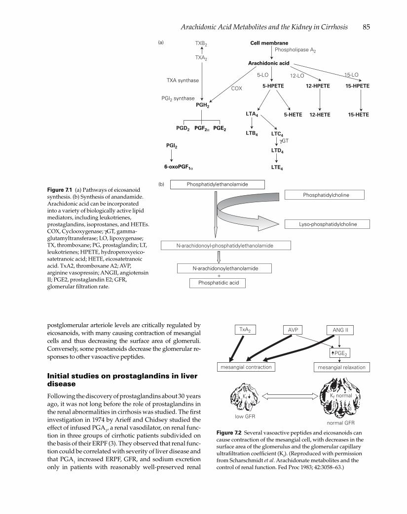
85Arachidonic Acid Metabolites and the Kidney in Cirrhosis
Figure 7.1 (a) Pathways of eicosanoid synthesis. (b) Synthesis of anandamide. Arachidonic acid can be incorporated into a variety of biologically active lipid mediators, including leukotrienes, prostaglandins, isoprostanes, and HETEs. COX, Cyclooxygenase; γGT, gamma-glutamyltransferase; LO, lipoxygenase; TX, thromboxane; PG, prostaglandin; LT, leukotrienes; HPETE, hydroperoxyeico-satetranoic acid; HETE, eicosatetranoic acid. TxA2, thromboxane A2; AVP, arginine vasopressin; ANGII, angiotensin II; PGE2, prostaglandin E2; GFR, glomerular fi ltration rate.
postglomerular arteriole levels are critically regulated by eicosanoids, with many causing contraction of mesangial cells and thus decreasing the surface area of glomeruli. Conversely, some prostanoids decrease the glomerular re-sponses to other vasoactive peptides.
Initial studies on prostaglandins in liver disease
Following the discovery of prostaglandins about 30 years ago, it was not long before the role of prostaglandins in the renal abnormalities in cirrhosis was studied. The fi rst investigation in 1974 by Arieff and Chidsey studied the effect of infused PGA1, a renal vasodilator, on renal func-tion in three groups of cirrhotic patients subdivided on the basis of their ERPF (3). They observed that renal func-tion could be correlated with severity of liver disease and that PGA1 increased ERPF, GFR, and sodium excretion only in patients with reasonably well-preserved renal
TxA2 AVP ANG II
PGE2
mesangial relaxationmesangial contraction
low GFRnormal GFR
Kf Kf normal
Figure 7.2 Several vasoactive peptides and eicosanoids can cause contraction of the mesangial cell, with decreases in the surface area of the glomerulus and the glomerular capillary ultrafi ltration coeffi cient (Kf). (Reproduced with permission from Scharschmidt et al. Arachidonate metabolites and the control of renal function. Fed Proc 1983; 42:3058–63.)
Cell membrane
Arachidonic acid
5-HPETE 12-HPETE 15-HPETE
5-HETE 12-HETE 15-HETE
Phospholipase A2
TXA2
PGH2
PGD2
PGI2
PGE2
LTA4
LTB4 LTC4
γGT
LTD4
LTE4
PGF2α
6-oxoPGF1α
TXB2
5-LO 12-LO 15-LO
COX
PGI2 synthase
TXA synthase
Phosphatidylethanolamide
Phosphatidylcholine
Lyso-phosphatidylcholine
N-arachidonoyl-phosphatidylethanolamide
N-arachidonoylethanolamide
Phosphatidic acid+
(a)
(b)

86 Chapter 7
function. The next study, by Boyer and Reynolds, pub-lished initially in abstract form in 1976 and then as a full paper in 1979, showed that administration of indometh-acin caused a decrease in ERPF and creatinine clearance (4,5). Moreover the decrease in GFR was greatest in those patients with ascites (Fig. 7.3). Infusion of PGA1 restored renal function in patients given indomethacin, increas-ing creatinine clearance from 47 to 63 ml/min, and ERPF from 355 to 580 ml/min in the seven patients in whom indomethacin caused decreases in GFR and ERPF. A fur-ther observation from these studies was that this effect was associated with a decrease in the fi ltration fraction (from 14% to 11%). That is, the amount of glomerular fi l-tration per milliliter of renal blood fl ow decreased. Filtra-tion fraction depends on efferent arteriolar tone as well as the glomerular capillary ultrafi ltration coeffi cient (Kf) as modulated by contraction of mesangial cells. Indometh-acin was also associated with a 60% decrease in plasma renin activity, which was restored by infusion of PGA1. At this time there were simultaneous publications by Zipser and colleagues, who, prompted by the initial ob-servations published by Boyer, showed that cirrhotic pa-tients with ascites had a 10-fold increase in urinary PGE2 excretion rate, and that indomethacin caused a marked reduction in creatinine clearance (from 72 to 32 ml/min) (6,7). Furthermore, plasma renin and aldosterone levels decreased following administration of indomethacin, and the pressor response to angiotensin II in cirrhotic patients was normalized by indomethacin. These two fundamental studies opened the doors to the study of prostaglandins and related compounds in cirrhosis. The important observation that nonsteroidal anti-infl amma-
tory drugs (NSAIDs) cause renal impairment in cirrhotic patients was of clear practical importance, but moreover indicated that renal prostaglandins are important in the maintenance of renal function in cirrhotic patients with ascites. This resulted in a multitude of studies in which the effects of different prostanoids and other eicosanoids on renal function were studied. Eicosanoids can affect renal function directly and indirectly, and these effects are summarized in Table 7.1.
Cyclooxygenase
There are many studies on the role of prostaglandins in renal homeostasis, and prostaglandins have a pivotal role in the regulation or preservation of renal function in patients with liver disease. Prostaglandin synthesis is partly dependent on the differential regulation of the two isoforms of cyclooxygenase (COX; prostaglandin G2/H2 synthase, COX1 and COX2). In the kidney COX1 is expressed primarily in collecting duct, and COX2 is predominantly expressed in the macula densa, and near-by cells in the cortical thick ascending limb (8). COX2 is induced by a variety of stimuli including cytokines and endotoxin (9).
Zhonghua Qi et al. also showed distinct and oppos-ing effects of COX1 and COX2 products on the systemic blood pressure response to angiotensin II (AII) in mice (10). Infusion of AII (150 pmol/kg/min) caused a prompt increase in mean arterial pressure (MAP), followed by a gradual decline. The mechanism of the spontaneous fall in blood pressure could relate to de-sensitization of the AII receptors (11), or the synthesis of compensa-tory vasodilators, such as nitric oxide (NO), bradykinin, or vasodilatory prostanoids (12,13). In the presence of a COX2 inhibitor (SC58236), the initial increase in blood pressure (BP) induced by AII is enhanced, and the subse-quent spontaneous decrease of BP markedly attenuated. These data suggest that in control mice the constrictor effects of AII are counteracted by augmented production of COX2-derived vasodilators, so that medullary blood fl ow remains unchanged.
AII selectively stimulated PGE2 and PGI2 production in the renal medulla via COX2 in interstitial cells, which produces a renal medullary vasodilator action. Renal
• Renal vasodilation• Renal vasoconstriction• Contraction or relaxation of mesangial cells, and modulation of glomerular capillary ultrafi ltration coeffi cient (Kf)• Permeability of collecting ducts to water• Sodium reabsorption in the tubules• Indirectly through systemic effects (e.g. vasodilation) or modulation of other mediators• Indirectly by altering gene transcription or enzyme activity
Table 7.1 Effects of eicosanoids on renal function.
150
100
Cre
at. c
lear
ance
(ml/m
in)
Cre
at. c
lear
ance
(ml/m
in)
50
0
150
100
50
0Pre +Indo Pre +Indo
Ascites No ascites
**
Figure 7.3 Administration of indomethacin to patients with ascites caused a prompt reduction of creatinine clearance. In contrast, indomethacin had no effect on creatinine clearance in patients without ascites. ** P < 0.05. (Adapted from Boyer et al. Effect of indomethacin and prostaglandin A1 on renal function and plasma renin activity in alcoholic liver disease. Gastroenterology 1979; 77:215–22.)

87Arachidonic Acid Metabolites and the Kidney in Cirrhosis
medullary blood fl ow has been intimately linked to the control of systemic blood pressure and renal sodium ex-cretion, and correlates with renal salt excretion: in fact acute infusion of a COX2 inhibitor causes a marked de-crease in urine volume and sodium excretion.
In contrast to COX2, COX1 is present in the collecting duct, a major site of salt and water reabsorption; howev-er, acute infusion of a COX1 inhibitor did not affect med-ullary blood fl ow, nor did it affect renal sodium or water excretion, or modify AII-induced diuresis or natriuresis. Zhonghua Qi et al. suggest that reduced renal medullary blood fl ow could contribute to salt retention (10).
These fi ndings have many clinical implications. Thus, administration of a NSAID to patients with decreased ef-fective arterial blood volume, such as occurs in decom-pensated cirrhosis, will induce a signifi cant decrease in renal function (14–16), since COX1-derived prostagland-ins (PGE2 and prostacyclin) are involved in the mainte-nance of renal function in cirrhosis and ascites (17). The role of COX1 in the maintenance of renal function is also highlighted by the observation that inhibition of COX1 by a selective COX1 inhibitor (SC-560) causes adverse renal hemodynamic changes, while the administration of an inhibitor of COX2 (celecoxib) had little effect (17). This is despite the observation that COX2 is upregulated in the kidney in rats with cirrhosis and ascites in response to decreased effective arterial blood volume.
The action of loop diuretics is known to depend on renal prostaglandins (18), and can be inhibited by NSAIDs. Prostaglandins also play a role in the modula-tion of action of antidiuretic hormone (ADH), a hormone important in renal water transport. Thus, PGE2 inhibits the hydro-osmotic effect of ADH, whereas NSAIDs en-hance the tubular effect of ADH (19).
Renal eicosanoids
Glomeruli comprise endothelial cells, epithelial cells, mesangial cells, and glomerular macrophages. Whole rat or rabbit glomeruli generate PGE2, PGI2, PGF2, and TXA2, with their dominance in that order, whereas human glomeruli synthesize PGI2 predominantly (20). Cultured rat and human mesangial cells produce PGE2, but in humans can also produce PGF2 and PGI2. The form of prostanoid produced by the epithelial cells in-cludes PGE2 or PGI2. Endothelial prostanoids are as yet undefi ned, but prostacyclin is likely to predominate. Leukotrienes are 5-lipoxygenase products synthesized by cells of the mononuclear cell lineage including infi l-trating infl ammatory cells and glomerular macrophag-es, but a transcellular metabolism of leukocyte-gener-ated LTA4 probably exists in glomeruli (21). Infusion of LTC4 into the renal artery causes renal vasoconstriction (22), an effect that may be mediated by LTD4, which acts preferentially on post-glomerular arteriolar resistance vessels and depresses Kf and thus GFR (23). The princi-
pal cysteinyl leukotriene binding to the kidney is LTD4 on high-affi nity receptors on the mesangial cell with a Kd of approximately 10 nM (24). It causes contraction of mesangial cells and in the rat glomerulus inhibits atrial natriuretic peptide (ANP)-induced cGMP synthesis (25). However, basal synthesis of cysteinyl leukotrienes is likely to be low, since the normal excretion rate of LTE4 is 5 ng/h (26). Isolated pig kidneys synthesize cystei-nyl leukotrienes in the absence of infused cells, but the sources of the LTs are unknown (27). Human and rat glomeruli generate both 12-HETE and 15-HETE, by the action of 12- and 15-lipoxygenase, and 12-lipoxygenase activity is exhibited by mesangial cells (28). 12-HETE has other actions in the adrenal gland relevant to cir-rhosis since it is involved in AII-mediated secretion of aldosterone (29).
It is now well established that various eicosanoids can affect both salt and water transport through direct actions on the epithelial cells lining the tubules. Proximal tubules produce only 1–5% as much cyclooxygenase products as do the collecting ducts (20). Prostaglandins clearly have some effect on tubular function since the thick ascending loop of Henle contains PGE2 receptors in high density within the medulla. In this site, PGE2 is believed to inhibit sodium chloride reabsorption. The predominant eicosa-noids produced in the proximal tubules are metabolites of the cytochrome P-450 pathway. The renal cytochrome P-450 system catalyzes the enzymatic conversion of ara-chidonic acid to the EETs (5,6- or 8,9- or 11,12- or 14,15-epoxides or the HETEs). These may have important ef-fects on tubular function. 20-HETE has been singled out for particular study by McGiff’s group in the USA and will be discussed in more detail later. Leukotrienes have no known action on renal tubules.
Prostaglandin E2
The renal actions of prostaglandin E2 are summarized in Table 7.2. Quantitatively, prostaglandin E2 is the pre-dominant prostanoid formed in the kidney (20). This has been demonstrated by direct measurement of prosta-glandin production by isolated renal segments, as well as by several studies demonstrating that urinary excretion of PGE2 is markedly higher than that of other prostanoids (6,7,30). In the rat and rabbit, it comprises about 60% of all prostanoids formed by the glomerulus, but in humans
• Major prostanoid made in the kidney• Stimulates release of renin from juxtaglomerular apparatus • Direct effect on glomeruli, and antagonizes response to pressor hormones• Antagonizes antidiuretic action of antidiuretic hormone • Renal vasodilator
Table 7.2 Renal actions of prostaglandin E2.

88 Chapter 7
and pigs, prostacyclin appears to be the predominant glomerular prostanoid (20). In the juxtaglomerular re-gion, PGE2 as well as PGI2 stimulate the release of renin (31). The collecting tubules are the predominant source of prostanoids, mainly PGE2 (95%). Medullary collecting ducts produce more PGE2 than cortical collecting tubules. The predominant actions are vasodilation and modula-tion of water excretion. Four PGE2 receptors have been identifi ed and cloned. These receptors are designated EP1 through EP4. Using in situ hybridization techniques, Breyer and colleagues have localized the EP receptors in the human kidney (32,33). The phosphatidyl-inositol-hydrolysis response-coupled EP1 receptor was highly ex-pressed in the cortical, outer medullary, and inner med-ullary collecting ducts. The Gi-coupled EP3 receptor was primarily expressed in the cortical and outer medullary collecting ducts as well as in the medullary thick ascend-ing limb. However, the EP3 receptor was absent from the inner medullary collecting duct. The Gs-coupled EP4 re-ceptor was highly expressed in the glomerulus. No EP2 receptors were identifi ed in the human kidney (32,33). PGE2 binding has also been characterized in isolated rat glomeruli, and the response of rat mesangial cells to PGE2 is indicative of receptors. Treatment with indomethacin increases receptor density in the inner medulla.
In the medullary thick ascending limb (TALH), PGE2 inhibits sodium chloride reabsorption both directly and as mediated by ADH. However, the cortical segment of the TALH exhibits fewer receptors for PGE2, and there is less evidence to implicate any functional relevance in this site. Inhibition of COX augments sodium reab-sorption in the loop of Henle and blocks inhibition of sodium reabsorption by furosemide. However, it is not clear whether these are direct effects or effects indirectly mediated by decreases in glomerular plasma fl ow. As noted, the collecting tubules are the predominant source of prostanoids, mainly PGE2 (95%). Medullary collect-ing ducts produce more PGE2 than cortical collecting tubules. Infusion of PGE2 or PGI2 into the renal artery causes natriuresis (20). This may comprise both a hemo-dynamic response and a direct tubular effect. In the col-lecting system, the predominant effect of PGE2 appears to involve water transport. PGE2 stimulates hydraulic conductivity in the cortical collecting duct, and inhibits the response to ADH (34). This is secondary to the effect of PGE2 on cAMP synthesis, both stimulating cAMP syn-thesis directly and suppressing it in response to ADH. Inhibition of COX impairs water excretion in cirrhosis (35). Although ADH stimulates PGE2 synthesis, it ap-pears that the concentration required far exceeds those that affect water metabolism.
After it was established that NSAIDs cause renal im-pairment, there followed many studies applying the then recently developed radioimmunoassays for the de-termination of urinary prostaglandins in cirrhosis and
the hepatorenal syndrome. In the fi rst study measuring urinary PGE2 excretion in cirrhosis, Zia and colleagues demonstrated that urinary PGE2 was increased in three conditions associated with high renin levels—namely, Bartter’s syndrome, renovascular hypertension, and cirrhosis with ascites (6). Several studies have since con-fi rmed that urinary PGE2 excretion is increased in patients with cirrhosis and ascites. In the original studies on the urinary origin of prostaglandins, it was shown that rela-tively little infused PGE2 appeared in the urine, suggest-ing that the kidney was the most likely source. Infusion of AII into both human volunteers and the renal arteries of dogs caused signifi cant increases in urinary PGE2 excre-tion (36). Zusman and Keiser had previously shown that addition of AII, as well as bradykinin or ADH, increased synthesis of PGE2 by renomedullary interstitial cells (37). Further studies investigated the effect of the loop diuretic furosemide, which causes a marked increase in urinary volume and sodium excretion. Infusion of furosemide increases renal cortical blood fl ow and renal synthesis of PGE2 (38). Planas et al. studied the effect of furosemide on renal function and the effect of lysine acetylsalicylic acid on furosemide-induced renal functional changes (39). They observed that acute injection of furosemide in-creased both ERPF and GFR and that lysine acetylsalicyl-ic acid decreased ERPF and GFR. Lysine acetylsalicylic acid blunted the response to furosemide and, conversely, furosemide protected against the adverse effects of lysine acetylsalicylic acid. In view of the latter observation, it is not clear whether or not furosemide improves renal function by increasing PGE2 synthesis, because, if inhi-bition of renal PGE2 synthesis was complete with lysine acetylsalicylic acid, then furosemide must be acting by a different mechanism, otherwise it would not have pre-served renal function. A subsequent study by Quiroga et al. demonstrated that 30% of patients with cirrhosis and ascites actually exhibited decreased GFR following furo-semide infusion, and that this was associated with a fall in urinary excretion of 6-oxo-PGF1 (40). This is discussed further in the section on prostacyclin.
A second but important issue is the effect of prostaglan-dins on water metabolism. The collecting duct is the ulti-mate regulator of renal salt and water excretion. Balance between intake and renal excretion of salt and water is fi ne-tuned by several hormones targeted to the collecting duct. Secretion of ADH from the posterior pituitary gland increases water permeability of the apical membrane of the collecting duct cell, resulting in increased water re-absorption (this is reviewed in Chapter 25). ADH acts on the V2 receptor, which is linked to a G protein, causing stimulation of adenyl cyclase on the plasma membrane, increasing intracellular cAMP levels. There are several counterregulatory mechanisms. Prostaglandin E2 plays a critical physiological and pathophysiological role in counteracting the effects of ADH, since PGE2 produc-

89Arachidonic Acid Metabolites and the Kidney in Cirrhosis
tion itself is stimulated by ADH, and inhibition of COX augments ADH antidiuresis (41). Grantham and Orloff have shown that PGE1 increases osmotic water perme-ability, but when added to tubules pretreated with ADH, PGE1 blunted osmotic water fl ow (42). Likewise, PGE2 increases osmotic water fl ow in rabbit cortical collecting ducts, but blunts the action of ADH. This effect seems to be secondary to inhibition of cAMP generation by ADH. PGE2 decreases cAMP production in microdissected rat or rabbit cortical collecting ducts. It appears that PGE2 inhibits cAMP production at low concentrations and yet has a stimulatory effect at high concentrations (43). The observation that this effect of PGE2 is pertussis sensitive suggests that PGE2 inhibits ADH-stimulated cAMP pro-duction by a G protein mechanism. However, PGE2 can clearly stimulate inositol triphosphate turnover, with formation of diacylglycerols and, presumably, activation of protein kinase C. Breakdown of PIP2 (phosphatidyl inositol biphosphate) may be a biochemical signal medi-ating the inhibitory effects of PGE2 on ADH-stimulated osmotic water fl ow. Experiments by Breyer have indi-cated that PGE2-mediated inhibition of ADH occurs at a pre-cAMP step, since PGE2 has no effect on 8-chloroph-enylthio-cAMP-mediated water transport (43). Exposure of cortical collecting ducts to pertussis toxin followed by ADH and then PGE2 demonstrated that pertussis blocks PGE2 inhibition of ADH-stimulated water transport. PGE2 also acts through other pathways in that it induces a depolarization of the luminal collecting duct membrane in a pertussis-toxin-insensitive pathway. PGE2 causes a prompt increase in cytosolic calcium in rabbit cortical collecting duct. Addition of the protein kinase C inhibi-tor staurosporine markedly attenuates the inhibitory effect of PGE2 on cAMP-analog-induced osmotic water permeability and augments the effect of PGE2 alone on water permeability. Thus, staurosporine blocks the in-hibitory pathway and reveals a dominant stimulatory effect of PGE2 on osmotic water permeability. It appears that when PGE2 is applied to the tubule after exposure to ADH, it interferes with a post-cAMP pathway, whereas when administered before ADH it interferes with cAMP generation but not with cAMP-generated water fl ow. To date there are no published studies on the interaction of PGE2 with the aquaporin system of water channels.
In normal animals, the excretion rate of PGE2 is directly proportional to urinary fl ow rate. This is clearly relevant to studies in cirrhosis, since urinary PGE2 excretion is highest in patients with ascites, in whom the urinary fl ow rate is decreased. Because infusion of PGE2 does not contribute to urinary levels, one can conclude that renal synthesis must be increased. In patients with impaired solute-free water clearance, in which urinary PGE2 excretion is decreased, marked decreases in urinary volumes and GFR are ob-served (41). When water-retaining cirrhotic patients are challenged with water to increase their urine output to the
level observed in the normal controls under water depri-vation conditions (0.6 ml/min), a urinary PGE2 excretion comparable to that of normals (0.28 vs. 0.18 ng/min) is ob-served (40). Nevertheless, patients with mildly impaired solute-free water clearance exhibit a much greater increase of PGE2 excretion than normal subjects. The observation that urinary PGE2 excretion has a strong correlation with GFR may indicate decreased renal synthesis of PGE2, or decreased excretion secondary to impaired urine fl ow rates and renal blood fl ow (30).
Prostaglandin E2 comprises more than 95% of prosta-noids produced in the collecting ducts, the major site of water control. Administration of indomethacin to patients with ascites, who have increased renal PGE2 production, decreases GFR and urine output. Approximately 25% of patients with cirrhosis and ascites have severely impaired solute-free water clearance, and the majority have mild impairment of solute-free water clearance in response to water loading. A study by Reznick et al. showed that, fol-lowing volume loading by insertion of a peritoneovenous shunt (PVS), ADH levels are suppressed, and yet solute-free water clearance is impaired, suggesting that factors other than ADH are involved (44). Cirrhotic patients with ascites generally show increased urinary PGE2 excretion. However, if these patients are subdivided into those with negative solute-free water clearance, in whom GFR is de-creased to about 43 ml/min, and those with positive sol-ute-free water clearance (GFR < 87 ml/min), urinary PGE2 excretion is lower in the former group (0.12 ng/min) com-pared with controls (0.26 ng/min) or those with ascites and a positive solute-free water clearance (0.6 ng/min). Moreover, following water loading, controls exhibited in-creased urinary PGE2, from 0.26 to 1.65 ng/min, as did cir-rhotic patients with intact water clearance (0.6 to 3.9 ng/min). However, patients with impaired water clearance could only increase urinary PGE2 from 0.12 to 0.28 ng/min. These data demonstrate that water loading increases uri-nary PGE2 excretion and that, in turn, increased PGE2 pro-duction may decrease water reabsorption in the collecting tubules. This study also demonstrated that administration of lysine acetylsalicylic acid caused a decrease in solute-free water clearance in all those cirrhotics with ascites who had a positive free water clearance (4.8 to 0.6 ml/min; nor-mals, 12 ml/min) (26). With respect to sodium metabolism, administration of misoprostol, a PGE1 analog, to patients with cirrhosis had a tendency to decrease urinary sodium excretion but had no effect on renal dysfunction induced by indomethacin (45). Whether this is a direct effect or is secondary to vasodilation is not known.
Prostacyclin
Prostacyclin is a renal vasodilator. Apart from its well-known formation by vascular endothelial cells, it is produced primarily in the renal cortex (20). In the renal

90 Chapter 7
cortex, it is made by glomerular endothelial cells, being the major prostanoid formed by human glomeruli, which produce lesser amounts of PGE2 or thromboxane. In the rat glomerulus, PGE2 synthesis exceeds that of pros-tacyclin or thromboxane synthesis by a factor of 2 to 3. The glomerulus is composed of endothelial, mesangial, and epithelial cells, as well as bone-marrow-derived glomerular macrophages located in the mesangium (46). Rat mesangial cells produce PGE2, but with substantial amounts of prostacyclin and thromboxane (47). Glomer-ular epithelial cells synthesize lesser amounts of prosta-noids than mesangial cells produce. Addition of AII, PAF, or ADH to cultured rat mesangial cells causes the forma-tion of PGE2, but no other eicosanoids. Human mesangial cells make both prostacyclin and PGE2 following stimu-lation with AII, PAF, or ADH (48). AII also augments both PGE2 and prostacyclin synthesis by whole glomeruli (49), which modify the actions of this hormone on the kidney (50). These prostanoids act on mesangial cells, increas-ing intracellular cAMP. Studies measuring glomerular planar surface area have shown that glomeruli contract in response to AII to 80% of their surface area (2). This decreases the surface area available for fi ltration and thus the Kf. Inhibition of prostaglandin synthesis by NSAIDs augments the response to AII, causing a 7% decrease in surface area alone, and a shift in the dose–response curve to AII, suggesting that endogenous prostaglandins limit fl uctuations of K f induced by vasoactive peptides, thus preserving the fi ltration fraction. Similarly, increasing prostaglandin synthesis by addition of arachidonic acid inhibits the response to AII. The effect of infused prosta-glandins is confounded by the fact that both prostacyclin and PGE2 cause release of renin, and thus release of AII by juxtaglomerular cells. Further, addition of converting en-zyme inhibitors to whole glomeruli increases both pros-tacyclin and PGE2 synthesis (51). When the effects of AII are blocked by antagonists, both PGE2 and prostacyclin are purely vasodilatory, and have no effect on K f. Infusion of AII after inhibition of COX causes reduction of ERPF, GFR, and Kf (52). Thus, endogenous prostaglandins are important for the preservation of renal function in states with an activated renin–angiotensin system. Models in which COX inhibition causes a reduction in RBF can be reversed by preventing AII synthesis by captopril (53).
Experiments in the dog (54) have shown that there is a six- to 10-fold increase in the formation of both PGE2 and prostacyclin in the kidney following an acute increase in renal venous pressure (RVP) from 5 to 40 mmHg, but no increase in thromboxane release. Renal blood fl ow was maintained despite the increased RVP, but treatment with indomethacin caused a prompt reduction of RBF and GFR. The changes in RVP are akin to those observed in cirrhotic subjects with and without ascites. A study by Mullane and Gliedman has shown a marked increase in RVP in cirrhot-ics, presumably as a consequence of altered structure in
the caudate lobe, through which the inferior vena cava passes (55). The presence of ascites increases RVP further, so that in the presence of tense ascites the intra-abdominal (and therefore renal) pressure may be as high as 30 mmHg. This may partly explain the close association of a mark-edly increased renal synthesis of PGE2 and prostacyclin with the presence of ascites.
With regard to the kidney in cirrhosis, it is diffi cult to be certain how much of the effect of COX inhibitors can be ascribed to prostacyclin and how much to PGE2. Whereas urinary PGE2 excretion refl ects renal synthesis, the same does not necessarily hold true for either 6-oxo-PGF1 (a stable prostacyclin hydrolysis product) or TXB2 (a stable thromboxane hydrolysis product). Infusion stud-ies using radiolabeled prostacyclin or TXB2 have shown that 6% of infused radioactivity appears in the urine (56,57). Although this may seem to be a small percentage, the overall size of the vascular endothelium (one of the largest “organs” in the body) makes such a contribution very signifi cant. Prostacyclin is the main COX product of blood vessels. The major systemic metabolite is 2,3-dinor-6-oxo-PGF1. Approximately 14% of infused 6-oxo-PGF1 is excreted in the urine unchanged (58), 21% as 2,3-dinor-6-oxo-PGF1, and 20% as 2,3-dinor-6,15-diketo-13,14-dihy-dro-20-carboxyl-PGF1. Organic acids such as PGE2 may be secreted actively. However, experiments by Rosenkranz et al. showed no effect of probenecid on the urinary excre-tion of 6-oxo-PGF1, suggesting that this compound is not handled by the organic acid pathway (59).
Guarner et al. fi rst suggested that endogenous prosta-cyclin may be responsible for the hyperdynamic circula-tion observed in cirrhosis. In their study they observed increased urinary excretion of the 2,3-dinor metabolite of prostacyclin in cirrhotics with and without ascites as well as in those with the hepatorenal syndrome (HRS) (60). These data are consistent with increased vascular synthesis of prostacyclin in cirrhosis. This paper im-plied that systemic prostacyclin synthesis was similar in the three cirrhotic groups, when assessed by urinary 2,3-dinor-6-oxo-PGF1 expressed as concentration/mg creatinine. Such an analysis assumes that renal function has no effect on the urinary excretion rate of 6-oxo-PGF1. Although this is probably true for PGE2, there seems to be no basis for assuming that it is true for other renal ei-cosanoids synthesized in the glomerulus or proximal to the collecting ducts. Indeed, studies by others had shown a correlation between urinary 6-oxo-PGF1 excretion and GFR (30). In a follow-up study, Moore and colleagues ob-served that the urinary excretion rates of both 6-oxo-PGF1 and its 2,3-dinor metabolite (as well as thromboxane and its dinor metabolite) were markedly elevated in patients with severe liver failure, and decreased in parallel to cre-atinine clearance as patients developed renal failure (61) (Fig. 7.4). This study demonstrated that, when corrected for GFR, production of both prostacyclin and thrombox-

91Arachidonic Acid Metabolites and the Kidney in Cirrhosis
700
600
300
200
100
0
0–1 1 2 5 6
0
20
40
60
80
Days following admission
Exc
retio
n ra
te (n
g/h)
Creatinine clearance (m
l/min)
Figure 7.4 Urinary excretion rates of thromboxane B2 and 6-oxo-PGF1 were followed serially in patients with alcoholic hepatitis during the development of the hepatorenal syndrome. Urinary excretion rates of both degradation products of thromboxane A2 (open triangles) and prostacyclin (open diamonds) were markedly elevated (normal levels, 2–6 ng/h) and fell in parallel with the fall in creatinine clearance (closed circles). This strongly suggests that the excretion rates of these compounds should be corrected for creatinine clearance. (Adapted from Moore et al. Systemic and renal production of thromboxane A2 and prostacyclin in decompensated liver disease and hepatorenal syndrome. Gastroenterology 1991; 100:1069–77.)
ane was directly proportional to severity of liver disease, rather than to renal function (Fig. 7.5). The magnitude of increased urinary excretion of systemic prostacyclin me-tabolites is well below that predicted from infusion of a vasodilatory dose of prostacyclin. Based on this, it was suggested by Moore et al. that the amount of prostacyclin formed is unlikely to be suffi cient to cause vasodilation (61). In retrospect, this is probably incorrect. The vasodi-lating effect of prostacyclin is probably localized to the vascular bed where it is synthesized, and such calcula-tions are therefore likely to be invalid. Moreover, studies by Zipser et al. and Bruix et al. have shown that adminis-tration of NSAIDs causes a modest increase in splanch-nic resistance and restores vascular responsiveness to in-fused AII (7,62) (Fig. 7.6). Although the latter observation could conceivably be attributable to an effect on the renal vascular resistance, the data of Bruix et al. make it much more likely that this is a direct vascular effect, mediated through inhibition of prostacyclin synthesis.
The production of prostacyclin by the vascular bed can be stimulated by catecholamines, renin, ADH, and endo-toxin, and has been observed in models of noncirrhotic
portal hypertension (63–65). Interestingly, Guarner et al. have also reported that intestinal decontamination with antibiotics caused a modest decrease in urinary excretion of 2,3-dinor-6-oxo-PGF1 (66). Cirrhotic patients have in-creased levels of endotoxin in hepatic, portal, and arterial blood, which are thought to be secondary to impaired reticuloendothelial function, and these endotoxin levels may stimulate phospholipases and prostaglandin syn-thesis. This author is aware of no studies on the effects of inhibiting the other hormone systems on systemic pros-tacyclin production. The observation that both systemic prostacyclin and thromboxane production are increased suggests that there is increased platelet–endothelial cell interaction, which itself may be enhanced during endo-toxemia (61).
Studies by Ros et al. have investigated the interac-tion between NO and prostaglandin synthesis in the carbon tetrachloride model of cirrhosis (67). This model has a three-fold elevation of urinary 6-oxo-PGF1 excre-tion, and urinary sodium excretion is 20% of the normal level, but GFR is preserved. Although this investigation was titled as a study on prostacyclin, the only reference to this compound was in its measurement in the urine. Since PGE2 is the major prostanoid made by rat kidney, it is likely that most of the effects of inhibition of COX described below are attributable primarily to inhibition of PGE2 synthesis. Inhibition of NO synthesis increased mean arterial pressure in the cirrhotic animals from 88 to 102 mmHg, and in controls from 116 to 123 mmHg. Renal vascular resistance doubled in the control ani-mals, and GFR remained unchanged. In the cirrhotic animals, however, renal vascular resistance (higher than controls: 6.76 ± 0.6 vs. 4.76 ± 0.7 mmHg/min/ml) showed no change, but GFR increased from 2.4 to 3.3 ml/min. Urine sodium excretion increased in each group by a factor of 2 to 3. Inhibition of prostaglandin synthesis had no effect on mean arterial pressure in ei-ther group and yet increased renal vascular resistance from 5.3 to 8.5 mmHg/min/ml in those with cirrhosis. This was accompanied by a fall in renal plasma fl ow but no change in GFR. In other words, inhibition of prosta-glandin synthesis alone caused a decrease in renal blood fl ow, but GFR was preserved by an increase in the fi l-tration fraction. Conversely, inhibition of NO synthesis caused the GFR to increase in the cirrhotic animals, de-spite no change in renal blood fl ow, indicative that this also caused an increase in fi ltration fraction. Inhibition of both parameters was additive. Mean arterial pres-sure increased in both controls and cirrhotic animals, and this was accompanied by a 60% fall in renal plasma fl ow, but with GFR unchanged. The effect of each treat-ment on the fi ltration fraction is shown in Table 7.3.
The most striking aspect of these data is the effect of each agent in increasing the fi ltration fraction. The fi ltra-tion fraction is affected by changes in efferent arteriolar

92 Chapter 7
100
75
35
30
20
10
0N Asc SH HRS
Clinical group
N Asc SH HRS
Clinical group
N Asc SH HRS
Clinical group
N Asc SH HRS
Clinical group
400
300
210
200
150
100
2,3–
dino
r–TX
B2/
Cre
at.C
lear
(pg/
ml.C
r.C
l)
TXB
2/C
reat
.Cle
ar (p
g/m
l.Cr.
Cl)
50
40
30
20
10
0
150
100
50
45
30
2,3–
dino
r–6–
Oxo
–PG
F 1α
/Cre
at.C
lear
(pg/
ml.C
r.Cl)
15
00
10
6–O
xo–P
GF 1
α /C
reat
.Cle
ar (p
g/m
l.Cr.C
l)
20
35
30
100
200
Figure 7.5 Urinary excretion of thromboxane B2, 6-oxo-PGF1, and their dinor metabolites in patients with ascites (Asc), severe liver failure but preserved renal function (SH), patients with the hepatorenal syndrome (HRS), and normal controls (N). There was a positive correlation of these levels with
the severity of liver disease as assessed by plasma bilirubin or coagulopathy. (Adapted from Moore et al. Systemic and renal production of thromboxane A2 and prostacyclin in decompensated liver disease and hepatorenal syndrome. Gastroenterology 1991; 100:1069–77.)

93Arachidonic Acid Metabolites and the Kidney in Cirrhosis
8
6
4
Car
diac
out
put
(l/m
in)
Baseline +Indomethacin Baseline +Indomethacin
1500
1000
500
****
Syst
. vas
c. r
esis
t. (d
yn·S
–1·c
m–5
)
Figure 7.6 Administration of indomethacin to patients with cirrhosis caused a decrease in cardiac output and an increase in systemic vascular resistance. These data suggest that endothelial prostacyclin production is important in the development of a hyperdynamic circulation. (Adapted from Bruix et al. Effects of prostaglandin inhibition on systemic and hepatic hemodynamics in patients with cirrhosis of the liver. Gastroenterology 1985; 88:430–5.)
tone and in factors that affect glomerular plasma fl ow and the Kf. What is most surprising is that this is in con-trast to predictions based on the known effects of PGE2, prostacyclin, or NO in inhibiting mesangial cell contrac-tion and thus acting as counterregulatory mechanisms against factors tending to decrease Kf and thus GFR. In-deed, these differences in the cirrhotic rat’s response to NSAIDs, in contrast to those observed in humans, high-light the differences between different animal species and human liver diseases.
Although a story emerged in the 1980s suggesting that an imbalance between the synthesis of the vaso-dilatory prostanoids (PGE2 and prostacyclin) and the renal vasoconstrictor thromboxane was causative in the pathogenesis of the hepatorenal syndrome (68), a critical look at the data does not support such a notion
(69). Although the current data support the idea that renal PGE2 synthesis is decreased in the hepatorenal syndrome, prostacyclin production is increased (61). An immunohistochemical study of antiprostacyclin synthase antibodies by Govindarajan et al. has shown that in normal kidneys there is intense staining in corti-cal and medullary peritubular capillaries in the adjacent renal interstitial cells and in the glomerular mesangial region. The immunochemical staining pattern was unchanged for prostacyclin synthase in patients with the hepatorenal syndrome (70). In contrast, there was a marked diminution of staining for COX (termed PG endoperoxide synthase in the paper) in the patients with the hepatorenal syndrome. Whether this phenom-enon is causally associated with the pathogenesis of the hepatorenal syndrome or refl ects a staining artefact pro-duced by prolonged reductions of renal perfusion is not known.
An interesting observation has emerged from studies on the effect of furosemide on renal function and pros-tacyclin and PGE2 synthesis. Administration of an i.v. bolus of furosemide increases urine output and urinary excretion of PGE2. On examining the effects of furosem-ide on GFR in patients with cirrhosis, one fi nds that some patients exhibit an increase and some a decrease in cre-atinine clearance. Despite the increase in urine volume, it was observed that in the group in which GFR increased (from 99 to 129 ml/min, n = 15) there was an associated increase in the urinary excretion of 6-oxo-PGF1 (from 478 to 1034 pg/min), and in the group with an associated de-crease in GFR (from 102 to 71 ml/min, n = 6) there was a corresponding fall in urinary 6-oxo-PGF1 excretion (from 1032 to 548 pg/min) (40). Moreover, there was a strong correlation between the change in 6-oxo-PGF1 excretion rate and the change of GFR. What is clear from these data is that basal urinary 6-oxo-PGF1 and PGE2 excretion are highest in those who exhibit a fall in GFR following injec-tion of furosemide. Previous studies have suggested that the group with the highest PGE2 excretion had relatively intact solute-free water clearance (41). Subsequent stud-ies have demonstrated that urinary 6-oxo-PGF1 should be corrected for creatinine clearance (Cr.Cl) (61). On re-examining the effect of furosemide on corrected 6-oxo-PGF1 excretion in this study, it is observed that following infusion of furosemide the urinary excretion of 6-oxo-PGF1 is 8 pg/ml Cr.Cl in the GFR-positive responders and 7.8 pg/ml Cr.Cl in the GFR-negative responders, compared with basal levels of 4.8 and 10.1 pg/ml Cr.Cl in the two groups, respectively (40). These data could be interpreted to suggest that renal prostacyclin and PGE2 synthesis are increased in the group most susceptible to the adverse effects of furosemide, and that these are compensatory mechanisms. Further studies are needed to determine the effect of prostacyclin on renal function in cirrhosis.
Filtration fraction
Normal rats Cirrhotic rats
Inhibition of NOS Before 22% 20% After 38% 28%Inhibition of COX Before 22% 20% After 26% 33%Inhibition of NOS and COX Before 22% 20% After 58% 66%
Table 7.3 Effects of inhibition of nitric oxide synthase (NOS) and/or cyclooxygenase (COX) on fi ltration fraction in rats with carbon tetrachloride-induced cirrhosis with ascites.
From Ros et al. Role of nitric oxide and prostacyclin in the control of renal perfusion in experimental cirrhosis. Hepatology 1995; 22:915–20.

94 Chapter 7
Thromboxane A2
Thromboxane A2 was fi rst described in 1975 by Ham-berg et al. (71). It is an unstable prostanoid formed by thromboxane synthase, an enzyme similar to cytochrome P-450, and rapidly undergoes hydrolysis to TXB2, with a half-life of 1–2 s. It acts on one or more specifi c cell sur-face G-protein-coupled receptors (20). At present, only one receptor has been identifi ed and cloned (72). Howev-er, it is likely that more than one receptor exists, because there are high- and low-affi nity binding sites in different tissues, and other agonists such as the F2-isoprostanes appear to act on a closely allied but separate group of receptors. Recent studies in rat and murine kidneys by immunostaining or in situ hybridization have localized thromboxane receptors in glomeruli, arterial walls, lumi-nal membranes of the medullary thick ascending limb of Henle’s loop, and both the luminal and basolateral mem-branes of the distal convoluted tubules and the basola-teral membranes of the collecting tubules (73–77). In the glomeruli, the receptor appears to be localized mainly along the lumina of the glomerular capillary loops. Pari-etal epithelial cells of the Bowman’s capsule, podocytes, and mesangial cells also demonstrate immunostaining for the receptor. These observations are supported in a study by Asano et al. showing that TXA2 receptor mRNA was abundantly expressed in the glomerulus, followed by the distal convoluted tubule as well as the medullary thick ascending limb of Henle’s loop and outer medul-lary collecting duct (76).
In the kidney, infusion of analogs of thromboxane such as U46619 causes marked renal vasoconstriction and a fall in GFR (78). This is presumably a direct effect on pre-glomerular vessels and the glomeruli. Exposure of me-sangial cells to U46619 causes contraction and a decrease in the surface areas of the glomeruli (79). This in turn de-creases the Kf and thus the fi ltration fraction. Infusions of TXB2 into human volunteers have demonstrated that it has a half-life of 7 min, and 5.3% was converted and excreted as 2,3-dinor-TXB2 (80). A study by Zipser and Martin showed that, following infusion of TXB2 into the brachial vein and renal artery, 2% and 14% are excreted unmetabolized, respectively (57). Although the fraction-al excretion of unchanged TXB2 is small, patients with thromboembolic disease have increased urinary concen-trations of TXB2, suggesting that systemically formed TXB2 may contribute signifi cantly to the overall urinary TXB2 profi le (81).
The role of thromboxane in the pathogenesis of the hepatorenal syndrome was fi rst proposed by Kronborg and Zipser (68,82,83). They measured urinary excretion of TXB2 in patients with cirrhosis and in those with al-coholic hepatitis who were developing hepatorenal syn-drome. Whereas the urinary excretion rate of PGE2 was low in patients developing hepatorenal syndrome and
high in those with cirrhosis, urinary TXB2 concentrations were high in those with hepatorenal syndrome and low in those with cirrhosis. This paper described their data in terms of urinary concentrations, because the urinary con-centration of TXB2 was unaffected by water deprivation and low urinary fl ow rates in normal controls (84). Based on the presentation of data showing that development of renal failure in liver disease causes a reduction in the ex-cretion of PGE2 and an increase in TXB2 concentrations, a story emerged of an imbalance of vasodilators (PGE2) and vasoconstrictors (thromboxane). Other studies, how-ever, took the more reasoned approach of quantifying the rate of formation of thromboxane by the urinary excretion rate of either TXB2 or 2,3-dinor-TXB2 (30,61). These stud-ies then found that urinary excretion of TXB2 was in fact low or similar to that in patients with decompensated liver disease but relatively well-preserved renal function. How-ever, since patients with thromboembolic disease have increased urinary TXB2 excretion (81), the contribution of nonrenal TXB2 to urinary levels needs consideration, and the control population should be matched as far as pos-sible for severity of liver disease to the target population (i.e. those with hepatorenal syndrome). Serial study of pa-tients in the developing phase of hepatorenal syndrome demonstrated that urinary excretion rates of both 6-oxo-PGF1 and TXB2 as well as their 2,3-dinor metabolites were markedly elevated in patients prior to the development of hepatorenal syndrome, and that their urinary excretion rates fell in parallel with the creatinine clearance (61) (see Fig. 7.4). These data could be interpreted as showing that renal synthesis of both thromboxane and prostacyclin fell as patients developed hepatorenal syndrome. However, the most likely explanation has to be that urinary excre-tion rates of these prostanoids are GFR dependent, and thus urinary excretion rates should be corrected for GFR. When this is done, a very different pattern emerges, that is, urinary 6-oxo-PGF1 and TXB2 are both markedly elevated in proportion to the severity of liver disease and appear to be independent of renal function (see Fig. 7.5). These data do not, of course, exclude a signifi cant contribution from renal thromboxane to the renal abnormalities that occur in liver disease, but they do not support a close associa-tion of urinary TXB2 and the development of hepatorenal syndrome. A study using the thromboxane inhibitor da-zoxiben in patients with hepatorenal syndrome was dis-appointing (85), and although often quoted as evidence against the role of thromboxane in the renal abnormali-ties, this study in fact neither proved nor disproved any-thing. The dosage of dazoxiben was too low for effective inhibition of renal synthesis of thromboxane, and the use of synthase inhibitors is confounded by the potential for PG endoperoxides to be directed down other pathways, such as to form PGD2, which can also act on the thrombox-ane receptor and cause renal vasoconstriction. The most important data have emerged from the studies of Laffi ,

95Arachidonic Acid Metabolites and the Kidney in Cirrhosis
Pinzani, and Gentilini (86–88), who studied the effects of both a thromboxane synthase inhibitor and a thrombox-ane receptor antagonist on water handling in cirrhotic pa-tients. They observed that administration of OKY 046, a thromboxane synthase inhibitor, to non-azotemic cirrhotic patients with ascites and avid sodium retention increased GFR by 20%, whereas renal blood fl ow was unchanged. In other words, the fi ltration fraction increased, presum-ably as a consequence of intrarenal thromboxane on me-sangial cells. Further studies showed that when adminis-tered with furosemide, OKY 046 enhanced the increase in furosemide-induced solute-free water clearance by a fac-tor of 4 (from 1 to 4 ml/min) and increased the natriuretic response to furosemide in this population. These studies also observed that furosemide not only increased urinary PGE2 excretion, as observed by others, but also increased GFR as well as urinary 6-oxo-PGF1 and TXB2 excretion. This group followed up these studies with an investiga-tion of a thromboxane receptor antagonist, which should not be complicated by diversion of endoperoxides into the PGD2 pathway. This study involved assessment of the ef-fects of thromboxane blockade during a water diuresis. In cirrhotics with ascites, thromboxane receptor blockade increased solute-free water clearance from 1.7 to 3.1 ml/min, but had no effect on urinary electrolyte excretion or renal function. There was a small increase in renal plasma fl ow. Thus, intrarenal thromboxane synthesis seems to cause a modest decrease in GFR in cirrhotics with ascites and to limit the diuretic response to furosemide or water loading (86).
Cysteinyl leukotrienes
The cysteinyl leukotrienes were initially termed slow reacting substance of anaphylaxis (SRS-A). It is now es-tablished that the cysteinyl leukotrienes comprise leu-kotrienes C4, D4, and E4 (LTC4, LTD4, and LTE4), which are formed by the action of 5-lipoxygenase (5-LO) on arachidonic acid to form 5-HPETE and LTA4, which in turn is converted to LTB4, a potent chemoattractant to neutrophils, or to LTC4 and thence to LTD4. Of the cystei-nyl leukotrienes, LTD4 is the most active on a molar basis, and much of the action of LTC4 may be related to the conversion of LTC4 to LTD4. The pathway is shown in Fig. 7.7. LTD4 is inactivated by metabolism to LTE4, which has insignifi cant biological activity and is excreted un-changed in the urine or bile or undergoes more extensive ω- or β-oxidation to the 2,3-dinor and tetranor metabo-lites (26,89–91).
These compounds elicit arteriolar vasoconstriction in the cardiac, pulmonary, and renal vascular beds, as well as augmentation of vascular permeability to macromol-ecules in postcapillary venules. The direct effects of infu-sion of LTC4 were fi rst reported by Badr and colleagues (22). Infusion of LTC4 into the rat produced a signifi cant
elevation of blood pressure with reductions in cardiac output and renal blood fl ow. These effects were abol-ished by a leukotriene receptor antagonist, but not by saralasin (an AII antagonist) or a COX inhibitor. Subse-quently, Gulbins et al. showed that LTD4 and LTE4 could cause contraction of preglomerular vessels in the rat kid-ney with a 50% reduction of GFR (92). A study by Barnett et al. showed that both LTC4 and LTD4 along with ADH, AII, and PAF, decreased the surface area of isolated rat glomeruli by approximately 10–15% (93). Whereas both PAF and AII caused a seven- to 10-fold increase in PGE2 synthesis, this was not observed when the glomeruli were incubated with LTC4 or LTD4. A subsequent study by Badr and colleagues demonstrated binding of LTD4 to cultured glomerular mesangial cells (24). Binding was found to be stereoselective, specifi c, saturable, and reversible. High- and low-affi nity binding sites were identifi ed. LTD4 was found to initiate a phosphatidyl-inositol response and the formation of IP3. The question as to whether the kidney could synthesize leukotrienes was for a long time complicated by the nature of many studies in which blood was allowed to circulate through the kidney either in vivo or in vitro. Blood cells can make LTA4 through the 5-lipoxygenase (5-LO) pathway, and this may be involved in a transcellular transfer and me-tabolism by other cell types that contain LTC4 (21). How-ever, perfusion of the isolated pig kidney by a blood-free perfusate, with oxygenation by FC43, a perfl uorinated oxygen carrier, by Moore and colleagues has shown un-equivocal increased synthesis of cysteinyl leukotrienes following injection of the calcium ionophore A23187 (27). Acute injection of lipopolysaccharide had no effect over a period of 1–2 h of perfusion. Moreover, this study dem-onstrated extensive metabolism of LTE4 by the kidney to form both ω- and β-oxidation products. Whether tissue-resident macrophages in the glomeruli were responsible for the stimulated increase in urinary LTE4 is not known, but transcellular transfer of LTA4 was effectively exclud-ed during these experiments.
Several studies have now shown increased plasma concentration of atrial natriuretic peptide (ANP), the ac-tion of which is to increase salt excretion and to increase GFR through a cGMP-dependent pathway. Both ANP and the NO donor, nitroprusside, increase glomerular cGMP, and this can be signifi cantly and dose-depend-ently inhibited by LTD4 (25). Following the demonstra-tion by Orning et al. that the major pathway for elimina-tion of cysteinyl leukotrienes in humans involved the kidney and hepatobiliary systems (89), it was suggested by Keppler et al. that leukotrienes may be involved in the pathogenesis of hepatorenal syndrome (94). Two studies, one by Moore et al. and the other by Huber et al., demonstrated increased urinary excretion of cysteinyl leukotrienes by patients developing hepatorenal syn-drome (26,95,96) (Fig. 7.8). Urinary excretion of LTE4 is

96 Chapter 7
GFR dependent, and increased synthesis was confi rmed by infusion of radiolabeled LTE4 into a patient with hepatorenal syndrome together with measurement of unchanged urinary radiolabeled LTE4 and endogenous LTE4 in the same subject (26). Huber et al. also noted that patients with hepatorenal syndrome had increased urinary levels of N-acetyl-LTE4, and suggested that this may be a more specifi c marker of renal synthesis (95). Further studies by Keppler ’s group confi rmed the pre-vious observations (26) that urinary excretion of LTE4 is increased in patients with biliary obstruction and de-creased after biliary drainage (97). Although the latter observation was quoted as supporting a diversion of biliary excretion through renal pathways (which must occur), it does not exclude increased synthesis second-ary to endotoxemia, which was relieved by biliary drainage. A recent study by Titos et al. (98) showed that there is over-expression of 5-LO mRNA in cirrhotic rat liver; in contrast, liver from control animals had low ex-pression of 5-LO mRNA, while hepatic concentrations of LTC4 synthase were similar in both groups. Moreover,
they showed that cultured rat hepatocytes with differ-ent degrees of purity showed different expression of 5-LO mRNA: in highly purifi ed hepatocytes there is no evidence of 5-LO mRNA expression. A transcellular me-tabolism is suggested to be involved in the formation of cysteinyl leukotrienes in rat hepatocytes. This involves the synthesis of LTA4 by an intermediate cell, uptake by hepatocytes, and then further metabolism to the respec-tive cysteinyl leukotriene. Titos et al. confi rmed what had been known for some time, namely that 5-LO is limited to cells of the myeloid series, and is only present in Kupffer cells in cirrhotic liver, and these cells may release LTA4, thus hepatocytes rich in LTC4 synthase transform LTA4 into cysteinyl leukotrienes (99,100). The studies of Titos et al. and Graupera et al. support the concept that hepatocytes have an important role in the formation of cysteinyl leukotrienes and the regulation of intrahepatic vascular tone. One of the major poten-tial targets for hepatic derived cysteinyl leukotrienes includes the hepatic stellate cell, in which expression of actin is upregulated in cirrhosis, and which can contract
COOH
COOH
COOH
COOH
COOHCOOH
COOH
COOH
H2N
H2NH2N
C5H11
C5H11C5H11
COOHOH OH
H
O
OHOH
OHH
N
OS
H
S
OO
S
N N
OH
LEUKOTRIENE A4
LEUKOTRIENE B4
LEUKOTRIENE C4
LEUKOTRIENE D4LEUKOTRIENE E4
ARACHIDONIC ACID
5-Lipoxygenase(via 5-HpETE)
LTA4 Hydrolase LTC4 Synthase
g-Glutamyl transferase
Amino-peptidase
Figure 7.7 Formation and breakdown of the cysteinyl leukotrienes.

97Arachidonic Acid Metabolites and the Kidney in Cirrhosis
300
200
100
65
60
40
20
0
Norm CLD Asc SH HRS CRF
Clinical group
Urin
ary
LTE
4 ex
cret
ion
(pg/
ml c
reat
.cle
ar)
Figure 7.8 Urinary excretion of leukotriene E4, the metabolic product of LTC4 and LTD4, was markedly elevated in patients with the hepatorenal syndrome (HRS). Control groups included normals, compensated liver disease (CLD), ascites (Asc), severe hepatic failure (SH), and chronic renal failure (CRF). (Adapted from Moore et al. Increased production of cysteinyl leukotrienes in hepatorenal syndrome. J Hepatol 1990; 11:263–71.)
in response to LTC4 or LTD4 (98). A role for cysteinyl leukotrienes in the pathogenesis of portal hypertension is supported by the observation that inhibition of 5-LO decreases portal pressure (this may involve the type 3 receptor, since antagonists at types 1 and 2 receptors do not decrease portal pressure) (99), and hepatic leuko-trienes may therefore have an indirect role on renal function through hepatorenal refl exes.
Cytochrome P-450 arachidonate products
Oxidation of arachidonic acid (AA) by a variety of cy-tochrome P450 (CYP) enzymes leads to the formation of HETEs (hydroxyeicosatetraenoic acids) and EETs (epoxyeicosatrienoic acids), the latter of which are hy-drolyzed to dihydroxyeicosatrienoic acids (DHTs) by cytosolic epoxide hydrolase. CYP-AA metabolites dif-fer from prostaglandins in that they are stored in tissue lipids and have a primary intracellular locus of action. One of the most important CYP-AA metabolites relevant to cirrhosis is the ω-hydroxylase product 20-HETE (101). 20-HETE affects movement of ions, constricts blood ves-
sels, is involved in tubuloglomerular feedback and renal autoregulation, and is mitogenic (102–105). Increased synthesis of 20-HETE is a major factor elevating blood pressure in the spontaneously hypertensive rat (106). It is predominantly formed in the renal arterioles with signifi cant production in the renal medulla (104). In the preglomerular arterioles, it causes vasoconstriction in a non-COX-dependent manner by inhibiting potassium channels causing vasoconstriction (104). 20-HETE, to-gether with 19-HETE, the function of which is unknown, is the principal CYP product in the medullary thick as-cending limb, and is also formed in the proximal tubules (107). The medullary thick ascending limb is crucial to the regulation of extracellular volume. The thick ascending limb is the target for loop diuretics such as furosemide. Both 19- and 20-HETE affect Na+-K+-ATPase activity and are vasoactive. Their vasoactivity is partially dependent on COX activity, which can metabolize 20-HETE to vaso-active compounds (105). 20-HETE exerts a furosemide-like effect on medullary thick ascending limb cells, and inhibits the ATP-sensitive potassium channel in the api-cal membrane of the rat medullary thick ascending limb, and thus potassium recycling, an essential requirement for the activity of the Na-K-2Cl-cotransporter.
Both 20-HETE and 19-HETE are released from the rabbit or rat kidney in response to AII or endothelin-1, respectively, but ADH and bradykinin have no effect on their formation. In humans, 20-HETE is excreted in the urine as the glucuronide (108). A study by Sacerdoti et al. has shown that urinary 20-HETE excretion is in-creased fi ve-fold above controls in patients with ascites, and two-fold above patients with cirrhosis alone (101) (Fig. 7.9). Moreover, Sacerdoti demonstrated that uri-nary 20-HETE excretion rate was highest in those with the lowest renal blood fl ow (101,109). Whether glucuro-nidation of the HETEs occurs in the kidney, or whether it occurs predominantly in the liver, thus refl ecting an extrarenal site of synthesis, is unknown. The mecha-nism of increasing 20-HETE synthesis is unclear, but prime candidates include AII as well as endothelin-1. Recent studies have shown that endothelin-1 stimulates a four-fold increase in urinary 20-HETE effl ux from the rat kidney (105), and that endothelin-1 levels are in-creased in both cirrhotics and patients with hepatorenal syndrome (110).
Isoprostanes
The isoprostanes were fi rst described in 1990, and are formed by peroxidation of arachidonic acid (111) (Fig. 7.10). They are formed as esterifi ed prostanoids following oxidation of arachidonyl-containing phospholipids (112). Measurement of plasma, urinary, or tissue F2-isoprostanes is now considered to be the gold standard for the assess-ment of lipid peroxidation in vivo (113,114). More

98 Chapter 7
importantly, isoprostanes also have potent biological ac-tivity causing intense renal vasoconstriction when infused into the rat (111,115). They act on a thromboxane-like recep-tor, with their renal actions being antagonized by throm-boxane receptor antagonists. However, various data indi-cate that they may interact with a receptor separate from the conventional thromboxane receptor, since transfection experiments can confer binding of thromboxane analogs, but not the F2-isoprostane 8-iso-PGF2 (116). Plasma con-centrations of the F2-isoprostanes are increased in liver disease, particularly so in hepatorenal syndrome (117). More recently, both E/D ring isoprostanes have been de-scribed as well as isothromboxanes (118,119). These results have enormous implications for assays of prostanoids em-ploying immunoassay, because some antisera will detect, with high cross-reactivity, similar isomeric forms (120). At present, the role of F2-isoprostanes or related compounds in the etiology of renal dysfunction or salt and water reten-tion in cirrhosis is unknown (121,122).
Platelet-activating factor
Originally described in 1972 by Benveniste, PAF (or PAF-
acether) has been characterized as 1-alkyl-2-acetyl-sn-glycero-3-phosphocholine. It causes platelet aggregation and vasoconstriction when administered intravenously. At high dosages it increases tissue permeability and causes cardiac depression and shock. It is produced and degraded by a wide variety of cell types. Specifi c PAF receptors have been identifi ed, and antagonists have been developed. PAF has been described in plasma and in urine. There are currently few data on PAF in cirrhosis, principally because it is present in very small amounts in biological fl uids and PAF is extremely diffi cult to meas-ure accurately, with many methodological pitfalls. Plante and colleagues have shown that infusion of PAF into the renal arteries of dogs causes a marked reduction of uri-nary sodium excretion, with no change in GFR or RBF (123). These effects could be blocked by the antagonist BN 52021. Studies of cirrhotic patients by Caramelo et al. showed that decompensated cirrhotics exhibited an eight-fold elevation of plasma PAF activity compared with controls, and a four-fold elevation in those with compen-sated cirrhosis (124). Serum PAF-acetylhydrolase activity was similar in all groups. Moreover, Caramelo et al. sug-gested that PAF is implicated as an important mediator of hyperdynamic circulation associated with liver cirrhosis (124). Recently, a study by Yang Y et al. (125) showed that concentrations of PAF, present in Kupffer cells in cirrhotic rat liver, were increased under basal conditions and in-creased further upon stimulation by endothelin-1 (ET-1) (126,127). Cytokines such as interleukin-1 (IL-1) and tumor necrosis factor (TNF) have been implicated in the augmentation of PAF synthesis (128).
Kallikrein–kinin system
The kallikrein–kinin system is a vasodilatory system of peptides produced in a variety of tissues including the kidney. Initially, kinins are synthesized as internal se-quences within several larger B2-microglobulins known as kininogens. They are released by the action of an en-dopeptidase of the trypsin type found in blood, tissue, and urine. These kinin-releasing endopeptidases are termed kallikreins. The kallikreins normally exist in an inactive form, becoming activated by tissue injury, sepsis, and hypoxia (129). One of the most potent kinins released is bradykinin. It causes formation of NO and vasodila-tion. Endothelial factors that are neither prostanoids nor NO seem to account for the major components of the vasodilatory responses to bradykinin. One of these, en-dothelial-derived hyperpolarizing factor, is postulated to be a labile arachidonic acid metabolite. In the isolated rat kidney, following inhibition of both COX and nitric oxide synthase, one can assess the effect of bradykinin, which stimulates phospholipases and nitric oxide synthase. In-hibition of COX and nitric oxide synthase decreases the vasodilatory response to bradykinin by 15% and 40%, re-
15
10
5
20–H
ETE
(ng/
min
)
0
Normals Comp.cirr Ascites
*
**
Figure 7.9 Urinary 20-HETE is markedly increased in patients with ascites, compared with controls and compensated cirrhotics. 20-HETE is excreted as a glucuronide into the urine. It is involved in renal autoregulation and blood pressure control. Comp. Cirr, Compensated cirrhosis. (Adapted from Sacerdoti et al. Eicosanoid excretion in hepatic cirrhosis: predominance of 20-HETE. J Clin Invest 1997; 100:1264–70.)

99Arachidonic Acid Metabolites and the Kidney in Cirrhosis
COOH
CO2H
CO2H
CO2H
CO2H
CO2H
CO2H
CO2H
CO2H
CO2H
CO2H
CO2HCO2H
CO2H
CO2H
CO2H CO2H
CO2H
CO2H
O2O2
OOH
OH OH
OH
OOH
OOH
OOH
OH
OH OH OH
OH OHOHOH
OH
[H] [H] [H] [H]
CO2H
O2
O2
OO
O O OO
OO
O-O
O-O
O-O
O-O
O-O
O-OO-O
O-O
O2
O2
O2
O2
ARACHIDONIC ACID
10
10
10
10
5-Series 12-Series 8-Series 15-Series
Figure 7.10 Pathway for the formation of F2-isoprostanes. These biologically active prostanoids are formed by non-enzymatic oxidation of arachidonic acid. Unlike conventional prostanoids, they may be formed as esterifi ed complexes on phospholipids and subsequently released as preformed prostaglandins into the circulation.
spectively. The third component, comprising 45% of the vasodilatory activity, is greatly reduced by CYP inhibi-tors. Bradykinin also increases capillary permeability and causes formation and release of TNF and superox-ide by macrophages and endothelial cells, respectively. Bradykinin is broken down by an aminopeptidase and angiotensin converting enzyme in the lung, with an equally large capacity of the liver to degrade this kinin, mainly through the aminopeptidase system. Infusion of intrarenal bradykinin (a nonapeptide) causes increases in both salt and water excretion. It is not, however, as-sociated with an increase in urinary kinin secretion (130). This suggests that fi ltered kinins are rapidly degraded by kininase II in the proximal tubules within the kidney. This further suggests that urinary kinins are derived from the kidney. Renal kallikreins are formed in the renal cortex, with little activity in the medulla. Stop-fl ow studies have shown that renal kallikrein is secreted into the tubular lumen in the distal tubules, and immunohistochemical studies have colocalized it to the enzyme renin, located in the wall of the glomerular afferent arteriole. Infusion of AII or a volume challenge increases urinary kallikrein activity, and spironolactone, which antagonizes the renal actions of aldosterone, decreases urinary levels (130).
The plasma kallikrein–kinin system is activated in cir-rhosis. The effects of the serine protease inhibitor, apro-tinin, which is known to bind plasma kallikrein, were investigated by MacGilchrist et al. in cirrhotic patients (131). Infusion of aprotinin caused acute increases in urinary sodium excretion, renal plasma fl ow, and GFR. Systemic hemodynamics was essentially unaffected. The effects were quite dramatic, with GFR increasing from 66 to 103 ml/min (134). With respect to urinary/renal kallikrein activity, a study by Pérez-Ayuso and col-leagues has shown increased urinary kallikrein activity in cirrhotics with ascites compared with controls (132). Activity was, however, decreased in those developing hepatorenal syndrome. Whether this refl ects decreased renal synthesis or decreased urinary excretion in those with renal failure is unknown. At present, there are few studies on the role of the renal kallikrein–kinin system in cirrhosis. It is almost as if interest has dwindled with the emergence of new and exciting mediators, and further studies are required in this interesting area.
Endocannabinoids
Recent studies have highlighted the importance of

100 Chapter 7
anandamide, an endocannabinoid, in the pathogenesis of vasodilation in cirrhosis (133). Anandamide, otherwise known as arachidonoyl ethanolamide, acts on the CB1
receptor present in vascular tissue, as well as the central nervous system and in certain peripheral tissues includ-ing pituitary gland, immune cells, reproductive tissues, gastrointestinal tissues, sympathetic ganglia, heart, lung, urinary bladder and adrenal gland, and on CB2 receptors expressed mainly by immune cells, particularly B cells and natural killer cells (134).
These two endocannabinoid receptors have been cloned (135,136). Endogenous agonists for these recep-tors include arachidonoyl ethanolamide (anandamide), 2-arachidonyl glycerol, and 2-arachidonyl glyceryl ether (noladin ether). Anandamide behaves as a partial can-nabinoid receptor agonist with marginally greater CB1 than CB2 affi nity. The pharmacological properties of 2-arachidonyl glycerol and 2-arachidonyl glyceryl ether have been less well characterized. Anandamide and 2-arachidonyl glycerol may both serve as neurotransmit-ters or neuromodulators, and are probably synthesized by neurons. Once released, they are rapidly removed from the extracellular space by a membrane transport process. Once within the cell, anandamide is thought to be hydrolyzed to arachidonic acid and ethanolamine by the microsomal enzyme, fatty acid amide hydrolase (FAAH); 2-arachidonyl glycerol can also be hydrolyzed enzymatically, both by FAAH and by other hydrolases that are yet to be characterized.
Recent studies suggest that the vascular cannabinoid CB1 receptor is involved in the pathogenesis of vasodila-tion in cirrhosis. Using the CCl4-treated rat model of cir-rhosis, Batkai and colleagues showed that i.v. injection of SR141716A, a selective CB1 cannabinoid receptor antag-onist, caused a sustained increase in systolic blood pres-sure in cirrhotic rats, but had no effect in control animals (Fig. 7.11) (133). A similar response was also observed in rats with biliary cirrhosis (133). Injection of anandamide causes a triphasic blood pressure response in normal rats, in that there is a sharp transient decrease in blood pres-sure that lasts 5–10 s (phase I), a short-lasting (30–60 s) pressor response (phase II), followed by further decrease in blood pressure which lasts up to 6 min (phase III). The prolonged depressor phase is known to be CB1 receptor-mediated. In rats with biliary cirrhosis, the fi rst two non-specifi c phases of the effect of anandamide were present but the subsequent hypotensive component was absent. A similar trend was observed in rats with CCl4-induced cirrhosis (133). These studies also showed that injection of a monocyte extract isolated from the blood of cirrhotic rats or patients with cirrhosis causes CB1-receptor-medi-ated hypotension when injected into normal rats (133).
A study of Ros et al. confi rmed the fi nding of Batkai S et al. and emphasizes that the cardiovascular responses we observed in cirrhotic rats are probably mediated by
circulating blood cells, mainly monocytes, acting via CB1 cannabinoid receptors (137). The same study also inves-tigated the relationship between NO and anandamide. The use of a NO synthesis inhibitor did not prevent the hypotension mediated by anandamide (137); this is in contrast to results from Deutsch et al. (138), who showed that anandamide stimulated renal endothelial cells to release NO. A similar conclusion has been reported by Fimiani and colleagues (139). The role of anandamide in vasodilation, cardiac and renal function will prove to be very interesting, and there is much work to be done in the future at dissecting out the pathways involved.
References 1 Robinson JR. Refl ections on renal function. Oxford: Blackwell,
1988. 2 Schlondorff D, Aynedjian HS, Satriano JA, Bank N. In
vivo demonstration of glomerular PGE2 responses to physiological manipulations and experimental agents. Am J Physiol 1987; 252:F717–F723.
3 Arieff AI, Chidsey CA. Renal function in cirrhosis and the effects of prostaglandin A1. Am J Med 1974; 56:695–703.
4 Boyer TD, Reynolds TB. Prostaglandin insuffi ciency: a role in the hepatorenal syndrome? Gastroenterology 1976; 899: Abstract.
5 Boyer TD, Zia P, Reynolds TB. Effect of indomethacin and prostaglandin A1 on renal function and plasma renin activity in alcoholic liver disease. Gastroenterology 1979; 77:215–22.
6 Zia P, Zipser R, Speckart P, Horton R. The measurement of urinary prostaglandin E in normal subjects and in high-renin states. J Lab Clin Med 1978; 92:415–22.
7 Zipser R, Hoefs J, Speckart P et al. Prostaglandins: modulators of renal function and pressor resistance in chronic liver disease. J Clin Endocrinol Metab 1979; 48:895–900.
8 Breyer MD, Harris RC. Cyclooxygenase 2 and the kidney.
Figure 7.11 The effect of CB-receptor blockade on cardiovascular parameters in rats with CCl4-induced cirrhosis. Systolic blood pressure (SBP) was monitored in conscious cirrhosis and control rats, SR141716A (3 mg/kg) was injected intravenously at 0 min. Data represented as means ± SE; n = 6, control; 7, cirrhotic. *P < 0.05 compared with corresponding baseline value.
90
100
110
120
130
140
150
-10 0 10 20 30 40 50 60 70 80 90
Time ( minutes)
SB
P (m
mH
g)
*
* *
*
*
Time (minutes)
150
140
130
120
110
100
90
SB
P (m
mH
g)
–10 0 10 20 30 40 50 60 70 80 90

101Arachidonic Acid Metabolites and the Kidney in Cirrhosis
Curr Opin Nephrol Hypertens 2001; 10:89–98. 9 Graupera M, Garcia-Pagan JC, Abraldes JG et al.
Cyclooxygenase-derived products modulate the increased intrahepatic resistance of cirrhotic rat livers. Hepatology 2003; 37:172–81.
10 Zhonghua Qi, Chuan-Ming Hao, Robert I et al. Opposite effects of cyclooxygenase-1 and -2 activity on the pressor response to angiotensin II. J Clin Invest 2002; 110:61–9.
11 Balmforth AJ, Shepherd FH, Warburton P, Ball SG. Evidence of an important and direct role for protein kinase C in agonist-induced phosphorylation leading to desensitization of the angiotensin AT1A receptor. Br J Pharmacol 1997; 122:1469–77.
12 Siragy HM, Senbonmatsu T, Ichiki T et al. Increased renal vasodilator prostanoids prevent hypertension in mice lacking the angiotensin subtype-2 receptor. J Clin Invest 1999; 104:181–8.
13 Tsutsumi Y, Matsubara H, Masaki H et al. Angiotensin II type 2 receptor overexpression activates the vascular kinin system and causes vasodilation. J Clin Invest 1999; 104:925–35.
14 Arroyo V, Planas R, Gaya J et al. Sympathetic nervous activity, renin–angiotensin system and renal excretion of prostaglandin E2 in cirrhosis. Relationship to functional renal failure and sodium and water excretion. Eur J Clin Invest 1983; 13:271–8.
15 Arroyo V, Ginès P, Rimola A, Gaya J. Renal function abnormalities, prostaglandins, and effects of non steroidal anti-infl ammatory drugs in cirrhosis with ascites. An overview with emphasis on pathogenesis. Am J Med 1986; 81:104–22.
16 Dunn MJ. Role of eicosanoids in the control of the renal function in severe hepatic disease. Gastroenterology 1984; 87:1392–5.
17 Lopez-Parra M, Claria J, Planaguma A et al. Cyclooxygenase-1 derived prostaglandins are involved in the maintenance of renal function in rats with cirrhosis and ascites. Br J Pharmacol 2002; 135:891–900.
18 Katayama S, Attallah AA, Sthal RA et al. Mechanism of furosemide-induced natriuresis by direct stimulation of renal prostaglandin E2. Am J Physiol 1984; 247:F555–F561.
19 Bosch-Marce M, Claria J, Titos E et al. Selective inhibition of cyclooxygenase-2 spares renal function and prostaglandin synthesis in cirrhotic rats with ascites. Gastroenterology 1999; 116:1167–75.
20 Badr KF, Jacobson H. Arachidonic acid metabolites and the kidney. In: Brenner BM, Rector FC, eds. The Kidney, 4th edn. Philadelphia: WB Saunders, 1991; 584–622.
21 Brady HR, Lamas S, Papayianni A et al. Lipoxygenase product formation and cell adhesion during neutrophil–glomerular endothelial cell interaction. Am J Physiol 1995; 268:F1–F12.
22 Badr KF, Baylis C, Pfeffer JM et al. Renal and systemic hemodynamic responses to intravenous infusion of leukotriene C4 in the rat. Circ Res 1984; 54:492–9.
23 Badr KF, Schreiner GF, Wasserman M, Ichikawa I. Preservation of the glomerular capillary ultrafi ltration coeffi cient during rat nephrotoxic serum nephritis by a specifi c leukotriene D4 receptor antagonist. J Clin Invest 1988; 81:1702–9.
24 Badr KF, DeBoer DK, Schwartzberg M, Serhan CN. Lipoxin A4 antagonizes cellular and in vivo actions of
leukotriene D4 in rat glomerular mesangial cells: evidence for competition at a common receptor. Proc Natl Acad Sci USA 1989; 86:3438–42.
25 Terada Y, Tomita K, Yoshiyama N et al. Leukotriene D4 inhibits atrial natriuretic factor-dependent cGMP production in rat glomerulus. Am J Physiol 1989; 256:F95–F99.
26 Moore KP, Taylor GW, Maltby NH et al. Increased production of cysteinyl leukotrienes in hepatorenal syndrome. J Hepatol 1990; 11:263–71.
27 Moore KP, Taylor GW, Gove C et al. Synthesis and metabolism of cysteinyl leukotrienes by the isolated pig kidney. Kidney Int 1992; 41:1543–8.
28 Sraer J, Rigaud M, Bens M et al. Metabolism of arachidonic acid via the lipoxygenase pathway in human and murine glomeruli. J Biol Chem 1983; 258:4325–30.
29 Stern N, Yanagawa N, Saito F et al. Potential role of 12 hydroxyeicosatetraenoic acid in angiotensin II-induced calcium signal in rat glomerulosa cells. Endocrinology 1993; 133:843–7.
30 Rimola A, Ginès P, Arroyo V et al. Urinary excretion of 6-keto-prostaglandin F1a, thromboxane B2 and prostaglandin E2 in cirrhosis with ascites. J Hepatol 1986; 3:111–17.
31 Miyamori I, FitzGerald GA, Brown MJ, Lewis PJ. Prostacyclin stimulates the renin angiotensin aldosterone system in man. J Clin Endocrinol Metab 1979; 49:943–4.
32 Breyer MD, Jacobson HR, Breyer RM. Functional and molecular aspects of renal prostaglandin receptors. J Am Soc Nephrol 1996; 7:8–17.
33 Breyer MD, Davis L, Jacobson HR, Breyer RM. Differential localization of prostaglandin E receptor subtypes in human kidney. Am J Physiol 1996; 270:F912–F918.
34 Hebert RL, Jacobson HR, Breyer MD. PGE2 inhibits AVP-induced water fl ow in cortical collecting ducts by protein kinase C activation. Am J Physiol 1990; 259:F318–F325.
35 Mirouze D, Zipser RD, Reynolds TB. Effect of inhibitors of prostaglandin synthesis on induced diuresis in cirrhosis. Hepatology 1983; 3:50–5.
36 Nadler J, Zipser RD, Coleman R, Horton R. Stimulation of renal prostaglandins by pressor hormones in man: comparison of prostaglandin E2 and prostacyclin (6 keto prostaglandin F1). J Clin Endocrinol Metab 1983; 56:1260–2.
37 Zusman RM, Keiser HR. Prostaglandin biosynthesis by rabbit renomedullary interstitial cells in tissue culture. Stimulation by angiotensin II, bradykinin, and arginine vasopressin. J Clin Invest 1977; 60:215–23.
38 Gerber JG, Nies S. Interaction between furosemide induced renal vasodilatation and the prostaglandin system. Prostaglandin Med 1981; 6:135–45.
39 Planas R, Arroyo V, Rimola A et al. Acetylsalicylic acid suppresses the renal hemodynamic effect and reduces the diuretic action of furosemide in cirrhosis with ascites. Gastroenterology 1983; 84:247–52.
40 Quiroga J, Zozaya JM, Labarga P et al. Renal prostacyclin infl uences renal function in non-azotemic cirrhotic patients treated with furosemide. J Hepatol 1991; 12:170–5.
41 Pérez-Ayuso RM, Arroyo V, Camps J et al. Evidence that renal prostaglandins are involved in renal water metabolism in cirrhosis. Kidney Int 1984; 26:72–80.
42 Grantham JJ, Orloff J. Effect of prostaglandin E1 on the permeability response of the isolated collecting tubule

102 Chapter 7
to vasopressin, adenosine 39,59-monophosphate, and theophylline. J Clin Invest 1968; 47:1154–61.
43 Noland TD, Carter CE, Jacobson HR, Breyer MD. PGE2 regulates cAMP production in cultured rabbit CCD cells: evidence for dual inhibitory mechanisms. Am J Physiol 1992; 263:C1208–C1215.
44 Reznick RK, Langer B, Taylor BR et al. Hyponatremia and arginine vasopressin secretion in patients with refractory hepatic ascites undergoing peritoneovenous shunting. Gastroenterology 1983; 84:713–18.
45 Hsia H-C, Lin H-C, Tsai Y-T et al. The effects of chronic administration of indomethacin and misoprostol on renal function in cirrhotic patients with and without ascites. Scand J Gastroenterol 1995; 30:1195–9.
46 Schlondorff D. The glomerular mesangial cell: an expanding role for a specialized pericyte. FASEB J 1987; 1:272–81.
47 Scharschmidt LA, Dunn MJ. Prostaglandin synthesis by rat glomerular mesangial cells in culture. J Clin Invest 1983; 71:1756–65.
48 Ardaillou N, Nivez MP, Striker G, Ardaillou R. Prostaglandin synthesis by human glomerular cells in culture. Prostaglandins 1983; 26773–84.
49 Scharschmidt LA, Douglas JG, Dunn MJ. Angiotensin II and eicosanoids in the control of glomerular size in the rat and human. Am J Physiol 1986; 250:F348–F356.
50 Dunn MJ, Scharschmidt LA. Prostaglandins modulate the glomerular actions of angiotensin II. Kidney Int 1987; 31:S-95–S-101.
51 Galler M, Backenroth R, Folkert VM, Schlondorff D. Effect of converting enzyme inhibitors on prostaglandin synthesis by whole glomeruli and aortic strips from rats. J Pharmacol Exp Ther 1982; 220:23–8.
52 Bayliss C, Brenner BM. Modulation by prostaglandin synthesis inhibitors of the action of exogenous angiotensin II on glomerular ultrafi ltration in the rat. Circ Res 1978; 43:889–98.
53 Pinilla JM, Alberola A, Gonzales JD et al. Role of prostaglandins on the renal effects of angiotensin and interstitial pressure during volume expansion. Am J Physiol 1993; 265:R1469–R1474.
54 Fiksen-Olsen MJ, Romero JC. Renal effects of prostaglandin inhibition during increases in renal venous pressure. Am J Physiol 1991; 260:525–4.
55 Mullane JR, Gliedman ML. Elevation of the pressure of the abdominal inferior vena cava as a cause of the hepatorenal syndrome in cirrhosis. Surgery 1966; 59:1135–46.
56 Fitzgerald GA, Brash AR, Falardeau P, Oates JA. Estimated rate of prostacyclin secretion into the circulation of normal man. J Clin Invest 1981; 68:1272–6.
57 Zipser RD, Martin K. Urinary excretion of arterial blood prostaglandins and thromboxanes in man. Am J Physiol 1982; 242:171–7.
58 Brash AR, Jackson EK, Saggese CA et al. Metabolic disposition of prostacyclin in humans. J Pharmacol Exp Ther 1983; 226:78–87.
59 Rosenkranz B, Fischer B, Reimann I et al. Identifi cation of the major metabolite of prostacyclin and 6-keto PGF1 in man. Biochem Biophys Acta 1980; 619:207–13.
60 Guarner F, Guarner C, Prieto J et al. Increased synthesis of systemic prostacyclin in cirrhotic patients. Gastroenterology 1986; 90:687–94.
61 Moore K, Ward PS, Taylor GW, Williams R. Systemic and renal production of thromboxane A2 and prostacyclin in decompensated liver disease and hepatorenal syndrome. Gastroenterology 1991; 100:1069–77.
62 Bruix J, Bosch J, Kravetz D et al. Effects of prostaglandin inhibition on systemic and hepatic hemodynamics in patients with cirrhosis of the liver. Gastroenterology 1985; 88:430–5.
63 Hamilton G, Rosza I, Hutton R et al. Portal vein prostacyclin activity in experimental portal hypertension in rats. Clin Sci 1981; 60:327–9.
64 Nadler J, Zipser RD, Horton R. The effect of adrenergic stimulation on urinary prostaglandin E2 and 6 keto PGF1alpha in man. Prostaglandins 1983; 26:519–30.
65 Hamilton G, Phing RC, Hutton RA et al. The relationship between prostacyclin activity and pressure in the portal vein. Hepatology 1982; 2:236–42.
66 Guarner C, Soriano G, Such J et al. Systemic prostacyclin in cirrhotic patients. Gastroenterology 1992; 102:303–9.
67 Ros J, Clària J, Jiménez W et al. Role of nitric oxide and prostacyclin in the control of renal perfusion in experimental cirrhosis. Hepatology 1995; 22:915–20.
68 Zipser RD, Radvan GH, Kronborg IJ et al. Urinary thromboxane B2 and prostaglandin E2 in the hepatorenal syndrome: evidence for increased vasoconstrictor and decreased vasodilator factors. Gastroenterology 1983; 84:697–703.
69 Moore K. The hepatorenal syndrome. Clin Sci 1997; 92:433–43.
70 Govindarajan S, Nast CC, Smith WL et al. Immunohistochemical distribution of renal prostaglandin endoperoxide synthase and prostacyclin synthase: diminished endoperoxide synthase in the hepatorenal syndrome. Hepatology 1987; 7:654–9.
71 Hamberg M, Svensson J, Samuelsson B. Thromboxanes: a new group of biologically active compounds derived from prostaglandin endoperoxide. Proc Natl Acad Sci USA 1975; 72:2994–8.
72 Abe T, Takeuchi K, Takahashi N et al. Rat kidney thromboxane receptor: molecular cloning, signal transduction, and intrarenal expression localization. J Clin Invest 1995; 96:657–64.
73 Takahashi N, Takeuchi K, Abe T et al. Immunolocalization of rat thromboxane receptor in the kidney. Endocrinology 1996; 137:5171–3.
74 Mannon RB, Coffman TM, Mannon PJ. Distribution of binding sites for thromboxane A2 in the mouse kidney. Am J Physiol 1996; 271:1131–7.
75 Bresnahan BA, Le Breton GC, Lianos EA. Localization of authentic thromboxane A2/prostaglandin H2 receptor in the rat kidney. Kidney Int 1996; 49:1207–13.
76 Asano K, Taniguchi S, Nakao A et al. Distribution of thromboxane A2 receptor mRNA along the mouse nephron segments. Biochem Biophys Res Commun 1996; 226:613–17.
77 D’Angelo DD, Terasawa T, Carlisle SJ et al. Characterization of a rat kidney thromboxane A2 receptor: high affi nity for the agonist ligand I-BOP. Prostaglandins 1996; 52:303–16.
78 Ferrario RG, Foulkes R, Salvati P, Patrono C. Hemodynamic and tubular effects of endothelin and thromboxane in the isolated perfused rat kidney. Eur J Pharmacol 1989; 17:127–34.

103Arachidonic Acid Metabolites and the Kidney in Cirrhosis
79 Mene P, Dunn MJ. Contractile effects of TxA2 and endoperoxide analogues on cultured rat glomerular mesangial cells. Am J Physiol 1986; 251:F1029–F1035.
80 Roberts LJ II, Sweetman BJ, Oates JA. Metabolism of thromboxane B2 in man. Identifi cation of twenty urinary metabolites. J Biol Chem 1981; 256:8384–93.
81 Klotz TA, Cohn LS, Zipser RD. Urinary excretion of thromboxane B2 in patients with venous thromboembolic disease. Chest 1984; 85:329–35.
82 Zipser RD, Lifschitz MD. Prostaglandins and related compounds. In: Epstein M, ed. The Kidney in Liver Disease, 3rd edn. Baltimore: Williams & Wilkins, 1988; 393–416.
83 Kronborg IJ, Radvan G, Zipser RD. Urinary excretion of prostaglandins and thromboxanes in the hepatorenal syndrome. Adv Prostaglandin Thromboxane Leukot Res 1983; 11:539–46.
84 Zipser RD, Smorlesi C. Regulation of urinary thromboxane B2 in man: infl uence of urinary fl ow rate and tubular transport. Prostaglandins 1984; 27:258–71.
85 Zipser RD, Kronborg I, Rector W et al. Therapeutic trial of thromboxane synthesis inhibition in the hepatorenal syndrome. Gastroenterology 1984; 87:1228–32.
86 Laffi G, Marra F, Carloni V et al. Thromboxane-receptor blockade increases water diuresis in cirrhotic patients with ascites. Gastroenterology 1992; 103:1017–21.
87 Pinzani M, Laffi G, Meacci E et al. Intrarenal thromboxane A2 generation reduces the furosemide-induced sodium and water diuresis in cirrhosis with ascites. Gastroenterology 1988; 95:1081–7.
88 Gentilini P, Laffi G, Meacci E et al. Effects of OKY 046, a thromboxane-synthase inhibitor, on renal function in nonazotemic cirrhotic patients with ascites. Gastroenterology 1988; 94:1470–7.
89 Orning L, Kaijser L, Hammarstrom S. In vivo metabolism of LTC4 in man. Urinary excretion of LTE4. Biochem Biophys Res Commun 1985; 130:214–20.
90 Maltby N, Taylor GW, Ritter J et al. Leukotriene C4 elimination and metabolism in man. J Allergy Clin Immunol 1990; 85:3–9.
91 Huber M, Muller J, Leier I et al. Metabolism of cysteinyl leukotrienes in monkey and man. Eur J Biochem 1990; 194:309–15.
92 Gulbins E, Parekh N, Rauterberg EW et al. Cysteinyl leukotriene actions on the microcirculation of the normal and split hydronephrotic rat kidney. Eur J Clin Invest 1991; 21:184–96.
93 Barnett R, Goldwasser P, Scharshmidt LA, Schlondorff D. Effects of leukotrienes on isolated rat glomeruli and cultured mesangial cells. Am J Physiol 1986; 250:F838–F844.
94 Keppler D, Hagmann W, Rapp S et al. The relation of leukotrienes to liver injury. Hepatology 1985; 5:883–91.
95 Huber M, Kastner S, Scholmerich J et al. Analysis of cysteinyl leukotrienes in human urine: enhanced excretion in patients with liver cirrhosis and hepatorenal syndrome. Eur J Clin Invest 1989; 19:53–60.
96 Farzaneh-Far R, Moore K. Cysteinyl-leukotrienes and the liver. Prostaglandins Other Lipid Mediat 2003; 72:35–50.
97 Uemura M, Buchholz U, Kojima H et al. Cysteinyl leukotrienes in the urine of patients with liver diseases. Hepatology 1994; 20:804–12.
98 Titos E, Claria J, Bataller R et al. Hepatocyte-derived
cysteinyl leukotrienes modulate vascular tone in experimental cirrhosis. Gastroenterology 2000; 119:794–805.
99 Graupera M, Garcia-Pagan JC, Titos E et al. 5-Lipoxygenase inhibition reduces intrahepatic vascular resistance of cirrhotic rat livers: a possible role of cysteinyl-leukotrienes. Gastroenterology 2002; 122:387–93.
100 Alric L, Orfi la C, Carrere N et al. Reactive oxygen intermediates and eicosanoid production by Kupffer cells and infi ltrated macrophages in acute and chronic liver injury induced in rats by CCl4. Infl ammation Res 2000; 49:700–7.
101 Sacerdoti D, Balazy M, Angeli P et al. Eicosanoid excretion in hepatic cirrhosis: predominance of 20-HETE. J Clin Invest 1997; 100:1264–70.
102 Zou A-P, Imig JD, Kaldunski M et al. Inhibition of renal vascular 20-HETE production impairs autoregulation of renal blood fl ow. Am J Physiol 1994; 266:F275–F282.
103 Zou A-P, Imig JD, Kaldunski M et al. Effect of P-450 ω-hydroxylase metabolites of arachidonic acid on tubuloglomerular feedback. Am J Physiol 1994; 266:F934–F941.
104 Imig JD, Zou A-P, Stec DE et al. Formation and actions of 20-hydroxyeicosatetraenoic acid in rat renal arterioles. Am J Physiol 1996; 270:R217–R227.
105 McGiff JC. Cytochrome P-450 metabolism of arachidonic acid. Annu Rev Pharmacol Toxicol 1991; 31:339–69.
106 Sacerdoti D, Escalante B, Abraham NG et al. Treatment with tin prevents the development of hypertension in spontaneously hypertensive rats. Science 1989; 243:388–90.
107 Carroll MA, Sala A, Dunn CE et al. Structural identifi cation of cytochrome P450-dependent arachidonate metabolites formed by rabbit medullary thick ascending limb cells. J Biol Chem 1991; 266:12306–12.
108 Prakash C, Zhang JY, Falck JR et al. 20-Hydroxyeicosa-tetraenoic acid is excreted as a glucuronide conjugate in human urine. Biochem Biophys Res Commun 1992; 185:728–33.
109 Sacerdoti D, Gatta A, McGiff J. Role of cytochrome P450-dependent arachidonic acid metabolites in liver physiology and pathophysiology. Prostaglandins Other Lipid Mediat 2003; 72:51–71.
110 Moore KP, Wendon J, Frazer M et al. Plasma immunoreactive endothelin in hepatorenal syndrome and liver disease. N Engl J Med 1992; 327:1774–9.
111 Morrow JD, Hill KE, Burk RF et al. A series of prostaglandin F2-like compounds are produced in vivo in humans by a non-cyclooxygenase, free radical-catalyzed mechanism. Proc Natl Acad Sci USA 1990; 87:9383–7.
112 Morrow JD, Awad JA, Boss HJ et al. Non-cyclo-oxygenase-derived prostanoids (F2-isoprostanes) are formed in situ on phospholipids. Proc Natl Acad Sci USA 1992;89:10721–5.
113 Roberts LJ, Morrow J. The generation and actions of isoprostanes. Biochem Biophys Acta 1997; 1345:121–35.
114 Moore KP, Roberts LS. Measurement of lipid peroxidation. Free Rad Res 1998; 28:659–71.
115 Takahashi K, Nammour TK, Fukunaga M et al. Glomerular actions of a free radical generated novel prostaglandin, 8-epiprostaglandin F2. J Clin Invest 1992; 90:136–41.
116 Fukunaga M, Makita N, Roberts LJI et al. Evidence for the existence of F2-isoprostane receptors on rat vascular smooth

104 Chapter 7
muscle cells. Am J Physiol 1993; 264:C1619–C1624.117 Morrow JD, Moore KP, Awad J et al. Marked overproduction
of non-cyclooxygenase derived F2-isoprostanes in the hepatorenal syndrome. J Lipid Mediators 1993; 6:417–20.
118 Morrow JD, Minton TA, Mukundan CR et al. Free radical-induced generation of isoprostanes in vivo. J Biol Chem 1994; 269:4317–26.
119 Morrow JD, Awad JA, Wu A et al. Nonenzymatic free radical-catalyzed generation of thromboxane-like compounds (isothromboxanes) in vivo. J Biol Chem 1996; 271:23185–90.
120 Morrow JD, Harris TM, Roberts LJ II et al. Noncyclooxygenase oxidative formation of a series of novel prostaglandins: analytical ramifi cations for measurement of eicosanoids. Anal Biochem 1990; 184:1–10.
121 Holt S, Marley R, Fernando B et al. Acute cholestasis-induced renal failure: effects of antioxidant and ligands for the thromboxane A2 receptor. Kidney Int 1999; 55:271–7.
122 Guha IN, Moore K. F2-isoprostanes and the liver. Prostaglandins Other Lipid Mediat 2003; 72:73–84.
123 Plante GE, Hebert RL, Braquet P, Sirois P. Effect of platelet activating factor on renal hemodynamics and sodium excretion. Prostaglandins 1985; 30:708.
124 Caramelo C, Fernandez-Gallardo S, Santos JC et al. Increased levels of platelet activating factor in blood from patients with cirrhosis of the liver. Eur J Clin Invest 1987; 17:7–11.
125 Yang Y, Harvey SAK, Gandhi CR. Kupffer cells are a major source of increased platelet activating factor in the CCl4-induced cirrhotic rat liver. J Hepatol 2003; 39:200–7.
126 Mustafa SB, Gandhi CR, Harvey SAK et al. Endothelin stimulates platelet activating factor synthesis by cultured rat Kupffer cells. Hepatology 1995; 21:545–53.
127 Gandhi CR, Kuddus RH, Uemura T, Rao AS. Endothelin stimulates transforming growth factor-β1 and collagen synthesis in stellate cells from control but not cirrhotic rat liver. Eur J Pharmacol 2000; 406:311–18.
128 Luckey SW, Petersen DR. Activation of Kupffer cells during the course of carbon tetrachloride-induced liver injury and
fi brosis in rats. Exp Mol Pathol 2001; 71:226–40.129 Griswold JA, Beall CV, Baker CRF Jr et al. Bradykinin
metabolism in the liver and lung of the rat. J Surg Res 1996; 66:12–20.
130 Abe K, Sato M, Imai Y et al. Renal kallikrein–kinin: its relation to renal prostaglandins and renin–angiotensin–aldosterone in man. Kidney Int 1981; 19:869–80.
131 MacGilchrist A, Craig KJ, Hayes PC, Cumming AD. Effect of the serine protease inhibitor, aprotinin, on systemic haemodynamics and renal function in patients with hepatic cirrhosis and ascites. Clin Sci 1994; 87:329–35.
132 Pérez-Ayuso RM, Arroyo V, Camps J et al. Renal kallikrein excretion in cirrhotics with ascites: relationship to renal hemodynamics. Hepatology 1984; 4:247–52.
133 Batkai S, Jarai Z, Wagner JA, Goparaju SK et al. Endocannabinoids acting at vascular CB1 receptors mediate the vasodilated state in advanced liver cirrhosis. Nat Med 2001; 7:827–32.
134 Pertwee RG, Ross RA. Cannabinoids receptors and their ligands. Prostaglandins Leukot Essent Fatty Acids 2002; 66:101–21.
135 Matsuda LA, Lolait SJ, Brownstein MJ et al. Structure of a cannabinoid receptor and functional expression of the cloned cDNA. Nature 1990; 346:561–4.
136 Munro S, Thomas KL, Abu-Shaar M. Molecular characterization of a peripheral receptor for cannabinoids. Nature 1993; 365:61–5.
137 Ros J, Claria J, To-Figueras J et al. Endogenous cannabinoids: a new system involved in the homeostasis of arterial pressure in experimental cirrhosis in the rat. Gastroenterology 2002; 122:85–93.
138 Deutsch DG, Goligorsky MS, Schmid PC et al. Production and physiological actions of anandamide in the vasculature of the rat kidney. J Clin Invest 1997; 100:1538–46.
139 Fimiani C, Mattocks D, Cavani F et al. Morphine and anandamide stimulate intracellular calcium transients in human arterial endothelial cells: coupling to nitric oxide release. Cell Signal 1999; 11:189–93.

105
Chapter 8Nitric Oxide and Systemic and Renal Hemodynamic Disturbances in Cirrhosis
Manuel Morales-Ruiz and Wladimiro Jiménez
General concepts in nitric oxide biology
Much of the chemistry of nitric oxide (NO) is character-ized by the fact that it is a radical species. This special property confers to NO the potential to interact in cells with heme-containing proteins, thiols, oxygen and other species with unpaired electrons such as superoxide (O2
-). For example, the best-known target of NO is the soluble guanylate cyclase (sGC). The interaction between NO and the heme group of sGC stimulates the enzymatic ca-talysis of this enzyme resulting in increased intracellular 3’,5’-cyclic guanylate monophosphate (cGMP) levels that ultimately signal relaxation (1).
NO is endogenously produced in several cells types through nitric oxide synthases (NOS) which catalyze L-arginine and oxygen into L-citrulline and NO. Three isoforms of NOS have so far been cloned and character-ized: neuronal NOS (nNOS or NOS1) (2), inducible NOS (iNOS or NOS2) (3,4), and endothelial NOS (eNOS, or NOS3) (5,6). nNOS produces NO in low concentrations (nM) and its enzymatic activation is dependent on the Ca2+/calmodulin pathway. Under physiological condi-tions, nNOS is involved in the relaxation of sphincters (gut, bladder, urethra), penile erection, neurotransmis-sion, and memory formation, while high levels of NO following stimulation of N-methyl-D-aspartate (NMDA) receptors may induce neurotoxicity (7–9). By contrast, iNOS expression is mainly induced by proinfl ammato-ry stimuli such as bacterial lipopolysaccharide, and/or infl ammatory cytokines [tumor necrosis factor-α, inter-leukin-1 and/or interferon (IFN)-γ] and its enzymatic ac-tivation is unaffected by the intracellular concentration of Ca2+. iNOS was originally discovered in macrophages and was immediately appreciated as an important com-ponent of the antimicrobial and the infl ammatory activ-ity, despite the fact that it can be harmful and cause tissue damage in the host (10). Of the three isoforms, eNOS is the most sensitive to the intracellular concentration of Ca2+. Studies using pharmacological inhibition of eNOS have demonstrated that NO production in the endothelium is
important to maintain vascular tone and inhibit leuko-cyte and platelet adherence (11). In addition, eNOS gene disruption resulted in fertile and viable eNOS-/- with a hypertensive phenotype, increase in smooth muscle cell growth in response to vascular injury, predisposition to form neointimal proliferation, and a poor response to growth factor-stimulated angiogenesis (12).
Nitric oxide and the pathogenesis of systemic circulatory dysfunction in cirrhosis
Evidence supporting a role for NO in the pathogenesis of arterial vasodilation in cirrhosis includes early in-vestigations from our laboratory demonstrating that cirrhotic rats with ascites show higher pressor respon-siveness to the administration of increasing doses of NG-nitro-L-arginine (L-NNA) than control animals (Fig. 8.1) (13). In agreement with this study, the normalization of NO production in cirrhotic rats (measured as the aortic
0 25 50 250 500 1000
L-NNA (mg·kg-1·min-1)
MA
P(m
mH
g)
160
130
120
110
100
90
150
140
p<0.001
p<0.005
p<0.005
p<0.005
**
*
***
**
*
Figure 8.1 Pressor effect of NG-nitro-L-arginine (L-NNA) in cirrhotic rats with ascites. Mean arterial pressure (MAP) under basal conditions and after administration of sustained, increasing dose of L-NNA in cirrhotic (empty circles) and control (full circles) rats. P < 0.01 vs. baseline values. Reprinted with permission (13).

106 Chapter 8
concentration of cGMP) by the administration of low doses of NG-nitro-L-arginine methyl ester (L-NAME) was associated with the normalization of systemic hemody-namics and the improvement of renal sodium and water excretion in cirrhotic rats with ascites (14). This increased NO production was also directly assessed by measuring cGMP as a biological marker of NO bioactivity (15,16). In the fi rst of these articles, Ros et al. (15) described that the cGMP accumulation measured in a coincubation assay was higher in arterial vessels of cirrhotic rats with ascites than in vessels of control rats. Moreover, they observed that denudation of endothelium reversed these differ-ences. Similar results were obtained by Niederberger and colleagues with the additional contribution that the higher levels of cGMP were obtained in cirrhotic rats with ascites compared with rats with portal hyperten-sion induced by portal vein ligation (PVL), which also presented elevated NOS activity (17).
Impaired pressor response to endogenous vasoconstric-tors is a common fi nding in advanced liver disease. A num-ber of groups investigating this phenomenon in different models of cirrhosis or portal hypertension have claimed a role for NO. Castro et al. (18) showed that NOS, but not cyclooxygenase activity, is the mechanism which accounts for the hyporesponsiveness to angiotensin II shown by isolated aortic rings of cirrhotic animals. Furthermore, the same group has reported that aortic rings from cirrhotic rats with ascites present an enhanced response to acetyl-choline compared with control animals (16). In addition, Sievers et al. showed a partial restoration of the pressor effect of vasoconstrictors in a different vascular territory (the splanchnic vasculature) of cirrhotic rats with ascites in response to NOS inhibition (19). Similar evidence has also been described in mesenteric preparations of portal hypertensive rats induced by partial portal vein ligation (20) and in cirrhotic rats in vivo (21,22).
The NOS isoform responsible for NO hyperproduc-tion in cirrhosis has also been investigated. Two stud-ies observed an increased mRNA abundance of eNOS and iNOS in arterial vessels of cirrhotic rats treated with CCl4. However, although eNOS protein was correspond-ingly increased and activated in the cirrhotic condition, no iNOS protein expression was found in these animals (23,24). Moreover, Xu et al. showed evidence of an in-creased expression of nNOS in aorta of cirrhotic animals in the same cirrhosis model (25). Paralleling the results obtained in CCl4-induced cirrhosis rats, PVL animals showed higher eNOS protein levels and enhanced eNOS activity in arterial vessels, but not iNOS protein expres-sion (26–28). Results derived from the bile duct ligation (BDL) model of cirrhosis are more controversial. While Liu et al. are in agreement with eNOS being the main en-zymatic source of NO (29), Tazi et al. detected iNOS pro-tein in aorta of cirrhotic animals supporting a potential role of both eNOS (which they proposed to be upregu-
lated by shear stress) and iNOS (30). In addition, there is only one study (31) simultaneously addressing whether a differential expression of the eNOS and iNOS isoforms occurs in the different organs of rats with portal hyper-tension, with and without cirrhosis. Stumm et al. (31) ob-served that eNOS was expressed mainly in endothelial cells in all organs studied and that iNOS expression was only present in thymus and gut but not in the arterial ves-sels of the three experimental models mentioned above.
Collectively, these data provide a framework for the key role of NO in the hyperdynamic circulatory state; however, an important question still remains to be an-swered. What is the temporal relationship between the onset of the hyperdynamic circulation and the activation of NO production in cirrhosis? As seen in the above-men-tioned studies, splanchnic and systemic hemodynamics are, to a great extent, corrected by the inhibition of NO synthesis. However, since these studies were performed after the onset of the hyperdynamic circulatory state, it is diffi cult to extrapolate whether NO is the cause or the consequence of this abnormality. This problem has recently been addressed by two independent groups. Iwakiri and colleagues (32) found that PVL rats maintain their hyperkinetic circulation in spite of being genetically engineered to lack the eNOS and iNOS genes (32). These results, however, were not confi rmed by Theodorakis et al., who demonstrated that deletion of the eNOS rather than the iNOS gene preferentially protects PVL rats from portal hypertension and increased abdominal aortic blood fl ow (33). Clearly, more research is needed to de-fi ne whether increased NO precedes or is a consequence of the hyperdynamic circulation in cirrhosis.
Evidence of increased NO production has also been ob-tained in human cirrhosis. For example, plasma (34,35), portal (36–38) and hepatic vein (37) or exhaled breath (39,40) concentration of nitrites and nitrates (NOx) are elevated in cirrhotic patients compared with controls. Moreover, NOS activity has been reported in neutrophils and monocytes of cirrhotic patients with ascites (41,42) and iNOS protein has been detected in peritoneal mac-rophages of cirrhotic patients but not healthy subjects (43), consistent with similar results obtained in cirrhotic rats (44). Several groups have investigated the impact of NO inhibition in the hyperdynamic circulation of cir-rhotic patients. Campillo et al. showed that the inhibition of NO synthesis in the forearm arterial bed of patients with advanced cirrhosis corrected arterial hyporespon-siveness to vasoconstrictors (45). Other studies, aimed at investigating the effects of the systemic administration of NG-monomethyl-L-arginine (L-NMMA) in cirrhotic patients, showed a statistical association between NOS inhibitor treatment and an increase in arterial pressure (46,47).
Most studies assessing the contributory role of NO to the circulatory dysfunction in cirrhosis have focused on

107Nitric Oxide and Systemic and Renal Hemodynamic Disturbances in Cirrhosis
the vasodilator properties of this substance. Recent data, however, indicate that eNOS activity also affects vascu-lar remodeling in cirrhotic rats (48). Fernández-Varo et al. demonstrated that conductive vessels of cirrhotic rats undergo an intense process of vascular remodeling, char-acterized by a decrease in the thickness and the total area of the vascular wall (Fig. 8.2), which is corrected by long-term inhibition of NOS activity. In addition, this pharma-cological treatment resulted in higher arterial pressure and peripheral resistance and lower arterial compliance (which is a measure of the elasticity of the arterial sys-tem) than cirrhotic rats receiving vehicle. These results indicate that active vasodilation and vascular remode-ling may be part of a complex multifactorial process that plays a major role in the hyperdynamic circulatory syn-drome occurring in cirrhosis.
Pathophysiological role of nitric oxide in the cirrhotic liver
NO has emerged as an important regulator of hepatic vascular tone under physiological conditions (49–52). Therefore, it is also likely to have important implications in the pathogenesis of the increased intrahepatic vascular
resistance in portal hypertension. In this regard, there are studies showing that NO production and eNOS protein ac-tivity are decreased in perfused cirrhotic livers from CCl4-treated rats and isolated endothelial cells from CCl4-treated rats and BDL rats, respectively (53,54). In addition, Sarela et al. have shown that the activity of intrahepatic calcium-dependent NOS was lower in cirrhotic patients compared with noncirrhotic subjects (36). In the study performed by Rockey et al. (54), equal levels of mRNA and protein eNOS were associated with a signifi cant decrease in sinusoidal eNOS activation in cirrhotic animals, suggesting a post-translational control of eNOS activity. In this context, it has recently been shown that enhanced expression and interac-tion of caveolin-1 with eNOS may contribute to impaired NO production and reduced NOS activity in livers from CCl4-treated and BDL rats (55–57). Similar to these fi nd-ings, Yokomory and colleagues demonstrated by immuno-histochemistry and Western blot that liver specimens from cirrhotic patients presented an over-expression of caveo-lin-1 (58). However, although the authors presented data of eNOS and caveolin-1 colocalization, the in vivo interac-tion of these two proteins still needs to be demonstrated in human cirrhosis. Another important mechanism leading to impaired eNOS activation has recently been described in CCl4-induced cirrhosis rats (59). The major fi nding of this report is the impaired Akt activation in cirrhotic livers and the subsequent decrease in the incorporation of phosphate groups into serine 1176 in the rat eNOS sequence, which is one of the important steps in the process of eNOS activation (60). The in vivo experiments performed in the same study showed that the i.v. administration of an adenoviral vector carrying a constitutively active mutant of Akt to cirrhotic rats increased eNOS phosphorylation and enhanced intra-hepatic release of NO, as estimated by the hepatic content of cGMP. Furthermore, restoring eNOS activity by myr-Akt gene delivery normalizes portal pressure, decreases supe-rior mesenteric blood fl ow and ameliorates arterial hypo-tension in cirrhotic rats (Fig. 8.3). From the preceding dis-cussion, it can be predicted, therefore, that Akt-dependent eNOS phosphorylation may be an important mechanism in the control of intrahepatic tone. Thus, pharmacological activation of Akt remains an area of further investigation in cirrhosis.
Pulmonary circulation and nitric oxide in cirrhosis
Enhanced production of NO in the lung of cirrhotic patients with hypoxemia has been demonstrated in a number of investigations assessing NO concentration in exhaled air (39,40,61–63). There is substantial evi-dence obtained from isolated perfused and ventilated lungs that the mixed exhaled NO level refl ects the rate of pulmonary vascular production (64). In addition, liver transplantation (65) results in a decreased respiratory
Control rats Cirrhotic rats
Thoracicaorta
Abdominalaorta
Mesentericartery
Renalartery
Figure 8.2 Vascular remodeling in cirrhosis. Photo-micrographs of representative cross-sections of thoracic and abdominal aorta, and mesenteric and renal arteries from a control rat and a cirrhotic rat with ascites. Note the marked reduction of the wall thickness and the diminution in the number of nuclei in the cirrhotic vessel (hematoxylin and eosin staining; original magnifi cations, x 200). Reprinted with permission (48).
Control rats Cirrhotic rats
Thoracic aorta
Abdominal aorta
Mesenteric artery
Renal artery

108 Chapter 8
NO level which is signifi cantly related to oxygenation improvement in cirrhotic patients.
Experimentally, the role of NO in the pathogenesis of the hepatopulmonary syndrome has been investigated in BDL rats (66). These animals develop intrapulmonary vasodilation associated with increased circulating levels of NO and blunted pressor response to phenylephrine in isolated pulmonary artery rings. This impaired va-soconstrictor response in pulmonary arterial vessels is reversed by NOS activity inhibition (66), thus pointing to NO as an important mediator of the pulmonary va-sodilation observed in BDL rats with hepatopulmonary syndrome. However, recent studies conducted in isolat-ed lungs, instead of isolated vessels, of BDL rats did not confi rm these fi ndings. The blunted hypoxic pulmonary vasoconstriction characteristic of these animals was not reversed by either NOS activity inhibition with L-NNA or sGC activity inhibition with 1H-[1,2,4]oxadiazolol[4,3-α]quinoxaline-1-one (ODQ) (67).
Confl icting results have also been obtained regarding the NOS isoform responsible for the increased NO pro-duction in the lungs of BDL rats. Fallon et al. (66) found a strong relationship between intrapulmonary shunting and lung eNOS protein overexpression in rats with bili-ary cirrhosis. In this study no differences were detected in pulmonary iNOS protein expression between cirrhotic and control animals. Increased expression of eNOS in BDL rats with pulmonary vasodilation has been also de-scribed by Carter et al. (67), although two other investi-gations did not fi nd any differences in either pulmonary mRNA messenger or protein eNOS expression (68,69).
In contrast, it has been shown that the increased exhaled NO in BDL rats is mainly related to increased production by pulmonary intravascular macrophages expressing iNOS, together with a moderate increase in pulmonary expression of eNOS (70).
The heart and nitric oxide in cirrhosis
The heart is another functionally compromised organ in cirrhotic patients, but whether this is a consequence of arterial vasodilation or there is a specifi c cirrhotic cardio-myopathy has been the subject of extensive discussion (71). Cardiac function abnormalities in cirrhosis are not apparent. Cirrhotic patients display a reduction in right ventricular volume, which is probably secondary to the reduced venous return from the systemic circulation and, in addition, they have increased left ventricular preload and volume, which is an indication of left ventricular dysfunction (72,73). This abnormality may remain latent because of low vascular resistance or, in other words, re-duced afterload. The existence of an abnormal ventricu-lar behavior can, however, be unveiled during exercise, since it has been demonstrated that left ventricular end-diastolic pressure increases and the stroke index and the left ventricular ejection fraction decrease when challeng-ing these patients with physiological or pharmacological stress (74,75). Moreover, cardiac structural abnormali-ties, including increased left ventricle thickness, have also been described (76).
Early investigations by Van Obbergh et al. (77) in BDL rats showed that isolated working heart preparations of
β
0
50
100
150
β
Figure 8.3 myr-Akt transduction of cirrhotic livers reduces portal pressure and increases mean arterial pressure. (a) Liver tissue from control rats and cirrhotic rats were subjected to immunoblot analysis with anti-phospho-Akt, anti-total Akt, anti-phospho-eNOS, or anti-total eNOS specifi c antibodies. Representative Western blot analyses showed impaired Akt and eNOS activation in the liver of cirrhotic rats. (b) Transduction of myr-Akt into the liver of cirrhotic animals produced a 40% decrease in portal pressure (PP) and a signifi cant increase in mean arterial pressure (MAP) compared with β-galactosidase-transduced cirrhotic rats. Reprinted with permission (59).

109Nitric Oxide and Systemic and Renal Hemodynamic Disturbances in Cirrhosis
these animals present decreased coronary pressure and contractility in comparison with control rats. Moreover, nonspecifi c NOS activity inhibition with L-NMMA sig-nifi cantly increased these values in cirrhotic but not in control rats. This study is, therefore, the fi rst experimen-tal indication suggesting a role for NO in the blunted ven-tricular contractility frequently found in cirrhosis. In line with these results, Liu et al. (78) more recently observed that L-NAME treatment restores the depressed isolated left ventricular papillary muscle contractile response to isoprenaline in BDL rats, without any signifi cant effect in control animals. This abnormal ventricular muscle con-tractile response was associated with increased ventricu-lar iNOS, but not eNOS mRNA and protein and soluble cGMP content. This investigation, therefore, demonstrat-ed that activation of the inducible NO metabolic pathway occurs in biliary cirrhotic rats and supports the concept that increased NO production might play a role in the pathogenesis of cirrhotic cardiomyopathy. However, whether this is a specifi c characteristic of hearts from cir-rhotic BDL rats or it is a general phenomenon also occur-ring in other experimental models of cirrhosis and in the heart of patients with advanced liver disease still remains to be elucidated.
Renal dysfunction and nitric oxide in cirrhosis
Advanced liver disease is accompanied by a cohort of renal function abnormalities, including enhanced tubu-lar sodium and water reabsorption, impaired glomerular function and marked renal vasoconstriction (79). The in-tensity of these disturbances varies widely from patient to patient and they are closely related to the severity of the liver disease. They are, however, related to the origin of ascites formation and, eventually, to the development of the hepatorenal syndrome, which is a major cause of death in cirrhosis (79).
The results of numerous functional investigations have demonstrated that NO is an important mediator of the renal excretory and hemodynamic alterations in cirrhosis (80,81). In fact, the renal excretion of nitrates and nitrites is increased in cirrhotic rats (82) and acute NOS activity inhibition ameliorates renal excretory function in these animals (13,80,82). Chronic inhibition of NOS activity also improves sodium and water excretion (25,83). These effects are mediated by the increase in blood pressure re-sulting from the inhibition of systemic vascular NOS but also by a direct intrarenal effect, since the administration of NOS inhibitors to cirrhotic rats at doses not affecting arterial pressure also ameliorates diuresis and natriuresis (80,84). Taken together these fi ndings strongly suggest that increased synthesis of NO occurs in the kidneys of rats with experimental cirrhosis. However, the studies aimed at characterizing the NOS isoforms involved in this phe-nomenon are more controversial. Although early work
by Kanwar et al. (85) showed decreased Ca2+-dependent and -independent renal NOS activity in BDL rats, most recent investigations show increased iNOS mRNA and/or protein expression in the kidneys of rats with biliary cirrhosis (86,87). In contrast, investigations performed in CCl4-induced cirrhosis and ascites rats have clearly demonstrated that the renal tissue of these animals over-expresses eNOS mRNA in comparison with control rats. Moreover, higher eNOS protein abundance was found in cirrhotic than in control kidneys, which was mainly lo-cated by immunological staining in the endothelial lining of the renal arterioles (88). These fi ndings are in keeping with functional investigations by Garcia-Estañ et al. (89) showing that acetylcholine, a NO-dependent vasodilator acting through the eNOS pathway, induces a higher renal vasodilator response in cirrhotic rats with ascites than in control animals.
The improvement in renal excretory function ob-served in most studies inhibiting NOS activity in cir-rhotic rats laid the rationale to assess whether NO inhi-bition may improve renal function in human cirrhotics. In a randomized, placebo-controlled, crossover study, La Villa et al. (46) examined the hemodynamic renal and neurohormonal effects of the acute inhibition of NOS activity in a group of seven patients with compen-sated cirrhosis. L-NMMA administration signifi cantly increased blood pressure, systemic vascular resistance, renal blood fl ow, glomerular fi ltration rate, and sodium excretion. The authors attributed the favorable effects on renal function produced by L-NMMA to the fact that NOS inhibition improved arterial blood volume, thereby ameliorating renal perfusion and sodium ex-cretion. More recently, however, Thiesson et al. (47) failed to confi rm these fi ndings. Although in this study L-NMMA administration increased blood pressure and dose-dependently decreased renin and angiotensin II, sodium and water excretion did not improve, probably because of a signifi cant decrease in renal blood fl ow. Therefore, additional clinical studies should be carried out to defi ne whether NOS inhibitors might be useful in the treatment of ascites in cirrhosis.
Spontaneous bacterial peritonitis, circulatory failure and nitric oxide in cirrhosis
Spontaneous bacterial peritonitis (SBP) is a common complication of patients with advanced liver disease (90,91). In spite of the effi cacy of the current antibiotic therapy in resolving peritoneal infection, the mortality rate in cirrhotic patients with SBP still remains high. The pathogenesis of this phenomenon is unclear, although it seems to be related to the development of an extreme ar-terial vasodilation resulting in oliguria, marked sodium retention, pronounced overactivity of the renin–angio-

110 Chapter 8
tensin and sympathetic nervous systems, and renal fail-ure (92).
There are several studies indicating that NO is pro-duced in the peritoneal cavity of cirrhotic patients. Actually, ascites contains signifi cant quantities of ni-trates and nitrites (35), being the highest levels found in patients with SBP (93). Bories et al. (94) also described that, different from what is observed in patients with other infections, NO concentration in ascites is higher than that found in serum of cirrhotics with SBP at the onset of bacterial infection. In addition, they suggest-ed that SBP leads to a long-lasting local production of NO. In agreement with these investigations, studies performed in our laboratory (43) showed higher con-centrations of nitrates and nitrites in ascitic fl uid of pa-tients with SBP than in those without bacterial infection. Moreover, we also found that peritoneal macrophages from patients with cirrhosis and ascites when cultured in vitro release signifi cant amounts of NO-
2, while cells from non-infected cirrhotics do not. Furthermore, peri-toneal macrophages from cirrhotic patients with SBP, which were immediately harvested after therapeutic paracentesis, express iNOS mRNA and protein, while those obtained from ascites of patients without perito-nitis do not (Fig. 8.4). Expression of the iNOS enzyme in peritoneal macrophages of patients with SBP has also been observed by Bories et al. (95), who also proposed that IFN-γ plays a determinant role in upregulating NO production, particularly under conditions of infection. Therefore, it is conceivable that peritoneal macrophages producing large amounts of NO at the site of infection may contribute to the accentuation of the splanchnic vasodilation in cirrhotic patients with SBP. However, since experimental studies have shown that the intra-peritoneal administration of a selective iNOS inhibitor to cirrhotic rats with ascites provokes bacterial perito-nitis (44), it is also possible that the induction of iNOS contributes to the control of SBP or its associated pa-thology in human cirrhosis. The two possibilities are not mutually exclusive.
New therapeutic strategies for cirrhosis based on tissue specifi c nitric oxide availability
Experimental models of cirrhosis and portal hyperten-sion have improved our understanding of the patho-physiological role of NO (Table 8.1). There is now a strong body of evidence supporting the concept of in-creased NOS activity in the systemic and splanchnic areas of both cirrhotic patients and rats with experi-mental cirrhosis and/or portal hypertension. These re-sults raise the possibility that the use of specifi c NOS inhibitors could be explored as treatment for the hyper-dynamic state in cirrhosis. However, as seen in other
sections, in addition to inducing vasodilation, NO also possesses other biological functions, one of the most relevant being its bactericidal activity (44). Thus, the at-tempts to inhibit any NOS isoform must be carefully considered. This situation substantially differs from the defective eNOS activation described in cirrhotic livers where the restoration of intrahepatic eNOS activity may be associated with undesirable side effects such as the aggravation of systemic vasodilation, as observed with the use of conventional NO-donors (96). Several phar-macological and gene therapy approaches have been followed to specifi cally deliver NO to this organ. These new therapies include the use of liver-targeted NO do-nors, such as the NO-releasing derivative of ursodeoxy-cholic acid NCX-1000 (97), and the hepatic gene trans-fer of nNOS (98), eNOS (99) or a constitutively active
Non-SBP
After diagnosis of SBP
Figure 8.4 Immunocytochemical localization of iNOS protein in human peritoneal macrophages. Cells were obtained by therapeutic paracentesis, centrifuged, and seeded on slides. Adherent cells were fi xed and stained for iNOS using a specifi c monoclonal antibody. From ref. (43), reprinted with permission.

111Nitric Oxide and Systemic and Renal Hemodynamic Disturbances in Cirrhosis
mutant of Akt (59). Although all these methods have successfully reduced portal pressure in experimental models of cirrhosis, the relevance of these new thera-peutic approaches in cirrhotic patients remains to be de-termined. Moreover, some confl ictive issues should be solved before the promising translation of any of these treatments to patients. For example, in contrast to the results obtained in the transduction of a wild-type form of eNOS, BDL rats transduced with a mutant form of eNOS that mimics phosphorylated eNOS did not dis-play reduced portal pressure (55). These fi ndings are diffi cult to reconcile and raise the need for agreement between different methods of transgene delivery, type of viral vectors and the dose to be used. Indeed, stud-ies of the precise molecular mechanisms involved in the effi ciency of viral uptake or viral cell targeting in each experimental model of cirrhosis would be desirable.
Further research should provide a solid basis for thera-peutic approaches to specifi cally supplement NO or ac-tivate eNOS in cirrhotic livers and suppress splanchnic and systemic NO overproduction, without affecting the immunological response to bacterial infection. We pre-dict that the combination of all these theoretical concepts in the design of new therapeutic approaches for the treat-ment of cirrhosis will produce superior results to those obtainable with the consideration of only a single patho-physiological role of NO in cirrhosis.
Acknowledgements
Supported by grants from the Ministerio de Ciencia y Tec-nología (SAF 99-0016, SAF 2001-2585, SAF 2003-02597), the Fondo de Investigación Sanitaria (01/1514 and 02/FS020588), and Fundació la Marató de TV3 (000610). M.M-R. is an investigator of the Programa Ramón y Cajal (Ministerio de Ciencia y Tecnología).
References 1 Ignarro LJ, Adams JB, Horwitz PM, Wood KS. Activation
of soluble guanylate cyclase by NO-hemoproteins involves NO-heme exchange. Comparison of heme-containing and heme-defi cient enzyme forms. J Biol Chem 1986; 261:4997–5002.
2 Bredt DS, Hwang PM, Glatt CE, Lowenstein C, Reed RR, Snyder SH. Cloned and expressed nitric oxide synthase structurally resembles cytochrome P-450 reductase. Nature 1991; 351:714–18.
3 Lowenstein CJ, Glatt CS, Bredt DS, Snyder SH. Cloned and expressed macrophage nitric oxide synthase contrasts with the brain enzyme. Proc Natl Acad Sci USA 1992; 89:6711–15.
4 Lyons CR, Orloff GJ, Cunningham JM. Molecular cloning and functional expression of an inducible nitric oxide synthase from a murine macrophage cell line. J Biol Chem 1992; 267:6370–4.
5 Lamas S, Marsden PA, Li GK, Tempst P, Michel T. Endothelial nitric oxide synthase: molecular cloning and characterization of a distinct constitutive enzyme isoform. Proc Natl Acad Sci USA 1992; 89:6348–52.
6 Sessa WC, Harrison JK, Barber CM et al. Molecular cloning and expression of a cDNA encoding endothelial cell nitric oxide synthase. J Biol Chem 1992; 267:15274–6.
7 Bredt DS, Snyder SH. Nitric oxide: a physiologic messenger molecule. Annu Rev Biochem 1994; 63:175–95.
8 Huang PL, Dawson TM, Bredt DS, Snyder SH, Fishman MC. Targeted disruption of the neuronal nitric oxide synthase gene. Cell 1993; 75:1273–86.
9 Moncada S, Palmer RM, Higgs EA. Nitric oxide: physiology, pathophysiology, and pharmacology. Pharmacol Rev 1991; 43:109–42.
10 Nathan C. Nitric oxide as a secretory product of mammalian cells. FASEB J 1992; 6:3051–64.
11 Fleming I, Busse R. Molecular mechanisms involved in the regulation of the endothelial nitric oxide synthase. Am J Physiol Regul Integr Comp Physiol 2003; 284:R1–12.
12 Vallance P, Leiper J. Blocking NO synthesis: how, where and why? Nat Rev Drug Discov 2002; 1:939–50.
Tissue/organ NO production Mechanism Pathophysiological consequences
Splanchnic and Elevated Increased eNOS activity by Akt Vasodilationsystemic phosphorylation, shear stress and Increase in splanchnic blood fl owvasculature Hsp90 interaction Vascular remodeling Increased eNOS protein expression Angiogenesis?Liver Decreased Impaired eNOS activation due to Increase in intrahepatic resistance increased eNOS–caveolin-1 association and impaired Akt activityLung Elevated Macrophage expression of iNOS Pulmonary vasodilation Pulmonary overexpression of eNOSHeart Elevated iNOS expression Decreased coronary pressure and contractility Abnormal ventricular contractile responseKidney Elevated iNOS and/or eNOS protein expression Decreased renal blood fl ow, glomerular fi ltration rate and impaired excretory functionPeritoneal cavity Elevated Increased iNOS expression in Microbicidal activity peritoneal macrophages Splanchnic vasodilation?
Table 8.1 Overview of the pathophysiological role of tissue-specifi c NO production in cirrhosis.

112 Chapter 8
13 Clària J, Jiménez W, Ros J et al. Pathogenesis of arterial hypotension in cirrhotic rats with ascites: role of endogenous nitric oxide. Hepatology 1992; 15:343–9.
14 Niederberger M, Martin PY, Ginès P et al. Normalization of nitric oxide production corrects arterial vasodilation and hyperdynamic circulation in cirrhotic rats. Gastro-enterology 1995; 109:1624–30.
15 Ros J, Jiménez W, Lamas S et al. Nitric oxide production in arterial vessels of cirrhotic rats. Hepatology 1995; 21:554–60.
16 Clària J, Jiménez W, Ros J et al. Increased nitric oxide-dependent vasorelaxation in aortic rings of cirrhotic rats with ascites. Hepatology 1994; 20:1615–21.
17 Niederberger M, Ginès P, Martin PY et al. Comparison of vascular nitric oxide production and systemic hemodynamics in cirrhosis versus prehepatic portal hypertension in rats. Hepatology 1996; 24:947–51.
18 Castro A, Jiménez W, Clària J et al. Impaired responsiveness to angiotensin II in experimental cirrhosis: role of nitric oxide. Hepatology 1993; 18:367–72.
19 Sieber CC, López-Talavera JC, Groszmann RJ. Role of nitric oxide in the in vitro splanchnic vascular hyporeactivity in ascitic cirrhotic rats. Gastroenterology 1993; 104:1750–4.
20 Sieber CC, Groszmann RJ. In vitro hyporeactivity to methoxamine in portal hypertensive rats: reversal by nitric oxide blockade. Am J Physiol 1992; 262:G996–1001.
21 Lee FY, Albillos A, Colombato LA, Groszmann RJ. The role of nitric oxide in the vascular hyporesponsiveness to methoxamine in portal hypertensive rats. Hepatology 1992; 16:1043–8.
22 Hartleb M, Moreau R, Cailmail S, Gaudin C, Lebrec D. Vascular hyporesponsiveness to endothelin 1 in rats with cirrhosis. Gastroenterology 1994; 107:1085–93.
23 Martin PY, Xu DL, Niederberger M et al. Upregulation of endothelial constitutive NOS: a major role in the increased NO production in cirrhotic rats. Am J Physiol 1996; 270:F494–F499.
24 Morales-Ruiz M, Jiménez W, Pérez-Sala D et al. Increased nitric oxide synthase expression in arterial vessels of cirrhotic rats with ascites. Hepatology 1996; 24:1481–6.
25 Xu L, Carter EP, Ohara M et al. Neuronal nitric oxide synthase and systemic vasodilation in rats with cirrhosis. Am J Physiol Renal Physiol 2000; 279:F1110–F1115.
26 Cahill PA, Foster C, Redmond EM, Gingalewski C, Wu Y, Sitzmann JV. Enhanced nitric oxide synthase activity in portal hypertensive rabbits. Hepatology 1995; 22:598–606.
27 Cahill PA, Redmond EM, Hodges R, Zhang S, Sitzmann JV. Increased endothelial nitric oxide synthase activity in the hyperemic vessels of portal hypertensive rats. J Hepatol 1996; 25:370–8.
28 Heller J, Sogni P, Tazi KA et al. Abnormal regulation of aortic NOS2 and NOS3 activity and expression from portal vein-stenosed rats after lipopolysaccharide administration. Hepatology 1999; 30:698–704.
29 Liu H, Song D, Lee SS. Increased nitric oxide synthase expression in aorta of cirrhotic rats. Life Sci 1999; 64:1753–9.
30 Tazi KA, Barriere E, Moreau R et al. Role of shear stress in aortic eNOS up-regulation in rats with biliary cirrhosis. Gastroenterology 2002; 122:1869–77.
31 Stumm MM, D’Orazio D, Sumanovski LT, Martin PY, Reichen J, Sieber CC. Endothelial, but not the inducible, nitric oxide synthase is detectable in normal and portal
hypertensive rats. Liver 2002; 22:441–50.32 Iwakiri Y, Cadelina G, Sessa WC, Groszmann RJ. Mice
with targeted deletion of eNOS develop hyperdynamic circulation associated with portal hypertension. Am J Physiol Gastrointest Liver Physiol 2002; 283:G1074-G1081.
33 Theodorakis NG, Wang YN, Skill NJ et al. The role of nitric oxide synthase isoforms in extrahepatic portal hypertension: studies in gene-knockout mice. Gastroenterology 2003; 124:1500–8.
34 Albillos A, Rossi I, Cacho G et al. Enhanced endothelium-dependent vasodilation in patients with cirrhosis. Am J Physiol 1995; 268:G459–G464.
35 Guarner C, Soriano G, Tomás A et al. Increased serum nitrite and nitrate levels in patients with cirrhosis: relationship to endotoxemia. Hepatology 1993; 18:1139–43.
36 Sarela AI, Mihaimeed FM, Batten JJ, Davidson BR, Mathie RT. Hepatic and splanchnic nitric oxide activity in patients with cirrhosis. Gut 1999; 44:749–53.
37 Battista S, Bar F, Mengozzi G, Zanon E, Grosso M, Molino G. Hyperdynamic circulation in patients with cirrhosis: direct measurement of nitric oxide levels in hepatic and portal veins. J Hepatol 1997; 26:75–80.
38 Albornoz L, Motta A, Alvarez D et al. Nitric oxide synthase activity in the splanchnic vasculature of patients with cirrhosis: relationship with hemodynamic disturbances. J Hepatol 2001; 35:452–6.
39 Matsumoto A, Ogura K, Hirata Y et al. Increased nitric oxide in the exhaled air of patients with decompensated liver cirrhosis. Ann Intern Med 1995; 123:110–13.
40 Sogni P, Garnier P, Gadano A et al. Endogenous pulmonary nitric oxide production measured from exhaled air is increased in patients with severe cirrhosis. J Hepatol 1995; 23:471–3.
41 Criado-Jiménez M, Rivas-Cabanero L, Martin-Oterino JA, López-Novoa JM, Sanchez-Rodríguez A. Nitric oxide production by mononuclear leukocytes in alcoholic cirrhosis. J Mol Med 1995; 73:31–3.
42 Laffi G, Foschi M, Masini E et al. Increased production of nitric oxide by neutrophils and monocytes from cirrhotic patients with ascites and hyperdynamic circulation. Hepatology 1995; 22:1666–73.
43 Jiménez W, Ros J, Morales-Ruiz M et al. Nitric oxide production and inducible nitric oxide synthase expression in peritoneal macrophages of cirrhotic patients. Hepatology 1999; 30:670–6.
44 Morales-Ruiz M, Jimenez W, Ros J et al. Nitric oxide production by peritoneal macrophages of cirrhotic rats: a host response against bacterial peritonitis. Gastroenterology 1997; 112:2056–64.
45 Campillo B, Chabrier PE, Pelle G et al. Inhibition of nitric oxide synthesis in the forearm arterial bed of patients with advanced cirrhosis. Hepatology 1995; 22:1423–9.
46 La Villa G, Barletta G, Pantaleo P et al. Hemodynamic, renal, and endocrine effects of acute inhibition of nitric oxide synthase in compensated cirrhosis. Hepatology 2001; 34:19–27.
47 Thiesson HC, Skott O, Jespersen B, Schaffalitzky de Muckadell OB. Nitric oxide synthase inhibition does not improve renal function in cirrhotic patients with ascites. Am J Gastroenterol 2003; 98:180–6.
48 Fernández-Varo G, Ros J, Morales-Ruiz M et al. Nitric oxide synthase 3-dependent vascular remodeling and circulatory

113Nitric Oxide and Systemic and Renal Hemodynamic Disturbances in Cirrhosis
dysfunction in cirrhosis. Am J Pathol 2003; 162:1985–93.49 Zhang B, Borderie D, Sogni P, Soubrane O, Houssin D,
Calmus Y. NO-mediated vasodilation in the rat liver. Role of hepatocytes and liver endothelial cells. J Hepatol 1997; 26:1348–55.
50 Bauer C, Walcher F, Kalweit U, Larsen R, Marzi I. Role of nitric oxide in the regulation of the hepatic microcirculation in vivo. J Hepatol 1997; 27:1089–95.
51 Mittal MK, Gupta TK, Lee FY, Sieber CC, Groszmann RJ. Nitric oxide modulates hepatic vascular tone in normal rat liver. Am J Physiol 1994; 267:G416–G422.
52 Shah V, Haddad FG, Garcia-Cardena G et al. Liver sinusoidal endothelial cells are responsible for nitric oxide modulation of resistance in the hepatic sinusoids. J Clin Invest 1997; 100:2923–30.
53 Gupta TK, Toruner M, Chung MK, Groszmann RJ. Endothelial dysfunction and decreased production of nitric oxide in the intrahepatic microcirculation of cirrhotic rats. Hepatology 1998; 28:926–31.
54 Rockey DC, Chung JJ. Reduced nitric oxide production by endothelial cells in cirrhotic rat liver: endothelial dysfunction in portal hypertension. Gastroenterology 1998; 114:344–51.
55 Hendrickson H, Chatterjee S, Cao S, Morales-Ruiz M, Sessa WC, Shah V. Infl uence of caveolin on constitutively activated recombinant eNOS: insights into eNOS dysfunction in BDL rat liver. Am J Physiol Gastrointest Liver Physiol 2003; 285:G652–G660.
56 Shah V, Toruner M, Haddad F et al. Impaired endothelial nitric oxide synthase activity associated with enhanced caveolin binding in experimental cirrhosis in the rat. Gastroenterology 1999; 117:1222–8.
57 Shah V, Cao S, Hendrickson H, Yao J, Katusic ZS. Regulation of hepatic eNOS by caveolin and calmodulin after bile duct ligation in rats. Am J Physiol Gastrointest Liver Physiol 2001; 280:G1209–G1216.
58 Yokomori H, Oda M, Ogi M, Sakai K, Ishii H. Enhanced expression of endothelial nitric oxide synthase and caveolin-1 in human cirrhosis. Liver 2002; 22:150–8.
59 Morales-Ruiz M, Cejudo-Martin P, Fernández-Varo G et al. Transduction of the liver with activated Akt normalizes portal pressure in cirrhotic rats. Gastroenterology 2003; 125:522–31.
60 Fulton D, Gratton JP, Sessa WC. Post-translational control of endothelial nitric oxide synthase: why isn’t calcium/calmodulin enough? J Pharmacol Exp Ther 2001; 299:818–24.
61 Rolla G, Brussino L, Colagrande P et al. Exhaled nitric oxide and oxygenation abnormalities in hepatic cirrhosis. Hepatology 1997; 26:842–7.
62 Cremona G, Higenbottam TW, Mayoral V et al. Elevated exhaled nitric oxide in patients with hepatopulmonary syndrome. Eur Respir J 1995; 8:1883–5.
63 Delclaux C, Mahut B, Zerah-Lancner F et al. Increased nitric oxide output from alveolar origin during liver cirrhosis versus bronchial source during asthma. Am J Resp Crit Care Med 2002; 165:332–7.
64 Cremona G, Higenbottam T, Borland C, Mist B. Mixed expired nitric oxide in primary pulmonary hypertension in relation to lung diffusion capacity. Q J Med 1994; 87:547–51.
65 Rolla G, Brussino L, Colagrande P et al. Exhaled nitric oxide
and impaired oxygenation in cirrhotic patients before and after liver transplantation. Ann Intern Med 1998; 129:375–8.
66 Fallon MB, Abrams GA, Luo B, Hou Z, Dai J, Ku DD. The role of endothelial nitric oxide synthase in the pathogenesis of a rat model of hepatopulmonary syndrome. Gastroenterology 1997; 113:606–14.
67 Carter EP, Sato K, Morio Y, McMurtry IF. Inhibition of K(Ca) channels restores blunted hypoxic pulmonary vasoconstriction in rats with cirrhosis. Am J Physiol Lung Cell Mol Physiol 2000; 279:L903–L910.
68 Mizumoto M, Arii S, Furutani M et al. NO as an indicator of portal hemodynamics and the role of iNOS in increased NO production in CCl4-induced liver cirrhosis. J Surg Res 1997; 70:124–33.
69 Nelson RS, Eichinger MR. Role of nitric oxide (NO) in pulmonary dysfunction associated with experimental cirrhosis. Respir Physiol 2001; 126:65–74.
70 Nunes H, Lebrec D, Mazmanian M et al. Role of nitric oxide in hepatopulmonary syndrome in cirrhotic rats. Am J Resp Crit Care Med 2001; 164:879–85.
71 Blendis L, Wong F. Is there a cirrhotic cardiomyopathy? Am J Gastroenterol 2000; 95:3026–8.
72 Laffi G, Barletta G, La Villa G et al. Altered cardiovascular responsiveness to active tilting in nonalcoholic cirrhosis. Gastroenterology 1997; 113:891–8.
73 Moller S, Sondergaard L, Mogelvang J, Henriksen O, Henriksen JH. Decreased right heart blood volume determined by magnetic resonance imaging: evidence of central underfi lling in cirrhosis. Hepatology 1995; 22:472–8.
74 Kelbaek H, Eriksen J, Brynjolf I et al. Cardiac performance in patients with asymptomatic alcoholic cirrhosis of the liver. Am J Cardiol 1984; 54:852–5.
75 Ma Z, Lee SS. Cirrhotic cardiomyopathy: getting to the heart of the matter. Hepatology 1996; 24:451–9.
76 Pozzi M, Carugo S, Boari G et al. Evidence of functional and structural cardiac abnormalities in cirrhotic patients with and without ascites. Hepatology 1997; 26:1131–7.
77 Van Obbergh L, Vallieres Y, Blaise G. Cardiac modifi cations occurring in the ascitic rat with biliary cirrhosis are nitric oxide related. J Hepatol 1996; 24:747–52.
78 Liu H, Ma Z, Lee SS. Contribution of nitric oxide to the pathogenesis of cirrhotic cardiomyopathy in bile duct-ligated rats. Gastroenterology 2000; 118:937–44.
79 Arroyo V, Jiménez W. Renal function in liver disease. In: Greenberg A, Coffman TM, eds. Primer on Kidney Diseases, 3rd edn. San Diego, CA: Academic Press, 2001; 188–94.
80 Atucha NM, Garcia-Estan J, Ramirez A, Pérez MC, Quesada T, Romero JC. Renal effects of nitric oxide synthesis inhibition in cirrhotic rats. Am J Physiol 1994; 267:R1454–R1460.
81 Ros J, Clària J, Jiménez W et al. Role of nitric oxide and prostacyclin in the control of renal perfusion in experimental cirrhosis. Hepatology 1995; 22:915–20.
82 Ramírez A, Atucha NM, Pérez MC et al. Efectos de la inhibición de la síntesis de oxido nítrico en la hemodinámica y excreción renal de ratas con cirrosis biliar primaria. Nefrología 1994; 14:656–62.
83 Martin PY, Ohara M, Ginès P et al. Nitric oxide synthase (NOS) inhibition for one week improves renal sodium and water excretion in cirrhotic rats with ascites. J Clin Invest

114 Chapter 8
1998; 101:235–42.84 Atucha NM, Garcia-Estan J. Intrarenal alterations in
experimental liver cirrhosis. News Physiol Sci 1996; 11:48–52.
85 Kanwar S, Kubes P, Tepperman BL, Lee SS. Nitric oxide synthase activity in portal-hypertensive and cirrhotic rats. J Hepatol 1996; 25:85–9.
86 Criado M, Flores O, Ortiz MC et al. Elevated glomerular and blood mononuclear lymphocyte nitric oxide production in rats with chronic bile duct ligation: role of inducible nitric oxide synthase activation. Hepatology 1997; 26:268–76.
87 Porst M, Hartner A, Krause H, Hilgers KF, Veelken R. Inducible nitric oxide synthase and glomerular hemodynamics in rats with liver cirrhosis. Am J Physiol Renal Physiol 2001; 281:F293–F299.
88 Bosch-Marcé M, Morales-Ruiz M, Jiménez W et al. Increased renal expression of nitric oxide synthase type III in cirrhotic rats with ascites. Hepatology 1998; 27:1191–9.
89 Garcia-Estan J, Atucha NM, Sabio JM, Vargas F, Quesada T, Romero JC. Increased endothelium-dependent renal vasodilation in cirrhotic rats. Am J Physiol 1994; 267:R549–R553.
90 Guarner C, Soriano G. Spontaneous bacterial peritonitis. Semin Liver Dis 1997; 17:203–17.
91 Rimola A, Navasa M. Infections in liver disease. In: Bircher J, Benhamou JP, McIntyre N, Rizzetto M, Rodes J, eds. Oxford Textbook of Clinical Hepatology. Oxford: Oxford University Press, 1999; 1862–74.
92 Navasa M, Follo A, Filella X et al. Tumor necrosis factor
and interleukin-6 in spontaneous bacterial peritonitis in cirrhosis: relationship with the development of renal impairment and mortality. Hepatology 1998; 27:1227–32.
93 Coskun U, Ozenirler S, Sancak B, Bukan N. Serum and ascitic fl uid nitrate levels in patients with cirrhosis. Clin Chim Acta 2001; 306:127–32.
94 Bories PN, Campillo B, Azaou L, Scherman E. Long-lasting NO overproduction in cirrhotic patients with spontaneous bacterial peritonitis. Hepatology 1997; 25:1328–33.
95 Bories PN, Campillo B, Scherman E. Up-regulation of nitric oxide production by interferon-gamma in cultured peritoneal macrophages from patients with cirrhosis. Clin Sci (Lond) 1999; 97:399–406.
96 Groszmann RJ. Beta-adrenergic blockers and nitro-vasodilators for the treatment of portal hypertension: the good, the bad, the ugly. Gastroenterology 1997; 113:1794–7.
97 Fiorucci S, Antonelli E, Morelli O et al. NCX-1000, a NO-releasing derivative of ursodeoxycholic acid, selectively delivers NO to the liver and protects against development of portal hypertension. Proc Natl Acad Sci USA 2001; 98:8897–902.
98 Yu Q, Shao R, Qian HS, George SE, Rockey DC. Gene transfer of the neuronal NO synthase isoform to cirrhotic rat liver ameliorates portal hypertension. J Clin Invest 2000; 105:741–8.
99 Van de Casteele M, Omasta A, Janssens S et al. In vivo gene transfer of endothelial nitric oxide synthase decreases portal pressure in anaesthetised carbon tetrachloride cirrhotic rats. Gut 2002; 51:440–5.

115
Chapter 9Endothelin and Systemic, Renal, and Hepatic Hemodynamic Disturbances in Cirrhosis
Veit Gülberg and Alexander L. Gerbes
Introduction
Hemodynamic alterations in cirrhosis, namely the hy-perdynamic circulatory state, were detected more than 50 years ago (1). However, it took several decades until the current concept of the role of circulatory abnormalities in the pathogenesis of ascites formation and renal dysfunc-tion in liver disease was elaborated (2). A scholarly review of the key features of these hemodynamic alterations and their consequences is provided in Part 3 of this book. Brief-ly, generalized hyperdynamic circulation is seen in the splanchnic and systemic vascular beds in cirrhosis. The pathophysiological basis for the hyperdynamic circulation is controversial; however, the initiating event appears to be the development of systemic vasodilation (3): vasodila-tion activates neurohumoral systems of sodium retention and renal vasoconstriction and subsequently leads to an increase in intravascular volume. These pathophysiologi-cal events manifest clinically as increased cardiac output, reduced mean arterial pressure, and reduced systemic vascular resistance, considered to precipitate ascites for-mation and hepatorenal syndrome (HRS) (4).
Since peripheral vasodilation is considered the primary event in circulatory dysfunction of cirrhosis, the role of the vascular endothelium in regulation of hemodynamics in cirrhosis has gained much attention throughout recent years. Indeed, endothelial dysfunction can be observed in chronic liver disease, comprising enhanced vascular per-meability (5), increased release of endothelium-derived vasoactive substances such as nitric oxide (6), hyporeac-tivity to endogenous vasoconstrictors (7,8), and increased vascular compliance (9). Therefore, endothelin (ET), an en-dothelium-derived vasoactive peptide, attracted attention as a possible contributor to systemic, renal, and hepatic hemodynamic disturbances in cirrhosis. ET isopeptides and their receptors are widely expressed in a variety of tissues such as vessels, heart, lung, kidney, central nerv-ous system (10), and the liver (11,12). Besides their vasoac-tive properties, namely vasoconstriction and vasodilation, these peptides exert numerous biological activities such as
bronchoconstriction and hormone release, increase inot-ropy (13), and stimulate mitogenesis of fi broblasts (14) and vascular smooth muscle cells (15).
The present chapter summarizes in brief the biology of ETs and ET receptors, reviews the vascular effects of ETs and delineates the potential role of ETs in systemic, renal, and hepatic hemodynamics in cirrhosis of the liver.
Endothelin genes, isopeptides, and receptors
In 1988, a vasoconstrictor substance was isolated by Yana-gisawa et al. from the pig arterial endothelial cell culture and called ET-l (16). It is a peptide composed of 21 amino acids with two disulfi de bonds between amino acids l and 15 and 3 and 11. The loss of these bonds leads to the reduc-tion of biological activity. ET is processed by cleaving 164 amino acids from the 203-amino-acid pre-pro-endothelin by means of specifi c endopeptidases, resulting in big en-dothelin (39 amino acids). Big endothelin is subsequently converted to ET by an endothelin-converting enzyme. In man, the ET gene encoding 212-amino-acid prepro-ET is localized on chromosome 6 and consists of 5 exons and 4 introns. In subsequent studies further endothelin isomers, referred to as endothelin-2 (ET-2) and endothelin-3 (ET-3), were identifi ed (17). ET-2 is very similar to ET-1, while ET-3 differs from ET-1 at six out of 21 positions. These isomers are encoded by three independent genes. They differ in their chemical structure and potency of smooth muscle contracting effect (17). All endothelin isomers (ET-l, ET-2, ET-3) demonstrate structural similarity to toxic venom (sarafotoxins) produced by scorpions and snakes. ET-1 is produced mainly by endothelial cells, vascular smooth muscle cells, and, to a lesser extent, by astrocytes and neu-rons in the central nervous system, Sertoli cells, mesan-gium, and hepatocytes. ET-2 is mainly produced within the kidney and intestine, whereas the highest levels of ET-3 are found in the brain, where it is probably involved in neuro-nal function regulation. The factors stimulating endothe-lin production by endothelial cells include mechanical

116 Chapter 9
stimulation of the endothelium (shear stress), thrombin, calcium ions, vasoactive hormones, cytokines, growth fac-tors, endotoxins, and low-density and high-density lipo-proteins. The substances inhibiting endothelin synthesis comprise nitric oxide (NO), cGMP, atrial natriuretic pep-tide (ANP), prostacyclin (PGI2), and bradykinin.
Regulation of endothelin gene expression
The human ET-1 gene contains fi ve exons, four introns, and 5’- and 3’-fl anking regions, and spans approximately 6.8 kb of DNA. Each of the fi ve exons encodes a portion of prepro ET-1. The primary transcription start site in en-dothelial cells was mapped by S 1 nuclease protection to a position 98 base pairs downstream from the CAAT box and 31 base pairs from the TATAA box (17). Several char-acteristic regulatory elements are found within the ET-1 gene: motifs of the consensus binding sequence for the transcription factor nuclear factor-1 (18), four copies of the acute-phase reaction regulatory element, and sequences highly homologous to the consensus AP-1/Jun-binding site (17,19). The presence of an AP-1/Jun-binding site in the 5’-fl anking region could explain the rapid induction of ET-1 mRNA following treatment of endothelial cells with phorbol ester. The regulation of expression of ET-1 mRNA in cultured endothelial cells has been studied extensively. Increased mRNA levels have been observed after treat-ment of the cells with growth factors and cytokines such as thrombin (20), transforming growth factor (TGF)-β (21), tumor necrosis factor (TNF)-α (22), interleukin (IL)-1 (23), and insulin, or with vasoactive substances (24,25) such as norepinephrine, angiotensin II (AII), vasopressin, brady-kinin, and oxidized or acetylated low-density lipoprotein (LDL). The increase in expression of ET-1 mRNA induced by thrombin, AII, and vasopressin has been shown to be promoted by an increase in intracellular Ca2+ concentra-tion and activation of protein kinase C (PKC) via GTP-binding protein following stimulation of their receptors (20,26). Although no shear stress responsive element was detected in the ET-1 gene, mRNA expression and ET pro-duction in endothelial cells are regulated by fl uid shear stress: high shear stress sharply decreases mRNA levels (27,28), whereas low shear stress increases ET-1 mRNA ex-pression (29). Pulsatile stretch also causes enhanced pro-duction of ET-1 in endothelial cells (30). Shear stress ap-pears to regulate ET-1 gene transcription via an upstream cis element by a distinct mechanism not dependent on PKC or cAMP pathways (31). In contrast, the expression of ET-1 mRNA is inhibited by nitric oxide, prostacyclin, and ANP, presumably via cGMP-mediated inhibition of phosphatidylinositide metabolism (32). Heparin also de-creases ET-1 mRNA expression via inhibition of PKC (33). An important feature of ET-1 mRNA, which is conserved among different species, is the presence of two AUUUA sequences in the 3’-untranslated region. These sequences
are known to mediate selective mRNA degradation and probably account for the short half-life of the message.
Endothelin receptors and signal transduction
The existence of two distinct ET receptors was suspected from the observation that ET-1 is more potent than ET-3 in inducing vasoconstriction while both peptides elicit equipotent vasodilatory effects. This was confi rmed by the identifi cation and cloning of the ETA receptor (vaso-constrictor) and the ETB receptor (vasodilator), which be-long to the superfamily of G protein-coupled receptors (34,35). In addition, a third receptor, ETC, has been cloned whose function and distribution are largely unknown (36). So far, the latter has only been found in Xenopus lae-vis and no evidence of the expression of this receptor ex-ists in vertebrates.
These receptors exhibit distinct selectivity for ET iso-peptides. The ETA receptor binds ET-1 and ET-2 with a higher affi nity than ET-3 and the ETB receptor displays similar affi nities for all three isopeptides, while the ETC receptor is selectively activated by ET-3 (34,35). Like ET isopeptides, distribution of ETA and ETB receptors is wide and overlapping (35). In the last couple of years, the in-creasing availability of specifi c agonists and antagonists of ETA and ETB receptors has enabled discrimination be-tween ETA and ETB functions in a given tissue. In addition, heterogeneity of ETB receptors has recently been postu-lated, and a current hypothesis holds the existence of two putative ETB receptor subtypes, ETBl and ETB2, which dif-fer in their selectivity towards some agonists and antago-nists and appear to elicit specifi c biological effects (37). Accordingly, ETA and putative ETB2 receptors may induce contraction of vascular smooth muscle cells, while puta-tive ETBl receptors of endothelial cells promote vasodila-tion through production of NO or prostacyclin (37). It is now clear that the biological effects of ET are mediated by activation of diverse transduction pathways, which probably contribute to the isopeptides’ diversity of the responses triggered by ET (35). Binding of ET to both ETA and ETB receptors leads to an increase in cytosolic Ca2+ that may involve at least three mechanisms: an initial re-lease of Ca2+ from intracellular stores with resultant pro-duction of inositol phosphate 3 (IP3) by activated phos-pholipase C, followed by a rapid infl ux of extracellular calcium through calcium channels and an inhibition of the active extrusion of Ca2+ from the cell (38). Regulation of cyclic AMP levels by ET is not universal and displays cell specifi city. Stimulation of ETA receptors increases cAMP in vascular smooth muscle cells and rat anterior pituitary gland (39,40), while ETB receptors are associ-ated with an inhibition of the adenylyl cyclase system in rat kidney epithelial cells, rat inner medullary collect-ing duct and bovine endothelial cells (40–42). Opposite observations have also been reported, with ET binding leading to inhibition of adenylyl cyclase in rat brain cap-

117Endothelin and Systemic, Renal, and Hepatic Hemodynamic Disturbances in Cirrhosis
illary endothelial cells and rat myometrium (43,44), and ETB stimulation resulting in increased synthesis of cAMP in iris sphincter of smooth muscle cells (45). Activation of phospholipase A2 by ET constitutes an additional transduction pathway of the peptide in specifi c cell types and has been demonstrated following stimulation of ei-ther ETA (46,47) or ETB receptors (46,48), resulting in the production of arachidonic acid, with subsequent release of various eicosanoids, such as prostaglandins, leukot-rienes, and thromboxanes. Calcium-dependent stimula-tion of constitutive NO synthase occurs following activa-tion of ETB receptors in endothelial cells and mesangial cells, and leads to the generation of NO (49,50). Finally, it has been shown that ET may activate membrane-associ-ated proteins such as ras, Grb2, or pp60src that are known to initiate phosphorylation cascades leading to early gene expression and cell proliferation (51,52).
Vascular activity of endothelins
Endothelins, especially ET-1 which binds to both ETA and ETB receptors with similar affi nity, exert a dual vascular activity (Fig. 9.1). At pharmacological doses ET-1 induces a strong and long-lasting vasoconstriction after initial va-sodilation (16). The in vivo hemodynamic responses to in-travenously injected ET-1 are complex, depending upon the vascular bed, and include both direct vasoconstrictor and indirect effects, such as endothelium-mediated va-sodilation, and refl ex mediated responses. For instance, ET-1 produces a signifi cant decrease in cardiac output ir-respective of possessing a direct positive inotropic action (53). This might result from a decrease in venous return
due to severe vasoconstriction of large veins and cardiac ischemia due to extreme coronary vasoconstriction. ET-1 exerts vasoconstrictor and vasodilator actions through stimulation of ETA receptors in vascular smooth mus-cle and ETB receptors in endothelial cells, respectively, in principle. However, ETB receptors also contribute to vasoconstriction in some blood vessels, e.g. mesenteric (54) and coronary (55) vasculature. Further, the relative importance of ET receptor subtypes in vascular tissues differs among species and among vascular beds even in the same animal. For example, the renal vasoconstriction by ET-1 is mediated by a mixed population of ETA and ETB receptors in rats (56) but mostly by ETA receptors in dogs (57) and humans (58). Similarly, constrictions of pulmo-nary artery are mainly mediated by ETB receptors in rats (59) and rabbits (60), whereas ETA receptors predominate in guinea-pig (61) and human blood vessels (62).
Since ET-1 is endogenously synthesized and secreted in adjacent vascular endothelial cells or epithelial cells, it seems reasonable that ET-1 is a physiological regulator of vascular and/or other smooth muscle tone. Surpris-ingly, heterozygous knockout of the ET-1 gene results in moderately increased blood pressure in mice (63). A similar elevation of blood pressure was obtained in mice with heterozygous disruption of the ETB receptor (64,65) and in wild-type mice after treatment with BQ-788, an ETB-specifi c receptor antagonist (64). Thus, the ETB receptor seems dynamically involved in the main-tenance of systemic blood pressure in animals (66) and total peripheral vascular resistance in humans (67). All these studies suggest an important role for ETB receptors in the negative feedback control of the endogenously
ET-1 ET-3
EC
VSMC
contraction dilatation
NO PGI2
B1
B2A
Figure 9.1 Schematic view of the autocrine and paracrine vascular activities of endothelins: endothelin-1 (ET-1) binds with similar affi nity to both ETA (A) and ETB1/ETB2 (B1/B2) receptors. Endothelin-3 (ET-3) has considerably lower affi nity towards ETA receptors but binds to ETB receptors with similar affi nity as ET-1. Activation of ET receptors on vascular smooth muscle cells (VSMC) induces cell contraction, while stimulation of ET receptors on endothelial cells (EC) leads to release of vasodilating mediators such as nitric oxide (NO) or prostacyclin (PGI2). (Adapted from Gülberg V. Endothelin-1 und Endothelin-3 in der Pathophysiologie hämodynamischer Veränderungen bei Leberzirrhose. Doctoral thesis at the Faculty of Medicine, Ludwig-Maximilians University, Munich 1996.) Contraction Dilation
VSMC
EC
ET-1 ET-3
NO PGl2
A
B1
B2

118 Chapter 9
produced vasoconstrictor ET-1, both in animal models and in humans.
Endothelin in liver disease
Endothelin plasma concentrations in cirrhosis
Patients with advanced cirrhosis have elevated plasma ET-1 and ET-3 concentrations (68–71). ET levels are particularly high in those with HRS (68) or portopulmonary hyperten-sion (72). Moreover, plasma ET-l concentrations correlate positively with the severity of liver disease as measured by Child–Pugh score (70,73,74), the hepatic blood fl ow as determined by the D-sorbitol clearance and the hepatic ve-nous pressure gradient (75). Inverse correlations between ET plasma concentrations have been reported with the func-tional liver cell mass measured by the galactose elimination capacity (73) and with creatinine clearance (68,71,76), re-spectively. It has therefore been suggested that ET-1 release may be upregulated in cirrhosis as a compensatory mecha-nism to maintain arterial pressure (68). However, maneu-vers that increase blood volume such as head-out water
immersion or volume expansion using saline and albumin infusions failed to decrease circulating ET-1 concentrations in cirrhotic patients (71,77). In addition, changes in posture failed to induce signifi cant changes of ET-1 concentrations in cirrhotic patients with or without ascites (78,79), while only ET-3 increased in patients without ascites when as-suming the upright position (78).
Endothelin production sites in cirrhosis
As mentioned above, elevated arterial and venous plas-ma concentrations of ET-1 and ET-3 have been described. Interestingly, there is an increased hepatosplanchnic re-lease of ET in these patients (73). Indeed, in the liver of cirrhotic rats increased concentrations of ET-1 peptide (80,81) and, moreover, an increased receptor density have been described (80). As demonstrated by Leivas et al. (82), this results from enhanced transcription of the ET-1 gene in the cirrhotic rat liver while the content of ET-1 mRNA does not differ from control animals in other territories such as aorta, lung, and kidney. In addition, the expression of the transcripts of ET-1 (11), as well as
1.00.9
Control
PhenoCirrhosis
0.8
ET-1
ext
ract
ion 0.7
0.6
0.50.4
0.30.2
0.10.0
*
† ¶
1.0
0.9
0.8
0.7
0.60.5
0.4ET-1
ext
ract
ion
0.3
0.2
0.1
0.00.00 0.25 0.50 0.75 1.00 1.25 1.50 1.75
PhenoControlCirrhosis
r = – 0.462p = 0.002
2.00 2.25 2.50
Endothelin-1 (fmol/ml)
1.0
0.9
0.8
0.7
0.6
ET-1
ext
ract
ion
0.5
0.4
0.3
0.2
0.1
0.0
BQ123
Baseline Antagonist
BQ788
†
Figure 9.2 Hepatic endothelin-1 extraction, its infl uence on endothelin plasma concentrations and possible role of the ETB receptors in experimental cirrhosis. Endothelin-1 extraction by the cirrhotic liver is signifi cantly impaired compared with control and phenobarbital induced rat liver as determined by the indicator dilution method (a). Decreased hepatic extraction of endothelin-1 resulted in increased circulating peptide concentrations in this experimental setting (b). Selective blockade of ETB (BQ788) but not of ETA receptors (BQ123) resulted in a signifi cant reduction of hepatic endothelin extraction (c). It has therefore been suggested that the reduced hepatic clearance of endothelin-1 results from reduced access of the peptide to ETB receptors mainly located on hepatic stellate cells in the space of Disse which is a consequence of sinusoidal capillarization. (Reproduced, with permission, from Tran Duc et al. (2003), Clinical Science, 105, 227–34. © the Biochemical Society and the Medical Research Society. )
(a) (b)
(c)

119Endothelin and Systemic, Renal, and Hepatic Hemodynamic Disturbances in Cirrhosis
ETA receptor and ETB receptor mRNA, is signifi cantly en-hanced in liver samples of cirrhotic patients (83, Fig. 9.2). In addition, it has recently been shown that cirrhosis re-duces hepatic ET-1 clearance, probably by capillarization of hepatic sinusoids and reduced access to ETB receptors (84, Fig. 9.3).
Role of endothelin in the pathogenesis of portal hypertension
Expression of ET-1 in isolated liver cells was detected mainly in sinusoidal endothelial cells and stellate cells and to a lesser extent in Kupffer cells. Following liver in-jury by bile duct ligation, ET-1 mRNA increased mainly in stellate cells and to a lesser extent in endothelial cells (85). These data support the fi nding of ET-1 overexpres-sion in stellate cells and sinusoidal endothelial cells of human cirrhotic liver (11). Hepatic stellate cells are lo-cated in the space of Disse and surround the sinusoidal capillary composed of endothelial cells and Kupffer cells. This anatomical location suggests a role for stellate cells in the regulation of the diameter of liver sinusoids and
thus of portal pressure, possibly by endocrine or para-crine effects. ET receptors have been found mainly in stellate cells and sinusoidal endothelial cells, but also in Kupffer cells and hepatocytes (86–88). As shown recent-ly, the ET-induced decrease in the hepatic microvascular blood fl ow is predominantly mediated by the contraction of stellate cells (87–91). Furthermore, a recent study sug-gests an effect of ET on hepatic microvascular exchange in cirrhosis, possibly by affecting the fenestration of si-nusoidal endothelial cells (92). This might be another mechanism contributing to the decrease of hepatic func-tion in cirrhosis.
ET causes an increase of portal pressure in vivo as well as in isolated perfused liver (93,94). In cirrhotic liver, but not in controls, portal pressure decreases upon administra-tion of an ET-receptor antagonist (95). These data suggest a role of ET in modulating portal pressure, particularly in portal hypertension. In liver injury or cirrhosis as well as with ischemia-reperfusion injury, there is Kupffer cell ac-tivation as well as an increase in portal pressure. Interest-ingly, the activation of Kupffer cells seems to contribute to the increase of portal pressure. This is partly mediated
15
10
5
0
p < 0.05 p < 0.05 p < 0.05
20
15
10
5
0
35
30
25
20
15
10
5
0
Non-cirrhotic
CirrhoticNon-cirrhotic
CirrhoticNon-cirrhotic
Cirrhotic
Prepro ET-1 cDNA(molecules/100 pg total RNA)
ETA cDNA(molecules/100 pg total RNA)
ETB cDNA(molecules/100 pg total RNA)
15
10NS
5
0
20
15
10
5
0
NormalHVPG
HighHVPG
NormalHVPG
HighHVPG
NormalHVPG
HighHVPG
p < 0.01 p < 0.05
35
30
25
20
15
10
5
0
Prepro ET-1 cDNA(molecules/100 pg total RNA)
ETA cDNA(molecules/100 pg total RNA)
ETB cDNA(molecules/100 pg total RNA)
Figure 9.3 Hepatic expression of endothelin-1 and its receptors in specimen from human livers. Quantitative polymerase chain reaction demonstrated signifi cantly increased gene expression of ET-1 and its receptors in human cirrhotic livers compared with specimen from healthy controls (a). When comparing cirrhotic patients with normal and increased hepatic venous pressure gradients the most striking difference was observed for ETA receptor expression, followed by ETB expression, while the difference in prepro ET-1 expression did not reach statistical difference (b). (Reproduced with permission from Leivas et al., J Vasc Res 1998; 35:186–93. Courtesy of S. Karger AG, Basel.)
(a)
(b)

120 Chapter 9
by ET, as was recently shown in isolated perfused rat liver (96). Therapeutic interventions to ameliorate portal hypertension in experimental cirrhosis probably require blockade of ETA and ETB receptors (97). However, effec-tiveness of this approach is still controversial (98).
Contribution of endothelins to systemic hemodynamic alterations
As mentioned above, maneuvers that increase blood vol-ume such as head-out water immersion or volume ex-pansion using saline and albumin infusions failed to de-crease circulating ET-1 concentrations in cirrhotic patients (71,77). These observations as well as the lack of effect of chronic ET antagonism on systemic hemodynamics in cirrhotic rats (82) raised doubt about the relevance of ET in the regulation of arterial pressure in cirrhosis. How-ever, recently it has been demonstrated that the response to regional infusion of ET-1 is blunted even in patients with compensated cirrhosis and normal systemic hemo-dynamics (99): blockade of the ETA receptor resulted in a higher increase of forearm blood fl ow in these patients compared with that in healthy controls, whereas ETB an-tagonism resulted in similar vasoconstriction. Simulta-neous blockade of nitric oxide synthase completely abol-ished the effect of ETB antagonism in healthy controls, while it was only attenuated in patients with compensat-ed cirrhosis (100). In contrast to these observations, ET-1 infusion induced vasodilation in patients with end-stage cirrhosis at concentrations that caused vasoconstriction both in healthy controls and in patients after orthotopic liver transplantation (101). In decompensated cirrhosis, unlike in patients with well-compensated cirrhosis, ETA receptor antagonists induced a similar increase of fore-arm blood fl ow as in healthy controls. Taking into ac-count that ET-1 is mainly released in a direction to act on adjacent vascular smooth muscle cells and endothelial cells, a signifi cant disturbance of the endothelin system may be assumed already in compensated cirrhosis with almost normal circulating ET plasma concentrations. It is suggested that with deteriorating liver disease the blunt-ed vascular response to ET-1 results in vasodilation and that normal tonic vascular activity to ET can be restored after liver transplantation.
Role of endothelins in renal dysfunction
In the last decade, a large body of evidence has accumulat-ed indicating that locally produced vasoactive substanc-es, including ET-1, play an important role in the regula-tion of kidney hemodynamics and its excretory function. The renal medulla is an important site of generation of ET-1 and actually contains the highest concentrations of immunoreactive ET-1 in the body (102,103). Likewise, re-markable amounts of ET-1 are detectable in most renal
cell types along the nephron, where they act in proximity to their production site. ET-1 affects three aspects of renal function: (i) hemodynamics of the kidney, (ii) tubular han-dling of electrolytes and water, and (iii) proliferation and mitogenesis of certain renal cell types such as mesangial cells and vascular smooth muscle cells.
The renal vasculature is highly sensitive to ET-1 activ-ity compared with other vascular beds. ET-1 is the most potent renal vasoconstrictor agent known. Systemic in-fusion of ET markedly decreases renal blood fl ow (RBF) as a result of a profound and sustained increase in renal vascular resistance. The sustained renal vasoconstriction results from constriction of the glomerular afferent and efferent arterioles as well as the arcuate and interlobular arterioles. The sustained renal vasoconstriction is often preceded by a transient vasodilatory response, possibly due to ETB receptor-mediated release of NO (102,104). Glomerular fi ltration rate (GFR) is also decreased in re-sponse to systemic infusion of ET. Similarly, short-term infusion of ET-1 into the renal artery decreases RBF, GFR, and urinary fl ow rate (102). Long-term infusion of ET-1 into conscious dogs resulted in increased renal vascu-lar resistance and decreased renal perfusion/fi ltration (102,105). In most clearance studies, the adverse hemo-dynamic effects of ET-1 were associated with decreased sodium excretion, which was attributed to the high doses of ET-1. A systemic infusion of ET-1 at high doses resulted in a profound antidiuretic and antinatriuretic response, apparently secondary to the decreased RBF and GFR. However, when ET-1 was administered at low doses it induced diuretic and natriuretic effects (102).
In this context, accumulating evidence suggests that the stimulatory effects of ET-1 on water and sodium excretion are mediated through ETB receptors. First, pretreatment of rats with a highly selective ETB antagonist signifi cantly abolishes the diuretic and natriuretic responses induced by big ET-1 (106). This fi nding is in line with several re-ports that demonstrated high abundance of ETB recep-tors in the renal medullary collecting duct epithelium, the main inhibitory site of ET-1 action on sodium and water reabsorption. Second, activation of ETB receptors stimulates the release of NO, which plays a crucial role in the regulation of renal hemodynamics and excretory function (107). Indeed, inhibition of NO production cou-pled to ETB activation has been shown to reduce sodium excretion and suppress the pressure-natriuresis response and the diuretic and natriuretic responses induced by big ET-1. Moreover, it has been shown that ET-1 acts through ETB receptors to induce transient medullary vasodilation, which may contribute to the diuretic/natriuretic actions of locally produced ET-1 in the renal medulla (107).
Since the renal ET system may contribute to the body fl uid volume and electrolyte balance, it has been sug-gested that perturbation in this system may also add to renal dysfunction in cirrhosis and HRS (108). Several

121Endothelin and Systemic, Renal, and Hepatic Hemodynamic Disturbances in Cirrhosis
observations seem to support this contention: thus the highest circulating concentrations of ET-1 and ET-3 have been reported in patients with HRS (68). In addition, the inverse correlation between indicators of renal function and circulating ET concentrations are in line with this as-sumption (68,71,75,76,78). To date, however, there is no evidence for enhanced renal synthesis of ET in HRS and studies investigating urinary excretion of ET provide confl icting results (109,110). Remarkably, in a pilot study including three patients with HRS, the selective ETA re-ceptor antagonist BQ-123 improved GFR without affect-ing systemic hemodynamics (111).
Conclusion
In conclusion, there is increasing evidence for a contri-bution of endothelin to systemic, renal, and hepatic he-modynamic disturbances in cirrhosis. While elevated plasma concentrations of ETs may merely represent the tip of the iceberg in decompensated cirrhosis, vascular hyporesponsiveness to the vasoconstrictive activity can already be observed in well-compensated cirrhotic pa-tients. Several investigations using either selective or nonselective ET receptor antagonists suggest a role of the ETs and ET receptors in systemic vasodilation, portal hy-pertension, and renal vasoconstriction. However, due to the complex derangement of ET receptors, depending on the vascular territory, it has not been classifi ed whether selective ETA or ETB antagonists and nonselective antago-nists, respectively, will provide an effective therapeutic approach to the hemodynamic alterations underlying ascites formation and hepatorenal syndrome in patients with cirrhosis.
References
1 Kowalski HJ, Abelmann WH. The cardiac output at rest in Laennec’s cirrhosis. J Clin Invest 1953; 32:1025–33.
2 Schrier R, Arroyo V, Bernardi M, Epstein M, Henriksen JH, Rodés J. Peripheral arterial vasodilation hypothesis: a proposal for the initiation of renal sodium and water retention. Hepatology 1988; 8:1151–7.
3 Wong F, Blendis L. New challenge of hepatorenal syndrome: prevention and treatment. Hepatology 2001; 34:1242–51.
4 Gupta TK, Chen L, Groszmann RJ. Pathophysiology of portal hypertension. Baillièrés Clin Gastroenterol 1997; 11:203–19.
5 Caramelo C, Fernández-Munoz D, Santos JC et al. Effect of volume expansion on hemodynamics, capillary permeability and renal function in conscious, cirrhotic rats. Hepatology 1986; 6:129–34.
6 Ros J, Jiménez W, Lamas S et al. Nitric oxide production in arterial vessels of cirrhotic rats. Hepatology 1995; 21:554–60.
7 Heller J, Schepke M, Gehnen N et al. Altered adrenergic responsiveness of endothelium-denuded hepatic arteries
and portal veins in patients with cirrhosis. Gastroenterology 1999; 116:387–93.
8 Albillos A, Rossi I, Cacho G et al. Enhanced endothelium-dependent vasodilation in patients with cirrhosis. Am J Physiol 1995; 268:G459–G464.
9 Henriksen JH, Fuglsang S, Bendtsen F, Christensen E, Møller S. Arterial compliance in patients with cirrhosis: stroke volume-pulse pressure ratio as simplifi ed index. Am J Physiol (Gastrointest Liver Physiol) 2001; 280:G584–G594.
10 Masaki T, Yanagisawa M, Goto K. Physiology and pharmacology of endothelins. Med Res Rev 1992; 12:391–421.
11 Pinzani M, Milani S, De Franco R et al. Endothelin 1 is overexpressed in human cirrhotic liver and exerts multiple effects on activated hepatic stellate cells. Gastroenterology 1996; 110:534–48.
12 Housset C, Rockey DC, Bissell DM. Endothelin receptors in rat liver: lipocytes as a contractile target for endothelin 1. Proc Natl Acad Sci USA 1993; 90:9266–70.
13 Ishikawa T, Li LM, Shinmi O et al. Characteristics of binding of endothelin-1 and endothelin-3 to rat hearts. Developmental changes in mechanical responses and receptor subtypes. Circ Res 1991; 69:918–26.
14 Takuwa N, Takuwa Y, Yanagisawa M, Yamashita K, Masaki T. A novel vasoactive peptide endothelin stimulates mitogenesis through inositol lipid turnover in Swiss 3T3 fi broblasts. J Biol Chem 1989; 264:7856–61.
15 Weber H, Webb ML, Serafi no R et al. Endothelin-1 and angiotensin-II stimulate delayed mitogenesis in cultured rat aortic smooth muscle cells: evidence for common signaling mechanisms. Mol Endocrinol 1994; 8:148–58.
16 Yanagisawa M, Kurihara H, Kimura S et al. A novel peptide vasoconstrictor peptide produced by vascular endothelial cells. Nature 1988; 332:411–15.
17 Inoue A, Yanagisawa M, Kimura S et al. The human endothelin family: three structurally and pharmacologically distinct isopeptides predicted by three separate genes. Proc Natl Acad Sci USA 1989; 86:2863–7.
18 Gronostajski RM. Site-specifi c DNA binding of nuclear factor I: effect of the spacer region. Nucleic Acids Res 1987; 15:5545–59.
19 Inoue A, Yanagisawa M, Takuwa Y, Mitsui Y, Kobayashi M, Masaki T. The human preproendothelin-1 gene. Complete nucleotide sequence and regulation of expression. J Biol Chem 1989; 264:14954–9.
20 Emori T, Hirata Y, Imai T et al. Cellular mechanism of thrombin on endothelin-1 biosynthesis and release in bovine endothelial cell. Biochem Pharmacol 1992; 44:2409–11.
21 Kurihara H, Yoshizumi M, Sugiyama T et al. Transforming growth factor-beta stimulates the expression of endothelin mRNA by vascular endothelial cells. Biochem Biophys Res Commun 1989; 159:1435–40.
22 Marsden PA, Brenner BM. Transcriptional regulation of the endothelin-1 gene by TNF-alpha. Am J Physiol 1992; 262:C854–C861.
23 Maemura K, Kurihara H, Morita T, Oh-hashi Y, Yazaki Y. Production of endothelin-1 in vascular endothelial cells

122 Chapter 9
is regulated by factors associated with vascular injury. Gerontology 1992; 38 (Suppl. 1):29–35.
24 Marsden PA, Dorfman DM, Collins T, Brenner BM, Orkin SH, Ballermann BJ. Regulated expression of endothelin 1 in glomerular capillary endothelial cells. Am J Physiol 1991; 261:F117–F125.
25 Imai T, Hirata Y, Emori T, Yanagisawa M, Masaki T, Marumo F. Induction of endothelin-1 gene by angiotensin and vasopressin in endothelial cells. Hypertension 1992; 19:753–7.
26 Emori T, Hirata Y, Ohta K et al. Cellular mechanism of endothelin-1 release by angiotensin and vasopressin. Hypertension 1991; 18:165–70.
27 Sharefkin JB, Diamond SL, Eskin SG, McIntire LV, Dieffenbach CW. Fluid fl ow decreases preproendothelin mRNA levels and suppresses endothelin-1 peptide release in cultured human endothelial cells. J Vasc Surg 1991; 14:1–9.
28 Malek A, Izumo S. Physiological fl uid shear stress causes downregulation of endothelin-1 mRNA in bovine aortic endothelium. Am J Physiol 1992; 263:C389–C396.
29 Yoshizumi M, Kurihara H, Sugiyama T et al. Hemodynamic shear stress stimulates endothelin production by cultured endothelial cells. Biochem Biophys Res Commun 1989; 161:859–64.
30 Sumpio BE, Widmann MD. Enhanced production of endothelium-derived contracting factor by endothelial cells subjected to pulsatile stretch. Surgery 1990; 108:277–81.
31 Malek AM, Greene AL, Izumo S. Regulation of endothelin 1 gene by fl uid shear stress is transcriptionally mediated and independent of protein kinase C and cAMP. Proc Natl Acad Sci USA 1993; 90:5999–6003.
32 Emori T, Hirata Y, Imai T, Eguchi S, Kanno K, Marumo F. Cellular mechanism of natriuretic peptides-induced inhibition of endothelin-1 biosynthesis in rat endothelial cells. Endocrinology 1993; 133:2474–80.
33 Imai T, Hirata Y, Emori T, Marumo F. Heparin has an inhibitory effect on endothelin-1 synthesis and release by endothelial cells. Hypertension 1993; 21:353–8.
34 Sokolovsky M. Endothelins and sarafotoxins: physiological regulation, receptor subtypes and transmembrane signaling. Pharmacol Ther 1992; 54:129–49.
35 Huggins JP, Pelton JT, Miller RC. The structure and specifi city of endothelin receptors: their importance in physiology and medicine. Pharmacol Ther 1993; 59:55–123.
36 Karne S, Jayawickreme CK, Lerner MR. Cloning and characterization of an endothelin-3 specifi c receptor (ETC receptor) from Xenopus laevis dermal melanophores. J Biol Chem 1993; 268:19126–33.
37 Douglas SA, Meek TD, Ohlstein EH. Novel receptor antagonists welcome a new era in endothelin biology. Trends Pharmacol Sci 1994; 15:313–16.
38 Jouneaux C, Mallat A, Serradeil-Le Gal C, Goldsmith P, Hanoune J, Lotersztajn S. Coupling of endothelin B receptors to the calcium pump and phospholipase C via Gs and Gq in rat liver. J Biol Chem 1994; 269:1845–51.
39 Domae M, Yamada K, Inoue T, Satoh M, Furukawa T.
Endothelins stimulate cyclic AMP accumulation in the isolated rat anterior pituitary gland: possible involvement of ETA receptor activation and prostaglandin E2 production. J Pharmacol Exp Ther 1994; 270:55–60.
40 Eguchi S, Hirata Y, Imai T, Marumo F. Endothelin receptor subtypes are coupled to adenylate cyclase via different guanyl nucleotide-binding proteins in vasculature. Endocrinology 1993; 132:524–9.
41 Ozaki S, Ihara M, Saeki T, Fukami T, Ishikawa K, Yano M. Endothelin ETB receptors couple to two distinct signaling pathways in porcine kidney epithelial LLC-PK1 cells. J Pharmacol Exp Ther 1994; 270:1035–40.
42 Edwards RM, Stack EJ, Pullen M, Nambi P. Endothelin inhibits vasopressin action in rat inner medullary collecting duct via the ETB receptor. J Pharmacol Exp Ther 1993; 267:1028–33.
43 Khac LD, Naze S, Harbon S. Endothelin receptor type A signals both the accumulation of inositol phosphates and the inhibition of cyclic AMP generation in rat myometrium: stimulation and desensitization. Mol Pharmacol 1994; 46:485–94.
44 Ladoux A, Frelin C. Endothelins inhibit adenylate cyclase in brain capillary endothelial cells. Biochem Biophys Res Commun 1991; 180:169–73.
45 El-Mowafy AM, Abdel-Latif AA. Characterization of iris sphincter smooth muscle endothelin receptor subtypes which are coupled to cyclic AMP formation and polyphosphoinositide hydrolysis. J Pharmacol Exp Ther 1994; 268:1343–51.
46 Hughes AK, Padilla E, Kutchera WA, Michael JR, Kohan DE. Endothelin-1 induction of cyclooxygenase-2 expression in rat mesangial cells. Kidney Int 1995; 47:53–61.
47 Schramek H, Wang Y, Konieczkowski M, Rose PM, Sedor JR, Dunn MJ. Endothelin-1 stimulates cytosolic phospholipase A2 in Chinese hamster ovary cells stably expressing the human ETA or ETB receptor subtype. Biochem Biophys Res Commun 1994; 199:992–7.
48 D’Orleans-Juste P, Claing A, Telemaque S, Maurice MC, Yano M, Gratton JP. Block of endothelin-1-induced release of thromboxane A2 from the guinea pig lung and nitric oxide from the rabbit kidney by a selective ETB receptor antagonist, BQ-788. Br J Pharmacol 1994; 113:1257–62.
49 Owada A, Tomita K, Terada Y, Sakamoto H, Nonoguchi H, Marumo F. Endothelin (ET)-3 stimulates cyclic guanosine 3’,5’-monophosphate production via ETB receptor by producing nitric oxide in isolated rat glomerulus, and in cultured rat mesangial cells. J Clin Invest 1994; 93:556–63.
50 Tsukahara H, Ende H, Magazine HI, Bahou WF, Goligorsky MS. Molecular and functional characterization of the non-isopeptide-selective ETB receptor in endothelial cells. Receptor coupling to nitric oxide synthase. J Biol Chem 1994; 269:21778–85.
51 Cazaubon SM, Ramos-Morales F, Fischer S, Schweighoffer F, Strosberg AD, Couraud PO. Endothelin induces tyrosine phosphorylation and GRB2 association of Shc in astrocytes. J Biol Chem 1994; 269:24805–9.
52 Herman WH, Simonson MS. Nuclear signaling by endothelin-1. A Ras pathway for activation of the c-fos serum response element. J Biol Chem 1995; 270:11654–61.

123Endothelin and Systemic, Renal, and Hepatic Hemodynamic Disturbances in Cirrhosis
53 Lerman A, Hildebrand FL Jr, Aarhus LL, Burnett JC Jr. Endothelin has biological actions at pathophysiological concentrations. Circulation 1991; 83:1808–14.
54 Gardiner SM, Kemp PA, March JE, Bennett T, Davenport AP, Edvinsson L. Effects of an ET1-receptor antagonist, FR139317, on regional haemodynamic responses to endothelin-1 and [Ala11,15]Ac-endothelin-1 (6–21) in conscious rats. Br J Pharmacol 1994; 112:477–86.
55 Teerlink JR, Breu V, Sprecher U, Clozel M, Clozel JP. Potent vasoconstriction mediated by endothelin ETB receptors in canine coronary arteries. Circ Res 1994; 74:105–14.
56 Warner TD, Battistini B, Allcock GH, Vane JR. Endothelin ETA and ETB receptors mediate vasoconstriction and prostanoid release in the isolated kidney of the rat. Eur J Pharmacol 1993; 250:447–53.
57 Brooks DP, DePalma PD, Pullen M, Nambi P. Characterization of canine renal endothelin receptor subtypes and their function. J Pharmacol Exp Ther 1994; 268:1091–7.
58 Davenport AP, Kuc RE, Maguire JJ, Harland SP. ETA receptors predominate in the human vasculature and mediate constriction. J Cardiovasc Pharmacol 1995; 26 (Suppl. 3):S265–S267.
59 McCulloch KM, MacLean MR. Endothelin B receptor-mediated contraction of human and rat pulmonary resistance arteries and the effect of pulmonary hypertension on endothelin responses in the rat. J Cardiovasc Pharmacol 1995; 26 (Suppl. 3):S169–S176.
60 Beck GR Jr, Douglas SA, Elliott JD, Ohlstein EH. Agonist-dependent inhibition by peptide and nonpeptide endothelin receptor antagonists in the rabbit isolated pulmonary artery. J Cardiovasc Pharmacol 1995; 26 (Suppl. 3):S385–S388.
61 Matsuda H, Kawaguchi A, Uematsu M, Ohmori F, Nagata S, Miyatake K. Endothelins contract guinea-pig pulmonary artery and enhance its adrenergic response via ET(A) receptors. Clin Exp Pharmacol Physiol 1996; 23:379–85.
62 Fukuroda T, Kobayashi M, Ozaki S et al. Endothelin receptor subtypes in human versus rabbit pulmonary arteries. J Appl Physiol 1994; 76:1976–82.
63 Kurihara Y, Kurihara H, Suzuki H et al. Elevated blood pressure and craniofacial abnormalities in mice defi cient in endothelin-1. Nature 1994; 368:703–10.
64 Ohuchi T, Kuwaki T, Ling GY et al. Elevation of blood pressure by genetic and pharmacological disruption of the ETB receptor in mice. Am J Physiol 1999; 276:R1071–R1077.
65 Berthiaume N, Yanagisawa M, Labonte J, D’Orleans-Juste P. Heterozygous knock-out of ET(B) receptors induces BQ-123-sensitive hypertension in the mouse. Hypertension 2000; 36:1002–7.
66 Gratton JP, Cournoyer G, Loffl er BM, Sirois P, D’Orleans-Juste P. ET(B) receptor and nitric oxide synthase blockade induce BQ-123-sensitive pressor effects in the rabbit. Hypertension 1997; 30:1204–9.
67 Strachan FE, Spratt JC, Wilkinson IB, Johnston NR, Gray GA, Webb DJ. Systemic blockade of the endothelin-B receptor increases peripheral vascular resistance in healthy men. Hypertension 1999; 33:581–5.
68 Moore K, Wendon J, Frazer M, Karani J, Williams R, Badr K. Plasma endothelin immunoreactivity in liver disease and the hepatorenal syndrome. N Engl J Med 1992; 327:1774–8.
69 Gülberg V, Gerbes AL, Vollmar AM, Paumgartner G. Endothelin-3 like immunoreactivity in plasma of patients with cirrhosis of the liver. Life Sci 1992; 51:1165–9.
70 Møller S, Emmeluth C, Henriksen JH. Elevated circulating plasma endothelin-1 concentrations in cirrhosis. J Hepatol 1993; 19:285–90.
71 Asbert M, Ginès A, Ginès P et al. Circulating levels of endothelin in cirrhosis. Gastroenterology 1993; 104:1485–91.
72 Benjaminov FS, Prentice M, Sniderman KW, Siu S, Liu P, Wong F. Portopulmonary hypertension in decompensated cirrhosis with refractory ascites. Gut 2003; 52:1355–62.
73 Gerbes AL, Møller S, Gülberg V, Henriksen JH. Endothelin-1 and -3 plasma concentrations in patients with cirrhosis: role of splanchnic and renal passage and liver function. Hepatology 1995; 21:735–9.
74 Isobe H, Satoh M, Sakai H, Nawata H. Increased plasma endothelin-1 levels in patients with cirrhosis and esophageal varices. J Clin Gastroenterol 1993; 17:227–30.
75 Møller S, Gülberg V, Henriksen JH, Gerbes AL. Endothelin-1 and endothelin-3 in cirrhosis: relations to systemic and splanchnic haemodynamics. J Hepatol 1995; 23:135–44.
76 Uchihara M, Izumi N, Sato C, Marumo F. Clinical signifi cance of elevated plasma endothelin concentration in patients with cirrhosis. Hepatology 1992; 16:95–9.
77 Gülberg V, Gerbes AL. Relation of endothelins to volume regulating neurohumoral systems in patients with cirrhosis of the liver. Eur J Clin Invest 1995; 25:893–8.
78 Bernardi M, Gülberg V, Colantoni A, Trevisani F, Gasbarrini A, Gerbes AL. Plasma endothelin-1 and -3 in cirrhosis: relationship with systemic hemodynamics, renal function and neurohumoral systems. J Hepatol 1996; 24:161–8.
79 Saló J, Francitorra A, Follo A et al. Increased plasma endothelin in cirrhosis. Relationship with systemic endotoxemia and response to changes in effective blood volume. J Hepatol 1995; 22:389–98.
80 Gandhi CR, Sproat LA, Subbotin VM. Increased hepatic endothelin-1 levels and endothelin receptor density in cirrhotic rats. Life Sci 1996; 58:55–62.
81 Luo B, Abrams GA, Fallon MB. Endothelin-1 in the rat bile duct ligation model of hepatopulmonary syndrome: correlation with pulmonary dysfunction. J Hepatol 1998; 29:571–8.
82 Leivas A, Jimenez W, Lamas S et al. Endothelin does not play a major role in the homeostasis of arterial pressure in cirrhotic rats with ascites. Gastroenterology 1995; 108:1842–8.
83 Leivas A, Jiménez W, Bruix J et al. Gene expression of endothelin-1 and ET(A) and ET(B) receptors in human cirrhosis: relationship with hepatic hemodynamics. J Vasc Res 1998; 35:186–93.
84 Tran Duc AT, Schwab AJ, Simard A, Villeneuve L, Dupuis J. Reduction in hepatic endothelin-1 clearance in cirrhosis. Clin Sci (Lond) 2003; 105:227–34.
85 Rockey DC, Fouassier L, Chung JJ et al. Cellular localization

124 Chapter 9
of endothelin-1 and increased production in liver injury in the rat: potential for autocrine and paracrine effects on stellate cells. Hepatology 1998; 27:472–80.
86 Gondo K, Ueno T, Sakamoto M, Sakisaka S, Sata M, Tanikawa K. The endothelin-1 binding site in rat liver tissue: light- and electron-microscopic autoradiographic studies. Gastroenterology 1993; 104:1745–9.
87 Housset C, Rockey DC, Bissell DM. Endothelin receptors in rat liver: lipocytes as a contractile target for endothelin 1. Proc Natl Acad Sci USA 1993; 90:9266–70.
88 Stephenson K, Harvey SA, Mustafa SB, Eakes AT, Olson MS. Endothelin association with the cultured rat Kupffer cell: characterization and regulation. Hepatology 1995; 22:896–905.
89 Bauer M, Zhang JX, Bauer I, Clemens MG. ET-1 induced alterations of hepatic microcirculation: sinusoidal and extrasinusoidal sites of action. Am J Physiol 1994; 267:G143–G149.
90 Pinzani M, Failli P, Ruocco C et al. Fat-storing cells as liver-specifi c pericytes. Spatial dynamics of agonist-stimulated intracellular calcium transients. J Clin Invest 1992; 90:642–6.
91 Kawada N, Tran-Thi TA, Klein H, Decker K. The contraction of hepatic stellate (Ito) cells stimulated with vasoactive substances. Possible involvement of endothelin 1 and nitric oxide in the regulation of the sinusoidal tonus. Eur J Biochem 1993; 213:815–23.
92 Reichen J, Gerbes AL, Steiner MJ, Sagesser H, Clozel M. The effect of endothelin and its antagonist Bosentan on hemodynamics and microvascular exchange in cirrhotic rat liver. J Hepatol 1998; 28:1020–30.
93 Gandhi CR, Stephenson K, Olson MS. Endothelin, a potent peptide agonist in the liver. J Biol Chem 1990; 265:17432–5.
94 Okumura S, Takei Y, Kawano S et al. Vasoactive effect of endothelin-1 on rat liver in vivo. Hepatology 1994; 19:155–61.
95 Rockey DC, Weisiger RA. Endothelin induced contractility of stellate cells from normal and cirrhotic rat liver: implications for regulation of portal pressure and resistance. Hepatology 1996; 24:233–40.
96 Bilzer M, Gerbes AL. Prolonged modulation of the hepatic circulation by Kupffer cell-derived oxygen species. In: Wisse E, Knook DL, Balabaud C, eds. Cells of the Hepatic Sinusoid, Vol. 6. Leiden: Kupffer Cell Foundation, 1997; 200–1.
97 Kojima H, Sakurai S, Kuriyama S et al. Endothelin-1 plays a major role in portal hypertension of biliary cirrhotic rats through endothelin receptor subtype B together with
subtype A in vivo. J Hepatol 2001; 34:805–11. 98 Poo JL, Jiménez W, Maria Muñoz R et al. Chronic blockade
of endothelin receptors in cirrhotic rats: hepatic and hemodynamic effects. Gastroenterology 1999; 116:161–7.
99 Helmy A, Jalan R, Newby DE, Johnston NR, Hayes PC, Webb DJ. Altered peripheral vascular responses to exogenous and endogenous endothelin-1 in patients with well-compensated cirrhosis. Hepatology 2001; 33:826–31.
100 Helmy A, Newby DE, Jalan R, Hayes PC, Webb DJ. Enhanced vasodilatation to endothelin antagonism in patients with compensated cirrhosis and the role of nitric oxide. Gut 2003; 52:410–15.
101 Vaughan RB, Angus PW, Chin-Dusting JP. Evidence for altered vascular responses to exogenous endothelin-1 in patients with advanced cirrhosis with restoration of the normal vasoconstrictor response following successful liver transplantation. Gut 2003; 52:1505–10.
102 Kohan DE. Endothelins in the normal and diseased kidney. Am J Kidney Dis 1997; 29:2–26.
103 Markewitz BA, Kohan DE. Role of intrarenal endothelin in the generation and maintenance of hypertension. Miner Electrolyte Metab 1995; 21:342–52.
104 Haynes WG, Webb DJ. Endothelin as a regulator of cardiovascular function in health and disease. J Hypertens 1998; 16:1081–98.
105 Kitamura K, Tanaka T, Kato J, Ogawa T, Eto T, Tanaka K. Immunoreactive endothelin in rat kidney inner medulla: marked decrease in spontaneously hypertensive rats. Biochem Biophys Res Commun 1989; 162:38–44.
106 Hoffman A, Abassi ZA, Brodsky S, Ramadan R, Winaver J. Mechanisms of big endothelin-1-induced diuresis and natriuresis: role of ETB receptors. Hypertension 2000; 35:732–9.
107 Abassi Z, Gurbanov K, Rubinstein I, Better OS, Hoffman A, Winaver J. Regulation of intrarenal blood fl ow in experimental heart failure: role of endothelin and nitric oxide. Am J Physiol Renal Physiol 1998; 274:F766–F774.
108 Arroyo V, Ginès P, Gerbes AL et al. Defi nition and diagnostic criteria of refractory ascites and hepatorenal syndrome in cirrhosis. Hepatology 1996; 23:164–76.
109 Kitano M, Uemasu J, Kawasaki H. Increased urinary endothelin excretion in patients with liver cirrhosis. J Gastroenterol Hepatol 1996; 11:347–52.
110 Saló J, Fernández-Esparrach G, Ginès P et al. Urinary endothelin-like immunoreactivity in patients with cirrhosis. J Hepatol 1997; 27:810–16.
111 Soper CP, Latif AB, Bending MR. Amelioration of hepatorenal syndrome with selective endothelin-A antagonist. Lancet 1996; 347:1842–3.

125
Chapter 10Carbon Monoxide and the Heme Oxygenase System in Cirrhosis
Richard W. Lambrecht, Mercedes Fernández, Ying Shan, and Herbert L. Bonkovsky
Heme oxygenase and its products
The term “heme oxygenase” is used to describe a fam-ily of structurally related, microsomal proteins that are responsible for the physiological degradation of heme (1–3). In the presence of oxygen and NADPH, these heme oxygenase proteins work in combination with NADPH-ferrihemoprotein reductase (NADPH-cytochrome P450 reductase) to catalyze the enzymatic conversion of heme into carbon monoxide (CO), biliverdin, and ferrous iron (Fig. 10.1) (4). The alpha-meso bridge in the heme mac-rocycle is the specifi c target of heme oxygenase. It is this carbon atom that is released as CO, and heme oxygenase is the sole source of CO derived from the physiological breakdown of heme. CO and the two other heme-derived products, biliverdin and ferrous iron, have been shown to have important cellular functions, which are summa-rized in Fig. 10.1 and in more detail below. New func-tions of CO continue to be uncovered, and, doubtless, we still have much to learn about this simple yet powerful molecule.
In mammals, three isoforms of heme oxygenase, called HO-1, HO-2, and HO-3, have been described (5–7), all
products of separate genes. Some important characteris-tics of these three isoforms are presented in Table 10.1.
HO-1, which was fi rst described nearly 35 years ago (4), is highly inducible by a number of conditions, as shown in Table 10.2. Although some have speculated that heme induces HO-1 by increasing oxidative stress on cells, several earlier studies indicated that, at least in hepatocytes, heme-dependent upregulation of HO-1 did not occur by a stress pathway, nor in a manner like that of transition metals (2,14,23,56). Thus, for example, stressful perturbations, such as arsenical- or hydrogen peroxide-induced oxidative stress, heat shock, or cobalt ions led to HO-1 upregulation by activating mitogen-activated protein kinase pathways (especially JNK and ERK), whereas heme had no effect (14). Then, too, anti-oxidants and thiol donors decreased stress-dependent HO-1 induction but had no effect on heme induction (17,56). In addition, cobalt protoporphyrin, which can-not activate oxygen to more damaging reactive oxygen intermediates (ROIs), is nevertheless a highly potent and effi cacious inducer of HO-1 (24,25). Recently, Ogawa et al. have proposed a novel mechanism by which HO-1 is in-duced by its substrate heme (57). These, and subsequent
Heme
ROIs
Fe
CO
Biliverdin
Bilirubin
Ferritin; TfR-1
Effects on hemoproteinsEffects on neuronsEffects on smooth muscle
Inflammation{
ROIs
Figure 10.1 The products of the heme oxygenase reaction and some of their important functions. (Adapted with permission from Ref. (3).) CO, Carbon monoxide; Fe, iron; ROIs, reactive oxygen intermediates; TfR, transferrin receptor.
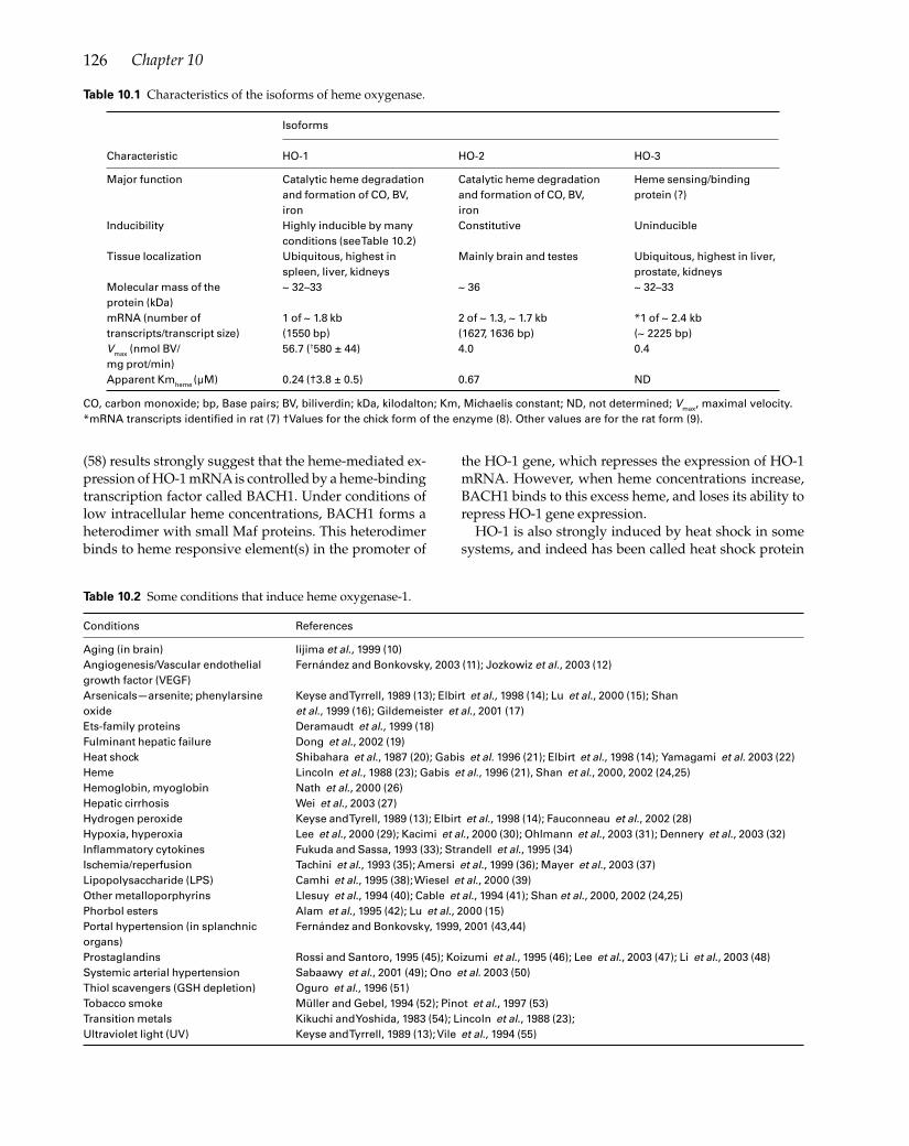
126 Chapter 10
(58) results strongly suggest that the heme-mediated ex-pression of HO-1 mRNA is controlled by a heme-binding transcription factor called BACH1. Under conditions of low intracellular heme concentrations, BACH1 forms a heterodimer with small Maf proteins. This heterodimer binds to heme responsive element(s) in the promoter of
the HO-1 gene, which represses the expression of HO-1 mRNA. However, when heme concentrations increase, BACH1 binds to this excess heme, and loses its ability to repress HO-1 gene expression.
HO-1 is also strongly induced by heat shock in some systems, and indeed has been called heat shock protein
Isoforms
Characteristic HO-1 HO-2 HO-3
Major function Catalytic heme degradation Catalytic heme degradation Heme sensing/binding and formation of CO, BV, and formation of CO, BV, protein (?) iron ironInducibility Highly inducible by many Constitutive Uninducible conditions (see Table 10.2)Tissue localization Ubiquitous, highest in Mainly brain and testes Ubiquitous, highest in liver, spleen, liver, kidneys prostate, kidneysMolecular mass of the ~ 32–33 ~ 36 ~ 32–33protein (kDa)mRNA (number of 1 of ~ 1.8 kb 2 of ~ 1.3, ~ 1.7 kb *1 of ~ 2.4 kbtranscripts/transcript size) (1550 bp) (1627, 1636 bp) (~ 2225 bp)Vmax (nmol BV/ 56.7 (†580 ± 44) 4.0 0.4mg prot/min)Apparent Kmheme (µM) 0.24 (†3.8 ± 0.5) 0.67 ND
Table 10.1 Characteristics of the isoforms of heme oxygenase.
CO, carbon monoxide; bp, Base pairs; BV, biliverdin; kDa, kilodalton; Km, Michaelis constant; ND, not determined; Vmax, maximal velocity. *mRNA transcripts identifi ed in rat (7) †Values for the chick form of the enzyme (8). Other values are for the rat form (9).
Conditions References
Aging (in brain) Iijima et al., 1999 (10)Angiogenesis/Vascular endothelial Fernández and Bonkovsky, 2003 (11); Jozkowiz et al., 2003 (12)growth factor (VEGF) Arsenicals—arsenite; phenylarsine Keyse and Tyrrell, 1989 (13); Elbirt et al., 1998 (14); Lu et al., 2000 (15); Shan oxide et al., 1999 (16); Gildemeister et al., 2001 (17)Ets-family proteins Deramaudt et al., 1999 (18)Fulminant hepatic failure Dong et al., 2002 (19)Heat shock Shibahara et al., 1987 (20); Gabis et al. 1996 (21); Elbirt et al., 1998 (14); Yamagami et al. 2003 (22)Heme Lincoln et al., 1988 (23); Gabis et al., 1996 (21), Shan et al., 2000, 2002 (24,25)Hemoglobin, myoglobin Nath et al., 2000 (26)Hepatic cirrhosis Wei et al., 2003 (27)Hydrogen peroxide Keyse and Tyrell, 1989 (13); Elbirt et al., 1998 (14); Fauconneau et al., 2002 (28)Hypoxia, hyperoxia Lee et al., 2000 (29); Kacimi et al., 2000 (30); Ohlmann et al., 2003 (31); Dennery et al., 2003 (32)Infl ammatory cytokines Fukuda and Sassa, 1993 (33); Strandell et al., 1995 (34)Ischemia/reperfusion Tachini et al., 1993 (35); Amersi et al., 1999 (36); Mayer et al., 2003 (37)Lipopolysaccharide (LPS) Camhi et al., 1995 (38); Wiesel et al., 2000 (39)Other metalloporphyrins Llesuy et al., 1994 (40); Cable et al., 1994 (41); Shan et al., 2000, 2002 (24,25)Phorbol esters Alam et al., 1995 (42); Lu et al., 2000 (15)Portal hypertension (in splanchnic Fernández and Bonkovsky, 1999, 2001 (43,44)organs) Prostaglandins Rossi and Santoro, 1995 (45); Koizumi et al., 1995 (46); Lee et al., 2003 (47); Li et al., 2003 (48)Systemic arterial hypertension Sabaawy et al., 2001 (49); Ono et al. 2003 (50)Thiol scavengers (GSH depletion) Oguro et al., 1996 (51)Tobacco smoke Müller and Gebel, 1994 (52); Pinot et al., 1997 (53)Transition metals Kikuchi and Yoshida, 1983 (54); Lincoln et al., 1988 (23); Ultraviolet light (UV) Keyse and Tyrrell, 1989 (13); Vile et al., 1994 (55)
Table 10.2 Some conditions that induce heme oxygenase-1.

127Carbon Monoxide and the Heme Oxygenase System in Cirrhosis
32 (HSP32), refl ecting its molecular weight of 32 kDa (13). The human HO-1 gene, known as HMOX1, is located on chromosome 22 and consists of 5 exons and 4 introns (59). Of the three isoforms, HO-1 is the most catalytically ac-tive (See Table 10.1 for Vmax and Km values).
Yachie et al. described a patient with an inborn defi ciency of HO-1 (60). The patient was 26 months old when he fi rst presented with recurrent fever, a generalized erythema-tous rash, growth retardation, and marked hepatome-galy. Analysis of the patient’s HO-1 gene indicated a loss of exon 2 from the maternal allele and a two-nucleotide deletion in exon 3 of the paternal allele. Both parents were phenotypically normal. A lymphoblastoid cell line derived from the patient was overly sensitive to heme-in-duced cell injury. Unfortunately, the patient died at age 6 years. Autopsy showed increased iron levels in the liver and kidney, and amyloid deposits in the liver and adrenal glands (61). Overall, this patient showed a more severe in-volvement of endothelial cells and the reticuloendothelial system than was seen in mice with a targeted disruption of the HO-1 gene, where disruption of iron metabolism is the predominant fi nding (62,63). These HO-1 knockout mice were highly sensitive to hepatic injury by iron, implying an important role of this enzyme in iron homeostasis, as well as its important metabolic and cytoprotective roles.
In contrast to the highly inducible HO-1, HO-2 appears to be a constitutive enzyme that is found mostly in the brain and testes (Table 10.1). The human HO-2 gene, known as HMOX2, is located on chromosome 16, and also consists of 5 exons and 4 introns (59). Although no human examples of HO-2 defi ciency have been reported, HO-2 knockout mice have been described (64). These mice exhibited nor-mal hippocampal long-term potentiation, but showed less responsiveness to electrical stimulation, consistent with a role for CO, generated by HO-2, in the modulation of non-adrenergic, noncholinergic relaxation of myenteric gan-glia. These HO-2 knockout mice also exhibited decreased ejaculatory muscle refl ex activity, suggesting that CO plays a role in male reproductive behavior (65). When exposed to hyperoxia, these mice showed increased toxicity, probably due to increased pulmonary levels of iron (66). CO gener-ated by HO-2 has been shown to be an endogenous hyper-polarizing factor in the gastrointestinal tract, as evidenced by the fi ndings that HO-2 knockout mice had depolarized smooth muscle cells and the membrane potential gradient in the gut was abolished (67). Thus, CO probably plays an important role in normal gut function.
A third isoform of heme oxygenase, referred to as HO-3, has been described (7). A cDNA, isolated from rat brain, corresponded to a new protein that was closely related to HO-2 (approximately 90% amino acid sequence similar-ity). When expressed in Escherichia coli, the HO-3 protein did not cross-react with polyclonal antibodies to HO-1 or HO-2. It was a relatively poor catalyst for heme degrada-tion (see Vmax and Km values in Table 10.1) and produced
spectra consistent with being a hemoprotein. In a subse-quent study, HO-3 was found to be mainly expressed in rat hippocampus, cerebellum, and cortex (68). HO-3 was also expressed in primary cultures of rat type 1 astrocytes. The precise function of HO-3 is unclear, but, given the relative-ly low ability of this protein to catalyze the degradation of heme, it is likely that it is involved in heme sensing or heme binding. A BLAST search (in January 2005) of the rat HO-3 amino acid sequence showed that its closest match was to mouse HO-2, but no sequences designated as HO-3 were found for any species besides the rat.
The role of heme oxygenase is to catalytically convert heme into equimolar amounts of CO, biliverdin, and fer-rous iron, but this function can be viewed from two dis-tinct, but related, perspectives (69). The fi rst view is to consider heme oxygenase as one of the means for control-ling the intracellular concentrations of heme, specifi cally the small, and rapidly turning over, “regulatory” heme pool that is in dynamic equilibrium with the heme that is incorporated into hemoproteins. This regulation is done in coordination with 5-aminolevulinate synthase (ALAS), the rate-controlling enzyme of heme synthesis. When the rate of synthesis of heme is balanced with the rate of deg-radation of heme (controlled by heme oxygenase), the size of the “regulatory” heme pool is maintained. If the intracellular concentration of heme increases, then HO-1 is induced and ALAS is repressed, until the level of heme returns to normal. The removal of excess heme from the cell is a critical process, because elevated levels of heme are potentially very toxic to cells. However, as described above, induction of HO-1 occurs at lower, relatively non-toxic concentrations of heme.
A second way to view the function of heme oxygenase is from the point of view of generating physiologically im-portant products, in this case, CO, biliverdin, and ferrous iron. CO has structural and biological properties that are similar to nitric oxide (NO), which is now known to be central to a number of signaling pathways and functions (70). Both CO and NO can activate guanylate cyclase, and thus increase the levels of cyclic GMP, resulting in relaxa-tion of smooth muscle (71). When this relaxation occurs in blood vessels, vasodilation results with a correspond-ing lowering of blood pressure. Relaxation of smooth muscle in the gastrointestinal tract causes relaxation of sphincters and decreased peristaltic activity. However, NO is 50–100 times more powerful as a guanylate cyclase activator than is CO. Thus, when both NO and CO pro-ductions are intact, NO effects predominate. CO also has effects that are independent of guanylate cyclase, includ-ing its ability to bind tightly to hemoproteins. For exam-ple, the binding of CO to a cytochrome P450-dependent epoxygenase results in its inhibition, with a subsequent decrease in the contractility of bile canaliculi (72). CO also affects the central nervous system, causing reductions in systemic blood pressure and production of neuronal

128 Chapter 10
adaptation (73) and retrograde potentiation (74), and the enteric nervous system, as already described.
Biliverdin is a potent antioxidant, as is bilirubin, which is made from biliverdin in most mammals by the action of biliverdin reductase (75,76). The concentrations of these two products of heme degradation are usually suffi -ciently high to act as defenses against oxidative stress-in-duced injury to mammalian tissues. Their sites of action, however, are probably determined by their solubilities. Because unconjugated bilirubin is quite soluble in lipids, it is most effective in biological membranes. Biliverdin is more water soluble and thus more likely to inhibit oxida-tive damage in more aqueous environments.
In contrast to the antioxidant effects of biliverdin and bilirubin, the release of ferrous iron by the heme oxyge-nase-mediated degradation of heme might result in in-creased oxidative stress to the cells and tissues, because this iron can potentially act to catalyze Fenton-type chem-istry with the production of highly toxic reactive oxygen species (77–80). The increase in intracellular iron also acts to modulate the expression levels of a number of mRNA species, including the divalent cation transporter (DCT-1), ferroportin (IREG), ferritin, transferrin receptor-1, and the erythroid form of ALAS. Such changes, specifi cally the upregulation of ferritin and the downregulation of transferrin-1 receptors, tend to limit any further increase in iron concentrations, and thus to restrict any further damage due to iron toxicity.
Heme oxygenase in hepatic cirrhosis and portal hypertension
In the last few years, evidence has accumulated show-ing that the HO enzymes and their by-products are im-portant players in the pathophysiology of cirrhosis and portal hypertension. For example, increased hepatic HO activity and/or expression of HO-1 mRNA and protein
SO SO SO SOPH PH PH PH
Mesentery
Intestine
ColonSpleen
524 bp HO-1
437 bp HO-2
309 bp GAPDH
0
0.1
0.2
0
0.5
1
Mesentery Intestine Liver Spleen
Hem
e ox
ygen
ase
activ
ity(n
mol
bilir
ubin
l. min
-1. m
g pr
otei
n-1 )
Hem
e ox
ygen
ase
activ
ity(n
mol
bili
rubi
n.m
in-1
. mg
prot
ein-
1 )
*
*
*
*
Figure 10.2 Expression of HO-1 and HO-2 mRNAs in splanchnic organs from portal hypertensive (PH) rats and sham-operated (SO) control animals. Glyceraldehyde-3-phosphate dehydrogenase (GAPDH) mRNA expression is shown as a control. Representative of four reverse transcription-polymerase chain reaction (RT-PCR) analyses. (Reproduced with permission from ref. (43).)
Figure 10.3 HO enzymatic activity (nmol bilirubin/min/mg protein) in microsomal fractions obtained from the mesentery, intestine, liver, and spleen of portal hypertensive rats (hatched bars) and sham-operated control animals (open bars). Data are shown as mean ± SEM of four to fi ve assays. *P < 0.05 vs. sham-operated rats. (Reproduced with permission from ref. (44).)
have recently been described in experimental (27) and human hepatic cirrhosis (81,82). Recent data, derived from our studies, indicate that the levels of both the mRNA and protein expression of the inducible HO-1 isoform are increased in the liver and splanchnic organs from rats with portal hypertension due to portal vein con-striction, compared with sham-operated control animals (Fig. 10.2) (43). In contrast, the constitutive HO-2 isoform is expressed at similar levels in the liver and splanchnic organs from both experimental groups (Fig. 10.2) (43). In addition, the observed increase in HO-1 protein levels results in a higher HO enzymatic activity in splanchnic organs from portal hypertensive rats (Fig. 10.3) (44). In

129Carbon Monoxide and the Heme Oxygenase System in Cirrhosis
rats subjected to bile duct ligation (83), there was a pro-gressively increased expression of HO-1 in aorta and mesenteric arteries, compared with sham operated rats. This increase in HO-1 expression coincided with the pro-gression from acute cholestatic liver injury (week 1) to fully developed cirrhosis with intense systemic arterial vasodilation (week 4). Again, no change in the expres-sion of HO-2 was observed. HO-1 over-expression has also been found in the lungs of portal hypertensive rats (84), and in the liver (27) and cardiac ventricles (85) from rats with secondary biliary cirrhosis. In addition, recent-ly it was shown that cirrhotic patients have an increased production of CO, a product of the HO reaction, which increases further in the presence of a severe bacterial in-fection, such as spontaneous bacterial peritonitis (86).
Taken together, these results suggest the importance of HO-1 in the pathophysiology of portal hypertension and shed light on new avenues for further investigations and development of new therapeutic applications. The precise mechanisms whereby HO-1 gene expression is upregulated in portal hypertension and liver cirrhosis are still unknown. As already described, HO-1 is tran-scriptionally activated by numerous physical or chemical factors (Table 10.2), some of which may be increased dur-ing portal hypertension, including cytokines, endotoxin, and shear stress. In addition, the pathophysiological sig-nifi cance of HO-1 induction in portal hypertension is not yet fully understood. Some of the potential roles of HO-1 in portal hypertension and liver injury are discussed below.
Heme oxygenase and splanchnic arteriolar vasodilation
The portal hypertensive syndrome is characterized by in-creased portal blood fl ow, which is the result of a marked arteriolar vasodilation in splanchnic organs draining into the portal vein (87). Although the mechanisms underly-ing this splanchnic vasodilation are not fully understood, previous studies have suggested that this could be due, at least in part, to an overproduction of endogenous vasodi-lators, and to a decreased vascular reactivity to vasocon-strictors. Several lines of experimental evidence indicate that the vasodilator NO (70) plays an important role in the pathogenesis of these hemodynamic disturbances ((88–92) and see Chapter 8).
CO, a product of HO activity, shares many properties with NO. Both CO and NO are involved in the homeo-static control of cardiovascular tone and in the modula-tion of blood vessel function, including their relaxation and inhibition of platelet aggregation (93). As described above, both NO and CO enhance the activity of soluble guanylate cyclase, leading to a several-fold increase in the production of cyclic guanosine monophosphate (cGMP), although the potency of CO in this regard is about 50–100
times lower than that of NO (94). Other mechanisms of action of CO, independent of cGMP, are also possible, in-cluding stimulation of calcium-activated potassium (KCa) channels with subsequent hyperpolarization of smooth muscle cells, inhibition of a cytochrome P450-dependent monooxygenase system, and/or inhibition of endothelin release from endothelial cells (94).
Given the similarities between the chemical struc-tures and biological actions of CO and NO, it is likely that both vasoactive systems contribute to the circulato-ry abnormalities of portal hypertension. In this regard, we have recently demonstrated that HO-1 plays a role in modulating mesenteric vascular reactivity in portal hypertensive rats (44). Thus, in perfused mesenteric vascular beds, in the absence of NO synthase inhibition, the HO inhibitor zinc mesoporphyrin (ZnMP) had no effect on hyporeactivity to the vasoconstrictor potassi-um chloride (a depolarizing agent) (Fig. 10.4). Vascular hyporeactivity to potassium chloride was only partially corrected by a NO synthase inhibitor (L-NAME), but it was completely reversed by a combination of ZnMP and L-NAME. In addition, the magnitude of the effect of simultaneous perfusion with ZnMP plus L-NAME on pressure response to potassium chloride was signifi -cantly higher than the addition of the effects of ZnMP and L-NAME when perfused alone. This fi nding could suggest the existence of a synergistic regulatory inter-action between the HO and NO synthase systems in mediating the splanchnic hyporeactivity to potassium chloride in portal hypertensive rats. Because ZnMP had no effects in normal animals, the consequences of HO inhibition on vascular reactivity were selective for portal hypertensive rats. When we assessed pressure responses to the vasoconstrictor methoxamine (an α1-adrenergic agonist), we found that the hyporeactivity to methoxamine was unaffected by HO inhibition, but it was completely overcome by NO synthase blockade (44). The effect of simultaneous inhibition of both HO and NO synthase on pressure response to methoxam-ine was not signifi cantly different from that obtained after blockade of only NO synthase, indicating that HO plays little, if any, role in modulating vasoconstrictive responses to methoxamine in mesenteric vessels of portal hypertensive rats. Taken together, these fi ndings indicate that the mesenteric hyporeactivity to α1-adren-ergic stimulation in portal hypertensive rats is mediated mainly by NO synthase, whereas membrane depolari-zation is synergistically regulated by both NO synthase and HO. These studies also suggest that alterations in the vasoconstriction stimulated by both membrane de-polarization and α1-adrenergic activation contribute to the decreased vascular reactivity in portal hyperten-sion. Abnormal depolarizations have been previously observed under some pathological conditions, such as arterial hypertension (95), and hypoxia (96).

130 Chapter 10
0
50
100
150
200
250
BeforeZnMP + L-NAME
PH rats, n=5SO rats, n=4
***
###@
BeforeZnMP + L-NAME
AfterZnMP + L-NAME
AfterZnMP + L-NAME
0
50
100
150
200
250
BeforeL-NAME
PH rats, n=8SO rats, n=4
**
##&
BeforeL-NAME
AfterL-NAME
AfterL-NAME
0
50
100
150
200
250
BeforeZnMP
PH rats, n=5SO rats, n=4
*#
Pre
ssur
e re
spon
se t
o K
Cl
(mm
Hg)
BeforeZnMP
AfterZnMP
AfterZnMP
Pre
ssur
e re
spon
se t
o K
Cl
(mm
Hg)
Pre
ssur
e re
spon
se t
o K
Cl
(mm
Hg)
a.
b.
c.
It should be noted that depolarizing agents cause con-traction of vascular smooth muscle cells by activation of voltage-dependent calcium channels, resulting in calci-um entry to the cytoplasm from the extracellular space (97). α1-Adrenergic agonists induce vasoconstriction by generation of inositol triphosphate and calcium release from the endoplasmic reticulum (97). In mesenteric ves-sels, this α1-adrenergic-stimulated vasoconstriction is independent of changes in membrane potential (98). Therefore, it is conceivable that there might be distinct mechanisms underlying the hyporeactivity to vasocon-strictors in portal hypertension.
The role of HO in modulating mesenteric vascular reactivity in portal hypertension could be related to the fact that this enzymatic system is responsible for the en-dogenous production of the powerful vasodilator, CO
(94,99). Because, as already mentioned, NO is a more po-tent activator of soluble guanylate cyclase than CO (94), when NO synthase and HO are both active, guanylate cyclase activation is mainly caused by NO. Therefore, it is not surprising that in our studies the effect of HO inhibi-tion on vascular tone was detectable only after inhibition of NO synthase (44). Because CO-induced smooth mus-cle cell relaxation occurs only if CO is produced close to its target (i.e. soluble guanylate cyclase) (94), it is likely that the upregulated HO-1 involved in modulation of mesenteric vascular reactivity in portal hypertension is located within arterial walls (i.e. in the endothelium or smooth muscle or both) (43).
Rats treated with carbon tetrachloride to produce a chemically-induced cirrhosis had decreased perfusion pressure and lessened response of the superior mesenter-
Figure 10.4 Pressure response to potassium chloride (KCl; mmHg) in mesenteric vascular beds of portal hypertensive (PH) rats and sham-operated (SO) control animals, before (open bars) and after (hatched bars) perfusion with 10 µM ZnMP alone (a), 100 µM L-NAME alone (b), or 10 µM ZnMP plus 100 µM L-NAME (c). Data are shown as mean ± SEM. *P < 0.01 vs. SO rats before ZnMP; #P < 0.01 vs. SO rats after ZnMP; **P < 0.01 vs. SO rats before L-NAME; ##P < 0.05 vs. SO rats after L-NAME; &P < 0.05 vs. PH rats before L-NAME; ***P < 0.01 vs. SO rats before ZnMP plus L-NAME; @P < 0.01 vs. PH rats before ZnMP plus L-NAME; ###P < 0.05 vs. PH rats after L-NAME. (Reproduced with permission from Ref. (44).) L-NAME, Nitro-L-arginine methyl ester, an inhibitor of nitric oxide synthase; PH, portal hypertensive; SO, sham operated; ZnMP, zinc mesoporphyrin, an inhibitor of heme oxygenase.
(a)
(b)
(c)

131Carbon Monoxide and the Heme Oxygenase System in Cirrhosis
ic vein to several vasoconstrictors (KCl, phenylephrine, and endothelin-1), compared with untreated rats (100). Treatment of the cirrhotic rats with tin mesoporphyrin (to inhibit HO activity) increased perfusion pressure, and partially restored the constrictor responses to KCl, phenylephrine, and endothelin-1. Stepwise inhibition of CO and NO production resulted in a successive increase in response to vasoconstriction, suggesting that both CO and NO regulate vascular tone in the superior mesenteric vein. In contrast to previous studies (43,44), the level of HO-2 protein was increased, whereas the level of HO-1 protein was unchanged. Rats transfected with an adeno-virus containing the human HO-1 gene showed reduced perfusion pressure and lower vasoconstrictor response to phenylephrine. These results support the idea that HO is a critical component in the mechanisms that control splanchnic resistance in carbon tetrachloride-induced cirrhosis in rats.
Heme oxygenase and increased intrahepatic vascular resistance
Vascular resistance is increased in the cirrhotic liver. This increase formerly was thought to be a fi xed, mechanical consequence of the distortion of the hepatic vascular architecture caused by fi brosis, scarring, and nodule formation (87). However, there is increasing evidence that the vascular resistance of the cirrhotic liver can be modifi ed by vasoactive agents, which modulate the responsiveness of several contractile elements located within the hepatic vascular bed, both at the sinusoidal level and at extrasinusoidal sites (101). In this regard, it has long been recognized that the liver ’s vasculature includes vessels with abundant vascular smooth mus-cle cells, which are therefore contractile (e.g. small por-tal venules in portal areas). In addition, hepatic stellate cells are located in the perisinusoidal space of Disse, in close contact with hepatocytes, endothelial cells, and Kupffer cells. These nonparenchymal cells contain contractile proteins, and have the ability to relax in re-sponse to locally produced vasodilators, such as NO, and to contract in response to vasoconstrictors, such as endothelin (101). An imbalance between these vasoac-tive molecules may result in a sustained contraction of stellate cells, and subsequent constriction of sinusoids, which could be partly responsible for the increased he-patic vascular resistance in cirrhosis. In this regard, it has been shown that, in the injured liver, endothelin-1 production is increased (101), and NO synthesis is re-duced (92).
Physiologically, in normal livers, HO is also involved in modulation of hepatic sinusoidal tone and portal venous regulation (72). These effects are thought to be secondary to the generation of the vasodilator CO from the consti-tutive HO-2 isoform, activation of soluble guanylate cycl-
ase, and subsequent production of cGMP, similar to that of NO. HO-2 is expressed mainly in parenchymal cells, but also in Kupffer cells and hepatic stellate cells (43). In portal hypertension and cirrhosis, the expression and enzymatic activity of HO-1 are increased signifi cantly in the liver, specifi cally in hepatocytes and Kupffer cells (43,44,81,82). Under these pathological situations, CO overproduction due to HO-1 induction could be neces-sary to improve sinusoidal perfusion and to protect liver homeostasis (72). The increased expression of HO-1 in the cirrhotic liver may also represent a refi ned intracel-lular system to control NO synthase activity, because in-creased HO-1 activity may limit the availability of heme, both a substrate for HO and an important cofactor for NO synthase (93). In addition, HO-1 has been shown to be protective in several models of liver injury, including he-patic porphyria (102), acetaminophen-induced hepatitis (103), hepatic ischemia-reperfusion injury (36), hemor-rhagic shock (104), endotoxin-mediated hepatic dysfunc-tion (105), and liver transplantation (69,106).
Heme oxygenase and oxidative stress
HO-1 is a stress-inducible enzyme, which can be ex-pressed and upregulated in virtually all cells facing con-tact with noxious stimuli. Hence, HO-1 induction can be regarded as a general response to oxidant stress. First, HO enzymatically breaks down heme, thereby mitigat-ing the hazardous cellular effects of this pro-oxidant (2). Second, biliverdin and its reduced product bilirubin are both potent free radical scavengers with antioxidant and anti-infl ammatory properties (75,76). Third, iron released from heme enhances the synthesis of ferritin, which addi-tionally has antioxidant capabilities (107). Fourth, HOs, or at least HO-1, prevent free iron accumulation in the cells, not only indirectly by stimulating ferritin produc-tion, but also directly by extruding iron outside the cell (108). In this activity, HO-1 cooperates with a recently identifi ed Fe-ATP pump (109). An indication for a role of HO-1 in iron extrusion is the accumulation of iron in the cells of HO-1 knockout mice (62,63), or in the liver and kidney of a boy who lacked a functional HO-1 gene (60). Accordingly, gene transfer of HO-1 to cells derived from HO-1 knockout animals restored the cells’ capability to control their cellular iron level (108). In cells of the car-diovascular system, CO inhibits infl ammatory response, infl uencing synthesis of cytokines, and cell proliferation, and preventing cell apoptosis. Those effects are mediated through both cGMP-dependent and cGMP-independent mechanisms (110,111).
Heme oxygenase and angiogenesis
Recent data indicate the involvement of HO-1 in the regulation of angiogenesis. Thus, over-expression of

132 Chapter 10
HO activity by HO-1 gene transfer into endothelial cells causes a signifi cant increase in cell proliferation and capillary formation (112). In addition, HO-1 activity modulates the production of the potent angiogenic fac-tor vascular endothelial growth factor (VEGF) (12,113). More recently, we have demonstrated, using an in vivo model of angiogenesis in chick chorioallantoic mem-branes, that VEGF increases the expression of HO-1 protein by a mechanism dependent on an increase in cytosolic calcium levels and activation of protein kinase C (11). Moreover, we have shown that HO-1 activity is involved in the signaling pathway(s) by which VEGF stimulates in vivo angiogenesis in chorioallantoic mem-branes, because this effect is markedly attenuated by the HO inhibitor ZnMP (11).
Previous studies had found evidence of increased an-giogenesis and VEGF over-expression in the abdominal cavities of portal hypertensive rats and cirrhotic patients (114–119). The pathophysiological signifi cance of this neovascularization is not well understood, but it could be involved in the formation of portosystemic collateral circulation and/or in the increased splanchnic blood fl ow during portal hypertension (87). Therefore, the pos-sibility exists that HO-1 over-expression could play a role modulating angiogenic processes in portal hyperten-sion. This is a new and fascinating area for investigation, which, by incorporating advances in angiogenesis and anti-angiogenic factors, may lead to novel approaches for therapy of portal hypertension.
Summary
In summary, HOs are a family of heme-degrading en-zymes with critical and primordial roles in protecting against oxidative and other stress and against exces-sive and inappropriate levels of intracellular heme. In addition, the products of the HO reaction, namely, CO, biliverdin (bilirubin), and iron have numerous and im-portant physiological functions. Emerging evidence points to the importance of HO and these products in the pathophysiology of cirrhosis and portal hypertension, probably chiefl y serving to abrogate adverse hemody-namic effects. Indeed, evidence continues to accumulate that upregulation of and/or gene therapy with HO-1 may decrease ischemia-reperfusion injury, not only in the liver, but also in other parenchymal organs. Seen in this light, HO and its products deserve additional study and will one day probably fi nd a place in our therapeutic armamentarium.
References
1 Maines MD. The heme oxygenase system: a regulator of second messenger gases. Ann Rev Pharmacol Toxicol 1997;
37:517–54. 2 Elbirt KK, Bonkovsky HL. Heme oxygenase: recent
advances in understanding its regulation and role. Proc Assoc Am Physicians 1999; 111:438–47.
3 Bonkovsky HL, Elbirt KK. Heme oxygenase: its regulation and role. In: Cutler R, Rodriguez H, eds. Oxidative Stress and Aging: Advances in Basic Science, Diagnostics, and Intervention. River Edge, NJ: World Scientifi c, 2002; 690–706.
4 Tenhunen R, Matsumi M, Schmid R. The enzymatic conversion of heme to bilirubin by microsomal heme oxygenase. Proc Natl Acad Sci USA 1968; 61:748–55.
5 McCoubrey WK Jr, Maines MD. The structure, organization and differential expression of the gene encoding rat heme oxygenase-2. Gene 1994; 139:155–61.
6 Shibahara S, Yoshizawa M, Suzuki H et al. Functional analysis of cDNAs for two types of human heme oxygenase and evidence for their separate regulation. J Biochem (Tokyo) 1993; 113:214–18.
7 McCoubrey WK Jr, Huang TJ, Maines MD. Isolation and characterization of a cDNA from the rat brain that encodes hemoprotein heme oxygenase-3. Eur J Biochem 1997; 247:725–32.
8 Bonkovsky HL, Healey JF, Pohl J. Purifi cation and characterization of heme oxygenase from chick liver. Comparison of the avian and mammalian enzymes. Eur J Biochem 1990; 189:155–66.
9 Maines MD, Trakshel GM, Kutty RK. Characterization of two constitutive forms of rat liver microsomal heme oxygenase. Only one molecular species of the enzyme is inducible. J Biol Chem 1986; 261:411–19.
10 Iijima N, Tamada Y, Hayashi S et al. Expanded expression of heme oxygenase-1 (HO-1) in the hypothalamic median eminence of aged as compared with young rats: an immunocytochemical study. Neurosci Lett 1999; 271:113–16.
11 Fernández M, Bonkovsky HL. Vascular endothelial growth factor increases heme oxygenase-1 protein expression in the chick embryo chorioallantoic membrane. Br J Pharmacol 2003; 139:634–40.
12 Jozkowicz A, Huk I, Nigisch A et al. Heme oxygenase and angiogenic activity of endothelial cells: stimulation by carbon monoxide and inhibition by tin protoporphyrin-IX. Antioxid Redox Signal 2003; 5:155–62.
13 Keyse SM, Tyrrell RM. Heme oxygenase is the major 32-kDa stress protein induced in human skin fi broblasts by UVA radiation, hydrogen peroxide, and sodium arsenite. Proc Natl Acad Sci USA 1989; 86:99–103.
14 Elbirt KK, Whitmarsh AJ, Davis RJ, Bonkovsky HL. Mechanism of sodium arsenite-mediated induction of heme oxygenase-1 in hepatoma cells. Role of mitogen-activated protein kinases. J Biol Chem 1998; 273:8922–31.
15 Lu TH, Shan Y, Pepe J et al. Upstream regulatory elements in chick heme oxygenase-1 promoter: a study in primary cultures of chick embryo liver cells. Mol Cell Biochem 2000; 209:17–27.
16 Shan Y, Lambrecht RW, Hong LT, Bonkovsky HL. Effects of phenylarsine oxide on expression of heme oxygenase-1 reporter constructs in transiently transfected cultures

133Carbon Monoxide and the Heme Oxygenase System in Cirrhosis
of chick embryo liver cells. Arch Biochem Biophys 1999; 372:224–9.
17 Gildemeister OS, Pepe JA, Lambrecht RW, Bonkovsky HL. Induction of heme oxygenase-1 by phenylarsine oxide. Studies in cultured primary liver cells. Mol Cell Biochem 2001; 226:17–26.
18 Deramaudt BM, Remy P, Abraham NG. Upregulation of human heme oxygenase gene expression by Ets-family proteins. J Cell Biochem 1999; 72:311–21.
19 Dong H, Toyoda N, Yoneyama H et al. Gene expression profi le analysis of the mouse liver during bacteria-induced fulminant hepatitis by a cDNA microarray system. Biochem Biophys Res Commun 2002; 298:675–86.
20 Shibahara S, Muller RM, Taguchi H. Transcriptional control of rat heme oxygenase by heat shock. J Biol Chem 1987; 262:12889–92.
21 Gabis KK, Gildemeister OS, Pepe JA et al. Induction of heme oxygenase-1 in LMH cells. Comparison of LMH cells to primary cultures of chick embryo liver cells. Biochim Biophys Acta 1996; 1290:113–20.
22 Yamagami K, Enders G, Schauer RJ et al. Heat-shock preconditioning protects fatty livers in genetically obese Zucker rats from microvascular perfusion failure after ischemia reperfusion. Transpl Int 2003; 16:456–63.
23 Lincoln BC, Healey JF, Bonkovsky HL. Regulation of hepatic haem metabolism. Disparate mechanisms of induction of haem oxygenase by drugs and metals. Biochem J 1988; 250:189–96.
24 Shan Y, Pepe J, Lu TH et al. Induction of the heme oxygenase-1 gene by metalloporphyrins. Arch Biochem Biophys 2000; 380:219–27.
25 Shan Y, Pepe J, Lambrecht RW, Bonkovsky HL. Mapping of the chick heme oxygenase-1 proximal promoter for responsiveness to metalloporphyrins. Arch Biochem Biophys 2002; 399:159–66.
26 Nath KA, Haggard JJ, Croatt AJ et al. The indispensability of heme oxygenase-1 in protecting against acute heme protein-induced toxicity in vivo. Am J Pathol 2000; 156:1527–35.
27 Wei CL, Lee KH, Khoo HE, Hon WM. Expression of haem oxygenase in cirrhotic rat liver. J Pathol 2003; 199:324–34.
28 Fauconneau B, Petegnief V, Sanfeliu C et al. Induction of heat shock proteins (HSPs) by sodium arsenite in cultured astrocytes and reduction of hydrogen peroxide-induced cell death. J Neurochem 2002; 83:1338–48.
29 Lee PJ, Camhi SL, Chin BY et al. AP-1 and STAT mediate hyperoxia-induced gene transcription of heme oxygenase-1. Am J Physiol Lung Cell Mol Physiol 2000; 279:L175–L182.
30 Kacimi R, Chentoufi J, Honbo N et al. Hypoxia differentially regulates stress proteins in cultured cardiomyocytes: role of the p38 stress-activated kinase signaling cascade, and relation to cytoprotection. Cardiovasc Res 2000; 46:139–50.
31 Ohlmann A, Giffhorn-Katz S, Becker I et al. Regulation of heme oxygenase-1 gene expression by anoxia and reoxygenation in primary rat hepatocyte cultures. Exp Biol Med (Maywood) 2003; 228:584–9.
32 Dennery PA, Visner G, Weng YH et al. Resistance to
hyperoxia with heme oxygenase-1 disruption: role of iron. Free Radic Biol Med 2003; 34:124–33.
33 Fukuda Y, Sassa S. Effect of interleukin-11 on the levels of mRNAs encoding heme oxygenase and haptoglobin in human HepG2 hepatoma cells. Biochem Biophys Res Commun 1993; 193:297–302.
34 Strandell E, Buschard K, Saldeen J, Welsh N. Interleukin-1 beta induces the expression of hsp70, heme oxygenase and Mn-SOD in FACS-purifi ed rat islet beta-cells, but not in alpha-cells. Immunol Lett 1995; 48:145–8.
35 Tacchini L, Schiaffonati L, Pappalardo C et al. Expression of HSP 70, immediate-early response and heme oxygenase genes in ischemic-reperfused rat liver. Lab Invest 1993; 68:465–71.
36 Amersi F, Buelow R, Kato H et al. Upregulation of heme oxygenase-1 protects genetically fat Zucker rat livers from ischemia/reperfusion injury. J Clin Invest 1999; 104:1631–9.
37 Mayer RD, Wang X, Maines MD. Nitric oxide inhibitor N omega -nitro-l-arginine methyl ester potentiates induction of heme oxygenase-1 in kidney ischemia/reperfusion model: a novel mechanism for regulation of the oxygenase. J Pharmacol Exp Ther 2003; 306:43–50.
38 Camhi SL, Alam J, Otterbein L et al. Induction of heme oxygenase-1 gene expression by lipopolysaccharide is mediated by AP-1 activation. Am J Respir Cell Mol Biol 1995; 13:387–98.
39 Wiesel P, Foster LC, Pellacani A et al. Thioredoxin facil-itates the induction of heme oxygenase-1 in response to infl ammatory mediators. J Biol Chem 2000; 275:24840–6.
40 Llesuy SF, Tomaro ML. Heme oxygenase and oxidative stress. Evidence of involvement of bilirubin as physiological protector against oxidative damage. Biochim Biophys Acta 1994; 1223:9–14.
41 Cable EE, Pepe JA, Karamitsios NC et al. Differential effects of metalloporphyrins on messenger RNA levels of delta-aminolevulinate synthase and heme oxygenase. Studies in cultured chick embryo liver cells. J Clin Invest 1994; 94:649–54.
42 Alam J, Camhi S, Choi AM. Identifi cation of a second region upstream of the mouse heme oxygenase-1 gene that functions as a basal level and inducer-dependent transcription enhancer. J Biol Chem 1995; 270:11977–84.
43 Fernández M, Bonkovsky HL. Increased heme oxygenase-1 gene expression in liver cells and splanchnic organs from portal hypertensive rats. Hepatology 1999; 29:1672–9.
44 Fernández M, Lambrecht RW, Bonkovsky HL. Increased heme oxygenase activity in splanchnic organs from portal hypertensive rats: role in modulating mesenteric vascular reactivity. J Hepatol 2001; 34:812–17.
45 Rossi A, Santoro MG. Induction by prostaglandin A1 of haem oxygenase in myoblastic cells: an effect independent of expression of the 70 kDa heat shock protein. Biochem J 1995; 308 (Pt 2):455–63.
46 Koizumi T, Odani N, Okuyama T et al. Identifi cation of a cis-regulatory element for delta 12-prostaglandin J2-induced expression of the rat heme oxygenase gene. J Biol Chem 1995; 270:21779–84.
47 Lee TS, Tsai HL, Chau LY. Induction of heme oxygenase-

134 Chapter 10
1 expression in murine macrophages is essential for the anti-infl ammatory effect of low dose 15-deoxy-Delta 12,14-prostaglandin J2. J Biol Chem 2003; 278:19325–30.
48 Li L, Grenard P, Nhieu JT et al. Heme oxygenase-1 is an antifi brogenic protein in human hepatic myofi broblasts. Gastroenterology 2003; 125:460–9.
49 Sabaawy HE, Zhang F, Nguyen X et al. Human heme oxygenase-1 gene transfer lowers blood pressure and promotes growth in spontaneously hypertensive rats. Hypertension 2001; 38:210–15.
50 Ono K, Mannami T, Iwai N. Association of a promoter variant of the haeme oxygenase-1 gene with hypertension in women. J Hypertens 2003; 21:1497–503.
51 Oguro T, Hayashi M, Numazawa S et al. Heme oxygenase-1 gene expression by a glutathione depletor, phorone, mediated through AP-1 activation in rats. Biochem Biophys Res Commun 1996; 221:259–65.
52 Muller T, Gebel S. Heme oxygenase expression in Swiss 3T3 cells following exposure to aqueous cigarette smoke fractions. Carcinogenesis 1994; 15:67–72.
53 Pinot F, el Yaagoubi A, Christie P et al. Induction of stress proteins by tobacco smoke in human monocytes: modulation by antioxidants. Cell Stress Chaperones 1997; 2:156–61.
54 Kikuchi G, Yoshida T. Function and induction of the microsomal heme oxygenase. Mol Cell Biochem 1983; 53–54:163–83.
55 Vile GF, Basu-Modak S, Waltner C, Tyrrell RM. Heme oxygenase 1 mediates an adaptive response to oxidative stress in human skin fi broblasts. Proc Natl Acad Sci USA 1994; 91:2607–10.
56 Cable EE, Gildemeister OS, Pepe JA et al. Mechanism of induction of heme oxygenase by metalloporphyrins in primary chick embryo liver cells: evidence against a stress-mediated response. Mol Cell Biochem 1997; 169:13–20.
57 Ogawa K, Sun J, Taketani S et al. Heme mediates derepression of Maf recognition element through direct binding to transcription repressor Bach1. EMBO J 2001; 20:2835–43.
58 Shan Y, Lambrecht RW, Ghaziani T et al. Role of Bach-1 in regulation of heme oxygenase-1 in human liver cells: insights from studies with small interfering RNAs. J Biol Chem 2004; 279:51769–74.
59 Kutty RK, Kutty G, Rodríguez IR et al. Chromosomal localization of the human heme oxygenase genes—heme oxygenase-1 (Hmox1) maps to chromosome 22Q12 and heme oxygenase-2 (Hmox2) maps to chromosome 16P13.3. Genomics 1994; 20:513–16.
60 Yachie A, Niida Y, Wada T et al. Oxidative stress causes enhanced endothelial cell injury in human heme oxygenase-1 defi ciency. J Clin Invest 1999; 103:129–35.
61 Kawashima A, Oda Y, Yachie A et al. Heme oxygenase-1 defi ciency: the fi rst autopsy case. Hum Pathol 2002; 33:125–30.
62 Poss KD, Tonegawa S. Heme oxygenase 1 is required for mammalian iron reutilization. Proc Natl Acad Sci USA 1997; 94:10919–24.
63 Poss KD, Tonegawa S. Reduced stress defense in heme oxygenase 1-defi cient cells. Proc Natl Acad Sci USA 1997;
94:10925–30. 64 Poss KD, Thomas MJ, Ebralidze AK et al. Hippocampal
long-term potentiation is normal in heme oxygenase-2 mutant mice. Neuron 1995; 15:867–73.
65 Burnett AL, Johns DG, Kriegsfeld LJ et al. Ejaculatory abnormalities in mice with targeted disruption of the gene for heme oxygenase-2. Nat Med 1998; 4:84–7.
66 Dennery PA, Spitz DR, Yang G et al. Oxygen toxicity and iron accumulation in the lungs of mice lacking heme oxygenase-2. J Clin Invest 1998; 101:1001–11.
67 Farrugia G, Lei S, Lin X et al. A major role for carbon monoxide as an endogenous hyperpolarizing factor in the gastrointestinal tract. Proc Natl Acad Sci USA 2003; 100:8567–70.
68 Scapagnini G, D’Agata V, Calabrese V et al. Gene expression profi les of heme oxygenase isoforms in the rat brain. Brain Research 2002; 954:51–9.
69 Bauer M. Heme oxygenase in liver transplantation: heme catabolism and metabolites in the search of function. Hepatology 2003; 38:286–8.
70 Moncada S, Palmer RM, Higgs EA. Nitric oxide: physiology, pathophysiology, and pharmacology. Pharmacol Rev 1991; 43:109–42.
71 Villamor E, Perez-Vizcaino F, Cogolludo AL et al. Relaxant effects of carbon monoxide compared with nitric oxide in pulmonary and systemic vessels of newborn piglets. Pediatr Res 2000; 48:546–53.
72 Suematsu M, Ishimura Y. The heme oxygenase-carbon monoxide system: a regulator of hepatobiliary function. Hepatology 2000; 31:3–6.
73 Zufall F, Leinders-Zufall T. Identifi cation of a long-lasting form of odor adaptation that depends on the carbon monoxide cGMP second-messenger system. J Neurosci 1997; 17:2703–12.
74 Stevens CF, Wang Y. Reversal of long-term potentiation by inhibitors of haem oxygenase. Nature 1993; 364:147–9.
75 Stocker R, Yamamoto Y, McDonagh AF et al. Bilirubin is an antioxidant of possible physiological importance. Science 1987; 235:1043–6.
76 Vachharajani TJ, Work J, Issekutz AC, Granger DN. Heme oxygenase modulates selectin expression in different regional vascular beds. Am J Physiol Heart Circ Physiol 2000; 278:H1613–H1617.
77 Crichton RR, Wilmet S, Legssyer R, Ward RJ. Molecular and cellular mechanisms of iron homeostasis and toxicity in mammalian cells. J Inorg Biochem 2002; 91:9–18.
78 Bonkowsky HL, Healey JF, Sinclair PR et al. Iron and the liver. Acute and long-term effects of iron-loading on hepatic haem metabolism. Biochem J 1981; 196:57–64.
79 Bonkovsky HL, Lambrecht RW, Shan Y. Iron as a co-morbid factor in non-hemochromatotic liver disease. Alcohol 2003; 31:1–8.
80 Bonkovsky HL, Lambrecht RW. Iron-induced liver injury. Clin Liver Dis 2000; 4:409–29.
81 Makino N, Suematsu M, Sugiura Y et al. Altered expression of heme oxygenase-1 in the livers of patients with portal hypertensive diseases. Hepatology 2001; 33:32–42.
82 Matsumi M, Takahashi T, Fujii H et al. Increased heme oxygenase-1 gene expression in the livers of patients with

135Carbon Monoxide and the Heme Oxygenase System in Cirrhosis
portal hypertension due to severe hepatic cirrhosis. J Int Med Res 2002; 30:282–8.
83 Chen YC, Ginès P, Yang J et al. Increased vascular heme oxygenase-1 expression contributes to arterial vasodilation in experimental cirrhosis in rats. Hepatology 2004; 39:1075–87.
84 Schroeder RA, Ewing CA, Sitzmann JV, Kuo PC. Pulmonary expression of iNOS and HO-1 protein is upregulated in a rat model of prehepatic portal hypertension. Dig Dis Sci 2000; 45:2405–10.
85 Liu H, Song D, Lee SS. Role of heme oxygenase-carbon monoxide pathway in pathogenesis of cirrhotic cardiomyopathy in the rat. Am J Physiol Gastrointest Liver Physiol 2001; 280:G68–G74.
86 De las Heras D, Fernández J, Ginès P et al. Increased carbon monoxide production in patients with cirrhosis with and without spontaneous bacterial peritonitis. Hepatology 2003; 38:452–9.
87 Bosch J, Pizcueta P, Feu F et al. Pathophysiology of portal hypertension. Gastroenterol Clin North Am 1992; 21:1–14.
88 Pizcueta P, Piqué JM, Fernández M et al. Modulation of the hyperdynamic circulation of cirrhotic rats by nitric oxide inhibition. Gastroenterology 1992; 103:1909–15.
89 Sieber CC, Groszmann RJ. Nitric oxide mediates hypo-reactivity to vasopressors in mesenteric vessels of portal hypertensive rats. Gastroenterology 1992; 103:235–9.
90 Garcia-Pagan JC, Fernández M, Bernadich C et al. Effects of continued NO inhibition on portal hypertensive syndrome after portal vein stenosis in rat. Am J Physiol 1994; 267:G984–G990.
91 Fernández M, Garcia-Pagan JC, Casadevall M et al. Evidence against a role for inducible nitric oxide synthase in the hyperdynamic circulation of portal-hypertensive rats. Gastroenterology 1995; 108:1487–95.
92 Wiest R, Groszmann RJ. Nitric oxide and portal hypertension: its role in the regulation of intrahepatic and splanchnic vascular resistance. Semin Liver Dis 1999; 19:411–26.
93 Foresti R, Motterlini R. The heme oxygenase pathway and its interaction with nitric oxide in the control of cellular homeostasis. Free Radic Res 1999; 31:459–75.
94 Wang R, Wang Z, Wu L. Carbon monoxide-induced vasorelaxation and the underlying mechanisms. Br J Pharmacol 1997; 121:927–34.
95 Hermsmeyer K. Cellular basis for increased sensitivity of vascular smooth muscle in spontaneously hypertensive rats. Circ Res 1976; 38:53–7.
96 McMurtry IF, Davidson AB, Reeves JT, Grover RF. Inhibition of hypoxic pulmonary vasoconstriction by calcium antagonists in isolated rat lungs. Circ Res 1976; 38:99–104.
97 Peiper U, Griebel L, Wende W. Activation of vascular smooth muscle of rat aorta by noradrenaline and depolarization: two different mechanisms. Pfl ugers Arch 1971; 330:74–89.
98 Hussain MB, Marshall I. Alpha(1)-adrenoceptor subtypes mediating contractions of the rat mesenteric artery. Eur J Pharmacol 2000; 395:69–76.
99 Moreau R. Heme oxygenase: protective enzyme or portal
hypertensive molecule? J Hepatol 2001; 34:936–9.100 Sacerdoti D, Abraham NG, Oyekan AO et al. Role of the
heme oxygenases in abnormalities of the mesenteric circulation in cirrhotic rats. J Pharmacol Exp Ther 2004; 308:636–43.
101 Rockey D. The cellular pathogenesis of portal hypertension: stellate cell contractility, endothelin, and nitric oxide. Hepatology 1997; 25:2–5.
102 Stonard MD, Poli G, De Matteis F. Stimulation of liver heme oxygenase in hexachlorobenzene-induced hepatic porphyria. Arch Toxicol 1998; 72:355–61.
103 Chiu H, Brittingham JA, Laskin DL. Differential induction of heme oxygenase-1 in macrophages and hepatocytes during acetaminophen-induced hepatotoxicity in the rat: effects of hemin and biliverdin. Toxicol Appl Pharmacol 2002; 181:106–15.
104 Pannen BH, Kohler N, Hole B et al. Protective role of endogenous carbon monoxide in hepatic microcirculatory dysfunction after hemorrhagic shock in rats. J Clin Invest 1998; 102:1220–8.
105 Kyokane T, Norimizu S, Taniai H et al. Carbon monoxide from heme catabolism protects against hepatobiliary dysfunction in endotoxin-treated rat liver. Gastroenterology 2001; 120:1227–40.
106 Ke B, Shen XD, Buelow R et al. Heme oxygenase-1 gene transfer prevents CD95/FasL-mediated apoptosis and improves liver allograft survival via carbon monoxide signaling pathway. Transplant Proc 2002; 34:1465–6.
107 Balla G, Jacob HS, Balla J et al. Ferritin: a cytoprotective antioxidant strategem of endothelium. J Biol Chem 1992; 267:18148–53.
108 Ferris CD, Jaffrey SR, Sawa A et al. Haem oxygenase-1 prevents cell death by regulating cellular iron. Nat Cell Biol 1999; 1:152–7.
109 Baranano DE, Wolosker H, Bae BI et al. A mammalian iron ATPase induced by iron. J Biol Chem 2000; 275:15166–73.
110 Brouard S, Otterbein LE, Anrather J et al. Carbon monoxide generated by heme oxygenase 1 suppresses endothelial cell apoptosis. J Exp Med 2000; 192:1015–26.
111 Otterbein LE, Bach FH, Alam J et al. Carbon monoxide has anti-infl ammatory effects involving the mitogen-activated protein kinase pathway. Nat Med 2000; 6:422–8.
112 Deramaudt BM, Braunstein S, Remy P, Abraham NG. Gene transfer of human heme oxygenase into coronary endothelial cells potentially promotes angiogenesis. J Cell Biochem 1998; 68:121–7.
113 Dulak J, Jozkowicz A, Foresti R et al. Heme oxygenase activity modulates vascular endothelial growth factor synthesis in vascular smooth muscle cells. Antioxid Redox Signal 2002; 4:229–40.
114 Sumanovski LT, Battegay E, Stumm M et al. Increased angiogenesis in portal hypertensive rats: role of nitric oxide. Hepatology 1999; 29:1044–9.
115 Sieber CC, Sumanovski LT, Stumm M et al. In vivo angiogenesis in normal and portal hypertensive rats: role of basic fi broblast growth factor and nitric oxide. J Hepatol 2001; 34:644–50.
116 Tsugawa K, Hashizume M, Migou S et al. Role of vascular endothelial growth factor in portal hypertensive

136 Chapter 10
gastropathy. Digestion 2000; 61:98–106.117 Tsugawa K, Hashizume M, Tomikawa M et al. Immuno-
histochemical localization of vascular endothelial growth factor in the rat portal hypertensive gastropathy. J Gastroenterol Hepatol 2001; 16:429–37.
118 Perez-Ruiz M, Ros J, Morales-Ruiz M et al. Vascular endothelial growth factor production in peritoneal
macrophages of cirrhotic patients: regulation by cytokines and bacterial lipopolysaccharide. Hepatology 1999; 29:1057–63.
119 Cejudo-Martin P, Ros J, Navasa M et al. Increased production of vascular endothelial growth factor in peritoneal macrophages of cirrhotic patients with spontaneous bacterial peritonitis. Hepatology 2001; 34:487–93.

Part 3Systemic and Splanchnic Hemodynamic Abnormalities in Cirrhosis


139
Chapter 11The Systemic Circulation in Cirrhosis
Søren Møller and Jens Henriksen
Introduction
Apart from obvious signs of portal hypertension and chronic liver dysfunction, patients with cirrhosis often present clinically with signs of circulatory dysfunction. Among these signs are palmar erythema and reddish skin are signs of vasodilation, raised and bounding pulse, and a low systemic blood pressure. Increased cardiac output in patients with cirrhosis was already described 50 years ago by Kowalski and Abelmann (1) and a hyperdynamic circulation is today a well-charac-terized element in the clinical appearance of these pa-tients (2). The hyperdynamic syndrome comprises an increase in the heart rate, cardiac output, and plasma volume, and a decrease in the systemic vascular resist-ance with a low normal or decreased arterial blood pres-sure (3–7). Within the last two decades, documentation has been produced that a number of neurohumoral homeostatic systems, like the renin–angiotensin–aldos-terone (RAAS) and sympathetic nervous systems (SNS), and the hypothalamic/neuropituitary release of vaso-pressin are highly activated in the majority of patients with cirrhosis (8–11). Over the last decade, intensive research has revealed that the nitric oxide (NO) and en-dothelin systems are implicated in the hemodynamic derangement found in cirrhosis (12–17). In addition, ex-perimental and clinical evidence suggests that a cardiac dysfunction is present in cirrhotic patients and it ap-pears that a latent cirrhotic cardiomyopathy may play a role in the circulatory disturbances in cirrhosis (18,19). The last 10 years have also shown that the retention of fl uid and the formation of ascites are closely linked to the derangement in systemic hemodynamics, and it is apparent that the abnormal distribution of blood fl ow and volume is important for kidney dysfunction and sodium and water retention (10,11,20,21).
This chapter reviews some basic elements of the sys-temic circulation in cirrhosis in relation to recent inves-tigations of hemodynamic alterations in different vascu-lar areas and bioactive substances with potential effects on the physiology and pathophysiology of circulatory homeostasis and dysregulation in cirrhosis.
The hyperdynamic circulation in cirrhosis
A hyperdynamic circulation is often absent in early stages of compensated cirrhosis and mild portal hypertension, but as the disease progresses from the portal hyperten-sive, pre-ascitic stage to the decompensated, portal hy-pertensive, ascitic stage, there is an overall direct relation between the severity of the cirrhosis, for instance, as re-fl ected by the Child score, and the degree of hyperdy-namic circulation (3,22–24) (Fig. 11.1). In clinical practice, however, the pattern may be more complex and some pa-tients with early cirrhosis may exhibit a hyperdynamic circulatory state and a few patients with decompensated cirrhosis and considerable fl uid retention may present a relatively normal circulation. It is noteworthy that the circulation is hyperdynamic only in the supine position, and the cardiac output in particular is not signifi cantly elevated in most cirrhotic patients in the upright position (25,26). Moreover, pharmacological treatment, for exam-ple with β-blockers, may attenuate the hyperdynamic cir-culatory state. But on the whole, there is a direct relation between the degree of systemic circulatory derangement and the progression of the liver disease.
Pathophysiology of arteriolar vasodilation
There is both experimental and clinical support for the presence of an excess of circulating vasodilator(s). Cross-perfusion studies have demonstrated that vasodilation is inducible by transferral of blood from portal hyper-tensive animals (27). In humans, peripheral vasodilation may be accomplished either by overproduction of circu-lating vasodilators or by vasodilators of intestinal or sys-temic origin that escape degradation in the diseased liver or bypass the liver through portosystemic collaterals (28). A predominantly splanchnic vasodilation precedes renal sodium and water retention and plasma volume expan-sion, which correlates with activated counterregulatory vasoconstrictor systems (7,28). Schrier and coworkers in 1988 proposed the so-called “peripheral arterial vasodi-lation hypothesis” (29). According to this theory, primary

140 Chapter 11
splanchnic arteriolar vasodilation leads to arterial un-derfi lling with reduction of the systemic overall vascular resistance and arterial blood pressure. A reduced effec-tive blood volume, which is the part of the blood volume where baroreceptors are located, leads to activation of vasoconstrictor systems and secondary sodium-water retention (7,21,29,30). Thus, most of the hemodynamic changes seen in cirrhosis can be explained by this theory (Fig. 11.2).
Circulating vasodilators
A number of candidates have been identifi ed as listed in Table 11.1. In recent years, research has focused especial-ly on NO, calcitonin gene-related peptide (CGRP), and adrenomedullin, and therefore these substances will be discussed in detail below.
Other substances with vasodilating properties that have been implicated are natriuretic peptides, tumor necrosis factor (TNF)-α), interleukins, substance P, and, recently, endocannabinoids (15,31–37).
Nitric oxide
NO is a gaseous molecule with potent vasodilating properties which is synthesised from L-arginine by NO synthase (NOS) in the vascular endothelium (38). Three isoforms have been identifi ed: inducible NOS (iNOS), constitutive endothelial NOS (ecNOS), and neuronal NOS (ncNOS) (15,39). iNOS is synthesized in various
cells, such as macrophages, smooth muscle cells, hepatic stellate cells, and hepatocytes stimulated by infl amma-tory cytokines (15). eNOS is produced in response to different stimuli, such as shear stress, increases in blood fl ow, and endogenous vasodilators such as bradyki-nin, endotoxins, TNF-α, and probably adrenomedullin (15,38). In portal hypertension, there seems to be a dimin-ished release of NO from sinusoidal endothelial cells in the cirrhotic liver (39,40) and experimental gene transfer of eNOS to cirrhotic livers has been shown to decrease portal pressure (41). In the systemic circulation, however, there is evidence of eNOS upregulation, which is prob-ably related to shear stress (31,38,42). Exhaled air from cirrhotic patients contains higher NO levels than that of controls and correlates with the severity of disease and degree of hyperdynamic circulation (43,44). In 1991 Val-lance and Moncada proposed that NO is implicated in the peripheral vasodilation in cirrhosis (12). This hypoth-esis has been substantially supported by the fi nding of amelioration of the hyperdynamic circulation after NO inhibition. Thus, in animal models and cirrhotic patients, blockade of NO formation signifi cantly increases arterial blood pressure and decreases plasma volume and sodium retention (45–48). The results of some studies, however, have failed to support a major role of NO for the vasodi-lation in cirrhosis (49,50). Taken together, however, there is a large body of evidence that the NO production in the systemic circulation is increased in patients with cirrhosis and plays a major role in the arteriolar and splanchnic va-sodilation and vascular hyporeactivity (51) (see below).
Cardiac output (L/min) Mean arterial pressure (mmHg)
Systemic vascular resistance (dyn*s*cm5) Heart rate (min-1)
P = 0.0003 p < 0.0001
p = 0.0002p < 0.0001
A B C A B C
A B C A B C
(a) (b)
(c) (d)
10
8
6
4
0
100
90
80
0
1600
1200
800
0
100
80
60
0
Figure 11.1 Values of cardiac output, mean arterial blood pressure, systemic vascular resistance, and heart rate in 151 patients with cirrhosis: Child class A: n = 44; Child class B: n = 61; Child class C: n = 46. The increase in cardiac output and heart rate is most pronounced in patients with advanced disease. The mean arterial blood pressure and systemic vascular resistance are lowest in patients with advanced cirrhosis. Data from ref. (24).

141The Systemic Circulation in Cirrhosis
The NO production seems to precede development of the hyperdynamic circulation, but the initiating stimuli are still not clarifi ed (15).
Calcitonin gene-related peptide
CGRP, a 37-amino-acid neuropeptide with a neurotrans-mitter function, is on a molar basis the most powerful vasodilating peptide known (52). It has been reported to be elevated in cirrhosis and to increase with the severity of the liver disease, with the highest concentrations seen in patients with ascites and the hepatorenal syndrome
(52,53). However, the part this powerful vasodilator plays in central hemodynamic alterations in cirrhosis and portal hypertension is unknown. Møller et al. report-ed CGRP to be highly elevated and directly correlated to the cardiac output and negatively to the systemic vascu-lar resistance (54); these fi ndings were further substanti-ated in other patients in whom a covariation of CGRP with central hemodynamics was found (32). Henriksen et al. have recently demonstrated a signifi cant relation-ship between CGRP and arterial compliance, an index of the mechanical state of the arterial system (55,56). A high value in arterial compliance, as assessed by the ratio
Effective arterial blood volumeArterial blood pressure
CirrhosisPortal hypertensionPortosystemic shuntingHepatocellular failure
Splanchnic and arteriolar vasodilatation (NO, CGRP, etc)
Splanchnic blood flow
Systemic vascular resistance
Cardiac outputHeart rate
Sodium-waterretention
Plasmavolume SNS
RAASVasopressinET-1
Arterial bloodpressure
Figure 11.2 Pathophysiology of the splanchnic and peripheral arteriolar vasodilation and systemic hemodynamic changes in cirrhosis. Endogenous vasodilators may escape hepatic degradation, owing to portosystemic shunting and/or hepatocellular damage, and induce vasodilation preferentially in the splanchnic vascular area. Reduced systemic vascular resistance leads to a reduced effective arterial blood volume and hence activation of vasoconstrictor systems. The hemodynamic
and clinical consequences are increases in cardiac output, heart rate, and plasma volume and decreased renal blood fl ow, low arterial blood pressure, and fl uid and water retention. The development of the hyperdynamic circulation may increase portal infl ow and further aggravate the portal pressure in a vicious cycle. SNS, Sympathetic nervous system; RAAS, renin–angiotensin–aldosterone system; ET-1, endothelin-1; NO, nitric oxide; CGRP, calcitonin gene-related peptide.

142 Chapter 11
between the stroke volume and the pulse pressure, was related to high circulating CGRP, which further supports the hypothesis that CGRP is implicated in the systemic vasodilation and hence the hyperdynamic circulation in cirrhosis. The results of experimental studies with CGRP antagonists in portal hypertensive animals also favor a role for CGRP in modulating vasodilation in portal hy-pertension, but the fi nal defi nition of its role in the he-modynamic alterations in cirrhosis must await human studies with specifi c antagonists (33,57).
Adrenomedullin
Adrenomedullin is a vasodilating peptide of 52 amino acids with sequence similarity to CGRP (58). It is pri-marily released from the adrenal medulla and induc-es relaxation of smooth muscle cells (58). Injection of adrenomedullin in rats induces pronounced vasore-laxation, probably by the release of NO, and leads to a decrease in the systemic vascular resistance and arte-rial blood pressure (58). Various authors have reported increased circulating concentrations of adrenomedullin with relationship to the severity of liver disease in pa-tients with cirrhosis (34,59,60). The circulating levels of
adrenomedullin seem to be higher in decompensated patients with cirrhosis and correlate with the activity of pressor substances, such as endothelin, renin, vaso-pressin, and catecholamines (34,59,61). After liver trans-plantation, circulating adrenomedullin levels remain increased and may have a function in volume regula-tion (62). However, the potential role played by this very potent NO-generating agent in the hyperdynamic circulation and abnormal volume distribution in cirrho-sis needs further research.
Resistance to pressor hormones
It has been known for several years that patients with cir-rhosis are hyporesponsive to the pressor effects of potent vasopressors such as norepinephrine, angiotensin II (AII), and vasopressin (63–65). There may be an upward shift in the pressor concentration giving 50% effect as maximal effect and reduced maximal effect (66,67). This may be brought about by a change in receptor affi nity, a decrease in the numbers of receptors, and a variety of post-recep-tor defects (66,68). Helmy et al. have reported hypore-sponsiveness to AII and endothelin-1, chiefl y because of enhanced NO generation (51,69). However, specifi c inhibition of these systems indicates that they contrib-ute to the maintenance of basal vascular tone in cirrhosis (17,70). At present, the sustained systemic vasodilation, in spite of high activation of all vasoconstrictor systems, is most likely to be related to changes in receptor affi nity, downregulation of receptors, and post-receptor defects, but future research should disclose further the molecular pathophysiology.
Neurohumoral counterregulation
As described above and in Fig. 11.2, the splanchnic arteri-al vasodilation leads to activation of a number of vasoac-tive systems of importance to the circulatory homeostasis and reactivity, autonomic function, and sodium-water retention in cirrhosis (10,11).
Numerous studies have shown marked activation of SNS, RAAS, the endothelin system, and increased release of vasopressin (see Table 11.1 and Part 2 of this book) (10,11). Recently, the use of specifi c inhibitors such as cap-topril, losartan, propranolol, and endothelin-antagonists in patients with cirrhosis has improved our knowledge of the importance of these systems in the counterregulatory process (17,69–72). The pronounced activation of these systems indicates a decreased effective arterial blood vol-ume and emphasizes the importance of the size of the central and arterial blood volume (73,74).
Substantial evidence supports peripheral and splanch-nic vasodilation as a key feature in the imbalance be-tween vasoconstricting and vasodilating forces in cirrho-sis (69,75–77).
Table 11.1 Vasodilatory and vasoconstricting forces involved in disturbed hemodynamics in cirrhosis.
Vasodilator factors Adenosine Adrenomedullin Atrial natriuretic peptide (ANP) Bradykinin Brain natriuretic peptide (BNP) Calcitonin gene-related peptide (CGRP) Carbon monoxide (CO) Endocannabinoids Endothelin-3 (ET-3) Endotoxin Enkephalins Glucagon Histamine Interleukins Natriuretic peptide of type C (CNP) Nitric oxide (NO) Prostacyclin (PGI2) Substance P Tumor necrosis factor-α (TNF-α) Vasoactive intestinal polypeptide (VIP)
Vasoconstrictor factors Angiotensin II Endothelin-1 (ET-1) Epinephrine and norepinephrine Neuropeptide Y Renin–angiotensin–aldosterone system (RAAS) Sympathetic nervous system (SNS) Vasopressin (ADH)

143The Systemic Circulation in Cirrhosis
Hemodynamics of specifi c vascular beds
The overall systemic vascular resistance is decreased in patients with cirrhosis (23). However, a closer look at single individual organs and tissues may reveal areas of overperfusion, normal perfusion, and hypoperfusion (see Table 11.2) (15,78). Thus, in advanced cirrhosis, for example, renal blood fl ow is low and a major part of the increased cardiac output passes through the hepato-splanchnic inlet to be returned through portosystemic collaterals (7,79).
Modern technology used to assess regional perfusion has shown that the circulation in most of the vascular territories is disturbed in cirrhosis, and the circulatory abnormalities are appropriately characterized as a uni-versal dyscirculatory or hyperkinetic syndrome (80,81). The hemodynamic changes in specifi c vascular beds in
cirrhosis are summarized in Table 11.2. In the following section, the hemodynamics of the major vascular beds is discussed.
The hepatic and splanchnic circulation
From a hemodynamic point of view, the level of portal pressure is determined by the hepatic vascular resistance and portal infl ow. Factors that determine the hepatic vascular resistance include both structural and dynam-ic components (75). Among the structural components are histological characteristics such as steatosis, fi brosis, and regenerative nodules. Dynamic structures include cells with contractile properties such as hepatic stellate cells, myofi broblasts, and smooth muscle cells (82). Por-tal venous infl ow is mainly determined by the degree of splanchnic vasodilation. In healthy subjects, the hepatic blood fl ow equals the splanchnic blood fl ow, but patients with portal hypertension have a substantial portosys-temic collateral circulation and an increased mesenteric infl ow of up to several liters per minute has been re-ported (82,83). Thus, a large part of the increased cardiac output is returned through portosystemic collaterals. In man, collateral blood fl ow through the azygos vein has been determined by the constant infusion thermodilu-tion technique (84). The azygos blood fl ow is especially important, as the azygos vein drains esophageal varices and an increase in azygos fl ow is associated with an in-creased risk of variceal bleeding (85). In addition, there may be portopulmonary collaterals and mesenteric arte-riolar dilations, which are not co-determined by this tech-nique (86,87). The increased splanchnic blood fl ow can be reduced pharmacologically by β-blockers, nitrates, oc-treotide, terlipressin, and infusion of these drugs may in some patients in part reverse the hyperkinetic mesenteric circulation (82,88).
As mentioned above and outlined in Chapter 8, there seems to be a defective sinusoidal eNOS-derived produc-tion of NO (15). In addition, recent investigations of en-dogenous vasoactive substances have focused especially on endothelin-1, AII, catecholamines, and leukotrienes in the increased hepatic-sinusoidal resistance (6,89,90). The hemodynamic imbalance with a predominant sinusoidal constriction may contribute signifi cantly to the develop-ment of portal hypertension and be an important target for treatment.
The role of the increased portal-sinusoidal and me-senteric capillary pressure in the genesis and perpetua-tion of ascites is considered in Chapters 12 and 14.
The renal circulation
Renal hypoperfusion in cirrhosis and its role in sodium-water retention are discussed elsewhere in this book (see Chapters 27). However, some features of systemic hemo-
Table 11.2 Hemodynamic changes in specifi c vascular beds in cirrhosis.
Systemic circulation Plasma volume ↑ Total blood volume ↑ Noncentral blood volume ↑ Central and arterial blood volume ↓ (→) Cardiac output ↑ Arterial blood pressure ↓ (→) Heart rate ↑ Systemic vascular resistance ↓
Heart Left atrial volume ↑ Left ventricular volume → (↑) Right atrial volume → ↑ ↓ Right ventricular volume → ↑ ↓ Right atrial pressure → ↑ Right ventricular end diastolic pressure → Pulmonary artery pressure → ↑ Pulmonary capillary wedge pressure → Left ventricular end diastolic pressure →
Hepatic and splanchnic circulation Hepatic blood fl ow ↓ → (↑) Hepatic venous pressure gradient ↑ Postsinusoidal resistance ↑
Renal circulation Renal blood fl ow ↓ Glomerular fi ltration rate ↓ →
Pulmonary circulation Pulmonary blood fl ow ↑ Pulmonary vascular resistance ↓ (↑)
Cerebral circulation Cerebral blood fl ow ↓ → (↑)
Cutaneous and skeletal muscle circulation Skeletal muscular blood fl ow ↑ → ↓ Cutaneous blood fl ow ↑ → ↓

144 Chapter 11
dynamic alterations are important for the low renal blood fl ow and renal dysfunction in cirrhosis. Decreased mean arterial blood pressure and increased renal venous hydro-static pressure, especially in patients with ascites, will re-duce the effective renal perfusion pressure (91). Activation of RAAS may contribute to the decreased renal perfusion, but the system may also have more complex regulatory effects within the kidney (7,8). Studies have shown that vasopressin does not change the renal perfusion sub-stantially (92,93). Norepinephrine, epinephrine, and en-dothelin-1 are powerful renal vasoconstrictors and prob-ably important elements in the renal hypoperfusion and sodium-water retention in advanced cirrhosis (92,94,95). Local vasodilators, like prostaglandins, are most likely operative in order to compensate, at least in part, the pro-gressive renal vasoconstriction seen in advanced cirrhosis (96,97). Nicholls and coworkers and Lenz and coworkers have shown that normalization of the low arterial blood pressure in decompensated cirrhosis, either by infusion of norepinephrine or by vasopressin, increases the renal per-fusion and sodium excretion in some patients (98,99). As Gentillini et al. reported, patients with compensated cirrho-sis and arterial hypertension seem to have a lesser degree of renal dysfunction than do arterial hypotensive patients (26). Infusion of pressor doses of AII to decompensated patients has also been found to increase renal perfusion and normalize arterial blood pressure (7,92). Accordingly, a combination of prolonged administration of ornipres-sin or terlipressin and albumin infusion has recently been reported to reverse the hepatorenal syndrome (100,101). Forrest et al. (102) administered low dose theophylline and observed an increase in renal venous blood fl ow of more than 50%, which suggests that adenosine plays a role in the renal vasoconstriction in cirrhosis. A major problem with intensive diuretic treatment and effective treatment of por-tal hypertension is the adverse effects on the systemic he-modynamics, as deranged systemic hemodynamics will in itself reduce renal function. Treatment with α-adrenergic blocking agents and, potentially, with endothelin-1 block-ers should reverse the renal vasoconstriction, but their effect on arterial blood pressure may overrule benefi cial local renal effects (95,103). Gerbes et al. recently reported the results of a multicenter study of a vasopressin recep-tor antagonist (VPA-985) on hyponatremia in patients with cirrhosis (104). They found that vasopressin receptor blockade improved serum sodium without causing fur-ther deterioration in the renal function or the arterial blood pressure. This type of drug may thus have a role in the management of these patients.
The pulmonary circulation
Pulmonary vascular resistance is most often decreased in cirrhosis (105). Analysis of the pulmonary circula-tion in relation to lung function may be obscured by the
presence of cardiac dysfunction or the result of heavy smoking, which is commonly the case in patients with alcoholic cirrhosis (106). Thus, there may be considerable chronic obstructive lung disease in addition to hepatic dysfunction. But independently of smoking status, these patients have a compromised lung function with reduced transfer factor and ventilation/perfusion abnormalities (54,86,106,107). It has now been documented that areas with a high perfusion rate in relation to alveolar ven-tilation are present in a substantial number of patients (54,106). Besides the abnormal ventilation/perfusion ratio and the presence of regular pulmonary arterio-venous shunts, portopulmonary shunts have also been described (86,105,106,108).
The condition with reduced transfer factor, abnormal ventilation/perfusion ratio or shunts, low arterial oxygen saturation, and pulmonary hyperdynamics is termed the hepatopulmonary syndrome (86,109,110). Fallon et al. have recently reported increased pulmonary vascular endothe-lial NOS and increased expression of endothelin-receptors and NO-dependent pulmonary vasodilation in experi-mental models (111,112), which is supported by clinical studies showing increased NO in the exhaled air of cir-rhotic patients (44,113). The hepatopulmonary syndrome has been reversed by successful orthotopic liver transplan-tation in some patients (86,114) and recently by insertion of a transjugular intrahepatic portosystemic shunt (TIPS) (115). The frequency of the hepatopulmonary syndrome in patients with cirrhosis is not yet established. Different re-ports have given different frequencies of reduced arterial oxygen saturation in cirrhotic patients varying from about 10% to as high as 70% (24,116,117).
Thus, it may be concluded that a hyperkinetic con-dition with reduced pulmonary vascular resistance is present in cirrhotic patients without chronic obstructive lung disease, but, when these patients do develop chronic obstructive lung disease, pulmonary hypertension with increased pulmonary vascular resistance may follow, as in other patients with chronic pulmonary dysfunction.
The cerebral circulation
Although the last decade has seen a considerable increase in our understanding of cerebral perfusion, most stud-ies have been directed towards patients with cerebral ischemia, hypertension, stroke, and related conditions. Only a few studies with confl icting results have been carried out in cirrhosis. Cerebral perfusion seems to be normal in patients without encephalopathy (118,119), re-duced in patients with early encephalopathy, but both reduced and increased perfusion with cerebral edema have been reported in comatose cirrhotic patients (120). In patients with fulminant hepatic failure, autoregulation is absent and therefore cerebral blood fl ow changes with alterations in arterial blood pressure (121). Autoregula-

145The Systemic Circulation in Cirrhosis
tion of the cerebral blood fl ow is generally preserved in patients with cirrhosis, but it may be impaired in patients with the advanced disease (119,121). In some patients, however, regional cerebral blood fl ow may be impaired and the abnormalities may reverse after liver transplan-tation (122,123). Acute changes in cerebral blood fl ow have also been reported in relation to insertion of TIPS (124). The regulation of the cerebral circulation in pa-tients with cirrhosis is complex and should be made the topic of much future research.
The peripheral circulation
The cutaneous and muscular blood fl ow may be in-creased in patients with cirrhosis (125). Palmar erythema, spider nevi, and potatory face were early recognized as clinical signs of cutaneous hyperperfusion. These types of circulatory abnormalities illustrate the capillary hy-perperfusion and the presence of arteriovenous fi stulae. Muscular blood fl ow is reported to be increased, normal, and reduced in patients with cirrhosis (126–128). Evalua-tion of limb blood fl ow (brachial artery, femoral artery) by color Doppler and spectral Doppler has failed to disclose a clear hyperdynamic perfusion of the limbs (126,127). Moreover, estimates of skin blood fl ow by nuclear medi-cine techniques have shown normal capillary skin blood fl ow in cirrhotic patients (129) and Doppler measure-ments of the forearm blood fl ow seem to be the same in patients with cirrhosis and controls (49,130). Venous oc-clusion plethysmography of the arm and thigh may give a combination of cutaneous and muscular blood fl ow (125) but this method has also given identical baseline values in patients and controls (51,65). The cutaneous and muscular circulations in cirrhosis are, however, im-portant topics for new research.
At present, it can be concluded that cardiac output is increased in patients with cirrhosis and their systemic vascular beds are hyperperfused, normoperfused, and hypoperfused, but the exact distribution of the increased cardiac output to the different organs, tissues, and types of vessels remains to be clarifi ed. The blood fl ow pattern will probably change as the disease advances through the preportal hypertensive pre-ascitic, portal hypertensive pre-ascitic, portal hypertensive ascitic stages, to the full-blown hepatorenal syndrome.
Circulatory dysfunction in cirrhosis
Numerous observations favor the presence of a surplus of circulating vasodilators combined with a potential re-sistance to pressor substances and an autonomic nervous dysfunction as a major cause of the general circulatory dysfunction and vascular hyporeactivity (21,28,29,74,77,131,132). Moreover, these pathophysiological mecha-nisms may be involved in cardiac dysfunction and im-
paired homeostasis of the arterial blood pressure and blood volume. Each of these aspects is discussed in this section.
Background of vascular hyporeactivity
The pathophysiological background of the hyporeactiv-ity of the vascular system in cirrhosis is still under debate. The reduction in systemic vascular resistance and the de-creased responsiveness may be associated either with an inability of the vessels to respond to constrictor infl uences or with the presence of vasodilators. Various studies have shown impaired responses to circulatory challenges, such as pressor stimuli, as a cause of the vascular hyporespon-siveness (67,133–135). Data from experimental studies have shown decreased reactivity to potent vasoconstric-tors like AII (51,136,137) and catecholamines (63,135). The reduced responsiveness could be explained either by a de-crease in α-adrenergic receptor sensitivity or by α-adren-ergic responsiveness and both could play an additional role in the altered vascular reactivity (64,138). As men-tioned above, research in recent years has paid attention primarily to potent vasodilators such as NO, CGRP, and adrenomedullin (15,56,61). Several animal experiments and human studies have provided ample support for the role of these vasodilating substances in the hyporeactivity in cirrhosis (45,47,48,69,139–141), whereas others have re-ported opposite fi ndings (64,130). Arterial compliance, as-sessed as the stroke volume relative to the pulse pressure, is an index of the stiffness of the arterial system (142), and it refl ects both the composition of the arterial wall (collagen and elastic fi bers, deposits, etc.) and the dynamics due to alterations in the smooth vascular tone. Arterial compli-ance is increased in cirrhosis (26,55) with signifi cant rela-tions to CGRP, but not to vasoconstrictor systems such as the SNS or the endothelin system (55,56,143). The results of these studies suggest that the vascular hyporeactivity in cirrhosis expressed by the increased arterial compliance is closely associated with the vasodilatory derangement in cirrhosis. NO may also affect vascular remodeling in cir-rhosis. Thus, Fernández-Varo et al. recently demonstrated reduced wall thickness and wall area in cirrhotic rats (144). After NO-blockade, wall thickness and wall area increased and arterial compliance decreased, which suggest that NO is involved both in static and in dynamic vascular changes (144). Although the role played by NO is still under discus-sion, an increasing number of studies point to this gas as a major candidate in decreased vascular contractility/va-sodilation which is consistent with the “peripheral arterial vasodilation hypothesis” (Fig. 11.2) (51,135).
Autonomic dysfunction
Evidence of autonomic defects in patients with cirrho-sis has emerged from various studies on hemodynamic

146 Chapter 11
responses to standard cardiovascular refl ex tests, such as Valsalva ratio, heart rate variability, and isometric ex-ercise (145–149). Most studies on this issue have found a high prevalence of autonomic dysfunction in cirrho-sis with associations to liver dysfunction and survival (72,149). The results of Mohamed et al. suggest that the autonomic dysfunction is temporary, arises as a conse-quence of liver dysfunction, and may be reversible after liver transplantation (150). Whereas most studies have focused on defects in the SNS, recent investigations have emphasized the importance of a vagal impairment for sodium and fl uid retention (72,145,147). Sympathetic re-sponses to dynamic exercise seem to be normal in patients with cirrhosis, but those to isometric exercise are clearly impaired (133,151). Similarly, blood pressure responses to orthostasis are impaired, probably because of a blunted barorefl ex function (148,152,153). Abnormal cardiovascu-lar responses to pharmacological stimulations with AII, norepinephrine, and vasopressin in terms of impaired responses in blood fl ow and blood pressure have been reported in patients with cirrhosis (134,154,155). Dillon et al. (72) have described the correction of autonomic dys-function in cirrhosis by captopril, which indicates that vagal dysfunction in cirrhosis is partly caused by a neu-romodulation by AII. Involvement of the RAAS is also supported by data from Villa et al., who recently reported that canrenone, an aldosterone antagonist, normalized cardiac responses to postural changes in compensated cirrhotic patients (153).
At present the pathophysiological basis for the au-tonomic dysfunction is unknown, but could take place within the central nervous system, through damage to the peripheral nerves or changes in neurotransmission in terms of a post-receptor defect as an explanation of the vascular hyporeactivity. From the amount of available data, a multifactorial etiology to the hyporesponsiveness in cirrhosis seems most likely (Fig. 11.3).
Homeostasis of the arterial blood pressure
The level of the arterial blood pressure depends on the car-diac output and the systemic vascular resistance. The lat-ter is determined by the tone and relaxation of the smooth muscle cells in the small arteries and arterioles, which are governed by complex local and central neurohumoral regulation (142,156). SNS, RAAS, and vasopressin are all involved, but the role of the endothelin system in the maintenance of the arterial blood pressure in cirrhosis is still under discussion, as a relation to arterial pressure has been demonstrated only in some studies (16,157,158). Po-tent vasodilators such as NO, CGRP, histamine, bradyki-nin, and serotonin have been implicated in the blood pres-sure regulation (159). Guevara et al. reported a signifi cant inverse relation of the potent vasodilator adrenomedullin to arterial blood pressure and endothelin in patients with cirrhosis, which suggests that these two potent vasoactive systems play a role in blood pressure regulation in cirrho-sis (34). NOS blockade results in higher arterial blood pres-
Autonomic
dysfunction
Vagal dysfunctionSNS dysfunctionBaroreflex impairment
Vasoconstrictors
NorepinephrineAngiotensin IIAdrenalinNeuropeptide YVasopressinET-1
Vasodilators
NOCGRPAdrenomedullinANP, CNPTNF-EndocannabinoidsCO, others?
Arterioles
Cardiodepressant
substances
NOTNF-Bile acidsEndotoxinsCOEthanol
Arteries
Figure 11.3 The causes of vascular hyporeactivity in cirrhosis may originate in the central nervous system, the autonomic nervous system, from local mediators or within the smooth muscle cell/heart muscle cell. An autonomic dysfunction may act at cardiac, arterial, and arteriolar levels. Vasodilators and vasoconstrictors may act variably at cardiac, arterial, and arteriolar levels. At the smooth cellular (arteriolar) level, hyporeactivity could be caused by increased concentrations of vasodilators (NO, nitric oxide; CGRP, calcitonin gene-related peptide; ANP, atrial natriuretic peptide; CNP, c-natriuretic peptide; TNF-α, tumor necrosis factor-α; endocannabinoids; CO, carbon monoxide) and/or decreased sensitivity to vasoconstrictors (ET-1, endothelin-1); SNS, sympathetic nervous system.

147The Systemic Circulation in Cirrhosis
sure in cirrhotic rats (144), and recently, Ros et al. found that inhibition of the endocannabinoid CB1 receptor increased arterial blood pressure in experimental cirrhosis and that anandamide from the monocytes of cirrhotic rats may con-tribute to arterial hypotension in cirrhotic animals. How-ever, the signifi cance for human cirrhosis awaits further studies (37).
The circulatory outcome of the decreased systemic vascular resistance in cirrhosis is an increase in heart rate (HR), cardiac output, and expanded plasma volume. The arterial blood pressure is kept low normal, depending on the state of the disease, as a circulatory compromise be-tween vasodilating and counterregulatory vasoconstrict-ing forces, local factors, and Starling forces (10,29,73,156) (Fig. 11.2). Previous studies have shown a relation be-tween the degree of arterial hypotension in cirrhosis and the severity of hepatic dysfunction, signs of decompen-sation, and survival (3,10,160).
The arterial blood pressure possesses a circadian vari-ation, but is maintained within its normal range by an arterial negative feedback baroreceptor refl ex (161). In most studies, values of arterial blood pressure in cir-rhosis are reported as single measurements in the rest-ing, supine, and awake patient. Møller et al. (5) reported the results of 24-h determinations in cirrhotic patients. During the day, the systolic, diastolic, and mean arterial blood pressures were substantially reduced compared with those of age- and sex-matched controls, whereas at night the values were unexpectedly normal. Conse-quently, the drop from daytime to night-time and the rise from night-time to daytime showed lower values than those of the controls. It is known from other diseases, such as uremia and different types of heart failure, that the circulation of patients classifi ed as “nondippers” is abnormally regulated (162). The fact that at night the pa-tients had a combination of normal blood pressure and increased heart rate suggests abnormal regulation of the circulation also in cirrhosis. Prolonged rest in the supine position (as during sleep) would be expected to lessen the abnormal distribution of the blood volume and im-prove the ability to maintain a normal “sleeping” arte-rial blood pressure, only at the cost of an increased heart rate and cardiac output. The upright position during the day or shifting from the supine to the upright position may further aggravate central hypovolemia, and nor-mal arterial blood pressure cannot be maintained even when the heart rate and cardiac output are increased. The negative correlation of the arterial blood pressure to the Child score during the day and at night confi rms that the hemodynamic derangement is related to the severity of the liver disease. These results were later confi rmed in other patients with signifi cant relation to splanchnic hemodynamics (163).
The above-mentioned results suggest that the systemic hypotension, the abnormal diurnal variation in arterial
blood pressure, and the marked activation of neurohu-moral systems contribute to the sodium and fl uid reten-tion and the abnormal regulation and distribution of the circulating medium in patients with cirrhosis.
Homeostasis of blood volumes
Most investigators agree that in cirrhosis with advanced decompensation the central and arterial blood volume is decreased (73,74,164,165). There is, however, still some controversy about the size of the central and arterial blood volume in the pre-ascitic stage (73,74,164,166). The powerful pressor and sodium-water retaining systems are activated in cirrhosis. In most cases there seems to be a progressive increase from normal values to slightly increased values in preportal hypertensive patients to a further increase in portal hypertensive pre-ascitic pa-tients to highly increased values in ascitic patients and patients with the hepatorenal syndrome (8). However, mechanisms and time frames may differ from one patient to another and in accordance with the etiology of the cir-rhosis (167). This is topic for important future research.
Recent research indicates that patients with early cirrhosis are able to expand their central and arterial blood volume in response to plasma volume expansion (166,168). Patients with advanced cirrhosis are unable to do this. Both categories of patients, however, respond to plasma volume expansion with a further decrease in sys-temic vascular resistance and no change in arterial blood pressure (166,168). We recently reported that, although patients with advanced disease are unable to expand their central blood volume, renin activity falls signifi -cantly, which indicates that albumin infusion affects the effective arterial blood volume (168). These results also agree with those of investigations into central volume expansion on changing from the upright to the supine position (164,169).
The increased cardiac output and plasma volume in cir-rhosis should be considered secondary to the activation of neurohumoral mechanisms consequent to arterial va-sodilation, low arterial blood pressure, and reduced cen-tral and arterial blood volume. However, a non-volume-dependent activation of the sympathetic nervous system through hepatic refl exes, owing to portal hypertension, should be considered (170). This has been documented in animal experiments and there are indications of the pres-ence of such a refl ex in man (170). Although the relative importance of non-volume-dependent sympathetic acti-vation and volume/arterial pressure-dependent activa-tion of sympathetic nervous activity and other neurohu-moral systems has not yet been fi nally settled, the latter is probably the far most important quantitatively.
Cardiac dysfunction
Cardiac output is increased in cirrhosis primarily be-

148 Chapter 11
cause of the increased heart rate, but the stroke volume may also be raised in some patients (1,171,172). Circu-lation times are rapid even in the presence of cardiac enlargement. The cardiac output may be increased as a compensatory response consequent to the peripheral vasodilation, the presence of portosystemic shunts, and arteriovenous shunts. We recently reported that the red blood cell cardiac output is normal in cirrhosis, and the low blood viscosity in addition to vasodilation and ex-panded blood volume are major determinants of the hy-perdynamic circulation (173).
At fi rst glance, cardiac function in patients with cirrho-sis may appear normal at rest, but after physical or phar-macological strain, cardiac contractility seems impaired and has thus given the clinical entity the name “cirrhotic cardiomyopathy”, which is different from alcoholic heart muscle disease (18,19,174). The mechanisms underlying cirrhotic cardiomyopathy are considered in Chapter 15. Patients with cirrhosis may have abnormal heart volumes, pressures, and cardiac function, but determination of the myocardial mass and heart volumes has given different results, depending on the methods used (175,176). Most studies of patients with cirrhosis have shown the myo-cardial mass within the normal range (172,177). Howev-er, some authors have reported an increased left ventricu-lar mass with left eccentric hypertrophy (174,178). Some studies have shown a normal left ventricular end-diasto-lic volume and an enlarged left atrium (175,176,179). In others, increased end-diastolic and end-systolic volumes of the left ventricle have been found (148,172). With re-spect to the right heart, reduced, normal, and increased atrial and ventricular volumes have all been reported (172,179,180).
In most studies of cirrhotic patients, the right ventricu-lar pressure (RVP), pulmonary artery pressure (PAP), and left atrial or pulmonary capillary wedge pressure (PCWP) are in the upper normal limit, but within the normal range during rest (Table 11.2). Right atrial pres-sure (RAP) is often slightly raised in cirrhosis, especially in decompensated patients, and paracentesis has been shown to lower the RAP, PAP, and PCWP (181), which indicates that the transmural pressures may be normal. A persistently increased RAP may raise a suspicion of right ventricular dysfunction.
Physical and pharmacological strain may affect car-diac pressures. Thus, after exercise the left ventricular end-diastolic pressure increases, but the cardiac stroke index and left ventricular ejection fraction fall during ex-ercise, which indicates an abnormal ventricular response to an increase in ventricular fi lling pressure (19). Phar-macologically increased left ventricular afterload leads to an increase in PCWP without changing cardiac output (182). A similar pattern is seen after insertion of TIPS, but the increased cardiac pressures tend to normalize with time (183,184), and after infusion of plasma expanders
(185). Infusion of a plasma protein solution, however, increases cardiac output, as well as the RAP, PAP, and PCWP, whereas infusion of packed red blood cells may not change these variables (185).
In the resting cirrhotic patient, the work of the heart is reduced, and cardiac failure may become manifest only under strain. The left ventricular ejection fraction (LVEF), i.e. the stroke volume relative to the left ventricular end-diastolic volume, has been reported to be normal at rest in some studies (186,187), increased in others (148,172,174), and reduced in one study of patients with ascites (180). The maximal aerobic exercise capacity and heart rate are lower in most patients with cirrhosis than in normal subjects (187). After exercise, the LVEF increases signifi -cantly less in cirrhotic patients than in controls (182). The reduced cardiac performance is probably caused by a combination of blunted heart rate response to exercise, reduced myocardial contractility, and profound wast-ing of skeletal muscle with impaired oxygen extraction (187,188). Normalization of the low systemic vascular re-sistance with vasoconstrictors results in a rise in the left atrial and left ventricular fi lling pressures (19). Hence, at-tempts to normalize the reduced cardiac afterload seem to unmask a latent ventricular failure, which appears to be resistant to inotropic drugs (18). The expanded blood volume in advanced cirrhosis increases cardiac preload leading to a persistent increase in cardiac output, which may overload the heart with an impaired cardiac con-tractility as the outcome (25,172,189). The potential ef-fects of changes in preload in patients with cirrhosis are illustrated in Fig. 11.4.
The morphological basis for cirrhotic cardiomyopathy is cardiac hypertrophy, patchy fi brosis, subendothelial edema, and increased heart weight (18). These changes may affect the stiffness of the myocardial wall and re-sult in impaired left ventricular fi lling and diastolic dys-function. Determinants of diastolic dysfunction include impaired left ventricular diastolic fi lling despite a high stroke volume and delayed early diastolic transmitral fi lling (171). Studies of ventricular diastolic fi lling in cirrhosis support the presence of a subclinical myocar-dial disease with diastolic dysfunction, which, in ascitic patients, improves after paracentesis and worsens after TIPS (171,174,180,184). In addition, peptides that refl ect ventricular dysfunction [brain natriuretic peptide (BNP) and proBNP] are increased in a substantial number of pa-tients with cirrhosis (190).
The hyperkinetic syndrome
Complications to cirrhosis relating to the hyperdynamic circulation and the liver are part of a multi-organ failure and may affect the prognosis of the patient signifi cantly. The function of the central circulation in cirrhosis is dis-turbed with an increased cardiac output and heart rate

149The Systemic Circulation in Cirrhosis
and a decreased central and arterial blood volume. Cardi-ac pressures and performance, the systolic, and diastolic functions are clearly impaired with relation to the degree of liver dysfunction. The impaired cardiac contractility termed cirrhotic cardiomyopathy is different from that seen in alcoholic heart muscle disease. The cirrhotic heart is overloaded with a high-output failure and at the same time hyperdynamic and dysfunctional, and strain may unmask a latent heart failure. In addition, the circula-tion and function of a variety of organs are disturbed, including the lungs, kidneys, brain, and peripheral tis-sues (Fig. 11.5). This aspect may be important to consider in the clinical handling of the patient and in the assessment of prognosis (85). At present, no specifi c treatment can be recommended and the only radical treatment option is liver transplantation.
Conclusion
The hyperdynamic circulation in cirrhosis is related to the degree of portal hypertension and liver failure and is more pronounced in patients with fl uid retention than in those without. Peripheral and splanchnic vasodilation in relation to portal hypertension with portosystemic collat-erals is probably responsible for the abnormal blood vol-ume distribution with reduced “effective arterial blood volume” in the central and arterial parts of the circulation with activation of baroreceptor and volume receptor re-fl exes as the outcome. Cardiac function may be impaired owing to the presence of a cirrhotic cardiomyopathy. The enhanced systemic vasodilation and counterregulatory overactivity of vasoconstrictor systems seem to play a major role in the development of a multi-organ failure
A. Normal loading B. Increased preload C. Decreased afterload
Venous return Venous returnVenous return
Preload PreloadAfter-load Preload
After-load
After-load
SV
EDV
LA
ESV
LA
SVSV
LV
LA
Figure 11.4 Simplifi ed illustration of the effects on circulation and fi lling of the heart chambers of increased preload and decreased afterload. (A) Normal loading. (B) Increased preload. Increased venous return leads to increased left atrial (LA) and left ventricular end-diastolic volumes (EDV), and increased stroke volume (SV) and cardiac output. (C) Decreased
Figure 11.5 Hemodynamic consequences of the hyperdynamic circulation in cirrhosis. The circulation is affected in multiple organs, including the heart, lungs, liver, kidneys, and possibly the brain, and the hyperdynamic syndrome refers to universal hemodynamic alterations. Flow in muscles and skin is poorly investigated in cirrhosis. TLCO, carbon dioxide diffusing capacity; PO2, arterial oxygen saturation.
afterload. Reduced systemic vascular resistance leads to reduced ventricular end-systolic volume (ESV) and increased stroke volume and cardiac output. The hyperdynamic circulation in cirrhosis with increased sympathetic nervous activity may be a combination of B and C. From ref. (182).
Cirrhoticcardiomyopathy Hepato- Portal- Ence- HepatorenalCardiovascular pulmonary sinusoidal phalopathy syndromedysfunction syndrome hypertension
The hyperkinetic syndrome
Cardiac outputHeart rateArterial bloodpressureAbnormal bloodvolume distribution
Cerebral blood flow (?)
Renal blood flowGlomerularfiltration rateNa+ retention
Hepatic blood flowPostsinusoidalresistance
TLCOPO2Vn/Q

150 Chapter 11
in cirrhosis with impaired function and perfusion of kid-neys, lungs, brain, skin, and muscles. Although major questions remain unsolved, the circulatory and neuroen-docrine derangements play important roles in the clini-cal aggravation, renal disorders with sodium-water re-tention, hepatopulmonary function and ventilation, and circulatory reactivity. At the moment, liver transplanta-tion is the only radical treatment, but studies of specifi c neuroendocrine agonists and antagonist show promising results and these agents have a potential for future phar-macotherapy of the derangement of circulatory function in cirrhosis.
References
1 Kowalski HJ, Abelmann WH. The cardiac output at rest in Laënnec´s cirrhosis. J Clin Invest 1953; 32:1025–33.
2 Groszmann RJ. Hyperdynamic circulation of liver disease 40 years later: pathophysiology and clinical consequences. Hepatology 1994; 20:1359–63.
3 Llach J, Ginès P, Arroyo V et al. Prognostic value of arterial pressure, endogenous vasoactive systems, and renal function in cirrhotic patients admitted to the hospital for the treatment of ascites. Gastroenterology 1988; 94:482–7.
4 Groszmann RJ, Jensen JE. Pathophysiology of portal hypertension. In: Kaplowitz N, ed. Liver and Biliary Diseases. Baltimore: Williams & Wilkins, 1996; 551–62.
5 Møller S, Wiinberg N, Henriksen JH. Noninvasive 24-hour ambulatory arterial blood pressure monitoring in cirrhosis. Hepatology 1995; 22:88–95.
6 Møller S, Bendtsen F, Henriksen JH. Splanchnic and systemic hemodynamic derangement in decompensated cirrhosis. Can J Gastroenterol 2001; 15:94–106.
7 Arroyo V, Colmenero J. Ascites and hepatorenal syndrome in cirrhosis: pathophysiological basis of therapy and current management. J Hepatol 2003; 38 (Suppl. 1):S69–89.
8 Bernardi M, Trevisani F, Gasbarrini A, Gasbarrini G. Hepatorenal disorders. Role of the renin–angiotensin–aldosterone system. Semin Liver Dis 1994; 14:23–34.
9 Arroyo V, Clària J, Saló J, Jiménez W. Antidiuretic hormone and the pathogenesis of water retention in cirrhosis with ascites. Semin Liver Dis 1994; 14:44–58.
10 Møller S, Henriksen JH. Neurohumoral fl uid regulation in chronic liver disease. Scand J Clin Lab Invest 1998; 58:361–72.
11 Henriksen JH, Møller S, Ring-Larsen H, Christensen NJ. The sympathetic nervous system in liver disease. J Hepatol 1998; 29:328–41.
12 Vallance P, Moncada S. Hyperdynamic circulation in cirrhosis: a role for nitric oxide? Lancet 1991; 337:776–8.
13 Groszmann RJ. Nitric oxide and hemodynamic impairment. Digestion 1998; 59:6–7.
14 Rockey DC. The cellular pathogenesis of portal hypertension: stellate cell contractility, endothelin, and nitric oxide. Hepatology 1997; 25:2–5.
15 Wiest R, Groszmann RJ. The paradox of nitric oxide in cirrhosis and portal hypertension: too much, not enough.
Hepatology 2002; 35:478–91. 16 Møller S, Henriksen JH. Endothelins in chronic liver
disease. Scand J Clin Lab Invest 1996; 56:481–90. 17 Helmy A, Jalan R, Newby DE, Johnston NR, Hayes
PC, Webb DJ. Altered peripheral vascular responses to exogenous and endogenous endothelin-1 in patients with well-compensated cirrhosis. Hepatology 2001; 33:826–31.
18 Ma Z, Lee SS. Cirrhotic cardiomyopathy: getting to the heart of the matter. Hepatology 1996; 24:451–9.
19 Møller S, Henriksen JH. Cirrhotic cardiomyopathy: a pathophysiological review of circulatory dysfunction in liver disease. Heart 2002; 87:9–15.
20 Arroyo V, Jiménez W. Complications of cirrhosis. II. Renal and circulatory dysfunction. Lights and shadows in an important clinical problem. J Hepatol 2000; 32:157–70.
21 Schrier RW, Ecder T. Unifying hypothesis of body fl uid volume regulation: implications for cardiac failure and cirrhosis. Mt Sinai J Med 2001; 68:350–61.
22 Braillon A, Calès P, Valla D, Gaudy D, Geoffroy P, Lebrec D. Infl uence of the degree of liver failure on systemic and splanchnic haemodynamics and on response to propranolol in patients with cirrhosis. Gut 1986; 27:1204–9.
23 Møller S. Systemic haemodynamics in cirrhosis and portal hypertension with focus on vasoactive substances and prognosis. Dan Med Bull 1998; 45:1–14.
24 Møller S, Hillingsø J, Christensen E, Henriksen JH. Arterial hypoxaemia in cirrhosis: fact or fi ction? Gut 1998; 42:868–74.
25 Bernardi M, Fornale L, Dimarco C et al. Hyperdynamic circulation of advanced cirrhosis: a re-appraisal based on posture-induced changes in hemodynamics. J Hepatol 1995; 22:309–18.
26 Gentilini P, Romanelli RG, Laffi G et al. Cardiovascular and renal function in normotensive and hypertensive patients with compensated cirrhosis: effects of posture. J Hepatol 1999; 30:632–8.
27 Korthuis RJ, Benoit JN, Kvietys PR, Townsley MI, Taylor AE, Granger N. Humoral factors may mediate increased rat hindquarter blood fl ow in portal hypertension. Am J Physiol 1985; 249:H827–H833.
28 Groszmann RJ. Vasodilatation and hyperdynamic circulatory state in chronic liver disease. In: Bosch J, Groszmann RJ, eds. Portal Hypertension: Pathophysiology and Treatment. Oxford: Blackwell, 1994; 17–26.
29 Schrier RW, Arroyo V, Bernardi M, Epstein M, Henriksen JH, Rodés J. Peripheral arterial vasodilation hypothesis: A proposal for the initiation of renal sodium and water retention in cirrhosis. Hepatology 1988; 5:1151–7.
30 Møller S, Henriksen JH. Circulatory abnormalities in cirrhosis with focus on neurohumoral aspects. Semin Nephrol 1997; 17:505–19.
31 Martin PY, Gines P, Schrier RW. Mechanisms of disease: nitric oxide as a mediator of hemodynamic abnormalities and sodium and water retention in cirrhosis. N Engl J Med 1998; 339:533–41.
32 Møller S, Bendtsen F, Schifter S, Henriksen JH. Relation of calcitonin gene-related peptide to systemic vasodilatation and central hypovolaemia in cirrhosis. Scand J Gastroenterol 1996; 31:928–33.

151The Systemic Circulation in Cirrhosis
33 Hori N, Okanoue T, Sawa Y, Kashima K. Role of calcitonin gene-related peptide in the vascular system on the development of the hyperdynamic circulation in conscious cirrhotic rats. J Hepatol 1997; 26:1111–19.
34 Guevara M, Ginès P, Jiménez W et al. Increased adrenomedullin levels in cirrhosis: relationship with hemodynamic abnormalities and vasoconstrictor systems. Gastroenterology 1998; 114:336–43.
35 Batkai S, Jarai Z, Wagner JA et al. Endocannabinoids acting at vascular CB1 receptors mediate the vasodilated state in advanced liver cirrhosis. Nat Med 2001; 7:827–32.
36 Lee FY, Lin HC, Tsai YT et al. Plasma substance P levels in patients with liver cirrhosis: relationship to systemic and portal hemodynamics. Am J Gastroenterol 1997; 92:2080–4.
37 Ros J, Clària J, To-Figueras J et al. Endogenous cannabinoids: a new system involved in the homeostasis of arterial pressure in experimental cirrhosis in the rat. Gastroenterology 2002; 122:85–93.
38 Farzaneh-Far R, Moore K. Nitric oxide and the liver. Liver 2001; 21:161–74.
39 Rockey DC, Chung JJ. Reduced nitric oxide production by endothelial cells in cirrhotic rat liver: endothelial dysfunction in portal hypertension. Gastroenterology 1998; 114:344–51.
40 McNaughton L, Puttagunta L, Martinez-Cuesta MA et al. Distribution of nitric oxide synthase in normal and cirrhotic human liver. Proc Natl Acad Sci USA 2002; 99:17161–6.
41 Van de CM, Omasta A, Janssens S et al. In vivo gene transfer of endothelial nitric oxide synthase decreases portal pressure in anaesthetised carbon tetrachloride cirrhotic rats. Gut 2002; 51:440–5.
42 Tazi KA, Barriere E, Moreau R et al. Role of shear stress in aortic eNOS up-regulation in rats with biliary cirrhosis. Gastroenterology 2002; 122:1869–77.
43 Sogni P, Garnier P, Gadano A et al. Endogenous pulmonary nitric oxide production measured from exhaled air is increased in patients with severe cirrhosis. J Hepatol 1995; 23:471–3.
44 Söderman C, Leone A, Furst V, Persson MG. Endogenous nitric oxide in exhaled air from patients with liver cirrhosis. Scand J Gastroenterol 1997; 32:591–7.
45 Pizcueta P, Piqué JM, Fernández M et al. Modulation of the hyperdynamic circulation of cirrhotic rats by nitric oxide inhibition. Gastroenterology 1992; 103:1909–15.
46 Lee F-Y, Colombato LA, Albillos A, Groszmann RJ. N-nitro-L-arginine administration corrects peripheral vasodilatation and systemic capillary hypotension and ameliorates plasma volume expansion and sodium retention in portal hypertensive rats. Hepatology 1993; 17:84–90.
47 Niederberger M, Martin P-Y, Ginès P et al. Normalization of nitric oxide production corrects arterial vasodilation and hyperdynamic circulation in cirrhotic rats. Gastroenterology 1995; 109:1624–30.
48 La Villa G, Barletta G, Pantaleo P et al. Hemodynamic, renal, and endocrine effects of acute inhibition of nitric oxide synthase in compensated cirrhosis. Hepatology 2001; 34:19–27.
49 Calver A, Harris A, Maxwell JD, Vallance P. Effect of local inhibition of nitric oxide synthesis on forearm blood fl ow and dorsal hand vein size in patients with alcoholic cirrhosis. Clin Sci 1994; 86:203–8.
50 Fernández M, Garcia-Pagan JC, Casadevall M et al. Evidence against a role for inducible nitric oxide synthase in the hyperdynamic circulation of portal-hypertensive rats. Gastroenterology 1995; 108:1487–95.
51 Helmy A, Newby DE, Jalan R, Johnston NR, Hayes PC, Webb DJ. Nitric oxide mediates the reduced vasoconstrictor response to angiotensin II in patients with preascitic cirrhosis. J Hepatol 2003; 38:44–50.
52 Gupta S, Morgan TR, Gordan GS. Calcitonin gene-related peptide in hepatorenal syndrome. J Clin Gastroenterol 1992; 14:122–6.
53 Bendtsen F, Schifter S, Henriksen JH. Increased circulating calcitonin gene-related peptide (CGRP) in cirrhosis. J Hepatol 1991; 12:118–23.
54 Møller S, Becker U, Schifter S, Abrahamsen J, Henriksen JH. Effect of oxygen inhalation on systemic, central, and splanchnic haemodynamics in cirrhosis. J Hepatol 1996; 25:316–28.
55 Henriksen JH, Møller S, Schifter S, Bendtsen F. Increased arterial compliance in decompensated cirrhosis. J Hepatol 1999; 31:712–8.
56 Henriksen JH, Møller S, Schifter S, Abrahamsen J, Becker U. High arterial compliance in cirrhosis is related to elevated circulating calcitonin gene-related peptide (CGRP) and low adrenaline, but not to activated vasoconstrictor systems. Gut 2001;49:112–18.
57 Mollkaufmann C, Sumanovski LT, Sieber CC. Neurally-mediated vasodilatation in normal and portal hypertensive rats: role of nitric oxide and calcitonin gene-related peptide. J Hepatol 1998; 28:1031–6.
58 Richards AM, Nicholls MG, Lewis L, Lainchbury JG. Adrenomedullin. Clin Sci 1996; 91:3–16.
59 Fábrega E, Casafont F, Crespo J et al. Plasma adrenomedullin levels in patients with hepatic cirrhosis. Am J Gastroenterol 1997; 92:1901–4.
60 Fernández-Rodriguez CM, Prada IR, Prieto J et al. Circulating adrenomedullin in cirrhosis: relationship to hyperdynamic circulation. J Hepatol 1998; 29:250–6.
61 Genescà J, Gonzalez A, Catalán R et al. Adrenomedullin, a vasodilator peptide implicated in hemodynamic alterations of liver cirrhosis—relationship to nitric oxide. Dig Dis Sci 1999; 44:372–6.
62 Geny B, Ellero B, Charloux A, Brandenberger G, Doutreleau S, Piquard F. Circulating adrenomedullin is increased in relation with increased creatinine and atrial natriuretic peptide in liver-transplant recipients. Regul Pept 2003; 114:61–6.
63 Polio J, Sieber CC, Lerner E, Groszmann RJ. Cardiovascular hyporesponsiveness to norepinephrine, propranolol and nitroglycerin in portal-hypertensive and aged rats. Hepatology 1993; 18:128–36.
64 Karatapanis S, Mccormick PA, Kakad S et al. Alteration in vascular reactivity in isolated aortic rings from portal vein-constricted rats. Hepatology 1994; 20:1516–21.
65 Newby DE, Jalan R, Masumori S, Hayes PC, Boon NA,

152 Chapter 11
Webb DJ. Peripheral vascular tone in patients with cirrhosis: role of the renin–angiotensin and sympathetic nervous systems. Cardiovasc Res 1998; 38:221–8.
66 Jaue DN, Ma ZH, Lee SS. Cardiac muscarinic receptor function in rats with cirrhotic cardiomyopathy. Hepatology 1997; 25:1361–5.
67 Bandi JC, Fernández M, Bernadich C et al. Hyperkinetic circulation and decreased sensitivity to vasoconstrictors following portacaval shunt in the rat. Effects of chronic nitric oxide inhibition J Hepatol 1999; 31:719–24.
68 Moreau R, Komaichi H, Kirstetter P, Ohsuga M, Cailmail S, Lebrec D. Altered control of vascular tone by adenosine triphosphate-sensitive potassium channels in rats with cirrhosis. Gastroenterology 1994; 106:1016–23.
69 Helmy A, Newby DE, Jalan R, Hayes PC, Webb DJ. Enhanced vasodilatation to endothelin antagonism in patients with compensated cirrhosis and the role of nitric oxide. Gut 2003; 52:410–15.
70 Helmy A, Jalan R, Newby DE, Hayes PC, Webb DJ. Role of angiotensin II in regulation of basal and sympathetically stimulated vascular tone in early and advanced cirrhosis. Gastroenterology 2000; 118:565–72.
71 Girgrah N, Liu P, Collier J, Blendis L, Wong F. Haemodynamic, renal sodium handling, and neurohormonal effects of acute administration of low dose losartan, an angiotensin II receptor antagonist, in preascitic cirrhosis. Gut 2000; 46:114–20.
72 Dillon JF, Nolan J, Thomas H et al. The correction of autonomic dysfunction in cirrhosis by captopril. J Hepatol 1997; 26:331–5.
73 Henriksen JH, Bendtsen F, Gerbes AL, Christensen NJ, Ring-Larsen H, Sørensen TIA. Estimated central blood volume in cirrhosis—relationship to sympathetic nervous activity, beta-adrenergic blockade and atrial natriuretic factor. Hepatology 1992; 16:1163–70.
74 Møller S, Henriksen JH, Bendtsen F. Central- and non-central blood volumes in cirrhosis. Relation to anthropometrics and gender. Am J Physiol Gastrointest Liver Physiol 2003; 284:G970–G979.
75 Wiest R, Groszmann RJ. Nitric oxide and portal hypertension: its role in the regulation of intrahepatic and splanchnic vascular resistance. Semin Liver Dis 1999; 19:411–26.
76 Schrier RW, Abraham WT. Mechanisms of disease—hormones and hemodynamics in heart failure. N Engl J Med 1999; 341:577–85.
77 Møller S, Bendtsen F, Henriksen JH. Vasoactive substances in the circulatory dysfunction of cirrhosis. Scand J Clin Lab Invest 2001; 61:421–9.
78 Knotek M, Rogachev B, Schrier RW. Update on peripheral arterial vasodilation, ascites and hepatorenal syndrome in cirrhosis. Can J Gastroenterol 2000; 14 (Suppl. D):112D–121D.
79 Ring-Larsen H, Henriksen JH. Pathogenesis of ascites formation and hepatorenal syndrome: humoral and hemodynamic factors. Semin Liver Dis 1986; 6:341–51.
80 Braillon A, Moreau R, Hadengue A, Roulot D, Sayegh R, Lebrec D. Hyperkinetic circulatory syndrome in patients with presinusoidal portal hypertension. Effect of
propranolol. J Hepatol 1989; 9:312–18. 81 Sogni P, Moreau R, Gadano A, Lebrec D. The role of nitric
oxide in the hyperdynamic circulatory syndrome associated with portal hypertension. J Hepatol 1995; 23:218–24.
82 Bosch J, Abraldes JG, Groszmann R. Current management of portal hypertension. J Hepatol 2003; 38 (Suppl. 1):S54–68.
83 Lebrec D, Moreau R. Pathogenesis of portal hypertension. Eur J Gastroenterol Hepatol 2001; 13:309–11.
84 Escorsell A, Bandi JC, Andreu V et al. Desensitization to the effects of intravenous octreotide in cirrhotic patients with portal hypertension. Gastroenterology 2001; 120:161–9.
85 Møller S, Bendtsen F, Christensen E, Henriksen JH. Prognostic variables in patients with cirrhosis and oesophageal varices without prior bleeding. J Hepatol 1994; 21:940–6.
86 Fallon MB, Abrams GA. Pulmonary dysfunction in chronic liver disease. Hepatology 2000; 32:859–65.
87 Rassiat E, Barriere E, Durand F et al. Pulmonary hemo-dynamics and gas exchange after liver transplantation in patients with cirrhosis. Dig Dis Sci 2002; 47:746–9.
88 Lebrec D. Drug therapy for portal hypertension. Gut 2001; 49:441–2.
89 Rockey DC. Hepatic blood fl ow regulation by stellate cells in normal and injured liver. Semin Liver Dis 2001; 21:337–49.
90 Schepke M, Werner E, Biecker E et al. Hemodynamic effects of the angiotensin II receptor antagonist irbesartan in patients with cirrhosis and portal hypertension. Gastroenterology 2001; 121:389–95.
91 Fürth JD, Raine AEG, Ledingham JGG. Raised venous pressure: a direct cause of renal sodium retention in oedema. Lancet 1988; i:1033–6.
92 Henriksen JH, Ring-Larsen H. Hepatorenal disorders: role of the sympathetic nervous system. Semin Liver Dis 1994; 14:35–43.
93 Arroyo V, Jiménez W. Clinical need for antidiuretic hormone antagonists in cirrhosis. Hepatology 2003; 37:13–15.
94 Moore K, Wendon J, Frazer M, Karani J, Williams R, Badr K. Plasma endothelin immunoreactivity in liver disease and the hepatorenal syndrome. N Engl J Med 1992; 327:1774–8.
95 Anand R, Harry D, Holt S et al. Endothelin is an important determinant of renal function in a rat model of acute liver and renal failure. Gut 2002; 50:111–17.
96 Fevery J, Van Cutsem E, Nevens F, Van Steenbergen W, Verberckmoes R, De Groote J. Reversal of hepatorenal syndrome in four patients by peroral misoprostol (prostaglandin E1 analogue) and albumin administration. J Hepatol 1990; 11:153–8.
97 Moore K, Ward PS, Taylor GW, Williams R. Systemic and renal production of thromboxane A2 and prostacyclin in decompensated liver disease and hepatorenal syndrome. Gastroenterology 1991; 100:1069–77.
98 Nicholls KM, Shapiro MD, Kluge R, Schrier RW. Sodium excretion in advanced cirrhosis: effect of expansion of central blood volume and suppression of plasma aldosterone. Hepatology 1986; 82:709–19.

153The Systemic Circulation in Cirrhosis
99 Lenz K, Hörtnagel H, Druml W et al. Ornipressin in the treatment of functional renal failure in decompensated cirrhosis: effects on renal hemodynamics and atrial natriuretic factor. Gastroenterology 1991; 101:1060–7.
100 Guevara M, Ginès P, Fernández-Esparrach G et al. Reversibility of hepatorenal syndrome by prolonged administration of ornipressin and plasma volume expansion. Hepatology 1998; 27:35–41.
101 Ortega R, Ginès P, Uriz J et al. Terlipressin therapy with and without albumin for patients with hepatorenal syndrome: results of a prospective, nonrandomized study. Hepatology 2002; 36:941–8.
102 Forrest EH, Bouchier IAD, Hayes PC. Acute effect of low dose theophylline on the circulatory disturbances of cirrhosis. Gut 1997; 40:139–44.
103 Soper CP, Latif AB, Bending MR. Amelioration of hepatorenal syndrome with selective endothelin-A antagonist. Lancet 1996; 347:1842–3.
104 Gerbes AL, Gulberg V, Ginès P et al. Therapy of hyponatremia in cirrhosis with a vasopressin receptor antagonist: a randomized double-blind multicenter trial. Gastroenterology 2003; 124:933–9.
105 Herve P, Lebrec D, Brenot F et al. Pulmonary vascular disorders in portal hypertension. Eur Resp J 1998; 11:1153–66.
106 Agustí AGN, Roca J, Rodríguez-Roisin R. Mechanisms of gas exchange impairment in patients with liver cirrhosis. Clin Chest Med 1996; 17:49–66.
107 Krowka MJ. Hepatopulmonary syndromes. Gut 2000; 46:1–4.
108 Abrams GA, Nanda NC, Dubovsky EV, Krowka MJ, Fallon MB. Use of macroaggregated albumin lung perfusion scan to diagnose hepatopulmonary syndrome: a new approach. Gastroenterology 1998; 114:305–10.
109 Rodriguez-Roisin R, Agusti A, Roca J. The hepatopulmonary syndrome—new name, old complexities. Thorax 1992; 47:897–902.
110 Krowka MJ, Cortese DA. Hepatopulmonary syndrome—current concepts in diagnostic and therapeutic considerations. Chest 1994; 105:1528–37.
111 Fallon MB, Abrams GA, Luo B, Hou ZY, Dai J, Ku DD. The role of endothelial nitric oxide synthase in the pathogenesis of a rat model of hepatopulmonary syndrome. Gastroenterology 1997; 113:606–14.
112 Luo B, Liu L, Tang L et al. Increased pulmonary vascular endothelin B receptor expression and responsiveness to endothelin-1 in cirrhotic and portal hypertensive rats: a potential mechanism in experimental hepatopulmonary syndrome. J Hepatol 2003; 38:556–63.
113 Rolla G, Brussino L, Colagrande P et al. Exhaled nitric oxide and oxygenation abnormalities in hepatic cirrhosis. Hepatology 1997; 26:842–7.
114 Martinez GP, Barbera JA, Visa J et al. Hepatopulmonary syndrome in candidates for liver transplantation. J Hepatol 2001; 34:651–7.
115 Riegler JL, Lang KA, Johnson SP, Westerman JH. Transjugular intrahepatic portosystemic shunt improves oxygenation in hepatopulmonary syndrome. Gastroenterology 1995; 109:978–83.
116 Rodríguez-Roisin R, Krowka MJ. Is severe arterial hypoxaemia due to hepatic disease an indication for liver transplantation? A new therapeutic approach. Eur Resp J 1994; 7:839–42.
117 Schenk P, Fuhrmann V, Madl C et al. Hepatopulmonary syndrome: prevalence and predictive value of various cut offs for arterial oxygenation and their clinical consequences. Gut 2002; 51:853–9.
118 Almdal T, Schroeder T, Ranek L. Cerebral blood fl ow and liver function in patients with encephalopathy due to acute and chronic liver diseases. Scand J Gastroenterol 1989; 24:299–303.
119 Larsen FS, Olsen KS, Ejlersen E, Hansen BA, Paulson OB, Knudsen GM. Cerebral blood fl ow autoregulation and transcranial Doppler sonography in patients with cirrhosis. Hepatology 1995; 22:730–6.
120 Ede RJ, Gimson AES, Bihari D, Williams R. Controlled hyperventilation in the prevention of cerebral oedema in fulminant hepatic failure. J Hepatol 1986; 2:43–51.
121 Larsen FS, Knudsen GM, Hansen BA. Pathophysiological changes in cerebral circulation, oxidative metabolism and blood–brain barrier in patients with acute liver failure. Tailored cerebral oxygen utilization. J Hepatol 1997; 27:231–8.
122 Lagi A, Lavilla G, Barletta G et al. Cerebral autoregulation in patients with cirrhosis and ascites—a transcranial Doppler study. J Hepatol 1997; 27:114–20.
123 Dam M, Burra P, Tedeschi U et al. Regional cerebral blood fl ow changes in patients with cirrhosis assessed with Tc-99m-HM-PAO single-photon emission computed tomography: effect of liver transplantation. J Hepatol 1998; 29:78–84.
124 Jalan R, Newby DE, Damink SW, Redhead DN, Hayes PC, Lee A. Acute changes in cerebral blood fl ow and metabolism during portosystemic shunting. Liver Transpl 2001; 7:274–8.
125 Henriksen JH, Møller S. Hemodynamics, distribution of blood volume, and kinetics of vasoactive substances in cirrhosis. In: Epstein M, ed. The Kidney in Liver Disease. Philadelphia: Hanley & Belfus, 1996; 241–58.
126 Maroto A, Ginès P, Arroyo V et al. Brachial and femoral artery blood fl ow in cirrhosis—relationship to kidney dysfunction. Hepatology 1993; 17:788–93.
127 Luca A, Garcia Pagan JC, Feu F et al. Noninvasive measurement of femoral blood fl ow and portal pressure response to propranolol in patients with cirrhosis. Hepatology 1995; 21:83–8.
128 Piscaglia F, Zironi G, Gaiani S et al. Relationship between splanchnic, peripheral and cardiac haemodynamics in liver cirrhosis of different degrees of severity. Eur J Gastroenterol Hepatol 1997; 9:799–804.
129 Carella M, Hunter JO, Fazio S, Del Piano C, Bartoli GC. Capillary blood fl ow to the skin of forearm in cirrhosis. Angiology 1992; 43:969–74.
130 Campillo B, Chabrier PE, Pelle G et al. Inhibition of nitric oxide synthesis in the forearm arterial bed of patients with advanced cirrhosis. Hepatology 1995; 22:1423–9.
131 Wiest R, Shah V, Sessa WC, Groszmann RJ. NO overproduction by eNOS precedes hyperdynamic

154 Chapter 11
splanchnic circulation in portal hypertensive rats. Am J Physiol 1999; 276:G1043–G1051.
132 Safka V, Moreau R, Gadano A, Lebrec D. Vascular hyporesponsiveness to vasodilators in rats with cirrhosis. J Hepatol 1997; 26:382–6.
133 Bernardi M, Rubboli A, Trevisani F et al. Reduced cardiovascular responsiveness to exercise-induced sympathoadrenergic stimulation in patients with cirrhosis. J Hepatol 1991; 12:207–16.
134 MacGilchrist AJ, Sumner D, Reid JL. Impaired pressor reactivity in cirrhosis: evidence for a peripheral vascular defect. Hepatology 1991; 13:689–94.
135 Barriere E, Tazi KA, Pessione F et al. Role of small-conductance Ca2+-dependent K+ channels in in vitro nitric oxide-mediated aortic hyporeactivity to alpha-adrenergic vasoconstriction in rats with cirrhosis. J Hepatol 2001; 35:350–7.
136 Murray BM, Paller MS. Decreased pressor reactivity to angiotensin II in cirrhotic rats. Evidence for a post receptor defect in angiotensin action. Circ Res 1985; 57:424–31.
137 Bomzon A, Rosenberg AM, Gali D et al. Systemic hypotension and decreased pressor response in dogs with chronic bile duct ligation. Hepatology 1986; 6:595–600.
138 Bierbrier GS, Adams PC, Feldman RD. Vascular alpha-adrenergic responsiveness is reduced in cirrhosis. Clin Pharmacol Ther 1994; 56:668–71.
139 Niederberger M, Ginès P, Martin PY et al. Comparison of vascular nitric oxide production and systemic hemodynamics in cirrhosis versus prehepatic portal hypertension in rats. Hepatology 1996; 24:947–51.
140 Sieber CC, Sumanovski LT, Mollkaufmann C, Stadler GA. Hyposensitivity to nerve stimulation in portal hypertensive rats: role of nitric oxide. Eur J Clin Invest 1997; 27:902–7.
141 Pateron D, Tazi KA, Sogni P et al. Role of aortic nitric oxide synthase 3 (eNOS) in the systemic vasodilation of portal hypertension. Gastroenterology 2000; 119:196–200.
142 Henriksen JH, Fuglsang S, Bendtsen F, Christensen E, Møller S. Arterial compliance in patients with cirrhosis. High stroke volume/pulse pressure ratio as an index of elevated arterial compliance. Am J Physiol 2001; 280:G594.
143 Møller S, Gulberg V, Becker U, Gerbes AL, Henriksen JH. Elevated arterial compliance in patients with cirrhosis is not related to arterial endothelin-1. Scand J Gastroenterol 2002; 37:1064–9.
144 Fernandez-Varo G, Ros J, Morales-Ruiz M et al. Nitric oxide synthase 3-dependent vascular remodeling and circulatory dysfunction in cirrhosis. Am J Pathol 2003; 162:1985–93.
145 Hendrickse MT, Triger DR. Vagal dysfunction and impaired urinary sodium and water excretion in cirrhosis. Am J Gastroenterol 1994; 89:750–7.
146 Oliver MI, Miralles R, Rubiesprat J et al. Autonomic dysfunction in patients with non-alcoholic chronic liver disease. J Hepatol 1997; 26:1242–8.
147 Trevisani F, Sica G, Mainqua P et al. Autonomic dysfunction and hyperdynamic circulation in cirrhosis with ascites. Hepatology 1999; 30:1387–92.
148 Laffi G, Barletta G, Lavilla G et al. Altered cardiovascular responsiveness to active tilting in nonalcoholic cirrhosis.
Gastroenterology 1997; 113:891–8.149 Rangari M, Sinha S, Kapoor D, Mohan JC, Sarin SK.
Prevalence of autonomic dysfunction in cirrhotic and noncirrhotic portal hypertension. Am J Gastroenterol 2002; 97:707–13.
150 Mohamed R, Forsey PR, Davies MK, Neuberger JM. Effect of liver transplantation on QT interval prolongation and autonomic dysfunction in end-stage liver disease. Hepatology 1996; 23:1128–34.
151 MacGilchrist AJ, Reid JL. Impairment of autonomic refl exes in cirrhosis. Am J Gastroenterol 1990; 85:288–92.
152 Arranz CT, Chirico M, Costa MA et al. Evaluation of autonomic function and barorefl ex sensitivity in cirrhotic patients with portal hypertension. Med Sci Res 1997; 25:193–5.
153 Villa GL, Barletta G, Romanelli RG et al. Cardiovascular effects of canrenone in patients with preascitic cirrhosis. Hepatology 2002; 35:1441–8.
154 Moreau R, Hadengue A, Soupison T et al. Abnormal pressor response to vasopressin in patients with cirrhosis: evidence for impaired buffering mechanisms. Hepatology 1990; 12:7–12.
155 Ryan J, Sudhir K, Jennings G, Esler M, Dudley F. Impaired reactivity of the peripheral vasculature to pressor agents in alcoholic cirrhosis. Gastroenterology 1993; 105:1167–72.
156 Rowell LB. Human Cardiovascular Control. Oxford: Oxford University Press, 1993.
157 Møller S, Emmeluth C, Henriksen JH. Elevated circulating plasma endothelin-1 concentrations in cirrhosis. J Hepatol 1993; 19:285–90.
158 Trevisani F, Colantoni A, Gerbes AL et al. Daily profi le of plasma endothelin-1 and -3 in pre-ascitic cirrhosis: relationships with the arterial pressure and renal function. J Hepatol 1997; 26:808–15.
159 Vanhoutte PM. Vasodilatation: Vascular Smooth Muscle, Peptides, Autonomic Nerves, and Endothelium. New York: Raven Press, 1988.
160 Dumitrascu DL, Stanciu L, Dumitrascu D, Simon B. The prognostic signifi cance of arterial blood pressure in liver cirrhosis. Rom J Intern Med 1995; 33:155–9.
161 Eckberg DL, Sleight P. Human Barorefl exes in Health and Disease. Oxford: Clarendon Press, 1992.
162 Verdecchia P, Schillaci G, Porcelatti C. Dippers versus non-dippers. J Hypertension 1991; 9 (Suppl. 8):42–4.
163 Møller S, Christensen E, Henriksen JH. Continuous blood pressure monitoring in cirrhosis—relations to splanchnic and systemic haemodynamics. J Hepatol 1997; 27:284–94.
164 Henriksen JH, Bendtsen F, Sørensen TIA, Stadeager C, Ring-Larsen H. Reduced central blood volume in cirrhosis. Gastroenterology 1989; 97:1506–13.
165 Colombato LA, Albillos A, Groszmann J. The role of central blood volume in the development of sodium retention in portal hypertensive rats. Gastroenterology 1996; 110:193–8.
166 Møller S, Bendtsen F, Henriksen JH. Effect of volume expansion on systemic hemodynamics and central and arterial blood volume in cirrhosis. Gastroenterology 1995; 109:1917–25.
167 Blendis LM. Circulation in liver disease. Transplant Proc
155The Systemic Circulation in Cirrhosis
1993; 25:1741–3.168 Brinch K, Møller S, Bendtsen F, Becker U, Henriksen
JH. Plasma volume expansion by albumin in cirrhosis. Relation to blood volume distribution, arterial compliance and severity of disease. J Hepatol 2003; 39:24–31.
169 Henriksen JH, Kiszka-Kanowitz M, Bendtsen F, Møller S. Review article: volume expansion in patients with cirrhosis. Aliment Pharmacol Ther 2002; 16 (Suppl. 5):12–23.
170 Kostreva DR. Venous low-pressure baroreceptor refl exes arising from the liver, spleen, mesenteric bed, and hindlimb veins. In: Epstein M, ed. The Kidney in Liver Disease. Baltimore: Williams & Wilkins, 1988: 456–468.
171 Finucci G, Desideri A, Sacerdoti D et al. Left ventricular diastolic function in liver cirrhosis. Scand J Gastroenterol 1996; 31:279–84.
172 Møller S, Søndergaard L, Møgelvang J, Henriksen O, Henriksen JH. Decreased right heart blood volume determined by magnetic resonance imaging: evidence of central underfi lling in cirrhosis. Hepatology 1995; 22:472–8.
173 Henriksen JH, Bendtsen F, Møller S. Normal red cell cardiac output in the hyperkinetic syndrome of alcoholic cirrhosis. J Hepatol 2001; 34:782–3.
174 Wong F, Liu P, Lilly L, Bomzon A, Blendis L. Role of cardiac structural and functional abnormalities in the pathogenesis of hyperdynamic circulation and renal sodium retention in cirrhosis. Clin Sci 1999; 97:259–67.
175 Rector WG Jr, Hossack KF. Pathogenesis of sodium retention complicating cirrhosis: is there room for diminished “effective” arterial blood volume? Gastroenterology 1988; 95:1658–63.
176 Wong F, Liu P, Tobe S, Morali G, Blendis L. Central blood volume in cirrhosis—measurement with radionuclide angiography. Hepatology 1994; 19:312–21.
177 Ahmed SS, Howard M, Hove Wt, Leevy CM, Regan TJ. Cardiac function in alcoholics with cirrhosis: absence of overt cardiomyopathy—myth or fact? J Am Coll Cardiol 1984; 3:696–702.
178 Friedman HS, Fernando H. Ascites as a marker for the hyperdynamic heart of Laennec’s cirrhosis. Alcohol Clin Exp Res 1992; 16:968–70.
179 Rector WG Jr, Adair O, Hossack KF, Rainquet S. Atrial volume in cirrhosis: relationship to blood volume and plasma concentrations of atrial natriuretic factor.
Gastroenterology 1990; 99:766–70.180 Pozzi M, Carugo S, Boari G et al. Evidence of functional
and structural cardiac abnormalities in cirrhotic patients with and without ascites. Hepatology 1997; 26:1131–7.
181 Ruiz del Arbol L, Monescillo A, Jiménez W, Garcia-Plaza A, Arroyo V, Rodés J. Paracentesis-induced circulatory dysfunction: mechanism and effect on hepatic hemodynamics in cirrhosis. Gastroenterology 1997; 113:579–86.
182 Møller S, Henriksen JH. Cardiovascular dysfunction in cirrhosis. Pathophysiological evidence of a cirrhotic cardiomyopathy. Scand J Gastroenterol 2001; 36:785–94.
183 Lebrec D, Giuily N, Hadengue A et al. Transjugular intrahepatic portosystemic shunts: comparison with paracentesis in patients with cirrhosis and refractory ascites: a randomized trial. J Hepatol 1996; 25:135–44.
184 Huonker M, Schumacher YO, Ochs A, Sorichter S, Keul J, Rôssle M. Cardiac function and haemodynamics in alcoholic cirrhosis and effects of the transjugular intrahepatic portosystemic stent shunt. Gut 1999; 44:743–8.
185 Elizalde JI, Moitinho E, Garcia-Pagan JC et al. Effects of increasing blood hemoglobin levels on systemic hemo-dynamics of acutely anemic cirrhotic patients. J Hepatol 1998; 29:789–95.
186 Kelbæk H, Rabøl A, Brynjolf I et al. Haemodynamic response to exercise in patients with alcoholic liver cirrhosis. Clin Physiol 1987; 7:35–41.
187 Grose RD, Nolan J, Dillon JF et al. Exercise-induced left ventricular dysfunction in alcoholic and non-alcoholic cirrhosis. J Hepatol 1995; 22:326–32.
188 Epstein SK, Ciubotaru RL, Zilberberg MD et al. Analysis of impaired exercise capacity in patients with cirrhosis. Dig Dis Sci 1998; 43:1701–7.
189 Friedman HS, Cirillo N, Schiano F et al. Vasodilatory state of decompensated cirrhosis: relation to hepatic dysfunction, ascites, and vasoactive substances. Alcohol Clin Exp Res 1995; 19:123–9.
190 Henriksen JH, Gøetze JP, Fuglsang S, Christensen E, Bendtsen F, Møller S. Increased circulating pro-brain natriuretic peptide (proBNP) and brain natriuretic peptide (BNP) in patients with cirrhosis. Relation to cardiovascular dysfunction and severity of disease. Gut 2003; 52:1511–17.

156
Chapter 12The Splanchnic Circulation in Cirrhosis
Jaime Bosch and Juan Carlos García-Pagán
Introduction
Cirrhosis of the liver is accompanied by profound dis-turbances in the splanchnic hemodynamics. These are not limited to the intrahepatic circulation, but involve also the splanchnic and systemic vascular beds. These hemodynamic disturbances are responsible for the development of portal hypertension. This syndrome appears in the majority of patients with cirrhosis and is responsible for the most frequent and severe clini-cal complications of cirrhosis: massive gastrointesti-nal bleeding from gastroesophageal varices and other portal hypertension related lesions, ascites and renal dysfunction, and spontaneous bacterial peritonitis or systemic infections. Because of the combined impact of these complications, portal hypertension represents the main cause of death and of liver transplantation in pa-tients with cirrhosis.
This chapter reviews the mechanism and conse-quences of the circulatory disturbances associated with cirrhosis, and aims to provide a rational basis for the treatment of portal hypertension and its complica-tions.
Hemodynamic disturbances in the splanchnic circulation in cirrhosis
As in any vascular system, the pressure gradient along the portal venous system is the result of the product of portal blood fl ow and the vascular resistance that oppos-es that fl ow. According to Ohm’s law, this relationship is defi ned by the equation:
∆P = Q × R
in which ∆P is the portal pressure gradient, Q is blood fl ow within the portal venous system (that in portal hy-pertension includes also the portal-systemic collaterals), and R is the vascular resistance of the portal venous sys-tem, which in turn represents the sum of the serial resis-tance of the portal vein and the hepatic vascular bed, and of the parallel resistance of the collaterals. Factors infl u-
encing vascular resistance are interrelated by Poiselle's law in the equation:
R = 8uL/pr4
in which u is the coeffi cient of blood viscosity, L is the length of the vessel and r its radius. It follows that changes in the radius of the vessels is the main factor infl uencing vascular resistance, and are therefore, of major infl uence in the pathophysiology of portal hypertension (1). The relationship between the above-mentioned parameters is completely applicable to fl ow through rigid tubes. How-ever, in the vascular system other factors contribute to modulate resistance. Thus, contrary to what happens in rigid tubes, as pressure increases, elastic blood vessels will distend, offering less resistance to fl ow. This relation-ship between changes in the volume of a blood vessel for a given change in transmural pressure is known as compliance.
Portal pressure may increase because of an increase in portal blood fl ow, an increase in vascular resistance, or by a combination of both.
Splanchnic arteriolar vasodilation in cirrhosis
An increased portal venous infl ow is characteristically observed in advanced stages of portal hypertension (2,3). Increased portal venous infl ow is the result of a marked arteriolar vasodilation in splanchnic organs draining into the portal vein. Since the portal pressure gradient is determined by the product of portal-collat-eral blood fl ow and the resistance in the portal venous system, it follows that the increased portal infl ow main-tains and aggravates the portal hypertensive syndrome (3). Thus, in portal hypertension a vascular territory—the portal venous system—simultaneously exhibits increased blood fl ow and outfl ow vascular resistance (4,5).
Increased portal blood infl ow is likely to represent a multifactorial phenomenon, which may involve neuro-genic, humoral and local mechanisms (6). An increased amount of circulating or paracrine vasodilators and

157The Splanchnic Circulation in Cirrhosis
a vascular hyporesponsiveness to vasoconstrictors is thought to contribute to the systemic and splanchnic va-sodilation observed in portal hypertension. In addition, an enhanced response of the mesenteric vascular bed to endothelium-dependent vasodilators may also be in-volved (Fig. 12.1).
Several studies have focused on the potential role of circulating vasodilators. Cross-circulation studies be-tween portal hypertensive and normal animals have demonstrated the development of splanchnic vasodila-tion in the recipient, indicating that transferable humoral factors are implicated in the pathogenesis of the splanch-nic hyperemia (7). Many candidate substances have been proposed, mainly vasodilators of splanchnic origin that undergo hepatic metabolism and may accumulate in the systemic circulation because of reduced hepatic uptake due to liver disease and/or portal-systemic shunting (7). More recently, the role that paracrine vasodilators, such as nitric oxide (NO), play in the pathophysiology of splanchnic vasodilation have gained most of the at-tention.
Circulating vasodilators
Glucagon is probably the humoral vasodilator for which there is more evidence for a signifi cant role in promoting splanchnic hyperemia in portal hypertension. Glucagon
is a 29-amino-acid peptide derived from the sequential cleavage of a 160-amino-acid precursor preproglucagon, which is secreted by pancreatic α-cells and the oxyntic mucosa of the stomach and primarily inactivated by the liver.
Many studies have demonstrated that plasma glu-cagon levels are elevated in patients with cirrhosis as well as in experimental models of portal hypertension. Hyperglucagonism results, in part, from decreased he-patic clearance of glucagon, but more importantly from an increased secretion of glucagon by pancreatic α-cells (8). Support for a role of glucagon in modulating the splanchnic blood fl ow derives from physiological stud-ies showing that in rats with experimental portal hyper-tension, the normalization of circulating glucagon levels by means of glucagon antibodies or by somatostatin in-fusion partially reverses the increased splanchnic blood fl ow, a response that can be specifi cally blocked by pre-venting the fall in circulating glucagon by a concomitant glucagon infusion (Fig. 12.2) (9–10). Conversely, other studies showed that increasing the circulating glucagon levels of normal rats to values similar to those observed in portal hypertension cause a signifi cant increase in splanchnic blood fl ow. On the basis of these studies it has been suggested that hyperglucagonism may account for approximately 30–40% of the splanchnic vasodilation
Increased production of
circulating/local vasodilators
- Nitric Oxide- Prostacyclin- Glucagon- Endocannabinoids
Resistance to
endogenous
vasoconstrictors
Excessive
response
to
vasodilators
Figure 12.1 Mechanisms involved in the pathophysiology of the splanchnic vasodilation of cirrhosis.
Figure 12.2 Plasma glucagon and portal blood fl ow in portal vein ligated rats receiving vehicle, somatostatin (SMT) or SMT + glucagon.
0
200
400
0
3
6
9
pg/m
l
mL.
min
-1.1
00g-
1
Glucagon Portal blood flow
Vehicle SMT SMT +Glucagon
<0.05
Vehicle SMT SMT +Glucagon
<0.05
Resistance to endogenous
vasoconstrictors
Excessive response
to vasodilators
Increased production ofcirculating/local vasodilators
– nitric oxode – prostacylin – glucagon – endocannabinoids
Splanchnic arteriolar vasodilation in cirrhosis
Glucagon Portal blood fl ow
Vehicle SMT SMT + glucagon
Vehicle SMT SMT + glucagon
mL.
min
-1.1
00g-1

158 Chapter 12
of chronic portal hypertension. Glucagon may promote vasodilation by a dual mechanism: relaxing the vascular smooth muscle, and decreasing the sensitivity to endog-enous vasoconstrictors, such as norepinephrine, angi-otensin II and vasopressin, that have vasoactive effects mediated by the activation of adenyl cyclase. The role of glucagon in splanchnic hyperemia in portal hyperten-sion provided a rationale for the use of somatostatin and its synthetic analogs in the treatment of portal hyperten-sion. However, some studies showed that in experimen-tal portal hypertension, splanchnic vasodilation is not necessarily associated with hyperglucagonism, raising doubts on the relevance of the hyperglucagonism in the pathophysiology of portal hypertension (11).
Several other circulating vasodilators have also been involved in the pathogenesis of the splanchnic vasodi-lation. Bile acids are increased in portal hypertension and have vasodilatory properties. However, data in the literature are controversial (12,13) and their role in me-diating hyperdynamic circulation is not well defi ned. Likewise, the role that the capsaicin–calcitonin gene-related peptide (CGRP) vasodilatory pathway plays in the systemic and splanchnic vasodilation of portal hypertension is controversial (14–16). Indeed, perma-nent ablation of sensory neurons by neonatal capsai-cin treatment prevented in one study (15) the expected development of the hyperkinetic circulation when rats were induced to develop portal hypertension. This was not observed in another study despite confi rming al-most a complete depletion of CGRP (14). More recently, treatment of cirrhotic rats with a specifi c antagonist of CGRP promoted a signifi cant increase in the systemic and splanchnic vascular resistance. These effects were signifi cantly greater in cirrhotic than in control rats, suggesting a role of CGRP in the development of the hyperkinetic circulation (16).
Endocannabinoids
Recent data suggest a role for endocannabinoids in the hyperdynamic circulation of portal hypertension (17,18). Increased levels of the endogenous cannabinoid anan-damide have been found in the monocyte fraction of blood from cirrhotic humans and rats and an increased expression of the cannabinoid CB1 receptors was found in human hepatic endothelial cells (17). In addition, CB1 receptor blockade reduced portal blood fl ow and pres-sure and increased arterial pressure in cirrhotic rats (17,18). The mechanism of action is not well understood. It has been suggested that it could be due, at least in part, to an increased NO production, mediated by the activa-tion of endothelial CB1 receptors (17). However, the data are not conclusive (18).
Other candidates, including neuropeptides, adenosine, endotoxin and a variety of vasodilatory gastrointestinal
hormones, have also been studied. However, available evidence is scarce for most of them.
Local paracrine vasodilators
Most investigators now agree that local paracrine/auto-crine vasodilators, mainly NO, but also carbon monox-ide (CO) and prostacyclin, play a major role in the patho-genesis of the circulatory abnormalities associated with chronic portal hypertension.
Nitric oxide
Experimental studies using specifi c NO inhibitors have shown that NO is involved in the regulation of splanch-nic and systemic hemodynamics in portal hypertensive animals as well as normal animals (19). The administra-tion of specifi c NO antagonists causes splanchnic and systemic vasoconstriction. This vasoconstrictive effect is signifi cantly greater in portal hypertensive than in con-trol animals, suggesting that an excessive production of NO may be responsible, at least in part, for the vasodila-tion observed in portal hypertension (19,20). In addition, NO inhibition has been shown to correct the character-istic vascular hyporesponsiveness to vasoconstrictors that is present in portal hypertension, which is thought to contribute to the systemic and splanchnic vasodilation (Fig. 12.3) (21–24). The fi nding that patients with cirrho-sis have increased serum and urinary concentrations of nitrite and nitrate products of NO oxidation (NOx) also supports a role for NO in the genesis of the circulatory disturbances of portal hypertension (25). Portal venous NOx levels are higher than levels in blood from periph-eral veins (26,27), suggesting that there is an enhanced re-lease of NO from the splanchnic bed. This was confi rmed by in vitro studies on the splanchnic vasculature from portal hypertensive rats (28) where an overproduction of NO was clearly demonstrated. Interestingly, NO produc-tion correlated with the degree of shear stress and with the magnitude of the baseline vasodilation and impaired vasoconstrictor response (28). However, other studies suggest that, in addition to NO, other factors may also be involved in the hyporesponsiveness to vasoconstrictors (29,30).
The increased production of NO is due both to an in-creased expression and activity of eNOS (endothelial ni-tric oxide synthase) (28,30–34). Factors likely to activate eNOS include shear stress and circulating vasoactive factors such as endothelin, angiotensin II, vasopressin, and norepinephrine. As a consequence, it was postulat-ed that this increased production of NO might be more an adaptive response to the increased splanchnic blood fl ow (and therefore shear stress) than its cause. How-ever, recent studies have demonstrated that NO over-production by eNOS precedes the development of the

159The Splanchnic Circulation in Cirrhosis
hyperdynamic circulation of portal hypertensive rats (35). The posttranslational regulation of eNOS in por-tal hypertension has been further evidenced by recent studies in the partial portal vein ligated model of por-tal hypertension, showing that upregulation of eNOS catalytic activity, rather than eNOS overexpression, is the initial event that induces NO overproduction in the splanchnic circulation (36). Indeed, eNOS phosphory-lation by AKT seems to be the mechanism of the initial upregulation of eNOS activity and NO-mediated hypo-responsiveness to vasoconstrictors (36). Later on, other mechanisms for an increased production of NO become important. Among these, it has been demonstrated than an enhanced signaling of the molecular chaperone heat shock protein 90 (HSP90) contributes to the excessive eNOS activity observed in the mesenteric territory of the portal hypertensive rats (37,38). In more advanced stages, such as those in cirrhotic rats with ascites, it has been recently shown that bacterial translocation is associated with a further increase in eNOS activity in the mesenteric artery, probably as a consequence of the increased levels of tetrahydrobiopterin that follow the increase in tumor necrosis factor (TNF)-α promoted by bacterial translocation (30). Tetrahydrobiopterin is a known cofactor and enhancer of eNOS activity (39). A role for TNF mediating the activation of NO synthesis is also supported by studies showing that the injection of anti-TNF antibodies attenuates the hyperkinetic cir-culation and decreases portal pressure in rats with pre-hepatic portal hypertension induced by partial portal vein ligation (40,41).
Chronic NO inhibition precludes the development of systemic vasodilation and delays, but does not prevent, the development of splanchnic vasodilation (42), sug-gesting that NO plays a major role in the pathogenesis of the hyperdynamic circulation, but other factors may also be involved. This concept has been recently confi rmed by studies performed in mice with targeted disruption of the gene(s) encoding eNOS and iNOS, showing that
these animals are still able to develop the hyperdynamic circulation associated with portal hypertension (43).
Prostacyclin
Prostacyclin is an endogenous cyclooxygenase (COX)-derived vasodilator prostanoid produced by vascular endothelial cells that causes vascular smooth muscle relaxation by activating adenylate cyclase and aug-menting the intracellular level of cyclic AMP (44). Sev-eral studies have supported a role for prostaglandins in the hyperdynamic circulation of portal hypertension (45,46). Thus, it has been shown that patients with cir-rhosis have increased systemic levels of prostacyclin (47) and prostacyclin has also been found to be increased in the portal vein (46) and aorta (48) of portal-hyperten-sive rats. In addition, the inhibition of prostaglandin biosynthesis by indomethacin reduces the hyperdy-namic circulation and portal pressure in patients with cirrhosis and portal hypertension (49), as well as in experimental models of portal hypertension (45,50), and attenuates the vascular hyporesponsiveness to va-soconstrictors of the mesenteric vascular bed (45,51). Moreover, it has also been shown that indomethacin administration was also able to reduce the increased gastric blood fl ow observed in portal-hypertensive rats but not in sham operated controls, which suggests that prostaglandins are mediators of the increased gastric blood fl ow observed in portal hypertension (52). Thus, there is evidence suggesting that prostaglandins con-tribute to the hemodynamic abnormalities of portal hypertension.
Two different isoforms of COX are involved in the bio-synthesis of prostacyclin (44,53,54): COX-1 and COX-2. COX-1 is constitutively expressed but it can also be stim-ulated by factors similar to those that stimulate the con-stitutive isoform of NO synthase (eNOS) (53,55). COX-2 can also be found constitutively expressed in some tis-sues including the liver (56,57) and in the mesenteric
Figure 12.3 Mesenteric artery hyporesponsiveness to methoxamine in partial portal vein ligated rats (PVL) is corrected by NO inhibition. MVR, Mesenteric vascular resistance; MTX, methoxamine.
0
2
4
6
8
MV
R (m
mH
g.m
l-1.m
in.K
g B
W)
10 30 100 Mtx
Control
p<0.05
p<0.05
p<0.05
10 30 100 Mtx0
2
4
6
8
10
NS
NS
NS
MV
R (m
mH
g.m
l-1.m
in.K
g B
W)
PVLSham
NO inhibitionControl NO inhibition

160 Chapter 12
vascular bed (54). COX-2 is an inducible isoform of cy-clooxygenase that, similarly to the inducible isoform of NOS, is usually expressed or overexpressed after stimu-lation with proinfl ammatory agents (54). Recent studies have suggested that both isoenzymes COX-1 and COX-2 are involved in the increased prostacyclin production of the mesenteric vascular bed of portal vein-ligated rats which contributes to a decreased contraction to norepinephrine (48). Indeed, an upregulation of COX-1 and COX-2 expression was found in the mesenteric vascular bed and aorta of partial portal vein ligated rats (48). Either selective COX-1 or selective COX-2 inhi-bition reversed the hyporeactivity to norepinephrine and the increased endothelial-dependent vasodilation to acetylcholine. However, the effect of selective COX-2 inhibition was signifi cantly greater than that evoked by selective COX-1 inhibition (48). This differential ef-fect of COX-1 and COX-2 selective blockade suggests that COX-2 mainly produces vasodilatory products (i.e. PGI2), while COX-1 generates both vasodilatory and vasoconstrictor prostanoids (48). Removal of the func-tional endothelium also corrected the hyporesponse to norepinephrine of the mesenteric vascular bed of portal hypertensive rats. In this situation, COX blockade does not modify the response. Altogether, these data suggest that the generation of endothelial vasodilator pros-tanoids, in a major part from COX-2, is, at least in part, responsible for the increased mesenteric blood fl ow in portal hypertension (48).
In addition to hyporesponsiveness to vasoconstrictors in portal hypertension, several studies have shown an enhanced response to endothelium-dependent vasodila-tors in CCl4 cirrhotic rats (58), partial portal vein ligated rats (59), and in human cirrhosis (60), further suggesting an increased synthesis of NO.
Experimental studies clearly show that when one of the vasoactive mediators, such as NO biosynthesis (42) or prostaglandins (61), are chronically inhibited, other vasoactive pathways may be enhanced, preventing the correction of splanchnic vasodilation. This suggests that there is an interrelationship between these vasoac-tive systems, which are coupled to cause the splanchnic vasodilation. Furthermore, it appears that none of these vasoactive factors is the only factor responsible for the splanchnic vasodilation present in portal hypertension, which is likely to be multifactorial in origin.
Portosystemic collateral circulation
The development of portal-collateral circulation is one of the main complications of portal hypertension. In-creased portal pressure is the main factor leading to the formation of portosystemic collaterals that divert portal blood to the systemic circulation, bypassing the liver. Formation of collaterals is a complex process involving
the opening, dilatation, and hypertrophy of pre-exist-ing vascular channels (1). Recently, VEGF-driven active angiogenesis has been shown to modulate collateral formation (62). Also, in vivo arterial angiogenesis sug-gested that angiogenesis is increased in portal hyper-tensive rats, a situation that can be partially inhibited by NO antagonists (63).
Portal-collateral circulation itself may play a major role modulating portal hypertension. In advanced portal hy-pertension the collateral circulation may carry over 90% of the blood entering the portal system (1), and in these circumstances it is obvious that the vascular resistance of these vessels may markedly infl uence the overall re-sistance to portal blood fl ow and, therefore, portal pres-sure. These vessels have a substantial amount of smooth muscle, and may thus exhibit active changes in diameter promoted by vasoactive substances. The resistance of the collaterals, although lower than that of the cirrhotic liver, is higher than that of a normal liver. The elements that modulate collateral resistance are not well known. Stud-ies performed in isolated perfused portosystemic collat-eral bed suggest that NO may modulate portal-collateral vascular resistance (64). These vessels are hypersensitive to serotonin (65), which markedly increases their vascu-lar resistance, while this can be decreased by β-adrener-gic stimulation (64). Further evidence on the active mod-ulation of resistance in portal-collateral vessels has been reported in studies in intact portal hypertensive animals, in which the administration of selective 5-HT2 receptor blockers causes a signifi cant decrease in portal pressure without modifying the systemic hemodynamics and por-tal infl ow, suggesting that portal-collateral resistance is responsible for part of the increase in portal pressure (66). Therefore, active changes in collateral resistance may in-fl uence portal pressure.
Besides endogenous stimuli, the portal-collateral resis-tance increases signifi cantly by several pharmacological agents, especially by splanchnic vasoconstrictors. This is important because some of these substances, such as non-selective β-blockers, vasopressin and its derivatives, are used to reduce portal pressure. The increase in portal-col-lateral resistance brought about by these agents attenu-ates their portal pressure reducing effect (10,67). Another circumstance in which active changes in portal-collateral resistance appear to play a major role modulating the changes in portal pressure is the paradoxical increase of portal pressure observed in portal hypertensive animals after blood volume restitution following hemorrhage (68).
Gastroesophageal varices are the most clinically threat-ening portal-systemic collaterals, its rupture being one of the major causes of death of cirrhotic patients. It is well known that for varices to develop the portal pressure gra-dient must increase above 10 mmHg (1). Variceal bleed-ing is rarely, if ever, observed if the pressure gradient is

161The Splanchnic Circulation in Cirrhosis
less than 12 mmHg (1). Therefore, this threshold gradient defi nes clinically signifi cant portal hypertension.
Increased intrahepatic vascular resistance
Increased resistance to portal blood fl ow is the primary factor in the pathophysiology of portal hypertension and may occur at any site within the portal venous system. In cirrhosis, increased intrahepatic vascular resistance is the consequence of the distortion of the liver vascu-lar architecture caused by fi brosis, scarring and nodule formation. Careful pathological studies have suggested that thrombosis of medium and small portal and hepatic veins may contribute to the progression of cirrhosis and portal hypertension (69). In addition, the active contrac-tion of different contractile cell types, in response to sev-eral agonists, in the liver promotes a further increase in the intrahepatic resistance (70). It has been claimed that this dynamic and reversible component may represent up to 40% of the increased intrahepatic vascular resist-ance in cirrhosis.
Contractile elements infl uencing the hepatic vascular bed include vascular smooth muscle cells of the intrahe-patic vasculature (i.e. small portal venules in portal areas) (71), activated hepatic stellate cells (HSCs) (pericyte cells located in the perisinusoidal space of Disse with exten-sions that wrap around the sinusoids and reduces its cal-iber after contraction) (72,73), and hepatic myofi broblasts that may compress the regenerating nodules or venous shunts within the fi brous septa.
An increased production of vasoconstrictors and an exaggerated response of the hepatic vascular bed to them, as well as an insuffi cient release of vasodilators together with an insuffi cient response to vasodilators of the hepatic vascular bed, are the mechanisms that have been implicated in the pathogenesis of the dynamic com-ponent of the increased intrahepatic resistance of the cir-rhotic liver. An extensive review on the mechanisms of increased intrahepatic vascular resistance can be found in the following chapter (Chapter 14).
Acknowledgements
Supported by grants from the Fondo de Investigaciones Sanitarias (FIS 04/0655 and FIS 02/0739) and a grant by Instituto de Salud Carlos III (C03/02).
References 1 Bosch J, Garcia-Pagan JC. Complications of cirrhosis. I.
Portal hypertension. J Hepatol 2000; 32 (1 Suppl.):141–56. 2 Vorobioff J, Bredfeldt JE, Groszmann RJ. Hyperdynamic
circulation in portal-hypertensive rat model: a primary factor for maintenance of chronic portal hypertension. Am J Physiol 1983; 244:G52–G57.
3 Vorobioff J, Bredfeldt JE, Groszmann RJ. Increased
blood fl ow through the portal system in cirrhotic rats. Gastroenterology 1984; 87:1120–6.
4 Sikuler E, Groszmann RJ. Interaction of fl ow and resistance in maintenance of portal hypertension in a rat model. Am J Physiol 1986; 250:G205–G212.
5 Sikuler E, Kravetz D, Groszmann RJ. Evolution of portal hypertension and mechanisms involved in its maintenance in a rat model. Am J Physiol 1985; 248:G618–G625.
6 Benoit JN, Granger DN. Splanchnic hemodynamics in chronic portal hypertension. Semin Liver Dis 1986; 6:287–98.
7 Benoit JN, Barrowman JA, Harper SL, Kvietys PR, Granger DN. Role of humoral factors in the intestinal hyperemia associated with chronic portal hypertension. Am J Physiol 1984; 247:G486–G493.
8 Gomis R, Fernández-Alvarez J et al. Impaired function of pancreatic islets from rats with portal hypertension resulting from cirrhosis and partial portal vein ligation. Hepatology 1994; 19:1257–61.
9 Benoit JN, Zimmerman B, Premen AJ, Go VL, Granger DN. Role of glucagon in splanchnic hyperemia of chronic portal hypertension. Am J Physiol 1986; 251:G674–G677.
10 Kravetz D, Bosch J, Arderiu MT et al. Effects of somatostatin on splanchnic hemodynamics and plasma glucagon in portal hypertensive rats. Am J Physiol 1988; 254:G322–G328.
11 Sikuler E, Groszmann RJ. Hemodynamic studies in long- and short-term portal hypertensive rats: the relation to systemic glucagon levels. Hepatology 1986; 6:414–18.
12 Genecin P, Polio J, Groszmann RJ. Na restriction blunts expansion of plasma volume and ameliorates hyperdynamic circulation in portal hypertension. Am J Physiol 1990; 259:G498–G503.
13 Thomas SH, Joh T, Benoit JN. Role of bile acids in splanchnic hemodynamic response to chronic portal hypertension. Dig Dis Sci 1991; 36:1243–8.
14 Fernández M, Casadevall M, Schuligoi R et al. Neonatal capsaicin treatment does not prevent splanchnic vasodilatation in portal-hypertensive rats. Hepatology 1994; 20:1609–14.
15 Lee SS, Sharkey KA. Capsaicin treatment blocks development of hyperkinetic circulation in portal hypertensive and cirrhotic rats. Am J Physiol 1993; 264:G868–G873.
16 Hori N, Okanoue T, Sawa Y, Kashima K. Role of calcitonin gene-related peptide in the vascular system on the development of the hyperdynamic circulation in conscious cirrhotic rats. J Hepatol 1997; 26:1111–19.
17 Batkai S, Jarai Z, Wagner JA et al. Endocannabinoids acting at vascular CB1 receptors mediate the vasodilated state in advanced liver cirrhosis. Nat Med 2001; 7:827–32.
18 Ros J, Clària J, To-Figueras J et al. Endogenous cannabinoids: a new system involved in the homeostasis of arterial pressure in experimental cirrhosis in the rat. Gastroenterology 2002; 122:85–93.
19 Pizcueta MP, Piqué JM, Bosch J, Whittle BJ, Moncada S. Effects of inhibiting nitric oxide biosynthesis on the systemic and splanchnic circulation of rats with portal hypertension. Br J Pharmacol 1992; 105:184–90.
20 Pizcueta P, Piqué JM, Fernandez M et al. Modulation of the hyperdynamic circulation of cirrhotic rats by nitric oxide inhibition. Gastroenterology 1992; 103:1909–15.
21 Sieber CC, Groszmann RJ. Nitric oxide mediates

162 Chapter 12
hyporeactivity to vasopressors in mesenteric vessels of portal hypertensive rats. Gastroenterology 1992; 103:235–9.
22 Castro A, Jiménez W, Clària J et al. Impaired responsiveness to angiotensin II in experimental cirrhosis: role of nitric oxide. Hepatology 1993; 18:367–72.
23 Lee FY, Albillos A, Colombato LA, Groszmann RJ. The role of nitric oxide in the vascular hyporesponsiveness to methoxamine in portal hypertensive rats. Hepatology 1992; 16:1043–8.
24 Sieber CC, López-Talavera JC, Groszmann RJ. Role of nitric oxide in the in vitro splanchnic vascular hyporeactivity in ascitic cirrhotic rats. Gastroenterology 1993; 104:1750–4.
25 Guarner C, Soriano G, Tomas A et al. Increased serum nitrite and nitrate levels in patients with cirrhosis: relationship to endotoxemia. Hepatology 1993; 18:1139–43.
26 Battista S, Bar F, Mengozzi G, Zanon E, Grosso M, Molino G. Hyperdynamic circulation in patients with cirrhosis: direct measurement of nitric oxide levels in hepatic and portal veins. J Hepatol 1997; 26:75–80.
27 Sarela AI, Mihaimeed FM, Batten JJ, Davidson BR, Mathie RT. Hepatic and splanchnic nitric oxide activity in patients with cirrhosis. Gut 1999; 44:749–53.
28 Hori N, Wiest R, Groszmann RJ. Enhanced release of nitric oxide in response to changes in fl ow and shear stress in the superior mesenteric arteries of portal hypertensive rats. Hepatology 1998; 28:1467–73.
29 Heinemann A, Wachter CH, Fickert P, Trauner M, Stauber RE. Vasopressin reverses mesenteric hyperemia and vasoconstrictor hyporesponsiveness in anesthetized portal hypertensive rats. Hepatology 1998; 28:646–54.
30 Wiest R, Das S, Cadelina G, Garcia-Tsao G, Milstien S, Groszmann RJ. Bacterial translocation in cirrhotic rats stimulates eNOS-derived NO production and impairs mesenteric vascular contractility. J Clin Invest 1999; 104:1223–33.
31 Niederberger M, Ginès P, Martin PY et al. Comparison of vascular nitric oxide production and systemic hemodynamics in cirrhosis versus prehepatic portal hypertension in rats. Hepatology 1996; 24:947–51.
32 Martin PY, Xu DL, Niederberger M et al. Upregulation of endothelial constitutive NOS: a major role in the increased NO production in cirrhotic rats. Am J Physiol 1996; 270:F494–F499.
33 Wiest R, Hori N, Cadelina G, Das S, Groszmann RJ. Increased nitric oxide release in response to vasoconstrictors in the superior mesenteric arterial bed of cirrhotic rats. Hepatology 1997; 26:390A.
34 Martin PY, Ohara M, Ginès P et al. Nitric oxide synthase (NOS) inhibition for one week improves renal sodium and water excretion in cirrhotic rats with ascites. J Clin Invest 1998 ; 101:235–42.
35 Wiest R, Shah V, Sessa WC, Groszmann RJ. NO overproduction by eNOS precedes hyperdynamic splanchnic circulation in portal hypertensive rats. Am J Physiol 1999; 276:G1043–G1051.
36 Iwakiri Y, Tsai MH, McCabe TJ et al. Phosphorylation of eNOS initiates excessive NO production in early phases of portal hypertension. Am J Physiol Heart Circ Physiol 2002; 282:H2084–H2090.
37 Garcia-Cardena G, Fan R, Shah V et al. Dynamic activation of endothelial nitric oxide synthase by Hsp90. Nature 1998; 392:821–4.
38 Shah V, Wiest R, Garcia-Cardena G, Cadelina G, Groszmann RJ, Sessa WC. Hsp90 regulation of endothelial nitric oxide synthase contributes to vascular control in portal hypertension. Am J Physiol 1999; 277:G463–G468.
39 Stroes E, Kastelein J, Cosentino F et al. Tetrahydrobiopterin restores endothelial function in hypercholesterolemia. J Clin Invest 1997; 99:41–6.
40 López-Talavera JC, Cadelina G, Olchowski J, Merrill W, Groszmann RJ. Thalidomide inhibits tumor necrosis factor alpha, decreases nitric oxide synthesis, and ameliorates the hyperdynamic circulatory syndrome in portal-hypertensive rats. Hepatology 1996; 23:1616–21.
41 López-Talavera JC, Merrill WW, Groszmann RJ. Tumor necrosis factor alpha: a major contributor to the hyperdynamic circulation in prehepatic portal-hypertensive rats. Gastroenterology 1995; 108:761–7.
42 Garcia-Pagan JC, Fernández M, Bernadich C et al. Effects of continued NO inhibition on portal hypertensive syndrome after portal vein stenosis in rat. Am J Physiol 1994; 267:G984–G990.
43 Iwakiri Y, Cadelina G, Sessa WC, Groszmann RJ. Mice with targeted deletion of eNOS develop hyperdynamic circulation associated with portal hypertension. Am J Physiol Gastrointest Liver Physiol 2002; 283:G1074–G1081.
44 Smith WL. The eicosanoids and their biochemical mechanisms of action. Biochem J 1989; 259:315–24.
45 Sitzmann JV, Li SS, Lin PW. Prostacyclin mediates splanchnic vascular response to norepinephrine in portal hypertension. J Surg Res 1989; 47:208–11.
46 Hamilton G, Phing RC, Hutton RA, Dandona P, Hobbs KE. The relationship between prostacyclin activity and pressure in the portal vein. Hepatology 1982; 2:236–42.
47 Guarner C, Soriano G, Such J et al. Systemic prostacyclin in cirrhotic patients. Relationship with portal hypertension and changes after intestinal decontamination. Gastroenterology 1992; 102:303–9.
48 Potenza MA, Botrugno OA, De Salvia MA et al. Endothelial COX-1 and -2 differentially affect reactivity of MVB in portal hypertensive rats. Am J Physiol Gastrointest Liver Physiol 2002; 283:G587–G594.
49 Bruix J, Bosch J, Kravetz D, Mastai R, Rodés J. Effects of prostaglandin inhibition on systemic and hepatic hemodynamics in patients with cirrhosis of the liver. Gastroenterology 1985; 88:430–5.
50 Sitzmann JV, Li SS, Adkinson NF. Evidence for role of prostacyclin as a systemic hormone in portal hypertension. Surgery 1991; 109:149–53.
51 Wu YP, Li SS, Campbell KA, Sitzmann JV. Modulation of splanchnic vascular sensitivity to angiotensin II. Surgery 1991; 110:162–8.
52 Piqué JM, Leung FW, Kitahora T, Sarfeh IJ, Tarnawski A, Guth PH. Gastric mucosal blood fl ow and acid secretion in portal hypertensive rats. Gastroenterology 1988; 95:727–33.
53 Smith WL. Prostanoid biosynthesis and mechanisms of action. Am J Physiol 1992; 263:F181–F191.
54 Smith WL, Langenbach R. Why there are two cyclooxygenase isozymes. J Clin Invest 2001; 107:1491–5.
55 Smith WL, Garavito RM, DeWitt DL. Prostaglandin endoperoxide H synthases (cyclooxygenases)-1 and -2. J Biol Chem 1996; 271:33157–60.
56 Graupera M, Garcia-Pagan JC, Abraldes JG et al. Cyclooxygenase-derived products modulate the increased

163The Splanchnic Circulation in Cirrhosis
intrahepatic resistance of cirrhotic rat livers. Hepatology 2003; 37:172–81.
57 Graupera M, Garcia-Pagan JC, Pares M et al. Cyclooxygenase-1 inhibition corrects endothelial dysfunction in cirrhotic rat livers. J Hepatol 2003; 39:521.
58 Clària J, Jiménez W, Ros J et al. Increased nitric oxide-dependent vasorelaxation in aortic rings of cirrhotic rats with ascites. Hepatology 1994; 20:1615–21.
59 Heinemann A, Stauber RE. Vasodilator responses to nitric oxide are enhanced in mesenteric arteries of portal hypertensive rats. Eur J Clin Invest 1996; 26:824–6.
60 Albillos A, Rossi I, Cacho G et al. Enhanced endothelium-dependent vasodilation in patients with cirrhosis. Am J Physiol 1995; 268:G459–G464.
61 Fernández M, Garcia-Pagan JC, Casadevall M et al. Acute and chronic cyclooxygenase blockage in portal-hypertensive rats: infl uence in nitric oxide biosynthesis. Gastroenterology 1996; 110:1529–35.
62 Fernandez M, Vizzutti F, Garcia-Pagan JC, Rodés J, Bosch J. Anti-VEGF receptor-2 monoclonal antibody prevents portal-systemic collateral vessel formation in portal hypersensitive mice. Gastroenterology 2004; 126:886–94.
63 Sieber CC, Sumanovski LT, Stumm M, van der KM, Battegay E. In vivo angiogenesis in normal and portal hypertensive rats: role of basic fi broblast growth factor and nitric oxide. J Hepatol 2001; 34:644–50.
64 Mosca P, Lee FY, Kaumann AJ, Groszmann RJ. Pharmacology of portal-systemic collaterals in portal hypertensive rats: role of endothelium. Am J Physiol 1992; 263:G544–G550.
65 Cummings SA, Groszmann RJ, Kaumann AJ. Hypersensitivity of mesenteric veins to 5-hydroxytryptamine- and ketanserin-induced reduction of portal pressure in portal hypertensive rats. Br J Pharmacol 1986; 89:501–13.
66 Kaumann AJ, Morgan JS, Groszmann RJ. ICI 169,369
selectively blocks 5-hydroxytryptamine-2 receptors and lowers portal pressure in portal hypertensive rats. Gastroenterology 1988; 95:1601–6.
67 Kroeger RJ, Groszmann RJ. Increased portal venous resistance hinders portal pressure reduction during the administration of beta-adrenergic blocking agents in a portal hypertensive model. Hepatology 1985; 5:97–101.
68 Kravetz D, Bosch J, Arderiu M, Pilar PM, Rodés J. Hemodynamic effects of blood volume restitution following a hemorrhage in rats with portal hypertension due to cirrhosis of the liver: infl uence of the extent of portal-systemic shunting. Hepatology 1989; 9:808–14.
69 Wanless IR, Wong F, Blendis LM, Greig P, Heathcote EJ, Levy G. Hepatic and portal vein thrombosis in cirrhosis: possible role in development of parenchymal extinction and portal hypertension. Hepatology 1995; 21:1238–47.
70 Bathal PS, Grossmann HJ. Reduction of the increased portal vascular resistance of the isolated perfused cirrhotic rat liver by vasodilators. J Hepatol 1985; 1:325–9.
71 Kaneda K, Ekataksin W, Sogawa M, Matsumura A, Cho A, Kawada N. Endothelin-1-induced vasoconstriction causes a signifi cant increase in portal pressure of rat liver: localized constrictive effect on the distal segment of preterminal portal venules as revealed by light and electron microscopy and serial reconstruction. Hepatology 1998; 27:735–47.
72 Zhang JX, Pegoli W, Clemens MG. Endothelin-1 induces direct constriction of hepatic sinusoids. Am J Physiol 1994; 266:G624–G632.
73 Kawada N, Tran-Thi TA, Klein H, Decker K. The contraction of hepatic stellate (Ito) cells stimulated with vasoactive substances. Possible involvement of endothelin 1 and nitric oxide in the regulation of the sinusoidal tonus. Eur J Biochem 1993; 213:815–23.

164
Chapter 13Physiology of Hepatic Circulation in Cirrhosis
Roberto J. Groszmann and Mauricio R. Loureiro-Silva
Introduction
The understanding of the biological mechanisms involved in the pathogenesis of portal hypertension is essential for the development of rational therapies for this devastat-ing complication of chronic liver diseases. Ascites, gas-troesophageal varices, gastropathy, and portosystemic encephalopathy are all consequences of this important hemodynamic dysfunction. Increase in the intrahepatic vascular resistance is a primary event in the development of portal hypertension. Although anatomical abnormali-ties are the main cause of the increased vascular resist-ance in cirrhotic livers, an enhanced intrahepatic vascu-lar tone has been demonstrated in cirrhotic patients (1,2) and cirrhotic rats (3). This chapter is an overview of both anatomical and functional mechanisms that lead to the development of an increased intrahepatic vascular resist-ance in cirrhosis.
Anatomical mechanisms
It is extremely important to recognize that blood fl ow entering the portal system is not regulated by the liver needs but by changes in vascular resistance at the level of the splanchnic arterioles. Consequently, the liver does not regulate its own blood fl ow. In the presence of post-prandial splanchnic hyperemia, for example, the liver has to accommodate a large increase in portal blood fl ow. In normal conditions, the intrahepatic circulation is a low-resistance, high-capacitance vascular system that is able to adapt to a wide range of portal blood fl ows keep-ing the portal pressure in its normal low levels between 4 and 8 mmHg (4–7).
Different anatomical lesions have been implicated in the development of an increased intrahepatic resistance in chronic liver diseases. Fibrosis, hepatocyte enlargement, sinusoidal collapse, defenestration of endothelial sinusoi-dal cells, development of basement membrane in the space of Disse, and regenerative nodule formation, individually or in groups, impair the mechanisms that regulate the int-
rahepatic vascular resistance to portal blood fl ow in order to maintain a normal portal pressure.
Liver fi brosis represents the hepatic tissue repair proc-ess that results from an imbalance between production and degradation of connective tissue in response to a chronic injury (8). It is a multifunctional process that in-volves different liver cells, cytokines, chemokines, and growth factors (9). Although activated hepatic stellate cells are believed to represent the principal fi broblastic cell type involved in liver fi brogenesis, fi broblast cells (a different liver cell population) present within the portal fi eld or around central veins also play an important role in scar formation (10).
The distribution of extracellular matrix within the liver lobule depends on the nature of the pathogen and may change as liver disease develops (11). For example, por-tal hypertension in hepatic schistosomiasis results from a granulomatous reaction to parasite eggs in the portal venules causing periportal fi brosis (12). However, in late schistosomiasis, an elevated wedge hepatic venous pres-sure gradient may be observed, refl ecting the presence of an increased sinusoidal vascular resistance due to deposition of collagen in the space of Disse (perisinusoi-dal fi brosis) and consequent sinusoidal narrowing (13). Correlation and lack of correlation between portal hy-pertension and different types of liver fi brosis have been reported (14–19).
In advanced cirrhosis, collagen in the space of Disse (perisinusoidal fi brosis) may resemble a basement mem-brane and serve as a barrier hindering exchange processes between the sinusoidal blood and the hepatocytes. In fi -brotic livers from carbon tetrachloride-treated rats, sinu-soidal collagenization and a reduced number of fenestrae decreases the diffusion space for large molecules such as albumin. However, small molecules such as sucrose and water are still able to diffuse into the extravascular space in fi brotic livers (20). The transition from the normal per-meable perisinusoidal space to a partially impermeable collagenous membrane associated to endothelial defe-nestration has been described as capillarization of the

165Physiology of Hepatic Circulation in Cirrhosis
sinusoid (21). This process may be an intermediary step towards sinusoidal venulization, which would represent a defense mechanism to maintain the blood fl ow in the cirrhotic liver (22). With capillarization, the thickened space of Disse causes narrowing of the sinusoids and re-sults in increased vascular resistance (23,24).
Endothelial fenestration is an important determinant of transinusoidal exchange and also plays a role in modu-lating intrahepatic resistance. Endotoxemia, commonly observed in cirrhotic patients, can by itself produce a re-versible and signifi cant decrease in the porosity of the sinusoidal endothelium (25).
Although antifi brogenic therapy has been expanding rapidly (9), hepatocyte enlargement remains the only anatomical factor that can be effectively ameliorated re-sulting in reduction of portal hypertension. Abstinence from alcohol may resolve portal hypertension (26) along with hepatomegaly, liver cell enlargement, and steatosis. Shibayama et al. have studied the sinusoidal microcir-culatory effects of liver cell enlargement in rats with cir-rhosis induced by a diet defi cient in choline (27). In this model, sinusoidal stenosis is caused by liver cell enlarge-ment secondary to fat accumulation, and is correlated with increased sinusoidal vascular resistance, portal hy-pertension, and development of ascites. With a normal diet, hepatocyte size normalizes with disappearance of sinusoidal stenoses and marked decrease in vascular re-sistance. Similar conclusions were reported using a car-bon tetrachloride-induced cirrhotic model, in which the increase in sinusoidal resistance was correlated with the encroachment of enlarged hepatocytes on the sinusoidal space (28). In an additional study, septal fi brosis in the rat liver was shown to have a signifi cant impact on portal pressure only in the presence of deformation of the sinu-soids induced by hepatocyte enlargement (29).
However, in addition to hepatocyte enlargement, in-fl ammatory processes related to alcohol, choline-defi -cient diet, and other agents may also be involved in the development of portal hypertension by causing intrahe-patic circulatory dysfunction. Withdrawal of these con-ditions would reduce portal hypertension not only by decreasing hepatocyte enlargement but also by reducing the functional component determined by infl ammation.
It seems that a hepatocyte enlargement of at least 50% is necessary to increase the intrahepatic vascular resistance. The liver could not compensate for bigger hepatocyte en-largements due to the restriction of the hepatic cytoskel-eton resulting in portal pressure increase (30). In fact, no correlation between portal hypertension and a 45% enlargement of hepatocyte was observed (31). However, in a very peculiar study, high fat diet-fed ducks did not develop portal hypertension although they experienced a 350% increase in the ratio liver weight/body weight due to fatty infi ltration (32). Although the relationship between hepatocyte enlargement and the development
of portal hypertension has been extensively studied (26–35), it is still not completely understood.
Portal hypertension is frequently observed in acute liver failure and acute hepatitis. In these conditions, in-crease in portal pressure is probably caused by sinusoi-dal obstruction secondary to dropout of hepatocytes and, consequently, sinusoidal collapse. There is a direct cor-relation between degree of reticulin collapse and portal pressure in acute liver failure (36). In addition, severity of acute hepatitis, hepatic venous pressure gradient, and fractional area of sinusoidal collapse also demonstrate close correlation (37).
Functional mechanisms
Bhathal et al. have shown that, in carbon tetrachloride-treated cirrhotic livers, the total vascular resistance in-creases in more than 110% and that about 25% of the increase can be reversed by vasodilator substances (3). Interestingly, the magnitude of the vasodilation seemed to be similar for different vasodilators and was almost negligible in normal livers. Therefore, the microvascular bed in cirrhotic livers has an enhanced intrinsic vascular tone that can be pharmacologically reversed. This is par-ticularly interesting if we consider that, in portal hyper-tensive cirrhotic patients, a 20% decrease in hepatic vein pressure gradient is associated with a signifi cantly lower risk of variceal bleeding (38–40).
Although different factors such as circulating vaso-constrictor substances may interfere in the intrahepatic vascular tone, the increased vascular tone observed in cirrhotic livers seems to be determined mainly by an im-balance between the production of vasodilators (41–43) and vasoconstrictors (44,45) in intrahepatic vessels. Al-though different hepatic cells are able to produce vasoac-tive substances, endothelial cells are mainly responsible for the intrinsic intrahepatic vascular tone control. Inter-posed between the lumen and the contractile elements of the vessel wall, endothelial cells react to different intra-luminal stimuli, releasing vasoconstrictor and vasodila-tor substances to adjust the vascular tone to a particular situation (46). However, in cirrhotic livers, this endothe-lial function is impaired (41,42,45), allowing the develop-ment of an increased intrahepatic vascular tone.
Contractile elements of the intrahepatic circulation
Portal venules and hepatic venules contain limited amounts of smooth muscle in their walls but neverthe-less are contractile and respond to pharmacological agents (47). Endothelial cells, Kupffer cells, and stellate cells contain fi laments, tubules, and contractile proteins that suggest the presence of contractile activity in these cells (48). While both endothelial and Kupffer cells are

166 Chapter 13
responsive to a variety of pharmacological vasoactive agents and seem to regulate the sinusoidal blood fl ow by contracting or bulging into the sinusoid (47), the par-ticipation of normal stellate cells in the sinusoidal blood fl ow control is still a matter of debate (47–50). However, activated hepatic stellate cells (HSCs) present in cirrhotic livers are more likely to have a constrictive effect on si-nusoids (44).
Located in the space of Disse, HSCs are believed to function as pericytes controlling the sinusoidal diameter (51–53). The contractile properties of HSCs have been demonstrated in normal livers (49,53,54) and, with great enhancement, after liver injury (55) and the consequent activation of this cell type. Activated HSCs express the cytoskeletal α smooth muscle actin gene, characteristic of vascular smooth muscle, whose amount seems to corre-late with their capacity to contract (55,56). HSCs respond to several vasoactive substances, such as nitric oxide (NO), endothelin, carbon monoxide (CO), prostaglandin F2α, thrombin, angiotensin II, thromboxane A2, substance P, and arginine vasopressin (53,55,57–60). Hence, status of HSC activation and ratio of vasoconstrictive/vasodila-tory agents present in the hepatic microcirculation can af-fect the vascular resistance in cirrhotic liver.
Decreased production of vasodilators in cirrhotic livers
Nitric oxide
NO plays a central role in the intrinsic vascular tone control in different vascular beds (61–63) including the intrahepatic circulation (64,65). Most of the information about the role of NO in the intrinsic intrahepatic vascu-lar tone control comes from studies in which rat liver perfusion was used as an experimental model. Mittal et al. have shown that, after incubation in the presence of the NO synthase blocker Nω-nitro-L-arginine (NNA), the perfused liver developed a hyper-responsiveness to norepinephrine (64), suggesting that NO modulates the vascular tone in normal livers. Later, this result was confi rmed using different vasoconstrictors such as angi-otensin II, endothelin-1 (author’s unpublished data), and methoxamine (specifi c α1-adrenergic agonist) (66) or va-soconstrictive doses of bradykinin (65). Additional stud-ies confi rmed the role of NO in the normal intrahepatic vascular tone control. The hepatic vasculature is able to relax in response to acetylcholine (a NO-dependent vasodilator) (41) and produce NO in response to shear stress (41,67), phenomena that are inhibited by NO syn-thase inhibitors.
As mentioned before, an increased vascular tone is a signifi cant component of the increased intrahepatic vas-cular resistance in cirrhosis. In studies performed in the isolated perfused cirrhotic liver, the greatest reduction in
portal pressure by vasodilator compounds was observed after infusion of the NO donor sodium nitroprusside (3). Although this was indirect evidence of a defi cient pro-duction of NO in cirrhotic livers, more recent studies specifi cally focused on this abnormality. Both acetylcho-line-induced vasodilation and shear stress-induced NO production were shown to be severely impaired in cir-rhotic livers (41,43).
It has been shown that, although the expression of en-dothelial NO synthase in carbon tetrachloride-treated cirrhotic livers is similar to the expression in normal liv-ers, the activity of this enzyme is signifi cantly reduced in this experimental model of cirrhosis. This defi cient activ-ity is associated with increased protein levels of caveolin -1 and an increased binding of eNOS to caveolin-1, prob-ably preventing the binding of the enzyme to calmodulin and its subsequent activation (43). Later, this mechanism of NO production impairment was observed also in bile duct ligated rats, a model of liver fi brosis with cholestasis (68).
Recently, we have developed a modifi ed rat liver per-fusion model in which not only the perfusion pressure but also the sinusoidal pressure is measured during the experiment (66). Using this experimental model, we ob-served that, in normal livers, NO modulates the vascular tone in the presinusoidal, sinusoidal, and post-sinusoi-dal areas. In addition, we observed that a defi cient NO production was a major factor causing the increased int-rahepatic vascular tone in cirrhosis (Fig. 13.1). Defi cit in NO production was present in both the sinusoidal and post-sinusoidal areas but not in the presinusoidal, where NO production is normal. Actually, indirect evidence indicated that the presinusoidal vascular resistance was decreased in these livers (66).
Since a defi cient production of NO is involved in the development of the increased intrahepatic vascular resist-ance in cirrhosis, replenishment of the liver vasculature with this vasodilator is a target for treating portal hyper-tension in this condition. Nitrates have been used with success for treating different cardiovascular conditions. However, for different reasons, nitrates are not effective in treating portal hypertension in cirrhotic patients (69). We studied the vasodilation induced by nitroglycerin (NTG, a nitrate) and S-nitroso-N-acetylpenicillamine (SNAP) in normal and cirrhotic rat livers (Fig. 13.2) (70). Different from the spontaneous NO donor SNAP, NTG requires bioactivation to cause its vasodilatory effect (71,72). We observed that NTG caused less vasodilation in cirrhotic livers than in normal livers, indicating that its bioactivation is impaired in this condition (70). This may explain, at least in part, the poor response to nitrates among cirrhotic patients.
In cirrhotic livers, SNAP caused more vasodilation than NTG. However, the vasodilation induced by SNAP was still less in cirrhotic livers that in normal livers. This

167Physiology of Hepatic Circulation in Cirrhosis
indicated that, in addition to a decreased NO production, a decreased response to NO occurs in cirrhotic livers. This decreased response could be caused by: (i) increased NO inactivation by superoxide anion, a product of oxidative stress; (ii) dysfunction of the cGMP cascade, an enzymat-ic system that mediates the NO-induced vasorelaxation; and (iii) dysfunction of cGMP-independent mechanisms of NO-induced vasorelaxation.
Physical impairment of NO diffusion through struc-turally changed vessel walls was previously thought to be involved in the impaired vascular response to NO in cirrhotic livers. The intrahepatic vascular permeability, particularly to high-molecular-weight molecules such as albumin (MW 66 000), is decreased in cirrhotic livers (20,73). However, small molecules such as water and su-crose (MW 342.3) have a normal diffusion from the intra-vascular to the extravascular space in carbon tetrachlo-ride-treated cirrhotic livers (20), which practically rules out anatomical factors as cause of the impaired diffusion of NO (MW 30). An altered redox status, however, may impose an important spatial range constraint to NO be-cause of its high reactivity (74).
Portal hypertension in cirrhosis is frequently asso-ciated with splanchnic and systemic overproduction of NO, which is a key factor in the development of a hyperdynamic circulatory state (75–77). Liver-specifi c NO donors would replenish the intrahepatic circulation with NO, reducing the intrahepatic vascular resistance without causing additional impairment in the systemic circulation. NCX-1000, a ursodeoxycholic derivative, was designed to deliver NO exclusively to the liver. In cirrhotic rats, this compound was able to reduce signifi -cantly the hyper-responsiveness of the intrahepatic cir-culation to methoxamine during rat liver perfusion and, in vivo, abolish the portal pressure increase induced by increase in portal blood fl ow. These benefi cial hemo-dynamic effects of NCX-1000 were associated with an increased cGMP concentration in cirrhotic liver tissue (78).
NO infl uences vascular homeostasis not only by mod-ulating the vascular tone. NO inhibits platelet aggrega-tion, adhesion, and activation, thus keeping platelets in a resting stage. Consequently, decreased bioavailability of NO by injury to the endothelium may be accompanied
#
#
#
#0
0.2
0.4
0.6
-4.5 -4 -3.5
Methoxamine (Log [M])
Cirrhotic + NNA (n=13)
Normal + NNA (n=10)
Cirrhotic(n=12)
Normal(n=10)
p<0.017
Res
ista
nce
(cm
H2O
•ml-1
•min
)
Figure 13.1 In the absence of the nitric oxide synthase Nω-nitro-L-arginine (NNA), the vasoconstrictive response to methoxamine in cirrhotic livers was higher (P = 0.025) than in normal livers. NNA increased the response to methoxamine in normal (P < 0.001) and cirrhotic livers (P = 0.001) to the same level (P = 0.686). These results indicate that defi cient production of NO is a major factor causing the increased response to vasoconstrictors observed in cirrhotic livers. (Reproduced with permission from Loureiro-Silva et al., 2003 (66).)
Cirrhotic, NTG
Cirrhotic, SNAP
Normal, NTG
Normal, SNAP
10-7 3x10⁻⁷ 10-6 3x10⁻⁶ 10-5 3x10⁻⁵
90
75
60
45
30
15
0
Vas
odila
tion
(%)
NO donor (Log [M])
Figure 13.2 Vasorelaxation induced by the nitric oxide (NO) donors nitroglycerin (NTG) and S-nitroso-N-acetylpenicillamine (SNAP) in normal and cirrhotic rat livers after pre-constriction with methoxamine (10–4 M) during rat liver perfusion. SNAP and NTG caused similar vasorelaxant responses in normal livers (P = 0.430); in cirrhotic livers, SNAP produced a greater relaxation than NTG (P < 0.001). Both SNAP (P < 0.001) and NTG (P = 0.002) induced a greater vasorelaxation in normal livers than in cirrhotic livers. Comparison: repeated measures ANOVA. (Reproduced with permission from Dudenhoefer et al., 1999 (70).)
∆ R
esis
tanc
e (c
mH
2O.m
l-1.m
in)

168 Chapter 13
by increased thrombogenesis (79). In fact, thrombosis of portal and hepatic veins of medium and large size is fre-quently observed in liver cirrhosis, which may contribute to the progression of portal hypertension (80). Moreover, NO maintains the anti-adhesive nature of the endothe-lium by suppression of specifi c adhesion molecules lead-ing to inhibition of leukocyte and monocyte adhesion (81,82), which is a known key event in the development of atherosclerosis and thus endothelial dysfunction (83–84).
Finally, NO scavenges free oxygen radicals and di-rectly interferes with enzyme systems producing oxygen radicals (85,86), thereby protecting the endothelium from oxidative stress. Indeed, NO inhibition has been demon-strated to increase lipid peroxidation induced by carbon tetrachloride (87). Taken together, the vasodilatory, anti-atherogenic, and anti-thrombotic properties of NO imply that the endothelial dysfunction is one of the main factors causing increased intrahepatic vascular resistance in cir-rhosis.
Carbon monoxide
CO is a product of degradation of iron-protoporphy-rin IX by the enzyme heme-oxygenase (HO). Like NO, CO activates soluble guanylyl cyclase inducing several regulation processes, including vasorelaxation (88). HO exists in three isoforms: the inducible HO-1, the consti-tutive HO-2 (88), and HO-3 (89). HO-1, which is iden-tical to heat shock protein 32, is induced by different stimuli such as cytokines, heavy metals, and oxidants. The HO substrate iron-protoporphyrin can also upreg-ulate the inducible isoform (88). The liver is one of the major organs in which heme molecules are degraded by HO. While the inducible isoform HO-1 is observed predominantly in Kupffer cells, the constitutive isoform HO-2 is present in parenchymal cells but not in Kupffer cells. HO-2 in parenchymal cells appears to play a major role in regulation of microvascular tone in physiologi-cal conditions (90). However, induction of HO-1 also in-creases CO generation, resulting in vascular resistance decrease in perfused rat livers (91). Cyclic GMP-inde-pendent CO-induced vasorelaxation, a mechanism me-diated by inhibition of cytochrome P450, was observed in lipopolysaccharide-treated rat livers (92). In normal or pathological conditions, the decrease in hepatic vas-cular resistance occurs through CO release, resulting in sinusoidal relaxation. Although both isoforms were un-detectable in HSCs (90), the participation of these cells in the CO-mediated sinusoidal relaxation has been sug-gested (56).
Fernandez and Bonkovsky (93) have studied HO iso-form expression in partial portal vein-ligated (portal hypertensive) and sham-operated rats. Using reverse transcription-polymerase chain reaction and Western
blot analysis to assess the expression of HO mRNA and protein, the authors observed that HO-1 expression in hepatocytes and splanchnic organs was signifi cantly higher in portal vein-ligated animals than in sham-op-erated controls, and that HO-2 was expressed in all liver cell types and splanchnic organs of animals from both groups.
HO-1 activity in liver tissue from cirrhotic patients is higher than in normal liver tissue (94), suggesting that the CO production in cirrhotic livers is not impaired. Therefore, a hypothetical decreased release of CO is not involved in the development of the increased intra-hepatic vascular tone in cirrhosis. On the contrary, it has been suggested that an increased CO production could be involved in the decreased production of NO in cir-rhotic livers (94).
Prostacyclin
Prostacyclin (PGI2) is a prostaglandin with a potent vasodilator effect. In the liver, PGI2 is produced by en-dothelial cells and vascular smooth muscle cells. It causes vasorelaxation by activating adenyl cyclase and, consequently, increasing cAMP production. The same stimulatory factors that are involved in endothelial NO production also promote the release of PGI2 (95). Dur-ing rat liver perfusion, Mittal and collaborators observed that indomethacin (a prostaglandin synthesis inhibitor) increases the intrahepatic vascular response to norepine-phrine only in the presence of NO production inhibition, indicating that PGI2 plays a role in the intrahepatic vascu-lar tone control that is secondary to the role of NO (64).
There is little information about the production of PGI2 in cirrhotic livers. Studying the role of eicosanoids on the vascular tone control in cirrhotic rats, Graupera et al. re-ported that, after vasoconstriction induced by methox-amine (α1-adrenergic agonist), normal and cirrhotic liv-ers produced similar amounts of 6-keto prostaglandin F1α (the end metabolite of PGI2) (45). This fi nding indicates not only that the normal liver produces PGI2 in response to a hemodynamic challenge, but also that this produc-tion is preserved in cirrhotic livers. However, similarly to the increased production of CO, it does not seem to be enough to compensate for the decreased production of NO in cirrhotic livers.
Increased production of vasoconstrictors in cirrhotic livers
Endothelins
Endothelins (ETs) constitute a family of 21-amino-acid peptides with potent vasoconstrictive effect occurring in at least four isoforms, named ET-1, -2, -3, and β. Dif-ferent cell types, including sinusoidal endothelial cells

169Physiology of Hepatic Circulation in Cirrhosis
and HSCs, synthesize ETs (96). As major inducers of HSC contractility, ETs have emerged as possible mediators of the increased vascular tone observed in cirrhotic livers (53,59,97). ET-1 is the most extensively studied of these peptides since it is, in equimolar terms, the most potent vasoconstrictor known so far (98). Two types of G-pro-tein-coupled receptors, ETa and ETb, mediate the vascu-lar response to ETs (99). Both receptors are present on vascular smooth muscle cells and produce a sustained vasoconstriction (100). Endothelial cells express only ETb receptors, which bind ET-1 with lower affi nity than ETa re-ceptors and cause vasodilation by promoting the release of NO and prostacyclin (100). ET receptors are expressed in the liver mainly on HSCs and sinusoidal endothelial cells (SECs), but also to a lesser extent on Kupffer cells and hepatocytes (97,101).
In normal liver, ET-1 causes an increase in portal pres-sure in vivo as well as in isolated perfused liver, suggest-ing a contributory role of ET-1 in modulating hepatic vascular resistance (49,102–104). However, the site of action is controversial. Several reports suggest mainly a sinusoidal effect being mediated predominantly by the contraction of HSC (53,56,58). On the other hand, Kane-da et al. have located the point of maximal contraction in response to ET-1 at the distal segment of preterminal portal venules and thus, a presinusoidal level (105). Un-published data from our laboratory suggest that, in nor-mal perfused rat livers, ET-1 causes similar presinusoi-dal, sinusoidal, and post-sinusoidal vascular resistance increases. However, neither acute nor chronic blockade of endothelin receptors has shown signifi cant effects on portal pressure in normal rats (106–108). This suggests a minor role for ET-1 in physiological modulation of the intrahepatic vascular tone.
In liver cirrhosis, arterial and venous plasma concen-trations of ET-1 (and ET-3) are elevated and a hepato-splanchnic release of ETs, which seems to correlate with sinusoidal pressure, has been reported (109–112). Al-though this may be related to the abnormal vasodilatory state observed in systemic and splanchnic vascular beds, gene expression of ET-1 is largely enhanced in cirrhotic liver. Activated HSC and SEC seem to be the major sites of ET-1 synthesis (44,111,113). Additionally, an increased ET-receptor density (114) and an increased ET-receptor gene expression (113) are found in liver tissue of cirrhotic rats and patients, respectively. The degree of activation and ET-induced contractility of isolated murine HSCs increase proportionally to the progression of liver injury (55). However, ET-1-induced sinusoidal constriction is enhanced in the ethanol-induced fatty liver (115) but not in the cirrhotic liver, where ET-1 effects on hepatic he-modynamics have been reported to be blunted (55,111). Although short-term administration of nonselective (ETa and ETb) ET receptor blockers were reported to reduce portal hypertension in experimental cirrhosis (106–108),
chronic blockade of ET receptors failed to prevent the development of portal hypertension and hyperdynamic circulation in cirrhotic rats (116), rendering the exact role of ETs for the pathogenesis of portal hypertension in liver cirrhosis open to further investigation.
Interestingly, NO donors or endogenous NO oppose or even completely abolish the vasoconstrictive effect of ET-1 in perfused liver or activated HSCs in culture (49,58,117,118). Additionally, it has been demonstrated that NO has a blocking effect on ET generation (119). Therefore, NO production in the microcirculatory unit of the liver is of crucial importance to counterbalance at least the enhanced contractility of HSCs to different stimuli.
Eicosanoids
The families of prostaglandins, leukotrienes, and throm-boxanes are called eicosanoids. They are derived from 20-carbon essential fatty acids that contain three, four, or fi ve double bonds: 8,11,14-eicosatrienoic acid (dihomo-γ-linolenic acid), 5,8,11,14- eicosatetraenoic acid (arachi-donic acid), and 5,8,11,14,17-eicosapentaenoic acid. In humans, arachidonate is the most abundant precursor. To produce prostaglandins and thromboxane, arachi-donic acid is fi rst metabolized by cyclooxygenases, key enzymes in the infl ammatory process (120).
An excessive production of vasoconstrictor eicosa-noids, mainly thromboxane, was reported to be associ-ated to the hyper-responsiveness of cirrhotic livers to the α1-adrenergic agonist methoxamine (45). Although an increased basal (not stimulated) production has not been reported, this study indicates that eicosanoids may play a role in the increased intrahepatic vascular tone observed in cirrhotic livers.
References 1 Groszmann RJ, Kravetz D, Bosch J et al. Nitroglycerin
improves the hemodynamic response to vasopressin in portal hypertension. Hepatology 1982; 2:757–62.
2 Albillos A, Llebo JL, Banares R et al. Hemodynamic effects of α-adrenergic blockade with prazosin in cirrhotic patients with portal hypertension. Hepatology 1994; 20:611–17.
3 Bhathal PS, Grossman HJ. Reduction of the increased portal vascular resistance of the isolated perfused cirrhotic rat liver by vasodilators. J Hepatol 1985; 1:325–37.
4 Genecin P, Groszmann RJ. The biology of portal hypertension. In: Arias IM, Popper H, Jakoby WB, Schachter DA, Shafritz DA, eds. The Liver—Biology and Pathobiology, 3rd edn. New York: Raven Press, 1994; 1327–41.
5 Greenway CV, Stark RD. Hepatic vascular bed. Physiol Rev 1971; 51:23–65.
6 Groszmann RJ, Atterbury CE. The pathophysiology of portal hypertension. A basis for classifi cation. Semin Liver Dis 1982; 2:177–86.
7 Groszmann RJ, de Franchis R. Portal hypertension. In:

170 Chapter 13
Schiff E, Sorrel M, Maddrey C, eds. Schiff’s Diseases of the Liver. Philadelphia: Lippincott-Raven Publishers, 1999; 387–442.
8 Knittel T, Ramadori G. Current concepts of liver fi brosis. Forum trends. Exp Clin Med 1994; 4:236–57.
9 Rojkind M, Grennwel P. Pathophysiology of liver fi brosis. In: Arias IM, Popper H, Boyer JL et al., eds. The Liver—Biology and Pathobiology, 4th edn. New York: Raven Press, 2001; 721–38.
10 Knittel T, Kobold D, Saile B et al. Rat liver myofi broblasts and stellate cells: different cell populations of the fi broblast lineage with fi brogenic potential. Gastroenterology 1999; 117:1205–21.
11 Friedman S. Hepatic fi brosis—mechanisms and treatment strategies. N Engl J Med 1993: 328:1828–35.
12 Beker S, Valencia-Parparcen J. Portal hypertension syndrome. A comparative analysis of bilharzial fi brosis and hepatic cirrhosis. Am J Dig Dis 1968; 13:1047–54.
13 Ramos OL, Saad F, Leser WP. Portal hemodynamics and liver cell function in hepatic schistosomiasis. Gastroenterology 1964; 47:241–7.
14 Goodman ZD, Ishak KG. Occlusive venous lesions in alcoholic liver disease. A study of 200 cases. Gastroenterology 1982; 83:786–96.
15 Lieber CS, Zimmon DS, Kessler RE, DeCarli LM. Portal hypertension in experimental alcoholic liver injury. Clin Res 1976; 24:478(a).
16 Shibayama Y, Nakata K. The role of pericentral fi brosis in experimental portal hypertension in rats. Liver 1991; 11:94–9.
17 Miyakawa H, Iida S, Leo MA, Greenstein RJ, Zimmon DS, Lieber CS. Pathogenesis of precirrhotic portal hypertension in alcohol-fed baboons. Gastroenterology 1985; 88:143–50.
18 Orrego H, Blendis LM, Crossley IR et al. Correlation of intrahepatic pressure with collagen in the Disse space and hepatomegaly in humans and in the rat. Gastroenterology 1981; 80:546–56.
19 Shibayama Y, Nakata K. Signifi cance of septal fi brosis for disturbance of hepatic circulation. Liver 1992; 12:22–5.
20 Hung DY, Chang P, Cheung K, Winterford C, Roberts MS. Quantitative evaluation of altered hepatic spaces and membrane transport in fi brotic rat livers. Hepatology 2002; 36:1180–9.
21 Schaffner F, Popper H. Capillarization of the hepatic sinusoids in man. Gastroenterology 1963; 44:239–51.
22 Bhunchet E, Fujieda K. Capillarization and venularization of hepatic sinusoids in porcine serum-induced rat liver fi brosis: a mechanism to maintain liver blood fl ow. Hepatology 1993; 18:1450–8.
23 Orrego H, Medline A, Blendis LM, Rankin JG, Kreaden DA. Collagenization of the Disse space in alcoholic liver disease. Gut 1979; 20:673–9.
24 Popper H, Paronetto F, Schaffner F, Perez V. Studies on hepatic fi brosis. Lab Invest 1961; 10:265–78.
25 Dobbs BR, Rogers WT, Xing H-Y, Fraser R. Endotoxin-induced defenestration of the hepatic endothelium: a factor in the pathogenesis of cirrhosis? Liver 1994; 14:230–3.
26 Reynolds TB, Geller HM, Kuzma OT. Spontaneous decrease in portal pressure with clinical improvement in cirrhosis. N Engl J Med 1960; 263:734–9.
27 Shibayama Y, Nakata K. The role of sinusoidal stenoses in portal hypertension of liver cirrhosis. J Hepatol 1989;
8:60–6. 28 Shibayama Y. On the pathogenesis of portal hypertension
in cirrhosis of the liver. Liver 1988; 8:95–9. 29 Shibayama Y, Nakata K. Role of septal fi brosis in
development of hepatic circulatory disturbance in the presence of liver cell enlargement. Liver 1992; 12:84–9.
30 Colman JC, Britton RS, Orrego H. Relation between osmotically induced hepatocyte enlargement and portal hypertension. Am J Physiol 1983; 245:G382–G387.
31 Mastai R, Huet PM, Brault A, Belgiorno J. The rat liver microcirculation in alcohol-induced hepatomegaly. Hepatology 1989; 10:941–5.
32 Huet PM, Mastai R, Dagenais M, Cote J. Even the French foie gras de canard does not induce portal hypertension. Hepatology 1990; 12:1455–8.
33 Orrego H, Blendis LM, Israel Y. Hepatocyte enlargement and portal hypertension. Hepatology 1990; 12:1454.
34 Israel Y, Orrego H, Colman JC, Buthon RS. Alcohol-induced hepatomegaly: pathogenesis and role in the production of portal hypertension. Fed Proc 1982; 41:2472–7.
35 Israel Y, Orrego H. On the characteristics of alcohol-induced liver enlargement and its possible hemodynamic consequences. Pharmacol Biochem Behav 1983; 18 (Suppl. 1):433–7.
36 Navasa M, Garcia-Pagan JC, Bosch J et al. Portal hypertension in acute liver failure. Gut 1992; 33:965–8.
37 Valla D, Flejou JF, Lebrec D et al. Portal hypertension and ascites in acute hepatitis: clinical, hemodynamic and histological correlations. Hepatology 1989; 10:482–7.
38 Feu F, Garcia-Pagan JC, Bosch J et al. Relation between portal pressure response to pharmacotherapy and risk of recurrent variceal haemorrhage in patients with cirrhosis. Lancet 1995; 346:1056–9.
39 Escorsell A, Bordas JM, Castaneda B et al. Predictive value of the variceal pressure response to continued pharmacological therapy in patients with cirrhosis and portal hypertension. Hepatology 2000; 31:1061–7.
40 Villanueva C, Balanzo J, Novella MT et al. Nadolol plus isosorbide mononitrate compared with sclerotherapy for the prevention of variceal rebleeding. N Engl J Med 1996; 334:1624–9.
41 Gupta TK, Toruner TM, Chung MK, Groszmann RJ. Endothelial dysfunction in the intrahepatic microcirculation of cirrhotic rats. Hepatology 1998; 28:926–31.
42 Rockey DC, Chung JJ. Reduced nitric oxide production by endothelial cells in cirrhotic rat liver: endothelial dysfunction in portal hypertension. Gastroenterology 1998; 114:344–51.
43 Shah V, Toruner M, Haddad F et al. Impaired NOS activity associated with enhanced caveolin binding to eNOS in experimental liver cirrhosis. Gastroenterology 1999; 117:1222–8.
44 Pinzani M, Milani S, De Franco R et al. Endothelin 1 is overexpressed in human cirrhotic liver and exerts multiple effects on activated hepatic stellate cells. Gastroenterology 1996; 110:534–48.
45 Graupera M, Garcia-Pagan JC, Abraldes JG et al. Cyclooxygenase-derived products modulate the increased intrahepatic resistance of cirrhotic rat livers. Hepatology 2003; 37:172–81.
46 Vane JR, Anggard EE, Botting RM. Regulatory functions of the vascular endothelium. N Engl J Med 1990; 323:27–36.

171Physiology of Hepatic Circulation in Cirrhosis
47 McCuskey RS, Reilly FD. Hepatic microvasculature: dynamic structure and its regulation. Semin Liver Dis 1993; 13:1–12.
48 Oda M, Tsukada N, Honda K et al. Hepatic sinusoidal endothelium—its functional implication in the regulation of sinusoidal blood fl ow. In: Tsuchiya M, Asano M, Mishima Y et al., eds. Microcirculation—An Update, Vol. 2. Amsterdam: Excerpta Medica, 1983; 317–20.
49 Zhang JX, Pegoli W Jr, Clemens MG. Endothelin-1 induces direct constriction of hepatic sinusoids. Am J Physiol 1994; 266:G624–G632.
50 Ekataksin W, Kaneda K. Liver microvascular architecture: an insight into the pathophysiology of portal hypertension. Semin Liver Dis 1999; 19:359–82.
51 Wake K. Perisinusoidal fat-storing cells of the liver. In: Surrenti C, Casini A, Milani S, Pinzani M, eds. Fat-Storing Cells in Liver Fibrosis. Dordrecht: Kluwer, 1994; 1–10.
52 Sims DE. The pericyte—a review. Tissue Cell 1986; 18:153–74.
53 Pinzani M, Failli P, Ruocco C et al. Fat-storing cells as liver-specifi c pericytes—spatial dynamics of agonist-stimulated intracellular calcium transients. J Clin Invest 1992; 90:642–6.
54 Bauer M, Zhang JX, Bauer I, Clemens MG. ET-1 induced alterations of hepatic microcirculation: sinusoidal and extrasinusoidal sites of action. Am J Physiol 1994; 267:G143–G149.
55 Rockey DC, Weisiger RA. Endothelin induced contractility of stellate cells from normal and cirrhotic rat liver: implications for regulation of portal pressure and resistance. Hepatology 1996; 24:233–40.
56 Housset CN, Rockey DC, Friedman SL, Bissell DM. Hepatic lipocytes: a major target for endothelin-1. J Hepatol 1995; 22:55–60.
57 Suematsu M, Goda N, Sano T et al. Carbon monoxide: an endogenous modulator of sinusoidal tone in the perfused rat liver. J Clin Invest 1995; 96:2431–7.
58 Kawada N, Tran-Thi TA, Klein H, Decker K. The contraction of hepatic stellate (Ito) cells stimulated with vasoactive substances. Possible involvement of endothelin 1 and nitric oxide in the regulation of the sinusoidal tonus. Eur J Biochem 1993; 213:815–23.
59 Sakamoto M, Ueno T, Kin M et al. Ito cell contraction in response to endothelin-1 and substance P. Hepatology 1993; 18:978–83.
60 Bataller R, Nicolas JM, Ginès P et al. Arginine vasopressin induces contraction and stimulates growth of cultured human hepatic stellate cells. Gastroenterology 1997; 113:615–24.
61 Furchgott RF, Zawadski JV. The obligatory role of endothelial cells in the relaxation of arterial smooth muscle by acetylcholine. Nature 1980; 288:373–6.
62 Lowenstein CJ, Dinerman JL, Snyder SH. Nitric oxide: a physiological messenger. Ann Intern Med 1994; 120:227–37.
63 Moncada S, Higgs A. The L-arginine-nitric oxide pathway. N Engl J Med 1993; 329:2002–12.
64 Mittal MK, Gupta TK, Lee FY, Sieber CC, Groszmann RJ. Nitric oxide modulates hepatic vascular tone in normal rat liver. Am J Physiol 1994; 267:G416–G422.
65 Loureiro-Silva MR, Molina HM, Borges DR. The portal hypertensive response to bradykinin is mediated by the
B2-type receptor and modulated by nitric oxide. Intern Hepatol Commun 1995; 4:175–80.
66 Loureiro-Silva MR, Cadelina GW, Groszmann RJ. The defi cit in nitric oxide production in cirrhotic rat livers is located in the sinusoidal and post-sinusoidal areas. Am J Physiol (Gastrointest Liver Physiol) 2003; 284:G567–G574.
67 Shah V, Haddad FG, Garcia-Cardena G et al. Liver sinusoidal endothelial cells are responsible for nitric oxide modulation of resistance in the hepatic sinusoids. J Clin Invest 1997; 100:2923–30.
68 Shah V, Cao S, Hendrickson H, Yao J, Katusic Z. Regulation of hepatic eNOS by caveolin and calmodulin after bile duct ligation in rats. Am J Physiol (Gastrointest Liver Physiol) 2001; 280:G1209–G1216.
69 Garcia-Tsao G. Current management of the complications of cirrhosis and portal hypertension: variceal hemorrhage, ascites, and spontaneous bacterial peritonitis. Gastro-enterology 2001; 120:726–48.
70 Dudenhofer A, Toruner M, Cadelina G, Groszmann RJ. Bioactivation of nitroglycerin and vasomotor response to nitric oxide is impaired in cirrhotic rat livers [Abstract]. Hepatology 1999; 30:230A.
71 Brien JF, McLaughlin BE, Kobus SA, Kawamoto LH, Nakatsu JK, Marks GS. Mechanism of glyceryl trinitrate-induced vasodilation. I. Relationship between drug biotransformation, tissue cyclic GMP elevation and relaxation of rabbit aorta. J Pharm Exp Ther 1988; 244:322–7.
72 Feelisch M. The use of nitric oxide donors in pharmacological studies. Naunyn Schmied Arch Pharmacol 1998; 358:113–22.
73 Villeneuve J-P, Dagenais M, Huet P-M, Roy A, Lapoint R, Marleua D. The hepatic microcirculation in the isolated perfused human liver. Hepatology 1996; 23:24–31.
74 Stamler JS. Redox signaling: nitrosylation and related target interactions of nitric oxide. Cell 1994; 78:931–6.
75 Sieber CC, Lopez-Talavera JC, Groszmann RJ. Role of nitric oxide in the in vivo splanchnic hyporeactivity in ascitic cirrhotic rats. Gastroenterology 1993; 104:1750–4.
76 Ros J, Jimenez W, Lamas S et al. Nitric oxide production in arterial vessels of cirrhotic rats. Hepatology 1995; 21:554–60.
77 Niederberger M, Ginès P, Tsai P et al. Increased aortic cyclic guanosine monophosphate concentration in experimental cirrhosis in rats: evidence for a role of nitric oxide in the pathogenesis of arterial vasodilation in cirrhosis. Hepatology 1995; 21:1625–31.
78 Loureiro-Silva MR, Cadelina GW, Iwakiri Y, Groszmann RJ. A liver-specifi c nitric oxide donor improves the intra-hepatic vascular response to both portal blood fl ow increase and methoxamine in cirrhotic rats. J Hepatol 2003; 39:940–6.
79 Diodati JG, Dakak N, Gilligan DM, Quyyumi AA. Effect of atherosclerosis on endothelium-dependent inhibition of platelet activation in humans. Circulation 1998; 98:17–24.
80 Wanless IR, Wong F, Blendis LM, Greig P, Heathcote EJ, Levy G. Hepatic and portal vein thrombosis in cirrhosis: possible role in development of parenchymal extinction and portal hypertension. Hepatology 1995; 21:1238–47.
81 Busse R, Fleming I. Endothelial dysfunction in athero-sclerosis. J Vasc Res 1996; 33:181–94.
82 Kubes P, Suzuki M, Granger DN. Nitric oxide: an

172 Chapter 13
endogenous modulator of leukocyte adhesion. Proc Natl Acad Sci USA 1991; 88:4651–5.
83 Busse R, Fleming I, Hecker M. Signal transduction in endothelium-dependent vasodilation. Eur Heart J 1993; 14 (Suppl. I):2–9.
84 Böger RH, Bode-Böger SM, Frölich JC. The L-arginine-nitric oxide pathway: role in atherosclerosis and therapeutic implications. Atherosclerosis 1996; 127:1–11.
85 Keaney JF, Vita JA. Atherosclerosis, oxidative stress, and antioxidant protection in endothelium-derived relaxing factor action. Prog Cardiovasc Dis 1995; 38:129–54.
86 Clancy RM, Leszczynska-Piziak J, Abramson SB. Nitric oxide, an endothelial cell relaxation factor, inhibits neutrophil superoxide anion production via a direct action on the NADPH oxidase. J Clin Invest 1992; 90:1116–21.
87 Muriel P. Nitric oxide protection of rat liver from lipid peroxidation, prevents collagen accumulation, and liver damage induced by carbon tetrachloride. Biochem Pharmacol 1998; 56:773–9.
88 Maines MD. Heme oxygenase: function, multiplicity, regulatory mechanisms, and clinical applications. FASEB J 1988; 2:2557–68.
89 McCoubrey K, Huang TJ, Maines MD. Isolation and characterization of a cDNA from the rat brain that encodes hemoprotein heme oxygenase-3. Eur J Biochem 1997; 247:725–35.
90 Goda N, Suzuki K, Naito M et al. Distribution of heme oxygenase isoforms in rat liver. J Clin Invest 1998; 101:604–12.
91 Wakabayashi Y, Takamiya R, Mizuki A et al. Carbon monoxide overproduced by heme oxygenase-1 causes a reduction of vascular resistance in perfused rat liver. Am J Physiol 1999; 277:G1088–G1096.
92 Takanori K, Norimizu S, Tanaiai H et al. Carbon monoxide from heme catabolism protects against hepatobiliary dysfunction in endotoxin-treated rat liver. Gastroenterology 2001; 120:1227–49.
93 Fernandez M, Bonkovsky HL. Increased heme oxygenase-1 gene expression in liver cells and splanchnic organs from portal hypertensive rats. Hepatology 1999; 29:1672–9.
94 Makino N, Suematsu M, Sugiura Y et al. Altered expression of heme oxygenase-1 in the livers of patients with portal hypertensive diseases. Hepatology 2001; 33:32–42.
95 Cahill PA, Redmond EM, Sitzmann JV. Endothelial dysfunction in cirrhosis and portal hypertension. Pharmacol Ther 2001; 89:273–93.
96 Simonson MS, Dunn MJ. Endothelins: a family of regulatory peptides. Hypertension 1991; 17:856–63.
97 Housset C, Rockey DC, Bissell DM. Endothelin receptors in rat liver: lipocytes as a contractile target for endothelin 1. Proc Natl Acad Sci USA 1993; 90:9266–70.
98 Yanagisawa M, Kurihara H, Kimura S et al. A novel potent vasoconstrictor peptide produced by vascular endothelial cells. Nature 1988; 332:411–15.
99 Sokolovsky M. Endothelin receptor subtypes and their role in transmembrane signaling mechanisms. Pharmacol Ther 1995; 68:435–71.
100 Webb DJ, Monge JC, Rabelink TJ, Yanagisawa M. Endothelin: new discoveries and rapid progress in the clinic. Trends Pharmacol Sci 1998; 19:5–8.
101 Stephenson K, Gupta A, Mustafa SB, Halff GA. Endothelin-stimulated nitric oxide production in the isolated Kupffer
cell. J Surg Res 1997; 73:149–54.102 Oshita M, Takei Y, Kawano S et al. Roles of endothelin-1
and nitric oxide in the mechanism for ethanol-induced vasoconstriction in rat liver. J Clin Invest 1993; 91:1337–42.
103 Okumura S, Takei Y, Kawano S et al. Vasoactive effect of endothelin-1 on rat liver in vivo. Hepatology 1994; 19:155–61.
104 Gandhi CR, Stephenson K, Olson MS. Endothelin, a potent peptide agonist in the liver. J Biol Chem 1990; 265:17432–5.
105 Kaneda K, Ekataksin W, Sogawa M, Matsumura A, Cho A, Kawada N. Endothelin-1-induced vasoconstriction causes a signifi cant increase in portal pressure of rat liver: localized constrictive effect on the distal segment of preterminal portal venules as revealed by light and electron microscopy and serial reconstruction. Hepatology 1998; 27:735–47.
106 Reichen J, Gerbes AL, Steiner MJ, Sagesser H, Clozel M. The effect of endothelin and its antagonist Bosentan on hemodynamics and microvascular exchange in cirrhotic rat liver. J Hepatol 1998; 28:1020–30.
107 Sogni P, Moreau R, Gomola A et al. Benefi cial hemodynamic effects of bosentan, a mixed ET(A) and ET(B) receptor antagonist, in portal hypertensive rats. Hepatology 1998; 28:655–9.
108 Gandhi CR, Nemoto EM, Watkins SC, Subbotin VM. An endothelin receptor antagonist TAK 044 ameliorates carbon-tetrachloride induced acute liver injury and portal hypertension in rats. Liver 1998; 18:39–48.
109 Moller S, Gülberg V, Henriksen JH, Gerbes AL. Endothelin-1 and endothelin-3 in cirrhosis: relations to systemic and splanchnic haemodynamics. J Hepatol 1995; 23:135–44.
110 Gülberg V, Gerbes AL, Vollmar AM, Paumgartner G. Endothelin-3 like immunoreactivity in plasma of patients with cirrhosis of the liver. Life Science 1992; 51:1165–9.
111 Leivas A, Jimenez W, Lamas S et al. Endothelin 1 does not play a major role in the homeostasis of arterial pressure in cirrhotic rats with ascites. Gastroenterology 1995; 108:1842–8.
112 Gerbes AL, Moller S, Gülberg V, Henriksen JH. Endothelin-1 and -3 plasma concentrations in patients with cirrhosis: role of splanchnic and renal passage and liver function. Hepatology 1995; 21:735–9.
113 Leivas A, Jimenez W, Bruix J et al. Gene expression of endothelin-1 and ETa and ETb receptors in human cirrhosis: relationship with hepatic hemodynamics. J Vasc Res 1998; 35:186–93.
114 Gandhi CR, Sproat LA, Subbotin VM. Increased hepatic endothelin-1 levels and endothelin receptor density in cirrhotic rats. Life Sci 1996; 58:55–62.
115 Bauer M, Paquette NC, Zhang JX et al. Chronic ethanol consumption increases hepatic sinusoidal contractile response to endothelin-1 in the rat. Hepatology 1995; 22:1565–76.
116 Poo JL, Jimenez S, Munoz RM et al. Chronic blockade of endothelin receptors in cirrhotic rats: hepatic and hemodynamic effects. Gastroenterology 1999; 116:161–7.
117 Rockey DC, Chung JJ. Inducible nitric oxide synthase in rat hepatic lipocytes and the effect of nitric oxide on lipocyte contractility. J Clin Invest 1995; 95:1199–206.
118 Tran-Thi TA, Kawada N, Decker K. Regulation of

173Physiology of Hepatic Circulation in Cirrhosis
endothelin-1 action on the perfused rat liver. FEBS Lett 1993; 318:353–7.
119 Boulanger C, Luscher TF. Release of endothelin from the porcine aorta. Inhibition by endothelium-derived nitric oxide. J Clin Invest 1990; 85:587–90.
120 Campbell WB, Halushka PV. Lipid-derived autacoids. In: Hardman JG, Limbird LE, Molinoff PB, Ruddon RW, Gilman AG, eds. Goodman & Gilman’s The Pharmacological Basis of Therapeutics, 9th edn. New York: McGraw-Hill, 1996; 601–16.

174
Chapter 14Alterations of Hepatic and Splanchnic Microvascular Exchange in Cirrhosis: Local Factors in the Formation of Ascites
Jens H. Henriksen and Søren Møller
Introduction
Filtration of fl uid from microcirculatory vessels is a nor-mal physiological process that aids the exchange of sub-stances between plasma and tissue (1–3). Normal fl uid homeostasis is controlled by refi ned mechanisms that include fl uid intake, renal handling of water and salt, hemodynamic and oncotic forces, dynamic shifts in pro-teins between the various compartments of the body, and neurohumoral regulation. The balance between intravas-cular and extravascular fl uid is governed by the Starling forces, i.e. capillary and interstitial fl uid hydrostatic pres-sure, plasma and interstitial fl uid oncotic pressure, mi-crovascular permeability/hydraulic conductivity, and lymphatic drainage (2–5).
Endothelial cells of continuous capillaries have small pores (paracellular clefts), 0.8–2.0 nm in diameter, that allow passage of water and small solutes. Larger pores, 20–30 nm in diameter, allow leakage of proteins with di-ameters approaching 10–15 nm. The gastrointestinal tract has fenestrated capillaries with basement membrane. Si-nusoids with collections of fenestrae measuring 100 nm in diameter and no basement membranes are found al-most entirely in the liver and spleen (6,7). The microcir-culation in the liver consists of two vascular beds: the he-patic sinusoids and the peribiliary capillary plexus. The latter gets its blood from the hepatic artery, whereas the sinusoids receive their supply from a mixture of portal venous and hepatic arterial blood, as well as from the outfl ow of the peribiliary capillary plexus.
Surplus capillary fi ltrate from the liver and gastroin-testinal tract drains via capsular and hilar spaces through hepatic and intestinal lymphatics into the thoracic duct (8,9). The peritoneal space is chiefl y drained through lymphatics on the abdominal side of the diaphragm to the right lymphatic duct (2,10,11). If there is an imbalance between fi ltrative forces and those which drain the peri-toneal space, surplus fl uid appears and sequestration of
fl uid into the peritoneal cavity leads to the formation of ascites.
In heart, kidney, and liver disease, fl uid retention with the formation of edema is a characteristic feature in the pro-gressive stage, and in cirrhosis the accumulation of fl uid in the peritoneal cavity occurs in a substantial number of patients with advanced disease (12). Ascites (Greek askos = “leather bag,” “wineskin”) was formerly considered to be a local peritoneal edema (13). However, the pathogen-esis of hepatic ascites is much more complex than that of peripheral edema. Since Lieberman et al. proposed the “overfl ow theory” of ascites formation (14) and Schrier et al. put forward the “peripheral arterial vasodilatation theory” (15), much effort has been devoted to the systemic factors in the hepatic formation of ascites (kidney func-tion, blood volume expansion, hyperdynamic circulation, etc.). However, the last decades have seen a considerable advance in the understanding of microvascular dynamics and pathophysiology of the interstitium (2). This progress in knowledge has been achieved mainly by investigations with electron microscopy, studies with indicators of differ-ent molecular sizes, and advances in the understanding of the dynamics of lymphatics and peritoneal dialysis.
This chapter describes the factors that affect the fi ltra-tion rate of fl uid from the hepatosplanchnic microcir-culation. Some cornerstones and recent advances in the understanding of trans-sinusoidal and transperitoneal fl uid exchange in the normal state and in cirrhosis are considered.
The normal blood–lymph barrier
The normal hepatosplanchnic vascular bed is a low-pressure system with a portal venous pressure of about 8–10 mmHg, a small pressure gradient of 2–5 mmHg across the sinusoids of the liver, and with a small drop in pressure from the intestinal capillaries to the portal vein (16).

175Alterations of Hepatic and Splanchnic Microvascular Exchange in Cirrhosis
From a structural point of view, the normal blood–lymph barrier consists of the capillary endothelial cell, a basement membrane to which the endothelial cells are attached, occasionally by pericytes, which wrap around the capillary (in the gastrointestinal tract and peritoneal membrane, but not in hepatic or splenic capillaries), con-nective tissue spaces, and end with the terminal lym-phatics (2,6,17–20). The microcirculation in the normal liver has discontinuous capillaries (sinusoids) without a basement membrane (17,21–23). The fenestrae in the sinusoidal lining allow almost unrestricted passage of large and small proteins in the same proportion (bulk fl ow) (18,24). In the normal condition, the area of the fe-nestrae occupies about 10% of the capillary wall (25). The porous nature of the sinusoids and the architecture of the liver leave an impression of a microvasculature designed for rapid transport of large amounts of fl uid, solutes, and metabolites (Fig. 14.1). The exchange of material is either diffusive (i.e. governed by differences in concen-tration) or fi ltrative/convective (i.e. governed by sieving and fl uid volume fl ow, owing to differences in pressure) (7). Small molecules (sodium, chloride, glucose, lactate, etc.) and gases (oxygen, carbon dioxide) are transport-ed through the entire capillary wall almost entirely by simple diffusion or facilitated by carriers (7). In the liver, this transport rate is large and almost solely limited by the hepatic plasma fl ow rate (26). The transport of large molecules (proteins) is in the main fi ltrative/convective, but, at very low rates of lymph fl ow, diffusion may be perceptible. Permselectivity, i.e. preferential transport of smaller molecules owing to restriction of larger mol-ecules in the fenestrae, is only present for very large pro-teins (24,27). Studies by Goresky with albumin [mol. wt 69 000, Stokes–Einstein (S-E) diameter 7 nm] have shown that the sinusoidal–perisinusoidal communication is normally free, i.e. limited only by plasma fl ow (26). This is well understood from our present knowledge of the morphology of the liver sinusoidal lining. Electron mi-croscopic studies confi rm that restriction (permselectivi-ty) can be present only for very large molecules with an S-E diameter above approximately 20 nm (18,22,28). Thus, in the normal liver there is close agreement between results obtained by morphometric and by physiologi-cal methods. The perisinusoidal space (space of Disse) may therefore be regarded as a paravascular part of the plasma volume. [This concept is further stressed by the fact that the conventionally determined plasma volume (indicator dilution technique) includes this paravascular space in the liver.]
The transport of fl uid and solutes from the perisinusoi-dal space into portal connective tissue and subcapsular spaces is not clearly defi ned from a morphological point of view (6,8,18,29). However, most physiological studies with molecules of different sizes indicate no appreciable permselectivity here, except at very low rates of lymph
fl ow (24,26). The transport from portal and subcapsular interstitium to terminal lymphatics is considered to take place by bulk carriage (18,30). Owing to the large size of the fenestrae in the liver sinusoids and the bulk fl ow of fl uid with a high lymphatic content of protein, no oncotic pressure gradient exists across the sinusoids or the liver blood–lymph barrier (30), and it is possible that liver tis-sue is the barrier limiting the movement of plasma pro-teins from blood to lymph.
An exception is the peribiliary capillary plexus, which has continuous type capillaries with a much lower per-meability than the normal liver sinusoids (23). Filtration of fl uid with a low protein content from the peribiliary capillary plexus is the reason why the normal liver lymph–plasma protein ratio is not 1.00, but rather 0.95
Figure 14.1 Transmission electron micrograph of sinusoidal lining (bottom), perisinusoidal space (space of Disse) (middle), and microvili from a hepatocyte (top) in normal liver. About 10% of the area of the capillary wall has fenestrae with a diameter of about 100 nm and no basement membrane. This allows almost unrestricted passage of water, solutes, and proteins.

176 Chapter 14
(30,31). The signifi cance of the peribiliary capillary plex-us in normal fl uid dynamics is probably small and will not be considered further in this review.
The microcirculation in the gastrointestinal tract has a fenestrated capillary wall with a basement membrane that is relatively impermeable to macromolecules, but very permeable to water and smaller molecules (19,32,33). Therefore, only part of the plasma oncotic pressure is ex-erted across the microvascular endothelium. Published values of the transmicrovascular oncotic pressure gra-dient for the stomach, small intestine, and colon range from 11.5 to 13.0 mmHg (34). Intestinal capillaries show permselectivity with characteristic sieving (35). The in-testinal lymph–plasma protein ratio is normally rela-tively low (35,36) in accordance with the relatively tight capillary membrane here. Normally, fl uid from the gastro-intestinal interstitium is drained to intestinal lymphatics and further into the thoracic duct, and fl uid, solutes, and proteins are only to a very limited extent transported into the peritoneal space.
The blood capillaries in the peritoneal membrane are the continuous type with a basement membrane. Over the last decade a number of studies on transperitoneal dynamics have been performed in uremic patients under-going peritoneal dialysis (37–40). The essentials of these studies are that both diffusive and fi ltrative transport participate in the overall transperitoneal dynamics. The peritoneal clearance of low molecular substances, such as glucose and creatinine, is in the order of 5–10 ml/min. Whereas fl uid and low molecular solutes exhibit a major direct transperitoneal transport, proteins and other high molecular substances are in the main transported back into the bloodstream via the subdiaphragmatic lymphat-ics (2,40–44).
The Starling equation describes the transcapillary dy-namics (19,20):
Jv = Kf [(Pmv – Pt) – σ(πp – πt)]
where Jv is the fi ltration rate, Kf the fi ltration coeffi cient, Pmv the microvascular hydrostatic pressure, Pt the tissue hydrostatic pressure, σ the osmotic refl ection coeffi cient, πp the microvascular (plasma) oncotic pressure, and πt the tissue oncotic pressure. The fi ltration coeffi cient (Kf) is a measure of the hydraulic conductance of the microvascu-lar barrier between plasma and tissue. Kf is determined by two major factors: water permeability and the surface area available for exchange. The refl ection coeffi cient (σ) describes the fraction of the effective oncotic pressure generated across the microvasculature. Impermeable proteins have a σ value of 1, since they generate 100% of their osmotic pressure difference. Conversely, small freely permeable proteins (σ = 0) do not generate any os-motic pressure difference. Values of σ indicate that 92%, 85%, and 78% of the total plasma oncotic pressure gra-dient is generated across the microcirculatory barrier in
the small intestine, colon, and stomach, respectively, but that 0% is generated in the liver (31,35,45,46). In micro-circulatory beds in which σ for protein is > 0, the oncotic pressure gradient across the microvascular barrier is an important determinant of fl uid dynamics. For example, a fall in πp, caused by a reduction in the concentration of plasma proteins, can disturb the Starling forces and lead to an accumulation of interstitial fl uid in amounts too large to be cleared by the lymphatics.
Circulatory and microvascular changes in chronic liver disease
Portal-sinusoidal hypertension is an essential feature in the hepatic ascites syndrome. Various theories have been put forward as to its genesis (15,47–49). A decrease in the overall vascular cross-sectional area, an increase in vascular resistance located post-sinusoidally or close to the outlet of the sinusoids, and an increased mesenteric infl ow are important elements (47). The hydrostatic pres-sure in the sinusoids is increased in cirrhosis. Because the hepatic and splanchnic microcirculations are in se-ries, the events occurring in the liver are transmitted up-stream to the splanchnic organs in the form of increased pressure. A small presinusoidal component has been described in some patients with alcoholic liver disease, but most investigators agree on the concept that sinusoi-dal and portal pressure are increased to the same degree in cirrhosis. Thus, the elements in the Starling equation favor an outward movement of fl uid in portal-sinusoidal hypertension (8,30).
The sinusoidal lining is altered in cirrhosis (18,50,51). Most studies indicate a tightening of the sinusoidal wall. During the progression of cirrhosis, the sinusoids of the liver become transformed into less permeable capillaries, and some sinusoids develop a continuous, defenestrated endothelium, with development of a basement membrane and even pericytes. This transformation of the capillary barrier directly affects the rate at which fl uid and protein fi lter from the plasma into the surrounding tissue, as de-scribed by the Starling equation. The so-called capillariza-tion with appearance of a basement membrane and colla-genization of the perisinusoidal space was fi rst described by Schaffner and Popper in 1963 (50), and later studies on morphology have confi rmed these observations (28). These results, combined with newer investigations on protein kinetics, indicate a reduction in the number of fenestrae rather than in their size (18,52,53). This implies that the sinusoidal permselectivity remains small in cirrhosis, and consequently the effective oncotic pressure gradient across the sinusoids is also small (9). However, the tightening of the sinusoidal wall and collagenization of the perisinusoi-dal space decrease the hydraulic conductivity, but these alterations in the liver blood–lymph barrier only represent a limited counterweight against the increased sinusoidal

177Alterations of Hepatic and Splanchnic Microvascular Exchange in Cirrhosis
pressure. Tracer kinetic studies have shown a substantially increased blood-to-lymph transport of water, solutes, and proteins with increased trans-sinusoidal fi ltration and highly elevated hepatic lymph fl ow (53,54). Direct meas-urements on surgically exposed lymph vessels in cirrhosis have thus shown an enlarged thoracic duct with a lymph fl ow 5–15 times higher than normal values (2.5–3.5 l/24 h) (9,55). Whereas the lymph drainage keeps pace with the enhanced fi ltration, the spillover of fl uid into the perito-neal cavity is minimal and the patient will be free of ascites (9,18), but when the trans-sinusoidal fi ltration exceeds the transport capacity of the lymph vessels, surplus fl uid will pass into the peritoneal space. The rate of transport into the peritoneal space is, however, relatively small (< 5%) compared with that of lymph entering directly into the thoracic duct (18,42,43,54).
Tracer kinetic studies with protein indicators of dif-ferent molecular sizes have indicated that most of the intraperitoneal protein comes from the liver via trans-si-nusoidal fi ltration (18,24,30,56). Concentrations of small and large proteins in proportion to their level in plasma as well as equal relative transport rates suggest bulk carriage, i.e. fl ow through openings without restriction (18,24,27,30,52). The liver (and spleen) is the only location that can provide this type of transport. In contrast, earlier studies by Witte et al. have concluded that in advanced cirrhosis, ascitic fl uid is predominantly generated by the intestines (55). This may be true for the water and non-protein components of the ascitic fl uid, but later protein kinetic studies have shown that trans-sinusoidal fi ltration is the main origin of ascitic fl uid protein (7,24,27,52).
Figure 14.2 illustrates different forms of transport ki-netics from plasma to ascitic fl uid, and Table 14.1 sum-
marizes clearance rates from plasma to ascitic fl uid of substances with different molecular weights.
In patients with cirrhosis, increases in hydrostatic pressure in the hepatic and splanchnic microcirculations constitute a major factor in the formation of ascites. In-creased intravascular hydrostatic pressure also increases the amount of fi ltering surface area of the microcircula-tion through two major mechanisms. First, increased pressure promotes the opening of nonfl owing capillar-ies, which can then contribute to fl uid fi ltration. Second, a mechanism that might not be readily apparent from Starling’s equation is an increase in the length of fi ltering vs. absorbing sections of capillary (19).
In cirrhosis, there is a signifi cant vasodilation of lym-phatics in the intestine and an increase in the number of mesenteric lymphatic vessels. The lymph–plasma ratio of proteins in the intestine is reduced from 0.60 to 0.18. Thus, oncotic pressure in the interstitium is decreased to an even greater extent than in plasma. Considering that permeability to plasma proteins in the intestine stays relatively constant during cirrhosis, it is evident that the lymphatic dilution of protein is the result of greatly en-hanced capillary fi ltration.
The gastrointestinal transvascular fi ltration in portal hypertension is dominated by a protein-poor fi ltrate, which will equilibrate with the protein-rich fl uid coming from trans-sinusoidal fi ltration (55). On the assumption that lymph provides an accurate refl ection of the contents of interstitial fl uid, the oncotic pressure gradient across the microvascular barrier can be estimated from lymph and plasma either by using an osmometer or by measur-ing the protein concentration and subsequently apply-ing equations that relate protein concentration to osmotic
1.0
0.9
0.8
00 1 2 3 4 5 Hours
Hours
A
P
125I-albumin (mw 69,000)
51Cr-EDTA (mw 324) Ethanol (mw 46)CPS/mL mmol/l
P150
100
50
00 1 2 3 4 5 Hours0 1 2 3 4 5
Hours0 1 2 3 4 5
A
P
A
100
50
0
100
50
0
CPS/mL 24N+ (mw 24)
P
A
Rad
ioac
tive
conc
entr
atio
nLo
g sp
ecifi
c ac
tivity
(nor
mal
ized
)
Rad
ioac
tive
conc
entr
atio
nC
once
ntra
tion
of e
than
ol
in w
ater
Figure 14.2 Radioactive and chemical concentrations in plasma (P) and ascitic fl uid (A) after i.v. injection of substances with different molecular weights. Transport rates can be determined from these curves. A net transport from plasma into ascitic fl uid takes place within the fi rst 1–3 h of i.v. injection. Moreover, it is characteristic that when the plasma curve crosses the ascitic fl uid curve the slope of the ascitic concentration–time curve is close to 0, indicating no net transport. The deviation in plasma albumin activity around 40 min after injection is due to lymphatic recirculation of tracer.

178 Chapter 14
pressure. Basal fi ltration coeffi cients may vary by a factor of 20 among the splanchnic organs and liver (19,33,35,45). These variations may represent true differences in micro-vascular permeability or the exchange surface area, or may contain some technical artefacts of the evaluations of Kf. σ is approximately 0.8–0.9, whether the portal hy-pertension is acute or chronic. Intestinal capillary perme-ability does not appear to be substantially increased by either acute portal hypertension or cirrhosis (35).
Transport from the peritoneal cavity to the bloodstream
The dynamics of transperitoneal transport has recently been studied extensively in uremic patients on perito-neal dialysis (4,38–40,56,57). In many ways, the trans-port kinetics in these patients is similar to that of pa-tients with cirrhosis. In cirrhosis, ascitic fl uid is always equilibrated with the surrounding interstitial tissue and plasma, which gives it the same osmolality as that of the plasma (58,59). This is because of the effect of the small solute crystalloid osmotic pressure. As the Don-nan effect across the peritoneal membrane is small, no substantial electric potential difference exists across this membrane. It should, however, be kept in mind that dif-ferent substances are transported from ascitic fl uid back into plasma by different routes and by different mecha-nisms (18).
Transport of highly soluble substances with low molecular weight
An example is ethanol (mol. wt 46) which crosses all biological membranes easily (7). The transport rate is fast (transperitoneal clearance is about 20–50 ml/min), and a relatively slow equilibration is seen only in pa-tients with a very large ascitic volume (60). Transport takes place through the entire peritoneal membrane. Diffusion is by far the dominating transport process (see Table 14.1).
Transport of low molecular extracellular substances
These are plasma electrolytes, creatinine, glucose, 51Cr-EDTA, etc. These substances have a transperitoneal clear-ance of 5-45 ml/min (58,61–64). They are transported by diffusion, as well as by fi ltration (7). The main transport takes place across the entire peritoneal surface area. As the concentration of a number of these endogenous sub-stances is relatively stable over time, a signifi cant net transport takes place only during accumulation and re-moval of ascitic fl uid (net reabsorption, peritoneovenous shunting, or paracentesis).
Transport of high molecular substances
As mentioned above, the ascitic origin of most of these substances (albumin, transferrin, immunoglobulins, lipoproteins, etc.) is the result of trans-sinusoidal fi l-tration. The magnitude and mechanisms of protein transport from ascitic fl uid back into the plasma have been studied by introducing different protein tracers into the ascitic fl uid, followed by blood and ascitic fl uid sampling (10,18,24,30,37,39,43,65,66). Occurrence of a protein tracer in plasma after a substantial time lag, an almost linear rise in plasma concentration (Fig. 14.3), and a similar rate of transport of smaller and larger protein indicators suggest bulk fl ow transport through tubes (42,56,57). This is consistent with animal experi-ments, which have shown that the peritoneal cavity is mainly drained by lymphatics on the abdominal side of the diaphragm (39,42,44). The transport rate in the right lymphatic duct is relatively small (42). It should, however, be stressed that some protein kinetic studies, especially those performed in uremic patients and in animals, have indicated that there is also a signifi cant protein transport directly into the interstitial space of the peritoneal membrane (37,39,40). This agrees with recent kinetic results showing that some of the protein loss in patients on chronic ambulatory peritoneal di-
Table 14.1 Clearance from plasma to ascitic fl uid of high and low molecular substances in patients with cirrhosis.
IgG Albumin 51Cr-EDTA 24Na+ Ethanol
Molecular weight 150 000 69 000 342 24 46Mean clearance (ml/min) 0.2 0.2 26 44 51Range (0.04–0.6) (0.04–0.6) (19–32) (36–57) Ratio 0.95a 0.59b 1.16c
aThe immunoglobulin G (IgG)/albumin clearance ratio (0.95) is signifi cantly above the ratio between the free diffusion coeffi cients (0.6, P < 0.01), but not signifi cantly different from 1.0, which indicates that the transport is fi ltrative-convective.bThe 51Cr-EDTA/24Na+ clearance ratio (0.59) is signifi cantly above the ratio between the free diffusion coeffi cients (0.39, P < 0.01), but also signifi cantly below 1.0 (P < 0.01), which indicates that transport is a combination of diffusion and fi ltration. The latter is about 10 ml/min. cThe ethanol/24Na+ clearance ratio (1.16) is above the ratio between the free diffusion coeffi cients (0.81) and above 1.0, which indicates that the surface area for the transport of ethanol is larger than that for 24Na+. (Earlier studies on labeled water have showed erroneously high values, because this technique included diffusion of water in water.)

179Alterations of Hepatic and Splanchnic Microvascular Exchange in Cirrhosis
alysis may come from the interstitial space next to the peritoneal membrane (57). Whether this holds true in patients with cirrhosis is not known at present. In cir-rhosis most results indicate that the main transport of ascitic fl uid protein takes place through the lymphatic route (42,56,57). However, the overall transport rate of ascitic fl uid protein back into the plasma is relatively low (clearance about 0.05–0.2 ml/min, see Fig. 14.4). The capacity of this transport of ascitic fl uid protein back into plasma is the rate-limiting step for steady as-citic fl uid reabsorption (see below).
The porosity of the capillary membrane is currently under debate. Two-pore and three-pore models have been proposed (67,68). The latter includes facilitated transport of water by specifi c transport proteins (aq-uaporin) and possibly also an albumin transporter (al-bondin) (69,70). However, the importance of these spe-cifi c transport mechanisms has not been established in the peritoneal cavity, but their quantitative role is most likely to be minor (69,71).
Dynamics of local elements in the formation and therapy of hepatic ascites
The presence of protein in the peritoneal space is an im-portant requirement for fl uid sequestration at this site.
Reabsorption of isotonic protein-free fl uid is complete and relatively quick, owing to the effect of the plasma oncotic pressure (58,62,72). When protein is present in the peritoneal space (as in cirrhosis) fl uid and solute movements take place in the different areas, as described above, and ascitic fl uid is formed. The protein concen-tration in cirrhotic ascitic fl uid is much lower than that in plasma because of the hydrostatic/oncotic equilibra-tion over the large surface area of the gastrointestinal tract (24,61,73,74). Accordingly, the oncotic pressure in ascitic fl uid is close to that of the intestinal lymphatics (55). As intestinal blood capillaries are relatively imper-meable to protein, increased gastrointestinal capillary pressure produces a fi ltrate low in protein concentra-tion (74).
20
10
Pla
sma
radi
oact
ive
conc
entr
atio
n
1 3 5 Hours
CPS/mL131I-albumin (mw 69,000)125I-IgG (mw 160,000)
Figure 14.3 Time–activity curves in plasma after intraperitoneal injection of radiolabeled albumin and immunoglobulin G. The plasma activity curve shows a characteristic delay of 0.3–2 h before it rises in an almost straight line, with the same velocity of small and large molecular proteins, indicating transport through tubes (lymphatics).
100806040
20
10864
2
10.80.60.4
Tran
sper
itone
l sol
ute
clea
ranc
e (m
L/m
in/1
.7m
2 )
0.2
0.10.080.060.04
0.02
0.01
0 10 20 30
Solute diameter nm
dL = 20 nm dL = 30 nm
ds = 20 nmds = 9.5 nm
b12 - micro
b12
dL = 60 nm
dL = ∞
ds = ∞
Jv = 1.0 (mL/min)
= 4.6·104Ao___UreaCreatinineGlucose
IgMIgG
Alb
DX
Figure 14.4 Transperitoneal solute clearance vs. molecular radius of solute. The solid line is simulation for a two-pore system with a small pore diameter (ds) = 9.4 nm and a large pore diameter (ds) = 60 nm. Moreover, the presence of an unrestricted pore area over pore length (Ao/∆x) of 4.6 × 104 cm and a fi ltration rate (JV) of 1.0 ml/min are assumed. Simulation of transperitoneal solute clearances for different values of small pore diameters and large pore diameters are illustrated by dotted lines. It is seen that these values of low molecular and high molecular substances obtained in patients with uremia are close to the transport rates in cirrhotic patients with ascites. Data from Rippe and Stelin (56). Modifi ed and reproduced with the kind permission of the authors.

180 Chapter 14
Thus, ascitic fl uid may be considered to be a mixture of protein-rich fl uid from trans-sinusoidal fi ltration and protein-poor fl uid from transcapillary gastrointestinal fi ltration, the mixing ratio being governed by hydrostat-ic/oncotic forces (3). Consequently, the oncotic pressure of the ascitic fl uid will decrease, but the effective oncotic pressure gradient (i.e. plasma minus ascitic fl uid oncotic pressure) will increase and thereby counteract the fi ltra-tive force of the elevated portal-intestinal capillary pres-sure (the so-called protein “wash-down” effect) (3,61). The effective oncotic pressure gradient thus plays a role in the dynamics of transperitoneal/transintestinal fl uid, but the size of the oncotic pressure gradient is governed by the magnitude of the transmural hydrostatic pressure, i.e. the portal pressure (16,18,75). Hence, the effective on-cotic pressure gradient may be looked upon as a mirror image of the portal venous pressure.
Owing to the low capacity of ascitic fl uid-to-plasma transport of proteins, this transport becomes the rate-limiting step in the amelioration of ascites (76). During intensive diuretic treatment, plasma oncotic pressure may increase and portal pressure may be reduced (plas-ma volume contraction), which results in a decreased rate of ascites formation and increased transperitoneal trans-port of water and crystalloids back into plasma (77,78). However, the effective oncotic pressure gradient cannot become higher than the portal pressure, which stresses the presence of protein in the peritoneal cavity as a cru-cial element in fl uid accumulation here (61).
Transport rates to and from the peritoneal space are not only of academic interest, they are also important for therapy. Thus, for the reasons mentioned above, mobili-zation of fl uid by diuretic treatment should be adjusted to the rate of infl ow into the peritoneal cavity/rate of pro-tein transport back into plasma (59,61,62,72).
The slow peritoneal lymph drainage can be accel-erated by surgical implantation of an artifi cial “mega-lymphatic,” i.e. a peritoneovenous shunt (79,80). The astonishing ameliorating effect of this therapy on ascites illustrates the importance of effi cient drainage from the peritoneal cavity. However, numerous complications and side effects render the therapy of limited clinical value (79,80).
Another important way of removing ascitic fl uid and peritoneal protein is by paracentesis (81). A number of controlled clinical studies have proved the effi cacy of this treatment (for overview, see 82). Owing to the transport dynamics described above, paracentesis, particularly re-peated paracentesis, should never be performed without giving the patients a plasma expander, as this procedure will otherwise lead to a reduction in the effective plasma volume and vasodilation with postparacentesis circula-tory dysfunction with potential major adverse effects (82–84).
Effect of vasodilators/constrictors on microvascular fl uid dynamics
Acute portal hypertension alone is not enough to cause se-vere edema or ascites, owing to “protein wash-down” and lack of peritoneal protein. However, splanchnic arteriolar vasodilation may play a crucial role in the formation of ascites, transmitting arterial pressure directly through the arterioles with decreased resistance to the fi ltering capil-laries (85–87), which can deliver fl uid, when the liver sup-plies protein by trans-sinusoidal fi ltration. The autonomic nervous system and modulators with paracrine and au-tocrine activity (e.g. serotonin, nitric oxide, endothelins, substance P, vasoactive intestinal polypeptide, calcitonin gene-related peptide) may infl uence the microvascular fl uid exchange (85–94). This may be brought about by several mechanisms: recruitment of capillaries, altered balance between pre- and postcapillary vascular tonus, change in microvascular permeability, and altered recep-tor/transporter status. Unlike several microvascular beds, the sinusoids of the liver probably do not exhibit recruit-ment either in the normal or in the pathological state (7,95). Available evidence indicates that all sinusoids are open, al-though there may be a change in the contribution of portal venous/hepatic artery fl ow and some outfl ow regulation in different parts of the liver (96).
The sympathetic and parasympathetic nervous sys-tems modulate the vascular tone in the splanchnic area and in the liver. This holds true in the normal state and in patients with cirrhosis (97–99). There are indications that increased sympathetic nervous activity may raise portal venous pressure and α- and β-adrenoceptor blockers reduce portal and sinusoidal pressure (100–102). In this way, autonomic tone may contribute to trans-sinusoidal and transcapillary gastrointestinal fi ltration. The role played by neurogenic vasodilators, such as the calcitonin gene-related peptide and substance P for transvascular dynamics, is not known.
Contractile elements in the lining of the sinusoidal fenestrae have been described in animal experiments and in vitro growth of sinusoidal endothelial and stellate cells has shown a modulating effect of different vasoac-tive substances (103–105). Consequently, neurohumoral modulators may, at least in part, participate in the control of local trans-sinusoidal exchange. Capillary permeabili-ty increases in the presence of bradykinin and glucagons. Consequently, the leakage of plasma proteins caused by bradykinin and glucagon may reverse the lymphatic di-lution of protein observed under the increased capillary fi ltration. The role of these mechanisms in pathophysiol-ogy is at present merely speculative, but it opens up the possibility of pharmacological intervention.
The universal vasodilator, nitric oxide, and the en-dothelial vasoconstrictors (endothelins) play an im-

181Alterations of Hepatic and Splanchnic Microvascular Exchange in Cirrhosis
portant role in the systemic and splanchnic circulatory changes in cirrhosis (85–89,94,105,106). Their role in the local transvascular fl uid dynamics in cirrhosis is, how-ever, at present unclear.
Conclusion
Recent investigations of local factors in the genesis and perpetuation of hepatic ascites indicate that most fi nd-ings are explained by an imbalance between local trans-vascular fi ltration and lymph drainage. According to this concept, the amount of ascitic fl uid is governed by increased trans-sinusoidal fi ltration of protein-rich fl uid (consequent on portal-sinusoidal hypertension) with ad-ditional gastrointestinal transcapillary fi ltration of pro-tein-poor fl uid and transperitoneal hydrostatic/oncotic equilibration on the one hand and lymphatic drainage (fl uid, proteins) and direct transperitoneal transport (water, crystalloids) on the other (Fig. 14.5). Although the lymph drainage is increased in absolute terms, it may be relatively inadequate to remove surplus trans-sinusoi-
dal fi ltrate and protein, which consequently accumulate in the peritoneal space where a secondary equilibration takes place across the gastrointestinal capillaries and peritoneal membrane with formation of ascites as the outcome. Available evidence indicates that microvas-cular permeability may be decreased in this condition, owing to sinusoidal capillarization, and possibly also in the peritoneal membrane.
Local factors in the genesis, perpetuation, and amel-ioration of ascites are important for several aspects of therapy and should be taken into account whenever the pathophysiology and therapy of the hepatic ascites syndrome are being considered. Owing to its complex structural and pathophysiological changes, ascitic fl uid cannot be seen as a simple edema, and the Starling forces must be analyzed in detail, together with the circulatory changes in the systemic circulation and the kidney.
The infl uence of vascular modulators, such as nitric oxide and the endothelins, on capillary pressure and mi-crovascular permeability/hydraulic conductivity is at present not clear, but it is likely that the balance between
Hepaticvein
Spaceof Disse
Portalvein
Lymphatics
(a)Peritoneum
(a)0.5 l/24h
Portalvein
Spleen
Inferior vena cava
(d)10–40 l/24h
(b)1.5 l/24h
(c)10–25 l/24h
Thoracicduct
Rightlymphatic
duct
Hepaticvein
Figure 14.5 Summary of transperitoneal fl uid dynamics in cirrhosis. The size of the trans-sinusoidal (a), peritoneal lymphatic (b), thoracic duct (c), and transintestinal-transperitoneal (d) fl uid transport is indicated.

182 Chapter 14
pre- and postcapillary vascular tone may be regulated by these modulators. Their potential role in therapy belongs to the future.
Acknowledgements
The authors wish to express their gratitude to Ms Hanne L. Hansen, Ms Bente Henriksen, and Ms Rosemary Sø-rensen for helpful assistance during the preparation of the manuscript.
References
1 Michel CC. Transport of macromolecules through microvascular walls. Cardiovasc Res 1996; 32:644–53.
2 Reed RK, McHale NG, Bert JL, Winlove CP, Laine GA, eds. Interstitium, Connective Tissue and Lymphatics. London: Portland Press, 1995.
3 Aukland K, Nicolaysen G. Interstitial fl uid volume: local regulatory mechanisms. Physiol Rev 1981; 61:556–643.
4 Pearl W. Convection and permeation of albumin between plasma and interstitium. Microvasc Res 1975; 10:82–94.
5 Levick JR. Revision of the Starling principle: new views of tissue fl uid balance. J Physiol (Loud) 2004; 557:704.
6 Bennett HS, Luft JH, Hampton JC. Morphological classifi cations of vertebrae blood capillaries. Am J Physiol 1959; 196:381–90.
7 Crone C, Lassen NA, eds. Alfred Benzon Symposium II: Capillary Permeability. Copenhagen: Munksgaard, 1970.
8 Laine GA, Hall JT, Laine SH, Granger HJ. Transsinusoidal fl uid dynamics in canine liver during venous hypertension. Circ Res 1979; 45:317–23.
9 Witte CL, Witte MH, Dumont AE. Lymph imbalance in the genesis and perpetuation of the ascites syndrome in hepatic cirrhosis. Gastroenterology 1980; 78:1059–68.
10 Raybuck HE, Allen L, Harms WS. Absorption of serum from the peritoneal cavity. Am J Physiol 1960; 199:1021–4.
11 Zink J, Greenway CV. Intraperitoneal pressure in formation and reabsorption of ascites in cats. Am J Physiol 1977; 233:185–90.
12 Epstein M, ed. The Kidney in Liver Disease, 4th edn. Philadelphia: Hanley & Belfus, 1996.
13 Losowsky MS, Jones DP, Lieber CS, Davidson CS. Local factors in ascites formation during sodium retention in cirrhosis. N Engl J Med 1963; 268:651–3.
14 Lieberman FL, Denison EK, Reynolds TB. The relationship of plasma volume, portal hypertension, ascites, and renal sodium retention in cirrhosis: the overfl ow theory of ascites formation. Ann NY Acad Sci 1970; 170:202–12.
15 Schrier RW, Arroyo V, Bernardi M, Epstein M, Henriksen JH, Rodés J. Peripheral arterial vasodilation hypothesis: a proposal for the initiation of renal sodium and water retention in cirrhosis. Hepatology 1988; 8:1151–7.
16 Henriksen JH, Winkler K. Peritoneum and ascites formation. In: Bengmark S, ed. Peritoneum and Peritoneal Access. London: Butterworth, 1988; 94–110.
17 Leak LV. Lymphatic endothelial–interstitial interface. Lymphology 1987; 20:196–204.
18 Henriksen JH, Horn T, Christoffersen P. The blood–lymph barrier in the liver. A review based on morphological and functional concepts of normal and cirrhotic liver. Liver 1984; 4:221–32.
19 Taylor AE. Capillary fl uid fi ltration. Starling forces and lymph fl ow. Circ Res 1981; 49:557–75.
20 Aukland K, Reed RK. Interstitial-lymphatic mechanisms in the control of extracellular fl uid volume. Physiol Rev 1993; 73:1–78.
21 Huet P-M, Pomier-Layrargues G, Villeneuve J-P, Varin F, Viallet A. Intrahepatic circulation in liver disease. Semin Liver Dis 1986; 6:277–86.
22 Braet F, Wisse E. Structural and functional aspects of liver sinusoidal endothelial cell fenestrae: a review. Comp Hepatol 2002; 1:1–20.
23 Goresky CA, Huet M, Marleau D, Lough JO, Villeneuve JP. Blood tissue exchange in cirrhosis of the liver. In: Tygstrup N, Orlandi F, eds. Cirrhosis of the Liver: Methods and Fields of Research. Amsterdam: Elsevier Science Publishers B.V., 1987; 143–64.
24 Henriksen JH. Permselectivity of the liver blood–lymph (ascitic fl uid) barrier to macromolecules in decompensated cirrhosis: relation to calculated pore-size. Clin Physiol 1983; 3:163–71.
25 Horn T, Christoffersen P, Henriksen JH. Alcoholic liver injury: defenestration in non-cirrhotic livers. A scanning electron microscopic study. Hepatology 1987; 7:77–82.
26 Goresky CA. The interstitial space in the liver. Its partitioning effect. In: Crone C, Lassen NA, eds. Alfred Benzon Symposium II: Capillary Permeability. Copenhagen: Munksgaard, 1970; 415–33.
27 Garcia-Diaz M, Alcalde M, de Sande F et al. Low protein concentration in cirrhotic ascites is related to low ascitic concentrations of immunoglobulins G and A. Eur J Gastroenterol Hepatol 1995; 7:963–9.
28 Dubuisson L, Bedin C, Gonzalez P, Garcin H, Bioulac-Sage P, Balabaud C. Sinusoidal capillarization in a model of nodular regenerative hyperplasia induced by selenium intoxication in the rat. In: Wisse E, Knook DL, McCuskey RS, eds. Cells of the Hepatic Sinusoid, Vol 3. Leiden: The Kupffer Cell Foundation, 1991; 222–5.
29 Oikawa H, Masuda T, Kawaguchi J, Sato R. Three-dimensional examination of hepatic stellate cells in rat liver and response to endothelin-1 using confocal laser scanning microscopy. J Gastroenterol Hepatol 2002; 17:861–72.
30 Laine GA, Hall JT, Laine SH, Granger HJ. Transsinusoidal fl uid dynamics in canine liver. Circ Res 1979; 45:317–23.
31 Granger DN, Miller T, Allen R, Parker RE, Parker JC, Taylor AE. Permselectivity of cat liver blood–lymph barrier to endogenous macromolecules. Gastroenterology 1979; 77:103–9.
32 Yamanaka Y, Araki K, Ogata T. Three-dimensional organization of lymphatics in the dog small intestine: a scanning electron microscopic study on corrosion casts. Arch Histol Cytol 1995; 58:465–74.
33 Johnson PC, Richardson DR. The infl uence of venous pressure on fi ltration forces in the intestine. Microvasc Res 1974; 7:296–306.
34 Granger DN, Barrowman JA. Gastrointestinal and liver

183Alterations of Hepatic and Splanchnic Microvascular Exchange in Cirrhosis
edema. In: Staub NC, Taylor AE, eds. Edema. New York: Raven, 1984; 615–56.
35 Granger DN, Taylor AE. Permeability of intestinal capillaries to endogenous macromolecules. Am J Physiol 1980; 238:H457–H464.
36 Li B, Silver I, Szalai JP, Johnston MG. Pressure–volume relationships in sheep mesenteric lymphatic vessels in situ: response to hypovolaemia. Microvasc Res 1998; 56:127–38.
37 Flessner MF, Fenstermacher JD, Blasberg RG, Dedrick RL. Peritoneal absorption of macromolecules studied by quantitative autoradiography. Am J Physiol 1985; 248:H26–H32.
38 Rippe B, Rosengren BI, Venturoli D. The peritoneal microcirculation in peritoneal dialysis. Microcirculation 2001; 8:303–20.
39 Rippe B, Simonsen O, Stelin G. Correspondence. Perit Dial Int 1991; 11:181–6.
40 Flessner MF, Dedrick RL, Schultz JS. Exchange of macromolecules between peritoneal cavity and plasma. Am J Physiol 1985; 248:H15–H25.
41 Flesser MR, Parker PJ, Sieber SM. Peritoneal lymphatic uptake of fi brinogen and erythrocytes in the rat. Am J Physiol 1983; 244:H89–H96.
42 Zakaria ER, Simonsen O, Rippe A, Rippe B. Transport of tracer albumin from peritoneum to plasma: role of diaphragmatic, visceral, and parietal lymphatics. Am J Physiol 1996; 270:H1549–H1556.
43 Dichi JB, Dichi I, Maio R et al. Whole-body protein turnover in malnourished patients with Child class B and C cirrhosis on diets low to high in protein energy. Nutrition 2001; 17:239–42.
44 Krediet RT. Peritoneal physiology—impact on solute and fl uid clearance. Adv Ren Replace Ther 2000; 7:271–9.
45 Ricardson PD, Granger DN, Mailman D, Kvietys PR. Permeability characteristics of colonic capillaries. Am J Physiol 1980; 239:G300–G305.
46 Perry MA, Crook WJ, Granger DN. Permeability of gastric capillaries to small and large molecules. Am J Physiol 1981; 241:G478–G486.
47 Ekataksin W, Kaneda K. Liver microvascular architecture: an insight into the pathophysiology of portal hypertension. Semin Liver Dis 1999; 19:359–82.
48 Groszmann RJ. Vasodilatation and hyperdynamic circulatory state in chronic liver disease. In: Bosch, J, Groszmann, RJ, eds. Portal Hypertension. Pathophysiology and Treatment. Oxford: Blackwell, 1994; 17–26.
49 Colombato LA, Albinos A, Groszmann RJ. Role of central blood volume in the development of sodium retention in portal hypertensive rats. Gastroenterology 1996; 110:193–8.
50 Schaffner F, Popper H. Capillarization of hepatic sinusoid in man. Gastroenterology 1963; 44:239–42.
51 Moragas A, Allende H, Sans M. Characteristics of perisinusoidal collagenization in liver cirrhosis: computer-assisted quantitative analysis. Anal Quant Cytol Histol 1998; 20:169–77.
52 Salo J, Ginès A, Ginès P et al. Effect of therapeutic paracentesis on plasma volume and transvascular escape rate of albumin in patients with cirrhosis. J Hepatol 1997;
27:645–53. 53 Henriksen JH, Siemssen O, Krintel JJ, Malchow-Møller A,
Bendtsen F, Ring-Larsen H. Dynamics of albumin in plasma and ascitic fl uid in patients with cirrhosis. J Hepatol 2001; 34:53–60.
54 Henriksen JH, Lassen NA, Parving H-H, Winkler K. Filtration as the main transport mechanism of protein exchange between plasma and the peritoneal cavity in hepatic cirrhosis. Scand J Clin Lab Invest 1980; 40:503–13.
55 Witte MH, Witte CL, Dumont AE. Estimated net transcapillary water and protein fl ux in the liver and intestine of patients with portal hypertension from hepatic cirrhosis. Gastroenterology 1981; 80:265–72.
56 Rippe B, Stelin G. Simulations of peritoneal solute transport during CAPD. Application of two-pore formalism. Kidney Int 1989; 35:1234–44.
57 Joffe P, Henriksen JH. Bidirectional peritoneal transport of albumin in continuous ambulatory peritoneal dialysis. Nephrol Dial Transplant 1995; 10:1725–32.
58 Shear L, Swartz C, Shinaberger JA, Barry KG. Kinetics of peritoneal fl uid absorption in adult man. N Engl J Med 1965; 272:123–9.
59 Rippe B, Perry MA, Granger DN. Permselectivity of the peritoneal membrane. Microvasc Res 1985; 29:89.
60 Winkler K. Kinetics of elimination from the blood: principles and procedures. In: Henriksen JH, ed. Degradation of Bioactive Substances: Physiology and Pathophysiology. Boca Raton: CRC Press, 1991; 33–45.
61 Runyon BA, Montano AA, Akriviadis EA, Antillon MR, Irving MA, McHutchison JG. The serum-ascites albumin gradient is superior to the exudate-transudate concept in the differential diagnosis of ascites. Ann Intern Med 1992; 117:215–20.
62 Shear L, Santiago C, Gabuzda GJ. Compartmentalization of ascites and edema in patients with hepatic cirrhosis. N Engl J Med 1976; 282:1391–6.
63 Henriksen JH, Brøchner-Mortensen J, Malchow-Møller A, Schlichting P. Over-estimation of glomerular fi ltration rate by single injection (51Cr)EDTA plasma clearance determination in patients with ascites. Scand J Clin Lab Invest 1980; 40:279–84.
64 Joffe P, Henriksen JH. Aspects of osseous, peritoneal and renal handling of bisphosphonate during peritoneal dialysis: a methodological study. Scand J Clin Lab Invest 1996; 56:327–37.
65 Zink J, Greenway CV. Control of ascites absorption in anaesthetized cats: effect of intraperitoneal pressure, protein, and furosemide diuresis. Gastroenterology 1977; 73:1119–24.
66 Krediet RT, Struijk DG, Koomen GCM, Hoek FJ, Arisz L. The disappearance of macromolecules from the peritoneal cavity during continuous ambulatory peritoneal dialysis (CAPD) is not dependent on molecular size. Perit Dial Int 1990; 10:147–52.
67 Michel CC. The transport of albumin: a critique of the vesicular system in transendothelial transport. Am Rev Respir Dis 1992; 146:32–6.
68 Rippe BNS. Pores and intercullular junctions. In: Crystal RG, West JB, eds. The Lung. Philadelphia: Lippincott-Raven

184 Chapter 14
Publishers, 1997; 663–72. 69 Carlsson O, Nielsen S, Zakaria ER, Rippe B. In vivo
inhibition of transcellular water channels (aquaporin-1) during acute peritoneal dialysis in rats. Am J Physiol 1996; 271:H2254–H2262.
70 Schnitzer JE, McIntosh DP, Dvorak AM, Liu J, Oh P. Separation of caveolae from associated microdomains of GPI-anchored proteins. Science 1995; 269:1435–9.
71 Rippe B, Haraldsson B. Transport of macromolecules across microvascular walls: the two-pore theory. Physiol Rev 1994; 74:163–219.
72 Luttwak EM. Quantitative studies of ascitic fl uid. Surg Gyn Obs 1973; 136: 269–72.
73 Akriviadis EA, Kapuias D, Hadjigavriel M, Mitsiou A, Goulis J. Serum/ascites albumin gradient: its value as a rational approach to the differential diagnosis of ascites. Scand J Gastroenterol 1996; 31:814–17.
74 Henriksen JH. Colloid osmotic pressure in decompensated cirrhosis. A “mirror image” of portal venous hypertension. Scand J Gastroenterol 1985; 20:170–4.
75 Granger DN, Granger JP, Brace RA, Parker RE, Taylor AE. Analysis of the permeability characteristics of cat intestinal capillaries. Circ Res 1979; 44:335–44.
76 Abu-Hijleh MF, Habbal OA, Moqattash ST. The role of the diaphragm in lymphatic absorption from the peritoneal cavity. J Anat 1995; 186:453–67.
77 Wang T, Waniewski J, Heimburger O, Werynski A, Lindholm B. A quantitative analysis of sodium transport and removal during peritoneal dialysis. Kidney Int 1997; 52:1609–16.
78 Schrier RW, Gurevich AK, Cadnapaphornchai MA. Pathogenesis and management of sodium and water retention in cardiac failure and cirrhosis. Semin Nephrol 2001; 21:157–72.
79 Stanley MM, Shigeru O, Lee KK, Nemchausky BA, Greenlee HB, Allen JI. Peritoneovenous shunting as compared with medical treatment in patients with alcoholic cirrhosis and massive ascites. N Engl J Med 1989; 321:1632–8.
80 Ginès A, Planas R, Angeli P et al. Treatment of patients with cirrhosis and refractory ascites using LeVeen shunt with titanium tip: comparison with therapeutic paracentesis. Hepatology 1995; 22:124–31.
81 Quintero E, Arroyo V, Bory F et al. Paracentesis versus diuretics in the treatment of cirrhotics with tense ascites. Lancet 1985; i:611–12.
82 Henriksen JH, Kiszka-Kanowitz M, Bendtsen F, Møller S. Review article: volume expansion in patients with cirrhosis. Aliment Pharmacol Ther 2002; 16:12–23.
83 Brinch K, Møller S, Bendtsen F, Becker U, Henriksen JH. Plasma volume expansion by albumin in cirrhosis. Relation to blood volume distribution, arterial compliance and severity of disease. J Hepatol 2003; 39:24–31.
84 Ginès P, Guevara M, De Las Heras D, Arroyo V. Review article: albumin for circulatory support in patients with cirrhosis. Aliment Pharmacol Ther 2002; 16:24–31.
85 Tazi KA, Barriere E, Moreau R et al. Role of shear stress in aortic eNOS up-regulation in rats with biliary cirrhosis. Gastroenterology 2002; 122:1869–77.
86 Møller S, Henriksen JH. Endothelins in chronic liver
disease. Scand J Clin Lab Invest 1996; 56:481–90. 87 Wiest R, Groszmann RJ. The paradox of nitric oxide in
cirrhosis and portal hypertension: too much, not enough. Hepatology 2002; 35:478–91.
88 La Villa G, Barletta G, Pantaleo P et al. Hemodynamic, renal, and endocrine effects of acute inhibition of nitric oxide synthase in compensated cirrhosis. Hepatology 2001; 34:19–27.
89 Shah V, Haddad FG, Garcia-Cardena G et al. Liver sinusoidal endothelial cells are responsible for nitric oxide modulation of resistance in the hepatic sinusoids. J Clin Invest 1997; 100:2923–30.
90 Lee FY, Lin HC, Tsai YT et al. Plasma substance P levels in patients with liver cirrhosis: relationship with hemodynamic abnormalities and vasoconstrictor systems. Gastroenterology 1998; 114:336–43.
91 Nguyen LS, Villablanca AC, Rutledge JC. Substance P increases microvascular permeability via nitric oxide-mediated convective pathways. Am J Physiol 1995; 268:R1060–R1068.
92 Henriksen JH, Staun-Olsen P, Mogensen NB, Fahrenkrug J. Circulating endogenous vasoactive intestinal polypeptide (VIP) in patients with uraemia and liver cirrhosis. Eur J Clin Invest 1986; 16:211–16.
93 Møller S, Bendtsen F, Schifter S, Henriksen JH. Relation of calcitonin gene-related peptide (CGRP) to systemic vasodilatation and central hypovolaemia in cirrhosis. Scand J Gastroenterol 1996; 31:928–33.
94 Gerbes AL, Møller S, Gülberg V, Henriksen JH. Endothelin-1 and -3 plasma concentrations in patients with cirrhosis: role of splanchnic and renal passage and liver function. Hepatology 1995; 21:735–9.
95 Varin F, Huet PM. Hepatic microcirculation in the perfused cirrhotic rat liver. J Clin Invest 1985; 76:1904–12.
96 Huet PM, Pomier-Layrargues G, Villeneuve JP, Varin F, Viallet A. Intrahepatic circulation in liver disease. Semin Liver Dis 1986; 6:277–86.
97 Henriksen JH, Christensen NJ, Ring-Larsen H. Noradrenalin and adrenaline concentrations in various vascular beds in patients with cirrhosis. Relation to hemodynamics. Clin Physiol 1981; 1:293–304.
98 Hendrickse MR, Thuluvath PJ, Triger DR. Natural history of autonomic neuropathy in chronic liver disease. Lancet 1992; 339:1462–4.
99 Lewis FW, Cohen JA, Rector WG. Autonomic dysfunction in alcoholic cirrhosis: relationship to indicators of sympathetic activation and the occurrence of renal sodium retention. Am J Gastroenterol 1994; 89:1544–7.
100 Moreau R, Lee SS, Hadengue A, Braillon A, Lebrec D. Hemodynamic effects of a clonidine-induced decrease in sympathetic tone in patients with cirrhosis. Hepatology 1987; 7:149–54.
101 Albillos A, Bañares R, Barrios C et al. Oral administration of clonidine in patients with alcoholic cirrhosis. Hemodynamic and liver function effects. Gastroenterology 1992; 102:248–54.
102 Henriksen JH, Bendtsen G, Gerbes AL, Christensen NJ, Ring-Larsen H, Sørensen TIA. Estimated central blood volume in cirrhosis: relationship to sympathetic nervous

185Alterations of Hepatic and Splanchnic Microvascular Exchange in Cirrhosis
activity, beta-adrenergic blockade and atrial natriuretic factor. Hepatology 1992; 16:1163–70.
103 Fraser R, Clark SA, Bowler LM et al. The opposite effects of nicotine and pantethine on the porosity of the liver sieve and lipoprotein metabolism. In: Wisse E, Knook DL, Decker K, eds. Cells of the Hepatic Sinusoid, Vol. 2. Rijswijk, The Netherlands: Kupffer Cell Foundation, 1989; 335–8.
104 Pinzani M, Milani S, Defranco R et al. Endothelin-1 is overexpressed in human cirrhotic liver and exerts multiple
effects on activated hepatic stellate cells. Gastroenterology 1996; 110:534–48.
105 Rockey DC. Hepatic blood fl ow regulation by stellate cells in normal and injured liver. Semin Liver Dis 2001; 21:337–49.
106 Rockey DC, Chung JJ. Reduced nitric oxide production by endothelial cells in cirrhotic rat liver: endothelial dysfunction in portal hypertension. Gastroenterology 1998; 114:344–51.

186
Chapter 15The Heart in Cirrhosis
Hongqun Liu and Samuel S. Lee
Historical background
In 1884, M. Fluckiger reported the presence of cyanosis and clubbing in a 37-year-old woman with cirrhosis (1). This was one of the earliest publications noting signifi cant circulatory changes in cirrhotic patients. Seven decades later, with the development of more sophisticated meth-ods for measuring cardiac output and blood pressure, Kowalski and Abelmann fi rst systematically investigat-ed the cardiovascular abnormalities in cirrhosis (2). They determined that the circulation is hyperdynamic, mani-festing as increased cardiac output at rest, combined with decreased peripheral vascular resistance. After the exclu-sion of anxiety, anemia, thiamine defi ciency and other possible factors, they concluded that the cardiovascular changes were due to the peripheral vasodilation result-ing from overproduction of vasodilator material by the cirrhotic liver. Besides the vasodilation, prolongation of the electrocardiographic QT interval was also observed in some patients. Over the next few years, subsequent investigators confi rmed the observation of vasodilation in cirrhosis (2–6). For example, Murray and colleagues found that the increased cardiac output is related to liver function, and unrelated to the etiology of cirrhosis, ane-mia, or nutritional defi ciencies (2).
In view of the well-documented increase in resting car-diac output, it is perhaps not surprising that the heart itself remained unstudied until 1969. In that year, Gould et al. examined cardiac function under physiological challenge in patients with alcoholic cirrhosis (7). These authors found that exercise signifi cantly increased mean pulmonary artery pressure and left ventricular end-di-astolic pressure—the latter indicating increased ven-tricular diastolic fi lling. Despite the increased diastolic fi lling, in the majority of the subjects the stroke work index remained unchanged or even fell—a highly ab-normal result. In another study of patients with alcoholic cirrhosis, Regan et al. infused angiotensin, and found that a signifi cant rise in ventricular end-diastolic pressure occurred while stroke output failed to increase—again a subnormal response (8). Limas and colleagues, also studying patients with alcoholic cirrhosis, documented
abnormal ventricular responses to cardiovascular stim-uli, and also observed the failure of cardiac glycosides to improve contractility (9). Numerous subsequent studies in the 1970s and 1980s confi rmed blunted ventricular re-sponsiveness to physiological and pharmacological chal-lenges in patients with liver disease, and all assumed that this was due to alcohol, i.e. refl ective of mild alcoholic cardiomyopathy (reviewed in 10).
A paradigm shift occurred in the mid to late 1980s when a few studies showed that animal models, and later patients, with non-alcoholic cirrhosis also showed blunted cardiac response to stimuli. Caramelo and col-leagues in 1986 reported that volume infusion into rats with CCl4-induced cirrhosis depressed cardiac output by half (11). This and other studies thus led to the realiza-tion that this syndrome is not due to alcohol, but to cir-rhosis per se, and the term “cirrhotic cardiomyopathy” was coined (10,12,13). The hallmark of this syndrome is baseline hyperkinetic circulation with an attenuated car-diac response to stress. Cirrhotic cardiomyopathy may become unmasked by diverse interventions including pharmacological stress, exercise, and procedures ranging from insertion of trans jugular intrahepatic portosystemic stent-shunts (TIPS) to liver transplantation (12–15). Over the past decade, this syndrome has transformed from an academic curiosity to a clinically signifi cant entity that may complicate or affect the prognosis of several other conditions (summarized in Table 15.1). This chapter will summarize all aspects of cirrhotic cardiomyopathy, and other issues of the heart in cirrhosis.
Clinical descriptions of cirrhotic cardiomyopathy
Systolic contractile function
The pioneering study of Gould and his co-workers (7) found that under exercise stress, indices of left ventricular contractility such as the stroke index, mean systolic ejec-tion rate, stroke-work and stroke-power either showed a blunted increase or actually declined. Another study on cardiac response to exercise in cirrhotic patients was

187The Heart in Cirrhosis
carried out by Kelbaek and colleagues (17), who used echocardiography and radionuclide angiocardiography to investigate cardiac function in patients with alcoholic cirrhosis. This study found that, with exercise, the cardiac performance was impaired. Furthermore, echocardiog-raphy demonstrated that left atrial dimensions in the cir-rhotic patients were signifi cantly larger than in controls. Echocardiography also showed that ventricular wall compliance in cirrhotic patients was reduced. Radionu-clide angiocardiography demonstrated that at baseline, the left ventricular ejection fraction in cirrhotic patients and control subjects was not signifi cantly different. How-ever, when the subjects were exposed to graded exercise, ejection fraction in cirrhotic patients increased by only 6% compared with a 14% increment in the control group.
Other indices to evaluate exercise-induced left ven-tricular (LV) dysfunction are cardiac output and heart rate. Grose and colleagues (18) reported that during maximal exercise, cardiac output increases > 300% in healthy control subjects, compared with < 100% increase in cirrhotic patients. In normal subjects, the cardiac out-put increase is due to a progressive linear augmentation of heart rate and an enhancement of stroke volume. The latter is produced by increased left ventricular ejection fraction and decreased end-systolic volume. In cirrhotic patients, the exercise capacity was greatly reduced. There was a large reduction in peak heart rate response, i.e. chronotropic incompetence. Patients with severe chrono-tropic incompetence demonstrated the greatest degree of impaired exercise tolerance. In those patients, there was no increase in left ventricular ejection fraction and no de-crease in end-systolic volume, indicating that they were
unable to compensate for chronotropic incompetence by enhancing systolic contractility.
Posture change provides another challenge for the cirrhotic heart. Changing posture from supine to stand-ing normally decreases left ventricular end-diastolic volume. Bernardi and colleagues reported that, upon standing, healthy volunteers maintain cardiovascular homeostasis by increasing ejection fraction and heart rate, whereas in cirrhotic patients, stroke volume and cardiac output decrease despite marked increments in heart rate (19). This altered response to posture change in cirrhotic patients is further evidence of impaired my-ocardial contractility.
The cirrhotic heart also responds abnormally to phar-macological stress. Infusion of vasopressors to normalize the peripheral vasodilation of cirrhosis often unmasks an attenuated myocardial response. In one of the earli-est such studies, angiotensin infusion in doses suffi cient to normalize the peripheral vascular resistance in pa-tients with alcoholic cirrhosis produced clear evidence of myocardial dysfunction (9). Specifi cally, the pulmo-nary capillary wedged pressure (a refl ection of left ven-tricular end-diastolic pressure) approximately doubled, while cardiac output did not change—a highly aberrant response. Using echocardiography to assess contractil-ity, these authors also found that the short-acting cardiac glycoside ouabain did not affect contractile function in cirrhosis, while the same dosage of ouabain in normal volunteers resulted in signifi cant shortening of total electromechanical systole, pre-ejection period, and left ventricular pre-ejection time, that is, cardioacceleration. Since this study, many others have demonstrated similar
Table 15.1 Possible clinical syndromes associated with the heart in cirrhosis.
Clinical syndrome Role of cardiac Organ or tissue or problem dysfunction or problem Comments Reference
Heart, Infective Increased prevalence 0.34% vs. 0.1% of Snyder 1977 (16)endocardium endocarditis noncirrhotics in autopsy seriesHeart, Pericardial Increased prevalence Small effusions almost never Numerous case reportspericardium effusions clinically signifi cantLiver TIPS insertion Aggravate diastolic Usually transient; related to Huonker 1999 (25) dysfunction; precipitate increased preload. LVF in 12% Merli 2002 (26) overt LVF post-TIPS* *Gines 2002 (27)Liver Transplantation Precipitate overt LVF; Usually transient. 7–15% Sampathkumar 1998 (71) worsen outcomes deaths post-transplantation due Nasraway 1995 (66) to cardiac causes Rayes 1995 (65)Kidney Hepatorenal Precipitate hepatorenal Patients with HRS after SBP Ruiz del Arbol 2003 (75) syndrome in SBP syndrome? have inadequate LV functionKidney Salt/water CCM—involved in Inadequate pump function Wong 1999 (74) retention pathogenesis? decreases effective circulating volume?Whole body, Reduced quality CCM—involved in Fatigue, quality of life unrelated Girgrah 2003 (83)brain of life; fatigue pathogenesis? to CCM in one study to date
CCM, Cirrhotic cardiomyopathy; TIPS, transjugular intrahepatic portosystemic stent; LVF, left ventricular failure; HRS, hepatorenal syndrome; SBP, spontaneous bacterial peritonitis.

188 Chapter 15
blunted ventricular responsiveness to drug challenges (reviewed in 13,20).
Diastolic function
The most important determinants of left ventricular diasto-lic function include ventricular relaxation, passive proper-ties (stiffness), and transmitral pressure gradient. Using two-dimensional Doppler and pulsed and color Doppler echocardiography examination, Finucci and colleagues (21) showed that in cirrhotic patients, A-wave velocity was increased, E-wave velocity unchanged, and E/A ratio, a measure of the degree of diastolic relaxation, was signifi -cantly decreased. The deceleration time was prolonged, which indicates that transmitral pressure gradient was decreased, while the left ventricular end-diastolic volume was increased. These results therefore indicate impairment of diastolic relaxant function in cirrhotic patients. Pozzi et al. demonstrated a similar diastolic dysfunction in cirrhotic patients, irrespective of the postviral or alcoholic cause of liver disease (22). Another study by Wong et al. also demon-strated that the isovolumic relaxation time is signifi cantly prolonged in cirrhotic patients compared with controls (23), which means the passive properties (elastic compliance) of the left ventricle are decreased. Left atrial diameter was sig-nifi cantly increased in both pre-ascitic and ascitic patients compared with controls. Decreased E/A ratio consistent with other studies was also observed. The authors specu-lated that the etiology of the ventricular stiffness might be multifactorial, including myocardial hypertrophy, angio-tensin II, endothelin, cytokines, or increased sympathetic output with α-adrenergic stimulation (23).
Over the past two decades, TIPS have become popu-lar to treat recurrent variceal bleeding or resistant as-cites. This procedure shunts a signifi cant portal venous volume to the right side of the heart, and thus, by in-creasing preload, stresses the heart. Not surprisingly, several studies have documented abnormal ventricular contractile function following TIPS insertion (24–26). In particular, diastolic dysfunction appears to worsen with the increased preload stress. There are now several case reports of acute left ventricular failure (LVF) following TIPS, and in a large multicenter controlled trial compar-ing TIPS with large volume paracentesis to treat ascites, 12% of the TIPS group vs. none of the paracentesis group developed overt congestive heart failure (27).
Structural/histological changes
Cardiac structural and histological changes in cirrho-sis were fi rst noticed fi ve decades ago (12). These early postmortem investigations in patients with alcoholic cirrhosis demonstrated myocardial hypertrophy and ul-trastructural changes including myocyte edema, fi brosis, exudation, nuclear vacuolation, and unusual pigmenta-
tion. More recently, echocardiography shows that in cir-rhotic patients, the right and left atrial diameters, and the LV posterior wall + interventricular septum thickness are signifi cantly increased compared with controls (22). Another echocardiographic study reported that the in-terventricular septal thickness is signifi cantly increased in both pre-ascitic and ascitic patients compared with controls (23). LV relative wall thickness (defi ned as LV wall thickness + septal thickness relative to the internal dimensions of the LV) is also signifi cantly greater in as-citic patients but not pre-ascitic patients (23).
Electrophysiological changes
Following the original report of prolonged QT interval by Kowalski and Abelmann, sporadic reports of increased frequency of cardiac arrhythmias in cirrhosis appeared over the next few decades (reviewed in 10,13). These ar-rhythmias included atrial fi brillation, ectopic atrial and ventricular beats, and ventricular arrhythmias. Howev-er, the vast majority of these studies were done in patients with alcoholic cirrhosis, and the contribution of alcohol to these syndromes remained uncertain. For example, the “holiday heart” syndrome of acute atrial fi brillation induced by heavy alcohol intake during holidays is well known. In 1993, Kempler and colleagues demonstrated that prolonged QT interval is related to the degree of autonomic dysfunction in patients with primary biliary cirrhosis (28). Several further reports of prolonged QT interval followed in the next decade, and conclusively showed that this electrophysiological abnormality is found in cirrhosis of all causes (reviewed in 13). More-over, the prevalence of QT prolongation appears to in-crease in patients with more severe liver failure (29), and also appears to be reversible after liver transplantation (30,31). Moreover, Henriksen et al. documented that in patients with a prolonged QTc interval, the time interval between the electrocardiographic and actual mechanical onset of systole is substantially longer than in patients with a normal QTc interval (32). The clinical signifi cance of these repolarization changes is yet unclear. Prolonged QT interval has been associated, in certain noncirrhotic cardiac conditions, with a particular type of ventricular tachycardia, the “torsade de pointes” arrhythmia. How-ever, in the absence of a known inducer of torsade de pointes, such as hypokalemia, this particular arrhythmia has not been described in cirrhotic patients.
The underlying mechanism of the QT prolongation also remains incompletely elucidated. Animal studies have suggested some interesting possibilities. Moreau and colleagues (33) documented abnormal function of K+ and Ca2+ channels in peripheral vascular myocytes isolat-ed from cirrhotic or portal-hypertensive rats. In ventricu-lar myocytes from cirrhotic rats, Ward et al. demonstrated a substantial decrease in two types of K+ currents, both

189The Heart in Cirrhosis
the Ca2+-independent transient outward K+ current, It, and the sustained delayed rectifi er current (Isus) (34). The global effect of these K+ channel abnormalities on repo-larization would be to prolong the action potential and, therefore, the QT interval.
Pathogenic mechanisms of cirrhotic cardiomyopathy
A widely used animal model of experimental biliary cirrhosis is the rat with chronic (> 3 weeks) bile duct ligation (BDL). This model displays many of the charac-teristics of human cirrhosis including portal hyperten-sion with portosystemic collaterals, ascites, jaundice, muscle wasting, portal hypertensive gastropathy, and cardiovascular disturbances including hyperdynamic circulation, hepatopulmonary syndrome, and cirrhotic cardiomyopathy. The presence of cirrhotic cardiomy-opathy in this model is confi rmed by severely blunted contractile responsiveness to the α-adrenergic agonist isoproterenol (Fig. 15.1), and impaired ability to gener-ate the stimulatory second messenger cAMP (Fig. 15.2). We and other centers have therefore used this model ex-tensively in mechanistic studies of the heart in cirrhosis, detailed in this section.
Membrane lipid biochemical/biophysical changes
All mammalian cell plasma membranes are composed of a lipid bilayer. In these membranes, the ability of the lipid and protein moieties to move in various ways such as lateral diffusion, rotation, and wobbling, can be quan-tifi ed by measuring the movement signals generated by fl uorescent lipid probes inserted in the membrane. The biophysical term for this movement ability is membrane fl uidity (35). Since many membrane proteins including
virtually all receptors undergo some conformational change when occupied by the appropriate ligand, it is reasonable to believe that normal membrane fl uidity would be necessary for proper function of membrane-bound receptors or proteins. Over the past three decades, this has been abundantly demonstrated in many systems such as β-adrenoceptors in turkey erythrocytes (36). Many factors determine membrane fl uidity, chief among them the lipid composition of the bilayer. In particular, the ratio of membrane cholesterol to phospholipid con-tent appears to be a critical determinant of fl uidity, with higher ratios becoming less fl uid. In the cirrhotic BDL rat
30
25
20
15
10
5
0
8 7 6 5 4 3
SHAM
BDL
*
** *
cAM
P (p
mol
es/m
g pr
otei
n/m
in)
–LOG [isoproterenol] (M)
Figure 15.1 Representative tracings of isoproterenol dose–response in isolated left ventricular papillary muscles from bile duct-ligated (BDL) cirrhotic rat and sham-operated control (Sham). The muscles were electrically stimulated at 1 Hz. Note blunted responses to isoproterenol in the cirrhotic papillary muscles. (Reprinted from Journal of Hepatology, 26, Ma, Lee and Meddings, ‘Effects of altered cardiac membrane fl uidity on β-adrenergic receptor signalling in rats with cirrhotic cardiomyopathy’, 904–12., Copyright 1997, with permission from EASL.)
Figure 15.2 Isoproterenol-stimulated cAMP generation in cardiomyocyte plasma membranes from bile duct-ligated cirrhotic (BDL) rats and sham-operated controls. *Signifi cantly different from corresponding BDL value, P < 0.05. (Reproduced from Ma Z et al. Am J Physiol 1994; 267:G87–G93.)
BDL
Sham 2min
500m
g
[Isoproterenol] (mol/L)
10 10 10 10 100.3 x 10 0.3 x 10 0.3 x 10⁻¹⁰ –9 –8 –7 –6–8 –7 –6
↓ ↓↓↓ ↓ ↓↓↓

190 Chapter 15
model, we showed that the fl uidity of cardiac sarcolem-mal plasma membranes is decreased, associated with an increase in the membrane cholesterol content and thus the cholesterol/phospholipid ratio. Furthermore, resto-ration of the normal values for membrane fl uidity in vitro in cardiomyocyte membranes from cirrhotic rats also re-sults in normalization of isoproterenol-stimulated cAMP generation (Fig. 15.3) (37), thus suggesting that normal membrane fl uidity is needed for proper function of the β-adrenergic signaling pathway. Further elucidation of the α-adrenergic system in the cirrhotic heart is described in the next subsection.
β-Adrenergic receptor function
Because the β-adrenergic receptor system is the main de-terminant of ventricular contractility (Fig. 15.4), it would be reasonable to examine closely this system in the cir-rhotic heart. However, obtaining antemortem heart tis-sue from patients with cirrhosis is ethically unfeasible, so studies were generally done in animal models of cir-rhosis. The only β-adrenoceptor study done in human cirrhosis was the seminal work of Gerbes and colleagues, who examined lymphocyte β2-adrenoceptors in patients with severe cirrhosis (38). It is known that, in general, lymphocyte α2-adrenoceptors mirror the status of car-diac β-receptors (which are predominantly β1-adreno-ceptors). Given these considerations, these investigators found that lymphocyte β-adrenoceptor density is signifi -cantly lower in patients with decompensated cirrhosis, i.e. severe ascites.
In the BDL rat model, we have extensively studied adrenergic receptor function in cirrhotic cardiomyopa-thy. Cardiomyocyte sarcolemmal plasma membrane β-adrenoceptor density is signifi cantly decreased com-pared with controls (39), although the binding affi nity of β-adrenoceptors to the ligand isoproterenol is unchanged (39,40). Occupation of the receptor by an appropriate lig-
and induces a conformational change that activates the membrane-bound heterotrimeric G-protein (the stimu-latory Gs-subunit), and subsequently adenylate cyclase, to produce its second messenger, cAMP (Fig. 15.4). The cAMP then phosphorylates several intracellular proteins that lead to calcium release to stimulate actin–myosin cross-linking to produce cell contraction. We showed that the β-adrenoceptor signal transduction pathway is impaired at several different levels, including membrane
* *
***
36
32
28
24
20
16
12
60.20 0.18 0.16 0.14 0.12
Anisotropy parameter (r3)
cAM
P (p
mol
es/m
g pr
otei
n/m
in) SHAM
BDL
Cachannel
G protein
+
+
+
+ +
(PKA)
(PKG)
iCa
Myocyte contraction
cAMP cGMP
+
+–
–
–
–
bAR
a b g
SR
ACNO
HO
CO
Figure 15.3 Effects of the fl uidizing fatty acid analog A2C on cardiac plasma membrane fl uidity in BDL cirrhotic and sham-operated rats. Fluidity is represented by the anisotropy parameter rs; larger numbers indicate decreased fl uidity. The baseline cirrhotic value of rs is signifi cantly different from the baseline control membrane, and restoration of normal rs values signifi cantly increases cAMP generation. *Signifi cantly different from baseline value. (Reprinted from Gastroenterology, 121, Ward, Liu and Lee, ‘Altered cellular calcium regulatory systems in a rat model of cirrhotic cardiomyopathy’, 1209–18, Copyright 2001, with permission from American Gastroenterological Association.)
Figure 15.4 Schema of regulatory infl uences on cardiomyocyte contractility. βAR, β-adrenergic receptor; AC, adenylate cyclase; NO, nitric oxide; HO, heme oxygenase; CO, carbon monoxide; PKA, protein kinase A; PKG, protein kinase G; SR, sarcoplasmic reticulum; iCa, intracellular calcium. + denotes stimulatory infl uence; – denotes inhibitory infl uence. (Reproduced from Garcia Estan J et al., Clin Sci 2002; 102:213–22.)

191The Heart in Cirrhosis
content and function of the stimulatory Gs-protein (37), uncoupling of the receptor–ligand complex from G-pro-tein (40), and impaired activity of the adenylyl cyclase enzyme itself (37).
Because ventricular contractility is dependent on the interplay of stimulatory β-adrenergic and inhibitory muscarinic cholinergic receptors, contractile impairment may result from cardiac muscarinic M2 receptor overac-tivity. However, in the cirrhotic heart, muscarinic recep-tor characteristics including receptor density and binding affi nity are unchanged, although the overall muscarinic function is attenuated (41). The M2 system transduces its negative inotropic effect via stimulation of the inhibi-tory Gi-protein, and content of this protein is reduced in cardiomyocyte plasma membranes (37). Therefore, it is likely that the blunted muscarinic function is due to the marked decrease in its Gi-protein-linked signal transduc-tion, and we believe that this overall attenuation of the muscarinic system is compensatory, in response to the impaired β-adrenergic stimulatory system.
Calcium dynamics
The intracellular movement of free calcium is central to myocyte contraction, as this ion is the ultimate de-terminant of actin–myosin cross-linking. Calcium can either enter the cell through membrane voltage-gated or ligand-operated calcium channels, or be released from the sarcoplasmic reticulum (SR) in response to various signals such as ryanodine or caffeine [thus the SR calci-um release mechanism is termed the ryanodine receptor (RyR)]. Recent investigation of the cell calcium dynam-ics showed that the elicited peak inward current of the myocardial membrane L-type calcium channel (Ica,L) in BDL myocytes is signifi cantly less than that in con-trols (Fig. 15.5) (42). Indeed, at all membrane potentials, Ica,L current densities from cirrhotic cardiomyocytes are consistently decreased compared with controls, and the magnitude of isoproterenol-induced increment in Ica,L is also attenuated in cirrhotic cardiomyocytes. Moreo-ver, the L-type calcium channel protein expression is decreased in BDL ventricles compared with sham con-trols.
As for intracellular calcium kinetics, ryanodine re-ceptor binding experiments showed no difference be-tween BDL and controls; nor were differences found in mRNA transcription and protein expression of the key proteins involved in SR calcium release, the ryanodine receptor and sarcoplasmic reticulum Ca2+-pump AT-Pase (SERCA2) (42). These results demonstrate that the abnormalities of the calcium delivery system lie in the cardiomyocyte plasma membrane, whereas the intrac-ellular calcium-regulatory systems are intact. Forskolin, a direct stimulator of adenylate cyclase, proportionally increases Ica,L in BDL and controls to a similar degree
(42). This suggests that the defect in calcium kinetics in the plasma membrane is upstream to the adenylate cyclase enzyme.
Gases acting via cGMP
Nitric oxide (NO) and carbon monoxide (CO) are ubiqui-tous gases that transduce their signals mainly by stimu-lating guanylate cyclase to produce the second messenger cGMP. NO plays an important role in the general regu-lation of myocardial function (43). Several studies have demonstrated that NO inhibits aspects of β-adrenergic cardiac stimulation (44–46). The mechanism by which NO inhibits the β-adrenergic system remains incom-pletely clarifi ed, but studies have suggested the follow-ing possibilities: (i) direct inhibition of adenylate cyclase (47), (ii) decreasing extracellular Ca2+ infl ux in L-type Ca
(a)
(b)
BDL
sham
2 pA
/pF
100 ms
–60 –40 –20 20 40 60
V (mV)
–2
–4
–6
–8I (pA
/pF)
Figure 15.5 L-type Ca2+ currents from ventricular myocytes of BDL cirrhotic and sham-control rats. (a) Representative current density from BDL and sham myocytes, showing decrease in BDL. (b) Current–voltage relationships in the entire group of BDL (n = 19; open circles) and sham-control (n = 16; closed circles) myocytes, showing signifi cant decrease in the BDL group. (Reproduced from Ward C et al., Gastroenterology 2001; 121:1209–18.)

192 Chapter 15
channels by cGMP-dependent mechanisms or by mem-brane hyperpolarization secondary to cGMP-dependent activation of Ca2+-dependent K+ channels (48), or (iii) in-hibition of intracellular Ca2+ release by the SR ryanodine receptor (49).
It is now clear that NO is overproduced in cirrhosis (re-viewed in 50), with signifi cantly increased serum levels of the end-products of NO metabolism, nitrates/nitrites in cirrhotic patients (51) and animal models (52). The augmented NO results from the activation of both the endothelial-constitutive NO synthase (eNOS) and induc-ible (iNOS) isoforms. The former is upregulated by the shear stress produced by the hyperdynamic circulation, and the latter probably due to the increased levels of cyto-kines such as interleukins and tumor necrosis factor-α.
The possible role of NO in the genesis of cirrhotic car-diodepression has recently been reviewed (53). In the isolated working heart of BDL rats, Van Obbergh et al. (54) administered the NOS inhibitor NG-monomethyl-L-arginine and observed increased systolic pressure and the peak rate of rise of left ventricular pressure (dP/dt), whereas no signifi cant effects were seen in hearts from control animals. We found an increased iNOS mRNA transcription and protein expression in the ventricles of BDL-cirrhotic rats, while eNOS mRNA and protein were unchanged (55). The iNOS was localized to predominant-ly cardiomyocytes by immunohistochemistry. In control hearts, the NO donor S-nitroso-acetyl-penicillamine de-pressed papillary muscle contractility. Finally, the NOS inhibitor L-NAME reversed the decreased isolated papil-lary muscle contractility in cirrhotic rats (Fig. 15.6). These results indicate that NO produced by iNOS upregulation may be involved in cardiodepression in the cirrhotic rat.
CO is produced by the enzyme heme oxygenase (HO), which exists in two isoforms, inducible (HO-1, also known as heat shock protein-32) and constitutive (HO-2). HO catalyzes the oxidation of heme to CO, iron, and biliverdin. Although some studies also implicate CO as a negative inotropic agent in pathophysiological states, the evidence for such a role is not nearly as conclusive as that for NO. In an experimental canine model of right-sided congestive heart failure, HO-1 gene transcription is increased in the right ventricle, but not in the left (56). We reported that HO-1 mRNA transcription and protein expression are increased in cirrhotic ventricles compared with sham-operated controls (57). The ventricular cGMP content in BDL rats is signifi cantly increased compared with controls; this increase is abrogated by administra-tion of the HO inhibitor, zinc protoporphyrin IX (ZnPP). ZnPP also reverses the decreased papillary muscle con-tractility in cirrhotic rats, but has no signifi cant effect in muscles from control animals. Although these results suggest a possible pathogenic role of the HO–CO–cGMP pathway in cirrhotic cardiomyopathy, several questions remain to be clarifi ed. In particular, the interaction of the
NO and CO systems (in general, CO is a much weaker stimulator of guanylate cyclase than NO), the exact na-ture of which is still incompletely defi ned, must be elu-cidated before accepting the conclusion that the HO–CO system is involved in cirrhotic cardiomyopathy.
Coronary atherosclerosis in cirrhosis
Longstanding clinical dogma held that ischemic heart disease is rare in patients with cirrhosis. In part, this was probably based on the premise that because the liver con-trols lipid metabolism and cholesterol biosynthesis, liver dysfunction and/or failure should be associated with a lower incidence of coronary artery disease (CAD). Previ-ous studies have suggested that in the early to middle stages of the disease, very-low-density lipoprotein levels are normal while cardioprotective high-density lipopro-tein levels are high. In addition, the peripheral vasodi-lation that accompanies cirrhosis reduces arterial blood pressure, thus decreasing another major atherosclerosis risk factor. On the other hand, diabetes mellitus is an in-dependent risk factor for coronary atherosclerosis, and Type 2 diabetes is relatively common with patients with cirrhosis (58).
Coronary angiography showed a 30% prevalence of moderate to severe CAD in patients older than 50 un-dergoing evaluation for orthotopic liver transplanta-tion (59); amongst these patients with CAD, about 1/3 had clinically unsuspected disease only identifi ed by angiography. However, other studies have reported a lower prevalence: Plotkin and colleagues noted only 5%
120
80
40
0
10–6 10–7 10–8 10–9
Isoproterenol (M)
Con
trac
tility
(% o
f ba
sal)
ShamSham + L-NAMEBDLBDL + L-NAME
Figure 15.6 Effect of the NO synthase inhibitor L-NAME on isolated papillary muscle contractility. The maximal response (Rmax) of the BDL cirrhotic muscles is signifi cantly decreased; the other curves are not signifi cantly different from each other. (Reprinted from Gastroenterology, 118, Ward, Liu and Lee, ‘Contribution of nitric oxide to the pathogenesis of cirrhotic cardiomyopathy in bile duct-ligated rats’, 937–44, Copyright 2000, with permission from American Gastroenterological Association.)

193The Heart in Cirrhosis
of patients undergoing transplant assessment had CAD (60). An accurate prevalence of CAD in cirrhosis may be diffi cult to ascertain because many patients with severe CAD are not even referred for transplant assessment, so series from tertiary transplant centers suffer from refer-ral bias. A recent study showed that in the BDL rat, total cholesterol level and low-density (LDL) cholesterol were signifi cantly increased whereas high-density (HDL) cho-lesterols were signifi cantly lower compared with those in sham controls (61). The weight of evidence therefore suggests that cirrhosis is not protective against the devel-opment of CAD.
Liver transplantation and cardiac function
Candidates for liver transplantation suffer from severe end-stage liver failure. These patients generally show the greatest extent of cardiovascular abnormalities, includ-ing more pronounced hyperdynamic circulation. Wheth-er the degree of cirrhotic cardiomyopathy also correlates with advancing stages of liver failure remains unclear, although indirect evidence suggests that this is indeed the case. For example, the prevalence of electrocardio-graphic QT prolongation worsens with increasing liver failure (62).
Issues surrounding cirrhotic cardiomyopathy and liver transplantation have been recently reviewed (63). Expe-rience in liver transplant centers over the past three dec-ades has underscored the hazards of ignoring the heart in patients with end-stage liver failure. Given the tremen-dous physical and cardiovascular stress imposed by this lengthy and complicated surgery, it is not surprising that latent cardiac dysfunction may become overt. Moreover, the increased peripheral resistance seen after successful liver transplantation adds signifi cantly to the ventricu-lar workload by increasing afterload. A study found that 3 days after liver transplantation, systemic vascular re-sistance increased by 28%, and cardiac output decreased from 8.8 ± 0.7 l/min to 6.5 ± 0.5 l/min (64).
Even though most patients with overt heart disease such as CAD or congestive heart failure are excluded from liver transplantation, postoperative heart failure remains a signifi cant problem. Cardiac dysfunction contributes to 7–15% of deaths after liver transplantation (65,66); thus in some centers, this is the third most important cause of death after rejection and infection. Indeed, although overt or severe ventricular failure is still uncommon after liver transplantation, mild or subclinical cardiac dysfunction is surprisingly common in the immediate or later postoperative period. A study reported overt heart failure in only 2% of post-transplant patients during the fi rst postoperative week, but 56% showed radiographic evidence of pulmonary edema at some point during the postoperative period (67). Two other studies have re-
ported a high prevalence of LV contractile dysfunction noted up to several months post-transplantation, which the authors ascribed to cardiotoxicity of tacrolimus or cy-closporin (68,69). However, an accompanying editorial suggested instead cirrhotic cardiomyopathy as an alter-native explanation (70).
Whether systolic or diastolic dysfunction, like QT prolongation, improves after liver transplantation is not yet completely clear. However, small series suggest that contractility does indeed improve after transplantation. Sampathkumar et al. reported early overt LV failure in seven of 754 patients following transplantation, but by 15 months of follow-up, all had resolved, with no recur-rence of LVF (71). Similarly, Park et al. also found some improvement of hemodynamics in a predominantly pediatric population examined before and after trans-plantation (72).
Cirrhotic cardiomyopathy: a role in renal dysfunction?
Given that cardiodepression exists in cirrhosis, is there a relationship between myocardial dysfunction, periph-eral vascular changes and sodium retention in cirrhosis? Currently a popular theory to explain the pathogenesis of sodium/water retention leading to ascites is the “pri-mary peripheral vasodilatation” hypothesis (73). This theory suggests that a primary peripheral vasodilation decreases the effective circulating blood volume, thus inducing the kidneys to retain salt and water in an effort to maintain adequate volume status. A large amount of experimental work has attempted to support this theory, much of it based on NO as the primary determinant of peripheral vasodilation, but virtually all focused on the peripheral circulation (reviewed elsewhere in this book; see Chapters 8 and 16). In conceptual terms, the effective circulating volume depends on three factors: the actual blood/plasma volume, the tone of the vessels, and the pump that propels the fl uid. In cirrhosis, it has been well documented that the actual blood and plasma volume are signifi cantly expanded, so the problem does not lie with this factor. It then becomes clear that no matter what the state of the peripheral vessels, adequate pumping function is absolutely necessary to maintain an effective circulating volume. Therefore, cardiac contractility is a primary determinant of the effective circulating volume, and it becomes surprising that virtually no attention has been paid to the heart in this issue, at least until very re-cently.
Wong and colleagues found that LV size at end-dias-tole is decreased and end-diastolic volume parallels the change in LV size in ascitic cirrhotic patients, but not in normal controls and pre-ascitic cirrhosis (74). A reduced cardiac preload seems paradoxical in the presence of an increased total central blood volume in the ascitic cir-

194 Chapter 15
rhotic patients. However, these authors suggested that the central blood volume is preferentially distributed to the dilated pulmonary circulation and, coupled with decreased LV compliance, would reduce the LV preload enough to induce the kidneys to retain sodium and water. This study, although suggestive, does not offer direct ex-perimental confi rmation of a possible heart–kidney link in cirrhotic sodium retention.
More recently, Ruiz del Arbol and his Spanish col-leagues studied 23 patients admitted with spontaneous bacterial peritonitis (75). During the hospital course, de-spite infection resolution, eight patients subsequently developed hepatorenal syndrome, whereas 15 had un-impaired renal function. Cardiac output and arterial pressure in the latter group remained unchanged after infection resolution, whereas in the former group these parameters decreased signifi cantly with infection resolu-tion. Moreover, the baseline cardiac output in the renal-impaired group at admission was already signifi cantly lower than the renal-unimpaired group. Interestingly, sys-temic vascular resistance, an index of peripheral vascular tone, remained unchanged in the group that developed hepatorenal syndrome, indicating that the decreased car-diac output directly led to reduced renal perfusion. These interesting results are discussed in detail in the accom-panying editorial (76), but, in brief, strongly suggest that inadequate LV contractility is an important determinant of renal dysfunction associated with spontaneous bacte-rial peritonitis.
One additional point bears mention. If, indeed, NO overactivity is the primary mechanism underlying the peripheral vasodilation, and also cardiodepression as de-tailed previously, this dual action to aggravate renal sodi-um/water retention might explain the ineffectiveness of any compensatory diuretic and natriuretic mechanisms in advanced cirrhosis.
Management of cirrhotic cardiomyopathy
At present, because no defi nitive diagnostic criteria for cirrhotic cardiomyopathy exist, clinical management of patients who may have this syndrome is uncertain. Obvi-ously, it is diffi cult to make defi nitive recommendations for treatment when even clear identifi cation and diagno-sis of such patients remain unclear. Recent reviews have summarized empirical treatment recommendations (13,77). In the lack of accepted “gold standards” of diag-nosis, the clinician must maintain a high level of suspi-cion for the presence of the syndrome, especially in situ-ations that signifi cantly stress the cardiovascular system such as surgery, hemorrhage, infection, and vasoactive drug administration. As underscored in the fi rst sections of this chapter, static, unstressed cardiac tests such as resting echocardiography may not detect this syndrome.
However, the exact type of physiological or pharmaco-logical challenge to unmask the disorder remains to be clarifi ed. The good news is that attempts at a classifi ca-tion scheme that includes specifi c diagnostic criteria are currently under way, so these criteria should be available in 2005.
If a signifi cant cardiac stress such as liver transplan-tation, major surgery, or TIPS induces overt ventricular failure, standard treatment measures for noncirrhotic heart failure should be instituted. As mentioned above, sodium retention and cardiac dysfunction may become a positive-feedback cycle, so interrupting this cycle might arrest the deterioration of or even improve cardiac func-tion. Such measures include bed rest, salt restriction, diuretics, afterload reduction, and attempts to improve hypoxemia. Correcting hypoxemia in cirrhosis may be extremely diffi cult as it is likely to result from hepatopul-monary syndrome (78) with intrapulmonary shunting or ventilation/perfusion mismatch that does not respond to simple measures such as inhalational oxygen supple-mentation. Recommendations about inotropic-stimu-lating drugs in cirrhosis are currently based almost ex-clusively on animal studies, and the applicability to the human condition remains unclear. However, given the multiple defects in the cardiac β-adrenergic system de-scribed earlier, we believe that patients will probably not benefi t from β-agonists such as dobutamine and isopro-terenol. This contention is supported by studies in cir-rhotic patients, showing blunted cardiovascular respons-es to acute doses of dobutamine (79) and isoproterenol (80,81). Phosphodiesterase inhibitors that inhibit cAMP degradation, such as amrinone or milrinone, would be theoretically attractive because most of the β-adrenergic signaling defects are upstream of adenylate cyclase. Un-fortunately, the only available study with either of these two drugs is not promising. Orii and colleagues admin-istered amrinone to cirrhotic patients undergoing par-tial hepatectomy for hepatocellular carcinoma, to try to decrease hepatic ischemia-reperfusion injury (82). This drug did not signifi cantly change cardiac output or arte-rial pressure in these patients.
Cardiac glycosides such as digitalis are useful in some forms of noncirrhotic ventricular failure. In the only human study to date, the short-acting cardiac glycoside, ouabain, did not affect cardiac contractility in a small group of patients with alcoholic cirrhosis and relatively severe cardiomyopathy (9). Conclusions based on that study should be interpreted cautiously, as many patients may have had alcoholic cardiomyopathy.
In general, it would appear that besides nonspecifi c supportive measures, the best way to treat cirrhotic car-diomyopathy in the long run is to improve liver function. Liver transplantation notwithstanding, ways of improv-ing liver function are relatively limited at present, such as eliminating/inactiving viral hepatitis C or B, maintain-

195The Heart in Cirrhosis
ing alcohol abstinence in those with alcoholic cirrhosis, and reducing infl ammation in chronic infl ammatory cir-rhoses such as autoimmune hepatitis. Other than these measures, specifi c treatments for cirrhotic cardiomyopa-thy await the development of accepted diagnostic crite-ria and focused clinical trials.
Acknowledgements
Dr Lee is supported by a Senior Scholarship award from the Alberta Heritage Foundation for Medical Research and operating research grants from the Canadian Insti-tutes of Health Research.
References 1 Fluckier M. Vorkommen von trommelschlagelformigen
Fingerend-phalangen ohne cronische Veranderungen an der Lungen oder am Herzen. Wien Med Wochenschr 1884; 34:1457.
2 Kowalski HJ, Abelmann WH. The cardiac output at rest in Laennec’s cirrhosis. J Clin Invest 1953; 32:1025–33.
3 Murray JG, Dawson AM, Sherlock S. Circulatory changes in chronic liver disease. Am J Med 1958; 24:358–67.
4 Abelmann WH, Kowalski HJ, McNeely WF. The hemodynamic response to exercise in patients with Laennec’s cirrhosis. J Clin Invest 1955; 34:690–5.
5 Claypool JG. Hemodynamic studies in patients with Laennec’s cirrhosis. Am J Med Sci 1957; 234:48–55.
6 Bayley TJ, Segel N, Bishop JM. The circulatory changes in patients with cirrhosis of the liver at rest and during exercise. Clin Sci 1964; 26:227–35.
7 Gould L, Shariff M, Zahir M, Di Lieto M. Cardiac hemo-dynamics in alcoholic patients with chronic liver disease and a presystolic gallop. J Clin Invest 1969; 48:860–8.
8 Regan TJ, Levinson GE, Oldewurtel HA, Frank MJ, Weisse AB, Moschos CB. Ventricular function in noncardiacs with alcoholic fatty liver: role of ethanol in the production of cardiomyopathy. J Clin Invest 1969; 48:397–407.
9 Limas CJ, Guiha NH, Lekagul O, Cohn JN. Impaired left ventricular function in alcoholic cirrhosis with ascites. Ineffectiveness of ouabain. Circulation 1974; 49:754–60.
10 Lee SS. Cardiac abnormalities in liver cirrhosis. West J Med 1989; 151:530–5.
11 Caramelo C, Fernandez-Munoz D, Santos JC et al. Effect of volume expansion on hemodynamics, capillary permeability and renal function in conscious, cirrhotic rats. Hepatology 1986; 6:129–34.
12 Ma Z, Lee SS. Cirrhotic cardiomyopathy: getting to the heart of the matter. Hepatology 1996; 24:451–9.
13 Liu H, Lee SS. Cardiopulmonary dysfunction in cirrhosis. J Gastroenterol Hepatol 1999; 14:600–8.
14 Moller S, Henriksen JH. Cirrhotic cardiomyopathy: a pathophysiological review of circulatory dysfunction in liver disease. Heart 2002; 87:9–15.
15 Jimenez W, Arroyo V. Origins of cardiac dysfunction in cirrhosis. Gut 2003; 52:1392–4.
16 Snyder N, Atterbury CE, Pinto Correia J, Conn HO. Increased concurrence of cirrhosis and bacterial endocarditis. A clinical and postmortem study. Gastroenterology 1977;
73:1107–13.17 Kelbaek H, Eriksen J, Brynjolf I et al. Cardiac performance
in patients with asymptomatic alcoholic cirrhosis of the liver. Am J Cardiol 1984; 54:852–5.
18 Grose RD, Nolan J, Dillon JF et al. Exercise-induced left ventricular dysfunction in alcoholic and non-alcoholic cirrhosis. J Hepatol 1995; 22:326–32.
19 Bernardi M, Fornale L, Di Marco C et al. Hyperdynamic circulation of advanced cirrhosis: a re-appraisal based on posture-induced changes in hemodynamics. J Hepatol 1995; 22:309–18.
20 Liu H, Song D, Lee SS. Cirrhotic cardiomyopathy. Gastroenterol Clin Biol 2002; 26:842–7.
21 Finucci G, Desideri A, Sacerdoti D et al. Left ventricular diastolic function in liver cirrhosis. Scand J Gastroenterol 1996; 31:279–84.
22 Pozzi M, Carugo S, Boari G et al. Evidence of functional and structural cardiac abnormalities in cirrhotic patients with and without ascites. Hepatology 1997; 26:1131–7.
23 Wong F, Girgrah N, Graba J, Allidina Y, Liu P, Blendis L. The cardiac response to exercise in cirrhosis. Gut 2001; 49:268–75.
24 Wong F, Sniderman K, Liu P, Allidina Y, Sherman M, Blendis L. Transjugular intrahepatic portosystemic stent shunt: effects on hemodynamics and sodium homeostasis in cirrhosis and refractory ascites. Ann Intern Med 1995; 122:816–22.
25 Huonker M, Schumacher YO, Ochs A, Sorichter S, Keul J, Rossle M. Cardiac function and haemodynamics in alcoholic cirrhosis and effects of the transjugular intrahepatic portosystemic stent shunt. Gut 1999; 44:743–8.
26 Merli M, Valeriano V, Funaro S et al. Modifi cations of cardiac function in cirrhotic patients treated with transjugular intrahepatic portosystemic shunt (TIPS). Am J Gastroenterol 2002; 97:142–8.
27 Ginès P, Uriz J, Calahorra B et al. Transjugular intrahepatic portosystemic shunting versus paracentesis plus albumin for refractory ascites in cirrhosis. Gastroenterology 2002; 123:1839–47.
28 Kempler P, Szalay F, Varadi A et al. Prolongation of the QTc-interval refl ects the severity of autonomic neuropathy in primary biliary cirrhosis and in other non-alcoholic liver diseases. Z Gastroenterol 1993; 31 (Suppl. 2):96–8.
29 Bernardi M, Calandra S, Colantoni A et al. Q-T interval prolongation in cirrhosis: prevalence, relationship with severity, and etiology of the disease and possible pathogenetic factors. Hepatology 1998; 27:28–34.
30 Finucci G, Lunardi F, Sacerdoti D et al. Q-T interval prolongation in liver cirrhosis. Reversibility after orthotopic liver transplantation. Jpn Heart J 1998; 39:321–9.
31 Garcia Gonzalez M, Hernandez-Madrid A, Lopez-Sanroman A, Candela A, Nuno J, Barcena R. Reversal of QT interval electrocardiographic alterations in cirrhotic patients undergoing liver transplantation. Transplant Proc 1999; 31:2366–7.
32 Henriksen JH, Fuglsang S, Bendtsen F, Christensen E, Moller S. Dyssynchronous electrical and mechanical systole in patients with cirrhosis. J Hepatol 2002; 36:513–20.
33 Moreau R, Komeichi H, Kirstetter P et al. Effects of glibenclamide on systemic and splanchnic haemodynamics in conscious rats. Br J Pharmacol 1994; 112:649–53.
34 Ward CA, Ma Z, Lee SS, Giles WR. Potassium currents

196 Chapter 15
in atrial and ventricular myocytes from a rat model of cirrhosis. Am J Physiol 1997; 273:G537–544.
35 Le Grimellec C, Friedlander G, el Yandouzi EH, Zlatkine P, Giocondi MC. Membrane fl uidity and transport properties in epithelia. Kidney Int 1992; 42:825–36.
36 Ladbrooke BD, Chapman D. Thermal analysis of lipids, proteins and biological membranes. A review and summary of some recent studies. Chem Phys Lipids 1969; 3:304–56.
37 Ma Z, Miyamoto A, Lee SS. Role of altered beta-adrenoceptor signal transduction in the pathogenesis of cirrhotic cardiomyopathy in rats. Gastroenterology 1996; 110:1191–8.
38 Gerbes AL, Remien J, Jungst D, Sauerbruch T, Paumgartner G. Evidence for down-regulation of beta-2-adrenoceptors in cirrhotic patients with severe ascites. Lancet 1986; 1:1409–11.
39 Lee SS, Marty J, Mantz J, Samain E, Braillon A, Lebrec D. Desensitization of myocardial beta-adrenergic receptors in cirrhotic rats. Hepatology 1990; 12:481–5.
40 Ma Z, Meddings JB, Lee SS. Membrane physical properties determine cardiac beta-adrenergic receptor function in cirrhotic rats. Am J Physiol 1994; 267:G87–93.
41 Jaue DN, Ma Z, Lee SS. Cardiac muscarinic receptor function in rats with cirrhotic cardiomyopathy. Hepatology 1997; 25:1361–5.
42 Ward CA, Liu H, Lee SS. Altered cellular calcium regulatory systems in a rat model of cirrhotic cardiomyopathy. Gastroenterology 2001; 121:1209–18.
43 Hare JM, Colucci WS. Role of nitric oxide in the regulation of myocardial function. Prog Cardiovasc Dis 1995; 38:155–66.
44 Hare JM, Keaney JF Jr, Balligand JL, Loscalzo J, Smith TW, Colucci WS. Role of nitric oxide in parasympathetic modulation of beta-adrenergic myocardial contractility in normal dogs. J Clin Invest 1995; 95:360–6.
45 Balligand JL, Kelly RA, Marsden PA, Smith TW, Michel T. Control of cardiac muscle cell function by an endogenous nitric oxide signaling system. Proc Natl Acad Sci USA 1993; 90:347–51.
46 Balligand JL, Ungureanu D, Kelly RA et al. Abnormal contractile function due to induction of nitric oxide synthesis in rat cardiac myocytes follows exposure to activated macrophage-conditioned medium. J Clin Invest 1993; 91:2314–19.
47 Chung MK, Gulick TS, Rotondo RE, Schreiner GF, Lange LG. Mechanism of cytokine inhibition of beta-adrenergic agonist stimulation of cyclic AMP in rat cardiac myocytes. Impairment of signal transduction. Circ Res 1990; 67:753–63.
48 Cohen RA, Weisbrod RM, Gericke M, Yaghoubi M, Bierl C, Bolotina VM. Mechanism of nitric oxide-induced vasodilatation: refi lling of intracellular stores by sarcoplasmic reticulum Ca2+ ATPase and inhibition of store-operated Ca2+ infl ux. Circ Res 1999; 84:210–19.
49 Zahradnikova A, Minarovic I, Venema RC, Meszaros LG. Inactivation of the cardiac ryanodine receptor calcium release channel by nitric oxide. Cell Calcium 1997; 22:447–54.
50 Bomzon A, Blendis LM. The nitric oxide hypothesis and the hyperdynamic circulation in cirrhosis. Hepatology 1994; 20:1343–50.
51 Battista S, Bar F, Mengozzi G, Zanon E, Grosso M, Molino
G. Hyperdynamic circulation in patients with cirrhosis: direct measurement of nitric oxide levels in hepatic and portal veins. J Hepatol 1997; 26:75–80.
52 Mizumoto M, Arii S, Furutani M et al. NO as an indicator of portal hemodynamics and the role of iNOS in increased NO production in CCl4-induced liver cirrhosis. J Surg Res 1997; 70:124–33.
53 Garcia-Estan J, Ortiz MC, Lee SS. Nitric oxide and renal and cardiac dysfunction in cirrhosis. Clin Sci (Lond) 2002; 102:213–22.
54 van Obbergh L, Vallieres Y, Blaise G. Cardiac modifi cations occurring in the ascitic rat with biliary cirrhosis are nitric oxide related. J Hepatol 1996; 24:747–52.
55 Liu H, Ma Z, Lee SS. Contribution of nitric oxide to the pathogenesis of cirrhotic cardiomyopathy in bile duct-ligated rats. Gastroenterology 2000; 118:937–44.
56 Raju VS, Imai N, Liang CS. Chamber-specifi c regulation of heme oxygenase-1 (heat shock protein 32) in right-sided congestive heart failure. J Mol Cell Cardiol 1999; 31:1581–9.
57 Liu H, Song D, Lee SS. Role of heme oxygenase–carbon monoxide pathway in pathogenesis of cirrhotic cardiomyopathy in the rat. Am J Physiol Gastrointest Liver Physiol 2001; 280:G68–74.
58 Conn HO, Schreiber W, Elkington S, Johnson T. Cirrhosis and diabetes. I. Increased incidence of diabetes in patients with Laennec’s cirrhosis. Am J Dig Dis 1969; 14:837–52.
59 Carey WD, Dumot JA, Pimentel RR et al. The prevalence of coronary artery disease in liver transplant candidates over age 50. Transplantation 1995; 59:859–64.
60 Plotkin JS, Benitez RM, Kuo PC et al. Dobutamine stress echocardiography for preoperative cardiac risk stratifi cation in patients undergoing orthotopic liver transplantation. Liver Transpl Surg 1998; 4:253–7.
61 Ji H, Jiang JY, Xu Z et al. Change in lipid profi le and impairment of endothelium-dependent relaxation of blood vessels in rats after bile duct ligation. Life Sci 2003; 73:1253–63.
62 Bal JS, Thuluvath PJ. Prolongation of QTc interval: relationship with etiology and severity of liver disease, mortality and liver transplantation. Liver Int 2003; 23:243–8.
63 Myers RP, Lee SS. Cirrhotic cardiomyopathy and liver transplantation. Liver Transpl 2000; 6:S44–52.
64 Glauser FL. Systemic hemodynamic and cardiac function changes in patients undergoing orthotopic liver transplantation. Chest 1990; 98:1210–15.
65 Rayes N, Bechstein WO, Keck H, Blumhardt G, Lohmann R, Neuhaus P. Cause of death after liver transplantation: an analysis of 41 cases in 382 patients. Zentralbl Chir 1995; 120:435–8.
66 Nasraway SA, Klein RD, Spanier TB et al. Hemodynamic correlates of outcome in patients undergoing orthotopic liver transplantation. Evidence for early postoperative myocardial depression. Chest 1995; 107:218–24.
67 Donovan CL, Marcovitz PA, Punch JD et al. Two-dimensional and dobutamine stress echocardiography in the preoperative assessment of patients with end-stage liver disease prior to orthotopic liver transplantation. Transplantation 1996; 61:1180–8.
68 Roberts CA, Stern DL, Radio SJ. Asymmetric cardiac hypertrophy at autopsy in patients who received FK506

197The Heart in Cirrhosis
(tacrolimus) or cyclosporine A after liver transplant. Transplantation 2002; 74:817–21.
69 Therapondos G, Flapan AD, Dollinger MM, Garden OJ, Plevris JN, Hayes PC. Cardiac function after orthotopic liver transplantation and the effects of immunosuppression: a prospective randomized trial comparing cyclosporin (Neoral) and tacrolimus. Liver Transpl 2002; 8:690–700.
70 Hudnall SD. Cardiac hypertrophy postliver transplantation: a role for FK506 and cyclosporine A? Transplantation 2002; 74:757–8.
71 Sampathkumar P, Lerman A, Kim BY et al. Post-liver transplantation myocardial dysfunction. Liver Transpl Surg 1998; 4:399–403.
72 Park SC, Beerman LB, Gartner JC et al. Echocardiographic fi ndings before and after liver transplantation. Am J Cardiol 1985; 55:1373–8.
73 Schrier RW, Arroyo V, Bernardi M, Epstein M, Henriksen JH, Rodes J. Peripheral arterial vasodilation hypothesis: a proposal for the initiation of renal sodium and water retention in cirrhosis. Hepatology 1988; 8:1151–7.
74 Wong F, Liu P, Lilly L, Bomzon A, Blendis L. Role of cardiac structural and functional abnormalities in the pathogenesis of hyperdynamic circulation and renal sodium retention in cirrhosis. Clin Sci (Lond) 1999; 97:259–67.
75 Ruiz-del-Arbol L, Urman J, Fernandez J et al. Systemic, renal, and hepatic hemodynamic derangement in cirrhotic patients with spontaneous bacterial peritonitis. Hepatology 2003; 38:1210–18.
76 Lee SS. Cardiac dysfunction in spontaneous bacterial peritonitis: a manifestation of cirrhotic cardiomyopathy? Hepatology 2003; 38:1089–91.
77 Ma Z, Lee SS. Management of cirrhotic cardiomyopathy. In: Krawitt EL, ed. Medical Management of Liver Disease. New York: Marcel Dekker, 1999; 583–9.
78 Fallon MB. Hepatopulmonary syndrome: a good relationship gone bad. Gastroenterology 2003; 125:1261–4.
79 Mikulic E, Munoz C, Puntoni LE, Lebrec D. Hemodynamic effects of dobutamine in patients with alcoholic cirrhosis. Clin Pharmacol Ther 1983; 34:56–9.
80 Lenz K, Hortnagl H, Magometschnigg D, Kleinberger G, Druml W, Laggner A. Function of the autonomic nervous system in patients with hepatic encephalopathy. Hepatology 1985; 5:831–6.
81 Ramond MJ, Comoy E, Lebrec D. Alterations in isoprenaline sensitivity in patients with cirrhosis: evidence of abnormality of the sympathetic nervous activity. Br J Clin Pharmacol 1986; 21:191–6.
82 Orii R, Sugawara Y, Hayashida M et al. Effects of amrinone on ischaemia-reperfusion injury in cirrhotic patients undergoing hepatectomy: a comparative study with prostaglandin E1. Br J Anaesth 2000; 85:389–95.
83 Girgrah N, Reid G, Mackenzie S, Wong F. Cirrhotic cardiomyopathy: does it contribute to chronic fatigue and decreased health-related quality of life in cirrhosis? Can J Gastroenterol 2003; 17:545–51.


Part 4Ascites and Sodium Retention in Cirrhosis


201
Chapter 16Pathogenesis of Sodium Retention in Cirrhosis: the Arterial Vasodilation Hypothesis of Ascites Formation
Patricia Fernández-Llama, Pere Ginès, and Robert W. Schrier
Introduction
The kidney plays a central role in the pathogenesis of a number of complications commonly seen in patients with cirrhosis (1,2). During the natural course of cirrhosis, a progressive impairment in kidney function occurs and the kidneys are no longer able to maintain the extracellu-lar fl uid volume within normal limits (3). This is mainly due to an abnormally increased tubular sodium reabsorp-tion, which leads to an inability to adjust the amount of sodium excreted by the kidneys to the amount of sodium ingested with the diet. The clinical consequences of so-dium retention are an increase in total extracellular fl uid volume that results in the accumulation of fl uid within the peritoneal cavity as well as in the interstitial tissue. So-dium retention is often associated with an impaired abil-ity to eliminate free water, which may lead to dilutional hyponatremia due to a disproportionate increase in total body water relative to the total sodium content (4,5). As the disease progresses, a vasoconstriction of the renal cir-culation may develop, which causes renal hypoperfusion and reduced glomerular fi ltration rate (GFR) and, even-tually, renal failure (the so-called hepatorenal syndrome) (6). All these abnormalities of renal function contribute signifi cantly to the high morbidity and mortality charac-teristic of cirrhosis (7). (See Chapter 21.)
The mechanisms leading to sodium retention and as-cites and edema formation in cirrhosis are not completely understood. A number of factors are known to play an im-portant role, including several extrarenal and intrarenal vasoactive and sodium-retaining systems; disturbances in the systemic circulation characterized by arterial va-sodilation located mainly in the splanchnic circulation, which leads to reduced total systemic vascular resistance, increased cardiac output, and arterial hypotension; and the diseased liver causing sinusoidal portal hypertension (1–3) (see Chapters 11, 12, 13 and 14). The understanding of the pathogenesis of sodium retention in cirrhosis is es-
sential to the design of treatment strategies to decrease morbidity and improve survival of patients with this dis-ease. The fi rst part of this chapter describes the general fea-tures, nephron sites, and clinical consequences of sodium retention of cirrhosis. Detailed information about specifi c factors responsible for sodium retention in cirrhosis may be found elsewhere in this book (see Chapters 4, 5 and 6). The second part of the chapter describes the pathogenesis of ascites formation in cirrhosis as explained by the arte-rial vasodilation hypothesis, which links hemodynamic abnormalities present in patients with cirrhosis with the development of sodium retention.
Sodium retention in cirrhosis
General features
Sodium is the most prevalent osmotically active cation in the extracellular fl uid. The total content of sodium in the body determines the size of the extracellular fl uid volume in such a way that any increase in total sodium content is associated with increased extracellular fl uid volume, while any decrease in total sodium content determines a reduc-tion of extracellular fl uid volume (see Chapter 1) (8,9). To keep the extracellular fl uid volume constant, the organism has developed very precise mechanisms to adapt the ex-cretion of sodium to the intake of sodium, thus maintain-ing a neutral sodium balance. Because nonrenal sodium excretion is minimal, the regulation of sodium excretion takes places exclusively in the kidneys. Under normal cir-cumstances, an increase in sodium intake is followed by an increase in the renal excretion of sodium. Conversely, a decrease in sodium intake is associated with renal sodium retention. The adjustment of sodium excretion to changes in sodium intake takes place over several days and in-volves both sensor mechanisms that recognize alterations in extracellular fl uid volume and effector mechanisms that modulate renal sodium excretion (9–11).

202 Chapter 16
Since the pioneering studies by Farnsworth and Krakusin (12) and Eisenmenger et al. (13), it has been well established that sodium retention is the most common abnormality of kidney function in patients with cirrhosis and ascites and plays a fundamental role in the formation of ascites and edema. As in other sodium-retaining states, the total amount of sodium retained by cirrhotic patients, and the subsequent gain of extracellular fl uid, depends on the balance between sodium intake and sodium excre-tion. If the amount of sodium excreted in the urine is lower than that ingested, patients accumulate extracellular fl uid as ascites and/or edema. By contrast, if the amount of so-dium excreted in the urine is greater than that ingested, patients lose extracellular fl uid and ascites and/or edema decrease. Therefore, both sodium ingestion and excretion are very important factors in the accumulation of ascites and should be monitored in patients with ascites. The im-portant role of sodium retention in the pathogenesis of ascites formation is supported by the fact that ascites can disappear in some patients by just reducing sodium intake or by increasing urinary sodium excretion with the ad-ministration of diuretics (13,14). Although no studies have been performed assessing the chronological relationship between sodium retention and the formation of ascites in patients with cirrhosis, investigations in experimental animals have provided conclusive evidence indicating that sodium retention precedes ascites formation, further emphasizing the important role of this abnormality of renal function in the pathogenesis of ascites in cirrhosis (15–18). Moreover, as discussed in more detail later, the pharmacological increase of sodium reabsorption caused by the administration of mineralocorticoids or nonsteroi-dal anti-infl ammatory drugs (NSAIDs) may induce the development of ascites in patients in the pre-ascitic stage of cirrhosis (19–22).
The intensity of sodium retention in cirrhosis with as-cites varies considerably from patient to patient. Some patients have only a moderate impairment in sodium ex-cretion with relatively preserved urinary sodium concen-tration, whereas sodium excretion is markedly impaired in others, who have very low or even undetectable urine sodium (1,2) (Fig. 16.1). The proportion of patients with marked sodium retention depends on the population of cirrhotic patients considered. Most patients who require hospitalization for the treatment of ascites have marked sodium retention, as they excrete less than 10 mEq/day of sodium (23). As could be anticipated, sodium retention is particularly intense in patients with refractory ascites (24–26). By contrast, when a population of cirrhotic pa-tients with mild or moderate ascites is considered, the pro-portion of patients with marked sodium retention is low and most patients excrete > 10 mEq/day spontaneously (i.e. without diuretic therapy). The response to diuretics is usually better in patients with moderate sodium retention than in those with marked sodium retention (14,27). It is
important to emphasize that the increased sodium reten-tion in cirrhosis should not be viewed as a fi xed and irre-versible disorder. Rather, the intensity of sodium retention usually changes during the course of cirrhosis. In some patients with marked sodium retention, sodium excre-tion may improve spontaneously over time. This occurs frequently in patients with alcoholic cirrhosis who abstain from alcohol after their fi rst episode of ascites. Converse-ly, patients with moderate sodium retention may show a progression toward avid sodium retention. Although no studies have been reported assessing sequential changes in sodium excretion for prolonged periods of time in cir-rhotic patients with ascites, it is the clinical experience that in most patients the intensity of sodium retention tends to increase with time.
Patients with cirrhosis in the pre-ascitic stage (with-out a past history of ascites or edema) do not exhibit overt sodium retention but may have subtle abnormali-ties in renal sodium handling (19,20,28–43) (Table 16.1). The fi nding of increased blood volume in these patients strongly supports the existence of sodium retention suf-fi cient to expand the intravascular volume but without causing ascites or edema (29–31). Pre-ascitic cirrhotic pa-tients come into sodium balance as long as their sodium intake is maintained within normal limits. However, some patients may be unable to handle a sodium load and develop ascites and/or edema in conditions of high sodium intake or when intravenous saline solutions are given (28,32,33). This abnormal renal sodium handling of some pre-ascitic patients is also evidenced by the lack of escape to the sodium-retaining effect of mineralocorti-
50
40
30
20
10
0
<1 1–10 10–40 >40
6%
17%
37%40%
Sodium excretion (mEq/day)
% o
f pa
tient
s
Figure 16.1 Urinary sodium excretion in a series of 216 cirrhotic patients hospitalized for the treatment of an episode of ascites. All patients were studied after a minimum of 5 days on a 50 mEq/day sodium diet and without diuretic therapy. Values in healthy subjects studied under the same conditions are 40–60 mEq/day. (Reproduced by permission from Ginès P, Fernández-Esparrach G, Arroyo V, Rodés J. Pathogenesis of ascites in cirrhosis. Semin Liver Dis 1997; 17:175–89.)

203Pathogenesis of Sodium Retention in Cirrhosis
coids (19,20). Healthy subjects treated with mineralocor-ticoids for several days show an early phase characterized by sodium retention that results in increased extracel-lular fl uid volume and plasma expansion, followed by increased sodium excretion with return of extracellular fl uid volume and plasma volume to normal values de-spite the persistent administration of mineralocorticoids. This escape phenomenon is aimed at preventing a per-sistent sodium retention and subsequent development of edema and is due to the suppression of sodium-retain-ing mechanisms together with activation of natriuretic mechanisms. Approximately one-quarter of pre-ascitic cirrhotic patients do not show this escape phenomenon and develop marked sodium retention with formation of ascites and edema when treated with mineralocorticoids (20). Finally, it has been shown that patients in the pre-ascitic stage under a normal sodium diet retain sodium while they are in upright posture, whereas they show an exaggerated natriuresis, compared with healthy subjects, during bed rest (34,35). It has been suggested that this increased natriuresis during recumbency is responsible for the maintenance of sodium balance and may help pre-vent the formation of ascites or edema that would occur as a consequence of sodium retention that takes place during standing.
These subtle abnormalities in renal sodium handling that present in compensated pre-ascitic cirrhotic patients and are responsible for the increased blood volume are probably a homeostatic mechanism to compensate for the increased vascular capacitance of the splanchnic vas-cular bed in cirrhosis due to arterial vasodilation. This interpretation is supported by the observation that a pharmacologically induced vasodilation is followed by sodium retention and increased plasma volume and as-cites and edema formation in most pre-ascitic cirrhotic patients (44,45). It is also supported by the fi nding that patients who develop ascites or edema while on a high-sodium diet or who fail to escape to the sodium-retaining effect of mineralocorticoids are those with more marked abnormalities in systemic hemodynamics, as indicated by higher cardiac output and lower total systemic vascu-lar resistance (20). An alternative interpretation suggests that abnormal sodium handling in pre-ascitic cirrhotic patients is related to the degree of liver dysfunction (33,38).
Sodium transport in renal tubules
Between 90 and 99% of fi ltered sodium is reabsorbed along the renal tubules. Approximately 60–70% of the fi ltered load of sodium and water is absorbed in the proximal tu-bules. The remaining 30–40% is delivered to the thick as-cending limb, where as much as 20–30% of delivered sodi-um is absorbed in the absence of water reabsorption. The distal convoluted tubule and the collecting duct are each responsible for 5–10% of sodium reabsorption. While the so-called distal nephrons (i.e. distal convoluted tubule and collecting duct) reabsorb a small percentage of the sodium that is fi ltered by the glomerulus, this amount is suffi cient to develop important pathological conditions. It has been clearly documented that an increase or decrease in sodium reabsorption at this part of the nephron can induce hyper-tension or hypotension, respectively (46).
Renal tubule sodium absorption is mediated by mem-brane proteins named sodium transporters and channels (Fig. 16.2). In the proximal tubule there are two main pro-teins that facilitate sodium transport in the apical or lu-minal plasma membrane, namely the sodium hydrogen exchanger isoform 3 (NHE-3) and the sodium phosphate cotransporter type 2 (NaPi-2) (47). In the thick ascend-ing limb of the loop of Henle apical sodium transport is mediated mainly by the sodium two-chloride potassium cotransporter (NKCC2/BSC1) (47). Loop diuretics such as furosemide or bumetanide inhibit this apical sodium transporter in the loop of Henle and therefore produce natriuresis. Apical sodium transport in the distal tubule is facilitated by the sodium chloride cotransporter also called thiazide sensitive cotransporter (NCC/TSC) (47). Thiazide diuretics produce natriuresis by inhibiting the NCC/TSC protein. Finally, in the connecting tubules and collecting ducts, sodium transport in the apical membrane is mediated by the epithelial sodium channel (ENaC) (47). This channel is a heteromeric protein formed by three subunits known as α, β, and γ subunits. The sodium rea-bsorption in the collecting duct can be blocked by amilo-ride and triamterene. These potassium-sparing diuretics block the epithelial sodium channel. Spironolactone, a specifi c aldosterone antagonist, is also considered a potas-sium-sparing diuretic, although it has a different mecha-nism of action. It has been demonstrated that NCC/TSC and ENaC are both regulated by aldosterone (48,49). The
Table 16.1 Evidence for abnormal renal sodium handling in patients with cirrhosis in the pre-ascitic stage.
Feature References
Increased blood volume 33–35Inability to excrete an acute or chronic sodium load 25,32,36,37,42,45,46Lack of escape to mineralocorticoids 24,25Reduced sodium excretion in upright posture 38Increased sodium excretion during recumbency 39Increased atrial natriuretic peptide levels 44,45,47

204 Chapter 16
nnnnnnn
1. PT: NHE-3 NaPi-2
2.TAL:NKCC2/ BSC1
3. DCT: NCC/TSC
4.CNT, CD:-EnaC -ENaC -ENaC
All segments basolateral
transport of sodium in the basolateral plasma membrane along the renal tubules is mediated by the sodium potas-sium ATPase (NaKATPase) (47).
Nephron sites of sodium retention in cirrhosis
Glomerular fi ltration rate is normal or only moderately re-duced in most patients with cirrhosis and ascites; therefore, the retention of sodium in this condition is mainly due to an increase in tubular sodium reabsorption (50). The tu-bular site where sodium retention occurs in cirrhosis has been investigated extensively, both in animal models and patients, using different approaches. The results of these studies show discrepant fi ndings. Micropuncture tech-niques have been applied to study sodium reabsorption in experimental models of cirrhosis and ascites. These studies showed that sodium retention is markedly increased in the proximal convoluted tubule (51,52). The micropuncture technique allows the assessment of the amount of sodium inside the renal tubule at different nephron segments and,
therefore, estimates sodium reabsorption along the renal tubule. This technique, however, has some limitations be-cause it only allows exploration of the cortical segments of the superfi cial nephrons. It has been postulated that in cirrhosis there is a redistribution of renal plasma fl ow from the superfi cial to the juxtamedullary nephrons (53). There-fore this technique may not provide precise information on segmental sodium reabsorption.
Another method used in humans to investigate the ne-phron site of sodium retention is based on the measure-ment of free water clearance after a water load. The gen-eration of free water occurs in the loop of Henle by the reabsorption of sodium chloride without a concomitant reabsorption of water. Free water clearance, therefore, depends on the amount of sodium delivery to this seg-ment and on the plasma levels of the antidiuretic hormone (ADH). In normal conditions, the water load suppresses ADH and free water clearance depends largely on sodium delivery beyond the proximal tubule. Patients with cirrho-sis and ascites showed a reduction in maximum diluting ability, urine fl ow rate, and free water clearance, indicating the existence of an increased proximal fractional reabsorp-tion of sodium (54). The main disadvantage of measure-ment of free water clearance in decompensated cirrhosis is the existence of a non-osmotic hypersecretion of ADH. Because of this, free water clearance mainly refl ects water reabsorption in the collecting tubules due to the action of this hormone. In addition, even in the complete absence of ADH, some water is reabsorbed in the collecting duct.
Another method that has been extensively used to study the nephron site of sodium retention in cirrhosis, particularly in humans, is lithium clearance. Lithium is not bound to plasma proteins and is freely fi ltered by the glomerulus in the same way as sodium, potassium, and others ions are fi ltered. Lithium is reabsorbed in the proxi-mal tubule in a proportion similar to that of sodium and water, but is neither reabsorbed nor secreted in the distal convoluted tubule and collecting duct. Because the frac-tion of fi ltered lithium excreted in the urine is probably close to that delivered from the proximal tubule to the loop of Henle, the clearance of lithium is equal to the fl ow of tubular fl uid leaving the proximal tubule and entering the loop of Henle (55). Thus, lithium clearance has evolved as a measure of the ability of the proximal and distal tubule to reabsorb sodium and water. This technique has been used for many years and nowadays some researchers are still using it. The lithium clearance method can be applied to patients with cirrhosis with or without ascites, and can be performed under different conditions of sodium intake. Lithium clearance studies in liver cirrhosis have provided interesting results. The majority of the studies done in cir-rhotic patients with ascites showed that fractional lithium clearance is reduced, pointing to an increase in proximal sodium reabsorption (56,57). Interestingly, fractional distal sodium reabsorption is also increased, especially in those
Figure 16.2 Sodium transporter distribution along the nephron. PT, Proximal tubule; NH-3,sodium hydrogen exchanger isoform 3; NaPi-2, sodium phosphate cotransporter type 2; TAL, thick ascending limb; NKCC2/BSC1, sodium two-chloride potassium cotransporter; DCT, distal convolutes tubule; NCC/TSC, thiazide sensitive cotransporter; CNT, connecting tubule; CD, collecting duct; EnaC, epithelial sodium channel.
3. DCT:NCC/TSC
All segments membranebasolateral: NaKATPase
1. PT:NHE-3NaPi-2
4. CNT, CD:α-ENaCβ-ENaCγ-ENaC
2. TAL:NKCC2/BSC1

205Pathogenesis of Sodium Retention in Cirrhosis
patients whose sodium balance remained positive. When lithium clearance studies were performed in cirrhotic pa-tients without ascites an increase in distal fractional so-dium reabsorption was also observed under conditions of normal sodium diet (58) or low-sodium diet (100 mmol/day) (59). Although lithium clearance has provided inter-esting data regarding intrarenal sodium reabsorption, it is still an indirect measurement of intrarenal sodium han-dling. In addition, substantial reabsorption of lithium in the loop of Henle and in distal and collecting tubules has been reported, particularly in conditions of effective hypo-volemia (60–62).
Other studies have used the natriuretic and kaliuretic response to diuretic administration in cirrhotic patients to estimate where the sodium is reabsorbed in the renal tubule (63). It has been observed that after the administra-tion of furosemide, patients with diuretic-resistant ascites have a decrease in fractional sodium excretion compared with cirrhotic patients with diuretic-responsive ascites; however, kaliuresis was similar in both groups, which suggests that the proximal tubule is the site of enhanced sodium reabsorption in these patients with diuretic-re-sistant ascites. Clinical studies using spironolactone to block the mineralocorticoid receptor indicate that this agent induces natriuresis in a large proportion of cirrhot-ic patients with ascites without renal failure; the results point to a major role of increased sodium reabsorption in the distal sites of the nephron in these patients (27,64–66). Therefore, despite numerous studies in cirrhosis, this subject is still not fully understood due to limitations of the methodologies used so far.
Over the past decade, most of the sodium transporters and channels located in the renal tubule have been cloned and sequenced. This has allowed the possibility of obtain-ing polyclonal antibodies against these proteins and the development of a new approach for the analysis of renal sodium transport in physiological and pathological condi-tions (67,68). This approach, known as the targeted pro-teomic approach, is based on the assumption that sodium reabsorption by the different nephron segments parallels the amount of the specifi c transporters and channels in the luminal membrane of tubular cells, and offers vari-ous advantages over the techniques used so far (69–72). This approach allows exploring the entire tubule of both superfi cial and deep nephrons. A study where proteomic analysis was applied to investigate sodium retention in an experimental model of cirrhosis with ascites induced by CCl4 has recently been reported (73). The study shows dif-ferent changes along the renal tubule sodium transporters. First, increased abundance of the sodium chloride cotrans-porter of the distal tubule (NCC/TSC) and the epithelial sodium channel of the collecting duct (ENaC), both of which have been shown to be aldosterone-induced trans-porters (48,49); second, an increased abundance of the so-dium potassium two-chloride cotransporter of the thick
ascending limb (NKCC/BSC1); and third, a decreased abundance of the proximal sodium transporters, namely sodium hydrogen exchanger type 3 (NHE-3) and sodium phosphate cotransporter isoform 2 (NaPi-2). Therefore, these data do not support the existence of increased so-dium reabsorption in the proximal tubule in CCl4 cirrhotic rats with ascites. In contrast, they point to an important role for hyperaldosteronism and a possible role of the thick ascending limb of the loop of Henle in the pathogenesis of sodium retention and ascites formation in cirrhosis. These fi ndings are in agreement with results from a pre-vious study showing marked increases in renal NKCC2 in another rat model of liver cirrhosis (common bile duct ligation) (74). This result, together with the fi nding that cir-rhotic rats have a greater diuretic and natriuretic response to furosemide compared with that of control rats, points to a role of the thick ascending limb of Henle’s loop in the renal sodium retention in rats with secondary biliary liver cirrhosis (75). A proteomic profi le of renal transporters and channels has also been used to study the water excretion defect in an experimental model of liver cirrhosis (76). It is important to note that this method assesses the abundance of each renal tubule transporter protein as an estimation of the degree of sodium reabsorption in the different neph-ron segments.
An extensive concept exists that sodium retention pre-dominantly occurs in the collecting tubules in the early stages after the onset of ascites, and in the proximal tu-bules in the fi nal phases of the disease. This proteomic approach has therefore provided interesting results and points to other parts of the nephron, apart from the proxi-mal and distal tubule, as an important site of the nephron involved in sodium retention in cirrhosis.
Clinical consequences
Because sodium is retained together with water iso-os-motically in the kidney, sodium retention is associated with fl uid retention, leading to expansion of extracellular fl uid volume and increased amount of fl uid in the intersti-tial tissue. In some patients with cirrhosis, the total extra-cellular fl uid volume may increase up to 40 l (compared with the normal average 14 l in a 70-kg healthy adult), which represents an approximate cumulative gain of 3380 mEq of sodium (26 l of excess times 130 mEq/l). In most patients with cirrhosis, sodium retention is mani-fested by the development of ascites. The most common clinical symptom of ascites is discomfort due to abdomi-nal swelling. In cases with marked accumulation of fl uid, physical activity and respiratory function may be impaired. Other clinical consequences related to the pres-ence of ascites are the appearance of abdominal wall her-nias and the spontaneous infection of ascitic fl uid (also known as spontaneous bacterial peritonitis; see Chap-ters 34 and 35). Both complications, especially the latter,

206 Chapter 16
contribute markedly to the high morbidity and mortality associated with the presence of ascites.
Accumulation of fl uid in the subcutaneous tissue, as edema, is also common in patients with cirrhosis and so-dium retention and in most cases occurs concomitantly with the existence of ascites. Edema is most commonly observed in the lower extremities, but generalized edema may occur as well. Mild or moderate edema may decrease or even disappear during bed rest and reappear during the daytime, refl ecting an increased natriuresis in the su-pine position compared with the upright position (35). Both hypoalbuminemia and increased venous pressure in the inferior vena cava due either to constriction of the vena cava within the liver or increased intra-abdominal pressure caused by ascites contribute to the high inci-dence of leg edema in cirrhotic patients with ascites.
Sodium retention in cirrhosis may also be manifested by pleural and/or pericardial effusions. Clinically sig-nifi cant pleural effusions occur in up to 10% of patients with cirrhosis (77–79). In most cases the effusion is mild or moderate, more frequent on the right side, and associated with the presence of ascites. Left-sided effusions are not uncommon, but usually occur in patients who have right-sided effusions as well. Occasionally, large right pleural effusions may exist in the absence of ascites and constitute the main manifestation of sodium retention (78,80). These cases usually recur after therapy and are due to the exist-ence of anatomical defects in the diaphragm that cause a communication between the peritoneal and pleural cavi-ties. The gradient between the positive intra-abdominal pressure and the negative intrathoracic pressure explains the passage of all fl uid formed in the peritoneal cavity to the pleural cavity. Although less commonly than ascitic fl uid, pleural fl uid may also become infected spontane-ously, a condition known as spontaneous bacterial em-pyema (81,82). Finally, between one- and two-thirds of cirrhotic patients with ascites also have mild or moderate pericardial effusions, as demonstrated by echocardiogra-phy (83,84), that disappear after the elimination of ascites. These pericardial effusions in patients with cirrhosis are not associated with clinical symptoms.
Assessment of sodium excretion in clinical practice
The assessment of the urinary excretion of sodium is useful in the clinical management of patients with cirrhosis and ascites as it allows the physician to quantify precisely the in-tensity of sodium retention. Urine must be collected under conditions of fi xed and controlled sodium intake (usually a low-sodium diet of approximately 80 mEq/day during the previous 5–7 days), as sodium intake may infl uence sodium excretion. The amount of sodium ingested does not affect the excretion of sodium in patients with marked sodium retention, who have very low urine sodium concentrations
regardless of the amount of sodium taken with the diet, but may affect sodium excretion in patients with mild or moderate sodium retention. Diuretics should not be given during the 5–7-day period prior to urine collection to avoid a pharmacological increase in sodium excretion. This pe-riod of time is particularly important in patients receiving spironolactone or other aldosterone antagonists that have a very prolonged half-life. Finally, although the measure-ment of sodium concentration in a spot of urine may pro-vide a rough estimate of sodium excretion, the assessment of sodium excretion in a 24-h period is preferable because it is more representative of sodium excretion throughout the day and takes into account the urine output. Sodium excre-tion is calculated as follows: sodium excretion (mEq/day) = urine sodium (mEq/l) × urine volume (l/day).
In clinical practice, sodium excretion should be measured under the conditions stated above in patients with fi rst-onset ascites or when there are signs suggestive of a pro-gressive sodium retention (i.e. marked increase in ascites or edema despite compliance with the sodium-restricted diet and diuretic therapy). On the other hand, the measurement of sodium excretion in patients under diuretic therapy is very useful to monitor the response to treatment.
Because the amount of extracellular fl uid volume re-tained as ascites or edema depends on the balance between sodium output and sodium intake, the measurement of sodium excretion is of major importance in decisions con-cerning the dietary management of cirrhotic patients with ascites. Furthermore, sodium excretion is one of the best predictors of the response to diuretic treatment. Therefore, the measurement of urine sodium concentration is very helpful in establishing the therapeutic schedule in cirrhotic patients with ascites. Patients with marked sodium reten-tion in whom a positive sodium balance is anticipated de-spite a restriction in sodium intake should be started on moderately high doses of aldosterone antagonists (e.g. spironolactone 100–200 mg/day) alone or in association with loop diuretics (e.g. furosemide 20–40 mg/day). Con-versely, patients with moderate sodium restriction would be likely to respond to low doses of aldosterone antagonists (e.g. spironolactone 50–100 mg/day) without loop diuret-ics. Finally, the severity of sodium retention also provides prognostic information in patients with ascites. Patients with a baseline urine sodium concentration < 10 mEq/day have a median survival time of only 1.5 years compared with 4.5 years in patients with urine sodium concentra-tions > 10 mEq/day (Fig. 16.3) (see Chapter 21) (85–87).
Pathogenesis of ascites formation in cirrhosis
Sodium retention in cirrhosis occurs in the setting of marked alterations in the systemic circulation character-ized mainly by a reduction in total systemic vascular re-sistance and increased cardiac output (1–3) (see Chapters

207Pathogenesis of Sodium Retention in Cirrhosis
11 and 12). A key question on the pathogenesis of ascites is how renal function abnormalities are linked to these cir-culatory changes. One possible explanation is that the re-duction in total systemic vascular resistance and increased cardiac output represent an adaptive response of the sys-temic circulation to the expansion of the extracellular fl uid (ECF) volume and plasma volume secondary to renal so-dium retention. Alternatively, the disturbances in systemic hemodynamics may represent the primary factor respon-sible for abnormalities in renal function. Although contro-versy between these two different interpretations still ex-ists, most data obtained from clinical studies support the
latter explanation. Another important question is why in cirrhosis the fl uid retained by the kidneys is localized pre-dominantly in the peritoneal cavity as ascites rather than in the interstitial tissue. This part of the chapter reviews the current knowledge on these important issues.
Regulation of extracellular fl uid volume and formation of edema: the concept of effective arterial blood volume and arterial underfi lling
Two major mechanisms have been proposed to account for sodium retention in major edematous states: a prima-ry abnormality in the kidney and a secondary response of the kidney to a disturbed systemic circulation (Fig. 16.4) (88). In the fi rst type, known as primary edema, there is a primary defect in renal sodium excretion that results in expansion of the ECF volume and subsequent forma-tion of edema. The increase in plasma volume leads to high cardiac output, arterial hypertension, suppression of vasoconstrictor and antinatriuretic systems (renin–an-giotensin–aldosterone system; sympathetic nervous sys-tem), and activation of natriuretic systems (natriuretic peptides) as a homeostatic response to prevent the ex-cessive increase of ECF volume. However, these changes are not operative because the impaired kidneys are un-responsive to variations in humoral factors. In the sec-ond type, known as secondary edema, sodium retention occurs because of a primary disturbance in the systemic circulation resulting in the activation of antinatriuretic mechanisms that remain persistently activated despite the progressive expansion of the ECF volume.
The precise regulation of the ECF volume by the kidneys requires the existence of sensor mechanisms strategically located within the cardiopulmonary and arterial circula-
1
0.8
0.6
0.4
0.2
00 1 2 3 4 5 6 7 8 9 10
Years
Urine sodium
> = 10 mEq/day
< = 10 mEq/day
Sur
viva
l
Figure 16.3 Long-term survival according to glomerular fi ltration rate in a series of 204 patients with cirrhosis admitted to the hospital for the treatment of ascites. (Reproduced from Schrier RW, ed. Diseases of the Kidney and Urinary Tract, 7th edn. Philadelphia: Lippincott, Williams and Wilkins, 2000.)
Primary edema
Sodium retention
Expansion ofextracellular fluid volume
High cardiac output EDEMAHigh plasma volumeSuppression of anti-nartriuretic systemsActivation of natriuretic systems
Secondary edema
Arterial vasodilation High or low cardiac output
Reduced effective arterial blood volume
Activation of antinatriuretic systems
Sodium retention
EDEMA
Figure 16.4 Proposed pathogenic mechanisms of edema in major edematous states: primary vs. secondary edema. (Reproduced from Arroyo V, Ginès P, Rodés J, Schrier RW, Ascites and Renal Dysfunction in Liver Disease, 1st edn, Oxford: Blackwell, 1999; 412.)
High cardiac outputHigh plasma volumeSuppression of anti-natriuretic systemsActivation of natriuretic systems
Expansion of extracellular fl uid volume
EDEMA
Activation of antinatriuretic systems
Sodium retention
Sodium retention
Arterial vasodilationHigh or low cardiac output
Reduced effectivearterial blood volume
EDEMA
Primary edema Secondary edema

208 Chapter 16
tions capable of sensing changes in volume or pressure (88–94). Receptors located in cardiac atria and pulmonary arteries detect changes in central blood volume through atrial or pulmonary artery distension, whereas receptors located in the aorta, carotid sinus, and juxtaglomerular apparatus regulate the pressure of the arterial circulation. Both types of receptors send afferent impulses that travel along the glossopharyngeal and vagus nerves to the nu-cleus tractus solitarius in the medulla and hypothalamus, resulting in inhibitory infl uences on the main vasocon-strictor and antinatriuretic systems. The cardiac atria also have sensor mechanisms that involve the release of atrial natriuretic peptide in response to increases in atrial trans-mural pressure. Receptors also exist within the hepatic cir-culation and central nervous system, but their physiologi-cal signifi cance remains to be elucidated.
Observations made in subjects with arteriovenous fi s-tulas suggest that arterial receptors are more important than cardiopulmonary receptors in the regulation of ECF volume (88,95,96). In this condition, the shunting of blood from the arterial tree to the venous compartment gives rise to sodium retention in association with a hyperdynamic circulation including an increased cardiac output, reduced systemic vascular resistance, and increased venous return to the heart. Closure of the fi stulas, which induces stimula-tion of arterial receptors, results in prompt natriuresis and a decrease in central venous pressure that would by itself be associated with renal sodium and water retention. Con-versely, the restoration of fi stula patency results in sodium retention despite a rise in the central venous fi lling pres-sure, which by itself would stimulate cardiopulmonary receptors and induce renal sodium excretion. Moreover, under several pathological conditions, sodium retention persists despite the presence of expanded ECF volume and total blood volume, which indicates that it is not the size of the ECF volume or total blood volume that regulates so-dium excretion. These apparent contradictions led to the introduction of the term “effective arterial blood volume” (EABV), which implies that not all body fl uid compart-ments are equally effi cient in sensing changes in body fl uid volume (88–91). It is likely that the body fl uid compartment that modulates renal sodium excretion and ECF volume is the arterial circulation and that the main determinants of the fullness of the arterial circulation are cardiac output and systemic vascular resistance (97,98). It is for this reason that we have used the term “arterial underfi lling” for a decrease in EABV. Under normal circumstances, EABV is well corre-lated with ECF volume. However, this correlation is lost in major edematous states because EABV remains contracted despite progressive expansion of total ECF volume.
Ascites as primary edema: the overfl ow theory
The existence of a primary renal sodium retention in cir-rhosis with ascites was proposed in an attempt to explain
the paradox of coexistence of sodium retention and in-creased plasma volume in patients with ascites (99,100). According to this theory, the expansion of plasma vol-ume would result in increased cardiac index and reduced systemic vascular resistance as circulatory mechanisms of adaptation to the excess of intravascular volume. The existence of portal hypertension and circulating hyper-volemia would lead to “overfl ow” of fl uid within the peritoneal cavity. It has been proposed that the primary signal for sodium retention would arise from the liver, as a consequence of either intrahepatic portal hyperten-sion, by means of hepatic low-pressure baroreceptors, or liver failure, by means of decreased hepatic clearance of a sodium-retaining factor or reduced hepatic synthesis of a natriuretic factor (101–108). A strong body of evidence has been accumulated over the last two decades indicat-ing that the hemodynamic pattern of cirrhotic patients with ascites does not correspond to that predicted by the overfl ow theory because the arterial vascular compart-ment is not overfi lled, as arterial pressure is low in most patients despite the increased plasma volume and car-diac index. Moreover, there is marked overactivity of va-soconstrictor mechanisms, which would be suppressed if there were overfi lling in the systemic arterial circulation (1,2) (reviewed in Chapter 11).
Because of the increasing evidence against the exist-ence of vascular overfi lling in cirrhosis with ascites, the overfl ow theory has been redefi ned recently to explain changes that occur in the pre-ascitic stage of cirrhosis. Proponents of this theory suggest that in the pre-ascit-ic stage of cirrhosis, subtle sodium retention leading to plasma volume expansion would have two components: one related to the circulatory changes occurring in the splanchnic circulation aimed at maintaining the EABV, and one related to the existence of intrahepatic portal hy-pertension (109,110). Although the discussion of circula-tory and renal abnormalities in pre-ascitic cirrhosis is be-yond the scope of this chapter, recent studies in patients with cirrhosis without ascites indicate that the existence of arterial vasodilation is of crucial importance in the de-velopment of sodium retention and ascites formation in this condition. In fact, pre-ascitic cirrhotic patients with sinusoidal portal hypertension treated with mineralocor-ticoids exhibited impaired mineralocorticoid escape and developed ascites only when marked arterial vasodila-tion was present (20). Moreover, a signifi cant proportion of pre-ascitic cirrhotic patients developed sodium reten-tion and ascites and edema when treated with the vasodi-lator drugs prazosin or carvedilol, α-adrenergic blockers (44,45). It is important to note that the development of sodium retention in these studies was not related to the degree of portal hypertension or liver failure. In fact, in patients receiving α-adrenergic blockers, sodium reten-tion occurred despite a marked reduction in portal pres-sure and improvement in liver perfusion (44,45).

209Pathogenesis of Sodium Retention in Cirrhosis
Ascites as secondary edema: from the classic theory of ascites to the arterial vasodilation theory
The traditional concept of ascites formation in cirrhosis (111,112) considers that the key event in ascites formation in cirrhosis is a “backward” increase in hydrostatic pressure in the hepatic and splanchnic circulations owing to the in-creased resistance to portal fl ow. This would cause disrup-tion of the Starling equilibrium and increased fi ltration of fl uid into the interstitial space. Initially, this capillary hyper-fi ltration is compensated by an increased lymphatic fl ow that returns the fl uid to the systemic circulation by way of the thoracic duct. However, as portal hypertension increas-es, the lymphatic system is not able to drain the excess inter-stitial fl uid, which then accumulates in the peritoneal cavity as ascites. Loss of fl uid from the intravascular compartment results in true hypovolemia, which is then sensed by cardi-opulmonary and arterial receptors, resulting in compensa-tory renal sodium retention. The retained fl uid cannot ad-equately fi ll the intravascular compartment and suppress the sodium-retaining signals to the kidney, because fl uid is continuously leaking into the peritoneal cavity, thus creat-ing a vicious cycle. In cases involving extreme hypovolem-ia, renal vasoconstriction develops, leading to the hepato-renal syndrome. This underfi ll hypothesis is similar to the backward theory of edema formation in heart failure, which suggests that sodium retention and formation of edema are
secondary to the disruption of Starling equilibrium in the microcirculation owing to the backward increase in capil-lary hydrostatic pressure (113). This classical underfi lling theory of ascites formation, however, does not correspond to the systemic hemodynamic abnormalities associated with cirrhosis. If this theory were correct, changes in sys-temic circulation would consist of reductions in plasma vol-ume and cardiac index and an increase in systemic vascu-lar resistance. However, fi ndings in patients with cirrhosis and ascites are exactly the opposite, with increased plasma volume and cardiac index and reduced systemic vascular resistance (1–3,114) (see Chapters 11 and 12).
These traditional backward theories of edema forma-tion in cirrhosis and heart failure have been replaced by new theories that fi t more precisely with the modern concepts of regulation of ECF. As previously discussed, a reduction in EABV or arterial underfi lling seems to be the main determinant of sodium retention in major ede-matous states (88–91,97). Arterial vasodilation would be the triggering factor for sodium retention in cirrhosis, whereas a reduction in cardiac output would be the trig-gering factor in heart failure. The arterial vasodilation theory considers that the reduction in EABV in cirrhosis with ascites is not attributable to true hypovolemia, as proposed by the classical underfi lling theory, but rather to a disproportionate enlargement of the arterial tree secondary to arterial vasodilation (Fig. 16.5) (3,115). Ac-cording to this theory, portal hypertension is the initial
Figure 16.5 Hemodynamic, neurohumoral, and renal function abnormalities in cirrhosis without and with ascites according to the arterial vasodilation theory. (Reproduced from Arroyo V, Ginès P, Rodés J, Schrier RW, Ascites and Renal Dysfunction in Liver Disease, 1st edn, Oxford: Blackwell, 1999; 414–15.)
Preascitic cirrhosis
Sinusoidal portal hypertension
Splanchnic arterial vasodilation
Effective arterial blood volume
Arterial receptor mediated activation of rennin angiotensin and
sympathetic nervous system and antidiurectic hormone
Sodium retention
Increase in plasma volume
Normalization of the activity of sodium-retaining systems
Normal sodium excretion
No ascites
Cirrhosis with ascites
Sinusoidal portal hypertension
Splanchnic arterial vasodilation
Effective arterial blood volume
Arterial receptor mediated activation of renin angiotensin and sympathetic
nervous system and antidiurectic hormone
Sodium retention
Increase in plasma volume
Persistent activation of sodium-retaining systems
Continuous sodium retention
Ascites
Adequate to normalize circulatory hemeostasis
Inadequate to normalize circulatory homeostasis

210 Chapter 16
event with resultant splanchnic arteriolar vasodilation causing underfi lling of the arterial circulation. The ar-terial receptors then sense the arterial underfi lling and stimulate the sympathetic nervous system and the renin–angiotensin–aldosterone system and cause non-osmotic hypersecretion of antidiuretic hormone. Renal sodium and water retention are the fi nal consequences of this compensatory response to arterial underfi lling. In the early stages of cirrhosis, when splanchnic arteriolar va-sodilation is moderate and the lymphatic system is able to return the increased lymph production to the systemic circulation, the EABV is stabilized by transient periods of
sodium retention. The fl uid retained by the kidneys in-creases plasma volume and suppresses the signals stimu-lating the antinatriuretic systems, and sodium retention terminates. Therefore, no ascites or edema is formed at this early stage, and the relationship between EABV and ECF volume is maintained. As liver disease progresses, splanchnic arterial vasodilation increases, thus resulting in a more intense arterial underfi lling and more marked sodium and water retention. At this time, the EABV can no longer be maintained by the increased plasma vol-ume, probably because the retained fl uid leaks from the splanchnic circulation into the peritoneal cavity as ascites
Sinusoidal portal hypertension
Splanchnic arterial vasodilation
Decreased effective arterial blood volume
High-pressure baroreceptor-mediated activation of renin angiotensin and sympathetic nervous
system and arginine vasopressin
Sodium and water retention
Increase in plasma volume
Normalization of the activity of sodium and water retaining systems
Persistent activation of sodium and water retaining systems
Normal sodium and water excretion
No ascites
Continuous sodium and water retention
Renal vasoconstriction
Ascites formation
Hepatorenal syndrome
Adequate to normalize circulatory
homeostasis
Inadequate to normalize circulatory
homeostasis
Figure 16.6 Pathogenesis of functional renal abnormalities and ascites formation in cirrhosis according to the arterial vasodilation hypothesis. (Reproduced from Schrier RW, ed. Diseases of the Kidney and Urinary Tract, 7th edn. Philadelphia: Lippincott, Williams and Wilkins, 2000.)

211Pathogenesis of Sodium Retention in Cirrhosis
and/or from the systemic circulation to the interstitial tissue as edema. A persistent stimulation of vasocon-strictor systems occurs in an attempt to maintain EABV. The activation of these systems perpetuates renal sodium and water retention, which accumulates as ascites. The correlation between EABV and ECF volume is no longer maintained, because EABV remains contracted despite progressive expansion of ECF volume. The hepatorenal syndrome probably represents the most extreme manifes-tation of the reduction in EABV (3,115) (Fig. 16.6). Stud-ies in experimental models of portal hypertension aimed at investigating the chronological relationship between abnormalities in the systemic circulation and sodium re-tention indicate that arterial vasodilation with reduced systemic vascular resistance precedes sodium retention and subsequent plasma volume expansion (116,117).
The arterial vasodilation theory provides a reasonable explanation not only for the circulatory changes and acti-vation of antinatriuretic systems observed in cirrhosis with ascites, but also for the preferential location of retained fl uid in the peritoneal cavity. The existence of splanchnic arte-rial vasodilation causes a “forward” increase in splanchnic capillary pressure that enhances the effects of portal hy-pertension on the fi ltration coeffi cient in splanchnic capil-laries, which facilitates the formation of ascites (118) (see Chapters 3 and 14 for a detailed discussion).
Conclusion
In recent years, much progress has been made in the un-derstanding of factors involved in the pathogenesis of ascites and renal dysfunction in cirrhosis. It is currently believed that ascites and renal dysfunction are the fi nal adverse consequences of overactivity of several vasocon-strictor and sodium-retaining mechanisms. The link be-tween the diseased liver and activation of these vasocon-strictor mechanisms is not completely known, but much evidence indicates that it consists of a circulatory dys-function that affects mainly the arterial circulation and is characterized by an inability to maintain a normal effec-tive arterial blood volume. Efforts should now be focused on the evaluation of factors responsible for the circulato-ry dysfunction of cirrhosis. Research on the pathogenesis of this circulatory dysfunction may yield clues that will help in the design of more pathophysiologically oriented therapeutic approaches to the management of ascites and the renal functional abnormalities of cirrhosis.
Acknowledgements
Supported in part by grants from the Fondo de Investi-gación Sanitaria (FIS 020701-1), del Ministerio de Cien-cia y Tecnologia (PN 03696 and PN 0300), Red Nacional de Investigación en Hepatologia y Gastroenterologia (C03/02).
References
1 Ginès P, Rodés J. Clinical disorders of renal function in cirrhosis with ascites. In: Arroyo V, Ginès P, Rodés J, Schrier RW, eds. Ascites and Renal Dysfunction in Liver Disease. Oxford: Blackwell, 1999; 36–62.
2 Ginès P, Cardenas A, Schrier RW. Liver disease and the kidney. In: Schrier RW, ed. Diseases of the Kidney and Urinary Tract. Philadelphia: Lippincott, Williams & Wilkins, 2001; 2167–203.
3 Schrier RW, Niederbeger M, Weigert A, Ginès P. Peripheral arterial vasodilation: determinant of functional spectrum of cirrhosis. Semin Liver Dis 1994; 14:14–22.
4 Ginès P, Berl T, Bernardi M et al. Hyponatremia in cirrhosis: from pathogenesis to treatment. Hepatology 1998; 28:851–64.
5 Cardenas A, Ginès P. Pathogenesis and treatment of fl uid and electrolyte imbalance in cirrhosis. Semin Nephrol 2001; 21:308–16.
6 Arroyo V, Ginès P, Gerbes AL et al. Defi nition and diagnostic criteria of refractory ascites and hepatorenal syndrome in cirrhosis. International Ascites Club. Hepatology 1996; 23:164–76.
7 Ginès P, Martin PY, Niederberger M. Prognostic signifi cance of renal dysfunction in cirrhosis. Kidney Int 1997; 52 (Suppl. 61):S77–S82.
8 González-Campoy JM, Knox FG. Integrated responses of the kidney to alterations in extracellular fl uid volume. In: Seldin DW, Giebisch G, eds. The Kidney: Physiology and Pathophysiology. New York: Raven Press, 1992; 2041–98.
9 Ginès P, Humphreys M, Schrier RW. Edema. In: Massry SG, Glassock RJ, eds. Textbook of Nephrology. Baltimore: Williams & Wilkins, 1995; 582–9.
10 Abboud F, Thames M. Interaction of cardiovascular refl exes in circulatory control. In: Shepherd JT, Abboud FM, Geiger SR, eds. Handbook of Physiology. The Cardiovascular System. Baltimore: Waverly Press, 1983; 675–753.
11 Shepherd JT. Cardiac mechanoreceptors. In: Fozzard HA, Haber A, Jennings RB et al., eds. The Heart and Cardiovascular System. New York: Raven Press, 1992; 1481–504.
12 Farnsworth EB, Krakusin JS. Electrolyte partition in patients with edema of various origins. J Lab Clin Med 1948; 33:1545–54.
13 Eisenmenger WJ, Blondheim SH, Bongiovanni AM, Kunkel HG. Electrolyte studies on patients with cirrhosis of the liver. J Clin Invest 1950; 29:1491–9.
14 Arroyo V, Rodés J. A rational approach to the treatment of ascites. Postgrad Med J 1975; 51:558–62.
15 Levy M. Sodium retention and ascites formation in dogs with experimental portal cirrhosis. Am J Physiol 1977; 233:F572–F585.
16 Levy M, Allotey JB. Temporal relationships between urinary salt retention and altered systemic hemodynamics in dogs with experimental cirrhosis. J Lab Clin Med 1978; 92:560–9.
17 López-Novoa JM, Rengel MA, Hernando L. Dynamics of ascites formation in rats with experimental cirrhosis. Am J Physiol 1980; 238:F353–F357.
18 Jiménez W, Martínez-Pardo A, Arroyo V et al. Temporal relationship between hyperaldosteronism, sodium retention and ascites formation in rats with experimental

212 Chapter 16
cirrhosis. Hepatology 1985; 5:245–50. 19 Denison EK, Lieberman FL, Reynolds TB. 9-Fluorohydro-
cortisone induced ascites in alcoholic disease. Gastroenterology 1971; 61:497–503.
20 La Villa G, Salmerón JM, Arroyo V et al. Mineralocorticoid escape in patients with compensated cirrhosis and portal hypertension. Gastroenterology 1992; 102:2114–19.
21 Epstein M, Lifschitz M, Ramachandran M, Rappaport K. Characterization of renal PGE responsiveness in decompensated cirrhosis: implications for renal sodium handling. Clin Sci 1982; 63:555–63.
22 Daskalopoulos G, Kronborg I, Katkov W et al. Sulindac and indomethacin suppress the diuretic action of furosemide in patients with cirrhosis and ascites: evidence that sulindac affects renal prostaglandins. Am J Kidney Dis 1985; 6:217–21.
23 Ginès P, Fernández-Esparrach G, Arroyo V, Rodés J. Pathogenesis of ascites in cirrhosis. Semin Liver Dis 1997; 17:175–89.
24 Blendis LM, Greig PD, Langer B et al. The renal and hemodynamic effects of the peritoneovenous shunt for intractable hepatic ascites. Gastroenterology 1979; 77:250–7.
25 Ginès P, Arroyo V, Vargas V et al. Paracentesis with intravenous infusion of albumin as compared with peritoneovenous shunting in cirrhosis with refractory ascites. N Engl J Med 1991; 325:829–35.
26 Ginès P, Uriz J, Calahorra B et al. Transjugular intrahepatic portosystemic shunting versus paracentesis plus albumin for refractory ascites in cirrhosis. Gastroenterology 2002; 12:1839–47.
27 Bernardi M, Laffi G, Salvagnini M et al. Effi cacy and safety of the stepped care medical treatment of ascites in liver cirrhosis: a randomized controlled clinical trial comparing two diets with different sodium content. Liver 1993; 13:156–62.
28 Papper S, Rosenbaum JD. Abnormalities in the excretion of water and sodium in “compensated” cirrhosis of the liver. J Lab Clin Med 1952; 40:523–30.
29 Bosch J, Arroyo V, Rodés J. Hepatic and systemic hemodynamics and the renin–angiotensin–aldosterone system in cirrhosis. In: Epstein M, ed. The Kidney in Liver Disease. New York: Elsevier Biomedical, 1983; 286–308.
30 Lieberman FL, Reynolds TB. Plasma volume in cirrhosis of the liver: its relation to portal hypertension, ascites, and renal failure. J Clin Invest 1967; 46:1297–307.
31 Bernardi M, Trevisani F, Santini C et al. Aldosterone related blood volume expansion in cirrhosis before and after the early phase of ascites formation. Gut 1983; 24:761–6.
32 Caregaro L, Lauro S, Angeli P et al. Renal water and sodium handling in compensated liver cirrhosis: mechanism of the impaired natriuresis after saline loading. Eur J Clin Invest 1985; 15:360–5.
33 Wood LJ, Massie D, McLean AJ, Dudley FJ. Renal sodium retention in cirrhosis: tubular site and relation to hepatic dysfunction. Hepatology 1988; 8:831–6.
34 Bernardi M, Di Marco C, Trevisani F et al. Renal sodium retention during upright posture in preascitic cirrhosis. Gastroenterology 1993; 105:188–93.
35 Trevisani F, Bernardi M, Gasbarrini A et al. Bed-rest-induced
hypernatriuresis in cirrhotic patients without ascites: does it contribute to maintain “compensation”? J Hepatol 1992; 16:190–6.
36 Bernardi M, Di Marco C, Trevisani F et al. The hemodynamic status of preascitic cirrhosis: an evaluation under steady-state conditions and after postural change. Hepatology 1992; 16:341–6.
37 Bernardi M, Trevisani F, Gasbarrini A, Gasbarrini G. Hepatorenal disorders: role of the renin–angiotensin–aldosterone system. Semin Liver Dis 1994; 14:23–34.
38 Wong F, Massie D, Hsu P, Dudley F. Renal response to a saline load in well-compensated alcoholic cirrhosis. Hepatology 1994; 20:873–81.
39 Simon MA, Diez J, Prieto J. Abnormal sympathetic and renal response to sodium restriction in compensated cirrhosis. Gastroenterology 1991; 101:1354–60.
40 Trevisani F, Colantoni A, Sica G et al. High plasma levels of atrial natriuretic peptide in preascitic cirrhosis: indirect evidence of reduced natriuretic effectiveness of the peptide. Hepatology 1995; 22:132–7.
41 Wong F, Liu P, Allidina Y, Blendis LM. Pattern of sodium handling and its consequences in patients with preascitic cirrhosis. Gastroenterology 1995; 108:1820–7.
42 Naccarato R, Messa P, D’Angelo A et al. Renal handling of sodium and water in early chronic liver disease. Evidence for a reduced natriuretic activity of the cirrhotic urinary extracts in rats. Gastroenterology 1981; 81:205–10.
43 Warner LC, Campbell PJ, Morali G et al. The response of atrial natriuretic factor (ANF) and sodium excretion to dietary sodium challenges in patients with chronic liver disease. Hepatology 1990; 12:460–6.
44 Albillos A, Lledo JL, Rossi I et al. Continuous Prazosin administration in cirrhotic patients: effects on portal hemodynamics and on liver and renal function. Gastroenterology 1995;109:1257–65.
45 Bañares R, Moitinho E, Matilla A et al. Randomized comparison of long term carvedilol and propranolol adminstration in the treatment of portal hypertension in cirrhosis.Hepatology 2002; 36: 1367–73.
46 Schnermann J. NaCl transport defi ciencies—hemodynamics to the rescue. Pfl ugers Arch Eur J Physiol 2000; 439:682–90.
47 Greger R. Physiology of renal sodium transport. Am J Med Sci 2000; 319:51–62.
48 Kim GH, Masilamani S, Turner R, Mitchell C, Wade JB, Knepper MA. The thiazide-sensitive Na-Cl cotransporter is an aldosterone-induced protein. Proc Natl Acad Sci USA 1998; 95:14552–7.
49 Masilamani S, Kim GH, Mitchell C, Wade JB, Knepper MA. Aldosterone-mediated regulation of ENaC α, β, and α subunit proteins in rat kidney. J Clin Invest 1999; 104:R19–R23.
50 Epstein M. Renal sodium handling in liver disease. In: Epstein M, ed. The Kidney in Liver Disease. Philadelphia: Hanley & Belfus, 1996; 1–31.
51 Lopez-Novoa JM, Rengel MA. A micropuncture study of salt and water retention in chronic experimental cirrhosis. Am J Physiol 1977; 232:F315–F318.
52 Bank N, Aynedjian HS. A micropuncture study of renal salt and water retention in chronic bile duct obstruction.

213Pathogenesis of Sodium Retention in Cirrhosis
J Clin Invest 1975; 55:994–1002. 53 Wilkinson SP, Smith IK, Clarke M et al. Intrarenal
distribution of plasma fl ow in cirrhosis as measured by transit renography: relationship with plasma renin activity, and sodium and water excretion. Clin Sci Mol Med 1977; 52:469–75.
54 Chiandussi L, Bartoli E, Arras S. Reabsorption of sodium in the proximal renal tubule in cirrhosis of the liver. Gut 1978; 19:497–503.
55 Thomsen K. Lithium clearance: a new method for determining proximal and distal tubular reabsorption of sodium and water. Nephron 1984; 37:217–23.
56 Angeli P, Gatta A, Caregaro L et al. Tubular site of renal sodium retention in ascitic liver cirrhosis evaluated by lithium clearance. Eur J Clin Invest 1990; 20:111–17.
57 Diez J, Simon MA, Anton F, Indart FJ, Prieto J. Tubular sodium handling in cirrhotic patients with ascites is analysed by renal lithium clearance method. Eur J Clin Invest 1990; 20:266–71.
58 Sansoe G, Ferrari A, Baraldi E, Castellana CN, De Santis MC, Manenti F. Renal distal tubular handling of sodium in central fl uid volume homoeostasis in preascitic cirrhosis. Gut 1999; 45:750–5.
59 Anastasio P, Frangiosa A, Papalia T et al. Renal tubular function by lithium clearance in liver cirrhosis. Semin Nephrol 2001; 21:323–6.
60 Koomans HA, Boer WH, Dorhout Mees EJ. Evaluation of lithium clearance as a marker of proximal tubule sodium handling. Kidney Int 1989; 36:2–12.
61 Shalmi M, Petersen JS, Thomsen K, Christensen S. Distal lithium reabsorption in the sodium-restricted rat is not dependent on the urinary sodium to lithium concentration ratio. Clin Sci (Lond) 1990 ; 79:109–12.
62 Boer WH, Koomans HA, Dorhout Mees EJ. Lithium clearance in healthy humans suggesting lithium reabsorption beyond the proximal tubules. Kidney Int 1990; 28:S39–S44.
63 Rector WG Jr. “Diuretic-resistant” ascites. Observations on pathogenesis. Arch Intern Med 1986; 146:1597–600.
64 Bernardi M, Laffi G, Salvagnini M et al. Effi cacy and safety of the stepped care medical treatment of ascites in liver cirrhosis: a randomized controlled clinical trial comparing two diets with different sodium content. Liver 1993; 13:156–62.
65 Eggert RC. Spironolactone diuresis in patients with cirrhosis and ascites. Br Med J 1970; 4:401–3.
66 Perez-Ayuso RM, Arroyo V, Planas R et al. Randomized comparative study of effi cacy of furosemide versus spironolactone in nonazotemic cirrhosis with ascites. Relationship between the diuretic response and the activity of the renin–aldosterone system. Gastroenterology 1983; 84:961–8.
67 Knepper MA, Masilamani S. Targeted proteomics in the kidney using ensembles of antibodies. Acta Physiolog Scand 2001; 173:11–21.
68 Fernandez-Llama P, Andrews P, Ecelbarger CA, Nielsen S, Knepper MA. Concentrating defect in experimental nephrotic syndrome: altered expression of aquaporins and thick ascending limb Na++ transporters. Kidney Int 1998;
54:170–9. 69 Yun CH, Gurubhagavatula S, Levine SA et al. Glucocorticoid
stimulation of ileal Na+ absorptive cell brush border Na+/H+ exchange and association with an increase in message for NHE-3, an epithelial Na+/H+ exchanger isoform. J Biol Chem 1993; 268:206–11.
70 DiGiovanni SR, Nielsen S, Christensen EI, Knepper MA. Regulation of collecting duct water channel expression by vasopressin in Brattleboro rat. Proc Natl Acad Sci USA 1994; 91:8984–8.
71 Ecelbarger CA, Knepper MA, Verbalis JG. Increased abundance of distal sodium transporters in rat kidney during vasopressin escape. J Am Soc Nephrol 2001; 12:207–17.
72 Rubera I, Rossier BC, Hummler E. Inactivation of sodium-transporting proteins in the kidney. Pfl ugers Arch 2003; 445:463–9.
73 Fernandez-Llama P, Ageloff S, Fernandez-Varo G et al. Sodium retention in cirrhotic rats is associated with increased renal abundance of sodium transporter proteins. Kidney Int 2005; 67:622–30.
74 Jonassen TE, Marcussen N, Haugan K et al. Functional and structural changes in the thick ascending limb of Henle’s loop in rats with liver cirrhosis. Am J Physiol 1997; 273:R568–R577.
75 Jonassen TE, Sorensen AM, Petersen JS, Andreasen F, Christensen S. Increased natriuretic effi ciency of furosemide in rats with carbon tetrachloride-induced cirrhosis. Hepatology 2000; 31:1224–30.
76 Fernandez-Llama P, Jimenez W, Bosch-Marce M, Arroyo V, Nielsen S, Knepper MA. Dysregulation of renal aquaporins and Na-Cl cotransporter in CCl4-induced cirrhosis. Kidney Int 2000; 58:216–28.
77 Johnston RF, Loo RV. Hepatic hydrothorax. Studies to determine the source of the fl uid and report of thirteen cases. Ann Intern Med 1964; 611:385–401.
78 Lieberman FL, Hidemura R, Peters RL, Reynolds TB. Pathogenesis and treatment of hydrothorax complicating cirrhosis with ascites. Ann Intern Med 1966; 64:341–51.
79 Strauss RM, Boyer TD. Hepatic hydrothorax. Semin Liver Dis 1997; 17:227–33.
80 Singer JA, Kaplan MM, Katz RL. Cirrhotic pleural effusion in the absence of ascites. Gastroenterology 1977; 73:575–7.
81 Flaum MA. Spontaneous bacterial empyema in cirrhosis. Gastroenterology 1976; 70:416–17.
82 Xiol X, Castellví JM, Guardiola J et al. Spontaneous bacterial empyema of cirrhotic patients: a prospective study. Hepatology 1996; 23:719–23.
83 Shah A, Variyam E. Pericardial effusion and left ventricular dysfunction associated with ascites secondary to hepatic cirrhosis. Arch Intern Med 1988; 148:585–8.
84 Bhasin DK, Grover A, Singh M et al. Pericardial effusion in alcoholic cirrhosis. Arch Intern Med 1991; 151:816.
85 Arroyo V, Bosch J, Gaya J et al. Plasma renin activity and urinary sodium excretion as prognostic indicators in nonazotemic cirrhosis with ascites. Ann Intern Med 1981; 94:198–201.
86 Genoud E, Gonvers JJ, Schaller MD et al. Valeur pronostique du système rénine-angiotensine dans la réponse à la restriction sodée et le pronostic de l’ascite cirrhotique

214 Chapter 16
d’origine alcoolique. Schweiz Med Wochenschr 1986; 116:463–9.
87 Llach J, Ginès P, Arroyo V et al. Prognostic value of arterial pressure, endogenous vasoactive systems, and renal function in cirrhotic patients admitted to the hospital for the treatment of ascites. Gastroenterology 1988; 94:482–7.
88 Palmer BF, Alpern RJ, Seldin DW. Pathophysiology of edema formation. In: Seldin DW, Giebisch G, eds. The Kidney. Physiology and Pathophysiology, 2nd edn. New York: Raven, 1992; 2099–142.
89 González-Campoy JM, Knox FG. Integrated responses of the kidney to alterations in extracellular fl uid volume. In: Seldin DW, Giebisch G, eds. The Kidney. Physiology and Pathophysiology, 2nd edn. New York: Raven, 1992; 2041–98.
90 Schrier RW. Pathogenesis of sodium and water retention in high-output and low-output cardiac failure, nephrotic syndrome, cirrhosis and pregnancy (1). N Engl J Med 1988; 319:1065–72.
91 Schrier RW. Pathogenesis of sodium and water retention in high-output and low-output cardiac failure, nephrotic syndrome, cirrhosis and pregnancy (2). N Engl J Med 1988; 319:1127–34.
92 Ginès P, Humphreys M, Schrier RW. Edema. In: Massry SG, Glassock RJ, eds. Textbook of Nephrology, 3rd edn. Baltimore: Williams & Wilkins, 1995; 582–9.
93 Persson PB. Modulation of cardiovascular control mechanisms and their interaction. Physiol Rev 1996; 76:193–244.
94 Abassi ZA, Winaver J, Skorecki KL. Control of extracellular fl uid volume and the pathophysiology of edema formation. In: Brenner BM, ed. The Kidney, 7th edn. Philadelphia: Saunders, 2004; 777–855.
95 Epstein FH, Post RS, McDowell M. The effect of an arteriovenous fi stula on renal hemodynamics and electrolyte excretion. J Clin Invest 1953; 32:233–41.
96 Epstein FH, Shadle OW, Ferguson TB, McDowell ME. Cardiac output and intracardiac pressure in patients with arteriovenous fi stulas. J Clin Invest 1953; 32:543–7.
97 Schrier RW. Body fl uid volume regulation in health and disease: a unifying hypothesis. Ann Intern Med 1990; 113:155–9.
98 Abraham WT, Schrier RW. Body fl uid regulation in health and disease. In: Schrier RW, Abboud FM, Baxter JD, Fauci AS, eds. Advances in Internal Medicine, vol. 39. Chicago: Mosby Yearbook, 1994.
99 Lieberman FL, Ito S, Reynolds TB. Effective plasma volume in cirrhosis with ascites. Evidence that a decreased value does not account for renal sodium retention, a spontaneous reduction in glomerular fi ltration rate (GFR), and a fall in GFR during drug-induced diuresis. J Clin Invest 1969; 48:975–81.
100 Lieberman FL, Denison EK, Reynolds TB. The relationship of plasma volume, portal hypertension, ascites, and renal sodium retention in cirrhosis: the overfl ow theory of ascites formation. Ann NY Acad Sci 1970; 170:202–12.
101 Unikowsky B, Wexler MJ, Levy M. Dogs with experimental cirrhosis of the liver but without intrahepatic hypertension do not retain sodium or form ascites. J Clin Invest 1983;
72:1594–604.102 Kostreva DR, Castaner A, Kampine JP. Refl ex effects of
hepatic baroreceptors on renal and cardiac sympathetic nervous activity. Am J Physiol 1980; 238:R390–R394.
103 Levy M, Wexler MJ. Hepatic denervation alters fi rst-phase urinary sodium excretion in dogs with cirrhosis. Am J Physiol 1987; 253:F664–F671.
104 Levy M, Wexler MJ. Sodium excretion in dogs with low grade caval constriction: role of hepatic nerves. Am J Physiol 1987; 253:F672–F678.
105 Wood LJ, Massie D, McLean AJ, Dudley FJ. Renal sodium retention in cirrhosis: tubular site and relation to hepatic dysfunction. Hepatology 1988; 8:831–6.
106 Wensing G, Sabra R, Branch RA. The onset of sodium retention in experimental cirrhosis in rats is related to a critical threshold of liver function. Hepatology 1990; 11:779–86.
107 Rector WG Jr, Lewis F, Robertson AD, Everson GT. Renal sodium retention complicating alcoholic liver disease: relation to portosystemic shunting and liver function. Hepatology 1990; 12:455–9.
108 Ahloulay M, Dechaux M, Hassler C et al. Cyclic AMP is a hepatorenal link infl uencing natriuresis and contributing to glucagon-induced hyperfi ltration in rats. J Clin Invest 1996; 98:2251–8.
109 Levy M. The genesis of urinary sodium retention in pre-ascitic cirrhosis: the overfl ow theory. Gastroenterol Int 1992; 5:186–91.
110 Levy M. Pathogenesis of sodium retention in early cirrhosis of the liver: evidence for vascular overfi lling. Semin Liver Dis 1994; 14:4–13.
111 Witte MH, Witte CL, Dumont AE. Progress in liver disease: physiological factors involved in the causation of cirrhotic ascites. Gastroenterology 1971; 61:742–50.
112 Witte CL, Witte MH, Dumont AE. Lymph imbalance in the genesis and perpetuation of the ascites syndrome in hepatic cirrhosis. Gastroenterology 1980; 78:1059–68.
113 Braunwald E, Colucci WS, Grossman W. Clinical aspects of heart failure; high-output heart failure; pulmonary edema. In: Braunwald E, ed. Heart Disease. A Textbook of Cardiovascular Medicine. Philadelphia: WB Saunders, 1997; 445–70.
114 Groszmann RJ. Hyperdynamic circulation of liver disease 40 years later: pathophysiology and clinical consequences. Hepatology 1994; 20:1359–63.
115 Schrier RW, Arroyo V, Bernardi M et al. Peripheral arterial vasodilation hypothesis: a proposal for the initiation of renal sodium and water retention in cirrhosis. Hepatology 1988; 8:1151–7.
116 Colombato LA, Albillos A, Groszmann RJ. Temporal relationship of peripheral vasodilatation, plasma volume expansion and the hyperdynamic circulatory state in portal-hypertensive rats. Hepatology 1991; 15:323–8.
117 Albillos A, Colombato LA, Groszmann RJ. Vasodilatation and sodium retention in prehepatic portal hypertension. Gastroenterology 1992; 102:931–5.
118 Korthius RJ, Kinden DA, Brimer GE et al. Intestinal capillary fi ltration in acute and chronic portal hypertension. Am J Physiol 1988; 254:G339–G345.

215
Chapter 17Experimental Models of Cirrhosis and Ascites
Joan Clària and Wladimiro Jiménez
The availability of animal models is crucial for the study of human diseases. This is especially true for those dis-eases with a clear metabolic and/or nonhereditary com-ponent, such as liver cirrhosis. Consequently, it is not surprising that our current understanding of the mecha-nisms underlying liver cirrhosis and the pathophysiol-ogy of the associated complications is, to a great extent, based on experimental models of liver disease. Although several common models of experimentally induced liver disease have been developed, carbon tetrachloride (CCl4)-induced cirrhosis in rats is, by and large, the most popular model, mainly because of its availability, versa-tility, and reproducibility. In this chapter, we discuss the advantages and disadvantages of the rat CCl4-induced model compared with other models of cirrhosis, and pro-vide an overview and an update of the progress achieved in the study of renal dysfunction and formation of ascites in these animals.
Animal models of liver cirrhosis
The species used as animal models of liver disease are, in order of frequency, rats, mice, rabbits, and dogs. Ac-cording to the etiology, the experimental models of liver disease can be classifi ed as cholestatic, nutritional, alco-holic, immunological, and toxic (Table 17.1). This chapter will only address the cholestatic, nutritional, and toxic models because they have been the most useful in defi n-ing the pathophysiology of liver cirrhosis. Immunologi-cal models such as subcutaneous injection of Schistosoma mansoni and repeated administration of concanavalin A
to mice, or alcoholic models such as continuous intragas-tric infusion of ethanol into rats are not discussed because cirrhosis is either not well established in these models or it develops over a long term. These animal models are reviewed elsewhere (1,2).
Cholestatic cirrhosis
Biliary cirrhosis
Bile duct ligation (BDL) has been used as an experimen-tal model of cirrhosis in the rat, rabbit, dog, and mon-key (3,4). The technique is quite simple and involves the double ligation of the extrapancreatic common bile duct close to the liver hilus with a silk thread and excision of the bile duct between the two knots. Following this procedure, biliary cirrhosis is usually fully developed 4 weeks postoperatively. From the histological point of view, this model is characterized by signifi cant infi ltra-tion of connective tissue in the portal zone and enhanced proliferation of bile duct epithelial cells and hepatocytes (5). The hemodynamic disturbances developed by BDL animals include sinusoidal portal hypertension and a hyperdynamic state characterized by increased portal pressure and cardiac output and decreased systemic and splanchnic vascular resistance with a highly variable de-gree of portosystemic shunting (4,5). BDL rats have also been shown to develop ascites and renal impairment (6). The main drawbacks of this model are its relatively high mortality and the species differences in the response to biliary obstruction.
Table 17.1 Classifi cation of animal models of liver disease according to the etiology.
Cholestatic Nutritional Hepatotoxic Immunological Alcoholic
Bile duct ligation Choline-methionine- Dimethylnitrosamine Schistosoma mansoni Ethanol-containing diets defi cient diets Thioacetamide Concanavalin A D-galactosamine Carbon tetrachloride

216 Chapter 17
Nutritionally induced hepatic cirrhosis
Choline/methionine-defi cient diets
Rats and mice that have been fed with choline/me-thionine-defi cient diets develop centrolobular or cen-tral-portal fi brosis and cirrhosis after 12–24 weeks of treatment (7). Liver injury induced by low-choline and low-methionine diets is probably secondary to the se-vere depletion of hepatic anti-oxidant mechanisms, such as reduced gluthathione and S-adenosylmethio-nine, which leads to hepatic infl ammation and subse-quently to fi brosis and cirrhosis. Rats fed with choline/methionine-defi cient diets display signs of portal hy-pertension, ascites, and collateral blood circulation (8), but in general hemodynamic and renal studies have been somehow overlooked in this model. By contrast, this experimental model has frequently been used for studies of liver carcinogenesis. The varied responses among the animals, the distinct species variations in the susceptibility to choline defi ciency, and the diffi culty in studying the histopathological stages are considered the main disadvantages of this model.
Hepatotoxin-induced cirrhosis
Dimethylnitrosamine-induced cirrhosis
Dimethylnitrosamine (DMNS) is a common hepatic car-cinogenic agent that is metabolized by the microsomes to an active methylating moiety which causes hepatocellu-lar injury by means of covalent binding and methylation of nucleic acids (DNA adducts) and proteins in liver cells (9). The DMNS model has been used in rats and dogs and DMNS has been administered either by feeding or intra-peritoneal injections (10,11). After 3–4 weeks of DMNS administration, animals become cirrhotic and develop portal hypertension, sodium retention and accumulate ascites (10,11). Unlike other models, DMNS does not cause steatosis in hepatocytes. An important feature of this model is that cirrhosis appears to be stable or progres-sive for several months after discontinuation of DMNS administration. The main disadvantage is that DMNS is highly toxic and is a potent carcinogenic agent.
Thioacetamide-induced cirrhosis
Administration of thioacetamide (TAA) (0.03% in tap water) over a period of 2 or 3 months results in a charac-teristic lesion in rat livers which corresponds to patterns of macronodular cirrhosis (12,13). The exact mechanism by which TAA induces liver cirrhosis is unknown, but TAA is a potent oxidant agent that produces liver infl am-mation, fi brosis, and cirrhosis secondary to the induction of hepatic oxidative stress. TAA-induced cirrhotic rats
exhibit portal hypertension and ascites and these animals constitute the animal model that most closely reproduces the changes in amino acid metabolism and the develop-ment of encephalopathy seen in human cirrhosis (12–16). Unfortunately, little is known about the hemodynamic and renal features of TAA-induced cirrhosis.
D-galactosamine-induced cirrhosis
D-galactosamine (GalN) is a hepatotoxin that induces liver damage by depleting uridine triphosphate and other uridine nucleotides, thus diminishing RNA and protein synthesis. Liver injury is usually produced by repetitive injection of GalN (at least three times per week) into the peritoneum of mice, rats, and dogs in which cirrhosis is fully established after 7–12 weeks of administration of the hepatotoxin (17,18). GalN induces progressive liver dysfunction accompanied by the development of portal hypertension, a hyperdynamic circulatory state (i.e. in-creased cardiac output and decreased peripheral vascu-lar resistance), a signifi cant decrease in renal blood fl ow, and the accumulation of ascites (17,18). Nevertheless, rather than being an animal model useful for the study of renal dysfunction and ascites formation, the GalN model is considered to resemble the acute liver injury seen in human hepatitis.
CCl4-induced cirrhosis
CCl4 is, at large, the most widely used hepatotoxin for induction of liver cirrhosis in laboratory animals. CCl4-induced liver damage is mainly produced by the tri-chloromethyl radical (•CCl3), a reactive metabolite that is originated during the oxidative metabolism of CCl4 by members of the cytochrome P450 family (19,20) (Fig. 17.1). The reactive species •CCl3 elicits production of reactive oxygen intermediates and causes peroxida-tive degradation of the polyunsaturated fatty acids of the cellular membrane phospholipids (20,21). Thus, lipid peroxidation of the hepatic tissue is considered to be re-sponsible for hepatocellular damage and enhanced pro-duction of connective tissue in the CCl4 cirrhosis model. Several compounds, including phenobarbital, acetone, and ethanol, which induce the activity of the microsomal cytochrome P450, are able to potentiate the hepatotoxic-ity of the CCl4 metabolites and accelerate the formation of cirrhosis (19,20) (Fig. 17.1). Since the microsomal enzyme oxidizing system is closely modulated by oxygen, which inhibits CCl4 metabolism (19,20), hypoxic conditions are required for ensuring CCl4 toxicity. Therefore, hepatocel-lular injury and necrosis are not uniformly distributed within the liver lobule and are confi ned to areas where oxygen tension is low, such as the centrolobular region (19,20). Interestingly, the phenobarbital-inducible cyto-chrome P450 isoform, the most important CCl4-metabo-

217Experimental Models of Cirrhosis and Ascites
lizing enzyme in the rat liver, is predominantly localized in the centrolobular region (20).
The fi rst investigation characterizing the main features of CCl4-induced cirrhosis was performed by Cameron and Karunaratne in 1936 (22). Following this initial study, CCl4 has been extensively used to induce cirrhosis in rats, mice, pigs, guinea pigs, hamsters, baboons, and dogs (23,24). Since CCl4 can be absorbed through the lungs and the gastrointestinal tract, the hepatotoxin can be given by different routes including subcutaneous, intramuscular, or intraperitoneal injections, gastric gavage or inhalation in a closed chamber (25–31). Therefore, theoretically the combination of different animal species (seven) with the different routes of administration (fi ve) results in thirty-fi ve different ways to induce experimental cirrhosis by CCl4. In practice, however, most experimental studies are performed in rats and mice in which CCl4 is administered either by inhalation or injection.
To induce cirrhosis in rats by CCl4 injection, doses rang-ing between 0.5 and 1 ml/kg, administered at time inter-vals of 3–5 days, are required. The hepatotoxin is given emulsifi ed with mineral oil (v/v) and animals may or may not be previously submitted to microsomal enzyme induction. Subcutaneous injections yield low mortal-ity rates of about 5% but cirrhosis develops only after 20 weeks or more (25). Intramuscular (26) or intraperito-neal (27) injections reduce this period from 5 to 10 weeks, especially if phenobarbital is given throughout the cir-rhosis induction program. However, these routes of CCl4 administration can cause infl ammation and necrosis of
the subcutaneous or muscular tissue which are diffi -cult and laborious to cure, and may increase mortality by 50% or more. The dose of CCl4 and the time interval to produce cirrhosis depends on the method employed, but in general, cirrhosis appears more rapidly when CCl4 is given in higher doses at short intervals. The time re-quired to produce cirrhosis varies widely from animal to animal. While some rats show a fully developed cirrhosis 10 weeks after treatment with CCl4 and phenobarbital, others exhibit only mild histological changes. Proctor and Chatamra (28) were able to obtain a more homogeneous response by adjusting each dose of CCl4 to the individual loss of body weight. These authors gave CCl4 orally, via a nasogastric tube, to rats pretreated with phenobarbital and 75% of the animals developed ascites within a pe-riod of 8–10 weeks of treatment. However, oral admin-istration of CCl4, especially in repetitive doses, results in higher mortality rates and less consistent induction of liver damage (28).
In our laboratory, cirrhosis is induced in male rats weighing 160–180 g by repeated inhalation of CCl4 in a closed chamber (24,29), using the method of McLean and McLean (30) modifi ed by López-Novoa et al. (31,32). To shorten the time period required to induce cirrhosis, phe-nobarbital is given together with drinking water (0.3 g/l) starting 1 week prior to and throughout the cirrhosis in-duction program. The size of the CCl4 inhalation cham-ber is 70 × 25 × 30 cm and fi ve to seven rats are included in each inhalation session. Air from a high-pressure unit is passed via a fl owmeter (1 l/min), bubbling through a fl ask containing CCl4 into the closed chamber. Animals are exposed to CCl4 twice weekly (usually Monday and Friday) starting with 0.5 min of bubble air and 0.5 min of the gas atmosphere. The dose is increased to 1 min of bubble air and 1 min of gas atmosphere on the fourth inhalation session. Afterwards, the dosage is increased in a 1-min-step fashion every three sessions until 5 min of bubble air and 5 min in gas atmosphere are reached. Since high doses of CCl4 have anesthetic properties, ani-mals need to be continuously checked to prevent respira-tory arrest. Therefore, it is highly recommended to build the inhalation chamber with transparent materials such as glass or methacrylate. Following this procedure, se-vere fi brosis and cirrhosis can be obtained at week 6 after starting the inhalation sessions and these lesions are al-most constantly present at the 10th week. Ascites usually develops after week 10, although the initial episodes of ascites are often transient, occurring during the fi rst or second day following CCl4 inhalation and disappearing spontaneously thereafter. In most of the cases, it is neces-sary to maintain CCl4 administration to generate massive ascites. The mortality rate prior to the occurrence of as-cites (8–10 weeks) is 15% but increases to around 25% in rats with permanent ascites (12–16 weeks) (24,29–35). It must be taken into consideration that CCl4 is potentially
Figure 17.1 Mechanism for carbon tetrachloride (CCl4) hepatotoxicity. Transformation of CCl4 to the •CCl3 free radical by hepatic microsomal cytochrome P450 (Cyt P450) is essential for the induction of lipid peroxidation and hepatic injury by this hepatotoxin. Previous exposure to phenobarbital, a potent inducer of Cyt P450, increases CCl4 injury.
CCl4
CCl4
CCl3.Lipid
peroxidation
Cyt P450
Phenobarbital
Cl
Cl C Cl
Cl

218 Chapter 17
toxic for the investigator. This risk can be avoided by per-forming the inhalation session in a fume hood. In our ex-perience, inhalation of CCl4 in a closed chamber is a safe, highly reproducible, and relatively predictable method to induce cirrhosis in the rat.
In the CCl4-induced cirrhosis model, apparent he-patocellular injury with centrilobular necrosis and ste-atosis and signifi cant infl ammation constitute the main histological features during early stages of intoxication (23,24,33). During chronic administration, from 6 to 9 weeks of CCl4 intoxication, the major features are he-patocyte necrosis, infl ammatory infi ltration, hepatocyte regeneration, and perivenular and periportal deposition of connective tissue. Continued hepatocyte regenera-tion and active fi brogenesis combined with reticulin col-lapse and capillarization result in marked architectural distortion, which fi nally leads to micronodular cirrhosis (23,24,36). In order to avoid recovery of the damaged tis-sue, the time interval between the administration of each dose of CCl4 should not be too long (23,24). It is gener-ally recommended to give two doses of CCl4 per week. In any event, the evolution of liver disease in rats chroni-cally intoxicated with CCl4 evolves in three phases: (i) an initial stage of acute liver injury, with focal liver cell necrosis, acidophilic degeneration, and steatosis; (ii) a pre-cirrhotic stage, characterized by marked centrilobu-lar and sinusoidal fi brosis, with formation of thin fi brous septa which begin to dissect the liver lobules, but without nodular regeneration; and (iii) a stage of well-established cirrhosis, in which the combination of nodular regenera-tion of liver cell plates surrounded by thick connective tissue septa with proliferating bile ducts defi ne a picture of a monolobular (micronodular) cirrhosis. Macroscopi-cally, the liver appears enlarged, fi rm, brownish, with a fi nely nodular surface. In advanced stages, however, the liver may be smaller than normal, while its surface be-comes grossly nodular. Fibrosis and cirrhosis may be as-sociated with variable degrees of fatty degeneration and liver cell necrosis, which are more prominent the fi rst few days after CCl4 administration. Stages (i) and (ii) are reversible after stopping CCl4, but not when cirrhosis is well established. Although cirrhosis usually remains un-altered for weeks, there is some reabsorption of collagen, which results in a thinner fi brous septa in the advanced stages of cirrhosis.
The CCl4-induced model in rats has been extensively used in the study of the pathophysiology of liver cirrho-sis, mainly because it allows, as discussed before, differ-ent routes of administration of the hepatoxin and presents a high reproducibility and relatively consistent histologi-cal and pathophysiological features. Nevertheless, some objections have been raised against the CCl4-induced cir-rhosis model. First, it has been argued that CCl4 can in-jure organs other than the liver, particularly the kidneys. After analyzing the broad literature in this fi eld, one can
positively affi rm that the number of extrahepatic lesions caused by CCl4 intoxication is minimal (2). In this regard, no evidence of renal tubular damage has been observed under microscopic examination in rats with CCl4-induced cirrhosis and ascites (31,32). Moreover, the urinary excre-tion of N-acetyl-β-d-glucosaminidase, a sensitive marker of tubular necrosis, does not increase during the cirrhosis induction program (37). Second, the use of phenobarbital has been contended as a nonselective enzymatic induc-tor for the inhalation protocol. In this regard, chronic ad-ministration of low doses of CCl4 combined with alcohol has been proposed as a reasonable alternative model of portal hypertension (38), although a complete evaluation of this model is necessary. Finally, CCl4-induced cirrhosis is a limited model that is not suitable for post-hepatitis or alcoholic cirrhosis. Moreover, this model does not satis-factorily develop hepatic encephalopathy and other ab-normalities in nitrogen metabolism observed in human disease.
Intrahepatic, splanchnic, and systemic hemodynamic derangements in rats with CCl4-induced cirrhosis
Rats with CCl4-induced cirrhosis present both intrahepat-ic and extrahepatic circulatory abnormalities that closely reproduce those observed in human cirrhosis (39–42). The most relevant intrahepatic hemodynamic changes in these animals are increased hepatic vascular resistance resulting in portal hypertension and impaired sinusoidal exchange, which represents a major determinant of liver failure in cirrhosis. The chronic increase in portal pres-sure promotes the opening of porto-systemic collaterals that divert blood from the portal vein to the systemic circulation, thus bypassing the liver. The ensuing portal-systemic shunting is one of the major consequences of the portal hypertension syndrome. This alteration is fully present in the CCl4-induced cirrhotic rat, although its in-cidence varies from animal to animal. Mean values of portal-systemic shunting range from 31 ± 13% in a group of rats studied by Vorobioff et al. (half had ascites) (26) to 68 ± 11% in a series of rats with cirrhosis and ascites stud-ied in our laboratory (29). Portal hypertension in these animals is, to a great extent, maintained by a hyperdy-namic portal venous infl ow secondary to a generalized vasodilation within the splanchnic vascular bed (26,40).
Rats with CCl4-induced cirrhosis exhibit an important systemic hemodynamic derangement characterized by arterial hypotension, low peripheral resistance, high car-diac index, and increased blood volume (26,43,44). Al-though the pathogenesis of these systemic hemodynamic alterations in cirrhosis is not completely understood, it is probably related to the presence of portal hypertension, splanchnic arteriolar vasodilation, and portal-systemic shunting. Early cross-perfusion experiments in CCl4-in-

219Experimental Models of Cirrhosis and Ascites
duced cirrhotic rats demonstrated the existence of one or more endogenous vasodilator factors that could ac-count, at least in part, for the hyperkinetic condition of the cardio-circulatory system in these animals (45). Since then, many substances have been proposed as potential candidates, including prostacyclin, adenosine, bile acids, neurotensin, substance P, vasoactive intestinal peptide, secretin, gastrin, cholecystokinin, calcitonin gene- related peptide, and glucagon (46–50), but defi nitive proof of their involvement is still lacking. Although new systems, such as the endogenous cannabinoid anandamide (51,52), have been implicated in the pathogenesis of arterial va-sodilation, the compound that has received the most at-tention is nitric oxide (NO). NO is a powerful vasodilator that is locally produced by the actions of NO synthase (NOS) which catalyzes the conversion of L-arginine into L-citrulline with NO as the gaseous byproduct (53 and reviewed in 54). In mammals, NOS exists in three dif-ferent isoforms: neuronal (NOS1), inducible (NOS2) and endothelial constitutive (NOS3) (54). The original studies linking NO to the pathogenesis of arterial vasodilation in cirrhosis were obtained in rats with CCl4-induced cir-rhosis in the early 1990s (55). Further evidence indicat-ing that NO participates in the pathogenesis of arterial vasodilation derives from studies showing that chronic administration of orally active NO synthesis inhibitors to cirrhotic rats with ascites at a dosage that normalizes the activity of NOS in the arterial wall is associated with normalization of systemic hemodynamic disturbances, suppression of vasoconstrictor systems, and marked improvement in sodium and water excretion (56,57 and reviewed in 58). Increased NO activity has also been re-ported as the mechanism underlying the impaired pres-sor responsiveness to vasoconstrictors frequently seen in decompensated cirrhosis (59,60). Indeed, NOS3 expres-sion, NO production, and NO-dependent vasorelaxation have been found to be increased in arterial vessels from cirrhotic rats with ascites (61–63). A recent investigation also points to an overactivity of NOS3 as the potential mechanism for vascular remodeling occurring in large conductive vessels of CCl4-cirrhotic rats (64). The in-creased activity of NO in the systemic vasculature of rats with experimental cirrhosis cannot be considered a gen-eralized phenomenon, because NO activity may change from one vascular bed to another. For instance, NOS3 is increased in the splanchnic, systemic, and renal circula-tion but decreased in the intrahepatic circulation where it is believed to play a critical role in the pathogenesis of sinusoidal portal hypertension in cirrhosis (65,66).
Renal dysfunction in rats with CCl4-induced cirrhosis
The experimental model of CCl4-induced cirrhosis in rats enables the execution of longitudinal and time-course
studies. For this reason, this model has proven to be very useful in investigating the chronological relationship be-tween hemodynamic derangements and the appearance of renal disorders in cirrhosis. This has been especially true for delineating the sequence of events leading to so-dium and water retention and the formation of ascites in cirrhosis (29–32,35,67,68). According to these studies, the progression of portal hypertension in cirrhosis leads to a marked vasodilation in the splanchnic and systemic arte-rial vascular beds, resulting in decreased arterial blood pressure and the homeostatic activation of the endog-enous vasoactive systems [the renin–angiotensin–aldo-sterone system (RAAS) and sympathetic nervous system (SNS), and antidiuretic hormone (ADH)]. In their attempt to return arterial pressure to normal levels, these endog-enous vasoconstrictor systems induce marked renal so-dium and water retention which constitutes a critical step for the accumulation of fl uid in the peritoneal cavity (i.e. formation of ascites). Because glomerular fi ltration rate is unchanged in CCl4-induced cirrhotic rats, the predomi-nant mechanism of sodium and water retention in the kidneys of these animals is the presence of an increased tubular reabsorption. In the following pages there is a brief description of the neurohumoral systems and fac-tors involved in the regulation of sodium and water renal handling and their role in the pathogenesis of renal dys-function in experimental cirrhosis.
Renin–angiotensin–aldosterone system
The RAAS is one of the most extensively investigated en-dogenous vasoactive systems. The RAAS is activated in most patients with cirrhosis, ascites, and marked sodium retention who also present hyperaldosteronism (69). The role of aldosterone in the pathogenesis of sodium reten-tion in cirrhosis was fi rst addressed in our laboratory by using the CCl4-cirrhotic rat model (29). These animals showed a close chronological and quantitative relation-ship between the activation of the RAAS, as estimated by the urinary excretion of aldosterone (UAldV), and the onset of sodium retention (Fig. 17.2). Sodium excre-tion, sodium balance and UAldV were measured daily throughout the cirrhosis-induction protocol. During the fi rst weeks of the study and prior to sodium retention, rats treated with CCl4 showed a similar UAldV as that of the control animals. However, within the week during which sodium retention started in cirrhotic rats, UAldV was signifi cantly higher compared with control animals. UAldV in cirrhotic rats particularly increased during the week that ascites was fi rst detected, when sodium reten-tion was more intense and sodium balance more positive (Fig. 17.2). To assess the effect of aldosterone inhibition on renal sodium retention, a group of cirrhotic rats re-ceived, from the beginning of the CCl4 treatment, a daily subcutaneous injection of spironolactone. None of the

220 Chapter 17
cirrhotic animals treated with spironolactone developed ascites within 13 weeks of inhaling CCl4. By contrast, all animals in which the tubular effect of aldosterone was not blocked developed ascites.
The mechanisms underlying the activation of the RAAS in cirrhosis are probably related to the systemic circulatory disturbances present in this disease. This contention was evaluated by assessing the temporal re-lationship between the onset of hyperaldosteronism and sodium retention and the decrease in arterial pressure in spontaneously hypertensive rats submitted to the CCl4 program of cirrhosis induction (67). In these animals, the onset of hyperaldosteronism and sodium retention was chronologically related to a signifi cant decrease in arte-rial pressure, with arterial hypertension disappearing in all animals when sodium retention became evident (67).
Sympathetic nervous system
The SNS is another important mechanism involved in the regulation of renal function. Given that stimulation of renal nerves decreases renal blood fl ow and the glo-merular fi ltration rate and promotes sodium reabsorp-tion in the proximal tubule, loop of Henle, and distal nephron, the SNS is thought to play an important role in the pathogenesis of sodium retention and ascites forma-tion in cirrhosis. In fact, rats with CCl4-induced cirrho-sis have increased circulating levels of norepinephrine when sodium retention is present (70), but not when the sodium balance is normal (71). In addition, cirrhotic rats with ascites submitted to acute bilateral surgical renal denervation exhibit a normal renal ability to excrete a so-dium overload (72).
Antidiuretic hormone
Patients with cirrhosis and ascites are unable to elimi-nate solute-free water in response to a water overload (73). These patients present dilutional hyponatremia and hypo-osmolality as a consequence of renal water reten-tion. Although new topics of research, such as dysregu-lation of renal water channels (i.e. aquaporins) (74), are constantly emerging as relevant factors involved in the impairment of the renal diluting ability in cirrhosis, the presence of a non-osmotic hypersecretion of ADH is, by far, considered to be the key event in this renal disorder (73). Most of the data supporting this contention have been obtained in the experimental model of CCl4-in-duced cirrhosis in rats.
Linas et al. (75) were the fi rst to demonstrate that cir-rhotic rats with ascites exhibit a decreased ability to ex-crete free water associated with elevated plasma levels of ADH. These authors also showed that this impairment was not present in Brattleboro rats (rats with congenital diabetes insipidus) induced to cirrhosis, suggesting that ADH is a key factor in the pathogenesis of renal water retention in cirrhosis. Subsequently, a longitudinal study performed by Camps et al. (35) established the existence of a close chronological relationship between the hy-persecretion of ADH and the impairment of solute-free water excretion in rats with CCl4-induced cirrhosis. A conclusive proof of the role of ADH in the pathogenesis of this renal disorder in cirrhosis was obtained by showing that renal water metabolism in rats with CCl4-induced cirrhosis, ascites, and impaired free water excretion was normalized following the administration of a V2-ADH receptor antagonist (76). Unfortunately, this selective V2-ADH analog disclosed agonistic activity in humans (77).
The initial attempts to develop aquaretic drugs (i.e. drugs that increase solute-free water excretion) rapidly evolved into the development of two families of drugs: the nonpeptide V2-ADH receptor antagonists and the
Figure 17.2 Temporal relationship between urinary sodium excretion (UNaV), sodium balance, and urinary excretion of aldosterone (UAldV) in rats with CCl4-induced cirrhosis. Week 0 represents the week in which ascites was fi rst detected. Weeks 3, 2, and 1 represent the three consecutive weeks before the appearance of ascites. (a) P < 0.05 and (b) P < 0.01 for weeks 3 and 2. Reprinted by permission of the publisher from Jiménez et al. (29).
0.0
1.0
2.0
3.0
(a)
(a)
UN
aV(m
Eq/
day)
0.00
0.25
0.50
0.75
1.00
(a)
(b)
Sod
ium
bal
ance
(mE
q/da
y)
0
50
100
150
200
(a)
(b)
3 2 1 0
UA
ldV
(ng/
day)
Weeks

221Experimental Models of Cirrhosis and Ascites
κ-opioid agonists. The fi rst class of drugs inhibits the tu-bular effect of ADH whereas the second reduces ADH secretion by the neurohypophysis. Some of these novel drugs have been tested in the CCl4-experimental model before their use in clinical trials. The nonpeptide V2-ADH receptor antagonist, OPC-31260, was found to increase solute-free water excretion in cirrhotic rats with ascites and dilutional hyponatremia (78). In these animals, a more prolonged aquaretic effect was obtained with an-other nonpeptide V2 receptor antagonist, SR-121463A (79). Moreover, a dual nonpeptide V1a/V2-ADH antago-nist (conivaptan) of recent development has proven to be therapeutically useful for the treatment of water retention in rats with cirrhosis and ascites without affecting arterial pressure (80). On the other hand, the acute subcutaneous administration of the κ-opioid agonist, RU-51599 (nira-voline), to rats with cirrhosis, ascites, and impaired sol-ute-free water excretion resulted in a signifi cant decrease in ADH plasma levels and the normalization of renal water metabolism, with the aquaretic effect of this com-pound being comparable to that of nonpeptide V2-ADH antagonists (78,81) (Fig. 17.3). Taken together, these fi nd-ings raise the possibility that within the next few years patients with decompensated cirrhosis, ascites, and di-lutional hyponatremia will be managed with diuretics in combination with a class of such aquaretic drugs (82).
Endogenous natriuretic systems
Endogenous natriuretic peptides (ENP) are a family of at least four different substances that inhibit renal tubular sodium reabsorption: atrial natriuretic peptide (ANP) (83), brain natriuretic peptide (BNP) (84), C-type natri-uretic peptide (CNP) (85), and urodilatin (86). ANP and BNP are mainly synthesized in the heart, CNP is synthe-sized in the central nervous system and in vascular endo-thelium, whereas urodilatin is found exclusively in the kidney (83–86). All these peptides decrease mean arterial pressure and all, except CNP, promote diuresis and natri-uresis in healthy subjects and euvolemic animals.
Early investigations in patients with cirrhosis revealed the existence of normal or increased plasma levels of im-munoreactive ANP and BNP despite having marked so-dium retention (87–91). This paradoxical observation has been extensively investigated in the experimental model of CCl4-induced cirrhosis in rats. By giving pharmaco-logical doses of ANP to rats with CCl4-induced cirrhosis, Olivera et al. (92) and López et al. (93) demonstrated renal resistance to the diuretic and natriuretic effects of ANP in these animals. More recently, Angeli et al. reported that the administration of a selective ENP receptor antagonist to rats with CCl4-induced cirrhosis and ascites leads to a marked impairment in renal plasma fl ow, glomerular fi l-tration rate, urine volume, and urinary excretion of sodium (94). These renal effects were associated with an activation
of the RAAS without changes in systemic hemodynamics (94). These results strongly suggest that ENP counteract the antinatriuretic and renal vasoconstrictor effects of the endogenous vasoactive systems in cirrhosis.
Prostaglandins
By antagonizing the vascular and tubular effects of the en-dogenous vasoconstrictor systems, prostaglandins (PGs) are involved in the maintenance of renal perfusion and the modulation of sodium and ADH-mediated water rea-bsorption (95). Cirrhosis with ascites probably represents the medical condition in which the compensatory role
Figure 17.3 Individual values of percentage of the water load excreted (a) and minimum urinary osmolality (b) before and following a subcutaneous injection of either Ringer´s solution, d(CH2)5Tyr(Et)-VAVP (SKF 100398, V2-ADH receptor antagonist), or RU-51599 (niravoline, κ-opioid agonist) to rats with cirrhosis, ascites, and impaired water excretion. Reprinted by permission of the publisher from Bosch-Marcé et al. (81).
0
25
50
75
100
125
Group Iringer
Group IISKF 100398
Group IIIniravoline
0
100
200
300
400
500
600
700
Group Iringer
Group IISKF 100398
Group IIIniravoline
(a)
% of water load excreted
(b)
Minimum urinary osmolality (mOsm/kg)

222 Chapter 17
of PGs in the maintenance of renal function is most evi-dent (95–97). In fact, the administration of aspirin or other nonsteroidal anti-infl ammatory drugs (NSAIDs), such as indomethacin, ibuprofen, meclofenamate, and naproxen, to patients with cirrhosis and ascites produces an acute and reversible impairment in renal hemodynamics, so-dium excretion, and solute-free water clearance (96,97). PG inhibition with NSAIDs also dramatically decreases the natriuretic response to loop diuretics (i.e. furosemide and spironolactone) in patients with cirrhosis and ascites (98). The critical role of PGs in the maintenance of renal function can also be appreciated in rats with cirrhosis and ascites. In cirrhotic rats, there is a close temporal relation-ship between sodium retention, RAAS activation, and the urinary excretion of the stable prostacyclin metabolite, 6-keto-PGF1α (68). Moreover, administration of NSAIDs to rats with CCl4-induced cirrhosis is associated with a sig-nifi cant impairment in renal perfusion and glomerular fi ltration rate (99,100). The clinical consequence of these fi ndings is that cirrhotic patients cannot be treated with NSAIDs because of the high risk of developing worsening ascites, dilutional hyponatremia, and renal failure.
Cyclooxygenase (COX) is the key enzyme in the forma-tion of PGs from arachidonate and is the major therapeutic target for NSAIDs (101). Two isoforms of COX, designated COX-1 and COX-2, have been identifi ed (reviewed in 102). Conventional NSAIDs inhibit both COX-1 and COX-2 and this feature accounts not only for the therapeutic actions but also for the unwanted side-effects of these drugs. The discovery of the two isoforms of COX has led to the de-velopment of novel compounds that, unlike conventional NSAIDs, selectively inhibit COX-2 (103). These novel com-pounds have a clinical effi cacy comparable to that of con-ventional NSAIDs because they effectively inhibit infl am-
mation but display less unwanted side-effects because they spare physiological PG production (103). Therefore, these novel anti-infl ammatory compounds are of great in-terest in conditions such as cirrhosis with ascites, in which renal function is critically dependent on PGs. Recent stud-ies in our laboratory performed in the CCl4 experimental model of cirrhosis indicate that despite having increased renal COX-2 expression, renal function in cirrhotic rats ap-pears to be mainly dependent on PGs derived from COX-1 (104,105) (Fig. 17.4). Therefore, since selective COX-2 in-hibitors such as celecoxib did not compromise renal func-tion in these animals, these novel compounds are likely to be the anti-infl ammatory drugs of choice not only in pa-tients with chronic liver diseases but also in circumstances that are more susceptible to NSAID-induced renal failure, such as congestive heart failure and nephrotic syndrome.
Ascites formation in rats with CCl4-induced cirrhosis
Ascites is the accumulation of fl uid into the peritoneal cavity and is one of the fundamental signs of decompen-sated liver disease. Rats submitted to the CCl4 inhalation protocol develop ascites when the disease is quite ad-vanced, liver failure is prominent, and the hemodynamic abnormalities are well established (24,29,32,39). Rats usually exhibit the fi brotic precirrhotic stage and devel-op portal hypertension within 4–6 weeks of CCl4 treat-ment, although systemic hemodynamic disturbances are not fully evident until cirrhosis is present, commonly within 8–10 weeks of CCl4 treatment (24,29,32,39). After 10–12 weeks of hepatotoxin administration, rats start to retain sodium and accumulate fl uid in the peritoneal cav-ity.
Figure 17.4 Analysis of mRNA (a) and protein (b) expression of COX-1 and COX-2 in renal tissue isolated from control (CT) and cirrhotic rats with ascites (CH). (c) Effects of COX inhibition on renal function in rats with cirrhosis and ascites. Urinary sodium excretion (UNaV) and glomerular fi ltration rate (GFR) were determined before and 60 min after administration of selective COX-2 inhibitors [SC-236 (crossed bars) and
celecoxib (dotted bars)], nonselective COX inhibitors [6-MNA (gray bars) and ketorolac (dashed bars)], and a selective COX-1 inhibitor [SC-560 (solid bars)] to cirrhotic rats. *P < 0.05 and **P < 0.01 vs. baseline conditions. Reprinted by permission of the publisher from Bosch-Marcé et al. (104) and López-Parra et al. (105).
C+ CT CH
(a)
C+
COX-2 72 kDa
COX-1 70 kDa
CT-1
CT-2
CH-1
CH-2
CT-1
CT-2
CH-1
CH-2
(b)
COX-1
COX-2
GAPDH 300 bp
586 bp
465 bp
-100
-80
-60
-40
-20
0
Per
cent
dec
reas
e(%
)
UNaV GFR
*
* *
*
** **
(c)

223Experimental Models of Cirrhosis and Ascites
Ascites in cirrhosis has low total protein and lipid content. This feature is the parameter most widely used to distinguish transudative ascites resulting from portal hypertension (i.e. cirrhosis) from exudates such as those caused by neoplastic peritoneal lesions. Ascites caused by cirrhosis tends to have a high serum-to-ascites albumin gradient, or, in other words, low albumin concentration in the ascitic fl uid. Free fatty acids, triglycerides, phospho-lipids, and cholesterol are also consistently present in the ascites of cirrhotic animals (106). Cytological examina-tion of ascites reveals a stable population of macrophages (107). Low cell counts of polymorphonuclear leukocytes, usually < 150 cells/ml, are also present in cirrhotic ascitic fl uid (108). These infl ammatory cells produce relevant amounts of arachidonic acid-derived products (leukotri-ene B4, lipoxin A4, thromboxane A2, PGE2, and prostacy-clin) and several proinfl ammatory cytokines such as in-terleukin-1 and -6 and tumor necrosis factor-α (109–111). Recent fi ndings by Morales-Ruiz et al. (112) indicate that peritoneal macrophages of cirrhotic rats are also an impor-tant source of NO, which plays a major role in the host defense response against bacterial infection in these ani-mals. The suitability of CCl4-induced cirrhotic rats as an experimental model to assess the mechanisms underlying the pathogenesis of bacterial translocation and spontane-ous bacterial peritonitis is addressed later in this book (see Chapter 33). In any event, since most of the products and mediators released by cells present in the ascitic fl uid carry potent biological properties (113), ascites should no longer be considered as just an inert fl uid and the consequence of renal sodium and water retention.
Concluding remarks
As reviewed in this chapter, experimental models of cir-rhosis and, in particular, CCl4-induced cirrhotic rats are a very useful tool to investigate the pathophysiology of hemodynamic disorders, renal dysfunction, and asci-tes formation seen in human cirrhosis. Specifi cally, rats induced to cirrhosis by inhalation of CCl4 exhibit most of the features observed in human cirrhosis, including widespread hepatic fi brosis and nodule formation ac-companied by marked intrahepatic, splanchnic, and systemic hemodynamic alterations, abnormal vascular reactivity, hypervolemia, renal sodium and water reten-tion, and accumulation of ascites in the peritoneal cavity. In addition, CCl4-induced cirrhotic rats display a hyper-activity of the endogenous vasoactive systems (RAAS, SNS, and ADH) and a marked imbalance in the biosyn-thesis of endogenous natriuretic substances (ANP and BNP) and COX-derived products (PGs).
Acknowledgements
Our laboratories are supported in part by grants from
Ministerio de Ciencia y Tecnología (SAF 03/00586 and SAF 03/02597) and Instituto de Salud Carlos III (C03/02).
References
1 Kimura K, Ando K, Ohnishi H et al. Immunopathogenesis of hepatic fi brosis in chronic liver injury induced by repeatedly administered concanavalin A. Int Immunol 1999; 11:1491–500.
2 Wu J, Norton PA. Animal models of liver fi brosis. Scand J Gastroenterol 1996; 31:1137–43.
3 Kountouras J, Billing BH, Scheuer PJ. Prolonged bile duct obstruction: a new experimental model for cirrhosis in the rat. Br J Exp Pathol 1984; 65:305–11.
4 Bosch J, Enriquez R, Groszmann RJ, Storer EH. Chronic bile duct ligation in the dog: hemodynamic characterization of a portal hypertensive model. Hepatology 1983; 3:1002–7.
5 Zimmermann H, Blaser H, Zimmermann A, Reichen J. Effect of development on the functional and histological changes induced by bile-duct ligation in the rat. J Hepatol 1994; 20:231–9.
6 Holt S, Marley R, Fernando B et al. Acute cholestasis-induced renal failure: effects of antioxidants and ligands for the thromboxane A2 receptor. Kidney Int 1999; 55:271–7.
7 Ghoshal AK, Ahluwalia M, Farber E. The rapid induction of liver cell death in rats fed a choline-defi cient methionine-low diet. Am J Pathol 1983; 113:309–14.
8 Shibayama Y, Nakata K. The role of sinusoidal stenoses in portal hypertension of liver cirrhosis. J Hepatol 1989; 8:60–6.
9 Encell L, Foiles PG, Gold B. The relationship between N-nitrosodimethylamine metabolism and DNA methylation in isolated rat hepatocytes. Carcinogenesis 1996; 17:1127–34.
10 Jenkins SA, Grandison A, Baxter JN, Day DW, Taylor I, Shields R. A dimethylnitrosamine-induced model of cirrhosis and portal hypertension in the rat. J Hepatol 1985; 1:489–99.
11 Levy M. Sodium retention and ascites formation in dogs with experimental portal cirrhosis. Am J Physiol 1977; 233:F572–F585.
12 Makin AJ, Hughes RD, Williams R. Systemic and hepatic hemodynamic changes in acute liver injury. Am J Physiol 1997; 272:G617–G625.
13 Li X, Benjamin IS, Alexander B. Reproducible production of thioacetamide-induced macronodular cirrhosis in the rat with no mortality. J Hepatol 2002; 36:488–93.
14 Muller A, Machnik F, Zimmermann T, Schubert H. Thioacetamide-induced cirrhosis-like liver lesions in rats—usefulness and reliability of this animal model. Exp Pathol 1988; 34:229–36.
15 Yurdaydin C, Li Y, Ha JH, Jones EA, Rothman R, Basile AS. Brain and plasma levels of opioid peptides are altered in rats with thioacetamide-induced fulminant hepatic failure: implications for the treatment of hepatic encephalopathy with opioid antagonists. J Pharmacol Exp Ther 1995; 273:185–92.

224 Chapter 17
16 Zimmermann C, Ferenci P, Pifl C et al. Hepatic encephalopathy in thioacetamide-induced acute liver failure in rats: characterization of an improved model and study of amino acid-ergic neurotransmission. Hepatology 1989; 9:594–601.
17 Javle P, Yates J, Kynaston HG, Parsons KF, Jenkins SA. Hepatosplanchnic haemodynamics and renal blood fl ow and function in rats with liver failure. Gut 1998; 43:272–9.
18 Sielaff TD, Hu MY, Rollins MD et al. An anesthetized model of lethal canine galactosamine fulminant hepatic failure. Hepatology 1995; 21:796–804.
19 Williams AT, Burk RF. Carbon tetrachloride hepatotoxicity: an example of free radical-mediated injury. Semin Liv Dis 1990; 10:279–84.
20 Vessey DA. Metabolism of xenobiotics by the human liver. In: Zakim D, Boyer TD, eds. Hepatology. A Textbook of Liver Disease, 4th edn. Philadelphia: W.B. Saunders Co., 2002; 257–305.
21 McCay PB, Lai EK, Poyer JL, DuBose CM, Janzen EG. Oxygen- and carbon-centered free radical formation during carbon-tetrachloride metabolism. Observation of lipid radicals in vivo and in vitro. J Biol Chem 1984; 259:2135–43.
22 Cameron JR, Karunaratne WAE. Carbon tetrachloride cirrhosis in relation to liver regeneration. J Pathol Bacteriol 1936; 42:1–21.
23 Perez-Tamayo R. Is cirrhosis of the liver experimentally produced by CCl4 an adequate model of human cirrhosis? Hepatology 1983; 3:112–20.
24 Jiménez W, Clària J, Arroyo V, Rodés J. Carbon tetrachloride induced cirrhosis in rats: a useful tool for investigating the pathogenesis of ascites in chronic liver disease. J Gastroenterol Hepatol 1992; 7:90–7.
25 Seyers JM. Interstitial collagen polymorphins in rat liver with CCl4 induced cirrhosis. Biochem Biophys Acta 1980; 629:490–8.
26 Vorobioff J, Bredfeldt JE, Groszmann RJ. Increased blood fl ow through portal system in cirrhotic rats. Gastroenterology 1984; 87:1120–6.
27 Shuppan D, Dumont JM, Kim KY, Hennings G, Hahn EG. Serum concentration of the aminoterminal procollagen type III peptide in the rat refl ex early formation of connective tissue in experimental liver cirrhosis. J Hepatol 1986; 3:27–37.
28 Proctor E, Chatamra K. High yield micronodular cirrhosis in the rat. Gastroenterology 1982; 83:1183–90.
29 Jiménez W, Martínez A, Arroyo V et al. Temporal relationship between hyperaldosteronism, sodium retention and ascites formation in rats with experimental cirrhosis. Hepatology 1985; 5:245–50.
30 McLean EK, McLean AEM, Satton PM. Instant cirrhosis, an improved method for producing cirrhosis of the liver in rats by simultaneous administration of CCl4 and phenobarbitone. Br J Exp Pathol 1969; 50:502–6.
31 López-Novoa JM, Navarro V, Rodicio JL, Hernando L. Cirrosis experimental de instauración rápida. Cronologia de aparición de las lesiones hepáticas. Patología 1976; 9:233–40.
32 López-Novoa JM, Rengel M, Hernando L. Dynamics of
ascites formation in rats with experimental cirrhosis. Am J Physiol 1980; 238:F353–F357.
33 Tsukamoto H, Matsuoka M, French SW. Experimental models of hepatic fi brosis: a review. Semin Liv Dis 1990; 10:56–65.
34 Jiménez W, Martínez-Pardo A, Arroyo V, Gaya J, Rivera F, Rodés J. Atrial natriuretic factor: reduced cardiac content in cirrhotic rats with ascites. Am J Physiol 1986; 250:F749–F752.
35 Camps J, Solá J, Arroyo V et al. Temporal relationship between the impairment of free water excretion and antidiuretic hormone hypersecretion in rats with experimental cirrhosis. Gastroenterology 1987; 93:498–505.
36 Martínez-Hernández A, Martínez J. The role of capillarization in hepatic failure: studies in carbon tetrachloride-induced cirrhosis. Hepatology 1991; 14:864–74.
37 Camps J, Solá X, Rimola A et al. Comparative study of aminoglycoside nephrotoxicity in normal rats and rats with experimental cirrhosis. Hepatology 1988; 4:837–44.
38 Hall PM, Plummer JL, Ilsley AH, Cousins MJ. Hepatic fi brosis and cirrhosis after chronic administration of alcohol and ‘low-dose’ carbon tetrachloride vapor in the rat. Hepatology 1991; 13:815–19.
39 Caramelo C, Fernández-Muñoz D, Santos JC et al. Effect of volume expansion on hemodynamics, capillary permeability and renal function in conscious cirrhotic rats. Hepatology 1986; 6:129–34.
40 Vorobioff J, Bredfeldt JE, Groszmann RJ. Hyperdynamic circulation in portal hypertensive rat model: a primary factor for maintenance of chronic portal hypertension. Am J Physiol 1983; 244:G52–G57.
41 Varin F, Huet PM. Hepatic microcirculation in the perfused cirrhotic rat liver. J Clin Invest 1985; 76:1904–12.
42 Sherman IA, Pappas SC, Fisher MM. Hepatic microvascular changes associated with development of liver fi brosis and cirrhosis. Am J Physiol 1990; 258:H460–H465.
43 Reichen J, Egger B, Ohara N, Zeltner TB, Zysset T, Zimmermann T. Determinants of hepatic function in liver cirrhosis in the rat. Multivariate analysis. J Clin Invest 1988; 82:2069–76.
44 Fernández-Muñoz D, Caramelo C, Santos JC, Blanchart A, Hernando L, López-Novoa JM. Systemic and splanchnic hemodynamic disturbances in conscious rats with experimental liver cirrhosis without ascites. Am J Physiol 1985; 249:G316–G320.
45 Bosch J. Effect of pharmacological agents on portal hypertension: a haemodynamic appraisal. Clin Gastroenterol 1985; 14:169–84.
46 Garcia-Tsao G, Lee F, Barden GE, Cartun R, West B. Bacterial translocation to mesenteric lymph nodes is increased in cirrhotic rats with ascites. Gastroenterology 1995; 108:1835–41.
47 Benoit JN, Zimmerman B, Premen AJ, Go VLM, Granger DN. Role of glucagon in splanchnic hyperemia of chronic portal hypertension. Am J Physiol 1986; 251:G674–G677.
48 Polio J, Groszmann RJ. Hemodynamic factors involved in the development and rupture of esophageal varices: a

225Experimental Models of Cirrhosis and Ascites
pathophysiologic approach to treatment. Semin Liver Dis 1986; 6:318–31.
49 Bruix J, Bosch J, Kravetz D, Mastai R, Rodés J. Effects of prostaglandin inhibition on systemic and hepatic hemodynamics in patients with cirrhosis of the liver. Gastroenterology 1985; 88:430–5.
50 Bendtsen F, Schifter S, Henriksen JH. Increased circulating calcitonin gene-related peptide (CGRP) in cirrhosis. J Hepatol 1991; 12:118–23.
51 Batkai S, Jarai Z, Wagner JA et al. Endocannabinoids acting at vascular CB1 receptors mediate the vasodilated state in advanced liver cirrhosis. Nat Med 2001; 7:827–32.
52 Ros J, Clària J, To-Figueras J et al. Endogenous cannabinoids: a new system involved in the homeostasis of arterial pressure in experimental cirrhosis in the rat. Gastroenterology 2002; 122:85–93.
53 Palmer RMJ, Ferrige AG, Moncada S. Nitric oxide release accounts for the biological activity of endothelium-derived relaxing factor. Nature 1987; 327:524–6.
54 Ignarro LJ. Nitric oxide as a unique signaling molecule in the vascular system: a historical overview. J Physiol Pharmacol 2002; 53:503–14.
55 Clària J, Jiménez W, Ros J et al. Pathogenesis of arterial hypotension in cirrhotic rats with ascites: role of endogenous nitric oxide. Hepatology 1992; 15:343–9.
56 Martin PY, Ohara M, Ginès P et al. Nitric oxide synthase (NOS) inhibition for one week improves renal sodium and water excretion in cirrhotic rats with ascites. J Clin Invest 1998; 101:235–42.
57 Niederberger M, Ginès P, Tsai P et al. Increased aortic cyclic guanosine monophosphate concentration in experimental cirrhosis in rats: evidence for a role of nitric oxide in the pathogenesis of arterial vasodilation in cirrhosis. Hepatology 1995; 21:1625–31.
58 Martin PY, Ginès P, Schrier RW. Nitric oxide as a mediator of hemodynamic abnormalities and sodium and water retention in cirrhosis. N Engl J Med 1998; 339:533–41.
59 Castro A, Jiménez W, Clària J et al. Impaired responsiveness to angiotensin II in experimental cirrhosis: role of nitric oxide. Hepatology 1993; 18:367–72.
60 Sieber CC, López-Talavera JC, Groszmann RJ. Role of nitric oxide in the in vitro splanchnic vascular hyporeactivity in ascitic cirrhotic rats. Gastroen tero logy 1993; 104:1750–4.
61 Morales-Ruiz M, Jiménez W, Pérez-Sala D et al. Increased nitric oxide synthase expression in arterial vessels of cirrhotic rats with ascites. Hepatology 1996; 24:1481–6.
62 Ros J, Jiménez W, Lamas S et al. Nitric oxide production in arterial vessels of cirrhotic rats. Hepatology 1995; 21:554–60.
63 Clària J, Jiménez W, Ros J et al. Increased nitric oxide dependent vasorelaxa tion in aortic rings of cirrhotic rats with ascites. Hepatology 1994; 20:1615–21.
64 Fernández-Varo G, Ros J, Morales-Ruiz M et al. Nitric oxide synthase 3-dependent vascular remodeling and circulatory dysfunction in cirrhosis. Am J Pathol 2003; 162:1985–93.
65 Loureiro-Silva MR, Cadelina GW, Groszmann RJ. Defi cit in nitric oxide production in cirrhotic rat livers is located in the sinusoidal and postsinusoidal areas. Am J Physiol Gastrointest Liver Physiol 2003; 284:G567–G574.
66 Yu Q, Shao R, Qian HS, George SE, Rockey DC. Gene
transfer of the neuronal NO synthase isoform to cirrhotic rat liver ameliorates portal hypertension. J Clin Invest 2000; 105:741–8.
67 López C, Jiménez W, Arroyo V et al. Temporal relationship between the decrease in arterial pressure and sodium retention in conscious spontaneously hypertensive rats with carbon tetrachloride-induced cirrhosis. Hepatology 1991; 13:585–9.
68 Solá J, Camps J, Arroyo V et al. Longitudinal study of renal prostaglandin excretion in cirrhotic rats: relationship with the renin–aldosterone system. Clin Sci 1988; 75:263–9.
69 Arroyo V, Bosch J, Mauri M et al. Renin, aldosterone and renal hemodynamics in cirrhosis with ascites. Eur J Clin Invest 1979; 9:69–73.
70 Clària J, Jiménez W, Arroyo V et al. Effect of V1-vasopressin receptor blockade on arterial pressure in conscious rats with cirrhosis and ascites. Gastroenterology 1991; 100:494–501.
71 Villamediana LM, Godofredo D, Santos JC, García-Villalon AL, Caramelo C, López-Novoa JM. Vascular reactivity to norepinephrine in rats with cirrhosis of the liver. Can J Physiol Pharmacol 1988; 66:567–72.
72 Koepke JP, Jones S, Dibona GF. Renal nerves mediate blunted natriuresis to atrial natriuretic peptide in cirrhotic rats. Am J Physiol 1987; 252:1019–23.
73 Arroyo V, Clària J, Saló J, Jiménez W. Antidiuretic hormone and the pathogenesis of water retention in cirrhosis with ascites. Semin Liver Dis 1994; 14:44–58.
74 Fernández-Llama P, Jiménez W, Bosch-Marcé M, Arroyo V, Nielsen S, Knepper MA. Dysregulation of renal aquaporins and Na-Cl cotransporter in CCl4-induced cirrhosis. Kidney Int 2000; 58:216–28.
75 Linas SL, Anderson RJ, Guggenheim SJ, Robertson GL, Berl T. Role of vasopressin in impaired water excretion in conscious rats with experimental cirrhosis. Kidney Int 1981; 20:173–80.
76 Clària J, Jiménez W, Arroyo V et al. Blockade of the hydro-osmotic effect of vasopressin normalizes water excretion in cirrhotic rats. Gastroenterology 1989; 97:1294–9.
77 Kinter LB, Huffman WF, Stassen FL. Antagonists of the antidiuretic activity of vasopressin. Am J Physiol 1988; 254:F165–F177.
78 Bosch-Marcé M, Poo JL, Jiménez W et al. Comparison of two aquaretic drugs (niravoline and OPC-31260) in cirrhotic rats with ascites and water retention. J Pharmacol Exp Ther 1999; 289:194–201.
79 Jiménez W, Gal CS, Ros J et al. Long-term aquaretic effi cacy of a selective nonpeptide V(2)-vasopressin receptor antagonist, SR121463, in cirrhotic rats. J Pharmacol Exp Ther 2000; 295:83–90.
80 Fernández-Varo G, Ros J, Cejudo M et al. Effect of the V(1a)/V(2)-AVP receptor antagonist, Conivaptan, on renal water metabolism and systemic hemodynamics in rats with cirrhosis and ascites. J Hepatol 2003; 38:755–61.
81 Bosch-Marcé M, Jiménez W, Angeli P et al. Aquaretic effect of the k-opioid agonist RU 51599 in cirrhotic rats with ascites and water retention. Gastroenterology 1995; 109:217–23.
82 Arroyo V, Jiménez W. Clinical need for antidiuretic hormone antagonists in cirrhosis. Hepatology 2003; 37:13–15.

226 Chapter 17
83 Brenner BM, Ballerman BJ, Gunning ME, Zeidel ML. Diverse biological actions of atrial natriuretic peptide. Physiol Rev 1990; 70:655–99.
84 Mukoyama M, Nakao K, Hosoda K et al. Brain natriuretic peptide as a novel cardiac hormone in humans. J Clin Invest 1991; 87:1402–12.
85 Sudoth T, Minamino N, Kangawa K, Matsuo H. C-type natriuretic peptide (CNP): a new member of the natriuretic peptide family identifi ed in porcine brain. Biochem Biophys Res Commun 1990; 168:863–70.
86 Schulz-Knappe P, Forssman K, Herbest F, Hick D, Pipkorn R, Forssman WG. Isolation and structural analysis of urodilatin, a new peptide of the cardiodilatin-(ANP)-family, extracted from human urine. Klin Wochenschr 1988; 66:752–93.
87 Gerbes AL, Arendt RM, Ritter D, Jüngst D, Zähringer J, Paumgartner G. Plasma atrial natriuretic factor in patients with cirrhosis. N Engl J Med 1985; 313:1609–10.
88 Fernández-Cruz A, Marco J, Cuadrado LM et al. Plasma levels of atrial natriuretic peptide in cirrhotic patients. Lancet 1985; 2:1439–40.
89 Ginès P, Jiménez W, Arroyo V et al. Atrial natriuretic factor in cirrhosis with ascites: plasma levels, cardiac release and splanchnic extraction. Hepatology 1988; 8:636–42.
90 La Villa G, Romanelli RG, Raggi VC et al. Plasma levels of brain natriuretic peptides in patients with cirrhosis of the liver. Hepatology 1992; 16:156–61.
91 Jiménez W, Gutkowska J, Ginès P, Arroyo V, Rivera F, Rodés J. Molecular forms and biological activity of atrial natriuretic factor in patients with cirrhosis and ascites. Hepatology 1991; 14:601–7.
92 Olivera A, Gutkowska J, Rodríguez-Puyol D, Fernández-Cruz A, López-Novoa JM. Atrial natriuretic peptide in rats with experimental cirrhosis of the liver without ascites. Endocrinology 1988; 122:840–6.
93 López C, Jiménez W, Arroyo V et al. Role of altered systemic hemodynamics in the blunted renal response to atrial natriuretic peptide in rats with cirrhosis and ascites. J Hepatol 1989; 9:217–26.
94 Angeli P, Jiménez W, Arroyo V et al. Renal effects of natriuretic peptide receptor blockade in cirrhotic rats with ascites. Hepatology 1994; 20:948–54.
95 Arroyo V, Ginès P, Rimola A, Gaya J. Renal function abnormalities, prostaglandins and effects of non-steroidal antiinfl ammatory drugs in cirrhosis with ascites. An overview with emphasis on pathogenesis. Am J Med 1986; 81:104–22.
96 Arroyo V, Planas R, Gaya J et al. Sympathetic nervous activity, renin–angiotensin system and renal excretion of prostaglandin E2 in cirrhosis. Relationship to functional renal failure and sodium and water excretion. Eur J Clin Invest 1983; 13:271–8.
97 Pérez-Ayuso RM, Arroyo V, Camps J et al. Evidence that renal prostaglandins are involved in renal water metabolism in cirrhosis. Kidney Int 1984; 26:72–80.
98 Planas R, Arroyo V, Rimola A, Pérez-Ayuso RM, Rodés J. Acetylsalicylic acid suppresses the renal hemodynamic
effect and reduces the diuretic action of furosemide in cirrhosis and ascites. Gastroenterology 1983; 84;247–52.
99 Leehey DJ, Uckerman MT, Rahman MA. Role of prostaglandins and thromboxane in the control of renal hemodynamics in experimental liver cirrhosis. J Lab Clin Med 1989; 113:309–15.
100 Ros J, Clària J, Jiménez W et al. Role of nitric oxide and prostacyclin in the control of renal perfusion in experimental cirrhosis. Hepatology 1995; 22:915–20.
101 Vane JR. Inhibition of prostaglandin synthesis as a mechanism of action for the aspirin-like drugs. Nature 1971; 231:232–5.
102 Tanabe T, Tohnai, N. Cyclooxygenase isozymes and their gene structures and expression. Prostaglandins Other Lipid Mediat 2002; 68–69:95–114.
103 FitzGerald G, Patrono C. The Coxibs, selective inhibitors of cyclooxygenase-2. N Engl J Med 2001; 345:433–42.
104 Bosch-Marcé M, Clària J, Titos E et al. Selective inhibition of cyclooxygenase-2 spares renal function and prostaglandin synthesis in cirrhotic rats with ascites. Gastroenterology 1999; 116:1167–75.
105 López-Parra M, Clària J, Planagumà A et al. Cyclooxygenase-1 derived prostaglandins are involved in the maintenance of renal function in rats with cirrhosis and ascites. Br J Pharmacol 2002; 135:891–900.
106 Jungst D, Gerbes AL, Martin R, Paumgartner G. Value of ascitic lipids in the differentiation between cirrhotic and malignant ascites. Hepatology 1986; 6:239–43.
107 Mayanski DN, Schwartz YSH, Kutina SN, Zubakhin AA, Mayanskaya NN, Tsyrendorjiev DD. Mononuclear phagocyte system responsiveness in CCl4-induced liver cirrhosis. Int J Exp Pathol 1993; 74:229–36.
108 Lebrun L, Pelletier G, Briantais MJ, Galanaud P, Etienne JP. Impaired functions of normal peripheral polymorphonuclear leukocytes in cirrhotic ascitic fl uid. J Hepatol 1992; 16:98–101.
109 Pruimboom WM, Bac DJ, Van Dijk APM et al. Levels of soluble intercellular adhesion molecule 1, eicosanoids and cytokines in ascites of patients with liver cirrhosis, peritoneal cancer and spontaneous bacterial peritonitis. Int J Immunopharmacol 1995; 17:375–84.
110 Ouwendijk RJTH, Zijlstra FJ, Wilson JHP, Vincent JE, Bonta IL. Production of leukotrienes and prostaglandins by human ascites cells. Eur J Clin Invest 1985; 15:327–31.
111 Clària J, Titos E, Jiménez W et al. Altered biosynthesis of leukotrienes and lipoxins and host defense disorders in patients with cirrhosis and ascites. Gastroenterology 1998; 115:147–56.
112 Morales-Ruiz M, Jiménez W, Ros J et al. Nitric oxide production by peritoneal macrophages of cirrhotic rats: a host response against bacterial peritonitis. Gastroenterology 1997; 112:2056–64.
113 Cejudo-Martín P, Ros J, Navasa M et al. Increased production of vascular endothelial growth factor in peritoneal macrophages of cirrhotic patients with spontaneous bacterial peritonitis. Hepatology 2001; 34:487–93.

227
Chapter 18Medical Treatment of Ascites in Cirrhosis
Paolo Angeli and Angelo Gatta
Bed rest and low sodium diet
The aim of medical treatment for ascites in patients with cirrhosis is to mobilize the intra-abdominal fl uid by in-ducing a negative sodium balance. The achievement of a negative sodium balance can be pursued by reducing the sodium intake and/or enhancing renal sodium excretion by means of diuretics. In approximately 10–20% of cir-rhotics with ascites, this can be obtained simply with bed rest and reducing dietary sodium intake (9–12). Sponta-neous mobilization of ascites (spontaneous diuresis) is more likely to occur in patients without any previous history of ascites (10). Several lines of evidence suggest that in patients with cirrhosis and ascites, the upright po-sition is associated with a striking activation of antinatri-uretic factors such as the renin–angiotensin–aldosterone system and the sympathetic nervous system (13). As a result, baseline urinary sodium excretion as well as the natriuretic response to diuretics may be reduced by the upright position (14). Bed rest is probably of no real ben-efi t in patients with preserved renal function; however, it may be benefi cial in those with impaired renal function and/or a poor response to diuretics.
Low-sodium diet is used in the attempt to revert or alleviate positive sodium balance and improve diuretic drug effectiveness. It is widely used and almost univer-sally recommended. However, such a strong agreement is not backed by the results of controlled clinical trials. Five studies have compared the effi cacy of different di-etary regimes (13,15–18). When dietary sodium restric-tion (22 mmol/day) was com pared with unrestricted diet, dietary sodium restriction was not associated with a signifi cant benefi t of response rate to medical treatment, diuretic drug dosage, and costs (hospitalization time) (16–18), although the time for complete disappearance of ascites was signifi cantly shorter (17,18). No statisti-cally signifi cant differences in survival between patients receiving restricted or unrestricted diet were found, al-though the survival of patients with previous gastroin-testinal bleeding was better in the low-sodium diet group (18). In another study, the use of sodium-restricted diet
Ascites, or the accumulation of fl uid in the peritoneal cavity, is a common complication of end-stage cirrhosis, which accounts for over 75% of cases presenting with as-cites. A diagnostic paracentesis is essential in all patients investigated for ascites prior to any therapy to exclude other causes of ascites and to exclude the possibility of spontaneous bacterial peritonitis (1). The ascitic fl uid should be examined by microscopy, as well as for the de-termination of albumin or protein content, and cultured by direct inoculation into blood culture bottles. An as-citic neutrophil count of ≥ 250 PMN/mm3 is diagnostic of spontaneous bacterial peritonitis (2). In patients with suspected malignant or pancreatic ascites, ascites cytolo-gy or amylase should be examined. The use of ascitic pro-tein in the differential diagnosis and the consequent divi-sion of ascites into transudates (ascites protein < 25 g/l) and exudates (ascites protein ≥ 25 g/l) often leads to a misinterpretation of the causes of ascites. In fact, a low ascites protein is rare in patients with cardiac ascites (3), while it occurs in 30% of patients with tuberculous peritonitis and 20% of patients with malignancy (4). The difference between serum albumin and ascitic fl uid albu-min concentration (named serum–ascites albumin gradi-ent) is more sensitive and specifi c than ascites protein to distinguish ascites due to portal hypertension (gradient ≥ 11 g/l) from ascites secondary to other pathogenic fac-tors (gradient < 11 g/l).
Ascites is associated with a worsening of the prognosis in patients with cirrhosis. The probability of survival at 1 and 5 years after the appearance of ascites has been esti-mated at 50% and 20%, respectively (5). Even if the nega-tive prognostic value of ascites in patients with cirrhosis is without doubt an epiphenomenon of the progression of liver disease (6), ascites per se may be the cause of cer-tain complications in these patients such as spontaneous bacterial peritonitis (7). In addition, the discomfort and other unwanted side-effects (8) associated with the accu-mulation of ascitic fl uid may strongly affect the quality of life in patients with chronic liver disease. These observa-tions represent the rationale for the treatment of ascites in these patients.

228 Chapter 18
(22 mmol/day) was associated with a higher incidence of diuretic-induced renal failure and hyponatremia com-pared with an unrestricted diet (14). Finally, a controlled and slightly reduced sodium in take (120 mmol/day) has proved to be equivalent to a low-sodium diet (50 mmol/day) in terms of rate of response, need for diuretics, and even spontaneous diuresis (13).
The main criticism of these studies is that they probably suffer from noncompliance with the diet, although com-pliance was monitored by measurement of urinary sodi-um excretion (13). As a result, it is the current opinion that in cirrhotic patients who need diuretics to achieve a nega-tive sodium balance, dietary salt intake should be mod-erately restricted unless there is a spontaneous improve-ment of the renal ability to excrete sodium. Recently, the International Ascites Club has suggested that dietary salt should be moderately restricted to 5.2 g/day (90 mmol) and should be continued unless there is a normalization of the renal ability to excrete sodium (19). Patient edu-cation is crucial for the maintenance of dietary sodium restriction, particularly following hospital discharge. A more severe reduction in dietary sodium content is con-sidered unnecessary and even potentially detrimental (15) in the conventional therapy of ascites. In particular, severe sodium-restricted diets are unpalatable, leading to poor patient compliance and impaired nutritional sta-tus. While severe sodium restriction may not be advis-able in most patients with cirrhosis and ascites (20,21), it may be considered in selected cases. However, consider-ing the negative prognostic value of malnutrition (22), an adequate caloric diet should have priority over a marked sodium restriction in undernourished patients, even in those cases of diuretic-resistant ascites. The use of salt substitutes that contain potassium is contraindicated in the management in patients with cirrhosis, as they may induced hyperkalemia.
The medical treatment of ascites in cirrhotic patients should include both salt restriction and diuretics simul-taneously, since a positive response to diet alone is unpre-dictable, rare, and in any case too slow.
Diuretics
Loop diuretics, in particular furosemide, and potassium-sparing diuretics, in particular aldosterone antagonists, are the drugs most commonly used in the treatment of ascites in patients with cirrhosis (23,24). Other diuretics such as thiazide diuretics and metolazone, which inhibit sodium reabsorption in the cortical diluting segment or actetazolamide, an inhibitor of carbonic anhydrase activ-ity in the renal proximal tubule, are sometimes consid-ered only for treatment of ascites resistant to high doses of both an aldosterone antagonist and a loop diuretic (25). All diuretics inhibit the absorption of Na+, Cl– and water by their interaction with luminal uptake mechanisms.
Diuretics are kidney specifi c, not because they interfere with proteins specifi c for the kidney but because, with the only exception of aldosterone antagonists, they are secreted into the lumen of the tubule. This secretion, to-gether with the water absorption along the tubule, con-tributes to the high free concentrations of diuretics in the tubule lumen. Furosemide and thiazides are taken up across the basolateral membrane by an anion exchanger while amiloride and triamterene are taken up by a cation exchanger. The exit step into the lumen is less clear but may involve ATP-driven pumps (26).
Loop diuretics
Loop diuretics are organic acids that reach the renal tubu-lar lumen by being actively secreted via the organic acid transport pathway of the straight segment of the proxi-mal tubule (27). Once in the luminal compartment, loop diuretics are carried to the thick ascending limb of the loop of Henle where their primary action is to block the Na+-K+-2Cl– transporter across the luminal membrane of the epithelial cells (28,29). This transporter usually car-ries one Na+ ion, one K+ ion, and two Cl– ions, but net NaCl transport occurs because the bulk of K+ recycles across the luminal membrane through K+ channels. The K+ recycling across the luminal membrane contributes to a transepithelial voltage along the thick ascending limb of the loop of Henle that is oriented with the lumen posi-tive. This voltage drives reabsorption of one additional Na+ ion through the paracellular pathway for each Na+ ion absorbed transcellularly. The Na+-K+-2Cl– pathway appears to possess four ion-binding sites. Three of the sites must be occupied with Na+, K+, and Cl– before a sec-ond Cl– ion can bind to the carrier (30). Loop diuretics, such as furosemide and bumetanide, block the action of the Na+-K+-2Cl– pathway directly, competing with Cl– for the binding at the second anion site of the transport pro-tein (31). In addition, torasemide may act through some metabolites by interfering with the Cl– channels in the ba-solateral membrane, thus preventing the passive exit of Cl– ions from the cells (32). Finally, observations that treat-ment with several different nonsteroidal anti-infl amma-tory drugs (NSAIDs) decrease the natriuretic response to furosemide lead to speculation that renal prostaglandins might be involved in the tubular effects of loop diuretics (33). Access of the loop diuretic to the organic acid secre-tory site should also be determined mainly by the amount of blood fl owing to the proximal nephron (34). Changes in the renal blood fl ow might then infl uence the amount of the drug reaching the tubular lumen. The avidity of the transport system for the loop diuretics, however, ap-pears to be so great that considerable decrements in renal blood fl ow must occur before access to the transport sites becomes limited (35). The dose–response curves for loop diuretics relating the diuretic excretion rate to sodium

229Medical Treatment of Ascites in Cirrhosis
excretion follow a sigmoidal pattern. When a threshold level is exceeded, the natriuretic response increases in parallel with the rate of diuretic excretion until a maxi-mum natriuresis is achieved. The plateau of the curve implies that there is a point at which additional drug becomes superfl uous. Once the maximum is reached, a further increase in the natriuretic effect could be gained by giving multiple doses rather than a larger single dose. In healthy subjects, maximum natriuresis occurs with 40 mg of furosemide or 1 mg of bumetanide (36).
In normal subjects, all loop diuretics are absorbed quickly, with peak concentration attained within 0.5–2 h. The bioavailability of bumetanide and torasemide is ap-proximately 80%, while that of furosemide ranges from 40% to 60%. The elimination half-life of loop diuretics is approximately 1–2 h. The onset of effect occurs within 30–60 min after an oral administration, peaking at 30–120 min and returning to baseline values within another 2–3 h. The pharmacokinetics of loop diuretics in patients with cirrhosis resembles those in normal subjects. A de-crease in protein binding of loop diuretics associated with consequent slight effects on volume distribution and on half-life has been observed in cirrhotic patients with hypoalbuminemia when compared with healthy subjects (37). On the other hand, the elimination half-life of torasemide, which is the only loop diuretic to be exten-sively metabolized in the liver, was found to be 4.8 h in cirrhotic patients (32).
Two major groups of potassium-sparing diuretics or collecting duct diuretic drugs are available. Sodium-channel blockers, namely amiloride and triamterene, act from the luminal surface to inhibit Na+ uptake by princi-pal epithelial cells of the collecting duct. The actual Na+ channel, also called epithelial Na+ channel (ENaC), is probably made up of four subunits and its function is closely controlled by aldosterone. The ENaC is blocked with high affi nity by amiloride and triamterene and is downregulated by aldosterone antagonists (38). As a re-sult of their action on Na+ reabsorption, the magnitude of the transepithelial voltage, oriented in the lumen negative direction, declines and K+ secretion is inhibited (28,39,40). Amiloride and triamterene are both basic com-pounds and therefore are secreted into the urine by the organic base pathway. Limited data are available on the pharmacokinetics of these drugs. Approximately 30–90% of the ingested drug is absorbed from the gastrointestinal tract. The elimination half-life of the drug ranges from 6 to 10 h. Most of the amiloride is excreted unchanged into the urine; thus, a prolonged half-life may be expected in azotemic patients (41,42). Triamterene is quickly absorbed from the gastrointestinal tract. The bioavailability after an oral dose ranges from 30 to 70%. Approximately 80% of the absorbed drug is converted in the liver into a major active metabolite. The elimination half-life of the parent drug and its principal metabolite ranges from 3.3 to 5.5 h
and from 2.1 to 5.3 h, respectively. In patients with liver disease the half-life of the native drug may be prolonged up to 13 h without any change in the excretion rate of the major active metabolite. By contrast, in azotemic patients the elimination half-life of the active metabolite is mark-edly increased without any change in the clearance of the parent drug. Thus, triamterene seems very complex to use in patients with liver or renal failure (43,44).
The second class of collecting duct diuretics is repre-sented by the competitive aldosterone antagonists, which prevent aldosterone from stimulating Na+ reabsorption and K+ secretion (45). Aldosterone stimulates renal Na+ reabsorption by increasing both the permeability of the luminal membrane of principal cells to Na+ and the ac-tivity of the Na+/K+ ATPase pump in the basolateral membrane (46,47). The action of aldosterone involves interaction with a cytosolic receptor located in the blood side of the cell followed by a secondary interaction with a receptor located in the nucleus. Subsequently, RNA-mediated protein synthesis produces an aldosterone-induced protein, which is responsible for the changes in electrolyte transport. Spironolactone and other aldos-terone antagonists competitively inhibit the binding of aldosterone to the specifi c receptor protein in the cyto-plasm, thus blocking the synthesis of the aldosterone-induced protein (36,48). The relatively long half-life of this protein accounts for the lag of 2–4 days between the start and the end of the administration of these drugs and the onset or the disappearance of their natriuretic effect (49). Spironolactone is rapidly and almost com-pletely absorbed from the gastrointestinal tract. It un-dergoes extensive metabolism in man, leading to numer-ous biologically active compounds. The antialdosteronic activity of this drug has been traditionally attributed to canrenone, which was thought to be the major spironol-actone metabolite. However, recent studies have shown that it accounts for only 10–25% of the antimineraloco-rticoid activity of the drug and that other metabolites, in particular 7α-thiomethylspironolactone, appear to be quantitatively the major metabolites in serum (50). It should also be pointed out that, since spironolactone and other aldosterone antagonists act in the basolateral side of the tubular cells, their activity does not depend on the fi ltration or tubular secretion of the drugs but on their plasma levels. Spironolactone metabolites are tightly bound to plasma proteins, from which they are released slowly to the target organs (i.e. the kidney). The half-life of canrenone in healthy subjects has been estimated to range between 10 and 35 h after single or multiple doses of the drug, respectively. Controversial data exist on the pharmacokinetics of spironolactone in cirrhotic patients. Normal (51) or impaired (52) metabolism and excretion of spironolactone and its metabolites has been reported. Once daily or alternate day administration of aldoster-one antagonists is advised in clinical practice.

230 Chapter 18
Diuretic treatment of uncomplicated ascites
Ascites may be complicated by several conditions, such as spontaneous bacterial peritonitis and hepatorenal syn-drome. As a consequence, uncomplicated ascites is de-fi ned by the existence of a normal renal function and an ascitic neutrophil count of < 250 mm3. The use of diuret-ics in uncomplicated ascites is considered the treatment of choice for patients with grade 2 or moderate ascites which is characterized by moderate symmetrical disten-sion of abdomen. Grade 1 or mild ascites, which is detect-able by ultrasound examination, should not be treated, while diuretics should follow paracentesis in the treat-ment of grade 3 or large ascites (Table 18.1). The follow-ing observations will mainly deal with the management of uncomplicated ascites (19).
A rational approach to the diuretic therapy of ascites in patients with cirrhosis must take into account the marked alterations of renal sodium handling which occur in this clinical condition. In healthy subjects more than 99% of the sodium fi ltered each day is reabsorbed along the renal tu-bule. Almost two-thirds of fi ltered sodium are reabsorbed in the proximal tubule, 25–40% along the loop of Henle, 5–10% along the distal convoluted tubule, and only 2–5% along the collecting duct. These data coupled with the lack of activation of the renin–angiotensin–aldosterone system might explain the higher natriuretic potency of loop diuretics compared with distal diuretics under nor-mal conditions. The increase of sodium excretion follow-ing the administration of a high dose of furosemide or spironolactone may account for up to 30% and 2% of the fi ltered sodium, respectively (28). Renal sodium reten-tion is a well-known feature in patients with cirrhosis and ascites. Considerable evidence demonstrates that it is at-tributable primarily to increased tubular sodium reab-sorption rather than to a decrease of fi ltered sodium load, but the nephron sites which are involved still represent a controversial point (53). Studies which have evaluated renal sodium handling by clearance techniques during maximum water diuresis have emphasized alternatively proximal, diluting, or distal tubular sites (54–56). A major role of a tubular site beyond the proximal tubule has been suggested in studies using phosphate clearance (57,58). By means of lithium clearance, an index of the fl uid de-livery to the distal tubule (59), studies of some authors
(62) and our studies too (60,61) have shown an increased sodium reabsorption in the proximal tubule in cirrhotic patients with ascites compared with control subjects (60–62). Similar fi ndings were obtained in cirrhotic rats with ascites by means of lithium clearance (63). Distal sodium reabsorption, when evaluated as a percent of the distal delivery of sodium, was also higher in the cirrhotic pa-tients. The proximal tubule seems to be quantitatively the primary site in renal sodium retention in cirrhosis with ascites, while the distal tubule reabsorbs almost all the sodium delivered, defi ning the fi nal level of sodium into the urine (61). The mediators of the enhanced proximal tubular reabsorption of sodium have not been elucidated completely, while the increased reabsorption of sodium along the distal tubule is related in most cirrhotic patients with ascites to hyperaldosteronism (53). In this scenario, it is not surprising to observe that the administration of aldosterone antagonist to non-azotemic cirrhotics with ascites is followed by a good natriuretic response in most patients, while the administration of standard doses of a loop diuretic is less effective. In the only randomized study comparing spironolactone (150–300 mg/day) with furosemide (80–160 mg/day) in non-azotemic cirrhotic patients with ascites, spironolactone was signifi cantly more effective than furosemide (64). Alternative explana-tions may involve alterations of the pharmacokinetics of furosemide in these patients and an altered expression or activity of the sodium-potassium-chloride transporter in the loop of Henle (29). The bioavailability of furosemide has been reported to be normal in non-azotemic cirrhot-ics with ascites (37,65), while alterations in the distribu-tion volume of the drug due to the diffusion of the drug into ascites can account for only 8% of the administered dose (66). Finally, although there is a study showing an impaired tubular secretion of furosemide into the urine (67), other published reports found no alterations in the delivery of the drug to its site of action (68–72). So far no data exist on possible changes in the expression or in the activity of the sodium-potassium-chloride trans-porter in the loop of Henle in patients with cirrhosis (29). Thus, if a reduced delivery of furosemide into the urine may play a role in the resistance to its natriuretic effect in some patients, the most likely mechanism of resistance to furosemide is pharmacodynamic in nature. Likewise, a pharmacodynamic explanation appears to be reason-able in justifying the higher effectiveness of aldosterone
Table 18.1 Treatments of uncomplicated ascites proposed by the International Ascites Club.
Grades of ascites Treatments
Grade 1 or mild ascites which is only detectable by ultrasound examination No treatmentGrade 2 or moderate ascites which is manifest by moderate symmetrical Low-sodium diet and diuretics distension of abdomenGrade 3 or large ascites with marked abdominal distension Total paracentesis plus low-sodium diet and diuretics

231Medical Treatment of Ascites in Cirrhosis
antagonists compared with other collecting duct diuret-ics in the treatment of ascites in non-azotemic cirrhotic patients. The only randomized study comparing po-tassium canrenoate (150–500 mg/day) with amiloride (20–60 mg/day) showed that potassium canrenoate was signifi cantly more effective than amiloride in these pa-tients (73).
The most rational treatment of cirrhotic patients with ascites appears to be the administration of an aldoster-one antagonist. A stepwise sequential therapy with in-creasing oral doses of an aldosterone antagonist (up to 400 mg/day) may be effective in mobilizing ascites in 60–80% of non-azotemic cirrhotic patients with ascites who do not respond to bed rest and dietary sodium re-striction (11,12,74). The effective dosage of aldosterone antagonists depends on plasma aldosterone levels (75). Patients with moderately increased plasma levels require low doses of these drugs (100–150 mg/day), whereas patients with marked hyperaldosteronism may require as much as 200–400 mg/day. A further increase of the dosage up to 500–600 mg/day is of limited usefulness (11,12). Spironolactone should be given once per day with food in order to facilitate its intestinal absorption. Since transtubular potassium gradient (TTKG = urine potas-sium · plasma potassium-1/urine osmolality · plasma os-molality-1) is a good indicator of aldosterone bioactivity, it may be used as a guide to the proper use of aldosterone antagonists. A value of TTKG < 3.0 is indicative of a com-plete blockade of aldosterone activity (76). Due to phar-macokinetic and pharmacodynamic characteristics, the effect of aldosterone antagonists is not observed prior to 48 h from the start of the treatment. As a consequence, the
dosage of these drugs may be increased every 3–5 days. Amiloride may represent an alternative to aldosterone antagonists in patients with little or no activation of the renin–angiotensin–aldosterone system (73). If no natriu-resis occurs with the maximum dosage of an aldosterone antagonist, furosemide or another loop diuretic should be added at increasing oral doses in a stepwise fashion up to 160 mg/day (or equivalent doses of another loop diuretic). Higher doses are not justifi ed by any changes in loop diuretics disposition in cirrhotic subjects and thus are not advisable. Although no studies have addressed the question of whether loop diuretics should be given once daily or more frequently, a good strategy involves administering the effective individual dose twice daily. Pharmacokinetic data and daily dosages of the diuret-ics which are commonly prescribed in cirrhotic patients with ascites are reported in Table 18.2.
Large-volume paracentesis can be considered today the treatment of choice of tense ascites, thus reducing the number of patients in which a rapid increase of diure-sis by means of diuretics is necessary. Nevertheless, the onset of diuresis following a stepwise medical treatment of ascites may require too much time. Even in the absence of controlled clinical trials, a therapeutic regimen cur-rently used in many centers for the treatment of ascites in cirrhotic patients involves combining ab initio aldoster-one antagonist and furosemide (19,20,23). A therapeutic schedule begins with the administration of 40 mg/day of furosemide and 100–200 mg/day of an aldosterone an-tagonist. If there is no response within 4–5 days of treat-ment, the next step is to increase the dosages to 80 mg/day of furosemide and 200–300 mg/day of aldoster-
Usual Percentage of Elimination Diuretic drugs daily dosage, mg intestinal absorption half-life, h
Proximal diuretics Acetazolamide 250–1000 Not available 13†Loop diuretics Furosemide 20–160 40–100 1.4 Bumetanide 0.5–10 95 2.3 Ethacrynic acid 50–400 Close to 100† 0.5–1† Torasemide 10–40 80–90 4.8Distal convoluted tubule diuretics Hydrochlorothiazide 25–100 65–74† 5–15† Metolazone 2.5–10 64† 48†Collecting duct diuretics Group 1 (antialdosteronic drugs) Spironolactone 100–400 70† 126* Potassium canrenoate 100–400 Close to 100† 57.8 Canrenone 100–400 Close to 100† 57.8 Group 2 (others) Amiloride 5–20 30–90† 6–10† Triamterene 50–300 30–70† 13*
Table 18.2 Pharmacokinetic data and daily dosages of diuretics commonly prescribed in cirrhotic patients with ascites.
†Data obtained only in healthy subjects; *data relative to the parent drug.

232 Chapter 18
one antagonist, eventually peaking at 160 mg/day and 400 mg/day, respectively (19,20,23). The simultaneous administration of an aldosterone antagonist and a loop diuretic may offer three advantages. First, it leads to an earlier onset of diuresis. Second, it may reduce the inci-dence of hyperkalemia that is frequently observed when distal diuretics are given alone. Third, it may increase the effi cacy of aldosterone antagonist by increasing the delivery of sodium to the distal tubule (11). In a Spanish multicenter prospective observational study, the most frequent diuretic schedule on admission to hospital was the combination of spironolactone and furosemide at a dose of 100 mg and 40 mg, respectively (77). Neverthe-less, it has been recently observed that spironolactone alone requires less dose adjustment than spironolactone associated with furosemide (Fig. 18.1). As a consequence, it would be more suitable for starting the treatment of ascites on an outpatient basis (78).
The mobilization of ascites in either sequential or com-bined diuretic therapy should be monitored by daily weighing of the patient. An insuffi cient response to therapy is defi ned by a weight loss of < 1 kg in the fi rst week or 2 kg every week thereafter until ascites is ad-equately controlled. The safe upper limit of the rate of weight loss is contentious, but most experts agree that the rate of weight loss should not exceed 500 g per day in patients without peripheral edema or 1 kg per day when edema is present (19,79). Following mobilization of ascites obtained either by means of diuretic therapy or paracentesis, diuretics should be adjusted to maintain patients with minimal or no ascites, thus avoiding diuret-ic-induced complications. Many patients continue to re-quire a dietary sodium restriction and combined diuretic therapy in order to avoid the reaccumulation of ascites. Other patients, however, can be maintained without ascites with a moderate dietary sodium restriction and low doses of aldosterone antagonist (50–200 mg/day). In a placebo-controlled study, the oral administration of low doses of spironolactone was shown to be effective in preventing ascites recurrence in patients treated with paracentesis without any further increase in the preva-lence of paracentesis-induced circulatory dysfunction
(80). Finally, some patients may recover their ability to excrete sodium over a period of months and may be free of ascites on a normal sodium diet without diuretic ther-apy. Factors which can potentially affect the success rate of medical treatment of ascites in non-azotemic cirrhotic patients have not been investigated in a large number of patients (81). Low values of fractional sodium excretion, creatinine clearance, lithium clearance, and high values of plasma renin activity and plasma aldosterone concen-tration were found in patients who did not respond to the medical therapy of ascites compared with responders (11,12,25). In particular, creatinine clearance and plasma aldosterone concentration proved to be independent predictive factors of the response to the treatment. A low fi ltered sodium load as well as an increased proximal so-dium reabsorption reduce the delivery of sodium to the distal tubule and thus to the sites of action of loop diuret-ics and collecting duct diuretics. When the delivery of sodium to the distal tubule is reduced a refractoriness to aldosterone antagonists will appear which cannot al-ways be overcome by adding a loop diuretic (11). With respect to the degree of activation of the renin–angi-otensin–aldosterone system, the fi nding of a markedly increased plasma renin activity and plasma aldosterone concentration in nonresponders to aldosterone antago-nists, when compared with responders, probably refl ects different degrees of reduction in the effective arterial blood volume in these patients (82,83). In clinical prac-tice, most cirrhotic patients with diuretic-resistant ascites have hepatorenal syndrome, a syndrome characterized by the spontaneous development of increased levels of serum urea and serum creatinine concentration (20). In this respect it is important to stress that renal failure is greatly underestimated on the basis of serum creatinine levels and even on creatinine clearance values in cirrhotic patients (84). It should also be stressed that the conven-tional diuretic therapy of ascites should not be initiated in these patients until their renal function is stabilized. General recommendations for the rational medical treat-ment of ascites in patients with cirrhosis are summarized in Table 18.3.
0
25
50
75
100SpironolactoneSpironolactone plus FurosemideP = 0.002
%
P = 0.002
Figure 18.1 Need to reduce the diuretic dosage due to an excessive response in patients treated with spironolactone alone and in those receiving spironolactone plus furosemide. Data from Santos J, Planas R, Pardo A et al. Spironolactone alone or in combination with furosemide in the treatment of moderate ascites in nonazotemic cirrhosis. A randomized comparative study of effi cacy and safety. (Reproduced with permission from J Hepatol 2003; 39:187–92.)
SpironolactoneSpironolactone plus furosemide

233Medical Treatment of Ascites in Cirrhosis
Refractory ascites
Medical treatment based on low-sodium diet, aldoster-one antagonists and loop diuretics achieves an elevated response rate, up to 90% of patients (11,12). However, this data could be true in controlled clinical trials, where more severely ill patients are not included. In clinical practice, failure of medical treatment is far more likely to occur. In 1996 the International Ascites Club defi ned “refrac-tory ascites” as ascites that cannot be mobilized or the early recurrence of which cannot be satisfactorily pre-vented by medical therapy. Two types were identifi ed as diuretic-resistant ascites and diuretic-intractable ascites (85). The diagnostic criteria have been recently revised and are shown in Table 18.4 (19). It has been proposed that a natriuresis < 50 mEq over 8 h after the intravenous administration of 80 mg of furosemide may identify pa-tients with refractory ascites (86). The mechanism where-by ascites is resistant to diuretics in cirrhotic patients with hepatorenal syndrome is probably related to alterations in both the pharmacodynamics and the pharmacokinet-ics of diuretics (87). The delivery of sodium to the sites where loop diuretics and aldosterone antagonists inhibit sodium reabsorption may be markedly reduced in these patients as a result of both a decreased glomerular fi ltra-tion rate and an enhanced proximal sodium reabsorption. These alterations of renal sodium handling refl ect a more
marked activation of antinatriuretic factors over that of natriuretic factors (82,87). The access of loop diuretics to the organic acid secretory site and aldosterone antago-nist to the aldosterone receptors mainly depends upon the renal blood fl ow, which is impaired in patients with hepatorenal syndrome. In addition, severe hypoalbu-minemia may reduce the delivery of furosemide to the kidney in cirrhotic patients with ascites (35,87).
Before defi ning ascites as diuretic resistant, several factors which may interfere with the action of diuret-ics should be excluded (24). Inadequate dietary sodium restriction is the leading cause of the spurious diuretic-resistant ascites and should be suspected in any patient whose ascites does not decrease despite a good natriu-retic response. The initial step in the evaluation of a possi-ble diuretic-resistant ascites involves measuring urinary sodium excretion. Inadequate physical activity, bacte-rial infections, portal vein thrombosis, and hepatocellu-lar carcinoma are all conditions that in our experience may be related to the development of diuretic resistance in cirrhotic patients with ascites. Finally, the simultane-ous administration of NSAIDs may be another reason for diuretic resistance, since the natriuretic response to furosemide and spironolactone is reduced when these agents are given to these patients (88–90). The fi rst-line treatment option for cirrhotic patients with true diuretic-resistant ascites is repeated total paracentesis. After para-
• Most of these patients can be managed in the outpatient setting. Start with low-sodium diet (80 mmol/day) and antialdosteronic drug(100–200 mg/day), monitoring body weight daily. Low doses of furosemide (20–40 mg/day) may be added in case of poor response to antialdosteronic drug.• The goal of treatment should be to achieve a weight loss of 300–500 g/day in patients without peripheral edema and 1 kg/day in patients with peripheral edema. In most patients this therapeutic schedule is suffi cient to mobilize ascites. Patients should be instructed to reduce the dose of diuretics if a greater loss of weight occurs.• In nonresponders the dose of diuretics should be increased stepwise every 3–5 days to a maximum of 400 mg/day of antialdosteronic drug and 160 mg of furosemide.• Once ascites has been reduced sodium restriction should be maintained while the diuretic dosage should be reduced.• Patients not responsive to maximal diuretic doses or those who develop serious complications under diuretic treatment should be checked for refractory ascites. First, the compliance with low-sodium diet should be checked by determination of urine sodium excretion in patients who did not respond to maximal diuretic doses.
Table 18.3 Five general recommendations for the rational medical therapy of moderate uncomplicated ascites in patients with cirrhosis.
Table 18.4 Diagnostic criteria of refractory ascites proposed by the International Ascites Club.
1 Duration of treatment: patients should be on intensive diuretic treatment (aldosterone antagonist 400 mg/day and furosemide 160 mg/day) for at least 1 week and on a low-sodium diet (90 mmol/day).2 Lack of response: mean weight loss < 0.8 kg over 4 days and urinary sodium output less than sodium intake.3 Early ascites recurrence: reappearance of grade 2 or 3 ascites within 4 weeks of initial mobilization.4 Diuretic-induced complications:
• Hepatic encephalopathy is the development of encephalopathy in the absence of other precipitating factors.• Diuretic induced-renal failure is an increase of serum creatinine by > 100% to a value > 2 mg/dl in patients responding to treatment.• Diuretic induced-hyponatremia is defi ned as a decrease of serum sodium by > 10 mmol/l to a serum sodium < 125 mmol/l.• Diuretic-induced hypokalemia is defi ned as a decrease in serum potassium to < 3.5 mmol/l.• Diuretic-induced hyperkalemia is defi ned as an increase in serum potassium > 5.5 mmol/l.
234 Chapter 18
centesis patients should receive diuretics as tolerated in order to reduce the frequency of repeated paracentesis. Diuretics should be stopped if complications occur or if they are not effective (urine sodium < 30 mmol/day). Transjugular intrahepatic portosystemic shunt (TIPS) or peritoneovenous shunt (PVS) should be considered in patients requiring very frequent paracentesis (19). In a very small number of cases, paracentesis, TIPS or PSV may not be performed because of contraindications, thus some attempts to potentiate the effectiveness of the diu-retic therapy may be undertaken (91). Whether intrave-nous use of furosemide or the shift to other loop diuretic can be advantageous has not been assessed in control-led studies. In pilot studies the oral administration of torasemide (20 mg) has been shown to induce a good de-gree of natriuresis in fi ve patients with a poor response to furosemide (80 mg) (92). The effectiveness of torasemide in these patients may be related to either a larger amount of circulating parent drug due to a reduced liver metabo-lism (93), or to the basolateral component of torasemide action on the thick ascending limb of Henle’s loop (32). Nevertheless, the long-term effi cacy of this approach re-quires further verifi cation. Another approach involves adding a third diuretic such as a distal convoluted tubule diuretic (hydrochlorothiazide or metolazone). Metola-zone’s usefulness has been proven in three patients who failed to respond to high doses of furosemide alone (94). Recently, we observed that the addition of metolazone (2.5–10 mg once per day) to an aldosterone antagonist and a loop diuretic was also effective in four out of fi ve cirrhotic patients with true diuretic-resistant ascites. In all of these patients the treatment had to be discontinued after a few days due to the onset of diuretic-induced ad-verse effects (P. Angeli, unpublished data). The mecha-nism of this synergistic effect is still unknown. It has been proposed that a distal convoluted tubule diuretic may counteract increased sodium reabsorption in the distal tubule due to the adaptive processes that develop during chronic loop diuretic administration. Proximal diuretics and mannitol have been shown to act synergistically with loop diuretics and aldosterone antagonist (95–97), prob-ably by increasing the delivery of sodium to the distal tu-bule, but there is limited experience in their chronic use, particularly in patients with cirrhosis. Other strategies for the treatment of true diuretic-resistant ascites in cir-rhotic patients are suitable only in restricted situations. In a controlled clinical study it was observed that repeated infusions of human albumin solutions improved the ef-fi cacy of the conventional medical therapy of ascites. This supports the view that a reduction of effective circulating arterial volume may inhibit the response to diuretics in some patients (98). Accordingly, the weekly use of 100 ml of 20% human albumin solution in cirrhotic patients on diuretic treatment for ascites reduced the values of plasma renin activity considered to be the most sensible
index of a reduced effective circulating arterial volume (99). These results suggest that human albumin solutions may be useful in cirrhotic patients with diuretic-resistant ascites. The use of albumin is particularly effective when the plasma level of albumin is reduced to such an extent to contribute to ascites formation and/or to reduce the delivery of furosemide to the kidney or when signs of markedly reduced effective circulating arterial volume (arterial hypotension and/or hepatorenal syndrome) are present.
Complications of diuretic therapy
The use of diuretics in cirrhotic patients with ascites is as-sociated with several complications, among which renal failure, hepatic encephalopathy, electrolyte and acid-base disorders, gynecomastia, and muscle cramps are the most common. Some of these complications (i.e. muscle cramps) may also occur in cirrhotic patients who have never received diuretics, and thus the impact of diuretics on their prevalence is diffi cult to defi ne. The prevalence of diuretic-induced complications in cirrhotic patients ranges between 20 and 40% depending upon the types and doses of diuretics used and the clinical features of the patients included. In a study by Sherlock et al., in non-azotemic cirrhotic patients with ascites the prevalence of renal failure, hyponatremia, and hepatic encephalopathy was 34, 41, and 2%, respectively (100). Similarly, in 100 patients treated with spironolactone alone or spironol-actone plus furosemide, the incidence of these complica-tions in the whole series was 24, 22, and 27%, respectively (101). More recently, the prevalence of diuretic-induced adverse effects in non-azotemic cirrhotic patients with ascites who were treated by the conventional stepped care medical treatment was lower than 20% and no case of renal failure or hepatic encephalopathy was observed (11,12). Diuretic-induced renal failure is due to intravas-cular volume depletion. It often occurs as a result of a too aggressive diuretic therapy of ascites. Following the ad-ministration of a diuretic there is a fall in the plasma vol-ume that is equal to the fl uid lost in the urine. The plasma volume is then restored by fl uid present in the peritoneal cavity (ascites) or in the interstitium (edema). Ascites rea-bsorption, the net passage of ascitic fl uid to the intravas-cular compartment of extracellular fl uid, is a rate-limited process which averages 300–500 ml/day ranging from 100 to 1000 ml/day. If diuretic therapy causes a loss of fl uid above 300–500 ml/day, a reduction of intravascular volume may occur with a consequent prerenal azotemia. Reabsorption of the peripheral edema is not rate-limited and it can be mobilized at a rate equal to any reasonable rate of diuresis (102). Thus, the presence of peripheral edema makes a more rapid fl uid loss possible. Diuretic-induced renal failure is usually mild and reversible after the withdrawal of diuretic therapy. Despite anecdotal

235Medical Treatment of Ascites in Cirrhosis
reports of hepatorenal syndrome developing in patients who had received diuretics, in a controlled trial involv-ing cirrhotic patients with ascites, its prevalence was the same despite diuretic therapy (103). Diuretics have been traditionally considered among the potential precipitat-ing factors of hepatic encephalopathy in patients with cir-rhosis (104). The most accepted mechanism is an increase in the plasma ammonia level due to increased renal am-monia production following diuretic-induced hypokale-mic alkalosis. This renal effect of hypokalemia appears to be mediated by a transcellular cation exchange, in which K+ leaves the cell and electroneutrality is maintained in part by the movement of extracellular H+ into the cell. The ensuing intracellular acidosis is a potent stimulus for ammonia production by the renal tubular cells (105,106). Recent studies have shown that some diuretics may also impair the urea cycle, leading to a reduced hepatic trans-formation of ammonia into urea (107). Hypokalemia oc-curs frequently in cirrhotic patients with ascites when they are treated with loop diuretics or cortical diluting segment diuretics alone (100,108). Two factors contribute to the occurrence of hypokalemia in this clinical setting. First, the increased delivery of sodium to the distal tubule and thus to the site of action of aldosterone, and second, the activation of the renin–angiotensin–aldosterone sys-tem which is characteristic of most cirrhotic patients with ascites. With few exceptions (11), the combined use of an aldosterone antagonist and a loop diuretic will prevent the onset of hypokalemia in these patients. When aldos-terone antagonists or other potassium-sparing diuretics are used alone, especially in cirrhotic patients with renal function impairment, hyperkalemia may occur. In these patients a combined diuretic therapy, aldosterone an-tagonist plus a loop diuretic, is advisable. Aldosterone-antagonist-induced hyperkalemia is classically thought to be related to a decrease in urinary potassium excretion. In patients with cirrhosis and ascites spironolactone was, however, shown to produce an increase in potassium ex-cretion (64,109). The shift of potassium from the intracel-lular to the extracellular space due to the inhibition of aldosterone activity on internal potassium balance may represent the most effective mechanism of hyperkalemia in these patients (110).
Hyponatremia is another frequent complication of di-uretic therapy in patients with cirrhosis and ascites. It oc-curs in 20–40% of hospitalized patients with cirrhosis and ascites treated with diuretics (100,101). In 204 consecu-tive hyponatremic patients cirrhosis with ascites was the second cause of hyponatremia after cardiac failure (18% vs. 25%, respectively) (111). Hyponatremia in cirrhotic patients is usually mild and asymptomatic. In a series of 137 patients admitted to the hospital for ascites and then followed prospectively for at least 6 months, we found a serum sodium concentration < 120 mEq/l and 115 mEq/l in 6.5% and 3%, respectively (P. Angeli, unpublished
data). The symptoms of hyponatremia (anorexia, apa-thy, nausea, vomiting, seizures) associated with cirrhosis and ascites do not differ from those of hyponatremia in other diseases in which hyponatremia is coupled with an increase of extracellular fl uid volume (dilutional hy-ponatremia). In almost half of patients with hyponatrem-ia a precipitating factor may be detected, bacterial infec-tion being the most common (112,113). The survival of patients who develop hyponatremia is reduced mainly in those who develop it spontaneously (112). Most patients with spontaneous hyponatremia die due to hepatorenal syndrome (112). The treatment of hyponatremia in cir-rhotic patients with ascites is discussed extensively in another chapter (see Chapter 26).
The administration of collecting duct diuretics has also been associated with the development of metabolic acidosis in some cirrhotic patients with ascites (114). The reduction of H+ excretion due to the lumen’s negative voltage resulting from the inhibition of Na+ reabsorption is the cause of this disorder. However, metabolic acidosis seldom represents a clinical problem. The use of loop diu-retic alone may induce hypocloremi alkalosis (100) due to an increase in distal H+ secretion, which may be related to three factors: (i) increased plasma level of aldosterone, (ii) increased delivery of sodium to the distal tubule, and (iii) hypokalemia.
The most frequent complication related to the use of spironolactone in cirrhotic patients with ascites is prob-ably gynecomastia. This disorder is probably due to the alteration in sexual steroid metabolism induced and/or exacerbated by the antiandrogenic activity of spirono-lactone (115). At high doses, spironolactone reduces testosterone biosynthesis and increases the peripheral conversion of testosterone to estradiol, thus producing estrogen-like side-effects (116). It has been also observed that spironolactone inhibits the binding of testosterone to cytosolic and nuclear receptors in the target organs (117). Clinically, a lower incidence of antiandrogenic ef-fects and particularly of gynecomastia has been observed in cirrhotic patients treated with potassium canrenoate rather than with spironolactone (118,119). Gynecomas-tia induced by spironolactone may disappear or be re-duced when spironolactone is substituted by potassium canrenoate (120). Accordingly, a reduction of androgen-receptor activity was observed in animals as well as in patients treated with spironolactone, but not in those treated with potassium canrenoate (119,121). As a conse-quence, the higher antiandrogenic activity of spironolac-tone is probably related to active compounds other than canrenone. Finally, diuretic therapy in cirrhotic patients with ascites is frequently associated with muscle cramps. It has been recently shown that true muscle cramps occur more frequently in cirrhotic patients than in the general population. They often appear before the onset of ascites (99,122) but they become more frequent and severe after

236 Chapter 18
the start of diuretic treatment for ascites. A reduced ef-fective arterial blood volume, which characterizes the cirrhotic patients, may play a role in the pathogenesis of muscle cramps. So, diuretics may worsen cramps by causing a further decrease in arterial blood volume. For patients with severe incapacitating muscle cramps, diu-retics should be decreased or even stopped. In clinical practice, the oral administration of quinine or quinidine (123) or zinc sulfate (124) or weekly human albumin in-fusion (99) may reduce the frequency and the severity of muscle cramps in cirrhotic patients.
Contraindications to diuretic therapy
Diuretics are contraindicated in patients with renal fail-ure, hyponatremia, and ongoing bacterial infection. With respect to renal failure, there are no data to support the withdrawal of diuretics in patients with primary renal disease (i.e. diabetic nephropathy), although it is agreed that diuretics should be withheld from cirrhotic patients in whom renal failure is related to a reduction in arterial blood volume (i.e. prerenal failure or hepatorenal syn-drome).
There are no data on the level of serum sodium at which diuretics should be withdrawn, but most experts agree that diuretics should be stopped, at least temporarily, when serum sodium is lower than 120 mmol/l (19). Diu-retics should be stopped temporarily if hepatic encepha-lopathy worsens by two grades and their use re-assessed. Finally, loop diuretics should be reduced or stopped when serum potassium is < 3.5 mmol/l, while potassium-spar-ing diuretics should be reduced or stopped when serum potassium is > 5.5 mol/l or > 6 mol/l respectively (19).
The effects of diuretics on portal pressure
It is well established that portal hypertension induces several changes in the systemic circulation, the most im-portant being peripheral vasodilation, expanded plasma volume, and hyperdynamic circulation. Hyperdynamic circulation, which can be observed also in well-compen-sated cirrhosis with portal hypertension, is characterized by a decrease in arteriolar resistance and by an increase in systemic and regional blood fl ow mainly in the splanch-nic area. Arterial vasodilation per se is not suffi cient for the development of the hyperdynamic circulation, since it would result in no changes in systemic and regional blood fl ow. Thus, the accompanying increase in cardiac output, secondary to cardiac afterload reduction, is an important component of the hyperdynamic circulation of cirrhosis. The increased plasma volume, related to renal sodium retention resultant from activation of so-dium-retaining factors secondary to arterial underfi lling, may play an important role in the maintenance of hy-
perdynamic circulation and may worsen portal hyper-tension (125). In support of this hypothesis, it has been demonstrated that dietary sodium restriction blunts the development of plasma volume expansion, resulting in a reduction of portal pressure and in an improvement of the hyperdynamic circulation in rats with portal hy-pertension (126,127). The effect of a low-sodium diet and of oral administration of spironolactone on lowering portal pressure was confi rmed also in cirrhotic patients without ascites (128,129). Recently, it was shown that chronic spironolactone administration effectively lowers esophageal variceal pressure in cirrhotic patients with-out ascites (130). In contrast, no hemodynamic effect was observed in these patients after the oral administration of furosemide (131). The results of clinical and experimental studies on the potential synergistic effect of diuretics and β-blockers or nitrates in lowering portal pressure are still controversial. The acute administration of a β-blocker in-duces a further decrease in portal pressure in cirrhotic patients treated with spironolactone (129), while the acute administration of furosemide does not potentiate the decrease of portal hypertension in patients treated with a β-blocker (132). The chronic administration of na-dolol, a β-blocker, and spironolactone does not increase the effi cacy of nadolol alone in the prophylaxis of the fi rst variceal bleeding in cirrhotic patients (133). No synergis-tic effect was observed in reducing portal pressure when propranolol and spironolactone were administered in combination to rats with bile duct ligation (127). Simi-larly, the combination of spironolactone with nitrates did not have any additive effect on portal pressure in partial portal vein ligated rats (134). These preliminary reports suggest that spironolactone and furosemide may inter-act differently with the hepatic circulation and that the effect of spironolactone on portal pressure may involve factors other than diuretic effi cacy. Three more observa-tions strongly support this conclusion. First, it has been observed that the reduction in portal pressure induced by spironolactone is dose-independent (134). Second, the effect of spironolactone on portal pressure is not related to changes in plasma volume (128). Third, aldosterone antagonists have been shown to induce relaxation of iso-lated rat aorta rings precontracted by different agonists (135). Several lines of evidence suggest that the reduction of portal pressure could be due to direct effects of aldos-terone antagonists on factors affecting intrahepatic por-tal circulation rather than being mediated by a decrease in plasma volume (136). It has been hypothesized that this class of drugs may antagonize the profi brogenic ef-fect of aldosterone, particularly highlighted within the cardiovascular system (137). This hypothesis was re-cently supported by the observation that canrenone, an aldosterone antagonist, may reduce the contractility of activated human hepatic stellate cells and may be active as an antifi brogenic drug in the liver (138). Further stud-

237Medical Treatment of Ascites in Cirrhosis
ies are required in order to elucidate the complex mecha-nism of action of aldosterone antagonists on splanchnic hemodynamics in cirrhosis.
References
1 Reynolds TB. Ascites. Clin Liver Dis 2000; 4:151–68. 2 Rimola A, Garcia-Tsao G, Navasa M et al. Diagnosis,
treatment and prophylaxis of spontaneous bacterial peritonitis: a consensus document. International Ascites Club. J Hepatol 2000; 32:142–53.
3 Runyon BA. Cardiac ascites: a characterization. J Clin Gastroenterol 1988; 10:410–12.
4 Runyon BA, Montano AA, Akriviadis EA, Antillon MR, Irving MA, McHutchinson JG. The serum ascites albumin gradient is superior to the exudates-transudate concept in the differential diagnosis of ascites. Ann Intern Med 1992; 117:215–20.
5 Arroyo V, Ginès P, Planas R, Panes J, Rodés J. Management of patients with cirrhosis and ascites. Semin Liv Dis 1986; 6:353–69.
6 Schrier RW, Arroyo V, Bernardi M, Epstein M, Henriksen JH, Rodés J. Peripheral arterial vasodilation hypothesis: a proposal for the initiation of renal sodium retention and water retention in cirrhosis. Hepatology 1988; 8:1151–7.
7 Conn HO, Fessell JM. Spontaneous bacterial peritonitis in cirrhosis. Variations on a theme. Medicine 1971; 50:161–7.
8 Conn HO. Rational management of ascites. In: Popper H, Shaffer F, eds. Progress in Liver Disease, Vol. 4. New York. Grune & Stratton, 1972; 269–80.
9 Arroyo V, Rodés J. A rational approach to the treatment of ascites. Postgrad Med J 1975; 51:558–62.
10 Gentilini P, La Villa G, Laffi G et al. Sodium retention in cirrhosis: aspects of pathophysiology and treatment. Front Gastrointest Res 1986; 9:203–18.
11 Gatta A., Angeli P, Caregaro L, Menon F, Sacerdoti D, Merkel C. A pathophysiological interpretation of unresponsiveness to spironolactone in a stepped-care approach to the diuretic treatment of ascites in nonazotemic cirrhotic patients with ascites. Hepatology 1991; 14:231–6.
12 Bernardi M, Laffi G, Salvagnini M et al. Effi cacy and safety of the stepped care medical treatment of ascites in liver cirrhosis: a randomized controlled clinical trial comparing two diets with different sodium content. Liver 1993; 13:156–62.
13 Bernardi M, Santini C, Trevisani F, Baraldini M, Ligabue A, Gasparrini G. Renal function impairment induced by change in posture in patients with cirrhosis and ascites. Gut 1985; 26:629–35.
14 Ring-Larsen H, Henriksen JH, Wilken C, Clausen J, Pals H, Chistensen NJ. Diuretic treatment in decompensated cirrhosis and congestive heart failure: effects of posture. Brit Med J 1986; 292:1351–3.
15 Reynolds TB, Lieberman FL, Goodman AR. Advantages of treatment of ascites without sodium restriction and without complete removal of excess fl uid. Gut 1978; 19:549–53.
16 Strauss E, De Sa MF, Lacet CM, Cartapattida da Silva E, Dos Santos RW. Pradonizaçao de conducta terapeutica das
ascites do heapatopata cronico. Estudio prostectivo de 100 casos. GED 1985; 4:79–86.
17 Descos L, Gauthier A, Levy VG et al. Comparison of six treatments of ascites in patients with liver cirrhosis. A clinical trial. Hepatogastroenterology 1983; 30:15–20.
18 Gauthier A, Levy VG, Quinton A. Salt or not salt in the treatment of cirrhotic ascites: a randomized study. Gut 1986: 27:705–9.
19 Moore KP, Wong F, Ginès P et al. The management of ascites in cirrhosis: report of the Consensus Conference of the International Ascites Club. Hepatology 2003; 38:258–66.
20 Arroyo CV, Ginès P, Jiménez W, Rodés J. Ascites, renal failure, and electrolyte disorders in cirrhosis. Pathogenesis, diagnosis and treatment. In: McIntyre N, Benhamou JP, Bircher J, Rizzetto M, Rodés J. eds. Oxford Textbook of Clinical Hepatology, Vol. 1. Oxford: Oxford University Press, 1991; 429–70.
21 Sherlock S. Dietary sodium restriction during diuresis of ascites. Hepatology 1987; 7:789–90.
22 Caregaro L, Alberino F, Amodio P et al. Malnutrition in alcoholic and virus-related cirrhosis. Am J Clin Nutr 1996; 63:602–9.
23 Runyon BA. Care of patients with ascites. N Engl J Med 1994; 330:337–42.
24 Ginés P, Arroyo V, Rodés J. Pharmacotherapy of ascites associated with cirrhosis. Drugs 1992; 43:316–32.
25 Knauf H, Wenk E, Sholmerich J et al. Prediction of diuretic mobilization of cirrhotic ascites by treatment fractional sodium excretion. Klin Wochenschr 1990; 68:545–51.
26 Grenger R. New insights into the molecular mechanisms of the action of diuretics. Nephrol Dial Transplant 1999; 14:536–40.
27 Odlind B, Beermann B. Renal tubular secretion and effects of furosemide. Clin Pharmacol Ther 1980; 27:784–90.
28 Suki WN, Stinebaugh BJ, Frommer JP, Eknoyan G. Physiology of diuretic action. In: Seldin DW, Giebish G, eds. The Kidney, Physiology and Pathophysiology. New York: Raven Press, 1985; 2127–62.
29 Shankar SS, Brater DG. Loop diuretics: from the Na-K-2Cl transporter to clinical use. Am J Physiol Renal Physiol 2003; 284:F11–F21.
30 Haas M. Properties and diversity of (Na-K-Cl) cotransporters. Annu Rev Physiol 1989; 51:443–57.
31 Haas M, McManus TJ. Bumetanide inhibits (Na+, K+, Cl) co-transport at a chloride site. Am J Physiol 1983; 245:C235–C240.
32 Friedel AF, Buckley MMT. Torasemide. A review of its pharmacological properties and therapeutic potential. Drugs 1991; 41:81–103.
33 Nies AS, Gal J, Fadul S, Gerber JG. Indomethacin–furosemide interaction: the importance of renal blood fl ow. J Pharmacol Ther 1983; 226:27–32.
34 Rane A, Villeneuve JP, Stone WJ et al. Plasma binding and disposition of furosemide in the nephrotic syndrome and in uremia. Clin Pharmacol Ther 1978; 24:199–207.
35 Brater DC. Resistance to loop diuretics. Why it happens and what to do about it. Drugs 1985; 30:437–43.
36 Rose BD. Diuretics. Kidney Int 1991; 39:336–52.

238 Chapter 18
37 Brater DC. Clinical pharmacokinetics. In: Eynoyan G, Martinez-Maldonado M, eds. The Physiological Basis of Diuretic Therapy in Clinical Medicine. Orlando: Grune & Stratton, Inc., 1986; 27–55.
38 Kinsella JL, Aronson PS. Amiloride inhibition of the Na+-H+ exchanger in renal microvillus membrane vesicles. Am J Physiol 1981; 241:F374–F379.
39 Benos DJ. Amiloride: a molecular probe of sodium transport in tissues and cells. Am J Physiol 1982; 242:C131–C145.
40 Frehaland E, Hoshiko T, Machlup S. Cooperative blocking of apical sodium channels in epithelia. Biochim Biopys Acta 1983; 732:636–46.
41 Smith AJ, Smith RN. Kinetics and bioavailability of two formulations of amiloride in man. Br J Pharmacol 1973; 48:646–9.
42 Macfi e HL, Colvin CL, Anderson PO. New drug evaluations: amiloride. Drug Intell Clin Pharm 1981; 15:94–8.
43 Mutschler E, Gilfrich HJ, Knauf H, Mohrke W, Volger KD. Pharmacokinetics of triamterene in healthy subjects and patients with liver and kidney function disorders. Klin Wochenschr 1983; 15:883–91.
44 Knauf H, Mohrke W, Mutshler E. Delayed elimination of triamterene and its metabolite in chronic renal failure. Eur J Clin Pharmacol 1983; 24:453–6.
45 Sansom SC, O’Neil RG. Effects of mineralocorticoids on transport properties of cortical collecting duct basolateral membrane. Am J Physiol 1986; 251:F743–F757.
46 Bartlet-Bas C, Kjadouri C, Marsy S, Doucet A. Sodium-independent in vitro induction of Na+,K,-ATPase by aldosterone in renal target cells: permissive effect of triiodothyronine. Proc Natl Acad Sci USA 1988; 85:1707–11.
47 Bartlet-Bas C, Kjadouri C, Marsy S, Doucet A. Enhanced intracellular sodium concentration in kidney cells recruits a latent pool of Na-K-ATPase whose size is modulated by corticosteroids. Proc Natl Acad Sci USA 1990; 265:7799–803.
48 Corvol P, Claire M, Oblin ME, Geerin K, Rossier B. Mechanism of the antimineralcorticoid effects of spironolactones. Kidney Int 1981; 20:1–6.
49 Blaine EH. Basic principles of renal physiology and pharmacology. In: Cragoe EJ Jr, ed. Diuretics: Chemistry, Pharmacology, and Medicine. New York: John Wiley, 1983; 17–48.
50 Karim A. Spironolactone metabolism in man revisited. In: Brunner HR, ed. Contemporary Trends in Diuretic Therapy. Amsterdam: Excerpta Medica, 1986; 22–37.
51 Mutshler E. Knauf H. Pharmacokinetics of potassium retaining diuretics. In: Pushett JB, Greenberg A, eds. Diuretics: Chemistry, Pharmacology, and Clinical Applications. New York: Elsevier, 1984; 290–8.
52 Sungaila I, Bartle WR, Walker SE et al. Spironolactone pharmacokinetics and pharmacodynamics in patients with cirrhotic ascites. Gastroenterology 1992; 102:1680–5.
53 Epstein M. Renal sodium handling in liver disease. In: Epstein M, ed. The Kidney in Liver Disease. Philadelphia: Hanley & Belfus, 1996; 1–31.
54 Chiandussi L, Bartoli E, Arras S. Reabsorption of sodium
in the proximal renal tubule in cirrhosis of the liver. Gut 1978; 19:497–503.
55 Wilkinson SP, Jowett TP, Slater JDH, Arroyo V, Moodie H, Willims R. Renal sodium retention in cirrhosis: relation to aldosterone and nephron site. Clin Sci 1972; 56:169–77.
56 Chaimovitz C, Szylman P, Alroy G, Better OS. Mechanism of increased renal tubular sodium reabsorption in cirrhosis. Am J Med 1972; 52:198–202.
57 Rochamn J, Chaimovitz C, Szylman P. Better OS. Tubular handling of sodium and phosphate in cirrhosis with salt retention. Nephron 1978; 20:95–100.
58 Epstein M, Ramachandran M, De Nunzio AG. Interrelationship of renal sodium and phosphate handling in cirrhosis. Mineral Electrolyte Metab 1982; 7:305–15.
59 Thomsen K, Schou M, Steiness I, Hansen HE. Lithium as an indicator of proximal sodium reabsorption. Plugers Arch 1969; 308:180–4.
60 Angeli P, De Bei E, Dalla Pria M et al. Effects of amiloride on renal lithium handling in nonazotemic ascitic cirrhotic patients with avid sodium retention. Hepatology 1992; 15:651–4.
61 Angeli P, Gatta A, Caregaro L et al. Tubular site of renal sodium retention in ascitic liver cirrhosis evaluated by lithium clearance. Eur J Clin Invest 1990; 20:111–17.
62 Diez J. Simon A, Anton F, Indart FJ, Prieto J. Tubular sodium handling in cirrhotic patients with ascites as analyzed by the renal lithium clearance method. Eur J Clin Invest 1990; 20:266–71.
63 Jonassen TEN, Sorensen AM, Petersen JS, Andreasen F, Christensen S. Increased natriuretic effi ciency of furosemide in rats with carbon tetrachloride-induced cirrhosis. Hepatology 2000; 31:1224–30.
64 Pérez-Ayuso RM, Arroyo V, Planas R et al. Randomized comparative study of effi cacy of furosemide versus spironolactone in nonazotemic cirrhosis with ascites. Gastroenterology 1984; 84:961–8.
65 Sawhney VK, Gregory PB, Swezey SE, Blaschke TF. Furosemide disposition in cirrhotic patients. Gastro-enterology 1981; 81:1012–16.
66 Gonzalez G, Arancibia A, Rivas MJ, Caro P, Antezana C. Pharmacokinetics of furosemide in patients with hepatic cirrhosis. Eur J Clin Pharmacol 1982; 22:315–20.
67 Pinzani M, Daskalopuoulus G, Laffi G, Gentilini G, Zipster RD. Altered furosemide pharmacokinetics in chronic alcoholic liver disease with ascites contributes to diuretic resistance. Gastroenterology 1987; 92:294–8.
68 Allgulander C, Beerman B, Sjogren A. Furosemide pharmacokinetics in patients with liver disease. Clin Pharmacokinet 1980; 5:570–5.
69 Keller E, Hoppe-Seyler G, Mumm R, Schollmeyer P. Infl uence of hepatic cirrhosis and end-stage renal disease on pharmacokinetics and pharmacodynamics J Clin Pharmacol 1981; 20:27–33.
70 Verbeeck RK, Patwardhan RV, Villeneuve JP, Wilkinson GR, Branch RA. Furosemide disposition in cirrhosis. Clin Pharmacol Ther 1982; 31:719–35.
71 Traeger A, Hantze R, Penzlin M et al. Pharmacokinetics and pharmacodynamic effects of furosemide in patients with liver cirrhosis. Int J Clin Pharmacol 1985; 23:129–33.

239Medical Treatment of Ascites in Cirrhosis
72 Villeneuve JP, Verbeek RK, Wilkinson GR, Branch RA. Furosemide kinetics and dynamics in patients with cirrhosis. Clin Pharmacol Ther 1986; 40:14–20.
73 Angeli P, Dalla Pria M, De Bei E et al. Randomized clinical study of the effi cacy of amiloride and potassium canrenoate in nonazotemic cirrhotic patients with ascites. Hepatology 1994; 19:72–9.
74 Boyer TD, Warnock DG. Use of diuretics in the treatment of cirrhotic ascites. Gastroenterology 1983; 84:1051–5.
75 Bernardi M, Servadei D, Trevisani F, Rusticali AG, Gasbarrini G. Importance of plasma aldosterone concentration on natriuretic effect of spironolactone in patients with liver cirrhosis and ascites. Digestion 1985; 31:189–93.
76 Lim YS, Han JS, Kim KA, Yoon JH, Lee HS. Monitoring of transtubular potassium gradient in the diuretic management of patients with cirrhosis and ascites. Liver 2002; 22:426–32.
77 Lucena MI, Andrade RJ, Tognoni G, Hidalgo R, Sanchez de la Cuesta F, and the Spanish Collaborative Study Group on therapeutic management in liver disease. Multicenter hospital study on prescribing patterns for prophylaxis and treatment of complications of cirrhosis. Eur J Clin Pharmacol 2002; 58:435–40.
78 Santos J, Planas R, Pardo A et al. Spironolactone alone or in combination with furosemide in the treatment of moderate ascites in nonazotemic cirrhosis. A randomized comparative study of effi cacy and safety. J Hepatol 2003; 39:187–92.
79 Shear LS, Ching S, Gabuzda GJ. Compartmentalization of ascites and edema in patients with cirrhosis. N Engl J Med 1970; 282:1391–5.
80 Fernandez-Esparrach G, Guevara M, Sort P et al. Diuretic requirements after therapeutic paracentesis in non-azotemic patients with cirrhosis. A randomized double-blind trial of spironolactone versus placebo. J Hepatol 1997; 26:614–20.
81 Runyon BA. Refractory ascites. Semin Liv Dis 1993; 13:343–51.
82 Angeli P, Caregaro L, Menon F. Variability of atrial natriuretic peptide plasma levels in ascitic cirrhotics; pathophysiological and clinical implications. Hepatology 1992; 16:1389–94.
83 Wong F, Sheldon T, Lagault L et al. Refractory ascites in cirrhosis: roles of volume expansion and plasma atrial natriuretic factor level elevation. Hepatology 1993; 18:519–28.
84 Caregaro L, Menon F, Angeli P et al. Limitations of serum creatinine level and creatinine clearance as fi ltration markers in cirrhosis. Arch Intern Med 1994; 154:201–5.
85 Arroyo V, Ginès P, Gerbes AL et al. Defi nition and diagnostic criteria of refractory ascites and hepatorenal syndrome. Hepatology 1996; 23:164–76.
86 Sphar L, Villeneuve JP, Tran HK, Pmier-Layrargues GP. Furosemide-induced natriuresis as a test to identify cirrhotic patients with refractory ascites. Hepatology 2001; 33:28–31.
87 Arroyo V, Epstein M, Gallus G, Gentilini P, Ring-Larsen H, Salerno F. Refractory ascites in cirrhosis: mechanism and treatment. Gastroenterol Int 1989; 4:195–207.
88 Mirouze D, Zipster RD, Reynolds TB. Effects of inhibitors of prostaglandin synthesis on induced diuresis in cirrhosis. Hepatology 1983; 3:50–5.
89 Planas R, Arroyo V, Rimola A, Perez-Ayuso RM, Rodés J. Acetylsalicylic acid suppresses the renal haemodynamic effect and reduces the diuretic action of furosemide in cirrhosis with ascites. Gastroenterology 1983; 84:247–52.
90 Descalopoulos G, Laffi G, Morgan T et al. Immediate effects of furosemide on renal haemodynamics in chronic liver disease with ascites. Gastroenterology 1987; 92:1959–63.
91 Hellison DH. Diuretic drugs and treatment of edema: from clinic to bench and back again. Am J Kidney Dis 1994; 23:623–43.
92 Gerbes AL, Bertheau-Reitha U, Falkner C, Jungst D, Paumgartner G. Advantages of a new loop diuretic torasemide over furosemide in patients with cirrhosis and ascites. A randomized, double blind cross-over trial. J Hepatol 1993; 17:353–8.
93 Laffi G, Marra F, Buzzelli G et al. Comparison of the effects of torasemide and furosemide in nonazotemic cirrhotic patients with ascites: a randomized double-blind study. Hepatology 1991; 13:1101–5.
94 Epstein M, Lepp BA, Offman DS, Levinson R. Potentiation of furosemide by metolazone in refractory edema. Curr Ther Res 1977; 21:656–67.
95 Knauf H, Mutschler E. Low-dose segmental blockade of the nephron rather than high-dose diuretic monotherapy. Eur J Clin Pharmacol 1993; 44 (Suppl. 1):S63–S68.
96 Morali GA, Tobe SW, Skorecki KL, Blendis LM. Refractory ascites: modulation of atrial natriuretic factor unresponsiveness by mannitol. Hepatology 1992; 16:42–8.
97 Pamuk ON, Sonsuz A. The effect of mannitol infusion on the response to diuretic therapy in cirrhotic patients with ascites. J Clin Gastroenterol 2002; 35:403–5.
98 Gentilini P, Casini Raggi V, Di Fiore G et al. Albumin improves the response to diuretics in patients with cirrhosis and ascites: results of a randomized, controlled trial. J Hepatol 1999; 30:639–45.
99 Angeli P, Albino G, Carraro P et al. Cirrhosis and muscle cramps: evidence of a causal relationship. Hepatology 1996; 23:264–73.
100 Sherlock S, Senewiratne B, Scott A, Walker JG. Complications of diuretic therapy in hepatic cirrhosis. Lancet 1966; 1:1049–53.
101 Strauss E, De Sa MF, Lacet CM, Cartapattida da Silva E, Regina dos Santos W. Standardization of a therapeutic approach for ascites due to chronic liver disease: a prospective study of 100 cases. GED 1985; 4:79–86.
102 Pockros PJ, Reynolds TB. Rapid diuresis in patients with ascites from chronic liver disease: the importance of peripheral edema. Gastroenterology 1986; 90:1827–33.
103 Gregory P, Broekelschen PH, Hill MD et al. Complications of diuresis in the alcoholic patient with ascites: a controlled trial. Gastroenterology 1977; 73:534–8.
104 Ferenci P. Hepatic encephalopathy. In: McIntyre N, Benhamou JP, Bircher J, Rizzetto M, Rodés J, eds. Oxford Textbook of Clinical Hepatology, Vol. 1. Oxford: Oxford University Press, 1991; 473–83.
105 Baertl JM, Sancelta SM, Gabuzda GJ. Relation of potassium
240 Chapter 18
depletion to renal ammonium metabolism in patients with cirrhosis. J Clin Invest 1963; 42:696–707.
106 Jaeger P, Karlmark B, Giebish G. Ammonium transport in rat cortical tubule: relationship to potassium metabolism. Am J Physiol 1983; 245:F593–F600.
107 Haussinger D. Organization of hepatic nitrogen metabolism and its relation to acid-base homeostasis. Klin Wochenschr 1990; 68:1096–101.
108 Lieberman FL, Reynolds TB. The use of ethacrinic acid in patients with cirrhosis and ascites. Gastroenterology 1965; 49:531–8.
109 Campra JL, Reynolds TB. Effectiveness of high-dose spironolactone therapy in patients with chronic liver disease and relatively refractory ascites. Dig Dis Sci 1978; 23:1025–30.
110 Alexander EA, Levinsky NG. An extrarenal mechanism of potassium adaptation. J Clin Invest 1968; 47:740–8.
111 Gross P, Ketteler M, Hausamann C et al. Role of diuretics, hormonal derangements, and clinical setting of hyponatremia in medical patients. Klin Wochenschr 1988; 66:662–9.
112 Porcel A, Diaz F, Rendon P, Macias M, Martin-Herera L, Giron-Gonzales JA. Dilutional hyponatremia in patients with cirrhosis and ascites. Arch Intern Med 2002; 162:323–8.
113 Borroni G, Maggi A, Sangiovanni A, Cazzaniga M, Salerno F. Clinical relevance of hyponatremia for the hospital outcome of cirrhotic patients. Dig Liver Dis 2000; 32:605–11.
114 Gabow PA, Moore S, Schrier RW. Spironolactone induced hyperchloremic acidosis in cirrhosis. Ann Intern Med 1979; 90:338–40.
115 Johnson PJ. The effect of liver disease on the endocrine system. In: McIntyre N, Benhamou JP, Bircher J, Rizzetto M, Rodés J, eds. Oxford Textbook of Clinical Hepatology, Vol. 2. Oxford: Oxford University Press, 1991; 1214–24.
116 Loriaux DL, Menard R, Taylor A, Pita JC, Santen R. Spironolactone and endocrine dysfunction. Ann Intern Med 1976; 85:630–6.
117 Corvol P, Michael P, Menard J, Freifi ld M, Mahoudeau J. Antiandrogenic effect of spironolactone: mechanism of action. Endocrinology 1975; 97:52–8.
118 Bellati G, Ideo G. Gynaecomastia after spironolactone and potassium canrenoate. Lancet 1985; 1:626.
119 Andriulli A, Arrigoni A, Gindro T, Karbowiak I, Buzzetti G, Armanini D. Canrenone and androgen receptor-active materials in plasma of cirrhotic patients during long-term K-canrenoate or spironolactone therapy. Digestion 1989; 44:155–62.
120 Dupont A. Disappearance of spironolactone-induced gynecomastia during treatment with potassium canrenoate. Lancet 1985; 2:731.
121 Francavilla A, Di Leo A, Eagon PK et al. Effect of spironolactone and potassium canrenoate on cytosolic and nuclear androgen and estrogen receptors of rat liver. Gastroenterology 1987; 93:681–6.
122 Konikoff F, Theodor E. Painful muscle cramps: a symptom of liver cirrhosis? J Clin Gastroenterol 1986; 8:669–72.
123 Lee FY, Lee SD, Tsai YT et al. A randomized controlled trial of quinidine in the treatment of cirrhotic patients with
muscle cramps. J Hepatol 1991; 12:236–40.124 Kagelmans M. Preliminary observation: oral zinc sulfate
replacement is effective in treating muscle cramps in cirrhotic patients. J Am Col Nutr 2000; 19:13–15.
125 Rodés J. The evolution of knowledge on the patho-physiology of portal hypertension. In: De Franchis R, ed. Portal Hypertension II. Cambridge: Blackwell Science, 1996; 18–29.
126 Genecin P, Polio J, Groszmann RJ. Na restriction blunts expansion of plasma volume and ameliorates hyperdynamic circulation in portal hypertension. Am J Physiol 1990; 259:G498–G503.
127 Oberti F, Riffl et H, Maiga MY et al. Prevention of portal hypertension by propranolol and spironolactone in rats with bile duct ligation. J Hepatol 1997; 26:167–73.
128 Okumura H, Aramaki T, Katsuta Y et al. Reduction in hepatic pressure gradient as a consequence of volume contraction due to chronic administration of spironolactone in patients with cirrhosis and no ascites. Am J Gastroenterol 1991; 86:46–52.
129 Garcia-Pagan JC, Salmeron JM, Feu F et al. Effects of low sodium diet and spironolactone on portal pressure in patients with compensated cirrhosis. Hepatology 1994; 19:2095–9.
130 Nevens F, Lijnen P, VanBilloen H, Fevery J. The effect of long-term treatment with spironolactone on variceal pressure in patients with portal hypertension without ascites. Hepatology 1996; 23:1047–52.
131 Garcia-Pagan JC, Salmeron JM, Feu F et al. Effects of low sodium diet and spironolactone on portal pressure in patients with compensated cirrhosis. Hepatology 1994; 19:2095–9.
132 Sogni P, Soupison T, Moreau R et al. Hemodynamic effects of acute administration of furosemide in patients with cirrhosis receiving β-adrenergic antagonists. J Hepatol 1994; 20:548–52.
133 De Castele M, Van Roey G, Nevens F, Fevery J. Effects of varying doses of spironolactone without and with nitrates on portal vein pressure and kidney function in partial portal vein ligated rats. Hepatology 1996; 24:1492–6.
134 Abecasis R, Kravetz D, Fassio E et al. Nadolol plus spironolactone in the prophylaxis of the fi rst variceal bleed in nonascitic cirrhotic patients: a preliminary study. Hepatology 2003; 37:359–65.
135 Koda M, Murawaki Y, Kawasaki H, Ikawa S. Effects of canrenoate potassium, an aldosterone antagonist on portal hemodynamics in patients with compensated liver cirrhosis. Hepato-Gastroenterology 1996; 43:887–92.
136 Cargnelli G, Trevisi L, Debetto P, Luciani S, Bova S. Effects of canrenone on aorta and right ventricle of the rat. J Cardiovasc Pharmacol 2001; 37:540–7.
137 Ngamukos C, Grekin RJ. Nontraditional aspects of aldosterone physiology. Am J Physiol Endocrinol Metab 2001; 281:E1122–E1127.
138 Caligiuri A, De Franco RMS, Romanelli RG et al. Antifi brogenic effects of canrenone, an antialdosteronic drug on human hepatic stellate cells. Gastroenterology 2003; 124:504–20.

241
Chapter 19Paracentesis for Cirrhotic Ascites
Rosa María Morillas, Justiniano Santos, Silvia Montoliu, and Ramon Planas
Introduction
Ascites is the most common complication of cirrhosis. It develops late during the course of the disease, when there is hepatic insuffi ciency and severe portal hyperten-sion. Therefore, the development of ascites is associated with a signifi cant worsening of the prognosis (1). The 1-year survival probability rate of patients admitted to hos-pital for the treatment of an episode of ascites has been reported to be only 56% (2).
Since the introduction of modern diuretics in the 1960s, the treatment of ascites in patients with cirrhosis has been based on the combination of low-sodium diet and the ad-ministration of these drugs (3). This therapeutic schedule, however, is not entirely satisfactory, since it is frequently associated with complications, particularly hepatic en-cephalopathy, renal impairment, and dilutional hypona-tremia (4). In addition, between 5 and 10% of cirrhotics admitted to hospital for the treatment of an episode of tense ascites do not respond to diuretics. This condition, known as refractory ascites or diuretic-resistant ascites, is an important challenge and has stimulated investiga-tions of alternative treatments for ascites in patients with cirrhosis. Until very recently, the prosthesis designed by LeVeen et al. in 1974 (5) was the only alternative treatment to diuretics for the management of cirrhotic patients with ascites. However, this procedure may induce serious com-plications, is associated with a high rate of obstruction of the prosthesis, and does not improve survival (6–8).
Since the reintroduction of therapeutic paracentesis by Ginès et al. (9) in 1985, numerous studies have demonstrat-ed that this procedure is a rapid, effective, and safe therapy for tense ascites in patients with cirrhosis. It is, therefore, not surprising that contrary to previous clinical practice, which was dictated by fear of complications, at present therapeutic paracentesis is widely used in these patients.
Comparison of paracentesis plus intravenous albumin vs. diuretics in the treatment of tense ascites
The fi rst study re-evaluating paracentesis as a treatment of ascites was a randomized controlled trial comparing
repeated large-volume paracentesis (4–6 l/day until complete mobilization of ascites occurs) associated with intravenous albumin infusion (40 g after each tap) with standard diuretic therapy (furosemide plus spironolac-tone at increasing doses) in 117 cirrhotic patients with tense ascites and urinary sodium excretion of < 10 mEq/day (9,10). Patients not responding to the highest sched-uled doses of diuretics were treated by peritoneovenous shunting. Patients with severe renal impairment (serum creatinine concentration > 3 mg/dl) or severe liver failure (serum bilirubin concentration > 10 mg/dl, prothrom-bin time of < 40%) were not included. After treatment patients were discharged with diuretics. Patients devel-oping ascites during follow-up were treated according to their initial schedule. The main fi ndings of the study were: (i) paracentesis was more effective than diuretics in eliminating ascites and signifi cantly shortened the dura-tion of hospital stay; (ii) paracentesis had no deleterious effect on systemic hemodynamics and renal function; (iii) the incidence of hyponatremia, renal impairment, and hepatic encephalopathy was much lower in patients treated by paracentesis (5, 3, and 10%, respectively) than in patients receiving diuretics (30, 27, and 29%); and (iv) during follow-up there were no differences between the two groups in probability of readmission to a hospital and probability of survival.
The conclusions of the study were that paracentesis is more effective than diuretics in the mobilization of tense ascites, is associated with a lower incidence of complica-tions, shortens the duration of hospital stay, and does not affect the long-term course of the disease. Most of these results were later confi rmed in four further controlled trials, reported by Salerno et al. (11), Hagève et al. (12), Acharya et al. (13), and Solà et al. (14).
The issue of whether paracentesis should be repeated daily with 5-l paracentesis or a single total paracentesis has been resolved. In 38 cirrhotic patients with tense as-cites, Titó et al. (15) investigated whether ascites could be safely mobilized by total paracentesis (complete removal of ascites in a single tap) plus intravenous albumin infu-sion (6-8 g/l of ascites removed). The incidence of hypo-natremia, renal impairment, and hepatic encephalopa-thy in this series of patients (3, 0, 10 respectively) and the

242 Chapter 19
clinical course of the disease, as estimated by the prob-ability of survival, and causes of death, were comparable to those reported by the same group of investigators in patients treated with large-volume paracentesis (9).
Based on these studies, paracentesis is currently con-sidered the treatment of choice of tense ascites (16). Para-centesis is a treatment of ascites, but it does not improve circulatory or renal function. Therefore, patients treated by paracentesis require diuretic treatment to prevent the reaccumulation of ascitic fl uid. In a randomized con-trolled trial in 36 patients with tense ascites treated by total paracentesis plus intravenous albumin infusion, the incidence of ascites recurrence within 4 weeks after the procedure was only 18% in patients receiving spironolac-tone (225 mg/day) immediately after paracentesis com-pared with 93% in patients receiving a placebo (17).
Therapeutic paracentesis without volume expansion
There are only two randomized controlled trials aimed at investigating whether intravenous albumin admin-istration is necessary in cirrhotic patients treated with therapeutic paracentesis. Albumin is given to correct the decreased effective arterial blood volume that drives so-dium retention in cirrhosis and maintains ascites forma-tion. In the fi rst study, by Ginès et al. (18), 52 cirrhotic patients with tense ascites were treated by repeated large-volume paracentesis with intravenous albumin and 53 by paracentesis without albumin administration. There were no differences between the two groups in the number of patients with complete removal of ascites, loss of body weight, number of paracentesis procedures per patient, and duration of hospitalization. Confi rming pre-vious studies, paracentesis plus intravenous albumin did not induce signifi cant changes in standard renal function tests, plasma renin activity, and plasma aldosterone con-centration. In contrast, paracentesis without albumin was associated with a signifi cant increase in blood urea nitrogen, a constant and marked elevation in plasma renin activity and plasma aldosterone concentration, and a signifi cant reduction in serum sodium concen-tration. Side-effects occurred in 17% of patients treated with albumin and in 30% treated without albumin. This difference was due to a signifi cantly higher incidence of hyponatremia and/or renal impairment in patients not receiving albumin (20% vs. 2%). The incidence of other complications was similar in the two groups. Although the probability of survival was similar in the two groups, a multivariate analysis identifi ed the development of hy-ponatremia, renal impairment or both after paracentesis, and the occurrence of other complications during fi rst hospitalization (encephalopathy, gastrointestinal tract bleeding, and severe infection) as the only independent predictors of mortality.
The second study, by García-Compeán et al. (19), was performed in 35 cirrhotic patients with tense ascites treat-ed by total paracentesis: 17 patients were treated with albumin and 18 without volume expansion. There were no signifi cant differences in the incidence of side-effects between the groups. Unfortunately, the period of obser-vation in this study was only 24 h following paracente-sis, which is too short to assess the development of para-centesis-induced side-effects. Twenty-four hours after paracentesis, plasma renin activity decreased in patients treated with albumin and increased in patients not re-ceiving albumin. These changes between the two groups were signifi cant.
The results of these studies suggest that complete mobilization of ascites by paracentesis without plasma volume expansion is followed by impairment in effec-tive intravascular arterial volume. This contention is supported by fi ndings of several investigations assessing the effects of complete mobilization of ascites by large-volume paracentesis with and without albumin infusion on systemic hemodynamics, vasoactive hormones, and renal function (20–28). Despite the impairment in sys-temic hemodynamics in almost all patients treated with paracentesis without albumin infusion, only 20% of pa-tients develop renal or electrolyte complications, or both, probably because of intrarenal compensatory mecha-nisms that antagonize the effects of circulating hypo-volemia on kidney function. Because renal impairment and dilutional hyponatremia after paracentesis are not reversible in many cases and are associated with a poor prognosis (18), the prevention of the impairment in effec-tive arterial blood volume by the intravenous infusion of albumin is an important measure in cirrhotic patients with tense ascites treated with repeated large-volume or total paracentesis.
Use of other types of plasma expanders in cirrhotic patients treated with paracentesis
Although treatment of ascites by paracentesis with albu-min is cost-effective in comparison with diuretic therapy, the use of albumin is limited by its high price. In addi-tion, albumin is currently derived from human plasma, and therefore there is limited availability of this product. Finally, in underdeveloped countries human serum albu-min cannot be routinely prescribed in public hospitals. Therefore, it is not surprising that several groups have investigated whether albumin could be substituted by the less expensive synthetic plasma expanders in these patients. Five randomized controlled trials and one pilot study have so far been published concerning this aspect.
The fi rst study was performed by Planas et al. (29) and compared albumin vs. dextran 70 (6 g per 100 ml of dex-trose solution) in 88 cirrhotic patients with tense ascites

243Paracentesis for Cirrhotic Ascites
treated by total paracentesis. Salerno et al. (30) random-ized 54 cirrhotic patients with refractory ascites treated by total paracentesis to receive albumin (6 g per liter of ascites removed) or polygeline (Hemaccel: 3.5% saline solution of polygeline, 150 ml per liter of ascitic fl uid re-moved). Fassio et al. (31) performed a randomized con-trolled trial comparing dextran 70 (containing 6 g of dex-tran 70 per 100 ml isotonic saline solution) with human albumin in 41 cirrhotic patients and tense ascites treated daily with 5-l paracentesis until resolution of ascites. Solà et al. (14) treated 49 cirrhotic patients with tense ascites with total paracentesis plus intravenous dextran 40 infu-sion (10 g per 100 ml of dextrose solution; 8 g per liter of ascitic fl uid removed) in the setting of a randomized study that compared paracentesis vs. diuretic treatment. Finally, Cabrera et al. (32), in one pilot study performed in only 14 patients, have suggested that isotonic saline can also be a safe and cost-effective alternative plasma expander in cirrhotics with tense ascites treated with paracentesis.
The above-mentioned studies did not clarify suffi -ciently whether albumin could be substituted by the less expensive synthetic plasma expanders. Some studies re-ported a high incidence of paracentesis-induced circula-tory dysfunction, whereas in other studies no signifi cant changes in the activity of the renin–angiotensin–aldoste-rone system were observed. Moreover, the incidence of renal impairment or dilutional hyponatremia and the probability of hospital readmission for ascites and sur-vival following paracentesis appear to be similar in pa-tients with albumin or synthetic plasma expanders.
A recent study by Ginès et al., which represents the most extensive investigation to date assessing the role of synthetic plasma expanders and albumin in cirrhotic patients with tense ascites treated by paracentesis, has clarifi ed most aspects concerning this issue (33). In this multicenter, randomized, controlled trial 289 cirrhotic patients were randomized to treatment by total paracen-tesis plus albumin (97 patients), dextran 70 (93 patients) or polygeline (99 patients). The patients were followed during a mean period of approximately 1 year and when tense ascites developed during follow-up, patients were treated with total paracentesis and the same plasma ex-pander that was assigned at inclusion. The aim of the study was to investigate the clinical importance of para-centesis-induced circulatory dysfunction and compare the effi cacy of the different plasma expanders in prevent-ing this complication. Paracentesis-induced circulatory dysfunction (defi ned as an increase in plasma renin ac-tivity of more than 50% of the pretreatment value to a level > 4 ng/ml/h on the sixth day after paracentesis) oc-curred signifi cantly more frequently in patients treated with dextran 70 (34%) or polygeline (38%) than in those receiving albumin (18%).
Among 18 parameters analyzed, only the volume of ascites removed and the type of plasma expander had predictive value for the development of paracentesis-induced circulatory dysfunction (Fig. 19.1). The inci-dence of paracentesis-induced circulatory dysfunction in patients receiving either albumin or synthetic plasma expanders was similar when the volume of ascitic fl uid removed was ≤ 5 l. However, it was clearly higher in pa-tients treated with synthetic plasma expanders when the volume of ascites removed was > 5 l. Differences were particularly striking in patients in whom more than 9 l of ascites were drained. In contrast to the results obtained by Salerno et al. (30), in the study of Ginès et al. paracen-tesis-induced circulatory dysfunction was not spontane-ously reversible but persisted during the entire follow-up period.
Paracentesis-induced circulatory dysfunction was as-sociated with higher diuretic requirements during fol-low-up, shorter time to fi rst readmission for ascites or for any reason, and shorter survival (Fig. 19.2). Taken together, these data indicate that postparacentesis circu-latory dysfunction is not spontaneously reversible and is associated with an increased probability of ascites recurrence and reduced survival time. Albumin, which is more effective than dextran 70 and polygeline in the prevention of this abnormality, should be considered the plasma expander of choice in patients in whom more than 5 l of ascitic fl uid are removed.
Figure 19.1 Incidence of paracentesis-induced circulatory dysfunction according to the plasma expander used (albumin: empty bars; dextran 70 or polygeline: dashed bars) and the volume of ascitic fl uid drained (*P = 0.04; **P = 0.02 with respect to the incidence in patients receiving albumin). The fi gures above the bars represent patients developing paracentesis-induced circulatory dysfunction and patients at risk, respectively. (Reproduced by permission from Ginès A, Fernández-Esparrach G, Monescillo A et al. Randomized trial comparing albumin, dextran 70, and polygeline in cirrhotic patients with ascites treated by paracentesis. Gastroenterology 1996; 111:1002–10.)
60
50
40
30
20
10
0
1/74/24 11/64
35/112
5/21
29/52
<5 5–9 >9
Liters
*
**
% o
f pa
tient
s

244 Chapter 19
Changes in circulatory function following therapeutic paracentesis
Numerous studies have been published assessing the ef-fects of paracentesis on systemic hemodynamics, vasoac-tive hormones, and renal function in cirrhotic patients with tense ascites not receiving plasma volume expan-sion (19–22,24,26,28,34–38), and they have clearly dem-onstrated that paracentesis induces marked changes in cardiocirculatory function.
The mobilization of tense ascites by paracentesis is rap-idly followed by a signifi cant reduction in intra-abdomi-nal, inferior vena cava, pulmonary artery, pulmonary capillary wedged, and right atrial pressures; an increase in cardiac output and stroke volume; and a reduction in systemic vascular resistance (19–21,24,35,37). These changes are detectable after the extraction of the fi rst liter of ascitic fl uid, increase as the ascitic fl uid is removed up to a certain stage of drainage (5 l), and persist during a period of 12–24 h (19–21,24,35,37).
These initial and benefi cial cardiocirculatory changes can probably be explained by mechanical factors (24). Tense ascites acting on the diaphragm increases intratho-racic pressure to such an extent that the transmural fi lling pressure of the heart is reduced. This leads to a decreased fi lling of the heart, a decrease in venous return and cardi-ac output, and an increase in cardiopulmonary pressures. This sequence of events is comparable to that seen dur-ing pericarditis. Paracentesis, therefore, hypothetically improves cardiac function in a way similar to that of peri-cardiocentesis in patients with pericardial effusion. The fall in intraperitoneal pressure acting on the diaphragm reduces intrathoracic pressure and increases transmural fi lling pressure, venous return, heart volumes, and car-diac performance. The reduction of systemic vascular resistance would be a homeostatic response of the sys-temic circulation to accommodate the increased cardiac output and may be mediated by endothelial vasodilators sensitive to shear stress. These circulatory changes are as-sociated with a marked suppression of the plasma levels of renin and aldosterone (19,20,24,34), norepinephrine (38), and antidiuretic hormone and a signifi cant increase in the plasma concentration of atrial natriuretic peptide (34). Taken together, all these data indicate that therapeu-tic paracentesis in cirrhotic patients with tense ascites is followed by an immediate improvement in cardiocircu-latory function that suppresses the endogenous vasocon-strictor systems.
However, when paracentesis is performed without plasma volume expansion, when cardiocirculatory func-tion is assessed 12–24 h following total paracentesis, dif-ferent fi ndings are observed in many hemodynamic and neurohormonal measurements. Cardiac output decreases below baseline values (19–23,25,35), and central venous, right atrial, pulmonary artery, and pulmonary capillary pressures continue to be below baseline levels or decrease further (20,21,23–25). Systemic vascular resistance also remains below pretreatment values or even decreases further despite a signifi cant increase in the plasma levels of renin, aldosterone, and norepinephrine over baseline values, indicating an accentuation of arteriolar vasodi-lation and compensatory activation of the endogenous vasoconstrictor systems. The plasma concentration of atrial natriuretic peptide decreases with respect to base-line values (19–21,23,24,38). Finally, renal perfusion and glomerular fi ltration rate fall (20,35). The initial improve-ment in cardiocirculatory function, therefore, is transient, being followed by a deterioration of circulatory function, activation of the renin–angiotensin and sympathetic ner-vous systems and antidiuretic hormone, suppression of atrial natriuretic peptide, and impairment in renal func-tion. An important observation in some investigations is that this impairment in circulatory function can be pre-vented or reversed by the expansion of plasma volume with albumin (19,23) or dextran 70 (22).
Figure 19.2 Probability of readmission to the hospital during follow-up for ascites (top) and probability of survival (bottom) in patients who did (dashed line) and did not (solid line) develop paracentesis-induced circulatory dysfunction. (Reproduced by permission from Ginès A, Fernández-Esparrach G, Monescillo A et al. Randomized trial comparing albumin, dextran 70, and polygeline in cirrhotic patients with ascites treated by paracentesis. Gastroenterology 1996; 111:1002–10.)
1.0
0.8
0.6
0.4
0.2
0.0
1.0
0.8
0.6
0.4
0.2
0.0
0 2 4 6 8 10 12 14 16 18 20
Months
0 2 4 6 8 10 12 14 16 18 20
Months
A
A
B
B
Prob
abili
tyPr
obab
ility
p = 0.01
p = 0.01

245Paracentesis for Cirrhotic Ascites
Mechanism of paracentesis-induced circulatory dysfunction
The traditional concept that therapeutic paracentesis causes a deterioration of systemic hemodynamics as a result of a rapid reformation of ascites from the plasma volume and, therefore, of a decrease in circulating plasma volume has not been confi rmed in recent investigations. The complete mobilization of ascites by paracentesis is rapidly followed by reformation of ascites. However, this is due to a shift of peripheral edema to the intraperito-neal cavity. In fact, plasma volume and the transvascular escape rate of albumin, which estimates the passage of intravascular volume to the extracellular space, do not change after therapeutic paracentesis even in patients who develop circulatory dysfunction (28,36).
In contrast, paracentesis-induced circulatory dysfunc-tion is associated with a profound reduction in systemic vascular resistance (37). Although systemic vascular re-sistance usually falls after therapeutic paracentesis, the decrease is greater in patients with circulatory dysfunc-tion. It is important to note that this feature occurs in the setting of a marked activation of the renin–angiotensin and sympathetic nervous systems, which are potent vasoconstrictors. The decrease in vascular resistance measured, therefore, would have been greater if these homeostatic neurohormonal mechanisms were not acti-vated. These data indicate that paracentesis-induced cir-culatory dysfunction is predominantly caused by an ac-centuation of the arterial vasodilation already present in untreated cirrhotic patients with ascites. The mechanism by which paracentesis induces arterial vasodilation, the site where arteriolar vasodilation occurs, and the method by which the expansion of plasma volume with albumin almost totally prevents the development of paracentesis-induced circulatory dysfunction are unknown.
Paracentesis as a treatment for refractory ascites
Refractory ascites is an infrequent condition in cirrhosis. Only 5–10% of cirrhotic patients admitted to hospital for tense ascites do not respond to diuretics or present com-plications that preclude the administration of adequate doses of these drugs. The prognosis in this category of patients is therefore usually poor. While the survival rate of patients with ascites is approximately 50% at 2 years, the survival rate in cases of refractory ascites is reduced to 50% at 6 months.
Although specifi c studies on the pathophysiology of refractory ascites are non-existent, the clinical impres-sion is that the pathophysiological factors leading to this condition are an exaggeration of the factors primarily responsible for retention of sodium and water in cirrho-sis. Until the last decade peritoneovenous shunting was
the only therapeutic alternative in cirrhotic patients with refractory ascites. However, peritoneovenous shunting does not improve survival in these patients, has serious complications, and is associated with a high rate of ob-struction of the shunt. More recently, large-volume pa-racentesis, the transjugular intrahepatic portosystemic shunt, and liver transplantation have been used in the treatment of refractory ascites. Most cirrhotic patients with refractory ascites do not respond to diuretic ther-apy or develop complications that preclude the use of an effective diuretic dosage. However, some may show an acceptable natriuretic response. Therefore, diuretics may be continued in such patients if there is a signifi cant natriuresis (> 30 mEq/day), but for those who do not re-spond to diuretics it is probably safer to stop their use altogether.
Currently, large-volume paracentesis with concomi-tant administration of intravenous albumin constitutes the standard therapy for refractory ascites. Since thera-peutic paracentesis is a local therapy that does not mod-ify the mechanisms that lead to ascites formation, ascites will always recur in patients with refractory ascites un-less there is an improvement in liver disease (i.e. alco-holic hepatitis).
Transjugular intrahepatic portosystemic shunt (TIPS) works as a side-to-side portacaval shunt and, from a theoretical point of view, it should correct the two patho-physiological arms of ascites formation. By reducing portal pressure, it should improve the splanchnic arterial vasodilation, as well as the arterial vascular underfi ll-ing, thereby suppressing the endogenous vasoconstric-tor systems, improving renal perfusion and glomerular fi ltration rate, and increasing the response to diuretics. On the other hand, by decompressing both the splanch-nic and hepatic microcirculation, it should decrease the formation of lymph both in the liver and in the other splanchnic organs. Therefore, TIPS is theoretically a more defi nitive treatment for refractory ascites than therapeu-tic paracentesis plus albumin and, by preventing other complications of portal hypertension such as variceal he-morrhage or spontaneous bacterial peritonitis, it could have a benefi cial effect on survival. Despite its potential advantages for the treatment of ascites, TIPS has impor-tant limitations. Like surgical portosystemic shunts, TIPS can lead to liver failure and hepatic encephalopathy as a result of diversion of blood away from the liver into the systemic circulation. Another important problem of TIPS is its high rate of dysfunction/occlusion, which occurs in 50–70% of patients during the fi rst year. This is an impor-tant limitation of TIPS requiring frequent retreatments.
Paracentesis vs. peritoneovenous shunting
Two multicenter randomized studies comparing re-peated large-volume paracentesis associated with

246 Chapter 19
intravenous albumin and LeVeen shunt have been pub-lished (39,40). In these two investigations a total of 171 patients with refractory or recidivant ascites were in-cluded. Seventy-one patients had renal failure. Eighty-four patients were treated with paracentesis and 87 with LeVeen shunt (39 of these with a titanium tip to assess whether it is effective to prevent obstruction of the ve-nous end of the prosthesis). Although LeVeen shunt was clearly superior in the long-term control of ascites, it had no major impact on the course of the disease. Patients from both therapeutic groups did not differ in the time of fi rst readmission to hospital, total time in hospital dur-ing follow-up, and survival. Furthermore, frequent re-operations were required in the surgical group to keep the shunt patent. For all these reasons, there is little role for the use of peritoneovenous shunting in the treatment of refractory ascites. Peritoneovenous shunting may be useful only in patients who are not candidates for liver transplantation, TIPS placement, or repeated large-vol-ume paracentesis.
Paracentesis vs. transjugular intrahepatic portosystemic shunt
Since an increase in sinusoidal pressure is a prerequi-site for ascites formation in cirrhosis, surgical portaca-val shunts, either side-to-side or end-to-side, aimed at reducing the portal pressure and stemming the ascites formation, were initially used and rapidly abandoned for the management of these patients. Although surgical portal decompression is very effective for the treatment of ascites, it is associated with an extremely high early postoperative morbidity, signifi cant mortality, and a high incidence of hepatic encephalopathy. However, porto-systemic shunting in the treatment of refractory ascites has been reintroduced in recent years in the form of the TIPS.
Pathophysiological studies in cirrhotic patients with ascites have shown that immediately after TIPS the systemic vascular resistance is further reduced and the blood volume and cardiac output are signifi cantly in-creased, while the right atrial, the pulmonary artery, and the wedged pulmonary artery pressures increase. The in-creases in cardiopulmonary pressures and cardiac index are probably due to an increased venous return second-ary to the portacaval fi stula, and the decrease in systemic vascular resistance is probably a physiological response to accommodate the increase in cardiac index (41,42). These circulatory changes have been interpreted as in-dicative that TIPS impairs systemic hemodynamics and the hyperdynamic circulation in cirrhosis. Nevertheless, studies assessing the effects of TIPS on the degree of ac-tivity of endogenous vasoconstrictor systems do not sup-port this assumption. In fact, they indicate that effective arterial blood volume is markedly improved following
TIPS in patients with cirrhosis and ascites. TIPS insertion is associated with a signifi cant suppression of the plasma levels of renin, aldosterone, norepinephrine, and antidi-uretic hormone (41,43-47). The renal blood fl ow and the glomerular fi ltration rate increase, whereas the proximal tubular reabsorption of sodium is reduced, resulting in a higher urinary sodium excretion, which persists even long-term. A signifi cant increase in serum sodium con-centration is also observed. So, TIPS ameliorates renal perfusion and solute-free water excretion. However, these changes require 1–3 months to occur.
Although numerous studies suggest that TIPS is an effective therapy of refractory ascites in cirrhosis (48), only four randomized controlled trials on the effi cacy of TIPS in refractory ascites have been performed to date (49–52).
The fi rst study, by Lebrec et al. (49), included only 25 pa-tients with refractory ascites (13 randomized to TIPS and 12 to paracentesis). This trial demonstrated that intrahe-patic shunts are more effective than paracentesis for the treatment of refractory ascites. Compared with repeated paracentesis, TIPS procedure was effective in improving ascites in all Child class B patients but not in the class C patients. By contrast, severe hepatic encephalopathy was observed in 20% of the patients in the TIPS group, but not in any of the paracentesis group. Patients randomized to TIPS had a signifi cantly higher mortality than patients in the paracentesis group (at 2 years the overall survival rate was 29 ± 13% in the TIPS group and 56 ± 17% in the paracentesis group; P < 0.05). Further analysis revealed that this higher mortality was caused by a higher mortal-ity in Child class C patients.
In the second study, by Rössle et al. (50), 60 cirrhotic patients with refractory or recidivant ascites were ran-domized to TIPS (29 cases) or to large-volume paracen-tesis (31 cases). TIPS was, again, more effective than paracentesis for the control of ascites (at 3 months, 61% of the patients in the shunt group and 18% of those in the paracentesis group had no ascites; P = 0.006). Although the study included a larger number of patients than that by Lebrec et al., it was not large enough to show a sig-nifi cant difference in the primary end point, which was survival without liver transplantation. The 1- and 2-year probabilities of survival without liver transplanta-tion were 69% and 58% in the TIPS group, respectively, compared with 52% and 32% in the paracentesis group, respectively (P = NS). Although multivariate analysis revealed that TIPS was independently associated with survival (P = 0.02), these results should be taken with caution given the small number of outcome events (38 deaths) and the relative large number of variables analyzed (over 15). Moreover, in this study, paracente-sis of ≥ 4 l was followed by administration of albumin only “when clinically indicated”. As mentioned above, large-volume paracentesis without concomitant plas-

247Paracentesis for Cirrhotic Ascites
ma volume expansion is associated with postparacen-tesis-induced circulatory dysfunction in a majority of cases and its presence implies a poorer survival rate. It is therefore conceivable that the tendency for an im-proved survival in the TIPS group would be eliminated if all patients randomized to paracentesis had received albumin concomitantly. In addition, and somewhat sur-prisingly in view of the results of previous studies, the incidence of hepatic encephalopathy in the shunt group was not greater than that in the paracentesis group. Finally, mean urinary sodium excretion was 45 and 61 mmol/day, respectively, in the TIPS group and in the paracentesis group, indicating that sodium retention and the mechanisms driving sodium retention were not very important.
In the third trial, by Ginès et al. (51), a total of 70 cir-rhotic patients with refractory ascites were included in the study (35 in each group). Patients in the TIPS group showed a better control of ascites compared with patients treated with paracentesis, as indicated by a lower num-ber of patients developing ascites during follow-up (17 vs. 29; P = 0.003), and a lower number of episodes of asci-tes (3.6 ± 1 vs. 11.7 ± 3; P = 0.03). Moreover, TIPS reduced the incidence of hepatorenal syndrome (three vs. 11 pa-tients; P = 0.03). However, 13 patients in the TIPS group required angioplasty and/or the insertion of a new stent on 21 occasions due to dysfunction of TIPS. The incidence of encephalopathy during follow-up was higher in pa-tients treated with TIPS compared with those treated with paracentesis. The differences were signifi cant in relation to the mean number of episodes of encephalopa-thy per patient (2.2 ± 0.4 vs. 1.1 ± 0.2; P = 0.01) and the incidence of severe (grade III–IV) encephalopathy (21 vs. 12 patients; P = 0.03). The probability of survival without liver transplantation was similar in both groups (26% at
2 years in the TIPS group, and 30% in the paracentesis group) (Fig. 19.3). A multivariate analysis of survival, including variables obtained at inclusion as well as treat-ment modality, showed that baseline BUN levels and Child–Pugh score were the only independent predictors of survival. Finally, the calculated costs were higher in the TIPS group than in the paracentesis group.
In the North American multicenter, randomized, con-trolled trial (52), 109 cirrhotic patients with refractory ascites were randomized to either medical therapy (so-dium restriction, diuretics, and total paracentesis with intravenous infusion of albumin) (57 patients) or medical therapy plus TIPS (52 patients). Again, TIPS plus medical therapy was signifi cantly superior to medical therapy in preventing recurrence of ascites (Fig. 19.4), whereas the total number of deaths in the two groups was identical (21 patients in each group). There was no difference in the frequency of adverse events, apart from a trend towards more moderate and severe hepatic encephalopathy in the TIPS group (20 vs. 12 patients; P = 0.058).
The results of these studies demonstrate that although TIPS facilitates the control of ascites in patients with refractory ascites, its use does not improve the overall results obtained with repeated paracentesis and intra-venous albumin, as it usually increases the severity of hepatic encephalopathy and does not modify the natu-ral history of the disease. Moreover, the probability of TIPS malfunction is similar to that of peritoneovenous shunt obstruction and its costs are higher than repeated paracentesis plus albumin. Considering these results, the International Ascites Club proposes paracentesis as the fi rst-line treatment of refractory ascites. TIPS could be indicated in those patients requiring very frequent paracentesis (more than three times per month), without
Figure 19.3 Probability of survival after entry into the study in the two groups. Numbers below the graph are patients at risk at each time point (P+A, paracentesis plus albumin; T, TIPS). (Reproduced by permission from Ginès P, Uriz J, Calahorra B et al. Transjugular intrahepatic portosystemic shunting versus paracentesis plus albumin for refractory ascites in cirrhosis. Gastroenterology 2002; 123:1839–47.)
1.0
0.8
0.6
0.4
0.2
0.00
35 1618
1012
86
6235
6 12 18 24 Months
P+AT
TIPS
p=0.51
Paracentesis plus albumin
Pro
babi
lity

248 Chapter 19
previous episodes of hepatic encephalopathy or cardiac dysfunction, age > 70 years, and Child–Pugh score > 12.
Liver transplantation
In patients with cirrhosis, refractory ascites is associated with a poor prognosis, which is determined mainly by the impaired liver function and the risk of bleeding from esophageal varices. The ultimate treatment for this con-dition is therefore liver transplantation. Patients who are otherwise good candidates for transplantation should certainly be referred for evaluation for transplantation as soon as ascites refractory to diuretic treatment develops, or even earlier. Since most of these patients have some degree of renal impairment, they probably have a slightly reduced long-term survival after transplantation. How-ever, a systematic evaluation of the treatment success after liver transplantation in patients with cirrhosis and refractory ascites is not available.
Technique of paracentesis
Although paracentesis is a very simple procedure, sev-eral precautions should be taken to avoid local com-plications. It is advisable to perform the paracentesis under strict sterile conditions. The abdomen should be cleaned, disinfected, and draped in a sterile fashion, and the physician should wear sterile gown, gloves, mask, and cap during the entire procedure. We use a modifi ed Kus needle, which is a sharp-pointed blind metal needle, with a 7-cm 17-G metal blunt-edged cannula with side holes. Similar needles are now commercially available. Under local anesthesia, the Kus needle is inserted in the left abdominal quadrant. Once the needle enters the peritoneal cavity, the inner part is removed and the can-nula is connected to a suction pump with a large volume
capacity. The physician remains at the bedside through-out the treatment.
With this technique, the duration of the procedure ranges between 20 and 90 min, depending on the amount of ascitic fl uid removed. In cases submitted to total para-centesis, the procedure is fi nished when the fl ow from the cannula becomes intermittent despite gentle mobi-lization of the cannula within the abdominal cavity and turning the patient to his or her left side. After the para-centesis, the patient should recline for 2 h on the side op-posite to the paracentesis site to prevent leakage of ascitic fl uid. Samples of ascitic fl uid should be routinely taken for cell count, biochemical examination, and culture. The intravenous administration of albumin (8 g per liter of ascitic fl uid removed) is initiated at the end of the proce-dure and given for 4–6 h. The patient can be discharged within the fi rst day of the procedure.
Peripheral edema rapidly reabsorbs following the mo-bilization of ascites and usually disappears within the fi rst 2 days after treatment. Most of this fl uid goes to the peritoneal cavity in the form of ascites. It is therefore not uncommon for patients with massive peripheral edema to require a second paracentesis to remove the fl uid shifted from the interstitial space to the intraperitoneal compartment.
Contraindications and complications of paracentesis
Despite the fact that almost all published studies on para-centesis have excluded patients with spontaneous bacte-rial peritonitis, severe hepatic encephalopathy, thrombo-cytopenia, or severe jaundice, there is no evidence that these complications should be considered as contrain-dications for paracentesis in clinical practice. There are no data to support the correction of mild coagulopathy
Figure 19.4 Probability of being free of tense recurrent ascites in patients managed with medical therapy and TIPS vs. medical therapy. The number of subjects at risk in each arm is shown below the fi gure. (Reproduced by permission from Sanyal AJ, Genning C, Reddy KR et al. The North American Study for the treatment of refractory ascites. Gastroenterology 2003; 124:634–41.)
100
50
00 100 200 300 400
p < 0.0001
TIPS
% F
ree
of t
ense
rec
urre
ntas
cite
s
Time (days from randomization)
TPTP + TIPS
Time (days)0 30 60 180
626 20
5360
1029
2336
5752
Medical treatment
Time (days)0 30 60 180
626 20
5360
1029
2336
5752

249Paracentesis for Cirrhotic Ascites
with blood products prior to therapeutic paracentesis, but caution is needed when severe thrombocytopenia is present.
Acute complications following paracentesis are spo-radic, although bleeding occasionally occurs and may be fatal. The most common complication is paracentesis-in-duced circulatory dysfunction and renal impairment. To date, there are no studies identifying factors that predict the development of these complications.
Addendum
After writing this chapter, a fi fth trial comparing para-centesis vs TIPS in patients with cirrhosis and refractory ascites has been published (53).
References 1 Ginès P, Qintero E, Arroyo V et al. Compensated cirrhosis:
natural history and prognostic factors. Hepatology 1987; 7:122–8.
2 Llach J, Ginès P, Arroyo V et al. Prognostic value of arterial pressure, endogenous vasoactive systems, and renal function in cirrhotic patients admitted to the hospital for the treatment of ascites. Gastroenterology 1988; 94:482–7.
3 Pérez-Ayuso RM, Arroyo V, Planas R et al. Randomized comparative study of effi cacy of furosemide versus spironolactone in nonazotemic cirrhotics with ascites. Gastroenterology 1983; 84:961–8.
4 Arroyo V, Ginès P, Planas R, Panés J, Rodés J. Management of patients with cirrhosis and ascites. Semin Liver Dis 1986; 6:353–69.
5 LeVeen HH, Christoudias G, Moon JP. Peritoneovenous shunting for ascites. Ann Surg 1974; 180:580–91.
6 Smajda C, Franco D. The LeVeen shunt in the elective treatment of intractable ascites in cirrhosis: a prospective study in 140 patients. Ann Surg 1985; 201:488.
7 Stanley MM, Ochi S, Lee KK et al. and the Veterans Administration Cooperative Study on Treatment of Alcoholic Cirrhosis with Ascites. Peritoneovenous shunting as compared with medical treatment in patients with alcoholic cirrhosis and massive ascites. N Engl J Med 1989; 321:1632–8.
8 Bernhoft RA, Pellegrini CA, Way LW. Peritoneovenous shunt for refractory ascites: operative complications and long-term results. Arch Surg 1982; 117:631–5.
9 Quintero E, Ginès P, Arroyo V et al. Paracentesis versus diuretics in the treatment of cirrhotics with tense ascites. Lancet 1985; i: 611–12.
10 Ginès P, Arroyo V, Quintero E et al. Comparison of paracentesis and diuretics in the treatment of cirrhotics with tense ascites. Results of a randomized study. Gastroenterology 1987; 93:234–41.
11 Salerno F, Badalamenti S, Incerti P, Jempini S, Restelli B, Bruno S. Repeated paracentesis and IV albumin infusion to treat “tense” ascites in cirrhotic patients: a safe alternative therapy. J Hepatol 1987; 5:102–8.
12 Hagève H, Ink O, Ducreux G, Pelletier G, Buffet C, Etienne JP. Traitement de l’ascite chez les malades atteints de cirrhose sans hyponatrémie ni insuffi sance rénale. Gastroenterol
Clin Biol 1992; 16:751–5.13 Acharya SK, Balwinder S, Padhee AK et al. Large volume
paracentesis and intravenous dextran to treat tense ascites. J Clin Gastroenterol 1992; 14:31–5.
14 Solà R, Vila C, Andreu M et al. Total paracentesis with dextran-40 vs diuretics in the treatment of ascites in cirrhosis. A randomized controlled trial. J Hepatol 1994; 20:282–8.
15 Titó Ll, Ginès P, Arroyo V et al. Total paracentesis associated with intravenous albumin in the management of patients with cirrhosis and ascites. Gastroenterology 1990; 98:146–51.
16 Moore KP, Wong F, Ginès P et al. The management of ascites in cirrhosis: report on the Consensus Conference of the International Ascites Club. Hepatology 2003; 38:258–66.
17 Fernández-Esparrach G, Guevara M, Sort P et al. Diuretic requirements after therapeutic paracentesis in non-azotemic patients with cirrhosis. A randomized double blind trial of spironolactone versus placebo. J Hepatol 1997; 26:614–20.
18 Ginès P, Titó Ll, Arroyo V et al. Randomized comparative study of therapeutic paracentesis with and without intravenous albumin in cirrhosis. Gastroenterology 1988; 94:1493–502.
19 García-Compean D, Villareal JC, Cuevas HB et al. Total therapeutic paracentesis (TTP) with and without intravenous albumin in the treatment of cirrhotic tense ascites: a randomized controlled trial. Liver 1993; 13:233–8.
20 Simon DM, Mc Cain JR, Bonkovsky HL, Wells JO, Hartle DK, Galambos JT. Effects of therapeutic paracentesis on systemic and hepatic hemodynamics and on the renal and hormonal function. Hepatology 1987; 7:423–9.
21 Panos MZ, Moore K, Vlavianos P et al. Single, total paracentesis for tense ascites: sequential hemodynamic changes and right atrial size. Hepatology 1990; 11:662–7.
22 Terg G, Berreta J, Abecasis R, Romero G, Boerr L. Dextran administration avoids hemodynamic changes following paracentesis in cirrhotic patients. A safe and inexpensive option. Dig Dis Sci 1992; 37:79–83.
23 Luca A, Feu F, García-Pagán JC et al. Cambios hemodinámicos y humorales inmediatos y a las 24 horas de la paracentesis total con y sin albúmina. Gastroenterol Hepatol 1993; 16:230.
24 Pozzi M, Osculati Gl, Boari G et al. Time course of circulatory and humoral effects of rapid total paracentesis in cirrhotic patients with tense, refractory ascites. Gastroenterology 1994; 106:709–19.
25 Luca A, Fue F, García-Pagán JC et al. Favorable effects of total paracentesis on splanchnic hemodynamics in cirrhotic patients with tense ascites. Hepatology 1994; 20:30–3.
26 Kao HW, Rakov NE, Savage E, Reynolds TB. The effect of large volume paracentesis on plasma volume: a cause of hypovolemia? Hepatology 1985; 5:403–7.
27 Pinto PC, Amerian J, Reynolds TB. Large-volume paracentesis in nonedematous patients with tense ascites: its effects on intravascular volume. Hepatology 1988; 8:207–10.
28 Gentile S, Angelico M, Bologna E, Capocaccia L. Clinical, biochemical and hormonal changes after a single, large-volume paracentesis in cirrhosis with ascites. Am J Gastroenterol 1989; 84:279–84.
29 Planas R, Ginès P, Arroyo V et al. Dextran 70 versus albumin as plasma expanders in cirrhotic patients with tense ascites

250 Chapter 19
treated with total paracentesis: results of a randomized study. Gastroenterology 1990; 99:1736–44.
30 Salerno F, Badalamenti S, Lorenzano E, Moser P, Incierti P. Randomized comparative study of Hemaccel vs albumin infusion after total paracentesis in cirrhotic patients with refractory ascites. Hepatology 1991; 13:707–13.
31 Fassio E, Terg R, Landeira G et al. Paracentesis with Dextran 70 vs paracentesis with albumin in cirrhosis with tense ascites. Results of a randomized study. J Hepatol 1992; 14:310–16.
32 Cabrera J, Inglada L, Quintero E et al. Large-volume paracentesis and intravenous saline: effects on the renin–angiotensin system. Hepatology 1991; 14:1025–8.
33 Ginès A, Fernández-Esparrach G, Monescillo A et al. Randomized trial comparing albumin, dextran 70, and polygeline in cirrhotic patients with ascites treated by paracentesis. Gastroenterology 1996; 111:1002–10.
34 Salerno F, Badalamenti S, Moser P et al. Atrial natriuretic factor in cirrhotic patients with tense ascites. Effect of large-volume paracentesis. Gastroenterology 1990; 98:1063–70.
35 Wang SS, Lu CW, Chao Y et al. Total paracentesis in non-alcoholic cirrhotics with massive ascites: mid-term effects on systemic and hepatic haemodynamics and renal function. J Gastroenterol Hepatol 1994; 9:592–6.
36 Saló J, Ginès A, Ginès P et al. Effect of therapeutic paracentesis on plasma volume and transvascular escape rate of albumin in patients with cirrhosis. J Hepatol 1997; 26:645–53.
37 Ruiz Del Arbol L, Monescillo A, Jiménez W et al. Paracentesis-induced circulatory dysfunction: mechanism and effect on hepatic hemodynamics in cirrhosis. Gastroenterology 1997; 113:579–86.
38 Pozzi M, Carugo S, Boari G et al. Evidence of functional and structural cardiac abnormalities in cirrhotic patients with and without ascites. Hepatology 1997; 26:1131–7.
39 Ginès P, Arroyo V, Vargas V et al. Paracentesis with intravenous infusion of albumin as compared with peritoneovenous shunting in cirrhosis with refractory ascites. N Engl J Med 1991; 325:829–35.
40 Ginès A, Planas R, Angeli P et al. Treatment of patients with cirrhosis and refractory ascites using the LeVeen shunt with titanium tip: comparison with therapeutic paracentesis. Hepatology 1995; 22:124–31.
41 Arroyo V, Ginès P. TIPS and refractory ascites. Lessons from the recent history of ascites therapy. J Hepatol 1996; 25:221–3.
42 Wong F, Sniderman K, Liu P et al. Transjugular intrahepatic portosystemic stent shunt: effects on hemodynamics and sodium homeostasis in cirrhosis and refractory ascites. Ann Intern Med 1995; 122:816–22.
43 Azoulay D, Castaing D, Dennison A et al. Transjugular intrahepatic portosystemic shunt worsens the hyperdynamic circulatory state of the cirrhotic patient: preliminary report of a prospective study. Hepatology 1994; 19:129–32.
44 Somberg KA, Lake JR, Tomlanovich et al. Transjugular intrahepatic portosystemic shunts for refractory ascites: assessment of clinical and hormonal response and renal function. Hepatology 1995; 21:709–16.
45 Quiroga J, Sangro B, Nuñez M et al. Transjugular intrahepatic portal-systemic shunt in the treatment of refractory ascites: effect on clinical, renal, humoral and hemodynamic parameters. Hepatology 1995; 21:986–94.
46 Ochss A, Rössle M, Haag K et al. The transjugular intrahepatic portosystemic stent-shunt procedure for refractory ascites. N Engl J Med 1995; 332:1192–7.
47 Guevara M, Ginès P, Bandi JC et al. Transjugular intrahepatic portosystemic shunt in hepatorenal syndrome: effects on renal function and vasoactive systems. Hepatology 1998; 28:416–22.
48 Arroyo V, Cárdenas A. TIPS in the treatment of refractory ascites. In: Arroyo V, Bosch J, Bruguera M, Rodés J, Sánchez Tapias JM, eds. Treatment of Liver Diseases. Barcelona: Masson SA, 1999; 43–51.
49 Lebrec D, Giuily N, Hadengue A et al. Transjugular intrahepatic portosystemic shunts: comparison with paracentesis in patients with cirrhosis and refractory ascites. J Hepatol 1996; 25:135–44.
50 Rössle M, Ochs A, Gülberg V et al. A comparison of paracentesis and transjugular intrahepatic portosystemic shunting in patients with ascites. N Engl J Med 2000; 342:1701–6.
51 Ginès P, Uriz J, Calahorra B et al. Transjugular intrahepatic portosystemic shunting versus paracentesis plus albumin for refractory ascites in cirrhosis. Gastroenterology 2002; 123:1839–47.
52 Sanyal AJ, Genning C, Eddy KJ et al. The North American Study for the treatment of refractory ascites. Gastroenterology 2003; 124:634–41.
53 Salerno F, Merli M, Riggio O et al. Randomized controlled study of TIPS versus paracentesis plus albumin in cirrhosis with severe ascites. Hepatology 2004; 40(3):629–35.

251
Chapter 20Transjugular Intrahepatic Portosystemic Shunt (TIPS) for the Management of Refractory Ascites in Cirrhosis
Guadalupe Garcia-Tsao
The ideal treatment of cirrhotic ascites should: (i) act upon the pathophysiological mechanisms that deter-mine ascites formation, namely sinusoidal hypertension and sodium retention; (ii) be effective not only in elimi-nating ascites but in preventing its recurrence; (iii) be ac-companied by a low morbidity; (iv) improve survival by preventing the development of complications of ascites such as spontaneous bacterial peritonitis (SBP) and the hepatorenal syndrome (HRS); (v) improve quality of life; and (vi) be cost-effective.
In the majority of cases (80–90%), ascites resolves with the use of diuretics, which act by producing a negative sodium balance (see Chapter 18). It is the 10–20% of pa-tients in whom ascites is refractory to diuretics in which other therapies should be contemplated.
Refractory ascites is defi ned as ascites that cannot be mobilized or the early recurrence of which cannot be sat-isfactorily prevented by medical (i.e. diuretic) therapy. Refractory ascites assumes either diuretic-resistant as-cites (ascites that cannot be mobilized even with maximal diuretic therapy and proper sodium restriction) or diu-retic-intractable ascites (ascites that cannot be mobilized because maximal doses of diuretics cannot be reached given the development of diuretic-induced complica-tions) (1).
The transjugular intrahepatic portosystemic shunt (TIPS) has been considered a therapeutic option in refrac-tory ascites. By reversing the mechanisms responsible for the formation of ascites, TIPS should be effective not only in resolving ascites, but more importantly, in preventing its recurrence and its complications, thereby improving patient survival.
Mechanism of action of TIPS in the treatment of ascites
As mentioned in Chapters 13 and 16, two major mecha-nisms participate in ascites formation in cirrhosis: hepat-
ic sinusoidal hypertension and sodium retention. In turn, sodium retention results from splanchnic and peripheral vasodilation, leading to a decreased effective arterial blood volume and activation of sodium-retaining neuro-humoral factors (renin, angiotensin, aldosterone).
Sinusoidal portal hypertension plays a fundamental role in the development of ascites. It has been shown that ascites is present only in patients in whom the hepatic ve-nous pressure gradient (HVPG; which refl ects sinusoidal pressure) is above 12 mmHg (2,3). In fact, two recent stud-ies, one prospective (4) and one retrospective (5), showed that the development of ascites was signifi cantly lower in patients in whom the HVPG decreased either < 12 mmHg or more than 20% from baseline values. In another study of TIPS for portal hypertension, it was shown that the portosystemic pressure gradient increased to 12 mmHg in all patients who developed ascites post-TIPS place-ment (6). The measurement of portosystemic gradient in this setting can also be considered an indirect measure of sinusoidal pressure.
Surgical portosystemic shunt therapy has been shown to be effective in the treatment of cirrhotic ascites. A retro-spective study of trials in which one of the arms included a surgical shunt (either end-to-side portocaval shunt or distal splenorenal shunt) showed that the development of ascites was signifi cantly lower in patients treated with surgical shunts compared with nonshunted patients (7). Although an end-to-side portocaval shunt would decrease the development of ascites by decompressing splanchnic capillaries and decreasing blood fl ow into the sinusoids, it can also lead to greater ascites formation, particularly in patients with advanced cirrhosis in whom the outfl ow block is such that the portal vein becomes the outfl ow tract. In a study by Voorhees et al. (8) that com-pared different types of portocaval shunts, it was noted that although the end-to-side portocaval shunt relieved ascites in over two-thirds of the patients, there was a pro-portion of patients (12%) in whom ascites appeared after

252 Chapter 20
the shunt. This did not occur in patients with the side-to-side portocaval shunt or with the mesocaval shunts, with which there was 100% resolution of ascites and no pa-tients developed ascites after shunting. This is concord-ant with experimental studies in dogs with hepatic vein ligation in which side-to-side portocaval shunting results in lesser ascites formation and lesser ascites reaccumula-tion compared with an end-to-side portocaval shunt (9). An end-to-side portosystemic shunt will decompress col-laterals but may lead to greater formation of ascites, as it will close off the distal portion of the portal vein that, in refractory ascites, may act as an outfl ow tract and relieve intrasinusoidal pressure. On the other hand, the side-to-side portocaval shunt (and the mesocaval shunt), by connecting the side of the portal vein (or the mesenteric vein) to the low-pressure inferior vena cava, effectively decompresses not only the collaterals but also the sinu-soids. However, given a high operative morbidity and mortality in this patient population and a high rate of severe hepatic encephalopathy (10), the surgical side-to-side portocaval shunt is practically never used for the treatment of refractory ascites.
TIPS is a nonsurgical procedure by which an intrahe-patic artifi cial communication between the portal and the hepatic vein is created (11). It is performed by advancing a catheter introduced through the jugular vein into an hepatic vein and into a main branch of the portal vein. An expandable stent is then introduced connecting he-patic and portal systems and blood from the splanchnic and sinusoidal beds (high pressure) will be shunted to the hepatic vein (low pressure). It is performed by an in-terventional radiologist with experience in angiography and invasive hepatic procedures. TIPS decompresses the portal venous system, and thereby it decompresses por-tosystemic collaterals such as esophageal varices, and it has largely replaced the use of surgically placed shunts in the treatment of variceal hemorrhage. In fact, vari-ous randomized clinical trials that compare endoscopic therapy, another nonshunting therapy for variceal bleed-ing, vs. TIPS demonstrate a signifi cant benefi cial effect of TIPS in preventing recurrent variceal hemorrhage (12–14). However, these trials have identifi ed hepatic en-cephalopathy and shunt dysfunction as long-term prob-
lems of TIPS without any survival benefi t. The overall rate of post-TIPS encephalopathy in these trials is around 35% (a pooled risk increase of 16% compared with endo-scopic therapy) and the rate of TIPS dysfunction is 55% in a mean follow-up period of 16 months (12).
In addition to decompressing the portal venous sys-tem, TIPS has theoretical advantages in the treatment of refractory ascites because (i) it decompresses the sinu-soids and therefore should be as effective as a surgical side-to-side portocaval shunt in treating ascites without the morbidity and mortality associated with major sur-gery; and (ii) by connecting the portal vein with a sys-temic vein, the blood volume that is sequestered in the splanchnic circulation is transferred to the systemic cir-culation, effectively refi lling the decreased effective arte-rial blood volume that leads to sodium retention.
Effects of TIPS on hemodynamics and sodium-retaining mechanisms in cirrhotic patients with refractory ascites
Tables 20.1 and 20.2 summarize the results of several studies assessing the short-term (0–14 days) (15–19) and long-term (1–14 months) (15,16,18,20,21) effects of TIPS on systemic hemodynamics, liver and renal function, and neurohumoral systems in cirrhotic patients with refrac-tory ascites.
As shown in Table 20.1, immediately after TIPS there is an increase in cardiac output and a decrease in sys-temic vascular resistance (already decreased at baseline in these patients), without changes in mean arterial pres-sure. These changes persist for at least 1 month but are no longer present 3 months or more after TIPS insertion. The increase in cardiac index is most probably the result of an increase in venous return secondary to shunting of blood sequestered in the splanchnic circulation into the systemic circulation through the newly created por-tosystemic fi stula. The decrease in systemic vascular resistance (SVR) is probably the result of the increase in fl ow secondary to the increase in cardiac index (as shear stress increases, there is an increase in the synthesis of vasodilators such as nitric oxide). Table 20.1 also shows
Table 20.1 Effects on hemodynamics after TIPS in patients with refractory ascites.
30 min to 1 day 7 days 12–14 days 1 month 3–6 months 7–14 months
CO ↑ (15,16) ↑ (18) ↑ (15,16,20) Unchanged (18,20,21) Unchanged (20,21)SVR ↓ (15) ↓ (18) ↓ (15,16,20) Unchanged (18,20,21) Unchanged (20,21)MAP Unchanged (15,16) Unchanged (16) Unchanged (18) Unchanged (15,16,20) Unchanged (18,20,21) Unchanged (20,21)HVPG (mmHg) 24.4→7.5 (16) 22→11.4 (17) 20→13 (18) 30→19.2 (20) 30→19 (20) 30→16.8 (20) 23.6→10.4 (19)
CO, Cardiac output; SVR, systemic vascular resistance; MAP, mean arterial pressure; HVPG, hepatic venous pressure gradient. (References are shown in parentheses.)
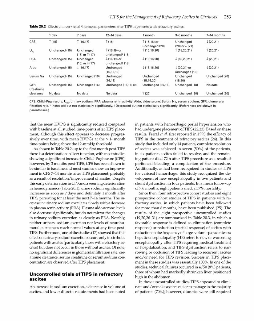
253TIPS for the Management of Refractory Ascites in Cirrhosis
that the mean HVPG is signifi cantly reduced compared with baseline at all studied time-points after TIPS place-ment, although this effect appears to decrease progres-sively over time, with mean HVPGs at the > 1- month time-points being above the 12-mmHg threshold.
As shown in Table 20.2, up to the fi rst month post-TIPS there is a deterioration in liver function, with most studies showing a signifi cant increase in Child–Pugh score (CPS); however, by 3 months post-TIPS, CPS has been shown to be similar to baseline and most studies show an improve-ment in CPS 7–14 months after TIPS placement, probably as a result of resolution/improvement of ascites. Despite this early deterioration in CPS and a seeming deterioration in hemodynamics (Table 20.1), urine sodium signifi cantly increases as soon as 7 days and defi nitely 1 month after TIPS, persisting for at least the next 7–14 months. The in-crease in urinary sodium correlates closely with a decrease in plasma renin activity (PRA). Plasma aldosterone levels also decrease signifi cantly, but do not mirror the changes in urinary sodium excretion as closely as PRA. Notably, neither urinary sodium excretion nor levels of neurohu-moral substances reach normal values at any time post-TIPS. Furthermore, one of the studies (17) showed that this effect on urinary sodium excretion occurs only in cirrhotic patients with ascites (particularly those with refractory as-cites) but does not occur in those without ascites. Of note, no signifi cant differences in glomerular fi ltration rate, cre-atinine clearance, serum creatinine or serum sodium con-centration are observed after TIPS placement.
Uncontrolled trials of TIPS in refractory ascites
An increase in sodium excretion, a decrease in volume of ascites, and lower diuretic requirements had been noted
in patients with hemorrhagic portal hypertension who had undergone placement of TIPS (22,23). Based on these results, Ferral et al. fi rst reported in 1993 the effi cacy of TIPS in the treatment of refractory ascites (24). In this study that included only 14 patients, complete resolution of ascites was achieved in seven (50%) of the patients, in six patients ascites failed to resolve, and the remain-ing patient died 72 h after TIPS procedure as a result of peritoneal bleeding, a complication of the procedure. Additionally, as had been recognized in studies of TIPS for variceal hemorrhage, this study recognized the de-velopment of new encephalopathy in two patients and shunt dysfunction in four patients. In a mean follow-up of 7.6 months, eight patients died, a 57% mortality.
Since then, four retrospective cohort studies and eight prospective cohort studies of TIPS in patients with re-fractory ascites, in which patients have been followed for more than 6 months, have been published (25). The results of the eight prospective uncontrolled studies (19,20,26–31) are summarized in Table 20.3, in which a favorable response is defi ned as elimination (complete response) or reduction (partial response) of ascites with reduction in the frequency of large-volume paracenteses; hepatic encephalopathy (HE) refers to new or worsening encephalopathy after TIPS requiring medical treatment or hospitalization; and TIPS dysfunction refers to nar-rowing or occlusion of TIPS leading to recurrent ascites and/or need for TIPS revision. Success in TIPS place-ment in these studies was essentially 100%. In one of the studies, technical failures occurred in 4/50 (8%) patients, three of whom had markedly shrunken liver positioned high in the abdomen.
In these uncontrolled studies, TIPS appeared to elimi-nate and/or make ascites easier to manage in the majority of patients (70%); however, diuretics were still required
1 day 7 days 12–14 days 1 month 3–6 months 7–14 months
CPS ↑ (15) ↑ (16,17) ↑ (16) ↑ (15,16) or Unchanged ↓ (20,21) unchanged (20) (20) or ↓ (21)UNa Unchanged (15) Unchanged ↑ (16,19) or ↑ (15,16,20) ↑ (18,20,21) ↑ (20,21) (16) or ↑ (17) unchanged* (18)PRA Unchanged (15) Unchanged ↓ (16,19) or ↓ (15,16,20) ↓ (18,20,21) ↓ (20,21) (16) or ↓ (17) unchanged† (18)Aldo Unchanged (15) ↓ (16,17) Unchanged ↓ (15,16,20) ↓ (20.21) or ↓ (20,21) (16,18,19) unchanged (18)Serum Na Unchanged (15) Unchanged (16) Unchanged Unchanged Unchanged Unchanged (20) (16,18) (15,16,20) (18,20)GFR Unchanged (15) Unchanged (16) Unchanged (16,18,19) Unchanged (15,16) Unchanged (18) No dataCreatinine clearance No data No data No data ↑ (20) Unchanged (20) Unchanged (20)
Table 20.2 Effects on liver/renal/hormonal parameters after TIPS in patients with refractory ascites.
CPS, Child–Pugh score; UNa, urinary sodium; PRA, plasma renin activity; Aldo, aldosterone; Serum Na, serum sodium; GFR, glomerular fi ltration rate. *Increased but not statistically signifi cantly. †Decreased but not statistically signifi cantly. (References are shown in parentheses.)

254 Chapter 20
(at lower doses) in essentially all the patients. Shunt dys-function (with consequent recurrence of ascites) occurred in a third of the patients and new or worsened hepatic encephalopathy occurred in over 25% of the patients. In a median follow-up of around 11 months, the observed mortality in these studies was quite variable but aver-aged around 50%.
As summarized recently (32), the procedure-related mortality of TIPS is very low (1–2%) and causes include hemoperitoneum, hemobilia, hemolysis, and sepsis. During TIPS placement, the liver capsule is frequently punctured (reported frequency around 33%), particular-ly with small, shrunken livers. However, intraperitoneal bleed only occurs in 1–2% of the cases. Fistulae may de-velop between the portal vein and an intrahepatic artery or the biliary tree (leading to hemobilia), but this is rare. Hemolysis may develop in 10–15% of patients and usu-ally resolves within 3–4 weeks of TIPS placement. Sepsis has been reported to occur in 2–10% of patients. In un-controlled studies of TIPS placement in patients with re-fractory ascites, the procedure-related complication rate is around 9% (29 of 326 patients), distributed as follows: intraperitoneal hemorrhage (3%), acute renal failure (3%) most described as being secondary to contrast media, sepsis (1.5%), and hemolysis (1.2%). A unique complica-tion is the development of strangulated umbilical hernia following resolution after TIPS (33).
Controlled trials comparing TIPS with serial large-volume paracenteses
Large-volume paracentesis (LVP) is the most commonly used method to treat refractory ascites. It is a local thera-py that does not modify any of the mechanisms that lead to ascites formation. As mentioned in Chapter 19, LVP is usually associated with intravenous albumin infusion to increase effective arterial volume, but this effect is tran-sient. Therefore, ascites will always recur in patients with refractory ascites, unless an improvement in liver disease occurs (e.g. resolution of alcoholic hepatitis), and the need
for repeated procedures, besides increasing the cost of LVP, also increases morbidity, including an impaired quality of life associated with ascites recurrence and repeated hos-pital visits. Additionally, LVP plus albumin has not been shown to improve survival (34,35). TIPS, by relieving sinu-soidal hypertension and reversing neurohumoral factors involved in sodium retention, is theoretically a more de-fi nitive treatment for ascites than LVP plus albumin and, by preventing other complications of portal hypertension and ascites, such as variceal hemorrhage, HRS, and SBP, it could have a benefi cial effect on survival. On the other hand, as shown in uncontrolled studies, TIPS has been shown to lead to the development of liver failure and/or hepatic encephalopathy as a result of diversion of blood away from the liver and into the systemic circulation.
Four prospective randomized trials of TIPS vs. LVP have been published in full to date (18,36–38) and are summarized in Tables 20.4 and 20.5.
The fi rst study, by Lebrec et al. (18), included only 25 patients (13 randomized to TIPS, 12 to LVP) and showed that treatment was effi cacious (no LVP needed for at least 1 month) in 83% of the patients in whom TIPS was placed, compared with none of the patients in the LVP group. Since patients were already requiring more than one LVP per month at the time of inclusion in the study, this re-sult was predictable. However, patients randomized to TIPS had a signifi cantly higher mortality than patients in the LVP group. Further analysis revealed that this higher mortality was due to a higher mortality in Child C pa-tients.
The study by Rossle et al. (36) included 60 patients with refractory or recidivant ascites (29 randomized to TIPS, 31 to LVP) and also demonstrated that elimination of ascites and/or elimination of the need for LVP was signifi cantly higher in the group treated with TIPS (84%) compared with the group treated with LVP (43%). Of note, LVP was not routinely followed by the administration of albumin in this study, and although the investigators found a ten-dency for a lower mortality in the TIPS group, differences failed to achieve statistical signifi cance.
Table 20.3 Uncontrolled prospective cohort studies of TIPS for refractory ascites.
Author/ Follow-up Favorable Responders New or Shunt reference number n (months) response requiring diuretics worse HE dysfunction Mortality
Quiroga (20) 17 15.5 15 (88%) NR 3 (18%) 9 (53%) 5 (29%)Ochs (26) 50 14.2 46 (92%) 46 (100%) 8 (16%) 21 (42%) 31 (62%)Somberg (19) 24 9.4–11 19 (79%) 2/5 (40%) 7 (29%) 9 (38%) NRCrenshaw (27) 54 9.1 40 (74%) 40 (100%) 12 (22%) 11 (20%) 27 (50%)Martinet (28) 30 8.8 26 (87%) 26 (100%) 14 (47%) 8 (31%) 17 (57%)Nazarian (29) 50 11.6 23 (46%) NR 20 (40%) 17 (34%) 30 (60%)Deschenes (30) 53 17.5 25 (47%) NR 14 (26%) 17 (32%) 23 (43%) (6 months)Peron (31) 48 11.2 35 (73%) NR 10 (21%) 18 (38%) ~ 70% 1-year cumul.Total 326 11.2 229 (70%) 114/117 (97%) 88 (27%) 110 (34%) 133/254 (52%)
NR, not reported.

255TIPS for the Management of Refractory Ascites in Cirrhosis
The study by Ginès et al. (37) included 70 patients with cirrhosis and refractory ascites (35 randomized to TIPS, and 35 to repeated LVP plus intravenous albumin). As expected, recurrence of ascites was greater in the LVP group. Contrary to other trials, the primary endpoint in this study was survival without liver transplantation. In a mean follow-up of 9.4 (TIPS group) and 10.8 (LVP+ albu-min group) months, 20 (57%) patients treated with TIPS and 18 (51%) treated with LVP died (a nonsignifi cant dif-ference), while seven patients in each group underwent liver transplantation. The probability of survival with-out liver transplantation was 41% at 1 year and 26% at 2 years in the TIPS group, compared with 35% and 30% in the paracentesis group, respectively (P = 0.51). The fre-quency of severe hepatic encephalopathy was greater in the TIPS group, as were the calculated costs.
In the most recent study, by Sanyal et al. (38), 109 sub-jects with refractory ascites were randomized to either medical therapy (sodium restriction, diuretics, and total paracentesis) (n = 57) or to medical therapy plus TIPS
(n = 52). As shown by all other studies, TIPS was signifi -cantly superior to medical therapy alone in preventing recurrence of ascites. There were no signifi cant differenc-es in the two arms with respect to overall and transplant-free survival. There was a higher incidence of moderate to severe encephalopathy in the TIPS group, but no dif-ferences in quality of life between study groups.
Meta-analysis of these four studies (D’Amico, person-al communication) reveals a signifi cant benefi t of TIPS (compared with LVP) in terms of recurrent ascites, with no differences in overall mortality.
Details of these four studies are summarized in Ta-bles 20.4 and 20.5. Table 20.4 shows the general charac-teristics of patients included in these studies. As can be observed, the cirrhotic patient populations are quite ho-mogeneous among studies, with a median age of 56 years, a CPS around 9.2, bilirubin levels around 1.9, and a simi-lar decrease in HVPG after TIPS insertion. Important dif-ferences appear in urinary sodium excretion, highest in the study by Rossle et al. (36), in which only 22% of the
CPS Bilirubin Creatinine Serum Na UNa (mEq/l) HVPG Author Tx n Age Alcohol (% Child C) (mg/dl) (mg/dl) (mEq/l) (% < 130) (% < 10) (mmHg)
Lebrec (18) LVP 12 52 83% 9.2 (33%) 1.6 1.0 130 < 5 22→20 TIPS 13 50 77% 9.3 (31%) 2.0 1.0 130 < 5 20→13Rossle (36) LVP 29 61 74% 8.7 (23%) 1.8 1.4 131 (13%) 61 (10%) – TIPS 31 58 83% 9.1 (38%) 1.8 1.3 130 (17%) 45 (40%) 24→10Ginès (37) LVP 35 56 60% 9.2 (43%) 2.4 1.4 – (48%) 9 – TIPS 35 59 51% 9.3 (37%) 2.0 1.4 – (54%) 7 19.1→8.7Sanyal (38) LVP 57 52 33% 9.3 (NR) 1.9 0.98 NR NR – TIPS 52 56 32% 9.2 (NR) 1.9 1.07 NR NR 19.8→8.3
Table 20.4 Baseline characteristics of patients included in controlled studies of TIPS vs. large-volume paracentesis (LVP) for refractory ascites.
NR, Not reported; CPS, Child–Pugh score; UNa, urine sodium; LVP, large-volume paracentesis; TIPS, transjugular intrahepatic portosystemic shunt. Values given are mean values or percentages. (References are shown in parentheses.)
Mean 1-year 2-year follow-up Favourable New or TIPS cumulative cumulative Author Tx n (months) response worse HE Dysfunction Mortality survival survival
Lebrec (18) LVP 12 NR 0 0 – 4 (33%) ~ 72%* 60% (at 4 months) TIPS 13 NR 5 (38%) 3 (23%) 3 (23%) 9 (69%) 29%* 29% (at 4 months)Rossle (36) LVP 29 44 7 (24%) 3 (10%) – 23 (79%) 52% 32% (at 3 months) TIPS 31 45 20 (64%) 6 (19%) 13 (42%) 15 (48%) 69% 58% (at 3 months)Ginès (37) LVP 35 11 6 (17%) 12 (34%)† – 18 (51%) 35% 30% TIPS 35 9 18 (51%) 21 (60%)† 13 (37%) 20 (57%) 41% 26%Sanyal (38) LVP 57 NR 9 (16%) 12 (21%) – 19 (33%) ~ 70%* ~ 60%* TIPS 52 NR 30 (58%) 20 (38%) 53% at 6 months 18 (35%) ~ 70%* ~ 60%*
*Derived from survival curve. Values given are mean values or percentages. †Severe encephalopathy; NR, not reported. (References are shown in parentheses.)
Table 20.5 Controlled studies of TIPS vs. large-volume paracentesis (LVP) for refractory ascites.

256 Chapter 20
patients had a urinary sodium excretion of < 10 mmol/day, while this was near 100% in all other studies. This difference may explain the higher survival rates in this study, which was only comparable to survival in the TIPS group of the study by Sanyal et al. (38), in which unfortu-nately sodium levels (urinary or serum) are not reported. Hyponatremia was more frequent in patients included in the study by Ginès et al. (37), in which patients also had higher serum creatinine levels. This study showed lower survival rates compared with the other two larger stud-ies, which is not surprising as hyponatremia and renal dysfunction have both been described as being predictors of a poor survival in cirrhosis (39,40), and more recently serum creatinine (> 1.7 mg/dl) and sodium (serum so-dium < 130 mmol/l) were identifi ed as the only inde-pendent predictors of survival in patients undergoing TIPS for variceal hemorrhage (41). Interestingly, while most earlier studies included a majority of patients with alcoholic etiology of cirrhosis, the study by Sanyal et al. (38) included mainly patients with non-alcoholic cirrho-sis, predominantly hepatitis C. Patients in this study had a reasonably good survival, which is rather unexpected considering that in this group of patients liver disease is assumedly progressive, while in patients with alcoholic cirrhosis, alcohol abstinence would be associated with an improved survival.
In these prospective controlled studies, TIPS was tech-nically unsuccessful in 5% of the cases (7/131). Although all the studies report a decrease in diuretic requirement, it appears that all patients continued to require diuretics. The procedure-related complications were only specifi ed in the study by Gines et al. (37) and consisted of heart failure in four (11%) and severe hemolysis in three (9%) patients.
Other benefi cial effects of TIPS in patients with refractory ascites
In the study by Ginès et al. (37), a preventive effect of TIPS on the development of HRS was observed. While de novo HRS (in patients without HRS at inclusion) or pro-gression from type 2 HRS to type 1 HRS occurred in 11 (31%) patients in the LVP plus albumin group, this only occurred in three (9%) patients randomized to the TIPS group. This benefi cial effect is probably related to the ef-fect that TIPS placement has on the hormonal systems involved in the pathogenesis of renal dysfunction in cir-rhosis with suppression of the activity of the renin–angi-otensin–aldosterone system (Table 20.2).
It has been noted that some of the patients in whom ascites resolves after successful insertion of TIPS also have a remarkable improvement in nutrition. A study performed in 10 patients with refractory ascites who were malnourished at baseline (as indicated by baseline reduced nitrogen index, percentage body fat, and rest-
ing energy expenditure) showed a signifi cant increase in dry weight, total body nitrogen (a measure of lean body mass not affected by the presence or absence of ascites), and resting energy expenditure 3 and 12 months after TIPS placement compared with baseline, indicating an improvement in body composition (42). This effect was ascribed to an increase in energy intake or enhanced nu-trient absorption, neither of which was confi rmed in the study.
TIPS placement has also been associated with an im-provement in quality of life (QOL) in patients in whom it is placed for management of refractory ascites. In an un-controlled study performed in 21 cirrhotic patients (43), QOL (assessed by the QOL index and by the patient’s rating of their QOL on a visual analog scale) signifi cantly improved; however, this improvement was observed only in patients with a complete response (elimination of ascites). Oddly, in patients with a partial response (pres-ence of ascites not requiring paracentesis) there was no signifi cant improvement in QOL, indicating that QOL in patients with ascites may be infl uenced not only by the invasiveness of repeated LVP but also by the presence of ascites, which may cause signifi cant abdominal dis-comfort. However, these results could not be confi rmed in the only randomized controlled study in which QOL was evaluated (38). In this study, the SF-36 questionnaire was performed at baseline and at months 6 and 12 after TIPS placement. Although both components, the physi-cal scale and the mental scale, improved signifi cantly in both study groups compared with baseline, there were no signifi cant differences between patients treated with LVP vs. patients randomized to TIPS.
Predictors of post-TIPS mortality
Several prognostic models have been developed to pre-dict survival in patients undergoing TIPS placement. The fi rst was the MELD (Model of End-Stage Liver Disease) model that was originally developed for patients under-going TIPS (44) and was then modifi ed slightly to predict survival in cirrhotic patients in general (45). MELD is a continuous function of serum bilirubin levels, interna-tional normalized ratio for prothrombin time, and serum creatinine to predict 3-month post-TIPS survival and was validated in an independent patient population. In an-other retrospective study from Emory University (46), variables found to be independent predictors of sur-vival were a bilirubin level > 3.0 mg/dl, an alanine ami-notransferase level > 100 IU/l, pre-TIPS encephalopathy, and urgency of TIPS. This model was also validated in a separate group of patients. In the MELD study, only 25% of patients had TIPS placed for management of refractory ascites, while in the Emory study 38% had TIPS placed for refractory ascites and 19% had it placed for the treat-ment of refractory hepatic hydrothorax. Neither of them

257TIPS for the Management of Refractory Ascites in Cirrhosis
showed that the indication for TIPS had an infl uence on survival.
Several recent studies have compared these models with the CPS and have shown divergent results. One study showed that the MELD score is better than CPS score in predicting 3-month survival but not in predict-ing long-term survival (47), while another study found no differences in short-term survival and only a marginal advantage of MELD in long-term survival (48). Another study showed that both scores were equally predictive of 1-month, 3-month, and 1-year survival (49), with a cut-off for the Child score of 11. Interestingly, in this study, which included 475 patients, those undergoing TIPS for refractory ascites (that constituted 32% of the patient population) had a signifi cantly poorer survival than pa-tients with variceal bleeding. This study also found that renal function was the strongest independent predictor of survival.
TIPS for refractory hepatic hydrothorax
Hepatic hydrothorax is produced by the passage of ascit-ic fl uid from the peritoneal cavity into the pleural space through small holes in the diaphragm. In this smaller space, small amounts of fl uid (2–3 l) lead to shortness of breath and requirement of therapeutic thoracentesis, which in some patients may be quite frequent despite the use of diuretics. It is considered that these patients have refractory hepatic hydrothorax and small, uncontrolled studies have shown that TIPS is effective in this setting (50–54). These studies are summarized in Table 20.6. The results are comparable to those of uncontrolled studies of TIPS for refractory ascites (Table 20.3). A favorable response (either resolution of the pleural effusion or a decrease in the need for thoracentesis) occurs in 67% of patients, new or worsened hepatic encephalopathy oc-curs in 30% of patients, and shunt dysfunction in half the patients. The mortality is around 46% in a follow-up period of 7 months, which would suggest that the mor-tality is higher in this group of patients, particularly in those who are nonresponders to TIPS, in whom a 100% mortality has been described (51,53,54). Therefore, al-
though TIPS seems to be an effective form of therapy for this condition, its use should be limited to patients with hepatic hydrothorax who require repeated thoracentesis for control of symptoms despite inpatient treatment with diuretics.
Conclusions
Since the publication of the fi rst uncontrolled study of TIPS for the treatment of refractory ascites 10 years ago, advances have been made in our understanding of the effect of TIPS in cirrhotic patients with refractory ascites, including the publication of four prospective randomized trials comparing TIPS vs. LVP. TIPS placement is associ-ated with a reduction in sinusoidal pressure and a signifi -cant improvement in urinary sodium excretion that corre-lates with suppression in plasma renin activity (indicative of an improvement in effective arterial blood volume). It is therefore clearly effective in preventing the recurrence of ascites, recurrence that is practically universal in patients treated with LVP. However, this benefi cial effect of TIPS is offset by an increase in the incidence of severe hepatic encephalopathy, a high incidence of shunt dysfunction, and a higher cost. Importantly, TIPS is not associated with a survival benefi t or with a quality of life benefi t when compared with LVP. Therefore, the consensus recommen-dation put forward recently is that fi rst-line treatment of refractory ascites is repeated LVP plus albumin and that TIPS should be considered when the frequency of para-centesis is greater than two to three times per month (55). A good predictor of post-TIPS survival is the CPS and a score > 11 should be considered a contraindication for TIPS placement. Patients with alcoholic cirrhosis who are drinking alcohol may improve with abstinence and there-fore TIPS should be delayed in these patients. It should be noted that all these studies used bare (uncovered) TIPS stents. New expanded polytetrafl uoroethylene (ePTFE)-covered stents have recently become available for TIPS placement and a recent retrospective study of 89 patients in whom ePTFE-covered stents were used (50% for re-fractory ascites) showed a survival benefi t for the covered stent when compared with patients in whom convention-
Follow-up Favorable New or worse Shunt Author n (months) response HE/liver failure dysfunction Mortality
Strauss (50) 5 10.4 2 (40%) NR 3 (60%) 2 (40%)Gordon (51) 24 7.2 19 (79%) 13 (54%) 13 (54%) 13 (68%)Jeffries (52) 12 5.8 7 (58%) 4 (33%) 7 (58%) 3 (25%)Siegerstetter (53) 40 16.0 28 (70%) 3 (8%) 16 (40%) 17 (42%)Spencer (54) 21 7.2 12 (57%) 9 (43%) 13 (61%) 12 (57%)Total 102 7.2 68 (67%) 29/97 (30%) 52 (51%) 47 (46%)
Table 20.6 Uncontrolled prospective cohort studies of TIPS for refractory hepatic hydrothorax.
HE, Hepatic encephalopathy; NR, not reported. (References are shown in parentheses.)

258 Chapter 20
al TIPS were placed (56). This benefi t needs to be prospec-tively evaluated in patients with refractory ascites.
Addendum
After writing this chapter, a fi fth trial comparing para-centesis vs TIPS in patients with cirrhosis and refractory ascites has been published (57).
References 1 Arroyo V, Ginès P, Gerbes AL et al. Defi nition and diagnostic
criteria of refractory ascites and hepatorenal syndrome in cirrhosis. Hepatology 1996; 23:164–76.
2 Rector WG. Portal hypertension: a permissive factor only in the development of ascites and variceal bleeding. Liver 1986; 6:221–6.
3 Morali GA, Sniderman KW, Deitel KM et al. Is sinusoidal portal hypertension a necessary factor for the development of hepatic ascites? J Hepatol 1992; 16:249–50.
4 Villanueva C, Minana J, Ortiz J et al. Endoscopic ligation compared with combined treatment with nadolol and isosorbide mononitrate to prevent recurrent variceal bleeding. N Engl J Med 2001; 345:647–55.
5 Abraldes JG, Tarantino I, Turnes J, Garcia-Pagan JC, Rodés J, Bosch J. Hemodynamic response to pharmacological treatment of portal hypertension and long-term prognosis of cirrhosis. Hepatology 2003; 37:902–8.
6 Casado M, Bosch J, Garcia-Pagan JC et al. Clinical events after transjugular intrahepatic portosystemic shunt: correlation with hemodynamic fi ndings. Gastroenterology 1998; 114:1296–303.
7 Castells A, Saló J, Planas R et al. Impact of shunt surgery for variceal bleeding in the natural history of ascites in cirrhosis: a retrospective study. Hepatology 1994; 20:584–91.
8 Voorhees ABJ, Price JBJ, Britton RC. Portosystemic shunting procedures for portal hypertension. Twenty-six year experience in adults with cirrhosis of the liver. Am J Surg 1970; 119:501–5.
9 Orloff MJ, Orloff MS, Orloff SL, Girard B. Experimental, clinical, and metabolic results of side-to-side portacaval shunt for intractable cirrhotic ascites. J Am Coll Surg 1997; 183:557–70.
10 Franco D, Vons C, Traynor O, De Smadja C. Should portosystemic shunt be reconsidered in the treatment of intractable ascites in cirrhosis? Arch Surg 1988; 123:987–91.
11 Rosch J, Uchida BT, Putnam JS, Buschman RW, Law RD, Hershey AL. Experimental intrahepatic portacaval anastomosis: use of expandable gianturco stents. Radiology 1987; 162:481–5.
12 Luca A, D’Amico G, LaGalla R, Midiri M, Morabito A, Pagliaro L. TIPS for prevention of recurrent bleeding in patients with cirrhosis: meta-analysis of randomized clinical trials. Radiology 1999; 212:411–21.
13 Papatheodoridis GV, Goulis J, Leandro G, Patch D, Burroughs AK. Transjugular intrahepatic portosystemic shunt compared with endoscopic treatment for prevention of variceal rebleeding. A meta-analysis. Hepatology 1999; 30:612–22.
14 Burroughs AK, Vangeli M. Transjugular intrahepatic portosystemic shunt versus endoscopic therapy:
randomized trials for secondary prophylaxis of variceal bleeding: an updated meta-analysis. Scand J Gastroenterol 2002; 37:249–52.
15 Wong F, Sniderman K, Liu P, Allidina Y, Sherman M, Blendis L. Transjugular intrahepatic portosystemic stent shunt: effects on hemodynamics and sodium homeostasis in cirrhosis and refractory ascites. Ann Intern Med 1995; 122:816–22.
16 Wong F, Sniderman K, Liu P, Blendis L. The mechanism of the initial natriuresis after transjugular intrahepatic portosystemic shunt. Gastroenterology 1997; 112:899–907.
17 Gerbes AL, Gulberg V, Waggershauser T, Holl J, Reiser M. Renal effects of transjugular intrahepatic portosystemic shunt in cirrhosis: comparison of patients with ascites, with refractory ascites or without ascites. Hepatology 1998; 28:683–8.
18 Lebrec D, Giuily N, Hadengue A et al. and a French Group of Clinicans and a Group of Biologists. Transjugular intrahepatic portosystemic shunts: comparison with paracentesis in patients with cirrhosis and refractory ascites: a randomized trial. J Hepatol 1996; 25:135–44.
19 Somberg KA, Lake JR, Tomlanovich SJ, LaBerge JM, Feldstein V, Bass NM. Transjugular intrahepatic portosystemic shunts for refractory ascites: assessment of clinical and hormonal response and renal function. Hepatology 1995; 21:709–16.
20 Quiroga J, Sangro B, Nunez M et al. Transjugular intrahepatic portal-systemic shunt in the treatment of refractory ascites: effect on clinical, renal, humoral, and hemodynamic parameters. Hepatology 1995; 21:986–94.
21 Wong W, Liu P, Blendis L, Wong F. Long-term renal sodium handling in patients with cirrhosis treated with transjugular intrahepatic portosystemic shunts for refractory ascites. Am J Med 1999; 106:315–22.
22 Zemel G, Katzen B, Becker GJ, Benenati JF, Sallee S. Percutaneous transjugular portosystemic shunt. J Am Med Assoc 1991; 266:390–3.
23 LaBerge JM, Ring EJ, Gordon RL et al. Creation of transjugular intrahepatic porto-systemic shunts with the Wallstent endoprosthesis: results in 100 patients. Radiology 1993; 187:413–20.
24 Ferral H, Bjarnason H, Wegryn SA et al. Refractory ascites: early experience in treatment with transjugular intrahepatic portosystemic shunt. Radiology 1993; 189:795–801.
25 Russo MW, Sood A, Jacobson IM, Brown RS. Transjugular intrahepatic portosystemic shunt for refractory ascites: an analysis of the literature on effi cacy, morbidity, and mortality. Am J Gastroenterol 2003; 98:2521–7.
26 Ochs A, Rossle M, Haag K et al. The transjugular intrahepatic portosystemic stent-shunt procedure for refractory ascites. N Engl J Med 1995; 332:1192–7.
27 Crenshaw WB, Gordon FD, McEniff NJ et al. Severe ascites: effi cacy of the transjugular intrahepatic portosystemic shunt in treatment. Radiology 1996; 200:185–92.
28 Martinet JP, Fenyves D, Legault L et al. Treatment of refractory ascites using transjugular intrahepatic portosystemic shunt (TIPS). A caution. Dig Dis Sci 1997; 42:161–6.
29 Nazarian GK, Bjarnason H, Dietz CAJ et al. Refractory ascites: midterm results of treatment with a transjugular intrahepatic portosystemic shunt. Radiology 1997; 205:173–80.
30 Deschenes M, Dufresne MP, Bui B et al. Predictors of clinical response to transjugular intrahepatic portosystemic shunt
259TIPS for the Management of Refractory Ascites in Cirrhosis
(TIPS) in cirrhotic patients with refractory ascites. Am J Gastroenterol 1999; 94:1361–5.
31 Peron JM, Barange K, Otal P et al. Transjugular intrahepatic portosystemic shunts in the treatment of refractory ascites: results in 48 consecutive patients. J Vasc Interv Radiol 2000; 11:1211–16.
32 Boyer TD. Transjugular intrahepatic portosystemic shunt: current status. Gastroenterology 2003; 124:1700–10.
33 Trotter JF, Suhocki PV. Incarceration of umbilical hernia following transjugular intrahepatic portosystemic shunt for the treatment of ascites. Liver Transpl Surg 1999; 5:209–10.
34 Ginès P, Arroyo V, Vargas V et al. Paracentesis with intravenous infusion of albumin as compared with peritoneovenous shunting in cirrhosis with refractory ascites. N Engl J Med 1991; 325:829–35.
35 Ginès A, Planas R, Angeli P et al. Treatment of patients with cirrhosis and refractory ascites by LeVeen shunt with titanium tip. Comparison with therapeutic paracentesis. Hepatology 1995; 22:124–31.
36 Rossle M, Ochs A, Gulberg V et al. A comparison of paracentesis and transjugular intrahepatic portosystemic shunting in patients with ascites. N Engl J Med 2000; 342:1701–7.
37 Ginès P, Uriz J, Calahorra B et al., for the International Study Group on Refractory Ascites in Cirrhosis. Transjugular intrahepatic portosystemic shunting versus repeated paracentesis plus intravenous albumin for refractory ascites in cirrhosis: a multicenter randomized comparative study. Gastroenterology 2002; 123:1839–47.
38 Sanyal AJ, Genning C, Reddy KR et al., North American Study for the Treatment of Refractory Ascites Group. The North American Study for the Treatment of Refractory Ascites. Gastroenterology 2003; 124:634–41.
39 Arroyo V, Rodés J, Gutiérrez Lizárraga MA, Revert L. Prognostic value of spontaneous hyponatremia in cirrhosis with ascites. Am J Dig Dis 1976; 21:249–56.
40 Llach J, Ginès P, Arroyo V et al. Prognostic value of arterial pressure, endogenous vasoactive systems, and renal function in cirrhotic patients admitted to the hospital for the treatment of ascites. Gastroenterology 1988; 94:482–7.
41 Schepke M, Roth F, Koch L et al. Prognostic impact of renal impairment and sodium imbalance in patients undergoing transjugular intrahepatic portosystemic shunting for the prevention of variceal rebleeding. Digestion 2003; 67:146–53.
42 Allard JP, Chau J, Sandokji K, Blendis LM, Wong F. Effects of ascites resolution after successful TIPS on nutrition in cirrhotic patients with refractory ascites. Am J Gastroenterol 2001; 96:2442–7.
43 Gulberg V, Liss I, Bilzer M, Waggershauser T, Reiser M, Gerbes AL. Improved quality of life in patients with refractory or recidivant ascites after insertion of transjugular intrahepatic portosystemic shunts. Digestion 2002; 66:127–30.
44 Malinchoc M, Kamath PS, Gordon FD, Peine CJ, Rank J,
TerBorg PCJ. A model to predict poor survival in patients undergoing transjugular intrahepatic portosystemic shunts. Hepatology 2000; 31:864–71.
45 Kamath PS, Wiesner RH, Malinchoc M et al. A model to predict survival in patients with end-stage liver disease. Hepatology 2001; 33:464–70.
46 Chalasani N, Clark WS, Martin LG et al. Determinants of mortality in patients with advanced cirrhosis after transjugular intrahepatic portosystemic shunting. Gastroenterology 2000; 118:138–44.
47 Salerno F, Merli M, Cazzaniga M et al. MELD score is better than Child–Pugh score in predicting 3-month survival of patients undergoing transjugular intrahepatic portosystemic shunt. J Hepatol 2002; 36:494–500.
48 Schepke M, Roth F, Fimmers R et al. Comparison of MELD, Child–Pugh, and Emory model for the prediction of survival in patients undergoing transjugular intrahepatic portosystemic shunting. Am J Gastroenterol 2003; 98:1167–74.
49 Angermayr B, Cejna M, Karnel F et al. Child–Pugh versus MELD score in predicting survival in patients undergoing transjugular intrahepatic portosystemic shunt. Gut 2003; 52:879–85.
50 Strauss RM, Martin LG, Kaufman SL, Boyer TD. Transjugular intrahepatic portal systemic shunt for the management of symptomatic cirrhotic hydrothorax. Am J Gastroenterol 1994; 89:1522.
51 Gordon FD, Anastopoulos HT, Crenshaw W et al. The successful treatment of symptomatic, refractory hepatic hydrothorax with transjugular intrahepatic portosystemic shunt. Hepatology 1997; 25:1366–9.
52 Jeffries MA, Kazanjian S, Wilson M, Punch J, Fontana RJ. Transjugular intrahepatic portosystemic shunts and liver transplantation in patients with refractory hepatic hydrothorax. Liver Transplant Surg 1998; 4:416–23.
53 Siegerstetter V, Deibert P, Ochs A, Olschewski M, Blum HE, Rossle M. Treatment of refractory hepatic hydrothorax with transjugular intrahepatic portosystemic shunt: long-term results in 40 patients. Eur J Gastroenterol Hepatol 2001; 13:529–34.
54 Spencer EB, Cohen DT, Darcy MD. Safety and effi cacy of transjugular intrahepatic portosystemic shunt creation for the treatment of hepatic hydrothorax. J Vasc Interv Radiol 2002; 13:385–90.
55 Moore KP, Wong F, Ginès P et al. The management of ascites in cirrhosis: report on the consensus conference of the International Ascites Club. Hepatology 2003; 38:258–66.
56 Angermayr B, Cejna M, Koenig F, Karnel F, Gangl A, Peck-Radosavljevic M, for the Vienna TIPS Study Group. Survival in patients undergoing transjugular intrahepatic portosystemic shunt: ePTFE-covered stentgrafts versus bare stents. Hepatology 2003; 38:1043–50.
57 Salerno F, Merli M, Riggio O et al. Randomized controlled study of TIPS versus paracentesis plus albumin in cirrhosis with severe ascites. Hepatology 2004; 40(3):629–35.

260
Chapter 21Prognosis of Patients with Cirrhosis and Ascites
Mónica Guevara, Andrés Cárdenas, Juan Uríz, and Pere Ginès
Cirrhosis is a chronic and progressive disease charac-terized by the development of complications related to portal hypertension and circulatory dysfunction, including ascites, dilutional hyponatremia, gastroin-testinal bleeding, hepatic encephalopathy, bacterial infections, and renal failure. Ascites is the most com-mon complication of cirrhosis and constitutes the fi rst manifestation of the disease in the majority of patients (1,3). The appearance of ascites is a marker of poor prognosis (1–7). However, these patients can be classi-fi ed in different groups according the severity of ascites and associated renal function abnormalities and their response to treatment. For instance, patients with mild sodium retention and normal solute-free water excre-tion and glomerular fi ltration rate (GFR) and good re-sponse to diuretics have a good prognosis that is very different compared with that of patients with refractory ascites. The identifi cation of factors predictive of sur-vival in patients with cirrhosis and ascites has been of pivotal importance because it identifi es patients that benefi t from liver transplantation at an early stage and provides accurate information to patients and their rela-tives regarding the clinical course and outcome (8). Re-cently some prognostic models that specifi cally address prognosis and survival of patients with cirrhosis and ascites have been proposed (7,9). Nevertheless the re-cent introduction of the Model for End-Stage Liver Dis-ease (MELD) score for organ allocation in the USA that includes variables of liver function and renal function has taken precedence among other prognostic scores in patients with cirrhosis (10). The purpose of this chapter is to review the current knowledge of the natural history and survival of cirrhosis with ascites.
Compensated cirrhosis
In the past, the diagnosis of cirrhosis was usually made when patients presented major clinical manifestations of the disease, such as ascites, jaundice, variceal bleed-
ing or hepatic encephalopathy. Presently, a signifi cant number of patients are diagnosed before the appear-ance of these complications, a condition commonly referred to as compensated cirrhosis. The survival and natural history of patients with compensated cirrhosis is well established in several large series of patients (1,2,5,11–17) (Table 21.1). This condition is associated with a long life expectancy. Studies published dur-ing the last decade have reported a greater long-term survival than those published during the 1980s. These differences may be related to several factors, includ-ing diagnosis of cirrhosis at earlier stages, screening for hepatocellular carcinoma (which could allow for an early therapy), improved therapeutic methods in the management of major complications of the disease, and effects of antiviral therapy on the evolution of pa-tients with hepatitis B or hepatitis C-related cirrhosis. Nevertheless, despite a relatively long survival, many patients with compensated cirrhosis go on to develop major complications that lead to increased morbidity and mortality. The probability of developing ascites in patients with compensated cirrhosis has been calculated to range between 10 and 50% 2–5 years after diagnosis (2,5,17). Risk factors for the development of ascites in patients with compensated cirrhosis have not specifi -cally been assessed. However, portal hypertension is a prerequisite for the development of ascites. This is dem-onstrated by the fact that patients submitted to surgi-cal portosystemic shunts and transjugular intrahepatic portosystemic shunts (TIPS) for treatment or preven-tion of variceal bleeding do not develop ascites during the course of the disease (18,19). Therefore, it is likely that a certain level of portal pressure is required for the development of ascites. A defi nite cut-off level has not been established, but ascites rarely develops in patients with a portal pressure, as assessed by hepatic venous pressure gradient, lower than 12 mmHg (20,21). Nev-ertheless, other factors undoubtedly play an important role because not all patients with portal pressure above this level develop ascites.

261Prognosis of Patients with Cirrhosis and Ascites
Cirrhosis with ascites
Survival of patients with ascites
The development of ascites in patients with cirrhosis is usually indicative of a poor prognosis. In most series, the probability of survival of cirrhotic patients with ascites ranges between 45 and 82% at 1 year of follow-up and decreases below 50% after 5 years (1–4,6–8) (Table 21.2). Long-term survivors (more than 10 years) are very un-common. It should be noted that most studies analyz-ing survival of cirrhosis with ascites were performed in patients admitted to hospital for management of ascites but without concomitant complications and included a large proportion of patients with a past history of ascites. Therefore, data may not be applicable to patients with mild or moderate sodium retention in which ascites is easily managed with diuretics usually without hospitali-zation, or to hospitalized patients with ascites and associ-ated severe complications (i.e. gastrointestinal bleeding, spontaneous bacterial peritonitis, or hepatocellular carci-noma). Furthermore, these fi gures cannot be extrapolat-
ed to estimate survival of patients with fi rst-onset ascites, because these patients have a better survival than those with previous episodes of ascites (Fig. 21.1).
A signifi cant proportion (10–15%) of patients with cir-rhosis and ascites develop severe sodium retention that cannot be appropriately treated either because patients do not respond to high doses of diuretics or because they develop side-effects that preclude their use. This condi-tion is known as refractory ascites (22). In most of these patients sodium retention is associated with severely impaired solute-free water excretion and reduced renal plasma fl ow and GFR. Patients with refractory ascites constitute a subset of patients with ascites with partic-ularly poor prognosis, the 1-year survival probability being of only 50% (6,9,23–26) (Table 21.3).
Prognostic factors of patients with ascites
A number of factors with prognostic value have been identifi ed in patients with cirrhosis and ascites. Some factors are related to liver dysfunction whereas others are secondary to circulatory and renal dysfunction com-monly present in these patients.
Survival probability (%) PatientsAuthor Study period (n) 1 year 5 years 10 years Comments
D’Amico et al. (5) 1959–76 90 80 53 30 All etiologies of cirrhosisSaunders et al. (1) 1974–80 435 94 67 NR All etiologies of cirrhosisGinès et al. (2) 1968–80 293 96 68 47 All etiologies of cirrhosisTanaka et al. (11) 1958–84 582 89 62 42 All etiologies of cirrhosisRealdi et al. (14) 1973–90 366 98 84 68 HBV-cirrhosisFattovich et al. (15.) 1982–92 384 100 91 79 HCV-cirrhosisZoli et al. (12) 1983–84 100 90 45 All etiologies of cirrhosisDeJongh et al. (13) 1970–90 98 95 71 NR HBV-cirrhosisFattovich et al. (16) NR 297 97 85 HBV/HCV-cirrhosisBenvegnu et al. (17) 1986–96 312 > 95 90 75 HBV/HCV-cirrhosis
Table 21.1 Survival of compensated cirrhosis.
Some percentages have been estimated from the survival curves included in the different articles. NR, not reported. Numbers given in parentheses correspond to the reference number.
Survival probability (%) PatientsAuthor Study period (n) 1 year 2 years 5 years 10 years
Powell and Klatskin (4) 1951–63 182 65 60 52 NRSaunders et al. (1) 1959–76 512 45 34 22 10Llach et al. (8) 1980–85 139 56 49 25 NRSalerno et al. (6) 1983–89 134 78 63 38 NRFernández-Esparrach et al. (7) 1980–90 216 59 46 27 16Planas et al. (3) 1998–2001 200 82 70 41 –
Table 21.2 Survival of cirrhosis with ascites.
Some percentages have been estimated from the survival curves included in the different articles. NR, not reported. *Numbers given in parentheses correspond to the reference number.

262 Chapter 21
Liver function
In clinical practice, the degree of liver dysfunction is usu-ally assessed by measuring the plasma levels of proteins synthesized by the liver, particularly albumin and coagu-lation factors, or the plasma levels of substances metabo-lized by the liver, especially bilirubin and bile acids (27). A reduction in the former and an increase in the latter indicate impaired liver function. A number of studies have shown that parameters of liver function correlate with prognosis and may be useful in clinical practice to estimate survival in the general population of patients with cirrhosis (28–32). It is, therefore, not surprising that some liver function tests have a strong prognostic value in the specifi c population of patients with cirrhosis and ascites. For example, increased serum bilirubin or reduced serum albumin are associated with a short survival in these patients (6–8,33). By contrast, prothrombin activity has no predictive value in patients with ascites (6–8). This lack of predictive value may be due to the fact that the prolongation of prothrombin time in pa-tients with cirrhosis occurs very late in the evolution of the disease (34). In other disease states such as primary biliary
cirrhosis or primary sclerosing cholangitis serum bilirubin levels in conjunction with serum albumin are good mark-ers of prognosis (35,36).
Renal function
It is now well established that the severity of renal and cir-culatory dysfunction is of major prognostic signifi cance in patients with cirrhosis and ascites. Hecker and Sherlock published the fi rst report of the prognostic value of renal and circulatory dysfunction in patients with liver disease and ascites in 1956 (37,38). These authors described nine patients with acute or subacute viral hepatitis or cirrho-sis who were admitted to hospital with impaired renal function and/or hyponatremia. Most patients developed progressive renal failure and severe hyponatremia dur-ing hospitalization and all of them died. The renal and electrolyte disturbances were associated with marked ar-terial hypotension. Since then, a number of studies have assessed the prognostic value of renal and hemodynamic abnormalities in patients with cirrhosis and ascites.
In 1981, Arroyo et al. (39) demonstrated the importance of sodium excretion as a prognostic factor in cirrhosis with ascites. Marked sodium retention was found to be associated with reduced survival. The importance of this simple parameter as guide to prognosis in cirrhosis with ascites has been confi rmed in subsequent studies (7,8,40). However it is essential to consider that for the evaluation of prognosis, sodium excretion should be measured in conditions of fi xed and controlled sodium intake (usual-ly a low-sodium diet of 70–90 mEq/day during 5–7 days) and off diuretics. Cirrhotic patients who, under these conditions, have a sodium excretion close to or greater than sodium intake have a good prognosis. On the other hand, patients with a markedly reduced sodium excre-tion (< 10 mEq/l) compared with their intake have a poor outcome (7,8,39,40) (see also Chapter 16).
no previous ascites
previous ascites
Years
p<0.01
1.0
0.8
0.6
0.4
0.2
01 2 3 4 5
Figure 21.1 Comparison of long-term survival of cirrhotic patients with fi rst-onset ascites and patients with previous episodes of ascites.
Survival probability (%) Study PatientsAuthor period (n) 6 months 1 year 2 years
Salerno et al. (6) 1983–89 24 75 58 38Guardiola et al. (9) 1980–92 100 75 42 38Ginès et al. (23) 1987–89 121 70 58 40Ginès et al. (24) 1991–93 89 70 50 40Ginès et al. (25) 1996–2000 70 50 38 20Sanyal et al. (26) 1997–2000 107 90 75 60
Table 21.3 Survival of patients with refractory ascites.
Some percentages have been estimated from the survival curves included in the different articles. Numbers given in parentheses correspond to the reference number.
No previous ascites
Previous ascites

263Prognosis of Patients with Cirrhosis and Ascites
An impaired ability to excrete solute-free water has also prognostic signifi cance in cirrhotic patients with ascites. In 1965, Shear et al. (41) prospectively investigated the re-lationship between free water excretion and short-term prognosis in a series of 32 cirrhotic patients with ascites. Water excretion was measured after an intravenous water load (dextrose) of 20 ml/kg body weight. All patients with markedly impaired water excretion died within a short period of time. By contrast, patients with preserved renal ability to excrete free water had a markedly better surviv-al. Cosby et al. (42) later showed that water excretion cor-relates with long-term prognosis in cirrhosis with ascites. In this study water excretion was evaluated by measuring diuresis during a 5-h period after the administration of a water load of 20 ml/kg of 5% dextrose i.v. in a group of 21 cirrhotic patients with ascites. Patients with normal (> 80% excretion over 5 h) or moderately impaired water excre-tion (20–80%) had a relatively good long-term prognosis, whereas patients with markedly impaired water excretion (< 20%) had very poor prognosis. Fernandez-Esparrach et al. (7) recently confi rmed the predictive value of water excretion in the evaluation of long-term survival in a large series of cirrhotic patients admitted to a single institution for the treatment of ascites. The renal capacity to excrete solute-free water was assessed by measuring diuresis after a water load of 20 ml/kg body weight of 5% dextrose i.v. Survival estimates for patients with normal diuresis after water load (> 8 ml/min) at 1, 5, and 10 years of follow-up were 85, 41, and 32%. Corresponding values for patients with moderately reduced (3–8 ml/min) or severely re-duced (< 3 ml/min) diuresis after water load were only 55, 26, and 13%, and 37, 13, and 3%, respectively (Fig. 21.2).
The development of dilutional hyponatremia in patients with cirrhosis and ascites is also a marker of poor prognosis, as it indicates the existence of a mark-edly impaired renal capacity to excrete solute-free water
(7,8,43,44). As discussed in Chapter 26, this is a common complication among hospitalized patients with cirrhosis and ascites and is associated with a poor survival. The appearance of dilutional hyponatremia after a precipi-tating event such as hemorrhage or infection is associ-ated with a better prognosis when compared with the spontaneous appearance of this complication (Fig. 21.3) (44). This is possibly related to a higher incidence of renal dysfunction and a more advanced stage of decompen-sated cirrhosis associated with spontaneous dilutional hyponatremia.
Finally, the intensity of renal vasoconstriction also correlates with prognosis in patients with ascites (45). In clinical practice, renal vasoconstriction is generally estimated by assessing GFR through serum creatinine and/or BUN levels. Slight increases in serum creatinine or BUN (from 1.2 to 1.5 mg/dl and 20 to 30 mg/dl, re-spectively) are indicative of important reductions in GFR and are associated with reduced survival (Fig. 21.4) (46). Estimation of GFR should be performed in the absence of diuretic therapy as diuretics may signifi cantly increase serum creatinine and BUN levels. Renal vasoconstriction can also be assessed in clinical practice by measuring the resistive index in renal arteries by Doppler ultrasonog-raphy. An increased renal resistive index is associated with reduced survival and high risk of developing hepa-torenal syndrome (47,48). Therefore, ultrasonographical evaluation of the renal circulation may provide useful prognostic information in cirrhosis with ascites.
Circulatory function
Circulatory dysfunction in patients with cirrhosis and ascites also correlates with survival. Patients with low arterial pressure have a poor prognosis compared with patients with normal arterial pressure (7,8). The activity of vasoconstrictor systems, which are activated as a homeostatic response to maintain systemic hemo-dynamics, also has prognostic value in cirrhosis with ascites. Patients with increased plasma renin activity or plasma norepinephrine levels have a shorter survival compared with patients with normal values of these pa-rameters (7,8,33,39,40).
Pulmonary function
Since the 1960s, pulmonary consequences of cirrhosis and portal hypertension such as hepatopulmonary syn-drome (HPS) have been extensively investigated. HPS is defi ned by the presence of chronic liver disease, arte-rial deoxygenation, and widespread intrapulmonary va-sodilation (49). The majority of patients have moderate to large ascites; however, HPS may appear in the absence of ascites. In a recent study, Schenk et al. evaluated the effect of HPS on survival in patients with cirrhosis (50). The
Figure 21.2 Survival of patients with cirrhosis and ascites as function of pretreatment water excretion, as assessed by measuring diuresis after water load.
PR
OB
AB
ILIT
Y
p<0.001
1.0
0.8
0.6
0.4
0.2
0.00 1 2 3 4 5
Prob
abili
ty
>8 mL/min
3–8 mL/min
<3 mL/min
Years

264 Chapter 21
study showed that the presence of HPS had a major infl u-ence on survival in patients with cirrhosis. HPS was an independent prognostic factor, together with age, Child–Pugh class, and renal function. The worst survival was showed in patients with Child–Pugh class C and HPS (fi ve times lower median survival). Thus, the authors suggested that HPS should be carefully sought after and that its presence should infl uence patient management, scoring systems and accelerate the evaluation process for liver transplantation.
Assessment of prognosis in cirrhosis with ascites
The prediction of the outcomes of patients with cirrho-sis has changed enormously over the past 5 years, par-ticularly with the introduction of the MELD (model for end-stage liver disease) score (10). The main objective of
prognostic models such as the Child–Pugh and MELD is to provide precise information in order to make an ac-curate prediction of survival in a specifi c patient. For this purpose several prerequisites are needed: (i) assessment of predictive factors of survival in a large group of pa-tients taking into account all possible clinical variables; (ii) development of a prognostic model (an index that combines variables with independent predictive value that allows the estimation of the probability of survival for individual patients) that should ideally use easily available parameters with strong predictive value; and (iii) the accuracy of the prognostic model should be vali-dated and tested in subsequent and prospective series of patients and, preferably, in different settings (51).
As mentioned previously, a number of variables with prognostic value, particularly those that take into account renal and circulatory function, have been identifi ed in
Figure 21.3 Survival of patients with cirrhosis with and without hyponatremia. The mortality in patients with precipitating factors is less than that in patients with spontaneous dilutional hyponatremia. (Reproduced with permission from Porcel et al. (44).)
Figure 21.4 Survival of patients with cirrhosis and ascites as function of serum creatinine measured in conditions of low-sodium diet and without diuretic therapy.
Patients with hyponatremia due to precipitating factors
Cirrhoticcontrols
Patients with spontaneous hyponatremia
Cu
mu
lati
ve S
urv
ival
% Cirrhotic controls
100
80
60
40
20
0
0 50 100 150 200 250 300 350 400
Cum
ulat
ive
surv
ival
%
Days
Patients with spontaneous hyponatremia
Patients with hyponatremia due to precipitating factors
Creatinine <1.2 mg/dL
Creatinine 1.2–1.5 mg/dL
Creatinine >1.5 mg/dL
p<0.001
1.0
0.8
0.6
0.4
0.2
0
1 2 3 4 5
Prob
abili
ty

265Prognosis of Patients with Cirrhosis and Ascites
these patients. Nonetheless, only one prognostic model that includes these variables (renal capacity to excrete a water load, mean arterial pressure, Child–Pugh class, and serum creatinine) has been proposed. However, this test has not gained acceptance and may not be easily ap-plicable in all centers (7) (Fig. 21.5). Another prognostic model for patients with refractory ascites included vari-ables of the Child–Pugh score, ascitic fl uid, previous epi-sodes of spontaneous bacterial peritonitis, and history of alcohol consumption, but not those of renal or circulatory function (9). Again, this model has not gained acceptance as a model for prognosis in patients with cirrhosis and ascites. Reluctance in accepting these models may be due to the fact that they were published at the time that the MELD score was proposed as a prognostic index for pa-tients with end-stage liver disease and was adopted as a model for organ allocation in the USA in 2002.
For several decades, the Child–Pugh classifi cation has been used in clinical practice to estimate survival of pa-tients with ascites. This classifi cation was originally de-signed to estimate the risk of death in cirrhotic patients submitted to surgery for the treatment of portal hyper-tension (52,53). This system includes variables such as as-cites, encephalopathy, serum bilirubin, serum albumin, and prothrombin time (Table 21.4). Subsequent to its ap-plication to estimate surgical risk, the use of Child–Pugh classifi cation was validated and extended to evaluate
long-term prognosis of cirrhosis (28,54,55). The simplic-ity of the Child–Pugh classifi cation has determined its wide utilization as prognostic model to evaluate survival in cirrhosis. However, the Child–Pugh classifi cation has important drawbacks that limit its use as a prognostic indicator for patients with ascites. First, it does not in-clude variables of renal or circulatory function, which are known to be very important prognostic factors in these patients. Second, prothrombin time which is one of the variables included in the classifi cation has not shown to be of prognostic value in patients with ascites (6,8). Moreover, it can vary from laboratory to laboratory de-pending on the control measures used, which frequently are not comparable. Third, because of its qualitative na-
Figure 21.5 Estimated median survival time as a function of the prognostic index developed for patients with cirrhosis and ascites. Reproduced with permission from Fernandez-Esparrach et al. (7). Inset: a sample of estimated median survival times and their corresponding values of prognostic index Two hypothetical examples of calculation of PI are as follows: Case 1. A patient with Child–Pugh class B and a diuresis after water load of 16 ml/min, mean arterial pressure of 90 mmHg, and serum creatinine of 0.6 mg/dl at presentation has a PI of − 2.53, which corresponds to an estimated median survival time of more than 5 years. Case 2. A patient with Child–Pugh class C and a diuresis after water load of 3 ml/min, mean arterial pressure of 80 mmHg, and serum creatinine of 2 mg/dl at presentation has a PI of − 0.47, which corresponds to an estimated median survival time between 6 months and 1 year.
Points
Variable 1 2 3
Encephalopathy Absent I–II III–IVBilirubin (mg/dl) < 2 2–3 > 3Ascites Absent Mild TenseProthrombin time (%) > 50 30–50 < 30Serum albumin (g/l) > 35 28–35 < 28
Table 21.4 Child–Pugh classifi cation.
Group A: 5–6 points; group B: 7–9 points; group C: ≥ 10 points.
–2.5 –1.5 –1 –0.5–2 0 0.5 1
Prognostic index
Est
imat
ed m
edia
n s
urv
ival
tim
e (y
ears
)
2
1
0
10
9
8
6
7
5
4
3
Estimated PImedian survival time
3 months 0.546 months –0.411 year –0.692 years –1.063 years –1.394 years –1.505 years –1.75> 5 years < –1.75
Prognostic index (PI) = −0.071 × diuresis after water load (in ml/min) − 0.0178 × meanarterial pressure (in mmHg) ± 0.4738 × Child–Pugh class (B = 0; C = 1) ± 0.3433 × serumcreatinine (in mg/dl).

266 Chapter 21
ture, the score does not distinguish patients with serum bilirubin values > 3 mg/dl, prothrombin ratio < 30%, or serum albumin > 28 g/l. Lastly, the Child–Pugh classifi -cation includes hepatic encephalopathy and ascites, two measures that are subject to a wide clinical interpretation and are not objective (56). The main problem with the Child–Pugh classifi cation is for patients that belong to the Child–Pugh class B. It is well known that Child–Pugh class A patients usually show good medium-term sur-vival without transplantation unless other complications occur, while Child–Pugh class C patients are considered the conventional candidates for liver transplantation. However, Child–Pugh class B patients are a heterogene-ous group in which patients could remain stable for a long period or on the other hand can suddenly deteriorate into class C. Although these pitfalls were known for years, no other prognostic model of wide applicability and objec-tive measures had been identifi ed until recently.
The MELD scoring system was created with the aims of better predicting survival in patients undergoing a transjugular intrahepatic shunt (TIPS) placement (57). In this model, International Normalized Ratio (INR), total serum bilirubin level, serum creatinine level, and etiol-ogy of cirrhosis were used to predict survival following placement of a TIPS for any cause. The formula developed (3.8 loge [Bilirubin {mg/dl}] + 11.2 loge [INR] + 9.6 loge [Creatinine {mg/dl}] + 6.4) (etiology: 0 if cholestatic or alcoholic and 1 otherwise) was found to accurately pre-dict survival (particularly at 3 months) after TIPS was placed (57). This prognostic index was modifi ed by re-moving the etiology and then implemented in the USA as the MELD model to establish priority of patients await-ing liver transplantation (10). The advantages of this sys-tem are that variables are objective and predictive. For example, bilirubin is a robust variable also included in the Child–Pugh classifi cation; serum creatinine as an es-timation of GFR is a well-known variable associated with prognosis in cirrhotic patients (8); and INR is the interna-tional normalized ratio for prothrombin time. This model
was derived from a heterogeneous group of patients at four medical centers in the USA and validated in an inde-pendent dataset from the Netherlands (10). Because sur-vival following portosystemic shunts was predominantly determined by the severity of the underlying disease, the authors of the model, by removing etiology of liver dis-ease, proposed that the same model could be used as a prognostic model in patients with end-stage liver disease as candidates to liver transplantation. The MELD score is therefore a slight modifi cation of the risk score used in the original TIPS model and only includes bilirubin, INR, and serum creatinine. The initial experience with the model re-vealed good 3-month prediction of survival (Fig. 21.6). An online worksheet is available to calculate the MELD score (http://www.mayoclinic.org/gi-rst/mayomodel5.html).
Recently, MELD was introduced as a tool to predict mortality risk and assess disease severity in patients with cirrhosis. The model’s validity was tested with a retro-spective dataset obtained from a heterogeneous group of patients with liver disease consisting of hospitalized adult patients, outpatients with primary biliary cirrho-sis, and a retrospectively generated cohort of cirrhotic patients from the USA and Italy from a time when liver transplantation was not available (58). In order to vali-date the MELD score as a prognostic index in determin-ing survival in liver transplant candidates, the authors chose the 3-month survival as the primary outcome. In this study the authors found that the MELD score was useful in grading the risk of death at 1 week, 3 months, and 1 year in patients considered for liver transplanta-tion (58) (Table 21.5). Interestingly, the etiology of liver disease and presence of portal hypertension and its com-plications did not infl uence the accuracy of the MELD model. However, most of the patients included in this study had compensated cirrhosis, and 75% of the pa-tients had a MELD score < 20, a cut-off in which patients show better survival. Therefore it is only a small group of patients that made the high-risk group, explaining the poor survival. Another result of this study was that
Figure 21.6 Estimated 3-month survival as a function of Model for End Stage Liver Disease (MELD) score. (Reproduced with permission from Wiesner et al. (54).)
100
80
60
40
20
0 0 10 20 30 40 50
% th
ree-
mon
th s
urvi
val
MELD score

267Prognosis of Patients with Cirrhosis and Ascites
the addition of clinical complications often seen in these patients, such as ascites, encephalopathy, variceal bleed-ing, or spontaneous bacterial peritonitis did not improve signifi cantly the ability of MELD score in determining prognosis.
In another study, the MELD score was validated in a prospective population of patients with chronic liver disease in a waiting list (59). The authors found that the waiting list mortality increased directly in proportion to the listing MELD score. In contrast with the previous study, most patients presented decompensated cirrhosis. Moreover, according to different cut-offs of MELD, there were a signifi cant number of patients in each group. This fact could explain, at least in part, differences in 3-month survival between the studies (Table 21.6). Another inter-esting result is that comparing the area under the receiv-er–operating characteristic curve (ROC) using 3-month mortality as the end point, the curve for the MELD score was signifi cantly higher than those for the Child–Pugh score (Fig. 21.7). These numbers suggest that the MELD score is able to predict 3-month mortality among patients with chronic liver disease on the liver waiting list and can be applied for allocation of donor livers. Finally, the MELD score was also retrospectively evaluated in a group of 129 cirrhotic patients in Italy with a follow-up period of 1 year (60). In this study, MELD score was an excellent
predictor of both 6-month and 12-month survival and its performance was as good as the Child–Pugh score.
In February 2002, the UNOS (United Network for Organ Sharing) changed the rules for the allocation of donor organs by replacing the Child–Pugh classifi cation with the MELD score. The advantages of MELD over the Child–Pugh are that MELD uses objective parameters, and introduces the renal function within the score, a vari-able which is well known to impact in the prognosis of cirrhotic patients (8). Moreover, there seems to be an im-portant component of prognosis when variation of the MELD score is considered over time. Proponents of this method state that mortality risk on the liver transplant waiting list is predicted more accurately by serial MELD score determinations than by single MELD because meas-urements at different time points suggests progression of the disease (61). As a result, UNOS has mandated fre-quent updates of MELD scores every 7 days in patients with MELD score ≥ 25, every 30 days for those between 19 and 24, every 90 days between 11 and 18 and every year for patients with ≤ 10.
An unanswered question is the validity of MELD as a predictor of survival in patients with ascites and cir-rhosis. As discussed above, other than abnormalities in liver function, patients with cirrhosis and ascites may develop derangements in renal function and systemic hemodynamics of variable intensity, including sodium and water retention, renal failure, arterial hypotension, and activation of vasoconstrictor systems. Interesting-ly, it has been shown that in these patients parameters assessing renal function and systemic hemodynam-ics abnormalities have better prognostic accuracy than liver function tests (6–8). However, these parameters are uncommonly used in the evaluation of prognosis of patients with ascites because of the lack of appropriate prognostic models. Although serum creatinine is includ-ed in the MELD score, patients with ascites and severe sodium retention and dilutional hyponatremia, which are known to be associated with a poor outcome (6,8), may have a normal serum creatinine. Unfortunately, some of these patients have low MELD scores, which precludes them from advancing in a transplant list. Un-published data from our Unit suggest that a different prognostic model, including variables of renal function, serum sodium, and Child–Pugh score, has better pre-dictability of survival than the MELD score in patients with ascites. Recent data from other groups indicate that
No. of No of deaths 3-month mortality 1-year mortality patients within 3 months concordance concordance
Hospitalized cirrhotics 282 59 0.87 0.85Outpatient cirrhotics 491 34 0.80 0.78PBC outpatients 326 5 0.87 0.87Historical cirrhotics 1179 220 0.78 0.73
Table 21.5 Concordance of Model for End Stage Liver Disease (MELD) scores in predicting 3-month and 1-year mortality in cirrhotics. PBC, primary biliary cirrhosis. (From Wiesner et al. (58) with permission.)
3-month mortality (%)
Kamath et al. (10) Weisner et al. (59)
MELD < 9 3.7 (1380) 1.9 (124) 10–19 20 (711) 6 (1800) 20–29 45.5 (137) 19.6 (1098) 30–39 74.5 (41) 52.6 (295) > 40 100 (9) 71.3 (120)Child–Pugh 7–9 ND 4.3 (318) 10–12 ND 11.2 (2357) 13–15 ND 49.5 (588)
Table 21.6 Differences in 3-month survival between two studies validating the Model for End Stage Liver Disease (MELD) score for a population of cirrhotic patients awaiting liver transplantation.
Numbers given in parentheses correspond to the number of patients included in each group. ND, not determined.

268 Chapter 21
a more accurate and improved prognostic model for pa-tients with cirrhosis and ascites may be the addition of serum sodium to the MELD score, which improves its prognostic accuracy.
Spontaneous bacterial peritonitis
Spontaneous bacterial peritonitis (SBP) is a common complication of cirrhotic patients with ascites. The patho-genesis, clinical fi ndings, and management of SBP are described extensively in Chapters 34 and 35. Although the short-term prognosis of patients with SBP has mark-edly improved in recent years, the mortality rate until recently was still high, ranging from 20 to 40%. A de-crease in these numbers is related to the implementation of i.v. albumin administration as a plasma expander at diagnosis of infection and 48 h later in order to prevent the development of renal failure (62). The incidence of functional renal failure in patients with SBP receiving albumin together with antibiotic therapy is 10%, com-pared with an incidence of 33% in patients not receiv-ing albumin. Most importantly, hospital mortality was lower in patients receiving albumin (10%) vs. those not receiving plasma expansion (29%). Although resolution of the infection is obtained in most patients, morbidity and mortality may occur due to the presence of advanced liver failure and/or severe concomitant complications, including encephalopathy, gastrointestinal bleeding, or renal failure. The prevention and therapy of this infec-tion-induced renal failure represent a major advance in the management of decompensated cirrhotic patients. Other factors found to be associated with an increased mortality rate in patients with SBP are pretreatment renal failure, old age, positive ascitic fl uid culture, and increased bilirubin levels.
Summary
The development of ascites is an important milestone in the natural history of cirrhosis. Cirrhotic patients that present with ascites have a probability of survival be-tween 45 and 80% at 1 year and it decreases below 50% at 5 years. It is therefore extremely important that these pa-tients be promptly recognized and referred for evaluation of liver transplantation. Of the several prognostic factors of survival identifi ed in patients with cirrhosis and as-cites, those parameters measuring renal and circulatory function have the strongest predictability of survival. Al-though these prognostic factors are very useful, an easily applicable prognostic model based on these parameters has been diffi cult to establish. The most commonly used prognostic model in clinical practice is the Child–Pugh score; however, this model does not include such varia-bles and does not on its own adequately perform survival prediction in patients with cirrhosis and ascites. In recent years other prognostic models developed in aims of bet-ter predicting survival have been introduced into clinical practice. The most utilized is the MELD score, a model that is now used in the USA for establishing priority for organ allocation in liver transplantation. In contrast to the Child–Pugh score, this model incorporates renal function (i.e. serum creatinine) as a variable and therefore may be a better predictor of survival in patients with cirrhosis and ascites. Unfortunately, many patients with cirrhosis and ascites have normal serum creatinine despite having low GFR, low serum sodium, and low arterial blood pres-sure, parameters with important prognostic information not included in either the Child–Pugh or MELD scores. Further studies are needed to improve the accuracy of prognostic models in patients with cirrhosis and ascites.
Acknowledgements
Supported in part by grants from the Fondo de Investi-gación Sanitaria (FIS 020701-1 and FIS 01/3045), del Min-isterio de Ciencia y Tecnologia (PN 03696 and PN 0300), Red Nacional de Investigación en Hepatología y Gastro-enterología (C03/02).
References 1 Saunders JB, Walters JRF, Davies P, Paton A. A 20-year
prospective study of cirrhosis. Brit Med J 1981; 282:263–6. 2 Ginès P, Quintero E, Arroyo V et al. Compensated cirrhosis:
natural history and prognostic factors. Hepatology 1987; 7:122–8.
3 Planas R, Balleste B, Alvarez MA et al. Natural history of decompensated hepatitis C virus-related cirrhosis. A study of 200 patients. J Hepatol 2004; 40:823–30.
4 Powel WJ, Klatskin G. Duration of survival in patients with Laennec’s cirrhosis. Am J Med 1968; 44:406–20.
5 D’Amico G, Morabito A, Pagliaro L et al. Survival and
1
0.8
0.6
0.4
0.2
0
0 0.2 0.4 0.6 0.8 1
Sen
sitiv
ity
1-Specifi city
MELD area = 0.83
CTP area = 0.76
CTP
MELD
Fig. 21.7 The area under the receiving–operating curve (ROC) for the Model for End Stage Liver Disease (MELD) score and Child (CTP) score. The end point is 3-month mortality. The dotted line represents the ROC based on chance alone with a c-statistic of 0.5. (Reproduced with permission from Wiesner et al. (54).)

269Prognosis of Patients with Cirrhosis and Ascites
prognostic indicators in compensated and decompensated cirrhosis. Dig Dis Sci 1986; 31:1507–13.
6 Salerno F, Borroni G, Moser P et al. Survival and prognostic factors of cirrhotic patients with ascites: a study of 134 patients. Am J Gastroenterol 1993; 88:514–19.
7 Fernandez-Esparrach G, Sanchez-Fueyo A, Ginès P et al. A prognostic model for predicting survival in cirrhosis with ascites. J Hepatol 2001; 34:46–52.
8 Llach J, Ginès P, Arroyo V et al. Prognostic value of arterial pressure, endogenous vasoactive systems, and renal function in cirrhotic patients admitted to the hospital for the treatment of ascites. Gastroenterology 1988; 94:482–7.
9 Guardiola J, Baliellas C, Xiol X et al. External validation of a prognostic model for predicting survival of cirrhotic patients with refractory ascites. Am J Gastroenterol 2002; 97:2374–8.
10 Kamath PS, Wiesner RH, Malinchoc M et al. A model to predict survival in patients with end-stage liver disease. Hepatology 2001; 33:464–70.
11 Tanaka R, Itoshima T, Nagashima H. Follow-up study of 582 liver cirrhosis patients for 26 years in Japan. Liver 1987; 31:468–75.
12 Zoli M, Cordiani MR, Marchesini G et al. Prognostic indicators in compensated cirrhosis. Am J Gastroenterol 1991; 86:1507–13.
13 DeJongh FE, Janssen HLA, DeMan RA et al. Survival and prognostic indicators in hepatitis B antigen-positive cirrhosis of the liver. Gastroenterology 1992; 103:1630–5.
14 Realdi D, Fattovich G, Hadziyannis S et al. Survival and prognostic factors in 366 patients with compensated cirrhosis type B: a multicenter study. J Hepatol 1994; 21:656–66.
15 Fattovich G, Giustina G, Degos F et al. Morbidity and mortality in compensated cirrhosis type C: a retrospective follow-up study of 384 patients. Gastroenterology 1997; 112:463–72.
16 Fattovich G, Pantalena M, Zagni I et al. Effect of hepatitis B and C virus infections on the natural history of compensated cirrhosis: a cohort study of 297 patients. Am J Gastroenterol 2002; 97:2886–95.
17 Benvegnu L, Gios M, Bocatto S, Alberti A. Natural history of compensated viral cirrhosis: a prospective study on the incidence and hierarchy of major complications. Gut 2004; 53:744–9.
18 Castells A, Salo J, Planas R et al. Impact of shunt surgery for variceal bleeding in the natural history of ascites in cirrhosis: a retrospective study. Hepatology 1994; 20:584–91.
19 Sanyal A, Freedman A, Luketic V et al. The natural history of portal hypertension after transjugular intrahepatic portosystemic shunts. Gastroenterology 1997; 112:889–98.
20 Morali GA, Sniderman KW, Deitel KM et al. Is sinusoidal portal hypertension necessary for the development of hepatic ascites? J Hepatol 1992; 16:249–50.
21 Abraldes JG, Tarantini I, Turnes J et al. Hemodynamic response to pharmacological treatment of portal hypertension and long-term prognosis of cirrhosis. Hepatology 2003; 37:902–8.
22 Arroyo V, Ginès P, Gerbes AL et al. Defi nition and diagnostic criteria of refractory ascites and hepatorenal syndrome in cirrhosis. International Ascites Club. Hepatology 1996; 23:164–76.
23 Ginès P, Arroyo V, Vargas V et al. Paracentesis with intravenous infusion of albumin as compared with peritoneovenous shunting in cirrhosis with refractory ascites. N Engl J Med 1991; 325:829–35.
24 Ginès A, Planas R, Angeli P et al. Treatment of patients with cirrhosis and refractory ascites by LeVeen shunt with titanium tip. Comparison with therapeutic paracentesis. Hepatology 1995; 22:124–31.
25 Ginès P, Uriz J, Calahorra B et al. Transjugular intrahepatic portosystemic shunting versus paracentesis plus albumin for refractory ascites in cirrhosis. Gastroenterology 2002; 123:1839–47.
26 Sanyal AJ, Genning C, Reddy KR et al. North American Study for the Treatment of Refractory Ascites Group. The North American Study for the Treatment of Refractory Ascites. Gastroenterology 2003; 124:634–41.
27 Friedman LS, Martin P, Munoz S. Liver function tests and the objective evaluation of the patient with liver disease. In: Zakim D, Boyer TD, eds. Hepatology. A Textbook of Liver Disease. Philadelphia: WB Saunders, 1996; 791–832.
28 Schlichting P, Christensen E, Andersen PK et al. Prognostic factors in cirrhosis identifi ed by Cox’s regression model. Hepatology 1983; 3:889–95.
29 Poynard T, Zourabichvili O, Hilpert G et al. Prognostic value of total serum bilirubin/gamma glutamyl transpeptidase ratio in cirrhotic patients. Hepatology 1984; 4:324–7.
30 Mannes GA, Thieme C, Stellard F et al. Prognostic signifi cance of serum bile acids in cirrhosis. Hepatology 1986; 6:50–3.
31 Sugimura T, Tsuji Y, Sakamoto M et al. Long-term prognosis and prognostic factors of liver cirrhosis in the 1980’s. J Gastroenterol Hepatol 1994; 9:154–61.
32 Afdhal N, Nunes D. Evaluation of liver fi brosis: a concise review. Am J Gastroenterol 2004; 99:1160–74.
33 Tage-Jensen U, Henriksen JH, Christensen E et al. Plasma catecholamine level and portal venous pressure as guides to prognosis in patients with cirrhosis. J Hepatol 1988; 6:350–8.
34 Christensen E, Schlichting P, Fauerholdt L et al. Changes of laboratory values with time in cirrhosis: prognostic and therapeutic signifi cance. Hepatology 1983; 3:889–95.
35 Parés A, Rodés J. Natural history of primary biliary cirrhosis. Clin Liver Dis 2003; 7:779–94.
36 Kim WR, Poterucha JJ, Wiesner RH et al. The relative role of the Child–Pugh classifi cation and the Mayo natural history model in the assessment of survival in patients with primary sclerosing cholangitis. Hepatology 1999; 29:1643–8.
37 Hecker R, Sherlock S. Electrolyte and circulatory changes in terminal liver failure. Lancet 1956; 1:1121–5.
38 Arroyo V, Hecker R, Sherlock S. Electrolyte and circulatory changes in terminal liver failure. J Hepatol 2002; 36:315–20.
39 Arroyo V, Bosch J, Gaya J et al. Plasma renin activity and urinary sodium excretion as prognostic indicators in nonazotemic cirrhosis with ascites. Ann Intern Med 1981; 94:198–201.
40 Genoud E, Gonvers JJ, Schaller MD, Essinger A, Haller E, Brunner HR. Valeur pronostique du système rénine-angiotensine dans la réponse à la restriction sodée et le pronostic de l’ascite cirrhotique d’origine alcoolique. Schweiz Med Wochenschr 1986; 116:463–9.
41 Shear L, Kleinerman J, Gabuzda GJ. Renal failure in patients

270 Chapter 21
with cirrhosis of the liver. I Clinical and pathological characteristics. Am J Med 1965; 39:184–98.
42 Cosby RL, Yee B, Schrier RW. New classifi cation with prognostic value in cirrhotic patients. Miner Electrolyte Metab 1989; 15:261–6.
43 Arroyo V, Rodés J, Gutiérrez-Lizarraga MA et al. Prognostic value of spontaneous hyponatremia in cirrhosis with ascites. Am J Dig Dis 1976; 21:249–56.
44 Porcel A, Diaz F, Rendon P et al. Dilutional hyponatremia in patients with cirrhosis and ascites. Arch Intern Med 2002; 162:323–8.
45 Cardenas A, Uriz J, Ginès P, Arroyo V. Hepatorenal syndrome. Liver Transplantation 2000; 6 (Suppl. 1):S63–71.
46 Ginès P, Cárdenas A, Schrier R. Liver dsease and the kidney. In: Schrier R, ed. Diseases of the Kidney and Urinary Tract, 7th edn. Philadelphia: Lippincott Williams & Wilkins, 2001; 2167–203.
47 Maroto A, Ginès A, Salo J et al. Diagnosis of functional renal failure of cirrhosis by Doppler sonography. Prognostic value of resistive index. Hepatology 1994; 20:839–44.
48 Platt JF, Ellis JH, Rubin JM et al. Renal duplex Doppler ultrasonography: a noninvasive predictor of kidney dysfunction and hepatorenal failure in liver disease. Hepatology 1994; 20:362–9.
49 Hoeper MM, Krowka MJ, Strassburg CP. Portopulmonary hypertension and hepatopulmonary syndrome. Lancet 2004; 363:1461–8.
50 Schenk P, Schoniger-Hekele M, Fuhrmann V. Prognostic signifi cance of the hepatopulmonary syndrome in patients with cirrhosis. Gastroenterology 2003; 125:1042–52.
51 Christensen E. Prognostic models including the Child–Pugh, MELD and Mayo risk scores. Where are we and where should we go? J Hepatol 2004; 41;344–50.
52 Child III CG, Turcotte JG. Surgery and portal hypertension. In: Child III CG, ed. The Liver and Portal Hypertension. Philadelphia: WB Saunders, 1964; 50–6.
53 Pugh RNH, Murray-Lyon IM, Dawson JL et al. Transection of the oesophagus for bleeding oesophageal varices. Br J
Surg 1973; 60:646–9.54 Christensen E, Schlingting P, Fauerholdt L et al. Prognostic
value of Child-Turcotte criteria in medically treated cirrhosis. Hepatology 1984; 4:430–5.
55 Propst A, Propst T, Zangerl G et al. Prognosis and life expectancy in chronic liver disease. Dig Dis Sci 1995; 40:1805–15.
56 Botero RC, Lucey MR. Organ allocation: model for end-stage liver disease, Child-Turcotte-Pugh, Mayo risk score, or something else. Clin Liver Dis 2003; 7:715–27.
57 Malinchoc M, Kamath P, Gordon F et al. A model to predict poor survival in patients undergoing transjugular intrahepatic portosystemic shunts. Hepatology 2000; 31:864–71.
58 Wiesner RH, McDiarmid SV, Kamath P et al. MELD and PELD: application of survival models to liver allocation. Liver Transplant 2001; 7:567–80.
59 Wiesner R, Edwards E, Freeman R et al. Model for end stage liver disease (MELD) and allocation of donor livers. Gastroenterology 2003; 124:91–6.
60 Botta F, Giannini E, Romagnoli P et al. Meld scoring system is useful in predicting prognosis in patients with liver cirrhosis and is correlated with residual liver function: a European study. Gut 2003; 52:134–9.
61 Everson GT. MELD: The answer or just more questions? Gastroenterology 2003; 124:251–4.
62 Heuman DM, Abou-assi SG, Habib A et al. Persistent ascites and low serum sodium identify patients with cirrhosis and low MELD scores who are at high risk for early death. Hepatology 2004; 40:802–10.
63 Biggins S, Rodriguez HJ, Bachetti P, Bass NM, Robert JP, Terrault NA. Serum sodium predicts mortality in patients listed for liver transplantation. Hepatology 2004; 41:32–9.
64 Sort P, Navasa M, Arroyo V et al. Effect of intravenous albumin on renal impairment and mortality in patients with cirrhosis and spontaneous bacterial peritonitis. N Engl J Med 1999; 5:403–9.

271
Chapter 22Liver Transplantation for Patients with Cirrhosis and Ascites
Antoni Rimola, Miguel Navasa, Luis Grande, and Juan-Carlos García-Valdecasas
Preoperative aspects
General considerations
At present liver transplantation is considered as the op-timal treatment for most patients with end-stage liver disease. In this setting, cirrhosis is currently the most im-portant indication for liver transplantation in adult pa-tients (1,2). However, one important problem is the low number of organ donors compared with the increasing number of potential candidates to receive a liver trans-plant. Therefore, the selection of such candidates is man-datory. Although different approaches may be used for this selection, the most adequate, from the medical point of view, is based on two main desirable goals (3-6): 1 Transplantation should be useful for candidates in terms of survival expectancy. This objective is achieved by accepting for liver transplantation those patients whose probability of survival after transplantation is es-timated to be clearly higher than the survival probability if they received conservative therapy that did not include liver transplantation.2 Survival after transplantation should be long enough to obtain reasonable benefi t from the scarce liver grafts. This second objective is achieved by rejecting for trans-plantation those patients with circumstances that repre-sent relevant risk factors for this procedure and which are expected to reduce unacceptably their probability of survival after transplantation. This involves a careful work-up for assessing the existence of possible contrain-dications for liver transplantation.
In this part of the chapter only specifi c issues concern-ing the selection of cirrhotic patients with ascites for liver transplantation will be addressed.
Adequate timing for transplantation
Adequate timing for liver transplantation in a particular patient is usually established by the calculation of his or
her estimated survival probability, both with and with-out transplantation.
Survival expectancy after liver transplantation
For a given patient the probability of surviving after trans-plantation can be extrapolated from the results obtained in patients who have undergone liver transplantation for similar conditions. However, in the case of cirrhotics with ascites there are several considerations to take into account. First, the results achieved after transplantation in cirrhotic patients with ascites have been specifi cally shown in very few reports (3,7). Furthermore, the results can vary from one center to another according to differ-ent factors, such as the experience of the transplant team (8) or the criteria for accepting and rejecting patients for transplantation (9). Therefore, for decision making in se-lecting liver transplant candidates it is recommended to use the survival obtained in the center where the candi-date is evaluated as the reference. As an illustrative exam-ple, Fig. 22.1A shows the cumulative survival obtained in cirrhotic patients with ascites who received liver trans-plantation in our center during the period 1988–2002, which was 84% at 1 year after transplantation and 73% at 3 years.
Survival expectancy with conventional management
The probability of survival of cirrhotic patients with ascites treated with conservative management without liver transplantation can be estimated from the results obtained in studies investigating the natural history and prognostic factors in these patients. Although the prog-nosis of cirrhotic patients with ascites as a whole is rela-tively poor (Fig. 22.1B), these patients represent a non-homogeneous population. Different parameters have been identifi ed as predictors of survival in these patients (10–16). Several of these parameters correspond to stand-ard clinical and laboratory data and consequently can

272 Chapter 22
easily be used for assessing prognosis of these patients in the clinical setting. The most relevant factors associ-ated with poor survival expectancy in cirrhotic patients with ascites are listed in Table 22.1. The presence of any of these factors has been found to be associated with a low survival probability, below 65% at 1 year and 50% at 3 years. Since these fi gures are clearly lower than the survival currently expected after transplantation, these factors can reasonably be taken as criteria for indicating liver transplantation in cirrhotic patients with ascites.
In the authors’ experience, around 80–90% of cirrhotic patients with ascites present one or more of the factors shown in Table 22.1. Therefore, most patients with as-cites should be considered as potential candidates for liver transplantation. In the remaining patients, however, survival expectancy with conservative management is similar to that with liver transplantation. Therefore, liver transplantation is not indicated in these patients because
no benefi t would be obtained if they underwent this pro-cedure.
In the last few years increasing importance has been paid to the Model for End-Stage Liver Disease (MELD) score as a method to establish the priority for graft alloca-tion in patients awaiting liver transplantation (2,17,18). However, no value for the MELD score has been defi ned yet to indicate liver transplantation in cirrhotic patients with ascites and, therefore, decide to list these patients for transplantation or not. Nevertheless, it should be noted that two of the three variables used to calculate the MELD score, serum bilirubin and serum creatinine (17), have consistently shown to have prognostic signifi cance in cirrhotic patients with ascites (Table 22.1). Therefore, it is possible that future studies could also establish the usefulness of the MELD score in the indication of liver transplantation in cirrhotic patients with ascites.
Contraindications and risk factors for liver transplantation
Circumstances considered as contraindications for liver transplantation in cirrhotic patients with ascites are es-sentially the same as those in every liver transplant can-didate and are summarized in Table 22.2. However, some of these circumstances, as well as other factors that are particularly frequent in ascitic cirrhotic patients, have a great infl uence on the post-transplantation results and require specifi c comments concerning their management while patients are on the waiting list for transplantation.
Renal failure
With few exceptions (19,20), renal impairment, a rela-tively frequent fi nding in cirrhotic patients with ascites, has been found to be associated with a marked decrease in survival expectancy after transplantation (7,21–27). In most of these studies, particularly those using multivari-ate analyses, renal dysfunction has been identifi ed as a variable with a strong signifi cant value for poor progno-sis after transplantation (21–23,26,27). As an example, in
0102030405060708090100
3 12 24 3600
102030405060708090
100
3 12 24 360 Months
A B
Figure 22.1 (a) Long-term survival probability after liver transplantation in 486 cirrhotic patients with ascites. Patients underwent transplantation at the Hospital Clinic, Barcelona, during the period 1988–2002. (b) Survival probability in 216 cirrhotic patients with ascites who were conventionally managed, that is, without liver transplantation. Patients were admitted to the Hospital Clinic, Barcelona, during the period 1980–1990.
Clinical variables: Ascites refractory to diuretic therapy Resolved spontaneous bacterial peritonitis Poor nutritional status Absence of hepatomegaly Arterial hypotension (mean arterial pressure < 85 mmHg) Hepatic encephalopathy
Laboratory variables: Low serum albumin level (< 28 g/l) High serum bilirubin (> 2.5 mg/dl) Low urinary sodium excretion (< 2 mEq/day)* Reduced urinary water excretion (< 6 ml/min)† Low serum sodium concentration (< 133 mEq/l) Renal failure (serum creatinine > 1.3 mg/dl and/or
BUN > 30 mg/dl)
Combined variables: High Child–Pugh score (> 10)
Table 22.1 Variables associated with a poor prognosis in cirrhotic patients with ascites.
*In the absence of diuretics. †Urine output after a water load of 20 ml/kg of body weight.
(a) (b)
1009080706050403020100
0 3 12 24 36 Months
1009080706050403020100
0 3 12 24 36

273Liver Transplantation for Patients with Cirrhosis and Ascites
one study which included only patients with nonbiliary cirrhosis (most presenting with ascites at the time of trans-plantation), the hospital survival rate in patients with normal pretransplantation blood urea nitrogen (BUN) values was 90%, whereas a survival rate of only 65% was obtained in patients with increased BUN values (7). Fur-thermore, in the same investigation (7), the 1- and 3-year probability of survival after transplantation was 87 and 73%, respectively, in patients with normal preoperative BUN levels, whereas it was only 62 and 49% respectively, in patients with increased BUN levels (Fig. 22.2).
Due to the negative impact of preoperative impaired kidney function on post-transplantation outcome, renal dysfunction is generally considered as a relative con-traindication for liver transplantation. Nevertheless, since survival after transplantation in patients with preoperative renal function impairment, although de-creased, is substantially better than the survival expect-
ancy without transplantation (< 50% at 1 year and < 25% at 3 years) (11,28,29), most liver transplant groups do not reject patients with preoperative renal failure for trans-plantation.
The reasons why renal function impairment negative-ly affects post-transplantation survival are unclear. Nev-ertheless, several mechanisms may be proposed. First, preoperative renal impairment may contribute to the de-velopment of serious postoperative renal failure, a very severe event in liver transplant recipients that, in turn, worsens the prognosis of these patients (7,21,23,26,30–32). On the other hand, since increased mortality has been reported in cirrhotic patients with ascites and renal dysfunction when these patients develop an important adverse event, for example, a severe infection (33–36), it is not surprising that a very aggressive surgical pro-cedure, such as liver transplantation, may also be more harmful to ascitic cirrhotic patients with renal function impairment than to those with normal renal function. Finally, some authors have reported an increased risk of postoperative sepsis in liver transplant recipients with preoperative renal dysfunction (24,37). This feature can also account for the decreased survival rate in these pa-tients, since infections are the most frequent cause of early postoperative death in patients who undergo liver transplantation.
Since renal impairment is associated with a poor out-come in patients who undergo liver transplantation, it seems reasonable for transplantation to be indicated before renal impairment develops. In this setting, it is interesting to remark that low serum sodium concentra-tion, high plasma renin activity, absence of hepatome-galy, and increased resistive index in renal Doppler ultrasonography (refl ecting early kidney vasoconstric-
General contraindications: Absolute Massive adhesions precluding hepatectomy Complete portal vein thrombosis* Short-term lethal extrahepatic disease† Severe infectious complications‡ Relative Poor nutritional status Renal failure Premortem stage of liver disease Advanced age Moderately severe extrahepatic disease Adverse psycho-social circumstances Other: history of abdominal surgery or extrahepatic malignancies
Specifi c contraindications in some populations:§ Alcoholic patients Important alcohol-related extrahepatic disease or neuropsychological abnormalities Risk factors for alcohol recidivism: abstinence < 6 months, unfavorable social or familial circumstances Patients with hepatitis B virus infection Positive hepatitis B virus DNA Patients with coexistent hepatocellular carcinoma Uninodular: tumor diameter > 5 cm Two or three nodules: tumor diameter > 3 cm More than three nodules Tumoral invasion of large hepatic vessels Extrahepatic metastases
Table 22.2 Contraindications for liver transplantation in cirrhotic patients with ascites.
*Alternative surgical techniques have been described for allowing transplantation. †There is the possibility of simultaneous transplantations when the extrahepatic disease involves an organ susceptible to transplantation (i.e. kidney, heart, or lung). ‡Contraindication disappears when infection resolves. §Mainly in relation to the risk of recurrence of liver disease after transplantation.
0
10
20
30
40
50
60
70
80
90
100
3 12 24 360 Months
Group II
Group I
Figure 22.2 Survival probability after liver transplantation in 162 cirrhotic patients with ascites classifi ed into two groups according to whether their preoperative blood urea nitrogen concentration was normal (≤ 25 mg/dl; Group I, n = 128) or increased (> 25 mg/dl; Group II, n = 34); P = 0.0014. (Reproduced by permission from Gonzalez E, Rimola A, Navasa M et al. Liver transplantation in patients with non-biliary cirrhosis: prognostic value of preoperative factors. J Hepatol 1998; 28:320–8.)
Months
Group I
Group II
1009080706050403020100
0 3 12 24 36

274 Chapter 22
tion) have been identifi ed as powerful predictors of de-velopment of renal failure in cirrhotic patients with as-cites (28,38). Thus, in cirrhotic patients with ascites and one or more of these factors, early liver transplantation could be indicated. Furthermore, other factors capable of impairing renal function in cirrhotic patients, such as the administration of nephrotoxic drugs (e.g. aminogly-cosides) or nonsteroidal anti-infl ammatory drugs, ag-gressive diuretic therapy, or large-volume paracentesis without plasma volume expansion, should be avoided (35,39–45).
Finally, if hepatorenal syndrome, the most common form of renal impairment in cirrhosis with ascites, devel-ops before liver transplantation, treatment with vasocon-strictor agents associated with plasma volume expansion should be attempted to reverse or improve renal dysfunc-tion. The survival of patients who respond to this thera-py increases substantially (46,47), and this could permit these patients to undergo transplantation. Furthermore, it has been recently reported that liver transplant recipi-ents with hepatorenal syndrome treated successfully with vasoconstrictors have a post-transplantation out-come similar to that of liver transplant recipients without a history of hepatorenal syndrome (48).
Hyponatremia
Cirrhotic patients with ascites frequently present low concentrations of serum sodium. Preoperative hypo-natremia has been reported by several authors to be as-sociated with an increased risk of neurological complica-tions after liver transplantation (49–51). The mechanism suggested for the increased risk of neurological disorders in hyponatremic patients is the rapid correction of the serum sodium concentration during the surgical trans-plantation procedure with the administration of a large volume of isotonic fl uids. The rise in serum sodium con-centration in patients developing post-transplantation neurological disorders has been reported to be approxi-mately twice as much as that observed in patients with-out these complications (49–51). Accordingly, one of the most characteristic neurological complications is central pontine myelinolysis, which has been described in up to 25% of liver transplant recipients dying in the early post-operative period (49,51,52). Nevertheless, other post-transplantation neurological problems, particularly con-fusion-disorientation disorders, are also observed with increased frequency in hyponatremic patients (50,51).
To minimize the adverse effect of hyponatremia in liver transplant recipients, some authors have proposed the administration of reduced amounts of fl uids containing sodium during surgery in patients with low preoperative plasma sodium concentration (51,53). However, the effi -cacy of this measure has not been established yet. On the other hand, it seems advisable that circumstances known
to be associated with the development of hyponatremia, such as overdiuresis, large-volume paracentesis without adequate plasma expansion or the administration of a high volume of fl uids without sodium (42,44,54), should be avoided in cirrhotic patients with ascites awaiting liver transplantation. A promising future treatment for hyponatremic patients may be the use of vasopressin antagonists with aquaretic properties (55). Preoperative correction of hyponatremia with the administration of large amounts of sodium is not recommended unless it can be done in the setting of living donor liver transplan-tation since the timing of transplantation can be precisely selected after the correction of hyponatremia.
Refractory ascites
In the authors’ experience, approximately 10–15% of the cirrhotic patients on the waiting list for liver transplanta-tion have refractory ascites. The different therapeutic ap-proaches proposed for this condition are discussed, with special consideration of their impact in patients waiting for liver transplantation.
A fi rst possibility is the use of large-volume paracen-tesis, which is currently considered as the standard ther-apy for refractory ascites (45,56). Although the effi cacy of paracentesis is only relative because ascites reappears in most patients and requires further and frequent para-centesis, this therapy is safe if adequate plasma volume expansion is used (14,44). In spite of its high cost, the best plasma volume expander is human albumin (57). Repeat-ed large-volume paracentesis without plasma volume expansion is frequently associated with the development of functional renal failure and marked electrolyte distur-bances (44), which, as mentioned earlier, can negatively affect the outcome of liver transplantation.
Another alternative for refractory ascites is the inser-tion of a transjugular intrahepatic portosystemic shunt (TIPS), which has been proposed as a bridge to liver transplantation for these patients (46,56). Several au-thors have reported that TIPS is highly effective in mo-bilizing ascites, reducing ascites recurrence, improving renal function parameters, and favorably infl uencing the pathophysiological factors of ascites formation (58–64). However, some negative aspects regarding the use of TIPS have been raised. One is the risk of impairment of liver function, with the possible development of he-patic encephalopathy (58,63,65), an important problem in patients on the waiting list for transplantation, who usually show a diminished liver function reserve. An-other is that, since shunt stenosis or occlusion occurs in a signifi cant proportion of patients, surveillance of TIPS patency must be undertaken regularly and interventions such as balloon dilation or additional stent placement are eventually necessary (66). In one comparative study be-tween TIPS and repeated paracentesis, patients treated

275Liver Transplantation for Patients with Cirrhosis and Ascites
with TIPS survived longer than those treated with pa-racentesis (62). However, other investigations also com-paring TIPS vs. paracentesis have reported that survival was similar in the two therapeutic groups or even worse in patients treated with TIPS (61,63,64). Interestingly, the placement of TIPS does not seem to lead to any major inconveniences for liver transplantation, although it has been reported that patients in whom the position of TIPS is inadequate (e.g. stent extension into the inferior caval or superior mesenteric veins) may have more severe he-morrhage during transplantation surgery and a higher rate of early re-operation compared with patients with a correctly placed TIPS (67).
A third alternative is the use of a peritoneovenous shunt. During the fi rst months after its insertion, peri-toneovenous shunting is effective in a large proportion of patients with refractory ascites (14,68). Therefore, it might reasonably be used as a bridge to liver transplan-tation. Furthermore, this technique can prevent the de-velopment of functional renal failure and hyponatremia, or can even reverse these complications if previously existing (14,68,69). However, peritoneovenous shunting is scarcely used at present because this therapy is associ-ated with additional risks, including shunt occlusion at the venous ending with the possibility of superior caval vein thrombosis, the development of infections related to the procedure, and peritoneal fi brosis (68,70). These problems are of relevance not only because they can sig-nifi cantly blunt the benefi cial effects of the shunting, but also because they may have possible adverse effects on the practice of liver transplantation. To the knowledge of the authors the impact of peritoneovenous shunts in patients with refractory ascites awaiting liver transplan-tation has never been specifi cally investigated.
Surgical options, particularly side-to-side portacaval shunts, have also been used in the therapy of refractory ascites (71). However, since this alternative can be associ-ated with a signifi cant mortality rate and an increased risk of liver function impairment, surgical portosystemic shunting does not appear to be the most adequate bridge therapy to liver transplantation in patients with refrac-tory ascites who are on the transplant waiting list.
Finally, intravenous reinfusion of concentrated ascitic fl uid, which has been reported to be as effective and safe as paracentesis in a small series of patients (72), has never been adequately investigated in these patients.
Spontaneous bacterial peritonitis
Spontaneous bacterial peritonitis (SBP) is a frequent complication in cirrhotic patients with ascites (73–77). Since the development of SBP is associated with a short survival, it is a well-established criterion for indicating liver transplantation (78,79). SBP can adversely affect the course of patients waiting for liver transplantation in sev-
eral ways. First, SBP is considered as a contraindication for liver transplantation while the infection is not under control, although this procedure can be performed with-out any apparent additional risk in patients with SBP that is not yet resolved but is clearly improving (80). Second, regardless of early diagnosis of the infection and appro-priate antibiotic therapy and general management, SBP is currently associated with a 10–30% in-hospital mortal-ity rate (36,79,81). Death can occur during the infectious episode or soon after the resolution of SBP and it is usu-ally caused by a combination of the consequences of the septic process and complications related to the underly-ing liver disease. Third, a signifi cant proportion of SBP patients develop renal and/or liver function impairment (34), which is probably triggered by the intense local and systemic infl ammatory response that usually occurs dur-ing the SBP episode and can persist several days or weeks after resolution of the infection (82–84). These features, particularly the impairment of renal function, not only contribute to the high mortality rate of these patients after SBP resolution (34,36,82), but may also negatively infl uence an eventual liver transplantation performed under these conditions. The administration of nephro-toxic drugs, such as aminoglycosides, increases the risk of renal dysfunction and should be avoided in patients with SBP (35,39,40,85,86).
The possible adverse affects of SBP in patients on the waiting list for liver transplantation make the preven-tion of this infectious complication very important. Since the causative organisms of most SBP are enteric bacte-ria, the effi cacy of selective intestinal decontamination in preventing this infectious complication has been inves-tigated in several randomized, controlled studies involv-ing different groups of cirrhotic patients with ascites and the highest risk of SBP, such as patients with gastroin-testinal hemorrhage, patients recovering from previous episodes of SBP or patients with low total ascitic fl uid protein concentration, resulting in a signifi cant reduction in SBP incidence and a signifi cantly prolonged survival in the groups receiving this prophylaxis (86–98). In these studies, selective intestinal decontamination was gener-ally obtained by the oral administration of norfl oxacin, a partly absorbed quinolone with a wide coverage against enterobacteria, although some authors have used other oral antimicrobial agents.
Past history of SBP does not represent any special risk factor for post-transplantation infectious complications (80). Furthermore, in several controlled trials, selective intestinal decontamination while patients are on the waiting list for liver transplantation has not been asso-ciated with any disadvantage for the post-transplanta-tion period (99–101). Nevertheless, some authors have reported that selective decontamination of the diges-tive tract affects the type of infection, especially when selective bowel decontamination is extended to the

276 Chapter 22
postoperative period, with a reduction in infections caused by Gram-negative bacteria but with an increase in infections caused by Gram-positive bacteria and fungi (100,102). These data, together with the fi nding that long-term norfl oxacin prophylaxis favors the development of infections caused by quinolone-resistant Gram-negative bacteria (77,103–106), could be important to select empir-ical antibiotic therapy appropriately if signs of infection develop after transplantation in patients treated with se-lective bowel decontamination preoperatively.
Intraoperative and early postoperative aspects
Outcome of ascites and its pathogenic mechanisms after liver transplantation
Portal hypertension is a major complication of cirrhosis and is responsible for many of the clinical manifestations observed in patients with advanced liver failure (107,108). In the cirrhotic liver, portal hypertension is caused by in-creased resistance to portal blood fl ow. However, in ad-vanced states, portal venous infl ow also increases because of splanchnic vasodilation, which maintains and aggra-vates the portal hypertensive state (107,108). Overactiv-ity of vasodilatory factors, including endothelial-derived factors and humoral agents, is a possible contributor to splanchnic vasodilation (107–111) (see Chapters 8 and 12 for detailed review). The systemic circulation is also hy-perkinetic, with increased cardiac output and reduced peripheral resistance. Peripheral vasodilation is thought to play a central role in the activation of neurohumoral va-soactive systems, sodium retention, and ascites formation (107,112) (reviewed in Chapters 11 and 16). Different stud-ies have evaluated whether these abnormalities are com-pletely reversed after orthotopic liver transplantation.
Ascites
Ascites is completely removed during the operation and, although some amount of ascitic fl uid can be obtained through abdominal drainage during the fi rst postoper-ative days in most patients, this problem is usually ir-relevant. In some patients, however, ascites may be an important complication and massive ascitic fl uid loss may cause coagulation abnormalities, infections of the abdominal cavity, and renal failure (111,113–116). Tissue injury and hemorrhage resulting from surgical manipu-lation and causing exudation of proteinaceous fl uid from infl amed serosal surfaces, and lymph fl uid resulting from the transection of lymphatic vessels during exten-sive abdominal dissection, may contribute to ascitic fl uid formation after liver transplantation. However, the most important mechanism responsible for post-transplanta-tion ascites is probably a diffi culty in the venous drainage
of the graft (111,117,118). In the case of insuffi cient anas-tomoses when using the piggyback technique (see later) or stricture of the inferior vena cava, reconstruction of the cavocaval anastomoses or dilation may be necessary.
Splanchnic hemodynamics
Portal pressure is normalized when the cirrhotic liver is replaced by a graft (115,118,119). However, the splanch-nic circulation still shows important abnormalities in the early post-transplantation period consisting of increased azygous and hepatic blood fl ow (118–123). Azygous blood fl ow, an index of gastroesophageal collateral blood fl ow in patients with portal hypertension, remains ele-vated after liver transplantation, suggesting that porto-collateral vessels remain patent for several weeks after the surgical procedure. This phenomenon has also been observed after portacaval shunting and indicates that closure of portosystemic collaterals occurs slowly after the normalization of portal pressure. In the case of liver transplantation, maintenance of increased cardiac output may contribute to delay in the closure of these collaterals (120,124). Another important factor may be the presence of functional stenosis due to poor fi xation of the graft, kinking of the vascular anastomoses, or inadequate graft size. This functional stenosis causes increased pressure in the hepatic veins which may result in some degree of liver congestion and increased portal pressure, which, in turn, may contribute to maintaining patent collaterals as alter-native pathways for portal blood fl ow, especially when the splanchnic circulation is hyperkinetic (118). In one study, measurements of azygous blood fl ow 2 months after liver transplantation showed a signifi cant fall par-alleling the normalization of portal pressure (118). In an-other study there was a signifi cant reduction in azygous blood fl ow 6 months after transplantation (119).
Acute graft rejection and recurrent hepatitis C virus (HCV) infection may also determine an increase in por-tal pressure and may also contribute to the maintenance of collateral circulation (111,119). Hepatic blood fl ow is increased after liver transplantation, but the mechanism is unclear (118–123). It has been suggested that sepsis and the existence of portosystemic shunts may infl uence post-transplantation hepatic blood fl ow (119). Although there are some discrepancies, it is currently accepted that the splanchnic circulation (mainly assessed by azygous blood fl ow and hepatic blood fl ow) tends to normalize over time (111,120).
Systemic hemodynamics
As for splanchnic hemodynamics, it is accepted that the hyperkinetic circulation of cirrhosis, with high cardiac index and low systemic vascular resistance, tends to nor-malize with time (111,118–121). After liver transplantation

277Liver Transplantation for Patients with Cirrhosis and Ascites
there is a clear increase in peripheral resistance. Actually, arterial hypertension is a very common phenomenon that can be observed in up to 70% of liver transplant re-cipients (125). The increase in peripheral resistance may be due to a combined effect of the liver graft reversing the vasodilation associated with portal hypertension and to the vasoconstrictor effect of the immunosuppressive agents, such as cyclosporin and tacrolimus (118,126). It is important to remark that increased circulating levels of glucagon, a powerful splanchnic vasodilator that can contribute to the hemodynamic abnormalities in cirrho-sis, are signifi cantly reduced shortly after liver trans-plantation, indicating a reduction in glucagon secretion, improved metabolism by the graft, or both. In addition, plasma renin activity and aldosterone levels, which are markedly increased in most cirrhotic patients with as-cites before transplantation as a consequence of the ac-tivation of the renin–angiotensin system in response to reduced effective arterial blood volume (112), normalize within a few weeks after liver transplantation, indicating that the effective arterial blood volume returns to normal (118). It has been pointed out that sepsis, anemia, acute graft rejection, and persistent portosystemic shunts in-duce modifi cations in hemodynamics in liver transplant patients and that these factors may account for the differ-ences observed in the time required for the normaliza-tion of the hyperdynamic syndrome after transplantation (119). Furthermore, post-transplantation patients can also be under very different situations that may alter hemo-dynamic evaluations, particularly the use of cyclosporin or tacrolimus, which promotes an important increase in peripheral resistance and arterial hypertension, with the possible administration of anti-hypertensive drugs.
Renal dysfunction
Hepatorenal syndrome, a syndrome characterized by the spontaneous development of oliguria and marked re-duction of renal perfusion and glomerular fi ltration rate in the absence of signifi cant structural abnormalities of the kidney, is the most extreme manifestation of the con-tinuous process of portal hypertension-induced arterial vascular underfi lling (127). Since liver transplantation normalizes effective arterial blood volume and the plas-ma levels of renin, norepinephrine, and antidiuretic hor-mone (118), liver transplantation should also normalize renal function in cirrhotic patients with ascites. Since the pioneer study of Iwatsuki et al. (128), several investiga-tions have reported a rapid normalization of renal func-tion in patients with hepatorenal syndrome following liver transplantation (19–21,26,129). However, it should be noted that many circumstances occurring during and after the transplant procedure can adversely infl uence renal function, as well as hemodynamics and endog-enous neurohumoral vasoactive systems. Therefore, the
outcome after transplantation depends on the interaction of different preoperative, intraoperative, and postopera-tive factors. The most relevant of the perioperative and postoperative (particularly, early postoperative) factors are discussed later in the chapter.
The surgical procedure
In its classical technique (130), orthotopic liver trans-plantation involves the clamping of the recipient inferior caval and portal veins. Occlusion of the inferior vena cava causes blockade of the renal venous outfl ow and a profound decrease of the venous return to the heart with a subsequent reduction in cardiac output and arte-rial pressure, which, in turn, is associated with reduced renal perfusion (131–138). Vigorous administration of fl uids and vasopressor drugs attenuates these hemody-namic derangements in most patients (138,139), although it does not completely reverse them. Therefore, a signifi -cant proportion of liver transplant recipients develop kidney function impairment during the surgical proce-dure, manifested as both decreased glomerular fi ltration rate and important tubular damage, which can persist during the early postoperative period (138). Interestingly, patients with lower arterial pressure prior to transplanta-tion are those at highest risk of early postoperative acute renal failure (138), thus suggesting that cirrhotic patients with ascites are particularly prone to this complication since these patients usually exhibit marked baseline he-modynamic alterations.
Three technical modifi cations have been introduced to reduce the intensity of the hemodynamic derangement and the risk of renal impairment: the use of venovenous bypass, the preservation of the inferior vena cava, and the temporary portocaval shunt, which are discussed below. Since children, particularly those weighing less than 20 kg, tolerate caval and portal vein clamping with-out important hemodynamic alterations (140,141), these modifi cations have specially been designed for adult liver transplant recipients.
Liver transplantation with the use of venovenous bypass
Venovenous bypass (VVBP), a support device introduced in 1984 (142) and, since then, routinely used by many liver transplant teams, allows the return of the blood from the inferior vena cava and portal vein territories to the superior vena cava. Therefore, VVBP attenuates the hemodynamic derangement caused by the inferior caval and portal vein clamping during the anhepatic phase of orthotopic liver transplantation (the period during which these veins remain clamped) (132,140,143,144). Further-more, it has been suggested that VVBP protects the kid-neys from damage secondary to the interruption of renal

278 Chapter 22
venous outfl ow (132,142,145,146) and reduces operative bleeding and transfusion requirements (132,147).
However, in spite of the potential benefi ts of VVBP, there is no consensus about its usefulness, and several au-thors have questioned the systematic use of VVBP since a number of patients tolerate the procedure without VVBP support (134,139,148,149). In the only prospective, con-trolled study performed up to the present to assess the effi cacy of VVBP compared with the classical technique in orthotopic liver transplantation (138), the following results were obtained: 1 During the anhepatic phase, hemodynamic derange-ment was much less apparent in patients undergoing transplantation with VVBP than in patients undergoing the classical technique (control group). Moreover, around 10% of the control patients needed VVBP to correct ad-equately the otherwise uncorrectable hemodynamic in-stability during the surgical procedure. 2 In spite of these fi ndings, no signifi cant differences were observed in renal function parameters (urine vol-ume, glomerular fi ltration rate, and markers of tubular damage) measured at different intraoperative and post-operative periods, except during the anhepatic phase, when the control group showed a more marked renal function impairment. 3 No relationship was found between the development of severe postoperative renal failure and the use of VVBP. 4 Blood loss and transfusion requirements were similar in the two groups.
Complications related to VVBP (air embolism, pul-monary thromboembolism, hypothermia, and vascular lesions) and the relatively high cost of the device are sig-nifi cant disadvantages of VVBP (138,139,150–152).
Liver transplantation with the preservation of recipient inferior vena cava (piggyback technique)
Essentially, this technique involves the tangential clamp-ing of the inferior vena cava of the recipient at the entrance of the hepatic veins, thus avoiding cross-clamping of the caval vein. Anastomosis of the donor inferior caval vein is then performed end-to-side with the recipient inferior vena cava at the hepatic vein outlet (153–156). The piggy-back technique has evident advantages over the classical operation and even over VVBP. Since the venous return to the heart can be physiologically maintained within the normal limits throughout the anhepatic phase, the hemodynamic alterations are dramatically minimized in most patients without the need for fl uid overload and aggressive vasopressor drug administration or exter-nal support with VVBP (157–166). Furthermore, since the piggyback technique also preserves kidney venous outfl ow (159,167), the suggestion that this technique pro-tects kidney function during liver transplantation can be raised. This suggestion is supported by the results of
a prospective, randomized, comparative trial between the piggyback technique and transplantation supported with VVBP (168), in which a 0% vs. 31% incidence of early postoperative renal impairment was observed, respec-tively. In addition, lower blood loss and shorter surgery time have been reported with the use of the piggyback technique (162,164,166,168,169).
However, although the piggyback technique seems to be the ideal procedure for orthotopic liver transplanta-tion in adult patients, several considerations should be raised. First, some technical diffi culties can be found in the dissection of the liver, particularly in patients with marked collateral circulation or caudate lobe hypertro-phy (168). Second, special attention must be paid in the tangential clamping of the inferior vena cava since a gen-erous lateral clamping signifi cantly impairs the blood fl ow through this vein, thus blunting the hemodynamic advantages of the procedure (170,171). Finally, some problems in hepatic venous outfl ow have been described, with the development of postsinusoidal portal hyperten-sion and ascites (115,169), although these problems can be avoided by technical modifi cations, such as the use of the three hepatic veins for donor graft anastomosis or cavocaval anastomosis (172–174).
Temporary portocaval shunt
Besides the preservation of the inferior vena cava, some groups have proposed the systematic use of a tempo-rary portocaval shunt (175). This technique should be performed at the beginning of the operation to facili-tate liver dissection during the recipient hepatectomy through a decrease in the pressure in both the portal vein and collateral vessels, thereby reducing operative hemorrhage. Furthermore, it has also been claimed that the temporary portocaval shunt may contribute to the maintenance of kidney function during the anhepatic phase. The only prospective and randomized trial per-formed up to now showed that the use of a temporary portocaval shunt during orthotopic liver transplanta-tion improved hemodynamic status, reduced intraop-erative transfusion requirements, and preserved renal function during the anhepatic phase (176). Moreover, serum creatinine levels during the early postoperative period were lower in the group with temporary porto-caval shunt. However, in spite of these benefi cial effects, the systematic use of the temporary portocaval shunt is probably not justifi ed.
Other perioperative factors
Apart from the aforementioned technique-related fac-tors, several other factors can lead to hemodynamic in-stability during liver transplantation, such as baseline hyperdynamic circulatory status, particularly evident

279Liver Transplantation for Patients with Cirrhosis and Ascites
in cirrhotics with ascites (108), or β-blockade secondary to the variceal bleeding prophylaxis (177), with the con-sequent decreased capacity to compensate the hemody-namic changes occurring during the operation. Another factor is the blood loss due to both the intensity of the surgical procedure and blood coagulation alterations. Although blood loss is currently mild or moderate (178), perioperative hemorrhage is still important in some pa-tients and can represent a contributory factor to hemo-dynamic instability. Furthermore, a proportion of pa-tients present a marked and sudden fall in arterial blood pressure immediately after graft reperfusion (179). This alteration usually lasts only a few minutes, but may be more prolonged in some patients, requiring aggressive administration of vasoactive drugs. The pathogenesis of this event, the so-called postreperfusion syndrome, has not been adequately established, although a marked decrease in peripheral vascular resistance is a key fea-ture (131,139,140). Finally, graft failure due to primary nonfunction or arterial thrombosis, which are infrequent but dramatic complications in the perioperative period of transplantation, can also contribute to hemodynamic instability (180–183). All these circumstances, leading to arterial hypotension and to the administration of potent vasoactive agents (most with marked renal vasoconstric-tor activity), may also play a role in the perioperative kid-ney function derangement observed in liver transplant patients.
Early postoperative factors
Many circumstances occurring after liver transplanta-tion are well known as factors capable of affecting the hemodynamics and renal function in these patients. As mentioned beforehand, the most important is the administration of cyclosporin or tacrolimus, which are powerful vasoconstrictor drugs, specially on the kidney vessels (184). The mechanism of this vasoconstriction is probably multifactorial, with the involvement of differ-ent endogenous vasoactive systems, such as endothelin, nitric oxide, sympathetic nervous system, and renal pros-taglandins (185). The administration of immunosuppres-sive agents is a common cause of reduced renal blood fl ow and glomerular fi ltration rate in liver transplant recipients, which, in its most severe extent, may be as-sociated with tubular necrosis of ischemic nature (186). However, acute renal failure developing soon after trans-plantation is usually due to the combination of different pre-, peri- and postoperative factors rather than only the nephrotoxic effect of these immunosuppressive drugs (21,187). Arterial hypertension, a common consequence of the vasoconstrictive effect of cyclosporin, is less fre-quently observed with the administration of tacrolimus (125,185,188). In spite of numerous data suggesting the possibility of reducing the adverse hemodynamic and
renal effects of these drugs, no effective prophylaxis has been introduced in daily clinical practice.
Severe infectious complications, another frequent event in liver transplant recipients, can also contribute to the development of postoperative renal function im-pairment, as a consequence of an inadequate systemic infl ammatory response, shock, or the administration of antimicrobial agents with potential nephrotoxic effects (i.e. aminoglycosides or amphotericin B).
Summary and conclusions
Liver transplantation in cirrhotic patients with ascites should achieve the resolution of ascites and reverse the mechanisms leading to ascites formation. In the ideal pa-tient, ascitic fl uid is eliminated and portal hypertension resolved at the time of the operation, and the remaining hemodynamic alterations, the endogenous vasoactive system derangement, and the possible renal dysfunc-tion preoperatively present in most cirrhotic patients are gradually reversed within a few days or weeks. How-ever, many intraoperative and postoperative factors can interfere with this healing process. The most important of these troublesome factors are those derived from the surgical procedure itself, the administration of drugs with powerful vasoconstrictor activity and nephrotoxic effects, and other factors such as hemorrhage and infec-tion. Therefore, since the fi nal result depends on the bal-ance between all these overlapping factors, efforts should be made in cirrhotic patients with ascites to establish the timing for transplantation before renal failure develops; to avoid events capable of preoperatively worsening the hemodynamics, renal function, and/or general status of the patients; to select the surgical technique that, accord-ing to the capabilities and experience of the transplant team, best preserves the hemodynamics and renal func-tion in these patients; and to prevent as much as possible the postoperative circumstances that are potentially ad-verse for the kidneys.
References
1 Adam R, McMaster P, O’Grady JG et al. Evolution of liver transplantation in Europe: report of the European Liver Transplant Registry. Liver Transplant 2003; 9:1231–43.
2 UNOS 2002, Annual Report. Electronic consultation to www.optn.org. Date of consultation: 26 Jan. 2004.
3 Rimola A, Navasa M, Rodés J et al. Specifi c indications. Chronic parenchymal liver disease. In: Neuberger J, Lucey MR, eds. Liver Transplantation: Practice and Management. London: BMJ Publishing Group, 1994; 34–104.
4 Rosen HR, Shackleton CR, Martin P. Indications for and timing of liver transplantation. Med Clin North Am 1996; 80:1069–102.
5 Kim WR, Dickson ER. The role of prognostic models in the timing of liver transplantation. Clin Liver Dis 1997; 1:263–

280 Chapter 22
79. 6 Neuberger J. Hope or effi cacy in donor liver allocation?
Transplantation 2001; 72:1173–6. 7 Gonzalez E, Rimola A, Navasa M et al. Liver transplantation
in patients with non-biliary cirrhosis: prognostic value of preoperative factors. J Hepatol 1998; 28:320–8.
8 Busuttil RW, Shaked A, Millis JM et al. One thousand liver transplants. The lessons learned. Ann Surg 1994; 219:490–9.
9 Keeffe EB. Selection of patients for liver transplantation. In: Maddrey WC, Schiff ER, Sorrell MF, eds. Transplantation of the Liver, 3rd edn. Philadelphia: Lippincott, Williams & Wilkins, 2001; 5–34.
10 Cosby RL, Yee B, Schrier RW. New classifi cation with prognostic value in cirrhotic patients. Miner Electrolyte Metab 1989; 15:261–6.
11 Llach J, Ginès P, Arroyo V et al. Prognostic value of arterial pressure, endogenous vasoactive systems, and renal function in cirrhotic patients admitted to the hospital for the treatment of ascites. Gastroenterology 1988; 94:482–7.
12 Titó LL, Rimola A, Ginès P, Llach J, Arroyo V, Rodés J. Recurrence of spontaneous bacterial peritonitis in cirrhosis: frequency and predictive factors. Hepatology 1988; 8:27–31.
13 Stanley MM, Ochi S, Lee KK et al. Peritoneovenous shunting as compared with medical treatment in patients with alcoholic cirrhosis and massive ascites. Veterans Administration Cooperative Study on Treatment of Alcoholic Cirrhosis with Ascites. N Engl J Med 1989; 321:1632–8.
14 Ginès P, Arroyo V, Vargas V et al. Paracentesis with intravenous infusion of albumin as compared with peritoneovenous shunting in cirrhosis with refractory ascites. N Engl J Med 1991; 325:829–35.
15 Salerno F, Borroni G, Moser P et al. Survival and prognostic factors of cirrhotic patients with ascites: a study of 134 outpatients. Am J Gastroenterol 1993; 88:514–19.
16 Fernandez-Esparrach G, Sanchez-Fueyo A, Ginès P et al. A prognostic model for predicting survival in cirrhosis with ascites. J Hepatol 2001; 34:46–52.
17 Kamath PS, Wiesner RH, Malinchoc M et al. A model to predict survival in patients with end-stage liver disease. Hepatology 2001; 33:464–70.
18 Freeman RB Jr, Wiesner RH, Harper A et al. The new liver allocation system: moving toward evidence-based transplantation policy. Liver Transpl 2002; 8:851–8.
19 Gonwa TA, Klintmalm GB, Levy M, Jennings LS, Goldstein RM, Husberg BS. Impact of pretransplant renal function on survival after liver transplantation. Transplantation 1995; 59:361–5.
20 Seu P, Wilkinson AH, Shaked A, Busuttil RW. The hepatorenal syndrome in liver transplant recipients. Am Surg 1991; 57:806–9.
21 Rimola A, Gavaler JS, Schade RR, El-Lakany S, Starzl TE, Van Thiel DH. Effects of renal impairment on liver transplantation. Gastroenterology 1987; 93:148–56.
22 Cuervas-Mons V, Millan I, Gavaler JS, Starzl TE, Van Thiel DH. Prognostic value of preoperatively obtained clinical and laboratory data in predicting survival following orthotopic liver transplantation. Hepatology 1986; 6:922–7.
23 Baliga P, Merion RM, Turcotte JG et al. Preoperative risk assessment in liver transplantation. Surgery 1992; 112:704–11.
24 Eickhoff DE, Pirsch JD, D’Alessandro AM et al. Pretransplant status and patient survival following liver transplantation. Transplantation 1995; 60:920–5.
25 Shaw BW, Wood RP, Stratta RJ et al. Stratifying the causes of death in liver transplant recipients. Arch Surg 1989; 124:895–900.
26 Lafayette RA, Paré G, Schmid CH, King AJ, Rohrer RJ, Nasraway SA. Pretransplant renal dysfunction predicts poorer outcome in liver transplantation. Clin Nephrol 1997; 48:159–64.
27 Nair S, Verma S, Thuluvath PJ. Pretransplant renal function predicts survival in patients undergoing orthotopic liver transplantation. Hepatology 2002; 35:1179–85.
28 Ginès A, Escorsell A, Ginès P et al. Incidence, predictive factors, and prognosis of the hepatorenal syndrome in cirrhosis with ascites. Gastroenterology 1993; 105:229–36.
29 Arroyo V, Colmenero J. Ascites and hepatorenal syndrome in cirrhosis: pathophysiological basis of therapy and current management. J Hepatol 2003; 38 (Suppl. 1):S69–89.
30 Bilbao I, Charco R, Balsells J et al. Risk factors for acute renal failure requiring dialysis after liver transplantation. Clin Transplant 1998; 12:123–9.
31 Brown RS, Lombardero M, Lake JR. Outcome of patients with renal insuffi ciency undergoing liver or liver-kidney transplantation. Transplantation 1996; 62:1788–93.
32 Andrés A, Morales JM, Farias J et al. Acute renal failure after liver transplantation in patients treated with cyclosporine. Transplant Proc 1992; 24:126–7.
33 Toledo C, Salmerón JM, Rimola A et al. Spontaneous bacterial peritonitis in cirrhosis: predictive factors of infection resolution and survival in patients treated with cefotaxime. Hepatology 1993; 17:251–7.
34 Follo A, Llovet JM, Navasa M et al. Renal impairment after spontaneous bacterial peritonitis in cirrhosis: incidence, clinical course, predictive factors and prognosis. Hepatology 1994; 20:1495–501.
35 Cabrera J, Arroyo V, Ballesta A et al. Aminoglycoside nephrotoxicity in cirrhosis: value of urinary β2-microglobulin to discriminate functional renal failure from acute tubular damage. Gastroenterology 1982; 82:97–105.
36 Sort P, Navasa M, Arroyo V et al. Effect of intravenous albumin on renal impairment and mortality in patients with cirrhosis and spontaneous bacterial peritonitis. N Engl J Med 1999; 341:403–9.
37 Mora NP, Cofer JB, Solomon H et al. Analysis of severe infections (INF) after 180 consecutive liver transplants: the impact of amphotericin B prophylaxis for reducing the incidence and severity of fungal infections. Transplant Proc 1991: 23:1528–30.
38 Platt JF, Ellis JH, Rubin JM, Merion RM, Lucey MR. Renal duplex Doppler ultrasonography: a noninvasive predictor of kidney dysfunction and hepatorenal failure in liver disease. Hepatology 1994; 20:362–9.
39 McCormick PA, Greenslade L, Kibbler CC, Chin JK, Burroughs AK, McIntyre N. A prospective randomized

281Liver Transplantation for Patients with Cirrhosis and Ascites
trial of ceftazidime versus netilmicin plus mezlocillin in the empirical therapy of presumed sepsis in cirrhotic patients. Hepatology 1997; 25:833–6.
40 Felisart J, Rimola A, Arroyo V et al. Cefotaxime is more effective than is ampicillin-tobramycin in cirrhotics with severe infections. Hepatology 1985; 5:457–62.
41 Planas R, Arroyo V, Rimola A, Pérez-Ayuso RM, Rodés J. Acetylsalicylic acid suppresses the renal hemodynamic effect and reduces the diuretic action of furosemide in cirrhosis with ascites. Gastroenterology 1983; 84:247–52.
42 Ginès P, Arroyo V, Quintero E et al. Comparison of para-centesis and diuretics in the treatment of cirrhotics with tense ascites. Results of a randomized study. Gastroenterology 1987; 93:234–41.
43 Simon DM, McCain JR, Bonkovsky HL, Wells JO, Hartle DK, Galambos JT. Effects of therapeutic paracentesis on systemic and hepatic hemodynamics and on renal and hormonal function. Hepatology 1987; 7:423–9.
44 Ginès P, Titó LL, Arroyo V et al. Randomized comparative study of therapeutic paracentesis with and without intravenous albumin in cirrhosis. Gastroenterology 1988; 94:1493–502.
45 Moore KP, Wong F, Ginès P et al. The management of ascites in cirrhosis: report on the consensus conference of the International Ascites Club. Hepatology 2003; 38:258–66.
46 Moreau R, Durand F, Poynard T et al. Terlipressin in patients with cirrhosis and type 1 hepatorenal syndrome: a retrospective multicenter study. Gastroenterology 2002; 122:923–30.
47 Ortega R, Ginès P, Uriz J et al. Terlipressin therapy with and without albumin for patients with hepatorenal syndrome: results of a prospective, nonrandomized study. Hepatology 2002; 36:941–8.
48 Restuccia T, Ortega R, Guevara M et al. Effects of treatment of hepatorenal syndrome before transplantation on posttransplantation outcome. A case–control study. J Hepatol 2004; 40:140–6.
49 Wszolek ZK, McComb RD, Pfeiffer RF et al. Pontine and extrapontine myelinolysis following liver transplantation. Relationship to serum sodium. Transplantation 1989; 48:1006–12.
50 Beltran J, Taura P, García-Valdecasas JC et al. Neurological disorders and intraoperative factors in liver transplantation. Transplant Proc 1993; 25:1749–50.
51 Abbasoglu O, Goldstein RM, Vodapally MS et al. Liver transplantation in hyponatremic patients with emphasis on central pontine myelinolysis. Clin Transplant 1998; 12:263–9.
52 Estol CJ, Faris AA, Martinez J, Ahdab-Barmada M. Central pontine myelinolysis after liver transplantation. Neurology 1989; 39:493–8.
53 Holt AW, McCall PR, Gutteridge GA, McNicol PL. Plasma osmolality changes during liver transplantation. Transplant Proc 1991; 23:1986–7.
54 Vila MC, Coll S, Sola R, Andreu M, Gana J, Marquez J. Total paracentesis in cirrhotic patients with tense ascites and dilutional hyponatremia. Am J Gastroenterol 1999; 94:2219–23.
55 Ferguson JW, Therapondos G, Newby DE, Hayes PC.
Therapeutic role of vasopressin receptor antagonism in patients with liver cirrhosis. Clin Sci 2003; 105:1–8.
56 Garcia-Tsao G. Current management of the complications of cirrhosis and portal hypertension: variceal hemorrhage, ascites, and spontaneous bacterial peritonitis. Gastroenterology 2001; 120:726–48.
57 Ginès A, Fernández-Esparrach G, Monescillo A et al. Randomized trial comparing albumin, dextran 79, and polygeline in cirrhotic patients with ascites treated by paracentesis. Gastroenterology 1996; 111:1002–10.
58 Ochs A, Röessle M, Haag K et al. The transjugular intrahepatic portosystemic stent-shunt procedure for refractory ascites. N Engl J Med 1995; 332:1192–7.
59 Quiroga J, Sangro B, Núñez M et al. Transjugular intrahepatic portal-systemic shunt in the treatment of refractory ascites: effect on clinical, renal, humoral, and hemodynamic parameters. Hepatology 1995; 21:986–94.
60 Wong F, Sniderman K, Liu P, Allidina Y, Sherman M, Blendis L. Transjugular intrahepatic portosystemic stent shunt: effects on hemodynamics and sodium homeostasis in cirrhosis and refractory ascites. Ann Intern Med 1995; 122:816–22.
61 Lebrec D, Giuily N, Hadengue A et al. Transjugular intrahepatic portosystemic shunts: comparison with paracentesis in patients with cirrhosis and refractory ascites: a randomized trial. J Hepatol 1996; 25:135–44.
62 Rossle M, Ochs A, Gulberg V et al. A comparison of paracentesis and transjugular intrahepatic portosystemic shunting in patients with ascites. N Engl J Med 2000; 342;1745–7.
63 Ginès P, Uriz J, Calahorra B et al. Transjugular intrahepatic portosystemic shunting versus paracentesis plus albumin for refractory ascites in cirrhosis. Gastroenterology 2002; 123:1839–47.
64 Sanyal AJ, Genning C, Reddy KR et al. The North American Study for the Treatment of Refractory Ascites. Gastroenterology 2003; 124:634–41.
65 Martinet JP, Fenyves D, Legault L et al. Treatment of refractory ascites using transjugular intrahepatic portosystemic shunt (TIPS)—a caution. Dig Dis Sci 1997; 42:161–6.
66 Hausegger KA, Sternthal HM, Klein GE, Karaic R, Stauber R, Zenker G. Transjugular intrahepatic portosystemic shunt: angiographic follow-up and secondary interventions. Radiology 1994; 191:177–81.
67 Barros P, Garcia-Pagan JC, Grande L et al. Infl uence of a previous portalsystemic shunting in the short term outcome in liver transplantation. Liver Transpl 2001; 7:C44.
68 Ginés A, Planas R, Angeli P et al. Treatment of patients with cirrhosis and refractory ascites by LeVeen shunt with titanium tip. Comparison with therapeutic paracentesis. Hepatology 1995; 22:124–31.
69 Blendis LM, Greig PD, Langer B, Baigrie RS, Ruse J, Taylor BR. The renal and hemodynamic effects of the peritoneovenous shunt for intractable ascites. Gastroenterology 1979; 77:250–7.
70 Bernhoft RA, Pellegrini CA, Way LW. Peritoneovenous shunt for refractory ascites: operative complications and long-term results. Arch Surg 1982; 117:631–5.
71 Franco D, Vons C, Traynor O, Smadja C. Should

282 Chapter 22
portosystemic shunt be reconsidered in the treatment of intractable ascites in cirrhosis? Arch Surg 1988; 123:987–91.
72 Graziotto A, Rossaro L, Inturri P, Salvagnini M. Reinfusion of concentrated ascitic fl uid versus total paracentesis. A randomized prospective trial. Dig Dis Sci 1997; 42:1708–14.
73 Llach J, Rimola A, Navasa M et al. Incidence and predictive factors of fi rst episode of spontaneous bacterial peritonitis in cirrhosis with ascites: relevance of ascitic fl uid protein concentration. Hepatology 1992; 16:724–7.
74 Andreu M, Sola R, Sitges-Serra A et al. Risk factors for spontaneous bacterial peritonitis in cirrhotic patients with ascites. Gastroenterology 1993; 104:1133–8.
75 Caly WR, Strauss E. A prospective study of bacterial infections in patients with cirrhosis. J Hepatol 1993; 18:353–8.
76 Rimola A, Navasa M. Infections in liver disease. In: Bircher J, Benhamou J-P, McIntyre N, Rizzetto M, Rodés J, eds. Oxford Textbook of Clinical Hepatology, 2nd edn. Oxford: Oxford University Press, 1999; 1861–74.
77 Fernández J, Navasa M, Gomez J et al. Bacterial infections in cirrhosis: epidemiological changes with invasive procedures and norfl oxacin prophylaxis. Hepatology 2002; 35:140–8.
78 Lucey MR, Brown KA, Everson GT et al. Minimal criteria for placement of adults on the liver transplant waiting list: a report of a national conference organized by the American Society of Transplant Physicians and the American Association for the Study of Liver Diseases. Liver Transpl Surg 1997; 3:628–37.
79 Rimola A. Liver transplantation in cirrhotic patients with spontaneous bacterial peritonitis. In: Conn HO, Rodés J, Navasa M, eds. Spontaneous Bacterial Peritonitis. The Disease, Pathogenesis, and Treatment. New York: Marcel Dekker, Inc., 2000; 285–95.
80 Van Thiel DH, Hassanein T, Gurakar A et al. Liver transplantation after an acute episode of spontaneous bacterial peritonitis. Hepatogastroenterology 1996; 43:1584–8.
81 Singh N, Wagener MM, Gayowski T. Changing epidemiology and predictors of mortality in patients with spontaneous bacterial peritonitis at a liver transplant unit. Clin Microbiol Infect 2003; 9:531–7.
82 Navasa M, Follo A, Filella X et al. Tumor necrosis factor and interleukin-6 in spontaneous bacterial peritonitis in cirrhosis. Relationship with the development of renal impairment and mortality. Hepatology 1998; 27:1227–32.
83 Coskun U, Ozenirler S, Sancak B, Bukan N. Serum and ascitic fl uid nitrate levels in patients with cirrhosis. Clin Chim Acta 2001; 306:127–32.
84 Rodriguez-Ramos C, Galan F, Diaz F, Elvira J, Martin-Herrera L, Giron-Gonzalez JA. Expression of proinfl ammatory cytokines and their inhibitors during the course of spontaneous bacterial peritonitis. Dig Dis Sci 2001; 46:1668–76.
85 Hampel H, Bynum GD, Zamora E, El-Serag HB. Risk factors for the development of renal dysfunction in hospitalized patients with cirrhosis. Am J Gastroenterol
2001; 96:2206–10. 86 Rimola A, García-Tsao G, Navasa M et al., and the
International Ascites Club. Diagnosis, treatment and prophylaxis of spontaneous bacterial peritonitis: a consensus document. J Hepatol 2000; 32:142–53.
87 Ginès P, Rimola A, Planas R et al. Norfl oxacin prevents spontaneous bacterial peritonitis recurrence in cirrhosis: results of a double-blind, placebo-controlled trial. Hepatology 1990; 12:716–24.
88 Rimola A, Bory F, Terés J, Pérez-Ayuso RM, Arroyo V, Rodés J. Oral, nonabsorbable antibiotics prevent infection in cirrhosis with gastrointestinal hemorrhage. Hepatology 1985; 5:463–7.
89 Soriano G, Guarner C, Teixidó M et al. Selective intestinal decontamination prevents spontaneous bacterial peritonitis. Gastroenterology 1991; 100:477–81.
90 Soriano G, Guarner C, Tomás A et al. Norfl oxacin prevents bacterial infection in cirrhotics with gastrointestinal hemorrhage. Gastroenterology 1992; 103:1267–72.
91 Guarner C, Sola R, Soriano G et al. Risk of a fi rst community-acquired spontaneous bacterial peritonitis in cirrhotics with low ascitic fl uid protein levels. Gastroenterology 1999; 117: 414–19.
92 Singh N, Gayoswski T, Yu VL, Wagener MM. Trimethoprim-sulfamethoxazole for the prevention of spontaneous bacterial peritonitis in cirrhosis: a randomized trial. Ann Intern Med 1995; 122:595–8.
93 Rolachon A, Cordier L, Bacq Y et al. Ciprofl oxacin and long-term prevention of spontaneous bacterial peritonitis: results of a prospective, controlled trial. Hepatology 1995; 22:1171–4.
94 Hsieh W-J, Lin H-C, Hwang S-J et al. The effect of ciprofl oxacin in the prevention of bacterial infection in patients with cirrhosis after upper gastrointestinal bleeding. Am J Gastroenterol 1998; 93:962–6.
95 Bernard B, Grangé JD, Nguyen Khac E, Amiot X, Opolon P, Poynard T. Antibiotic prophylaxis for the prevention of bacterial infections in cirrhotic patients with gastrointestinal bleeding: a meta-analysis. Hepatology 1999; 29:1655–61.
96 Grangé J-D, Roulot D, Pelletier G et al. Norfl oxacin primary prophylaxis of bacterial infections in cirrhotic patients with ascites: a double-blind randomized trial. J Hepatol 1998; 29:430–6.
97 Singh N, Gayowski T, Yu VL, Wagener MM. Trimethoprim-sulfamethoxazole for the prevention of spontaneous bacterial peritonitis in cirrhosis: a randomized trial. Ann Intern Med 1995; 122:595–8.
98 Bernard B, Grangé JD, Nguyen Khac E, Amiot X, Opolon P, Poynard T. Antibiotic prophylaxis for the prevention of bacterial infections in cirrhotic patients with ascites: a meta-analysis. Digestion 1998; 59 (Suppl. 2):54–7.
99 Arnow PM, Carandang GC, Zabner R, Irwin ME. Randomized controlled trial of selective bowel decontamination for prevention of infections following liver transplantation. Clin Infect Dis 1996; 22:997–1003.
100 Zwaveling JH, Maring JK, Klompmaker IJ et al. Selective decontamination of the digestive tract to prevent postoperative infection: a randomised placebo-controlled trial in liver transplantation. Crit Care Med 2002; 30:1204–9.

283Liver Transplantation for Patients with Cirrhosis and Ascites
101 Hellinger WC, Yao JD, Alvarez S et al. A randomised, prospective, double-blinded evaluation of selective bowel decontamination in liver transplantation. Transplantation 2002; 73:1904–9.
102 Patel R, Badley AD, Larson-Keller J et al. Relevance and risk factors of enterococcal bacteremia following liver transplantation. Transplantation 1996; 61:1192–7.
103 Novella M, Solà R, Soriano G et al. Continuous versus inpatient prophylaxis of the fi rst episode of spontaneous bacterial peritonitis with norfl oxacin. Hepatology 1997; 25:532–6.
104 Campillo B, Dupeyron C, Richardet J-P, Mangeney N, Leluan G. Epidemiology of severe hospital-acquired infections in patients with liver cirrhosis: effect of long-term administration of norfl oxacin. Clin Infect Dis 1998; 26:1066–70.
105 Ortiz J, Vila MN, Soriano G et al. Infections caused by Escherichia coli resistant to norfl oxacin in hospitalized cirrhotic patients. Hepatology 1999; 29:1064–9.
106 Campillo B, Dupeyron C, Richardet JP. Epidemiology of hospital-acquired infections in cirrhotic patients: effect of carriage of methicillin-resistant Staphylococcus aureus and infl uence of previous antibiotic therapy and norfl oxacin prophylaxis. Epidemiol Infect 2001; 127:443–50.
107 Ginès P, Cárdenas A, Arroyo V, Rodés J. Management of cirrhosis and ascites. N Engl J Med 2004; 350:1646–54.
108 Bosch J, Navasa M, García-Pagán JC, De Lacy AM, Rodés J. Portal hypertension. Med Clin N Am 1989; 73:931–53.
109 Clària J, Jimenez W, Ros J et al. Pathogenesis of arterial hypotension in cirrhotic rats with ascites: role of endogenous nitric oxide. Hepatology 1992; 15:343–9.
110 Benoit JN, Barrowman JA, Harper SL, Kvietys PR, Granger DN. Role of humoral factors in intestinal hyperemia associated with chronic portal hypertension. Am J Physiol 1984; 247:G486–G493.
111 Charmaine AS, Wertheim J, Olthoff K et al. Ascites after liver transplantation: a mystery. Liver Transpl 2004; 10:654–60.
112 Schrier RW, Arroyo V, Bernardi M et al. Peripheral arteriolar vasodilation hypothesis: a proposal for the initiation of renal sodium and water retention in cirrhosis. Hepatology 1988; 8:1151–7.
113 Crossin JD, Muradali D, Wilson S. US of liver transplants: normal and abnormal. Radiographics 2003; 23:1093–114.
114 Gane E, Langley P, Williams R. Massive ascitic fl uid loss and coagulation disturbances after liver transplantation. Gastroenterology 1995; 109:1631–8.
115 Cirera I, Navasa M, Rimola A et al. Ascites after liver transplantation. Liver Transpl 2000; 6:157–62.
116 Shaw AS, Ryan SM, Beese RC et al. Ultrasound of non-vascular complications in the post liver transplant patient. Clin Radiol 2003; 58:672–80.
117 Urbani L, Catalano G, Cioni R et al. Management of massive and persistent ascites and/or hydrothorax after liver transplantation. Transplant Proc 2003; 35:1473–5.
118 Navasa M, Feu F, García-Pagán JC et al. Hemodynamic and humoral changes after liver transplantation in patients with cirrhosis. Hepatology 1993; 17:355–60.
119 Hadengue A, Lebrec D, Moreau R et al. Persistence of systemic and splanchnic hyperkinetic circulation in liver
transplant patients. Hepatology 1993; 17:175–8.120 Bolognesi M, Sacerdoti D, Bombonato G et al. Change
in portal fl ow after liver transplantation: effect on hepatic arterial resistance indices and role of spleen size. Hepatology 2002; 35:601–8.
121 Gadano A, Hadengue A, Widmann JJ et al. Hemodynamics after orthotopic liver transplantation: study of associated factors and long-term effects. Hepatology 1995; 22:458–65.
122 Henderson JM, Millikan WJ, Hooks MH, Noe B, Kutner MH, Warren WD. Increased galactose clearance after liver transplantation: a measure of increased blood fl ow through the denervated liver? Hepatology 1989; 10:288–91.
123 Clements D, McMaster P, Elias E. Indocyanine green clearance in acute rejection after liver transplantation. Transplantation 1988; 43:383–5.
124 Henderson JM, Mackay GJ, Hooks M et al. High cardiac output of advanced liver disease persists after orthotopic liver transplantation. Hepatology 1992; 15:258–62.
125 Canzanello VJ, Schwartz L, Taler SJ et al. Evolution of cardiovascular risk after liver transplantation: a comparison of cyclosporine A and tacrolimus. Liver Transpl Surg 1997; 3:1–9.
126 Textor SC. De novo hypertension after liver transplantation. Hypertension 1993; 22:257–67.
127 Bataller R, Ginès P, Guevara M, Arroyo V. Hepatorenal syndrome. Sem Liv Dis 1997; 17:233–47.
128 Iwatsuki S, Popovtzer MM, Corman JL et al. Recovery from “hepatorenal syndrome” after orthotopic liver transplantation. N Engl J Med 1973; 289:1155–9.
129 Gonwa TA, Morris CA, Goldstein RM, Husberg BS, Klintmalm GB. Long-term survival and renal function following liver transplantation in patients with and without hepatorenal syndrome—experience in 300 patients. Transplantation 1991; 51:428–30.
130 Starzl TE, Iwatsuki S, Esquivel CO et al. Refi nements in the surgical technique of liver transplantation. Semin Liver Dis 1985; 5:349–56.
131 Carmichael FJ, Lindop MJ, Framan JV. Anesthesia for hepatic transplantation: cardiovascular and metabolic alterations and their management. Anesth Analg 1985; 64:108–16.
132 Veroli P, El-Hage C, Ecoffey C. Does adult liver transplantation without venovenous bypass result in renal failure? Anesth Analg 1992; 75:489–94.
133 Peachey T, Eason J, Potter D, Ginsburgh R. Observations on abdominal venous pressures during orthotopic liver transplantation in man. Transplant Proc 1989; 21:3526–7.
134 Wall WJ, Grant DR, Duff JR, Kutt JL, Ghent CN, Bloch MS. Liver transplantation without venous bypass. Transplantation 1987; 43:56–61.
135 Estrin JA, Belani KG, Ascher NL, Nura D, Payne W, Najarian JS. Hemodynamic changes on clamping and unclamping of major vessels during liver transplantation. Transplant Proc 1989; 21:3500–5.
136 Stock PG, Payne WD, Ascher NL et al. Rapid infusion technique as a safe alternative to veno-venous bypass in orthotopic liver transplantation (TX). Transplant Proc 1989; 21:2322–5.
137 Merritt WT, Beattie C, Peck R et al. Vena caval pressure

284 Chapter 22
gradients during liver transplantation. Transplantation 1990; 50:336–8.
138 Grande L. Rimola A, Cugat E et al. Effect of venovenous bypass on perioperative renal function in liver transplantation: results of a randomized, controlled trial. Hepatology 1996; 23:1418–28.
139 Mandell MS, Katz JJ, Wachs M, Gill E, Kam I. Circulatory pathophysiology and options in hemodynamic management during adult liver transplantation. Liver Transplant Surg 1997; 3:379–87.
140 Carton EG, Plevak DJ, Kranner PW, Rettke SR, Geiger HJ, Coursin DB. Perioperative care of the liver transplant patient: part 2. Anesth Analg 1994; 78:382–99.
141 Kam I, Lynch S, Todo S et al. Low fl ow venovenous bypasses in small dogs and pediatric patients undergoing replacement of the liver. Surg Gynecol Obstet 1986; 163:33–6.
142 Shaw BW, Martin DJ, Marquez JM et al. Venous bypass in clinical liver transplantation. Ann Surg 1984; 200:524–34.
143 Griffi th BP, Shaw BW, Hardesty RL, Iwatsuki S, Bahnson HT. Veno-venous bypass without systemic anticoagulation for transplantation of the human liver. Surg Gynecol Obstet 1985; 160:271–3.
144 Ringe B, Bornscheuer A, Blumhardt G, Bechstein WO, Wonigeit K, Pichlmayr R. Experience with veno-venous bypass in human liver transplantation. Transplant Proc 1987; 19:2416.
145 Gunning TC, Brown MR, Swygert TH et al. Perioperative renal function in patients undergoing orthotopic liver transplantation: a randomized trial of the effects of verapamil. Transplantation 1991; 51:422–7.
146 Rettke S, Chantigian R, Janossy T et al. Anesthesia approach to hepatic transplantation. Mayo Clin Proc 1989; 64:224–31.
147 Shaw BW, Martin DJ, Marquez JM et al. Advantages of venous bypass during orthotopic transplantation of the liver. Semin Liver Dis 1985; 5:344–8.
148 Stegall MD, Mandell S, Karrer F, Kam I. Liver transplantation without venovenous bypass. Transplant Proc 1995; 27:1254–5.
149 Taurá P, Beltrán J, Zavala E et al. Hemodynamic prediction of the need for venovenous bypass in orthotopic liver transplantation. Transplant Proc 1991; 23:1951–2.
150 Khoury G, Mann M, Porot M, Abdul-Rasool I, Busuttil R. Air embolism associated with veno-venous bypass during orthotopic liver transplantation. Anesthesiology 1987; 67:848–51.
151 Khoury G, Kauffman R, Musich J. Hypothermia related to use of veno-venous bypass during liver transplantation in man. Eur J Anaesthesiol 1990; 7:501–3.
152 Budd JM, Isaac JL, Bennett J, Freeman JW. Morbidity and mortality associated with large-bore percutaneous venovenous bypass cannulation for 312 orthotopic liver transplantations. Liver Transpl 2001; 7:359–62.
153 Calne RY. Surgical aspects of clinical liver transplantation in 14 cases. Br J Surg 1969; 56:729–36.
154 Tzakis A, Todo S, Starzl TE. Orthotopic liver transplantation with preservation of the inferior vena cava. Ann Surg 1989; 210:649–52.
155 Lerut J, Gertsch P, Blumgart LH. “Piggyback” adult orthotopic liver transplantation. Helv Chir Acta 1989; 56:527–30.
156 Belghiti J, Panis Y, Sauvanet A, Gayet B, Fekete F. A new technique of side to side caval anastomosis during orthotopic liver transplantation without inferior vena cava occlusion. Surg Gynecol Obstet 1992; 175:270–2.
157 Figueras J, Sabate A, Fabregat J et al. Hemodynamics during the anhepatic phase in orthotopic liver transplantation with vena cava preservation: a comparative study. Transplant Proc 1993; 25:2588–9.
158 Fleitas MG, Casanova D, Martino E et al. Could piggyback operation in liver transplantation be routinely used? Arch Surg 1994; 129:842–5.
159 Taurà P, Beltran J, García-Valdecasas JC et al. Renal function during orthotopic liver transplantation with preservation of the recipient’s inferior vena cava. Transplant Proc 1994; 26:3673–4.
160 Solares G, Maestre J, Pulgar S et al. Hemodynamic changes during adult liver transplantation with partial vena cava clamping. Transplant Proc 1993; 25:1850.
161 Cherqui D, Lauzet J-Y, Rotman N et al. Orthotopic liver transplantation with preservation of the caval and portal fl ows. Technique and results in 62 cases. Transplantation 1994; 58:793–6.
162 Lerut JP, Molle G, Donataccio M et al. Cavocaval liver transplantation without venovenous bypass and without temporary portacaval shunting: the ideal technique for adult liver grafting? Transpl Int 1997; 10:171–9.
163 Gonzalez FX, García-Valdecasas JC, Grande L et al. Vena cava vascular reconstruction during orthotopic liver transplantation. A comparative study. Liver Transplant Surg 1998; 4:133–40.
164 Busque S, Esquivel CO, Concecion W, So SK. Experience with the piggyback technique without caval occlusion in adult orthotopic liver transplantation. Transplantation 1998; 65:77–82.
165 Nemec P, Cerny J, Hokl J et al. Hemodynamic measurement in liver transplantation. Piggyback versus conventional techniques. Ann Transplant 2000 ; 5:35–7.
166 Moreno-Gonzalez E, Meneu-Diaz JG, Fundora Y et al. Advantages of the piggy back technique on intraoperative transfusion, fl uid consumption, and vasoactive drugs requirements in liver transplantation: a comparative study. Transplant Proc 2003; 35:1918–19.
167 Durand F, Aschehoug J, Sauvanet A et al. Preservation of renal perfusion and postoperative renal function by side-to-side cavo-caval anastomosis in liver transplant recipients. Transpl Int 1995; 8:407–10.
168 Jovine E, Mazziotti A, Grazi GL et al. Piggy-back versus conventional technique in liver transplantation: report of a randomized trial. Transpl Int 1997; 10:109–12.
169 Salizzoni M, Andorno E, Bossuto E et al. Piggyback techniques versus classical technique in orthotopic liver transplantation: a review of 75 cases. Transplant Proc 1994; 26:3552–3.
170 Cheung HK, Jawan B, Fung ST, Cheng KW, Wang CC, Lee JH. Comparison of hemodynamic changes during orthotopic liver transplantation between total clamping

285Liver Transplantation for Patients with Cirrhosis and Ascites
and partial clamping of the inferior vena cava. Transplant Proc 1996; 28:1721–2.
171 Perrigault PF, Pageaux GP, Grevy V et al. Hemodynamic changes after lateral inferior vena cava clamping in orthotopic liver transplantation. Transplant Proc 1995; 27:2513.
172 Margarit C, Lazaro JL, Hidalgo E et al. Cross-clamping of the three hepatic veins in the piggyback technique is a safe and well tolerated procedure. Transpl Int 1998; 11 (Suppl. 1):S248–S250.
173 Wu YM, Voigt M, Rayhill S et al. Suprahepatic venacavaplasty (cavaplasty) with retrohepatic cava extension in liver transplantation: experience with fi rst 115 cases. Transplantation 2001; 72:1389–94.
174 Lerut J, Ciccarelli O, Roggen F et al. Cavocaval adult liver transplantation and retransplantation without venovenous bypass and without portocaval shunting: a prospective feasibility study in adult liver transplantation. Transplantation 2003; 75:1740–5.
175 Belghiti J, Noun R, Sauvanet A. Temporary portocaval ana-stomosis with preservation of caval fl ow during orthotopic liver transplantation. Am J Surg 1995; 169:277–9.
176 Figueras J, Llado L, Ramos E et al. Temporary portocaval shunt during transplantation with vena cava preservation. Results of a prospective randomized study. Liver Transpl 2001; 7:904–11.
177 Groszmann RJ, Bosch J, Grace ND et al. Hemodynamic events in a prospective randomized trial of propranolol versus placebo in the prevention of a fi rst variceal hemorrhage. Gastroenterology 1990; 99:1401–7.
178 Rull R, García-Valdecasas JC, Grande L et al. Outcome after liver transplantation. Differences between two time periods: 1988–1991 and 1992–1995. Transplant Proc 1997; 29:471–2.
179 Aggarwal S, Kang YG, Freeman J, Fortunato FL, Pinsky
MR. Postreperfusion syndrome: cardiovascular collapse following hepatic reperfusion during liver transplantation. Transplant Proc 1987; 19:54–5.
180 Grande L, Rimola A, García-Valdecasas JC et al. Recovery of liver graft after initial poor function. Transplantation 1992; 53:228–30.
181 Sher LS, Pan S-H, Hoffman AL et al. Liver transplantation. In: Makowka L, ed. The Handbook of Transplantation Management. Austin: R.G. Landes Co., 1991; 193–253.
182 Colquhoun AD, Busuttil RW. Graft failure: cause, recognition, and treatment. In: Busuttil RW, Klintmalm GB, eds. Transplantation of the Liver. Philadelphia: W.B. Saunders Co., 1996; 607–16.
183 Imagawa DK, Busuttil RW. Technical problems: vascular. In: Busuttil RW, Klintmalm GB, eds. Transplantation of the Liver. Philadelphia: W.B. Saunders Co., 1996; 626–32.
184 Wilkinson AH, Gonwas TA, Distant DA. Renal failure in the adult transplant recipient. In: Busuttil RW, Klintmalm GB, eds. Transplantation of the Liver. Philadelphia: W.B. Saunders Co., 1996; 588–95.
185 Crippin JS. Late-onset complications and recurrent nonmalignant disease. In: Busuttil RW, Klintmalm GB, eds. Transplantation of the Liver. Philadelphia: W.B. Saunders Co., 1996; 648–58.
186 Smith SR. Renal function in the liver transplant patients. In: Killenberg Pg, Clavien P-A, eds. Medical Care of the Liver Transplant Patient. Malden: Blackwell Science, Inc., 1997: 280–91.
187 Calls J, Torras A, Llach J et al. Insufi ciencia renal aguda en el trasplante hepático. Nefrología 1992; 12:229–34.
188 The US Multicenter FK-506 Liver Study Group. A comparison of tacrolimus (FK506) and cyclosporine for immunosuppression in liver transplantation. N Engl J Med 1994; 331:1110–15.

286
Chapter 23A Practical Approach to Treatment of Patients with Cirrhosis and Ascites
Andrés Cárdenas and Pere Ginès
Ascites is the most common complication of cirrhosis, resulting in impaired quality of life, increased risk for infections, dilutional hyponatremia, renal failure, and mortality. This complication marks a defi ning point in the natural history of cirrhosis because nearly half of the patients will die in approximately 3 years without liver transplantation. The practical management of ascites in-volves a proper evaluation with laboratory, ascitic fl uid, radiological, endoscopic, and in some cases histologi-cal tests. One of the most important steps in the initial assessment of patients with ascites is referral of appro-priate candidates for liver transplantation, as it offers a defi nitive cure of cirrhosis and its complications. Taking proper care of patients with ascites can be challenging because they are prone to several complications, such as spontaneous bacterial peritonitis (SBP), dilutional hy-ponatremia, hepatic hydrothorax, hepatorenal syndrome and some other types of renal failure, hepatocellular car-cinoma, bleeding from esophageal or gastric varices, and hepatic encephalopathy. While the initial management of uncomplicated ascites with low-sodium diet and diuretic treatment is straightforward in the majority of patients, there is a group of patients that fail to respond to diuretics and become a real therapeutic challenge. The develop-ment of specifi c associated complications such as dilu-tional hyponatremia, SBP and hepatic hydrothorax may further challenge proper management of patients with ascites. This chapter will focus on the practical aspects of the evaluation and treatment of patients with ascites and cirrhosis.
History
Patients with cirrhosis presenting with ascites must al-ways be questioned about precipitating events such ex-cessive salt intake, alcohol consumption, infections, med-ications such as nonsteroidal anti-infl ammatory drugs or steroids, and noncompliance of diuretic therapy if they previously had ascites. Worsening liver disease, portal vein thrombosis, parenchymal renal failure (i.e. glomeru-
lar diseases), and development of hepatocellular carci-noma (most often associated with tumor invasion of the portal vein) may also precipitate the development of as-cites (1). In severe alcoholic hepatitis, ascites may appear rapidly but in the majority of cases there is resolution fol-lowing therapy and abstinence. More commonly, ascites develops insidiously over the course of weeks to months. The main symptoms are an increase in abdominal girth often accompanied by lower extremity edema. In patients with a large amount of ascites, respiratory function and physical activity may be impaired. Dyspnea may occur as a consequence of increasing abdominal distension and/or accompanying pleural effusions. Patients with SBP can present with fever, chills, abdominal pain, encephalopa-thy, and rebound abdominal tenderness (2). However, often they are asymptomatic in the initial stages and the diagnosis relies on the examination of peritoneal fl uid.
Other common manifestations of patients with ascites include abdominal discomfort, anorexia, malaise, weak-ness, malnutrition and jaundice. Increased intra-abdomi-nal pressure favors the formation of abdominal hernias in patients with cirrhosis and longstanding ascites (3). Umbilical hernias increase in size if ascites is not treated and sometimes cause signifi cant complications such as strangulation and rupture due to previous ulcer forma-tion on the surface. Rupture may be complicated with infection of the ascitic fl uid and delayed wound heal-ing. Inguinal hernias can also be problematic in patients with ascites. The best treatment is prevention of ascites because surgical treatment is usually not recommended in those with Child C cirrhosis (3). In patients with inci-sional hernias and ascites, the use of prosthetic meshes should be avoided due to the risk of bacterial infection.
The prevalence of muscle cramps (more than three episodes per week) in cirrhotic patients is approximately 50–60% and is related to the duration of cirrhosis and severity of impairment in circulatory function and pres-ence of ascites (4,5). Muscle cramps commonly occur in the lower extremities and can signifi cantly impair the patients’ quality of life (6). Therapies with quinidine (7)

287A practical approach to treatment of patients with cirrhosis and ascites
and plasma expansion with albumin (5) were reported to be benefi cial in decreasing muscle cramps in patients with cirrhosis.
Painful gynecomastia may occur in patients with cirrhosis and ascites (8). The exact mechanisms are un-known; however, it is postulated that estrogen excess in cirrhotic patients and the estrogenic effects of the spironolactone are possible causes (8). Gynecomastia commonly occurs in association with alcoholic liver dis-ease. In patients with ascites it is more commonly seen when patients are taking spironolactone. In this setting, gynecomastia can be very painful and cause signifi cant discomfort. Tamoxifen (20 mg po b.i.d.) was reported to be successful in this situation but further studies are needed in order to confi rm its benefi cial effects for mast-algia in patients with cirrhosis (8).
Examination
Most patients are malnourished and have signs of muscle wasting and atrophy, palmar erythema, spider nevi, jaun-dice, loss of body hair, splenomegaly, inguinal and um-bilical hernias and lower extremity edema. The physical examination is not completely reliable for detecting fl uid in abdominal cavity. Patients must have approximately 1500 ml of fl uid to be detected reliably by examination (9). The abdomen is distended with bulging fl anks; dullness to percussion in these areas indicates ascites. When free fl uid is present in the abdominal cavity, it moves to the fl anks and the intestines fl oat upward when the patient is supine. The air–fl uid level is higher than that normally found on the lateral aspect of the abdomen, and if the patient is turned on his/her side the dullness will shift and percussion over the uppermost part becomes tym-panic, because that area is occupied by intestines as fl uid shifts to the other side. This maneuver is called shifting dullness and is very sensitive for detecting ascites (10). Mild or moderate pleural effusions are also common in pa-tients with ascites. Large pleural effusions, usually greater than 500 ml, in cirrhotic patients without cardiopulmo-nary disease are known as hepatic hydrothorax.
The current classifi cation of ascites is described in Table 23.1. In Grade 1 ascites fl uid is detected only by ul-trasound, in Grade 2 ascites is moderate with symmetri-cal distension of the abdomen, and in Grade 3 ascites is large or tense with marked abdominal distension (11).
Diagnostic tests
Standard electrolyte, renal, hematology, and coagula-tion tests (Table 23.2) should be performed. Liver tests required include aminotransferases, bilirubin, albumin, total protein, alkaline phosphatase, prothrombin time, and α-fetoprotein. An abdominal ultrasound is useful to rule out hepatocellular carcinoma, evaluate the patency
of portal fl ow, presence of collateral veins and in some cases confi rm presence of ascites. An upper gastrointes-tinal endoscopy, to assess the presence and characteristics of esophageal or gastric varices or portal hypertensive gastropathy, should be performed. In patients with previ-ously unknown liver disease, the diagnosis of cirrhosis can be confi rmed either histologically or by a combination of exploratory, ultrasonographical, and endoscopic fi ndings. A percutaneous liver biopsy should be performed only after resolution of ascites, because its presence increases the risk of complications. However, if a liver biopsy is required it may be performed through a transjugular ap-proach. Urine collection over a 24-h period with the pa-tient on a low-salt diet and off diuretics for at least 4 days is useful for determining sodium excretion, a parameter that will help evaluate prognosis and determine how patients will respond to therapy. A strong prognostic marker of cir-rhotic patients with ascites is the ability to handle water by means of a water load test. In this test a patient receives 20 ml/kg of intravenous 5% dextrose and following this the renal excretion of water is measured. An impaired ability to excrete the water load (urine volume of < 8 ml/min) is associated with a poor prognosis (12).
Grade 1 ascites: fl uid is detected only by ultrasoundGrade 2 ascites: moderate amount of fl uid in the peritoneal cavity with symmetrical distension of the abdomen Grade 3 ascites: large or tense with marked abdominal distension
Table 23.1 Classifi cation of ascites in cirrhotic patients according to the International Ascites Club (11).
1 General evaluation A Complete history and physical examination B Arterial blood pressure, heart rate and pulse oxymetry C Standard hematology, coagulation, liver tests and α-
fetoprotein D Abdominal ultrasonography and Doppler fl ow (including the
kidneys) E Upper gastrointestinal endoscopy F Liver biopsy (selected cases)
2 Evaluation of renal function* A 24-h urine sodium B Serum electrolytes, serum blood urea nitrogen and serum
creatinine C Urine sediment and protein excretion D Renal water excretion (see text)
3 Diagnostic paracentesis A Total protein and albumin measurement B Cell count C Culture in blood culture bottles D Optional tests (see text)
Table 23.2 Evaluation of patients with cirrhosis and ascites.
*Renal and circulatory function should be assessed under low-sodium diet and without diuretic therapy.

288 Chapter 23
Diagnostic paracentesis (approximately 30 ml of fl uid) is required in all patients presenting with ascites de novo, requiring hospitalization, and in those with any evidence of clinical deterioration such as fever, abdomi-nal pain, gastrointestinal bleeding, hepatic encepha-lopathy, or hypotension (13). The technique, risks, and complications of performing an adequate diagnostic pa-racentesis are explained in Chapter 19. The ascitic fl uid in cirrhotics is transparent and yellow/amber in color. Basic parameters to be determined in ascitic fl uid are cell count, culture in blood culture bottles, albumin, and total protein. Glucose, lactate dehydrogenase, amylase, bilirubin, triglyceride, tuberculosis smear, and cytologi-cal analysis of the fl uid are optional and provide impor-tant information in the differential diagnosis of ascites in selected cases. The cell count is the most helpful test in determining bacterial infection. The ascitic fl uid white blood cell count is < 500/mm3 in most cases, with pre-dominance of mononuclear cells (> 75%) and very low number of polymorphonuclear (PMN) cells. An elevat-ed white blood cell count with predominance of PMN cells is indicative of a peritoneal infection. The diagnosis of SBP is made when the fl uid sample has a PMN count > 250/mm3 (13,14). Recently the use of reagent strips or “urine dipsticks” was validated for the rapid diagnosis of SBP (15,16). This test has a sensitivity of 96% and a specifi city of 89% in diagnosing SBP (16). One potential drawback of this method is that there is no cell count number or differential, therefore it is prudent also to ob-tain a concomitant cell count and differential if the rea-gent strips are going to be used. Cultures are positive in approximately 50% of patients if blood culture bottles are used (14,15). Bloody ascites (> 50 000 erythrocytes/mm3) may lead to a higher PMN count in the absence of infection, and in this case a correction factor of one PMN per 250 erythrocytes is recommended (14). A low pro-portion of patients may have a positive ascitic fl uid cul-ture without increased PMN cell count. This condition is known as bacterascites (see Chapter 34). Conversely, as indicated above, many patients with high PMN count suggestive of peritoneal infection may have a negative ascitic fl uid culture. This condition is known as culture-negative SBP and should be managed as culture-positive SBP (see Chapters 34 and 35) (14,15).
The difference between serum albumin concentration and ascites albumin concentration (serum–ascites al-bumin gradient) in patients with cirrhosis and ascites is usually > 1.1 g/dl; values < 1.1 g/dl suggest a cause of ascites other than cirrhosis (15). Most patients with cirrho-sis have a total ascitic fl uid protein concentration < 1.0 g/dl. However, values > 1.0g/dl are not common. Patients with protein concentration in ascitic fl uid < 1.0 g/l have a greater risk of developing SBP. The erythrocyte count in ascitic fl uid is usually low in patients with cirrhosis (< 1000 cells/mm3). Bloody ascites may be observed ei-
ther spontaneously or as a consequence of a traumatic tap or hepatocellular carcinoma.
Management
The fi rst and most important aspect of the management of all patients with cirrhosis and ascites is an evaluation for liver transplantation (13). Early referral is recom-mended due to the short survival some patients have once they develop ascites. Although there are no estab-lished prognostic models for patients with cirrhosis and ascites, predictive factors related to renal and circula-tory function may be useful in identifying candidates for liver transplantation. These factors include dilutional hy-ponatremia, low arterial blood pressure, serum creatinine > 1.2 mg/dl or 106 µmol/l, and intense sodium retention (urine sodium < 10 mEq/l) (17). In clinical practice, the easiest way of identifying patients that need priority of liver transplantation is to recognize those with severe renal functional abnormalities such as refractory ascites or hepatorenal syndrome and those with SBP. However, patients with these conditions have a short survival and may die while waiting for liver transplantation.
Cirrhotic patients with ascites are at risk of other com-plications of cirrhosis such as gastroesophageal variceal hemorrhage, hepatic encephalopathy, bacterial infec-tions, and hepatocellular carcinoma, thus preventive measures are recommended to reduce the incidence and severity of these complications. Patients with moderate or large gastroesophageal varices (even without a history of bleeding) should be given oral nonselective β-blockers such as propranolol or nadolol as prophylaxis to prevent variceal bleeding. In those patients without a history of bleeding, controversy exists as to whether variceal band ligation is as effective as medical therapy with β-blockers (18). Patients with a history of hepatic encephalopathy may benefi t from oral synthetic disaccharides such as lactulose to prevent recurrences. Screening for hepatocel-lular carcinoma with serum α-fetoprotein and liver ultra-sound every 6 months increases the likelihood of fi nding small lesions that can be managed promptly. The early identifi cation of bacterial infections in cirrhotic patients with or without ascites with the early institution of anti-biotics prevents signifi cant hepatic decompensation and prolonged hospital stays (19). Finally, every patient with advanced liver disease should be vaccinated for hepatitis A and B.
Nutrition
An evaluation by a nutritionist is recommended for edu-cation regarding appropriate caloric and salt intake. A low-sodium diet of 70–90 mmol/day is one of the main-stays of management (13). Sodium restriction below this level is generally unpalatable and compliance is poor.

289A practical approach to treatment of patients with cirrhosis and ascites
This restriction causes a negative sodium balance and loss of ascites and edema in those patients with a urinary sodium excretion > 100 mEq/day as measured by a 24-h urine collection. Although only 10–15% of patients reduce ascites with sodium restriction alone, it is essential when diuretics are added (13,15). Cirrhotics with alcoholic liver disease may improve ascites with abstinence.
Improvement of the nutritional status is of key impor-tance given that cirrhotics with advanced disease have decreased intake of nutrients, decreased absorption of nutrients, increased energy expenditure, and altered fuel metabolism with an accelerated starvation metabolism. Ascites per se seems to contribute to decreased oral intake and malnutrition (20). Cirrhotic patients with refractory ascites who undergo a transjugular intrahepatic porto-systemic shunt (TIPS) for therapy of ascites signifi cantly improve their nutritional status as measured by resting energy expenditure, total body nitrogen, body fat, and food intake (21).
Uncomplicated ascites
Grade 1–2 ascites
Patients with Grade 1 ascites (only detected by ultrasound) do not appear to require any specifi c treatment; however, no specifi c therapeutic or follow-up studies have been performed in this group of patients. Patients with Grade 2 ascites (moderate amount of fl uid with symmetrical dis-tension of the abdomen) usually have < 5 l of ascitic fl uid and are best treated with a low-sodium diet and diuretics (11,13). There is no need for hospitalization unless patients have associated complications. These patients respond well to low doses of diuretics. The best initial regimen for minimizing ascites is either spironolactone (50–100 mg/day) or amiloride (5–10 mg/day). Low doses of furosem-ide (20–40 mg/day) may be useful in the beginning (par-ticularly in patients with peripheral edema or anasarca) be-cause they increase natriuresis; however, higher doses may put the patients at risk for volume depletion and prerenal failure because these patients usually do not have severe sodium retention. Another therapeutic strategy is to use spironolactone alone up to 400 mg/day in progressively increasing doses and to add furosemide only when the highest dose of spironolactone has been achieved if there is no response (22). If there is no response, compliance with diet and medications should be confi rmed, and then diu-retics may be increased in a stepwise fashion every 7 days by doubling doses (ratio of 40 mg : 100 mg); furosemide up to 160 mg/day and spironolactone up to 400 mg/day.
Regardless of the strategy used, the goal is to achieve an average weight loss of 0.5 kg/day in patients with-out edema and 1 kg/day in those with peripheral edema (11). Urine sodium should be determined in those who do not lose weight; patients with urine sodium > 90 mEq/
day (i.e. urine sodium greater than theoretical sodium intake) are not compliant with sodium restriction. After minimizing ascites, sodium restriction can be maintained while the dose of diuretics may be reduced as needed or sodium intake can be mildly increased keeping the same dose of diuretics. Unfortunately, there are no studies as-sessing which is the best course of action after disappear-ance of ascites. The recommendations for the treatment of Grade 2 ascites are outlined in Fig. 23.1.
Grade 3 ascites
In patients with Grade 3 ascites (large amount of peritoneal fl uid with marked abdominal distension) the treatment of choice is large-volume paracentesis (11,13). The technique, risks, and complications for performing a large-volume paracentesis are explained in Chapter 19. Complete re-moval of ascites in one tap has been shown to be quick, effective, and associated with a lower number of compli-cations than conventional diuretic therapy (23). Following the paracentesis, plasma expansion with albumin (8 g/l of ascites removed) is recommended in all patients in order to prevent the post-paracentesis circulatory dysfunction (see Chapter 19). Albumin has proven superior to dextran 70, polygeline, and saline for large-volume paracentesis > 5 l but randomized studies have not shown a signifi cant dif-ference in survival between patients treated with albumin and those treated with other plasma expanders, probably due to a low sample size (13,24,25). The use of albumin is controversial because it has not shown improvement in survival and has an elevated cost; however, it has a supe-rior protective effect on the circulatory system compared with other expanders and for this reason it is the preferred plasma expander in patients treated with large-volume paracentesis (11,13). When < 5 l are removed an alternate synthetic plasma expander may be used (11,13,24,25).
Patients with ascites and a known history of cirrhosis and without any associated complications can be man-aged as outpatients. However, patients in whom tense ascites is the fi rst manifestation of cirrhosis or those with associated hepatic encephalopathy, gastrointestinal bleeding, or bacterial infections require hospitalization. Most of these patients have marked sodium retention and need be started or continued on relatively high doses of diuretics after paracentesis, together with a low-sodium diet. The recommendations for the treatment of Grade 3 ascites are outlined in Fig. 23.2.
Refractory ascites
Approximately 10% of patients with ascites are refrac-tory to treatment with diuretics (11). In refractory as-cites, a signifi cant increase in sodium excretion cannot be achieved, either because patients do not respond to high doses of diuretics (spironolactone 400 mg/day and

290 Chapter 23
Low sodium diet (70–90 mEq/day) Diuretics*
Patients without Patients with peripheral edema peripheral edema
Spironolactone* Spironolactone* +or amiloride* furosemide*
Maximum weight loss: Maximum weight loss0.5 kg/day 1 kg/day
Continue low sodium diet Reduce diuretic therapy as needed
Grade 2 ascites
Initialmanagement
Maintenancetherapy
Goal
Large-volumeparacentesis
Low sodium diet (70–90 mEq/day) Diuretic therapy*
Grade 3 ascites
Maintenancetherapy
< 5 liters > 5 liters
Synthetic plasma expanders Albumin (8 g/L of ascites tapped) (8 g/L of ascites tapped)
Initialmanagement
Figure 23.2 Treatment strategy for patients with cirrhosis and Grade 3 ascites. *If the patient was not on diuretics before the development of large ascites, start with spironolactone (200 mg/day as single dose) and furosemide 40 mg/day and adjust the dose to maintain the patient with mild or no ascites or edema. Check body weight daily and spot urine sodium weekly. Monitor the patient closely during the fi rst 3-4 weeks of therapy. If the patient was on diuretics start with a dose higher than the dose taken before paracentesis. If ascites and edema increase, check compliance with treatment and low-sodium diet. Increase the dose of diuretics in a stepwise fashion every 5–7 days up to 400 mg/day of spironolactone and 160 mg/day of furosemide.
Figure 23.1 Treatment strategy for patients with cirrhosis and Grade 2 ascites. *Recommended diuretics include spironolactone (50–200 mg/day) or amiloride (5–10 mg/day). Low doses of furosemide (20–40 mg/day) may be added to increase natriuresis in patients with peripheral edema. If there is no response, check compliance with treatment and low-sodium diet. Increase the dose of diuretics in a stepwise fashion to achieve weight loss of disappearance of ascites.

291A practical approach to treatment of patients with cirrhosis and ascites
furosemide 160 mg/day) or because they develop side-effects such as hyperkalemia, hyponatremia, hepatic encephalopathy, or renal failure that preclude their use. Treatment options include repeated large-volume para-centesis plus plasma expansion or TIPS. Peritoneovenous shunts, although very effective, were abandoned due to signifi cant complications when compared with paracen-tesis (26). Therapeutic paracentesis is usually performed as fi rst line of therapy for refractory ascites. Patients, on average, require a tap every 2–4 weeks and the majority may be treated as outpatients, making this option easy to perform, and cost saving. TIPS, a nonsurgical method of portal decompression, acts as a side-to-side porto-caval shunt that reduces portal pressure and decreases ascites and diuretic requirements in these patients. The main disadvantage with TIPS is frequent obstruction of the prosthesis (70% in 1 year) (11,27), which precipitates rapid reaccumulation of ascites in some patients; how-ever, newer polytetrafl uoroethylene-covered prosthe-ses improve TIPS patency and decrease the number of clinical relapses and reinterventions without increasing the risk of encephalopathy (28). Other major side-effects associated with TIPS include a 30% chance of hepatic encephalopathy, congestive heart failure, hemolytic ane-mia, and impairment in liver function (27).
TIPS is associated with a lower rate of ascites recur-rence when compared with large-volume paracentesis. However, there are no major differences in survival be-tween patients treated with large-volume paracentesis
vs. TIPS in randomized trials (29–31), and therefore large-volume paracentesis seems to be the treatment of choice because of its wider applicability and lower cost and less side-effects when compared with TIPS. TIPS placement should be evaluated on a case-by-case basis and may be a good option for patients with loculated fl uid or those requiring more than three to four taps per month who have preserved liver function without hepatic encepha-lopathy. The treatment recommendations for refractory ascites are outlined in Fig. 23.3.
Dilutional hyponatremia
Patients with cirrhosis and ascites are at risk of devel-oping hyponatremia. A common scenario is that of pa-tients receiving large doses of diuretics and showing excessive diuretic response; in this case, hyponatremia is considered to be hypovolemic in nature. Nonetheless, approximately 30% of patients with cirrhosis and ascites develop a low serum sodium spontaneously. In these cases hyponatremia is secondary to the elevated levels of antidiuretic hormone (see Chapter 25). Dilutional hy-ponatremia in cirrhotic patients is defi ned as serum so-dium < 130 mEq/l (32). In most patients hyponatremia is asymptomatic, but in some it may be associated with symptoms such as anorexia, poor concentration, leth-argy, nausea, vomiting, and occasionally seizures. The clinical implications of dilutional hyponatremia are not well known; however, it appears that in some patients
Large-volumeparacentesis plus albumin (8 g/L of ascites tapped)
Low sodium diet (70–90 mEq/day) Diuretic therapy if urine sodium > 30 mEq/day
Continue large-volume paracentesis with albumin as needed
Continue large-volume Consider paracentesis TIPS*
Refractory ascites
Maintenancetherapy
Initialmanagement
Good response Very frequent recurrence Loculated ascites
Figure 23.3 Treatment strategy for patients with refractory ascites. *Consider use of transjugular intrahepatic portosystemic shunt (TIPS) in patients with preserved hepatic function, no hepatic encephalopathy, compartmentalized fl uid, and those having repeated paracentesis more than three to four times per month.

292 Chapter 23
there may be an association with hepatic encephalopathy (33,34).
At present there is no pharmacological therapy for di-lutional hyponatremia; water restriction to 1 l/day may stop the progressive decrease in serum sodium concen-tration, but does not seem to improve serum sodium in cirrhotics with ascites and dilutional hyponatremia (35). The administration of hypertonic saline solutions is not recommended because it causes further expansion of ex-tracellular fl uid volume, leading to signifi cant accumu-lation of ascites and edema. Newer treatments with va-sopressin antagonists of the antidiuretic hormone seem to be the most promising approach to therapy. Phase II studies with these antagonists of the V2 receptor of anti-diuretic hormone indicate that these drugs increase the solute-free water excretion and improve serum sodium concentration in hyponatremic patients with cirrhosis and ascites (see Chapter 26) (35,36). Nonetheless, larger studies and more detailed information regarding the ef-fi cacy and safety of these drugs are needed before con-sidering them an established therapy for dilutional hy-ponatremia in cirrhosis.
Hepatic hydrothorax
Hepatic hydrothorax, defi ned as large pleural effusion, is an infrequent complication of cirrhosis with an estimat-ed prevalence among cirrhotic patients of around 5–10% (37). The pathophysiology is not completely understood but it seems to occur due to small diaphragmatic defects, which favor the passage of fl uid from the peritoneal to the pleural cavity due to the negative pressure in the pleural space favoring the transfer of fl uid across these defects (38). The effusion is usually right-sided, although it can be present on the left side. Patients with hepatic hydrot-horax are treated similarly to those with ascites. The fi rst step is starting the patient on a low-sodium diet and diu-retics as described above. Contrary to what occurs in the peritoneal cavity where relatively small amounts of fl uid (i.e. 2 l) do not cause discomfort, this amount of fl uid col-lection in the pleural cavity causes a signifi cant number of symptoms, such as shortness of breath, cough or chest pain. If these patients with effusions > 1.5–2 l have good natriuresis (urine sodium > 30 mEq/day), a therapeutic thoracentesis followed by diuretics is a fi ne option. How-ever, in those patients with impaired response to diuretics this measure may need to be repeated very often (about every 1–2 weeks), putting the patient at risk of complica-tions derived from a thoracentesis and causing a signifi -cant impairment in the patient’s quality of life.
In cases of refractory hydrothorax, which is defi ned as persistent hydrothorax despite fl uid and sodium restric-tion and use of maximal tolerable doses of diuretics, options include use of TIPS, pleurodesis, and repair of diaphrag-matics defects (38,39). TIPS for refractory hydrothorax is
benefi cial in selected cases as a bridge to transplantation in those with preserved hepatic function (39–41). However, serious adverse outcomes occur, particularly in those who are over the age of 60, have hepatic encephalopathy, and/or those with Child C cirrhosis (41). Pleurodesis is in gen-eral unsuccessful because rapid reaccumulation of fl uid does not allow the visceral and parietal pleural surfaces to adhere; in addition, it is associated with high morbidity and mortality (42). Chest tube placement should always be avoided, as it is a set-up for infections and massive fl uid losses (43). Repair of diaphragmatic defects using vid-eothoracoscopy may be considered in patients with large defects, but experience is limited (44). Finally, suitable patients with hepatic hydrothorax should be considered candidates for liver transplantation.
Acknowledgements
Supported in part by grants from the Fondo de Investi-gación Sanitaria (FIS 020701-1), del Ministerio de Cien-cia y Tecnologia (PN 03696 and PN 0300), Red Nacional de Investigación en Hepatologia y Gastroenterologia (C03/02).
References 1 Valla D, Condat B. Portal vein thrombosis in adults:
pathophysiology, pathogenesis and management. J Hepatol 2000; 32:865–71.
2 Fernandez J, Bauer TM, Navasa M, Rodés J. Diagnosis, treatment and prevention of spontaneous bacterial peritonitis. Baillière’s Best Pract Res Clin Gastroenterol 2000; 14:975–90.
3 Belghiti J, Durand F. Abdominal wall hernias in the setting of cirrhosis. Semin Liver Dis 1997; 17:219–26.
4 Abrams GA, Concato J, Fallon MB. Muscle cramps in pa-tients with cirrhosis. Am J Gastroenterol 1996; 91:1363–6.
5 Angeli P, Albino G, Carraro P et al. Cirrhosis and muscle cramps: evidence of a causal relationship. Hepatology 1996; 23:264–3.
6 Marchesini G, Bianchi G, Amodio P et al. Factors associated with poor health-related quality of life of patients with cirrhosis. Gastroenterology 2001; 120:170–8.
7 Lee FY, Lee SD, Tsai YT et al. A randomized controlled trial of quinidine in the treatment of cirrhotic patients with muscle cramps. J Hepatol 1991; 12:236–40.
8 Li CP, Lee FY, Hwang SJ et al. Treatment of mastalgia with tamoxifen in male patients with liver cirrhosis: a randomized crossover study. Am J Gastroenterol 2000; 95:1051–5.
9 Cattau EL Jr, Benjamin SB, Knuff TE, Castell DO. The accuracy of the physical exam in the diagnosis of suspected ascites. J Am Med Assoc 1982; 247:1164–6.
10 Williams JW Jr, Simel DL. Does this patient have ascites? How to divine fl uid in the abdomen. J Am Med Assoc 1992; 267:2645–8.
11 Moore KP, Wong F, Ginès P et al. The management of ascites in cirrhosis: report on the consensus conference of the International Ascites Club. Hepatology 2003; 38:258–66.
12 Fernandez-Esparrach G, Sanchez-Fueyo A, Gines P et al. A

293A practical approach to treatment of patients with cirrhosis and ascites
prognostic model for predicting survival in cirrhosis with ascites. J Hepatol 2001; 34:46–52.
13 Ginès P, Cardenas A, Arroyo V, Rodés J. Management of cirrhosis and ascites. N Engl J Med 2004; 350:1646–54.
14 Rimola A, Garcia-Tsao G, Navasa M et al. Diagnosis, treatment and prophylaxis of spontaneous bacterial peritonitis: a consensus document. International Ascites Club. J Hepatol 2000; 32:142–53.
15 Runyon B. Management of adult patients with ascites due to cirrhosis. Hepatology 2004; 39:841–56.
16 Castellote J, Lopez C, Gornals J et al. Rapid diagnosis of spontaneous bacterial peritonitis by use of reagent strips. Hepatology 2003; 37:893–6.
17 Llach J, Ginès P, Arroyo V et al. Prognostic value of arterial pressure, endogenous vasoactive systems, and renal function in cirrhotic patients admitted to the hospital for the treatment of ascites. Gastroenterology 1988; 94:482–7.
18 Lui HF, Stanley AJ, Forrest EH et al. Primary prophylaxis of variceal hemorrhage: a randomized controlled trial comparing band ligation, propranolol, and isosorbide mononitrate. Gastroenterology 2002; 123:735–44.
19 Garcia-Tsao G. Bacterial infection and antibiotics in cirrhosis. In: Arroyo V, Forns X, Garcia-Pagan JC, Rodes J, eds. Progress in the Treatment of Liver Diseases. Barcelona: Ars Medica, 2003; 43–50.
20 Donaghy A. Issues of malnutrition and bone disease in patients with cirrhosis. J Gastroenterol Hepatol 2002; 17:462–6.
21 Allard JP, Chau J, Sandokji K, Blendis LM, Wong F. Effects of ascites resolution after successful TIPS on nutrition in cirrhotic patients with refractory ascites. Am J Gastroenterol 2001; 96:2442–7.
22 Santos J, Planas R, Pardo A et al. Spironolactone alone or in combination with furosemide in the treatment of moderate ascites in nonazotemic cirrhosis. A randomized comparative study of effi cacy and safety. J Hepatol 2003; 39:187–92.
23 Ginès P, Arroyo V, Quintero E et al. Comparison of paracentesis and diuretics in the treatment of cirrhotics with tense ascites. Results of a randomized study. Gastroenterology 1987; 93:234–41.
24 Ginès A, Fernandez-Esparrach G, Monescillo A et al. Randomized trial comparing albumin, dextran 70, and polygeline in cirrhotic patients with ascites treated by paracentesis. Gastroenterology 1996; 111:1002–10.
25 Sola-Vera J, Minana J, Ricart E et al. Randomized trial comparing albumin and saline in the prevention of paracentesis-induced circulatory dysfunction in cirrhotic patients with ascites. Hepatology 2003; 37:1147–53.
26 Ginès P, Arroyo V, Vargas V et al. Paracentesis with intravenous infusion of albumin as compared with peritoneovenous shunting in cirrhosis with refractory ascites. N Engl J Med 1991; 325:829–35.
27 Boyer T. Transjugular intrahepatic portosystemic shunt: current status. Gastroenterology 2003; 124:1700–10.
28 Bureau C, Garcia-Pagan JC, Otal P et al. Improved clinical outcome using polytetrafl uoroethylene-coated stents for TIPS: results of a randomized study. Gastroenterology 2004; 126:469–75.
29 Rossle M, Ochs A, Gulberg V et al. A comparison of paracentesis and transjugular intrahepatic portosystemic shunting in patients with ascites. N Engl J Med 2000; 342:1701–7.
30 Ginès P, Uriz J, Calahorra B et al. Transjugular intrahepatic portosystemic shunting versus paracentesis plus albumin for refractory ascites in cirrhosis. Gastroenterology 2002; 123:1839–47.
31 Sanyal AJ, Genning C, Reddy RK et al. The North American study for the treatment of refractory ascites. Gastroenterology 2003; 124:634–41.
32 Ginès P, Berl T, Bernardi M et al. Hyponatremia in cirrhosis: from pathogenesis to treatment. Hepatology 1998; 28:851–64.
33 Hausinger D. Hepatic encephalopathy in chronic liver disease: a clinical manifestation of astrocyte swelling and low-grade cerebral edema? J Hepatol 2000; 32:1035–8.
34 Restuccia T, Gómez-Ansón B, Guevera M et al. Effect of dilutional hyponatremia on brain organic osmolytes and water content in patients with cirrhosis. Hepatology 2004; 39:1613–22.
35 Gerbes AL, Gulberg V, Ginès P et al.; VPA Study Group. Therapy of hyponatremia in cirrhosis with a vasopressin receptor antagonist: a randomized double-blind multicenter trial. Gastroenterology 2003; 124:933–9.
36 Wong F, Blei AT, Blendis LM, Thuluvath PJ. A vasopressin receptor antagonist (VPA-985) improves serum sodium concentration in patients with hyponatremia: a multicenter, randomized, placebo-controlled trial. Hepatology 2003; 37:182–91.
37 Cárdenas A, Kelleher T, Shopra S. Hepatic hydrothorax. Ailment Pharmacol Ther 2004; 20:271–9.
38 Lazaridis KN, Frank JW, Krowka MJ, Kamath PS. Hepatic hydrothorax: pathogenesis, diagnosis, and management. Am J Med 1999; 107:262–7.
39 Gordon FD, Anastopoulos HT, Crenshaw W et al. The successful treatment of symptomatic, refractory hepatic hydrothorax with transjugular intrahepatic portosystemic shunt. Hepatology 1997; 25:1366–9.
40 Siegerstetter V, Deibert P, Ochs A, Olschewski M, Blum HE, Rossle M. Treatment of refractory hepatic hydrothorax with transjugular intrahepatic portosystemic shunt: long-term results in 40 patients. Eur J Gastroenterol Hepatol 2001; 13:529–34.
41 Spencer EB, Cohen DT, Darcy MD. Safety and effi cacy of transjugular intrahepatic portosystemic shunt creation for the treatment of hepatic hydrothorax. J Vasc Interv Radiol 2002; 13:385–90.
42 Milanez de Campos JR, Filho LO, de Campos Werebe E et al. Thoracoscopy and talc poudrage in the management of hepatic hydrothorax. Chest 2000; 118:13–17.
43 Borchardt J, Smirnov A, Metchnik L, Malnick S. Treating hepatic hydrothorax. Br Med J 2003; 326:751–2.
44 Ferrante D, Arguedas MR, Cerfolio RJ, Collins BG, van Leeuwen DJ. Video-assisted thoracoscopic surgery with talc pleurodesis in the management of symptomatic hepatic hydrothorax. Am J Gastroenterol 2002; 97:3172–5.

294
Chapter 24Etiology, Diagnosis, and Management of Non-cirrhotic Ascites
Egbert Frick and Jürgen Schölmerich
Etiology and diagnosis
The term ascites is of Greek derivation (askos) and refers to a “bag“ or “sack.” Many diseases are known to lead to the formation of ascites, which is the accumulation of free fl uid within the peritoneal cavity (Table 24.1) (1). The most frequent cause of ascites in Europe and in North America is hepatic cirrhosis, followed by neoplasia and congestive heart failure as well as tuberculous peritonitis (1). In this chapter we will only deal with noncirrhotic ascites.
The causes of ascites may be grouped into two differ-ent pathological pathways (2). The fi rst group is not as-sociated with peritoneal disease and consists of diseases which (i) cause a general fl ow block such as sinusoidal portal hypertension (cirrhosis, acute alcoholic hepatitis, fulminant or subacute viral or toxic hepatitis, congestive heart failure, constrictive pericarditis, inferior vena cava obstruction, Budd–Chiari syndrome, and hepatic veno-occlusive disease, i.e. after bone marrow transplantation) (3); (ii) lead to hypoalbuminemia (nephrotic syndrome, protein-losing enteropathy, and malnutrition) (4,5); and (iii) include a variety of disorders which may lead to as-cites through different mechanisms such as myxedema, ovarian diseases (carcinoma, benign tumors, ovarian hy-perstimulation syndrome) (6), chronic pancreatitis, bil-iary tract leakage (secondary to liver trauma, biliary tract surgery, or transhepatic cholangiography) (7), diseases affecting the lymphatic system of the splanchnic area, and chronic renal failure.
In the second group ascites is formed as a consequence of primary peritoneal disease or as a result of peritoneal involvement in systemic processes such as tuberculosis, fungal (Candida albicans, Coccidioides immitis) infection (8), parasitic and granulomatous peritonitis (sarcoido-sis, Crohn’s disease, peritoneal granulomatous reaction to talc, cotton and wood fi bers, starch and barium) (9), primary or metastatic peritoneal tumors, vasculitis (sys-temic lupus erythematosus, Wegener’s granulomatosis, Henoch–Schönlein purpura) (10,11), eosinophilic gastro-enteritis, and Whipple’s disease.
Due to the low incidence of the various noncirrhotic ascites syndromes, only case reports and case series have been published over the last years and no randomized controlled studies have been performed. Thus, to de-scribe the best management for the treatment of noncir-rhotic ascites the different case reports and series can only be evaluated and compared for the possible best therapeutic approach (12).
Ascites due to extrahepatic portal and outfl ow block
In the group leading to ascites caused by mechanical ob-struction the most prevalent disorder is the Budd–Chiari syndrome. However, other diseases cause this type of ascites as well (e.g. congestive heart failure, constrictive pericarditis, obstruction of the inferior vena cava) (13). Ascites secondary to postsinusoidal portal hypertension generally shows a high total protein concentration in as-citic fl uid and low gradient of serum albumin to ascitic fl uid albumin (< 1.1 g/dl) similar to those reported in malignant ascites and tuberculous peritonitis (14).
The differentiation between chronic Budd–Chiari syn-drome and cirrhosis is often diffi cult on clinical grounds. The ascites fl uid protein concentration may be low in some of these patients with hepatic vein obstruction as a result of the capillarization of the hepatic sinusoids. These patients may also show signs of liver disease, ab-normal liver function tests, splenomegaly, and esopha-geal varices (15). Computed tomography and duplex-ultrasound will demonstrate Budd–Chiari syndrome if major hepatic veins are not visualized and the caudate lobe which drains autonomously into the caval vein is enlarged.
Ascites forms in high-output as well as low-output heart failure. As in cirrhosis, there appears to be de-creased effective arterial blood volume and subsequent activation of the vasopressin, renin–aldosterone, and sympathetic nervous systems (4,16). This state leads to renal vasoconstriction and sodium and water retention (2). Fluid then weeps from the congested hepatic sinu-

295Etiology, Diagnosis, and Management of Uncirrhotic Ascites
soids as lymph, as in cirrhotic ascites. If there is no evi-dence of liver disease the diagnosis can be established by the clinical condition of the patient and the ascitic fl uid concentrations of leukocytes, lactic dehydrogenase, cho-lesterol, and adenosine deaminase, which are low in as-cites secondary to hepatic outfl ow block (17).
Patients with ascites due to constrictive pericarditis often lack symptoms of congestive heart failure. It is
therefore important to seek physical fi ndings of this ill-ness (jugular venous distension with early diastolic col-lapse and inspiratory distension, a diastolic knock that may be confused with a third heart sound, and pulsus paradoxus) in any patient with high protein ascites. Clear lung fi elds on radiographic examination and low voltage of the electrocardiogram suggest constrictive pericardi-tis. The diagnosis is confi rmed by cardiac ultrasonogra-phy or catheterization, computed tomography, or mag-netic resonance imaging.
In patients with ascites due to a congenital web in the inferior vena cava, a cava-to-cava collateral circulation can be observed on the abdomen or on the back (13). The fi nal diagnosis is based on magnetic resonance angiog-raphy or duplex-ultrasound. Treatment depends on the underlying disease. In the Budd–Chiari syndrome the pathological mechanisms are still not clear. Thus therapy is symptomatic. A surgical shunt between vena porta and inferior vena cava or the insertion of a transjugular int-rahepatic portosystemic shunt (TIPS) are the most com-mon treatments. Liver transplantation may sometimes be necessary.
In patients with end-stage liver disease and tense as-cites, paracentesis is the best therapy to optimize the situation. Depending on the protein concentration of the ascites fl uid, albumin substitution may be required (18).
Schistosomiasis, a disease produced by trematodes that affect the intrahepatic portal bed and cause portal hypertension, may be associated with ascites (19).
All forms of intrahepatic sinusoidal portal hyperten-sion induced by different kinds of hepatic diseases such as fulminant hepatitis, subacute hepatitis, acute alcoholic hepatitis, or even massive liver metastasis, lead to more or less severe ascites formation in the peritoneal cavity. The relationship between sinusoidal pressure and the hepatic production of lymph is such that for every millimeter of mercury increase of sinusoidal pressure there is a 60% in-crease in hepatic lymph production. The macroscopic con-sequence of this phenomenon is a marked enlargement in liver size. This means that little interstitial fl uid can accu-mulate without increasing interstitial pressure. This high interstitial pressure explains the two consequences of si-nusoidal portal hypertension: an increase in lymph fl ow and the direct passage of hepatic lymph with high protein levels from the liver into the peritoneal cavity (20).
Pancreatic ascites
Pancreatic ascites occurs in approximately 3% of pa-tients with chronic pancreatitis as a result of the leakage of pancreatic fl uid from a pancreatic duct rupture, or from a pancreatic pseudocyst into the peritoneal cavity, or by a “chemical burn” of the peritoneum. Other less frequent etiologies include acute hemorrhagic pancreati-tis, abdominal trauma, and pancreatic cancer. Since most
Ascites not associated with peritoneal disease Intrahepatic sinusoidal portal hypertension Cirrhosis Acute alcoholic hepatitis Fulminant hepatitis (toxic or viral) Subacute hepatitis (toxic or viral) Hepatic veno-occlusive disease Massive liver metastasis Schistosomiasis Extrahepatic sinusoidal portal hypertension Congestive heart failure Constrictive pericarditis Inferior vena cava obstruction Hepatic vein obstruction (Budd–Chiari syndrome) Hypoalbuminemia Nephrotic syndrome Protein-losing enteropathy Malnutrition Miscellaneous disorders Myxedema Ovarian diseases: Carcinoma Benign tumors Ovarian hyperstimulation syndrome Pancreatic ascites Bile ascites Chylous ascites Nephrogenic ascites
Ascites due to primary peritoneal disease Malignant ascites Primary peritoneal mesothelioma Secondary peritoneal carcinomatosis Granulomatous peritonitis Tuberculous peritonitis Fungal and parasitic peritonitis (Candida albicans,
Histoplasma capsulatum, Coccidioides immitis, Cyptococcus neoformans, Schistosoma mansoni, Strongyloides stercoralis, Entamoeba histolytica)
Sarcoidosis Starch granulomatous peritonitis, barium peritonitis Vasculitis Systemic lupus erythematosus Wegener’s granulomatosis Henoch–Schönlein purpura Miscellaneous peritoneal disease Eosinophilic gastroenteritis Whipple’s disease Endometriosis
Table 24.1 Causes of ascites.

296 Chapter 24
patients with chronic pancreatitis are affected by alcohol-ism and rarely may develop massive ascites with little or no abdominal tenderness, the differential diagnosis of pancreatic ascites from cirrhotic ascites may be diffi cult on clinical grounds. Laboratory analyses are, therefore, essential to establish a correct diagnosis (21,22).
In virtually all cases of pancreatic ascites, serum and especially ascites fl uid amylase and lipase values are dramatically increased. The concentration of pancreatic enzymes in ascites fl uid is between 5 and 20 times greater than the serum levels obtained simultaneously. The as-cites fl uid protein concentration is generally > 3 g/dl and the fl uid is usually serous, but can be serosanguineous, turbid, or chylous. The concentration of methemalbumin in ascites is markedly increased in patients with hemor-rhagic pancreatitis and this fi nding has prognostic sig-nifi cance for a poor outcome. The concentration of leuko-cytes in ascites fl uid ranges between 70 and 2200 per µl, 80% being lymphocytes (23).
Ultrasound or computed tomography are important di-agnostic procedures for pancreatic ascites since they may detect the presence of a pseudocyst and other pancreatic abnormalities. Pseudocysts in patients with pancreatic ascites are usually small due to the continuous leakage of the cystic fl uid into the peritoneal space. Treatment aims at eliminating the cause of ascites. In most cases surgery is needed, although transcutaneous drainage of the cyst with and without somatostatin may be used in selected cases (24,25).
Drug therapy is not advisable for pancreatic ascites be-cause of the high proportion of failures. Interventional therapy with surgery or a transpapillary stent have a positive effect on the clinical outcome. The apparent lack of effect of somatostatin analogs could be attributed to the small number of cases and the heterogeneity of the doses used (12).
Bile ascites
The leakage of bile into the peritoneal cavity may lead to two different clinical pictures. Some patients develop signs and symptoms of peritonitis, including severe epi-gastric, right upper quadrant, or diffuse abdominal pain; rigidity of the abdomen; rebound tenderness; hypoten-sion and tachycardia; oliguria, and marked leukocytosis. Other cases have no symptoms other than those related to large quantities of bile ascites in the abdominal cavity. In both circumstances paracentesis yields a green ascitic fl uid with a bilirubin concentration considerably higher than that in plasma (21).
Between these two extremes there is a wide spectrum of symptoms. Some patients may initially have a very brief period of apparent peritoneal signs followed by rapid spontaneous resolution of symptoms and accumulation of bile ascites, whereas others initially develop ascites
with the signs of peritoneal irritation appearing at a later stage. The reason why intraperitoneal bile leakage in-duces these two types of clinical picture is unknown. Bile ascites usually occurs after biliary tract surgery (mainly cholecystectomy), percutaneous diagnostic procedures (liver biopsy and percutaneous transhepatic cholangi-ography), and trauma with injuries to the gallbladder, common bile duct, hepatic duct, or liver (26,27). The most common cause of bile peritonitis is the spontaneous per-foration of the gallbladder or bile ducts due to erosion by stones or cholecystitis. It has been suggested that in this latter condition the bacterial contamination of the bile or a higher concentration of bile salts (which are capable of inducing chemical irritation of the peritoneum) are im-portant factors in the development of bile peritonitis. The rate of leakage may also be important relative to the de-gree of symptomatology.
Bile ascites secondary to the spontaneous rupture of the common bile duct can also be observed in neonates as a consequence of congenital malformations of the bil-iary tract (stenosis, choledocal cyst) or stones. Bile ascites should be considered as a possible diagnosis in any cir-rhotic patient accumulating intra-abdominal fl uid after a liver biopsy. The diagnosis is made by measuring bi-lirubin levels in the ascites, which should be signifi cantly higher than those in serum. Treatment consists of surgical or interventional repair of the leak or malformation.
Chylous ascites
Chylous ascites is an infrequent feature in patients with cirrhosis. In most cases chylous ascites appears spontane-ously, but can be induced as well by surgical procedures, in particular on aortic aneurysms (28–30). Chylous ascites is macroscopically turbid and white (milky ascites) and separates into layers when standing. These characteris-tics are due to a high concentration of chylomicrons very rich in triglycerides. The diagnosis of chylous ascites is based on ascitic fl uid triglyceride concentration, which is usually < 110 mg/dl and always higher than the corre-sponding value in serum. The ascitic fl uid concentration of cholesterol and phospholipids is similar to that of non-chylous ascites (21). The proportion of lipids in chylous ascites is very similar to that of intestinal and human tho-racic duct lymph after the ingestion of a fat meal. Because patients with cirrhosis have elevated pressure within the splanchnic lymph vessels, it has been suggested that spontaneous chylous ascites in these patients may be a consequence of the rupture of these lymph vessels lead-ing to the leakage of whole intestinal lymph into the peri-toneal cavity.
Chylous ascites is a well-recognized complication in the postoperative period in patients who undergo renal transplantation, aortic aneurysm resection, lymphangio-ectomy, and pancreaticoduodenectomy (7,28–30). He-

297Etiology, Diagnosis, and Management of Uncirrhotic Ascites
patic cirrhosis is a relatively infrequent cause of chylous ascites. Obstruction of the lymphatic system due to ma-lignancies, especially lymphomas, is by far the most com-mon etiology in adults. Other diseases associated with chylous ascites in adults include portal vein thrombosis, nephrotic syndrome, tuberculosis, pancreatitis, Whip-ple’s disease, abdominal trauma, constrictive pericardi-tis, peritonitis, acquired immunodefi ciency syndrome (AIDS), and pulmonary fi brosis with thoracic duct ob-struction (8). Chylous ascites can occur in patients with a bile duct tumor, as well as patients receiving automated peritoneal dialysis (31).
Congenital malformations of the lymphatic system, in-cluding stenosis or atresia of the lymphatics and lymphatic mesenteric cysts, are the main causes of chylous ascites in children. There are a variety of different diseases associ-ated with chylous ascites or causing the development of chylous ascites. As often with very rare diseases, there are only case reports available on the occurrence of chylous ascites with systemic lupus erythematosus (32), carcinoid tumours (33), HIV/AIDS (34), or Kaposi’s sarcoma (35).
Chylous ascites should be differentiated from pseudo-chylous ascites, in which, although the macroscopic ap-pearance of ascitic fl uid may be identical, the triglyceride concentration is < 110 mg/dl.
The treatment of chylous ascites depends on the cause, but normally includes repeated abdominal paracenteses, low-fat (medium chain triglyceride) diets and parenteral hyperalimentation (36). Total parenteral nutrition is a pri-mary therapeutic modality in congenital chylous ascites (37). One case report described the successful treatment with somatostatin analog in chylous ascites after radical nephrectomy (38).
Malignant ascites
The mechanism of fl uid retention in patients with ma-lignancy-related ascites depends on the location of the
tumor. Peritoneal carcinomatosis appears to cause ascites by exudation of proteinaceous fl uid from tumor cells lin-ing the peritoneum (39), which may be induced by cy-tokines or other poorly defi ned soluble factors (Fig. 24.1). Extracellular fl uid enters the peritoneal cavity to re-es-tablish oncotic balance (40). Although the pathogenesis of ascites formation in patients with massive liver me-tastases has not received much scientifi c investigation (41), fl uid presumably accumulates owing to portal hy-pertension caused by stenosis or occlusion of portal veins by tumor nodules or tumor emboli (2). In patients with hepatocellular carcinoma, ascites forms as a result of the underlying cirrhosis-related portal hypertension and/or tumor-induced portal vein thrombosis (42,43).
The macroscopic appearance of malignant ascites is generally similar to that of cirrhotic ascites; less than 10% of malignant ascites are macroscopically hemorrhagic. Thus the differential diagnosis between these two condi-tions must be based on exploratory fi ndings and labo-ratory tests. If the total protein concentration of the as-citic fl uid is > 3.0 g/dl, the ascites is defi ned as exudate. Exudates are often malignant. Cytological examination of the ascitic fl uid for malignant cells, which is the ini-tial laboratory test to differentiate malignant ascites from other causes, has a sensitivity of only 65%. Even tests for tumor necrosis factor (TNF), lactic acid dehydrogenase (the activity of lactic acid dehydrogenase in malignant ascites may be higher than in cirrhotic ascites due to the leakage of the enzyme from malignant cells lining the peritoneum), interleukin-2 receptors, β2-microglobulin, and neopterin are not useful for the differentiation be-cause the sensitivity is as low as for cytological testing (44). Only the concentration of fi bronectin, which is sig-nifi cantly higher in malignant ascites than in most other forms of ascites (Fig. 24.2), has a sensitivity and specifi -city above 95% (45). A little less specifi c but as sensitive is the cholesterol concentration in the ascitic fl uid, which is increased in malignant ascites (46).
Cachexia
Reduced oncoticpressure
Increasedextravasation
Unknown factors (?)(MW: 30000-40000)
Change in
permeability
Intraperitonealfluid
Secretion by
tumor cells
Lymphdrainage
Mechanicobstruction
Venousdrainage
Peritoneum
Figure 24.1 Changes of fl uid-transport in malignant ascites.

298 Chapter 24
False-negative results of these tests are the rule when ascites is due to portal hypertension secondary to massive liver metastases with little or no peritoneal involvement. In this type of ascites the total ascitic protein concentra-tion is usually < 2.5 g/dl. The differential diagnosis be-tween cirrhotic ascites and the ascites secondary to mas-sive liver metastases, however, can easily be achieved by ultrasound or computed tomography (47) (Fig. 24.3). There is no specifi c therapy for malignant ascites. As an effective supportive treatment, paracentesis can be used in malignant ascites without substitution of albumin (48). Rarely, a peritoneovenous shunt can provide relief for otherwise intractable malignant ascites. Intraperitoneal injection of cytotoxic drugs or TNF-α has been used with some success (49).
Tuberculous peritonitis
The differential diagnosis between cirrhotic ascites and ascites due to tuberculous peritonitis is particularly im-portant since alcoholic cirrhosis may predispose to peri-toneal tuberculosis. Clinically, tuberculous peritonitis is characterized by fever, abdominal pain, anorexia, ab-
dominal tenderness, and ascites. However, none of these symptoms is invariably present. The proportion of pa-tients with pleural or pulmonary tuberculosis and with reactive tuberculin skin test ranges between 21% and 78% and between 30% and 89%, respectively, in different series (50). In women without active pulmonary tubercu-losis, peritoneal tuberculosis may present the local exten-sion of a tuberculous salpingitis (51). However, in many cases no active focus of tuberculosis, apart from the peri-toneal disease, can be detected. Results of examination of the peritoneal fl uid are often suggestive of tuberculous infection. There is an increased concentration of proteins (> 3 g/dl) and lymphocytes. It has been shown that the ascitic fl uid may be a transudate, particularly in cirrhot-ics with ascites and tuberculous peritonitis (52). But there is also evidence that tuberculosis, chlamydia infection, and coccidioidomycosis probably cause ascites as a re-sult of exudation of proteinaceous fl uid as in peritoneal carcinomatosis (2).
Ziehl–Neelsen-stained smears usually fail to show acid-fast bacilli. The rate of positive cultures of ascites fl uid for Mycobacterium tuberculosis varies markedly from series to series (from 8 to 69%), probably refl ecting dif-ferences in the technical approach. It has been suggested that the rate of positive cultures may be increased up to 80% by concentrating 1 l of fl uid by centrifugation. Nev-ertheless, diagnosis of tuberculous peritonitis cannot be based on cultures of ascitic fl uid since the usual tech-niques of culturing for acid-fast bacilli may require sev-eral weeks to obtain a defi nite result. The activity of lactic dehydrogenase in ascitic fl uid is increased in tuberculous peritonitis compared with cirrhosis (53). As in malignant ascites, the activity of this enzyme in tuberculous ascites is higher than in serum.
Recently, the activity of adenosine deaminase in the peritoneal fl uid has been proved to have good accuracy but poor sensitivity and imperfect specifi city in a pa-tient population in which the prevalence of tuberculosis is low and underlying cirrhosis is common. Adenosine deaminase is an enzyme involved in the catabolism of purine bases (catalyzing the deamination of adenosine with formation of inosine). It participates in the prolifera-tion and differentiation of lymphocytes and is increased in tuberculous effusions, probably as a consequence of the stimulation of cell-mediated immunity and T lym-phocytes (17,54,55).
Open peritoneal biopsy during laparotomy or minimal invasive laparotomy, blind and guided needle biopsy of the peritoneum, and laparoscopy with direct biopsy of the affected areas have been methods used to confi rm the diagnosis of tuberculous peritonitis. Characteristi-cally, the peritoneum shows scattered or confl uent mil-iary nodules of uniform size, with adhesions between bowel loops, liver capsule, and abdominal walls. His-tological appearance is characterized by the presence of
malignant cirrhosis + otherascites ascites causes
Fib
ron
ecti
nin
asc
ites
(mg
/dl)
50
30
10
Figure 24.2 Fibronectin concentration in ascites of patients with malignant ascites, cirrhosis and ascites, and other causes of ascites.
Malignant ascites
Other causes
Cirrhosis + ascites
50
Fibr
onec
tin in
asc
ites
(mg/
dl)
30
10

299Etiology, Diagnosis, and Management of Uncirrhotic Ascites
caseating granulomas. In some instances mycobacteria may be seen by staining with auramine-rhodamine and performing microscopy under ultraviolet light. Mycobac-terium tuberculosis can be cultured from the biopsy speci-men of the peritoneum. Although there is no defi nitive study to prove this, a test using PCR for mycobacterium tuberculosis is probably the best way to detect this cause of ascites (56).
The macroscopic and microscopic appearance of tu-berculous peritonitis is similar to that of other conditions causing granulomatous peritonitis, such as sarcoidosis, Crohn’s disease, and iatrogenic granulomatous peritoni-tis. The latter condition occurs after 0.15% of abdominal operations and is usually caused by a cell-mediated im-mune response to starch, talc, cotton fi bers, and wood fi bers originating from disposable surgical gowns and drapes (9). Iatrogenic granulomatous peritonitis appears 2–9 weeks postoperatively and is characterized by ab-dominal pain, tenderness, and fever, and frequently by
the accumulation of ascites. The observation of starch granules in the ascitic fl uid obtained by paracentesis can be diagnostic. Treatment for tuberculous peritonitis is the same as for pulmonary tuberculosis, and should be adapted to the current resistance status.
Other rare types of ascites
Other causes of ascites easily differentiated from cirrhotic ascites include nephrogenic ascites, myxedema, Meig’s syndrome, and yellow nail syndrome (57–59).
Nephrogenic ascites may become a severe problem in approximately 5% of patients maintained on chronic hemodialysis. It is postulated that hemodialysis and in nephrotic syndrome loss of protein in the urine lead to decreased effective arterial blood volume with activa-tion of vasopressin, renin–aldosterone, and sympathetic nervous systems with resulting renal sodium and water retention (4).
> 12
> 2.5
< 12
< 2.5
19
4
2
1
22
19
6
4
22
21
21
Fibronectin (mg/dl)
UltrasoundCEA ng/ml
Peritoneal carcinomatosis
Liver metastasis Other causes
Liver cirrhosis
2
17
22
(+) (–)
2
1
4
Figure 24.3 Distribution between the different causes of ascites by fi bronectin, CEA (carcinoembryonic antigen), and ultrasound.

300 Chapter 24
The ascitic fl uid protein concentration is usually > 3 g/dl and the white blood cell count ranges between 30 and 1500/µl. The amylase and lactic dehydrogenase activi-ties in ascitic fl uid are lower than the serum levels ob-tained simultaneously. Peritoneal biopsy shows minor infl ammation or fi brosis. The diagnosis of nephrogenic ascites is one of exclusion. Myxedema is a cause of gross ascites (about 3–4% of cases with myxedema develop signifi cant ascites). Because systemic changes in myx-edema may be mild, it may not be identifi ed as the cause of ascites, thus leading to unnecessary diagnostic proce-dures. The ascitic fl uid in myxedema may be serous or gelatinous, and characteristically shows a high protein concentration. Pathogenesis of myxedematous ascites is unknown. Meig’s syndrome consists of the association of ascites and hydrothorax with various types of benign ovarian tumors (fi broma, cystadenoma, struma ovarii). Another type of Meig’s syndrome is ovarian hyperstimu-lation syndrome occurring in patients treated with clomi-phene and human menopausal gonadotrophins (6). This syndrome is characterized by massive ascites, pleural effusions, signs of tachycardia, hemoconcentration, and oliguria. The formation of ascites and pleural effusions is probably due to increased capillary permeability in the ovarian vessels. However, recent work has shown that the pathogenesis of severe ovarian hyperstimulation syndrome (OHSS) is more complex. Peripheral vasodila-tion may alter microvascular hemodynamics and perme-ability, leading to a circulatory dysfunction with marked homeostatic activation of endogenous vasoactive systems having vasoconstrictor and sodium- and water-retaining activity (60,61). In this hyperstimulation syndrome the ovaries become enlarged and may exceed 10 cm in di-ameter. The patients should be treated by strict bed rest, low-sodium diet, plasma volume expansion with albu-min 50 g/day and diuretics, i.e. 20 mg furosemide every 8 h given intravenously (6).
Treatment of all these forms of ascites is aimed at the underlying cause (62–66).
In patients with AIDS, ascites is a relatively common fi nding. There is a 14% prevalence of ascites in patients with AIDS referred for abdominal complaints and elevat-ed liver enzymes (67). A very rare ascites form is ascites due to endometriosis (68).
References
1 Arroyo V, Ginès P, Jiménez W, Rodés J. Ascites, renal failure and electrolyte disorders in cirrhosis. Pathogenesis, diagnosis and treatment. In: McIntrye N, Benhamou J-P, eds. Oxford Textbook of Clinical Hepatology. Oxford: Oxford University Press, 1991; 427–70.
2 Runyon BA. Ascites. In: Schiff L, Schiff ER, eds. Diseases of the Liver, Vol 2. Philadelphia: J.B. Lippincott Co., 1993; 990–1015.
3 Schölmerich J: Aszites. Pathophysiologie-Diagnostik-Therapie. Berlin-Heidelberg-New York: Springer, 1991.
4 Schrier RW. Pathogenesis of sodium and water retention in high-output and low-output cardiac failure, nephrotic syndrome, cirrhosis, and pregnancy. N Engl J Med 1988; 319:1127–34.
5 Ackerman Z. Ascites in nephrotic syndrome. Incidence, patients characteristics, and complications. J Clin Gastroenterol 1996; 22:31–4.
6 Balasch J, Fabregues F, Arroyo V, Jiménez W, Creus M, Vanrell JA. Treatment of severe ovarian hyperstimulation syndrome by conservative medical approach. Acta Obstet Gynecol Scand 1996; 75:662–7.
7 Walker WM. Chylous ascites following pancreato-duodenectomy. Arch Surg 1967; 95:640–2.
8 Rollhauser C, Borum M. A rare case of chylous ascites from Mycobacterium avium intracellulare in a patient with AIDS. Review of the literature. Dig Dis Sci 1996; 41:2499–501.
9 Warshaw AL. Diagnosis of starch peritonitis by paracentesis. Lancet 1972; 18:1054–6.
10 Mier A, Weir W. Ascites in systemic lupus erythematosus. Ann Rheum Dis 1985; 44:778–9.
11 Santiago J, Blanco R, Gonzalez-Gay MA et al. Henoch–Schönlein purpura with hemorrhagic ascites and intestinal serositis. Gastroenterol Endosc 1996; 44:624–5.
12 Gomez-Cerezo J, Barbado Cano A, Suarez I, Soto A, Rios JJ, Vazquez JJ. Pancreatic ascites: study of therapeutic options by analysis of case reports and case series between the year 1975 and 2000. Am J Gastroenterol 1999; 94:994–9.
13 Vickers CR, West RJ, Hubscher SG, Elias E. Hepatic vein webs and resistant ascites. Diagnosis, management and implications. J Hepatol 1989; 8:287–93.
14 Runyon BA, Hoefs JC, Morgan TR. Ascites fl uid analysis in malignancy-related ascites. Hepatology 1988; 8:1104–10.
15 Molina E, Reddy KR. Noncirrhotic portal hypertension. Clin Liver Dis 2001; 5:769–87.
16 Zambraski EJ. Renal nerves in renal sodium-retaining states: cirrhotic ascites, congestive heart failure, nephrotic syndrome. Miner Electrolyte Metab 1989; 15:88–96.
17 Hillebrand DJ, Runyon BA, Yasmineh WG, Rynders GP. Ascites fl uid adenosine deaminase insensitivity in detecting tuberculous peritonitis in the United States. Hepatology 1996; 24:1408–12.
18 Frick E, Holstege A, Schölmerich J. Diagnostik und Therapie des Aszites. Internist 1995; 36:853–65.
19 Owen RL. Parasitic diseases. In: Sleisenger MH, Fordtran JS. Gastrointestinal Disease, 5th edn. Philadelphia: W.B. Saunders, 1993; 1190–224.
20 Laine GA, Hall JT, Laine SH, Granger HJ. Transsinusoidal fl uid dynamics in canine liver during venous hypertension. Circ Res 1979; 45:317–23.
21 Conn HO. The diagnosis and examination of ascites fl uid. Lab Res Methods Biol Med 1983; 7:529–65.
22 Sari R, Yildirim B, Sevinc A, Bahceci F, Hilmioglu F. The importance of serum and ascites fl uid alpha-fetoprotein, carcinoembryonic antigen, CA 19-9, and CA 15-3 levels in differential diagnosis of ascites etiology. Hepatogastroenterology 2001; 48:1616–21.
23 Maringhini A, Ciambra M, Patti R et al. Ascites, pleural, and

301Etiology, Diagnosis, and Management of Uncirrhotic Ascites
pericardial effusions in acute pancreatitis. A prospective study of incidence, natural history and prognostic role. Dig Dis Sci 1996; 41:848–52.
24 Gislason H, Gronbech JE, Soreide O. Pancreatic ascites: treatment by continuous somatostatin infusion. Am J Gastroenterol 1991; 86:519–21.
25 Rossi RL, Heiss FW, Braasch JW. Surgical management of chronic pancreatitis. Surg Clin North Am 1985; 65:79–101.
26 Howdieshell TR, Purvis J, Bates WB, Teeslink CR. Biloma and biliary fi stula following hepatography for liver trauma: incidence, natural history and management. Am Surg 1995; 61:165–8.
27 Rickes S, Neye H, Ortner M, Lochs H, Wermke W. Bile peritonitis in an alcoholic man after traumatic gallbladder perforation—differential ascitic decompensated liver cirrhosis. Z Gastroenterol 2001; 39:213–18.
28 Sarazin WG, Sauter KE. Chylous ascites following resection of a ruptured abdominal aneurysm. Arch Surg 1986; 121:246–7.
29 Savrin RA, High R. Chylous ascites after abdominal aortic surgery. Surgery 1985; 98:866–9.
30 Meinke AH, Estes NC, Ernst CB. Chylous ascites following abdominal aortic aneurysmectomy. Management with total parenteral hyperalimentation. Ann Surg 1979; 190:631–3.
31 Jain S, Cropper L, Rutherford P. Chylous ascites due to bile duct tumor in a patient receiving automated peritoneal dialysis. Nephrol Dial Transplant 2003; 18:224.
32 Lee CK, Han JM, Lee KN et al. Concurrent occurrence of chylothorax, chylous ascites, and protein-losing enteropathy in systemic lupus erythematosus. J Rheumatol 2002; 29:1330–3.
33 Kypson AP, Onaitis MW, Feldman JM, Tyler DS. Carcinoid and chylous ascites: an unusual association. J Gastrointest Surg 2002; 6:781–3.
34 Ekwcani CN. Chylous ascites, tuberculosis and HIV/AIDS: a case report. West Afr J Med 2002; 21:170–2.
35 Bargout R, Barker D. A curious case of ascites. Chylous ascites caused by Kaposi’s sarcoma. Postgrad Med 2003; 113:95–6.
36 Cardenas A, Chopra S. Chylous ascites. Am J Gastroenterol 2002; 97:1896–900.
37 Lee YY, Soong WJ, Lee YS, Hwang B. Total parenteral nutrition as a primary therapeutic modality in congenital chylous ascites: report of one case. Acta Paediatr Taiwan 2002; 43:214–16.
38 Leibovitch I, Mor Y, Golomb J, Ramon J. Chylous ascites after radical nephrectomy and inferior vena cava thrombectomy. Successful conservative management with somatostatin analogue. Eur Urol 2002; 41:220–2.
39 Nagy JA, Herzberg KT, Dvorak JM, Dvorak HF. Pathogenesis of malignant ascites formation: initiating events that lead to fl uid accumulation. Cancer Res 1993; 53:2631–43.
40 Pockros PJ, Woods S. Malignant ascites from peritoneal carcinomatosis is immobile in comparison to cirrhotic ascites. Hepatology 1988; 8:1450–7.
41 Behammer W, Kluge M, Rüschoff J, Männel D. Tumor necrosis factor effects on ascites formation in an experimental tumor model. J Interferon Cytokine Res 1996; 16:403–8.
42 Parsons SL, Watson SA, Steele RJ. Malignant ascites. Br J
Surg 1996; 83:6–14.43 Castaldo G, Oriani G, Cimino L et al. Total discrimination
of peritoneal malignant ascites from cirrhosis- and hepatocarcinoma-associated ascites by assays of ascites cholesterol and lactate dehydrogenase. Clin Chem 1994; 40:478–83.
44 Andus T, Gross V, Holstege A et al. High concentrations of soluble tumor necrosis factor receptors in ascites. Hepatology 1992; 16:749–55.
45 Lee CM, Changchien CS, Shyu WC, Liaw YF. Serum–ascites albumin concentration gradient and ascites fi bronectin in the diagnosis of malignant ascites. Cancer 1992; 70:2057–60.
46 Jungst D, Xie Y, Gerbes AL. Pathophysiology of elevated ascites fl uid cholesterol in malignant ascites. Increased ascites to serum relation of proteins and lipoproteins in patients with peritoneal carcinomatosis as compared to patients with cirrhosis of the liver. J Hepatol 1992; 14:244–8.
47 Goerg C, Schwerk WB. Malignant ascites: sonographic signs of peritoneal carcinomatosis. Eur J Cancer 1991; 27:720–3.
48 Schölmerich J. Strategies in the treatment of ascites. Hepato-Gastroenterol 1991; 5:365–70.
49 Ott MG, Mannel DN, Gallati H, Goerig M, Raeth U. Peripheral natural killer cell activity and intraperitoneal soluble p55 tumor necrosis factor receptor in patients with malignant ascites: two possible indicators for response to intraperitoneal combined tumor necrosis factor alpha and interferon gamma treatment. Cancer Immunol Immunother 1996; 42:31–7.
50 Marshall JB. Tuberculosis of the gastrointestinal tract and peritoneum. Am J Gastroenterol 1993; 88:744–50.
51 Yapar EG, Ekici E, Karasahin E, Gokem O. Sonographic features of tuberculous peritonitis with female genital tract tuberculosis. Ultrasound Obstet Gynecol 1995; 6:121–5.
52 Marshall JB, Vogele KA. Serum ascites difference in tuberculous peritonitis. Am J Gastroenterol 1988; 83:1259–61.
53 Sanchez-Lombrana JL, de la Vega J, Fernandez E et al. Tuberculous peritonitis: diagnostic value of ascites fl uid pH and lactate. Scand J Gastroenterol 1995; 30:87–91.
54 Ungerer JP, Oosthuizen HM, Retief JH, Bissbort SH. Signifi cance of adenosine deaminase activity and its isoenzymes in tuberculous effusions. Chest 1994; 106:33–7.
55 Fernández-Rodriguez CM, Pérez-Arguelles BS, Ledo L, Garcia-Vila LM, Pereira S, Rodríguez-Martínez D. Ascites adenosine deaminase activity is decreased in tuberculous ascites with low protein content. Am J Gastroenterol 1991; 86:1500–3.
56 Lima DM, Colares JK, da Fonseca BA. Combined use of the polymerase chain reaction and detection of adenosine deaminase activity on pleural fl uid improves the rate of diagnosis of pleural tuberculosis. Chest 2003; 124:909–14.
57 Garber DW, Marsh JB. Ascites fl uid lipoproteins in experimental nephrotic syndrome. Biochim Biophys Acta 1988; 959:253–61.
58 Duhra PM, Quigley EMM, Marsh MN. Chylous ascites, intestinal lymphangiectasia and “yellow-nail“ syndrome. Gut 1985; 26:1266–9.

302 Chapter 24
59 Malek NP, Ocran K, Tietge UJF et al. A case of the yellow nail syndrome associated with massive chylous ascites, pleural and pericardial effusions. Z Gastroenterol 1996; 34:763–6.
60 Balasch J, Fábregues F, Arroyo V. Peripheral arterial vasodilation hypothesis: a new insight into the pathogenesis of ovarian hyperstimulation syndrome. Hum Reprod 1998; 13:2718–30.
61 Fábregues F, Balasch J, Ginès P et al. Ascites and liver test abnormalities during severe ovarian hyperstimulation syndrome. Am J Gastroenterol 1999; 94:3373.
62 Orvieto R, Abir R, Kaplan B, Bar-Hava I, Ben-Rafael Z. The role of intravenous albumin in the prevention of severe ovarian hyperstimulation syndrome. A pilot experimental study. Clin Exp Obstet Gynecol 1999; 26:98–9.
63 Ben-Chetrit A, Eldar-Geva T, Gal M et al. The questionable use of albumin for the prevention of ovarian hyperstimulation syndrome in an IVF programme: a randomized placebo controlled trial. Hum Peprod 2001; 16:1880–4.
64 Takamizawa S, Shibahara H, Taneichi A et al. Dynamic
changes of the immunoglobulins in patient with severe ovarian hyperstimulation syndrome: effi cacy of a novel treatment using peritoneo-venous shunt. Am J Reprod Immunol 2002; 47:25–30.
65 Lincoln SR, Opsahl MS, Blauer KL, Black SH, Schulman JD. Aggressive outpatient treatment of ovarian hyper-stimulation syndrome with ascites using transvaginal culdocentesis and intravenous albumin minimizes hospitalization. J Assist Reprod Genet 2002; 19:159–63.
66 Gnoth C, Halbe E, Freundl G. Persistent ascites after ovarian hyperstimulation syndrome and administration of mifepristone (RU 486) for the termination of pregnancy. Arch Gynecol Obstet 2003; 268:65–8.
67 Saab S, Rickmann LS, Lyche KD. Ascites and the acquired immunodefi ciency syndrome. Report of 54 cases. Medicine 1996; 75:131–41.
68 Samora-Mata J, Feste JR. Endometriosis ascites: a case report. JSLS 1999; 3:229–31.

Part 5Hyponatremia and Water Retention in Cirrhosis


305
Chapter 25Pathogenesis of Hyponatremia: the Role of Arginine Vasopressin
San-e Ishikawa and Robert W. Schrier
Patients with liver cirrhosis have been shown to have impaired water and sodium excretion. In this setting, the non-osmotic release of arginine vasopressin (AVP) is an integral component of the comprehensive response to disorders of arterial vasodilation. Hyponatremia is the clinical hallmark of the non-osmotic release of AVP; however, the vasoconstrictor property of AVP cannot be excluded. Specifi cally, the non-osmotic release of AVP in cirrhosis occurs secondary to arterial vasodilation in the splanchnic circulation which is mediated through altera-tion in baroreceptor tone. This AVP release also occurs in concert with the activation of the renin–angiotensin–al-dosterone system (RAAS) and the sympathetic nervous system.
Secretion of arginine vasopressin
AVP is the peptide hormone of the posterior pituitary gland which is synthesized in both magnocellular and parvocellular neurosecretory neurons of the hypotha-lamus (1). Cell bodies of these neurons reside primarily in the supraoptic nuclei (SON) and the paraventricular nuclei (PVN) (2). The major projection of magnocellu-lar neurosecretory neurons is to the posterior pituitary gland. In the posterior pituitary gland a large amount of AVP, the associated neurophysin (NPII) and glycopep-tide are stored in neurosecretory granules. The magno-cellular neurons in the SON and PVN respond to both osmotic and non-osmotic stimulation and are involved in the control of body water content and blood pressure. The axon terminals of parvocellular neurons, which originate in the PVN, reach the zona externa of the me-dian eminence of the hypothalamus (3). AVP is secreted from these axons into the pituitary portal circulation, which stimulates adrenocorticotropic hormone (ACTH) via AVP V1b receptor activation (4). Physiological studies suggest that there is independent control of AVP secre-tion between magnocellular and parvocellular neurons.
Osmotic and non-osmotic stimulations are the two major factors that control AVP release. Osmoreceptors
reside in the anteroventral third ventricle (AV3V) region of the hypothalamus, particularly in the organum vas-culosum of the lamina terminalis (OVLT) (5,6), and are very sensitive to changes in plasma osmolality (Posm). This region is located outside the blood–brain barrier. There are neural inputs from the osmoreceptors to the PVN and SON, probably mediated via the cholinergic pathway (7). It is evident that other pathways from the brainstem to the magnocellular neurosecretory neurons of the SON and PVN are present (8). These pathways are based on catecholaminergic neurons (9). Afferent fi bers from arterial baroreceptors terminate in the nucleus of the tractus solitarius of the dorsomedial medulla ob-longata (10). Chemical inhibition and lesions of this nu-cleus increase plasma AVP levels, suggesting an effect attributable to the interruption of tonic baroreceptor in-hibition of AVP release (11). A series of studies with in-terruption of the glossopharyngeal and vagal pathways from arterial baroreceptors also demonstrated potent non-osmotic AVP stimulation (12,13). The A1 adrenergic cell group of the ventrolateral medulla is suggested to be involved in the afferent pathway from the nucleus of the tractus solitarius to neurosecretory AVP cells of the SON and PVN.
Biosynthesis of arginine vasopressin
AVP is generated from a precursor form of prepro AVP, which is encoded by the AVP gene on chromosome 20 (14). The AVP gene has three exons. Exon A encodes the signal peptide, AVP, and the N-terminal region of NPII. Exon B encodes the highly conserved central part of NP II. Exon C encodes the C-terminal region of NP II, and the glycoprotein domain (15). The hormone precursor, pre-pro AVP, contains signal peptide, AVP, NP II, and glyco-protein domains. Pro AVP is generated by removing the signal peptide from prepro AVP and by adding a carbo-hydrate chain to the glycoprotein domain. Additional post-translational processing of prepro AVP, which yields AVP, NPII, and glycoprotein, occurs within neurosecre-

306 Chapter 25
tory granules during their transport to axon terminals in the posterior pituitary. AVP, NP II, and the glycoprotein are stored in neurosecretory granules in axon terminals of the posterior pituitary, and are released into the blood-stream in response to osmotic or non-osmotic stimula-tion. The functional properties of NP II and glycoprotein are not completely understood.
The 5’-fl anking region of the AVP gene is thought to contain several putative regulatory elements. Mohr and Richter (16) showed the sequences of glucocorticoid re-sponsive element (GRE) and cAMP response element (CRE) in the promoter region along with activator pro-tein-2 (AP-2) binding sites. Glucocorticoid hormones suppress AVP mRNA expression in the parvocellular neurons of the PVN (17). Also, cAMP induces the expres-sion of AVP mRNA, and may be involved in osmotically stimulated AVP gene expression (18). A progressive in-crease in mRNA has been found in the magnocellular neurosecretory neurons of PVN and SON in rats receiv-ing hypertonic saline or in dehydrated rats (19). Mature mRNA is produced from the precursor RNA by splicing to remove the intron regions and adding the 7-methyl-guanine cap structure in the 5’-fl anking region and a polyadenylate tail [poly (A)] in the 3’-fl anking region. Only 2 h of water deprivation is necessary to induce an increased length of poly(A) tail of the AVP mRNA, an effect which occurs prior to any detectable change in Posm. On the other hand, the AVP mRNA accumula-tion occurred after dehydration for 2 days. Thus, at least two modes of mRNA regulation may be involved in AVP gene expression.
Osmotic and non-osmotic control of arginine vasopressin release
There is a close correlation between Posm and plasma AVP levels in healthy subjects and in subjects with vari-ous states of hydration (20). Linear regression analysis has yielded the osmotic threshold for AVP secretion and the sensitivity of osmoreceptors (Fig. 25.1). The osmotic threshold for AVP secretion is the point of the interception on the horizontal axis, i.e. approximately 280 mmol/kg. Several factors potentially affect the osmotic threshold. Posm decreases by 8–10 mmol/kg during pregnancy, and this decrease is followed by a decrement in the osmotic threshold for AVP release (21). The osmotic threshold is also infl uenced by non-osmotic stimuli (22). Decreases in circulatory arterial blood volume and blood pressure enhance the secretion of AVP by the osmotic stimulus, which shifts the osmot-ic threshold to the left in the absence of any change in the sensitivity (22). The sensitivity of osmoreceptors is rather extraordinary. A 1-mmol/kg change in Posm will alter AVP release; this sensitivity, however, is infl uenced by the nature of solutes. Increases in Posm produced
by sodium, sucrose and mannitol exert comparable os-motic effects, but this is not the case with urea or glucose (23,24). Also, other factors, such as the rate of change in Posm, age and the drinking behavior, may affect os-motic sensitivity.
Decreases in arterial blood pressure and circulating blood volume are potent non-osmotic stimuli for AVP secretion, mediating the high-pressure and low-pres-sure (left atrial) baroreceptors. It has been generally accepted that a decrement in blood pressure or blood volume of the order of 8–10% is necessary to stimulate AVP secretion. Baylis (25) demonstrated that the rela-tionship between plasma AVP concentrations and the percent fall in mean arterial blood pressure is exponen-tial. Several factors, including low cardiac output, left atrial distension, atrial tachycardia, nicotine, and hy-poxia are also evident as the non-osmotic stimulants for AVP release (26).
It is of value to note the separate osmotic and non-os-motic control of AVP release. Electrophysiological stud-ies verifi ed that the osmotic and non-osmotic pathways independently enter the same magnocellular neurosecre-tory neurons of the PVN and SON (27). The term “reset” osmostat suggests an intrinsic alteration in osmoreceptor cells so that a lower or higher level of Posm is sensed as normal (22). However, many clinical disturbances of Posm can be interpreted by competitive inputs of barore-ceptor and osmoreceptor pathways into the same popula-tion of neurosecretory cells, independent of any intrinsic
25
20
15
10
5
0
–30 –20 –10 0 10 20% change
Pla
sma
AD
H (p
g/m
l)
Pressure
VolumeBasal
Osmolality
Figure 25.1 Comparative sensitivities of osmoregulatory and baroregulatory arginine vasopressin (ADH, antidiuretic hormone) efferent mechanisms. (Reproduced by permission from Robertson GL. Regulation of vasopressin secretion. In: Seldin GW, Giebisch G, eds. The Kidney: Physiology and Pathophysiology. New York: Raven Press, 1992; 1595–613.)

307Pathogenesis of Hyponatremia
alteration in osmoreceptor sensitivity. In hyponatremic patients with syndrome of inappropriate ADH secretion (SIADH), cardiac failure and liver cirrhosis with ascites, the non-osmotic release of AVP may override the effect of hypo-osmolality.
Hydro-osmotic action of arginine vasopressin
AVP receptors on the basolateral membranes of renal col-lecting duct cells are functionally coupled to the Gs protein, leading to the activation of adenylate cyclase (28,29). These receptors are classifi ed as V2 receptors. Lolait et al. (30) and Birnbaumer et al. (31) independently cloned AVP V2 recep-tors in rat and human kidney, respectively. The human V2 receptor cDNA encodes 371 amino acids and has seven transmembrane domains in its structure, which is charac-teristic of G-protein-coupled receptors. Receptor occupan-cy with AVP leads to a conformational change of guanosine diphosphate to guanosine triphosphate in the α-subunit of Gs. This allows the activation of adenylate cyclase to promptly produce cyclic AMP. Cyclic AMP is the cellular second messenger that activates cAMP-dependent protein kinase A and is catabolized by cAMP-dependent phos-phodiesterase. Phosphorylation of protein kinase A then mediates the cellular signaling of AVP to the aquaporin-2 (AQP-2) water channels of collecting duct. This leads to increased AQP-2 mRNA expression and translocation of AQP-2 water channels from the membranes of cytoplasmic vesicles to the apical plasma membranes (Fig. 25.2).
Sasaki and his group (32,33) cloned the cDNA of rat and human AQP-2, by using a polymerase chain reac-
tion (PCR) cloning strategy. This protein is included in the members of the major intrinsic protein (MIP) family (34). Rat and human AQP-2 are 271-amino acid proteins, with 91% amino acid identity with one another and with 43% amino acid homology with AQP-1 (35). These water channels have six putative transmembrane domains, an internal tandem repeat, and a conserved NPA box. Chro-mosomal mapping of the AQP-2 gene assigned its loca-tion to chromosome 12q13.
Northern blot analysis showed that AQP-2 mRNA is extensively expressed in the kidney. Reverse tran-scriptase-PCR (RT-PCR) along the nephron segments revealed that the expression of AQP-2 mRNA is limited to the collecting duct from the cortical collecting duct (CCD) to inner medullary collecting duct (IMCD). Im-munohistochemical studies using the antibody against a synthetic peptide corresponding to C-terminal of AQP-2 showed that AQP-2 is localized only in princi-pal cells of the collecting duct. The AQP-2 staining was strong at apical and subapical regions (36). Immuno-electron microscopy demonstrated that AQP-2 resides in cytoplasmic vesicles in the subapical region in the normally hydrated condition (36). Either the admin-istration of exogenous AVP or dehydration produced translocation of AQP-2 from the cytoplasmic vesicles to the apical plasma membrane (36–38). An abrupt reduc-tion in plasma AVP concentrations or the AVP V2 recep-tor antagonist causes a prompt endocytosis of AQP-2 into the cytoplasmic vesicles (37,39). This observation supported AQP-2 as the AVP-regulated water channel and confi rmed the shuttle hypothesis for water channel traffi cking induced by AVP (40).
AQP3
cAMP
PKA
AVP
Recyclingvesicle
Exocyticinsertion
H2O
AQP4
AQP2
AQP2
AQP2
Basolateral membrane Luminal membrane
Endocyticretrieval
Recyclingvesicle
ATP
Ga s
Ga s
Figure 25.2 Schematic representation of the intracellular action of arginine vasopressin (AVP) in collecting duct cells.

308 Chapter 25
Hypersecretion of arginine vasopressin in cirrhosis
Water and sodium retention have been shown to occur in decompensated cirrhotic patients with ascites and edema (41–53). The role of AVP in mediating the abnormal water excretion has been demonstrated in clinical and experi-mental models of liver cirrhosis (42–45,47,50,52). Removal of the endogenous source of AVP by acute hypophysecto-my increased renal water excretion in the dog with acute portal vein constriction (49). However, a residual defect in water excretion persisted, implicating intrarenal fac-tors. Radioimmunoassay techniques directly showed the elevation of plasma AVP concentrations in cirrhotic rats and humans, and AVP secretion was not suppressed by an acute water load (42–47,50,51,54). In addition, hypoth-alamic AVP mRNA expression is signifi cantly elevated in the cirrhotic rat (55).
Bichet et al. (42,43,45) studied the mechanisms of im-paired water excretion in patients with liver cirrhosis. An impaired response to an acute water load is present in decompensated cirrhotic patients with ascites, but not in compensated patients without ascites. The decompen-sated cirrhotic patients exhibited hyponatremia, ascites, and peripheral edema. Basal concentrations of plasma AVP were 2.5 ± 0.5 pg/ml in the decompensated cirrhot-ic patients and 1.0 ± 0.5 pg/ml in the compensated pa-tients. After the acute water load (20 ml/kg), the minimal concentrations of plasma AVP were signifi cantly higher in the decompensated cirrhotic patients (1.3 ± 0.3 vs. 0.3 ± 0.2 pg/ml) in spite of hypo-osmolality. Activations of the sympathetic nervous system and the RAAS were also presented and correlated closely with the sodium and water retention in the cirrhotic patients.
Several mechanisms for the exaggerated secretion of AVP and other neurohumoral hormones in patients with cirrhosis have been proposed. The elevation of plasma AVP, the activation of RAAS and sympathetic nervous system in cirrhosis are in confl ict with the overfl ow hy-pothesis (42,43,45,48,56,57), in which it is suggested that primary renal sodium and water retention occurs and leads to total blood volume expansion. If this were the case, these hormonal systems should be suppressed and not stimulated. Alternatively, peripheral arterial vasodila-tion has been proposed to account for the neurohumoral stimulation and the initiation of water and sodium reten-tion in patients with cirrhosis (58,59). Arteriovenous fi s-tulas and a decrease in peripheral arterial vascular resist-ance, primarily in the splanchnic circulation, have been shown to occur in clinical and experimental cirrhosis. The peripheral vasodilation is accompanied by sodium and water retention, which increases absolute plasma volume, but this volume expansion is not suffi cient to refi ll totally the enlarged arterial vascular compartment. Thus, the el-evated plasma hormones, including plasma AVP, renin,
aldosterone, and norepinephrine, are not suppressed. The relative arterial underfi lling, which occurs as a result of the systemic arterial vasodilation, thus plays a key role in the baroreceptor-mediated secretion of these vasoactive substances in cirrhotic patients (Fig. 25.3) (60). There are results which indicate that nitric oxide is the mediator of the splanchnic vasodilation in cirrhosis and that inhibition of nitric oxide synthesis reverses the hyperdynamic circu-lation, suppresses plasma AVP, renin and aldosterone, and reverses the sodium and water retention (61).
Water retention and dilutional hyponatremia
Water retention has been clearly shown to occur in liver cirrhosis in rats, particularly those with ascites (Fig. 25.4) (49–55). An acute water loading test (30 ml/kg body weight) was associated with impaired urinary dilution and a decrease in the percent of water load excreted in the experimental rats with liver cirrhosis. The administration of either the peptide or nonpeptide AVP antagonist can reverse this impairment in water excretion (54).
In cirrhotic rats with nonsuppressible plasma AVP concentrations the expression of the AQP-2 mRNA was augmented by a magnitude 1.6 times greater than in con-trol rats (62,63). The animals were made by administering CCl4 subcutaneously at 1-week intervals for 3 months, and the rats had ascites at the time of the experiment. The upregulation of the AQP-2 mRNA was associated with an increase in the AQP-2 protein in the cirrhotic rats. The
Waterretention
Arterialpressure
Arterialvasoconstriction
Tubular waterreabsorption
Non-osmoticarginine vasopressin
hypersecretion
Effective arterialblood volume
Systemic vascularresistance
Systemiccirculation Kidney
Figure 25.3 Proposed mechanism of hypersecretion and renal and systemic effects of arginine vasopressin in cirrhosis with ascites.

309Pathogenesis of Hyponatremia
expression of AQP-2 mRNA was still increased in the cir-rhotic rats 1 h after an acute water load, and plasma AVP levels were also not suppressed. In contrast, the admin-istration of the nonpeptide AVP antagonist OPC-31260 totally reduced AQP-2 expression in the cirrhotic rats. Thus, AQP-2 water channels play an important role in the water retention of hepatic cirrhosis, and these water channels are stimulated by nonsuppressible levels of plasma AVP. The study in cirrhotic rats of CCl4 inhala-tion showed somewhat different results; that is, cellular redistribution of AQP-2 in collecting duct comparable to traffi cking to the plasma membrane was found without an increase in cellular AQP-2 abundance (64). The critical difference between these CCl4 models of cirrhosis may be the degree of liver and damage its decompensation.
Head-out water immersion induces an increase in central blood volume (65), and this maneuver has been performed in patients with liver cirrhosis to determine whether the impaired water excretion was reversible (45). Head-out water immersion did suppress plasma AVP, renin activity,
aldosterone, and norepinephrine in the decompensated cirrhotic patients and this suppression was followed by an increase in renal sodium and water excretion. Hemody-namic monitoring showed that head-out water immersion produced increases in cardiac index, right atrial pressure, and pulmonary capillary wedge pressure, and decreased systemic vascular resistance (66). However, head-out water immersion did not normalize renal water and so-dium excretion in these patients with liver cirrhosis and ascites. There are at least two potential explanations for the remaining abnormality in renal water and sodium excre-tion. First, any hepatorenal refl ex that might be initiated by increased intrahepatic pressure would not be expected to be corrected by head-out water immersion (67,68). Second, mean arterial pressure did not rise, in spite of the improve-ment of cardiac output, during head-out water immersion, because of a further decrease in peripheral vascular resist-ance in these cirrhotic patients (45).
Shapiro et al. (48) further evaluated the effect of pe-ripheral vasodilation on renal water excretion in these cirrhotic patients. To avoid the decrease in systemic vas-cular resistance, the patients were infused with norepi-nephrine during head-out water immersion. This com-bined maneuver normalized water and sodium excretion in decompensated cirrhotic patients. No differences were observed in inulin clearance, pulmonary capillary wedge pressure, right atrial pressure, and cardiac output, but mean arterial pressure rose in association with the nor-epinephrine-mediated effect on vascular resistance. These results therefore supported the peripheral vasodilation hypothesis of sodium and water retention in cirrhosis.
The treatment of ascites and hepatorenal syndrome has been major research topic, but many aspects of the problem remained undetermined. Recent results of clini-cal trials of AVP V2 receptor antagonists and AVP analogs are promising. Clinical use of AVP receptor antagonists would be expected to improve water retention in cirrhot-ic patients. Several groups have used nonpeptide AVP V2 receptor antagonist practically (69–71). VPA-985, in con-cert with water restriction of 1000 ml/day, signifi cantly decreased urinary osmolality and increased solute-free water clearance, and resulted in elevation of serum sodi-um concentration. In addition, new treatments have been proved to reverse hepatorenal syndrome in cirrhosis, i.e. the simultaneous administration of plasma volume ex-pansion and vasoconstriction. The administration of AVP analogs ornipressin or terlipressin, together with plasma volume expansion with albumin, dramatically improves circulatory function and serum creatinine levels in pa-tients with severe hepatorenal syndrome (72,73).
Arginine vasopressin as a vasoconstrictor hormone in cirrhosis
Peripheral arterial vasodilation therefore appears to be
13
12
11
10
9
8
7
6
5
4
3
2
1
0
–1
–2
Controls
Cirrhoticswithoutascites
Cirrhoticswith
ascites
Free water clearance (ml/min)
Figure 25.4 Solute-free water clearance after a water load of 20 ml/kg body weight in healthy subjects, cirrhotic patients without ascites, and cirrhotic patients with ascites. P < 0.001 when comparing patients with ascites vs. without ascites and healthy subjects.

310 Chapter 25
the primary event mediating water and sodium retention in decompensated cirrhosis (58,59). Elevations of plasma AVP, angiotensin, and norepinephrine therefore apparent-ly maintain arterial blood pressure in the face of this rela-tive arterial underfi lling. The V1 antagonist has been shown to reduce mean arterial pressure in cirrhotic animals after blockade of the renin–angiotensin system (Fig. 25.5), but this effect has not been consistent (53). Angiotensin II has been shown to have a role in maintaining arterial blood pres-sure in cirrhotic patients. Either saralasin, an angiotensin II antagonist, or inhibitors of angiotensin converting enzyme cause a reversible fall in blood pressure in cirrhotic patients (74). Similarly, the sympathetic nervous system plays a role in maintenance of blood pressure, since cirrhotic patients are very sensitive to α-adrenergic blockade (75).
Other factors involving water retention in cirrhosis
Renin–angiotensin–aldosterone system
There is evidence that the RAAS is important for sodium retention in cirrhosis. Removal of the source of aldos-terone by adrenalectomy or administration of spirono-lactone, an inhibitor of aldosterone, is associated with a natriuresis in cirrhosis (76–79). Also, spironolactone aug-ments the natriuretic response to head-out water immer-sion in cirrhotic patients (65). Saline loading can decrease plasma renin activity and aldosterone concentration to normal values in cirrhotic patients, but the same volume expansion reduces these hormonal levels to below nor-mal ranges in healthy subjects (80). Nonsuppressible concentrations of plasma aldosterone are closely related to the disorder of sodium retention in cirrhosis which is accompanied by iso-osmotic water retention.
Catecholamines
Plasma norepinephrine concentrations were found to
be higher in decompensated cirrhotic patients after an acute water load than in cirrhotic patients who normal-ly excrete the same water load (43). In this study there was a negative correlation between plasma norepine-phrine and the percent of water load excreted as well as between plasma norepinephrine and urinary sodium excretion (43). As mentioned earlier, the maneuver of head-out water immersion in combination with nore-pinephrine administration markedly improved water and sodium excretion in the decompensated cirrhosis, compared with norepinephrine or head-out water im-mersion alone (48). The increased activity of the sym-pathetic nervous system could result in enhanced renal water and sodium reabsorption in the proximal tubule in cirrhosis.
Atrial natriuretic peptide
Plasma levels of atrial natriuretic peptide (ANP) are ele-vated in cirrhotic patients (81,82). This fi nding was asso-ciated with an increase in ANP mRNA expression in the ventricle of cirrhotic rats (83). However, the natriuretic effect of ANP was blunted in patients with decompen-sated cirrhosis (84–88). Specifi cally, infusion of pharma-cological doses of ANP did not increase renal sodium excretion, but its natriuretic effect improved when arte-rial blood pressure was maintained in the cirrhotic pa-tients (86). Other studies showed the inability to mount an adequate sodium excretory response to head-out water immersion in patients with liver cirrhosis (85,88). During head-out water immersion plasma ANP levels increased further, and plasma renin activity and plasma aldosterone were suppressed. In these studies the uri-nary excretion of cyclic GMP increased in parallel with an increase in plasma ANP, yet no natriuresis was ob-served (85). Possible mechanisms for this renal resist-ance to ANP are as follows: (i) downregulation of renal ANP receptors, (ii) enhanced renal neutral endopepti-dase activity limiting ANP delivery to the hormone’s
100 p<0.001 p<0.001
80
60
Mea
n ar
teria
l pre
ssur
e (m
mH
g)
Basal SKF 100273
100
80
60
Mea
n ar
teria
l pre
ssur
e (m
mH
g)
Basal saralasin
p<0.05100
80
60
Mea
n ar
teria
l pre
ssur
e (m
mH
g)
Basal SKF 100273
saralasin+
Figure 25.5 Changes in mean arterial pressure induced by the administration of a V1 AVP receptor antagonist (SKF 100273), the administration of an angiotensin II antagonist (saralasin), and both in conscious rats with cirrhosis and ascites and impaired water excretion. (Reproduced by permission from Claria et al. Effect of V1-vasopressin receptor blockade on arterial pressure in conscious rats with cirrhosis and ascites. Gastroenterology 1991; 100:494–501.)

311Pathogenesis of Hyponatremia
collecting duct site of action, (iii) hyperaldosteronism which increases sodium reabsorption in the distal neph-ron, and (iv) diminished delivery of sodium to the distal renal tubule site of ANP action. The study by Abraham et al. (89) demonstrated that infusion of mannitol, which increased distal delivery of tubular sodium as assessed by lithium clearance, improved the natriuretic effect of exogenous ANP in decompensated cirrhosis. This study supported a normal biological responsiveness of renal ANP receptors in cirrhotic patients, but indicates that the natriuresis is limited by diminished distal sodium delivery.
Prostaglandins
The renal production of prostaglandins is increased in cirrhotic patients with ascites (90–92). These hormones are known to antagonize the renal vascular effect of en-dogenous vasoconstrictors and the tubular action of AVP (93,94). Nonsteroidal anti-infl ammatory drugs therefore should be administered with caution in these patients because they may induce acute renal failure and water retention (95). In this regard, the administration of in-domethacin has been found to be associated with a poor prognosis of liver cirrhosis (96).
In summary, arterial underfi lling secondary to splanchnic vasodilation in cirrhotic patients causes non-osmotic release of AVP and increased AQP-2 synthesis and traffi cking to the apical membrane of the collecting duct. These events primarily account for the water re-tention and hyponatremia and can be reversed by AVP V2 receptor antagonists. Diminished fl uid delivery to the distal nephron also limits normal water excretion and causes resistance to the action of ANP in cirrhotic patients.
References 1 Brimble MJ, Dyball REJ. Characterization of the responses
of oxytocin- and vasopressin-secreting neurons in the supraoptic nucleus to osmotic stimulation. J Physiol (Lond) 1977; 271:253–71.
2 Rhodes CH, Morrell JI, Pfaff DW. Immunohistochemical analysis of magnocellular elements in rat hypothalamus: distribution and numbers of neurophysin, oxytocin and vasopressin containing cells. J Comp Neurol 1981; 198:45–64.
3 Parry HB, Livett BG. A new hypothalamic pathway to the median eminence containing neurophysin and its hypertrophy in sheep with natural scrapie. Nature 1973; 242:63–5.
4 Rabadan-Diehl C, Makara G, Kiss A et al. Regulation of pituitary V1b vasopressin receptor messenger ribonucleic acid by adrenalectomy and glucocorticoid administration. Endocrinology 1997; 138:5189–94.
5 Thrasher TN, Keil L, Ramsay DJ. Lesions of the organum vasculosum of the lamina terminalis (OVLT) attenuates
osmotically-induced drinking and vasopressin secretion in the dog. Endocrinology 1982; 110:1837–9.
6 Thrasher TN, Keil LC. Regulation of drinking and vasopressin secretion: role of organum vasculosum lamina terminalis. Am J Physiol 1987; 253:R108–R120.
7 Sladek CD, Joynt RJ. Cholinergic involvement in osmotic control of vasopressin release by the organ cultured rat hypothalamo-neurohypophysial system. Endocrinology 1979; 105:367–71.
8 Harris MC, Dreifuss JJ, Legros JJ. Excitation of phasically fi ring supraoptic neurons during vasopressin release. Nature 1975; 258:80–2.
9 Head GA, Quail AW, Woods RL. Lesions of A1-noradrenergic cells affect AVP release and heart rate during hemorrhage. Am J Physiol 1987; 253:H1012–H1017.
10 Sved AF, Imaizumi T, Talman WT, Reis DJ. Vasopressin contributes to hypertension caused by nucleus tractus solitarius lesions. Hypertension 1985; 7:262–7.
11 Blessing WW, Willoughby JD. Inhibiting the rabbit caudal ventrolateral medulla prevents the baroreceptor-initiated secretion of vasopressin. J Physiol (Lond) 1985; 367:253–65.
12 Schrier RW, Liberman R, Ufferman PC. Mechanism of the antidiuretic effect of beta adrenergic stimulation. J Clin Invest 1972; 51:97–111.
13 Schrier RW, Berl T. Mechanism of the effect of alpha-adrenergic stimulation with norepinephrine on renal water excretion. J Clin Invest 1973; 52:502–11.
14 Simpson NE. The map of chromosome 20. J Med Genet 1988; 25:794–804.
15 Schmale H, Heinsohn S, Richter D. Structural organization of the rat gene for the arginine vasopressin-neurophysin precursor. EMBO J 1983; 2:763–7.
16 Mohr E, Richter D. Sequence analysis of the promoter region of the rat vasopressin gene. FEBS Lett 1990; 260:305–8.
17 Davis LG, Arentzen R, Reid JM et al. Glucocorticoid sensitivity of vasopressin mRNA levels in the paraventricular nucleus of the rat. Proc Natl Acad Sci USA 1986; 83:1145–9.
18 Verbeeck MAE, Adan RAH, Burbach JPH. Vasopressin gene expression is stimulated by cyclic AMP in homologous and heterologous expression systems. FEBS Lett 1990; 272:89–93.
19 Lightman SL, Young VWS. Vasopressin, oxytocin, dynorphin, enkephalin and corticotropin-releasing factor mRNA stimulation in the rat. J Physiol (Lond) 1987; 394:23–9.
20 Robertson GL, Mahr EA, Sinha T. Development and clinical application of a new method for the radioimmunoassay of arginine vasopressin in human plasma. J Clin Invest 1973; 52:2340–52.
21 Davidson JM, Shiells EA, Philips PP, Lindheimer MD. Serial evaluation of vasopressin release and thirst in human pregnancy. J Clin Invest 1988; 81:789–806.
22 Robertson GL, Athar S, Shelton RL. Osmotic control of vasopressin function. In: Robertson GL, ed. Disturbance in Body Fluid Osmolality. Washington DC: American Physiological Society, 1977; 125–248.
23 McKinley MJ, Denton DA, Weisinger RS. Sensors for antidiuresis and thirst: osmoreceptor or CSF sodium detectors. Brain Res 1978; 141:89–103.
24 Ishikawa S, Saito T, Yoshida S. The effect of osmotic pressure and angiotensin II on arginine vasopressin release from guinea pig hypothalamo–neurohypophyseal complex

312 Chapter 25
in organ culture. Endocrinology 1980; 106:1571–8.25 Baylis PH. Posterior pituitary function in health and
disease. Clin Endocrinol (Oxf) 1983; 12:747–70.26 Schrier RW, Berl T, Anderson RJ. Osmotic and nonosmotic
control of vasopressin release. Am J Physiol 1979; 236:F321–F332.
27 Kannan H, Yagi K. Supraoptic neurosecretory neurons: evidence for the existence of converging inputs both from carotid baroreceptors and osmoreceptors. Brain Res 1978; 1445:385–90.
28 Ishikawa S. Cellular action of arginine vasopressin in the kidney. Endocr J 1993; 40:373–86.
29 Knepper MA, Rector FC Jr. Urinary concentration and dilution. In: Brenner BM, Rector FC Jr, eds. The Kidney. Philadelphia: Saunders, 1995; 532–70.
30 Lolait SJ, O’Caroll AM, McBride OW, Konig M, Morel A, Brownstein MJ. Cloning and characterization of a vasopressin V2 receptor and possible link to nephrogenic diabetes insipidus. Nature 1992; 357:336–9.
31 Birnbaumer M, Seibold A, Gilbert S et al. Molecular cloning of the receptor for human antidiuretic hormone. Nature 1992; 357:333–5.
32 Fushimi K, Uchida S, Hara Y, Hirata Y, Marumo F, Sasaki S. Cloning and expression of apical membrane water channel of rat kidney collecting tubule. Nature 1993; 361:549–52.
33 Sasaki S, Fushimi K, Saito H et al. Cloning, characterization and chromosomal mapping of human aquaporin of collecting duct. J Clin Invest 1994; 93:1250–6.
34 Gorin MB, Yancey SB, Cline J, Revel JP, Horwitz J. The major intrinsic protein (MIP) of the bovine lens fi ber membrane: characterization and structure on cDNA cloning. Cell 1984; 39:49–59.
35 Preston GM, Agre P. Isolation of the cDNA for erythrocyte integral membrane protein of 28 KD: member of an ancient channel family. Proc Natl Acad Sci USA 1991; 88:11110–14.
36 Nielsen S, DiGiovanni SR, Christensen EI, Knepper MA, Harris HW. Cellular and subcellular immunolocalization of vasopressin-regulated water channel in rat kidney. Proc Natl Acad Sci USA 1993; 90:11663–7.
37 Hayashi M, Sasaki S, Tsuganezawa H et al. Expression and distribution of aquaporin of collecting duct are regulated by vasopressin V2 receptor in kidney. J Clin Invest 1994; 94:1778–83.
38 Yamamoto T, Sasaki S, Fushimi K et al. Vasopressin increases AQP-CD water channel in apical membrane of collecting duct cells in Bratteleboro rats. Am J Physiol 1995; 268:C1546–C1551.
39 Saito T, Ishikawa S, Sasaki S et al. Alteration in water channel AQP-2 by removal of AVP stimulation in collecting duct cells of dehydrated rats. Am J Physiol 1997; 272:F183–F191.
40 Stetson DL, Wade JB, Giebisch G. Morphologic alterations in the rat medullary collecting duct following potassium depletion. Kidney Int 1980; 17:45–56.
41 Arroyo V, Rodes J, Gutierrez-Lizarraga MA, Revert L. Prognostic value of spontaneous hyponatremia in cirrhosis with ascites. Digest Dis 1976; 21:249–56.
42 Bichet DG, Szatalowicz V, Chaimovitz C, Schrier RW. Role of vasopressin in abnormal water excretion in cirrhotic patients. Ann Intern Med 1982; 96:413–7.
43 Bichet DG, VanPutten VJ, Schrier RW. Potential role of in-creased sympathetic activity in impaired sodium and water excretion in cirrhosis. N Engl J Med 1982; 307:1552–7.
44 Reznick RK, Langer B, Taylor BR, Seif S, Blendis LM. Hyponatremia and arginine vasopressin secretion in patients with refractory hepatic ascites undergoing peritoneovenous shunting. Gastroenterology 1983; 84:713–8.
45 Bichet DG, Groves BM, Schrier RW. Mechanisms of improvement of water and sodium excretion by immersion in decompensated cirrhotic patients. Kidney Int 1983; 24:788–94.
46 Arroyo V, Planas R, Gaya J et al. Sympathetic nervous activity, renin–angiotensin system and renal excretion of prostaglandin E2 in cirrhosis. Eur J Clin Invest 1983; 13:271–8.
47 Perez-Ayuso RM, Arroyo V, Camps J et al. Evidence that renal prostaglandins are involved in renal water metabolism in cirrhosis. Kidney Int 1984; 26:72–80.
48 Shapiro MD, Nichollas KM, Groves BM et al. Inter-relationships between cardiac output and vascular resistance as determinants of effective arterial blood volume in cirrhotic patients. Kidney Int 1985; 28:206–11.
49 Anderson RJ, Cronin RE, McDonald KM, Schrier RW. Mechanisms of portal hypertension-induced alterations in renal hemodynamics, renal water excretion and renin secretion. J Clin Invest 1976; 58:964–70.
50 Better OS, Aisenbrey GA, Berl T et al. Role of antidiuretic hormone in impaired urinary dilution associated with chronic bile-duct ligation. Clin Sci 1980; 58:493–500.
51 Linas SL, Anderson RJ, Guggenheim SJ, Robertson GL, Berl T. Role of vasopressin in impaired water excretion in conscious rats with experimental cirrhosis. Kidney Int 1981; 20:173–80.
52 Claria J, Jimenez W, Arroyo V et al. Blockade of the hydro-osmotic effect of vasopressin normalizes water excretion in cirrhotic rats. Gastroenterology 1989; 97:1294–9.
53 Claria J, Jimenez W, Arroyo V et al. Effect of V1-vasopressin receptor blockade on arterial pressure in conscious rats with cirrhosis and ascites. Gastroenterology 1991; 100:494–501.
54 Tsuboi Y, Ishikawa S, Fujisawa G, Okada K, Saito T. Therapeutic effi cacy of the non-peptide AVP antagonist OPC-31260 in cirrhotic rats. Kidney Int 1994; 46:237–44.
55 Kim JK, Summer SN, Howard RL, Schrier RW. Vasopressin gene expression in rats with experimental cirrhosis. Hepatology 1993; 17:143–7.
56 Levy M, Wexler MJ. Renal sodium retention and ascites formation in dogs with experimental cirrhosis but without portal hypertension or increased splanchnic vascular capacity. J Lab Clin Med 1978; 91:520–36.
57 Nicholls KM, Shapiro MD, Kluge R, Chung HM, Bichet DG, Schrier RW. Sodium excretion in advanced cirrhosis: effect of expansion of central blood volume and suppression of plasma aldosterone. Hepatology 1986; 6:235–8.
58 Schrier RW. Pathogenesis of sodium and water retention in high-output and low-output cardiac failure, nephrotic syndrome, cirrhosis and pregnancy. N Engl J Med 1988; 319:1065–72.
59 Schrier RW. Pathogenesis of sodium and water retention in high-output and low-output cardiac failure, nephrotic syndrome, cirrhosis and pregnancy. N Engl J Med 1988; 319:1127–34.
60 Schrier RW, Howard RL. Unifying hypothesis of sodium and water regulation in health and disease. Hypertension 1991; 18:III164–III168.

313Pathogenesis of Hyponatremia
61 Martin PY, Ohara M, Ginès P et al. Chronic nitric oxide synthase (NOS) inhibition improves renal sodium and water excretion in cirrhotic rats with ascites. J Clin Invest 1998; 101:235–42.
62 Fujita N, Ishikawa S, Sasaki S et al. Role of water channel AQP-CD in water retention in SIADH and cirrhotic rats. Am J Physiol 1995; 269:F926–F931.
63 Asahina Y, Izumi N, Enomoto N et al. Increased gene expression of water channel in cirrhotic rat kidneys. Hepatology 1995; 21:169–73.
64 Fernadez-Llama P, Jimenez W, Bosch-Marcé M, Arroyo V, Nielsen S, Knepper MA. Dysregulation of renal aquaporins and Na-Cl cotransporter in CCl4-induced cirrhosis. Kidney Int 2000; 58:216–28.
65 Epstein M, Pins D, Schneider N, Levinson R. Determinants of deranged sodium and water homeostasis in decompensated cirrhosis. J Lab Clin Med 1976; 87:822–39.
66 Nicholls KM, Shapiro MD, Groves BS, Schrier RW. Factors determining renal response to water immersion in non-excretor cirrhotic patients. Kidney Int 1986; 30:417–21.
67 Levy M, Wexler MJ. Sodium excretion in dogs with low-grade caval constriction: role of hepatic nerves. Am J Physiol 1987; 253:F672–F678.
68 Levy M, Wexler MJ. Hepatic denervation alters fi rst-phase urinary sodium excretion in dogs with cirrhosis. Am J Physiol 1987; 253:F664–F671.
69 Gerbes AL, Gulberg V, Ginès P et al. Therapy of hyponatremia in cirrhosis with a vasopressin receptor antagonist: a randomized double-blind multicenter trial. Gastroenterology 2003; 124:933–9.
70 Wong F, Blei AT, Blendis LM, Thuluvath PJ. A vasopressin receptor antagonist (VPA-985) improves serum sodium concentration in patients with hyponatremia: a multicenter, randomized, placebo-controlled trial. Hepatology 2003; 37:181–91.
71 Guyader D, Patat A, Ellis-Grosse EJ, Orczyk GP. Pharmacodynamic effects of a nonpeptide antidiuretic hormone V2 antagonist in cirrhotic patients with ascites. Hepatology 2002; 36:1197–205.
72 Saner FH, Fruhauf NR, Schafers RF, Lang H, Malago M, Broelsch CE. Terlipressin plus hydroxyethyl starch infusion: an effective treatment for hepatorenal syndrome. Eur J Gastroenterol Hepatol 2003; 15:925–7.
73 Ortega R, Ginès P, Uriz J et al. Terlipressin therapy with and without albumin for patients with hepatorenal syndrome: results of a prospective, nonrandomized study. Hepatology 2002; 37:941–8.
74 Schroeder ET, Anderson GH, Goldman SH, Streeten DH. Effect of blockade of angiotensin II on blood pressure, renin and aldosterone in cirrhosis. Kidney Int 1976; 9:511–19.
75 Epstein M, Berk DP, Hollenberg NK. Renal failure in the patient with cirrhosis: the role of active vasoconstriction. Am J Med 1970; 49:175–85.
76 Guiseffi J, Werk EE, Larson PU et al. Effect of bilateral adrenalectomy in a patient with massive ascites and postnecrotic cirrhosis. N Engl J Med 1957; 257:796–802.
77 Gregory PB, Broekelschen PH, Hill MD et al. Complications of diuresis in the alcoholic patients with ascites: a controlled trial. Gastroenterology 1977; 73:534–8.
78 Perez-Ayuso RM, Arroyo V, Planas R et al. Randomized comparative study on effi cacy of furosemide versus spironolactone in nonazotemic cirrhosis with ascites:
relationship between the diuretic response and the activity of the renin–aldosterone system. Gastroenterology 1983; 84:961–8.
79 Bernardi M, Servadei U, Trevisani F. Importance of plasma aldosterone on natriuretic effect of spironolactone in patients with liver cirrhosis and ascites. Digestion 1985; 31:189–93.
80 Chonko AM, Bay WH, Stein JH, Ferris TF. The role of renin and aldosterone in the salt retention of edema. Am J Med 1977; 63:881–9.
81 Fernandez-Cruz A, Marco J, Cuadrado LM et al. Plasma levels of atrial natriuretic peptide in cirrhotic patients. Lancet 1985; 2:1439–40.
82 Kawasaki H, Uemasu J, Maeda N, Hirayama C, Kobayashi T, Sakurai H. Plasma levels of atrial natriuretic peptide in patients with chronic liver disease. Am J Gastroenterol 1987; 83:727–31.
83 Poulos JE, Gower WR, Fontanet HL, Kalmus GW, Vesely DL. Cirrhosis with ascites: increased atrial natriuretic peptide messenger RNA expression in rat ventricle. Gastroenterology 1995; 108:1496–503.
84 Fyhrquist F, Totterman KJ, Tikkanen I. Infusion of atrial natriuretic peptide in liver cirrhosis with ascites. Lancet 1985; 2:1439.
85 Skorecki Kl, Leung WM, Campbell P et al. Role of atrial natriuretic peptide in the natriuretic response to central volume expansion induced by head-out water immersion in sodium-retaining cirrhotic subjects. Am J Med 1988; 85:375–82.
86 Lopez C, Jimenez W, Arroyo V et al. Role of altered systemic hemodynamics in the blunted renal response to atrial natriuretic peptide in rats with cirrhosis and ascites. J Hepatol 1989; 9:217–26.
87 Ginès P, Tito L, Arroyo V et al. Renal insensitivity to atrial natriuretic peptide in patients with cirrhosis and ascites. Gastroenterology 1992; 102:280–6.
88 Legault L, Warner LL, Leung WM, Logan AG, Blendis LM, Skorecki KL. Assessment of atrial natriuretic peptide resistance in cirrhosis with head-out water immersion and atrial natriuretic peptide infusion. Can J Physiol Pharmacol 1993; 71:157–64.
89 Abraham WT, Lauwaars ME, Kim JK, Pena RL, Schrier RW. Reversal of atrial natriuretic peptide resistance by increasing distal tubular sodium delivery in patients with decompensated cirrhosis. Hepatology 1995; 22:737–43.
90 Zipser RD, Hoefs JC, Speckart PF, Zia PK, Horton R. Prostaglandins. Modulators of renal function and pressor resistance in chronic liver disease. J Clin Endocrinol Metab 1979; 48:895–900.
91 Zipser RD, Radvan G, Kronborg J, Duke R, Little T. Urinary thromboxane B2 and prostaglandin E2 in the hepatorenal syndrome: evidence for increased vasoconstrictor and decreased vasodilator factors. Gastroenterology 1982; 84:697–703.
92 Rimola A, Ginès P, Arroyo V et al. Urinary excretion of 6-keto-prostaglandin F1α, thromboxane B2 and prostaglandin E2 in cirrhosis with ascites. J Hepatol 1986; 3:111–17.
93 Perez-Ayuso RM, Arroyo V, Camps J et al. Evidence that renal prostaglandins are involved in renal water metabolism in cirrhosis. Kidney Int 1984; 26:72–80.
94 Ros J, Claria J, Jimenez W et al. Role of nitric oxide and prostacyclin in the control of renal perfusion in experimental

314 Chapter 25
cirrhosis. Hepatology 1995; 22:915–20.95 Arroyo V, Ginès P, Rimola A, Gaya J. Renal function
abnormalities, prostaglandins, and effects of nonsteroidal anti-infl ammatory drugs in cirrhosis with ascites. Am J Med 1986; 81 (Suppl. 2B ):104–22.
96 Kawasaki H, Nosaka Y, Yamada S, Uemasu J, Hirayama C, Suyama A. Effect of renal prostaglandins on survival in patients with liver cirrhosis. Am J Gastroenterol 1989; 84:285–9.

315
Chapter 26Management of Hyponatremia in Cirrhosis
Andrés Cárdenas and Pere Ginès
Sodium is critical in the regulation of intra- and extravas-cular volume and osmolality (reviewed in Chapter 1). Its concentration in blood is maintained by a complex inter-action between baroreceptors, osmoreceptors, and central neurohormonal systems, which include the thirst drive and the antidiuretic hormone or arginine vasopressin (AVP). An alteration of sodium concentration may lead to a range of symptoms, including mild cognitive and motor dysfunction, seizure, coma, and death. Abnormali-ties in AVP arise from increased or decreased production or a deranged action. Diseases such as cirrhosis cause hy-ponatremia by nature of an increased production of this hormone. In cirrhosis, splanchnic vasodilation leads to arterial underfi lling which unloads high-pressure baro-receptors that stimulate a non-osmotic activation of AVP leading to water retention and dilutional hyponatremia, a condition defi ned as serum sodium concentration < 130 mEq/l (reviewed in Chapter 25). The severity of water and sodium retention in cirrhosis varies consider-ably from patient to patient; therefore it is important to recognize these derangements early in order to predict re-sponse to therapy and survival. While there is treatment for sodium retention with low-sodium diet and diuretics, there is no available therapy yet for dilutional hyponatrem-ia other than fl uid restriction. Dilutional hyponatremia is a signifi cant complication in the natural history of cirrho-sis as it is associated with a poor prognosis (see Chapter 21). The recent discovery of aquaretic drugs (i.e. drugs that selectively increase solute-free water excretion) for the therapy of dilutional hyponatremia in experimental and human cirrhosis is promising and will probably be of great signifi cance in managing these patients. This chap-ter will focus on the epidemiology, clinical consequences, diagnosis, current management, and new treatment mo-dalities for dilutional hyponatremia in cirrhosis.
Defi nition and epidemiology
Dilutional hyponatremia in cirrhosis is defi ned as a re-duction in serum sodium concentration in the setting of an expanded extracellular fl uid volume as evidenced by the presence of ascites and/or edema. This is a common
complication with 30–35% prevalence among hospital-ized patients with cirrhosis and ascites (1,2). The presence of dilutional hyponatremia in a cirrhotic patient is asso-ciated with a poor survival (Fig. 26.1) (1–4). The devel-opment of dilutional hyponatremia after a precipitating event such as hemorrhage or infection is associated with a better prognosis when compared with the spontane-ous appearance of this complication (Fig. 26.1) (2). This is possibly related to a higher incidence of renal dysfunction and a more advanced stage of decompensated cirrhosis associated with spontaneous dilutional hyponatremia. Dilutional hyponatremia in cirrhosis occurs concomi-tantly with marked sodium retention; however, water is retained in excess of sodium, which leads to the dilution of body fl uids and decrease in serum sodium concentra-tion. The level of serum sodium concentration that de-fi nes dilutional hyponatremia has been arbitrarily set at a value below 130 mEq/l (1–5). This dilutional disorder must be distinguished from hypovolemic hyponatremia, which may develop in patients who are maintained on inappropriately high doses of diuretics and sodium re-striction after resolution of ascites and edema.
Although patients with hyponatremia arising from hy-pervolemic states such as cirrhosis, cardiac disease, syn-drome of inappropriate antidiuretic hormone secretion (SIADH) usually develop major clinical consequences from non-neurological causes, the brain can be severely affected with signifi cant morbidity and mortality. Hy-ponatremia can lead to devastating complications such as pulmonary edema, cerebral infarction, cortical blind-ness, respiratory arrest, and coma (6,7). Therefore the un-derstanding of the underlying mechanisms in cerebral adaptation to hyponatremia is of clinical relevance.
Cerebral adaptation to hyponatremia
As plasma osmolality falls due to hyponatremia the os-motic equilibrium between cells is preserved by either the removal of intracellular solutes or the dilution of intracel-lular solutes by the infl ux of water from the extracellular space (8). When plasma sodium falls, water moves into cells in order to attain osmotic equilibrium. In the brain,

316 Chapter 26
cell swelling leads to extrusion of intracellular solutes in order to decrease brain osmolality and therefore to try to match that of plasma. Once the solute removal is success-fully achieved, osmotic equilibrium will be maintained between brain and plasma, and no symptoms arise from hyponatremia. The mechanisms are complex and involve movement of water in the brain by selective aquaporin water channels, in particular aquaporin 4 (9). If solute ex-trusion is inadequate, water will continue to infl ux into cells and increased intracranial pressure, cerebral edema, and eventual tentorial herniation will ensue. Normally, in brain cells, initial swelling begins a process of effl ux of in-tracellular solutes to decrease brain osmolality toward that of plasma. If solutes are extruded an osmolar equilibrium will be reached between brain and plasma (i.e. patient will be asymptomatic despite having a low plasma sodium and osmolality). The mechanisms responsible for protecting the brain from edema have been previously studied (10). In animal models there is a rapid loss (within 24 h) of both electrolyte and organic osmolytes after the onset of hy-ponatremia; however, water effl ux becomes more marked after 1 day (10–13) (Fig. 26.2). The process that follows after recovery of hyponatremia is that of regain of electrolytes and osmolytes; sodium and chloride correct very quick-ly but the organic osmolytes take longer to recover (14) (Fig. 26.2). Organic osmolytes are intracellular compounds involved in the long-term adaptation of osmotic changes. These osmolytes, which include glutamine, glutamate, taurine, and myoinositol, account for about one-third of the solute loss in chronic hyponatremia (13,15). In cases where dilutional hyponatremia occurs slowly there is a process of adaptation where solutes leave the brain tissue (13). The reason why this occurs has not been completely elucidated but may be related to a protective mechanism that involves an initial increase in the interstitial hydraulic pressure, which produces a gradient for the extracellular fl uid to move out of the brain into the cerebrospinal fl uid
(12). In cases where correction of hyponatremia occurs rap-idly (either after aggressive treatment or water restriction alone) lack of adequate adaptation may lead to osmotic demyelination syndrome (15,16). In this severe complica-tion called central pontine myelinolysis (CPM), shrink-age of cerebral tissues leads to demyelination of pontine and extrapontine neurons. This is associated with severe neurological defi cits such as quadriplegia, pseudobulbar palsy, seizures, and coma (16).
In advanced cirrhosis with and without hepatic enceph-alopathy there is an alteration in brain osmolytes as dem-onstrated by magnetic resonance spectroscopy in human and experimental studies (17–20). There is an increase in brain glutamine and reduction in other organic osmo-lytes, particularly myoinositol (18,19). Hyperammone-mia, which commonly occurs in advanced cirrhosis, can give rise to brain glutamine levels (a consequence of the metabolism of ammonia which can only be metabolized in astrocytes) and contribute to cell swelling and, pos-sibly, cell dysfunction (Fig. 26.3) (17,19). The high levels of glutamine lead to a compensatory loss of myoinositol and other organic osmolytes (17,20). Similarly in chronic hyponatremia of cirrhosis, there is a marked decrease in myoinositol and other osmolytes (Fig. 26.3) (21).
The relationship between hyponatremia and hyperam-monemia, although less studied, has shed light on the complex interactions between these two states which com-monly occur in cirrhosis (Fig. 26.3). Córdoba et al. studied the effects of hyponatremia on osmolytes and its relation-ship to hyperammonemia in an animal model of acute liver failure (22). In this study hyponatremia was induced by desmopressin (DDAVP); afterwards a portocaval anastomosis and infusion of ammonia was performed. This resulted in a signifi cant increase in brain water in hyponatremic animals compared with that in nonhy-ponatremic controls. Hyponatremic animals showed a marked decrease in brain organic osmolytes. The release of
Cirrhotic controls
Patients with
hyponatremia due to
precipitating factors
Patients with
spontaneous
hyponatremia
100
80
400350300250200150100500
0
20
40
60
Cum
ulat
ive
surv
ival
%
Days
Figure 26.1 Survival of patients with cirrhosis with and without hyponatremia. The mortality in patients with precipitating factors is less than that in patients with spontaneous dilutional hyponatremia. (Adapted with permission from Porcel A et al. Dilutional hyponatremia in patients with cirrhosis and ascites. Arch Intern Med 2002; 162:323–8.)

317Management of Hyponatremia in Cirrhosis
osmotically active solutes leading to an intracellular deple-tion of these may limit the total amount of osmolytes avail-able to be released or compensate for brain swelling, as in-dicated in this study by exacerbation of brain edema after the infusion of ammonia. In the only other study reported so far in patients with cirrhosis and hyponatremia, levels
of organic osmolytes, as measured by magnetic resonance spectroscopy, correlated directly with serum sodium and osmolality; however, brain glutamine levels were higher in patients with cirrhosis when compared with those of healthy subjects and correlated with ammonia levels but not with serum sodium. The high levels of glutamine, which in noncirrhotic patients with hyponatremia are low, may be the consequence of a prolonged exposure of the brain to high ammonia levels (21). These fi ndings indicate that organic osmolytes are likely to play an important role in cerebral fl uid homeostasis in cirrhosis. Hyponatremia in cirrhosis is accompanied by signifi cant changes in os-molytes that possibly prevent signifi cant neurological damage and occur as high levels of glutamine trigger a compensatory response against cell swelling.
Clinical consequences
The most frequent symptoms in hyponatremia in pa-tients without liver disease are neurological in nature and related to the rapidity with which the serum sodium concentration falls. In the majority of cases symptoms at-tributable to hyponatremia occur in association with a rapid reduction in the plasma sodium concentration and refl ect neurological abnormalities secondary to cerebral edema, where a fall in plasma osmolality creates an os-molal gradient favoring movement of water arising from plasma and cerebrospinal fl uid into brain cells.
Usually patients with cirrhosis develop dilutional hy-ponatremia over several days to weeks, although occasion-ally some may present with an acute onset of hyponatremia (23). In most patients the degree of hyponatremia is mild, with levels ranging from 125 to 130 mEq/l. Nonethe-less, some patients may have lower values reaching 110–125 mEq/l. Although a detailed assessment of symptoms associated with dilutional hyponatremia in cirrhosis has not been made, patients may have symptoms such as nau-sea, vomiting, apathy, anorexia, lethargy, muscle cramps, disorientation, headaches, and confusion similar to those found in dilutional hyponatremia of different etiology. Neurological symptoms related to hyponatremia may be diffi cult to distinguish from hepatic encephalopathy, which also may occur concomitantly with dilutional hy-ponatremia. Complications of severe hyponatremia such as seizures, coma, respiratory arrest, brainstem herniation, severe brain damage, and death rarely occur in the setting of cirrhosis (23). A better and reliable characterization of symptoms related to dilutional hyponatremia in cirrho-sis could be performed if this condition were effectively reversed with newer pharmacological agents. A major clinical consequence of hyponatremia is what may occur following its correction. After adaptation to hyponatremia the excessive brain volume has returned toward normal and rapid correction of severe hyponatremia (< 115 mEq/l) may lead to the development CPM. As mentioned previ-
1200
1100
1000
900
800
700
600Adaptation Recovery
0 2 7 14 1 2 5
Bra
in e
lect
roly
tes
mm
ol/k
g D
BW
200
150
100
50
0Adaptation Recovery
0 2 7 14 1 2 5
Bra
in o
smol
ytes
mm
ol/k
g D
BW
Treatment in days
Figure 26.2 Changes in brain water, brain sodium and potassium and osmolytes after induction of hyponatremia with desmopressin (DDAVP). Brain Na+, K+ and Cl– and multiple organic osmolytes (glutamate, creatinine, taurine, myoinositol, glutamine and glycerophosphoryl-choline) decreased markedly by day 2 of hyponatremia. Brain electrolyte and most organic osmolyte contents then remained at these reduced levels throughout the duration of the hyponatremia (14 days). After rapid correction of the hyponatremia on day 14, electrolytes re-accumulated rapidly after fi rst 2 days of correction, before returning to normal levels by day 5 after correction. In contrast, brain organic osmolytes recovered much more slowly. Organic osmolytes constitute a signifi cant proportion of the brain solute losses that take place during hyponatremia, indicating that reductions in both organic osmolyte and electrolyte contents are necessary to accomplish brain volume regulation. (Adapted from Verbalis JG, Gullans SR. Hyponatremia causes large sustained reductions in brain content of multiple organic osmolytes in rats. Brain Res 1991; 567:274, and Verbalis JG, Gullans SR. Rapid correction of hyponatremia produces differential effects on brain osmolyte and electrolyte reaccumulation in rats. Brain Res 1993 ; 606:19.)

318 Chapter 26
ously, in CPM shrinkage of cerebral tissues leads to demy-elination of pontine and extrapontine neurons. This is a se-vere complication due to the rapid correction of sodium in hyponatremia and has devastating consequences that can rapidly lead to seizures and coma (16). Therefore, if thera-py is indicated for hyponatremia, three basic questions to be addressed should include: (i) what is the risk of osmotic demyelination? (ii) what is the appropriate rate of correc-tion and best way of raising serum sodium concentration? and (iii) what is the sodium defi cit? If these questions are answered appropriately then the risk of developing CPM can be signifi cantly reduced.
In patients undergoing liver transplantation donor serum sodium is an important predictor of graft survival (24). Many donors have hypernatremia and hyperosmolarity and there is evidence that signifi cant differences between donor serum sodium and recipient serum sodium predis-pose to early graft dysfunction, which in part could be re-lated to the development of CPM given the rapid correction to which the recipients would be submitted (24). Addition-ally, rapid correction in serum sodium in the recipient may lead to neurological complications such as CPM or seizures after liver transplantation (25). The development of CPM and extrapontine myelinolysis after liver transplantation has been well described and in all cases post-mortem ret-rospective review revealed that there were marked periop-erative rises in serum sodium levels exceeding 20 mEq/l, with the maximal rate of change occurring on the day of surgery (26). Fortunately, advances in liver transplantation have permitted a better identifi cation of patients at risk for
developing CPM; nonetheless, a low serum sodium in the recipient remains a common scenario and therefore efforts to avoid signifi cant serum sodium fl uctuation during the perioperative period remain a key element in preventing this devastating complication (27,28).
Another major clinical problem with dilutional hy-ponatremia is related to the challenge this complication poses on treating ascites effectively, because diuretics may worsen hyponatremia and precipitate hepatic en-cephalopathy. In addition, compliance with water re-striction is diffi cult to achieve and impairs quality of life in an already sick patient.
Treatment
Nonpharmacological therapy
The most accepted therapy for dilutional hyponatremia in cirrhosis and other water-retaining states is fl uid restric-tion of 1000 ml/day in order to prevent a further increase in total body water (23,29). Unfortunately, fl uid restriction in patients with cirrhosis and dilutional hyponatremia is not very effective in raising serum sodium levels. In a recent randomized, placebo-controlled study by Gerbes et al. (30), fl uid restriction did not increase serum sodium, but it did prevent it from decreasing further. Administra-tion of hypertonic saline solution is not recommended because additional expansion of the extracellular fl uid can worsen edema and ascites and its effect in increas-ing serum sodium is modest (31). In addition to water
Hyperammonemia
Increased glutaminesynthesis
Decreased extracellular osmolality
Astrocyte
Water shift fromextracellular space
Water shift fromextracellular space
Astrocyte
swelling
Hyponatremia
Sufficient adapatation Insufficient adaptation
Normalization of cell water content
Reduction of intracellular osmolytes
Persistent astrocyte swelling
Preserved neuronal function Hepatic encephalopathy
Increased extracellular osmolality
Figure 26.3 Proposed interaction between hyperammonemia and hyponatremia on brain astrocytes and possible pathogenic relationship with hepatic encephalopathy. High levels of brain ammonia in advanced cirrhosis are detoxifi ed in astrocytes. Here glutamine is produced and leads to increased intracellular osmolality and water shift from extracellular space with subsequent astrocyte swelling, which is further regulated by a reduction in osmolytes, leading to preserved neuronal function. In hyponatremia from cirrhosis a reduction mainly in myoinositol also leads to regulation and preserved neuronal function. In hepatic encephalopathy a hyponatremic state leads to more exacerbation of brain edema due to a decrease in organic osmolytes induced by hyponatremia. The release of osmotically active solutes leading to an intracellular depletion of these may limit the total amount of osmolytes available to be released or compensate for brain swelling (22).

319Management of Hyponatremia in Cirrhosis
restriction, patients must also follow a low-sodium diet (70–90 mmol/day) because they have marked sodium retention. Precipitating factors for developing dilutional hyponatremia are the use of nonsteroidal anti-infl amma-tory agents and large-volume paracentesis without vol-ume expansion (32,33).
Pharmacological therapy
Pharmacological therapies for dilutional hyponatremia in cirrhosis have focused on inhibiting the actions of AVP. One of the fi rst drugs used for dilutional hyponatremia was de-meclocycline, a tetracycline that inhibits the tubular effect of AVP, thereby increasing solute-free water clearance and serum sodium concentration (34). This drug was previous-ly used in patients with SIADH and those with cirrhosis and ascites. One of the drawbacks with this agent was the development of renal failure and it was therefore aban-doned in patients with cirrhosis (35,36). In a report from Decaux et al., urea was used to treat cirrhosis patients with ascites and hyponatremia. Urea at a dose of 30–90 g/day improved the renal response to diuretics with an increase in serum sodium concentration and urine volume (37). Al-though not reported, it is plausible that urea administra-tion could possibly trigger or perpetuate hepatic encepha-lopathy in patients with advanced liver disease. Despite the initial results of Decaux et al., no further studies have been published regarding the use of urea in this disorder.
The aim of therapy in dilutional hyponatremia is to in-crease solute-free water excretion. Different approaches to antagonizing the action of AVP include: (i) blockade of the V2 receptor for AVP with specifi c antagonists, (ii) in-hibition of central release of AVP with κ-opioid agonists, and (iii) amelioration of the effect of AVP in the collect-ing duct. The latter option is not feasible because deme-clocycline causes renal failure. Therefore, only V2 receptor antagonists and the κ-opioid agonists have been studied in animals and humans with water-retaining states such as cirrhosis, congestive heart failure (CHF), and SIADH. Nevertheless, none of these drugs is available for use in clinical practice.
κ-Opioid agonists
The effects of κ-opioid agonists on water excretion have been known for several years. These agents increase urine volume in man and experimental animals with a decrease in urine osmolality and without changes in electrolyte ex-cretion (38,39). The mechanism of diuresis of these agents is complex and has not been completely elucidated. In contrast to the µ-opioid agonists, which have an antidiu-retic effect, κ-opioid agonists inhibit AVP release from the neurohypophysis. Interestingly, these agonists can increase urine fl ow in isolated perfused kidneys, which also supports the existence of a local intrarenal mechanism
of action (40). In addition, it has been successfully docu-mented that κ-opioid agonists exert an aquaretic effect since the blockade of the receptor with opioid antagonists (i.e. naloxone) causes antidiuretic effects (41).
The effects of these agents on water excretion have been studied in experimental and human cirrhosis. In 1995, Bosch-Marcé et al. studied the effects of the κ-opioid ago-nist niravoline in rats with cirrhosis, ascites, and impaired water excretion. This compound produced a signifi cant increase in urine volume and solute-free water excretion without changes in urinary electrolytes or arterial blood pressure (42). In 1996, Moreau et al. also studied the effects of niravoline on renal function in rats with cirrhosis (43). After a single dose (3 mg/kg i.v.) urine fl ow and serum sodium increased signifi cantly with niravoline when com-pared with a vehicle; in addition, it signifi cantly decreased urinary osmolality and increased plasma osmolality and solute-free water clearance. However, this agent did not signifi cantly change urinary sodium excretion. The long-term effi cacy (10 days) of two aquaretic agents was also assessed by Bosch-Marcé et al. in a comparative study of OPC 31260 (a V2 receptor antagonist) and niravoline in rats with experimental cirrhosis (44). The effect of OPC 31260 resulted in a transient increase in urine fl ow and reduc-tion of urine osmolality that lasted for 2 days, whereas the administration of the κ-opioid agonist displayed aquaret-ic activity throughout the entire period of treatment. Al-though these fi ndings suggest that niravoline has a more prolonged action than OPC 31260, this has not been the case with other V2 receptor antagonists (see below).
In the only human study of cirrhosis with a κ-opioid, Gadano et al. reported the use of different single doses (0.5–2 mg i.v.) of niravoline in 18 patients. This compound induced a marked aquaretic effect with a signifi cant in-crease in urine volume and serum sodium and a signifi -cant decrease in urine osmolality (45). These effects were observed between 1 and 2 h after administration of a sin-gle dose with return of these parameters to basal values within 24 h. In two patients receiving the highest dose, personality disorders and mild confusion occurred within the fi rst hour with reversal of these effects after 8 h of dos-age. Although these drugs have a potent aquaretic effect, their potential side-effects on the central nervous system are worrisome and should be evaluated carefully in stud-ies including large numbers of patients. No recent studies have been performed with this group of drugs, probably due to the discouraging side-effects patients developed.
V2 receptor antagonists
Manning et al. discovered the fi rst peptide V2 receptor an-tagonists in the 1970s (46). However, these agents could not be used in the clinical setting due to their low oral bioavailability and their partial antidiuretic agonist activ-ity when given to humans. Further modifi cations to the

320 Chapter 26
parent molecule permitted the development of nonpep-tidic compounds that could hold on to the ability to bind to V2 receptors without activating a signal transduction cas-cade. In 1992 Yamamura et al. reported on an orally effec-tive nonpeptidic V2 receptor antagonist, OPC 31260, 100 times more selective for V2 than for V1 receptors (47). Re-cently, several other nonpeptide V2 receptor antagonists, including SR 121463, OPC 41061, conivaptan, and VPA 985 with greater affi nity for V2 receptors than OPC 31260, have been the focus of basic and clinical studies (48).
Yamamura et al. investigated the effects of OPC 31260 in water loaded rats; this agent inhibited the antidiuretic action of exogenous AVP, increased urine fl ow, and de-creased urine osmolality in a dose-dependent manner. Moreover, when given to Brattleboro rats (animals with a congenital AVP defi ciency) it was not associated with anti-diuretic effects, indicating that it did not have any effects as a partial V2 receptor agonist (47). In another study, Fuji-sawa et al. induced SIADH in rats by the administration of the AVP analog DDAVP; the administration of OPC 31260 increased serum sodium and osmolality levels and caused a marked increase in solute-free water excretion (49). Fur-ther studies by Ohnishi et al. demonstrated that in healthy men OPC 31260 also induced an increase in urine volume, a decrease in urine osmolality, and a remarkable rise in solute-free water excretion in a dose-dependent fashion without altering electrolyte excretion, blood pressure, or heart rate signifi cantly (50,51). Saito et al. also assessed the aquaretic effect of OPC 31260 in 11 patients with SIADH; urine volume and serum sodium concentration increased whereas urine osmolality decreased for 4 h after OPC 31260 was given intravenously (52).
In 1996 Serradeil-Le Gal et al. examined the effects of SR 121463. This compound caused a marked aquaresis with-out changes in electrolyte excretion in experimental ani-mals (53). Epstein et al. also reported the effects of VPA 985 in 60 healthy subjects; after a single dose (up to 400 mg), mean urine fl ow, serum sodium and solute-free water clearance increased, whereas urine osmolality decreased in a dose-related fashion (54). In 2003, Gheorghiade et al. evaluated the aquaretic effects of OPC 41061 (tolvaptan) at different doses in patients with CHF in a large, double-
blind, randomized trial (55). In this study the agent was well tolerated and caused a signifi cant decrease in body weight, edema and major elevation in serum sodium when higher doses of 45 and 60 mg were used. New V2 recep-tor antagonists continue to be developed and recently the aquaretic effects of YM087, conivaptan, and YM471 have been reported in animals and humans (56–58).
V2 receptor antagonists in experimental cirrhosis
More than a decade ago, Clària et al. investigated the role of a peptidic V2 antagonist [d(CH2)5Tyr(Et)VAVP] in the pathogenesis of water retention in cirrhotic rats (59). The agent normalized water excretion in nine out of 10 rats without signifi cant changes seen in a control group. Unfortunately, these peptidic V2 antagonists turned out to display very poor oral bioavailability and could not be used in humans. In the mid-1990s nonpeptidic V2 antago-nists were discovered, as discussed above, and Tsuboi et al. examined the role of OPC 31260 in rats with carbon tetrachloride-induced cirrhosis and controls (60). Water excretion after a water load increased fourfold and mini-mal urinary osmolality decreased by 30% after the admin-istration of this agent. Additionally, serum sodium and urinary sodium excretion increased signifi cantly when compared with the control group. However, in another study by Bosch-Marcé et al. the maximum effect this drug was limited to the fi rst 2 days of treatment, probably due to development of tachyphylaxis (44). Jiménez et al. in 2000 reported the long-term (10 days) aquaretic effi cacy of SR121463 in cirrhotic rats with ascites and impaired water excretion after a water load. The administration of the drug led to a signifi cant increase in urine volume and reduced urine osmolality when compared with a vehicle (Fig. 26.4) (61). This also resulted in a greater renal ability to excrete a water load and normalization in serum sodi-um and osmolality. In contrast to OPC 31260, the effects of SR121463 were sustained, and there was no tachy phylaxis. Finally, Fernández-Varo et al. in 2003 reported the effects of conivaptan, a combined receptor V1a/V2 antagonist, on renal water handling and systemic hemodynamics in cirrhotic rats with ascites (62). In this study the chronic
140
120
10080
60
40
20
0Pre Post Pre Post
Vehicle SR 121463
NS p<0.05
140
120
10080
60
40
20
0Pre Post Pre Post
Vehicle SR 121463
NS p<0.05
Control Cirrhosis
% o
f w
ater
load
exc
rete
d Figure 26.4 Individual values of the percentage of the water load excreted observed in control rats and in cirrhotic rats with ascites and impaired water excretion before and after completing oral administration of vehicle or the vasopressin V2 receptor antagonist SR121463 (0.5 mg/kg body weight). (Reprinted with permission from Jiménez W et al. J Pharmacol Exp Ther 2000; 295:83–90.)

321Management of Hyponatremia in Cirrhosis
administration (10 days) of this agent to rats with severe water retention and ascites produced an acute increase in urine volume and a reduction in urine osmolality. In this study the aquaretic effect lasted for only 5 days. Interest-ingly, there was a remarkable improvement in sodium ex-cretion that was probably due to the effect of conivaptan on the renin–angiotensin system. Most importantly, these effects occurred without affecting creatinine clearance or arterial pressure, an effect that could be expected due to the vasoactive action that the V1a receptor exerts over blood vessels mediating vasoconstriction.
V2 receptor antagonists in human cirrhosis
Inoue et al. reported in 1998 the fi rst study in humans ex-amining the therapeutic effect of OPC 31260 in patients
with cirrhosis and ascites (63). The oral administration 30 mg/day to eight patients with cirrhosis and ascites without hyponatremia was associated with an increase in the urine volume and solute-free water clearance at 2 and 4 h, respectively, and a decrease in urine osmolality at 2 h after administration. Since patients did not have hy-ponatremia at baseline, the effect on serum sodium was not assessed. Furthermore, neither the normal serum so-dium rise, nor urinary sodium excretion changed. In 2002, Guyader et al. reported the pharmacodynamics, safety, and pharmacokinetics of ascending single doses of VPA 985 (25–300 mg) in 27 patients with cirrhosis and ascites in a Phase I randomized, double-blind, placebo-controlled trial (64). VPA 985 produced a marked dose-related rise in urinary output and a dose-related decrease in urine osmo-lality when given at 300 mg/day (Fig. 26.5). Urine volume
Figure 26.5 Effects of the vasopressin V2 receptor antagonist VPA 985 at 300 mg on urine fl ow and osmolality in patients with cirrhosis and ascites. (Reprinted with permission from Guyader D et al. Pharmacodynamic effects of a nonpeptide antidiuretic hormone V2 antagonist in cirrhotic patients with ascites. Hepatology 2002; 36:1197–205.)
1000
800
600
400
200
00 2 4 6 8 10 12 14 16 18 20 22 24
274256257
156
8178
394463
00
200
400
600
800
1000
1200
2 4 6 8 10 12 14 16 18 20 22 24
535
203
452412
261
192144
62
Hours post dose
Hours post dose
Urin
e flo
w (m
L/m
in)
Urin
e os
mol
ality
(mO
sm/L
)

322 Chapter 26
increased signifi cantly with the 300-mg dose. Solute-free water clearance increased to levels > 3 ml/min for doses 100 mg and higher. In addition, signifi cant increases in urine sodium excretion and serum osmolality, sodium, and vasopressin levels were observed. In this relevant pharmocodynamic study the authors demonstrated the potential of VPA 985 as a therapeutic agent for water reten-tion in cirrhosis. However, since the main end-point was to evaluate the safety and effi cacy of this compound, they did not examine its role in managing patients with dilutional hyponatremia; in fact, all patients had serum sodium lev-els > 130 mEq/l. Both agents (VPA 985 and OPC 31260) in the above studies were clinically well tolerated and did not produce any signifi cant side-effects or changes in systemic hemodynamics.
Two multicenter, randomized, placebo-controlled tri-als published in 2003 evaluated the use of VPA 985 in cir-rhotic patients with dilutional hyponatremia. In the fi rst
trial, Wong et al. investigated the effects of VPA 985 on serum sodium during a 7-day period in 44 hospitalized patients with dilutional hyponatremia (33 with cirrhosis, fi ve with CHF, and fi ve with SIADH) (65). Patients with cirrhosis and CHF were kept on diuretics and escalating doses of VPA 985 ranging from 50 to 500 mg/day were given. VPA 985 had a signifi cant aquaretic response com-pared with placebo in those on diuretic therapy as well as patients not on diuretics (SIADH group). There was a dose-related increase in the net fl uid volume (urine output minus fl uid intake) and solute-free water clearance lead-ing to signifi cant increases in serum sodium and serum osmolality (Fig. 26.6). Regrettably, there was a high drop-out rate (12 patients or 27%; six due to dehydration and the other half due to other reasons). The highest doses of the drug (250–500 mg/day) were poorly tolerated and associ-ated with dehydration, as assessed by systemic postural hypotension, increased thirst, and marked sodium eleva-
Baseline Day 2 Day 3 Day 4 Day 5 Day 6 Day 7Day -1
-500
0
500
1000
1500
2000
2500
3000
3500
Net
flu
id v
olum
e in
ml/d
ay +
SE
M
Net fluid volume
Baseline Day 2 Day 3 Day 4 Day 5 Day 6 Day 7
Day -1
120
125
130
135
140
Net
flu
id v
olum
e in
ml/d
ay +
SE
M
Serum [Na]
Day 1
Day 2 Day 3 Day 4 Day 5 Day 6 Day 7-2
2
6
10
14
Cha
nge
in s
erum
[Na]
in
mm
ol/L
+S
EM
Change in Serum [Na]
Day 1
0
4
8
12
Day 3 Day 5 Day 7-1.5
-0.5
0.0
1.5
Free
wat
er c
lear
ance
in m
l/min
+S
EM
Free Water Clearance
BaselineDay -1
-1.0
0.5
1.0
A
B
C
D
Figure 26.6 Aquaretic effects of the vasopressin V2 receptor antagonist VPA-985 in patients with dilutional hyponatremia. Twenty-four hour net fl uid volume (urine output minus fl uid intake) (A) and renal solute-free water clearance (B) in four different study groups. ( �� ) Placebo; (�) 25 mg twice daily; (�) 125 mg twice daily; (�) 250 mg twice daily. Serum sodium
(C) and change in serum sodium (D) in the four study groups. Change in serum sodium value is the difference between the day in question and previous days’ serum sodium values. ( �� ) Placebo; (�) 25 mg twice daily; (�) 125 mg twice daily; (�) 250 mg twice daily. (Reprinted with permission from Wong et al. Hepatology 2003; 37;182–91.)
(a) (c)
(b) (d)

323Management of Hyponatremia in Cirrhosis
tion. As a result, half of the patients on the 500-mg/day dose had the medication held on several occasions. VPA 985 was effective and safe when given at a dose of 125–250 mg/day. The second study by Gerbes et al. included 60 patients with cirrhosis and dilutional hyponatremia on fl uid restriction who were randomly assigned to receive 100 or 200 mg/day of VPA 985 or placebo for 7 days (30). There was a signifi cant dose-dependent increase in serum sodium concentration as well as a signifi cant reduction in urine osmolality and body weight in both groups re-ceiving VPA 985, whereas no changes in these parameters were found in the placebo arm. Complete response de-fi ned by reaching a serum sodium > 136 mEq/l was ob-served in 27% in the 100-mg dose and 50% in the 200-mg dose (Fig. 26.7). Thirst sensation was the main side-effect in the 200-mg group, but not in the other groups. The 50% response in the 200-mg group would therefore be diffi cult to improve by increasing this dose due to limiting side-ef-fects of thirst and volume depletion. Two problems with this study were the small number of patients (n = 20) in each group, and that the effects of VPA 985 were evaluated only until serum sodium normalized without information on long-term response.
The fi ndings of these studies suggest that the effects of VPA 985 seem to be independent of sodium reabsorption proximal to the renal collecting tubules. This compound was administered in combination with diuretics without any signifi cant problems. In fact, other aquaretics such as tolvaptan and conivaptan have a signifi cant natriuretic effect in disease states like CHF. This makes VPA 985 an attractive drug for potential combination therapy with
diuretics in diseases where there is concomitant sodium retention and dilutional hyponatremia, such as cirrho-sis and ascites. Nonetheless, extreme caution would be needed when administering VPA 985 and diuretics such as furosemide. These two drugs may produce a marked aquaretic effect and lead to volume depletion and subse-quent dehydration and/or renal failure.
Until now, studies with V2 receptor antagonists in pa-tients with cirrhosis and dilutional hyponatremia have demonstrated that these drugs have a dose-related re-sponse when correcting hyponatremia over 1 week. The effects of the V2 receptor antagonists in cirrhotic pa-tients with dilutional hyponatremia are summarized in Table 26.1. Results of further Phase 2 and Phase 3 trials are needed in order to defi ne the indications and con-traindications regarding the use of V2 receptor antago-nists in clinical practice.
Summary
The presence of dilutional hyponatremia is a poor prog-nostic indicator of survival in patients with cirrhosis and ascites. All efforts should be made to prevent worsening of hyponatremia by prescribing fl uid restriction. A bet-ter understanding of the mechanisms involved in water regulation has enabled researchers to defi ne critical steps in the metabolism of water, mainly the regulation of AVP synthesis and release, the characterization of the V2 re-ceptor of collecting duct cells of the kidney where AVP exerts its action, and the discovery of a selective water channel, AQP2, which inserts water molecules from the luminal membrane of the collecting duct cells into cir-culation. The demonstration of these events responsible for the non-osmotic AVP hypersecretion and subsequent water retention in cirrhosis and ascites has been the back-ground for the development of the aquaretic drugs dis-cussed above. These drugs have been shown to enhance water excretion selectively in experimental animals and healthy subjects. Furthermore, in animals and patients with cirrhosis similar results have been observed.
Several conclusions may be drawn regarding the use of aquaretics in cirrhosis: (i) effective and safe treatments are sorely needed in an aim to improve prognosis in pa-tients with cirrhosis and dilutional hyponatremia; (ii) the type of aquaretics for cirrhotic patients differs signifi -cantly. Current data on V2 receptor antagonists indicate that they are promising and probably safe at low doses, but there are few data on κ-opioid agonists and the side-effects arising from the only human study published so far are troublesome; (iii) V2 receptor antagonists used in dilutional hyponatremia seem to be safe and effective in normal subjects, but may cause important side-effects such as volume depletion in cirrhotic patients. Although this problem seems to be dose related, the best aquaretic effects are achieved with high doses and therefore these
Days on study
10 7432
VPA 100 mg/day
650
20
40
60
80
Cum
ulat
ive
resp
onse
rat
es (%
)
Placebo*
VPA 200 mg/day
* p < 0.006
Figure 26.7 Time to response after administration of the V2 receptor antagonist VPA 985 at two different doses to patients with cirrhosis and ascites and dilutional hyponatremia. Response was defi ned as normalization of serum sodium to ≥ 136 mmol/l. Mean time to response was 4.8 days in the 200-mg group and 5.7 days in the 100-mg group. There was no effect in the placebo group. (Reprinted with permission from Gerbes et al. Gastroenterology 2003; 124:933–40.)

324 Chapter 26
drugs should be used with caution; (iv) the rapid correc-tion of serum sodium poses a theoretical risk of produc-ing pontine and extrapontine myelinolysis. Therefore, correction of hyponatremia should be done slowly. Final-ly, the clinical utility of these agents in cirrhosis has been assessed only in short-term studies. In the case of VPA 985, the aquaretic effects reached a plateau after 5 days of treatment and therefore data on the effi cacy after 7 days and potential long-term side-effects remain unknown.
The use of these agents in clinical practice has not been defi ned completely. However, the fact that solute-free water excretion is enhanced in the presence of diuretics makes it very attractive for patients with cirrhosis and as-cites with dilutional hyponatremia. In this case, manage-ment of chronic hyponatremia would be easier and sim-pler. Unfortunately, these drugs have not been extensively studied in humans with cirrhosis, ascites, and dilutional hyponatremia. Several of the V2 receptor antagonists dis-cussed above are currently being evaluated in Phase 3 studies; therefore the future of these drugs is promising. Future research with these compounds should not only focus on the serum sodium and water excretion, but must focus also on the changes in cerebral osmolytes (with the aid of magnetic resonance) that occur after correction of hyponatremia for a better understanding of the underly-ing neuropathophysiological processes that occur with this condition. Although good preliminary data exist on the effi cacy of these drugs, further large-scale effi cacy and safety trials are needed in order to consider aquaret-ics a real addition to the therapeutic armamentarium for management of complications of cirrhosis.
Acknowledgements
Supported in part by grants from the Fondo de Investi-gación Sanitaria (FIS 020701-1), del Ministerio de Cien-
cia y Tecnologia (PN 03696 and PN 0300), Red Nacional de Investigación en Hepatologia y Gastroenterologia (C03/02).
References 1 Arroyo V, Rodés J, Gutiérrez-Lizarraga MA et al. Prognostic
value of spontaneous hyponatremia in cirrhosis with ascites. Am J Dig Dis 1976; 21:249–56.
2 Porcel A, Diaz F, Rendon P et al. Dilutional hyponatremia in patients with cirrhosis and ascites. Arch Intern Med 2002; 162:323–8.
3 Arroyo V, Bosch J, Gaya-Beltran J et al. Plasma renin activity and urinary sodium excretion as prognostic indicators in nonazotemic cirrhosis with ascites. Ann Intern Med 1981; 94:198–201.
4 Fernández-Esparrach G, Sanchez-Fueyo A, Ginès P et al. A prognostic model for predicting survival in cirrhosis with ascites. J Hepatol 2001; 34:46–52.
5 Ginès P, Cárdenas A, Schrier R. Liver disease and the kidney. In: Schrier R, ed. Diseases of the Kidney and Urinary Tract, 7th edn. Philadelphia: Lippincott Williams & Wilkins, 2001; 2167–203.
6 Arieff AI. Hyponatremia, convulsions, respiratory arrest, and permanent brain damage after elective surgery in healthy women. N Engl J Med 1986; 314:1529–35.
7 Ayus JC, Arieff AI. Pulmonary complications of hyponatremic encephalopathy. Noncardiogenic pulmonary edema and hypercapnic respiratory failure. Chest 1995; 107:517–21.
8 Fraser C, Arieff AI. Epidemiology, pathophysiology, and management of hyponatremic encephalopathy. Am J Med 1997; 102:67–77.
9 Jung JS, Bhat RV, Preston GM et al. Molecular characterization of an aquaporin cDNA from brain: candidate osmoreceptor and regulator of water balance. Proc Natl Acad Sci USA 1994; 91:13052–6.
10 Berl T, Verbalis J. Pathophysiology of water metabolism. In: Brenner BM, ed. Brenner & Rector’s The Kidney, 7th edn. Amsterdam: Elsevier, 2004; 857–920.
Table 26.1 Published clinical studies using AVP V2 receptor antagonists in patients with cirrhosis and dilutional hyponatremia.
Author (ref.) Year Compound Dose Phase Patients (n) Findings
Inoue (63) 1998 OPC-31260 30 mg po I 8 Increased U vol and lowered U osm. CH2O increased. single dose UNa did not change. Patients did not have hyponatremiaGuyader (64) 2002 VPA-985† 25–300 mg po I 27 Increased U vol, CH2O, UNa, S osm and serum Na. single dose Patients did not have hyponatremia. No serious side- effectsWong (65) 2003 VPA-985† 50–500 mg/day II 44* Increased urine output, CH2O, S osm and serum Na. po for 7 days Dehydration and thirst were seen in patients at doses of 500 mg. Drop-out rate was 27%Gerbes (30) 2003 VPA-985† 100–200 mg/day II 60 Increased serum Na, decreased U osm and body po for 7 days weight. Thirst appeared as a side-effect in patients at the 200-mg dose
U osm, urinary osmolality; CH2O, solute-free water clearance; serum Na, serum sodium; U vol, urine volume; UNa, urine sodium; S osm, serum osmolality; po, per ora; i.v., intravenous. *Included fi ve patients with cardiac disease and fi ve with syndrome of inappropriate antidiuretic hormone secretion. †Randomized, double-blind, placebo-controlled trial.

325Management of Hyponatremia in Cirrhosis
11 Gullans SR, Verbalis JG. Control of brain volume during hyperosmolar and hypoosmolar conditions. Annu Rev Med 1993; 44:289–301.
12 Melton JE, Patlak CS, Pettigrew CD, Cserr HF. Volume reg-ulatory loss of Na+, Cl-, and K+ from rat brain during acute hyponatremia. Am J Physiol 1987; 252 (4 Pt 2):F661–9.
13 Verbalis JG, Gullans SR. Hyponatremia causes large sustained reductions in brain content of multiple organic osmolytes in rats. Brain Res 1991; 567:274–82.
14 Verbalis JG, Gullans SR. Rapid correction of hyponatremia produces differential effects on brain osmolyte and electrolyte reaccumulation in rats. Brain Res 1993; 606:19–27.
15 Lien YH, Shapiro JI, Chan L. Study of brain electrolytes and organic osmolytes during correction of chronic hyponatremia: implications for the pathogenesis of central pontine myelinolysis. J Clin Invest 1991; 88:303–9.
16 Sterns RH, Riggs JE, Schochet SS Jr. Osmotic demyelination syndrome following correction of hyponatremia. N Engl J Med 1986; 314:1535–42.
17 Butterworth RF. Pathogenesis of hepatic encephalopathy: new insights from neuroimaging and molecular studies. J Hepatol 2003; 39:278–85.
18 Seery JP, Taylor-Robinson SD. The application of magnetic resonance spectroscopy to the study of hepatic encephalopathy. J Hepatol 1996; 25:988–98.
19 Butterworth RF. Pathophysiology of hepatic encephalopathy: a new look at ammonia. Metab Brain Dis 2002; 17:221–7.
20 Haussinger D, Laubenberger J, vom Dahl S et al. Proton magnetic resonance spectroscopy studies on human brain myoinositol in hypo-osmolarity and hepatic encephalopathy. Gastroenterology 1994; 107:1475–80.
21 Restuccia T, Gómez-Ansón B, Guevera M et al. Effect of dilutional hyponatremia on brain organic osmolytes and water content in patients with cirrhosis. Hepatology 2004; 39:1613–22.
22 Córdoba J, Gottstein J, Blei AT. Chronic hyponatremia exacerbates ammonia-induced brain edema in rats after portacaval anastomosis. J Hepatol 1998; 29:589–94.
23 Ginès P, Berl T, Bernardi M et al. Hyponatremia in cirrhosis: from pathogenesis to treatment. Hepatology 1998; 28:851–64.
24 González FX, Rimola A, Grande L et al. Predictive factors of early postoperative graft function in human liver transplantation. Hepatology 1994; 20:565–73.
25 Estol CJ, Faris AA, Martinez AJ, Ahdab-Barmada M. Central pontine myelinolysis after liver transplantation. Neurology 1989; 39:496–8.
26 Wszolek ZK, McComb RD, Pfeiffer RF et al. Pontine and extrapontine myelinosis following liver transplantation. Relationship to serum sodium. Transplantation 1989; 48:1006–12.
27 Abbasoglu O, Goldstein RM, Vodapally MS et al. Liver transplantation in hyponatremic patients with the emphasis on central pontine myelinolysis. Clin Transplant 1998; 12:263–9.
28 Bronster DJ, Emre S, Boccagni P, Sheiner PA, Schwartz ME, Miller CM. Central nervous system complications in liver transplant recipients—incidence, timing, and long-term follow-up. Clin Transplant 2000; 14:1–7.
29 Gross P. Correction of hyponatremia. Semin Nephrol 2001; 21:269–72.
30 Gerbes AL, Gulberg V, Ginès P et al.; VPA Study Group. Therapy of hyponatremia in cirrhosis with a vasopressin receptor antagonist: a randomized double-blind multicenter trial. Gastroenterology 2003; 124:933–9.
31 Cárdenas A, Ginès P. Pathogenesis and treatment of dilutional hyponatremia in cirrhosis. In: Arroyo V, Forns X, Garcia-Pagan JC, Rodés J, eds. Progress in the Treatment of Liver Diseases. Barcelona: Ars Medica, 2003; 31–42.
32 Perez-Ayuso RM, Arroyo V, Camps J et al. Evidence that renal prostaglandins are involved in renal water metabolism in cirrhosis. Kidney Int 1984; 26:72–80.
33 Ginès P, Titó L, Arroyo V et al. Randomized comparative study of therapeutic paracentesis with and without intravenous albumin in cirrhosis. Gastroenterology 1988; 94:1493–502.
34 DeTroyer A, Pilloy W, Broeckaert J, Demanet JD. Demeclocycline treatment of water retention in cirrhosis. Ann Intern Med 1976; 85:336–7.
35 Pérez-Ayuso RM, Arroyo V, Camps J et al. Effect of demeclocycline on renal function and urinary prostaglandin E2 and kallikrein in hyponatremic cirrhotics. Nephron 1984; 36:30–3.
36 Carrilho F, Bosch J, Arroyo V, Mas A, Viver J, Rodés J. Renal failure associated with demeclocycline in cirrhosis. Ann Intern Med 1977; 87:195–7.
37 Decaux G, Mols P, Cauchie P, Flamion B, Delwiche F. Treatment of hyponatremic cirrhosis with ascites resistant to urea. Nephron 1986; 44:337–43.
38 Leander JD. Further study of kappa opioids in increased urination. J Pharmacol Exp Ther 1983; 227:35–41.
39 Bellissant E, Denolle T, Sinnassamy P et al. Systemic and regional hemodynamic and biological effects of a new kappa-opioid agonist, niravoline, in healthy volunteers. J Pharmacol Exp Ther 1996; 278:232–42.
40 Yamada K, Imai M, Yoshida S. Mechanism of diuretic action of U-62,066E, a kappa opioid receptor agonist. Eur J Pharmacol 1989; 160:229–37.
41 Blackburn TP, Borkowski KR, Friend J, Rance MJ. On the mechanisms of kappa-opioid-induced diuresis. Br J Pharmacol 1986; 89:593–8.
42 Bosch-Marcé M, Jiménez W, Angeli P et al. Aquaretic effect of the κ-opioid agonist RU 51599 in cirrhotic rats with ascites and water retention. Gastroenterology 1995; 109:217–23.
43 Moreau R, Cailmail S, Hamon G, Lebrec D. Renal and haemodynamic responses to a novel kappa opioid receptor agonist, niravoline (RU 51,599), in rats with cirrhosis. J Gastroenterol Hepatol 1996; 11:857–63.
44 Bosch-Marcé M, Poo JL, Jiménez W et al. Comparison of two aquaretic drugs (niravoline and OPC-31260) in cirrhotic rats with ascites and water retention. J Pharmacol Exp Ther 1999; 289:194–201.
45 Gadano A, Moreau R, Pessione F et al. Aquaretic effects of niravoline, a kappa-opioid agonist, in patients with cirrhosis. J Hepatol 2000; 32:38–42.
46 Sawyer WH, Pang PK, Seto J, McEnroe M, Lammec B, Manning M. Vasopressin analogs that antagonize antidiuretic responses by rats to the antidiuretic hormone. Science 1981; 212:49–51.
47 Yamamura Y, Ogawa H, Yamashita H et al. Characterization of a novel aquaretic agent, OPC-31260, as an orally effective, nonpeptide vasopressin V2 receptor antagonist. Br J Phamacol 1992; 105:787–91.

326 Chapter 26
48 Wong LL, Verbalis J. Vasopressin V2 receptor antagonists. Cardiovasc Res 2001; 51:391–402.
49 Fujisawa G, Ishikawa S, Tsuboi Y, Okada K, Saito T. Therapeutic effi cacy of non-peptide ADH antagonist OPC-31260 in SIADH rats. Kidney Int 1993; 44:19–23.
50 Ohnishi A, Orita Y, Okahara R et al. Potent aquaretic agent. A novel nonpeptide selective vasopressin V2 antagonist (OPC-31260) in men. J Clin Invest 1993; 92:2653–9.
51 Ohnishi A, Orita Y, Takagi N et al. Aquaretic effect of a potent, orally active, nonpeptide V2 antagonist in men. J Pharmacol Exp Ther 1995; 272:546–51.
52 Saito T, Ishikawa S, Abe K et al. Acute aquaresis by the nonpeptide arginine vasopressin (AVP) antagonist OPC-31260 improves hyponatremia in patients with syndrome of inappropriate secretion of antidiuretic hormone (SIADH). J Clin Endocrinol Metab 1997; 82:1054–7.
53 Serradeil-Le Gal C, Lacour C, Valette G et al. Characterization of SR121463A, a highly potent and selective, orally active vasopressin V2 receptor antagonist. J Clin Invest 1996; 98:2729–38.
54 Epstein M, Orczky G, Muralidharan G, Fruncillo R. VPA-985: a novel non-peptide competitive vasopressin receptor antagonist for the treatment of dilutional hyponatremia. J Am Soc Nephrol 1998; 9:0544 (Abstract).
55 Gheorghiade M, Niazi I, Ouyang J et al. Vasopressin V2-receptor blockade with tolvaptan in patients with chronic heart failure: results from a double-blind, randomized trial. Circulation 2003; 107:2690–6.
56 Burnier M, Fricker AF, Hayoz D, Nussberger J, Brunner HR. Pharmacokinetic and pharmacodynamic effects of YM087, a combined V1/V2 vasopressin receptor antagonist in normal subjects. Eur J Clin Pharmacol 1999; 55:633–7.
57 Wada K, Tahara A, Arai Y et al. Effect of the vasopressin receptor antagonist conivaptan in rats with heart failure
following myocardial infarction. Eur J Pharmacol 2002; 450:169–77.
58 Tsukada J, Tahara A, Tomura Y et al. Pharmacological characterization of YM471, a novel potent vasopressin V(1A) and V(2) receptor antagonist. Eur J Pharmacol 2002; 446:129–38.
59 Clària J, Jiménez W, Arroyo V et al. Blockade of the hydro-osmotic effect of vasopressin normalizes water excretion in cirrhotic rats. Gastroenterology 1989; 97:1294–9.
60 Tsuboi Y, Ishikawa S, Fujisawa G, Okada K, Saito T. Therapeutic effi cacy of the non-peptide AVP antagonist OPC-31260 in cirrhotic rats. Kidney Int 1994; 46:237–44.
61 Jiménez W, Serradeil-le-Gal C, Ros J et al. Long-term aquaretic effi cacy of a selective nonpeptide V(2)-vasopressin receptor antagonist, SR121463, in cirrhotic rats. J Pharmacol Exp Ther 2000; 295:83–90.
62 Fernández-Varo G, Ros J, Cejudo-Martin P et al. Effect of the V1a/V2-AVP receptor antagonist, conivaptan, on renal water metabolism and systemic hemodynamics in rats with cirrhosis and ascites. J Hepatol 2003; 38:755–61.
63 Inoue T, Ohnishi A, Matsuo A et al. Therapeutic and diagnostic potential of a vasopressin-2 antagonist for impaired water handling in cirrhosis. Clin Pharmacol Ther 1998; 63:561–70.
64 Guyader D, Patat A, Ellis-Grosse EJ, Orczyk GP. Pharmacodynamic effects of a nonpeptide antidiuretic hormone V2 antagonist in cirrhotic patients with ascites. Hepatology 2002; 36:1197–205.
65 Wong F, Blei AT, Blendis LM, Thuluvath PJ. A vasopressin receptor antagonist (VPA-985) improves serum sodium concentration in patients with hyponatremia: a multicenter, randomized, placebo-controlled trial. Hepatology 2003; 37:182–91.

Part 6Renal Failure and Hepatorenal Syndrome in Liver Disease


329
Chapter 27Pathogenesis of Renal Vasoconstriction in Cirrhosis
Mónica Guevara, Rolando Ortega, Pere Ginès, and Juan Rodés
A normal perfusion of the kidneys is crucial to the mainte-nance of kidney functions, including not only glomerular fi ltration rate (GFR) but also excretory functions. Under normal conditions, renal blood fl ow is maintained constant through a delicate equilibrium between vasoconstrictors and vasodilator factors acting on the renal circulation in such a way that any increase in the activity of vasoconstric-tor factors is rapidly followed by compensatory activation of renal vasodilators, which helps maintain renal perfusion within normal limits (1,2). It has been known for many years that such equilibrium in the renal circulation is of crucial importance in the setting of cirrhosis. In some instances, the equilibrium in the renal circulation can no longer be maintained, so that there is predominance of vasoconstric-tor factors and a reduction in renal perfusion occurs. Inves-tigations performed by several groups between the 1950s and 1970s using specifi c methods to assess renal plasma fl ow and GFR provided conclusive evidence of a reduced renal perfusion and GFR in patients with advanced cirrho-sis and demonstrated that hepatorenal syndrome (HRS) was due to a vasoconstriction of the renal circulation (3–6). Later studies showed that patients with less advanced cirrhosis also have some degree of renal vasoconstriction that tends to progress over time (7–9). While patients with pre-ascitic cirrhosis have normal renal perfusion with no evidence of renal vasoconstriction, patients with ascites show reduction of renal blood fl ow and GFR of variable degrees (Fig. 27.1) that usually follow other abnormalities of renal function such as sodium and solute-free water re-tention that are further impaired by the reduction in GFR. The aim of the current chapter is to discuss the mechanisms involved in the regulation of renal blood fl ow and GFR in patients with cirrhosis and the pathogenesis of renal vaso-constriction in patients with cirrhosis.
Mechanisms involved in the regulation of renal hemodynamics in cirrhosis
The exact mechanisms leading to renal vasoconstriction in cirrhosis are not completely known. Several factors,
including disturbances in systemic hemodynamics, in-creased activity of vasoconstrictor systems, and reduced activity of vasodilator factors acting on the renal circula-tion play a major role (10). The hemodynamic pattern of patients with HRS is characterized by hypervolemia, high cardiac output, low arterial pressure and total systemic vascular resistance, and marked activation of vasocon-strictor systems (3,11–15). This pattern of highly increased renal vascular resistance in the setting of low total sys-temic vascular resistance and compensatory overactivity of vasoconstrictor systems is very characteristic of HRS, although not exclusive, as it may be observed in renal fail-ure associated with sepsis (16). By contrast, this pattern is markedly different from that of hypovolemia, low cardiac output, and high total systemic vascular resistance char-acteristic of most clinical conditions associated with renal hypoperfusion of prerenal origin (17). The abnormalities in systemic circulation of cirrhosis are discussed in detail
0
10
20
30
40
50
<40 40-80 80-120 <120
18%
38%
30%
14%
% o
f p
ati
en
ts
GFR mL/minFigure 27.1 Glomerular fi ltration rate, measured by inulin clearance, in a series of 216 cirrhotic patients hospitalized for the treatment of an episode of ascites. Values in healthy subjects are 110–140 ml/min. (Reproduced by permission from Ginès P, Fernández-Esparrach G, Arroyo V, Rodés J. Pathogenesis of ascites in cirrhosis. Semin Liver Dis 1997; 17:175–89.)
GFR mL/min
% o
f pa
tient
s

330 Chapter 27
in Chapters 11, 12 and 15. This chapter will review the sys-temic and local vasoactive factors that are known to act on the renal circulation in cirrhosis. Factors that may po-tentially participate are also discussed. For simplicity, the vasoactive factors have been divided into vasoconstrictors and vasodilators. The former are the effectors of renal va-soconstriction, while the latter have a role as counterbal-ancing systems to maintain renal perfusion.
Renal vasoconstrictors
Renin–angiotensin–aldosterone system
The renin–angiotensin–aldosterone system (RAAS) was the fi rst major vasoconstrictor system to be investigated as a possible factor responsible for renal vasoconstriction in cirrhosis. It is now well known that the activation of RAAS is particularly intense in patients with HRS compared with patients with cirrhosis and ascites without HRS (see Chap-ter 4). Moreover, there is an inverse relationship between plasma renin activity (PRA), which estimates the activity of the RAAS, and renal plasma fl ow and GFR (18–20). Fur-ther indirect evidence of the role of the RAAS in the patho-genesis of renal vasoconstriction in HRS derives from clin-ical studies showing that treatment of patients with HRS with vasoconstrictor drugs and albumin or transjugular intrahepatic portosystemic shunts (TIPS) is followed by a marked improvement of renal plasma fl ow and GFR that occurs together with a suppression (complete normaliza-tion in some cases) of the increased activity of the RAAS (12,15,21–26). An additional piece of evidence supporting the role of the RAAS in the pathogenesis of HRS derives from the assessment of the temporal relationship between the activation of the RAAS and the development of renal
failure in patients with cirrhosis and spontaneous bacte-rial peritonitis (27). In this condition, the development of HRS is associated with a striking activation of the RAAS (Fig. 27.2). The administration of albumin prevents the ac-tivation of RAAS during the infection and reduces the rate of development of HRS. Interestingly, a recent study has shown that the benefi cial effect of albumin in this setting is due to an increase both in total systemic vascular resist-ance and in cardiac output (28).
Despite all this indirect evidence, the defi nite confi r-mation of a role for the RAAS in the pathogenesis of renal vasoconstriction in cirrhosis has not been provided and may be impossible. This would require the demonstra-tion that the pharmacological interruption of RAAS ac-tivity is associated with a reversal improvement of the renal vasoconstriction. Unfortunately, this approach cannot be used in clinical practice, as the blockade of the activity of the RAAS is associated with marked arterial hypotension in patients with cirrhosis and ascites due to the important effect of the RAAS in the maintenance of arterial pressure in this condition (29,30).
Sympathetic nervous system
As occurs with the RAAS, in advanced cirrhosis there is an increased activity of the sympathetic nervous system (SNS) (see Chapter 5). The plasma concentration of nore-pinephrine (NE) in the systemic circulation, an index of the activity of the SNS, is increased in most patients with ascites and normal or only slightly elevated in patients without ascites (19,31,32). Because the SNS is a powerful vasoconstrictor of the renal circulation (33), it is reasonable to presume that the increased renal sympathetic nervous activity in cirrhosis may be important in the pathogenesis
3 9
25
20
5
0
Pla
sma
reni
nac
tivity
(ng/
ml.h
)
60
*p<0.05
15
10
Day
25
3 9
20
5
060
15
10
Day
Cefotaxime + albuminCefotaxime Renal failureNo renal failure
* * **
*
*
**p<0.05
Figure 27.2 Left, Plasma renin activity on days 0, 3, 6, and 9 in patients with cirrhosis and spontaneous bacterial peritonitis treated with cefotaxime plus albumin (1.5 g/kg body weight at the time of diagnosis and 1 g/kg body weight on day 3) and in patients treated with cefotaxime alone. Right, Plasma renin activity on days 0, 3, 6, and 9 in patients with cirrhosis
and spontaneous bacterial peritonitis divided into those who developed renal failure during the infection and those who did not. (From Sort P, Navasa M, Arroyo V et al. Effect of intravenous albumin on renal impairment and mortality in patients with cirrhosis and spontaneous bacterial peritonitis. N Engl J Med 1999; 341:403–9.)

331Pathogenesis of Renal Vasoconstriction in Cirrhosis
of renal vasoconstriction and HRS. In fact, patients with HRS have signifi cantly higher plasma levels of NE than do patients with ascites without renal failure, and peripheral venous, arterial, and renal venous NE correlate inversely with renal blood fl ow (18,34,35). Further evidence in favor of the participation of the SNS in the pathogenesis of the HRS is that the improvement in renal blood fl ow and GFR after pharmacological treatment with vasoconstrictor drugs or TIPS placement in patients with HRS is paralleled by a marked suppression of the activity of the SNS (12,15,22,25) (Fig. 27.3). Unfortunately, as occurs with the RAAS, the inhibition of SNS activity in patients with cirrhosis, using the central α2-adrenergic clonidine, is associated with a marked fall in arterial pressure, which makes it impossible to test directly whether the SNS plays a major role in renal vasoconstriction in cirrhosis (36). The recent observation that the administration of norepinephrine intravenously together with albumin improves renal function in patients with HRS casts some doubt on the role of endogenous NE as renal vasoconstrictor in HRS (24). Nevertheless, it ap-pears that exogenous NE produces renal vasoconstriction only when given directly into the renal artery or at doses that produce arterial hypertension (37,38).
Neuropeptide Y, a neurotransmitter with a potent renal vasoconstrictor effect that is released in the setting of a marked activation of the SNS, may also have a role as renal vasoconstrictor in cirrhosis, as peripheral plasma levels of neuropeptide Y are increased in patients with HRS but not in patients with ascites without renal failure (39).
Endothelins
The endothelin family comprises three homologous pep-tides (ET-1, ET-2, and ET-3) with a very potent vasoactive action synthesized by a number of cell types, including
endothelial cells from the systemic arterial and venous circulation, sinusoidal endothelial cells and hepatic stel-late cells from the liver, and mesangial cells from the kidney (see Chapter 9 for review). The effects of ETs are mediated through two types of receptors, ETA and ETB, that exhibit distinct selectivity for ET isopeptides. The ETA receptor binds ET-1 and ET-2 with a higher affi nity than ET-3, while ETB displays similar affi nities for all three isopeptides. ETA is responsible for the vasoconstric-tor effect of ETs, whereas the stimulation of ETB causes mainly vasodilation through the activation of nitric oxide (NO) and prostaglandins. ET synthesized by endothelial cells is thought to participate in the regulation of vascular tone by acting as a paracrine substance on the underlying vascular smooth muscle cells (40).
Patients with cirrhosis have increased circulating lev-els of ET (reviewed in Chapter 9). Because of this and its marked vasoconstrictor effect, it has been proposed that ET contributes to the pathogenesis of renal vasoconstric-tion in cirrhosis. An inverse relationship between plasma levels of ET and renal plasma fl ow and GFR has been demonstrated in patients with ascites in some (41,42) but not all studies (43–46). However, the correlation between plasma levels of ET and renal function is weak in most studies. Moreover, there is no consistent relationship be-tween plasma levels of ET and the presence or absence of HRS (43,47–49). Nevertheless, it should be emphasized that because ET is a substance with a paracrine mode of action, its plasma levels do not necessarily refl ect lev-els of ET within the kidney, which so far have not been measured in human cirrhosis. Interestingly, improve-ment of renal function was reported in a small group of patients with HRS after administration of BQ-123, a selective antagonist of ETA receptors (50). Unfortunately, this interesting fi nding has not been followed by studies in larger groups of patients. In summary, a role for ET in the pathogenesis of HRS is possible but has not been demonstrated thus far. This confi rmation awaits studies using newly developed ET receptor antagonists in pa-tients with HRS.
Adenosine
Adenosine is an adenosine triphosphate (ATP) break-down product that in most vessels causes vasodilation and contributes to the metabolic control of organ per-fusion (51). In contrast to its vasodilatory effect in other organs, adenosine produces vasoconstriction in the renal vasculature (51). For this reason, adenosine has been investigated as possible mediator of renal vasoconstric-tion in cirrhosis. Adenosine-1 receptors are present on the afferent arteriole in the kidney and cells of the proxi-mal tubule, and stimulation of these receptors inhibit adenyl cyclase, resulting in renal vasoconstriction and sodium and water retention. Adenosine-2 receptors are
0
250
500
750
1000
1250
1500
Norepinephrine (pg/ml)
*
*
p< 0.01
BASAL 7 15 days
Figure 27.3 Effect of the administration of ornipressin plus albumin on plasma levels of norepinephrine in a peripheral vein in cirrhotic patients with hepatorenal syndrome. Shaded area represents values in healthy subjects.
p < 0.01
Basal
Norepinephrine (pg/ml)

332 Chapter 27
found in the vasculature of the systemic circulation and stimulation leads to vasodilation (51). The possible role of adenosine in the pathogenesis of renal functional ab-normalities in human cirrhosis has been assessed using several approaches. The acute administration of methyl-xanthines, which act as nonspecifi c adenosine antago-nists, to patients with cirrhosis and ascites is associated with an increase in renal blood fl ow, GFR, and sodium and water excretion (52), while the acute administration of an adenosine-1 receptor antagonist to patients with cir-rhosis and ascites induces a marked increase in sodium excretion and urine fl ow, without changes in renal he-modynamics (53). Conversely, the acute administration of dipyridamole, a drug that acts, at least in part, by in-creasing the levels of adenosine in the extracellular fl uid due to inhibition of the cellular uptake of this substance, is associated with renal vasoconstriction and increased sodium and water retention, particularly in patients with ascites and increased activity of the RAAS (54) (Fig. 27.4). Taken together, these results indicate that adenosine may have a role in renal vasoconstriction and associated renal abnormalities of cirrhosis.
Vasoconstrictor eicosanoids
In addition to the vasodilating prostaglandins, the kid-ney synthesizes a number of eicosanoids which have an-tinatriuretic and/or vasoconstrictor activities (see Chap-ter 7). The renal production of some of these eicosanoids with vasoconstrictor activity has been investigated in patients with cirrhosis. It was fi rst suggested that HRS could be the consequence of an imbalance between the renal synthesis of vasodilator and vasoconstrictor ei-cosanoids based on the observation of reduced urinary excretion of prostaglandin E2 (PGE2) and 6-keto-PGF1α [a metabolite of prostaglandin I2 (PGI2)] and increased urinary excretion of thromboxanes (i.e. TXB2) in patients with HRS (55). These fi ndings, however, were not con-
fi rmed by subsequent investigations in which the urinary excretion of TXB2 in patients with HRS was found to be decreased (56–58). Moreover, the inhibition of thrombox-ane synthesis does not improve renal function in these patients (59).
Eicosanoids other than thromboxanes with vasocon-strictor effect may potentially participate in the reduction of renal perfusion in cirrhosis. The possible role of leuko-trienes (LTs) in HRS has been somehow overlooked. In-deed, studies performed in the late 1980s and early 1990s showed that the urinary excretion of LTE4, the major metabolite of LTC4, and LTD4, and a useful measure of whole body production of cysteinyl-leukotrienes, is el-evated in HRS (60,61). More recently, the concentration of the vasoconstrictor metabolite 20-hydroxyeicosatetra-enoic acid has been shown to be markedly increased in the urine of patients with cirrhosis and ascites and in-versely correlated with renal blood fl ow (62). Finally, the possible role of a family of newly discovered prostag-landin F2-like compounds, termed F2-isoprostanes, that are produced in vivo as products of free radical catalyzed lipid peroxidation independent of the cyclooxygenase enzyme, has been explored. Patients with HRS have markedly increased plasma levels of F2-isoprostanes in the peripheral blood compared with those of patients with decompensated liver disease without renal failure and patients with chronic renal failure without liver dis-ease (63,64). The potential role of all these vasoconstrictor eicosanoids in the pathogenesis of renal vasoconstriction in cirrhosis deserves further investigation.
Renal vasodilators
Prostaglandins
Prostaglandins have a protective effect on renal function in pathophysiological conditions, such as cirrhosis with as-cites, associated with increased activity of renal vasocon-
0
90
30
60
120
Baseline BaselineDipyridamole0
200
400
600
800
1000
1200
Glo
mer
ular
filt
ratio
n r
ate
(mL/
min
)
Dipyridamole
Ren
al p
lasm
a flo
w (m
L/m
in)
Figure 27.4 Effect of dipyridamole, 0.4 mg/kg i.v., on glomerular fi ltration rate and renal plasma fl ow in cirrhotic patients with ascites. (Reproduced by permission from Llach J, Ginès P, Arroyo V et al. Effect of dipyridamole on kidney function in cirrhosis. Hepatology 1993; 17:59–64.)
Glo
mer
ular
fi ltr
atio
n ra
te (m
L/m
in)
Ren
al p
lasm
a fl o
w (m
L/m
in)

333Pathogenesis of Renal Vasoconstriction in Cirrhosis
strictor systems (65). A number of studies in patients with cirrhosis have provided data supporting the existence of an increased renal synthesis of PGs (reviewed in Chapter 8). Urinary excretion of PGE2 and 6-keto-prostaglandin F1α, which estimate the renal synthesis of PGE2 and PGI2, respectively, are increased in patients with cirrhosis and ascites without renal failure compared with healthy sub-jects and patients without ascites (18,56,58,66–70). Studies in experimental cirrhosis have extended these observa-tions, showing an increased activity and expression of cy-tosolic phospholipase A2 and cyclooxygenase (COX), the fi rst two enzymes of the metabolic cascade of eicosanoid synthesis (71–73). Of the two isoforms of COX, only COX-2, the inducible isoform, is overexpressed in the kidneys of rats with carbon tetrachloride-induced cirrhosis, while the constitutive COX-1 is expressed normally. Neverthe-less, COX-1 appears to be more important for the mainte-nance of renal function because its selective inhibition in rats with cirrhosis is associated with impairment in renal function, while the selective inhibition of COX-2 does not cause signifi cant effects on renal function (73,74). No data are available on the expression and activity of the different COX isoforms in human cirrhosis.
Support for the importance of renal PGs in the main-tenance of renal blood fl ow and GFR in cirrhosis with ascites derives from studies assessing the effects of non-steroidal anti-infl ammatory drugs (NSAIDs), which in-hibit renal PG synthesis. The administration of classic NSAIDs, which cause a nonselective inhibition of COX activity, even in single doses, to cirrhotic patients with as-cites causes a profound decrease in renal blood fl ow and GFR, particularly in patients with overactivity of endog-enous vasoconstrictor systems (in clinical practice these are patients with marked sodium retention requiring con-tinuous administration of moderate or high doses of diu-retics) (70,75–78) (Fig. 27.5). Therefore, NSAIDs should
not be given to patients with cirrhosis and moderate or severe ascites. Classic NSAIDs have little or no effect on renal blood fl ow and GFR in patients without ascites or with ascites but without overactivity of vasoconstrictor systems. However, in these patients the administration of NSAIDs may impair sodium excretion and lead to the de-velopment of ascites or increase ascites volume (P. Ginès, unpublished data). Besides, inhibition of PG synthesis may also impair solute-free water excretion and lead to the development of dilutional hyponatremia (79). There-fore, the use of classic NSAIDs should be avoided in all patients with cirrhosis.
The available information on the renal effects of drugs that inhibit selectively COX-2 activity in patients with cirrhosis and ascites is very limited. Two recent studies show that the administration of these drugs to patients with cirrhosis and ascites is not associated with deleteri-ous effects on renal function (80,81). However, in these studies drugs were given for a short period of time. Therefore, the effects of prolonged selective COX-2 inhi-bition on renal function in cirrhosis with ascites remain to be established.
Following the demonstration of an increased renal pro-duction of PGs and their participation in the maintenance of renal function in cirrhosis and ascites, it was proposed that the low GFR characteristic of HRS could be conse-quence of a spontaneous reduction in renal PG synthe-sis. Several studies reported that patients with HRS have lower urinary excretion of PGE2 and 6-keto-PGF1α than do patients with ascites without renal failure (18,56,58,67–69,82,83), while other studies reported a normal urinary excretion of vasodilator PGs in patients with HRS (84). Nevertheless, even a “normal” synthesis of PGs may be low relative to the increased activity of vasoconstrictor systems present in cirrhosis. Because patients with HRS have the greatest activation of renal vasoconstrictor sys-
40
100
60
80
120
140
Baseline BaselineSulindac0
2
4
6
8
10
12
Sulindac
Glo
mer
ular
filt
ratio
n ra
te (m
L/m
in)
Free
wat
er c
lear
ance
(mL/
min
)
Figure 27.5 Effect of sulindac, 400 mg for 3 days, on glomerular fi ltration rate and free water clearance in cirrhotic patients with ascites. (From Quintero E, Ginès P, Arroyo V et al. Sulindac reduces the urinary excretion of prostaglandins and impairs renal function in cirrhosis with ascites. Nephron 1986; 42:298–303.)

334 Chapter 27
tems, an imbalance between vasoconstrictor systems and the renal production of vasodilator PGs could account, at least in part, for the marked reduction of renal blood fl ow and GFR that occur in this condition (82).
Natriuretic factors
There is evidence for an increased production of peptides from the natriuretic peptide family (atrial natriuretic peptide, brain natriuretic peptide, C natriuretic peptide) in the setting of cirrhosis, particularly in patients with ascites (85–92). Since the major biological effect of these peptides is to increase the renal excretion of sodium, a role of these peptides in the pathogenesis of sodium retention in cirrhosis has been proposed (reviewed in Chapter 6). Because these peptides have also vasodilator properties, they could participate in the pathogenesis of arterial vasodilation and maintenance of renal perfusion. Data from studies in experimental cirrhosis suggest that they play an important role in the maintenance of renal perfusion and modulation of RAAS activity. In fact, the selective blockade of the natriuretic peptide A and B re-ceptors causes renal vasoconstriction and increased PRA and aldosterone levels in rats with cirrhosis and ascites (93). No data are available on the effects of inhibition or antagonism of natriuretic peptides in human cirrhosis. Therefore, the possible role of these peptides to counter-act the activity of renal vasoconstrictor systems remains speculative.
Nitric oxide
NO has been extensively studied as potential media-tor of arterial vasodilation in cirrhosis (94) (reviewed in Chapter 8). There is experimental evidence indicating that the production of NO is increased in the kidney of rats with cirrhosis, because kidneys from cirrhotic rats have an enhanced endothelium-dependent vasodilator response compared with control animals (95) and infu-sion of L-arginine, the precursor of NO, causes a greater increase in renal perfusion in cirrhotic rats compared with controls (96). Increased expression of NO synthase (NOS) in kidney from cirrhotic rats has been demon-strated in two studies, but there is disagreement on the isoform of NOS responsible for the increased synthesis of NO (96,97).
The importance of this increased renal production of NO in experimental cirrhosis is not known. Inhibition of NO in this setting does not cause renal hypoperfusion, but it is associated with an increased synthesis of PGs. By contrast, simultaneous inhibition of NO and PGs results in marked renal vasoconstriction (98,99). There-fore, it is possible that NO interacts with PGs to main-tain renal hemodynamics in experimental cirrhosis. No information is available on renal NO production and
its possible role in the modulation of renal function in human cirrhosis.
Renal kallikrein–kinin system
Some studies have reported that urinary kallikrein excre-tion is increased in patients with ascites without renal fail-ure and reduced in patients with HRS and correlates di-rectly with GFR, suggesting that the renal kallikrein–kinin system, an intrarenal system with vasodilator and natriu-retic activities, may contribute to the maintenance of renal hemodynamics in cirrhosis (100,101). Other investigations, however, have found reduced urinary kallikrein excretion in patients with ascites (102,103). More specifi c methods to evaluate the activity of the kallikrein–kinin system are needed before the role of this system in the homeostasis of renal function in cirrhosis can be defi ned.
Pathogenesis of hepatorenal syndrome
The pathophysiological hallmark of HRS is a severe vasoconstriction of the renal circulation (5,6,104–108). Any theory aimed at explaining the pathogenesis of HRS should provide an explanation for the existence of a marked renal vasoconstriction in the setting of a very pronounced arterial underfi lling in the systemic circu-lation characteristic of cirrhosis. In addition, it should provide an explanation for the continuum from com-pensated cirrhosis with normal or near-normal renal function to decompensated cirrhosis with ascites and sodium retention, severely decompensated cirrhosis with ascites and spontaneous dilutional hyponatremia and, fi nally, renal failure due to renal vasoconstriction (i.e. HRS).
The theory that best fi ts with the observed alterations in the renal and circulatory function in HRS is the ar-terial vasodilation theory, which proposes that HRS is the result of the effect of vasoconstrictor systems (see above) acting on the renal circulation. These constric-tor systems are activated as homeostatic mechanisms to counteract the extreme underfi lling of the arterial circu-lation and thus to maintain arterial pressure (104,109–111) (see also Chapter 16) (Fig. 27.6). As a result of this increased activity of the vasoconstrictor systems, renal perfusion and GFR are markedly reduced but tubular function is preserved. This is different from what oc-curs in acute tubular necrosis (ATN), in which renal failure is associated with a markedly impaired tubu-lar function. The activated vasoconstrictor systems are also responsible for the sodium retention and impaired solute-free water excretion that occur in the setting of HRS (109,110,112).
Most available data suggest that arterial underfi lling is due to a marked vasodilation of the splanchnic cir-culation related to an increased splanchnic production

335Pathogenesis of Renal Vasoconstriction in Cirrhosis
of vasodilator substances, particularly NO (reviewed in Chapter 8) (94,113). Sodium retention and ascites and edema formation would develop as a consequence of homeostatic activation of vasoconstrictor and antinatri-uretic mechanisms acting on the systemic circulation and kidneys to maintain effective arterial blood volume constant. In initial phases of decompensated cirrhosis, renal perfusion would be maintained within normal levels despite the activation of potent vasoconstrictor factors because of an increased synthesis of renal va-sodilators (mainly prostaglandins). In this setting, the
equilibrium between vasoconstrictor and vasodilator factors is very delicate and the kidney is very sensitive to further impairment in the systemic arterial circulation and/or decreases in the renal production of vasodilator factors. Therefore, any impairment in arterial underfi ll-ing, either spontaneous, due to progression of splanch-nic vasodilation, or triggered by specifi c clinical circum-stances (i.e. bacterial infections, especially spontaneous bacterial peritonitis, large-volume paracentesis without plasma expansion or gastrointestinal bleeding) would result in a further activation of vasoconstrictor systems.
Cirrhosis
Portal hypertension and splanchnic arterial
vasodilation
Low arterial pressure
Activation of vasoconstrictor systems
Renal vasoconstriction
Increased intrahepatic resistance
Increased splanchnic production of vasodilator factors
Decreased cardiac preloadLow systemic vascular
resistance
Abnormal distribution of blood volume
Cirrhotic cardiomyopathy
Impaired effective arterial blood volume
Cardiac output low relative to the markedly dilated arterial bed
Reduced of insufficiently increasedactivity of renal vasodilators
Increased activity of renal vasodilators
Maintenance of renal function
HEPATORENAL
SYNDROME
Figure 27.6 Hemodynamic changes and pathogenesis of hepatorenal syndrome in cirrhosis.

336 Chapter 27
If this is followed by a compensatory increase in the production of renal vasodilator factors, GFR would be maintained within normal or only moderately reduced levels and HRS would not develop. By contrast, if there is no increase in renal vasodilator factors or the activa-tion of vasoconstrictor factors overrides the effect of the increased production of renal vasodilators, then HRS would develop (Fig. 27.6).
The marked activation of vasoconstrictor systems not only causes vasoconstriction in the kidneys but also results in vasoconstriction of some extrarenal vas-cular beds, including lower and upper limbs and brain (11,114–116). The splanchnic area would escape from the effect of vasoconstrictors probably because of a marked-ly enhanced local production of vasodilators. The recent observation by several groups that the administration of vasoconstrictor drugs with a preferential effect on the splanchnic circulation, such as ornipresin or terlipressin, together with albumin administration improves renal function in patients with HRS is consistent with series of events proposed by the arterial vasodilation hypothesis (12,15,21,22,117,118).
Although arterial underfi lling in HRS is mainly due to splanchnic arterial vasodilation causing an abnormal dis-tribution of arterial blood volume, recent evidence sug-gests that an impaired cardiac function due to cirrhotic cardiomyopathy (reviewed in Chapters 15 and 28) may also play a role. Although cardiac output is high in most patients with HRS, it may be low relative the to markedly dilated splanchnic vascular bed (Fig. 27.6). It has been proposed that a reduction in cardiac output from the high values characteristic of cirrhosis with ascites to normal or moderately reduced values may trigger the development of HRS, particularly type 1 (14,119–121). The possible role of impaired cardiac function in the pathogenesis of HRS should be investigated further.
Conclusion
Vasoconstriction of the renal circulation is a common fi nding in patients with cirrhosis. It may already occur in early stages of the disease, but it becomes more marked over time. In most patients, marked vasoconstriction of the renal circulation occurs after patients have devel-oped an impaired excretion of sodium and solute-free water (which are manifested clinically by development of ascites and dilutional hyponatremia, respectively). It is now well established that vasoconstriction of the renal circulation occurs as a consequence of a marked impairment of the systemic arterial circulation, charac-terized by arterial vasodilation, that predominates in the splanchnic area, and compensatory activation of the endogenous vasoconstrictor systems. These activated vasoconstrictor systems cause an effect on the kidney circulation, which is roughly proportionate to their de-
gree of activation. Vasodilators of renal and extrarenal origin are subsequently activated to compensate for the increased activity of vasoconstrictor factors in the kid-ney. This situation results in a very delicate equilibrium in the renal circulation, which is then very sensitive to changes in the activity of vasoactive factors. Either an increase in the activity of vasoconstrictor factors (gener-ally as a result of a further impairment in the systemic arterial circulation) or a decrease in the activity of va-sodilator factors may lead to the development of HRS, which is the extreme expression of renal vasoconstric-tion in the kidney.
Acknowledgements
Supported in part by grants from the Fondo de Investi-gación Sanitaria (FIS 020701-1 and FIS 01/3045), del Min-isterio de Ciencia y Tecnologia (PN 03696 and PN 0300), Red Nacional de Investigación en Hepatología y Gastro-enterología (C03/02).
References 1 Arendshorst WJ, Navar Gl. Renal circulation and
glomerular hemodynamics. In: Schrier RW, ed. Disease of the Kidney and Urinary Tract, Vol. 1, 7th edn. Philadelphia: Lippincott Williams & Wilkins, 2001; 59–107.
2 Dworkin LD, Brenner BM. The renal circulation. In: Brenner BM, ed. The Kidney, 7th edn. Philadelphia: Saunders, 2004; 309–52.
3 Epstein M, Schneider N, Befeler B. Relationship of systemic and intrarenal hemodynamics in cirrhosis. J Lab Clin Med 1977; 89:1175–87.
4 Hecker R, Sherlock S. Electrolyte and circulatory changes in terminal liver failure. Lancet 1956; 271:1121–5.
5 Kew MC, Brunt PW, Varma RR, Hourigan KJ, Williams HS, Sherlock S. Renal and intrarenal blood-fl ow in cirrhosis of the liver. Lancet 1971; 2:504–10.
6 Schroeder ET, Shear L, Sancetta SM, Gabuzda GJ. Renal failure in patients with cirrhosis of the liver. Evaluation of intrarenal blood fl ow by para-aminohippurate extraction and response to angiotensin. Am J Med 1967; 43:887–96.
7 Bataller R, Ginès P, Guevara M, Arroyo V. Hepatorenal syndrome. Semin Liver Dis 1997; 17:233–47.
8 Ginès A, Escorsell A, Ginès P et al. Incidence, predictive factors, and prognosis of the hepatorenal syndrome in cirrhosis with ascites. Gastroenterology 1993; 105:229–36.
9 Ginès P, Fernández-Esparrach G, Arroyo V, Rodés J. Pathogenesis of ascites in cirrhosis. Semin Liver Dis 1997; 17:175–89.
10 Ginès P, Cardenas A, Schrier RW. Liver disease and the kidney. In: Schrier RW, ed. Disease of the Kidney and Urinary Tract. Philadelphia: Lippincott Williams & Wilkins, 2001; 2167–204.
11 Fernández-Seara J, Prieto J, Quiroga J et al. Systemic and regional hemodynamics in patients with liver cirrhosis and ascites with and without functional renal failure. Gastroenterology 1989; 97:1304–12.
12 Guevara M, Ginès P, Fernández-Esparrach G et al.

337Pathogenesis of Renal Vasoconstriction in Cirrhosis
Reversibility of hepatorenal syndrome by prolonged administration of ornipressin and plasma volume expansion. Hepatology 1998; 27:35–41.
13 Lancestremere RG, Davidson Pl, Earley LE, O’Brien FJ, Papper S. Renal failure in Laennec’s cirrhosis. II. Simultaneous determination of cardiac output and renal hemodynamics. J Clin Invest 1962; 41:1922–7.
14 Tristani FE, Cohn JN. Systemic and renal hemodynamics in oliguric hepatic failure: effect of volume expansion. J Clin Invest 1967; 46:1894–906.
15 Wong F, Pantea L, Sniderman K. Midodrine, octreotide, albumin, and TIPS in selected patients with cirrhosis and type 1 hepatorenal syndrome. Hepatology 2004; 40:55–64.
16 Schrier RW, Wang W, Poole B, Mitra A. Acute renal failure: defi nitions, diagnosis, pathogenesis, and therapy. J Clin Invest 2004; 114:5–14.
17 Anderson RJ, Schrier RW. Acute renal failure. In: Schrier RW, ed. Disease of the Kidney and Urinary Tract, 7th edn. Philadelphia: Lippincott Williams & Wilkins, 2001; 1093–136.
18 Arroyo V, Planas R, Gaya J et al. Sympathetic nervous activity, renin–angiotensin system and renal excretion of prostaglandin E2 in cirrhosis. Relationship to functional renal failure and sodium and water excretion. Eur J Clin Invest 1983; 13:271–8.
19 Bernardi M, Trevisani F, Santini C et al. Plasma norepinephrine, weak neurotransmitters, and renin activity during active tilting in liver cirrhosis: relationship with cardiovascular homeostasis and renal function. Hepatology 1983; 3:56–64.
20 Schroeder ET, Eich RH, Smulyan H, Gould AB, Gabuzda GJ. Plasma renin level in hepatic cirrhosis. Relation to functional renal failure. Am J Med 1970; 49:186–91.
21 Ortega R, Ginès P, Uriz J et al. Terlipressin therapy with and without albumin for patients with hepatorenal syndrome: results of a prospective, nonrandomized study. Hepatology 2002; 36:941–8.
22 Uriz J, Ginès P, Cardenas A et al. Terlipressin plus albumin infusion: an effective and safe therapy of hepatorenal syndrome. J Hepatol 2000; 33:43–8.
23 Angeli P, Volpin R, Gerunda G et al. Reversal of type 1 hepatorenal syndrome with the administration of midodrine and octreotide. Hepatology 1999; 29:1690–7.
24 Duvoux C, Zanditenas D, Hezode C et al. Effects of noradrenalin and albumin in patients with type I hepatorenal syndrome: a pilot study. Hepatology 2002; 36:374–80.
25 Guevara M, Ginès P, Bandi JC et al. Transjugular intrahepatic portosystemic shunt in hepatorenal syndrome: effects on renal function and vasoactive systems. Hepatology 1998; 28:416–22.
26 Ginès P, Guevara M. Good news for hepatorenal syndrome. Hepatology 2002; 36:504–6.
27 Sort P, Navasa M, Arroyo V et al. Effect of intravenous albumin on renal impairment and mortality in patients with cirrhosis and spontaneous bacterial peritonitis. N Engl J Med 1999; 341:403–9.
28 Fernández J, Navasa M, García-Pagán JC. Effect of intravenous albumin on systemic and hepatic hemodynamics and vasoactive neurohormonal systems in patients with cirrhosis and spontaneous bacterial peritonitis. J Hepatol 2004; 41:384–90.
29 Arroyo V, Bosch J, Mauri M, Ribera F, Navarro-Lopez F, Rodés J. Effect of angiotensin-II blockade on systemic and hepatic haemodynamics and on the renin–angiotensin–aldosterone system in cirrhosis with ascites. Eur J Clin Invest 1981; 11:221–9.
30 Schroeder ET, Anderson GH, Goldman SH, Streeten DH. Effect of blockade of angiotensin II on blood pressure, renin and aldosterone in cirrhosis. Kidney Int 1976; 9:511–19.
31 Henriksen JH, Ring-Larsen H, Christensen NJ. Sympathetic nervous activity in cirrhosis. A survey of plasma catecholamine studies. J Hepatol 1985; 1:55–65.
32 Henriksen JH, Ring-Larsen H, Christensen NJ. Catecholamines in plasma from artery, cubital vein, and femoral vein in patients with cirrhosis. Signifi cance of sampling site. Scand J Clin Lab Invest 1986; 46:39–44.
33 DiBona GF, Sawin LL, Jones SY. Characteristics of renal sympathetic nerve activity in sodium-retaining disorders. Am J Physiol 1996; 271:R295–R302.
34 Dudley F, Esler M. The sympathetic nervous system in cirrhosis. In: Arroyo V, Ginès P, Rodés J, Schrier RW, eds. Ascites and Renal Dysfunction in Liver Disease. Malden: Blackwell Science, 1999; 198–219.
35 Henriksen JH, Ring-Larsen H, Christensen NL. Autonomic nervous function in liver disease. In: Bomzom A, Blendis LM, eds. Cardiovascular Complications of Liver Disease, 1st edn. Boca Raton: CRC Press, 1990; 63–79.
36 Esler M, Dudley F, Jennings G et al. Increased sympathetic nervous activity and the effects of its inhibition with clonidine in alcoholic cirrhosis. Ann Intern Med 1992; 116:446–55.
37 Schaer GL, Fink MP, Parrillo JE. Norepinephrine alone versus norepinephrine plus low-dose dopamine: enhanced renal blood fl ow with combination pressor therapy. Crit Care Med 1985; 13:492–6.
38 Cronin RE, Erickson AM, de Torrente A, McDonald KM, Schrier RW. Norepinephrine-induced acute renal failure: a reversible ischemic model of acute renal failure. Kidney Int 1978; 14:187–90.
39 Uriz J, Ginès P, Ortega R et al. Increased plasma levels of neuropeptide Y in hepatorenal syndrome. J Hepatol 2002; 36:349–55.
40 Epstein M, Goligorsky MS. Endothelin and nitric oxide in hepatorenal syndrome: a balance reset. J Nephrol 1997; 10:120–35.
41 Moller S, Emmeluth C, Henriksen JH. Elevated circulating plasma endothelin-1 concentrations in cirrhosis. J Hepatol 1993; 19:285–90.
42 Bernardi M, Gulberg V, Colantoni A, Trevisani F, Gasbarrini A, Gerbes AL. Plasma endothelin-1 and -3 in cirrhosis: relationship with systemic hemodynamics, renal function and neurohumoral systems. J Hepatol 1996; 24:161–8.
43 Asbert M, Ginès A, Ginès P et al. Circulating levels of endothelin in cirrhosis. Gastroenterology 1993; 104:1485–91.
44 Saló J, Francitorra A, Follo A et al. Increased plasma endothelin in cirrhosis. Relationship with systemic endotoxemia and response to changes in effective blood volume. J Hepatol 1995; 22:389–98.
45 Martinet JP, Legault L, Cernacek P et al. Changes in plasma endothelin-1 and big endothelin-1 induced by transjugular intrahepatic portosystemic shunts in patients with cirrhosis

338 Chapter 27
and refractory ascites. J Hepatol 1996; 25:700–6. 46 Kitamura H, Shimada R, Kobayashi A et al. Plasma
concentration of endothelin-1 does not refl ect renal vasoconstriction as estimated by duplex ultrasonography in cirrhosis. Dig Dis Sci 1997; 42:542–5.
47 Saló J, Fernández-Esparrach G, Ginès P et al. Urinary endothelin-like immunoreactivity in patients with cirrhosis. J Hepatol 1997; 27:810–16.
48 Moore K, Wendon J, Frazer M, Karani J, Williams R, Badr K. Plasma endothelin immunoreactivity in liver disease and the hepatorenal syndrome. N Engl J Med 1992; 327:1774–8.
49 Bachmann-Brandt S, Bittner I, Neuhaus P, Frei U, Schindler R. Plasma levels of endothelin-1 in patients with the hepatorenal syndrome after successful liver transplantation. Transpl Int 2000; 13:357–62.
50 Soper CP, Latif AB, Bending MR. Amelioration of hepatorenal syndrome with selective endothelin-A antagonist. Lancet 1996; 347:1842–3.
51 Hansen PB, Schnermann J. Vasoconstrictor and vasodilator effects of adenosine in the kidney. Am J Physiol Renal Physiol 2003; 285:F590–F599.
52 Milani L, Merkel C, Gatta A. Renal effects of aminophylline in hepatic cirrhosis. Eur J Clin Pharmacol 1983; 24:757–60.
53 Stanley AJ, Forrest EH, Dabos K, Bouchier IA, Hayes PC. Natriuretic effect of an adenosine-1 receptor antagonist in cirrhotic patients with ascites. Gastroenterology 1998; 115:406–11.
54 Llach J, Ginès P, Arroyo V et al. Effect of dipyridamole on kidney function in cirrhosis. Hepatology 1993; 17:59–64.
55 Zipser RD, Radvan GH, Kronborg IJ, Duke R, Little TE. Urinary thromboxane B2 and prostaglandin E2 in the hepatorenal syndrome: evidence for increased vasoconstrictor and decreased vasodilator factors. Gastroenterology 1983; 84:697–703.
56 Laffi G, La Villa G, Pinzani M et al. Altered renal and platelet arachidonic acid metabolism in cirrhosis. Gastroenterology 1986; 90:274–82.
57 Uemura M, Buchholz U, Kojima H et al. Cysteinyl leukotrienes in the urine of patients with liver diseases. Hepatology 1994; 20:804–12.
58 Rimola A, Ginès P, Arroyo V et al. Urinary excretion of 6-keto-prostaglandin F1 alpha, thromboxane B2 and prostaglandin E2 in cirrhosis with ascites. Relationship to functional renal failure (hepatorenal syndrome). J Hepatol 1986; 3:111–17.
59 Zipser RD, Kronborg I, Rector W, Reynolds T, Daskalopoulos G. Therapeutic trial of thromboxane synthesis inhibition in the hepatorenal syndrome. Gastroenterology 1984; 87:1228–32.
60 Moore KP, Taylor GW, Maltby NH et al. Increased production of cysteinyl leukotrienes in hepatorenal syndrome. J Hepatol 1990; 11:263–71.
61 Huber M, Kastner S, Scholmerich J, Gerok W, Keppler D. Analysis of cysteinyl leukotrienes in human urine: enhanced excretion in patients with liver cirrhosis and hepatorenal syndrome. Eur J Clin Invest 1989; 19:53–60.
62 Sacerdoti D, Balazy M, Angeli P, Gatta A, McGiff JC. Eicosanoid excretion in hepatic cirrhosis. Predominance of 20-HETE. J Clin Invest 1997; 100:1264–70.
63 Moore K. Isoprostanes and the liver. Chem Phys Lipids 2004; 128:125–33.
64 Morrow JD, Moore KP, Awad JA et al. Marked
overproduction of non-cyclooxygenase derived prostanoids (F2-isoprostanes) in the hepatorenal syndrome. J Lipid Mediat 1993; 6:417–20.
65 Kokko K, Montero A, Lakkis F, Badr K. Hormones and the kidney. In: Schrier RW, ed. Disease of the Kidney and Urinary Tract, 7th edn. Philadelphia: Lippincott Williams & Wilkins, 2001; 265–313.
66 Epstein M, Lifschitz M, Ramachandran M, Rappaport K. Characterization of renal prostaglandin E responsiveness in decompensated cirrhosis: implications for renal sodium handling. Clin Sci (Lond) 1982; 63:555–63.
67 Guarner C, Colina I, Guarner F, Corzo J, Prieto J, Vilardell F. Renal prostaglandins in cirrhosis of the liver. Clin Sci (Lond) 1986; 70:477–84.
68 Moore K. Arachidonic acid metabolites and the kidney in cirrhosis. In: Arroyo V, Ginès P, Rodés J, Schrier RW, eds. Ascites and Renal Dysfunction in Liver Disease, 1st edn. Malden: Blackwell Science, 1999; 249–72.
69 Uemura M, Tsujii T, Fukui H et al. Urinary prostaglandins and renal function in chronic liver diseases. Scand J Gastroenterol 1986; 21:75–81.
70 Zipser RD, Hoefs JC, Speckart PF, Zia PK, Horton R. Prostaglandins: modulators of renal function and pressor resistance in chronic liver disease. J Clin Endocrinol Metab 1979; 48:895–900.
71 Vishwanath BS, Frey FJ, Escher G, Reichen J, Frey BM. Liver cirrhosis induces renal and liver phospholipase A2 activity in rats. J Clin Invest 1996; 98:365–71.
72 Niederberger M, Ginès P, Martin PY et al. Increased renal and vascular cytosolic phospholipase A2 activity in rats with cirrhosis and ascites. Hepatology 1998; 27:42–7.
73 López-Parra M, Clària J, Planagumá A et al. Cyclooxygenase-1 derived prostaglandins are involved in the maintenance of renal function in rats with cirrhosis and ascites. Br J Pharmacol 2002; 135:891–900.
74 Bosch-Marcé M, Clària J, Titos E et al. Selective inhibition of cyclooxygenase 2 spares renal function and prostaglandin synthesis in cirrhotic rats with ascites. Gastroenterology 1999; 116:1167–75.
75 Planas R, Arroyo V, Rimola A, Pérez-Ayuso RM, Rodés J. Acetylsalicylic acid suppresses the renal hemodynamic effect and reduces the diuretic action of furosemide in cirrhosis with ascites. Gastroenterology 1983; 84:247–52.
76 Boyer TD, Zia P, Reynolds TB. Effect of indomethacin and prostaglandin A1 on renal function and plasma renin activity in alcoholic liver disease. Gastroenterology 1979; 77:215–22.
77 Ackerman Z, Cominelli F, Reynolds TB. Effect of misoprostol on ibuprofen-induced renal dysfunction in patients with decompensated cirrhosis: results of a double-blind placebo-controlled parallel group study. Am J Gastroenterol 2002; 97:2033–9.
78 Quintero E, Ginès P, Arroyo V et al. Sulindac reduces the urinary excretion of prostaglandins and impairs renal function in cirrhosis with ascites. Nephron 1986; 42:298–303.
79 Pérez-Ayuso RM, Arroyo V, Camps J et al. Evidence that renal prostaglandins are involved in renal water metabolism in cirrhosis. Kidney Int 1984; 26:72–80.
80 Guevara M, Abecasis R, Terg R. Effect of celecoxib on renal function in cirrhotic patients with ascites. A pilot study. Scand J Gastroenterol 2004; 39:385–6.

339Pathogenesis of Renal Vasoconstriction in Cirrhosis
81 Clària J, Kent JD, López-Parra M et al. Effects of celecoxib and naproxen on renal function in patients with cirrhosis and ascites: A double-blind randomized controlled trial. J Hepatol 2005 (in press).
82 Arroyo V, Ginès P, Rimola A, Gaya J. Renal function abnormalities, prostaglandins, and effects of nonsteroidal anti-infl ammatory drugs in cirrhosis with ascites. An overview with emphasis on pathogenesis. Am J Med 1986; 81:104–22.
83 Parelon G, Mirouze D, Michel F et al. Urinary prosta-glandins in the hepatorenal syndrome of cirrhotic patients: role of thromboxane A2 and an imbalance of precursor polyunsaturated fatty acids. Gastroenterol Clin Biol 1985; 9:290–7.
84 Moore K, Ward PS, Taylor GW, Williams R. Systemic and renal production of thromboxane A2 and prostacyclin in decompensated liver disease and hepatorenal syndrome. Gastroenterology 1991; 100:1069–77.
85 Gerbes AL, Arendt RM, Paumgartner G. Atrial natriuretic factor. Possible implications in liver disease. J Hepatol 1987; 5:123–32.
86 Koepke JP, Jones S, DiBona GF. Renal nerves mediate blunted natriuresis to atrial natriuretic peptide in cirrhotic rats. Am J Physiol 1987; 252:R1019–R1023.
87 Henriksen JH, Schutten HJ, Bendtsen F, Warberg J. Circulating atrial natriuretic peptide (ANP) and central blood volume (CBV) in cirrhosis. Liver 1986; 6:361–8.
88 Jiménez W, Martinez-Pardo A, Arroyo V, Gaya J, Rivera F, Rodés J. Atrial natriuretic factor: reduced cardiac content in cirrhotic rats with ascites. Am J Physiol 1986; 250:F749–F752.
89 Fernández-Cruz A, Marco J, Cuadrado LM et al. Plasma levels of atrial natriuretic peptide in cirrhotic patients. Lancet 1985; 2:1439–40.
90 Ginès P, Jiménez W, Arroyo V et al. Atrial natriuretic factor in cirrhosis with ascites: plasma levels, cardiac release and splanchnic extraction. Hepatology 1988; 8:636–42.
91 Saló J, Jiménez W, Kuhn M et al. Urinary excretion of urodilatin in patients with cirrhosis. Hepatology 1996; 24:1428–32.
92 Gulberg V, Moller S, Henriksen JH, Gerbes AL. Increased renal production of C-type natriuretic peptide (CNP) in patients with cirrhosis and functional renal failure. Gut 2000; 47:852–7.
93 Angeli P, Jiménez W, Arroyo V et al. Renal effects of natriuretic peptide receptor blockade in cirrhotic rats with ascites. Hepatology 1994; 20:948–54.
94 Martin PY, Ginès P, Schrier RW. Nitric oxide as a mediator of hemodynamic abnormalities and sodium and water retention in cirrhosis. N Engl J Med 1998; 339:533–41.
95 Garcia-Estan J, Atucha NM, Sabio JM, Vargas F, Quesada T, Romero JC. Increased endothelium-dependent renal vasodilation in cirrhotic rats. Am J Physiol 1994; 267:R549–R553.
96 Bosch-Marcé M, Morales-Ruiz M, Jiménez W et al. Increased renal expression of nitric oxide synthase type III in cirrhotic rats with ascites. Hepatology 1998; 27:1191–9.
97 Criado M, Flores O, Ortiz MC et al. Elevated glomerular and blood mononuclear lymphocyte nitric oxide production in rats with chronic bile duct ligation: role of inducible nitric oxide synthase activation. Hepatology 1997; 26:268–76.
98 Ros J, Clària J, Jiménez W et al. Role of nitric oxide
and prostacyclin in the control of renal perfusion in experimental cirrhosis. Hepatology 1995; 22:915–20.
99 Clària J, Jiménez W, Ros J et al. Pathogenesis of arterial hypotension in cirrhotic rats with ascites: role of endogenous nitric oxide. Hepatology 1992; 15:343–9.
100 Greco AV, Porcelli G. Determinations of urinary kallikrein and urinary kininase excretion in human hypertension. Panminerva Med 1985; 27:119–24.
101 Pérez-Ayuso RM, Arroyo V, Camps J et al. Renal kallikrein excretion in cirrhotics with ascites: relationship to renal hemodynamics. Hepatology 1984; 4:247–52.
102 Hattori K, Hasumura Y, Takeuchi J. Role of renal kallikrein in the derangement of sodium and water excretion in cirrhotic patients. Scand J Gastroenterol 1984; 19:844–8.
103 Zipser RD, Kerlin P, Hoefs JC, Zia P, Barg A. Renal kallikrein excretion in alcoholic cirrhosis. Relationship to other vasoactive systems. Am J Gastroenterol 1981; 75:183–7.
104 Arroyo V, Ginès P, Gerbes AL et al. Defi nition and diagnostic criteria of refractory ascites and hepatorenal syndrome in cirrhosis. International Ascites Club. Hepatology 1996; 23:164–76.
105 Epstein M, Berk DP, Hollenberg NK et al. Renal failure in the patient with cirrhosis. The role of active vasoconstriction. Am J Med 1970; 49:175–85.
106 Papper S. Hepatorenal syndrome. In: Epstein M, ed. The Kidney in Liver Disease, 1st edn. New York: Elsevier Biomedical, 1978; 91–112.
107 Platt JF, Marn CS, Baliga PK, Ellis JH, Rubin JM, Merion RM. Renal dysfunction in hepatic disease: early identifi cation with renal duplex Doppler US in patients who undergo liver transplantation. Radiology 1992; 183:801–6.
108 Ring-Larsen H. Renal blood fl ow in cirrhosis: relation to systemic and portal haemodynamics and liver function. Scand J Clin Lab Invest 1977; 37:635–42.
109 Schrier RW, Arroyo V, Bernardi M, Epstein M, Henriksen JH, Rodés J. Peripheral arterial vasodilation hypothesis: a proposal for the initiation of renal sodium and water retention in cirrhosis. Hepatology 1988; 8:1151–7.
110 Schrier RW, Niederberger M, Weigert A, Ginès P. Peripheral arterial vasodilatation: determinant of functional spectrum of cirrhosis. Semin Liver Dis 1994; 14:14–22.
111 Arroyo V, Guevara M, Ginès P. Hepatorenal syndrome in cirrhosis: pathogenesis and treatment. Gastroenterology 2002; 122:1658–76.
112 Ginès P, Rodés J. Clinical disorders of renal function in cirrhosis with ascites. In: Arroyo V, Ginès P, Rodés J, Schrier RW, eds. Ascites and Renal Dysfunction in Liver Disease: Pathogenesis, Diagnosis and Treatment, 1st edn. Malden: Blackwell Science, 1999; 36–62.
113 Wiest R, Groszmann RJ. Nitric oxide and portal hypertension: its role in the regulation of intrahepatic and splanchnic vascular resistance. Semin Liver Dis 1999; 19:411–26.
114 Sugano S, Yamamoto K, Atobe T et al. Postprandial middle cerebral arterial vasoconstriction in cirrhotic patients. A placebo, controlled evaluation. J Hepatol 2001; 34:373–7.
115 Maroto A, Ginès P, Arroyo V et al. Brachial and femoral artery blood fl ow in cirrhosis: relationship to kidney dysfunction. Hepatology 1993; 17:788–93.
116 Guevara M, Bru C, Ginès P et al. Increased cerebrovascular resistance in cirrhotic patients with ascites. Hepatology

340 Chapter 27
1998; 28:39–44.117 Gulberg V, Bilzer M, Gerbes AL. Long-term therapy
and retreatment of hepatorenal syndrome type 1 with ornipressin and dopamine. Hepatology 1999; 30:870–5.
118 Moreau R, Durand F, Poynard T et al. Terlipressin in patients with cirrhosis and type 1 hepatorenal syndrome: a retrospective multicenter study. Gastroenterology 2002; 122:923–30.
119 Ginès P, Guevara M, Pérez-Villa F. Management of
hepatorenal syndrome: another piece of the puzzle. Hepatology 2004; 40:16–18.
120 Lee SS. Cardiac dysfunction in spontaneous bacterial peritonitis: a manifestation of cirrhotic cardiomyopathy? Hepatology 2003; 38:1089–91.
121 Ruiz-del-Arbol L, Urman J, Fernández J et al. Systemic, renal, and hepatic hemodynamic derangement in cirrhotic patients with spontaneous bacterial peritonitis. Hepatology 2003; 38:1210–18.

341
Chapter 28Hepatorenal Syndrome in Cirrhosis: Clinical Features, Diagnosis, and Management
Vicente Arroyo, Carlos Terra, Aldo Torre, and Pere Ginès
Hepatorenal syndrome (HRS) is an outstanding compli-cation in cirrhosis, with an annual incidence in patients with ascites of approximately 8% (1). It develops at the latest phase of the disease and, although initially con-sidered without impact in prognosis (patients would die with and not by the renal failure), there is now evidence that it is an important determinant of survival. During the last decade great advances have been made on HRS. First, it has become clear that it is a much more complex syndrome than initially thought. In addition to a func-tional renal failure due to severe renal vasoconstriction, HRS is characterized by a splanchnic arterial vasodila-tion and decreased heart function, vasoconstriction in other vascular territories such as the brain, muscle, skin, and the liver, an increased resistance to the portal venous fl ow and of portal hypertension, and an impairment in hepatic and cerebral function. Second, there are now reli-able diagnostic criteria for HRS and, therefore, investi-gations using a common terminology and homogeneous groups of patients have been possible. Finally, effective treatments of HRS have been developed. The aim of the current chapter is to review the pathogenesis, diagnosis, and treatment of HRS. The reader is referred to other re-view articles on this topic (2–10).
Historical hallmarks
The term HRS was created in the 1930s to defi ne the oc-currence of renal failure after biliary surgery or hepatic trauma (11–13). Subsequently it was extended to other types of acute renal failure in liver diseases. During the 1960s and 1970s American nephrologists popularized the term for the renal failure of cirrhosis, and it was fi nally accepted to defi ne the functional renal failure that spon-taneously occurs in cirrhosis in a consensus conference of the International Ascites Club in Chicago in 1995, in which investigators from the fi ve continents proposed a new defi nition and diagnostic criteria for HRS (2).
HRS in cirrhosis was fi rst recognized by Hecker and Sherlock in 1956 (14) in an article describing nine patients
who developed azotemia, progressive oliguria, and hy-ponatremia in the setting of a severe hepatic insuffi cien-cy. All patients died and post-mortem examination of the kidneys showed normal histology. This study is remark-able because it proposed 50 years ago current concepts on the pathogenesis and treatment of HRS. Since patients had arterial hypotension and highly oxygenated periph-eral venous blood, and cardiac output was very high in the only patient in whom this parameter was measured, they proposed that HRS is due to a reduction in renal perfusion secondary to peripheral arterial vasodilation. They treated three patients with volume expansion and norepinephrine. In the only patient in which this treat-ment was applied during several days there was a de-crease in blood urea and an increase in urine volume and serum sodium concentration. Finally, they suggested that not only renal function but also hepatic function could be affected by the circulatory dysfunction.
Studies during the 1960s showed that glomerular fi l-tration rate (GFR) in decompensated cirrhosis decreases with the progression of the disease in parallel with a fall in renal perfusion, HRS being the extreme expression of this process (15–20). Because the plasma volume is increased in cirrhosis (21,22), the concept of effective hypovolemia was proposed to explain the pathogenesis of the syndrome (23). Although plasma volume is increased, a substantial part of this volume is used to refi ll the dilated splanchnic venous bed (splanchnic pooling) and the effective circu-lating blood volume is actually decreased. Despite this hypothesis not being in agreement with previous studies showing high cardiac output and low peripheral vascular resistance in cirrhosis with ascites (24), thus suggesting an arterial vasodilation, it was the generally accepted mecha-nism of renal dysfunction in cirrhosis over many years. On this concept, treatments of HRS designed during the 1960s and 1970s were mainly directed to expanding the blood volume. The Rhodioascit apparatus and the LeVeen shunt were the main exponents of this tendency (25,26).
The current concept of the pathogenesis of HRS was delineated during the 1970s and 1980s. It was shown that

342 Chapter 28
the kidneys from patients with HRS regained normal renal function when transplanted to patients with chronic renal failure (27) and that HRS disappeared after liver trans-plantation (28). HRS was, therefore, a reversible renal im-pairment. The vasoactive systems were studied in depth (29–38) and the concept that HRS was due to an imbalance between an increased activity of the renin–angiotensin and sympathetic nervous systems and antidiuretic hor-mone, which are renal vasoconstrictors, and a reduced renal synthesis of vasodilators, such as prostaglandins or bradikinin, was fi rst suggested (39). Great advances were also made on circulatory function in cirrhosis. In-vestigations using specifi c antagonists of angiotensin II (AII) and vasopressin and inhibitors of the sympathetic nervous activity showed that arterial pressure in cirrhosis with ascites is critically dependent on the stimulation of the renin–angiotensin and sympathetic nervous systems and antidiuretic hormone (40–43). The activation of these systems represents a concerted response to counteract the arterial vasodilation present in these patients and, there-fore, to maintain arterial pressure. The potential role of endogenous vasodilators (glucagon, prostaglandins) in the circulatory dysfunction and in the resistance to the ef-fect of vasoconstrictors was also explored (44–48). Finally, investigations in patients and in experimental animals with cirrhosis and ascites demonstrated that circulatory dysfunction was related to an arterial vasodilation and not to a reduced circulating blood volume (42,45,49–51). Based on these studies, a new hypothesis (the Peripheral Arterial Vasodilation Theory) of renal dysfunction in cirrhosis was proposed (52).
The Peripheral Arterial Vasodilation Theory received great support during the 1990s. Nitric oxide (NO) and other vasodilatory substances (carbon monoxide and en-dogenous canabinoids) were found to be of major impor-tance in the pathogenesis of circulatory dysfunction in cirrhosis (53–58). It was also demonstrated that the site of arterial vasodilation in cirrhosis is the splanchnic circu-lation, there being vasoconstriction in other organs such as the muscle, skin, kidneys, and brain (59–64). Since splanchnic arterial vasodilation is due to portal hyper-tension and NO synthesis is increased in this vascular compartment (54) in cirrhosis, a link between the dis-eased liver, circulatory dysfunction, and HRS was clearly established.
Although therapeutic paracentesis was reintroduced in the 1980s and represented a major advance in the management of refractory ascites in patients with type 2 HRS (65,66), no effective treatment for HRS was designed until the 1990s. Several attempts to reverse HRS pharma-cologically by short-term administration of dopamine, prostaglandins, α-adrenergic antagonists, AII antago-nists, metaraminol, and octapressin failed to improve renal perfusion and GFR (20,67–76). The delineation of the mechanism of HRS in cirrhosis was essential for the
design of effective treatments. If circulatory dysfunc-tion in HRS is due to a splanchnic arterial vasodilation secondary to portal hypertension and to a decreased cardiac output, the administration of vasoconstrictors with preferential effect on the splanchnic circulation and the expansion of central blood volume (which increases cardiac preload and cardiac output) should be effective (64,77–79). Fortunately, this hypothesis was proved to be true. At present there are several studies indicating that HRS can be reversed by the simultaneous administration of i.v. albumin and vasoconstrictors, such as ornipressin or terlipressin. The same effect has been obtained after re-lieving portal hypertension with the use of transjugular intrahepatic portosystemic shunts (80).
Diagnosis and clinical types of hepatorenal syndrome
The fi rst step in the diagnosis of HRS is the demonstra-tion of a reduced GFR, and this is not easy in advanced cirrhosis. The muscle mass, and therefore, the release of creatinine, is considerably reduced in these patients and they may present normal serum creatinine concentration in the setting of a very low GFR (Fig. 28.1). Similarly, urea is synthesized by the liver, and may be reduced as a con-sequence of hepatic insuffi ciency. Therefore, false-nega-tive diagnosis of HRS is relatively common (81–83). Nev-ertheless, there is consensus to establish the diagnosis of HRS when serum creatinine has risen above 1.5 mg/dl or creatinine clearance has decreased to < 40 ml/min (2).
The second step is the differentiation of HRS from other types of renal failure. For many years this was based on the
Glo
mer
ular
filtra
tion
rate
(mL/
min
)
Serum creatinine (mg/dL)
0
50
100
150
200
86420
Figure 28.1 Relationship between glomerular fi ltration rate as assessed by inulin clearance and serum creatinine. (Reprinted from Zakin, Boyer, eds. Hepatology: A Textbook of Liver Disease, 3rd edn, Ginès P et al., Disorders of renal function in cirrhosis: pathophysiology and clinical aspects, 650–68, 1996, with permission from Elsevier.)

343Hepatorenal Syndrome in Cirrhosis
traditional parameters used by nephrologists to differen-tiate functional renal failure from acute tubular necrosis or rapidly progressive glomerulonephritis (oliguria, low urine sodium concentration, urine-to-plasma osmolality ratio greater than unity, normal fresh urine sediment, and no proteinuria). However, subsequently this was found to be clearly unsatisfactory. Acute tubular necrosis in pa-tients with cirrhosis and ascites is usually characterized by oliguria, low urine sodium concentration, and urine osmolality greater than plasma osmolality (84). In con-trast, relatively high urinary sodium concentration has been reported in some patients with HRS (85).
Because of this lack of specifi c tests, diagnosis of HRS should be based on the exclusion of other disorders that can cause renal failure in cirrhosis (2) (Table 28.1). Acute renal failure of prerenal origin due to renal fl uid losses (excessive diuretic treatment) or to extrarenal losses should be ruled out. If renal failure is secondary to vol-ume depletion, renal function improves rapidly after volume expansion, whereas no improvement occurs in HRS. Even if there is no history of fl uid losses, renal func-tion should be assessed after diuretic withdrawal and volume replacement (1.5 l of isotonic saline) to rule out any subtle reduction in plasma volume as the cause of renal failure. The presence of shock before the onset of renal failure points towards a diagnosis of acute tubu-lar necrosis. Hypovolemic shock due to variceal bleed-ing or to other causes of gastrointestinal hemorrhage is easily recognized. However, septic shock may be more diffi cult to diagnose because of the lack of symptoms of bacterial infections in some patients with cirrhosis and the fact that arterial hypotension is common in patients with HRS without bacterial infections. Conversely, cir-rhotic patients with infections may develop transient renal failure, which resolves after resolution of the infec-tion. Therefore, HRS in cirrhotic patients with bacterial infections should be diagnosed in patients without septic shock and only if renal failure persists following infection resolution. Cirrhotic patients are predisposed to develop
renal failure in the setting of treatments with aminogly-cosides (86), nonsteroidal anti-infl ammatory drugs (87), and vasodilators (renin–angiotensin inhibitors, prazosin, nitrates) (88). Therefore, treatment with these drugs in the days preceding the diagnosis of renal failure should be ruled out. Finally, patients with cirrhosis can develop renal failure due to intrinsic renal diseases, particularly glomerulonephritis. These cases can be recognized by the presence of proteinuria, hematuria, or both.
HRS is classifi ed into two types according to the in-tensity and form of presentation of renal failure (2). Type 1 HRS is characterized by a severe and rapidly progres-sive renal failure, which has been defi ned as doubling of serum creatinine reaching a level > 2.5 mg/dl in less than 2 weeks (2). Although type 1 HRS may arise spon-taneously, it frequently occurs in close relationship with a precipitating factor such as severe bacterial infection, gastrointestinal hemorrhage, major surgical procedure, or acute hepatitis superimposed on cirrhosis. The asso-ciation of HRS and spontaneous bacterial peritonitis has been carefully investigated (89–91). Type 1 HRS develops in approximately 25% of patients with spontaneous bac-terial peritonitis despite a rapid resolution of the infection with non-nephrotoxic antibiotics. Patients with intense infl ammatory response and high cytokine levels in plas-ma and ascitic fl uid are especially prone to develop type 1 HRS after the infection. Besides renal failure, patients with type 1 HRS after spontaneous bacterial peritonitis show signs and symptoms of severe liver insuffi ciency (jaundice, coagulopathy, and hepatic encephalopathy) and circulatory dysfunction (arterial hypotension, very high plasma levels of renin and norepinephrine) that worsen with the impairment in renal function (89–91). Type 1 HRS is the complication of cirrhosis with the poor-est prognosis, with a median survival time after the onset of HRS of only 2 weeks (1) (Fig. 28.2).
Type 2 HRS is characterized by a moderate and steady decrease in renal function (serum creatinine < 2.5 mg/dl). Patients with type 2 HRS show signs of liver failure and
Major criteria• Chronic or acute liver diseases with advanced hepatic failure and portal hypertension.• Low glomerular fi ltration rate as indicated by serum creatinine of > 1.5 mg/dL or 24-hour clearance creatinine < 40 mL/min.• Absence of shock, ongoing bacterial infection and current or previous treatment with nephrotoxic drugs. Absence of gastrointestinal fl uid losses (repeated vomiting or intense diarrhea) or renal fl uid losses (weight loss > 500 g/day for several days in patients with ascites without peripheral edema or 1000 g/day in patients with peripheral edema).• No sustained improvement in renal function (decrease in serum creatinine to 1.5 mg/dL or less or increase in creatinine clearance to 40 mL/min or more) following diuretic withdrawal and expansion of plasma volume with 1.5 L of isotonic saline.• Proteinuria < 500 mg/dL and no ultrasonographic evidence of obstructive uropathy or parenchymal renal disease.Additional criteria• Urine volume < 500 mL/day.• Urine sodium < 10 mEq/L.• Urine osmolality greater than plasma osmolality.• Urine red blood cells < 50 per high power fi eld.• Serum sodium concentration < 130 mEq/L.
Table 28.1 International Ascites Club’s diagnostic criteria of hepatorenal syndrome.

344 Chapter 28
arterial hypotension but to a lesser degree than patients with type 1 HRS. The dominant clinical feature is a severe ascites with poor or no response to diuretics (a condition known as refractory ascites). Patients with type 2 HRS are specially predisposed to develop type 1 HRS follow-ing infections or other precipitating events (89–91). The median survival of patients with type 2 HRS (6 months) is worse than that of patients with non-azotemic cirrhosis with ascites (92).
Pathogenesis of hepatorenal syndrome
The simplistic concept that HRS is a functional renal fail-ure developing in cirrhotic patients with ascites should no longer be maintained. It is a much more complex syn-drome. In addition to these abnormalities, which are the most evident clinically, these patients present a severe circulatory dysfunction characterized by vasodilation in the splanchnic circulation, vasoconstriction in all extra-splanchnic territories so far explored, such as the brain, liver, muscle, skin and the kidneys, an impaired cardiac function which may lead to a low cardiac output, and the disappearance of the hyperdynamic circulation, and an increased intrahepatic resistance to the portal venous fl ow. These changes in extra-splanchnic organs may ac-count for other clinical manifestations of the syndrome such as rapid deterioration of hepatic function, encepha-lopathy, and aggravation of portal hypertension.
Renal dysfunction
Chronologically, impairment of renal sodium metabolism is the fi rst renal function abnormality in cirrhosis. It can already been detected when the disease is still compen-sated (no ascites). For example, although patients with compensated cirrhosis are capable of excreting a regu-lar sodium intake, they may be unable to escape from
the effect of mineralocorticoids and to eliminate an acute sodium load normally (93). These features are observed only in patients with signifi cant portal hypertension and hyperdynamic circulation (high cardiac output and low systemic vascular resistance). As the disease progresses, the impairment in sodium metabolism increases and a critical point is achieved at which the patient is unable to excrete their regular sodium intake. Sodium is then retained with water and accumulates as ascites. GFR and the plasma concentrations of aldosterone and norepine-phrine are normal at this stage of the disease (94). Sodium retention is therefore unrelated to the renin–aldosterone system and sympathetic nervous system, the two most important sodium-retaining systems so far identifi ed. The plasma levels of natriuretic peptides are markedly increased (95), indicating that sodium retention is not due to a reduced production of endogenous natriuretic substances. The most accepted mechanism of sodium re-tention at this early stage of decompensated cirrhosis is a decrease in the effective arterial blood volume second-ary to splanchnic arterial vasodilation. This circulatory dysfunction, although greater than in compensated cir-rhosis, is not intense enough to stimulate the sympathetic nervous activity and the renin–angiotensin–aldosterone system. However, it would activate a still unknown ex-tremely sensitive sodium-retaining mechanism (renal or extrarenal) (96).
With the exception of alcoholic cirrhosis, in which renal function may improve after alcohol withdrawal, the degree of sodium retention increases with the pro-gression of disease. When it is intense, the plasma renin activity and the plasma concentration of aldosterone and norepinephrine are invariably elevated (30,97–99). At this stage of the disease the cardiac output and periph-eral vascular resistance do not differ from the previous phase (97). Circulatory dysfunction, however, is greater since an increased activity of the sympathetic nervous and renin–angiotensin systems is needed to maintain the arterial pressure. Renal perfusion and GFR are also nor-mal or only moderately decreased and they are critically dependent on an increased renal production of prosta-glandins and NO (30,39,100–102). These are vasodilators that antagonize the vasoconstrictor effect of AII and nor-epinephrine on the renal circulation. A syndrome indis-tinguishable from HRS can be produced in patients with cirrhosis, ascites, and increased plasma renin activity fol-lowing prostaglandin inhibition with nonsteroidal anti-infl ammatory drugs (30,100). The renal ability to excrete free water is reduced due to a non-osmotic hypersecretion of antidiuretic hormone (36). However, only few patients show signifi cant hyponatremia (serum sodium concen-tration < 130 mEq/min). Water retention and dilutional hyponatremia develop when renal water metabolism is severely impaired (free water clearance after water load
600500400300200100
1.0
0.8
0.6
0.4
0.2
0.0
TYPE 1TYPE 2
p<0.0001
0
Days
Sur
viva
l pro
babi
lity
Median survival
TYPE 1 (15 days)TYPE 2 (150 days)
Figure 28.2 Survival of patients with cirrhosis after the diagnosis of type 1 or type 2 hepatorenal syndrome (Ginès P et al. Hepatorenal syndrome. Lancet 2003; 362:1819–27.)

345Hepatorenal Syndrome in Cirrhosis
< 1 ml/min; normal 6–12 ml/min), and this rarely occurs in cirrhosis in the absence of renal failure (103).
HRS develops at the latest phase of the disease when patients already have severe impairment of circula-tory function, arterial hypotension, marked stimula-tion of the sympathetic nervous and renin–angiotensin systems, intense sodium retention, and dilutional hy-ponatremia. In fact, arterial pressure, plasma renin activity, plasma nor epinephrine concentration, and di-lutional hyponatremia in non-azotemic patients with cirrhosis and ascites are important predictors of de-velopment of HRS (14–20). Impairment in GFR in HRS is due to renal vasoconstriction (20). Renal histology shows no lesions, or lesions that do not justify the de-crease in renal function. Since AII and norepinephrine are powerful renal vasoconstrictors (29,73,74,104–106), renal failure in HRS is thought to be related to the ex-treme stimulation of the endogenous vasoconstrictor systems. The plasma concentration of endothelin, a va-soconstrictor peptide of endothelial origin, is increased in cirrhosis with ascites (107). However, it is probably not involved in the pathogenesis of HRS since plasma endothelin is similar in patients with and without HRS (108,109). Furthermore, plasma endothelin does not decrease following pharmacological resolution of HRS (64). The urinary excretion of prostaglandin E2 and 6-keto prostaglandin F1α (a prostacyclin metabolite) are decreased in patients with HRS, which is compatible with a reduced renal production of these substances (30,33). Renal failure in HRS could, therefore, be the consequence of an imbalance between the activity of the systemic vasoconstrictor systems and the renal pro-duction of vasodilators. Finally, renal hypoperfusion in HRS could also be amplifi ed by the stimulation of in-trarenal vasoconstrictors. For example, renal ischemia increases the generation of AII by the juxtaglomerular apparatus, the production of adenosine which, in ad-dition to being a renal vasoconstrictor, potentiates the vascular effect of AII, and the synthesis of endothelin. The observation that dipyridamole, an inhibitor of ad-enosine metabolism, impairs renal perfusion in patients with cirrhosis and ascites but not in normal subjects, indicates an increased sensitivity to intrarenal vasocon-strictors in decompensated cirrhosis (110). Other intra-renal vasoconstrictors potentially implicated in HRS are leukotrienes and F2-isoprostanes (111).
HRS is usually associated with an extremely low uri-nary sodium excretion. Sodium retention in patients with HRS is due to a decreased fi ltered sodium and an increased sodium reabsorption in the proximal tubule. The amount of sodium reaching the loop of Henle and distal nephron, the sites of action of furosemide and spironolactone, respectively, is very low. The delivery of furosemide and spironolactone to the renal tubules is reduced due to the renal hypoperfusion. It is, therefore,
not surprising that patients with HRS respond poorly to diuretics (112).
The cardiocirculatory dysfunction in cirrhosis and hepatorenal syndrome
Portal hypertension in patients with cirrhosis is associ-ated with a circulatory dysfunction characterized by in-creased cardiac output and heart rate and reduced pe-ripheral vascular resistance (74,97). Despite the increase in cardiac output, the central blood volume, that is the volume of blood contained in the heart, pulmonary circu-lation and aorta prior to the renal arteries, is reduced due to the extremely short transit time of the circulation from the right atria to the aorta (113). These features defi ne what is called hyperdynamic circulation: an increased blood volume fl owing very rapidly within the central vascular compartment as a consequence of a high car-diac output. This hyperdynamic circulation is a compen-satory mechanism to maintain the effective arterial blood volume and arterial pressure when there is arterial va-sodilation. When the hyperdynamic circulation is insuf-fi cient to compensate the changes in circulatory function induced by the arterial vasodilation, arterial hypotension occurs, leading to activation of pressure receptors in the aorta and carotid sinus and refl ex stimulation of the sym-pathetic nervous system, renin–angiotensin system and vasopressin release, and compensatory increase in arteri-al pressure (52). In fact, the administration of inhibitors of the renin–angiotensin and sympathetic nervous systems or V1 vasopressin antagonists to patients or experimen-tal animals with cirrhosis and high plasma levels of renin is associated with a marked decrease in arterial pressure, an effect not seen in healthy subjects, patients with com-pensated cirrhosis, or patients with ascites and normal plasma levels of renin and norepinephrine (40,41,114–117). Systemic hemodynamics in cirrhosis with ascites should, therefore, always be considered together with activity of these neurohormonal systems. For example, the small differences in peripheral vascular resistance be-tween patients with compensated cirrhosis and patients with cirrhosis and ascites do not mean small differences in the degree of arterial vasodilation, since it is obscured in the patients with ascites by the vascular effect of the endogenous vasoconstrictor systems.
Splanchnic arterial vasodilation is a constant feature in portal hypertension and plays a major role in many ab-normalities associated with this condition (118–123). It is the main determinant of the hyperdynamic circulation in cirrhosis (119–121). Also, it increases the infl ow of blood into the portal venous system, and by this mechanism portal pressure remains elevated despite the develop-ment of porto-caval collateral circulation (124). Finally, it is the main factor in the increased hydrostatic pressure and permeability in the splanchnic capillaries and in the formation of ascites (125,126). There is a splanchnic resist-

346 Chapter 28
ance to the vasoconstrictor effect of AII, catecholamines, and vasopressin in cirrhosis (127,128). This explains why the splanchnic vasodilation and the hyperdynamic cir-culation is maintained during the progression of the dis-ease when there is stimulation of the renin–angiotensin and sympathetic nervous systems and antidiuretic hor-mone. In contrast, these systems may induce arterial vasoconstriction in other organs, such as the liver (129), kidneys, brain, muscle and skin in patients with ascites (73–75,130) (Figs 28.3 and 28.4). NO is an important effec-tor of the splanchnic vasodilation in cirrhosis. The syn-thesis of this local vasodilator is increased in the splanch-nic circulation in cirrhosis (131,132). On the other hand, NO inhibition normalizes the hyperdynamic circulation and the response to vasoconstrictors in experimental cirrhosis (133–140). It has been proposed that vascular endothelial and smooth muscle cells are the sites of NO hyperproduction in cirrhosis, and that this is related with cytokines released locally as a consequence of bacterial translocation from the intestinal lumen (141,142). NO and other vasodilators such as substance P, vasoactive intestinal peptide, and calcitonin gene-related peptide are neurotransmitters of the splanchnic non-adrener-gic, noncholinergic nervous system (143). Therefore, it may also be possible that changes in the splanchnic organs secondary to portal hypertension stimulate the non-adrenergic, noncholinergic nervous system and the release of vasodilator neurotransmitters by the nervous terminals. In fact, the circulating plasma levels of vasoac-tive intestinal peptide, substance P, and calcitonin gene-related peptide are increased in decompensated cirrhosis (144–148). Other vasodilatory substances that may also participate in the splanchnic arterial vasodilation of cir-rhosis are carbon monoxide and endogenous cannabi-noids (57,58,149,150).
Investigations of circulatory function in cirrhotic pa-tients with renal failure suggest that a decrease in cardiac output is of major importance in the impairment of circu-latory function that characterizes HRS. Tristani and Cohn in 1967 (151) showed that cardiac output was normal or reduced in a signifi cant number of patients with HRS. The peripheral vascular resistance in these patients (although below normal values) was higher than that reported by other authors in patients without HRS. In 1979, the same group of investigators assessing cardiovascular function in patients with refractory and with diuretic-responsive ascites reported that the cardiac output was lower and the peripheral vascular resistance higher in the former group of patients (152). Finally, Ruiz del Arbol et al. (129) have recently studied patients with spontaneous bacte-rial peritonitis at infection diagnosis and following infec-tion resolution. The development of HRS was associated with a signifi cant decrease of cardiac output (Fig. 28.5), a feature not observed in patients who maintained serum creatinine within normal levels. At the end of treatment, mean arterial pressure and cardiac output were 10% and 30% lower, peripheral vascular resistance 32% higher and the plasma levels of renin and norepinephrine be-tween 5 and 10 times higher in patients developing HRS compared with those without HRS. These studies indi-cate that HRS develops in the absence of an aggravation of the arterial vasodilation already present before the syndrome and suggest that the most likely mechanism of HRS is a combination of arterial vasodilation and de-creased cardiac output.
A specifi c cardiomyopathy characterized by impaired ventricular contractility has been described in cirrhosis (153). The chronotropic function of the heart is also im-paired in cirrhosis due to autonomic dysfunction (153). Therefore, the reduction in cardiac output in HRS could
110
Brachial arteryblood flow (mL/min)
Glomerular filtration rate (mL/min)
90
70
50
30
100 100 120 140 16080604020
r=0.74p=0.0001
2.5
2.0
1.5
1.0
0.5
0.0II IVIIII
Brachial arteryblood flow (% of cardiac output)
Cirrhosiswithoutascites
Healthysubjects
Cirrhosiswith ascites
Hepatorenalsyndrome
Figure 28.3 Brachial artery blood fl ow in three groups of cirrhotic patients and healthy subjects (left graph) and relationship between brachial artery blood fl ow and glomerular fi ltration rate (right graph). (Maroto A et al. Brachial and femoral artery blood fl ow in cirrhosis: relationship to kidney dysfunction. Hepatology 1993; 17:788–93.)
Healthy subjects
Cirrhosis without ascites
Hepato-renal
syndrome
Cirrhosis with
ascites

347Hepatorenal Syndrome in Cirrhosis
be related to these problems. Another possible mecha-nism is a decreased venous return secondary to an in-creased venous compliance. Several features support this latter hypothesis. Cardiopulmonary pressures are nor-mal or reduced in HRS (151). Plasma volume expansion is associated with a marked increase in cardiac output in HRS (151). The i.v. administration of albumin in associa-tion with vasoconstrictors but not the treatment with va-soconstrictors alone improve circulatory and renal func-tion in HRS (76,154).
Infl uence of systemic circulatory dysfunction on hepatic hemodynamics
The mechanisms by which portal hypertension induces splanchnic arterial vasodilation and the hyperdynamic circulation has been investigated in depth. However, the reverse, i.e. that systemic circulatory dysfunction may im-pair the intrahepatic hemodynamics in cirrhosis, has re-ceived little attention. A signifi cant part of the increased intrahepatic vascular resistance in cirrhosis is unrelated to architectural changes (155,156), and there are data that in decompensated cirrhosis the renin–angiotensin and sympathetic nervous systems may be involved in this functional component of portal hypertension. AII and cat-echolamines reduce hepatic blood fl ow and increase intra-hepatic vascular resistance and portal pressure (157). The hepatic arterioles and venules and the sinusoidal pericytes (stellate cells) are the sites of action of these substances
(155,156). The degree of portal hypertension in cirrhosis correlates directly with the plasma levels of renin and norepinephrine (97,158). The blockade of AII with sarala-sin or the inhibition of the sympathetic nervous activity with clonidine in cirrhotic patients with ascites decreases the wedged hepatic venous pressure in the absence of changes in hepatic blood fl ow, indicating a reduction in intrahepatic vascular resistance (41,158,159). Finally, dur-
0.9
0.8
0.7
0.6
0.5
Cirrhosiswithout ascites
Healthysubjects
Cirrhosiswith ascites
Cer
ebra
l res
istiv
e in
dex
r=0.73
p<0.001
0.900.800.700.60Renal resistive index
0.9
0.8
0.7
0.6
0.5
1.000.950.850.750.55
Cer
ebra
l res
istiv
e in
dex
Figure 28.4 Upper graph: Resistive index in the middle cerebral artery in patients with compensated cirrhosis, patients with ascites, and healthy subjects. Lower graph: Relationship between the renal resistive index and the resistive index in the middle cerebral artery in cirrhotic patients. (Guevara M et al. Increased cerebrovascular resistance in cirrhotic patients with ascites. Hepatology 1998; 28:39–44.)
Car
diac
outp
ut (L
/min
)
8
6
4
A BFigure 28.5 Cardiac output in patients developing hepatorenal syndrome after spontaneous bacterial peritonitis at infection diagnosis (A) and after resolution of the infection (B). (Adapted from Ruiz del Arbol L et al. Systemic, renal, and hepatic hemodynamic derangement in cirrhotic patients with spontaneous bacterial peritonitis. Hepatology 2003; 38:1210–18.)
Healthy subjects
Cirrhosis without ascites
Cirrhosis with ascites
Renal resistive index
0.9
0.8
0.7
0.6
0.5
0.9
0.8
0.7
0.6
0.5
0.55 0.60 0.70 0.75 0.80 0.85 0.90 0.95 1.00
Cer
ebra
l res
istiv
e in
dex
Cer
ebra
l res
istiv
e in
dex r=0.73
p<0.001

348 Chapter 28
ing paracentesis, stimulation of the renin–angiotensin and sympathetic nervous systems is associated with an in-crease in hepatic venous pressure gradient (160).
Impairment in circulatory function associated with HRS during spontaneous bacterial peritonitis has been shown to be associated with an increase in intrahepatic resistance to the portal venous fl ow and portal pressure, a feature not observed in patients who maintain serum creatinine within normal levels (Fig. 28.6). With infec-tion resolution, the hepatic venous pressure gradient was 43% higher in patients who developed HRS after spontaneous bacterial peritonitis compared with those who did not develop HRS. The increase in hepatic ve-nous pressure gradient in patients developing HRS cor-related closely with the increase in plasma renin activity and norepinephrine concentration. Circulatory dys-function during spontaneous bacterial peritonitis in pa-tients developing HRS is also associated with a decrease in hepatic blood fl ow (Ruiz del Arbol, unpublished ob-servations). Circulatory dysfunction in HRS, therefore, affects not only the renal circulation and the circulation in other organs such as the brain, muscle and skin, but also the intrahepatic circulation (Fig. 28.7). This may explain the rapid deterioration of hepatic function and the frequent development of hepatic encephalopathy in type 1 HRS.
Treatment of hepatorenal syndrome
During decades many vasoactive drugs (dopamine, fenoldopan, prostaglandins, misoprostol, saralasin, phentolamine, dazoxiben, norepinephrine, metarami-nol, octapressin) have been assessed in patients with HRS either to improve systemic hemodynamics or to vasodilate the intrarenal circulation (161). In no case,
however, did renal function improve, leading to a gen-eral impression that HRS was an intractable terminal event of cirrhosis. It is important to remark that in these studies drugs were given during hours or a few days and we now know that this is insuffi cient to reverse HRS. The intractability of HRS was reinforced after the demonstration that the LeVeen shunt (26), a prosthesis designed in 1974 that communicates between the intra-peritoneal cavity and the systemic circulation, allowing the continuous passage of ascites to the circulation, also failed to improve renal function in patients with HRS de-spite a signifi cant suppression of the renin–angiotensin and sympathetic nervous systems (162). The concept that the poor prognosis associated with HRS was due to liver failure rather than to renal failure and that any improvement in renal function would have little impact on survival was an additional feature supporting the belief that the only possible therapy for patients with HRS was liver transplantation.
A better understanding of the pathogenesis of HRS, the recent observation that reversal of HRS requires a sustained improvement in circulatory function over a relatively long period of time (1–2 weeks) and, fi nally, the demonstration that renal failure is an important de-terminant of the poor prognosis of patients with HRS has increased the interest in the treatment of this complica-tion.
Liver transplantation
Liver transplantation is the treatment of choice for patients with HRS (163–167). Immediately after trans-plantation a further impairment in GFR may be ob-served and many patients require hemodialysis (35% of patients with HRS compared with 5% of patients
HV
PG
(mm
Hg)
15
20
25
30
At SBPdiagnosis
After SBPresolution
24±2
p<0.05
19±2
-4 -2 0 2 4 6 8 10 12-10
0
10
20
30
Pla
sma
reni
nac
tivity
(ng/
mL.
h)
Hepatic venous pressuregradient (mmHg)
Figure 28.6 Changes in hepatic venous pressure gradient (HVPG) (left graph) and its relation with plasma renin (right graph) in patients with circulatory dysfunction after spontaneous bacterial peritonitis (SBP). (Adapted from Ruiz
del Arbol L et al. Systemic, renal, and hepatic hemodynamic derangement in cirrhotic patients with spontaneous bacterial peritonitis. Hepatology 2003; 38:1210–18.)

349Hepatorenal Syndrome in Cirrhosis
without HRS). Because cyclosporin or tacrolimus may contribute to this impairment in renal function, it has been suggested to delay the administration of these drugs until a recovery of renal function is noted, usu-ally 48–72 h after transplantation. After this initial im-pairment in renal function, GFR starts to improve and reaches an average of 30–40 ml/min by 1–2 months postoperatively. This moderate renal failure persists during follow-up, is more marked than that observed after transplantation of patients without HRS, and is probably due to a greater nephrotoxicity of cyclosporin or tacrolimus in patients with HRS prior to transplan-tation. The hemodynamic and neurohormonal abnor-malities associated with HRS disappear within the fi rst month after transplantation and the patients regain a normal ability to excrete sodium and solute-free water (168).
Patients with HRS who undergo transplantation have more complications, spend more days in the intensive care unit, and have a higher in-hospital mortality rate
than transplanted patients without HRS (163–167). The long-term survival of patients with HRS who undergo liver transplantation, however, is good, with a 3-year probability of survival of 60% (163–167). This survival rate is only slightly reduced compared with that of pa-tients transplanted without HRS (which ranges between 70 and 80%). A recent study showed a clear inverse re-lationship between preoperative serum creatinine or creatinine clearance and survival after liver transplan-tation (169) (Fig. 28.8), indicating that not only the exist-ence of HRS but also the severity of HRS is important in determining post-transplantation outcome.
Cirrhotic patients with type 2 HRS have a suffi ciently prolonged survival to enable them to receive a liver graft. However, this is not the case in patients with type 1 HRS, in whom the expected survival is less than 2 weeks. This poor prognosis makes the applicability of liver transplantation very unlikely in this subset of patients unless survival could be increased by other measures.
resistance to portalvenous flow
Severe circulatory dysfunction,spontaneous or precipitated,
due to arterial vasodilation and decreased cardiac output
A-II, NE, ADH
Aggravation ofportal hypertension
Advanced cirrhosis
Regional arterialvasoconstriction
KidneysBrainLiver
HRSEncephalopathyLiver failure
Figure 28.7 Pathogenesis of hepatorenal syndrome. HRS, Hepatorenal syndrome; A-II, angiotensin II; NE, norepinephrine; ADH, antidiuretic hormone.
0.5
0.6
0.7
0.8
0.9
1.0
0 1000 2000 3000 4000 5000Days
Cum
ulat
ive
surv
ival 1
0.4
2
3
4
1) CrC >703) CrC: 20-39.9
2) CrC: 40-69.94) CrC <20
Figure 28.8 Relationship between survival after orthotopic liver transplantation and pretransplant renal function. Cr C, creatinine clearance. (Nair S et al. Pretransplant renal function predicts survival in patients undergoing orthotopic liver transplantation. Hepatology 2002; 35:1179–85.)
1) CrC >70 ml/min 2) CrC: 40–69.9 ml/min3) CrC: 20–39.9 ml/min 4) CrC <20 ml/min

350 Chapter 28
Volume expansion and vasoconstrictors
The fi rst study showing that HRS can be reversed phar-macologically was performed by Guevara et al. (76). These authors assessed the hemodynamic, neurohormonal, and renal function in 16 patients with HRS, most of them with type 1 HRS. Eight patients were treated for 3 days (albu-min was given at a dose of 1 g/kg on the fi rst day and 20–60 g/day for the next 2 days; ornipressin was given as an i.v. stepped dose infusion of 2–6 IU/h). A normaliza-tion of the plasma levels of renin and norepinephrine was obtained, indicating a marked improvement in circula-tory function (Fig. 28.9). However, only a slight increase in GFR (from 15 ± 4 ml/min to 24 ± 4 ml/min; normal values > 100 ml/min) was observed. The remaining eight patients were treated for 15 days. Ornipressin was given at a dose of 2 IU/h. Albumin was given at a dose of 1 g/kg during the fi rst day. The amount of albumin during the following days was adjusted according to values of plas-ma renin activity. In four patients treatment was stopped between 4 and 9 days after initiation of therapy due to ischemic complications in three cases and bacteremia in one. In these four patients a marked decrease in serum cre-atinine during therapy and a progressive impairment of renal function after treatment withdrawal were observed. In the remaining four patients who completed treatment, there was a signifi cant elevation in mean arterial pres-sure, a normalization of plasma renin activity, a marked decrease in plasma norepinephrine concentration, an in-crease in GFR, and a normalization in serum creatinine concentration (Fig. 28.10). These four patients died 12, 60, 62, and 133 days after treatment; HRS did not recur in any of them during follow-up.
In a subsequent study, the same group treated nine patients with HRS (six with type 1 and three with type 2
HRS) with terlipressin (0.5–2 mg/4 h i.v.) and i.v. albumin during 5–15 days (170). Reversal of HRS (normalization of serum creatinine) was observed in seven patients. No case developed ischemic complications. HRS did not recur in any patient. Five cases were transplant candidates and three were transplanted 5, 12, and 99 days after treatment. The two other patients died 30 and 121 days after the inclu-sion. The remaining four patients died 13–102 days after treatment. In both studies dilutional hyponatremia was corrected with the normalization of serum creatinine.
These observations have been confi rmed by other groups (Table 28.2). Gülberg et al. treated seven patients with type 1 HRS with ornipressin (6 IU/h), dopamine (2–3 µg/kg/min), and i.v. albumin (171). HRS was re-versed in four patients after 5–27 days of treatment. In one patient treatment had to be stopped due to intestinal ischemia. The remaining two patients did not respond. In two of the four patients responding to treatment, HRS re-curred 2 and 8 months later and they were retreated. HRS was reversed in one patient. In the other, treatment had to be stopped because of ventricular tachyarrhythmia. In total, two patients reached liver transplantation and one was alive 1 year after inclusion after two successful treat-ments. Mulkay et al. treated 12 patients with type 1 HRS with terlipressin (2 mg every 8–12 h) and albumin infu-sion (0.5–1 g/kg day during 5 days) for 1–9 weeks (172). HRS was reversed in seven patients. In the remaining fi ve cases serum creatinine also decreased but did not reach normal levels. Withdrawal of terlipressin without recur-rence of HRS was observed in six patients. No patient developed complications related to treatment. Three patients were transplanted 34, 36, and 111 days after inclusion. The remaining patients died with a median survival time of 42 days. Finally, Moreau et al. (173) in a retrospective study collected 99 patients with type 1 HRS
PR
A (n
g/m
L.h)
1250
1000
750
500
250
0
NE
(pg/
mL)
AN
P (fm
ol/mL)
0
10
baseline day 1
12
8
2
4
6
NE
PRA
ANP
40
60
80
100
20
0day 2 day 3
Figure 28.9 Plasma renin activity (PRA) (dashed line), plasma norepinephrine (NE) concentration (solid line), and atrial natriuretic peptide (ANP) level (dotted line) in eight patients with hepatorenal syndrome treated for 3 days with ornipressin
and plasma volume expansion. (Guevara M et al. Reversibility of hepatorenal syndrome by prolonged administration of ornipressin and plasma volume expansion. Hepatology 1998; 27:35–41.)
Baseline Day 1 Day 2 Day 3

351Hepatorenal Syndrome in Cirrhosis
from 24 centers treated with terlipressin. Most of them also received albumin. Improvement in renal function was obtained in 58% of cases. The probability of survival was markedly improved in patients who responded to therapy.
Catecholamines are also effective for the treatment of HRS. Angeli et al. used oral midodrine, an α-adrenergic agonist, i.v. albumin and subcutaneous octreotide (to suppress glucagon) in fi ve patients with type 1 HRS (174). Midodrine dosage was adjusted to increase mean arterial pressure 15 mmHg or more. Patients received treatment for at least 20 days in hospital and subsequently contin-ued treatment at home. In all cases there was an improve-ment in renal perfusion, GFR, blood urea nitrogen, serum creatinine, and serum sodium concentration and a sup-pression of plasma renin, aldosterone, and antidiuretic hormone to normal or near normal levels. Two patients
were transplanted 20 and 64 days after inclusion while on therapy. One patient who was not a candidate for liver transplantation was alive without treatment 472 days after discharge from hospital. The remaining two pa-tients died 29 and 75 days after inclusion. These results were compared with those obtained in eight patients with type 1 HRS treated with i.v. albumin plus dopamine (2–4 µg/kg/min). In all these eight patients a progressive worsening in renal function was observed. One patient was transplanted but died 15 days after transplantation because of a fungal infection. The remaining seven pa-tients died within 2 weeks after the initiation of treat-ment. Duvoux et al. treated 12 patients with type 1 HRS with intravenous albumin (to maintain central venous pressure > 7 mmHg) and norepinephrine (0.5–3 mg/h) for a minimum of 5 days (175). A signifi cant improve-ment in serum creatinine in association with a marked
Response (%)* Recurrence (%)** Liver transplantation (%) One-month survival (%)
Angeli et al. 1999† 5/5 (100) NR 2/5 (40) 4/5 (80)Uriz et al. 2000 7/9 (77) 0/7 (0) 3/9 (33) 6/9 (67)Mulkay et al. 2001 11/12 (92) 6/11 (55) 3/12 (25) 10/12 (80)Moreau et al. 2002 53/91 (58) NR 13/99 (13) 40/99 (40)Colle et al. 2002 11/18 (61) 7/11 (64) 2/18 (11) 7/18 (40)Halimi et al. 2002 13/18 (72) NR 2/18 (11) NRAlessandria et al. 2002 8/11 (73) 8/8 (100) NR NROrtega et al. 2002 14/21 (66) 2/14 (14) 3/21 (14) 11/21 (52)Duvoux et al. 2002‡ 10/12 (84) 0/10 (0) 3/8 (37) 11/19 (58)Solanki et al. 2003 5/12 (42) NR NR NR
Ser
umcr
eatin
ine
(mg/
dL)
6
5
4
3
2
1
0baseline 15 days7 days
Figure 28.10 Individual values of serum creatinine in eight patients with hepatorenal syndrome treated with ornipressin and albumin for 15 days. (Guevara M et al. Reversibility of hepatorenal syndrome by prolonged administration of ornipressin and plasma volume expansion. Hepatology 1998; 27:35–41.)
Table 28.2 Rate of response, recurrence, transplant, and survival in different series of patients with cirrhosis and hepatorenal syndrome treated with vasoconstrictors and iv albumin.
* The defi nition of response varies among studies. ** Recurrence of hepatorenal syndrome after treatment withdrawal in responder patients. The defi nition of recurrence also varies among studies. † Vasoconstrictor used: midodrine. ‡ Vasoconstrictor used: norepinephrine. Terlipressin was used in the other studies. NR, not reported.
Baseline 7 days 15 days
6
5
4
3
2
1
0
S
erum
cre
atin
ine
(mg/
dL)

352 Chapter 28
suppression of plasma renin activity was observed in 10 patients. Transient myocardial ischemia was observed in one patient. Three patients were transplanted and three were still alive after 8 months of follow-up.
Finally, Ortega et al. have recently assessed whether albumin is necessary in the treatment of HRS with vaso-constrictors (154). Twenty-one patients with HRS were studied. The fi rst 13 were treated with terlipressin (0.5–2 g/4 h) and albumin (1 g/kg the fi rst day; 20–40 g/day thereafter). The last eight patients received terlipressin alone. Treatment was given until normalization of serum creatinine or for a maximum of 15 days. In patients treated with terlipressin plus albumin there was a signifi cant in-crease in mean arterial pressure, a marked suppression of plasma renin activity and a decrease in serum creatinine. In contrast, no signifi cant changes in these parameters were observed in patients treated with terlipressin alone. A complete response (normalization of serum creatinine) was achieved in 10 patients treated with terlipressin plus albumin and in only two treated without albumin. Recur-rence of HRS occurred in only two patients. One-month survival without transplantation was 87% in patients receiving terlipressin plus albumin and 13% in patients receiving terlipressin alone.
These studies show that: (i) type 1 HRS is reversible fol-lowing treatment with i.v. albumin and vasoconstrictors; (ii) the two components of the treatment are important since HRS does not reverse when vasoconstrictors or plas-ma volume expansion are given alone; (iii) the constant in-fusion of vasoconstrictors (ornipressin or norepinephrine) is associated with ischemic complications, a feature not observed when they are given intermittently (terlipres-sin); (iv) there is a delay of several days between the im-provement in circulatory function and the increase in GFR; (v) reversal of HRS improves survival and a signifi cant number of patients may reach liver transplantation.
There are few data on the effectiveness of vasocon-strictors in type 2 HRS. Alessandria et al. (176) treated 11 patients with type 2 HRS with terlipressin and albumin. Normalization of serum creatinine was observed in eight cases. However, in all of them HRS recurred after dis-continuation of therapy. It appears that treatment with volume expansion and vasoconstrictors is effective only in type 1 HRS.
Transjugular intrahepatic portosystemic shunt (TIPS)
Since portal hypertension is the initial event of circula-tory dysfunction in cirrhosis, the decrease of portal pres-sure by porto-caval anastomosis is a rational approach for the treatment of HRS. There are several case reports showing reversal of HRS following surgical porto-caval shunts (177,178). However, the applicability of major sur-gical procedures in patients with HRS is small. The devel-
opment of TIPS has reintroduced the idea of treating HRS by reducing portal pressure.
Four studies assessing TIPS in the management of type 1 HRS have been reported (179–182) and recently re-viewed by Brensing et al. (80). In total, 30 patients were treated. In two series no liver transplantation was per-formed, whereas in the other two series three out of nine patients were transplanted 7, 13, and 35 days after TIPS. TIPS insertion was technically successful in all patients. Only one patient died as a consequence of the procedure. GFR improved markedly within 1–4 weeks after TIPS and stabilized thereafter. In one study specifi cally investigat-ing the neurohormonal systems, improvement in GFR and serum creatinine was related to a marked suppression of the plasma levels of renin and antidiuretic hormone (178). The suppression of plasma norepinephrine is lower than that of renin, a feature also observed in refractory ascites treated by TIPS. Follow-up data concerning hepatic func-tion was obtained from 21 patients. De novo hepatic en-cephalopathy or deterioration of pre-existing hepatic en-cephalopathy occurred in nine patients, but in fi ve it could be controlled with lactulose. Survival rates based on the 27 patients without early liver transplantation at 1 month, 3 months, and 6 months were 81, 59, and 44%, respective-ly. These studies strongly suggest that TIPS is useful in the management of type 1 HRS and improves survival.
Sequential treatment with vasoconstrictors and TIPS
One of the intriguing issues in the treatment of type 1 HRS with vasoconstrictors is the observation that, despite marked suppression of the renin–angiotensin and sym-pathetic nervous systems and normalization of serum creatinine, renal function does not reach normal levels in the majority of patients, and there is persistence of low GFR, which ranges between 30 and 50 ml/min in most cases (normal 120 ml/min). The reason for this feature is not known, but could be the existence of a component of renal failure unresponsive to changes in circulatory func-tion, or the fact that the effective arterial blood volume, although improved, is not normalized with pharmacol-ogy therapy. A recent study by Wong et al. (183) is consist-ent with this latter hypothesis. Treatment with TIPS in patients responding to pharmacological treatment (mi-dodrine, octreotide, and albumin) was associated with normalization in GFR in most cases (Fig. 28.11). Whether the effect of TIPS on the normalization of GFR was due to the correction of the arterial vasodilation, to an increase in cardiac preload and ventricular function, or both, re-mains to be investigated.
Other treatments
Hemodialysis and arteriovenous or veno-venous hemo-

353Hepatorenal Syndrome in Cirrhosis
fi ltration are frequently used in patients with HRS, but their effi cacy has not been adequately assessed (184). Recently, extracorporeal albumin dialysis, a system that uses an albumin-containing dialysate that is recirculated and perfused through charcoal and anion-exchanger columns, has been shown to improve systemic hemody-namics and reduce the plasma levels of renin in patients with type 1 HRS (185,186). In a small series of patients an improved survival has been reported (185). Further stud-ies are needed to confi rm these fi ndings.
Prevention of hepatorenal syndrome
Two randomized controlled studies in large series of patients have shown that HRS can be prevented in spe-cifi c clinical settings. In the fi rst study (187), the admin-istration of albumin (1.5 g/kg i.v. at infection diagnosis and 1 g/kg i.v. 48 h later) together with cefotaxime in
patients with cirrhosis and spontaneous bacterial peri-tonitis markedly reduced the incidence of impairment in circulatory function and the occurrence of type 1 HRS compared with a control group of patients receiving ce-fotaxime alone (10% incidence of HRS in patients receiv-ing albumin vs. 33% in the control group). Moreover, the hospital mortality rate (10% vs. 29%) and the 3-month mortality rate (22% vs. 41%) were lower in patients re-ceiving albumin (Fig. 28.12). In a second study (188), the administration of the tumor necrosis factor inhibitor pentoxifylline (400 mg t.i.d.) to patients with severe acute alcoholic hepatitis reduced the occurrence of HRS (8% in the pentoxifylline group vs. 35% in the placebo group) and hospital mortality (24% vs. 46%, respectively). Since bacterial infections and acute alcoholic hepatitis are two important precipitating factors of type 1 HRS, these pro-phylactic measures may decrease the incidence of this complication.
Plasma renin (ng/L±SEM)Glomerular filtration rate(mL/min±SEM)
PreRx
Endof Rx
M1 M3 M6 M12Time
W1
120
100
80
60
40
20
0
300
200
100
0
TIPS
MidodrineOctreotideAlbumin
PreRx
Endof Rx
M1 M3 M6 M12Time
MidodrineOctreotideAlbumin
TIPS
Figure 28.11 Glomerular fi ltration rate and plasma renin concentration in fi ve patients who received transjugular intrahepatic portosystemic shunt (TIPS) after medical treatment consisting of midodrine, octreotide, and albumin (Adapted from Wong F et al. The use of midodrine, octreotide
0.5
0.6
0.7
0.8
0.9
1.0
0 20 40 60 80 100Days
Pro
babi
lity
p<0.03
Cefotaxime + Albumin
Cefotaxime
Figure 28.12 Probability of survival in patients with spontaneous bacterial peritonitis treated with cefotaxime alone or cefotaxime plus albumin. (Sort P et al. Effect of intravenous albumin on renal impairment and mortality in patients with cirrhosis and spontaneous bacterial peritonitis in cirrhosis. N Engl J Med 1999; 341:403–9.)
and transjugular intrahepatic portosystemic stent shunt in the treatment of cirrhotic patients with ascites and renal dysfunction including hepatorenal syndrome. Hepatology 2004; 40:55–64.)
Cefotaxime + albumin

354 Chapter 28
Summary
HRS is a pivotal clinical event in cirrhosis. Although the most characteristic feature of the syndrome is renal fail-ure due to renal vasoconstriction, it is a more generalized process affecting the liver, heart, brain, and the splanchnic organs. There are two types of HRS. Type 1 HRS is charac-terized by a rapidly progressive impairment in circulatory and renal function. It usually develops in close relation-ship with a precipitating event, particularly severe bacte-rial infections, superimposed acute hepatitis or surgical procedures, and is associated with a poor prognosis (me-dian survival < 2 weeks). Type 2 HRS is characterized by a moderate and steady impairment of circulatory and renal function. Patients with type 2 HRS have a median survival of 6 months and their main clinical problem is refractory ascites. The pathogenesis of HRS is deterioration in ef-fective arterial blood volume due to splanchnic arterial vasodilation and reduction in venous return and cardiac output. It is therefore not surprising that the syndrome can be reversed by the simultaneous administration of i.v. albumin and vasoconstrictors. Intrarenal mechanisms are also important and require prolonged improvement in circulatory function to be deactivated. Vasoconstriction in the brain, muscles and skin, increases in intrahepatic vascular resistance and portal pressure, and impairment of hepatic blood fl ow and hepatic function are other com-ponents of the syndrome. Long-term administration of i.v. albumin and vasoconstrictors or the correction of portal hypertension with a transjugular intrahepatic porto-caval shunt are effective treatments of HRS, improve survival, and may serve as a bridge to liver transplantation, which is the treatment of choice in these patients.
Acknowledgements
Supported in part by grants from the Fondo de Investi-gación Sanitaria (FIS 020701-1), del Ministerio de Cien-cia y Tecnologia (PN 03696 and PN 0300), Red Nacional de Investigación en Hepatologia y Gastroenterologia (C03/02).
References 1 Ginès A, Escorsell A, Ginès P et al. Incidence, predictive
factors, and prognosis of hepatorenal syndrome in cirrhosis. Gastroenterology 1993; 105:229–36.
2 Arroyo V, Ginès P, Gerbes A et al. Defi nition and diagnostic criteria of refractory ascites and hepatorenal syndrome in cirrhosis. Hepatology 1996; 23:164–76.
3 Dagher L, Moore K. The hepatorenal syndrome. Gut 2001; 49:729–37.
4 Cardenas A, Uriz J, Ginès P, Arroyo V. Hepatorenal syndrome. Liver Transplantation 2000; 6 (Suppl. 1):S63–71.
5 Dagher L, Patch D, Marley R, Moore K, Burroughs A.
Pharmacological treatment of the hepatorenal syndrome in cirrhotic patients. Aliment Pharmacol Ther 2000; 14:515–21.
6 Arroyo V, Guevara M, Ginès P. Hepatorenal syndrome in cirrhosis: pathogenesis and treatment. Gastroenterology 2002; 122:1658–76.
7 Briglia AE, Anania FA. Hepatorenal syndrome. Defi nition, pathophysiology, and intervention. Crit Care Clin 2002; 18:345–73.
8 Gentilini P, Vizzutti F, Gentilini A, Zipoli M, Foschi M, Romanelli RG. Update on ascites and hepatorenal syndrome. Dig Liver Dis 2002; 34:592–605.
9 Ginès P, Guevara M, Arroyo V, Rodés J. Hepatorenal syndrome. Lancet 2003; 362:1819–27.
10 Ginès P, Cardenas A, Arroyo V, Rodés J. Management of cirrhosis and ascites. N Engl J Med 2004; 350:1646–54.
11 Helwig FC, Schulz CB. A liver kidney syndrome. Clinical, pathological and experimental studies. Surg Gynec Obst 1932; 55:570–80.
12 Wilensky AO. Occurrence, distribution and pathogenesis of so-called liver death and/or hepatorenal syndrome. Arch Surg 1939; 38:625–91.
13 Orr TG, Helwing FC. Liver trauma and the hepatorenal syndrome. Ann Surg 1939; 110:683–92.
14 Hecker R, Sherlock S. Electrolyte and circulatory changes in terminal liver failure. Lancet 1956; 2:1221–5.
15 Lancestremere RG, Davidson PL, Earley LE, O’Brien FJ, Papper S. Renal failure in Laennec’s cirrhosis. II. Simultaneous determination of cardiac output and renal hemodynamics. J Clin Invest 1962; 41:1922–7.
16 Baldus WP, Summerskill WHJ, Hunt JC. The kidney in cirrhosis. II Disorders in renal function. Ann Int Med 1964; 60:366–77.
17 Baldus WP, Summerskill WHJ, Hunt JC, Maher FT. Renal circulation in cirrhosis: observations based on catheterization of renal vein. J Clin Invest 1964; 43:1090–7.
18 Shear L, Hall WP, Gabuzda GJ. Renal failure in patients with cirrhosis of the liver. II Factors infl uencing maximal urinary fl ow rate. Am J Med 1965; 39:199–209.
19 Schroeder ET, Shear L, Sancetta SM, Gabuzda GJ. Renal failure in patients with cirrhosis of the liver. Evaluation of intrarenal blood fl ow by para-aminohippurate extraction and response to angiotensin. Am J Med 1967; 43:887–96.
20 Epstein M, Berk DP, Hollenberg NK et al. Renal failure in the patient with cirrhosis. The role of active vasoconstriction. Am J Med 1970; 49:175–85.
21 Perera GA. The plasma volume in Laennec’s cirrhosis of the liver. Ann Intern Med 1946; 24:643–8.
22 Lieberman FL, Reynolds TB. Plasma volume in cirrhosis of the liver. Its relation to portal hypertension, ascites and renal failure. J Clin Invest 1967; 46:1293–308.
23 Papper S, Belsky JL, Bleifer KH. Renal failure in Laennec’s cirrhosis of the liver. I. Description of clinical and laboratory features. Ann Intern Med 1959; 51:759–73.
24 Kowalsky HJ, Abelmann WH. The cardiac output in Laennec’s cirrhosis. J Clin Invest 1953; 32:1025–31.
25 Levi VG, Opolon P, Pauleau N, Caroli J. Treatment of ascites by reinfusion of concentrated peritoneal fl uid: review of 318 procedures in 210 patients. Postgrad Med J 1975; 51:564–6.
26 Le Veen HH, Christoudias G, Ip M, Luft R, Falk G, Grosberg S. Peritoneo-venous shunting for ascites. Ann Surg 1974;

355Hepatorenal Syndrome in Cirrhosis
180:580–90. 27 Koppel MH, Coburn JN, Mims MM, Goldtein H, Boyle
H, Rubini ME. Transplantation of cadaveric kidneys from patients with hepatorenal syndrome. Evidence for the functional nature of renal failure in advanced liver disease. N Engl J Med 1969; 280:1367–71.
28 Iwatsuki S, Popovtzer MM, Corman JL et al. Recovery from hepatorenal syndrome after orthotopic liver transplantation. N Engl J Med 1973; 289:1155–9.
29 Schroeder ET, Eich RH, Smulyan H, Gould AB, Gabuzda GJ. Plasma renin levels in hepatic cirrhosis. Relationship to functional renal failure. Am J Med 1970; 49:186–91.
30 Arroyo V, Planas R, Gaya J et al. Sympathetic nervous activity, renin–angiotensin system and renal excretion of prostaglandin E2 in cirrhosis. Relationship to functional renal failure and sodium and water excretion. Eur J Clin Invest 1983; 13:271–8.
31 Ring-Larsen H, Hesse B, Henriksen JH, Christensen NJ. Sympathetic nervous activity and renal and systemic hemodynamics in cirrhosis: plasma norepinephrine concentration, hepatic extraction, and renal release. Hepatology 1982; 2:304–10.
32 Zipser RD, Hoefs JC, Speckart PF, Zia PK, Horton R. Prostaglandins: modulators of renal function and pressor resistance in chronic liver disease. J Clin Endocrinol Metab 1979; 8:895–900.
33 Rimola A, Ginès P, Arroyo V et al. Urinary excretion of 6-keto-prostaglandin F1-alpha, thromboxane B2 and prostaglandin E2 in cirrhosis with ascites. Relationship to functional renal failure (hepatorenal syndrome). J Hepatol 1986; 3:111–17.
34 Govindarajan S, Nast CC, Smith WL, Koyle MA, Daskalopoulos J, Zipser RD. Immunohistochemical distribution of renal prostaglandin endoperoxide synthase and prostacyclin synthase: diminished endoperoxide synthase in the hepatorenal syndrome. Hepatology 1987; 7:654–9.
35 Bichet DG, Van Putten VJ, Schrier RW. Potential role of increased sympathetic activity in impaired sodium and water excretion in cirrhosis. N Engl J Med 1982; 307:1552–7.
36 Pérez-Ayuso RM, Arroyo V, Camps J et al. Evidence that renal prostaglandins are involved in renal water metabolism in cirrhosis. Kidney Int 1984; 26:72–80.
37 Bitchet D, Szatalowicz V, Chaimovitz C, Schrier RW. Role of vasopressin in abnormal water excretion in cirrhotic patients. Ann Intern Med 1982; 96:413–17.
38 Perez-Ayuso RM, Arroyo V, Camps J et al. Renal kallikrein excretion in cirrhotics with ascites: relationship to renal hemodynamics. Hepatology 1984; 4:247–52.
39 Arroyo V, Ginès P, Rimola A, Gaya J. Renal function abnormalities of prostaglandins and effects of nonsteroidal anti-infl ammatory drugs in cirrhosis with ascites. An overview with emphasis on pathogenesis. Am J Med 1986; 81:104–22.
40 Schroeder ET, Anderson GH, Goldman SH, Streeten DH. Effect of blockade of angiotensin II on blood pressure, renin and aldosterone in cirrhosis. Kidney Int 1976; 9:511–19.
41 Arroyo V, Bosch J, Mauri M, Ribera F, Navarro-Lopez F, Rodés J. Effect of angiotensin II blockade on systemic and hepatic hemodynamics and on the renin–angiotensin–
aldosterone system in cirrhosis with ascites. Eur J Clin Invest 1981; 11:221–9.
42 Clarià J, Jiménez W, Arroyo V et al. Effect of V1-vasopressin and angiotensin-II blockade on arterial pressure and endogenous vasoconstrictor systems in conscious rats with cirrhosis and ascites. Gastroenterology 1991; 100:494–501.
43 Willett IR, Esler M, Jennings G, Dudley FJ. Sympathetic tone modulates portal venous pressure in alcoholic cirrhosis. Lancet 1986; 2:939–43.
44 Lunzer MR, Newman SP, Bernard AG, Manghani KK, Sherlock SP, Ginsburg J. Impaired cardiovascular responsiveness in liver disease. Lancet 1975; 2:382–5.
45 Laragh JH, Cannon PG, Bentzel CJ, Sicinski AF, Meltzr JI. Angiotensin II norepinephrine, and renal transport of electrolytes and water in normal man and in cirrhosis with ascites. J Clin Invest 1963; 42:1179–92.
46 Murray BM, Paller MJ. Decreased pressor reactivity to angiotensin II in cirrhotic rats. Evidence for a post receptor defect in angiotensin action. Circ Res 1985; 57:424–31.
47 Benoit JN, Zimmerman B, Premen AJ, Go VL, Granger DN. Role of glucagon in splanchnic hyperemia of chronic portal hypertension. Am J Physiol 1986; 251:G674–G678.
48 Bruix J, Bosch J, Kravetz D, Mastai R, Rodés J. Effects of prostaglandin inhibition on systemic and hepatic hemodynamics in patients with cirrhosis of the liver. Gastroenterology 1985; 88:430–5.
49 Jiménez W, Martínez A, Arroyo V et al. Temporal relationship between hyperaldosteronism, sodium retention and ascites formation in rats with experimental cirrhosis. Hepatology 1985; 5:245–50.
50 Camps J, Sola J, Arroyo V et al. Temporal relationship between the impairment of free water excretion and antidiuretic hormone hypersecretion in rats with experimental cirrhosis. Gastroenterology 1987; 93:498–505.
51 López C, Jiménez W, Arroyo V et al. Temporal relationship between the decrease in arterial pressure and sodium retention in hypertensive rats with carbon tetrachloride induced cirrhosis. Hepatology 1991; 13:585–9.
52 Schrier RW, Arroyo V, Bernardi M, Epstein M, Henriksen JH, Rodés J. Peripheral arterial vasodilation hypothesis: a proposal for the initiation of renal sodium and water retention in cirrhosis. Hepatology 1988; 8:1151–7.
53 Martin PY, Ginès P, Schrier RW. Role of nitric oxide as mediator of hemodynamic abnormalities and sodium and water retention in cirrhosis. N Engl J Med 1998; 339:533–41.
54 Wiest R, Groszmann RJ. Nitric oxide and portal hypertension: its role in the regulation of intrahepatic and splanchnic vascular resistance. Semin Liver Dis 1999; 19:411–26.
55 Grange JD, Amiot X. Nitric oxide and renal function in cirrhotic patients with ascites: from physiopathology to practice. Eur J Gastroenterol Hepatol 2004; 16:567–70.
56 Helmy A, Newby DE, Jalan R, Johnston NR, Hayes PC, Webb DJ. Nitric oxide mediates the reduced vasoconstrictor response to angiotensin II in patients with preascitic cirrhosis. J Hepatol 2003; 38:44–50.
57 Sacerdoti D, Abraham NG, Oyekan AO, Yang L, Gatta A, McGiff JC. Role of the heme oxygenases in abnormalities of the mesenteric circulation in cirrhotic rats. J Pharmacol Exp Ther 2004; 308:636–43.

356 Chapter 28
58 De las Heras D, Fernandez J, Ginès P et al. Increased carbon monoxide production in patients with cirrhosis with and without spontaneous bacterial peritonitis. Hepatology 2003; 38:452–9.
59 Vorobioff J, Bredfeldt JE, Groszmann RJ. Increased blood fl ow through the portal system in cirrhotic rats. Gastroenterology 1984; 87:1120–6.
60 Kotelanski B, Groszmann RJ, Cohn JN. Circulation times in the splanchnic and hepatic beds in alcoholic liver disease. Gastroenterology 1972; 63:102–11.
61 Maroto A, Ginès A, Saló J et al. Diagnosis of functional renal failure of cirrhosis by Doppler sonography. Prognostic value of resistive index. Hepatology 1994; 20:839–44.
62 Fernández-Seara J, Prieto J, Quiroga J et al. Systemic and regional hemodynamics in patients with liver cirrhosis and ascites with and without functional renal failure. Gastroenterology 1989; 97:1304–12.
63 Maroto A, Ginès P, Arroyo V et al. Brachial and femoral artery blood fl ow in cirrhosis: relationship to kidney dysfunction. Hepatology 1993; 17:788–93.
64 Guevara M, Ginès P, Fernández-Esparrach G et al. Reversibility of hepatorenal syndrome by prolonged administration of ornipressin and plasma volume expansion. Hepatology 1998; 27:35–41.
65 Ginès P, Arroyo V, Quintero E et al. Comparison of paracentesis and diuretics in the treatment of cirrhotics with tense ascites. Results of a randomized study. Gastroenterology 1987; 92:234–41.
66 Titó L, Ginès P, Arroyo V et al. Total paracentesis associated with intravenous albumin in the management of patients with cirrhosis and ascites. Gastroenterology 1990; 98:146–51.
67 Barnardo DE, Baldus WP, Maher FT. Effects of dopamine on renal function in patients with cirrhosis. Gastroenterology 1970; 58:524–31.
68 Bennett WM, Keeffe E, Melnyk C, Mahler D, Rosch J, Porter GA. Response to dopamine hydrochloride in the hepatorenal syndrome. Arch Intern Med 1975; 135:964–71.
69 Arieff AI, Chidsey CA. Renal function in cirrhosis and the effects of prostaglandin A1. Am J Med 1974; 56:695–703.
70 Zussman RM, Axelrod L, Tolkoff–Rubin N. The treatment of the hepatorenal syndrome with intrarenal administration of prostaglandin E1. Prostaglandins 1977; 13:819–30.
71 Arroyo V, Bosch J, Rivera F, Rodés J. The renin angiotensin system in cirrhosis. Its relation to functional renal failure. In: Bartoli E, Chiandussi L, eds. Hepato-Renal Syndrome. Padua: Piccin Medical Books, 1979; 201–29.
72 Gornel DL, Lancestremere RG, Papper S, Lowestein LM. Acute changes in renal excretion of sodium and solute in patients with Laennec’s cirrhosis, induced by the administration of the pressor amine, metaraminol. J Clin Invest 1963; 41:594–603.
73 Cohn JN, Tristani FE, Khatri MI. Renal vasodilator therapy in the hepatorenal syndrome. Med Ann Dist Col 1970; 39:1–7.
74 Saló J, Ginès A, Quer JC et al. Renal and neurohumoral changes following simultaneous administration of systemic vasoconstrictors and renal vasodilators in cirrhotic patients with hepatorenal syndrome. J Hepatol 1996; 25:916–23.
75 Bacq Y, Gaudin C, Hadengue A et al. Systemic, splanchnic and renal hemodynamic effects of dopaminergic dose of dopamine in patients with cirrhosis. Hepatology 1991;
14:483–7. 76 Ginès A, Salmeron JM, Ginès P et al. Oral misoprostol
or intravenous prostaglandin E2 do not improve renal function in patients with cirrhosis and ascites with hyponatremia or renal failure. J Hepatol 1993; 17:220–6.
77 Lenz K, Druml W, Kleinberger G et al. Enhancement of renal function with ornipressin in a patient with decompensated cirrhosis. Gut 1985; 26:1385–6.
78 Lenz K, Hortnagl H, Druml W et al. Benefi cial effect of 8-ornithine vasopressin on renal dysfunction in decompensated cirrhosis. Gut 1989; 30:90–6.
79 Lenz K, Hortnagl H, Druml W et al. Ornipressin in the treatment of functional renal failure in decompensated liver cirrhosis. Gastroenterology 1991; 101:1060–7.
80 Brensing KA, Perz J, Sauerbruch T. TIPS in hepatorenal syndrome. In: Arroyo V, ed. Treatment of Liver Diseases. Barcelona: Masson, 1999; 53–9.
81 Papadakis MA, Arieff AI. Unpredictability of clinical evaluation of renal function in cirrhosis: a prospective study. Am J Med 1987; 82:845–52.
82 Caregaro L, Menon F, Angeli P et al. Limitations of serum creatinine level and creatinine clearance as fi ltration markers in cirrhosis. Arch Intern Med 1994; 154:201–5.
83 Sherman DS, Fish DN, Teitelbaum I. Assessing renal function in cirrhotic patients: problems and pitfalls. Am J Kidney Dis 2003; 41:269–78.
84 Cabrera J, Arroyo V, Ballesta AM et al. Aminoglycoside nephrotoxicity in cirrhosis. Value of urinary beta 2- microglobulin to discriminate functional renal failure from acute tubular damage. Gastroenterology 1982; 82:97–105.
85 Dudley FJ, Kanel GC, Wood LJ, Reynolds TB. Hepatorenal syndrome without avid sodium retention. Hepatology 1986; 6:248–51.
86 Hampel H, Bynum GD, Zamora E, El-Serag HB. Risk factors for the development of renal dysfunction in hospitalized patients with cirrhosis. Am J Gastroenterol 2001; 96:2206–10.
87 Brater DC, Anderson SA, Brown-Cartwright D, Toto RD. Effects of nonsteroidal antiinfl ammatory drugs on renal function in patients with renal insuffi ciency and in cirrhotics. Am J Kidney Dis 1986; 8:351–5.
88 Westphal JF, Brogard JM. Drug administration in chronic liver disease. Drug Saf 1997; 17:47–73.
89 Toledo C, Salmerón JM, Rimola A et al. Spontaneous bacterial peritonitis in cirrhosis: predictive factors of infection resolution and survival in patients treated with cefotaxime. Hepatology 1993; 17:251–7.
90 Follo A, Llovet JM, Navasa M et al. Renal impairment following spontaneous bacterial peritonitis in cirrhosis. Incidence, clinical course, predictive factors and prognosis. Hepatology 1994; 20:1495–501.
91 Navasa M, Follo A, Filella X et al. Tumor necrosis factor and interleukin-6 in spontaneous bacterial peritonitis in cirrhosis: relationship with the development of renal impairment and mortality. Hepatology 1998; 27:1227–32.
92 Rodés J, Arroyo V, Bosch J. Clinical types and drug therapy of renal impairment in cirrhosis. Postgrad Med J 1975; 51:492–7.
93 LaVilla G, Salmerón JM, Arroyo V et al. Mineralocorticoid escape in patients with compensated cirrhosis and portal hypertension. Gastroenterology 1992; 102:2114–19.
94 Saló J, Ginès A, Anibarro L et al. Effect of upright posture

357Hepatorenal Syndrome in Cirrhosis
and physical exercise on endogenous neurohormonal systems in cirrhotic patients with sodium retention and normal plasma renin, aldosterone and norepinephrine levels. Hepatology 1995; 22:479–87.
95 Ginès P, Jiménez W, Arroyo V et al. Atrial natriuretic factor in cirrhosis with ascites: plasma levels, cardiac release and splanchnic extraction. Hepatology 1988; 8:636–42.
96 Arroyo V, Jiménez W. Complications of cirrhosis. Renal and circulatory dysfunction lights and shadows in an important clinical problem. J Hepatol 2000; 32:157–70.
97 Bosch J, Arroyo V, Betriu A et al. Hepatic hemodynamics and the renin–angiotensin–aldosterone system in cirrhosis. Gastroenterology 1980; 78:92–9.
98 Bernardi M, Trevisani F, Cavaceni P. The renin–angiotensin–aldosterone system in cirrhosis. In: Arroyo V, ed. Ascites and Renal Dysfunction in Liver Disease. Malden, MA: Blackwell Science, 1999; 175–97.
99 Arroyo V, Bosch J, Mauri M et al. Renin, aldosterone and renal hemodynamics in cirrhosis with ascites. Eur J Clin Invest 1979; 9:69–73.
100 Boyer TD, Zia PK, Reynolds TB. Effect of indomethacin and prostaglandin A1 in renal function and plasma renin activity in alcoholic liver disease. Gastroenterology 1979;77:215–22.
101 Ros J, Claria J, Jiménez W et al. Role of nitric oxide and prostacyclin in the control of renal perfusion in experimental cirrhosis. Hepatology 1995; 22:915–20.
102 Angeli P, Jiménez W, Arroyo V et al. Renal effects of endogenous natriuretic peptides receptors blockade in cirrhotic rats with ascites. Hepatology 1994; 20:948–54.
103 Arroyo V, Rodés J, Gutierrez-Lizarraga MA, Revert L. Prognostic value of spontaneous hyponatremia in cirrhosis with ascites. Am J Dig Dis 1976; 21:249–56.
104 DiBona GF. Renal nerve activity in hepatorenal syndrome. Kidney Int 1984; 25:841–53.
105 Henriksen JH, Ring-Larsen H. Hepatorenal disorders: role of the sympathetic nervous system. Semin Liver Dis 1994; 116:446–55.
106 Dudley FJ, Esler MD. The sympathetic nervous system in cirrhosis. In: Arroyo V, ed Ascites and Renal Dysfunction in Liver Disease. Malden, MA: Blackwell Science, 1999; 198–219.
107 Moore K, Wendon J, Frazer M, Karani J, Williams R, Badr K. Plasma endothelin immunoreactivity in liver disease and the hepatorenal syndrome. N Engl J Med 1992; 327:1774–8.
108 Asbert M, Ginès A, Ginès P et al. Circulating levels of endothelin in cirrhosis. Gastroenterology 1993; 104:1485–91.
109 Saló J, Francitorra A, Follo A et al. Increased plasma endothelin in cirrhosis. Relationship with systemic endotoxemia and response to changes in effective blood volume. J Hepatol 1995; 22:389–98.
110 Llach J, Ginès P, Arroyo V et al. Effect of dipyridamole on kidney function in cirrhosis. Hepatology 1993; 17:59–64.
111 Moore KP. Arachidonic acid metabolites and the kidney in cirrhosis. In: Arroyo V, ed. Ascites and Renal Dysfunction in Liver Disease. Malden, MA: Blackwell Science, 1999; 249–72.
112 Arroyo V, Epstein M, Gallus G, Gentilini P, Ring-Larsen H, Salerno F. Refractory ascites in cirrhosis. Mechanism and management. Gastroenterol Int 1982; 2:195–207.
113 Henriksen JH, Bendtsen F, Sorensen TI, Stadeager C, Ring-Larsen H. Reduced central blood volume in cirrhosis. Gastroenterology 1989; 97:1506–13.
114 Pariente EA, Bataille C, Bercott E, Lebrec D. Acute effects of captopril on systemic and renal hemodynamics and renal function in cirrhotic patients with ascites. Gastroenterology 1985; 88:1255–9.
115 Daskalopoulos G, Pinzani M, Murray N, Hirschberg R, Zipser RD. Effects of captopril on renal function in patients with cirrhosis and ascites. J Hepatol 1987; 4:330–6.
116 Schepke M, Werner E, Biecker E et al. Hemodynamic effects of the angiotensin II receptor antagonist Irbesartan in patients with cirrhosis and portal hypertension. Gastroenterology 2001; 121:389–95.
117 Esler M, Dudley F, Jennings G et al. Increased sympathetic nervous activity and the effects of its inhibition with clonidine in alcoholic cirrhosis. Ann Intern Med 1992; 116:446–55.
118 Kotelanski B, Groszmann RJ, Cohn JN. Circulation times in the splanchnic and hepatic beds in alcoholic liver disease. Gastroenterology 1972; 63:102–11.
119 Cohn JN, Khatri IM, Groszmann RJ, Kotelanski B. Hepatic blood fl ow in alcoholic liver disease measured by an indicator dilution technique. Am J Med 1972; 53:704–14.
120 Groszmann R, Kotelanski B, Cohn JN, Khatri IM. Quantitation of portasystemic shunting from the splenic and mesenteric beds in alcoholic liver disease. Am J Med 1972; 53:715–22.
121 Vorovioff J, Bredfeldt JE, Groszmann RJ. Hyperdynamic circulation in portal-hypertensive rat model: a primary factor for maintenance of chronic portal hypertension. Am J Physiol 1983; 244:G52–G57.
122 Blanchet L, Lebrec D. Changes in splanchnic blood fl ow in portal hypertensive rats. Eur J Clin Invest 1982; 12:327–30.
123 Benoit JN, Barrowman JA, Harper SL, Kvietys PR, Granger DN. Role of humoral factors in the intestinal hyperemia associated with chronic portal hypertension. Am J Physiol 1984; 247:G486–G493.
124 Bosch J, García-Pagán JC. Complications of cirrhosis. I Portal hypertension. J Hepatol 2000; 32 (Suppl. 1):141–56.
125 Korthnis RJ, Kinden DA, Brinner GE, Slattery KA, Stogsdeill P, Granger DN. Intestinal capillary fi ltration in acute and chronic portal hypertension. Am J Physiol 1988; 254:G339–G345.
126 Benoit JN, Granger DN. Intestinal microvascular adaptation in chronic portal hypertension in the rat. Gastroenterology 1988; 94:471–6.
127 Lee FY, Albillos A, Colombato LA, Groszmann RJ. The role of nitric oxide in the vascular hyporesponsiveness to methoxamine in portal hypertensive rats. Hepatology 1992; 16:1043–8.
128 Sieber C, López-Talavera JC, Groszmann RJ. Role of nitric oxide in the in vitro splanchnic vascular hyporeactivity in ascitic cirrhotic rats. Gastroenterology 1993; 104:1750–4.
129 Ruiz-del-Arbol L, Urman J, Fernandez J et al. Systemic, renal, and hepatic hemodynamic derangement in cirrhotic patients with spontaneous bacterial peritonitis. Hepatology 2003; 3:1210–18.
130 Guevara M, Bru C, Ginès P et al. Increased cerebral vascular resistance in cirrhotic patients with ascites. Hepatology 1998; 28:39–44.
131 Hori N, Wiest R, Groszmann RJ. Enhanced release of nitric

358 Chapter 28
oxide in response to changes in fl ow and shear stress in the superior mesenteric arteries of portal hypertensive rats. Hepatology 1998; 28:1467–73.
132 Cahill PA, Redmond EM, Hodges R, Zhang SM, Sitzmann JV. Increased endothelial nitric oxide in the hyperemic vessels of portal hypertensive rats. J Hepatol 1996; 25:370–8.
133 Clària J, Jiménez W, Ros J et al. Pathogenesis of arterial hypotension in cirrhotic rats with ascites: role of endogenous nitric oxide. Hepatology 1992; 15:343–9.
134 Castro A, Jiménez W, Clària J et al. Impaired responsiveness to angiotensin II in experimental cirrhosis: role of nitric oxide. Hepatology 1993; 18:367–72.
135 Pizcueta P, Piqué JM, Fernández M et al. Modulation of the hyperdynamic circulation of cirrhotic rats by nitric oxide inhibition. Gastroenterology 1992; 103:1909–15.
136 Niederberger M, Martin PY, Ginès P et al. Normalization of nitric oxide production corrects arterial vasodilation and hyperdynamic circulation in cirrhotic rats. Gastroenterology 1995; 109:1624–30.
137 Mathie RT, Ralevic V, Moore KP, Burnstock G. Mesenteric vasodilator responses in cirrhotic rats: a role for nitric oxide? Hepatology 1996; 23:130–6.
138 Sieber C, Groszmann RJ. Nitric oxide mediates in vitro hyporeactivity to vasopressors in mesenteric vessels of portal hypertensive rats. Gastroenterology 1992; 103:235–9.
139 Atucha NM, Shah V, García-Cardena G, Sessa WE, Groszmann RJ. Role of endothelium in the abnormal response of mesenteric vessels in rats with portal hypertension and liver cirrhosis. Gastroenterology 1996; 111:1627–32.
140 Moncada S, Palmer RM, Higgs EA. Nitric oxide: physiology, pathophysiology, and pharmacology. Pharmacol Rev 1991; 43:109–42.
141 Groszmann RJ, Wiest R. Nitric oxide and endothelium as potential targets of portal hypertension. In: Arroyo V, ed. Treatment of Liver Diseases. Barcelona: Masson, 1999; 31–9.
142 Wiest R, Das S, Cadelina G, Garcia-Tsao G, Milstien S, Groszmann RJ. Bacterial translocation in cirrhotic rats stimulates eNOS-derived NO production and impairs mesenteric vascular contractility. J Clin Invest 1999; 104:1223–33.
143 Goyal RK, Irano I. The enteric nervous system. N Engl J Med 1996; 334:1106–15.
144 Gupta S, Morgan TR, Gordan GS. Calcitonin-gene related peptide in hepatorenal syndrome. J Clin Gastroenterol 1992; 14:122–6.
145 Bendtsen F, Schifter S, Henriksen JH. Increased circulation calcitonin-gene related peptide (CGRP) in cirrhosis. J Hepatol 1991; 12:118–23.
146 McNicol PL, Liu G, Shulkes A, Hardy KJ, Jones RM. Vasoactive intestinal peptide and calcitonin-gene related peptide levels and hemodynamics during human liver transplantation. Transplant Proc 1993; 25:1830–1.
147 Moller S, Bendtsen F, Schifter S, Henriksen JH. Relation of calcitonin gene-related peptide to systemic vasodilation and central hypovolaemia in cirrhosis. Scand J Gastroenterol 1996; 31:928–33.
148 Hori N, Okaneve T, Sawa Y, Kashima K. Role of calcitonin gene-related peptide in the vascular system on the development of the hyperdynamic circulation in conscious cirrhotic rats. Hepatology 1997; 26:111–19.
149 Ros J, Claria J, To-Figueras J et al. Endogenous cannabinoids: a new system involved in the homeostasis of arterial pressure in experimental cirrhosis in the rat. Gastroenterology 2002; 122:85–93.
150 Batkai S, Jarai Z, Wagner JA et al. Endocannabinoids acting at vascular CB1 receptors mediate the vasodilated state in advanced liver cirrhosis. Nat Med 2001; 7:827–32.
151 Tristani FE, Cohn JH. Systemic and renal hemodynamics in oliguric hepatic failure: effect of volume expansion. J Clin Invest 1967; 46:1894–906.
152 Lebrec D, Kotelanski B, Cohn JH. Splanchnic hemodynamic factors in cirrhosis with refractory ascites. J Lab Clin Med 1979; 93:301–9.
153 Ma Z, Lee SS. Cirrhotic cardiomyopathy: getting to the heart of the matter. Hepatology 1996; 24:451–9.
154 Ortega R, Ginès P, Uriz J et al. Terlipressin therapy with and without albumin for patients with hepatorenal syndrome: results of a prospective, nonrandomized study. Hepatology 2002; 36:941–8.
155 Rockey DC. Vasoactive agents and intrahepatic portal hypertension and fi brogenesis: implication for therapy. Gastroenterology 2000; 118:1261–5.
156 Gupta TK, Chung MK, Toruner M, Groszmann RJ. Endothelial dysfunction in the intrahepatic microcirculation of the cirrhotic rat. Hepatology 1998; 28:926–31.
157 Greenway CV, Stark RD. Hepatic vascular bed. Physiol Rev 1971; 51:23–65.
158 Willet IR, Escer M, Jennings G, Dudley FJ. Sympathetic tone modulates portal venous pressure in alcoholic cirrhosis. Lancet 1986; 2:939–43.
159 Esler M, Dudley F, Jennings G et al. Increased sympathetic nervous activity and the effects of its inhibition with clonidine in alcoholic cirrhosis. Ann Intern Med 1992; 116:446–55.
160 Ruiz del Arbol L, Monescillo A, Jiménez W, García Plaza A, Arroyo V, Rodés J. Paracentesis-induced circulatory dysfunction: mechanism and effect on hepatic hemodynamics in cirrhosis. Gastroenterology 1997; 113:579–86.
161 Arroyo V. Treatment of hepatorenal syndrome in cirrhosis. In: Arroyo V, ed. Ascites and Renal Dysfunction in Liver Disease. Malden, MA: Blackwell Science, 1999; 492–510.
162 Linas SL, Shaffer JW, Moore EE, Good JT Jr, Giansiracusa R. Peritoneovenous shunt in the management of the hepatorenal syndrome. Kidney Int 1986; 30:736–40.
163 Gonwa TA, Morris CA, Goldstein RM, Husbert BS, Klintmalm GB. Long-term survival and renal function following liver transplantation in patients with and without hepatorenal syndrome—experience in 300 patients. Transplantation 1991; 91:428–30.
164 Lerut J, Goffette P, Laterre PF, Donataccio M, Reynaert MS, Otte JB. Sequential treatment of hepatorenal syndrome and posthepatic cirrhosis by intrahepatic portosystemic shunt (TIPS) and liver transplantation. Hepatogastroenterology 1995; 42:985–7.
165 Gonwa TA, Klintmalm GB, Jennings LS, Goldstein RM, Husberg B. Impact of pretransplant renal function on survival after liver transplantation. Transplantation 1995; 59:361–5.
166 Seu P, Wilkinson AH, Shaked A, Busuttil RW. The hepatorenal syndrome in liver transplant recipients. Am Surg 1991; 57:806–9.

359Hepatorenal Syndrome in Cirrhosis
167 Rimola A, Gavaler JS, Schade RR, el-Lankany S, Starzl TE, Van Thiel DH. Effects of renal impairment on liver transplantation. Gastroenterology 1987; 93:148–56.
168 Navasa M, Feu F, García-Pagán JC et al. Hemodynamic and humoral changes after liver transplantation in patients with cirrhosis. Hepatology 1993; 17:355–60.
169 Nair S, Verma S, Thuluvath PJ. Pretransplant renal function predicts survival in patients undergoing orthotopic liver transplantation. Hepatology 2002; 35:1179–85.
170 Uriz J, Ginès P, Cardenas A et al. Terlipressin plus albumin infusion an effective and safe therapy of hepatorenal syndrome. J Hepatol 2000; 33:43–8.
171 Gülberg V, Bilzer M, Gerbes AL. Long-term therapy and retreatment of hepatorenal syndrome type 1 with ornipressin and dopamine. Hepatology 1999; 30:870–5.
172 Mulkay JP, Louis H, Donckter V et al. Long-term terlipressin administration improves renal function in cirrhotic patients with type 1 hepatorenal syndrome: a pilot study. Acta Gastroenterol Belg 2001; 64:15–19.
173 Moreau R, Durand F, Poynard T et al. Terlipressin in patients with cirrhosis and type 1 hepatorenal syndrome: a retrospective multicenter study. Gastroenterology 2002; 122:923–30.
174 Angeli P, Volpin R, Gerunda G et al. Reversal of type 1 hepatorenal syndrome with the administration of midodrine and octreotide. Hepatology 1999; 29:1690–7.
175 Duvoux C, Zanditenas D, Hezode C et al. Effects of noradrenalin and albumin in patients with type I hepatorenal syndrome: a pilot study. Hepatology 2002; 36:374–80.
176 Alessandria C, Venon WD, Marzano A, Barletti C, Fadda M, Rizzetto M. Renal failure in cirrhotic patients: role of terlipressin in clinical approach to hepatorenal syndrome type 2. Eur J Gastroenterol Hepatol 2002; 14:1363–8.
177 Schroeder ET, Numann PJ, Chamberlain BE. Functional renal failure in cirrhosis. Recovery after portacaval shunt. Ann Intern Med 1970; 72:293–8.
178 Ariyan S, Sweeney T, Kerstein MD. The hepatorenal syndrome: recovery after portacaval shunt. Ann Surg 1975;
181:847–9.179 Brensing KA, Textro J, Perz J et al. Long-term outcome
after transjugular intrahepatic portosystemic stent-shunt in non-transplant patients with hepatorenal syndrome: a phase II study. Gut 2000; 47:288–95.
180 Guevara M, Ginès P, Bandi JC et al. Transjugular intrahepatic portosystemic shunt in hepatorenal syndrome: effects on renal function and vasoactive systems. Hepatology 1998; 28:416–22.
181 Alam I, Bass NM, LaBerge JM, Ring E, Somberg KA. Treatment of hepatorenal syndrome with the transjugular intrahepatic shunt (TIPS) (Abstract). Gastroenterology 1995; 108:A1024.
182 Ochs A, Rössle M, Haag K et al. TIPS for hepatorenal syndrome. Hepatology 1994; 20:114A.
183 Wong F, Pantea L, Sniderman K. Midrodine, octreotide, albumin, and TIPS in selected patients with cirrhosis and type 1 hepatorenal syndrome. Hepatology 2004; 40:55–64.
184 Pérez GO, Golper TA, Epstein M, Oster JR. Dialysis hemofi ltration, and other extracorporeal techniques in the treatment of renal complications of liver disease. In: Epstein M, ed. The Kidney in Liver Disease, 4th edn. Philadelphia: Hanley & Belfus, 1996; 517–28.
185 Mitzner SR, Stange J, Klammt S et al. Improvement of hepatorenal syndrome with extracorporeal albumin dialysis MARS: results of a prospective, randomized controlled clinical trial. Liver Transpl 2000; 6:277–86.
186 Sorkine P, Abraham RB, Szold O et al. Role of molecular adsorbent recycling system (MARS) in the treatment of acute exacerbation of chronic liver failure. Crit Care Med 2001; 29:1332–6.
187 Sort P, Navasa M, Arroyo V et al. Effect of plasma volume expansion on renal impairment and mortality in patients with cirrhosis and spontaneous bacterial peritonitis. N Engl J Med 1999; 341:403–9.
188 Akriviadis E, Botla R, Briggs W, Han S, Reynolds T, Shakil O. Pentoxifylline improves short-term survival in severe acute alcoholic hepatitis: a double-blind, placebo-controlled trial. Gastroenterology 2000; 119:1637–48.

360
Chapter 29Glomerular Disease in Cirrhosis
Brian D. Poole, Robert W. Schrier, and Alkesh Jani
The association between renal disease and cirrhosis has been well established. It has become apparent that sev-eral distinct glomerulopathies can exist in the setting of cirrhosis. This chapter will examine the relationship between alcoholic as well as viral induced liver disease and glomerulonephritis including IgA nephropathy, cry-oglobulinemic glomerulonephritis, and membranous glomerulonephritis.
Prevalence
Renal dysfunction associated with cirrhosis was fi rst re-ported by Flint in 1863 (1) and subsequently glomerular abnormalities have been described in over 50% of pa-tients with cirrhosis (2). Fisher and Hellstrom identifi ed both membranous and proliferative glomerulonephri-tis associated with cirrhosis (3), while Bloodworth et al. coined the term “cirrhotic glomerulosclerosis” based on fi nding glomerulosclerosis in 78% of autopsied, cir-rhotic patients in a series from Boston City Hospital and Ohio State University Hospital (4). More sophisticated immunomorphological staining techniques have sub-sequently revealed the presence of γ-globulins along glomerular basement membranes (5) and in the mesan-gium of patients with cirrhosis and hepatitis (6). Callard et al. demonstrated the presence of IgA, IgG, IgM, and C3 within electron-dense deposits in the mesangium and capillary walls of glomeruli in 10 patients with liver cir-rhosis (7). Six of these patients had “marked” glomeru-losclerosis, two had cellular proliferation, and three had “thickening of the capillary loops”. Interestingly, none of the patients had proteinuria or renal insuffi ciency. Other studies, however, have demonstrated renal dysfunction especially when associated with a membranoprolifera-tive glomerulonephritis (8). Subsequent autopsy and biopsy studies confi rmed the presence of glomerular pathology, often associated with mesangial IgA deposi-tion in over 50% of cases (9,10). However, two unselected series of orthotopic liver transplant recipients demon-strated a lower prevalence of IgA deposition despite an almost universal presence of glomerular abnormalities on biopsy (11,12).
Glomerular pathology in cirrhosis
Since the description of “cirrhotic glomerulosclerosis” by Bloodworth et al. in 1959 (4), a variety of glomerular nephropathies have been described in cirrhotic patients and it is now recognized that there is no single glomeru-lar disease of cirrhosis (11). Glomerular nephropathies associated with cirrhosis are presented in Table 29.1.
Renal function
Given the variety of glomerular lesions that can occur in cirrhotic patients, it is not surprising that urinary fi ndings and the level of renal dysfunction are also variable. Na-kamoto et al. noted nephritic urine in 9.2% of cirrhotic pa-tients and nephrotic urine in 1.6%. The impact of hepatitis on renal disease was not assessed in this series (9). In a series of 34 patients with glomerulonephritis, and chron-ic alcoholic liver disease, Nochy et al. found 11 patients with nephrotic syndrome due to membranoproliferative glomerulonephritis (MPGN), membranous nephropathy (MGN), glomerulosclerosis, and pure endocapillary pro-liferative glomerulonephritis. Renal insuffi ciency was more common in patients with IgA-associated MPGN vs. patients with glomerulosclerosis. Microscopic hematuria was present in 85% of patients, while gross hematuria was found in 26% (8). Other studies, however, have reported the fi nding of glomerular abnormalities without any sig-nifi cant proteinuria or decline in renal function (7,11).
Acute renal failure (ARF) in patients with alcoholic cir-rhosis has been reported, with biopsy fi ndings indicating the presence of mild mesangial glomerulonephritis, IgA deposition by immunofl uorescence, and marked tubular necrosis (17). It should be recognized, however, that renal insuffi ciency or ARF is unusual in patients with glomeru-lar abnormalities and cirrhosis (7,8,11).
Treatment
Treatment of glomerular disease in cirrhotic patients is typically determined by the specifi c lesion. However, general measures such as restoration of normovolemia,

361Glomerular Disease in Cirrhosis
discontinuation of nephrotoxins such as nonsteroidal anti-infl ammatory medications, and control of hyperten-sion often lead to improvement in renal function regard-less of the underlying glomerular pathology. Proteinuria may increase the progression of renal failure (18). There-fore, treatment of proteinuria (19) and hypertension may reduce the rate of progression of renal disease (20). In this regard, angiotensin-converting enzyme (ACE) inhi-bition is particularly useful in patients without cirrhosis. Captopril protected against a decline in renal function in insulin-dependent diabetic nephropathy and was shown to be more effective than blood-pressure control alone (21). ACE inhibition also slows the progression of renal disease in nondiabetic renal disease (22). In non-azotemic, cirrhotic patients with ascites, ACE inhibition induced a signifi cant fall in blood pressure and improve-ment in creatinine clearance (23). However, other studies suggest that ACE inhibition in both ascitic and non-as-citic cirrhotic patients reduces glomerular fi ltration rate (24). ACE inhibition may be benefi cial in patients without azotemia or in the earliest stages of cirrhosis, but cau-tious observation of renal function, or complete avoid-ance should be considered in patients with advanced or decompensated cirrhosis (25). Angiotensin II (AII) block-ade has been shown to protect against the progression of nephropathy due to Type 2 diabetes independent of the reduction in blood pressure (26). These agents have also been shown to be safe and highly effective in the treatment of portal hypertension in well-compensated cirrhotics (27). AII inhibitors may therefore not have a deleterious effect on systemic or renal hemodynamics in this group of patients. However, these agents may have deleterious effects on mean arterial pressure and renal hemodynamics in patients with established cirrhosis (25). Therapies for individual glomerular lesions will be discussed in more detail in the following sections.
The association between cirrhosis and glomerular IgA deposition
Several autopsy series reports in the 1970s and 1980s iden-tifi ed the presence of IgA deposits in patients with cirrho-sis (14,28). Berger et al. studied 100 cirrhotic patients at autopsy, 90 of whom had alcoholic cirrhosis. Mesangial, granular IgA deposits were found in 58 patients, while
three cases demonstrated endomembranous IgA depos-its. IgA was the primary immunoglobulin identifi ed in these 61 cases. The clinical outcome of these fi ndings was not clear (28). Sinniah et al. (14) found mesangial IgA in 36% of 75 consecutive autopsies with liver cirrhosis. A history of alcoholism was present in 13 of the cases. Other than two cases of nephrotic syndrome, there was no his-tory of renal disease in any of the subjects. A more recent report of 250 consecutive autopsy cases without known renal disease demonstrated diffuse, granular, mesangial deposits of IgA in only 4.8% (29). Biopsy studies have yielded similar results. Callard et al. (7) obtained liver and kidney biopsies from 10 patients with cirrhosis dur-ing portocaval shunt procedures and found glomerular lesions were detected in nine of the patients, all associat-ed with IgA deposition. However, none of these patients had hypertension, proteinuria, or renal insuffi ciency. These reports suggest that renal IgA deposition can occur in patients with cirrhosis but appears in the vast majority of cases to be clinically latent.
Several reports suggest a link specifi cally between al-coholic cirrhosis and IgA deposition. Woodroffe found a much higher frequency of mesangial IgA deposition in autopsies of alcoholic cirrhosis compared with controls (30). Smith and Hoy suggested a possible association between ethanol consumption, mesangial glomerulone-phritis, and IgA deposition (31). There are also experi-mental models of alcohol-induced liver injury in which mesangial IgA deposition occurs (32,33). However, other studies have not demonstrated a similar association. Bene et al., in a series of 98 patients with alcoholic liver cirrhosis, did not fi nd an incidence of mesangial IgA ne-phropathy that was different from the general popula-tion (34). Sinniah was also unable to demonstrate a direct correlation between mesangial IgA deposition and alco-holism (14).
Despite the fi nding that IgA deposition in cirrhosis is usually clinically latent, some biopsy series suggest a dif-ferent outcome. This is not surprising, since biopsies are typically performed for renal dysfunction. Because of selection bias, the true incidence and clinical relevance of IgA nephropathy cannot be determined by these stud-ies. Berger et al. reported biopsy fi ndings from 11 patients with cirrhosis and “renal involvement”. Ten of the cases were due to alcoholic cirrhosis. Membranoproliferative
Minor glomerular abnormalities (11,12)IgA nephropathy/mesangial IgA deposition (7,11,12)Membranoproliferative GN associated with IgA deposition (8)Membranoproliferative GN +/– cryoglobulinemia (13)Membranous GN (8,14–16)Focal segmental mesangiolysis/aneurysms associated with mesangial IgA deposition (14)Glomerulosclerosis (4,8,11,12,15)Proliferative or necrotizing GNRapidly progressive glomerulonephritis (9)
Table 29.1 Glomerular pathology described in cirrhosis. GN, glomerulonephritis.

362 Chapter 29
glomerulonephritis with IgA as the main immunoglobu-lin was present in fi ve of the patients, all of whom had microscopic hematuria and nephrotic range proteinuria. Other changes seen with mesangial IgA deposition were focal glomerulosclerosis and mesangial proliferation (28). Nochy et al. reported IgA mesangial deposits in 26/34 patients with cirrhosis and “overt glomerulonephritis”, although only 19 patients had renal insuffi ciency. Further-more, 11/34 patients had cryoglobulins and 12/34 had a low C3 suggesting other possible etiologies for the cases of renal failure (8). Montoliu et al. could fi nd only two cases of IgA deposition associated with mesangiocapillary glomerulonephritis in a series of 12 HBsAg-negative cir-rhotic patients with proteinuria, hematuria and/or renal failure. Eight of these patients had alcoholic cirrhosis (35). There are case reports of acute renal failure associated with IgA nephropathy (17,36). These studies illustrate the tremendous variability in the effect of IgA deposition on renal function. They also illustrate that, contrary to earlier reports, a variety of glomerular disorders in addition to IgA nephropathy may cause renal dysfunction in cirrhotic patients.
Pathogenesis
The pathogenesis of IgA-mediated renal injury in cir-rhosis has not been fully elucidated. Several studies suggest that reduced clearance of polymeric, aberrantly glycosylated IgA1 may lead to increases in systemic levels of the antibody, with mesangial cell deposition, complement activation, and resultant renal mesangial cell injury. The majority of, if not all, mesangial IgA in IgA nephropathy is polymeric IgA subtype 1 (pIgA1) (37). IgA1 is unique among circulating immunoglobu-lins in having O-glycosylation as well as N-glycosyla-tion sites that are restricted to the hinge region. Remov-al of carbohydrates from the IgA1 molecule results in noncovalent self-aggregation and a signifi cant increase in adhesion to the extracellular matrix (ECM) proteins (38). Clearance of polymeric IgA is reduced in patients with IgA nephropathy (IgAN) (39). The main catabolic pathway for IgA1 is the hepatic asialoglycoprotein re-ceptor (ASGPR) (37). Roccatello et al. suggested that an abnormal IgA glycosylation pattern in patients with IgAN or with cirrhosis allowed IgA-containing immune complexes to escape clearance by the asialoglycopro-tein receptor system (40). An animal model of liver in-jury due to bile duct ligation demonstrated deposition of circulating IgA immune complexes in the glomeru-lar mesangium, presumably due to decreased hepatic clearance from serum (41). Bridging fi brosis and nodule formation induced in rats by a lipotrope-defi cient diet, intragastric infusions of commercial whiskey, or both was associated with mesangial IgA deposition, hema-turia, and proteinuria (32,33).
How the aberrantly glycosylated IgA results in glomer-ular injury is less clear. Human mesangial cells bind IgA and subsequently proliferate and produce cytokines (42). Aberrantly glycosylated IgA prepared in vitro modulates human mesangial cell integrin expression which affects cell growth, differentiation, and survival (43). Abnor-mally glycosylated IgA impairs vascular endothelial growth factor synthesis (44), which may lead to impaired vascular repair and sclerosis in IgAN. Aberrant IgA gly-cosylation may promote complement activation via the alternative pathway (45). Polymeric IgA but not mono-meric IgA can induce glomerular injury via alternative complement pathway activation (45). In human subjects, elevated plasma levels of C3 were associated with more severe renal injury (46). IgA glycoforms with altered gly-cosylation isolated from patients signifi cantly depressed
the proliferation and increased the apoptotic rate of cul-tured mesangial cells, in comparison with fractions iso-lated from control sera. Also, IgA from healthy control subjects treated in vitro with glycosidases signifi cantly depressed the proliferation and enhanced the apoptosis rates of cultured mesangial cells (47).
Treatment
There are no studies of the effi cacy of therapy for IgAN specifi cally in the setting of cirrhosis. For IgAN in gen-eral, treatment is typically determined by prognosis. The prognosis for IgAN is generally favorable, but some patients will experience progressive renal failure. One study suggested that even in patients with initially nor-mal GFR, 32% have a subnormal GFR within 5 years and 25% develop end-stage renal failure within 20 years (48). Risk factors for progression include the presence of hy-pertension, proteinuria > 1 g/day, and renal insuffi cien-cy (49–51). Even minimal (< 400 mg/day) proteinuria can lead to progression (52). A number of studies have suggested a benefi t of ACE inhibition in IgAN (53–55). These agents should therefore be considered early in the course of the disease except in patients with established or decompensated cirrhosis (see section above). Corti-costeroids have been used with variable success in small uncontrolled trials (56,57). However, in recent 5- and 10-year follow-up studies of a randomized controlled trial of patients with IgAN, proteinuria of 1–3.5 g/day, and plasma creatinine levels of ≤ 1.5 mg/dl, steroids signifi -cantly reduced proteinuria and prevented renal function deterioration (58,59). Combination therapy employ-ing cytotoxic agents is usually reserved for crescentic glomerulonephritis. The combination of intravenous cy-toxan, pulse methylprednisolone and oral prednisone in 12 patients with crescentic, proliferative IgAN reduced proliferative lesions, proteinuria and led to a lower in-cidence of end-stage renal disease vs. historical controls (60). Needless to say, the potentially severe side-effects of

363Glomerular Disease in Cirrhosis
such regimens must be considered carefully before using them in patients with cirrhosis. Several trials have exam-ined the effect of omega-3-polyunsaturated fatty acid fi sh oil on progression of IgAN with confl icting results, and a meta-analysis of fi ve trials suggested that fi sh oil did not have a discernible benefi t on progression (61–65).
Hepatitis C virus and renal disease
Introduction
The hepatitis C virus (HCV) is a lipid enveloped, posi-tive-sense, single-stranded RNA virus in the family fl avi-viridae. Houghton et al. fi rst cloned the virus in 1989 and subsequently it has been identifi ed as the major cause of non-A, non-B hepatitis (66,67). Infection with HCV is followed by an incubation period of 6–12 weeks and generally characterized by a mild, anicteric hepatitis. The majority of cases develop persistent viremia with up to 70% of patients progressing to chronic hepatitis (68). Those patients developing chronic hepatitis have a risk between 2% and 40% of developing cirrhosis within 20 years of infection (69).
HCV has been associated with a number of extrahepatic manifestations that are probably secondary to the persist-ent viremia with concomitant antibody production that promotes immune complex diseases. In a case–control study, Pawlotsky reported the presence of rheumatoid factor in 70% of patients, cryoglobulins in 36% of patients, antismooth muscle antibody in 21%, and a positive anti-nuclear antibody in 13% (70). Clinically, HCV has been associated with autoimmune thyroiditis (71), porphyria cutanea tarda (72), lichen planus (70), Sjögren’s syndrome (73), as well as renal disease. In the kidney, HCV most often manifests as cryoglobulinemic glomerulonephritis in association with Type II cryoglobulinemia (13,74).
Cryoglobulinemia
Cryoglobulins are immunoglobulins that precipitate in the cold and become soluble upon warming. Three types of cryoglobulins have been described (Table 29.2) (75). In Type I cryoglobulinemia, the cryoprecipitate contains one monoclonal antibody and is generally associated with a plasma cell dyscrasia such as multiple myeloma or Wal-
denstrom’s macroglobulinemia. In Type II, which is termed a mixed cryoglobulinemia (MC), there is a polyclonal IgG, as well as a monoclonal anti-IgG, that is generally an IgM antibody with rheumatoid factor activity. This form can be secondary to lymphoma or chronic lymphocytic leuke-mia, but is termed “essential” when there is no identifi -able cause. Type III is also a mixed cryoglobulin, but both the IgG and rheumatoid factor IgM are polyclonal. Both types of MC typically present as a systemic vasculitis with the triad of palpable purpura, arthralgia, and weakness (76,77). Renal disease occurs in over 50% of patients, but is most common with Type II MC. The presence of renal disease carries the worst prognosis (77,78).
Hepatitis C virus and cryoglobulinemia
The prevalence of MC in patients with HCV infection ranges from 36 to 50%, with Type III MC being the most common (70,79,80). It is now thought that the majority of essential MC is secondary to infection with HCV. Patients with MC are overwhelmingly positive (> 90%) for the presence of anti-HCV antibodies (74,81). Furthermore, HCV RNA as well as anti-HCV antibody has been found in a higher concentration in the cryoprecipitate than in the serum (13,81). In a study by Misiani (74), of 51 pa-tients with essential mixed cryoglobulinemia associated with glomerulonephritis, 98% were positive for HCV when tested using an ELISA directed against the c22/c200 antigens. These antigens are structural (c22) and nonstructural proteins (c200) that are found in the HCV. Furthermore, they found that the cryoprecipitates con-tained anti-HCV in 41% of the patients, but this increased to 94% when rheumatoid factor activity was abolished by dithiothreitol. The authors hypothesized that the rheu-matoid factor somehow masked the anti-HCV in the cryoprecipitate. Given the very high prevalence of HCV infection in patients with cryoglobulinemia, it suggests a strong association and is likely to be pathogenic.
Cryoglobulinemia and glomerulonephritis
As noted previously, renal involvement occurs in over 50% of patients with MC and it generally presents with a moderate degree of renal insuffi ciency as well as micro-scopic hematuria and proteinuria that is often nephrotic in
Type Composition Etiology
I Monoclonal IgG, IgA, or IgM Multiple myeloma, Waldenström’s macroglobulinemia, lymphoproliferative disordersII Polyclonal IgG and monoclonal IgM HCV, hematological malignancies with rheumatoid factor activityIII Polyclonal IgG and polyclonal IgM Autoimmune disease, HCV, HBV, infective endocarditis
Table 29.2 Classifi cation of cryoglobulins. HCV, hepatitis C virus. HBV, hepatitis B virus.

364 Chapter 29
nature (13). Occasionally, a more fulminant presentation with an acute nephritic syndrome and severe renal failure will occur. Renal biopsy most frequently reveals a Type I MPGN with subendothelial deposits(13), but has some unique features that differentiate it from the idiopathic form. The glomerulus is markedly hypercellular sec-ondary to both endocapillary proliferation and massive monocyte infi ltration. This leads to accentuation of the normal lobular pattern and double contours of the base-ment membranes that are more pronounced than in the idiopathic form. Furthermore, the subendothelial depos-its sometimes fi ll the capillary lumen, especially in those patients with a fulminant presentation. These large, eosi-nophilic, periodic acid–Schiff-positive deposits are termed intraluminal thrombi. By immunofl uorescence, the com-position of these deposits is identical to that of circulating Type II cryoglobulins containing IgG and IgM as well as C3. Electron microscopy often reveals ultrastructural fea-tures of the deposits such as granular, fi nely fi brillar, or im-munotactoid features. In addition, approximately 30% of patients biopsied for renal dysfunction will have evidence of a small/medium vessel vasculitis that can be associated with systemic signs of vasculitis (82).
These typical histological features are termed cry-oglobulinemic MPGN and occur in approximately 80% of patients with HCV-associated Type II MC and renal involvement. In the remaining 20% with cryoglobuline-mic MPGN, the renal biopsy reveals a mild mesangial proliferative GN with moderate or no infi ltration of leu-kocytes. In these patients clinical suspicion must be high to make the diagnosis of cryoglobulinemic GN (83).
Pathogenesis
Johnson et al. fi rst reported the association of HCV and MPGN in 1993 in their series of eight patients, all of whom were HCV+ with evidence of renal disease (13). Renal biopsies in all eight patients revealed MPGN. Approxi-mately 66% were positive for MC, but all were positive for IgM rheumatoid factor that was able to bind anti-HCV antibodies in vitro. In this study it was not possible to de-tect HCV antigens in the glomeruli, but recently HCV-related proteins have been isolated from the glomeruli of patients with cryoglobulinemic MPGN. In a study by Sansonno (84), HCV-related antigens were identifi ed in the immune deposits in the glomerular capillary wall.
Therefore, a hypothesis of the pathogenesis of cryo-globulinemic GN is that HCV infects peripheral blood mononuclear cells and stimulates them to produce poly-clonal cryoglobulins (85). In some patients there is a shift allowing the abnormal proliferation of a clonal set of B lymphocytes, that then induce the production of mono-clonal rheumatoid factor IgMκ (86,87). It has been shown that this IgMκ has inherent affi nity for an unidentifi ed component of the glomerulus (88). Therefore, it is thought
that deposition of this antibody, probably in association with HCV antigens, is responsible for the activation of complement and the subsequent infl ux of leukocytes that are mostly monocytes. This subsequently results in in-tense infl ammation and renal pathology.
Noncryoglobulinemic glomerulonephritis
There is some controversy over the association of noncry-oglobulinemic HCV and MPGN. Johnson et al. reported on 34 patients with MPGN and HCV in whom 40% had no detectable cryoglobulins (89). The majority of these patients subsequently became cryoglobulin-positive, but fi ve remained persistently negative. The clinical course in the cryoglobulin-negative group was similar to the cryoglobulin-positive patients. In opposition to these fi ndings is a retrospective autopsy study of HCV+ pa-tients with unreported cryoglobulin status that reported the prevalence of MPGN in HCV+ patients to be much lower than previously thought. In that study, only three out of 114 (2.6%) biopsies were consistent with MPGN and the authors conclude that there is unlikely to be an association between HCV and noncryoglobulinemic MPGN (90).
While it is apparent that MPGN is commonly associ-ated with HCV infection and cryoglobulinemia, there are reports of other glomerulopathies as well. Membranous glomerulonephritis has also been reported in associa-tion with HCV, and HCV-related proteins have also been found in renal biopsies of patients with HCV and mem-branous GN (16,91,92). These patients present with ne-phrotic syndrome, renal insuffi ciency, and no detectable cryoglobulins. Furthermore, focal segmental glomeru-losclerosis (93), thrombotic microangiopathy (94), fi bril-lary glomerulonephritis (95), IgAN (96), and crescentic glomerulonephritis have all been reported in association with HCV (97).
Clinical features and course
As noted previously, patients typically present with mod-erate renal insuffi ciency and urine sediment that reveals microscopic hematuria and proteinuria. Hypertension is also very common. On laboratory investigation patients are generally hypocomplementemic, most notably with a very low C4 and normal or mildly reduced C3 level. Most will also exhibit a positive rheumatoid factor in addition to cryoglobulins (83).
The outcome in patients with idiopathic MPGN has generally been favorable. The course is usually pro-longed, with a slow rate of disease progression. Up to 60% of untreated patients will progress to end-stage renal dis-ease within 10–15 years, while 25–40% will maintain ap-parently normal renal function. As with other glomerular diseases, bad prognostic signs at presentation include the

365Glomerular Disease in Cirrhosis
nephrotic syndrome, renal insuffi ciency, hypertension, and, on renal biopsy, crescents. Even within this group, however, some patients will undergo spontaneous remis-sion. The long-term prognosis tends to be relatively good in patients who present with asymptomatic hematuria and proteinuria and who have focal, rather than diffuse, glomerular involvement on renal biopsy (98).
Therapy
Prior to the recognition that the majority of MC was sec-ondary to HCV, the therapy consisted of plasmapheresis to remove circulating cryoglobulins as well as steroids and cytoxan to decrease production (82). With the recog-nition that HCV is the etiological agent, focus has shifted to antiviral therapies. In liver-limited HCV disease, the combination of pegylated interferon-alpha (IFN-α) and ribavirin has led to sustained virological responses in 40–80% of patients depending on the causative HCV genotype. Patients with genotype 1 are less likely to re-spond than those patients with genotype 2 (99,100). Un-fortunately, the use of ribavirin is limited in patients with renal dysfunction due to its renal clearance and its ability to cause hemolytic anemia.
In patients with HCV-related renal disease, early stud-ies with standard IFN-α therapy for 6 months showed effi cacy in achieving clearance of viremia accompanied by improvement of renal function, levels of cryoglobulin, as well as proteinuria. However, upon discontinuation of the drug there was a relapse, with recurrence of renal dysfunction and proteinuria in most patients (13,101).
Recently, reports of using combination therapy in pa-tients with cryoglobulinemic MPGN have emerged (102–104). With careful monitoring of the ribavirin dose based on hemoglobin levels, and in some cases using long-term IFN-α therapy, it has been possible to induce a sustained remission as well as improve renal histology. In a study of three patients (103) with cryoglobulinemic MPGN and moderate renal insuffi ciency, the use of IFN-α and ribavi-rin for 1 year was associated with improved renal func-tion and renal histology, as well as a sustained virological response for up to 2 years. Importantly, in studies of liver-limited HCV, the single best predictor of response was the genotype of HCV. In the majority of cases, the evidence of response to combination therapy in cryoglobulinemic MPGN has been evaluated in genotype 2, which is much less treatment-resistant than genotype 1.
Hepatitis B virus and renal disease
Introduction
The hepatitis B virus (HBV) is a DNA virus in the hepadna virus family. The complete viral particle is known as the Dane particle and consists of an outer envelope, an inner
core, as well as viral DNA. There are three antigens, the surface antigen (HBsAg), the core antigen (HBcAg), and the e antigen (HBeAg), to which there is an immune re-sponse resulting in the formation of antibodies (105).
HBV is one of the most common infectious diseases in the world. The incidence is lowest in the western hemi-sphere, with a much higher prevalence in Asia (106). In-fection generally results in transient viremia with minor illness, followed by viral clearance in several weeks. In a minority of patients the infection is not cleared, result-ing in chronic hepatitis that can progress to cirrhosis. The formation and tissue deposition of antigen and antibody in immune complexes is responsible for the extrahepatic manifestations such as glomerulonephritis (GN) and vasculitis associated with infection by HBV.
Hepatitis B virus and glomerulonephritis
The association between membranous GN (MGN) and HBV was fi rst noted in 1971 by Combes et al. in a patient with transfusion-related hepatitis. The patient had the Australia antigen, now identifi ed as HbsAg, in his serum, and subsequently developed nephrotic syndrome with a biopsy consistent with MGN. Immunofl uorescence (IF) revealed the Australia antigen and it was hypothesized that the deposition of the antigen in the glomerulus was responsible for the renal pathology (107). This report was followed by other confi rmatory reports, but these early investigators were not able to elute HBV antigens from the glomeruli and therefore relied upon IF for diagnosis. Unfortunately, this method has been shown to be non-specifi c secondary to endogenous IgM with antiglobulin activity that resulted in false-positive staining for HBsAg (108). When a more specifi c method of detecting HBV an-tigens was developed, it was determined that it was not HBsAg responsible for immune complex deposition, but rather HBeAg. In this method, a monoclonal antibody to the e antigen (HBeAb) was developed and then digest-ed to the F(ab′)2 fragment. This avoided the nonspecifi c binding present with the complete immunoglobulin.
In a study of 16 patients who were seropositive for HBsAg and a renal biopsy consistent with MGN, it was discovered that the immune deposits in the glomeruli of 10 out of 16 patients with MGN were positive for HBeAg, whereas no patients were positive for HBsAg or HBcAg. Using controls of patients with idiopathic MGN or lupus-associated MGN there was no false-positive staining for HBeAg. In the patients negative for antigen staining it was noted that they had seroconverted from HBeAg positivity to HBeAb positivity. The authors postulate that these patients may have been recovering and lost the HBeAg deposits. This led to the idea that it was the HBeAg responsible for development of MGN (109).
A later study by Lai et al. (110) reported on 100 patients with a biopsy-proven glomerulonephritis who were also

366 Chapter 29
seropositive for HBsAg. Using monoclonal Fab antibod-ies it was found that 39 patients had evidence of HBV an-tigens deposited in the kidney, thereby meeting the strict-er criteria for HBV-associated glomerulonephritis. When using these stricter criteria, there were three morphologi-cal entities identifi ed: MGN, MPGN, and IgAN. These entities were often seen as mixed lesions with HBeAg present in subepithelial deposits and HBsAg present in the mesangium. When correlating morphological fi nd-ings with that of antigen specifi city it was found that, in those patients in whom HBsAg was found, the diagnosis of MPGN or IgAN was more likely; alternatively, in pa-tients with HBeAg positivity, MN was most common. In patients with both antigens there was generally a mixed lesion. Therefore, it appears that the specifi c antigen de-posited plays a role in the pathology and therefore the histological diagnosis.
Therapy
In a trial designed to investigate the clinical features and outcome of HBV-associated MGN, Lin et al. (111) studied 34 children with HBV-associated MGN defi ned morpho-logically, as well as requiring the renal presence of HBV antigens via monoclonal Fab antibodies. They found that 88% of children had detectable HBeAg in the glomeruli and all were seropositive for HBsAg. Nearly all patients initially had nephrotic range proteinuria and many were also hypocomplementemic. In those patients treated with steroids, they noted a poor response rate with fre-quent relapses or persistent proteinuria. There were four cases that presented with decreased renal function and had persistent or relapsed proteinuria that underwent repeat biopsy. They found that these patients had pro-gressed histologically.
The effi cacy of corticosteroids has been further inves-tigated by Lai et al. (112), who found that not only did corticosteroids fail to improve patients clinically, but they stimulated viral replication as well. In this study, 6 months of therapy with steroids was associated with early regression of the nephrotic syndrome in three of eight patients, whereas fi ve of eight patients had persist-ent but reduced proteinuria. In historical controls, two of seven patients experienced spontaneous remission of proteinuria. Furthermore, steroid therapy was associated with increased serum levels of HBeAg as well as serum HBV DNA titers.
Given these disappointing results with steroids, focus has shifted to the effi cacy of antiviral therapy. The most experience is with the use of IFN-α. Lin et al. (113) has reported on the use of IFN in children with HBV-associ-ated MGN in a prospective, controlled trial. These pa-tients had nephrotic range proteinuria with normal renal function, were seropositive for HBsAg and HbeAg, and had all failed a trial with corticosteroids. They were rand-
omized to receive IFN 5 million units three times weekly for 1 year or supportive therapy only. After 3 months, 80% of the treated group had resolved their proteinuria and at 2 years there were no relapses. In comparison, the control group at 3 months had no remissions. While there was a 35% remission rate in the control group at 2 years, 15% continued with nephrotic range proteinuria and 50% continued with relapsing periods of proteinuria. Furthermore, the patients in the control group remained seropositive for HBsAg as well as HBeAg, while in the treatment group there was signifi cant seroconversion. Unfortunately, with the limited follow-up it is unclear whether the control group would have also resolved their proteinuria as was the trend, and whether becom-ing seronegative for antigens is a signifi cant event.
Hepatitis B virus and polyarteritis nodosa
Classical polyarteritis nodosa (PAN) is a systemic, necro-tizing arteritis involving small to medium-sized arter-ies that presents as a systemic illness with multiorgan involvement including the skin, nervous system, gas-trointestinal tract, coronary arteries, and kidneys. In the kidneys it is the renal arteries that are affected by focal vessel wall fi brinoid necrosis and infi ltration of infl am-matory cells. Typically, the glomeruli are not involved. Therefore, PAN will not present as a glomerulonephritis and is associated with bland urine sediment. If a patient presents with an arteritis and glomerulonephritis, one should consider the diagnosis of a small-vessel vasculitis such as microscopic polyarteritis, which is more closely associated with the antineutrophil cytoplasmic antibody (ANCA)-positive vasculitides such as Wegener’s granu-lomatosis (114).
The association of HBV infection and PAN was fi rst demonstrated in 1970 by Gocke (115), who detected the Australia antigen (HBsAg) in the vessels involved by vasculitis. In an autopsy series by Michalak (116) it was found that the most involved vessels had immune complexes containing HBsAg, whereas those that were healed had no evidence of the immune complexes. This was taken to support the idea that the HBsAg and its an-tibody are deposited into vessel walls and thus respon-sible for the pathological vasculitis, and that resolution of the vasculitis occurs when the immune complexes are removed.
Therapy for HBV-associated PAN has been evaluated by Guillevin et al. (117). They studied 33 patients with PAN, all of whom were seropositive for HBsAg and HBeAg along with histological evidence of active vascu-litis. The patients were treated with prednisone to control the active vasculitis and then vidarabine to decrease viral replication and induce seroconversion. Furthermore, patients were treated with plasma exchange to remove immune complexes. At 6 months, the majority (78%) of

367Glomerular Disease in Cirrhosis
patients were under control and after 5 years over 70% of patients had made a complete recovery. Seroconver-sion to either HBsAb or HBeAb occurred in > 50% of pa-tients.
Liver disease and renal tubular acidosis
Renal tubular acidosis (RTA) refers to a group of disor-ders characterized by an inability of the renal tubules to reabsorb fi ltered bicarbonate, excrete hydrogen ions, or generate ammonium. These disorders are characterized by a normal anion gap (hyperchloremic) metabolic aci-dosis. Shear et al. observed that hepatic encephalopathy occurred more frequently in patients with cirrhosis and RTA than in those without this tubular abnormality. The cause of the cirrhosis was not reported. Some patients were found to have impaired urinary acidifi cation with glucosuria, suggesting a proximal defect, while others had features typical of distal RTA (118).
Prevalence
A defect in urinary acidifi cation has been found in 21–45% of cirrhotic patients (119–121). Golding found renal tubular acidosis in 32% of 117 patients with chronic liver disease due to active chronic hepatitis, primary biliary cirrhosis, cryptogenic cirrhosis, and alcoholic cirrhosis (121). The acidifi cation defect was either an incomplete or an “overt” distal RTA (gradient type), with the in-complete form being by far the most common. The RTA was seen most frequently with primary biliary cirrhosis and active chronic hepatitis, although other reports have suggested alcoholic cirrhosis is the most common cause (119).
Etiology
There are several disorders which cause cirrhosis and RTA (see Table 29.3). Type I distal RTA has been associ-ated with cryptogenic cirrhosis (119,122), primary biliary cirrhosis (123,124), and alcoholic cirrhosis (118,119). Prox-imal RTA with aminoaciduria has also been described with primary biliary cirrhosis and cirrhosis associated with the Fanconi syndrome (122). Incomplete distal RTA, so called because it is clinically silent and is only exposed upon measuring the urinary pH after an acid load, has been described in primary biliary cirrhosis (121, 125) and alcoholic cirrhosis (119). Wilson’s disease can cause both proximal (126) and distal RTA (127).
Pathogenesis
Copper deposition in the tubules may cause RTA in both Wilson’s disease (128,129) and primary biliary cirrhosis (125). Decreased distal delivery of sodium has also been
implicated as a cause of RTA in cirrhosis (119). Others have attributed the development of RTA in cirrhosis to immunological causes (121). Of note, cholestyramine has been implicated as a pathogenic factor in a 70-year-old female with primary biliary cirrhosis, hyperchloremic metabolic acidosis, and no evidence of renal tubular dysfunction. Medication effects should therefore be con-sidered in the evaluation of normal anion gap metabolic acidosis in cirrhosis (130).
Treatment
The aims of treatment are to correct the acidemia and to prevent progression of nephrocalcinosis and the develop-ment of chronic renal failure. Therapy requires appropri-ate amounts of alkali in the form of either bicarbonate or citrate. The amount of alkali should be equivalent to the amount of bicarbonate lost in the urine plus the amount of acid generated by protein catabolism. Proximal RTA is characterized by massive urinary loss of bicarbonate (up to 10–20 mmol/kg per 24 h) and is best treated with a mixture of sodium and potassium citrate. In distal RTA, the goal is to balance hydrogen ion production so less base is required for this disorder, typically 1–2 mmol/kg per 24 h (131).
References 1 Flint A. Clinical report on hydroperitoneum, based on an
analysis of 46 cases. Am J Med Sci 1863; 45:306–9. 2 Newell GC. Cirrhotic glomerulonephritis: incidence,
morphology, clinical features, and pathogenesis. Am J Kidney Dis 1987; 9:183–90.
3 Fisher EAH, Hellstrom HR. The membranous and proliferative glomerulonephritis of hepatic cirrhosis. Am J Pathol 1959; 32:48.
4 Bloodworth JM Jr, Sommers SC. “Cirrhotic glomerulo-sclerosis”, a renal lesion associated with hepatic cirrhosis. Lab Invest 1959; 8:962–78.
5 Sakaguchi H, Dachs S, Grishman E, Paronetto F, Salomon M, Churg J. Hepatic glomerulosclerosis. An electron microscopic study of renal biopsies in liver diseases. Lab Invest 1965; 14:533–45.
6 Salomon MI, Sakaguchi H, Churg J et al. Renal lesions in
Proximal RTA Primary biliary cirrhosis (132) Cirrhosis associated with the Fanconi syndrome (122) “Chronic active hepatitis and cirrhosis” (120) Wilson’s disease (126)Incomplete and complete distal RTA Cryptogenic cirrhosis (119,122) Primary biliary cirrhosis (123–125) Alcoholic cirrhosis (118,119) Wilson’s disease (127)
Table 29.3 Types of cirrhosis associated with renal tubular acidosis (RTA).

368 Chapter 29
hepatic disease: a study based on kidney biopsies. Arch Intern Med 1965; 115:704–9.
7 Callard P, Feldmann G, Prandi D et al. Immune complex type glomerulonephritis in cirrhosis of the liver. Am J Pathol 1975; 80:329–40.
8 Nochy D, Callard P, Bellon B, Bariety J, Druet P. Association of overt glomerulonephritis and liver disease: a study of 34 patients. Clin Nephrol 1976; 6:422–7.
9 Nakamoto Y, Iida H, Kobayashi K et al. Hepatic glomerulonephritis. Characteristics of hepatic IgA glomerulonephritis as the major part. Virchows Arch A Pathol Anat Histol 1981; 392:45–54.
10 Kawaguchi K, Koike M. Glomerular lesions associated with liver cirrhosis: an immunohistochemical and clinicopathologic analysis. Hum Pathol 1986; 17:1137–43.
11 Axelsen RA, Crawford DH, Endre ZH et al. Renal glomerular lesions in unselected patients with cirrhosis undergoing orthotopic liver transplantation. Pathology 1995; 27:237–46.
12 Crawford DH, Endre ZH, Axelsen RA et al. Universal occurrence of glomerular abnormalities in patients receiving liver transplants. Am J Kidney Dis 1992; 19:339–44.
13 Johnson RJ, Gretch DR, Yamabe H et al. Membrano-proliferative glomerulonephritis associated with hepatitis C virus infection. N Engl J Med 1993; 328:465–70.
14 Sinniah R. Heterogeneous IgA glomerulonephropathy in liver cirrhosis. Histopathology 1984; 8:947–62.
15 Fukuda Y. Renal glomerular changes associated with liver cirrhosis. Acta Pathol Jpn 1982; 32:561–74.
16 Stehman-Breen C, Willson R, Alpers CE, Gretch D, Johnson RJ. Hepatitis C virus-associated glomerulonephritis. Curr Opin Nephrol Hypertens 1995; 4:287–94.
17 Praga M, Costa JR, Shandas GJ, Martinez MA, Miranda B, Rodicio JL. Acute renal failure in cirrhosis associated with macroscopic hematuria of glomerular origin. Arch Intern Med 1987; 147:173–4.
18 Ruggenenti P, Perna A, Remuzzi G. Retarding progression of chronic renal disease: the neglected issue of residual proteinuria. Kidney Int 2003; 63:2254–61.
19 Remuzzi G, Bertani T. Pathophysiology of progressive nephropathies. N Engl J Med 1998; 339:1448–56.
20 Peterson JC, Adler S, Burkart JM et al. Blood pressure control, proteinuria, and the progression of renal disease. The Modifi cation of Diet in Renal Disease Study. Ann Intern Med 1995; 123:754–62.
21 Lewis EJ, Hunsicker LG, Bain RP, Rohde RD. The effect of angiotensin-converting-enzyme inhibition on diabetic nephropathy. The Collaborative Study Group. N Engl J Med 1993; 329:1456–62.
22 Levey AS. Clinical practice. Nondiabetic kidney disease. N Engl J Med 2002; 347:1505–11.
23 Ohnishi A, Murakami S, Harada M et al. Renal and hormonal responses to repeated treatment with enalapril in non-azotemic cirrhosis with ascites. J Hepatol 1994; 20:223–30.
24 Gentilini P, Romanelli RG, La Villa G et al. Effects of low-dose captopril on renal hemodynamics and function in patients with cirrhosis of the liver. Gastroenterology 1993; 104:588–94.
25 Vlachogiannakos J, Tang AK, Patch D, Burroughs AK. Angiotensin converting enzyme inhibitors and angiotensin
II antagonists as therapy in chronic liver disease. Gut 2001; 49:303–8.
26 Lewis EJ, Hunsicker LG, Clarke WR et al. Renoprotective effect of the angiotensin-receptor antagonist irbesartan in patients with nephropathy due to type 2 diabetes. N Engl J Med 2001; 345:851–60.
27 Schneider AW, Kalk JF, Klein CP. Effect of losartan, an angiotensin II receptor antagonist, on portal pressure in cirrhosis. Hepatology 1999; 29:334–9.
28 Berger J, Yaneva H, Nabarra B. Glomerular changes in patients with cirrhosis of the liver. Adv Nephrol Necker Hosp 1977; 7:3–14.
29 Waldherr R, Rambausek M, Duncker WD, Ritz E. Frequency of mesangial IgA deposits in a non-selected autopsy series. Nephrol Dial Transplant 1989; 4:943–6.
30 Woodroffe AJ. IgA, glomerulonephritis and liver disease. Aust NZ J Med 1981; 11(Suppl. 1):109–11.
31 Smith SM, Hoy WE. Frequent association of mesangial glomerulonephritis and alcohol abuse: a study of 3 ethnic groups. Mod Pathol 1989; 2:138–43.
32 Amore A, Coppo R, Roccatello D et al. Experimental IgA nephropathy secondary to hepatocellular injury induced by dietary defi ciencies and heavy alcohol intake. Lab Invest 1994; 70:68–77.
33 Amore A, Roccatello D, Picciotto G et al. Processing of IgA aggregates in a rat model of chronic liver disease. Clin Immunol Immunopathol 1997; 84:107–14.
34 Bene MC, De Korwin JD, Hurault de Ligny B, Aymard B, Kessler M, Faure GC. IgA nephropathy and alcoholic liver cirrhosis. A prospective necropsy study. Am J Clin Pathol 1988; 89:769–73.
35 Montoliu J, Darnell A, Torras A, Revert L. Glomerular disease in cirrhosis of the liver: low frequency of IgA deposits. Am J Nephrol 1986; 6:199–205.
36 Aggarwal M, Manske CL, Lynch PJ, Paller MS. Henoch–Schönlein vasculitis as a manifestation of IgA-associated disease in cirrhosis. Am J Kidney Dis 1992; 20:400–2.
37 Floege J, Feehally J. IgA nephropathy: recent developments. J Am Soc Nephrol 2000; 11:2395–403.
38 Kokubo T, Hiki Y, Iwase H et al. Protective role of IgA1 glycans against IgA1 self-aggregation and adhesion to extracellular matrix proteins. J Am Soc Nephrol 1998; 9:2048–54.
39 Roccatello D, Picciotto G, Coppo R et al. Clearance of polymeric IgA aggregates in humans. Am J Kidney Dis 1989; 14:354–60.
40 Roccatello D, Picciotto G, Torchio M et al. Removal systems of immunoglobulin A and immunoglobulin A containing complexes in IgA nephropathy and cirrhosis patients. The role of asialoglycoprotein receptors. Lab Invest 1993; 69:714–23.
41 Emancipator SN, Gallo GR, Razaboni R, Lamm ME. Experimental cholestasis promotes the deposition of glomerular IgA immune complexes. Am J Pathol 1983; 113:19–26.
42 Westerhuis R, Van Zandbergen G, Verhagen NA, Klar-Mohamad N, Daha MR, van Kooten C. Human mesangial cells in culture and in kidney sections fail to express Fc alpha receptor (CD89). J Am Soc Nephrol 1999; 10:770–8.
43 Peruzzi L, Amore A, Cirina P et al. Integrin expression and IgA nephropathy: in vitro modulation by IgA with altered glycosylation and macromolecular IgA. Kidney Int 2000;

369Glomerular Disease in Cirrhosis
58:2331–40. 44 Amore A, Conti G, Cirina P et al. Aberrantly glycosylated
IgA molecules downregulate the synthesis and secretion of vascular endothelial growth factor in human mesangial cells. Am J Kidney Dis 2000; 36:1242–52.
45 Nikolova EB, Tomana M, Russell MW. The role of the carbohydrate chains in complement (C3) fi xation by solid-phase-bound human IgA. Immunology 1994; 82:321–7.
46 Zwirner J, Burg M, Schulze M et al. Activated complement C3: a potentially novel predictor of progressive IgA nephropathy. Kidney Int 1997; 51:1257–64.
47 Amore A, Cirina P, Conti G, Brusa P, Peruzzi L, Coppo R. Glycosylation of circulating IgA in patients with IgA nephropathy modulates proliferation and apoptosis of mesangial cells. J Am Soc Nephrol 2001; 12:1862–71.
48 Rekola S, Bergstrand A, Bucht H. Deterioration of GFR in IgA nephropathy as measured by 51Cr-EDTA clearance. Kidney Int 1991; 40:1050–4.
49 Bartosik LP, Lajoie G, Sugar L, Cattran DC. Predicting progression in IgA nephropathy. Am J Kidney Dis 2001; 38:728–35.
50 Haas M. Histologic subclassifi cation of IgA nephropathy: a clinicopathologic study of 244 cases. Am J Kidney Dis 1997; 29:829–42.
51 Alamartine E, Sabatier JC, Guerin C, Berliet JM, Berthoux F. Prognostic factors in mesangial IgA glomerulonephritis: an extensive study with univariate and multivariate analyses. Am J Kidney Dis 1991; 18:12–19.
52 Szeto CC, Lai FM, To KF et al. The natural history of immunoglobulin A nephropathy among patients with hematuria and minimal proteinuria. Am J Med 2001; 110:434–7.
53 Cattran DC, Greenwood C, Ritchie S. Long-term benefi ts of angiotensin-converting enzyme inhibitor therapy in patients with severe immunoglobulin A nephropathy: a comparison to patients receiving treatment with other antihypertensive agents and to patients receiving no therapy. Am J Kidney Dis 1994; 23:247–54.
54 Rekola S, Bergstrand A, Bucht H. Deterioration rate in hypertensive IgA nephropathy: comparison of a converting enzyme inhibitor and beta-blocking agents. Nephron 1991; 59:57–60.
55 Praga M, Gutierrez E, Gonzalez E, Morales E, Hernandez E. Treatment of IgA nephropathy with ACE inhibitors: a randomized and controlled trial. J Am Soc Nephrol 2003; 14:1578–83.
56 Tamura S, Ueki K, Ideura H et al. Corticosteroid therapy in patients with IgA nephropathy and impaired renal function. Clin Nephrol 2001; 55:192–5.
57 Kobayashi Y, Hiki Y, Fujii K, Kurokawa A, Tateno S. Moderately proteinuric IgA nephropathy: prognostic prediction of individual clinical courses and steroid therapy in progressive cases. Nephron 1989; 53:250–6.
58 Pozzi C, Andrulli S, Del Vecchio L et al. Corticosteroid effectiveness in IgA nephropathy: long-term results of a randomized, controlled trial. J Am Soc Nephrol 2004; 15:157–63.
59 Pozzi C, Bolasco PG, Fogazzi GB et al. Corticosteroids in IgA nephropathy: a randomised controlled trial. Lancet 1999; 353(9156):883–7.
60 Tumlin JA, Lohavichan V, Hennigar R. Crescentic, proliferative IgA nephropathy: clinical and histological
response to methylprednisolone and intravenous cyclophosphamide. Nephrol Dial Transplant 2003; 18:1321–9.
61 Donadio JV Jr, Bergstralh EJ, Offord KP, Spencer DC, Holley KE. A controlled trial of fi sh oil in IgA nephropathy. Mayo Nephrology Collaborative Group. N Engl J Med 1994; 331:1194–9.
62 Donadio JV Jr, Grande JP, Bergstralh EJ, Dart RA, Larson TS, Spencer DC. The long-term outcome of patients with IgA nephropathy treated with fi sh oil in a controlled trial. Mayo Nephrology Collaborative Group. J Am Soc Nephrol 1999; 10:1772–7.
63 Pettersson EE, Rekola S, Berglund L et al. Treatment of IgA nephropathy with omega-3-polyunsaturated fatty acids: a prospective, double-blind, randomized study. Clin Nephrol 1994; 41:183–90.
64 Bennett WM, Walker RG, Kincaid-Smith P. Treatment of IgA nephropathy with eicosapentanoic acid (EPA): a two-year prospective trial. Clin Nephrol 1989; 31:128–31.
65 Dillon JJ. Fish oil therapy for IgA nephropathy: effi cacy and interstudy variability. J Am Soc Nephrol 1997; 8:1739–44.
66 Choo QL, Kuo G, Weiner AJ, Overby LR, Bradley DW, Houghton M. Isolation of a cDNA clone derived from a blood-borne non-A, non-B viral hepatitis genome. Science 1989; 244(4902):359–62.
67 Kuo G, Choo QL, Alter HJ et al. An assay for circulating antibodies to a major etiologic virus of human non-A, non-B hepatitis. Science 1989; 244(4902):362–4.
68 Villano SA, Vlahov D, Nelson KE, Cohn S, Thomas DL. Persistence of viremia and the importance of long-term follow-up after acute hepatitis C infection. Hepatology 1999; 29:908–14.
69 Meyers CM, Seeff LB, Stehman-Breen CO, Hoofnagle JH. Hepatitis C and renal disease: an update. Am J Kidney Dis 2003; 42:631–57.
70 Pawlotsky JM, Ben Yahia M, Andre C et al. Immunological disorders in C virus chronic active hepatitis: a prospective case–control study. Hepatology 1994; 19:841–8.
71 Pateron D, Hartmann DJ, Duclos-Vallee JC, Jouanolle H, Beaugrand M. Latent autoimmune thyroid disease in patients with chronic HCV hepatitis. J Hepatol 1992; 16:244–5.
72 Herrero C, Vicente A, Bruguera M et al. Is hepatitis C virus infection a trigger of porphyria cutanea tarda? Lancet 1993; 341(8848):788–9.
73 Haddad J, Deny P, Munz-Gotheil C et al. Lymphocytic sialadenitis of Sjögren’s syndrome associated with chronic hepatitis C virus liver disease. Lancet 1992; 339(8789):321–3.
74 Misiani R, Bellavita P, Fenili D et al. Hepatitis C virus infection in patients with essential mixed cryoglobulinemia. Ann Intern Med 1992; 117:573–7.
75 Brouet JC, Clauvel JP, Danon F, Klein M, Seligmann M. Biologic and clinical signifi cance of cryoglobulins. A report of 86 cases. Am J Med 1974; 57:775–88.
76 Meltzer M, Franklin EC, Elias K, McCluskey RT, Cooper N. Cryoglobulinemia—a clinical and laboratory study. II. Cryoglobulins with rheumatoid factor activity. Am J Med 1966; 40:837–56.
77 Gorevic PD, Kassab HJ, Levo Y et al. Mixed cryoglobulinemia: clinical aspects and long-term follow-

370 Chapter 29
up of 40 patients. Am J Med 1980; 69:287–308. 78 Wong VS, Egner W, Elsey T, Brown D, Alexander GJ.
Incidence, character and clinical relevance of mixed cryoglobulinaemia in patients with chronic hepatitis C virus infection. Clin Exp Immunol 1996; 104:25–31.
79 Lunel F, Musset L, Cacoub P et al. Cryoglobulinemia in chronic liver diseases: role of hepatitis C virus and liver damage. Gastroenterology 1994; 106:1291–300.
80 Adinolfi LE, Utili R, Attanasio V et al. Epidemiology, clinical spectrum and prognostic value of mixed cryoglobulinaemia in hepatitis C virus patients: a prospective study. Ital J Gastroenterol 1996; 28:1–9.
81 Agnello V, Chung RT, Kaplan LM. A role for hepatitis C virus infection in type II cryoglobulinemia. N Engl J Med 1992; 327:1490–5.
82 D’Amico G, Colasanti G, Ferrario F, Sinico RA. Renal involvement in essential mixed cryoglobulinemia. Kidney Int 1989; 35:1004–14.
83 D’Amico G. Renal involvement in hepatitis C infection: cryoglobulinemic glomerulonephritis. Kidney Int 1998; 54:650–71.
84 Sansonno D, Gesualdo L, Manno C, Schena FP, Dammacco F. Hepatitis C virus-related proteins in kidney tissue from hepatitis C virus-infected patients with cryoglobulinemic membranoproliferative glomerulonephritis. Hepatology 1997; 25:1237–44.
85 Ferri C, Monti M, La Civita L et al. Infection of peripheral blood mononuclear cells by hepatitis C virus in mixed cryoglobulinemia. Blood 1993; 82:3701–4.
86 Perl A, Gorevic PD, Ryan DH, Condemi JJ, Ruszkowski RJ, Abraham GN. Clonal B cell expansions in patients with essential mixed cryoglobulinaemia. Clin Exp Immunol 1989; 76:54–60.
87 Franzin F, Efremov DG, Pozzato G, Tulissi P, Batista F, Burrone OR. Clonal B-cell expansions in peripheral blood of HCV-infected patients. Br J Haematol 1995; 90:548–52.
88 Fornasieri A, Armelloni S, Bernasconi P et al. High binding of immunoglobulin M kappa rheumatoid factor from type II cryoglobulins to cellular fi bronectin: a mechanism for induction of in situ immune complex glomerulonephritis? Am J Kidney Dis 1996; 27:476–83.
89 Johnson RJ, Gretch DR, Couser WG et al. Hepatitis C virus-associated glomerulonephritis. Effect of alpha-interferon therapy. Kidney Int 1994; 46:1700–4.
90 Gopalani A, Ahuja TS. Prevalence of glomerulopathies in autopsies of patients infected with the hepatitis C virus. Am J Med Sci 2001; 322:57–60.
91 Davda R, Peterson J, Weiner R, Croker B, Lau JY. Membranous glomerulonephritis in association with hepatitis C virus infection. Am J Kidney Dis 1993; 22:452–5.
92 Okada K, Takishita Y, Shimomura H et al. Detection of hepatitis C virus core protein in the glomeruli of patients with membranous glomerulonephritis. Clin Nephrol 1996; 45:71–6.
93 Stehman-Breen C, Alpers CE, Fleet WP, Johnson RJ. Focal segmental glomerular sclerosis among patients infected with hepatitis C virus. Nephron 1999; 81:37–40.
94 Herzenberg AM, Telford JJ, De Luca LG, Holden JK, Magil AB. Thrombotic microangiopathy associated with cryoglobulinemic membranoproliferative glomerulo-nephritis and hepatitis C. Am J Kidney Dis 1998; 31:521–6.
95 Markowitz GS, Cheng JT, Colvin RB, Trebbin WM, D’Agati VD. Hepatitis C viral infection is associated with fi brillary glomerulonephritis and immunotactoid glomerulopathy. J Am Soc Nephrol 1998; 9:2244–52.
96 Gonzalo A, Navarro J, Barcena R, Quereda C, Ortuno J. IgA nephropathy associated with hepatitis C virus infection. Nephron 1995; 69:354.
97 Usalan C, Erdem Y, Altun B et al. Rapidly progressive glomerulonephritis associated with hepatitis C virus infection. Clin Nephrol 1998; 49:129–31.
98 Tarantino A, Campise M, Banfi G et al. Long-term predictors of survival in essential mixed cryoglobulinemic glomerulonephritis. Kidney Int 1995; 47:618–23.
99 Manns MP, McHutchison JG, Gordon SC et al. Peginterferon alfa-2b plus ribavirin compared with interferon alfa-2b plus ribavirin for initial treatment of chronic hepatitis C: a randomised trial. Lancet 2001; 358(9286):958–65.
100 Fried MW, Shiffman ML, Reddy KR et al. Peginterferon alfa-2a plus ribavirin for chronic hepatitis C virus infection. N Engl J Med 2002; 347:975–82.
101 Misiani R, Bellavita P, Fenili D et al. Interferon alfa-2a therapy in cryoglobulinemia associated with hepatitis C virus. N Engl J Med 1994; 330:751–6.
102 Misiani R, Bellavita P, Baio P et al. Successful treatment of HCV-associated cryoglobulinaemic glomerulonephritis with a combination of interferon-alpha and ribavirin. Nephrol Dial Transplant 1999; 14:1558–60.
103 Rossi P, Bertani T, Baio P et al. Hepatitis C virus-related cryoglobulinemic glomerulonephritis: long-term remission after antiviral therapy. Kidney Int 2003; 63:2236–41.
104 Bruchfeld A, Lindahl K, Stahle L, Soderberg M, Schvarcz R. Interferon and ribavirin treatment in patients with hepatitis C-associated renal disease and renal insuffi ciency. Nephrol Dial Transplant 2003; 18:1573–80.
105 Kimmel PAM, Moore J Jr. Viral glomerular diseases. In: Schrier R, ed. Diseases of the Kidney and Urinary Tract. Philadelphia: Lippincott Williams & Wilkins, 2001; 1633–64.
106 Alter MJ, Hadler SC, Margolis HS et al. The changing epidemiology of hepatitis B in the United States. Need for alternative vaccination strategies. J Am Med Assoc 1990; 263:1218–22.
107 Combes B, Shorey J, Barrera A et al. Glomerulonephritis with deposition of Australia antigen–antibody complexes in glomerular basement membrane. Lancet 1971; 2(7718):234–7.
108 Maggiore Q, Bartolomeo F, L’Abbate A, Misefari V. HBsAg glomerular deposits in glomerulonephritis: fact or artifact? Kidney Int 1981; 19:579–86.
109 Hirose H, Udo K, Kojima M et al. Deposition of hepatitis B e antigen in membranous glomerulonephritis: identifi cation by F(ab’)2 fragments of monoclonal antibody. Kidney Int 1984; 26:338–41.
110 Lai FM, Lai KN, Tam JS, Lui SF, To KF, Li PK. Primary glomerulonephritis with detectable glomerular hepatitis B virus antigens. Am J Surg Pathol 1994; 18:175–86.
111 Lin CY. Hepatitis B virus-associated membranous nephropathy: clinical features, immunological profi les and outcome. Nephron 1990; 55:37–44.
112 Lai KN, Tam JS, Lin HJ, Lai FM. The therapeutic dilemma of the usage of corticosteroid in patients with membranous nephropathy and persistent hepatitis B virus surface

371Glomerular Disease in Cirrhosis
antigenaemia. Nephron 1990; 54:12–17.113 Lin CY. Treatment of hepatitis B virus-associated
membranous nephropathy with recombinant alpha-interferon. Kidney Int 1995; 47:225–30.
114 Jennette JAF, Falk RJ. Renal involvement in systemic disease. In: Greenberg A, ed. Primer on Kidney Diseases. San Diego: Academic Press, 1998; 200–7.
115 Gocke DJ, Hsu K, Morgan C, Bombardieri S, Lockshin M, Christian CL. Association between polyarteritis and Australia antigen. Lancet 1970; 2(7684):1149–53.
116 Michalak T. Immune complexes of hepatitis B surface antigen in the pathogenesis of periarteritis nodosa. A study of seven necropsy cases. Am J Pathol 1978; 90:619–32.
117 Guillevin L, Lhote F, Leon A, Fauvelle F, Vivitski L, Trepo C. Treatment of polyarteritis nodosa related to hepatitis B virus with short term steroid therapy associated with antiviral agents and plasma exchanges. A prospective trial in 33 patients. J Rheumatol 1993; 20:289–98.
118 Shear L, Bonkowsky HL, Gabuzda GJ. Renal tubular acidosis in cirrhosis. A determinant of susceptibility to recurrent hepatic precoma. N Engl J Med 1969; 280:1–7.
119 Caregaro L, Lauro S, Ricci G, Merkel C, Milani L, Gatta A. Distal renal tubular acidosis in hepatic cirrhosis: clinical and pathogenetic study. Clin Nephrol 1981; 15:143–7.
120 Gentilini P, Laffi G, Buzzelli G et al. Functional renal alterations in chronic liver diseases. Digestion 1980; 20:73–8.
121 Golding PL. Renal tubular acidosis in chronic liver disease. Postgrad Med J 1975; 51:550–6.
122 Smith PM, Middleton JE, Williams R. Renal tubular
acidosis and cirrhosis. Postgrad Med J 1967; 43:439–42.123 Golding PL, Mason AS. Renal tubular acidosis and
autoimmune liver disease. Gut 1971; 12:153–7.124 Golding PL, Smith M, Williams R. Multisystem involvement
in chronic liver disease. Studies on the incidence and pathogenesis. Am J Med 1973; 55:772–82.
125 Pares A, Rimola A, Bruguera M, Mas E, Rodés J. Renal tubular acidosis in primary biliary cirrhosis. Gastroenterology 1981; 80:681–6.
126 Morgan HG, Stewart WK, Lowe KG, Stowers JM, Johnstone JH. Wilson’s disease and the Fanconi syndrome. Q J Med 1962; 31:361–84.
127 Wilson DM, Goldstein NP. Bicarbonate excretion in Wilson’s disease (hepatolenticular degeneration). Mayo Clin Proc 1974; 49:394–400.
128 Walshe JM. Effect of penicillamine on failure of renal acidifi cation in Wilson’s disease. Lancet 1968; 1(7546):775–8.
129 Leu ML, Strickland GT, Gutman RA. Renal function in Wilson’s disease: response to penicillamine therapy. Am J Med Sci 1970; 260:381–98.
130 Eaves ER, Korman MG. Cholestyramine induced hyperchloremic metabolic acidosis. Aust NZ J Med 1984; 14:670–2.
131 Rodriguez Soriano J. Renal tubular acidosis: the clinical entity. J Am Soc Nephrol 2002; 13:2160–70.
132 Leeson PM, Fourman P. A disorder of copper metabolism treated with penicillamine in a patient with primary biliary cirrhosis and renal tubular acidosis. Am J Med 1967; 43:620–35.

372
Chapter 30Drug-induced Renal Failure in Cirrhosis
Francesco Salerno and Salvatore Badalamenti
Mechanisms of drug-induced renal failure
The kidney is a highly vascularized organ whose func-tion is largely dependent on blood supply. The regulation of renal hemodynamics depends on changes in arteriolar resistances that govern both the renal blood fl ow (RBF) and the glomerular fi ltration rate (GFR) by means of autoregulation and tubuloglomerular feedback mecha-nisms (1). Autoregulation is an intrinsic mechanism present also in denervated kidneys, namely in afferent arterioles. When systemic arterial pressure increases, afferent arterioles constrict, maintaining a constant hy-draulic pressure within the glomerular capillaries and avoiding abrupt changes of GFR. On the other hand, a decrease of arterial pressure is followed by the relaxation of afferent arterioles that preserves GFR and RBF. The maximal relaxation of afferent arterioles is reached with a mean arterial pressure of approximately 70 mmHg, and further reductions result in a striking fall of GFR and RBF. The mechanisms governing the autoregulation of renal vessels are not completely known, but the stretch recep-tors of the arteriolar wall, regulating Ca2+ infl ux across the cell wall, are considered to play a major role (1,2).
Tubuloglomerular feedback is regulated in the macula densa of the early distal tubule, in juxtaposition with the glomerulus and afferent and efferent arterioles of the same nephron (2). The macula densa is sensitive to changes in chloride ion concentration and transport. When GFR rises, the increased delivery of chloride to the macula densa is followed by vasoconstriction of afferent arterioles and reduction of the fi ltration surface area, probably through mesangial cell contraction (3). This mechanism restores GFR to the normal range. Intrarenal angiotensin II (AII) may be the hormonal mediator of the tubuloglomerular feedback system, but changes of osmolarity or chloride concentration in the interstitium probably also play a role in regulating the arteriolar resistance (3).
The glomerulus contains receptors for many substances that can affect the fi ltration rate (2,4,5). AII has vasocon-strictor properties that predominantly act on the efferent arterioles, whereas norepinephrine increases the tone
of both afferent arterioles (by a direct mechanism) and efferent arterioles (mediated by the renin–angiotensin system). The fi nal result is a renal vasoconstriction and reduction of total RBF with GFR remaining unchanged or only slightly decreased. Under certain circumstances, this phenomenon may contribute to shifting large amounts of blood to coronary or brain vascular beds.
The glomerulus is also capable of the de novo synthe-sis of a number of vasoactive substances that may mark-edly affect renal function in the absence of signifi cant alterations in systemic hemodynamics (6). Among them, some intrarenal vasodilators, such as prostaglandins and bradykinin, preserve the kidney from excessive ischemia due to hyperactivity of vasoconstrictors (7). Many drugs can affect the synthesis and the release of the mediators of this autoregulatory pathway, namely AII and prosta-glandins. Overt clinical renal failure can occur when such drugs are given to patients with hypovolemia including patients with advanced cirrhosis. The nonsteroidal anti-infl ammatory drugs (NSAIDs) are of paramount impor-tance among these drugs.
On account of its anatomical and functional charac-teristics, the kidney presents a series of pathways for in-juries caused by drugs. On this basis nephrotoxic drugs can be classifi ed as: (i) affecting renal hemodynamics; (ii) directly damaging tubular cells; (iii) interfering with the immune system or causing immune complex-mediated glomerular or tubular disease.
In the fi rst type of drug-mediated renal damage, GFR and RBF both fall because the drug interferes with AII or prostaglandins. In fact, both substances act to preserve renal perfusion and function under different situations such as sodium depletion (overuse of diuretics, low-sodium diet), arterial underfi lling (heart failure, chronic liver disease, dehydration), chronic parenchymal dis-eases of the kidneys and renal artery stenosis (8). Under all such conditions, the pharmacological inhibition of the renin–angiotensin system and/or prostaglandin synthe-sis causes acute renal failure.
The second group of drugs exerts a direct nephrotoxic activity independent of renal hemodynamics (9,10). The most common mechanism of damage involves intracel-

373Drug-induced Renal Failure in Cirrhosis
lular accumulation of the drug by pinocytosis, such as occurs with aminoglycosides. In most cases the affected cells are those of the proximal tubule, where a high pro-portion of reabsorption occurs. These drugs can interact with the components of the cellular wall; the damage is documented by the urinary loss of enzymes of the renal tubular brush border, but damage to mitochondria, lyso-somes, and nuclei can also occur. The toxicity is dose-de-pendent and the mechanism involves drug interference with phospholipases and calcium metabolism, degen-eration of proteases and phosphatases, and production of free radicals with peroxidation of membrane phospho-lipids; the generation of autacoids and prostaglandins may be impaired (11). The derangement in prostaglandin metabolism may explain the relationship between tubu-lar damage and reduction in GFR.
Many factors predispose the kidney to these two path-ways of drug injury. Nephrotoxic insults that are not in-jurious to healthy individuals may cause severe damage in dehydrated patients, elderly subjects, and patients with vascular diseases. Anoxic injury can be exacerbated when drug-induced membrane damage or mitochon-drial uncoupling cause increased oxygen consumption precisely when oxygen delivery is low. Drugs interfer-ing with prostaglandins or AII may worsen the oxygen deprivation.
A third type of drug-induced kidney injury is immu-nologically mediated and may lead to glomerulonephri-tis, small vessel vasculitis, or tubulointerstitial disease (12). The traditional pathogenic hypothesis of activation of humoral immunity is valid only in a small number of drug-induced kidney diseases (e.g. tubular basal mem-brane antibodies disease), whereas a primary role for the cell-mediated immune system with a predominant ac-tivity of the cytotoxic T lymphocytes has been substanti-ated (12).
Increased risk of nephrotoxicity in cirrhotic patients
Liver cirrhosis involves a complex series of hemodynam-ic changes with associated organ derangements that can increase the risk of drug nephrotoxicity. In patients with cirrhosis and portal hypertension the total blood vol-ume is increased, but an abnormal distribution leads to splanchnic vascular bed congestion at the expense of cen-tral blood volume (13). This reduces RBF and activates the intrarenal mechanisms that protect GFR.
In some cases of liver disease glomerular cells may be damaged by deposition of antibodies or antigen–anti-body complexes. This has been reported for patients with diseases related to hepatitis B and hepatitis C virus in-fection and for patients with alcoholic liver disease (14). Anemia, diabetes, and frequent bacterial infections are additional uninvestigated risk factors.
Moreover, the diseased liver can lead to abnormal drug metabolism, and the bioavailability of drugs may change in patients with liver disease because of different distri-bution volumes and altered hepatic clearances. A defect of hepatic clearance causes drug accumulation and may enhance toxicity. It is also worth noting that, due to hy-poalbuminemia, the protein binding of drugs is often reduced in cirrhosis, so there is a larger proportion of un-bound active drug. The free fraction of some NSAIDs is increased in patients with liver disease (15). This calls for careful dosage and regimen adjustment.
Nephrotoxicity of drugs employed in cirrhotic patients
Diuretics
Diuretics are frequently prescribed to cirrhotic patients. In view of the presence of hyperaldosteronism, aldoster-one antagonists are employed alone or combined with loop diuretics to treat cirrhotic patients with edema (16). The kidney dysfunction caused by diuretics is due to in-travascular fl uid depletion. As ascites may be mobilized at a limited rate (17), overdiuresis easily leads to hypo-volemia and prerenal azotemia. Conversely, leg edema is more easily reabsorbed than ascites. This explains why diuretic-induced renal failure is less frequent in as-citic patients with ascites and leg edema than in those who have only ascites (18) (Fig. 30.1). Approximately 25–30% of cirrhotic patients with ascites treated with diuretics develop worsening of renal function due to
45
30
15
00 5 10 15
Day
BU
N m
g/dl
No edemaEdema
Figure 30.1 Behavior of blood urea nitrogen (BUN) in cirrhotic ascitic patients with edema (closed circles) and without edema (open circles) during rapid diuresis induced by diuretic therapy. (From Pockros PJ, Reynolds TB. Rapid diuresis in patients with ascites from chronic liver disease: the importance of peripheral edema. Gastroenterology 1986; 90:1827–33.)

374 Chapter 30
intravascular volume depletion (19). This type of renal dysfunction is almost always reversible when diuretics are tapered or withdrawn and/or fl uids are given. How-ever, if the impairment of renal function persists because of an uninterrupted administration of diuretics, tubular necrosis may occur (20). Moreover, prerenal azotemia that is sustained by overdiuresis easily enhances the ne-phrotoxicity of other drugs or of contrast media. Due to the high rate of renal dysfunction in cirrhotic patients treated with diuretics, it is advisable to identify patients at higher risk and closely monitor their kidney function during therapy. However, due to altered muscle and he-patic metabolism, the serum concentrations of urea and creatinine in cirrhotics are often lower than in healthy subjects and thus underestimate incipient renal failure (21–23). Therefore, until more accurate methods of GFR estimation are adequately validated in cirrhotic patients, care should be exercised in interpreting serum creatinine and urea-dependent measures.
Nonsteroidal anti-infl ammatory drugs
NSAIDs are widely used, and many of them are over-the-counter drugs. Their wide use is due to their multi-ple actions, analgesic, antipyretic, anti-infl ammatory, and platelet-inhibitory. The anti-infl ammatory activity is mainly due to the inhibition of cyclooxygenase, the key enzyme in prostaglandin synthesis (24). NSAIDs reduce the release of prostaglandin E2 (PGE2) and prostacyclin (PGI2) in different tissues including the kidney. In the dis-eased kidney, PGE2 and PGI2 participate in the regulation of GFR, sodium excretion, and solute-free water excretion. This activity is protective in case of renal ischemia. Indeed, in many cirrhotic patients with ascites the renal produc-tion of prostaglandins is increased to counteract the activ-ity of vasoconstrictors (25,26). Therefore, in such patients NSAIDs frequently induce renal ischemia with fall of GFR, but sometimes may also cause dilutional hyponatremia.
The renal effects of several NSAIDs in cirrhotic patients have been investigated with the purpose of identifying if any agent can spare prostaglandin synthesis and renal function. These studies showed that renal failure fre-quently occurs in cirrhotic patients receiving indometh-acin (27), ibuprofen (28), acetylsalicylic acid (29), and naproxen (30). Even sulindac, a drug reported to spare kidney function in patients with Bartter’s syndrome (31), chronic glomerulonephritis (32) and systemic lupus ery-thematosus, reduced urinary prostaglandin excretion, GFR and solute-free water clearance when given to cir-rhotic patients with ascites for three consecutive days (33). The mechanism by which sulindac affects the renal synthesis of prostaglandins in cirrhotics with ascites, but not in patients with other diseases, was attributed to changes in the kinetics of the drug in patients with chronic liver disease (33).
A strategy to prevent the nephrotoxicity of NSAIDs in cirrhotics could be that of combining the drug with a compound with a protective renal activity. As prosta-glandins are important factors in defending renal blood perfusion, some suggested to give misoprostol in combi-nation with NSAIDs. Misoprostol was shown to prevent the indomethacin-induced nephrotoxicity in compen-sated cirrhotic patients (34). However, when misopros-tol was given to cirrhotics with ascites, it was unable to prevent the renal dysfunction caused by indomethacin (35), or it had a minimal and insignifi cant effect against the renal complications induced by ibuprofen (36). In ad-dition, several side-effects, like fever and nausea, made uncertain the chronic administration of misoprostol to patients with cirrhosis.
Imidazole-salicylate was investigated in 10 cirrhotic patients with ascites in a placebo-controlled study (37). Salicylate is a weak inhibitor of prostaglandin synthesis that appeared to be effective in controlling the infl amma-tion of patients with rheumatoid arthritis (38), and imida-zole inhibits the enzyme thromboxane-synthase (39), and therefore may cause shunting of endoperoxides toward the synthesis of vasodilator prostaglandins. After two doses of the drug, renal prostanoid production, under basal conditions and after stimulation with furosemide, remained unmodifi ed. Accordingly, the diuretic activ-ity of furosemide and the patients’ renal function were spared (Fig. 30.2). Whether or not a more protracted ad-ministration would similarly spare renal function is still unknown.
Another way to spare renal function in patients with advanced liver disease who require analgesic or antipy-retic therapy is to use acetaminophen combined with codeine (40). This drug might also avoid gastrointes-tinal complications that occur with NSAIDs. Acetami-nophen, however, is highly toxic for the liver and the kidney if overdosed. Kidney toxicity may occur even with normal doses of the drug in patients who are glu-tathione-depleted (e.g. from chronic alcoholism or pro-longed starvation) or who take drugs inducing the P-450 microsomal oxidase enzymes, such as anticonvulsants. Acute renal failure caused by acetaminophen manifests as acute tubular necrosis (41). N-acetylcysteine is rec-ommended for hepatic injury due to acetaminophen, but has not been shown to improve renal tubular dam-age (42). This suggests the existence of additional uni-dentifi ed factors that contribute to acetaminophen ne-phrotoxicity.
New approaches to spare the renal function of cirrhotic patients treated with NSAIDs include the use of selec-tive inhibitors of cyclooxygenase-2 (COX-2) and that of nitric oxide-releasing drugs. Anti-COX-2 drugs might be of particular interest for the hepatologist also for their potential antifi brotic effect in the liver (43). These com-pounds selectively inhibit the inducible isoform of COX-

375Drug-induced Renal Failure in Cirrhosis
2, but spare the constitutive form of the enzyme (COX-1). Since COX-2 is mainly activated during infl ammatory processes by cytokines, mitogens and endotoxin, it was suggested that the selective inhibition of this enzyme may achieve its therapeutic goal without affecting the physiological processes governed by the constitutively expressed COX-1 enzyme, such as prostaglandin pro-duction by the kidney. In a controlled study carried out in rats with experimentally induced cirrhosis, Bosch-Marcé et al. (44) showed that a compound structurally related to celecoxib, the most prescribed COX-2 inhibitor, spared the renal function of the animals whereas the nonselec-tive drug ketorolac produced a profound and reversible impairment of both renal hemodynamics and excretory function. Much late evidence, however, suggests that COX-2 also modulates renal hemodynamics and func-tion (45–47). Fluid retention and renal dysfunction have been recently described in a patient with cirrhosis (48) as well as in patients with heart failure and hypertension receiving COX-2 inhibitors (49). In addition, great con-cern has been recently raised because the use of some of these compounds was associated with an increased risk of coronary injury. Therefore, great caution should be ex-ercised prescribing COX-2 inhibitors to cirrhotic patients, particularly those with ascites, until the effects of such drugs on renal function are better investigated in the fu-ture.
Finally, since endothelial nitric oxide (NO) has been shown to protect the gastric mucosa, NO-releasing de-rivatives of standard NSAIDs have been synthesized and tested in experimental animals (50). Such drugs might be tolerated by the kidney better than their parent com-pound that does not release NO, as NO has been shown to exert an important paracrine function in the kidney controlling renal blood fl ow, renal autoregulation, GFR, renin secretion and salt excretion (51). Their use in pa-tients or experimental animals with liver damage has not yet been investigated.
Aminoglycoside antibiotics
Despite their potential nephrotoxicity, aminoglycoside antibiotics still retain a prominent role in the therapy of serious Gram-negative infections. This family of drugs (streptomycin, gentamicin, amikacin, kanamycin, to-bramycin, and netilmycin) has a narrow toxic–therapeu-tic ratio, and it has been estimated that renal dysfunction occurs in 10–20% of all courses of therapy (52). Several factors increase susceptibility to aminoglycoside neph-rotoxicity, such as older age, prolonged therapy, frequent doses, concurrent use of other nephrotoxic drugs, potas-sium depletion, and decreased intravascular volume, par-ticularly secondary to overzealous use of loop diuretics. In patients with renal disease, drug accumulation may lead to ototoxicity and additional renal damage with tubular necrosis; with very high blood drug levels, neuromuscu-lar blockade may occur. Nephrotoxicity is due, at least in part, to the fact that these drugs are fi ltered freely by the glomerulus and are concentrated in the proximal tubular cells. In the rat, high tissue levels of gentamicin persist in the renal cortex for up to 28 days (53). This explains why acute tubular necrosis may occur after drug administra-tion has been discontinued. Acute tubular necrosis is re-lated to the number of cationic amino groups of the mol-ecule. Streptomycin, with three cationic amino groups, is less nephrotoxic than neomycin, with six, whereas gen-tamicin, tobramycin, netilmycin, amikacin, and kanamy-cin exert intermediate nephrotoxicity. The cationic amino groups favor both the interaction between cell membrane and the cell surface receptor, recently identifi ed as glyco-protein 300 or megalin, and that between the drug and the consequent accumulation in lysosomes (54). Within lysosomes, aminoglycosides inhibit phospholipases, causing deposition of phospholipids and formation of myeloid lamellar bodies. Finally, the release of lysosome enzymes into the cytoplasm causes tubular cell necrosis (55). The fi rst laboratory feature of this type of acute tu-bular necrosis is the increased urine excretion of low-mo-lecular-weight proteins such as β2-microglobulin because of reduced reabsorption in the proximal tubule level (56). Measuring urinary β2-microglobulin concentrations, Ca-brera et al. (57) found a high incidence of aminoglycoside
pg/min
Baseline Afterfurosemide
600
400
200
0
PGE2
pg/min
Baseline Afterfurosemide
1600
1200
800
400
0
TXB2
pg/min
Baseline Afterfurosemide
3000
2000
1000
0
6Keto–PGF1 alpha
Figure 30.2 Urinary excretion of prostaglandin E2 (PGE2), 6-keto-prostaglandin F1α (6Keto-PGF1 alpha), and thromboxane B2 (TXB2) in nine patients with cirrhosis and ascites before and after 80 mg of furosemide. Placebo, open bars; imidazole-salicylate, hatched bars. (From Salerno F, Lorenzano E, Maggi A et al. Effects of imidazole-salicylate on renal function and the diuretic action of furosemide in cirrhotic patients with ascites. J Hepatol 1993; 19:279–84.)

376 Chapter 30
nephrotoxicity in cirrhotic patients (31%), particularly in those with pre-existent renal impairment (56% of patients with pretreatment creatinine concentration > 1.2 mg/dl developed renal damage). This suggests that urinary ex-cretion of β2-microglobulin should be monitored when aminoglycosides are given to patients with liver disease (Fig. 30.3). However, patients with advanced liver dis-ease, jaundice, and functional renal failure may spon-taneously develop asymptomatic tubular damage, thus reducing the specifi city of urinary β2-microglobulin as a marker of aminoglycoside toxicity (58).
Besides the acute tubular damage, aminoglycosides may also impair renal function by decreasing prosta-glandin synthesis. In a retrospective analysis of a dou-ble-blind controlled trial of tobramycin vs. cefotaxime, Moore et al. (59) found that cirrhotic patients had a fi ve times higher risk of developing renal failure with to-bramycin than the general population with infection. To explain this susceptibility, the investigators proposed that aminoglycosides inhibit renal phosphatidylinositol-specifi c phospholipase C, an enzyme important for the release of arachidonic acid and prostaglandin synthesis (11,60). Many animal studies have provided some indica-tions for preventing aminoglycoside toxicity in humans. Among them, promising data have been reported for free radical scavengers, calcium loading or polyaminoacid administration (61).
In conclusion, considering the frequency of tubular damage, the diffi culties of differential diagnosis, and the availability of new antibiotics effective against Gram-negative bacteria, it is advisable to avoid aminoglyco-sides in cirrhotic patients whenever possible.
Antihypertensive drugs
Angiotensin-converting enzyme inhibitors and angiotensin II receptor antagonists
Angiotensin-converting enzyme (ACE) inhibitors, such as captopril, are potent inhibitors of the generation of AII. It was suggested that these drugs may be useful in treating cirrhotic patients with avid sodium retention and incipient renal failure, because AII causes constric-tion of cortical renal blood vessels, increases portal pres-sure, and stimulates aldosterone release. However, two human studies, at least, showed that captopril given to non-azotemic cirrhotic patients at doses from 25 to 150 mg decreased blood pressure and impaired renal function and the natriuretic response to furosemide (62,63). The adverse renal effects of captopril in cirrhot-ics were attributed to its ability to dilate the efferent ar-terioles of the glomerulus through inhibition of AII, but other mechanisms were not excluded because captopril acts on other vasoactive systems as well (64,65). A further study, employing a low dose (12.5 mg) of captopril, con-fi rmed that inhibition of AII is deleterious for the kidney function of cirrhotic patients even when basal renin activ-ity is normal and when blood pressure is not decreased by the drug (66). This result confi rms that AII exerts a protective role on the renal mechanism of autoregulation even in cirrhotic patients with normal plasma renin activ-ity. Therefore, ACE inhibitors are harmful in cirrhotic pa-tients with ascites and should be given with great caution to patients even with compensated cirrhosis.
According to the demonstration that AII induces con-traction and proliferation of hepatic stellate cells (67,68), it was speculated that the administration of specifi c AII receptor antagonists to patients with chronic liver disease could produce two important clinical benefi cial effects: reduction of portal pressure, and inhibition of hepatic fi -brosis. Losartan and irbesartan have been investigated in cirrhotic patients with the aim of preventing esopha-geal varices rupture (69–73). Unlike ACE inhibitors, these drugs selectively inhibit AII activity, without affecting other vasoactive systems such as that of kinins (74). Their ability to reduce portal pressure seems to be excel-lent also at doses lower than those employed in patients with arterial hypertension (69). However, their effi cacy in preventing variceal bleeding is still debated (70), the dosage of the drug being essential to obtain a signifi cant effect (75). With regard to their tolerability, it is worth mentioning that in one study 22% of cirrhotic patients
Figure 30.3 Urinary concentration of β2-microglobulin (µg/l) in normal subjects, cirrhotic patients with and without functional renal failure (FRF), and in noncirrhotic patients with acute tubular damage (ATN). (From Cabrera J, Arroyo V, Ballesta AM et al. Aminoglycoside nephrotoxicity in cirrhosis. Gastroenterology 1982; 82:97–105.)
100,000
10,000
1,000
100
Normalsubjects
Bet
a·2·
mic
rogl
obul
in (m
g/l)
CirrhosisNo FRF FRF
ATN

377Drug-induced Renal Failure in Cirrhosis
receiving irbesartan developed severe hypotension and renal function impairment (71). Such patients tended to be Child class C patients with more severe liver disease, lower serum sodium levels, and highly elevated plasma renin activity (PRA). Thus, if the prophylactic utility of AII receptor antagonists in liver cirrhosis is confi rmed, their chronic use in cirrhotic patients will require careful monitoring of renal function and sodium handling.
Calcium-channel blockers
Unlike ACE inhibitors, calcium-channel blockers, anoth-er group of widely employed antihypertensive drugs, do not seem to be dangerous for the kidney of cirrhotic patients. Wong et al. (76) gave 10 mg of nifedipine to 11 cirrhotic patients without ascites. Despite a fall in renal perfusion pressure, renal plasma fl ow increased and GFR was preserved. The investigators suggested that nifed-ipine lowered renal vascular resistance by preferentially affecting the tone of the afferent arterioles. However, the drug’s effects in patients with decompensated cirrhosis or with hepatorenal syndrome have not been studied. In such patients the benefi cial effects on renal circula-tion might be counteracted by a signifi cant fall in arterial pressure and systemic vascular resistance.
Some calcium-channel blockers, such as verapamil, were considered for treating portal hypertension be-cause of their ability to decrease hepatic resistance in experimental cirrhosis (77), but two consecutive studies in cirrhotic patients failed to confi rm any benefi cial ef-fect of verapamil on portal circulation and liver function (78,79).
Nitrates
Nitrates may be prescribed to patients with cirrhosis. In particular, patients who do not respond to β-block-ers with reduction of portal pressure are given isosorb-ide-mononitrate in association with β-blockers. This is because nitrates lower hepatic vascular resistance and portal pressure (80–82) and may reduce the risk of bleed-ing from esophageal varices, as confi rmed by some trials (83,84).
Because nitrates are powerful venous and mild arte-rial vasodilators, they cause hypotension and decreased cardiac output. These effects can be harmful for the renal hemodynamics of cirrhotic patients who suffer from ef-fective arterial hypovolemia. Indeed, Salmerón et al. (85) showed that the short-term administration of isosorbide-5-mononitrate to cirrhotic patients caused hypotension, increased PRA and plasma aldosterone concentration, and reduced GFR, urinary sodium excretion, and solute-free water excretion. These effects were more marked in patients with ascites and were confi rmed by Salerno et al. (86), who also explored the effect of long-term ad-
ministration of isosorbide-5-mononitrate by measuring renal function and the activity of vasoactive systems after 3 months of therapy with 40 mg twice a day. The negative short-term renal effects of the drug waned surprisingly at 3 months, suggesting that long-term treatment can be tolerated by most cirrhotic patients. The different short- and long-term effects of isosorbide-5-mononitrate can be explained by the following two mechanisms: (i) pharma-cological tolerance, described during long-term admin-istration of nitrates (87), may attenuate also the adverse effects; (ii) the untoward effects on renal hemodynamics and GFR may be of short duration and rapidly reversible, hence insignifi cant for daily renal function. The safety of long-term nitrate use was confi rmed by evidence that using a combination of isosorbide-5-mononitrate and propranolol or nadolol to prevent esophageal bleeding did not affect renal function (88,89).
Somatostatin and octreotide
Both somatostatin and octreotide, its analog with a longer half-life, are employed in cirrhotic patients to stop active bleeding from gastroesophageal varices (90–92). Their abil-ity to reduce splanchnic blood fl ow and portal pressure is thought to be mediated, at least in part, by inhibition of glucagon release (93,94). However, the hemodynamic ef-fects of somatostatin and octreotide are not limited to the splanchnic area. Indeed, they can affect renal circulation, which is probably also due to inhibition of glucagon re-lease. Several studies in healthy subjects showed that so-matostatin and octreotide impair GFR, RBF (95,96), and the ability to excrete solute-free water (97). Cirrhotic patients who are actively bleeding from varices can be treated with an infusion of somatostatin at the rate of 250–500 µg/h for 3–5 days. It is diffi cult to assess the renal effects of a drug in bleeding patients because systemic hemodynamics may be unstable and many variables can affect renal perfusion and function, including blood loss, blood or plasma trans-fusion, and other concomitant drug therapies. To avoid such interference, Ginès et al. (98) studied the renal effects of a 2-h infusion of somatostatin in 23 non-azotemic cir-rhotic patients who were not bleeding. Somatostatin sig-nifi cantly reduced RBF, GFR, solute-free water clearance, and, in patients with ascites, sodium excretion. Because the drug did not affect systemic hemodynamics, PRA, sympa-thetic nervous activity, vasopressin, and atrial natriuretic peptide, the investigators attributed the renal effects to a direct vasoconstrictor effect of the drug on the renal circu-lation. The notion that this vasoconstriction was mediated by the inhibition of glucagon release was weakened by the lack of correlation between changes in renal function and plasma glucagon levels. In clinical practice, the adverse ef-fects of somatostatin on renal function do not preclude its use in bleeding patients, taking into account that the drug is given for only a few days, the renal damage appears to

378 Chapter 30
be a reversible functional impairment without permanent consequences, and thus the drug’s effi cacy in controlling bleeding overrides its side-effects.
Some somatostatin analogs with long-term activity, such as octreotide, have superior pharmacokinetic prop-erties that make them useful for long-term prophylaxis of portal hypertension (99). In contrast to somatosta-tin, octreotide improved systemic hemodynamics and improved sodium retention in portal hypertensive rats (100). In 11 cirrhotic patients receiving 100 µg of octre-otide subcutaneously before every meal, renal perfusion and function were unaffected after 2 weeks of therapy, al-though the postprandial glucagon release was still blunt-ed (101). This result suggests that long-term octreotide administration is safe for patients with liver disease.
Other drugs employed infrequently
Dipyridamole
Adenosine is an endogenous nucleoside derived mainly from the intracellular catabolism of adenosine triphos-phate, with vasodilator properties in most vascular ter-ritories, particularly the coronary circulation (102,103), but with vasoconstrictor activity in the kidney. Adenos-ine enhances the responsiveness of afferent arterioles to AII and may therefore lower GFR, especially when the release of AII is increased (104). Dipyridamole is a drug that blocks the cellular uptake of adenosine, resulting in increased extracellular levels (105,106). Its only current recommended therapeutic use is for primary prophylax-is of thromboembolism in patients with prosthetic heart valves (107).
Llach et al. (108) investigated the renal effects of dipyri-damole (0.4 mg/kg i.v.) in 20 patients with cirrhosis. The drug did not affect the renal function in patients with compensated cirrhosis or in those with ascites and nor-mal PRA, but markedly reduced RBF, GFR, urine fl ow, solute-free water clearance, and sodium excretion in patients with ascites and overactivity of the renin–angi-otensin system. This suggests that dipyridamole should not be given to patients with severe liver disease. Indirect confi rmation was provided by Milani et al. (109), who re-ported that theophylline, a competitive receptor antag-onist of adenosine, increased RBF and GFR in cirrhotic patients with impaired renal perfusion.
Demeclocycline
In humans and laboratory animals, demeclocycline inter-feres with the action of antidiuretic hormone (ADH) on the collecting duct and may cause nephrogenic diabetes insipidus (110). This tetracycline has therefore been em-ployed for the treatment of symptomatic hyponatremia due to inappropriate secretion of ADH in patients not re-
sponsive to simple water restriction (111). Since patients with advanced liver disease may develop progressive dilutional hyponatremia related to ADH hypersecretion, they have been treated with demeclocycline (112–115); however, although effective in raising serum sodium levels, the drug markedly decreased GFR in cirrhotic patients.
The most accurate of these studies (115) showed that the decrease of GFR was related to a decrease in RBF and was reversible after discontinuation of the drug. This, along with a normal result of urinalysis and normal excretion of β2-microglobulin, suggested that the renal damage was of functional origin.
Because the urinary excretion of kallikrein decreases dur-ing demeclocycline treatment, the investigators proposed that the antibiotic might cause intrarenal vasoconstriction through an inhibitory effect on vasodilators kinins. Why this nephrotoxic effect is frequent in patients with chronic liver disease, but not in other conditions associated with hyponatremia, was attributed to the high plasma levels of the drug found in cirrhotic patients because of impaired biliary excretion. On account of the high incidence of un-toward renal effects, demeclocycline is now considered unsafe for patients with liver cirrhosis.
Amphotericin B
Amphotericin B is the most active agent against life-threatening systemic fungal infections. A full course of therapy is associated with a derangement of renal func-tion in most patients (116). Amphotericin B complexes with cholesterol in cell membrane of distal tubule caus-ing a variety of tubular alterations, as resistance to vaso-pressin and tubular acidosis; moreover, it causes renal vasoconstriction and renal insuffi ciency, which is gener-ally dose-related. There is increasing evidence that in cir-rhotic patients with ascites, not only spontaneous bacte-rial peritonitis, but also spontaneous mycotic peritonitis may occur (117), requiring antifungal therapy, although the frequency appears to be extremely low. Different for-mulations of the drug with phospholipids or liposomes as vehicle are reported to attenuate nephrotoxicity in hu-mans (118), but no data are yet available in cirrhosis.
References 1 Ito S, Abe K. Contractile properties of afferent and efferent
arterioles. Clin Exp Pharmacol Physiol 1997; 24:532–5. 2 Paul RV, Ploth DW. The renal circulation. In: Massry SG,
Glassock RJ, eds. Textbook of Nephrology. Philadelphia: Lippincott Williams & Wilkins, 2001; 43–55.
3 Navar LG, Majid DS. Interactions between arterial pressure and sodium excretion. Curr Opin Nephrol Hypertens 1996; 5:64–714.
4 Blantz RC, Thomson SC, Peterson OW, Gabbai FB. Physiologic adaptations of tubuloglomerular feedback

379Drug-induced Renal Failure in Cirrhosis
system. Kidney Int 1990; 38:577–83. 5 Dworkin L, Ichikawa I, Brenner BM. Hormonal modulation
of glomerular function. Am J Physiol 1983; 244:F95–F104. 6 Khan RZ, Badr KF. Kidney and endocrine system:
eicosaenoids. In: Massry SG, Glassock RJ, eds. Textbook of Nephrology. Philadelphia: Lippincott Williams & Wilkins, 2001; 151–607.
7 Hall JE, Brands MW. The renin–angiotensin systems: renal mechanisms and circulatory homeostasis. In: Seldin DW, Giebish G, eds. The Kidney: Physiology and Pathophysiology. New York: Raven Press, 1992; 1475–9.
8 Benabe JE, Martinez Maldonado M. Renin angiotensin system. In: Massry SG, Glassock RJ, eds. Textbook of Nephrology. Philadelphia: Lippincott Williams & Wilkins, 2001; 166–172.
9 Becker BN, Shulamn G. Nephrotoxicity of antiviral therapies. Curr Opin Nephrol Hypertens 1996; 5:375–9.
10 Moore SD, Smith CR, Lipsky JJ et al. Risk factors for nephrotoxicity in patients treated with aminoglycoside. Ann Intern Med 1984; 100:352–7.
11 Lipsky JJ, Lietman PS. Aminoglycoside inhibition of a renal phosphatidylinositol phospholipase C. J Pharmacol Exp Ther 1982; 220:287–91.
12 Wilson CB, Dixon FJ. The renal response to immunological injury. In: Brenner BM, Rector FC, eds. The Kidney. Philadelphia: WB Saunders, 1986; 800–90.
13 Badalamenti S, Graziani G, Salerno F, Ponticelli C. Hepatorenal syndrome. New perspectives in pathogenesis and treatment. Arch Intern Med 1993; 153:1957–67.
14 Appel GB. Immune-complex glomerulonephritis: deposits plus interest. N Engl J Med 1993; 328:505–6.
15 Brater DC. Pharmacokinetics and pharmacodynamics in cirrhosis. In: Epstein M, ed. The Kidney in Liver Disease. Baltimore: Williams & Wilkins, 1988; 551–71.
16 Perez-Ayuso RM, Arroyo V, Planas R et al. Randomized comparative study of effi cacy of furosemide versus spironolactone in nonazotemic cirrhosis with ascites. Relationship between the diuretic response and the activity of the renin–aldosterone system. Gastroenterology 1983; 84:961–8.
17 Shear L, Ching S, Gabudza GJ. Compartmentalization of ascites and edema in patients with hepatic cirrhosis. N Engl J Med 1970; 282:1391–6.
18 Pockros PJ, Reynolds TB. Rapid diuresis in patients with ascites from chronic liver disease: the importance of peripheral edema. Gastroenterology 1986; 90:1827–33.
19 Ginès P, Arroyo V, Quintero E et al. Comparison of paracentesis and diuretics in the treatment of cirrhotics with tense ascites. Results of a randomized study. Gastroenterology 1987; 1993:234–41.
20 Epstein M. Hepatorenal syndrome: emerging perspectives of pathophysiology and therapy. J Am Soc Nephrol 1994; 4:1735–53.
21 Takabatake T, Ohta H, Ishida Y et al. Low serum creatinine levels in severe hepatic disease. Arch Intern Med 1988; 148:1313–15.
22 Caregaro L, Menon F, Angeli P et al. Limitations of serum creatinine levels and creatinine clearance as fi ltration markers in cirrhosis. Arch Intern Med 1994; 154:201–5.
23 Sherman DS, Fish DN, Teitelbaum I. Assessing renal function in cirrhotic patients: problems and pitfalls. Am J Kidney Dis 2003; 41:269–78.
24 Vane JR. Inhibition of prostaglandin synthesis as a mechanism of action for the aspirin-like drugs. Nature [New Biol] 1971; 231:232–5.
25 Arroyo V, Planas R, Gaya R et al. Sympathetic nervous system activity, renin–angiotensin system and renal excretion of prostaglandin E2 in cirrhosis. Relationship to functional renal failure and sodium and water excretion. Eur J Clin Invest 1983; 13:271–8.
26 Zipser RD, Radvan GH, Kronborg IJ et al. Urinary thromboxane B2 and prostaglandin E2 in the hepatorenal syndrome: evidence of vasoconstrictor and decreased vasodilator factors. Gastroenterology 1983; 84:697–703.
27 Boyer TD, Zia PK, Reynolds TB. Effect of indomethacin and prostaglandin A1 in renal function and plasma renin activity in alcoholic liver disease. Gastroenterology 1979; 77:215–22.
28 Zipser RD, Hoefs JC, Speckart PF et al. Prostaglandins: modulators of renal function and pressor resistance in chronic liver disease. J Clin Endocrinol Metab 1979; 48:895–900.
29 Planas R, Arroyo V, Rimola A et al. Acetylsalicylic acid suppresses the renal hemodynamic effect and reduces the diuretic action of furosemide in cirrhosis with ascites. Gastroenterology 1983; 84:247–52.
30 Mirouze D, Zipser RD, Reynolds TB. Effects of inhibitors of prostaglandin synthesis on induced diuresis in cirrhosis. Hepatology 1983; 3:50–5.
31 Ciabattoni G, Pugliese F, Cinotti GA, Patrono C. Renal effects of anti-infl ammatory drugs. Eur J Rheumatol Infl amm 1980; 3:219–21.
32 Ciabattoni G, Cinotti GA, Pierucci A et al. Effects of sulindac and ibuprofen in patients with chronic glomerular disease. Evidence for the dependence of renal function on prostacyclin. N Engl J Med 1984; 310:279–83.
33 Quintero E, Ginès P, Arroyo V et al. Sulindac reduces the urinary excretion of prostaglandins and impairs renal function in cirrhosis with ascites. Nephron 1986; 42:298–303.
34 Wong F, Massie D, Hsu P et al. The effect of misoprostol on indomethacin induced renal dysfunction in well compensated cirrhosis. J Hepatol 1995; 23:1–7.
35 Hsia HC, Lin HC, Tsai YT et al. The effects of chronic administration of indomethacin and misoprostol on renal function in cirrhotic patients with and without ascites. Scand J Gastroenterol 1995; 30:1194–9.
36 Ackerman Z, Cominelli F, Reynolds TB. Effects of misoprostol on ibuprofen-induced renal dysfunction in patients with decompensated cirrhosis: results of a double-blind placebo-controlled parallel group study. Am J Gastroenterol 2002; 97:2033–9.
37 Salerno F, Lorenzano E, Maggi A et al. Effects of imidazole-salicylate on renal function and the diuretic action of furosemide in cirrhotic patients with ascites. J Hepatol 1993; 19:279–84.
38 Preston SJ, Arnold MH, Beller EM et al. Comparative analgesic and anti-infl ammatory properties of sodium salicylate and acetylsalicylic acid (aspirin) in rheumatoid arthritis. Br J Clin Pharmacol 1989; 27:607–11.
39 Needelman P, Raz A, Ferrendelli JA, Minkes M. Application of imidazole as a selective inhibitor of thromboxane synthetase in human platelets. Proc Natl Acad Sci USA 1977; 74:1716–20.

380 Chapter 30
40 deCraen AJM, DiGiulio G, Lampe-Schoenmaeckers AEJM et al. Analgesic effi cacy and safety of paracetamol–codeine combinations versus paracetamol alone: a systematic review. Br Med J 1996; 313:321–5.
41 Blakely P, McDonald BR. Acute renal failure due to acetaminophen ingestion: a case report and review of the literature. J Am Soc Nephrol 1995; 6:48–53.
42 Anker AL, Smilksten MJ. Acetaminophen concepts and controversies. Emerg Med Clinic North Am 1994; 12:335–49.
43 Yamamoto H, Kondo M, Nakamori S et al. JTE-522, a cyclooxygenase-2 inhibitor, is an effective chemo-preventive agent against rat experimental liver fi brosis. Gastroenterology 2003; 125:556–71.
44 Bosch-Marcé M, Clària J, Titos E et al. Selective inhibition of cyclooxygenase 2 spares renal function and prostaglandin synthesis in cirrhotic rats with ascites. Gastroenterology 1999; 116:1167–75.
45 Harris RC, McKanna JA, Akai Y et al. Cyclooxygenase-2 is associated with the macula densa of rat kidney and increases with salt restriction. J Clin Invest 1994; 94:2504–10.
46 Vio CP, Cespedes C, Gallardo P et al. Renal identifi cation of cyclooxygenase-2 in a subset of thick ascending limb cells. Hypertension 1997; 30:687–92.
47 Weir MR. Renal effects of nonselective NSAIDs and coxibs. Cleve Clin J Med 2002; 69 (Suppl. 1):S153–S158.
48 Ofran Y, Bursztyn, M, Ackerman Z. Rofecoxib induced renal dysfunction in a patient with compensated cirrhosis and heart failure. Am J Gastroenterol 2001; 96:1941.
49 Perazella MA, Eras J. Are selective cyclooxygenase-2 inhibitors nephrotoxic? Am J Kidney Dis 2000; 35:937–40.
50 Wallace JL, Reuter B, Cicala C et al. Novel non-steroidal anti-infl ammatory drug derivatives with markedly reduced ulcerogenic properties in the rat. Gastroenterology 1994; 107:173–9.
51 Bachman S, Mundel P. Nitric oxide in the kidney: synthesis, localization, and function. Am J Kidney Dis 1994; 24:112–29.
52 Smith CR, Lipsky JJ, Laskkin OL et al. Double-blind comparison of the nephrotoxicity and auditory toxicity of gentamicin and tobramycin. N Eng J Med 1980; 302:1106–9.
53 Fabre J, Rudhardt, Blanchard P, Regamey C. Persistence of sisomycin and gentamicin in renal cortex and medulla compared with other organs and serum of rats. Kidney Int 1976; 10:444–8.
54 Schmitz C, Hilper J, Jacobsen C et al. Megalin defi ciency offers protection from renal aminoglycoside accumulation. J Biol Chem 2002; 277:618–22.
55 Bennett WM. Aminoglycoside nephrotoxicity. Nephron 1983; 35:73–7.
56 Schentag GG, Surfi n TA, Plaut ME et al. Early detection of aminoglycoside nephrotoxicity with urinary β2-microglobulin. J Med 1978; 9:201–10.
57 Cabrera J, Arroyo V, Ballesta AM et al. Aminoglycoside nephrotoxicity in cirrhosis. Gastroenterology 1982; 82:97–105.
58 Rector WG, Kanel GC, Rakela J, Reynolds TB. Tubular dysfunction in the deeply jaundiced patient with hepatorenal syndrome. Hepatology 1985; 5:321–26.
59 Moore RD, Smith CR, Lietman PS. Increased risk of
renal dysfunction due to interaction of liver disease and aminoglycosides. Am J Med 1986; 80:1093–7.
60 Lipsky JJ, Lefkowith J, Lietman PS. Cytoplasmatic and membrane effects of aminoglycosides. A hypothesis for the mechanism of aminoglycoside toxicity. In: Solez K, Whelton A, eds. Acute Renal Failure, Correlations between Morphology and Function. New York: Marcel Dekker, 1984; 261–71.
61 Ali BH, Mousa HM. Effect of dimethylsulfoxide on gentamicin-induced nephrotoxicity in rats. Hum Exp Toxicol 2001; 20:199–203.
62 Pariente EA, Bataille C, Bercoff E, Lebrec D. Acute effects of captopril on systemic and renal hemodynamics and on renal function in cirrhotic patients with ascites. Gastroenterology 1985; 88:1255–9.
63 Daskalopoulos G, Pinzani M, Murray N et al. Effects of captopril on renal function in patients with cirrhosis and ascites. J Hepatol 1987; 4:330–6.
64 Greene LJ, Camargo ACM, Krieger EM et al. Inhibition of the conversion of angiotensin I to II and potentiation of bradykinin by small peptides present in Bothrops jararaca venom. Circ Res 1972; 31(Suppl. 2):62–71.
65 Momose N, Fukuo K, Morimoto S, Ogihara T. Captopril inhibits endothelin-1 secretion from endothelial cells through bradykinin. Hypertension 1993; 21:921–4.
66 Gentilini P, Romanelli RG, LaVilla G et al. Effects of low-dose captopril on renal hemodynamics and function in patients with cirrhosis of the liver. Gastroenterology 1993; 104:588–94.
67 Pinzani M, Failli P, Ruocco C et al. Fat-storing cells as liver specifi c pericytes. Spatial dynamics of agonist-stimulated intracellular calcium transients. J Clin Invest 1992; 90:642–6.
68 Bataller R, Ginès P, Nicolas JM et al. Angiotensin II induces contraction and proliferation of human hepatic stellate cells. Gastroenterology 2000; 118:1149–56.
69 Schneider AW, Kalk JF, Klein CP. Effect of losartan, an angiotensin II receptor antagonist, on portal pressure in cirrhosis. Hepatology 1999; 29:334–9.
70 Gonzales-Abraldes J, Albillos A, Bañares R et al. Random-ized comparison of long-term losartan versus propranolol in lowering portal pressure in cirrhosis. Gastroenterology 2001; 121:382–8.
71 Schepke M, Werner E, Biecker E et al. Hemodynamics effects of the angiotensin II receptor antagonist irbesartan in patients with cirrhosis and portal hypertension. Gastroenterology 2001; 121:389–95.
72 Debernardi-Venon W, Barletti C, Alessandria C et al. Effi cacy of irbesartan, a receptor selective antagonist of angiotensin II, in reducing portal hypertension. Dig Dis Sci 2002; 47:401–4.
73 De BK, Bandyopadhyay K, Das TK et al. Portal pressure response to losartan compared with propranolol in patients with cirrhosis. Am J Gastroenterol 2003; 98:1371–6.
74 Wong PC, Price WA Jr, Chiu AT et al. Nonpeptide angiotensin II receptor antagonists. VIII. Characterization of functional antagonism displayed by DuP 753, an orally active antihypertensive agent. Exp Ther 1990; 252:719–25.
75 Blendis LM, Wong F. Does losartan work after all? Am J Gastroenterol 2003; 98:1222–3.
76 Wong F, Massie D, Hsu P, F Dudley. Nifedipine: its effects on renal hemodynamics and sodium homeostasis in well-

381Drug-induced Renal Failure in Cirrhosis
compensated alcoholic cirrhosis. J Hepatol 1994; 21:64–9. 77 Reichen J, Le M. Verapamil favorably infl uences hepatic
microvascular exchange and function in rats with cirrhosis of the liver. J Clin Invest 1986; 78:448–55.
78 Navasa M, Bosch J, Reichen J et al. Effects of verapamil on hepatic and systemic hemodynamics and liver function in patients with cirrhosis and portal hypertension. Hepatology 1988; 8:850–4.
79 Vinel JP, Caucanas JP, Combis JM et al. Verapamil has no effect on porto-hepatic pressure gradient, hepatic blood fl ow and elimination function of the liver in patients with liver cirrhosis. J Hepatol 1989; 8:302–7.
80 Blei AT, Garcia-Tsao G, Groszmann RJ et al. Hemodynamic evaluation of isosorbide dinitrate in alcoholic cirrhosis. Gastroenterology 1987; 93:576–83.
81 Garcia-Tsao G, Groszmann RJ. Portal hemodynamics during nitroglycerin administration in cirrhotic patients. Hepatology 1987; 7:805–9.
82 Navasa M, Chesta J, Bosch J, Rodés J. Reduction of portal pressure by isosorbide-5-mononitrate in patients with cirrhosis. Gastroenterology 1989; 96:1110–18.
83 Garcia Pagan JC, Feu F, Bosch J, Rodés J. Propranolol compared with propranolol plus isosorbide-5-mononitrate in patients with cirrhosis and portal hypertension. Ann Intern Med 1990; 114:869–73.
84 Villanueva C, Balanzo J, Novella MT et al. Nadolol plus isosorbide mononitrate compared with sclerotherapy for the prevention of variceal bleeding. N Engl J Med 1996; 334:1624–9.
85 Salmerón JM, del Arbol LR, Ginès A et al. Renal effects of acute isosorbide-5-mononitrate administration in cirrhosis. Hepatology 1993; 17:800–6.
86 Salerno F, Borroni G, Lorenzano E et al. Long-term administration of isosorbide-5-mononitrate does nor impair renal function in cirrhotic patients. Hepatology 1996; 23:1135–40.
87 Elkayam U. Tolerance to organic nitrates: evidence, mechanisms, clinical relevance, and strategies for prevention. Ann Intern Med 1991; 114:667–77.
88 Morillas RM, Planas R, Cabré E et al. Propranolol plus isosorbide-5-mononitrate for portal hypertension in cirrhosis: long-term hemodynamic and renal effects. Hepatology 1994; 20:1502–8.
89 Merkel C, Gatta A, Donada C et al. Long-term effect of nadolol or nadolol plus isosorbide-5-mononitrate on renal function and ascites formation in patients with cirrhosis. Hepatology 1995; 22:808–13.
90 Tyden G, Samnegard H, Thulin L, Friman L. Treatment of bleeding esophageal varices with somatostatin. N Engl J Med 1978; 299:1466–7.
91 Kravetz D, Bosch J, Terés J et al. Comparison of intravenous somatostatin and vasopressin in treatment of acute variceal hemorrhage. Hepatology 1984; 4:442–6.
92 Jenkins SA, Baxter JN, Corbet W et al. A prospective random-ized controlled clinical trial comparing somatostatin and vasopressin in controlling acute variceal hemorrhage. Br Med J 1985; 290:275–8.
93 Jenkins SA, Devitt P, Day DW et al. Effects of somatostatin on hepatic hemodynamics in the cirrhotic rat. Digestion 1986; 33:126–34.
94 Pizcueta MP, Garcia-Pagan JC, Fernández M et al. Glucagon hinders the effects of somatostatin on portal hypertension.
Gastroenterology 1991; 101:1710–15. 95 Tulassay Z, Tulassay T, Szucs L, Nagi I. Effects of long-
acting somatostatin analogue on renal functions. Horm Metab Res 1990; 22:555–6.
96 Vora J, Owens DR, Luzio S et al. Renal response to intravenous somatostatin in insulin-dependent diabetic patients and normal subjects. J Clin Endocrinol Metab 1987; 64:975–9.
97 Walker BJ, Nelstrop JA, Joekes AM. Somatostatin and water excretion. Lancet 1983; i:1101–2.
98 Ginès A, Salmeron JM, Ginès P et al. Effects of somatostatin on renal function in cirrhosis. Gastroenterology 1992; 103:1868–74.
99 Blei AT. Somatostatin analogue: treatment for portal hypertension? Hepatology 1986; 6:1432–3.
100 Albillos A, Colombato LA, Lee F, Groszmann RJ. Octreotide ameliorates vasodilatation and Na+ retention in portal hypertensive rats. Gastroenterology 1993; 104:575–9.
101 Malesci A, Tacconi M, Valentini A et al. Octreotide long-term treatment in patients with portal hypertension: persistent inhibition of postprandial glucagon response without major changes in renal function. J Hepatol 1997; 26:816–25.
102 Collis MG. The vasodilator role of adenosine. Pharmacol Ther 1989; 41:143–62.
103 Fredholm BB, Sollevi A. Cardiovascular effects of adenosine. Clin Physiol 1986; 6:1–21.
104 Spielman WS, Thompson CI. A proposed role for adenosine in the regulation of renal hemodynamics and renin release. Am J Physiol 1982; 242:F423–F425.
105 Arend LJ, Thompson CI, Spielman WS. Dipyridamole decreases glomerular fi ltration in the sodium-depleted dog: evidence for mediation by intrarenal adenosine. Circ Res 1985; 56:242–51.
106 Sollevi A, Ostergren J, Fagrell B, Hjemdahl P. Theophylline antagonizes cardiovascular responses to dipyridamole in man without affecting increases in plasma adenosine. Acta Physiol Scand 1984; 121:165–71.
107 Majerus PW, Broze GJ, Miletich JP, Tollefsen DM. Anticoagulant, thrombolytic and antiplatelet drugs. In: Goodman LS, Gilman A, Rall TW, Nies AS, Taylor P, eds. The Pharmacological Basis of Therapeutics. New York: Pergamon Press, 1990; 1311–31.
108 Llach J, Ginès P, Arroyo V et al. Effect of dipyridamole on kidney function in cirrhosis. Hepatology 1993; 17:59–64.
109 Milani L, Merkel C, Gatta A. Renal effects of aminophylline in hepatic cirrhosis. Eur J Clin Pharmacol 1983; 24:757–60.
110 Singer I, Rotenberg D. Demeclocycline-induced nephrogenic diabetes insipidus: in-vivo and in-vitro studies. Ann Intern Med 1973; 79:679–83.
111 Forrest JM, Cox M, Hong C et al. Superiority of demeclocycline over lithium in the treatment of chronic syndrome of inappropriate antidiuretic hormone. N Engl J Med 1978; 298:173–7.
112 DeTroyer A, Pilloy W, Broeckaert I et al. Demeclocycline treatment of water retention in cirrhosis. Ann Intern Med 1976; 85:336–7.
113 Oster JR, Epstein M, Ulano HB. Deterioration of renal function with demeclocycline administration. Curr Ther Res 1976; 20:794–801.
114 Carrilho F, Bosch J, Arroyo V et al. Renal failure associated with demeclocycline in cirrhosis. Ann Intern Med 1977;

382 Chapter 30
87:195–7.115 Perez-Ayuso RM, Arroyo V, Camps J et al. Effect
of demeclocycline on renal function and urinary prostaglandin E2 and kallikrein in hyponatremic cirrhotics. Nephron 1984; 36:30–7.
116 Barrett JP, Vardulaki KA, Conlon C et al. A systematic review of the antifungal effectiveness and tolerability of Amphotericin B formulations. Clin Ther 2003; 25:1295–
320.117 Nair S, Kumar KS, Sachan P, Corpuz M. Spontaneous
fungal peritonitis (Candida glabrata) in a patient with cirrhosis. J Clin Gastroenterol 2001; 32:362–4.
118 Ostrosky-Zeichner L, Marr KA, Rex JH, Cohen SH. Amphotericin B: time for a new ‘gold standard’. Clin Infect Dis 2003; 37:415–25.

383
Chapter 31Clinical Disorders of Renal Function in Acute Liver Failure
John G. O’Grady
Acute liver failure (ALF) is a core term that identifi es a heterogeneous group of patients developing encepha-lopathy less than 12 weeks after the onset of jaundice in the absence of underlying pre-existing liver disease (1). These patients have important differences in clinical fea-tures and prognosis depending on the etiology, the time from the onset of jaundice to the development of enceph-alopathy, and the functional hepatic reserve.
The development of renal failure in such patients is commonplace: early studies in patients with severe en-cephalopathy (grade III–IV coma) reported an incidence of between 38 and 53%, depending on etiology (2). De-spite improvements in the management of patients with ALF, the incidence of renal failure remains essentially un-changed. Renal dysfunction is a useful predictor of out-come in patients with paracetamol-induced liver failure both with and without transplantation and it is one of the criteria contributing to the selection of patients for liver transplantation in this variant of ALF (3).
There are fundamental differences between paraceta-mol-induced disease and other etiologies of ALF. In para-cetamol toxicity, renal failure is a prominent early feature and is probably a refl ection of direct renal toxicity rather than a sequel to liver failure. In viral-induced liver fail-ure, renal dysfunction is a late complication that appears to progress from functional renal failure to acute tubular necrosis. The most useful parameters available to assess renal dysfunction are urinary volume, urinary sodium concentration, and serum creatinine concentration. The measurement of serum urea concentration is of limited value as the failing liver struggles to synthesize urea in amounts that refl ect substrate available.
Research into the pathogenesis of the renal injury in ALF has been limited by the comparative rarity of the condition, and much of the available data have come from studies carried out principally in patients with cir-rhosis and hepatorenal syndrome. Functional renal fail-ure or the hepatorenal syndrome share many features with the renal impairment seen in ALF. The key patho-physiological features associated with the development
of renal failure are a decrease in renal blood fl ow as a consequence of systemic hypotension, and vasocon-striction within the renal circulation. The causes of va-soconstriction are multifactorial, including activation of the sympathetic nervous system and the renin–angi-otensin–aldosterone system (RAAS) and the develop-ment of endotoxemia with production of eicosanoids and free radicals.
Defi nitions of acute liver failure
The terminology used to describe patients with acute liver dysfunction suffi cient to cause encephalopathy has changed over the past 25 years. Key changes concern the use of time between onset of symptoms and develop-ment of encephalopathy and, more recently, the period between the appearance of jaundice and encephalopathy to identify patients with differing prognoses and risks for the major complications, including cerebral edema, renal failure, and multi-organ failure. To avoid confusion in the discussion of different authors’ work, reference will be made to the defi nition used in individual studies. Those defi nitions in most common use are outlined below and compared in Fig. 31.1.1 Fulminant hepatic failure (FHF)—Trey and Davidson. A potentially reversible condition, the consequence of se-vere liver injury, with the onset of hepatic encephalopa-thy within 8 weeks of the fi rst symptoms and in the ab-sence of pre-existing liver disease (4).2 Late-onset hepatic failure (LOHF). The development of hepatic encephalopathy between 8 and 24 weeks from the onset of symptoms in the absence of underlying liver disease (5).3 Fulminant liver failure. The development of encepha-lopathy within 2 weeks of the onset of jaundice in the absence of underlying liver disease (6).4 Subfulminant liver failure (SFLF). The development of encephalopathy between 2 and 12 weeks after the onset of jaundice in the absence of underlying liver disease (6).

384 Chapter 31
5 Acute liver failure (ALF). This is a core term applied to patients with acute liver dysfunction who develop en-cephalopathy. A prefi x is applied to the term according to the time period between the onset of jaundice and en-cephalopathy. Hyperacute liver failure is used to describe those who develop encephalopathy within 8 days, acute liver failure describes those who develop encephalopa-thy within 9–28 days, and subacute liver failure describes those with a development period of 4–12 weeks (1).
Defi nitions of renal failure
Early studies of the hepatorenal syndrome in cirrhosis characterized two types of renal failure, namely func-tional renal failure and acute tubular necrosis (7).1 Functional renal failure. Functional renal failure is an otherwise unexplained impairment of renal function that is potentially reversible and is characterized by a low urinary sodium concentration (10 mEq/l), a urine to plasma creatinine ratio > 20, a normal urinary sediment, and normal histological appearance on renal biopsy (8). This term may be applied only when other causes of renal impairment, such as hypovolemia, sepsis, and drug ne-phrotoxicity, have been excluded.2 Acute tubular necrosis. Acute tubular necrosis (ATN) is characterized by acute, usually reversible, renal failure in which the urinary sodium concentration is > 20 mEq/l, the urine to plasma creatinine ratio is < 20, and urinal-ysis reveals mild proteinuria, with granular or tubular cell casts. Abnormalities on renal biopsy include tubular cell necrosis and tubular obstruction by cell debris and casts.
Whether these two forms of acute renal failure repre-sent distinct entities or opposite ends of a clinical spec-
trum is unclear, as intermediate forms occur commonly in patients with ALF (9).
Defi nition of hepatorenal syndrome
In those patients with ALF and functional renal failure it has been suggested that the pathogenesis is similar to those patients with chronic liver disease with hepatore-nal syndrome (HRS). A new defi nition for this syndrome has been proposed, which separates patients into two groups: type 1 HRS characterized by a rapidly progres-sive reduction of renal function as defi ned by a doubling of the initial serum creatinine to a level of > 2.5 mg/dl or a 50% reduction of the initial 24-h creatinine clearance to < 20ml/min in less than 2 weeks; and type 2 HRS being that in which the renal failure does not have such a rap-idly progressive course (10).
Incidence
It has long been recognized that there is an association between ALF and renal dysfunction. Three early series described renal dysfunction in between 42 and 80% of patients (11–13). The important relationship between renal impairment and etiology was not immediately rec-ognized. In one large series of 160 patients with fulmi-nant hepatic failure (FHF) and grade IV encephalopathy published in 1977, renal failure occurred in 53% of cases secondary to paracetamol overdose compared with 38% in those with viral hepatitis and other causes. This differ-ence did not reach statistical signifi cance, and the authors concluded that renal failure was not higher in paraceta-mol cases and that paracetamol was not directly nephro-toxic (2). This conclusion could not be easily reconciled
0
8
24
LOHF
SALF
ALF
SFHF
HALF FHFFHF 0124
12
26
Jaundice toencephalopathy
(weeks)
Symptoms toencephalopathy
(weeks)
Trey &Davidson+ Gimson
O’Grady Benhamou
Figure 31.1 A comparison of the terminology in common use to describe patients with acute liver dysfunction characterized by the appearance of encephalopathy. FHF, Fulminant hepatic failure; LOHF, late-onset hepatic failure; HALF, hyperacute liver failure; ALF, acute liver failure; SALF, subacute liver failure; SFHF, subfulminant hepatic failure.

385Clinical Disorders of Renal Function in Acute Liver Failure
with the observations that renal failure may occur in the absence of signifi cant liver damage after paracetamol ingestion and that renal dysfunction often precedes the other clinical manifestations of liver failure (14–16). The fi rst clear distinction between renal failure complicating paracetamol hepatotoxicity and other causes of acute liver failure was seen in the controlled trials of charcoal hemo-perfusion in patients with grade III or grade IV encepha-lopathy (17). Of 137 patients studied between 1982 and 1985, 75% of those patients with paracetamol-induced FHF developed renal failure compared with 30% in the nonparacetamol group, as defi ned by a urine output of < 300 ml and a serum creatinine > 300 µmol/l (3.4 mg/dl). This study also demonstrated the potential for recov-ery in the paracetamol patients with renal failure. After the cohort with a severe metabolic acidosis, which was a very strong independent determinant of poor outcome, was excluded, the study reported a 50% survival rate in patients developing cerebral edema and renal failure.
A subsequent review of 560 patients admitted to the Liver Unit at Kings’ College Hospital with paracetamol-related hepatotoxicity between 1987 and 1993 found the incidence of renal failure requiring dialysis had de-creased from 60 to 46% over that time period (18). This change may refl ect an alteration in case mix rather than a true reduction in the incidence of renal dysfunction. Over a similar time period, a study of patients with nonpara-cetamol-related liver failure reported the incidence of renal failure as 36% in FHF and 41% in LOHF (19).
Sodium retention and decreased solute-free water clearance
Abnormalities in plasma sodium and effective arterial cir-culating volume occur commonly in patients with severe hepatic necrosis and impending ALF. In patients with grade IV encephalopathy and a glomerular fi ltration rate (GFR) of > 40 ml/min, marked renal retention of sodium occurs with decreased solute-free water clearance in 50% of cases (20). In addition to the non-osmotic release of vasopressin, this may represent increased sodium reab-sorption in the proximal part of the nephron so that in-suffi cient sodium is delivered to the ascending limb of the loop for generation of solute-free water. Aldosterone, which acts in the collecting ducts, would not contribute to this pattern of sodium reabsorption, although plasma levels are increased (21). Angiotensin II (AII) enhances proximal sodium reabsorption, and a close inverse re-lationship is seen between AII and sodium excretion. Hyponatremia not only refl ects a decrease in solute-free water excretion but also the frequent administration of large volumes of hypotonic crystalloid to correct hypo-volemia. Another proposed cause of hyponatremia is impairment in sodium pump function within the cell membrane that leads to a high intracellular sodium con-
centration (22). A circulating toxin has been identifi ed that suppresses membrane sodium-potassium adenos-ine triphosphatase (Na/K ATPase) activity, which may explain this fi nding (23).
Pathophysiology of renal impairment in acute liver failure
The pathophysiological abnormalities that result in so-dium retention and decreased solute-free water excre-tion and in some cases progression to ATN are best con-sidered by compartmentalizing the disease process into three specifi c phases before considering each abnormal-ity individually. There are many interactions among the proposed mechanisms below; these are summarized in Fig. 31.2.1 Onset of liver disease. Prior to the development of HRS, it has been suggested that there are early disorders of sodium and solute-free water excretion (20). Studies in paracetamol-related liver injury suggest a role for a de-crease in the synthesis of renal prostaglandins (24).2 Functional renal failure. As liver failure develops, a phase of avid conservation of sodium with a urine to plasma creatinine ratio of > 20 is observed. Hypovolemia arises due to systemic vasodilation, which leads to a decrease in renal blood fl ow as renal perfusion pressure falls. Renal vasoconstriction is seen as a consequence of overactiva-tion of the RAAS (21,25,26) and stimulation of the sympa-thetic nervous system (SNS) (27,28). In addition to these changes, endotoxin appears in the systemic circulation, and there is a pathological increase in circulating and in-trinsic vasoconstrictors, including cysteinyl leukotrienes and endothelins (29,30).3 Acute tubular necrosis. As renal blood fl ow and renal perfusion pressure become critically reduced, the clini-cal picture of ATN evolves, with an increase in urinary sodium concentration and decrease in urine to plasma creatinine ratio to < 20. The most important feature as-sociated with the development of ATN is the degree of endotoxemia.
Renal blood fl ow and the systemic circulation
As ALF develops there is a fall in systemic vascular re-sistance (SVR), which initially is accompanied by an in-crease in cardiac output so that the mean arterial pres-sure (MAP) is preserved. When the cardiac output can no longer increase to compensate for the fall in SVR, the MAP falls (31). The renal perfusion pressure (RPP) is de-pendent on the MAP and to a lesser extent on the renal venous pressure (RVP) (RPP = MAP – RVP). The RVP is normally < 5 mmHg. Whether there is any change in RVP in ALF is unknown, as no studies to date have ad-dressed this issue. Normally, alterations in renal blood fl ow arise from changes in smooth muscle tone within the

386 Chapter 31
afferent arteriole. Autoregulation of renal blood fl ow oc-curs until the RPP falls below 60–70 mmHg. Patients with ALF typically have a MAP of between 60 and 80 mmHg early in the illness, and thus any fall in MAP will result in a pressure-dependent decline in renal blood fl ow (Fig. 31.3). Hemodynamic studies in patients with ALF invariably demonstrate such reductions in renal blood fl ow (13,20,32).
Peripheral vasodilation
Nitric oxide (NO) is a potent vasodilator synthesized from L-arginine by a family of complex enzymes known
as nitric oxide synthases (NOS), which include at least three isoforms (33). NO synthesis occurs within many tissues, but in ALF the important sources are thought to be endothelial cells, hepatocytes, and macrophages—spe-cifi cally, Kupffer cells. Activation of the NOS family is mediated by many exogenous factors, including bacterial toxins, tumor necrosis factor (TNF), and interleukin (IL)-1, all of which are increased in ALF (34–37). Indirect evidence shows NO to be present in increased amounts in ALF via the demonstration of elevated circulating levels of nitrite and nitrate, the end products of its metabolism (38), and increased citrulline levels, a by-product of its synthesis
Liver failure
VasodilatationEndotoxin Shunts
Effective arterial plasma volume
Bradykinin
??
?Sodium retention
Sympathetic drive
renal afferent arteriolarvasoconstriction
Renin
Angiotensin II Aldosterone
Renal efferentarteriolar constriction
Glomerular filtration rate
Hepatorenal syndrome
Mesangial cellcontraction
TXA2LTC4/LTD4 PGE2/PGI2
+
+
+
–
–
–
Figure 31.2 Diagram showing the proposed mechanisms for the development of functional renal failure in acute liver failure. TXA2, thromboxane A2; LTC4, leukotriene C4; LTD4, leukotriene D4; PGE2, prostaglandin E2; PGl2, prostaglandin I2.
25 50 75 100
HRSEffect of renal sympatheticstimulation on autoregulation
Normal3
2
1
Renal blood flowml/min/g
Renal perfusion pressure (mmHg)
Figure 31.3 Renal blood fl ow autoregulation and the effect of activation of the sympathetic nervous system in acute liver failure. HRS, hepatorenal syndrome.

387Clinical Disorders of Renal Function in Acute Liver Failure
(39). The half-life of NO is less than 1 s, but NO can react with thiol groups and hemoglobin to form nitrosothiols and nitrosohemoglobin, respectively, products that are more stable and retain their vasoactive effect (40,41).
The infusion of L-NMMA, an agent that blocks NO syn-thesis, has been undertaken in a small number of patients with ALF. Improvements in MAP and SVR were seen, but larger doses were required than previously reported in sepsis. Infusion of L-NMMA resulted not only in a fall in cardiac output but also in oxygen consumption. Studies in isolated perfused kidneys have shown that infusion of L-NMMA decreases renal blood fl ow and medullary ox-ygenation (42). In ALF it may be that increased NO syn-thesis, although causing systemic vasodilation, may still be of benefi t to microcirculatory fl ow, especially within the renal circulation.
Splanchnic pooling
In patients with cirrhosis, there is pooling of blood within the splanchnic circulation due to vasodilation. Such va-sodilation is partly related to portal hypertension, the de-velopment of portosystemic shunts, minor arteriovenous fi stulae, and portovenous fi stulae. A single study in ALF patients reported an increased portal pressure gradient in 24 out of 25 patients The mean hepatic venous pres-sure gradient (HVPG) was 12.8 mmHg (normal value 5 mmHg). Measurements of HVPG were higher in those patients with ascites (15.1 vs. 9.3 mmHg) and in those with renal failure (14.4 vs. 10.1 mmHg). Transjugular liver biopsies taken at the same time as the pressure measure-ments revealed a correlation between portal pressure and the degree of liver parenchymal collapse seen on reticulin staining and measured by a morphometric analysis. In all patients there was an associated decrease in SVR, in-creased cardiac output, and a decreased MAP (43).
Sympathetic nervous system
The kidney is richly innervated by the SNS. Stimulation of the SNS leads to afferent arteriolar constriction, a re-duction in renal blood fl ow, and increased sodium reab-sorption by a direct effect on the proximal tubule epithe-lium (44). If renal hypoperfusion occurs, activation of the RAAS will lead to an additional increase in proximal and distal tubular sodium reabsorption. The precise role for SNS stimulation in the pathogenesis of HRS is unclear, as studies of patients with cirrhosis have shown that SNS blockade does not reverse the renal vasoconstriction (45), and surgical denervation in an animal model does not in-variably result in a signifi cant increase in renal perfusion pressure and natriuresis (46).
Acute liver failure is associated with activation of the SNS with elevated levels of catecholamines in the periph-eral blood (47,48). The mechanism by which SNS activa-
tion occurs is probably due to a decrease in the effective arterial blood volume seen as the SVR falls. There is ad-ditional experimental evidence for a hepatorenal refl ex through the stimulation of hepatic baroreceptors by in-trasinusoidal portal hypertension (49). As portal hyper-tension has been identifi ed in some patients with ALF, this may be of relevance.
Renin–angiotensin–aldosterone system and atrial natriuretic factor
The RAAS is of major importance in the control of sys-temic blood pressure and extracellular fl uid volume. Increased renin production as a result of renal hypoper-fusion results in increased production of AII. AII caus-es predominantly efferent arteriolar vasoconstriction, which, although decreasing renal blood fl ow, preserves glomerular fi ltration. AII also stimulates the production of aldosterone, which promotes renal retention of sodi-um.
Whether the RAAS is of pathological signifi cance in the evolution of HRS or is merely a compensatory mecha-nism secondary to a decreased effective arterial plasma volume and decreased renal blood fl ow is unclear. Evi-dence for the latter comes from studies in patients with chronic liver disease that show that the infusion of sarala-sin, an AII receptor blocker, or an angiotensin-converting enzyme inhibitor failed to improve renal function (50). Nevertheless, since both types of drugs caused a reduc-tion in arterial pressure and renal perfusion pressure, the role of the RAAS in the pathogenesis of HRS could not be established.
Plasma renin activity (PRA) increases progressively after ingestion of paracetamol and tends to be higher in those with severe liver damage (25). However, the proportionate rise in PRA did not differentiate between those patients who did and did not develop renal failure. Although high levels of plasma renin, AII, and aldos-terone have been observed, renin substrate concentra-tion are markedly decreased (in some cases to less than one-fortieth of normal), with increased conversion of AII to inactive peptides. This indicates that the full expres-sion of the stimulated system had not necessarily oc-curred (21). The former observation was not unexpected given that renin substrate is synthesized within the liver (51,52). An inverse relationship has been seen between systolic blood pressure and PRA, suggesting a homeo-static response to hypotension, in the absence of a corre-lation between plasma aldosterone concentrations and renal sodium excretion. This suggests that a defi ciency of a putative circulating natriuretic factor, or resistance to such a factor, may be contributing to the sodium reten-tion (21).
Human atrial natriuretic factor (hANF) is such a sub-stance and falling circulating levels could contribute to

388 Chapter 31
sodium retention. It is secreted by the cardiac atria in response to changes in pressure as measured by stretch receptors in the atria wall. No defi ciency of hANF was seen after paracetamol ingestion, but, to the contrary, levels rose with increasing severity of renal impairment (26). This fi nding might be due to the fact that hANF is excreted by the kidney.
Endotoxin
Endotoxin, a constituent of the cell wall of Gram-negative bacteria, is absorbed into the portal circulation from the gastrointestinal tract and cleared by the reticuloendothe-lial cells of the liver. If small bowel permeability increases or liver failure occurs, endotoxin appears in the systemic circulation. The renal and hemodynamic abnormalities that follow are similar to those seen in Gram-negative sepsis and ALF. In a pilot study of 22 patients with FHF, 14 (63%) were found to have circulating endotoxin by the Limulus assay (53). There was a strong correlation be-tween the presence of endotoxin and the development of renal failure with highly signifi cant falls in GFR and RPF in those patients with detectable endotoxin. In nine cases of functional renal failure, there was progression to ATN in six. A further four patients were initially found to have ATN, and in the fi nal case the type of renal failure was indeterminate. All 14 patients with endotoxemia died, whereas six of eight patients who were persistently endo-toxin negative survived. In a follow-up study, these fi nd-ings were confi rmed, with 13 (93%) of 14 patients with FHF testing positive for endotoxin compared with one (14%) of seven controls (2). Renal vasoconstriction that occurs in the presence of endotoxemia may be caused by a primary effect of endotoxin on the kidney, or more likely as a result of macrophages and endothelium acti-vation with release of potent vasoconstrictors, including TNF-α, cysteinyl leukotrienes, endothelins, and throm-boxane A2.
Endothelin
Endothelins are a family of peptides with 21 amino acids that are initially synthesized as propeptides and then cleaved to give big endothelin and, subsequently, en-dothelins. Endothelin 1 is found principally in vascular endothelium and causes long-acting vasoconstriction. Previous studies have shown the renal circulation to be particularly sensitive to this peptide (54,55). In healthy individuals, endothelins increase when an upright pos-ture is assumed, and one stimulus to their increased pro-duction may therefore be hypovolemia. Other stimuli to endothelin synthesis may include bacterial endotoxemia, hypoxia, and oxidant stress (56,57). No study to date has examined the role of endothelins specifi cally in ALF, al-though in one study of HRS that included fi ve patients
with ALF endothelin levels were found to be signifi cant-ly elevated (30).
Lipid peroxidation
Disturbances in microcirculatory fl ow have been postu-lated in ALF as a consequence of endothelial cell activation and the formation of white cell and platelet plugs that cul-minate in tissue ischemia (58). In addition, patients have depleted antioxidant levels (59). The combination of these features will promote local oxidant stress and free radical formation, resulting in cell damage by lipid peroxidation and DNA damage. Oxidant stress increases both phos-pholipase A2 activity and the availability of lipid hydroper-oxides, which may explain the increased production of prostaglandins, thromboxane, and leukotrienes. Reliable evidence in human subjects of free radical-mediated cell damage is now available by measuring F2 isoprostanes, a recently discovered product of lipid peroxidation. F2 iso-prostanes are a group of prostaglandin F2-like compounds produced by cyclooxygenase-independent, free radical-mediated catalyzed peroxidation of arachidonic acid. F2 isoprostane levels are markedly increased measured in patients with paracetamol-induced hepatotoxicity and coexisting renal failure but decrease after infusion of su-peroxide dismutase, suggesting a pathway of stimulation involving superoxide, a toxic free radical. NO, which, as already discussed, is found in supranormal levels in ALF, may react with superoxide to form peroxynitrite, a potent free radical that will stimulate lipid peroxidation.
Cysteinyl leukotrienes and thromboxane A2
Studies in animals have shown that leukotrienes LTC4 and LTD4 and thromboxane A2 (TXA2) act as vasocon-strictors within the kidney, reducing both RBF and GFR (60–62). In addition, LTD4 causes mesangial cell contrac-tion that reduces glomerular capillary permeability. Uri-nary LTE4 levels are increased in ALF by a factor of up to 20-fold once levels have been corrected for the severity of renal dysfunction (29). Thromboxane B2 (TXB2), a sta-ble metabolite of TXA2,, has been measured serially after paracetamol administration (40 mg/kg/day) and levels were found to be reduced transiently by up to 40–50% before returning to normal levels at 12 h post-ingestion (24). These fi ndings argue against TXA2 causing vasocon-striction soon after paracetamol overdose.
Renal prostaglandins
In patients with cirrhosis, renal function has been shown to be dependent on renal prostaglandin (PG) synthesis (63). The administration of nonsteroidal anti-infl ammatory agents to these patients reduces RBF and GFR signifi cant-ly (64). In addition, patients with cirrhosis and preserved

389Clinical Disorders of Renal Function in Acute Liver Failure
renal function have increased levels of PGE2, suggesting a protective role for vasodilatory prostaglandins within the kidney (65). In ALF the urinary excretion of 6-keto-PGF1a was increased in patients with preserved renal function compared with patients with renal failure or healthy con-trols, suggesting a protective role for 6-keto-PGF1a in the pathogenesis of renal dysfunction in ALF.
Specifi c etiologies of acute liver failure
Paracetamol
The incidence of renal dysfunction is highest in para-cetamol-induced ALF with rates of 50–70% in patients with advanced encephalopathy (2,66). However, renal failure can develop in the absence of any signifi cant de-gree of liver injury (67,68). Paracetamol is concentrated fi ve- to seven-fold in the renal medulla, where it can be metabolized by mixed-function oxidases to N-acetyl-p-benzoquinone imine (NAPQI), the agent responsible for hepatotoxicity. NAPQI may cause local damage by arylation and oxidation with subsequent interference with Ca21 homeostasis. By restoring levels of glutathione, N-acetylcysteine (NAC) may protect against renal fail-ure in addition to the established benefi t with respect to liver toxicity (69). The histological lesion is that of ATN. Examination of renal biopsies and autopsy specimens shows evidence of coagulative necrosis of proximal tu-bular cells, tubular dilation, collection of cellular debris and casts within damaged tubules, rupture of tubular membranes, interstitial edema, and infi ltration with lymphocytes and plasma cells (15,70,71). There have also been reports of interstitial nephritis and distal tubular damage (68,72,73).
Other drugs
Many drugs cause simultaneous injury to both hepato-cytes and renal tubular cells, resulting in the most severe cases of combined liver and renal failure. The mecha-nisms for the renal damage seen are varied and include tubular damage and a hypersensitivity reaction leading to acute tubulointerstitial nephritis. The pathological conditions caused by individual drugs are outside the scope of this chapter, and therefore important drugs and their principal pathological features are summarized in Table 31.1.
Pregnancy
Acute fatty liver of pregnancy is a disease predominant-ly of the third trimester of pregnancy, characterized by nausea, vomiting, and abdominal pain, and progressing if untreated to hypertension, jaundice, encephalopathy, renal failure, and bleeding. Historical reports suggest an
incidence of renal failure as high as 60%, but earlier rec-ognition has led to a decrease in prevalence (74). Renal histological results may be within normal limits or lim-ited to fatty vacuolization and other nonspecifi c changes. In those patients with the most severe disease, ATN oc-curs, most likely as a result of hypoperfusion and sepsis. Patients with eclampsia may have changes compatible with disseminated intravascular coagulation (DIC) with extensive fi brin deposition (75).
Differential diagnosis of renal failure in patients with acute liver failure
In patients who present with hyperacute liver failure there is normally little doubt as to the cause of renal im-pairment, and in the majority of patients there is no sig-nifi cant past medical history to advocate a second patho-logical condition. Heavy proteinuria is unusual in ALF, and if found on urinalysis, a full series of renal investiga-tions should be undertaken. Numerous drugs that may be taken as part of an overdose can cause renal damage, and a full history should be taken when possible from the patient. If possible, a corroborative history should be ob-tained from close relatives, partners, or friends who may be able to look for evidence of other drug ingestion, such as empty prescription bottles. In cases of doubt, a serum sample should be saved for toxicology analysis.
An assessment of potential iatrogenic nephrotoxicity from drugs prescribed for patients with ALF is impor-
Acute tubular necrosisHalogenated hydrocarbons Carbon tetrachloride Chloroform TetrachloroethyleneAmatoxinsTetracyclineRifampicinSulfonamidesHalogenated anesthetics Halothane Methoxyfl urane FluroxeneArsenicCopper sulfate
Interstitial nephritisSulfonamidesRifampicinAllopurinolPhenytoin
Other renal injurySalicylatesMethoxyfl urane
Table 31.1 Drugs and toxins that produce combined hepatic and renal damage.

390 Chapter 31
tant, for example, aminoglycosides, antifungal agents, and diuretics. In patients with oliguria, it is becoming common practice to administer loop-acting diuretics, such as furosemide, intravenously, in an attempt to con-vert patients with oliguric renal failure into a polyuric state. The clinical relevance of renal function early in the time course of paracetamol-induced liver injury cannot be overstated, as serum creatinine concentration is one of the criteria used to decide in which patients liver trans-plantation should be considered.
Prognosis of renal failure in acute liver failure
Historically, renal impairment has been associated with a poor prognosis in the context of ALF. In early studies, renal failure requiring dialysis was invariably associated with death (12,13). A subsequent multivariate analysis of 588 patients with ALF was undertaken to determine fac-tors of prognostic signifi cance (3). The time period of this study was 1973–1985. Of these 588 patients, 570 (98%) developed severe (grade IV) encephalopathy, and the remainder developed moderate (grade III) encephalopa-thy. In patients with paracetamol-related FHF, a serum creatinine level of < 100 mol/l (1.3 mg/dl) on admission was associated with a survival of 65%, compared with 40% in those with a level of 100–300 mol/l (1.3–4 mg/dl) and 23% in those in whom the creatinine level was > 300 mol/l (3.4 mg/dl). No such correlation was seen in patients with nonparacetamol-related ALF. In a further validation exercise in patients with paracetamol-induced FHF, serum creatinine concentration was the least dis-criminating variable as a single predictor of outcome, but a value of > 300 mol/l (3.4 mg/dl) in combination with grade III encephalopathy and a prothrombin time of > 100 s resulted in an outcome sensitivity of 67%. Serum creatinine is also of prognostic signifi cance in patients with Amanita phalloides (mushroom) poisoning.
In those patients who recover from their illness with-out needing liver transplantation, renal function can be expected to return to normal. There has been no longi-tudinal follow-up of these patients with tests other than serum electrolytes to confi rm a complete biochemical recovery, and renal biopsy would be unethical in this group with normal renal function due to the risks of the procedure.
Renal function as a preoperative risk factor for early post-transplant mortality
Emergency liver transplantation is now an established treatment for patients with ALF, with survival fi gures re-ported to be between 65 and 90%. We utilize the King’s College Hospital criteria to defi ne the group of patients in
whom transplantation is appropriate (3). Once a patient has been identifi ed as in need of liver transplantation, attention turns to the maintenance of the patient in a sta-ble condition until a donor liver becomes available. The management of the patient over this period is of critical importance because the severity of illness at the time of surgery correlates with outcome. In nonparacetamol-in-duced ALF, serum creatinine is the most powerful deter-minant of short-term survival after liver transplantation, while the grade of encephalopathy, Acute Physiology and Chronic Health Evaluation (APACHE) III score, and organ system failure scores are also relevant (76). In cases of paracetamol-related ALF, bilirubin and APACHE III scores were higher in nonsurvivors, but renal function measured by serum creatinine concentration was of no prognostic value at the time of transplantation. In a 2-year experience including 181 consecutive patients with ALF, in whom 27 patients proceeded to transplantation, pre-transplant renal failure was associated with prolonged mechanical ventilation (13 vs. 6 days; P < 0.05) and pro-longed intensive care stay (17 vs. 8 days; P < 0.05). Pre-transplant renal failure did not predict renal dysfunction at 1 year (77).
Treatment
In all cases treatment should initially focus on the iden-tifi cation of any precipitating causes, such as hypovo-lemia, the administration of nephrotoxic drugs, or sep-sis. Patients with ALF will have a warm periphery due to vasodilation and may mistakenly be thought to have adequate organ perfusion. This vasodilation is also the reason why the routine use of dopamine to increase renal perfusion has largely been abandoned. Urine output should be monitored from the outset and, if < 40 ml/h, appropriate invasive hemodynamic monitoring should be instituted and intravascular pressures should be op-timized. Once rehydration has been achieved, a sample of urine is analyzed for urinary sodium, protein, cells, casts, and culture. If the patient remains oliguric despite these measures, some physicians advocate the adminis-tration of furosemide intravenously in an attempt to re-store urine output, but this approach should be regarded as controversial until supported by convincing data. Intra-abdominal hypertension is now clearly identifi ed as a risk factor for renal impairment and this may be of relevance to patients with ascites in whom renal function may benefi t from paracentesis (78).
It is widely assumed that there is merit in instituting renal support therapy early in patients with coexisting liver and renal failure. Hence, the development of anu-ria is often the trigger to commence renal support. Other indications for dialysis include hyperkalemia refractory to medical therapy, volume overload, hyperlactatemia or acidosis. Intermittent dialysis is hazardous in the patient

391Clinical Disorders of Renal Function in Acute Liver Failure
with severe ALF and grade III/IV encephalopathy. The rapid osmotic and electrolyte shifts of intermittent dialy-sis are risk factors for the development of cerebral edema (79). In addition, this mode of renal support is frequently accompanied by transient hypotension on commence-ment of dialysis, which may compromise the cerebral circulation and provoke cerebral edema. Continuous peritoneal dialysis is ineffective and may lead to bleeding and intra-abdominal sepsis. Continuous venovenous he-modiafi ltration (CVVHD) is thus the modality of choice for renal support.
Anticoagulation of the extracorporeal circuit is man-datory and is tailored according to the patient’s platelet count and bleeding tendency. For those patients with a platelet count > 60 000/µl heparin is utilized to main-tain the activated clotting time between 150 and 200 s. In those patients with a platelet count < 60 000/µl who are not actively bleeding, prostacyclin is used in a dose of 2.5–5 ng/kg/min (Flolan, Epoprostenol). Patients with a low platelet count and associated bleeding may benefi t from heparin-bonded circuitry that obviates the need for systemic anticoagulation. Patients on a CVVHD circuit may develop cerebral edema requiring treatment with 20% mannitol. A two- to three-fold increase in volume of ultrafi ltrate should be removed over the subsequent hour to protect against a paradoxical worsening in intracranial pressure and to increase the effi cacy of CVVHD.
There has been considerable recent interest in the role of biological and nonbiological liver support devices in ALF and other types of liver failure. Neither the ELAD nor BAL systems, based on human hepatoma cells and porcine cells, respectively, have been shown specifi cal-ly to improve renal function in patients with ALF. The MARS system of albumin dialysis has been shown to im-prove renal function in patients with chronic liver disease with coexisting hyperbilirubinemia and renal failure. Al-though these patient characteristics are very relevant to patients with ALF, a similar effect has not been demon-strated in this clinical situation.
References 1 O’Grady JG, Schalm SW, Williams R. Acute liver failure:
redefi ning the syndromes [published erratum appears in Lancet 1993; 342:1000]. Lancet 1993; 342:273–5.
2 Wilkinson SP, Moodie H, Arroyo VA, Williams R. Frequency of renal impairment in paracetamol overdose compared with other causes of acute liver damage. J Clin Pathol 1977; 30:141–3.
3 O’Grady JG, Alexander JM, Hayllar KM, Williams R. Early indicators of prognosis in fulminant hepatic failure. Gastroenterology 1989; 97:439–45.
4 Trey C, Davidson CS. The management of fulminant hepatic failure. In: Popper H, Schaffner F, eds. Progress in Liver Failure. New York: Grune & Stratton, 1970; 282–98.
5 Gimson AE, O’Grady J, Ede RJ et al. Late onset hepatic
failure: clinical, serological and histological features. Hepatology 1986; 6:288–94.
6 Bernuau J, Rueff B, Benhamou JP. Fulminant and subfulminant liver failure: defi nitions and causes. Semin Liver Dis 1986; 6:97–106.
7 Shear L, Kleinerman J, Gabuzda GJ. Renal failure in patients with cirrhosis of the liver. I. Clinical and pathological characteristics. Am J Med 1965; 39:184–92.
8 Vesin P. Ultrastruktur Steroids Toffwechsel Durchblutung Leber und Niere. In: Martini GA, Sherlock S, eds. Aktuelle Probleme der Hepatologie. Stuttgart: Thieme, 1970; 98–109.
9 Baldus W. Renal failure in advanced liver disease. Prog Liver Dis 1973; 4:251–67.
10 Arroyo V, Ginès P, Gerbes AL et al. Defi nition and diagnostic criteria of refractory ascites and hepatorenal syndrome in cirrhosis. Hepatology 1997; 23:164–76.
11 Ritt DJ, Whelan G, Eugenbrodt EH et al. Acute hepatic necrosis with stupor or coma. Medicine 1969; 48:151–72.
12 Wilkinson SP, Blendis LM, Williams R. Frequency and type of renal and electrolyte disorders in fulminant hepatic failure. Br Med J 1974; 1:186–9.
13 Ring-Larsen H, Palazzo U. Renal failure in fulminant hepatic failure and terminal cirrhosis: a comparison between incidence, types, and prognosis. Gut 1981; 22:585–91.
14 Gabriel R, Caldwell J, Hartley RB. Acute tubular necrosis, caused by therapeutic doses of paracetamol? Clin Nephrol 1982; 18:269–71.
15 Bjorck S, Svalander CT, Aurell M. Acute renal failure after analgesic drugs including paracetamol (acetaminophen). Nephron 1988; 49:45–53.
16 Kleinknecht D, Landais P, Goldfarb B. Analgesic and non-steroidal anti-infl ammatory drug-associated acute renal failure: a prospective collaborative study [published erratum appears in Clin Nephrol 1987; 27:46]. Clin Nephrol 1986; 25:275–81.
17 O’Grady JG, Gimson AE, O’Brien CJ et al. Controlled trials of charcoal hemoperfusion and prognostic factors in fulminant hepatic failure. Gastroenterology 1988; 94:1186–92.
18 Makin AJ, Wendon J, Williams R. A 7-year experience of severe acetaminophen-induced hepatotoxicity (1987–1993). Gastroenterology 1995; 109:1907–16.
19 Ellis AJ, Saleh M, Smith H et al. Late-onset hepatic failure: clinical features, serology and outcome following transplantation. J Hepatol 1995; 23:363–72.
20 Wilkinson SP, Arroyo VA, Moodie H et al. Abnormalities of sodium excretion and other disorders of renal function in fulminant hepatic failure. Gut 1976; 17:501–5.
21 Bernardi M, Wilkinson SP, Wernze H et al. The renin–angiotensin–aldosterone system in fulminant hepatic failure. Scand J Gastroenterol 1983; 18:369–75.
22 Alam AN, Wilkinson SP, Poston L et al. Intracellular electrolyte abnormalities in fulminant hepatic failure. Gastroenterology 1977; 72:914–17.
23 Alam AN, Poston L, Wilkinson SP et al. A study in vitro of the sodium pump in fulminant hepatic failure. Clin Sci Mol Med 1978; 55:355–63.
24 Berg KJ, Djoseland O, Gjellan A et al. Acute effects of paracetamol on prostaglandin synthesis and renal function in normal man and in patients with renal failure. Clin Nephrol 1990; 34:255–62.
25 Wilkinson R, James OF, Roberts SH et al. Plasma renin activity

392 Chapter 31
during the development of paracetamol (acetaminophen) induced acute renal failure in man. Clin Nephrol 1979; 11:196–201.
26 Panos MZ, Anderson JV, Forbes A et al. Human atrial natriuretic factor and renin-aldosterone in paracetamol induced fulminant hepatic failure. Gut 1991; 32:85–9.
27 Baldus W, Sumerskill W, Hunt J, Maher F. Renal circulation in cirrhosis: observations based on catheterisation of the renal vein. J Clin Invest 1964; 34:1090–7.
28 Henriksen J, Ring-Larsen H, Christensen N. Sympathetic nervous activity in cirrhosis—a survey of plasma catecholamine studies. J Hepatol 1985; 1:55–7.
29 Moore KP, Taylor GW, Maltby NH et al. Increased production of cysteinyl leukotrienes in hepatorenal syndrome. J Hepatol 1990; 11:263–71.
30 Moore K, Wendon J, Frazer M et al. Plasma endothelin immunoreactivity in liver disease and the hepatorenal syndrome. N Engl J Med 1992; 327:1774–8.
31 Bihari DJ, Gimson AE, Williams R. Cardiovascular, pulmonary, and renal complications of fulminant hepatic failure. Semin Liver Dis 1986; 6:119–28.
32 Guarner F, Hughes RD, Gimson AE, Williams R. Renal function in fulminant hepatic failure: haemodynamics and renal prostaglandins. Gut 1987; 28:1643–7.
33 Moncada S, Palmer RMJ, Higgs EA. Nitric oxide: physiology, pathophysiology, and pharmacology. Pharmacol Rev 1991; 43:109–42.
34 Muto Y, Nouri AK, Meager A et al. Enhanced tumour necrosis factor and interleukin-1 in fulminant hepatic failure. Lancet 1988; 2:72–4.
35 Sheron N, Goka J, Wendon J, Williams R. Highly elevated levels of plasma cytokines in fulminant hepatic failure: correlations with multiorgan failure and death. Hepatology 1990; 12:939A.
36 Miwa Y, Ellis AJ, Hughes RD et al. Effect of ELAD support on plasma HGF and TGF-β1 in acute liver failure. Int J Artif Organs 1996; 19: 240–4.
37 Arima S, Ren Y, Juncos LA, Ito S. Platelet-activating factor dilates efferent arterioles through glomerulus-derived nitric oxide. J Am Soc Nephrol 1996; 7:90–6.
38 Wendon JA, Harrison P, Heaton N, Williams R. Serum nitrates as markers of postoperative morbidity. Lancet 1994; 344:410–11.
39 Chase R, Davies M, Trewby P et al. Plasma amino acid profi les in patients with fulminant hepatic failure treated by repeated polyacrylonitrile membrane haemodialysis. Gastroenterology 1978; 75:1033–40.
40 Arnelle DR, Stamler JS. NO1, NO, and NO2 donation by S-nitrosothiols: implications for regulation of physiological functions by S-nitrosylation and acceleration of disulfi de formation. Arch Biochem Biophys 1995; 31:279–85.
41 Stamler JS, Jaraki O, Osborne J et al. Nitric oxide circulates in mammalian plasma primarily as an S-nitroso adduct of serum albumin. Proc Natl Acad Sci USA 1992; 89:7674–7.
42 Brezis M, Heyman SN, Dinour D et al. Role of nitric oxide in renal medullary oxygenation. Studies in isolated and intact rat kidneys. J Clin Invest 1991; 88:390–5.
43 Navasa M, García-Pagán JC, Bosch J et al. Portal hypertension in acute liver failure. Gut 1992; 33:965–8.
44 Di-Bona GF. The function of renal nerves. Rev Physiol Biochem Pharmacol 1982; 94:75–181.
45 Epstein M, Berk DP, Hollenberg NK et al. Renal failure in the
patient with cirrhosis: the role of active vasoconstriction. Am J Med 1970; 49:175–87.
46 Atucha N, Cegarra M, Ramírez A et al. Pressure diuresis and natriuresis in cirrhotic rats. Am J Physiol 1993; 265:G1045–G1049.
47 Hortnagel H, Singer E, Lenz K. Substance P is markedly increased in plasma of patients with hepatic coma. Lancet 1984; 1:480–3.
48 Gaudin C, Braillon A, Moreau R et al. Relation between plasma catecholamines, the severity of the liver disease and hemodynamics in patients with cirrhosis. Gastroenterol Clin Biol 1989; 13:701–6.
49 Kostreva DR, Castaner A, Kampine JP. Refl ex effects of hepatic baroreceptors on renal and cardiac sympathetic nerve activity. Am J Physiol 1980; 238:R390–R394.
50 Arroyo V, Bosch J, Rivera F, Rodés J. The renin angiotensin system in cirrhosis: its relationship to functional renal failure. In: Bartoli E, Chiandussi L, eds. Hepatorenal Syndrome. Padova: Piccin Medical Books, 1979; 202–12.
51 Page IH, McSwain B, Knapp GM, Andrus WD. The origin of renin activator. Am J Physiol 1941; 135:214–22.
52 Brachtel D, Wernze H. Renin–angiotensin system, blood pressure homeostasis and renal function in galactosamine-induced fulminant hepatic failure in the guinea pig. Clin Physiol Biochem 1988; 6:95–105.
53 Wilkinson SP, Arroyo V, Gazzard BG et al. Relation of renal impairment and haemorrhagic diathesis to endotoxaemia in fulminant hepatic failure. Lancet 1974; 1:521–4.
54 Badr KF, Murray JJ, Breyer MD et al. Mesangial cell, glomerular and renal vascular responses to endothelin in the rat kidney. Elucidation of signal transduction pathways. J Clin Invest 1989; 83:336–42.
55 Pernow J, Boutier JF, Franco CA et al. Potent selective vasoconstrictor effects of endothelin in the pig kidney in vivo. Acta Physiol Scand 1988; 134:573–4.
56 Pernow J, Hemsen A, Hallen A, Lundberg JM. Release of endothelin-like immunoreactivity in relation to neuropeptide Y and catecholamines during endotoxin shock and asphyxia in the pig. Acta Physiol Scand 1990; 140:311–22.
57 Boulanger CM, Tanner FC, Bea ML et al. Oxidized low density lipoproteins induce mRNA expression and release of endothelin from human and porcine endothelium. Circ Res 1992; 70:1191–7.
58 Bihari D, Gimson AE, Waterson M, Williams R. Tissue hypoxia during fulminant hepatic failure. Crit Care Med 1985; 13:1034–9.
59 Singh S, Shackleton G, Ah-Singh E et al. Antioxidant defences in the bile duct ligated rat. Gastroenterology 1992; 103:1625–9.
60 Zipser RD, Radvan GH, Kronberg IJ et al. Urinary thromboxane B2 and prostaglandin E2 in the hepatorenal syndrome: evidence for increased vasoconstrictor and decreased vasodilator factors. Gastroenterology 1983; 84:697–703.
61 Badr KF, Baylis C, Pfeffer JM. Renal and systemic haemo-dynamic responses to intravenous infusion of leukotriene C4 in the rat. Circ Res 1985; 54:492–9.
62 Badr KF, Brenner BM, Ichikawa I. Effects of leukotriene D4 on glomerular dynamics in the rat. Am J Physiol 1987; 253:F239–F243.
63 Dunn MJ. Role of eicosanoids in the control of renal function

393Clinical Disorders of Renal Function in Acute Liver Failure
in severe hepatic disease. Gastroenterology 1984; 87:1392–5.
64 Boyer TD, Zia PK, Reynolds TB. Effects of indomethacin and prostaglandin A1 on renal function and plasma renin activity in alcoholic liver disease. Gastroenterology 1979; 77:215–22.
65 Guarner C, Colina I, Guarner F et al. Renal prostaglandins in cirrhosis of the liver. Clin Sci 1986; 70:477–84.
66 Makin A, Williams R. The current management of paracetamol overdosage. Br J Clin Pract 1994; 48:144–8.
67 Prescott LF, Proudfoot AT, Cregeen RJ. Paracetamol-induced acute renal failure in the absence of fulminant liver damage. Br Med J Clin Res Ed 1982; 284:421–2 (Letter).
68 Cobden I, Record CO, Ward MK, Kerr DN. Paracetamol-induced acute renal failure in the absence of fulminant liver damage. Br Med J Clin Res Ed 1982; 284:21–2.
69 Prescott LF. The nephrotoxicity and hepatotoxicity of antipyretic analgesics. Br J Clin Pharmacol 1979; 7:453–62.
70 Price LM, Poklis A, Johnson DE. Fatal acetaminophen poisoning with evidence of subendocardial necrosis of the heart. J Forensic Sci 1991; 36:930–5.
71 Kleinman JG, Breitenfi eld RV, Roth DA. Acute renal failure associated with acetaminophen ingestion: report of a case and review of the literature. Clin Nephrol 1980; 14:201–5.
72 Boberg KM, Schrumpf E, Rogstad B et al. Behandling av paracetamol forgiftning: indikasjon for levertransplantasjon? Tidsskrift Norske Loegeforening 1994; 114:1199–203.
73 Pusey CD, Saltissi D, Bloodworth L et al. Drug associated acute interstitial nephritis: clinical and pathological features and the response to high dose steroid therapy. Q J Med 1983; 52:194–211.
74 Lindheimer MD, Katz AI, Ganavel D, Grunfeld JP. Acute renal failure in pregnancy. In: Brenner BM, Lazarus JM, eds. Acute Renal Failure. New York: Churchill Livingstone, 1988; 597–620.
75 Fox H. Histopathology of pre-eclampsia and eclampsia in pregnancy. In: Sharp F, Symonds EM, eds. Hypertension in Pregnancy: Proceedings of the Sixteenth Study Group of the Royal College of Obstetricians and Gynecologists . Perinatology Press, 1987; 119–30.
76 Devlin J, Wendon J, Heaton N et al. Pretransplantation clinical status and outcome of emergency transplantation for acute liver failure. Hepatology 1995; 21:1018–24.
77 Mendoza A, Fernández F, Mutimer DJ. Liver transplantation for fulminant hepatic failure: importance of renal failure. Transpl Int 1997; 10:55–60.
78 Sugrue M, Jones F, Deane SA, Bishop G, Baumann A, Hillman K. Intra-abdominal hypertension is an independent cause of post-operative renal impairment. Arch Surg 1999; 1082–5.
79 Davenport A, Will EJ, Davison AM et al. Changes in intracranial pressure during haemofi ltration in oliguric patients with grade IV hepatic encephalopathy. Nephron 1989; 53:142–6.

394
Chapter 32Renal Dysfunction in Obstructive Jaundice
Antonio Sitges-Serra and Javier Padillo
Patients with obstructive jaundice (OJ) have an increased risk for a wide array of postoperative complications, name-ly, bleeding, infections, poor wound healing, and renal failure (1,2) (Table 32.1). The three major consequences of biliary obstruction—immunosuppression, malnutrition, and hemodynamic disturbances—are all implicated in the pathogenesis of these complications. Several branches of the immune afferent and efferent pathways have been found to be impaired in animals and in humans with OJ (3,4). Anorexia, fat malabsorption, and metabolic/infl am-matory disturbances induced by malignant tumors lead to vitamin defi ciencies and malnutrition of the marasmus or the mixed marasmus–kwashiorkor type, the latter associ-ated with marked hypoalbuminemia.
Hemodynamic disturbances and septic complications make patients with OJ susceptible to acute renal failure, the most dreaded postoperative complication in this set-ting due to its refractoriness to conventional therapies and its high mortality rate. Multivariate analysis of fac-tors predicting postoperative mortality in OJ (5–7) have shown that creatinine and urea plasma concentrations are two of the more relevant markers of risk together with serum albumin and the presence of cholangitis. In this chapter, evidence linking biliary obstruction with renal dysfunction will be reviewed, emphasizing the patho-
genesis of this association according to the most recent experimental and clinical data.
Historical background
Hans von Haberer had trained with von Eiselberg in Vienna and was a leading surgeon and gastric physiolo-gist of his time (8). In 1911, he and Paul Clairmont (9) described a series of fi ve patients with OJ due to lithiasis of the common bile duct, who died of acute renal fail-ure in the postoperative period although, apparently, kidney function was normal before surgery. The asso-ciation of liver disease with renal dysfunction received several names in the years that followed this fi rst pub-lication on the subject. In 1932 the term “hepatorenal syndrome” was introduced by Helwing and Schutz (10). Caroli et al. (11) used the term of “angiocholites urémigènes” to emphasize the association of acute chol-angitis with acute renal failure. In the following years, the term “hepatorenal syndrome” was specifi cally as-cribed to renal dysfunction complicating liver cirrhosis with ascites and became separated from renal failure in connection with OJ.
Zollinger in the USA and Dawson in the UK were at the origin of the modern history of research on the syndrome of hypotension, oliguria, and death following surgery for OJ. In a series of clinical observations and experimental studies, Zollinger and Williams observed that morbidity and mortality associated with surgical procedures on the biliary tract were low, except in the presence of jaundice. Dogs were found to suffer from a severe weight loss after bile duct ligation, to be more prone to anuria after bleed-ing, and to have a reduced plasma volume measured with iodinated albumin (12,13).
Dawson, at the time a senior surgical registrar at King’s College in London, suggested that jaundice made the kidneys more susceptible to fail when exposed to isch-emia. In an elegant experimental study in monorenal rats with bile duct ligation, he showed that kidneys which had undergone clamping of the pedicle failed more often and more severely in jaundiced than in control animals. The deleterious effect of ischemia could be partially re-
Frequency Mortality
Complication Patients % Patients %
Wound infection 36 9.7 3 8.3Upper GI hemorrhage 27 7.2 13 48.1*Sepsis 24 6.4 6 25Intra-abdominal abscess 18 4.8 5 27.8*Pancreatitis 14 3.8 8 14.3Laparotomy wound dehiscence 12 3.2 3 25Acute renal failure 13 3.5 8 61*
Table 32.1 Frequency of postoperative complications and their relationship to mortality in a consecutive series of 373 patients with obstructive jaundice (2).
*Signifi cantly different death rates between patients with and without the complication. GI, Gastrointestinal.

395Renal Dysfunction in Obstructive Jaundice
lieved by the osmotic diuretic mannitol (14). Dawson then carried out a pilot nonrandomized study in which seven jaundiced patients received perioperative manni-tol with apparent success. Accordingly, he recommended perioperative forced diuresis with mannitol (15) which during the 1970s and 1980s enjoyed much popularity in the UK (16).
Prevalence of renal dysfunction and acute renal failure
Baseline assessment
Many studies have reported measurements of the glo-merular fi ltration rate (GFR) in the preoperative period of patients with OJ. A selection of those with more than 20 cases is shown in Table 32.2. As can be observed, roughly one-third of patients coming to surgery because of biliary obstruction have renal impairment (creatinine clearance < 60–70 ml/min) before the operation. In addition, OJ is usually associated with mild hyponatremia in the range of 135–137 mEq/l (17).
The prevalence of preoperative renal dysfunction is not infl uenced by the benign or malignant etiology of the biliary obstruction, nor does it seem to be related to the duration of jaundice. The main limitation of human studies is that variables such as the degree of bile duct obstruction, the precise onset of jaundice, or the pres-ence of associated diseases or treatments that may affect renal function are diffi cult or even impossible to assess. This is why the more relevant data on the characteristics
and time phases of renal dysfunction in OJ have been ob-tained from animal studies.
Postoperative acute renal failure
The prevalence and mortality of postoperative acute renal failure in patients with OJ are shown in Table 32.2. There are some limitations in interpreting these data since renal failure was not defi ned uniformly. The most common presentation form is the rapid development of oliguria and anuria in the postoperative period, usually in connec-tion with an episode of hypotension due to severe sepsis, bleeding, or inadequate fl uid replacement. Patients with acute cholangitis are particularly susceptible to acute renal failure, especially if they develop septic shock (18). High blood urea nitrogen is an independent risk factor for com-plications and death in this setting (19).
The prognosis of postoperative acute renal failure as-sociated with OJ is dismal. In fact, it is worse than post-operative renal failure due to other causes. Amerio et al. (20) compared 67 sequential cases of acute renal failure and jaundice with 168 without jaundice. The mortality rates were 57 and 42% respectively (P < 0.05). Patients with a serum bilirubin > 20 mg/dl had a mortality rate of 85%, whereas patients with bilirubin < 10 mg/dl had a mortality of only 33% (P < 0.01). Average blood pressure was lower in jaundiced than in nonjaundiced patients. In recent years, however, more careful periop-erative fl uid therapy, and improved surgical techniques have substantially reduced this complication (21).
Renal pathophysiology in obstructive jaundice
It is likely that acute renal failure, the most dramatic form of renal dysfunction associated with OJ, is the fi nal mani-festation of underlying subtle renal changes that decom-pensate when renal function is challenged by a stressful perioperative insult such as sepsis, hypotension, hypo-volemia, or nephrotoxic drugs. Thus, it is essential to know the baseline renal function changes induced by OJ that make jaundiced patients particularly susceptible to acute renal failure.
Animal studies
Most data on the pathophysiology of renal function in OJ come from animal studies. As discussed above, stud-ies in man are limited because patients usually seek con-sultation when jaundice is well established, they present with different degrees of biliary obstruction, and they may suffer either from renal diseases or conditions that may infl uence the renal physiology (heart failure, chronic liver disease, hypertension, administration of diuretics or nonsteroidal anti-infl ammatory drugs).
% creatinine No. of clearance % with Mortality Author Year patients < 60 ml/min ARF of ARF
Braasch 1977 279 – 8 –Allison 1979 24 25 17 72Pitt* 1981 155 16 6 32Cahill 1983 30 53 – –Dixon 1983 373 – 3 61Armstrong 1984 120 – 6 100Thompson 1986 40 42 – –Pain 1986 24 21 29 14Thompson 1987 334 – 11 74Gubern 1988 31 45 10 100Pain 1991 102 21 1 Parks 1995 17* 0 0Gallardo 1997 30 33* – –Padillo** 2003 63 44 – –
Table 32.2 Prevalence of preoperative renal dysfunction and acute postoperative renal failure (ARF) in patients undergoing surgery for obstructive jaundice.
*Cut-off for creatinine clearance set at 70 ml/min.**Unpublished observations.

396 Chapter 32
The models
The dog and the rat with bile duct ligation were the most employed animal models in early studies and data from those investigations have been reviewed by Better and Bomzon (22). These models, however, were comparable to the human setting only during the fi rst week. Later on, the high death rates, the reversibility of jaundice or the development of ascites or progressive hepatic failure made these models too different from the usual clinical picture. The introduction of the rabbit model (23) helped signifi cantly in advancing our knowledge about renal pathophysiology in OJ, particularly when balance studies in metabolic cages were introduced al-lowing studies in the conscious animal and avoiding the hemodynamic and renal alterations induced by general anesthesia (17,24,25). The rabbit model has consistently shown striking behavioral, compositional, and endo-crine similarities with human OJ. This can now be better understood in the light of recent phylogenetic studies which have shown that the order of Lagomorpha is sig-nifi cantly more closely related to primates than it is to rodents (26).
A biphasic renal response
A common fi nding from studies in different animal spe-cies is that the renal response to bile duct ligation has two distinct phases in time (22).
Early renal dysfunction
Shortly after biliary obstruction there is a rapid weight loss with inappropriate diuresis and natriuresis, and a preserved or even increased GFR. Although the nature of weight loss was initially poorly understood, it now seems clear that immediately after biliary obstruction there is volume depletion as a result of marked reduction in water and chow intakes while diuresis and natriuresis are inappropriately preserved. Careful balance studies carried out in metabolic cages have shown that very low water and chow (sodium) intakes are early features of bile duct ligated animals (24,25,27). In studies performed during the fi rst 4 days after bile duct ligation or sham op-eration, water and chow intakes in OJ rabbits were 60 and 8%, respectively, of those observed in the sham-operated controls. As early as 24 h after bile duct ligation, animals experience profound hypodipsia (27). Obstructive jaun-dice downshifts the linear correlation existing between water intake and diuresis (Fig. 32.1); thus, for a given water intake, diuresis is higher in OJ animals than in con-trols (24). As a result of poor ad libitum intakes, 4 days after bile duct ligation, jaundiced rabbits had a largely negative cumulative water balance. Despite this, both the solute-free water clearance and the fractional sodium ex-cretion were the same as in control animals (Table 32.3). These fi ndings were confi rmed in a subsequent work (27) in which a natriuretic/diuretic drive could be detected already at 24 h after bile duct ligation (Table 32.4). It is
Water intake(ml/days 1–4)
Diuresis(ml/days 1–4)
� Control� � Obstructive jaundice
900
800
700
600
500
400
300
200
100
50 100 150 200 250 300 350 400 450 500 550 600 650
�
�
��
�
��
� �
�
�
�
�
�
�
r = 0.71
r = 0.76
Figure 32.1 Correlation between cumulative water intake and diuresis in bile duct ligated (open circles) or sham-operated rabbits (black squares). Although in both groups correlation was highly signifi cant (P < 0.001), downshifting of the regression line is obvious for rabbits with OJ (from (24)). OJ, obstructive justice.

397Renal Dysfunction in Obstructive Jaundice
clear from these data that crude sodium excretion at a given point cannot be appropriately interpreted if fl uid and sodium intakes and balances and fractional sodium excretion are not concomitantly measured.
To elucidate the role of reduced water and sodium in-takes in the pathophysiology of early renal dysfunction, experiments were made comparing jaundiced rabbits with paired-fed paired-watered controls (25). Similar re-ductions were observed in weight and in creatinine clear-ance (Table 32.5). Urine osmolality and diuresis, howev-er, were higher in control than in the jaundiced animals, suggesting that, in addition to a poor intake, a defective renal concentration mechanism was also present. Ozawa et al. (28) did also remark an early reduction of the osmo-lar clearance and solute-free water reabsorption after bile duct ligation in the rabbit model.
Late renal dysfunction
Between 3 and 7 days after complete biliary obstruction, depending on the species and maybe on individual varia-tions, a second phase of renal derangements can be dem-onstrated. The kidneys try to compensate the fl uid de-pletion that has occurred shortly after biliary obstruction
by increasing sodium reabsorption and reducing water excretion. A sodium-retaining response is observed with reduction of the solute-free water clearance and the frac-tional sodium excretion (Table 32.6). The renal plasma fl ow is reduced and so is the GFR (23,24). However, a low urine osmolality indicates that this response is only partially successful. It is likely that this is due to the very limited sodium intake and to the persistence of a defec-tive urine concentration capability. This unstable situa-tion between an early natriuretic/diuretic drive linked to biliary obstruction, and a late compensatory renal response goes on for some time until hepatic function deteriorates. Data on renal function beyond the second week are mainly restricted to animal models that develop ascites, portal hypertension, or hepatic insuffi ciency and thus bear little similarity to the usual patients with OJ.
In summary, experimental data indicate that the most striking features of the early derangements of water and sodium metabolism after bile duct ligation are hypo-phagia, hypodipsia, and inappropriate diuresis. Later on, a partially successful compensatory response can be observed, with a tendency towards water and sodium retention. At this time, renal function is impaired with a low plasma renal fl ow and decreased GFR.
OJ Group SO Group (n = 17) (n = 14) P-value
Water balance (ml)* – 2.4 ± 65 219 ± 146 0.0001Bilirubin (mg/dl) 3.1 ± 0.5 0.19 ± 0.1 0.0001Creatinine clearance (ml/min) 6.8 ± 3.6 10.5 ± 4 NSSerum osmolality (mOsm/kg) 270 ± 18 269 ± 8 NSUrine osmolality (mOsm/kg) 840 ± 242 945 ± 502 NSPlasma Na (mEq/l) 139 ± 1 141 ± 3 NSFree water clearance (ml/min) – 0.10 ± 0.04 – 0.16 ± 0.09 NSNa excretion (mEq/24 h) 1.9 ± 1.5 4.7 ± 1.75 0.0001Na intake (mEq/24 h) 0.73 ± 0.27 7.1 ± 1.7 0.0001Na Fe (%) 0.29 ± 0.31 0.38 ± 0.30 NS
Table 32.3 Water balance (days 1–4) and renal function studies in rabbits on the fourth day after bile duct obstruction (OJ) or sham operation (SO) (24).
*Insensibles losses (approx. 80 ml/day) have not been subtracted.
OJ group SO group (n = 17) (n = 14) P-value
Water intake (ml) 9 ± 15 66 ± 30 0.0008Diuresis (ml) 63 ± 20 58 ± 30 NSWater balance (ml)* – 54 ± 30 3 ± 37 0.0001Bilirubin (mg/dl) 2.9 ± 1.3 0.1 ± 0.09 0.0001Weight loss (%) – 5.1 ± 1 – 3.8 ± 2 0.05Serum osmolality (mOsm/kg) 290 ± 9 299 ± 6 NSPlasma Na (mEq/l) 145 ± 4 145 ± 6 NSFree water clearance (ml/min) – 0.08 ± 0.04 – 0.08 ± 0.05 NSNa excretion (mEq/24 h) 4.8 ± 4.2 1.7 ± 1.8 0.06Na intake (mEq/24 h) 0.78 ± 0.33 15.81 ± 4.8 0.0001Na Fe (%) 0.27 ± 0.31 0.61 ± 0.30 0.001
*Insensibles losses (approx. 80 ml/day) have not been subtracted.
Table 32.4 Water and sodium metabolism parameters 24 h after common bile duct ligation (OJ group) or sham operation (SO group) in the rabbit (27).

398 Chapter 32
Human studies
Gallardo et al. (29) investigated 43 patients with OJ not complicated by cholangitis at admission, before any treat-ment (fl uids, radiographic examinations, drainage) was started. Accurate creatinine clearance determinations were possible in 30 patients, of whom 10 showed values below 70 ml/min. These patients showed the same diuresis (1360 vs. 1511 ml/24 h) and a higher fractional sodium excretion (0.84 ± 0.6 vs. 0.38 ± 0.3%; P = 0.02) than patients with cre-atinine clearances > 70 ml/min. They also showed a mar-ginally lower plasma sodium (136 vs. 138 mEq/l; P = 0.06) with similar daily sodium urinary excretion. Interestingly, patients with a lower creatinine clearance showed marked hypoalbuminemia (29 vs. 34 g/l; P = 0.01). All other renal parameters, and water and sodium-regulating hormone levels were similar in the two groups. When comparing patients with malignant with those with benign OJ, the
fi rst had more pronounced hypoalbuminemia (30 vs. 35 g/l; P = 0.01), lower plasma sodium (136 vs. 138 mEq/l; P = 0.05), and lower values for plasma aldosterone and vasopressin, suggesting an extracellular water space rela-tively expanded in comparison with patients with benign OJ. Creatinine clearances and fractional sodium excretions were similar.
Pathogenesis of renal dysfunction
There is an ongoing debate on the mechanisms by which OJ induces alterations in the renal handling of water and sodium. Less is known about the mechanisms involved in early hypophagia and hypodipsia. It is likely that a single “pathogenetic factor” cannot be identifi ed and that renal dysfunction in OJ is the result of several mech-anisms. The current hypotheses on the pathogenesis of renal dysfunction/failure in OJ can be grouped into four
SO group OJ group Group SO-2 (n = 8) (n = 8) (n = 8) P-value*
Cumulative balances Weight loss (%) 6 ± 2 14 ± 4 13 ± 2 < 0.01Water intake (ml) 898 ± 221 280 ± 146 280 < 0.01Chow intake (g) 363 ± 57 57 ± 85 57 < 0.01Total diuresis 604 ± 181 316 ± 57 238±60 < 0.01
Biochemistry Bilirubin (mmol/l) 3.4 ± 1.7 152 ± 49 3.4 ± 1.7** 0.01Plasma Na (mEq/l) 143 ± 5 138 ± 6 145 ± 4** < 0.05Serum osmolality (mOsm/kg) 286 ± 11 293 ± 19 290 ± 26 NSUrine osmolality (mOsm/kg) 817 ± 417 935 ± 288 1373 ± 373** < 0.05Total urinary Na excretion (mEq) 47 ± 9 17 ± 7 16 ± 6 < 0.01
Renal function ∆ Creatinine clearance (%) + 4 ± 36 – 37 ± 19 – 44 ± 28 < 0.01Osmolar clearance (ml/min) 0.2 ± 0.05 0.1 ± 0.04 0.1 ± 0.02 < 0.01Free water clearance (ml/min) – 0.01 ± 0.06 – 0.01 ± 0.04 – 0.1 ± 0.02 NSFractional Na excretion (%) 1 ± 0.4 0.3 ± 0.1 0.5 ± 0.6 < 0.01
*Overall P-value. **Signifi cant differences between OJ and SO-2.
Table 32.5 Renal profi les of sham-operated, bile duct-ligated (OJ) and sham-operated paired-fed and paired-watered paired-fed rabbits (SO-2) on the sixth postoperative day (adapted from (25)).
OJ Group SO Group (n = 17) (n = 14) P-value
Water balance (ml)* 11 ± 65 379 ± 146 0.0001Bilirubin (mg/dl) 5.3 ± 2.3 0.11 ± 0.3 0.0001Creatinine clearance (ml/min) 3 ± 1 11 ± 6 0.0001Renal plasma fl ow (ml/min)** 15 ± 4 25.6 ± 3.7 0.0001Serum osmolality (mOsm/kg) 266 ± 23 268 ± 17 NSUrine osmolality (mOsm/kg) 653 ± 151 1103 ± 561 0.004Plasma Na (mEq/l) 139 ± 3 139 ± 5 NSFree water clearance (ml/min) – 0.05 ± 0.02 – 0.16 ± 0.10 0.002Na excretion (mEq/24 h) 1.1 ± 0.7 8 ± 4.4 0.0001Na intake (mEq/24 h) 0.78 ± 0.33 15.81 ± 4.8 0.0001Na Fe (%) 0.27 ± 0.31 0.61 ± 0.30 0.001
Table 32.6 Water balance (days 7–10) and renal function studies in rabbits on the tenth day after bile duct obstruction or sham operation (from reference (24)).
*Insensibles losses (approx. 80 ml/day) have not been subtracted. **Measured on day 7.

399Renal Dysfunction in Obstructive Jaundice
main categories: (i) direct nephrotoxicity of biliary prod-ucts; (ii) endotoxemia-induced microcirculatory changes in the renal vascular bed; (iii) hemodynamic (central and peripheral) changes induced by biliary products; and (iv) extracellular fl uid depletion and hypovolemia.
Nephrotoxicity of biliary products
Dawson (15) found that there was a close correlation be-tween the plasma bilirubin concentration and the magni-tude of postoperative fall of creatinine clearance and sug-gested that bilirubin exerted a direct toxic action on the kidney. He also noted that deaths due to acute renal failure were most commonly observed in patients with plasma bilirubin > 20 mg/dl. He did not prove, however, that this correlation existed before surgery. In fact, several studies have failed to demonstrate a correlation between preoper-ative plasma bilirubin concentrations and creatinine clear-ance or its postoperative fall, although in some of them mannitol was administered perioperatively (30–32).
A seminal experiment on the potential nephrotoxicity of bile products was carried out by C. Topuzlu, a surgi-cal resident at the University of Vermont (33). This au-thor injected 15 ml of bile intravenously to anesthetized dogs. A prompt natriuretic and diuretic response with an unchanged GFR was noted, associated, in some cases, to a reduction of cardiac output and hypotension. The au-thors inferred that bilirubin decreased proximal tubule sodium and water reabsorption. In this study, however, the plasma bilirubin concentration was near normal since its average increase was only 0.6 mg/dl. No morphologi-cal studies were carried out. As will be discussed later, the current interpretation of this experiment may be dif-ferent from that afforded initially by the authors.
Aoyagi and Lowenstein (34) could not demonstrate the nephrotoxic effects of conjugated bilirubin or bile salts alone, but showed that these biliary products could impair renal function if superimposed on renal ischemia. Bile salts, however, were administered in doses resulting in plasma concentrations of cholate and taurocholate much higher than those attained in the clinical setting. In studies in the ba-boon, Bomzon et al. (22) did not observe intrarenal hemody-namic changes or alterations in the pressor responses when taurocholate was administered intra-arterially in similar concentrations to those observed in human OJ. However, similar to the study of Topuzlu and Stahl (33), a natriuretic response was noted without changes in the GFR.
In summary, although biliary components may induce functional changes at the proximal tubule level in acute experiments, there is lack of proof that, at the usual con-centrations seen in OJ, they can be toxic to renal glomeru-lar or epithelial cells.
Endotoxemia
Rats with OJ are very sensitive to the acute administra-
tion of endotoxin that causes death associated with wide-spread fi brin deposition in the microcirculation, includ-ing the kidney (35–37).
Bailey (30) determined blood endotoxin using the Limu-lus test in 24 patients with OJ. Sixteen had portal and 13 had peripheral endotoxemia. Patients with OJ and endotox-emia had a lower creatinine clearance than those without (70 vs. 92 ml/min), although the difference was not statis-tically signifi cant. The magnitude of fall of the creatinine clearance, however, was more marked in the endotoxemic patients. In an experimental study (30), the death rates of jaundiced rats given intravenous lead acetate plus oral endotoxin were determined. Mortality was 13/15 in the control group but only 1/15 rats died if fed sodium tau-cholate for 2 days before the administration of endotoxin. The benefi cial effect of bile salts was supposed to be due to inhibition of endotoxin absorption in the gut. However, endotoxemia was not determined and the cause of death was not ascertained. Renal function was not assessed.
Cahill (38) carried out a nonrandomized trial of anti-en-dotoxin therapy based on oral bile salts. Twenty-fi ve pa-tients with OJ were studied, and compared with 25 non-jaundiced patients, matched for age and sex, undergoing upper abdominal surgery of similar magnitude. An un-selected sequential group of jaundiced patients given so-dium deoxycholate for 48 h preoperatively was compared with the fi rst two groups. None of the eight patients given preoperative bile salts had portal or peripheral endotox-emia whereas 12 of 25 patients with OJ not given sodium deoxycholate had portal and systemic endotoxemia and they suffered from a signifi cant deterioration of postop-erative renal function. However, some of the results of this study are paradoxical: (i) endotoxemic jaundiced patients had similar preoperative renal function to controls; (ii) the jaundiced patients given oral bile salts fared even better than the nonjaundiced controls, in terms of postoperative decrease in creatinine clearance; and (iii) when bile salts were measured in the bowel contents, the same concentra-tion was found in jaundiced patients given or not sodium deoxycholate. It is worth noting that in these studies, jaun-diced patients were given perioperative mannitol infu-sions, making interpretation of data even more diffi cult.
Thompson et al. (39) carried out a randomized trial of oral administration of ursodeoxycholic acid. Portal endotoxemia was signifi cantly reduced in the treated patients, but there was no difference in systemic venous endotoxemia, renal function, or postoperative morbid-ity and mortality. Again, perioperative mannitol infu-sions were administered to all patients. This study was criticized on the basis that ursodeoxycholic acid is one the bile salts with less anti-endotoxin properties. In another randomized trial, in which jaundiced patients were given the bile salts deoxycholate or chenodeoxy-cholate (40), the prevalence of endotoxemia at operation was signifi cantly lower in patients given deoxycholate

400 Chapter 32
but not in those receiving chenodeoxycholate. Postop-erative fall in creatinine clearance was less pronounced in the deoxycholate group, but no statistical analysis was given. No explanation was afforded on the lack of effect of chenodeoxycholic acid, which, in vitro, seems to have very similar anti-endotoxin properties to those of deoxycholate.
Administration of lactulose has also been tried to diminish the potential noxious effects of endotoxin on renal function. In one experimental study, increased survival after renal ischemia was noticed but lactulose did not prevent deterioration of renal function (41). In a randomized trial, patients with OJ received preoperative oral lactulose, deoxycholate, or acted as a control group. Only one patient in the control group developed severe postoperative oliguria. Postoperative deterioration of renal function was reduced in both therapeutic groups (42). The authors pointed out that the incidence of renal failure in the control group was lower than in previous studies and that this might be due to the introduction of adequate preoperative hydration.
There is no doubt that biliary obstruction is associated with increased plasma levels not only of endotoxin but also of some other cytokines such as the proinfl ammato-ry interleukin-6 and tumor necrosis factor-α (43). This cy-tokine response appears to be more signifi cant in patients with malignant obstruction and bears some relationship to both increased liver enzymes and hypoalbuminemia. However, its precise link with impaired renal function remains controversial, particularly from a therapeutic point of view.
Hemodynamic changes
The crucial element of the hemodynamic and the hy-povolemic hypotheses is that the kidney is an innocent bystander that fails secondarily to circulatory derange-ments. The hemodynamic hypothesis has put particular emphasis on the effects of bile components on the heart and on the peripheral arteries, whereas the hypovolemic hypothesis has stressed the presence of an extracellular water and plasma volume defi cit. In the end, both theo-ries would explain renal dysfunction as a result of the synergistic effects of low circulatory performance and underfi lling of the vascular tree. The absence of morpho-logical renal changes specifi c for kidney failure in OJ has also given further support to the hemodynamic and hy-povolemic hypotheses, which emphasize the functional characteristics of renal alterations rather than the direct damage to the nephron from bile components.
Effects of jaundice on the heart
The Haifa group initially reported blunted in vitro con-tractile response to β-agonists in myocardial tissue taken
from dogs with choledochocaval shunts (44). In another study (45), dogs with choledochocaval shunts were inves-tigated in vivo. This experimental model does not cause liver damage, to which bile duct-ligated dogs are par-ticularly prone, but induces more severe cholemia than bile duct ligation. After 2 weeks of deep jaundice, at the time of heart function assessment, mean bilirubin and al-kaline phosphatase values were 33 mg/dl and 17 000 U, respectively. Maximal rate of increase of left ventricular pressure (dp/dt) decreased after choledochocaval shunt from 4543 ± 593 mmHg/s to 3666 ± 648 mmHg/s. The same group has shown that rats with bile duct ligation for 3 days have impaired indices of basal cardiac contrac-tility but the affi nity and number of heart β-receptors and the heart responses to norepinephrine and β-agonists were unaffected by jaundice (46).
In agreement with these experimental hemodynamic fi ndings, our group described increased plasma levels of atrial natriuretic peptide both in experimental and clini-cal OJ, which may indicate the presence of a subclinical heart insuffi ciency (see below).
Effects of jaundice on the peripheral arteries
Peripheral resistance to pressor hormones has been well documented in liver cirrhosis (47). In OJ there is also a blunted response of the vascular smooth muscle to pres-sor stimuli (48). Cioffi et al. (49) investigated the renal ar-tery contractile response to norepinephrine in rabbits after bile duct ligation or sham operation. Kidneys and renal arteries from bile duct-ligated animals were removed after 7–14 days of jaundice and studies were done in an ex vivo renal perfusion model and on isolated renal arteries. At any concentration of norepinephrine, renal perfusion pressure was lower in kidneys removed from jaundiced animals. Isolated segments of renal arteries had also a signifi cantly blunted response to the amine. These effects could be re-versed by indomethacin and it was suggested that they could be mediated by prostaglandins. However, the fact that baseline perfusion pressure was higher in jaundiced rabbits than in controls suggests that the pressor response may also be blunted due to the exhaustion of the local constrictor mechanisms due to prolonged hypersecretion of vasoconstrictor prostaglandins (thromboxane) shown to occur in OJ (50). Utkan et al. (51) confi rmed a blunted contractile response of femoral arteries to norepinephrine and serotonin in a dog model, which could be reversed by removing the vascular endothelium. This would suggest that excessive release of the endothelium-derived relaxing factor might be involved in the pathogenesis of abnormal vascular reactivity in OJ.
More recently, an increased secretion of the potent va-soconstrictor endothelin-1 by renal vascular endothelial cells has been implicated in the pathogenesis of renal ar-tery vasoconstriction in OJ (52).