Arthrology SHANDONG UNIVERSITY Liu Zhiyu. Classification two major types Continuous joints...
39
Arthrology SHANDONG UNIVERSITY Liu Zhiyu Liu Zhiyu
-
Upload
madison-lewis -
Category
Documents
-
view
298 -
download
3
Transcript of Arthrology SHANDONG UNIVERSITY Liu Zhiyu. Classification two major types Continuous joints...
Arthrology
SHANDONG UNIVERSITY
Liu ZhiyuLiu Zhiyu
Classification
two major types Continuous joints Discontinuous joints synovial joints

Continuous joints Fibrous joints
Syndesmosis
Suture
Cartilaginous joints
Synchondrosis
Symphysis
Synosteosis
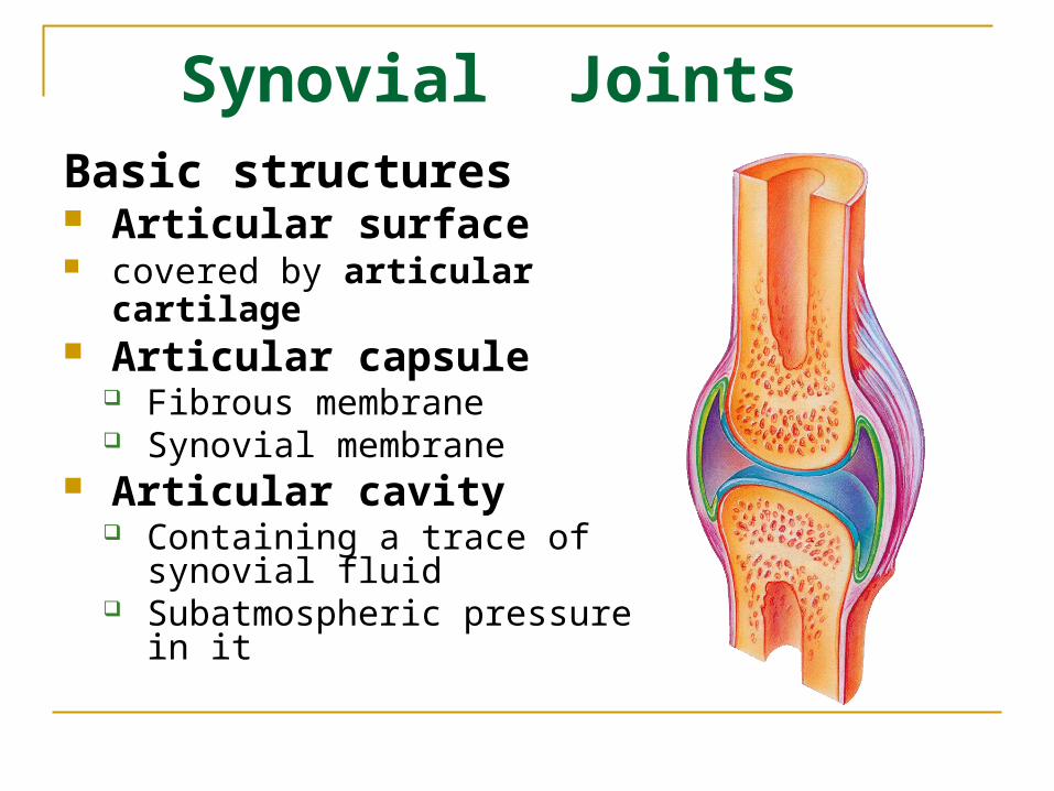
Synovial Joints Basic structures Articular surface covered by articular cartilage Articular capsule
Fibrous membrane Synovial membrane
Articular cavity Containing a trace of synovial
fluid Subatmospheric pressure in it
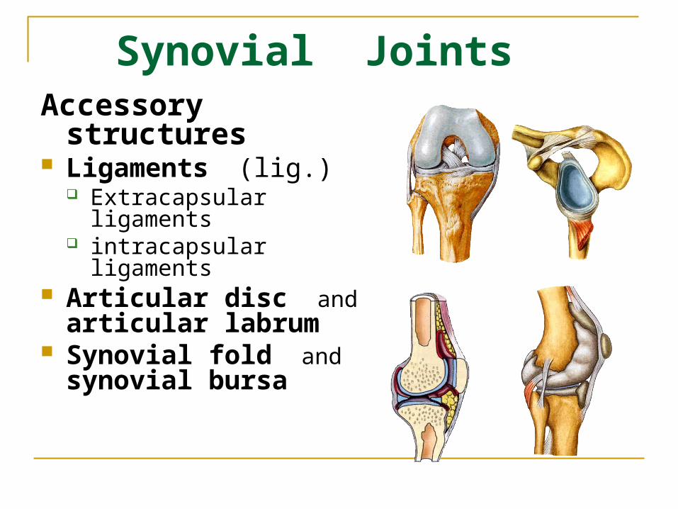
Synovial Joints Accessory structures Ligaments (lig.)
Extracapsular ligaments intracapsular ligaments
Articular disc and articular labrum
Synovial fold and synovial bursa
Joint Movement Terminology
Translation Flexion and extension Adduction and abduction Rotation
Medial and lateral rotation Pronation ans supination Inversion and eversion
Circumduction

flexion\ extension

adduction\ abduction

Rotation Bone revolves around its
own longitudinal axis medial rotation is turning of
anterior surface in towards the midline
lateral rotation is turning of anterior surface away from the midline
Pronation Supination Inversion Eversion

Circumduction Movement of a distal end of a body part in a circle Combination of flexion, extension, adduction and
abduction Occurs at ball and socket, saddle and condyloid joints
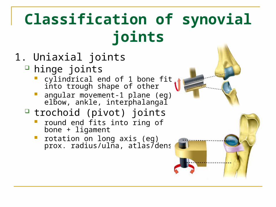
Classification of synovial joints
1. Uniaxial joints hinge joints
cylindrical end of 1 bone fits into trough shape of other
angular movement-1 plane (eg) elbow, ankle, interphalangal
trochoid (pivot) joints round end fits into ring of bone +
ligament rotation on long axis (eg) prox.
radius/ulna, atlas/dens

Classification of synovial joints
2. Biaxial joints ellipsoid joints
egg-shape articular surface + oval concavity
side-to-side, back+forth movement (eg) metacarpophalangeal (knuckle)
saddle joints articular surface both concave +
convex side-to-side, back-forth
movement (eg) carpometacarpal jt of thumb

Classification of synovial joints
3. Multiaxial joints : ball-and-socket joint
spherical head + round socket
multiaxial movement(eg) shoulder, femur
plane joints articular surface in flat plane Short gliding movement (eg) intertarsal, articular
processes of vertebrae

Sprains• Torn or stretched
ligaments.• Spine, ankle & knee are
common sites.• Completely torn ligaments
must be repaired. Edema associated with injury can further degrade the ligament.

Dislocations

Arthritis
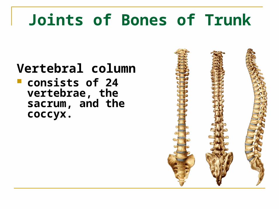
Joints of Bones of Trunk
Vertebral column consists of 24 vertebrae,
the sacrum, and the coccyx.

Joints of the vertebral bodies
Intervertebral discs Lie between bodies of
adjacent vertebrae, composed of:
Nucleus pulposus an inner soft, pulpy, highly
elastic structure (gelatinous core )
Annulus fibrosus an outer fibrous ring consisting
of fibrocartilage

Joints of the vertebral bodies
Anterior longitudinal ligament
Strong band covering the anterior part of the vertebral bodies and intervertebral discs running from the anterior margin of foramen magnum to the S1~S2
Maintains stability of the intervertebral disc and prevents hyperextension of the vertebral column

Joints of the vertebral bodies
Posterior longitudinal ligament
Attached to the posterior aspect of the intervertebral discs and posterior edges of the vertebral bodies from C2 vertebra to sacrum
Prevents hyperflexion of the vertebral column and posterior protrusion of the discs

Hemination of nucleus pulposus
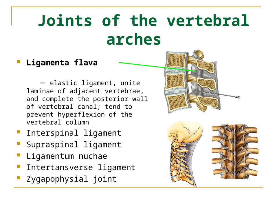
Joints of the vertebral arches
Ligamenta flava
― elastic ligament, unite laminae of adjacent vertebrae, and complete the posterior wall of vertebral canal; tend to prevent hyperflexion of the vertebral column
Interspinal ligament Supraspinal ligament Ligamentum nuchae Intertansverse ligament Zygapophysial joint
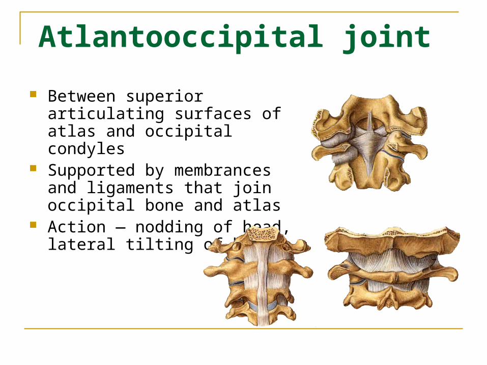
Atlantooccipital joint
Between superior articulating surfaces of atlas and occipital condyles
Supported by membrances and ligaments that join occipital bone and atlas
Action ― nodding of head, lateral tilting of head
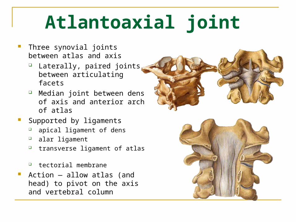
Atlantoaxial joint Three synovial joints between atlas
and axis Laterally, paired joints between
articulating facets Median joint between dens of
axis and anterior arch of atlas Supported by ligaments
apical ligament of dens alar ligament transverse ligament of atlas tectorial membrane
Action ― allow atlas (and head) to pivot on the axis and vertebral column

The vertebral column as a whole
Anterior aspect:
the breadth of vertebral bodies increases from C2 to S2, then diminishes rapidly
Dorsal aspect: the spinous processes of cervical
vertebrae are short and bifid; the spinous processes of thoracic
vertebrae are long, point obliquely downward and overlapped each other;
the spinous processes of lumber vertebrae are nearly horizontally.
Lateral aspect:
shows four physiological curves

The vertebral column as a whole
Four physiological curves
Cervical curvature
Thoracic curvature
Lumbar curvature
Sacral curvature
convex forward
convex backward

The vertebral column as a whole

Movement of the vertebral column
flexion extension lateral flexion rotation
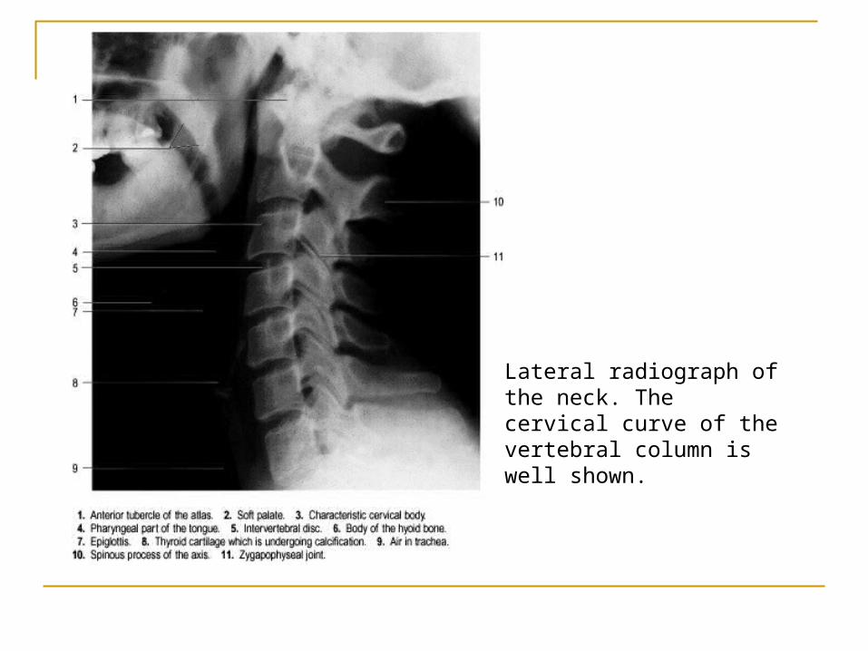
Lateral radiograph of the neck. The cervical curve of the vertebral column is well shown.
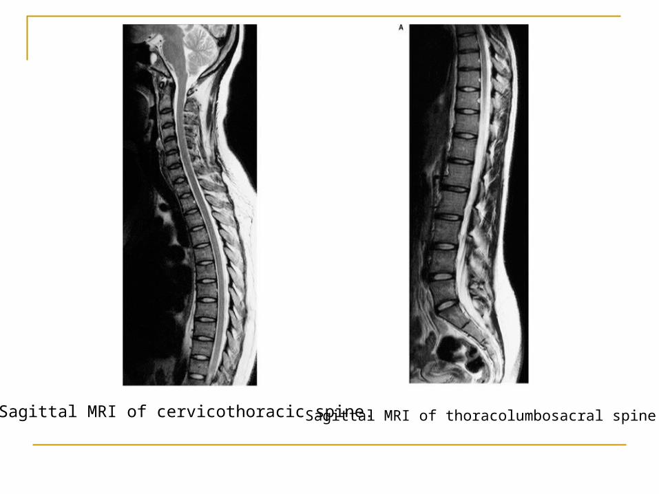
Sagittal MRI of thoracolumbosacral spine. Sagittal MRI of cervicothoracic spine.

Spina bifida cystica
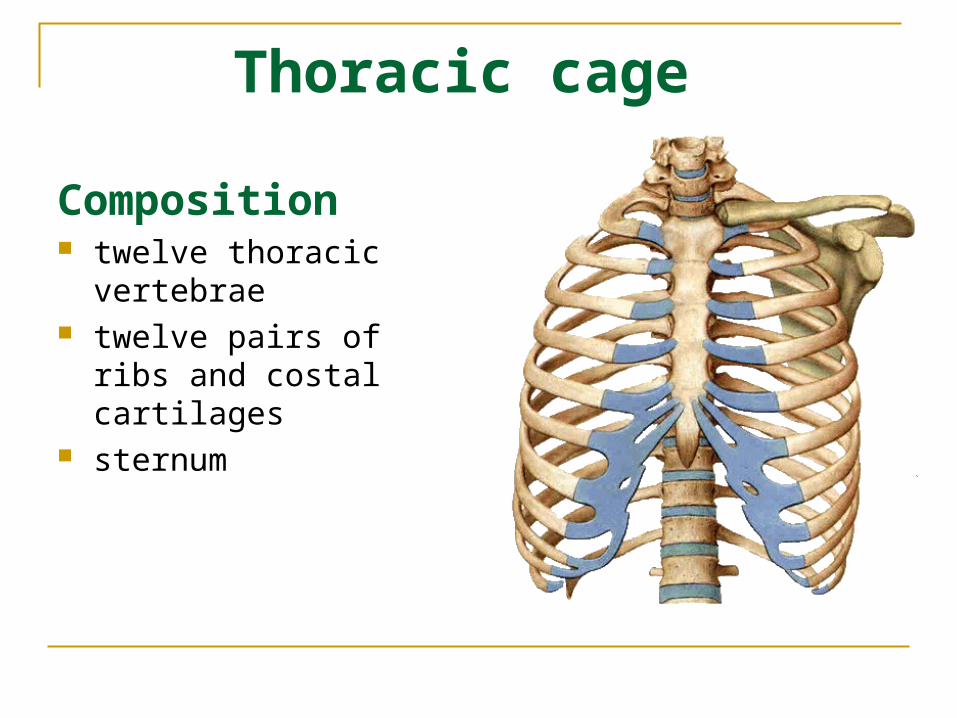
Thoracic cage
Composition twelve thoracic vertebrae twelve pairs of ribs and
costal cartilages sternum
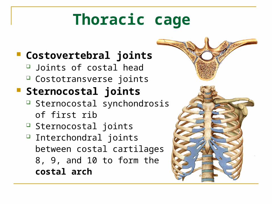
Thoracic cage
Costovertebral joints Joints of costal head Costotransverse joints
Sternocostal joints Sternocostal synchondrosis of
first rib Sternocostal joints Interchondral joints between
costal cartilages 8, 9, and 10 to form the costal arch
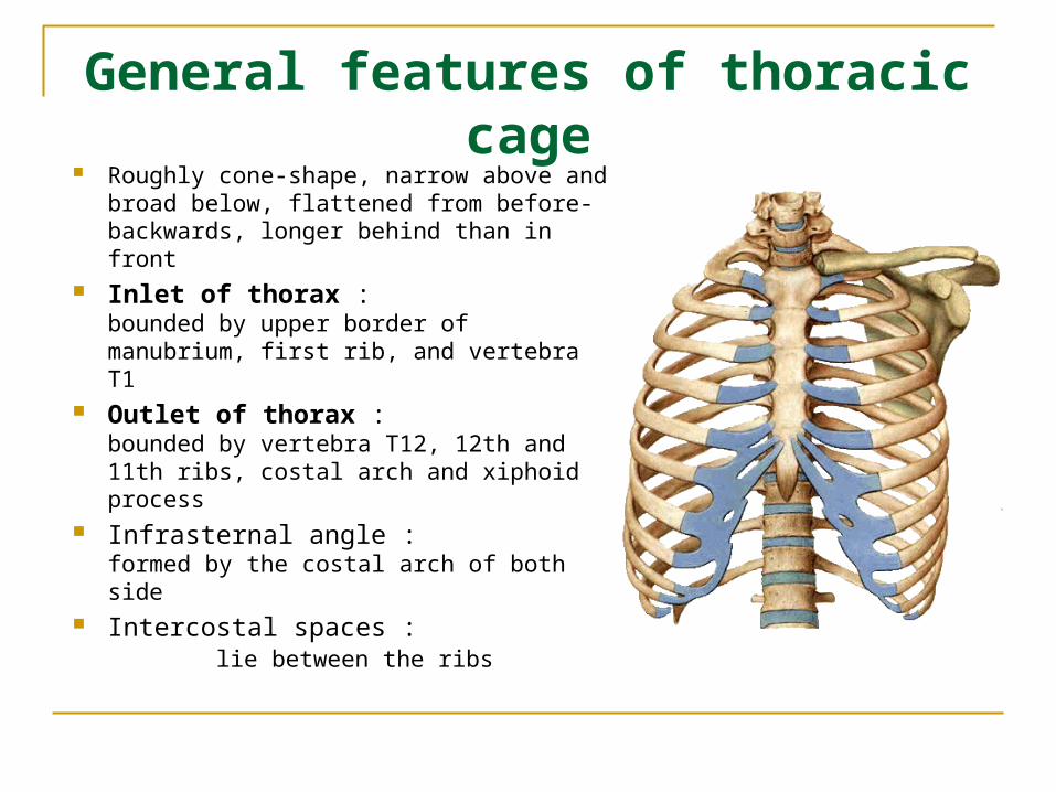
General features of thoracic cage
Roughly cone-shape, narrow above and broad below, flattened from before-backwards, longer behind than in front
Inlet of thorax : bounded by upper border of manubrium, first rib, and vertebra T1
Outlet of thorax : bounded by vertebra T12, 12th and 11th ribs, costal arch and xiphoid process
Infrasternal angle : formed by the costal arch of both side
Intercostal spaces : lie between the ribs

Thoracic cage Function:
protects the organs in the thoracic cavity and upper abdominal cavity
plays a vital role in the process of breathing
Inspiration Expiration


Joints of skull
Continuous joints: Sutures Synchondrosis or synosteosis Temporomandibular joint
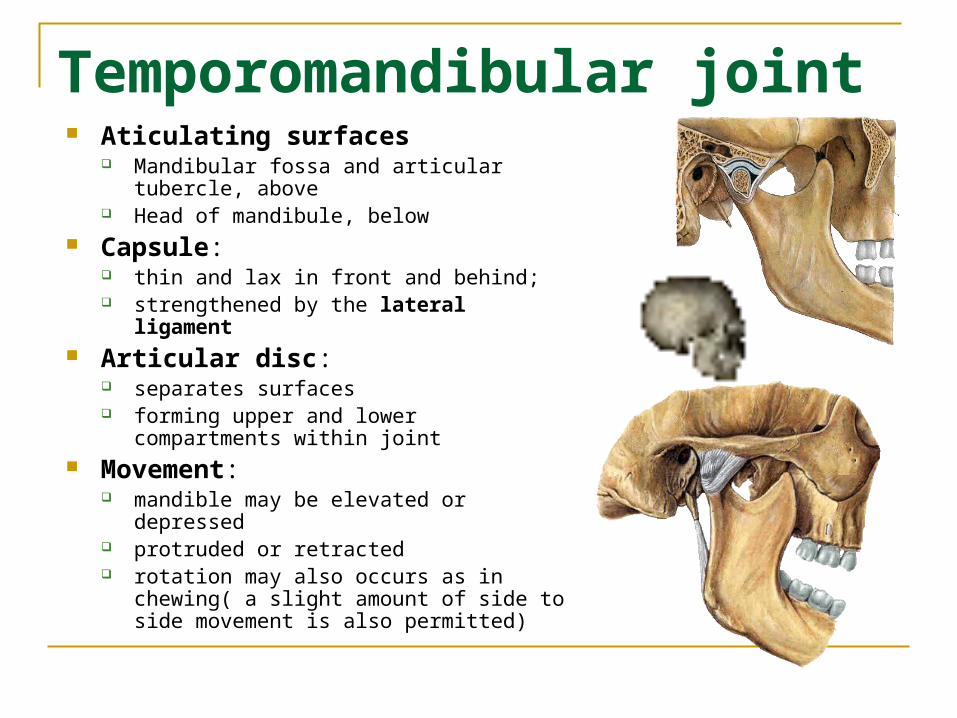
Temporomandibular joint Aticulating surfaces
Mandibular fossa and articular tubercle, above
Head of mandibule, below Capsule:
thin and lax in front and behind; strengthened by the lateral ligament
Articular disc: separates surfaces forming upper and lower compartments within
joint Movement:
mandible may be elevated or depressed protruded or retracted rotation may also occurs as in chewing( a
slight amount of side to side movement is also permitted)