![Emergent Reversal of Anticoagulation in the ED and ICU ... · PDF fileLovenox (enoxaparin sodum injection) [prescribing information]. 2013 Oct. Thromb Haemost 2015;113:931. ... •](https://static.fdocuments.in/doc/165x107/5a9627c87f8b9ab6188c8799/emergent-reversal-of-anticoagulation-in-the-ed-and-icu-enoxaparin-sodum-injection.jpg)
Arterioscler Thromb Vasc Biol 1993 Nordt 1822 8
8
T K Nordt, K J Klassen, D J Schneider and B E Sobel cells by glucose and its implications for local fibrinolysis. Augmentation of synthesis of plasminogen activator inhibitor type-1 in arterial endothelial Print ISSN: 1079-5642. Online ISSN: 1524-4636 Copyright © 1993 American Heart Association, Inc. All rights reserved. Avenue, Dallas, TX 75231 is published by the American Heart Association, 7272 Greenville Arteriosclerosis, Thrombosis, and Vascular Biology doi: 10.1161/01.ATV.13.12.1822 1993;13:1822-1828 Arterioscler Thromb Vasc Biol. http://atvb.ahajournals.org/content/13/12/1822 World Wide Web at: The online version of this article, along with updated information and services, is located on the http://atvb.ahajournals.org//subscriptions/ at: is online Arteriosclerosis, Thrombosis, and Vascular Biology Information about subscribing to Subscriptions: http://www.lww.com/reprints Information about reprints can be found online at: Reprints: document. Permissions and Rights Question and Answer Further information about this process is available in the is being requested is located, click Request Permissions in the middle column of the Web page under Services. Clearance Center, not the Editorial Office. Once the online version of the published article for which permission can be obtained via RightsLink, a service of the Copyright Arteriosclerosis, Thrombosis, and Vascular Biology Requests for permissions to reproduce figures, tables, or portions of articles originally published in Permissions: by guest on October 14, 2014 http://atvb.ahajournals.org/ Downloaded from by guest on October 14, 2014 http://atvb.ahajournals.org/ Downloaded from
-
Upload
syamsu-akbar-tasima -
Category
Documents
-
view
215 -
download
0
description
fff
Transcript of Arterioscler Thromb Vasc Biol 1993 Nordt 1822 8

T K Nordt, K J Klassen, D J Schneider and B E Sobelcells by glucose and its implications for local fibrinolysis.
Augmentation of synthesis of plasminogen activator inhibitor type-1 in arterial endothelial
Print ISSN: 1079-5642. Online ISSN: 1524-4636 Copyright © 1993 American Heart Association, Inc. All rights reserved.
Avenue, Dallas, TX 75231is published by the American Heart Association, 7272 GreenvilleArteriosclerosis, Thrombosis, and Vascular Biology
doi: 10.1161/01.ATV.13.12.18221993;13:1822-1828Arterioscler Thromb Vasc Biol.
http://atvb.ahajournals.org/content/13/12/1822World Wide Web at:
The online version of this article, along with updated information and services, is located on the
http://atvb.ahajournals.org//subscriptions/
at: is onlineArteriosclerosis, Thrombosis, and Vascular Biology Information about subscribing to Subscriptions:
http://www.lww.com/reprints
Information about reprints can be found online at: Reprints:
document.Permissions and Rights Question and AnswerFurther information about this process is available in theis being requested is located, click Request Permissions in the middle column of the Web page under Services.Clearance Center, not the Editorial Office. Once the online version of the published article for which permission
can be obtained via RightsLink, a service of the CopyrightArteriosclerosis, Thrombosis, and Vascular Biology Requests for permissions to reproduce figures, tables, or portions of articles originally published inPermissions:
by guest on October 14, 2014http://atvb.ahajournals.org/Downloaded from by guest on October 14, 2014http://atvb.ahajournals.org/Downloaded from

1822
Augmentation of Synthesis of PlasminogenActivator Inhibitor Type-1 in ArterialEndothelial Cells by Glucose and ItsImplications for Local Fibrinolysis
Thomas K. Nordt, Kevin J. Klassen, David J. Schneider, Burton E. Sobel
Because of the frequent occurrence of premature cardiovascular disease in patients with non-insulin-dependent, type II diabetes mellitus (NIDDM), the attenuated fibrinolytic activity of plasma from type IIdiabetic patients with increased concentrations of plasminogen activator inhibitor type-1 (PAI-1), and thefact that insulin stimulates synthesis of PAI-1 by human hepatic cells in vitro, we and others havehypothesized that accelerated vascular disease in type II diabetes may result in part from impairedfibrinolysis secondary to excessive elaboration of PAI-1 stimulated by insulin. Alternatively, thehyperglycemia associated with type II diabetes could influence the synthesis and secretion of PAI-1directly. The present study was performed to determine whether PAI-1 secretion is or is not sensitive tothe prevailing concentration of glucose in the conditioned medium of endothelial and liver cells, which arethought to be the major sources of circulating PAI-1 in vivo. Confluent cells were exposed to 0, 2.8, 5.6,11.1, or 22.2 mmol/L (0, 50,100, 200, or 400 mg/dL) glucose in medium without serum and subsequentlyto media with or without insulin (7.3 nmoI/L). Secretion of PAI-1 by highly differentiated humanhepatoma (Hep G2) cells did not increase as a function of increasing concentrations of glucose, whetheror not insulin was present. In contrast, with pig aortic endothelial cells, the secretion of PAI-1 increasedsignificantly with extracellular glucose with or without insulin. The increases in PAI-1 were specific (asshown by metabolic labeling experiments) and not attributable to osmotic effects (as shown byreplacement of glucose by sorbitol). Furthermore, the changes were paralleled by a specific, significantincrease in the concentration of PAI-1 mRNA. These results indicate that increases in PAI-1 activity intype II diabetic patients are likely to be attributable to direct effects of glucose on the synthesis of PAI-1by arterial endothelial cells, in addition to the effects of insulin on the synthesis of PAI-1 by liver cells. Theeffects of glucose on endothelial cells may contribute to reduced local fibrinolysis, thereby exacerbatingatherogenesis. (Arterioscler Thromb. 1993;13:1822-1828.)
KEYWORDS • hyperglycemia • fibrinolysis • atherogenesis • atherosclerosis • cardiovasculardisease • type II diabetes • non-insulin-dependent diabetes mellitus
A ccelerated atherosclerosis is associated with di-/ \ verse conditions in which insulin resistance and
- Z \ ~ hyperinsulinemia are prominent, includingtype II, non-insulin-dependent diabetes mellitus(NIDDM).1 Because the activity of plasminogen activa-tor inhibitor type-1 (PAI-1), the primary physiologicalinhibitor of tissue-type plasminogen activator (TPA) inplasma, is increased in NIDDM2-3 and because patho-physiological concentrations of insulin stimulate PAI-1synthesis in human hepatic cells (although not in humanendothelial cells45), we and others have hypothesizedthat the accelerated atherosclerosis may be secondary,in part, to an unfavorable shift in the balance between
Received May 27, 1993; revision accepted September 22, 1993.From the Cardiovascular Division, Washington University
School of Medicine, St Louis, Mo.Preliminary results of this study were presented at the 65th
Scientific Sessions of the American Heart Association and werepublished in abstract form {Circulation 1992;86[suppl I]:I-491).
Correspondence to Thomas K. Nordt, MD, CardiovascularDivision, Box 8086, Washington University School of Medicine,660 South Euclid Ave, St. Louis, MO 63110.
thrombosis and fibrinolysis.6'7 Under conditions of suchan imbalance, cells lining the vessel walls may beexposed inordinately to clot-associated mitogens thatcan potentiate the migration and proliferation of vascu-lar smooth muscle cells,8 chemotaxis and activation ofmacrophages,9 and, accordingly, atherogenesis. Com-plex atherosclerotic plaques frequently contain compo-nents of thrombi,10 a phenomenon consistent with thismechanism.
If impaired fibrinolysis is a contributor to acceleratedatherosclerosis in patients with NIDDM and otherconditions characterized by insulin resistance, then elu-cidation of the mechanisms responsible should facilitatethe development of effective strategies for retarding theotherwise accelerated atherosclerosis. Unfortunately,however, it is not clear whether the increases in activityof PAI-1 and its concentration in plasma are a conse-quence of augmentation of PAI-1 synthesis by factorsother than hyperinsulinemia per se."-'6
We and others have shown that insulin increasesPAI-1 synthesis directly in Hep G2 cells in vitro inconcentrations that are seen in the plasma in patients
by guest on October 14, 2014http://atvb.ahajournals.org/Downloaded from
Nordt et al Hyperglycemia and Fibrinolysis 1823
with NIDDM.45 The present study was performed todetermine whether or not increases in the concentrationof glucose consistent with the hyperglycemia seen inNIDDM can influence PAI-1 synthesis directly. Be-cause arterial and venous, fetal and adult endothelialcells may differ,17 we used adult aortic endothelial cellsto simulate human arterial endothelial cells that arethought to be pivotal determinants of atherogenesis.
MethodsCell Cultures
Hep G2 cells were acquired from the American TypeCulture Collection (Rockville, Md) and grown to con-fluence in minimum essential medium with Earle's saltsand L-glutamine (Life Technologies, Grand Island, NY)supplemented with 10% NuSerum (Collaborative Bio-medical, Bedford, Mass), 30 U/mL penicillin, and 30/u,g/mL streptomycin (Life Technologies). Monolayersof confluent cells were serum starved in Dulbecco'smodified Eagle's medium with Ham's nutrient mixtureF-12 and /V-2-hydroxyethylpiperazine-/V'-2-ethane-sulfonic acid (DME, Washington University MedicalSchool Tissue Culture Support Center, St Louis, Mo)for 16 hours to permit PAI-1 synthesis to decline tobasal levels. Cells were exposed to selected concentra-tions of glucose in glucose-free DME supplementedwith cell culture-grade D-glucose (Sigma Chemical, StLouis, Mo). After serum starvation of the cells wascomplete, the medium was changed so that cells couldbe exposed to medium constituted with cell culture-grade bovine insulin (Sigma) first dissolved in distilledwater with 1% glacial acetic acid (at 10 mg/mL insulin,according to the manufacturer's recommendation) andthen further diluted in DME with 10% fetal bovineserum (FBS, Life Technologies). The FBS was used tominimize the adherence of insulin to the storage andtransfer devices. Its final concentration was 0.1%. Con-trol experiments were performed with vehicle alone. Allsolutions and components were verified to be endotoxinfree with the Limulus amebocyte assay (Pyrotell assay,Associates of Cape Cod, Woods Hole, Mass). Palmiticacid (Sigma) was dissolved in distilled water, suspendedin 10% bovine albumin (final concentration), and usedas an alternative energy source for the cells.
Aortic endothelial cells were harvested from aortas offarm pigs (30 to 60 kg) with the use of collagenase type2 (Worthington Biochemicals, Freehold, NJ) accordingto a protocol approved by the Washington UniversityAnimal Studies Committee. They were grown to conflu-ence in medium 199 (Life Technologies) with 20% FBS,30 U/mL penicillin, 30 yxg/mL streptomycin, and 50^.g/mL endothelial cell growth supplement (ECGS,Collaborative Biomedical) in positively charged cell-culture dishes (Polystyrene Plastek C, Tekmat, Ash-land, Mass). Confluent cell monolayers were exposed toserum-free medium (DME with 50 Aig/mL ECGS) for48 hours to permit PAI-1 synthesis to decline to basallevels.
In conditioned media of both Hep G2 and pig endo-thelial cells, the concentrations of glucose were verifiedspectrophotometrically in assays with hexokinase andglucose-6-phosphate dehydrogenase (SVR GlucoseTest, Behring Diagnostics, Somerville, NJ) and the useof a glucose autoanalyzer (Gemeni, Electro-Nucleonics,
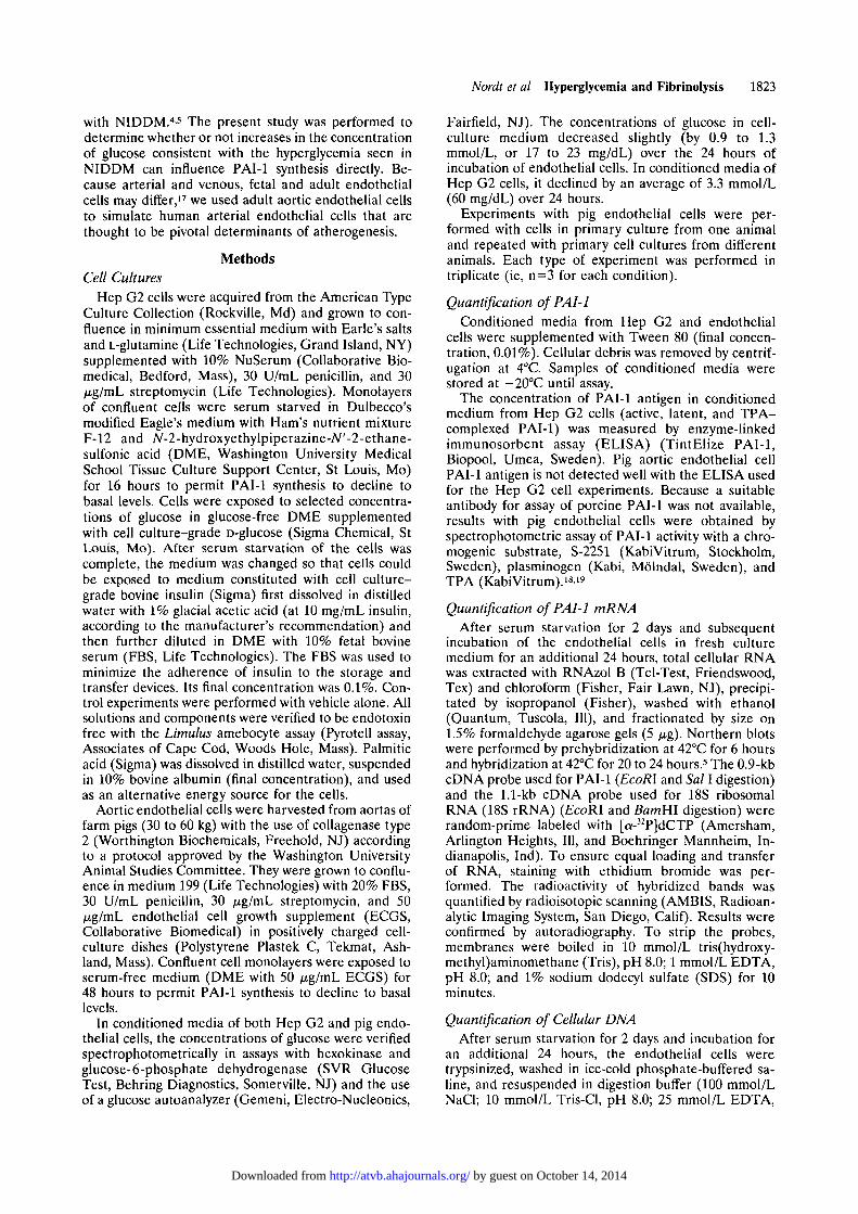
Fairfield, NJ). The concentrations of glucose in cell-culture medium decreased slightly (by 0.9 to 1.3mmol/L, or 17 to 23 mg/dL) over the 24 hours ofincubation of endothelial cells. In conditioned media ofHep G2 cells, it declined by an average of 3.3 mmol/L(60 mg/dL) over 24 hours.
Experiments with pig endothelial cells were per-formed with cells in primary culture from one animaland repeated with primary cell cultures from differentanimals. Each type of experiment was performed intriplicate (ie, n=3 for each condition).
Quantification of PAI-1Conditioned media from Hep G2 and endothelial
cells were supplemented with Tween 80 (final concen-tration, 0.01%). Cellular debris was removed by centrif-ugation at 4°C. Samples of conditioned media werestored at — 20°C until assay.
The concentration of PAI-1 antigen in conditionedmedium from Hep G2 cells (active, latent, and TPA-complexed PAI-1) was measured by enzyme-linkedimmunosorbent assay (ELISA) (TintElize PAI-1,Biopool, Umea, Sweden). Pig aortic endothelial cellPAI-1 antigen is not detected well with the ELISA usedfor the Hep G2 cell experiments. Because a suitableantibody for assay of porcine PAI-1 was not available,results with pig endothelial cells were obtained byspectrophotometric assay of PAI-1 activity with a chro-mogenic substrate, S-2251 (KabiVitrum, Stockholm,Sweden), plasminogen (Kabi, Molndal, Sweden), andTPA (KabiVitrum).'819
Quantification of PAI-1 mRNAAfter serum starvation for 2 days and subsequent
incubation of the endothelial cells in fresh culturemedium for an additional 24 hours, total cellular RNAwas extracted with RNAzol B (Tel-Test, Friendswood,Tex) and chloroform (Fisher, Fair Lawn, NJ), precipi-tated by isopropanol (Fisher), washed with ethanol(Quantum, Tuscola, 111), and fractionated by size on1.5% formaldehyde agarose gels (5 ng). Northern blotswere performed by prehybridization at 42°C for 6 hoursand hybridization at 42°C for 20 to 24 hours.5 The 0.9-kbcDNA probe used for PAI-1 (EcoRl and Sal I digestion)and the 1.1-kb cDNA probe used for 18S ribosomalRNA (18S rRNA) (£coRI and BamHl digestion) wererandom-prime labeled with [a-32P]dCTP (Amersham,Arlington Heights, 111, and Boehringer Mannheim, In-dianapolis, Ind). To ensure equal loading and transferof RNA, staining with ethidium bromide was per-formed. The radioactivity of hybridized bands wasquantified by radioisotopic scanning (AMBIS, Radioan-alytic Imaging System, San Diego, Calif). Results wereconfirmed by autoradiography. To strip the probes,membranes were boiled in 10 mmol/L tris(hydroxy-methyl)aminomethane (Tris), pH 8.0; 1 mmol/L EDTA,pH 8.0; and 1% sodium dodecyl sulfate (SDS) for 10minutes.
Quantification of Cellular DNAAfter serum starvation for 2 days and incubation for
an additional 24 hours, the endothelial cells weretrypsinized, washed in ice-cold phosphate-buffered sa-line, and resuspended in digestion buffer (100 mmol/LNaCl; 10 mmol/L Tris-Cl, pH 8.0; 25 mmol/L EDTA,
by guest on October 14, 2014http://atvb.ahajournals.org/Downloaded from

1824 Arteriosclerosis and Thrombosis Vol 13, No 12 December 1993
100
80
60
~ 40
20
• Control03 Insulin 7.3 nM
2.8 5.6 11.1 16.7
Glucose (mM)
22.2
FIG 1. Bar graph showing the concentra-tion of plasminogen activator inhibitortype-1 (PAI-1) protein in conditioned me-dium of Hep G2 cells exposed to 0, 2.8,5.6,11.1,16.7, or 22.2 mmol/L (0, 50, 100,200, 300, or 400 mg/dL) glucose underconditions of serum starvation for 16 hoursfollowed by subsequent 24-hour incuba-tions with 7.3 nmol/L insulin or vehiclealone. Results are means of four experi-ments performed in triplicate (*P<.05 vsPAI-1 at 2.8 mmol/L glucose). The insetshows the dose-response curve for PAI-1protein in conditioned medium of Hep G2cells exposed to increasing concentrationsof insulin in regular DME (10 mmol/L glu-cose) for 24 hours after serum starvationfor 16 hours (*P<.05, ***P<.0005). DMEindicates Dulbecco's modified Eagle's me-dium with Ham's nutrient mixture F-12 andA/-2-hydroxyethylpiperazine-/V'-2-ethane-sulfonic acid.
pH 8.0; 0.5% SDS; and 0.1 mg/mL proteinase K fromSigma). After incubation with gentle agitation at 50°Cfor 16 hours, DNA was extracted by phenol/chloroform/isoamyl alcohol (25:24:1, vol/vol/vol, saturated with 10mmol/L Tris, pH 8.0, and 1 mmol/L EDTA; Sigma) andprecipitated with 7.5 mol/L ammonium acetate and100% ethanol. The pellets were rinsed with 70% etha-nol and resuspended in TE buffer (10 mmol/L Tris-Cland 1 mmol/L EDTA, pH 8.0).
The concentrations of solubilized DNA were mea-sured fluorometrically (TKO 100 Mini Fluorometer,Hoefer, San Francisco, Calif; excitation wavelength, 365nm; emission wavelength, 460 nm) in TEN buffer (10mmol/L Tris, 1 mmol/L EDTA, and 100 mmol/L NaCl,pH 7.4) with to-benzimidazole (a fluorochrome fromHoechst, No. 33258, highly specific for DNA, acquiredfrom Calbiochem-Novabiochem, La Jolla, Calif) andcalf thymus DNA as a standard (activated type XV,Sigma).
Metabolic Labeling of Protein Synthesizedby Cells in Culture
To assay the rates of synthesis of cellular protein, cellswere metabolically labeled. After serum starvation for 2days and stimulation of the cells in culture for 23 hours,DME was replaced by DME devoid of methionine.After 30 minutes, the cells were exposed to freshmedium containing 50 /xCi/mL [35S]methionine(Tran35S-Label, ICN, Irvine, Calif). After an additional60 minutes, the cells were transferred to fresh DMEwith the usual concentration of unlabeled methioninebut no radioactively labeled methionine. Protein inconditioned medium, supplemented with Tween 80, wasprecipitated with 12.5% (wt/vol) trichloroacetic acid.The cells were washed with phosphate-buffered salineand lysed with ice-cold buffer (10 mmol/L Tris, pH 7.4;150 mmol/L NaCl; 1% Nonidet P-40; 0.5% sodiumdeoxycholate; 0.1% SDS; 1 mmol/L phenylmethylsulfo-nyl fluoride; and 1 mmol/L iodoacetate). Cell lysateswere scraped, disrupted by sonification on ice for 1
minute, and centrifuged at 10 OOOg for 10 minutes. Thesupernatant fractions were precipitated with 12.5%trichloroacetic acid. Radioactivity in the precipitatesfrom conditioned media and cell lysates was assayed byliquid scintillation spectrometry.
Statistical AnalysisResults are means ±SE. Differences were assessed
with one-way ANOVA and Student's t tests. Signifi-cance was defined as F<.05.
ResultsAccumulation of PAI-1 by Hep G2 Cells Exposed toSelected Concentrations of Glucose
The concentrations of glucose were selected to be inthe range between mild hypoglycemia (2.8 mmol/L) andsevere hyperglycemia (22.2 mmol/L) in NIDDM. It wasrecognized prospectively that medium devoid of glucosemight compromise PAI-1 synthesis nonspecifically be-cause of deprivation of a substrate needed for energyfor cell metabolism. Accordingly, our analysis of thepotential response of PAI-1 synthesis to augmentationof the concentration of glucose focused on the changeselicited by concentrations >2.8 mmol/L (50 mg/dL).
Monolayers of confluent Hep G2 cells were exposedto 0, 2.8, 5.6, 11.1, 16.7, or 22.2 mmol/L (0, 50,100, 200,300, or 400 mg/dL) glucose in conditioned mediumdevoid of serum for 16 hours and in fresh media with orwithout insulin during subsequent incubations for 24hours. PAI-1 synthesis was assayed after exposure of thecells either to 7.3 nmol/L (1000 /xU/mL) insulin, aconcentration to which hepatocytes are likely to beexposed in vivo20 by portal venous blood draining di-rectly from the pancreas into the liver, or to vehiclealone. PAI-1 was assayed in the conditioned medium todefine the magnitude of increases induced by glucose orinsulin.
The concentration of PAI-1 protein in conditionedmedium did not increase when the concentration ofglucose was increased from 2.8 to 22.2 mmol/L (Fig 1).
by guest on October 14, 2014http://atvb.ahajournals.org/Downloaded from

40 r
Nordt et al Hyperglycemia and Fibrinolysis 1825
40 r
2.8 5.6 11.1 22.2Glucose (mM)
FIG 2. Bar graph of plasminogen activator inhibitortype-1 (PAI-1) activity in conditioned medium of pig aorticendothelial cells exposed to 0, 2.8, 5.6, 11.1, or 22.2mmol/L (0, 50, 100, 200, or 400 mg/dL) glucose inmedium without serum for 48 hours followed by subse-quent 24-hour incubations in fresh medium. Results aremeans of four experiments performed in triplicate(*P<.05, **P<.005, ***P<.0005 vs PAI-1 at 2.8 mmol/Lglucose). AU indicates arbitrary units.
Thus, values were consistently close to the concentra-tions of PAI-1 seen with 2.8 and 5.6 mmol/L glucose(41 ±3 ng/mL). Insulin did not elicit accumulation ofPAI-1 in the absence of glucose (29±3 ng/mL PAI-1without insulin and 32±4 ng/mL with insulin), probablybecause of the lack of an adequate energy supply. In thepresence of 0.25 mmol/L palmitic acid as an alternativesource of energy (without concomitant glucose), insulindid stimulate accumulation of PAI-1 by 18±2 ng/mL,consistent with this hypothesis. In the presence of allother concentrations of glucose, insulin stimulatedPAI-1 synthesis between 1.6- and 1.8-fold, consistentwith the results we obtained with insulin and its precur-sors in Hep G2 cells.21 At the highest concentration ofglucose (22.2 mmol/L), PAI-1 actually declined mod-estly in the presence and absence of insulin.
PAI-1 Activity in Conditioned Mediumof Endothelial Cells
To determine whether endothelial cells behaved sim-ilarly to Hep G2 cells, pig aortic endothelial cells wereexposed to 0, 2.8, 5.6,11.1, and 22.2 mmol/L (0, 50,100,200, and 400 mg/dL) glucose while being serum starvedfor 2 days and subsequently while exposed to insulin for24 hours. When glucose was present in concentrationsof at least 2.8 mmol/L (50 mg/dL), the concentrations ofglucose decreased slightly over the 24 hours of incuba-tion. The decreases in concentrations of glucose ob-served (decreases of 1.3 mmol/L with 2.8 and 5.6mmol/L glucose, 1.2 mmol/L with 11.1 mmol/L, and 0.9mmol/L with 22.2 mmol/L) were similar to those seen byothers.22
As shown in Fig 2, PAI-1 activity was 12.0±2.4arbitrary units (AU)/mL under basal conditions whenglucose was absent from the conditioned medium. With2.8 mmol/L glucose, PAI-1 activity increased to16.6±0.8 AU/mL, presumably because of the greateravailability of energy for protein biosynthesis. Increasesin the concentration of glucose to 5.6, 11.1, and 22.2mmol/L led to further increases in PAI-1 activity in
30
Illll2.82.8
2.8 8.3 19.4 Sorbitol (mM)2.85 6
2.811.1
2.8 Glucose (mM)22.2 Total (mM)
FIG 3. Bar graph of plasminogen activator inhibitortype-1 (PAI-1) activity in conditioned medium of pig aorticendothelial cells exposed to 2.8 mmol/L (50 mg/dL)glucose plus 0,2.8,8.3, or 19.4 mmol/L sorbitol (to obtaintotal concentrations of 0, 2.8, 5.6,11.1, or 22.2 mmol/L) inmedium devoid of serum for 48 hours followed by sub-sequent 24-hour incubations with the vehicle used forinsulin alone. Results represent means of three experi-ments performed in triplicate. AU indicates arbitrary units.
comparison with PAI-1 activity with 2.8 mmol/L glucose(23.0±1.7 [P=.0U], 26.4±0.8 [P<.001], and 28.6+2.0AU/mL [P=.001], respectively). Insulin concentrationsof 0.01, 0.1, and 1 nmol/L did not elicit additionalincreases of PAI-1 activity in conditioned medium.Even 7.3 nmol/L insulin (1000 /xU/mL), a supraphysio-logical concentration, did not increase PAI-1 activity.
When sorbitol was substituted for glucose to simulateits osmotic effects (Fig 3), PAI-1 activity ranged from11.9±2.5 (with 5.6 mmol/L total concentration of glu-cose plus sorbitol) to 18.0±0.9 AU/mL (with 11.1 mmol/L). With the highest amounts of glucose plus sorbitol,PAI-1 activity increased, but not significantly so(F=.O81 with 22.2 mmol/L compared with PAI-1 with2.8 mmol/L). Thus, sorbitol exerted no marked stimu-lation of PAI-1 synthesis. Accordingly, osmotic effectsof glucose per se are not likely to substantially influencePAI-1 synthesis.
Effects of Glucose on the Concentration of PAI-1mRNA in Endothelial Cells
Aortic endothelial cells were incubated with 2.8, 5.6,11.1, or 22.2 mmol/L glucose (50, 100, 200, or 400mg/dL) while being serum starved for 2 days and weresubsequently exposed to fresh medium for 24 hours.Subsequently, RNA was extracted and assayed. Twospecies of PAI-1 mRNA (3.2 and 2.2 kb) were detectedin cell lysates. The 3.2-kb form constituted approxi-mately 95% of total PAI-1 mRNA.
As shown in Fig 4 and the Table, the concentration oftotal PAI-1 mRNA increased significantly when cellswere exposed to increasing concentrations of glucose.The radioactivity detected increased from 159±10 to280±12 counts per minute. In regular DME (with aconcentration of 10 mmol/L glucose), 7.3 nmol/L insulindid not increase PAI-1 mRNA compared with that seenwith DME without insulin over 24 hours, confirming thelack of an effect of insulin on PAI-1 activity.
To confirm the conclusion that increases in PAI-1mRNA were not simply a result of an increased numberof cells, we performed additional parallel experiments
by guest on October 14, 2014http://atvb.ahajournals.org/Downloaded from

1826 Arteriosclerosis and Thrombosis Vol 13, No 12 December 1993
400 r
350
~ 300
O 250
2 0 0I| 150
100
50
• 3 2 kb
•22 kb
B 3 2 kb PAI-1 mRNA
• 2 2 kb PAI-1 mRNA
2.8 22.25.6 11.1Glucose (mM)
FIG 4. Bar graph of plasminogen activator inhibitortype-1 (PAI-1) mRNA expression (2.2- and 3.2-kb forms)in pig aortic endothelial cells exposed to 2.8, 5.6,11.1, or22.2 mmol/L (50, 100, 200, or 400 mg/dL) glucose inmedium without serum for 48 hours followed by subse-quent 24-hour incubations in fresh medium. The insetshows a representative autoradiogram. The upper bandis 3.2-kb and the lower band 2.2-kb PAI-1 mRNA. Resultsrepresent means of three experiments performed in trip-licate (**P<.005 vs PAI-1 mRNA at 2.8 mmol/L).
in which DNA was measured. The results are shown inthe Table, with those of PAI-1 mRNA values normal-ized for DNA. The total amount of DNA per cultureflask remained virtually constant regardless of the con-centration of glucose. This observation is consistentwith the results by Kaiser et al,22 who exposed confluentcalf aortic endothelial cells to selected concentrations ofglucose for 24 to 48 hours and saw no change in cellnumber. A decrease in cell number and thymidineincorporation was seen by Maiello et al,23 in humanumbilical vein endothelial cells exposed to high concen-trations of glucose. This decrease may have occurredbecause of the long incubations in medium (13±4 daysfor primary cultures).
To verify the consistency of loading and transfer ofRNA to the membranes, additional experiments wereperformed in which 18S rRNA was quantified. Themembranes with the size-fractionated RNA used forPAI-1 mRNA assay were stripped first because the 18SrRNA bands (at 1.9 kb) would have interfered with the2.2-kb PAI-1 bands. Thus, after stripping, the mem-branes were rehybridized with a cDNA probe for 18SrRNA. No significant glucose-dependent changes in 18S
rRNA concentrations were evident (Table). In fact, thePAI-1 mRNA to 18S rRNA ratio increased with in-creasing concentrations of glucose, indicative of a rela-tively specific effect of glucose on the concentration ofPAI-1 mRNA.
Effects of Glucose on Overall Protein Synthesis inEndothelial Cells
Metabolic labeling of all newly synthesized proteinwas performed to identify potentially nonspecific effectsof glucose on the synthesis of protein. Metabolic label-ing was preferred to chemical quantification of totalprotein content in conditioned medium because of thepossible interference by the protein in FBS in theconditioned medium that was used to avoid unspecificbinding. Confluent pig aortic endothelial cells wereserum starved for 2 days and stimulated with 7.3 nmol/Linsulin or vehicle alone for the subsequent 23 hours inmedia with 2.8, 5.6, 11.1, or 22.2 mmol/L (50, 100, 200,or 400 mg/dL) glucose, 0.5 hour in methionine-freemedium, and 1 hour in the same medium spiked withlabeled methionine. These concentrations of glucosewere maintained throughout the incubations with me-thionine-free medium and with the medium containinglabeled methionine. At the midpoint of the incubationwith labeled methionine, the duration of exposure ofthe cells to medium corresponded to that precedingharvesting of conditioned medium for assay of PAI-1and harvesting of RNA in the experiments describedabove. Assay of cell lysates 1 hour and of conditionedmedia 24 hours after the addition of labeled methioninedid not indicate any influence of changes in the concen-tration of glucose on protein biosynthesis. Exposure ofthe cells to insulin elicited a modest, nonsignificantincrease in overall protein biosynthesis.
DiscussionThe present study was performed to identify the
potential contribution of hyperglycemia, as opposed tohyperinsulinemia, to the increases in PAI-1 in plasmaseen in hyperinsulinemic, type II diabetic patients. Be-cause the liver and endothelium are considered to be theprimary sources of plasma PAI-1,24 we studied Hep G2cells and pig aortic endothelial cells as model systems.The Hep G2 cell line is an "immortal," well-differenti-ated human hepatoma cell line that responds similarly tohuman hepatocytes in primary culture in terms of induc-tion of PAI-1 synthesis by insulin.25 The pig aorticendothelial cells were used in primary culture onlybecause secretion of PAI-1 by endothelial cells is consti-
Plasminogen Activator Inhibitor Type-1 mRNA in Endothelial Cells
Glucose, mmol/L
2.8
5.6
11.1
22.2
PAI-1 mRNA, cpm
159+10
232 ±3
260 ±8
280±12
DNA, fig
19±2
20±2
20±2
22±3
PAI-1 mRNAto DNA
Ratio, cpm
159±10
220 ±3
247 ±8
241 ±10
18S rRNA,cpm
548±130
525 ±21
571 +64
525±117
PAI-1 mRNAto 18S rRNARatio, cpm
159±10
242±3
250 ±8
292±11
Plasminogen activator inhibitor type-1 (PAI-1) mRNA in pig aortic endothelial cells was exposed to 2.8, 5.6, 11.1,or 22.2 mmol/L (50, 100, 200, or 400 mg/dL) glucose during serum starvation for 48 hours followed by 24-hourincubations. Results are means±SE. PAI-1 mRNA values that are normalized for DNA contents and concentrationsof 18S rRNA are shown in columns 4 and 6.
by guest on October 14, 2014http://atvb.ahajournals.org/Downloaded from
Nordt et al Hyperglycemia and Fibrinolysis 1827
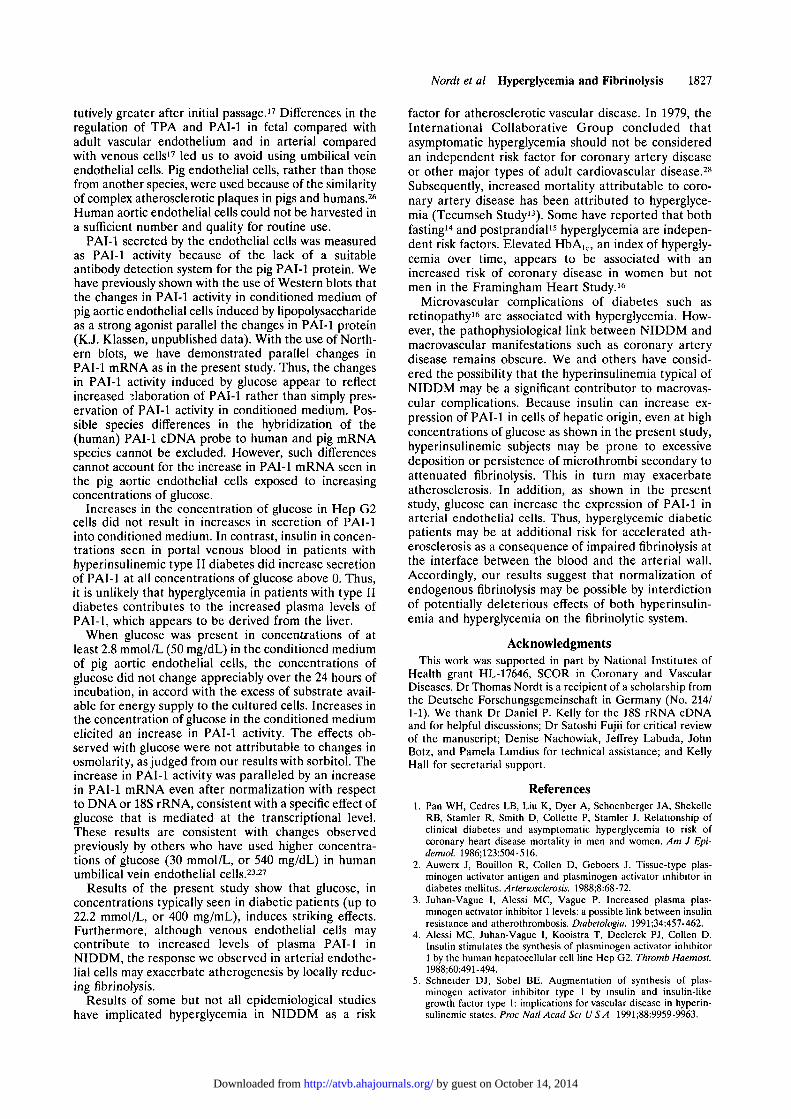
tutively greater after initial passage.17 Differences in theregulation of TPA and PAI-1 in fetal compared withadult vascular endothelium and in arterial comparedwith venous cells17 led us to avoid using umbilical veinendothelial cells. Pig endothelial cells, rather than thosefrom another species, were used because of the similarityof complex atherosclerotic plaques in pigs and humans.26
Human aortic endothelial cells could not be harvested ina sufficient number and quality for routine use.
PAI-1 secreted by the endothelial cells was measuredas PAI-1 activity because of the lack of a suitableantibody detection system for the pig PAI-1 protein. Wehave previously shown with the use of Western blots thatthe changes in PAI-1 activity in conditioned medium ofpig aortic endothelial cells induced by lipopolysaccharideas a strong agonist parallel the changes in PAI-1 protein(K.J. Klassen, unpublished data). With the use of North-ern blots, we have demonstrated parallel changes inPAI-1 mRNA as in the present study. Thus, the changesin PAI-1 activity induced by glucose appear to reflectincreased elaboration of PAI-1 rather than simply pres-ervation of PAI-1 activity in conditioned medium. Pos-sible species differences in the hybridization of the(human) PAI-1 cDNA probe to human and pig mRNAspecies cannot be excluded. However, such differencescannot account for the increase in PAI-1 mRNA seen inthe pig aortic endothelial cells exposed to increasingconcentrations of glucose.
Increases in the concentration of glucose in Hep G2cells did not result in increases in secretion of PAI-1into conditioned medium. In contrast, insulin in concen-trations seen in portal venous blood in patients withhyperinsulinemic type II diabetes did increase secretionof PAI-1 at all concentrations of glucose above 0. Thus,it is unlikely that hyperglycemia in patients with type IIdiabetes contributes to the increased plasma levels ofPAI-1, which appears to be derived from the liver.
When glucose was present in concentrations of atleast 2.8 mmol/L (50 mg/dL) in the conditioned mediumof pig aortic endothelial cells, the concentrations ofglucose did not change appreciably over the 24 hours ofincubation, in accord with the excess of substrate avail-able for energy supply to the cultured cells. Increases inthe concentration of glucose in the conditioned mediumelicited an increase in PAI-1 activity. The effects ob-served with glucose were not attributable to changes inosmolarity, as judged from our results with sorbitol. Theincrease in PAI-1 activity was paralleled by an increasein PAI-1 mRNA even after normalization with respectto DNA or 18S rRNA, consistent with a specific effect ofglucose that is mediated at the transcriptional level.These results are consistent with changes observedpreviously by others who have used higher concentra-tions of glucose (30 mmol/L, or 540 mg/dL) in humanumbilical vein endothelial cells.23-27
Results of the present study show that glucose, inconcentrations typically seen in diabetic patients (up to22.2 mmol/L, or 400 mg/mL), induces striking effects.Furthermore, although venous endothelial cells maycontribute to increased levels of plasma PAI-1 inNIDDM, the response we observed in arterial endothe-lial cells may exacerbate atherogenesis by locally reduc-ing fibrinolysis.
Results of some but not all epidemiological studieshave implicated hyperglycemia in NIDDM as a risk
factor for atherosclerotic vascular disease. In 1979, theInternational Collaborative Group concluded thatasymptomatic hyperglycemia should not be consideredan independent risk factor for coronary artery diseaseor other major types of adult cardiovascular disease.28
Subsequently, increased mortality attributable to coro-nary artery disease has been attributed to hyperglyce-mia (Tecumseh Study13). Some have reported that bothfasting14 and postprandial15 hyperglycemia are indepen-dent risk factors. Elevated HbAlc, an index of hypergly-cemia over time, appears to be associated with anincreased risk of coronary disease in women but notmen in the Framingham Heart Study.15
Microvascular complications of diabetes such asretinopathy16 are associated with hyperglycemia. How-ever, the pathophysiological link between NIDDM andmacrovascular manifestations such as coronary arterydisease remains obscure. We and others have consid-ered the possibility that the hyperinsulinemia typical ofNIDDM may be a significant contributor to macrovas-cular complications. Because insulin can increase ex-pression of PAI-1 in cells of hepatic origin, even at highconcentrations of glucose as shown in the present study,hyperinsulinemic subjects may be prone to excessivedeposition or persistence of microthrombi secondary toattenuated fibrinolysis. This in turn may exacerbateatherosclerosis. In addition, as shown in the presentstudy, glucose can increase the expression of PAI-1 inarterial endothelial cells. Thus, hyperglycemic diabeticpatients may be at additional risk for accelerated ath-erosclerosis as a consequence of impaired fibrinolysis atthe interface between the blood and the arterial wall.Accordingly, our results suggest that normalization ofendogenous fibrinolysis may be possible by interdictionof potentially deleterious effects of both hyperinsulin-emia and hyperglycemia on the fibrinolytic system.
AcknowledgmentsThis work was supported in part by National Institutes of
Health grant HL-17646, SCOR in Coronary and VascularDiseases. Dr Thomas Nordt is a recipient of a scholarship fromthe Deutsche Forschungsgemeinschaft in Germany (No. 214/1-1). We thank Dr Daniel P. Kelly for the 18S rRNA cDNAand for helpful discussions; Dr Satoshi Fujii for critical reviewof the manuscript; Denise Nachowiak, Jeffrey Labuda, JohnBotz, and Pamela Lundius for technical assistance; and KellyHall for secretarial support.
References
1. Pan WH, Cedres LB, Liu K, Dyer A, Schoenberger JA, ShekelleRB, Stamler R, Smith D, Collette P, Stamler J. Relationship ofclinical diabetes and asymptomatic hyperglycemia to risk ofcoronary heart disease mortality in men and women. Am J Epi-demiol. 1986;123:504-516.
2. Auwerx J, Bouillon R, Collen D, Geboers J. Tissue-type plas-minogen activator antigen and plasminogen activator inhibitor indiabetes mellitus. Arteriosclerosis. 1988;8:68-72.
3. Juhan-Vague I, Alessi MC, Vague P. Increased plasma plas-minogen activator inhibitor 1 levels: a possible link between insulinresistance and atherothrombosis. Diabetologia. 1991;34:457-462.
4. Alessi MC, Juhan-Vague I, Kooistra T, Declerck PJ, Collen D.Insulin stimulates the synthesis of plasminogen activator inhibitor1 by the human hepatocellular cell line Hep G2. Thromb Haemost.1988;60:491-494.
5. Schneider DJ, Sobel BE. Augmentation of synthesis of plas-minogen activator inhibitor type 1 by insulin and insulin-likegrowth factor type 1: implications for vascular disease in hyperin-sulinemic states. Proc Natl Acad Sci U S A 1991;88:9959-9963.
by guest on October 14, 2014http://atvb.ahajournals.org/Downloaded from
1828 Arteriosclerosis and Thrombosis Vol 13, No 12 December 1993
6. Sobel BE, Hirsh J. Principles and practice of coronary throm-bolysis and conjunctive treatment. Am J Cardiol. 1991;68:382-388.Editorial.
7. Eisenberg PR, Miletich JP, Sobel BE. Factors responsible fordifferential procoagulant effects of diverse plasminogen activatorsin plasma. Fibrinotysis. 1991;5:217-224.
8. Jawien A, Bowen-Pope DF, Lindner V, Schwartz SM, Clowes AW.Platelet-derived growth factor promotes smooth muscle migrationand intimal thickening in a rat model of balloon angioplasty. J ClinInvest. 1992;89:507-511.
9. Deuel TF, Senior RM, Huang JS, Griffin GL. Chemotaxis ofmonocytes and neutrophils to platelet-derived growth factor. J ChnInvest. 1982;69:1046-1049.
10. Schwartz CJ, Valente AJ, Kelley JL, Sprague EA, Edwards EH.Thrombosis and the development of atherosclerosis: Rokitanskyrevisited. Semin Thromb Hemost. 1988;14:189-195.
11. van Wersch JWJ, Westerhuis LWJJM, Venekamp WJRR. Gly-cometabolic control and fibrinolysis in diabetic patients. Haemo-stasis. 1990;20:241-250.
12. Landin K, Stigendal L, Eriksson E, Krotkiewski M, Risberg B,Tengborn L, Smith U. Abdominal obesity is associated with animpaired fibrinolytic activity and elevated plasminogen activatorinhibitor-1. Metabolism. 1990;39:1044-1048.
13. Butler WJ, Ostrander LD, Carman WJ, Lamphiear DE. Mortalityfrom coronary heart disease in the Tecumseh study. Am J Epi-demiol. 1985;121:541-547.
14. Barrett-Connor E, Wingard DL, Criqui MH, Suarez L. Is bor-derline fasting hyperglycemia a risk factor for cardiovasculardeath? J Chronic Dis. 1984;37:773-779.
15. Donahue RP, Abbott RD, Reed DM, Yano K. Postchallengeglucose concentration and coronary heart disease in men ofJapanese ancestry: Honolulu Heart Program. Diabetes. 1987;36:689-692.
16. Singer DE, Nathan DM, Anderson KM, Wilson PWF, Evans JC.Association of HbA,c with prevalent cardiovascular disease in theoriginal cohort of the Framingham heart study. Diabetes. 1992;41:202-208.
17. van Hinsbergh VWM, Binnema D, Scheffer MA, Sprengers ED,Kooistra T, Rijken DC. Production of plasminogen activators and
inhibitor by serially propagated endothelial cells from adult humanblood vessels. Arteriosclerosis. 1987;7:389-400.
18. Chmielewska J, Wiman B. Determination of tissue plasminogenactivator and its "fast" inhibitor in plasma. Clin Chem. 1986;32:482-485.
19. Lucore CL, Sobel BE. Interactions of tissue-type plasminogenactivator with plasma inhibitors and their pharmacologic impli-cations. Circulation. 1988;77:660-669.
20. Misbin RI, Merimee TJ, Lowenstein JM. Insulin removal byisolated perfused rat liver. Am J Physiol. 1976;230:171-177.
21. Nordt TK, Schneider DJ, Sobel BE. Augmentation of the synthesisof plasminogen activator inhibitor type-1 (PAI-1) by precursors ofinsulin: a potential risk factor for vascular disease. Circulation. Inpress.
22. Kaiser N, Sasson S, Feener EP, Boukobza-Vardi N, Higashi S,Moller DE, Davidheiser S, Przybylski RJ, King GL. Differentialregulation of glucose transport and transporters by glucose invascular endothelial and smooth muscle cells. Diabetes. 1993;42:80-89.
23. Maiello M, Boeri D, Podesta F, Cagliero E, Vichi M, Odetti P,Adezati L, Lorenzi M. Increased expression of tissue plasminogenactivator and its inhibitor and reduced fibrinolytic potential ofhuman endothelial cells cultured in elevated glucose. Diabetes1992;41:1009-1015.
24. Kooistra T. The use of cultured human endothelial cells and hepa-tocytes as an in vitro model system to study modulation ofendogenous fibrinolysis. Fibrinolysis. 1990;4(suppl 2):33-39.
25. Kooistra T, Bosma PJ, Tons HAM, van den Berg AP, Meyer P,Princen HMG. Plasminogen activator inhibitor 1: biosynthesis andmRNA level are increased by insulin in cultured human hepa-tocytes. Thromb Haemost. 1989;62:723-728.
26. Hughes HC. Swine in cardiovascular research. LabAnim Sci. 1986;36:348-350.
27. Cagliero E, Roth T, Roy S, Maiello M, Lorenzi M. Expression ofgenes related to the extracellular matrix in human endothelialcells. J Biol Chem. 1991;266:14244-14250.
28. The International Collaborative Group. Joint discussion. J ChronicDis. 1979:32:829-837.
by guest on October 14, 2014http://atvb.ahajournals.org/Downloaded from