MAKATI MEDICAL CENTER DEPARTMENT OF MEDICINE MEDICAL GRANDROUNDS
Upload
berniece-simonCategory
view
229download
5
Arterial Blood GasArterial Blood Gas INTERPRETATIONINTERPRETATION
Manuel Antonio Ko, MDManuel Antonio Ko, MDSection of Pulmonary MedicineSection of Pulmonary Medicine
Department of Internal MedicineDepartment of Internal MedicineMakati Medical CenterMakati Medical Center

Objectives
Learn how to systematically interpret Arterial
Blood Gas results
Identify the different causes of abnormalities in
the ABG results
Problem solving exercises

Indications for Arterial Blood Gas Determination
• Evaluate Ventilatory, Oxygenation, Acid
Base and Oxygen carrying capacity of
blood
• Monitor severity and disease progression
• Quantify patient’s response to therapeutic intervention and or diagnostic evaluation
Respircare, 1992, 317
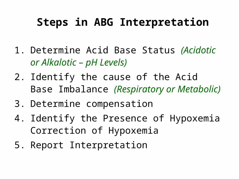
1. Determine Acid Base Status (Acidotic or Alkalotic – pH Levels)
2. Identify the cause of the Acid Base Imbalance (Respiratory or Metabolic)
3. Determine compensation
4. Identify the Presence of Hypoxemia Correction of Hypoxemia
5. Report Interpretation
Steps in ABG Interpretation
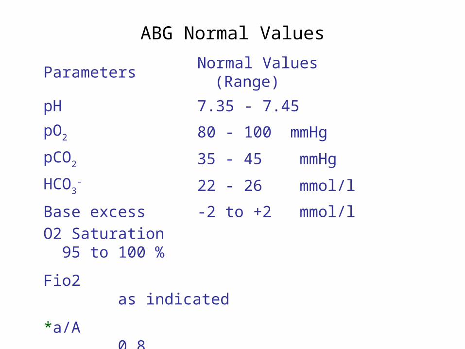
ABG Normal Values
Parameters Normal Values (Range)
pH 7.35 - 7.45
pO2 80 - 100 mmHg
pCO2 35 - 45 mmHg
HCO3- 22 - 26 mmol/l
Base excess -2 to +2 mmol/l
O2 Saturation 95 to 100 %
Fio2 as indicated
*a/A 0.8
* Not available in some ABG Machines

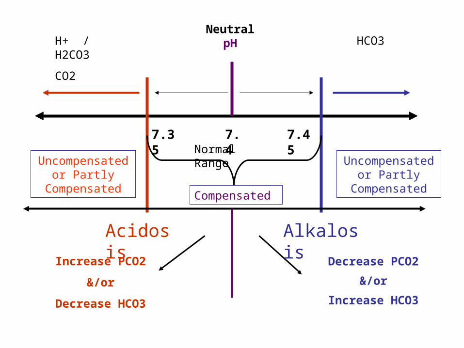
Neutral pH
7.47.35 7.45Acidosis AlkalosisNormal Range
H+ / H2CO3
CO2
HCO3
Normal
or
Compensated?

ABG Normal Values
Parameters Normal Values (Range)
pH 7.35 - 7.45
pO2 80 - 100 mmHg
pCO2 35 - 45 mmHg
HCO3- 22 - 26 mmol/l
Base excess -2 to +2 mmol/l
Base Excess – quick eye view of the adequacy of the buffer mechanism. If there is more than enough HCO3 to balance the pH (compensation)
Neutral pH
7.47.35 7.45
Acidosis Alkalosis
Normal Range
H+ / H2CO3
CO2
HCO3
Compensated
Uncompensated or Partly
Compensated
Uncompensated or Partly
Compensated
Increase PCO2
&/or
Decrease HCO3
Decrease PCO2
&/or
Increase HCO3

– Acute Respiratory Acidosis • PaCO2 increase by 10 mmHg decreases pH
0.08 • Bicarbonate increases 1 meq/L per 10 mmHg
PaCO2 rise
– Chronic Respiratory Acidosis • PaCO2 increase by 10 mmHg decreases pH
0.03 • Bicarbonate increases 4 meq/L per 10 mmHg
PaCO2 rise
Compensation

Acute Respiratory Acidosis Causes:
1. Central Nervous System Depression Sedative Medications (e.g. Benzodiazepines) Cerebrovascular Accident Head Trauma
2. Neuromuscular Disease Myasthenia Gravis Guillain-Barre Polio Muscular Dystrophy Hypokalemia
3. Impaired lung motion Pleural Effusion Pneumothorax Crush injury

Acute Respiratory Acidosis
Causes:
4. Acute airway obstruction Foreign Body Aspiration Tumor Laryngospasm (e.g. Croup, Epiglottitis) Bronchospasm (e.g. Asthma, COPD)
5. Acute Respiratory Disease Severe Pneumonia Pulmonary edema
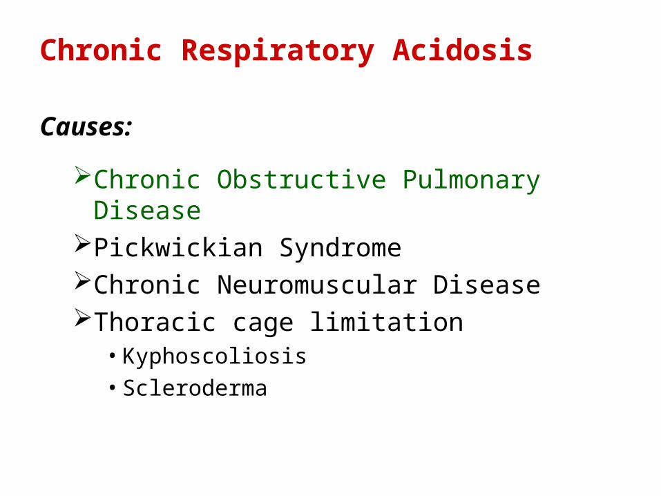
Chronic Respiratory Acidosis
Causes:
Chronic Obstructive Pulmonary DiseasePickwickian Syndrome Chronic Neuromuscular Disease Thoracic cage limitation
• Kyphoscoliosis• Scleroderma

– Acute Respiratory Alkalosis • PaCO2 decreases by 10 mmHg increases pH by
0.08 • Bicarbonate decreases 2 meq/L per 10 mmHg
PaCO2 fall
– Chronic Respiratory Alkalosis • PaCO2 decrease by 10 mmHg increases pH by
0.03
• Bicarbonate decreases 4 meq/L per 10 mmHg PaCO2 fall
Compensation
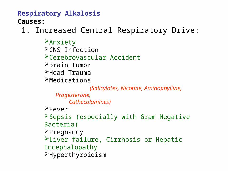
Respiratory Alkalosis Causes:
1. Increased Central Respiratory Drive:
Anxiety CNS Infection Cerebrovascular AccidentBrain tumor Head Trauma Medications
(Salicylates, Nicotine, Aminophylline, Progesterone, Cathecolamines)
Fever Sepsis (especially with Gram Negative Bacteria) Pregnancy Liver failure, Cirrhosis or Hepatic Encephalopathy Hyperthyroidism

Respiratory Alkalosis Causes:
2. Increased Chemoreceptor Stimulation
Anemia Carbon Monoxide Poisoning Pulmonary edema Pneumonia Pulmonary Embolism High altitude (decreased FIO2) Restrictive lung disease (early)
3. Iatrogenic with Mechanical Ventilation

Metabolic Acidosis
– PaCO2 decreased
PaCO2 drops 1.2 mmHg per 1 meq/L bicarbonate fall
Calculated PaCO2 = 1.5 x HCO3 + 8 (+/- 2)
Measured PaCO2 discrepancy: respiratory disorder
Useful in High Anion Gap Metabolic Acidosis
Compensation

Elevated Anion Gap Acidosis
• Anion Gap Definition
– Difference between calculated serum anions and
cations
• Calculation
– AG = Serum Na – (Serum Cl + Serum HCO3)
• Interpretation
– Normal Anion Gap: 12 +/- 2 meq/L
Metabolic Acidosis

Elevated Anion Gap Acidosis (Mnemonic: "MUD PILERS") causes:
Methanol Intoxication Uremia Diabetic Ketoacidosis (DKA) or starvation ketosis Paraldehyde, Phenformin Isopropyl Alcohol, Isoniazid Lactic Acidosis Ethylene Glycol, ethyl alcohol Rhabdomyolysis Salicylates Other Causes: Hyperalbuminemia, administered anions
Metabolic Acidosis

Normal Anion Gap (Hyperchloremic Acidosis)
A.) Hypokalemia with Metabolic acidosis: 1. Diarrhea/ Vomiting2. Ureteral diversion
– Uretero-sigmoidostomy – Ileal bladder – Ileal ureter
3. Renal Tubular Acidosis (proximal or distal) *4. Mineralocorticoid Deficiency
– Angiotensin Deficiency: Liver Failure – ACE Inhibitor – Renin Deficiency
» Aging » Extracellular fluid volume expansion » Lead » Beta Blockers » Prostaglandin Inhibitor » Methyldopa
5. Carbonic Anhydrase Inhibitor – Acetazolamide – Mefenamic acid
6. Post-hypocapnia

Normal Anion Gap (Hyperchloremic Acidosis)
B.) (Hyperkalemic or normal Potassium) Metabolic Acidosis
1. Renal Failure (Early)*
2. Renal Disease* – SLE Interstitial Nephritis – Amyloidosis – Hydronephrosis – Sickle Cell Nephropathy
3. Acidifying agents – Ammonium Chloride – Calcium Chloride – Arginine
4. Sulfur toxicity
Metabolic Acidosis
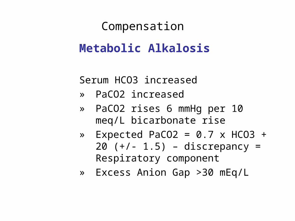
Metabolic Alkalosis
Serum HCO3 increased » PaCO2 increased » PaCO2 rises 6 mmHg per 10 meq/L
bicarbonate rise » Expected PaCO2 = 0.7 x HCO3 + 20
(+/- 1.5) – discrepancy = Respiratory component
» Excess Anion Gap >30 mEq/L
Compensation
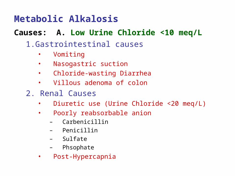
Metabolic Alkalosis
Causes: A. Low Urine Chloride <10 meq/L 1.Gastrointestinal causes
• Vomiting • Nasogastric suction • Chloride-wasting Diarrhea • Villous adenoma of colon
2. Renal Causes • Diuretic use (Urine Chloride <20 meq/L) • Poorly reabsorbable anion
– Carbenicillin
– Penicillin
– Sulfate
– Phsophate
• Post-Hypercapnia
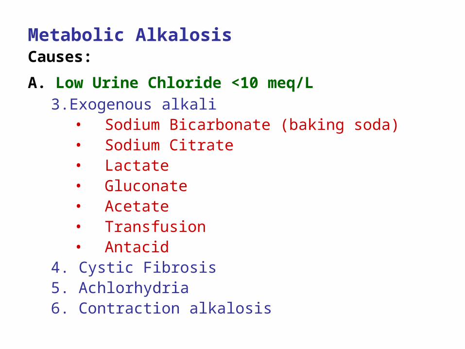
Metabolic Alkalosis Causes:
A. Low Urine Chloride <10 meq/L 3.Exogenous alkali
• Sodium Bicarbonate (baking soda) • Sodium Citrate • Lactate • Gluconate • Acetate • Transfusion • Antacid
4. Cystic Fibrosis 5. Achlorhydria 6. Contraction alkalosis
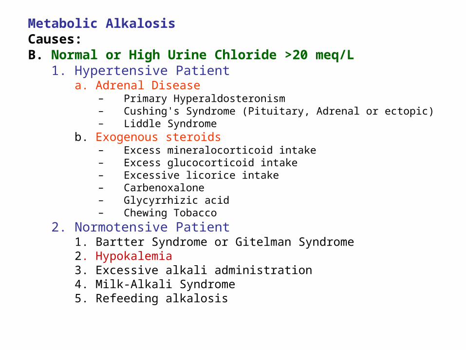
Metabolic Alkalosis Causes: B. Normal or High Urine Chloride >20 meq/L
1. Hypertensive Patient a. Adrenal Disease
– Primary Hyperaldosteronism – Cushing's Syndrome (Pituitary, Adrenal or ectopic) – Liddle Syndrome
b. Exogenous steroids – Excess mineralocorticoid intake – Excess glucocorticoid intake – Excessive licorice intake – Carbenoxalone – Glycyrrhizic acid – Chewing Tobacco
2. Normotensive Patient 1. Bartter Syndrome or Gitelman Syndrome 2. Hypokalemia 3. Excessive alkali administration 4. Milk-Alkali Syndrome5. Refeeding alkalosis

Neutral pH
7.47.35 7.45
Acidosis Alkalosis
Normal Range
H+ / H2CO3
CO2
HCO3
Compensated
Uncompensated or Partly
Compensated
Uncompensated or Partly
Compensated
Increase PCO2
&/or
Decrease HCO3
Decrease PCO2
&/or
Increase HCO3
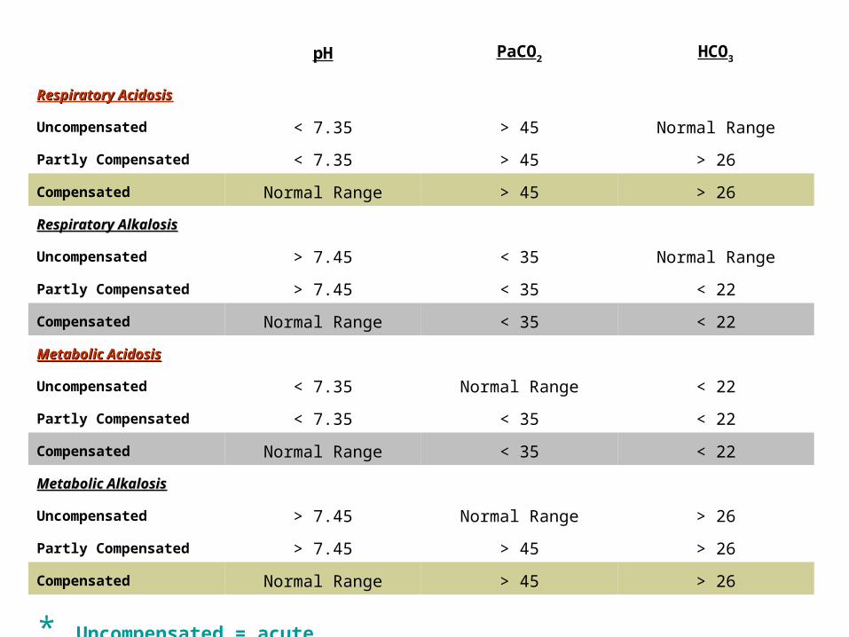
pH PaCO2 HCO3
Respiratory AcidosisRespiratory Acidosis
Uncompensated < 7.35 > 45 Normal Range
Partly Compensated < 7.35 > 45 > 26
Compensated Normal Range > 45 > 26
Respiratory AlkalosisRespiratory Alkalosis
Uncompensated > 7.45 < 35 Normal Range
Partly Compensated > 7.45 < 35 < 22
Compensated Normal Range < 35 < 22
Metabolic AcidosisMetabolic Acidosis
Uncompensated < 7.35 Normal Range < 22
Partly Compensated < 7.35 < 35 < 22
Compensated Normal Range < 35 < 22
Metabolic AlkalosisMetabolic Alkalosis
Uncompensated > 7.45 Normal Range > 26
Partly Compensated > 7.45 > 45 > 26
Compensated Normal Range > 45 > 26
* Uncompensated = acute

pH PaCO2 HCO3
Combined Acidosis<7.35 > 45 < 22
Combined Alkalosis>7.45 < 35 > 26
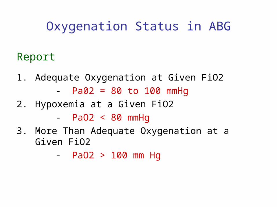
Oxygenation Status in ABG
Report
1. Adequate Oxygenation at Given FiO2
- Pa02 = 80 to 100 mmHg
2. Hypoxemia at a Given FiO2
- PaO2 < 80 mmHg
3. More Than Adequate Oxygenation at a Given FiO2
- PaO2 > 100 mm Hg

Respiratory Components in ABG
• PaO2 – Partial Pressure of Arterial Oxygen
- NV: 80-100mmHG
• PCO2 – Partial Pressure of Carbon Dioxide
- NV: 35-45 mmHg
• SaO2 – Oxygen Saturation in the Blood
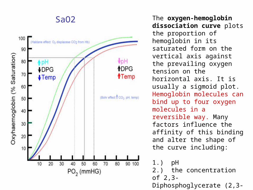
The oxygen-hemoglobin dissociation curve plots the proportion of hemoglobin in its saturated form on the vertical axis against the prevailing oxygen tension on the horizontal axis. It is usually a sigmoid plot. Hemoglobin molecules can bind up to four oxygen molecules in a reversible way. Many factors influence the affinity of this binding and alter the shape of the curve including:
1.) pH2.) the concentration of 2,3-Diphosphoglycerate (2,3-DPG)3.) the type of hemoglobin molecules (adult vs fetal types)4.) the presence of poisons especially carbon monoxide
SaO2
Hypoxemia in ABG
• Inadequate oxygenation
• Pa02 < 80 at a given level of FiO2 - FiO2 (Fraction of Inspired O2)
- 21% at room air ( proportion of O2 in
the atmosphere )

Respiratory Failure
A. Type 1 – Hypoxemia without CO2 retention (Normal PCO2)
ex.: Pulmonary Edema, Pneumonia
B. Type 2 – There is CO2 retention (Increased PCO2)
ex.: Hypoventilation (reduced alveolar ventilation)
pump failure, airway obstruction, neuromuscular
weakness
C. Combined
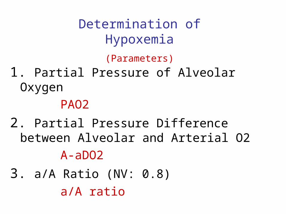
1. Partial Pressure of Alveolar Oxygen
PAO2
2. Partial Pressure Difference between Alveolar and Arterial O2
A-aDO2
3. a/A Ratio (NV: 0.8)
a/A ratio
Determination of Hypoxemia
(Parameters)

PaO2 is generally lower than PAO2
- Physiologic shunt ( unoxygenated blood coming from the coronary arteries draining
into the Thebesian Veins and parts of the Bronchial Arteries both draining directly
into the Pulmonary Veins and bypassing the gas exchange mechanism of the lungs +
diffusion of O2 to the alveolar capillaries)
Widens when there is pulmonary shunting (Pneumonia, edema & etc.).
Also widens when on supplemental oxygen ( more accurate when calculated at room air)
Minimal or no widening if the problem is pure hypoventilation
1
A-aO2Alveolar-Arterial Oxygen Tension Difference (PAO2 – PaO2)

PAO2 = 100 mmHg
Alveolar PO2
(PAO2)
Arterial PO2 (PaO2)Capillary PO2
PaO2 = 80-90mmHg (lower)
O2 Diffusion through the Capillaries + Physiologic
Shunt
A-aDO2
NORMAL

PAO2 = 100 mmHg
Alveolar PO2
(PAO2)
Arterial PO2 (PaO2)Capillary PO2
PaO2 = 50 mmHg (much lower)
O2 Diffusion through the Capillaries + Physiologic
Shunt + hypoxia
Widened A-aDO2
Disease
HYPOXEMIA
Type 1 Resp. Failure

PAO2 = inc. 300 mmHg
Alveolar PO2
(PAO2)
Arterial PO2 (PaO2)Capillary PO2
PaO2 = inc. 75 mmHg
( but slightly lower than normal)
O2 Diffusion through the Capillaries + Physiologic
Shunt + hypoxia
Widened A-aDO2
Disease
Supplemental O2
HYPOXEMIA
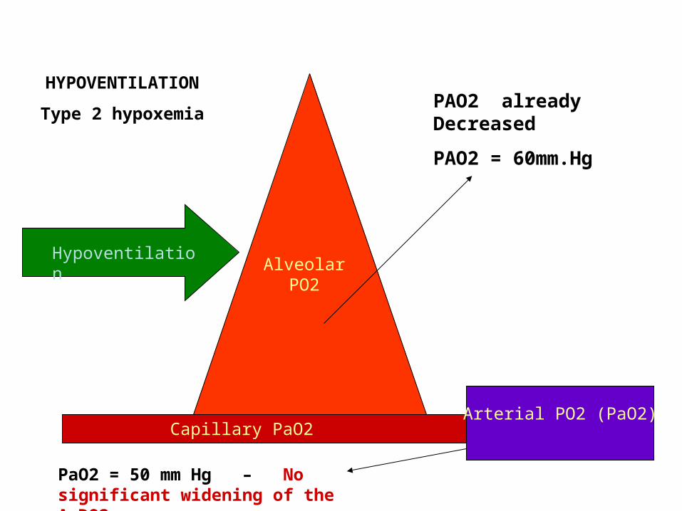
PAO2 already Decreased
PAO2 = 60mm.Hg
Alveolar PO2
Capillary PaO2
Hypoventilation
HYPOVENTILATION
Type 2 hypoxemia
Arterial PO2 (PaO2)
PaO2 = 50 mm Hg – No significant widening of the AaDO2

PACO2 = Decreased
Alveolar PCO2
(PACO2)
Arterial or Mixed Venous PCO2
(PaCO2/ PVCO2)Capillary PCO2
Increase PCO2
Airway Obstruction
Hypoventilation
HYPERCARBIA
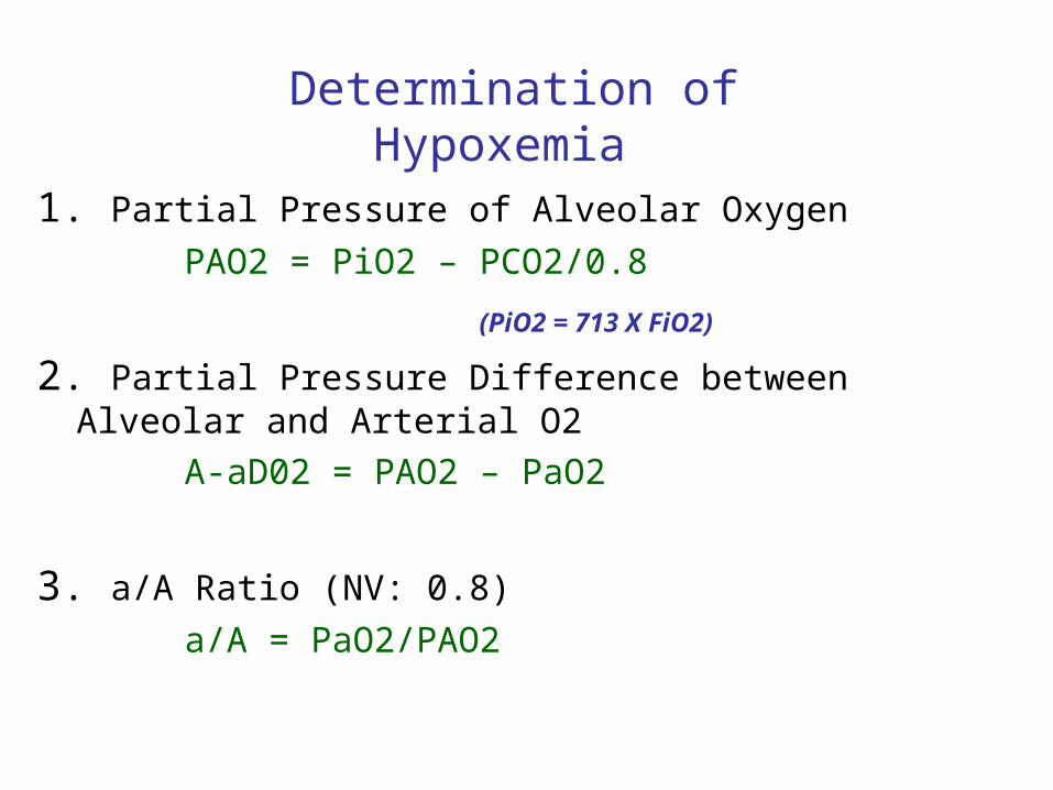
1. Partial Pressure of Alveolar Oxygen
PAO2 = PiO2 – PCO2/0.8
(PiO2 = 713 X FiO2)
2. Partial Pressure Difference between Alveolar and Arterial O2
A-aD02 = PAO2 – PaO2
3. a/A Ratio (NV: 0.8)
a/A = PaO2/PAO2
Determination of Hypoxemia
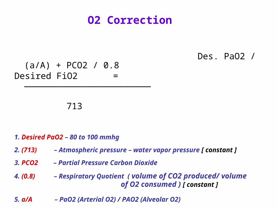
Des. PaO2 / (a/A) + PCO2 / 0.8Desired FiO2 = ________________________
713
1. Desired PaO2 – 80 to 100 mmhg
2. (713) – Atmospheric pressure – water vapor pressure [ constant ]
3. PCO2 – Partial Pressure Carbon Dioxide
4. (0.8) – Respiratory Quotient ( volume of CO2 produced/ volume of O2 consumed ) [ constant ]
5. a/A – PaO2 (Arterial O2) / PAO2 (Alveolar O2)
O2 Correction

P/F Ratio
PaO2/ Fio2 (simple estimate)
• Indicates range of hypoxemia• P/F ratio > or equal 400 - Normal• P/F ratio < 400 - Hypoxemia

Giving O2 Supplement
• Supplemental O2 conversion
- LPM Oxygen = LPM x 4 + 20 ( Fi02)
ex: 2 (LPM O2) x 4 + 20 = 28% FiO2
(using O2 cannula, O2 mask)
(O2 supplementation is not provided by direct FiO2)
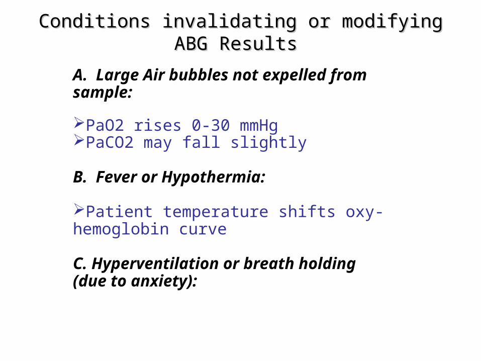
Conditions invalidating or modifying ABG ResultsConditions invalidating or modifying ABG Results
A. Large Air bubbles not expelled from sample:
PaO2 rises 0-30 mmHg PaCO2 may fall slightly
B. Fever or Hypothermia:
Patient temperature shifts oxy-hemoglobin curve
C. Hyperventilation or breath holding (due to anxiety):
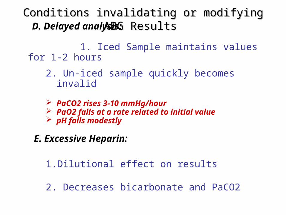
Conditions invalidating or modifying ABG ResultsConditions invalidating or modifying ABG Results
D. Delayed analysis:
1. Iced Sample maintains values for 1-2 hours
2. Un-iced sample quickly becomes invalid
PaCO2 rises 3-10 mmHg/hour PaO2 falls at a rate related to initial value pH falls modestly
E. Excessive Heparin:
1. Dilutional effect on results
2. Decreases bicarbonate and PaCO2

• 65 year old male, seen at the ER, diagnosed case of Chronic Renal Failure presently undergoing Hemodialysis. He was placed on Multivent Mask at 40% FiO2. Chest X-ray shows Increase Broncho-vascular markings with concomitant interstitial and alveolar infiltrates on the Right Lower Lobe
PPE:• T= 37.8, RR: 26, BP: 90/60, CR: 115• Pale skin and Diaphoretic• Use of accessory muscle and abdominal breathing pattern
observed• fine crackles on the right lower lung fields with expiratory
wheezes all over • bipedal edema noted
Case 1

ABG and Labs Values
pH 7.29
PCO2 50 mmHg
PaO2 59 mmHg
HCO3 17 mmol/L
BE -3.5
FiO2 40%
SaO2 79%
K 5.0
Na 132
Cl 95
1. Interpret Acid Base Status?
2. Determine if Hypoxemia Exists ?
3. Compute the a-AO2 gradient ?
4. Compute for the desired FiO2 ?
5. Compute Anion Gap ?
6. Initial Diagnosis?
7. What are your immediate plans for the patient ?

pH PaCO2 HCO3
Combined Acidosis<7.35 > 45 < 22
Combined Alkalosis>7.45 < 35 > 26

Acute Respiratory Acidosis
Causes:
4. Acute airway obstruction Foreign Body Aspiration Tumor Laryngospasm (e.g. Croup, Epiglottitis) Bronchospasm (e.g. Asthma, COPD)
5. Acute Respiratory Disease
Severe Pneumonia Pulmonary Edema

Elevated Anion Gap Acidosis
• Anion Gap Definition
– Difference between calculated serum anions and
cations
• Calculation
– AG = Serum Na – (Serum Cl + Serum HCO3)
• Interpretation
– Normal Anion Gap: 12 +/- 2 meq/L
Metabolic Acidosis

Elevated Anion Gap Acidosis (Mnemonic: "MUD PILERS") causes:
Methanol Intoxication
Uremia Diabetic Ketoacidosis (DKA) or starvation ketosis Paraldehyde, Phenformin Isopropyl Alcohol, Isoniazid Lactic Acidosis Ethylene Glycol, ethyl alcohol Rhabdomyolysis Salicylates Other Causes: Hyperalbuminemia, administered anions
Metabolic Acidosis

Interpretation
• Combined Respiratory and metabolic Acidosis
• Inadequate Oxygenation at 40% FiO2

• 25 year old female, seen at the ER, diagnosed case of Asthma, who complained of difficulty of breathing 2 days ago but apparently she is feeling much better now after inhaling bronchodilators. Past Medical History is unremarkable except for Asthma. She went to the ER because of persistent coughing. Chest X-ray is normal
PPE: • T= 36.8, RR: 14, BP:110/70, CR: 70• Her breath sounds are clear• She refuses to receive supplemental oxygen• Someone, for some reason, ordered an ABG and the results showed
Case 2
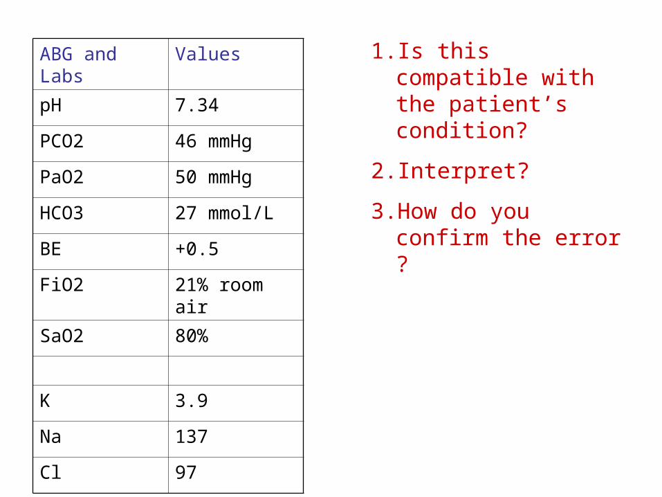
ABG and Labs Values
pH 7.34
PCO2 46 mmHg
PaO2 50 mmHg
HCO3 27 mmol/L
BE +0.5
FiO2 21% room air
SaO2 80%
K 3.9
Na 137
Cl 97
1. Is this compatible with the patient’s condition?
2. Interpret?
3. How do you confirm the error ?

Problem
• Compensated Respiratory Acidosis with hypoxemia at room air (21% FiO2)
• Determine O2 sat by Pulse Oximeter
• SaO2 = SpO2 +/- 4% (difference)
• Probably Mixed Venous Blood Sample obtained
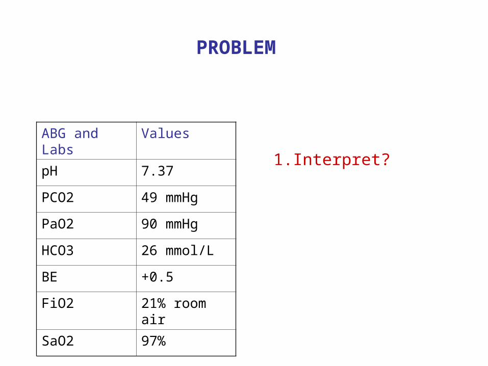
ABG and Labs Values
pH 7.37
PCO2 49 mmHg
PaO2 90 mmHg
HCO3 26 mmol/L
BE +0.5
FiO2 21% room air
SaO2 97%
1. Interpret?
PROBLEM

ABG and Labs Values
pH 7.37
PCO2 49 mmHg
PaO2 90 mmHg
HCO3 25 mmol/L
BE +0.5
FiO2 21% room air
SaO2 97%
1. Normal ?
2. Compensated Respiratory Alkalosis
3. Consider PCO2 levels instead ?(directly measured over HCO3 which is only estimated by machine calculation)
4. Have sample repeated (Consider Error) ?

THANK YOU
for your attention


![[Smarter Makati Presentation : Makati City, Philipines]](https://static.fdocuments.in/doc/165x107/558624e6d8b42a56578b4595/smarter-makati-presentation-makati-city-philipines.jpg)