ADVANCED EKG INTERPRETATION Micelle J. Haydel, M.D. LSU New Orleans Emergency Medicine.
88
ADVANCED EKG INTERPRETATION Micelle J. Haydel, M.D. LSU New Orleans Emergency Medicine
-
Upload
kristopher-hindes -
Category
Documents
-
view
225 -
download
4
Transcript of ADVANCED EKG INTERPRETATION Micelle J. Haydel, M.D. LSU New Orleans Emergency Medicine.
ADVANCED EKG INTERPRETATION
Micelle J. Haydel, M.D.
LSU New Orleans
Emergency Medicine

Image Sources
• My patients
• www.ecglibrary.com
• The Alan E. Lindsay Ecg Learning Center http://medlib.med.utah.edu/kw/ecg/intro.html
• The EKG of the week from NCEMI http://www.ncemi.org
• Emergency Medicine Education http://www.emedu.org
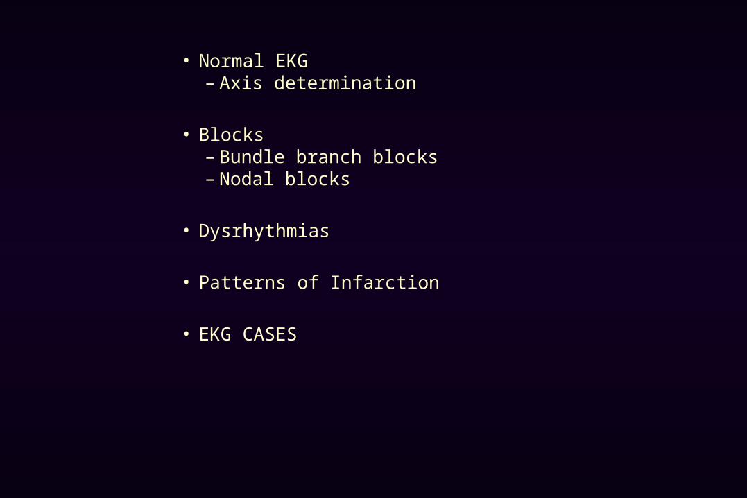
• Normal EKG– Axis determination
• Blocks – Bundle branch blocks– Nodal blocks
• Dysrhythmias
• Patterns of Infarction
• EKG CASES
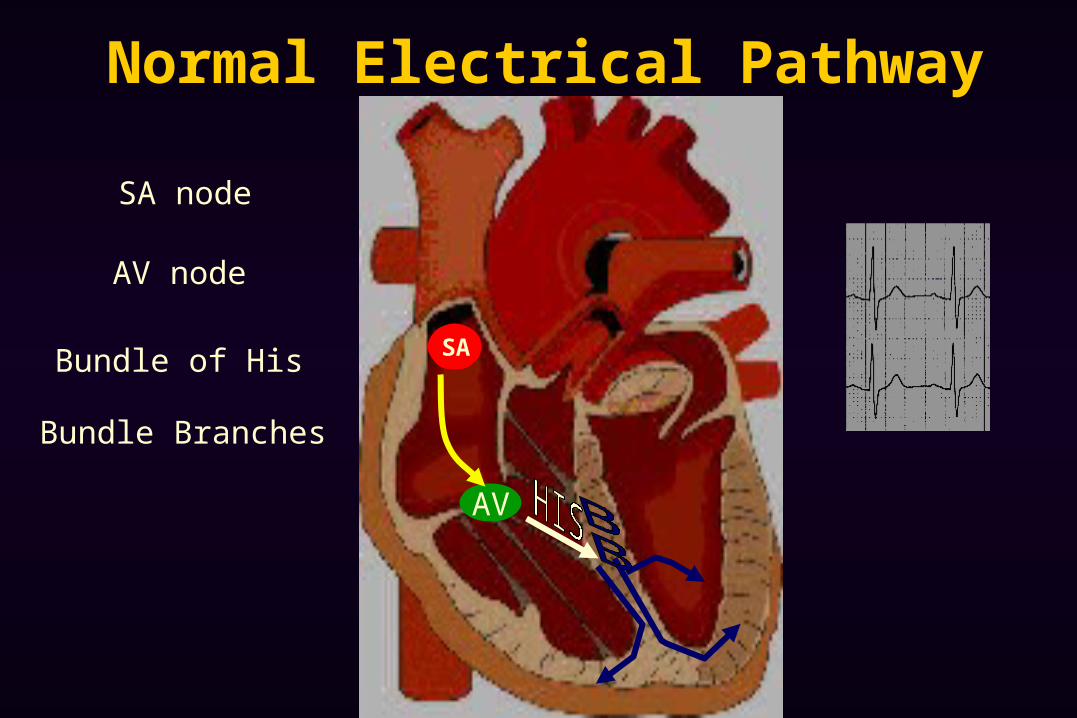
Normal Electrical Pathway
AV
AV node
SA node
Bundle of His
Bundle Branches
SA
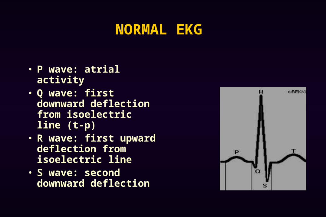
NORMAL EKG
• P wave: atrial activity• Q wave: first downward
deflection from isoelectric line (t-p)
• R wave: first upward deflection from isoelectric line
• S wave: second downward deflection
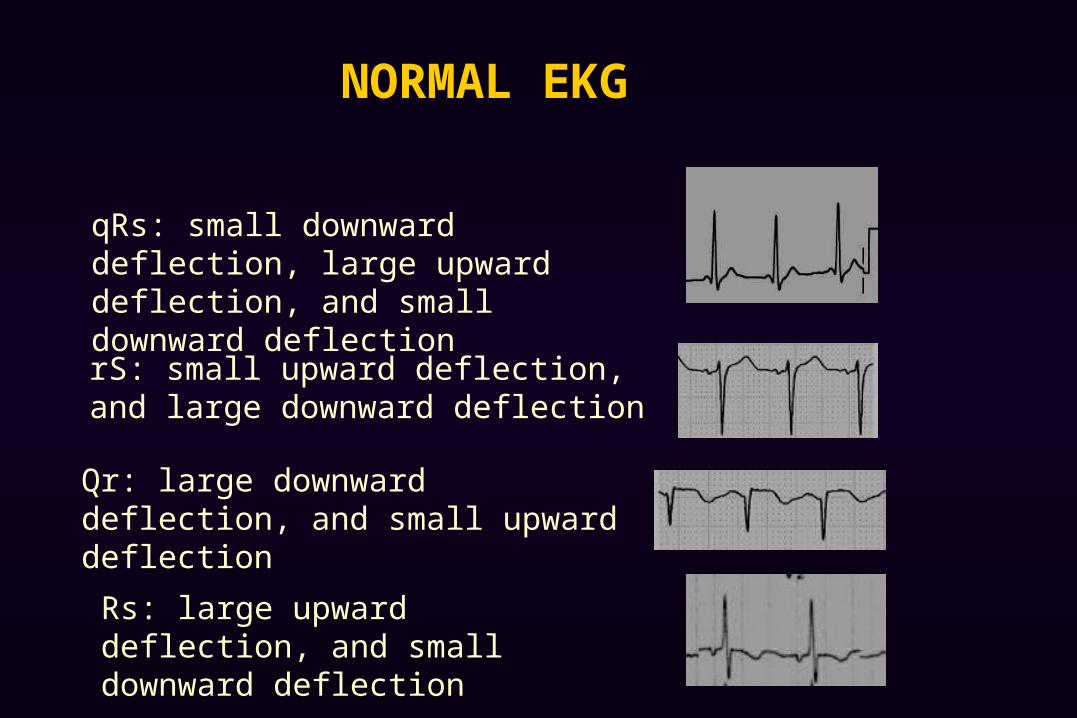
NORMAL EKG
rS: small upward deflection, and large downward deflection
Qr: large downward deflection, and small upward deflection
qRs: small downward deflection, large upward deflection, and small downward deflection
Rs: large upward deflection, and small downward deflection
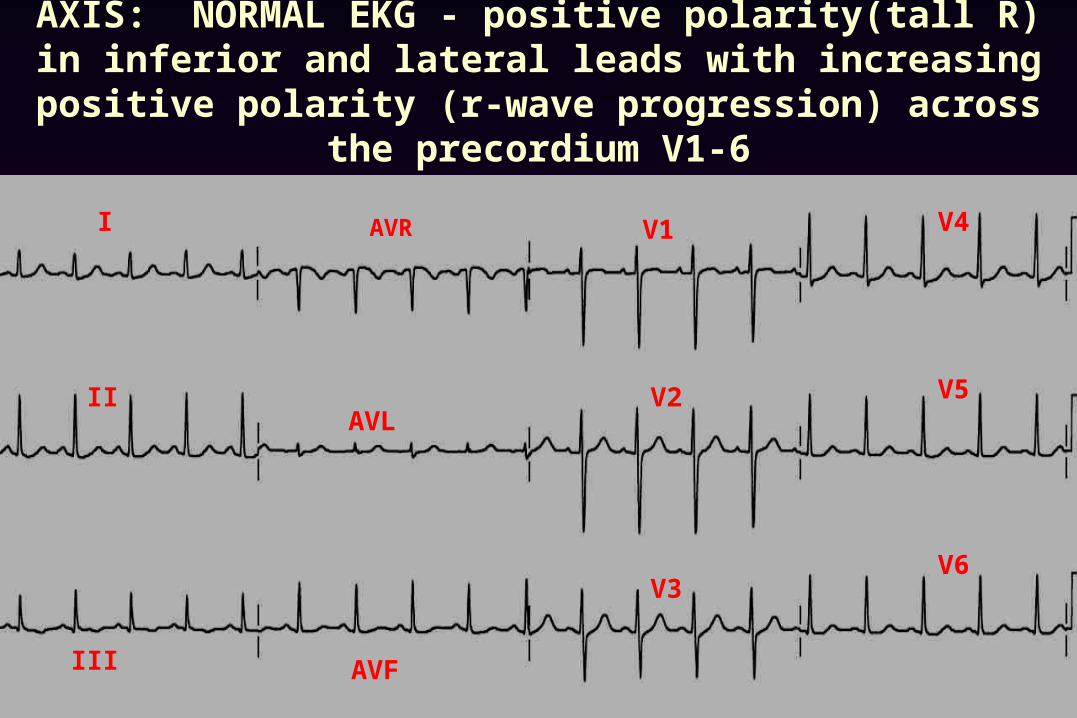
AXIS: NORMAL EKG - positive polarity(tall R) in inferior and lateral leads with increasing positive polarity (r-wave
progression) across the precordium V1-6
AVF
I
II
III
AVL
V1
V2
V3
V4
V5
V6
AVR
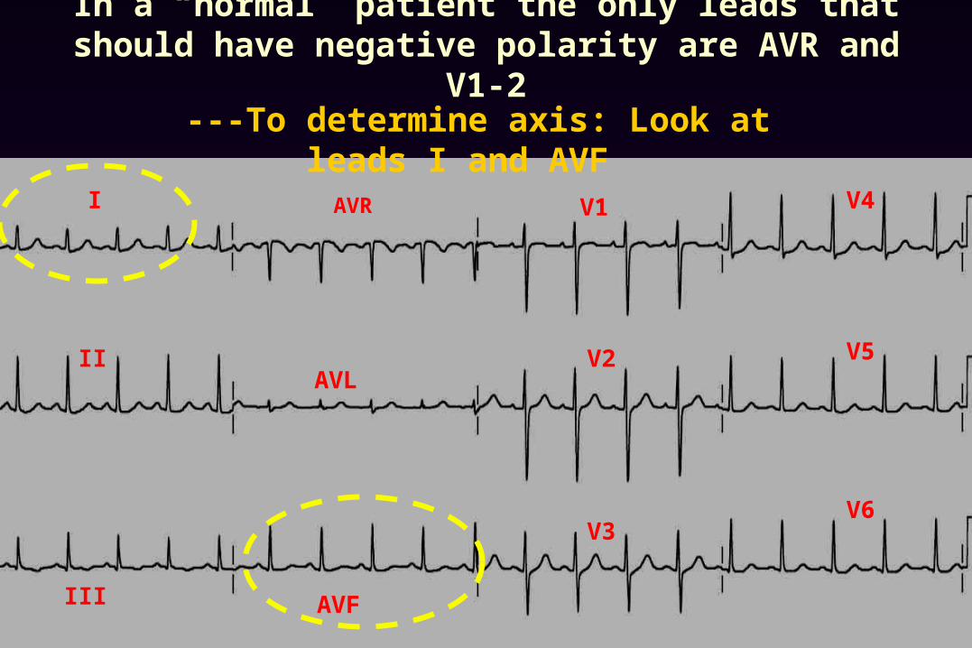
In a “normal” patient the only leads that should have negative polarity are AVR and V1-2
AVF
I
II
III
AVL
V1
V2
V3
V4
V5
V6
AVR
---To determine axis: Look at leads I and AVF
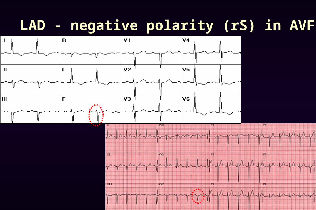
LAD - negative polarity (rS) in AVF

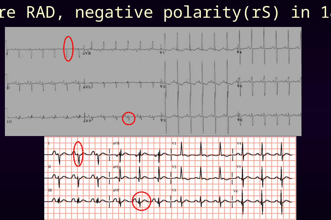
RAD: negative polarity(rS) in lead I
Severe RAD, negative polarity(rS) in 1& AVF
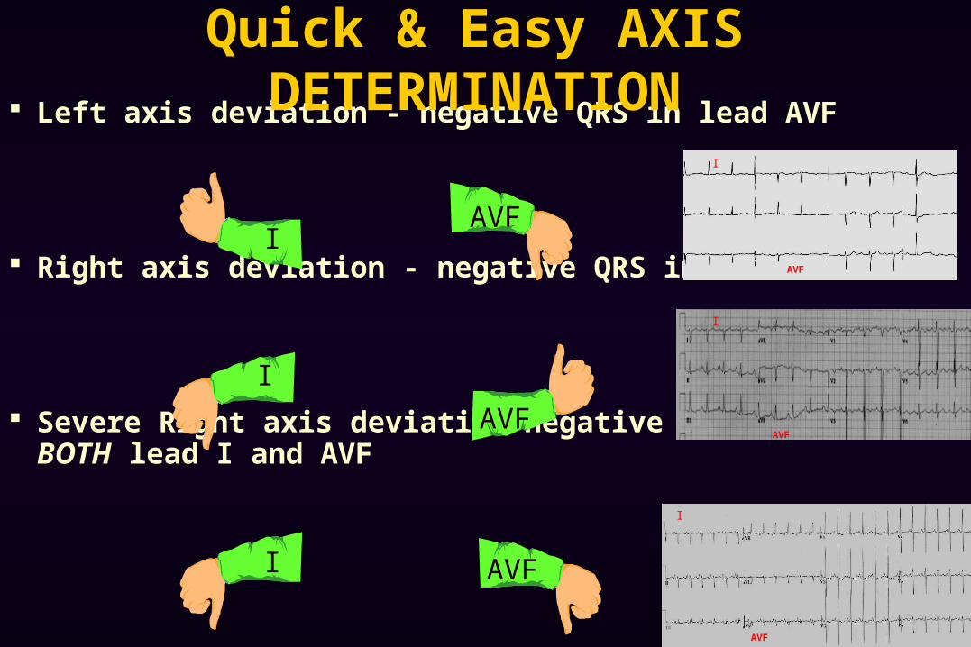
Left axis deviation - negative QRS in lead AVF
Right axis deviation - negative QRS in lead I
Severe Right axis deviation negative QRS in BOTH lead I and AVF
Quick & Easy AXIS DETERMINATION
AVF
AVF
AVF
AVF
AVF
AVF
I
I
I
I
I
I
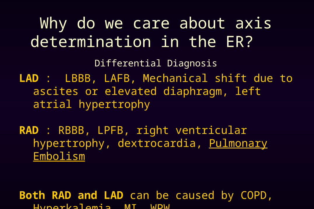
Why do we care about axis determination in the ER?
Differential Diagnosis
LAD : LBBB, LAFB, Mechanical shift due to ascites or elevated diaphragm, left atrial hypertrophy
RAD : RBBB, LPFB, right ventricular hypertrophy, dextrocardia, Pulmonary Embolism
Both RAD and LAD can be caused by COPD, Hyperkalemia, MI, WPW
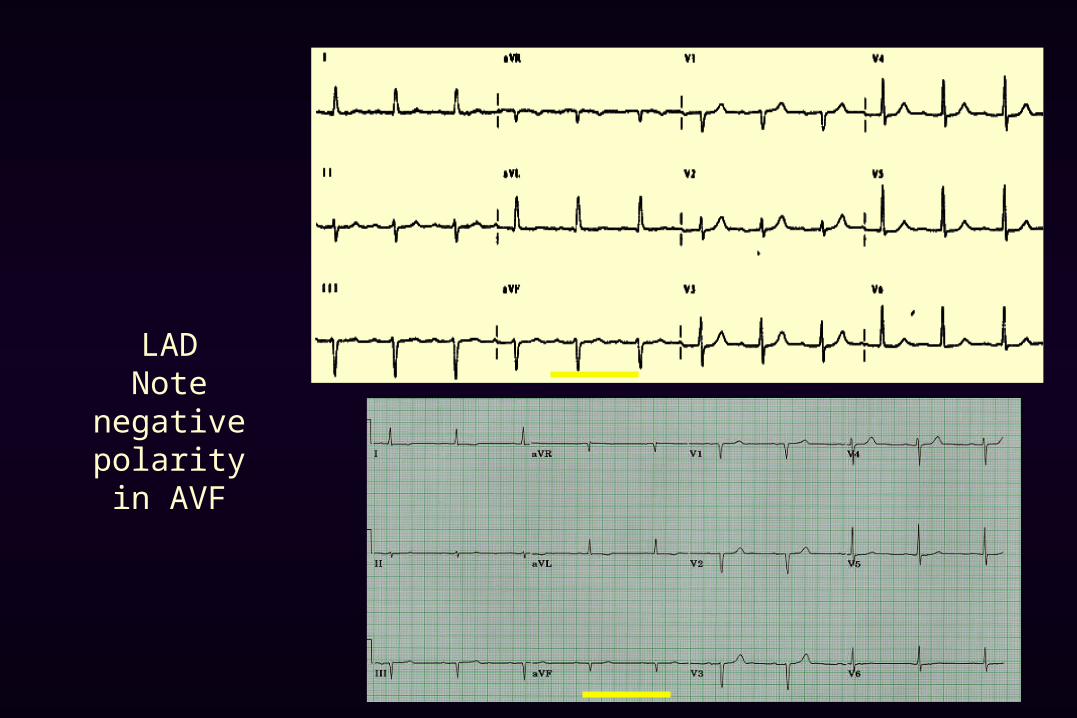
LADNote negative
polarity in AVF

RADNote negative polarity (rS) in
I
Severe RADNote negative polarity (rS) in
I & AVF
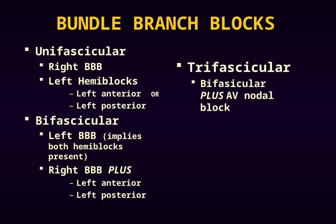
BUNDLE BRANCH BLOCKS Unifascicular
Right BBB Left Hemiblocks
– Left anterior OR
– Left posterior
Bifascicular Left BBB (implies both
hemiblocks present)
Right BBB PLUS– Left anterior
– Left posterior
Trifascicular Bifasicular PLUS AV
nodal block

Right Bundle Branch Block
QRS > 0.12 sec Predominantly
positive rSR’ in
V 1-2
Wide slurred S in lead I
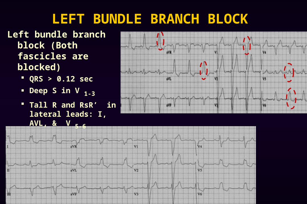
LEFT BUNDLE BRANCH BLOCKLeft bundle branch block
(Both fascicles are blocked) QRS > 0.12 sec
Deep S in V 1-3
Tall R and RsR’ in lateral leads: I, AVL, & V 5-6
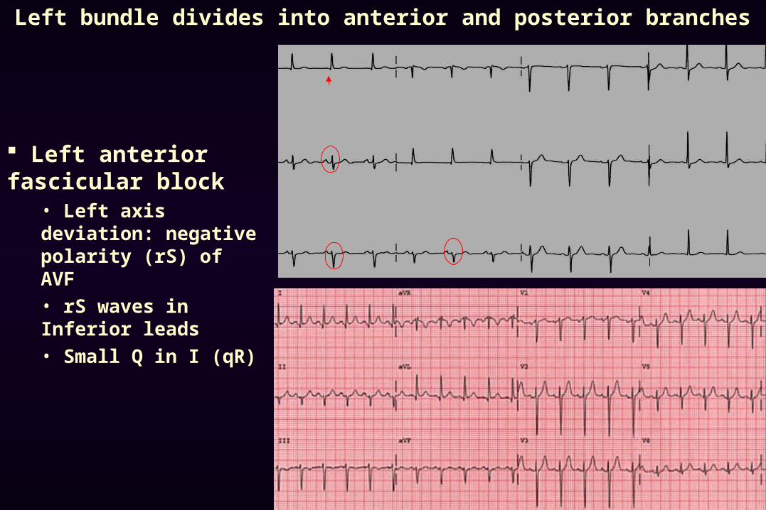
Left bundle divides into anterior and posterior branches
Left anterior fascicular block
• Left axis deviation: negative polarity (rS) of AVF
• rS waves in Inferior leads
• Small Q in I (qR)
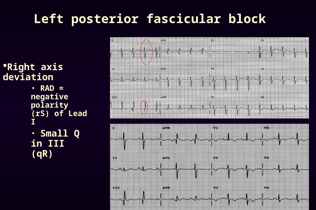
Left posterior fascicular block
Right axis deviation• RAD = negative polarity (rS) of Lead I
• Small Q in III (qR)
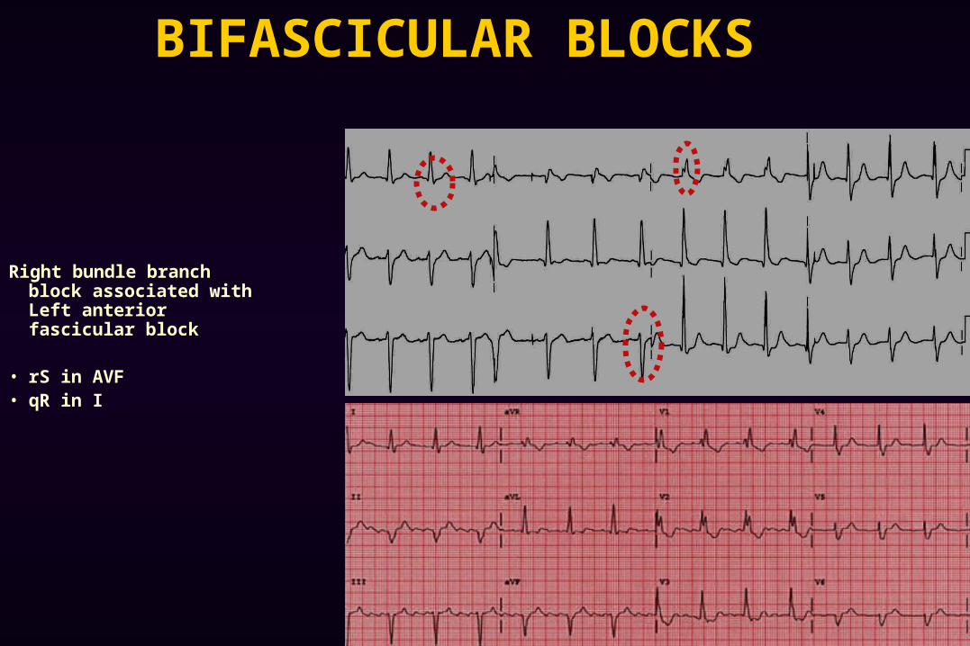
BIFASCICULAR BLOCKS
Right bundle branch block associated with Left anterior fascicular block
• rS in AVF• qR in I
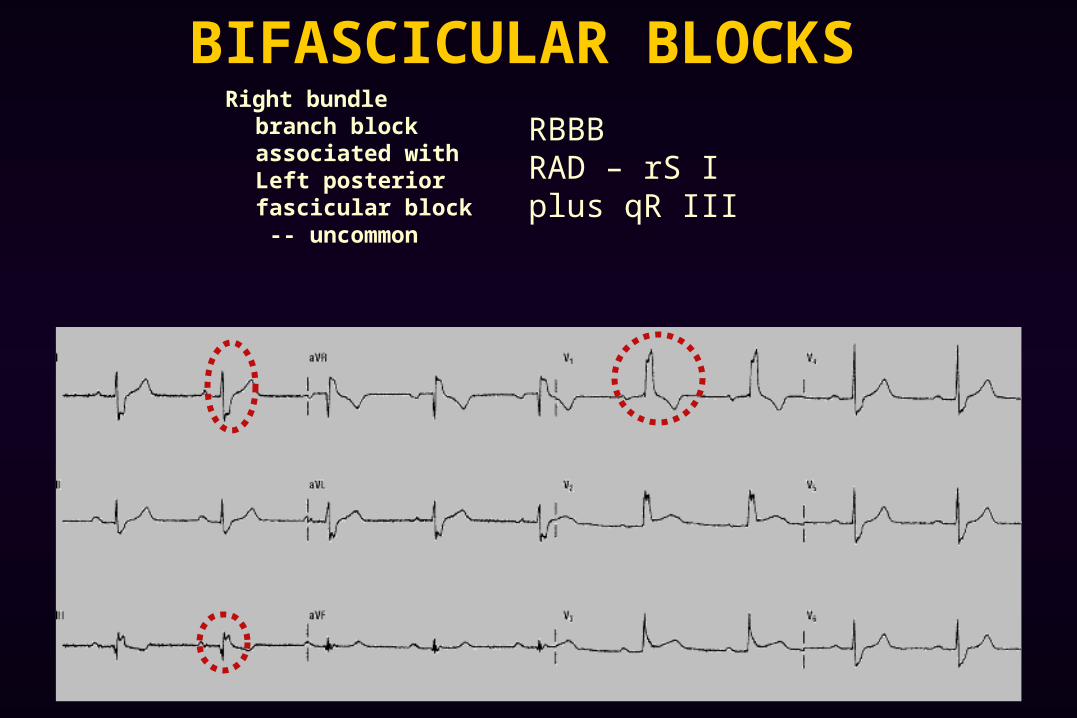
BIFASCICULAR BLOCKS Right bundle branch
block associated with Left posterior fascicular block -- uncommon
RBBB RAD – rS Iplus qR III
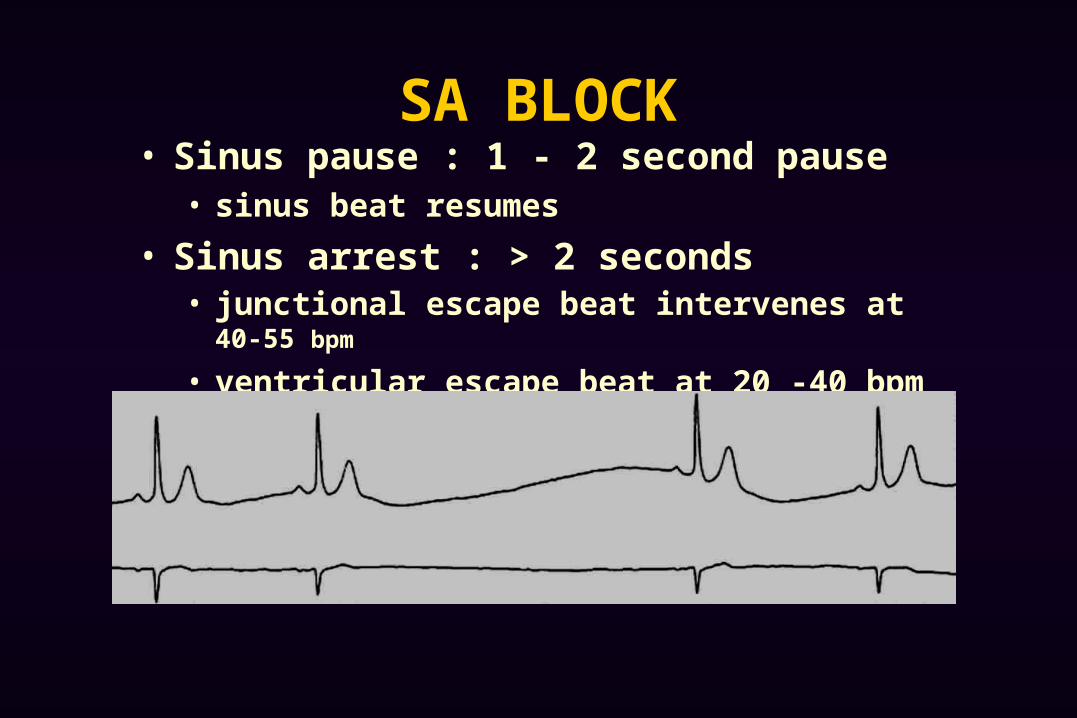
SA BLOCK• Sinus pause : 1 - 2 second pause
• sinus beat resumes
• Sinus arrest : > 2 seconds• junctional escape beat intervenes at 40-55 bpm
• ventricular escape beat at 20 -40 bpm
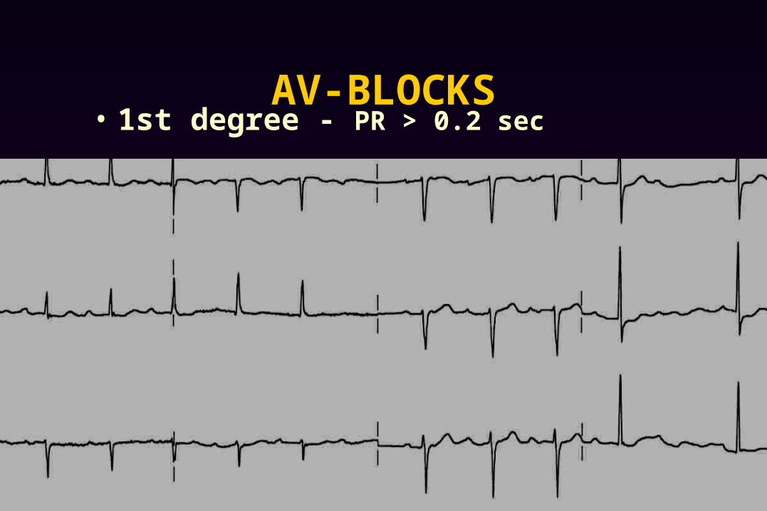
AV-BLOCKS• 1st degree - PR > 0.2 sec
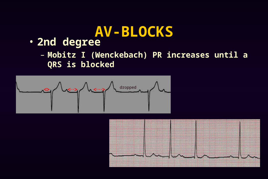
AV-BLOCKS• 2nd degree
– Mobitz I (Wenckebach) PR increases until a QRS is blocked
dropped
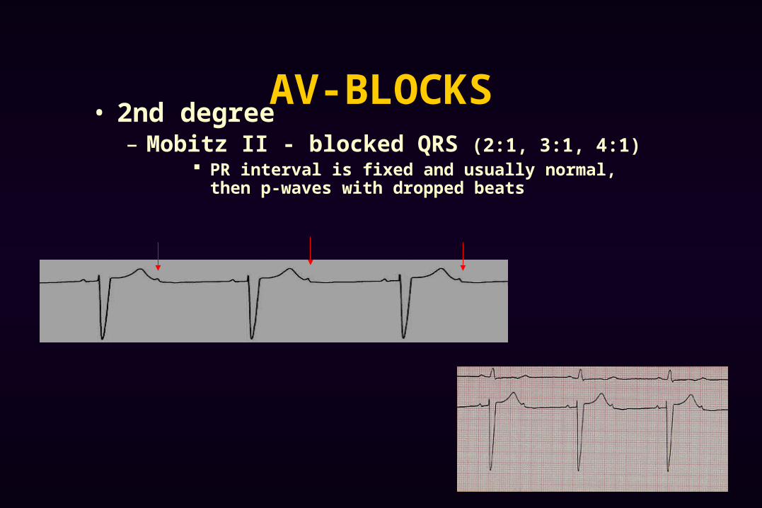
AV-BLOCKS• 2nd degree
– Mobitz II - blocked QRS (2:1, 3:1, 4:1) PR interval is fixed and usually normal, then p-waves with
dropped beats
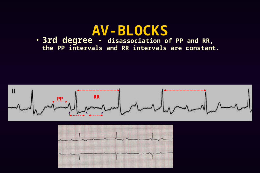
AV-BLOCKS• 3rd degree - disassociation of PP and RR, the PP
intervals and RR intervals are constant.
RRPP
PEARLS Differential diagnosis for slow irregularly irregular rhythm
Second Degree heart block : wenckebach Third Degree heart block
If you see Left Axis Deviation, think about LAFB If you see Right Axis Deviation, think about LPFB
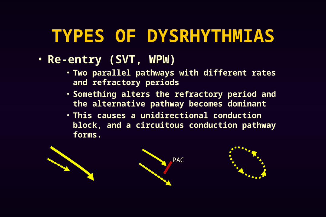
TYPES OF DYSRHYTHMIAS• Re-entry (SVT, WPW)
• Two parallel pathways with different rates and refractory periods
• Something alters the refractory period and the alternative pathway becomes dominant
• This causes a unidirectional conduction block, and a circuitous conduction pathway forms.
PAC
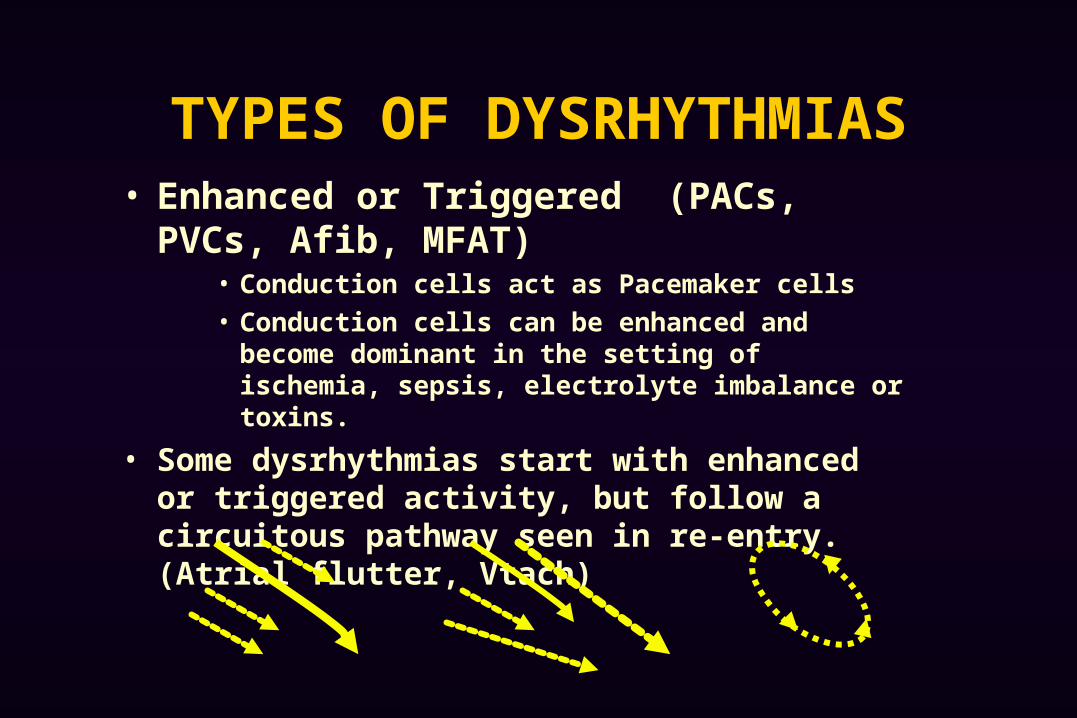
TYPES OF DYSRHYTHMIAS• Enhanced or Triggered (PACs, PVCs, Afib,
MFAT)• Conduction cells act as Pacemaker cells
• Conduction cells can be enhanced and become dominant in the setting of ischemia, sepsis, electrolyte imbalance or toxins.
• Some dysrhythmias start with enhanced or triggered activity, but follow a circuitous pathway seen in re-entry. (Atrial flutter, Vtach)

A 60 yo with COPD c/o palpitations & SOB. The EKG shows: a. Atrial Fibrillationb. Premature Atrial Complexesc. Multi-Focal Atrial Tachycardiad. Paroxismal Atrial Tachycardia with block
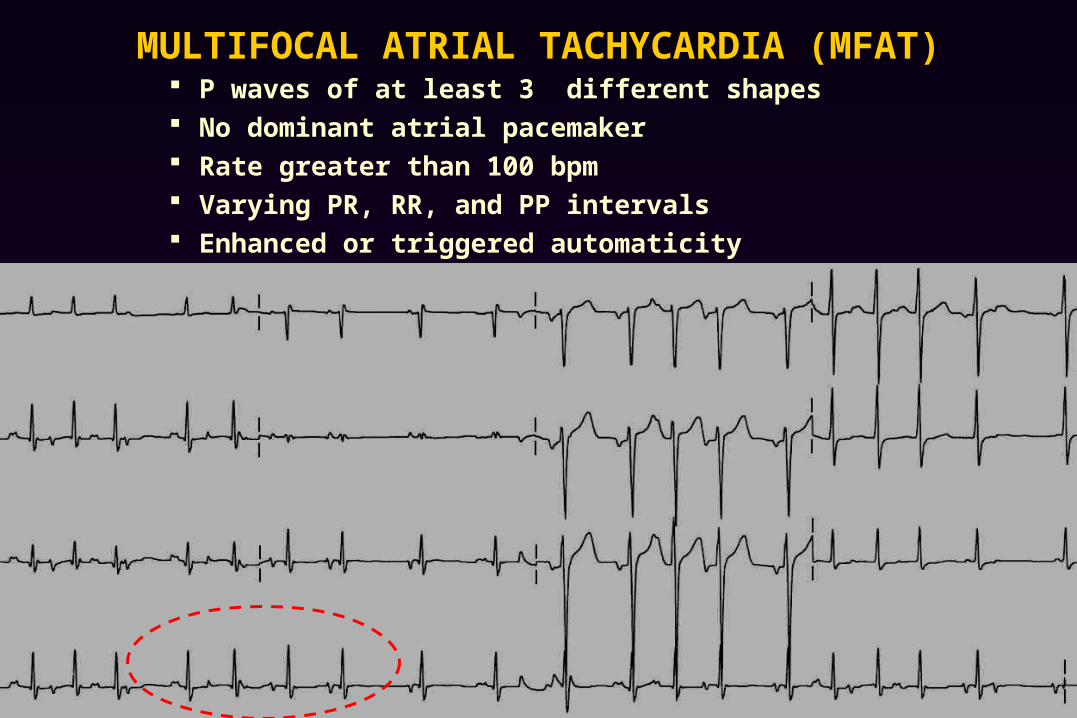
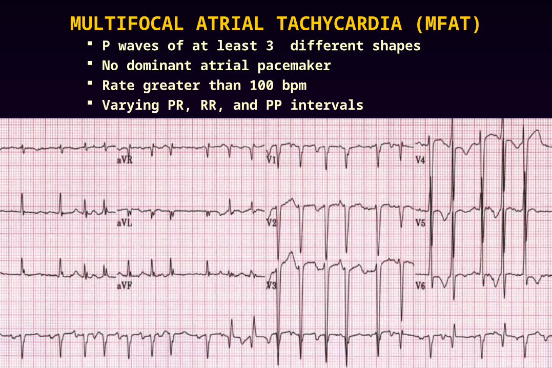
MULTIFOCAL ATRIAL TACHYCARDIA (MFAT) P waves of at least 3 different shapes No dominant atrial pacemaker Rate greater than 100 bpm Varying PR, RR, and PP intervals Enhanced or triggered automaticity
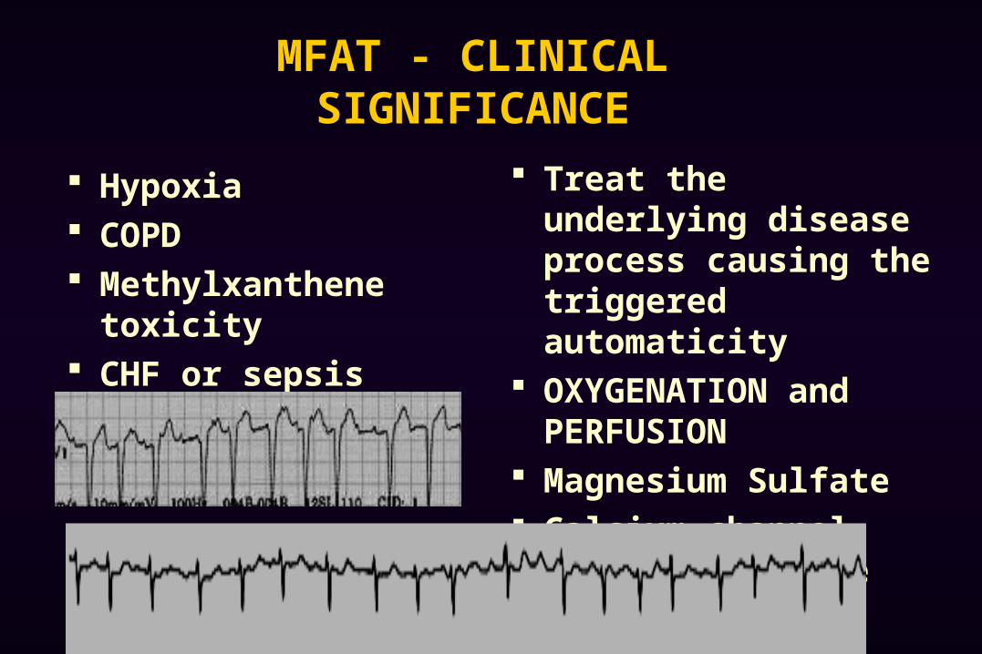
MFAT - CLINICAL SIGNIFICANCE
Hypoxia COPD Methylxanthene
toxicity CHF or sepsis
Treat the underlying disease process causing the triggered automaticity
OXYGENATION and PERFUSION
Magnesium Sulfate Calcium channel blocker
for rate control prn
MULTIFOCAL ATRIAL TACHYCARDIA (MFAT) P waves of at least 3 different shapes No dominant atrial pacemaker Rate greater than 100 bpm Varying PR, RR, and PP intervals
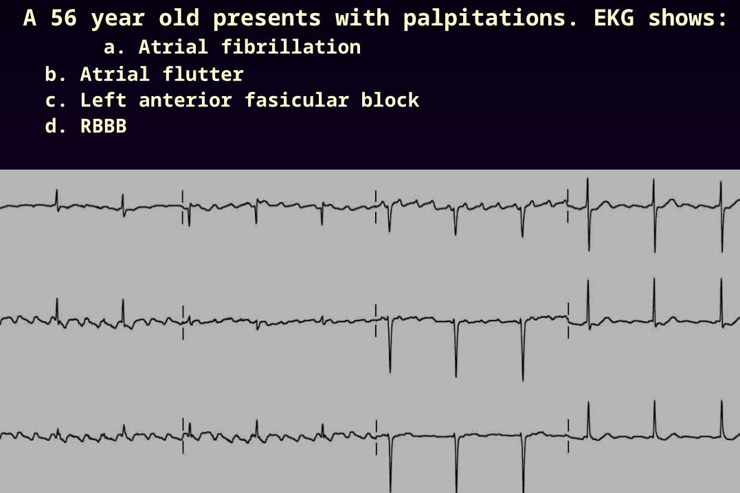
A 56 year old presents with palpitations. EKG shows: a. Atrial fibrillation
b. Atrial flutterc. Left anterior fasicular blockd. RBBB
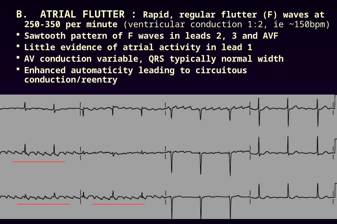
B. ATRIAL FLUTTER : Rapid, regular flutter (F) waves at 250-350 per minute (ventricular conduction 1:2, ie ~150bpm)
Sawtooth pattern of F waves in leads 2, 3 and AVF Little evidence of atrial activity in lead 1 AV conduction variable, QRS typically normal width Enhanced automaticity leading to circuitous conduction/reentry
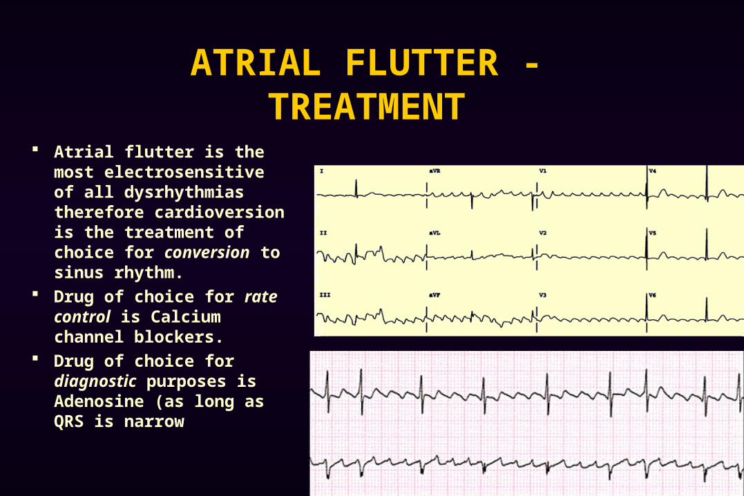
ATRIAL FLUTTER - TREATMENT
Atrial flutter is the most electrosensitive of all dysrhythmias therefore cardioversion is the treatment of choice for conversion to sinus rhythm.
Drug of choice for rate control is Calcium channel blockers.
Drug of choice for diagnostic purposes is Adenosine (as long as QRS is narrow
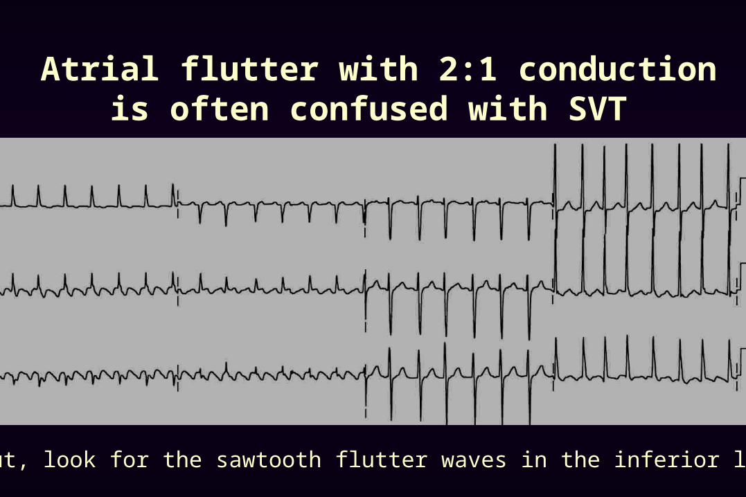
Atrial flutter with 2:1 conduction is often confused with SVT
But, look for the sawtooth flutter waves in the inferior leads.

Same patient after adenosine, showing prominent flutter waves.
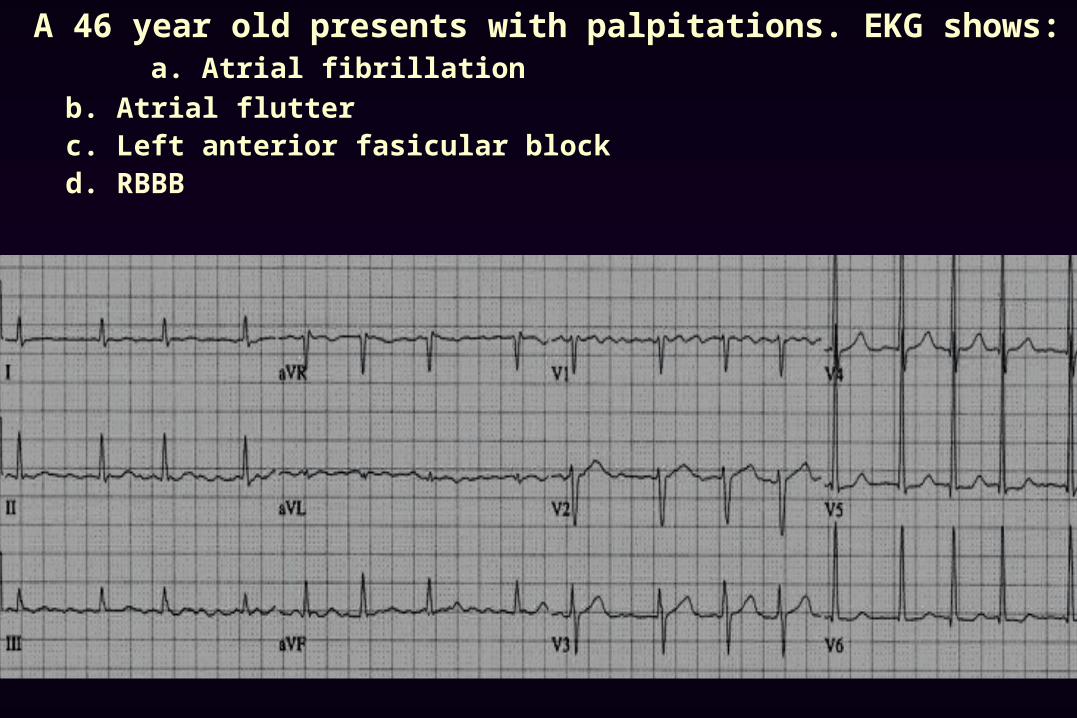
A 46 year old presents with palpitations. EKG shows: a. Atrial fibrillation
b. Atrial flutterc. Left anterior fasicular blockd. RBBB
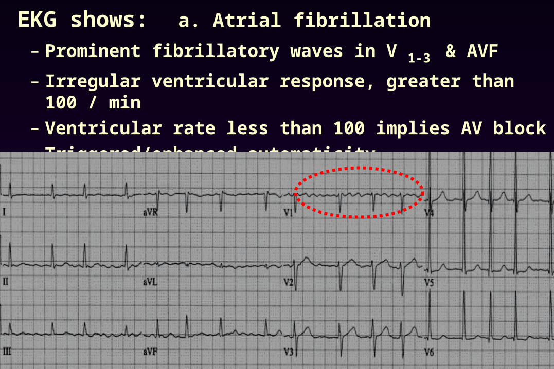
EKG shows: a. Atrial fibrillation
– Prominent fibrillatory waves in V 1-3 & AVF
– Irregular ventricular response, greater than 100 / min
– Ventricular rate less than 100 implies AV block
– Triggered/enhanced automaticity
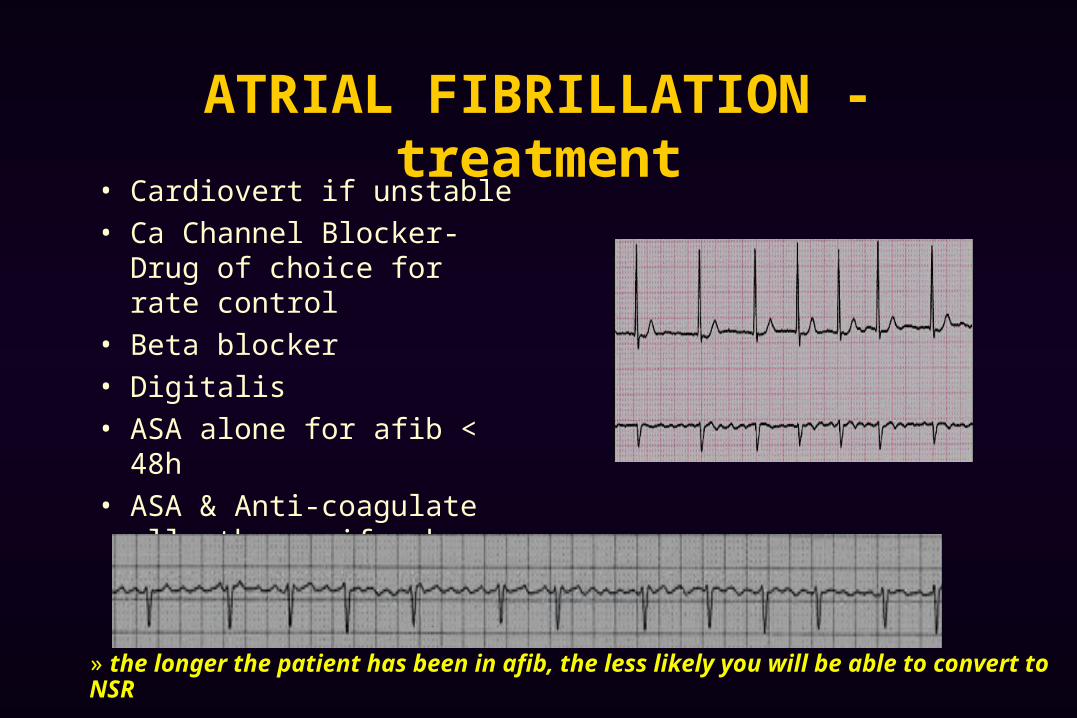
ATRIAL FIBRILLATION - treatment• Cardiovert if unstable
• Ca Channel Blocker- Drug of choice for rate control
• Beta blocker
• Digitalis
• ASA alone for afib < 48h
• ASA & Anti-coagulate all others, if unknown or >48h
» the longer the patient has been in afib, the less likely you will be able to convert to NSR
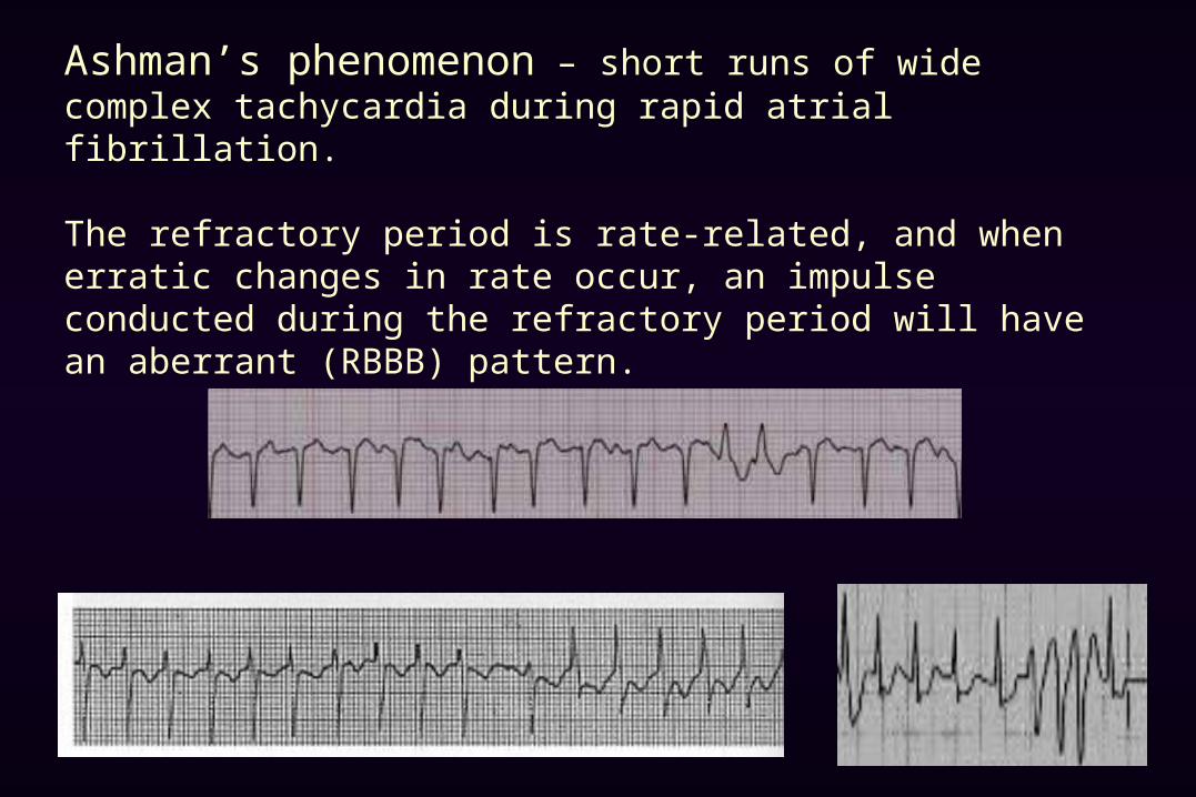
Ashman’s phenomenon – short runs of wide complex tachycardia during rapid atrial fibrillation.
The refractory period is rate-related, and when erratic changes in rate occur, an impulse conducted during the refractory period will have an aberrant (RBBB) pattern.
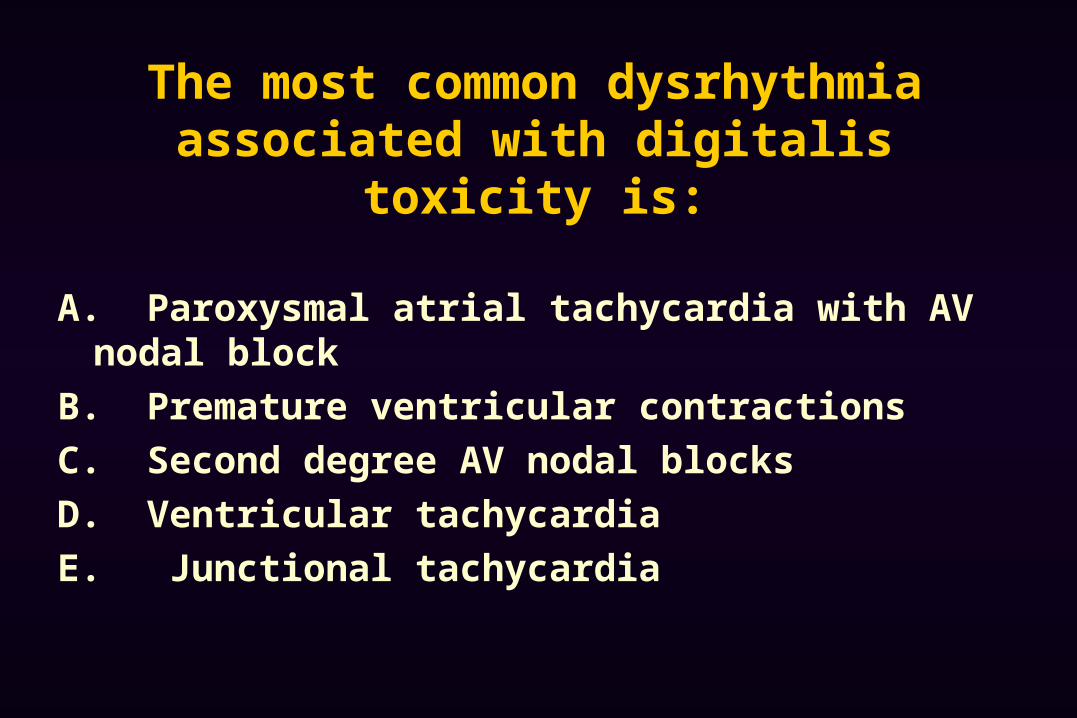
The most common dysrhythmia associated with digitalis toxicity is:
A. Paroxysmal atrial tachycardia with AV nodal block
B. Premature ventricular contractions
C. Second degree AV nodal blocks
D. Ventricular tachycardia
E. Junctional tachycardia
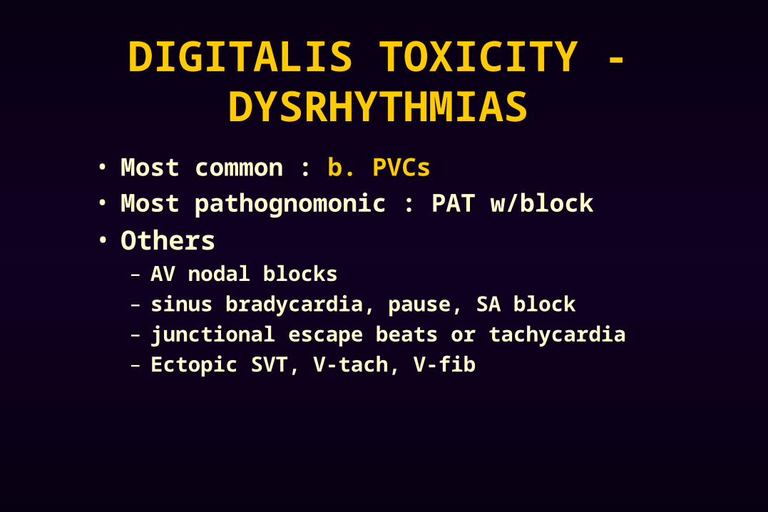
DIGITALIS TOXICITY - DYSRHYTHMIAS
• Most common : b. PVCs
• Most pathognomonic : PAT w/block
• Others – AV nodal blocks
– sinus bradycardia, pause, SA block
– junctional escape beats or tachycardia
– Ectopic SVT, V-tach, V-fib

Paroxysmal atrial tachycardia with block is pathognomonic for digitalis toxicity.
Note the p waves at a rate > 100 & blocked QRS complexes. (Don’t mistake for aflutter with variable conduction or 3rd degree block)
Note the blocked Impulses!!

A 23 yo male with c/o palpitations, EKG shows: a. Atrial fibrillationb. MFATc. SVTd. PAT with block
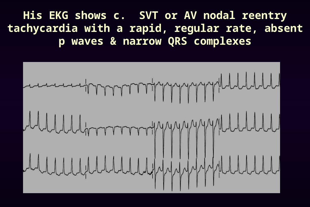
His EKG shows c. SVT or AV nodal reentry tachycardia with a rapid, regular rate, absent p waves & narrow QRS complexes
AV
SA
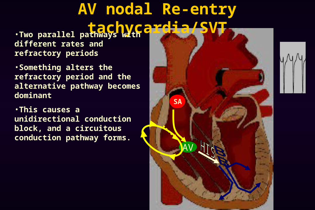
AV nodal Re-entry tachycardia/SVT•Two parallel pathways with different rates and refractory periods
•Something alters the refractory period and the alternative pathway becomes dominant
•This causes a unidirectional conduction block, and a circuitous conduction pathway forms.
AV
SA
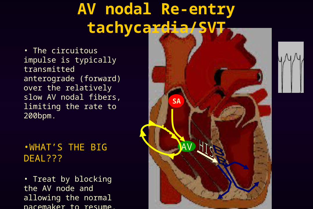
AV nodal Re-entry tachycardia/SVT
• The circuitous impulse is typically transmitted anterograde (forward) over the relatively slow AV nodal fibers, limiting the rate to 200bpm.
•WHAT’S THE BIG DEAL???
• Treat by blocking the AV node and allowing the normal pacemaker to resume.
• Adenosine• Ca channel blocker• Beta blocker
AV
SA
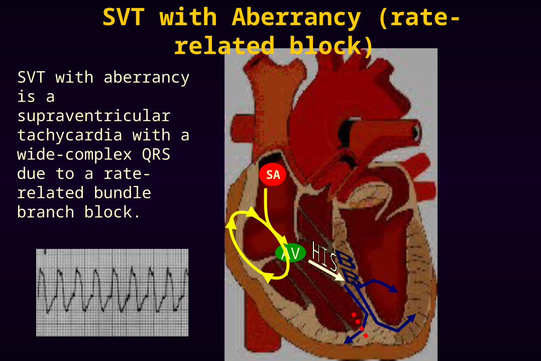
SVT with aberrancy is a supraventricular tachycardia with a wide-complex QRS due to a rate-related bundle branch block.
SVT with Aberrancy (rate-related block)
AV
SA
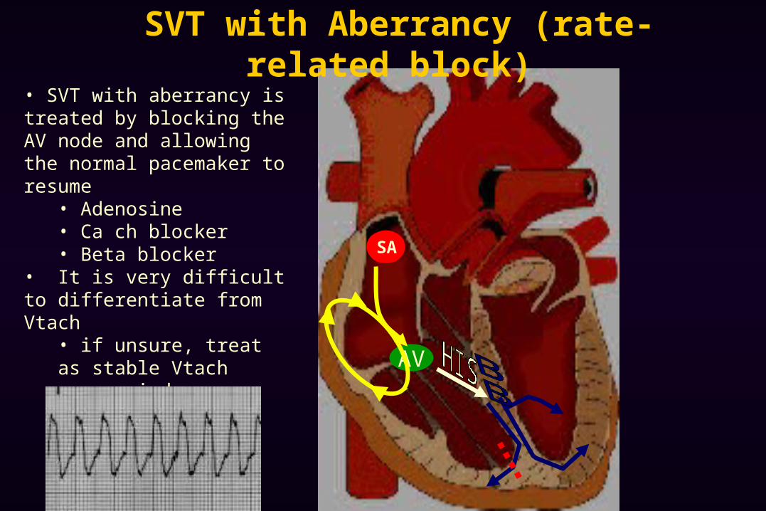
• SVT with aberrancy is treated by blocking the AV node and allowing the normal pacemaker to resume
• Adenosine• Ca ch blocker• Beta blocker
• It is very difficult to differentiate from Vtach
• if unsure, treat as stable Vtach
• amiodarone• procainamide
SVT with Aberrancy (rate-related block)
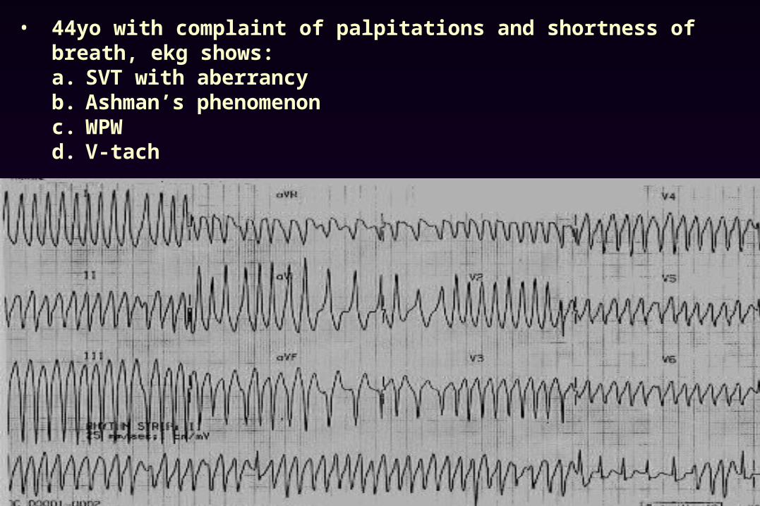
• 44yo with complaint of palpitations and shortness of breath, ekg shows:a. SVT with aberrancyb. Ashman’s phenomenonc. WPWd. V-tach
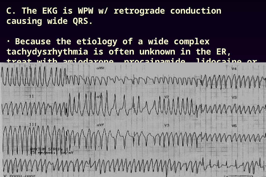
C. The EKG is WPW w/ retrograde conduction causing wide QRS.
• Because the etiology of a wide complex tachydysrhythmia is often unknown in the ER, treat with amiodarone, procainamide, lidocaine or cardioversion. (avoid procainamide in TCA OD or prolonged qt toursades)
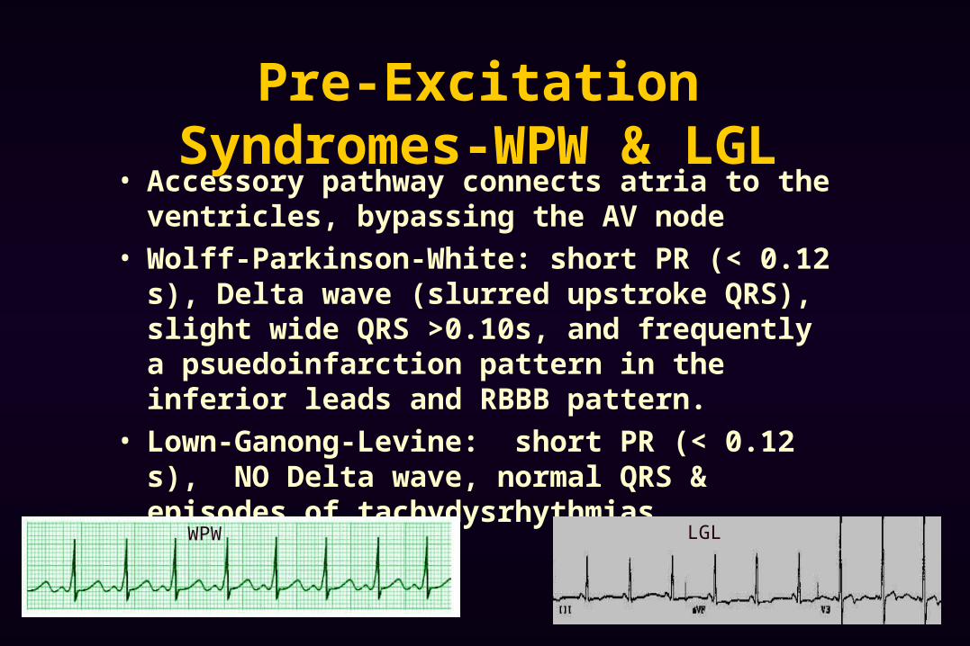
Pre-Excitation Syndromes-WPW & LGL
• Accessory pathway connects atria to the ventricles, bypassing the AV node
• Wolff-Parkinson-White: short PR (< 0.12 s), Delta wave (slurred upstroke QRS), slight wide QRS >0.10s, and frequently a psuedoinfarction pattern in the inferior leads and RBBB pattern.
• Lown-Ganong-Levine: short PR (< 0.12 s), NO Delta wave, normal QRS & episodes of tachydysrhythmias
LGLWPW
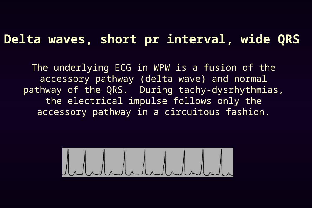
Delta waves, short pr interval, wide QRS
The underlying ECG in WPW is a fusion of the accessory pathway (delta wave) and normal pathway of the QRS. During tachy-
dysrhythmias, the electrical impulse follows only the accessory pathway in a circuitous fashion.
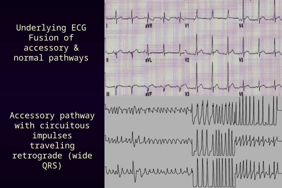
Underlying ECGFusion of accessory &
normal pathways
Accessory pathway with circuitous impulses traveling retrograde
(wide QRS)
AV
SA
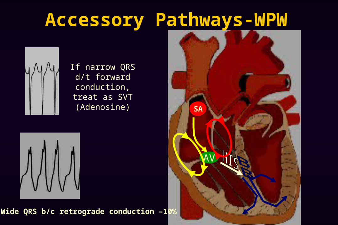
Accessory Pathways-WPW
If narrow QRS d/t forward conduction,
treat as SVT (Adenosine)
Wide QRS b/c retrograde conduction –10%
AV
SA
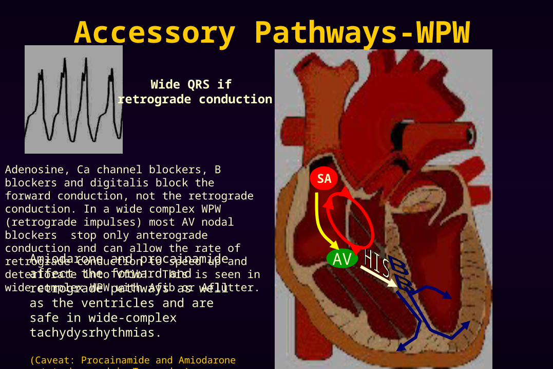
Accessory Pathways-WPW
Wide QRS if retrograde conduction
Amiodarone and procainamide affect the forward and retrograde pathways as well as the ventricles and are safe in wide-complex tachydysrhythmias.
(Caveat: Procainamide and Amiodarone not to be used in Toursades)
Adenosine, Ca channel blockers, B blockers and digitalis block the forward conduction, not the retrograde conduction. In a wide complex WPW (retrograde impulses) most AV nodal blockers stop only anterograde conduction and can allow the rate of retrograde conduction to speed up and deteriorate into Vfib! This is seen in wide complex WPW with Afib or Aflutter.
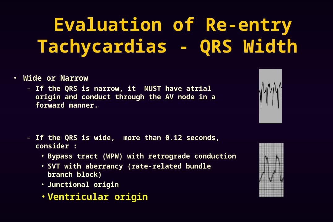
Evaluation of Re-entry Tachycardias - QRS Width
• Wide or Narrow – If the QRS is narrow, it MUST have atrial origin and conduct
through the AV node in a forward manner.
– If the QRS is wide, more than 0.12 seconds, consider :
• Bypass tract (WPW) with retrograde conduction
• SVT with aberrancy (rate-related bundle branch block)
• Junctional origin
• Ventricular origin
Re-entry Tachycardias - Treatment Modalities
• Based on hemodynamic stability & QRS width – Unstable : synchronized cardioversion– Stable :
• Narrow complex – vagal maneuvers, adenosine, calcium channel blockers or beta blockers
• Wide complex – Amiodarone, Lidocaine or Procainamide to treat both anterograde and retrograde impulses and ventricular dysrhythmias
Beware: it is very difficulty to tell the difference between the wide-complex tachy-dysrhythmias. It is safer to treat as presumed V-tach.
PEARLS Wide complex QRS tachydysrhythmias of unknown
etiology – use amiodorone, procainamide, lidocaine Differential diagnosis for rapid, irregularly
irregular rhythm MFAT Atrial Fib Atrial flutter with variable conduction
SVT at 150 or 300, consider Atrial flutter
DYSRHYTHMIAS OF VENTRICULAR ORIGIN
Idioventricular rhythms Ventricular Tachycardia Ventricular Fibrillation Torsades de pointes
VENTRICULAR DYSRHYTHMIAS - Etiology
V Tach, V Fib & Idioventricular rhythms – typically caused by an ischemic focus which allows a rapid reentry dysrhythmia
Torsades de pointes - caused by a prolonged QT interval
Brugada syndrome – sodium ion channel-apathy
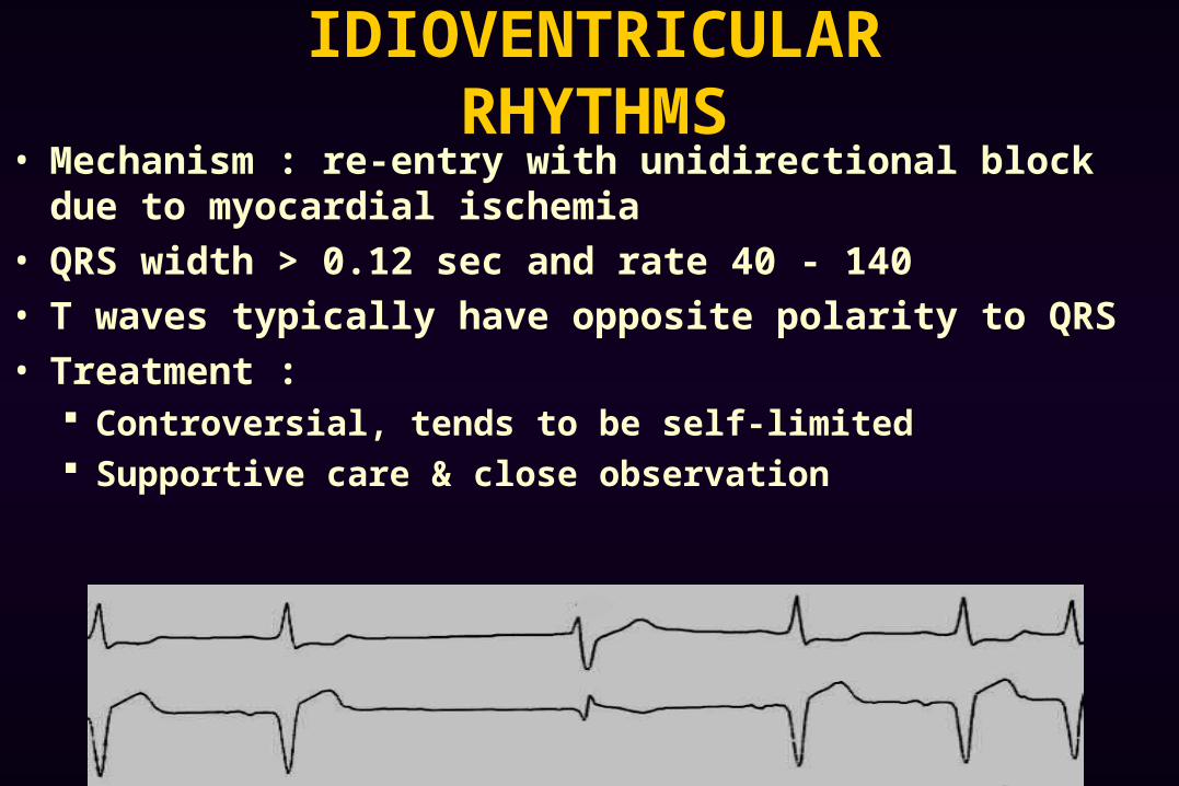
IDIOVENTRICULAR RHYTHMS
• Mechanism : re-entry with unidirectional block due to myocardial ischemia
• QRS width > 0.12 sec and rate 40 - 140
• T waves typically have opposite polarity to QRS
• Treatment : Controversial, tends to be self-limited Supportive care & close observation
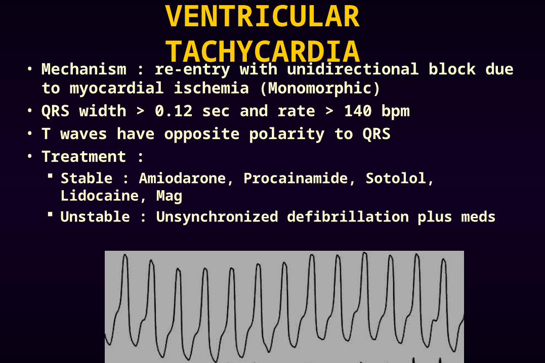
VENTRICULAR TACHYCARDIA
• Mechanism : re-entry with unidirectional block due to myocardial ischemia (Monomorphic)
• QRS width > 0.12 sec and rate > 140 bpm
• T waves have opposite polarity to QRS
• Treatment : Stable : Amiodarone, Procainamide, Sotolol, Lidocaine, Mag Unstable : Unsynchronized defibrillation plus meds
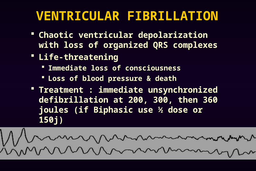
VENTRICULAR FIBRILLATION
Chaotic ventricular depolarization with loss of organized QRS complexes
Life-threatening Immediate loss of consciousness Loss of blood pressure & death
Treatment : immediate unsynchronized defibrillation at 200, 300, then 360 joules (if Biphasic use ½ dose or 150j)
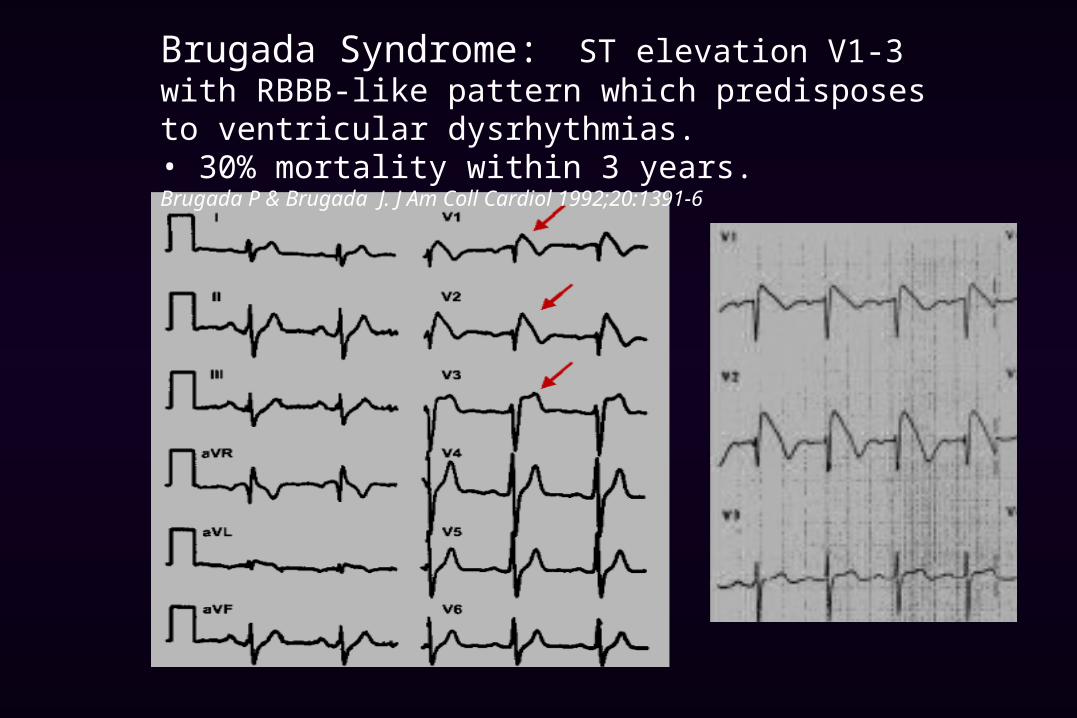
Brugada Syndrome: ST elevation V1-3 with RBBB-like pattern which predisposes to ventricular dysrhythmias.• 30% mortality within 3 years. Brugada P & Brugada J. J Am Coll Cardiol 1992;20:1391-6

Brugada Syndrome: Look for ST elevation V1-3• part of the syncope or palpitation work-up • immediate cardiology referral for ICD placement
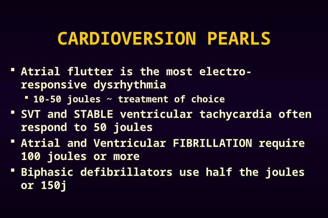
CARDIOVERSION PEARLS
Atrial flutter is the most electro-responsive dysrhythmia 10-50 joules ~ treatment of choice
SVT and STABLE ventricular tachycardia often respond to 50 joules
Atrial and Ventricular FIBRILLATION require 100 joules or more
Biphasic defibrillators use half the joules or 150j
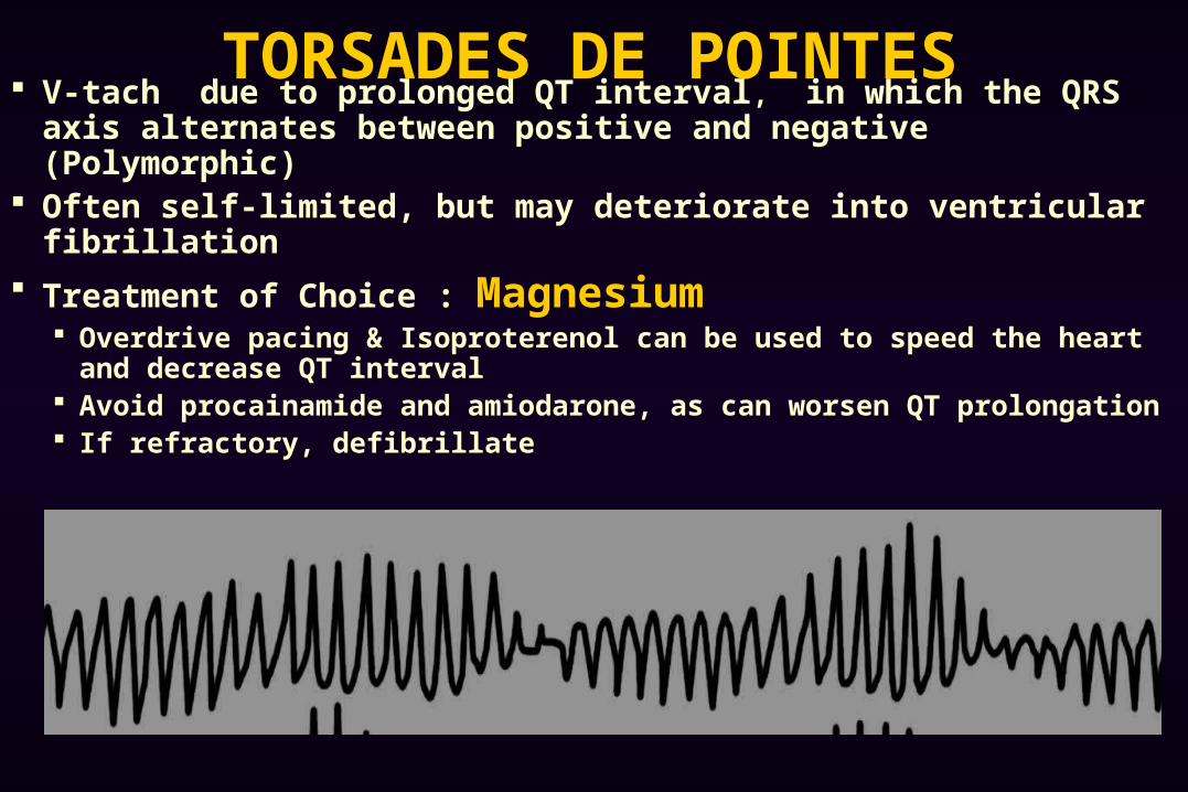
TORSADES DE POINTES V-tach due to prolonged QT interval, in which the QRS axis
alternates between positive and negative (Polymorphic) Often self-limited, but may deteriorate into ventricular fibrillation
Treatment of Choice : Magnesium Overdrive pacing & Isoproterenol can be used to speed the heart and
decrease QT interval Avoid procainamide and amiodarone, as can worsen QT prolongation If refractory, defibrillate
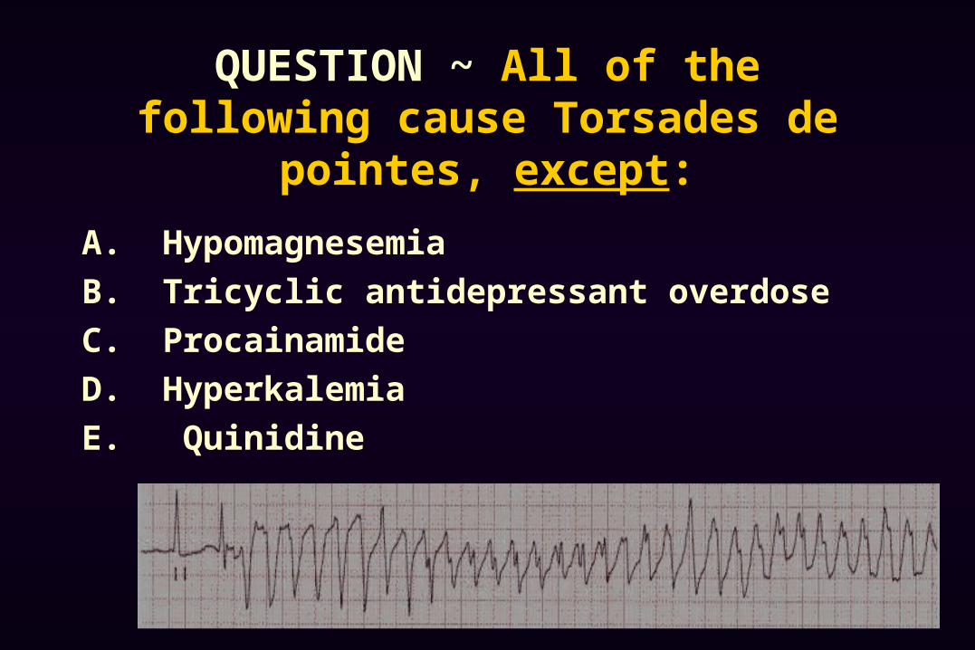
QUESTION ~ All of the following cause Torsades de pointes, except:
A. Hypomagnesemia
B. Tricyclic antidepressant overdose
C. Procainamide
D. Hyperkalemia
E. Quinidine
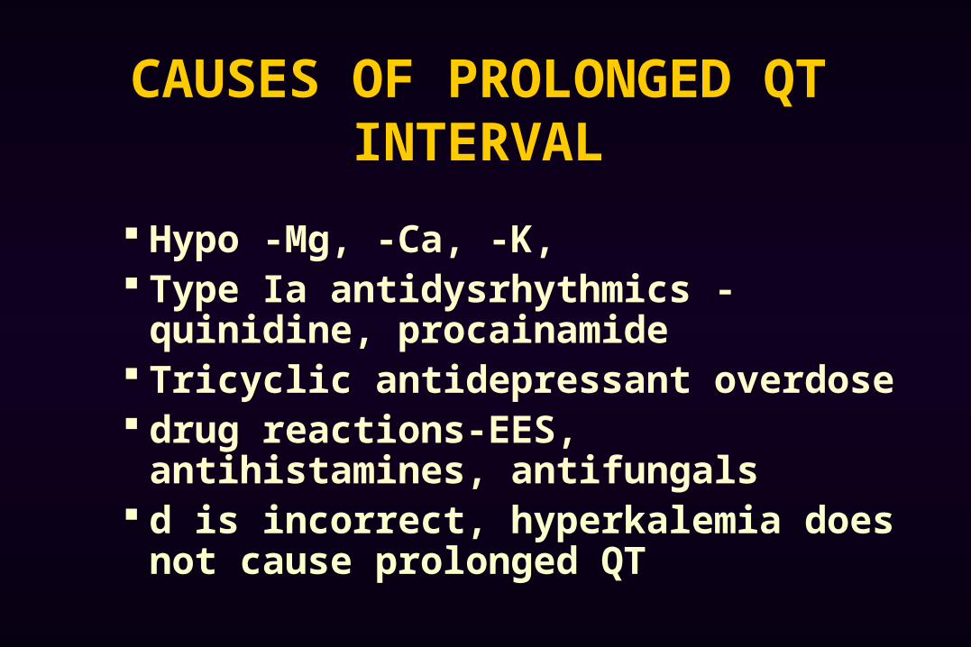
CAUSES OF PROLONGED QT INTERVAL
Hypo -Mg, -Ca, -K, Type Ia antidysrhythmics - quinidine,
procainamide Tricyclic antidepressant overdose drug reactions-EES, antihistamines,
antifungals d is incorrect, hyperkalemia does not cause
prolonged QT
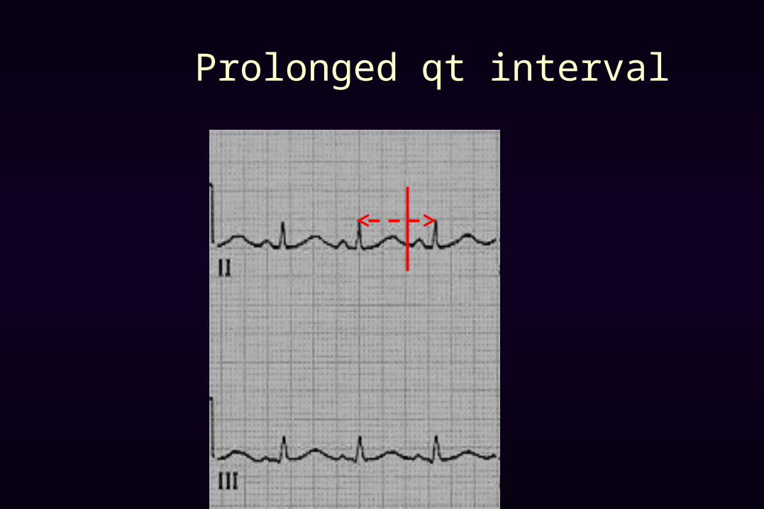
Prolonged qt interval
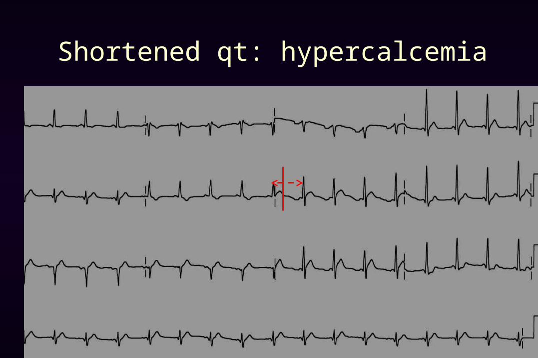
Shortened qt: hypercalcemia
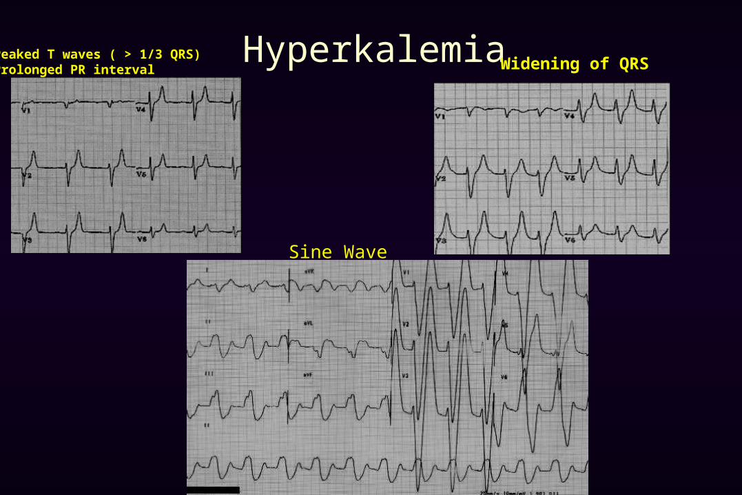
HyperkalemiaPeaked T waves ( > 1/3 QRS)Prolonged PR interval Widening of QRS
Sine Wave
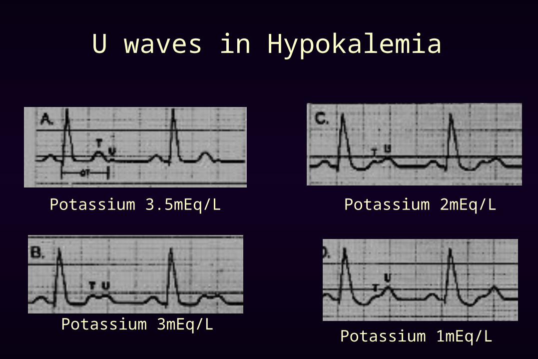
U waves in Hypokalemia
Potassium 3.5mEq/L
Potassium 3mEq/L
Potassium 2mEq/L
Potassium 1mEq/L
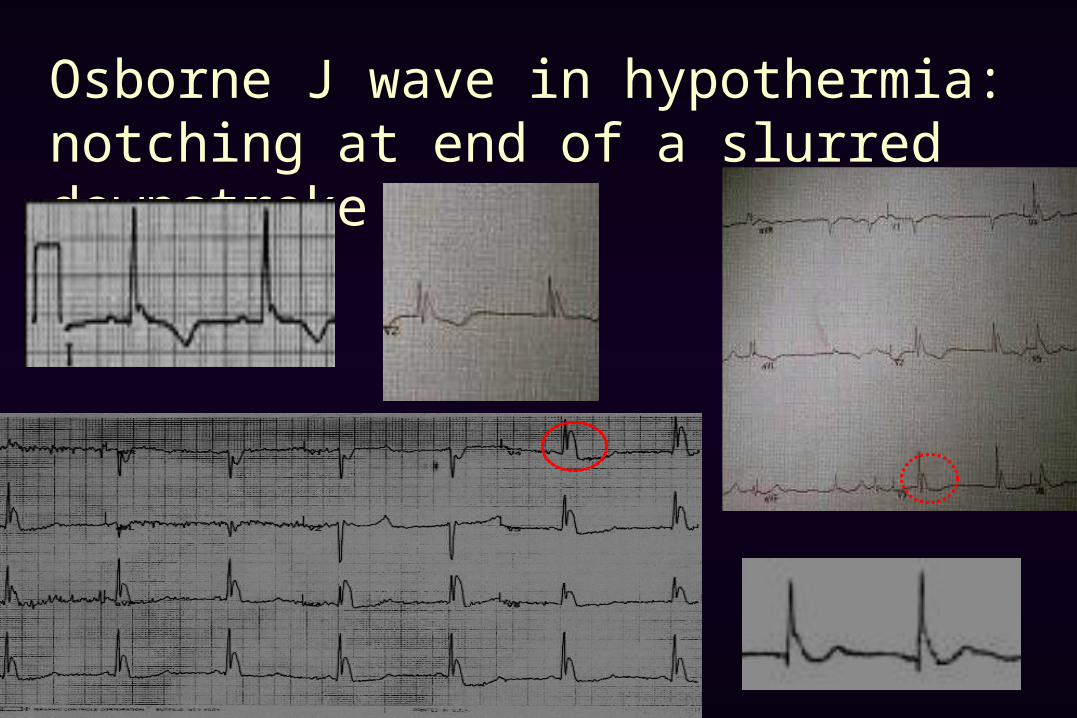
Osborne J wave in hypothermia: notching at end of a slurred downstroke of QRS

Tricyclic Antidepressant Overdose• tall r in AVR• slurring of the terminal portion of the rS in AVR
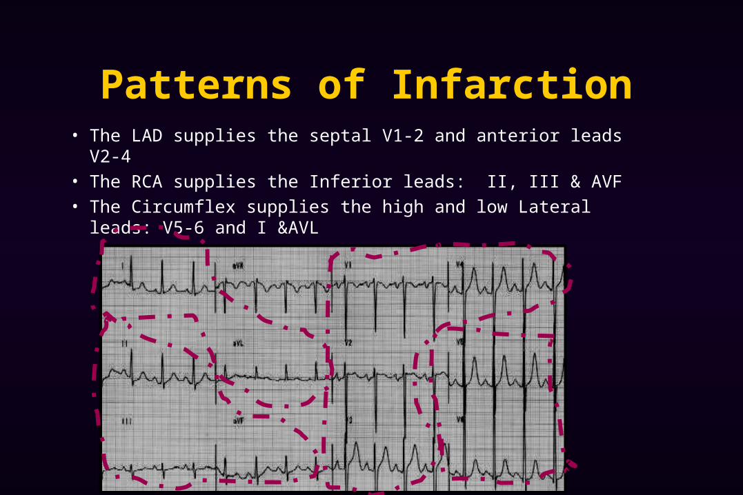
Patterns of Infarction• The LAD supplies the septal V1-2 and anterior leads V2-4
• The RCA supplies the Inferior leads: II, III & AVF
• The Circumflex supplies the high and low Lateral leads: V5-6 and I &AVL
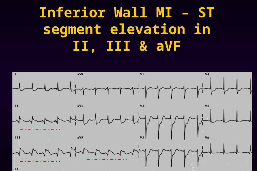
Inferior Wall MI – ST segment elevation in II, III & aVF

Anterior Wall MI – ST segment elevation in V2-4
Septal MI – ST segment elevation V1-2
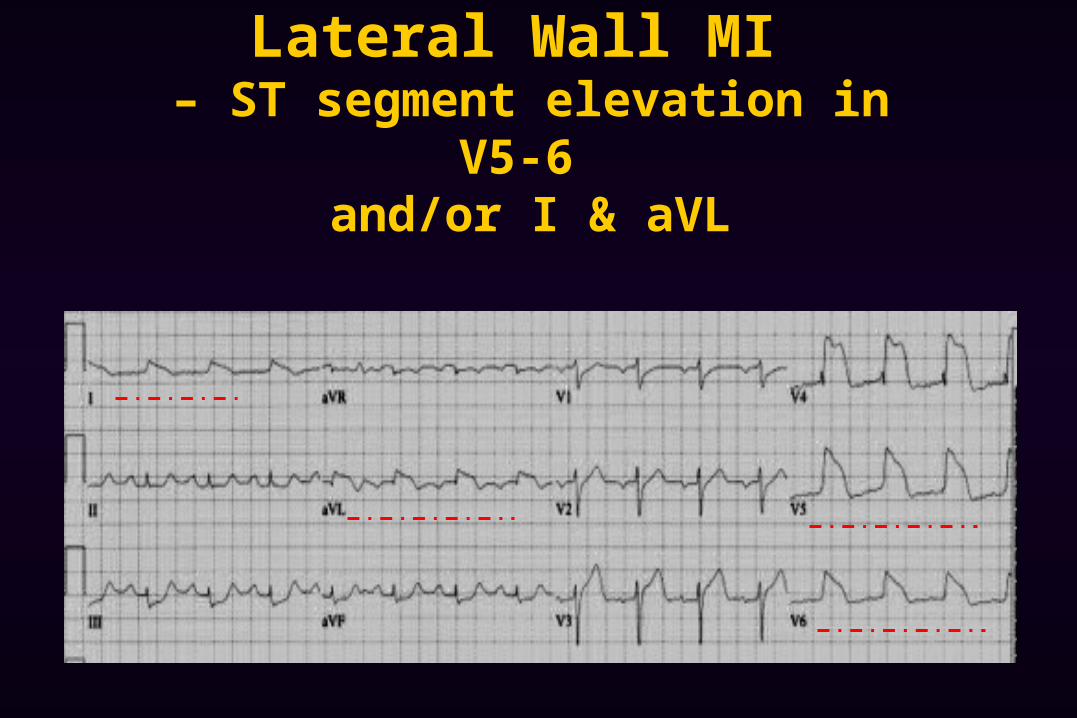
Lateral Wall MI – ST segment elevation in V5-6
and/or I & aVL
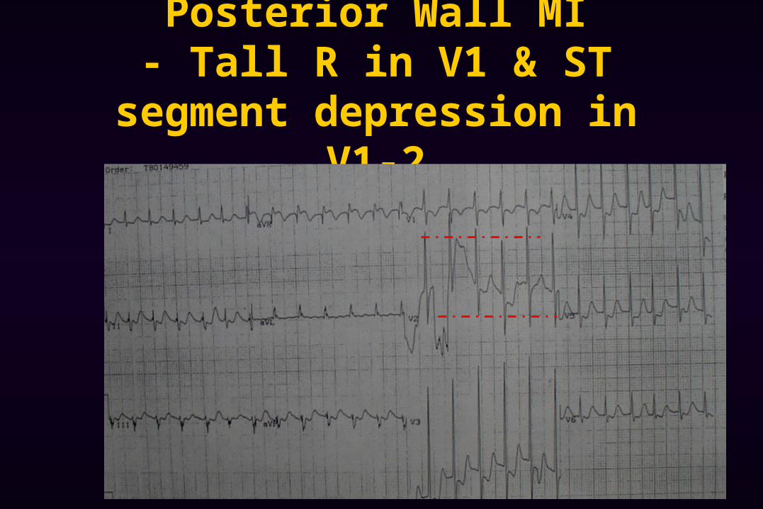
Posterior Wall MI- Tall R in V1 & ST segment
depression in V1-2
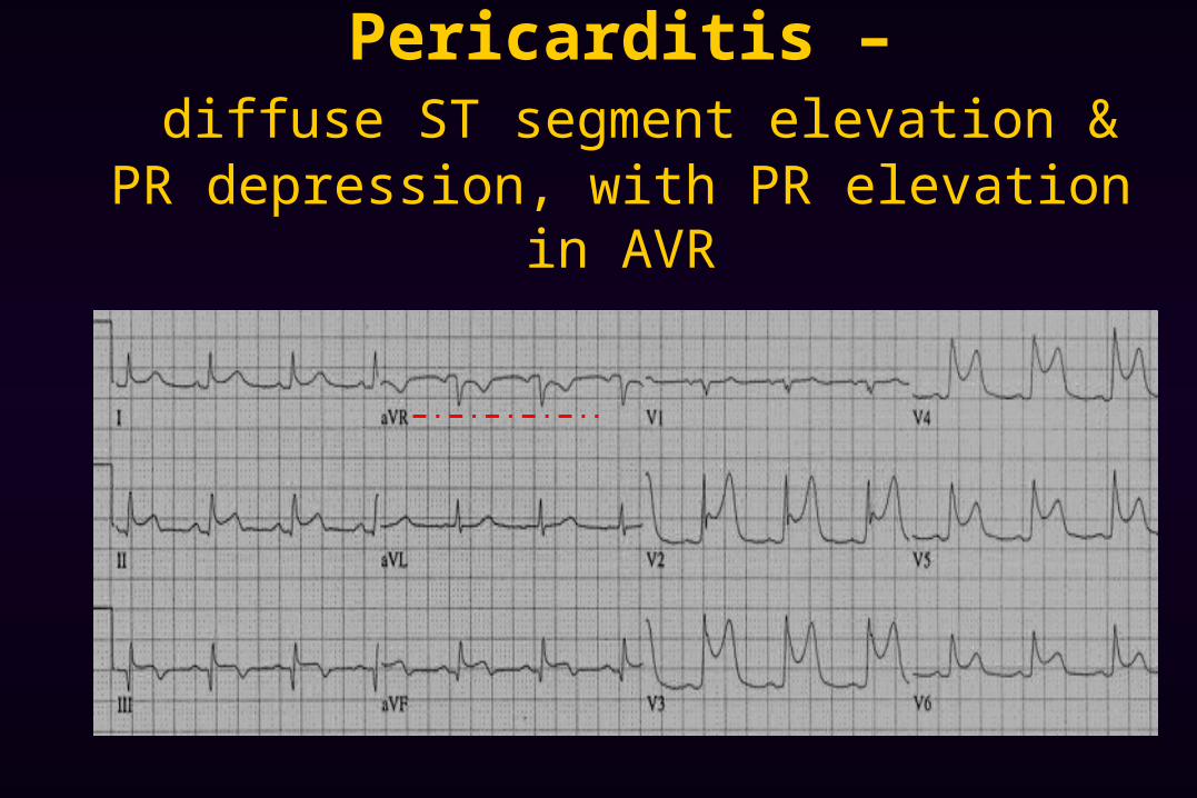
Pericarditis – diffuse ST segment elevation & PR depression,
with PR elevation in AVR
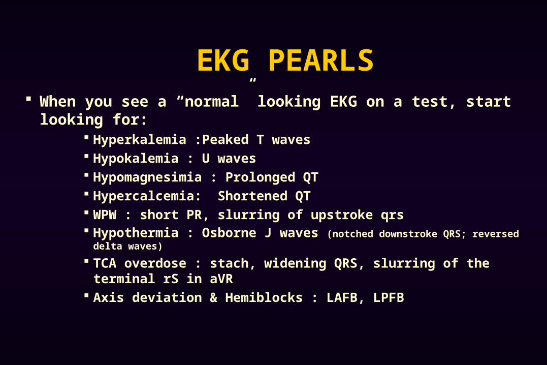
EKG PEARLS When you see a “normal” looking EKG on a test, start looking for:
Hyperkalemia :Peaked T waves Hypokalemia : U waves Hypomagnesimia : Prolonged QT Hypercalcemia: Shortened QT WPW : short PR, slurring of upstroke qrs Hypothermia : Osborne J waves (notched downstroke QRS; reversed delta waves)
TCA overdose : stach, widening QRS, slurring of the terminal rS in aVR
Axis deviation & Hemiblocks : LAFB, LPFB
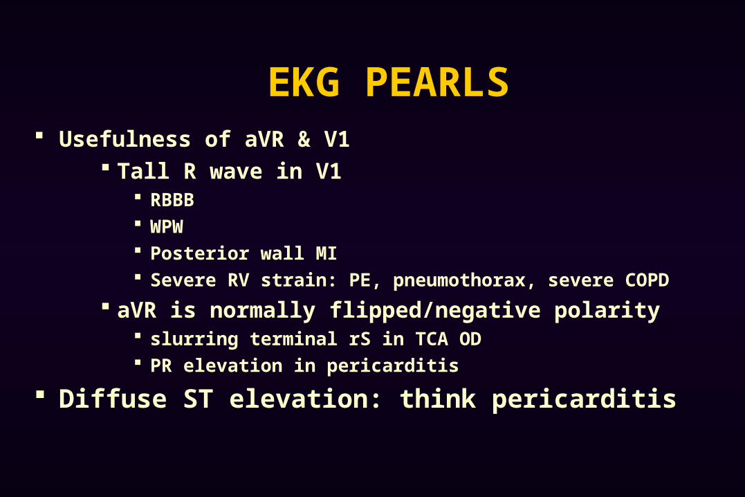
EKG PEARLS Usefulness of aVR & V1
Tall R wave in V1 RBBB WPW Posterior wall MI Severe RV strain: PE, pneumothorax, severe COPD
aVR is normally flipped/negative polarity slurring terminal rS in TCA OD PR elevation in pericarditis
Diffuse ST elevation: think pericarditis

?