
ARACHNODACTYLY AND ECTOPIA LENTIS IN A FATHER AND … · dactyly and ectopia lentis, a ' forme...
7
CASE REPORTS ARACHNODACTYLY AND ECTOPIA LENTIS IN A FATHER AND DAUGHTER BY RICHARD W. B. ELLIS, M.D., F.R.C.P. Assistant Physician for Children's Diseases, Guy's Hospital, London For many years after the original description of ' dolichostenomelie ' by Marfan (1896), this peculiar type of local gigantism of the extremities received little general recognition until Ormond (1924, 1929) drew the attention of ophthalmologists to the frequent association of the skeletal deformities with congenital dislocation of the lenses. Burch (1936), in a comprehensive review, was largely responsible for further- ing recognition of the condition in America, and the number of cases that have been reported, mostly in the ophthalmological literature, during the past ten years is an indication that arachnodactyly is not of extreme rarity, and that many milder cases and 'formes frustes ' have previously passed unrecognized. Stewart (1939) considers that the condition is not uncommon amongst mental defectives, although the intelligence of reported cases has generally been described as normal. Most of the reports have appeared in the French and German literature, but cases have been described in this country by Poynton (1903), Thursfield (1920), Poynton and Maurice (1923), Ormond and Williams (1924), Cameron (1924), Fowler (1924), Young (1929), Ormond (1929, 1930), Ellis (1931), Hudson (1932), King (1934), Sorsby (1935), Cockayne (1935), and Stewart (1939). As the name arachnodactyly (Achard, 1902) implies, the most striking fea- ture of the condition is usually the extreme length and slenderness of the digits. The description is not universally applicable, however, since some cases, e.g. the first of those here reported, whilst clearly being mild examples of arachnodactyly, have the arms and legs principally affected, whilst the hands and feet are practically normal. The increased length of the extremities results in a characteristic build, the patients commonly being considerably above the average height, but so slender and with so little subcutaneous fat that the weight is often greatly below the normal. Deformities of the spine, particularly scoliosis and kyphosis, and of the thorax (pigeon-breast, Trichterbrust) are common. Laxity of- the ligaments tends to make the patients clumsy and graceless, whilst deformities of the feet (pes planus, hammer-toe) are the general rure. Other bony abnormalities such as spina bifida (Brock, 1927; Zuber, 1928; Weill, 1932) have been described, whilst in three instances (Ellis, 1931; Semah, 1938; Boudet et al., 1939) arachnodactyly has been associated with fragilitas ossium and blue sclerotics. 267 on February 13, 2020 by guest. Protected by copyright. http://adc.bmj.com/ Arch Dis Child: first published as 10.1136/adc.15.84.267 on 1 January 1940. Downloaded from
Transcript of ARACHNODACTYLY AND ECTOPIA LENTIS IN A FATHER AND … · dactyly and ectopia lentis, a ' forme...

CASE REPORTS
ARACHNODACTYLY AND ECTOPIA LENTIS IN AFATHER AND DAUGHTER
BY
RICHARD W. B. ELLIS, M.D., F.R.C.P.
Assistant Physician for Children's Diseases, Guy's Hospital, London
For many years after the original description of ' dolichostenomelie ' byMarfan (1896), this peculiar type of local gigantism of the extremities receivedlittle general recognition until Ormond (1924, 1929) drew the attention ofophthalmologists to the frequent association of the skeletal deformities withcongenital dislocation of the lenses.
Burch (1936), in a comprehensive review, was largely responsible for further-ing recognition of the condition in America, and the number of cases that havebeen reported, mostly in the ophthalmological literature, during the past tenyears is an indication that arachnodactyly is not of extreme rarity, and thatmany milder cases and 'formes frustes ' have previously passed unrecognized.Stewart (1939) considers that the condition is not uncommon amongst mentaldefectives, although the intelligence of reported cases has generally beendescribed as normal. Most of the reports have appeared in the French andGerman literature, but cases have been described in this country by Poynton(1903), Thursfield (1920), Poynton and Maurice (1923), Ormond and Williams(1924), Cameron (1924), Fowler (1924), Young (1929), Ormond (1929, 1930),Ellis (1931), Hudson (1932), King (1934), Sorsby (1935), Cockayne (1935), andStewart (1939).
As the name arachnodactyly (Achard, 1902) implies, the most striking fea-ture of the condition is usually the extreme length and slenderness of thedigits. The description is not universally applicable, however, since some cases,e.g. the first of those here reported, whilst clearly being mild examples ofarachnodactyly, have the arms and legs principally affected, whilst the hands andfeet are practically normal. The increased length of the extremities results in acharacteristic build, the patients commonly being considerably above theaverage height, but so slender and with so little subcutaneous fat that the weightis often greatly below the normal. Deformities of the spine, particularlyscoliosis and kyphosis, and of the thorax (pigeon-breast, Trichterbrust) arecommon. Laxity of- the ligaments tends to make the patients clumsy andgraceless, whilst deformities of the feet (pes planus, hammer-toe) are the generalrure. Other bony abnormalities such as spina bifida (Brock, 1927; Zuber,1928; Weill, 1932) have been described, whilst in three instances (Ellis, 1931;Semah, 1938; Boudet et al., 1939) arachnodactyly has been associated withfragilitas ossium and blue sclerotics.
267
on February 13, 2020 by guest. P
rotected by copyright.http://adc.bm
j.com/
Arch D
is Child: first published as 10.1136/adc.15.84.267 on 1 January 1940. D
ownloaded from
ARCHIVES OF DISEASE IN CHILDHOOD
In about two-thirds of the cases the skull is dolichocephalic. The orbitalridges have sometimes been described as prominent and the jaw as prognathous.The teeth may be normal or-long and slender and the palate high and arched.
Radiologically the bones of the extremities, particularly the phalanges,metacarpals and metatarsals, show the extreme length and slenderness observedclinically. Ossification is usually normal for age; extra epiphyses have beendescribed in the digits, but these do not appear to be the rule. Apart fromdolichocephaly, the skull shows no characteristic changes, and the sella turcicais usually normal, though it may be reduced in size (Young, 1929). Weill(1932) found calcification in the region of the pineal in one instance.
Congenital morbus cordis occurs sufficiently often (in one-third or more ofthe cases) to be regarded as an integral part of the complete syndrome. It waspresent in the second of my cases.
Many other additional congenital abnormalities have been described, eitherin association with arachnodactyly or occurring in other members of affectedfamilies. Some degree of webbing of the fingers is common; the ears are oftenprominent or malformed; the musculature is poorly developed.
The incidence of respiratory infections appears unusually high, and a numberof patients (Hudson, 1933; Piper and Irvine-Jones, 1926; Borger, 1914) havesuccumbed to pneumonia. It is of interest that in the two latter cases, in whichpost-mortem examination was performed, congenital abnormalities of thelungs were found. Severe asthma occurred in the second case here reported.
Congenital dislocation of the lenses, giving rise to tremulous irides (' iri-dodonesis '), small miotic pupils and occasionally to recurrent attacks of glau-coma, is found in approximately 50 per cent. of cases of arachnodactyly(Ormond, 1930). The dislocated lenses are often opaque (as in case 1) andresistant to absorption (Hudson, 1933).
The following two cases illustrate most of the clinical features of arachno-dactyly, and its association with ectopia lentis.
Case reportsFAMILY HISTORY. The patients to be described are Stephen V., aged thirty-
two years, and his daughter, Ruth V. aged four years. Neither Stephen V.'sparents nor he and his wife Ellen V. were blood relations. Stephen V.'s paternalgrandfather is said to have been blind (cause unknown). Stephen V.'s fatherdied in Pembury Hospital in 1940 from cardiac failure following hyperpiesia;his eyes and limbs were normal and at autopsy there was no evidence of con-genital morbus cordis. Stephen V. has four brothers and two sisters; onebrother suffers from defective sight (cause unknown), but is otherwise normal,the others being normal both as regards sight and build.
Ellen V., aged thirty-one years (wife of Stephen V. and mother of Ruth V.),has been blind from birth. The right eye shows interstitial keratitis andopacity of the lens, whilst on the left there is advanced syphilitic choroiditis andpallor of the optic nerve. The Wassermann and Kahn reactions are stillstrongly positive, although she has been under treatment for congenital syphilissince 1937. She suffers from migrainous headaches and has had epileptic attackson several occasions. Her heart shows left-sided enlargement; there is a shortpresystolic murmur and a blowing systolic murmur in the mitral area. There
268
on February 13, 2020 by guest. P
rotected by copyright.http://adc.bm
j.com/
Arch D
is Child: first published as 10.1136/adc.15.84.267 on 1 January 1940. D
ownloaded from

ARACHNODACTYLY AND ECTOPIA LENTIS
has only been one pregnancy (Ruth V.), during the latter months of which shesuffered from oedema.
Case 1. Stephen V., aged thirty-two years, has been practically blind frombirth. He was brought up in a blind school until the age of sixteen and wassubsequently trained as a basket-maker. At the age of ten an unsuccessfuloperation on his right eye was performed at the Brighton Eye Hospital. Hesays that he learnt to walk late and ' was always falling about ' ; he was thoughtto be abnormally tall and thin during later childhood and adolescence. He hasgood general health, and apart from the limitations imposed by his eyesight isnormally active.
On examination he is of striking build, being tall (6 ft. 2 in.) and exceptionallyslender, the length of the arms and legs being particularly noticeable. He takesa size 141 collar and all ready-made clothes are much too loose for him. Thehands and feet, however, are not abnormal and he can wear size 8 shoes.
The skull is not abnormal, except for slight prominence of the orbital ridgesand somewhat prognathous jaw. The teeth, particularly those of the upperjaw, are long, widely-spaced and project forward. The ears are also ratherprominent.
There are no abnormalities of the spine, thorax, heart, lungs or abdomen.There is poor muscular development, and very little subcutaneous fat, thoughthe patient is actually stronger than his appearance suggests.
EYES. There is bilateral iridodonesis. Both lenses are dislocated. On theright the lens lies one sixth within the pupil and is opaque; on the left the lensis two-thirds within the pupil and is semi-opaque. Unaided vision on the rightis one-sixtieth and on the left two-sixtieths.
WASSERMANN and KAHN reactions negative.Case 2. Ruth V., a girl, aged four years at the time of examination (May,
1940), was born four weeks prematurely weighing six and a halfpounds; deliverywas normal. The child was thought to be of unusual length at birth, and thoughshe sucked and cried normally, she always appeared thin. She was fed on acow's milk formula. At no time had she been cyanotic. An umbilical herniawas strapped for the first year.
At nine months of age the child developed pertussis, and at eleven monthswas admitted to Pembury County Hospital with infantile eczema. Recurrentasthmatic attacks, which have become more severe and frequent as she has gotolder, began during the second year. They have been associated with the gradualappearance of the thoracic deformity which she now shows. There is no knownfood or other idiosyncrasy, and the asthmatic attacks occur both in winter andin summer. The child has been admitted to hospital six times on account ofasthma since 1937.
During the first year it was noted that the child did not see normally. Shecut two teeth at three months of age and had sixteen teeth at a year old. Shespoke words at eighteen months and began to walk at two years old. She isstill very liable to fall over.
On examination (May, 1940). A tall, extremely slender child, who is activebut ungainly and unsteady on her feet. Height 441 in. (normal for age 38 in.)Weight 35 pounds (normal for age 34 pounds, for height 45 pounds). Fig. 1shows the characteristic length of the limbs and narrowness of the trunk, whilstthe anxious expression of the face is also somewhat typical. The head measures17-5 cm. in length and 12-5 cm. in breadth. The teeth are normal. The palateis high and arched. There is dorsal kyphosis and laxity of joint ligaments,particularly of the wrists, elbows and knees.
THE HANDS (fig. 2) measure 51 in. from the tip of the middle finger to theradial styloid (normal control 41 in.), and show the typical ' spider fingers.'
269
on February 13, 2020 by guest. P
rotected by copyright.http://adc.bm
j.com/
Arch D
is Child: first published as 10.1136/adc.15.84.267 on 1 January 1940. D
ownloaded from

ARCHIVES OF DISEASE IN CHILDHOOD
FIG 1.-Ruth V., showing tall, slender build and deformity of thorax.
FIG. 2.-Ruth V.: Hands showing arachnodactyly.
THE FEET (fig. 3) are excessively long and slender. There is a hammer-toedeformity of the third and fourth toes on both sides.
THORAX. There is a well-marked pigeon-breast deformity of the chest, withflattening of the lower ribs anteriorly. Scattered wheezing expiratory rales areheard over both lungs.
HEART. The apex beat is palpable in the left fifth intercostal space im-mediately outside the nipple line, and the area of cardiac dullness extends half
270
on February 13, 2020 by guest. P
rotected by copyright.http://adc.bm
j.com/
Arch D
is Child: first published as 10.1136/adc.15.84.267 on 1 January 1940. D
ownloaded from
ARACHNODACTYLY AND ECTOPIA LENTIS
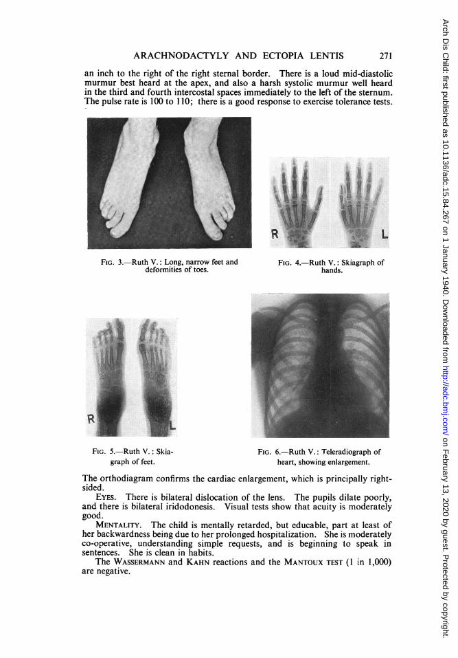
an inch to the right of the right sternal border. There is a loud mid-diastolicmurmur best heard at the apex, and also a harsh systolic murmur well heardin the third and fourth intercostal spaces immediately to the left of the sternum.The pulse rate is 100 to 1 10; there is a good response to exercise tolerance tests.
FIG. 3.-Ruth V.: Long, narrow feet anddeformities of toes.
FIG. 5.-Ruth V.: Skia-graph of feet.
FIG. 4.-Ruth V.: Skiagraph ofhands.
FIG. 6.-Ruth V.: Teleradiograph ofheart, showing enlargement.
The orthodiagram confirms the cardiac enlargement, which is principally right-sided.
EYES. There is bilateral dislocation of the lens. The pupils dilate poorly,and there is bilateral iridodonesis. Visual tests show that acuity is moderatelygood.
MENTALITY. The child is mentally retarded, but educable, part at least ofher backwardness being due to her prolonged hospitalization. She is moderatelyco-operative, understanding simple requests, and is beginning to speak insentences. She is clean in habits.
The WASSERMANN and KAHN reactions and the MANTOUX TEST (1 in 1,000)are negative.
271
m
on February 13, 2020 by guest. P
rotected by copyright.http://adc.bm
j.com/
Arch D
is Child: first published as 10.1136/adc.15.84.267 on 1 January 1940. D
ownloaded from

ARCHIVES OF DISEASE IN CHILDHOOD
DiscussionThe association of congenital abnormalities of the osseous system with
congenital abnormalities of the eye is seen in other conditions than arachno-dactyly. Examples are gargoylism (chondro-osteo-dystrophy and cornealopacities); congenital coloboma of the macula and apical dystrophy of handsand feet (Sorsby, 1935); punctate epiphyseal dysplasia associated with congenitalcataract; and the Laurence-Moon-Biedl syndrome (polydactyly and retinitispigmentosa, etc.). With the possible exception of punctate epiphyseal dysplasia,all these conditions may show a familial incidence and the ocular and osseousdefects occur separately. In the case of the Laurence-Moon-Biedl syndrome,in which affected families have been studied in some detail, it has been sug-gested that a linkage of genes has taken place (Cockayne et al., 1935; Sorsbyet al., 1939).
The two cases reported here illustrate the hereditary factor in both arachno-dactyly and ectopia lentis, a ' forme fruste ' of arachnodactyly with ectopialentis in the father being followed by the appearance of the classical picture ofarachnodactyly with ectopia lentis in the child. (The mother's blindness,though making the family a lamentable social phenomenon, is not regarded asbeing related to the condition in the child; there is no evidence that parentalsyphilis plays any part in the production of either arachnodactyly or ectopialentis.) There was no consanguinity of the parents.
There are numerous reports in the literature in which arachnodactyly andectopia lentis, combined or separately, have occurred in several siblings or in aparent and children. (Unfortunately few authors state whether or not there wasconsanguinity of the parents.)
Thus Achard (1902) described arachnodactyly in two sisters, their motherand maternal grandfather; Villard et al. (1931) reported a family consisting of afather (antecedents unknown) who suffered from ectopia lentis, a mother whowas normal, and five children. Of the latter, the eldest boy and girl showedboth arachnodactyly and ectopia lentis, a third child was normal, the fourth(a girl) had ectopia lentis only, and the fifth (a boy) was normal. Weve (1931)observed twenty-three cases of arachnodactyly in six families, in three of whichthere was certain evidence of hereditary transmission. The association ofarachnodactyly and ectopia lentis was seen in a father, two sons, and a daughter.Weill (1932) observed both conditions in a mother and son, whilst in a secondfamily the mother showed dislocation and calcification of the lenses associatedwith dwarfism, and her son ectopia lentis and a forme fruste of arachnodactyly.Young (1929) found arachnodactyly and associated congenital abnormalitiesin a mother, daughter and son. Harrison and Klainer (1939) give a familytree showing arachnodactyly in two siblings in one generation, and three in thenext.
Although further familial studies are required definitely to establish the modeof inheritance and linkage of the two conditions, it seems probable that botharachnodactyly and ectopia lentis may be transmitted as Mendelian dominants,either separately or in association. There is no sex-linkage in either condition.
Thanks are due to Dr. W. C. Long, Honorary Radiologist to the Kent and
272
on February 13, 2020 by guest. P
rotected by copyright.http://adc.bm
j.com/
Arch D
is Child: first published as 10.1136/adc.15.84.267 on 1 January 1940. D
ownloaded from

ARACHNODACTYLY AND ECTOPIA LENTIS 273
Sussex Hospital, for the skiagraphs, and to Dr. L. B. Langmead for thephotographs.
REFERENCESAchard, C. (1902). Bull. Soc. mid. H6p. Paris, 19, 834.Borger, F. (1914). Z. Kinderheilk., 12, 161.Boudet, G., Balmes, J., and Barnay, J. (1939). Bull. Soc. Pediat. Paris. 37, 188.Brock, J. (1927). Klin. Wschr., 6, 2289.Burch, F. E. (1936). Arch. Ophthal., N.Y., 15, 645.Cameron, H. C. (1924). Proc. roy. Soc. Med., 17, 32.Cockayne, E. A. (1935). Ibid., 29, 120.Cockayne, E. A., Krestin, D., and Sorsby, A. (1935). Quart. J. Med., N.S., 4, 93.Ellis, R. W. B. (1931). Proc. roy. Soc. Med., 24, 54.Fowler, J. S. (1924). Guy's Hosp. Rep., 74, 395 and 398.Futcher, P. H., and Southworth, H. (1938). Arch. intern. Med., 61, 693.Harrison, J., and Klainer, M. J. (1939). New Engl. J. Med., 220, 621.Hudson, A. C. (1933). Proc. roy. Soc. Med., 26, 35.King, E. F. (1934). Ibid., 27, 298.Marfan, A. B. (1896). Bull. Soc. mid. H6p. Paris, 13, 220.Ormond, A. W. (1929). Int. ophthal. Congr., 2, 645.
(1930). Guy's Hosp. Rep., 80, 68.and Williams, R. G. (1924). Ibid., 74, 385.
Piper, R. K., and Irvine-Jones, E. (1926). Amer. J. Dis. Child., 31, 832.Poynton, F. J. (1903). Trans. med. Soc. Lond., 26, 338.
and Maurice, W. B. (1923). Ibid., 45, 21.Semah, F. (1938). Riv. Clin. pediat., 36, 228.Sorsby, A. (1935). Brit. J. Ophthal., 19, 65.
Avery, H., and Cockayne, E. A. (1939). Quart. J. Med., N. S., 8, 51.Stewart, R. M. (1939). Arch. Dis. Childh., 14, 64.Thursfield, H. (1920). St Bart.'s Hosp. med. Rep., 53, 35.Villard, H., Viallefont, H., and Temple, J. (1931). Bull. Soc. Ophthal. Paris, 46, 384.Weill, G. (1932). Ann. Oculist., Paris, 169, 21.Weve, H. (1931). Arch. Augenheilk., 104, 1.Young, M. L. (1929). Arch. Dis. Childh., 4, 190.Zuber, A. (1928). Bull Soc. Pediat. Paris, 26, 311.
on February 13, 2020 by guest. P
rotected by copyright.http://adc.bm
j.com/
Arch D
is Child: first published as 10.1136/adc.15.84.267 on 1 January 1940. D
ownloaded from


![Open Access Corneal Microstructural Analysis in Weill ... · microspherophakia, with high lenticular myopia, and ectopia lentis [2]. The constant corneal finding is an increased central](https://static.fdocuments.in/doc/165x107/5f0f97bc7e708231d444ee05/open-access-corneal-microstructural-analysis-in-weill-microspherophakia-with.jpg)


![CASE REPORT / ПРИКАЗ БОЛЕСНИКА Delayed diagnosis of ... · and ectopia lentis (EL) [1]. It has an estimated incidence of 1:50,000–200,000, sufficiently high to consider](https://static.fdocuments.in/doc/165x107/5e452e7fa3e3b7377054df81/case-report-delayed-diagnosis-of-and-ectopia.jpg)


