Approach to Proximal Humerus Fractures: An Observational ...
5
40 40 International Journal of Scientific Study | November 2019 | Vol 7 | Issue 8 Approach to Proximal Humerus Fractures: An Observational Study in a Tertiary Care Centre at Bilaspur Arunesh Singh 1 , Pranay Kumar Srivastava 2 , Abhishek Ghatge 3 1 Associate Professor, Department of Orthopaedics, Chhattisgarh Institute of Medical Sciences, Bilaspur, Chhattisgarh, India, 2 Assistant Professor, Department of Orthopaedics, Pt. JNM Medical College and Dr. BRAM Hospital, Raipur, Chhattisgarh, India, 3 Department of Orthopaedics, Knee and Shoulder surgeon, Gajanan Memorial Hospital, Bilaspur ,Chhattisgarh, India There are a large number of surgical treatment options for fracture of proximal humerus ranging from cross K-wires, titanium elastic nailing system/Ender’s nails used in the form of J nails, proximal humerus plates, and even prosthetic replacement in the form of hemiarthroplasty for severely comminuted fractures. However, there is no general consensus available for deciding the management protocol. Neer’s classification is widely accepted for the classification of these fractures into two-part, three-part, four-part, fracture-dislocations, and articular surface fractures. [1] Management becomes difficult in three-part and four-part fractures and further for fracture-dislocations and articular surface fractures and humeral head fractures. We here present our experience with proximal humerus internal locking system (PHILOS) plate with complex fractures and fracture-dislocations of proximal humerus at a tertiary care center at Bilaspur. Purpose of the Study The purpose of the study was to find out the functional outcome of PHILOS plating in complex fractures and INTRODUCTION The proximal humerus fractures are a relatively common entity and are very difficult to treat as the shoulder joint is a versatile joint with a high degree of motion. The fracture comprises 4–5% of all fractures and as high as 45% of the humerus fractures. The fractures are seen in all ages from children to the elderly. In the elderly, the fracture is mostly osteoporosis related, which makes it all the more difficult to treat. In young adults, the fracture management is tough due to high functional demands, frequent comminution and the fracture being a high energy trauma. Original Article Abstract Introduction: The proximal humerus fractures are a relatively common entity and are very difficult to treat as the shoulder joint is a versatile joint with high degree of motion. The fracture comprises 4–5% of all fractures and as high as 45% of the humerus fractures. Materials and Methods: A total of 19 patients of complex fractures of proximal humerus and fracture-dislocations of proximal humerus, who were managed with proximal humerus internal locking system plating were included in the study. Results: In 11 patients, the right-sided injury was seen and in eight patients, injury was left sided. The age of the patients ranged from 28 years to 56 years with a mean age of 42.7 years. Most of our patients were Neer’s three-part and four-part fractures, and fracture-dislocations as two-part fractures were again managed differently (conservatively or J nails, cross K-wires, Rush nails, and external fixators). We had five fracture-dislocations, 12 patients with comminuted three- and four-part fractures, and two patients with two-part fractures. Conclusion: Proximal humeral fractures are relatively common fractures and difficult to treat entities for the age; they occur frequent comminution and difficulty in achieving reduction and hardware application. Key words: Fracture, Patients, Proximal Access this article online www.ijss-sn.com Month of Submission : 09-2019 Month of Peer Review : 10-2019 Month of Acceptance : 11-2019 Month of Publishing : 11-2019 Corresponding Author: Dr. Pranay Kumar Srivastava, Flat Number E-7, Ralas Enclave , Daganiya, Raipur, Chhattisgarh, India. Print ISSN: 2321-6379 Online ISSN: 2321-595X
Transcript of Approach to Proximal Humerus Fractures: An Observational ...

4040International Journal of Scientific Study | November 2019 | Vol 7 | Issue 8
Approach to Proximal Humerus Fractures: An Observational Study in a Tertiary Care Centre at BilaspurArunesh Singh1, Pranay Kumar Srivastava2, Abhishek Ghatge3
1Associate Professor, Department of Orthopaedics, Chhattisgarh Institute of Medical Sciences, Bilaspur, Chhattisgarh, India, 2Assistant Professor, Department of Orthopaedics, Pt. JNM Medical College and Dr. BRAM Hospital, Raipur, Chhattisgarh, India, 3Department of Orthopaedics, Knee and Shoulder surgeon, Gajanan Memorial Hospital, Bilaspur ,Chhattisgarh, India
There are a large number of surgical treatment options for fracture of proximal humerus ranging from cross K-wires, titanium elastic nailing system/Ender’s nails used in the form of J nails, proximal humerus plates, and even prosthetic replacement in the form of hemiarthroplasty for severely comminuted fractures. However, there is no general consensus available for deciding the management protocol. Neer’s classification is widely accepted for the classification of these fractures into two-part, three-part, four-part, fracture-dislocations, and articular surface fractures.[1] Management becomes difficult in three-part and four-part fractures and further for fracture-dislocations and articular surface fractures and humeral head fractures.
We here present our experience with proximal humerus internal locking system (PHILOS) plate with complex fractures and fracture-dislocations of proximal humerus at a tertiary care center at Bilaspur.
Purpose of the StudyThe purpose of the study was to find out the functional outcome of PHILOS plating in complex fractures and
INTRODUCTION
The proximal humerus fractures are a relatively common entity and are very difficult to treat as the shoulder joint is a versatile joint with a high degree of motion. The fracture comprises 4–5% of all fractures and as high as 45% of the humerus fractures. The fractures are seen in all ages from children to the elderly. In the elderly, the fracture is mostly osteoporosis related, which makes it all the more difficult to treat. In young adults, the fracture management is tough due to high functional demands, frequent comminution and the fracture being a high energy trauma.
Original Article
AbstractIntroduction: The proximal humerus fractures are a relatively common entity and are very difficult to treat as the shoulder joint is a versatile joint with high degree of motion. The fracture comprises 4–5% of all fractures and as high as 45% of the humerus fractures.
Materials and Methods: A total of 19 patients of complex fractures of proximal humerus and fracture-dislocations of proximal humerus, who were managed with proximal humerus internal locking system plating were included in the study.
Results: In 11 patients, the right-sided injury was seen and in eight patients, injury was left sided. The age of the patients ranged from 28 years to 56 years with a mean age of 42.7 years. Most of our patients were Neer’s three-part and four-part fractures, and fracture-dislocations as two-part fractures were again managed differently (conservatively or J nails, cross K-wires, Rush nails, and external fixators). We had five fracture-dislocations, 12 patients with comminuted three- and four-part fractures, and two patients with two-part fractures.
Conclusion: Proximal humeral fractures are relatively common fractures and difficult to treat entities for the age; they occur frequent comminution and difficulty in achieving reduction and hardware application.
Key words: Fracture, Patients, Proximal
Access this article online
www.ijss-sn.com
Month of Submission : 09-2019 Month of Peer Review : 10-2019 Month of Acceptance : 11-2019 Month of Publishing : 11-2019
Corresponding Author: Dr. Pranay Kumar Srivastava, Flat Number E-7, Ralas Enclave , Daganiya, Raipur, Chhattisgarh, India.
Print ISSN: 2321-6379Online ISSN: 2321-595X

Singh, et al.: Approach to Proximal Humerus Fractures
4141 International Journal of Scientific Study | November 2019 | Vol 7 | Issue 8
One patient who did not turn up for follow-up was excluded from the study. One patient of fracture-dislocation of humerus was taken for surgery but due to lack of bone stock and improper hold of the implant, the fracture fixation could not be done. The patient later denied shoulder arthroplasty and was excluded from the study.
The patients’ age distribution was from 28 years to 56 years. In all these patients, primary management of the fracture by slab immobilization/shoulder immobilizer was given, and cold fomentation and anti-inflammatory agents were started at the time of admission. After all routine investigations and proper pre-operative assessment, the patients were taken for surgery. All patients were operated by the author and his team. Standard beach chair position was utilized for the surgery and in all patients, deltopectoral approach was taken. General anesthesia with brachial plexus block for post-operative pain was instituted for all patients. Preoperatively, two shots of the third-generation cephalosporin were given. The mean time between injury and surgery was 10.9 days. The mean duration of surgery was 137 min. Postoperatively, arm pouch sling was applied. Physiotherapy was started as soon as
fracture-dislocations of proximal humerus, in patients presenting to a tertiary care center at Bilaspur from April 1, 2014, to March 31, 2016.
MATERIALS AND METHODS
A total of 19 patients of complex fractures of proximal humerus and fracture-dislocations of proximal humerus, who were managed with PHILOS plating were included in the study.
Inclusion CriteriaAll the proximal humerus fractures managed by PHILOS plates during the study period were included in the study between 18 and 60 years of age.
Exclusion CriteriaPatients above 60 years of age or below 18 years of age were excluded from the study as protocol for their management was different and age would become one of the important confounding variables in terms of bone quality and muscle strength in patients above 60 years of age and delayed healing in such patients due to severe osteoporosis.
As opposed to plate fixation, children with proximal humerus fractures in our institute, were managed by K wire fixation and were excluded from the study.
Table 1: Pain scoresPain ScoresNo pain 35Slight, occasional, no compromise in activity 30Mild, no effect on ordinary activity 25 Moderate, tolerable, makes concessions 15Marked, serious limitations 5Totally disabled 0
Table 2: FunctionFunction ScoresStrength–normal 10Good 8Fair 6Poor 4Trace 2Zero 0Reaching–top of head 2Mouth 2Belt buckle 2Opposite axilla 2Brassiere hook 2Stability–lifting 2Throwing 2Pounding 2Pushing 2Hold overhead 2
Table 3: Range of movementRange of movement ScoresFlexion–180° 6
170° 5130° 4100° 380° 2<80° 1
Abduction–180° 6170° 5140° 4100° 380° 2<80° 1
Extension–45° 330° 215° 1 <15° 0
External rotation–60° 530° 310° 1<10° 0
Internal rotation–90° 570° 450° 330° 2<30° 0
Table 4: AnatomyRotation, Angulation, Joint incongruity, Retracted Tuberosities, Failure Metal, Myositis, Non Union, Avn
Scores
None 10Mild 8Moderate 4Marked 0–2
Singh, et al.: Approach to Proximal Humerus Fractures
4242International Journal of Scientific Study | November 2019 | Vol 7 | Issue 8
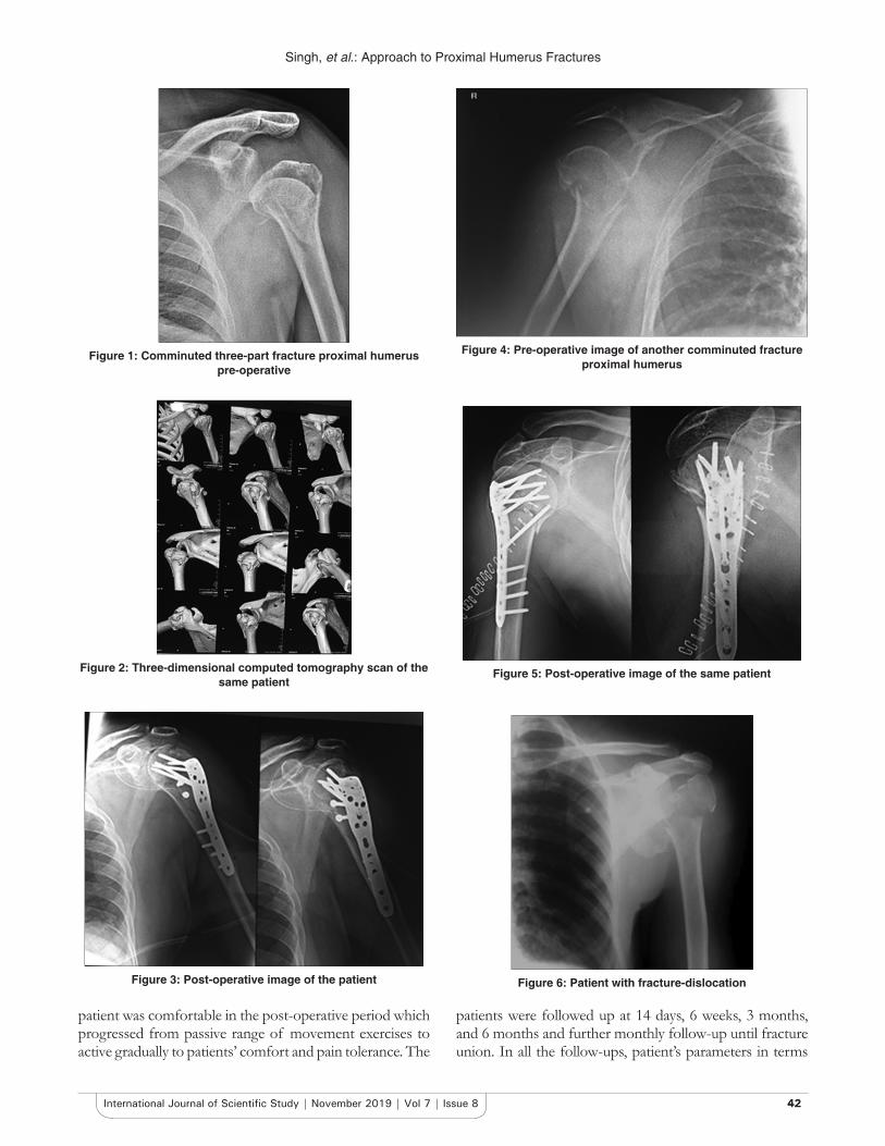
Figure 1: Comminuted three-part fracture proximal humerus pre-operative
Figure 2: Three-dimensional computed tomography scan of the same patient
Figure 3: Post-operative image of the patient
Figure 4: Pre-operative image of another comminuted fracture proximal humerus
Figure 5: Post-operative image of the same patient
Figure 6: Patient with fracture-dislocation
patient was comfortable in the post-operative period which progressed from passive range of movement exercises to active gradually to patients’ comfort and pain tolerance. The
patients were followed up at 14 days, 6 weeks, 3 months, and 6 months and further monthly follow-up until fracture union. In all the follow-ups, patient’s parameters in terms

Singh, et al.: Approach to Proximal Humerus Fractures
4343 International Journal of Scientific Study | November 2019 | Vol 7 | Issue 8
of pain, function, and range of movements were noted and patient’s X-rays anteroposterior views and axial views were taken to see for union. One patient did not turn up for follow-up after suture removal and was excluded from the study. Another patient, who was taken for surgery but due to inadequate bone left for fixation due to a large defect in the humeral head, had to be kept out of the study. The patient was later given the option of shoulder hemiarthroplasty but she refused. Finally, 19 patients were included in the study who had completed all the mentioned follow-ups or achieved union. The outcome was evaluated by Neer’s score which is an objective score taking into consideration pain, function, range of movement, and anatomy.
RESULTS
A score of 90–100 was excellent, 80–89 satisfactory, 70–79 unsatisfactory, and <70 was considered failure (Neer’s scoring system) [Tables 1-4].
A total of 19 patients were included in the study. There were 12 male patients and 7 female patients. In 11 patients, the right-sided injury was seen and in eight patients, injury was left sided. The age of the patients ranged from 28 years to 56 years with a mean age of 42.7 years. Most of our patients were Neer’s three-part and four-part fractures, and fracture-dislocations as two-part fractures were again managed differently (conservatively or J nails, cross K-wires, Rush nails, and external fixators). We had five fracture-dislocations, 12 patients with comminuted three- and four-part fractures, and two patients with two-part fractures. Union was achieved in all patients at a mean of 4.6 months. We had no patients who had avascular necrosis (AVN). Six patients had excellent results. In nine patients, the result was satisfactory. Three patients had unsatisfactory results and in one patient, we had infection due to which he needed implant removal in the 8th month post-operative period and had poor result. As such, there was no fixation failure after primary fixation.
Figure 1 depicts the pre operative X ray of a patient with comminuted 3 part fracture of proximal humerus. The CT scan of the same patient is shown in Figure 2. We can see how effectively CT scan can delineate the fracture configuration. The patient was taken for PHILOS plating and the plate was applied. Post operative X rays of the patient can be seen in Figure 3 showing good reduction.
X rays of another patient with comminuted fracture proximal humerus is seen in Figure 4. The post op X rays of the same patient are shown in Figure 5.
Figure 6 depicts X rays of a patient with fracture dislocation. The patient was managed successfully by PHILOS plating.The patient had good reduction [Figure 7] and good functional outcome with good range of movement [Figure 8].
DISCUSSION
Proximal humerus fractures are one of the most difficult to treat entities. The treatment becomes all the more difficult due to reduced bone stock due to osteoporosis in elderly population where these fractures are common.
There are multiple treatment options in the management of theses fractures including percutaneous pinning, J nailing, external fixator application, Rush nails, MultiLoc nails, hemiarthroplasty, simple non-locking plates, and internally locked plates or PHILOS. As per literature for a periarticular and intra-articular fracture, one has to achieve good anatomical reduction and rigid stable internal fixation which can allow early mobilization and early fracture healing
Figure 7: Post-operative image of the same patient
Figure 8: Clinical image of a patient operated for fracture-dislocation with satisfactory results

Singh, et al.: Approach to Proximal Humerus Fractures
4444International Journal of Scientific Study | November 2019 | Vol 7 | Issue 8
at the same time. Percutaneous K-wires, external fixators, J nails, and Rush nails are more biological and less invasive. Neither the reduction achieved with them is anatomical (due to closed techniques) nor the fixation is rigid enough for initiation of early range of movement. To add to it, complications such as pin-tract infections further limit their use.[2] With non-locking plates used the chances of loss of reduction, screw cutout and fixation failure are high as the region of fixation is mainly metaphyseo-epiphyseal region which is mainly cancellous bone and the situation is confounded by osteoporosis. In addition, most of the fractures we catered to were comminuted three-part, four-part fractures or fracture-dislocations, in which there were multiple fragments and fixation with simple plates was tough to achieve and hold. The torsional stability is better with locking plates that were confirmed by Siffri et al.[3] in their cadaveric studies. MultiLoc nailing, in a study by Hessmann et al.,[4] has yielded good results. However, we have little experience with such MultiLoc nailing devices. In view of all the above, we performed our study with PHILOS plates.
Screw cutouts and implant failures, we had none. Owsley and Gorczyca et al.[5] in their study had a screw cutout rate of 23% and Doshi et al.[6] in their study had it at 5.66%. We had one case where we could not achieve fixation due to no hold of the screws due to very less bone stock and comminution. This patient was not included in the study. If we include the patient in the study, as a case of failed fixation or failure to achieve fixation, our fixation failure rate was 5% which is comparable to Doshi et al. We had no cases of AVN which is again comparable to Doshi et al.
Computed tomography scan (CT) is of undue importance before proceeding for surgery and deciding the plan of action. In most of the cases of fracture especially those with a fracture-dislocation, the CT scan was performed at the fragments of fracture identified preoperatively so that planning of reduction and fixation could be done preoperatively. We suggest a pre-operative CT scan for all comminuted proximal humerus fracture patients. It is also important so as to see if the humeral head is also fractured. In case of humeral head fracture, it is better to go for replacement as there are high chances of AVN and loss of fixation.
Finally, accuracy of reduction and rigidity of fixation are another deciding factor for outcome in such cases. A valgus reduction is a must with little space for varus reduction. A varus reduction may lead to loss of fixation, implant cutout,
and high stress on rotator cuff, leading to poor functional outcome.[7] Furthermore, we must stress here on the fact that in all cases, author and his team aimed for anatomical reduction with special care to restore the medial calcar and maintain the calcar with oblique calcar screws (superiorly directed inferomedially placed) in most of the cases. This was in accordance with Krappinger et al.[8] who said that anatomical fracture reduction and good medial cortical alignment are the most important factors for secondary displacement. Gardner et al.[9] have previously shown that screw cutout rates are high following loss of medial support.
CONCLUSION
Proximal humeral fractures are relatively common fractures and difficult to treat entities for the age; they occur frequent comminution and difficulty in achieving reduction and hardware application. However, with the advent of locking plates, the results are good in our experience. Understanding the fracture anatomy and obtaining near anatomical reduction with medial calcar restoration is the key to surgical management of these fractures.
REFERENCES
1. Neer CS 2nd. Displaced proximal humeral fractures. I. Classification and evaluation. J Bone Joint Surg Am 1970;52:1077-89.
2. Jaura G, Sikdar J, Singh S. Long term results of PHILOS plating and percutaneous K-wire fixation in proximal humerus fractures in the elderly. Malays Orthop J 2014;8:4-7.
3. Siffri PC, Peindl RD, Coley ER, Norton J, Connor PM, Kellam JF, et al. Biomechanical analysis of blade plate versus locking plate fixation for a proximal humerus fracture: Comparison using cadaveric and synthetic humeri. J Orthop Trauma 2006;20:547-54.
4. Hessmann MH, Nijs S, Mittlmeier T, Kloub M, Segers MJ, Winkelbach V, et al. Internal fixation of fractures of the proximal humerus with the multiLoc nail. Oper Orthop Traumatol 2012;24:418-31.
5. Owsley KC, Gorczyca JT. Fracture displacement and screw cutout after open reduction and locked plate fixation of proximal humeral fractures [corrected]. J Bone Joint Surg Am 2008;90:233-40.
6. Doshi C, Sharma GM, Naik LG, Badgire KS, Qureshi F. Treatment of proximal humerus fractures using PHILOS plate. J Clin Diagn Res 2017;11:RC10-RC13.
7. Voigt C, Kreienborg S, Megatli O, Schulz AP, Lill H, Hurschler C, et al. How does a varus deformity of the humeral head affect elevation forces and shoulder function? A biomechanical study with human shoulder specimens. J Orthop Trauma 2011;25:399-405.
8. Krappinger D, Bizzotto N, Riedmann S, Kammerlander C, Hengg C, Kralinger FS, et al. Predicting failure after surgical fixation of proximal humerus fractures. Injury 2011;42:1283-8.
9. Gardner MJ, Weil Y, Barker JU, Kelly BT, Helfet DL, Lorich DG, et al. The importance of medial support in locked plating of proximal humerus fractures. J Orthop Trauma 2007;21:185-91.
How to cite this article: Singh A, Srivastava P, Ghatge A. Approach to Proximal Humerus Fractures: An Observational Study in a Tertiary Care Centre at Bilaspur. Int J Sci Stud 2019;7(8):40-44.
Source of Support: Nil, Conflict of Interest: None declared.