APPLIED SCIENCES AND ENGINEERING Copyright © 2019 ... · 610064, China. 2Department of Orthopaedic...
17
Zhang et al., Sci. Adv. 2019; 5 : eaax6946 2 August 2019 SCIENCE ADVANCES | RESEARCH ARTICLE 1 of 16 APPLIED SCIENCES AND ENGINEERING Application of hydroxyapatite nanoparticles in tumor-associated bone segmental defect Kun Zhang 1 *, Yong Zhou 2 *, Cong Xiao 2 , Wanlu Zhao 1 , Hongfeng Wu 1 , Jiaoqing Tang 3 , Zhongtao Li 3 , Sen Yu 4 , Xiangfeng Li 1 , Li Min 2 , Zhentao Yu 4 , Gang Wang 1 , Lin Wang 3 , Kai Zhang 1 , Xiao Yang 1† , Xiangdong Zhu 1† , Chongqi Tu 2† , Xingdong Zhang 1 Hydroxyapatite (HA) has been widely applied in bone repair because of its superior biocompatibility. Recently, a proliferation-suppressive effect of HA nanoparticles (n-HA) against various cancer cells was reported. This study was aimed at assessing the translational value of n-HA both as a bone-regenerating material and as an antitumor agent. Inhibition of tumor growth, prevention of metastasis, and enhancement of the survival rate of tumor-bearing rabbits treated with n-HA were demonstrated. Activated mitochondrial-dependent apoptosis in vivo was confirmed, and we observed that a stimulated immune response was involved in the n-HA–induced antitumor effect. A porous titanium scaffold loaded with n-HA was fabricated and implanted into a critical-sized segmental bone defect in a rabbit tumor model. The n-HA–releasing scaffold not only showed a prominent effect in suppressing tumor growth and osteolytic lesion but also promoted bone regeneration. These findings provide a rationale for using n-HA in tumor-associated bone segmental defects. INTRODUCTION Bone tumors and cancer metastases to the bone pose serious threats to human health. In the United States, approximately 2500 new cases of primary bone cancer are diagnosed annually, and approximately half of the patients with cancer will exhibit bone metastasis (1). The three most common types of primary bone malignant tumors are osteosarcoma, Ewing’s sarcoma, and chondrosarcoma. The most common sites of involvement are long bones and adjacent soft tissues, characterized by local symptoms such as pain, tumefaction, and bone destruction. Most bone tumors are osteolytic rather than sclerotic, which causes a destructive lesion. The standard clinical treatment strategies for bone cancer include surgical approaches to resect and reconstruct the affected bone followed by adjuvant radiation or chemotherapy (2). Although many load-bearing artificial implants have been adopted clinically, poor implant-bone osseointegration and new bone formation difficulty in the tumor environment remain a major challenge to orthopedic surgeons (3). In addition, incomplete surgical resection of the surrounding affected soft tissues risks the spread of tumor cells, which results in 8% tumor recurrence or metastasis (4). It is imperative to develop an implant combined with both antitumor and bone regeneration functions. One strategy is to combine three- dimensional (3D) printing technology with photothermal features for in situ ablation applications. Using this strategy, Ma et al. ( 5) fabricated a 3D-printed calcium phosphate composite scaffold and modified it with graphene oxide to transfer infrared laser energy into the photothermal effect. As a cost-effective approach, polydopamine and black phosphorus can also be used as photothermal agents assembled onto the surface of the scaffold (6, 7). Although promising results have been demonstrated with these scaffolds, their capacities for tumor suppression and bone regeneration were tested separately. A suitable animal model to assess both functions is urgently needed. Another strategy is to load antitumor drugs into bone cements or scaffolds for direct local delivery. In a rat osteosarcoma model, cisplatin- and/or caffeine-containing bone cement has been reported to provide effective drug delivery and reduce the risk of local recurrence (8). Recently, a hydroxyapatite (HA) composite scaffold loaded with zoledronic acid has been proposed for tumor-induced bone defect repair as well (9). However, the comparison with intravenous adminis- tration was absent, and the side effect of the local chemotherapeutic agents could not be avoided. Investigations of implants designed and applied for segmental bone defect repair within the tumor environment are still in their infancy. As the main inorganic constituent of human bone and teeth, HA [Ca 10 (PO 4 ) 6 (OH) 2 ] plays an important role in hard tissue engineering because of its superior biocompatibility and bioactivity compared with other materials. Since the 1970s, HA-based biomaterials have been applied clinically in orthopedic and dental repair. Surface modification with an HA layer on prosthetic metal implants has also been widely adopted to enhance bone integration. Note that, in recent years, HA nanoparticles (n-HA) were found to be capable of inhibiting proliferation and inducing apoptosis in various cancer cells (10) including osteosarcoma cells (11), breast cancer cells (12), gastric cancer cells (13), colon cancer cells (14), and liver cancer cells (15) but spare the normal cells. The mechanism has been demonstrated in several aspects. First, phagocytosis of the tumor cells and thus the cytoplasmic internalization of the nanoparticle were more than that of the normal cells (16). Second, elevated intra- cellular oxidative stress (17), inhibited endoplasmic reticulum function (18), and, eventually, activated mitochondrion-mediated apoptosis pathway (19) were preferentially observed in tumor cells treated with n-HA. These studies provide insight into the cellular mechanism of the antitumor ability of n-HA, and most of these conclusions are based on in vitro experiments. Much less is known about the antitumor effects of n-HA in vivo, and the underlying mechanism is yet to be explored. 1 National Engineering Research Center for Biomaterials, Sichuan University, Chengdu, 610064, China. 2 Department of Orthopaedic Surgery, West China Hospital of Sichuan University, Chengdu 610041, China. 3 Department of Dermatology, West China Hospital of Sichuan University, Chengdu 610041, China. 4 Shaanxi Key Laboratory of Biomedical Metal Materials, Northwest Institute for Non-ferrous Metal Research, Xi’an 710016, China. *These authors contributed equally to this work as first authors. †Corresponding author. Email: [email protected] (X.Y.); [email protected] (X. Zhu); [email protected] (C.T.) Copyright © 2019 The Authors, some rights reserved; exclusive licensee American Association for the Advancement of Science. No claim to original U.S. Government Works. Distributed under a Creative Commons Attribution NonCommercial License 4.0 (CC BY-NC). on January 4, 2020 http://advances.sciencemag.org/ Downloaded from
Transcript of APPLIED SCIENCES AND ENGINEERING Copyright © 2019 ... · 610064, China. 2Department of Orthopaedic...

Zhang et al., Sci. Adv. 2019; 5 : eaax6946 2 August 2019
S C I E N C E A D V A N C E S | R E S E A R C H A R T I C L E
1 of 16
A P P L I E D S C I E N C E S A N D E N G I N E E R I N G
Application of hydroxyapatite nanoparticles in tumor-associated bone segmental defectKun Zhang1*, Yong Zhou2*, Cong Xiao2, Wanlu Zhao1, Hongfeng Wu1, Jiaoqing Tang3, Zhongtao Li3, Sen Yu4, Xiangfeng Li1, Li Min2, Zhentao Yu4, Gang Wang1, Lin Wang3, Kai Zhang1, Xiao Yang1†, Xiangdong Zhu1†, Chongqi Tu2†, Xingdong Zhang1
Hydroxyapatite (HA) has been widely applied in bone repair because of its superior biocompatibility. Recently, a proliferation-suppressive effect of HA nanoparticles (n-HA) against various cancer cells was reported. This study was aimed at assessing the translational value of n-HA both as a bone-regenerating material and as an antitumor agent. Inhibition of tumor growth, prevention of metastasis, and enhancement of the survival rate of tumor-bearing rabbits treated with n-HA were demonstrated. Activated mitochondrial-dependent apoptosis in vivo was confirmed, and we observed that a stimulated immune response was involved in the n-HA–induced antitumor effect. A porous titanium scaffold loaded with n-HA was fabricated and implanted into a critical-sized segmental bone defect in a rabbit tumor model. The n-HA–releasing scaffold not only showed a prominent effect in suppressing tumor growth and osteolytic lesion but also promoted bone regeneration. These findings provide a rationale for using n-HA in tumor-associated bone segmental defects.
INTRODUCTIONBone tumors and cancer metastases to the bone pose serious threats to human health. In the United States, approximately 2500 new cases of primary bone cancer are diagnosed annually, and approximately half of the patients with cancer will exhibit bone metastasis (1). The three most common types of primary bone malignant tumors are osteosarcoma, Ewing’s sarcoma, and chondrosarcoma. The most common sites of involvement are long bones and adjacent soft tissues, characterized by local symptoms such as pain, tumefaction, and bone destruction. Most bone tumors are osteolytic rather than sclerotic, which causes a destructive lesion. The standard clinical treatment strategies for bone cancer include surgical approaches to resect and reconstruct the affected bone followed by adjuvant radiation or chemotherapy (2). Although many load-bearing artificial implants have been adopted clinically, poor implant-bone osseointegration and new bone formation difficulty in the tumor environment remain a major challenge to orthopedic surgeons (3). In addition, incomplete surgical resection of the surrounding affected soft tissues risks the spread of tumor cells, which results in 8% tumor recurrence or metastasis (4).
It is imperative to develop an implant combined with both antitumor and bone regeneration functions. One strategy is to combine three- dimensional (3D) printing technology with photothermal features for in situ ablation applications. Using this strategy, Ma et al. (5) fabricated a 3D-printed calcium phosphate composite scaffold and modified it with graphene oxide to transfer infrared laser energy into the photothermal effect. As a cost-effective approach, polydopamine and black phosphorus can also be used as photothermal agents assembled
onto the surface of the scaffold (6, 7). Although promising results have been demonstrated with these scaffolds, their capacities for tumor suppression and bone regeneration were tested separately. A suitable animal model to assess both functions is urgently needed. Another strategy is to load antitumor drugs into bone cements or scaffolds for direct local delivery. In a rat osteosarcoma model, cisplatin- and/or caffeine-containing bone cement has been reported to provide effective drug delivery and reduce the risk of local recurrence (8). Recently, a hydroxyapatite (HA) composite scaffold loaded with zoledronic acid has been proposed for tumor-induced bone defect repair as well (9). However, the comparison with intravenous adminis-tration was absent, and the side effect of the local chemotherapeutic agents could not be avoided. Investigations of implants designed and applied for segmental bone defect repair within the tumor environment are still in their infancy.
As the main inorganic constituent of human bone and teeth, HA [Ca10(PO4)6(OH)2] plays an important role in hard tissue engineering because of its superior biocompatibility and bioactivity compared with other materials. Since the 1970s, HA-based biomaterials have been applied clinically in orthopedic and dental repair. Surface modification with an HA layer on prosthetic metal implants has also been widely adopted to enhance bone integration. Note that, in recent years, HA nanoparticles (n-HA) were found to be capable of inhibiting proliferation and inducing apoptosis in various cancer cells (10) including osteosarcoma cells (11), breast cancer cells (12), gastric cancer cells (13), colon cancer cells (14), and liver cancer cells (15) but spare the normal cells. The mechanism has been demonstrated in several aspects. First, phagocytosis of the tumor cells and thus the cytoplasmic internalization of the nanoparticle were more than that of the normal cells (16). Second, elevated intra-cellular oxidative stress (17), inhibited endoplasmic reticulum function (18), and, eventually, activated mitochondrion-mediated apoptosis pathway (19) were preferentially observed in tumor cells treated with n-HA. These studies provide insight into the cellular mechanism of the antitumor ability of n-HA, and most of these conclusions are based on in vitro experiments. Much less is known about the antitumor effects of n-HA in vivo, and the underlying mechanism is yet to be explored.
1National Engineering Research Center for Biomaterials, Sichuan University, Chengdu, 610064, China. 2Department of Orthopaedic Surgery, West China Hospital of Sichuan University, Chengdu 610041, China. 3Department of Dermatology, West China Hospital of Sichuan University, Chengdu 610041, China. 4Shaanxi Key Laboratory of Biomedical Metal Materials, Northwest Institute for Non-ferrous Metal Research, Xi’an 710016, China.*These authors contributed equally to this work as first authors.†Corresponding author. Email: [email protected] (X.Y.); [email protected] (X. Zhu); [email protected] (C.T.)
Copyright © 2019 The Authors, some rights reserved; exclusive licensee American Association for the Advancement of Science. No claim to original U.S. Government Works. Distributed under a Creative Commons Attribution NonCommercial License 4.0 (CC BY-NC).
on January 4, 2020http://advances.sciencem
ag.org/D
ownloaded from

Zhang et al., Sci. Adv. 2019; 5 : eaax6946 2 August 2019
S C I E N C E A D V A N C E S | R E S E A R C H A R T I C L E
2 of 16
In this study, we present a previously unidentified strategy for bone tumor treatment by endowing a 3D-printed titanium scaffold with an antitumor function via surface modification with n-HA. In vitro coculture experiments first demonstrated that the synthesized rod-shaped n-HA can induce apoptosis in malignant VX2 tumor cells, while no effect was observed in normal L929 cells. Later, an intra-muscular tumor model in immunocompetent rabbits showed that treatment with n-HA notably suppressed the development of tumors and further reduced metastasis to the lung. However, these effects were not observed in animal groups treated with either micro-sized HA (-HA) or nano–titanium dioxide (n-TiO2). The activation of the mitochondrial apoptosis pathway has been con-firmed at the gene and protein levels in vivo. In addition, an activated host immunoresponse was identified. Furthermore, the potential therapeutic application of the n-HA–loaded porous scaffold in seg-mental bone defect repair in a malignant tumor environment was demonstrated. The n-HA–loaded scaffold was effective in limiting tumor size in situ, ameliorating cortical bone lysis and improving new bone regeneration. The results add value in moving n-HA as a candi-date toward clinical antitumor application.
RESULTSCharacterization of the particles and scaffoldIn addition to n-HA, we used -HA and n-TiO2 particles in the current study as control groups to determine whether the antitumor effect originated from material composition or particle size. X-ray diffraction analysis (XRD) confirmed that both n-HA and -HA particles exhibited a typical phase composition of HA. n-TiO2 showed both anatase and rutile phases with a ratio of 80:20 (Fig. 1A). Representative transmission electron microscopy (TEM) images revealed that the n-HA was rod shaped, and the average particle size was approximately 46.6 ± 15.8 nm in length and 13.3 ± 2.3 nm in width with a 46.4% crystallinity. The scanning electron microscopy (SEM) micrographs showed that -HA was, on average, 32.2 ± 8.9 m in diameter and that the average particle size of n-TiO2 was 19.8 ± 2.9 nm (Fig. 1B).
Porous titanium scaffolds were manufactured by the selective laser sintering method. The pore diameter of the scaffolds was 504.1 ± 15.1 m, and the average porosity was 65.11 ± 0.95%. The compressive strength of the scaffolds was 94.15 ± 4.75 MPa, and the elastic modulus was 1.65 ± 0.50 GPa, which satisfied the load-bearing requirement of the rabbit cortical bone to be replaced. To enhance the bioactivity, the printed scaffolds were subjected to acid-alkali treatment (20). Then, the n-HA coating was achieved by a slurry foaming method combined with an impregnation process (21). Clusters of n-HA were aligned on the scaffold surface, and the thickness of the layer was 28.9 ± 8.5 m (Fig. 1C). No other phase was introduced during the coating preparation because the Ca/P atomic molar ratio was still approximately 1.67 for n-HA, characterized by energy-dispersive spectrometry (EDS). The release of n-HA from the scaffold was faster in an acidic environment, and more than 11% of the total coated n-HA was released on day 7 (Fig. 1D). The re-leased n-HA nanoparticles from the scaffold were rod shaped and main-tained a pure phase composition (Fig. 1, E and F).
Cell viability and apoptosis cocultured with different particlesTo test the in vivo antitumor ability and toxicity, we chose a wide concentration range of n-HA for coculture with VX2 tumor cells or
normal L929 fibroblast cells. -HA and n-TiO2 underwent the same experimental procedure and served as controls. The viability of tu-mor cells was reduced with a higher concentration of n-HA when the time was prolonged (Fig. 2A). In the current study, the maximum inhibition rate of VX2 tumor cells was 18.1 ± 3.4% and was achieved after 5 days of coculture with n-HA (1000 g/ml). In contrast, the viability of L929 fibroblasts was not affected when the concentration of n-HA was below 1000 g/ml in the culture medium (fig. S1A). For the other two particles, -HA did not reduce the viability of VX2 or L929 cells but rather enhanced it at several time points within the concentration range of 100 to 500 g/ml. n-TiO2 not only decreased the viability of VX2 tumor cells when the concentration was above 500 g/ml but also inhibited L929 cell growth at all concentrations with prolonged time. Confocal observations of cells and the nucleus confirmed the reduction of cell number in the n-HA and n-TiO2 groups, accompanied by condensation of chromatin (Fig. 2, B and C). The apoptotic rates of the VX2 cells treated with different concen-trations of n-HA, -HA, and n-TiO2 for 3 days were quantified (Fig. 2D). Both n-HA and n-TiO2 increased the apoptotic rate of the cells in a dose-dependent manner. With the n-HA concentration rising from 100 to 1000 g/ml, the percentage of early and late apoptotic cells increased from 8.99 ± 1.4% to 13.08 ± 1.6%. The highest percentage of total apoptotic cells caused by n-TiO2 was approximately 11.03% at 500 g/ml. In contrast, the cell apoptotic rate was unchanged with increasing concentrations of -HA. Several different cell lines were also cocultured with n-HA and showed that the inhibition effect occurred preferentially on the cancer cells (fig. S1B).
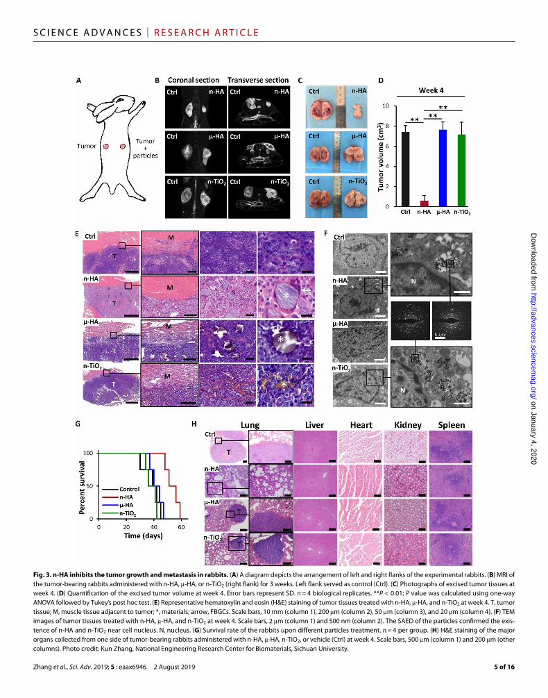
Effect of n-HA treatment on tumor growth and metastasesThe in vivo antitumor ability of n-HA was evaluated by an intra-muscular VX2 tumor model on both flanks of the rabbits with one-sided administration of n-HA, -HA, or n-TiO2 particles (Fig. 3A). At week 3, the magnetic resonance imaging (MRI) of individual animals showed that the right-side n-HA–treated tumor size was approximately 10 times smaller than the control on the left, whereas -HA or n-TiO2 had no effect on suppression of tumor growth (Fig. 3B and fig. S1C). Harvested tumor samples at week 4 confirmed the antitumor ability of n-HA (Fig. 3, C and D). We paid attention to the interface between the tumor and muscle tissue. Histo-logical staining of the invasion of the tumor (violet) into the adjacent muscle layers (red) on the control side of the rabbit is shown in Fig. 3E. However, a clear barrier created by the aggregated particles was observed in the n-HA–treated group, which was not identified in the -HA or n-TiO2 groups. Note that diverse immune cells were found surrounding the n-HA particles, including foreign body multinucleated giant cells (FBGCs), mononuclear cells, lym-phocyte cells, and neutrophil cells (Fig. 3E and fig. S2A). In contrast, -HA or n-TiO2 did not constrain the tumor cells from disrupting surrounding muscles, and few immune cells were observed. Further TEM observation indicated that n-HA and n-TiO2 could be inter-nalized into the tumor cells, but this was not observed for -HA (Fig. 3F). The nanoparticles that appeared in the cytoplasm were then analyzed by in situ selected- area electron diffraction (SAED) to confirm their composition. The ultrastructure morphology of the VX2 cells treated with n-HA was significantly altered, and some typical abnormal characteristics were documented, including aggre-gation of chromatin on the nucleus edge, expansion of nuclear membrane, swollen and vacuolated mitochondria, and formation of apoptotic body (fig. S2B).
on January 4, 2020http://advances.sciencem
ag.org/D
ownloaded from

Zhang et al., Sci. Adv. 2019; 5 : eaax6946 2 August 2019
S C I E N C E A D V A N C E S | R E S E A R C H A R T I C L E
3 of 16
Histological observation at week 4 of major organs including the lung, liver, heart, kidney, and spleen indicated that metastasis to the lung can be effectively prevented by n-HA but not by -HA or n-TiO2. Abnormalities were not found in other organs, which also suggested the biosafety of the particles being used. Because of the eliminated metas-tasis, rabbits with tumor only on the right flank and treated with n-HA had a longer survival time compared to other material-treated groups (Fig. 3, G and H). The vessels that formed inside of the tumor tissue were labeled with immunohistochemical staining of CD31 and -smooth muscle actin (-SMA) and further quantified (fig. S3A). Positively stained endothelial cells lining the abundant vessel networks were observed in the tumor tissue treated with -HA or n-TiO2, but significantly, less were observed in the n-HA group. The vessel area at week 4 was significantly smaller than the control or other material-treated groups (fig. S3B).
Activation of the mitochondrial apoptosis pathway by n-HALongitudinal progression of tumors that interfered with n-HA was studied at weeks 2 to 5. The pictures of excised tumors at different
weeks revealed that the size of the tumor on the n-HA–treated flank was greatly limited (Fig. 4A). Tumor necrosis could be found in the left flank control. The color of the tumor tissue treated with n-HA was paler than in the other samples, suggesting less blood supply. Quantification of the tumor volume showed that the left control reached a size of 9.65 ± 0.77 cm3 within 5 weeks, whereas the n-HA–treated tumor was approximately 60% smaller (Fig. 4B). We conducted histological observation of the tumors treated with n-HA at different weeks, showing a consistent barrier at the muscle-tumor interface and diverse kinds of immune cells, especially the FBGCs formed by the union of macrophages (fig. S3C). As reported, macrophages have a distinct, tissue-specific phenotype in response to signals present within micro-environments. Two extreme forms of macrophages are generally referred to as M1 and M2 activation. M1 enhances the ability to kill and phagocytose pathogens, up-regulate proinflammatory cytokines, and display cytotoxicity for tumor growth, while M2 promotes tissue repair and vascularization of tumors (22). Thus, in the current study, immuno-histochemistry (IHC) analyses of nitric oxide synthase 2 (NOS2), a
Fig. 1. Characterization of particles and scaffold. (A) XRD patterns of n-HA, -HA, and n-TiO2 particles. The standard spectra of HA, antase TiO2, and rutile TiO2 are given below. a.u., arbitrary units. (B) Representative TEM image of n-HA, SEM image of -HA, and TEM image of n-TiO2. (C) SEM images of 3D-printed porous titanium scaffold subjected to acid-alkali treatment, coated with n-HA and surface/cross-sectional alignment of n-HA with EDS confirmation. The dotted yellow line indicates the interface between n-HA coating and scaffold. The yellow arrow marks the average thickness of n-HA layer. Ca/P indicates atomic molar ratio of the selected region. (D) Weight change of particles released from n-HA/scaffolds immersed in tris-HCl solution for 7 days. Error bars represent SD. n = 3 replicates. (E) Representative TEM image of the released n-HA particles. (F) XRD pattern of the released n-HA particles. The asterisk indicates characteristic peaks of HA.
on January 4, 2020http://advances.sciencem
ag.org/D
ownloaded from

Zhang et al., Sci. Adv. 2019; 5 : eaax6946 2 August 2019
S C I E N C E A D V A N C E S | R E S E A R C H A R T I C L E
4 of 16
Fig. 2. In vitro tumor cell viability and apoptosis cocultured with different particles. (A) VX2 cells viability determined by CCK-8 assay when cocultured with n-HA, -HA, and n-TiO2 for 1, 3, and 5 days. Error bars represent SD. *P < 0.05 compared to 0 g/ml; †P < 0.05 compared to 1000 g/ml; P values were calculated using one-way analysis of variance (ANOVA) followed by Tukey’s post hoc test. n = 3 biological replicates. OD, optical density. (B) Representative fluorescent images of VX2 cells stained with fluorescein diacetate (green) and (C) nucleus stained with 4′,6-diamidino-2-phenylindole (blue) treated with n-HA, -HA, and n-TiO2 at different concentrations for 3 days. Arrowheads indicate condensed chromatin. (D) Representative dot plots of annexin V fluorescein isothiocyanate (FITC) apoptosis detection results of VX2 cells treated with n-HA, -HA, and n-TiO2 at different concentrations for 3 days.
on January 4, 2020http://advances.sciencem
ag.org/D
ownloaded from
Zhang et al., Sci. Adv. 2019; 5 : eaax6946 2 August 2019
S C I E N C E A D V A N C E S | R E S E A R C H A R T I C L E
5 of 16
Fig. 3. n-HA inhibits the tumor growth and metastasis in rabbits. (A) A diagram depicts the arrangement of left and right flanks of the experimental rabbits. (B) MRI of the tumor-bearing rabbits administered with n-HA, -HA, or n-TiO2 (right flank) for 3 weeks. Left flank served as control (Ctrl). (C) Photographs of excised tumor tissues at week 4. (D) Quantification of the excised tumor volume at week 4. Error bars represent SD. n = 4 biological replicates. **P < 0.01; P value was calculated using one-way ANOVA followed by Tukey’s post hoc test. (E) Representative hematoxylin and eosin (H&E) staining of tumor tissues treated with n-HA, -HA, and n-TiO2 at week 4. T, tumor tissue; M, muscle tissue adjacent to tumor; *, materials; arrow, FBGCs. Scale bars, 10 mm (column 1), 200 m (column 2), 50 m (column 3), and 20 m (column 4). (F) TEM images of tumor tissues treated with n-HA, -HA, and n-TiO2 at week 4. Scale bars, 2 m (column 1) and 500 nm (column 2). The SAED of the particles confirmed the exis-tence of n-HA and n-TiO2 near cell nucleus. N, nucleus. (G) Survival rate of the rabbits upon different particles treatment. n = 4 per group. (H) H&E staining of the major organs collected from one side of tumor-bearing rabbits administered with n-HA, -HA, n-TiO2, or vehicle (Ctrl) at week 4. Scale bars, 500 m (column 1) and 200 m (other columns). Photo credit: Kun Zhang, National Engineering Research Center for Biomaterials, Sichuan University.
on January 4, 2020http://advances.sciencem
ag.org/D
ownloaded from

Zhang et al., Sci. Adv. 2019; 5 : eaax6946 2 August 2019
S C I E N C E A D V A N C E S | R E S E A R C H A R T I C L E
6 of 16
critical marker for M1 macrophages, and Arginase 1, a critical marker for M2 macrophages, were performed. The results demonstrated that n-HA activated M1 macrophage transformation rather than M2 macrophages in vivo (fig. S3D).
To examine whether the mitochondrial apoptosis pathway was involved, as indicated by several in vitro studies (10, 13), here, we
investigated the expression of several marker proteins using Western blotting (Fig. 4, C and D) and IHC staining (Fig. 4E) of the tumor tissue at different weeks. First, decreased protein expression of Ki-67 and increased expression of TUNEL (terminal deoxynucleotidyl transferase–mediated deoxyuridine triphosphate nick end labeling) were observed in the n-HA–treated tumors at certain weeks, indicating
Fig. 4. Activation of mitochondrial apoptosis pathway by n-HA. (A) Longitudinal observation of the excised tumor treated with n-HA from weeks 2 to 5 (2W to 5W). Ctrl, left flank control without any treatment. (B) Quantification of the excised tumor volume. Error bars represent SD. n = 4 per group. (C and D) Expressions of mitochondrial apoptosis-related markers in tumor tissues measured by Western blotting (WB) at week 4. VEGF, vascular endothelial growth factor; GAPDH, glyceraldehyde phosphate dehydrogenase. Error bars represent SD. n = 3 per group. (E) Immunohistochemical analyses of Ki-67, Cyt C, p53, Bcl-2, and Bax and TUNEL assay of tumor tissues treated with n-HA at week 5 in comparison with control. Scale bar, 50 m. *P < 0.05, **P < 0.01 compared to control, two-way t test. Photo credit: Kun Zhang, National Engineering Research Center for Biomaterials, Sichuan University.
on January 4, 2020http://advances.sciencem
ag.org/D
ownloaded from

Zhang et al., Sci. Adv. 2019; 5 : eaax6946 2 August 2019
S C I E N C E A D V A N C E S | R E S E A R C H A R T I C L E
7 of 16
a less cell proliferative activity and more induced apoptosis (fig. S4). Second, higher expression of the p53 protein was found in the n-HA group, which responds to cellular stresses to suppress oncogenesis (23). Moreover, our results demonstrated that n-HA induced signifi-cantly higher Bcl-2 associated X protein (BAX) and lower Bcl-2 pro-tein expression over time. BAX is a proapoptotic protein, whereas Bcl-2 is an anti-apoptotic protein in the mitochondrial apoptotic pathway, which together regulate the release of apoptotic factor cytochrome C (Cyt C) from the mitochondria into the cell cytosol (24). Accordingly, increased protein expression of Cyt C was observed at weeks 4 and 5 in this study.
Other mechanisms that contribute to the antitumor effect of n-HATo further investigate the potential antitumor mechanism of n-HA, we performed an mRNA expression profile microarray. More than 1200 genes were extensively regulated in the n-HA–treated tumor compared to the untreated control, including 754 up-regulated genes and 534 down-regulated genes (P < 0.05, twofold change as the cut-off) (Fig. 5A). The expression of several genes was further confirmed by quantitative reverse transcription polymerase chain reaction analysis and labeled in the volcano plot (the abbreviations and full names of genes are provided in table. S1). Among them, n-HA significantly up-regulated the expression of genes involved in tumor apoptosis, including apoptosis mediated by mitochondria (HTRA4, ANKRD1, and VDAC3), and genes belonging to the tumor necrosis factor (TNF) superfamily [FAS (fatty acid synthase), TNF-, TNFSF10, and TNFSF13]. This suggests that the extrinsic death receptor apoptosis pathway might also be activated by n-HA (25). Meanwhile, genes expressed by different types of immune cells were also up-regulated in the n-HA group (TNF, DCSTAMP, MRC1, CLEC7A, TLR4, RLA-DR-ALPHA, and GZMK), revealing that n-HA might have stimulated the innate immune system and recruited numerous immune cells, including macrophages, dendritic cells, mononuclear cells, and cytotoxic lymphocyte cells, to the tumor microenvironment. In addition, genes involved in cellular calcium transport (ATP2A1, SLC8A1, TRDN, SRL, CACNB1, RYR1, CASQ2, CALB1, STC1, and CACNA2D1) were also expressed differentially, suggesting that intracellular calcium homeostasis might be disturbed by n-HA. Furthermore, genes that were reported to be overexpressed in cancer with poor prognosis were significantly down-regulated in the n-HA group (CHI3L2, NFE2L3, SBSN, GPR64, MMP3, EFNA1, SCAI, NEDD9, and SERP2NB2). VEGFC (vascular endothelial growth factor C) and PPAPDC1A (phospholipid phosphatase 4), which are major contributors to tumor angiogenesis and poor prognosis, were also down-regulated (26). Moreover, genes related to the Wnt signaling pathway, including WNT10A, WNT11, and CTNNB1, were down-regulated.
To identify the pathways involved, we carried out gene set enrich-ment analysis using multiple databases including the Kyoto Encyclo-pedia of Genes and Genomes (KEGG) database and the Gene Ontology (GO) database (table S2). On the basis of the KEGG databases, we found that treatment with n-HA results in the increased expression of genes associated with apoptosis, lysosome, and both the calcium signaling and cytokine-cytokine receptor pathways (Fig. 5B). In addition, with respect to the GO database, treatment with n-HA results in the increased expression of genes involved in the regulation of cytokine production involved in immune response, humoral immune response, response to xenobiotic stimulus, and positive regulation
of calcium ion import (fig. S5A). There were a total of 10 up-regulation– related pathways and 4 down-regulation–related pathways in the n-HA group versus the control (P < 0.05). The up-regulated pathways could be divided into three components: (i) cell death mediators: natural killer cell–mediated cytotoxicity, apoptosis, and necroptosis pathways; (ii) antitumor cytokines: nuclear factor B signaling and chemokine signaling pathways and cytokine-cytokine receptor interaction; and (iii) calcium homeostasis: calcium signaling, endocrine, and other factor-regulated calcium reabsorption, lysosome, and cy-clic guanosine 3′,5′-monophosphate–dependent protein kinase sig-naling pathways (Fig. 5, C and D). The down- regulated pathways include the Wnt signaling pathway, proteoglycans in cancer, the hippo signaling pathway, and hepatocellular carcinoma. The corresponding protein-protein interaction network is presented in fig. S5B. These results suggest that the interference of n-HA may influence multi-ple aspects of the tumor microenvironment and suppress tumor- promoting functions.
The secretory inflammatory cytokines from macrophages stimulated by n-HA inhibit the migration of tumor cellsWe noticed that the expression of TNF- was 6.27-fold higher (P = 0.002) in the n-HA–treated tumor sample than in the control. In our previous work, we identified an increased protein secretion of several inflammatory cytokines when coculturing macrophages with the degradation products of calcium phosphate bioceramics (27). Thus, to determine whether the observed elevated TNF- expression could be attributed to the stimulation of macrophages upon treatment with n-HA, we cocultured mouse RAW 264.7 macrophages with or without n-HA (500 g/ml) for 3 days and collected their conditioned medium. The results of the enzyme- linked immunosorbent assay (ELISA) indicated that coculturing with n-HA could significantly enhance the secretion of TNF- and monocyte chemoattractant protein-1 (MCP-1), as well as decrease the secretion of IGF (insulin-like growth factor) and VEGF, from macrophages (Fig. 5E). We conducted a wound-healing assay to explore whether the n-HA, or its conditioned medium with macro-phages, would affect tumor cell migration (Fig. 5F). Tumor cells cocultured with the macrophage-conditioned medium with n-HA (CM@n-HA) significantly delayed wound closure compared to the normal medium (Ctrl), the n-HA–added medium, or the macrophage- alone conditioned medium (CM) group. Transwell migration assay also showed that the minimum number of tumor cells migrated through the chamber after treatment with the CM@n-HA (Fig. 5G), suggesting that the secretion of macrophage upon stimulation with n-HA can decrease the migration of tumor cells.
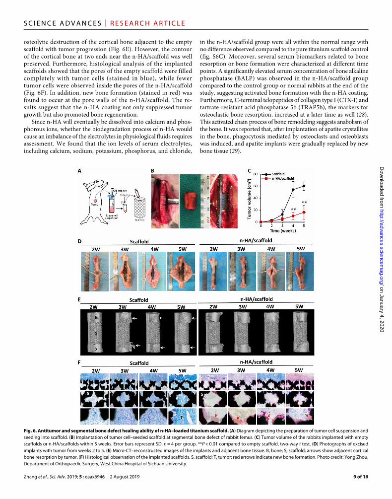
Segmental bone defect repair with n-HA/scaffold under tumor environmentA 15-mm critical-sized defect was created on rabbit femoral diaphysis via osteotomy. A concentration of VX2 tumor cell suspension (1 × 10⁷/ml) was derived from fresh tumor tissue and mixed with collagen in a 1:1 volumetric ratio (Fig. 6A). 3D-printed porous titanium scaffolds with or without n-HA coating were filled by the as-prepared mixture and then stabilized onto the created bone defect (Fig. 6B). After 5 weeks of implantation, the in situ tumor volume of the rabbits implanted with the n-HA loaded scaffold (n-HA/scaffold) was 73.8% smaller than the ones implanted with the empty scaffold (Fig. 6C). The image of the harvested femur samples demonstrates an inhibitory effect of tumor growth by the n-HA coating with prolonged time (Fig. 6D).
on January 4, 2020http://advances.sciencem
ag.org/D
ownloaded from

Zhang et al., Sci. Adv. 2019; 5 : eaax6946 2 August 2019
S C I E N C E A D V A N C E S | R E S E A R C H A R T I C L E
8 of 16
In addition, rabbits treated with the n-HA/scaffold had a higher body weight and better physical condition according to our observations, including their response to stimulation, willingness to accept food, and running ability (fig. S6A). Tumor metastasis with scaffold
implantation was also assessed at the end of the study (fig. S6B). Lung metastases were found in the control scaffold group, while no metastases were observed in the n-HA/scaffold group. The recon-structed microcomputed tomography (micro-CT) images revealed
Fig. 5. n-HA regulates gene expressions related to tumor suppression, calcium homeostasis, and immune response. (A) Volcano plot showing differentially regulated genes in the n-HA–treated tumor tissue as compared to the nontreated control. Genes with an absolute fold change of >2 and a P value of <0.05 are highlighted in green and red, denoting down- and up-regulated genes, respectively. (B) Gene set enrichment analysis of the regulated gene pathways with the Kyoto Encyclopedia of Genes and Genomes database. NES, normalized enrichment score. (C) Circular visualization of the results of gene-annotation enrichment analysis. (D) Heat map of genes that were differentially expressed in n-HA versus control tumor tissues with a fold change of >2 and a P value of <0.05. (E) Enzyme-linked immunosorbent assay of inflammatory cytokines secreted by mouse RAW 264.7 macrophages into culture medium (macrophages conditioned medium) after 3 days of coculturing with n-HA. *P < 0.05, **P < 0.01 compared to control, two-way t test. Error bars represent SD. n = 3 biological replicates. TGF-, transforming growth factor–; FGF, fibroblast growth factor. (F) Wound healing assay of mouse 4T1 tumor cells treated with control medium (Ctrl), n-HA, macrophages conditioned medium (CM), or macrophages conditioned medium with n-HA (CM@n-HA) for 24 hours. (G) Transwell assay after crystal violet staining showing serum-induced migration of 4T1 tumor cells treated with Ctrl, n-HA, CM, or CM@n-HA for 24 hours.
on January 4, 2020http://advances.sciencem
ag.org/D
ownloaded from
Zhang et al., Sci. Adv. 2019; 5 : eaax6946 2 August 2019
S C I E N C E A D V A N C E S | R E S E A R C H A R T I C L E
9 of 16
osteolytic destruction of the cortical bone adjacent to the empty scaffold with tumor progression (Fig. 6E). However, the contour of the cortical bone at two ends near the n-HA/scaffold was well preserved. Furthermore, histological analysis of the implanted scaffolds showed that the pores of the empty scaffold were filled completely with tumor cells (stained in blue), while fewer tumor cells were observed inside the pores of the n-HA/scaffold (Fig. 6F). In addition, new bone formation (stained in red) was found to occur at the pore walls of the n-HA/scaffold. The re-sults suggest that the n-HA coating not only suppressed tumor growth but also promoted bone regeneration.
Since n-HA will eventually be dissolved into calcium and phos-phorous ions, whether the biodegradation process of n-HA would cause an imbalance of the electrolytes in physiological fluids requires assessment. We found that the ion levels of serum electrolytes, including calcium, sodium, potassium, phosphorus, and chloride,
in the n-HA/scaffold group were all within the normal range with no difference observed compared to the pure titanium scaffold control (fig. S6C). Moreover, several serum biomarkers related to bone resorption or bone formation were characterized at different time points. A significantly elevated serum concentration of bone alkaline phosphatase (BALP) was observed in the n-HA/scaffold group compared to the control group or normal rabbits at the end of the study, suggesting activated bone formation with the n-HA coating. Furthermore, C-terminal telopeptides of collagen type I (CTX-I) and tartrate-resistant acid phosphatase 5b (TRAP5b), the markers for osteoclastic bone resorption, increased at a later time as well (28). This activated chain process of bone remodeling suggests anabolism of the bone. It was reported that, after implantation of apatite crystallites in the bone, phagocytosis mediated by osteoclasts and osteoblasts was induced, and apatite implants were gradually replaced by new bone tissue (29).
Fig. 6. Antitumor and segmental bone defect healing ability of n-HA–loaded titanium scaffold. (A) Diagram depicting the preparation of tumor cell suspension and seeding into scaffold. (B) Implantation of tumor cell–seeded scaffold at segmental bone defect of rabbit femur. (C) Tumor volume of the rabbits implanted with empty scaffolds or n-HA/scaffolds within 5 weeks. Error bars represent SD. n = 4 per group. **P < 0.01 compared to empty scaffold, two-way t test. (D) Photographs of excised implants with tumor from weeks 2 to 5. (E) Micro-CT–reconstructed images of the implants and adjacent bone tissue. B, bone; S, scaffold; arrows show adjacent cortical bone resorption by tumor. (F) Histological observation of the implanted scaffolds. S, scaffold; T, tumor; red arrows indicate new bone formation. Photo credit: Yong Zhou, Department of Orthopaedic Surgery, West China Hospital of Sichuan University.
on January 4, 2020http://advances.sciencem
ag.org/D
ownloaded from

Zhang et al., Sci. Adv. 2019; 5 : eaax6946 2 August 2019
S C I E N C E A D V A N C E S | R E S E A R C H A R T I C L E
10 of 16
DISCUSSIONThe antitumor effect of n-HA was initially observed and reported by Hideki et al. (30), who assessed the capability of n-HA as an adria-mycin carrier and accidentally discovered that the control group of pure n-HA inhibited the proliferation of Ca-9 cancer cells. In 2003, Cao et al. (31) further reported that n-HA was cytotoxic to Bel-7402 cancer cells due to its internalization into the cytoplasm. Later, by comparing several cancer and normal cell lines, their research group reached a conclusion that the inhibitory effect preferentially existed in the proliferation of cancer cells (MGC-803, Os-732, and Bel-7402) not identified in that of the normal cells (L-02, MRC-5, and HaCaT) (18). These studies shed light on the application of n-HA as a safe tumor-suppressing agent that might avoid the known side effects of chemotherapeutic agents. On the basis of this understanding, Qing et al. (32) conducted a pilot study (2012) to investigate the potential of n-HA to be used in a bone tumor scenario. Apoptosis was observed in MG63 human osteosarcoma cells cocultured with n-HA (100 to 500 g/ml). For the human primary osteoblasts, promoted cell viability was observed with the same n-HA concentration range. Our group and Qian’s group (33) attributed this selective effect of n-HA to an up-regulated mitochondrial-dependent apoptosis pathway in the tumor cells, in which Cyt C, Bcl-2, and Bax were involved. Nonetheless, to explore the antitumor ability of n-HA, most of the pre-vious studies were based on in vitro cellular experiments, and a few were conducted using a nude mouse tumor model (10, 33). Until re-cently, we confirmed the strong inhibitory activities of n-HA with 4T1 tumor-bearing BALB/c mice (34). In the current study, we therefore sought to explore the application of n-HA in bone tumors with clin-ical relevance and present a more comprehensive mechanism.
Coculture with malignant VX2 tumor cells and L929 cells in the present study revealed that n-HA can preferentially induce apoptosis to the tumor cells, whereas n-TiO2 showed cytotoxicity to both VX2 and L929 cells. Consistent with our findings, Tay et al. (35) also found selective apoptosis effects between n-HA and n-TiO2, in which normal human epithelial cells treated with n-HA induced minimal apoptosis, while n-TiO2–treated cells presented approxi-mately 28% early apoptosis after 24 hours of exposure. The particle shape and size of n-HA have also been reported to play an important role in its cytotoxicity (36). Nanoparticles with rod-like shapes could more easily pass through the cell membrane than spherical shapes (37). In our previous work, n-HA particles with different shape and size were synthesized, and the induced cellular response was assessed in vitro (38–40). In line with literature, rod-shaped n-HA showed the most prominent inhibitory effect on proliferation of tumor cell lines in our pilot experiment. Therefore, in the current study, n-HA, used either in the form of particles or coatings, was rod-like in shape and synthesized by a wet chemical method with special posttreatment. Our TEM observations with SAED confir-mation provided compelling evidence that n-HA could enter the tumor cells in vivo.
Apart from the inhibitory effect on in situ tumor size, our study discovered that n-HA is also able to prevent tumor metastasis. It has been reported in the literature and shown in our -HA and n-TiO2 groups that VX2 tumor–bearing rabbits can easily develop lung metastasis due to their high malignancy (41). No pathological change was identified in the histological analysis of the major organs including the heart, liver, spleen, lung, and kidney of the n-HA–treated group. Furthermore, the levels of serum electrolytes, including calcium and phosphorus ions, in the n-HA group were all within the normal
range until the end of this study, suggesting little adverse effect from biodegradation of the n-HA particles. The biosafety of nanosized materials when applied in vivo is of considerable concern. In 2000, Aoki et al. (42) reported that a continuous intravenous administration of n-HA (26 mg/kg) to beagle dogs in 2-week intervals for 2 years would cause no chronic damage or permanent side effects. In another study conducted by Wang et al. (43), after intraperitoneal injection with n-HA solution (8.3 mg/ml) three times per week for 4 weeks in in Sprague-Dawley rats, serum levels of aspartate aminotransferase, alanine aminotransferase, blood urea nitrogen, and creatinine were unchanged, indicating no induced liver damage or adverse impacts on renal function. Moreover, our data showed that the in vivo tumor- suppressive concentration of n-HA is one order of magnitude lower than the doses used in the above indicated studies. Together, these results suggest that synthetic n-HA is a safe nontoxic antitumor material.
As mentioned earlier, previous in vitro studies have demonstrated that mitochondria-dependent intrinsic pathways might be involved in n-HA–induced apoptosis. Therefore, for the samples derived from our in vivo tumor model, we first characterized the expression of several mitochondria apoptotic marker proteins. Our results showed that the n-HA group had significantly higher expression of BAX, p53, and Cyt C but lower expression of Bcl-2. The elevated expression of Cyt C is considered as an indicator of mitochondrial damage (24). It is generally accepted that Bcl-2 is an antiapoptotic protein and that Bax is a proapoptotic protein, which plays an important role in regulating mitochondrial membrane permeability (44). Moreover, enlarged and vacuolated mitochondria and condensed nuclei in apoptotic tumor cells were observed in the n-HA–treated group. These results confirmed that n-HA has the potential to trigger the mitochondrial apoptotic pathway in vivo. In agreement with our study, Press et al. (10) also reported that, after treatment with n-HA in human glioma-bearing nude mice, the expression of Ki-67 and Bcl-2 proteins was significantly decreased.
We believe that the underlying mechanism to explain the antitumor effect of n-HA is more complicated than an activated mitochondrial apoptosis pathway. Therefore, a gene profile microarray analysis was performed with n-HA–treated in vivo tumor samples. Multiple genes belonging to the TNF superfamily were significantly up- regulated by n-HA, including FAS, TNF-, TNFSF10 (known as TRAIL), and TNFSF13 (known as APRIL). These genes are the most representative cytokines, which are expressed principally by the immune cells, and regulate diverse cell functions, including activation of immunoresponse, apoptosis, and inhibition of tumor-igenesis (45). As death receptors, the TNF superfamily can directly trigger cell apoptosis via the extrinsic pathway with inflammatory cytokines (46). In our previous osteoimmunomodulation studies, we reported that the degradation debris of an HA composite could recruit a variety of immune cells, especially macrophages, to produce proinflammatory cytokines, such as TNF- (27). In the current study, diverse immune cells were recognized only in the n-HA–treated group, especially the FBGCs, which are formed by the union of macrophages. In agreement with our findings, a previous work also found that FBGCs can be induced by HA-based bone substitute material in human dental implantation (47). Moreover, the n-HA group was positively stained with M1 polarization macrophage marker, which might have also contributed to the production of proinflam-matory cytokines with antitumor activity (48). Thus, we hypothesized that macrophages and their derivatives might contribute to the secretion
on January 4, 2020http://advances.sciencem
ag.org/D
ownloaded from

Zhang et al., Sci. Adv. 2019; 5 : eaax6946 2 August 2019
S C I E N C E A D V A N C E S | R E S E A R C H A R T I C L E
11 of 16
of inflammatory cytokines when in contact with n-HA. We detected a high protein concentration of TNF- in the macrophage-conditioned medium after coculturing with n-HA. Furthermore, this conditioned medium could effectively inhibit the migration of the tumor cells in vitro. To the best of our knowledge, we therein discovered that the host immunoresponse stimulated by n-HA can be related to its antitumor and antimetastatic ability.
Apart from the changes in apoptosis-related genes and the inflam-matory cytokines, the gene expression of sarcoplasmic/endoplasmic reticulum calcium adenosine triphosphatase (ATPase) 1 (ATP2A1) and Na+/Ca2+ exchanger (SLC8A1) was prominently up-regulated, while the Stanniocalcin-1 (STC1) and voltage-dependent calcium channel subunit -2/∆-1 (CACNA2D1) were down-regulated. These results revealed that the cellular internalization of n-HA could disturb intracellular calcium homeostasis, possibly due to its degradation or adsorption of calcium ions. Consistent with our previous study, we found that the gene expression of the sodium-dependent phosphate transporter 1 (SLC20A1) and the plasma membrane calcium-transporting ATPase 2 (ATP2B2) were significantly up-regulated with the n-HA coating on the implant surface (21). Intracellular calcium homeostasis, as an important indicator, is related not only to cell death but also to osteoblastic differentiation (49, 50). These results suggest that the n-HA–induced turbulence in intracellular ion flow might also participate in the observed antitumor effect.
In one of our clinical trials, we used a porous n-HA/polyamide 66 composite as a bone void filler in the defect created by a benign tumor at the metaphysis of long bones. The radiologic analysis of 21 patients showed that the composite had been completely incorporated with the host bone with no tumor reoccurrence 18 months after implantation (51). This demonstrates that the porous n-HA polymer composite is an efficient, safe, and highly cost-effective bone substitute material for nonload-bearing sites. However, for the malignant bone tumor–induced segmental defect at the load-bearing site, i.e., diaphysis of long bone (52), an effective antitumor bone substitute material is currently not available. Robust metal implants incorporating n-HA can be ideal candidates. HA coatings on the surface of metal implants have been clinically accepted for a better osseointegration outcome. The differential regulation of osteoblast and osteoclast activity by surface topography of HA coatings was proven (53). Our group and other groups had previously demonstrated that calcium phosphate biomaterials with HA phase composition not only hold osteocon-ductivity but also have osteoinductivity by recruiting mesenchymal stem cells to the site of implantation (54, 55).
Therefore, we planned to develop a 3D-printed porous scaffold coated with a layer of n-HA aimed at critical-sized segmental bone repair under a tumor environment. In this design, the 3D-printing technology provided the implant a suitable physiological femur shape and a porous structure for new bone regeneration to take place. Surface modification with a considerable amount of rod-shaped releasable n-HA gave the scaffold an antitumor ability. Several methods have been reported to generate HA coatings onto a scaffold surface, including plasma spraying, electrochemical deposition, and biomimetic mineral-ization. However, as tested in our pilot trails, these traditional methods merely provide the scaffold with good bioactivity but are not able to achieve functional n-HA release at sufficient concentration. Therefore, we developed a novel approach to synthesize a thick n-HA coating on the porous scaffold in situ based on the slurry foaming method and impregnation process, modified from our previous work (21, 56). From the release experiments, the weight-changing records revealed
that n-HA could be continuously released from the scaffolds. The degraded particle size, shape, and physicochemical properties were finely controlled to match the n-HA used in the cellular study.
To reflect both the antitumor and bone regeneration ability of the scaffold, a suitable animal model is required. To establish an animal model with bone tumors, rats or mice are the first choice in many drug screening studies, but their small bone sizes limit the implantation of a biomaterial (8). The rabbit VX2 tumor model has been successfully used to mimic a variety of cancers because of its high malignance, including bone tumors (57). Therefore, in this study, we established a rabbit femur bone tumor model with VX2. Our results demonstrated that the n-HA coating was effective in suppressing tumor growth and promoted new bone regeneration inside the pores. The micro-CT reconstructed images revealed that the n-HA/scaffold also ameliorated the lytic destruction lesion of the adjacent cortical bone. Moreover, serum BALP, CTX-I, and TRAP5b significantly increased in the n-HA group, indicating activation in bone remodeling. In summary, the n-HA synthesized in this study has a selective effect to inhibit tumor growth and is safe with respect to normal cells and tissue. Moreover, we developed a practical strategy for the application of n-HA/scaffolds in segmental bone defect repair after tumor resection. Although our study has shown promising results in animal models, questions remain about the efficacy in human disease. It is not clear whether n-HA will be effective in the human cancer environment because of the difference between the animal tumor model and human disease. Thus, we are now launching clinical trials with n-HA–containing hydrogel for postsurgical cancer treatment. Clinical outcomes, including the biosafety and antitumor ability of rod-shaped n-HA, are promising. Despite these findings, we realized that the antitumor effect may also vary based on the different physical and chemical properties of n-HA. A series of n-HA shape–dependent and crystallinity-dependent experiments will be conducted to better illustrate the mechanism of this effect.
MATERIALS AND METHODSStudy designThe aim of this study was to assess n-HA as a potential antitumor material and its application in segmental bone defect repair under a tumor environment. Rod-shaped n-HA was synthesized and char-acterized by XRD and TEM observation. In vitro antitumor efficacy and biological toxicity of the synthesized n-HA were examined by coculturing with VX2 tumor cells or L929 fibroblasts. -HA and n-TiO2 were also used and served as control materials to eliminate compositional and size effect. The antitumor performance of the n-HA was further evaluated in vivo in terms of tumor growth suppression and metastasis prevention. The VX2 malignant tumor cells, mixed with or without materials, were planted subcutaneously into immunocompetent rabbits. The tumor sizes, cell internaliza-tion of the particles, and pathological observation of the main organs were assessed by MRI, TEM, and H&E (hematoxylin and eosin) staining. In addition, IHC, Western blotting, gene expression profile microarray, and cell migration assays were conducted to explore the potential antitumor mechanism of the n-HA. Subse-quently, 3D-printed porous titanium implants with releasable n-HA coating were specifically designed for segmental bone defect repair with in situ tumor. Besides histological staining, micro-CT technology was also used to visualize the tumor-induced osteolysis adjacent to the scaffolds, with or without the n-HA coating. The
on January 4, 2020http://advances.sciencem
ag.org/D
ownloaded from

Zhang et al., Sci. Adv. 2019; 5 : eaax6946 2 August 2019
S C I E N C E A D V A N C E S | R E S E A R C H A R T I C L E
12 of 16
number of animals for each group was determined with statistical significance.
Preparation of the particlesThe analytical grade of Ca(NO3)2∙4H2O, (NH4)2HPO4, and NH3·H2O were purchased from Chengdu Kelong Chemical Reagent Co. Ltd. (China). The rod-shaped n-HA was synthesized by traditional wet chemical method with special posttreatment. Briefly, according to the Ca/P molar ratio of 1.67, 1 liter of 0.5 M (NH4)2HPO4 was added dropwise into 1.67 liter of 0.5 M Ca(NO3)2∙4H2O. The solution was kept at pH ≈ 10 by adding NH3·H2O and was stirred continuously for 4 hours at 70°C. The slurry was then aged for 24 hours and washed with deionized water six times to remove the impurity. Next, the slurry was filtered, freeze-dried for 24 hours, calcinated at 300°C for 2 hours, and then sifted with a 200-mesh sieve to acquire the n-HA. The -HA, prepared by spray drying method (58), was supplied by the Engineering Research Center in Biomaterials, Sichuan University, China. The n-TiO2, synthesized via gas-phase method, was purchased from Evonik Degussa (Germany).
Scaffolds design and fabricationOn the basis of our previous work of porous titanium implant design for load-bearing sites (59, 60), a porous titanium cylinder (7 × 15 mm3) was considered to bridge the two ends of fractured bone, mimicking the clinically applied implants for critical-sized segmental bone defect. To match the elastic modulus of the natural bone at the fracture site, the pore diameter and porosity of the scaffold were designed to be 500 m and 65%, respectively. The scaffolds were modeled using Pro/ENGINEER 5.0 software and manufac-tured by selective laser sintering (EP-M250, Eplus3D, China) in the Northwest Institute for Non-ferrous Metal Research (Xi’an, China). To enhance its bioactivity and bonding of n-HA with raw substrate, the scaffolds were then subjected to acid-alkali treatment to form a microporous TiO2 network at the surface. Briefly, the scaffolds were in turn soaked in a mixture solution of 48% H2SO4 and 18% HCl for 1 hour and 6 M NaOH for 5 hours at 70°C, followed by thorough washing and vacuum drying (61). A novel slurry foaming method combined with impregnation process was used to fabricate the n-HA coating on the scaffolds. Briefly, 100 ml of n-HA slurry (140 mg/ml) was mixed with 20 g of 2% methyl cellulose (MC-400, Chengdu Kelong Chemical Reagent Co. Ltd., China) and 20 ml of H2O2 solution. After vacuum degassing, the scaffolds were immediately immersed into the heated and stirred slurry until it was fully impregnated. Last, scaffolds were taken out, centrifuged, and dried at 180°C for 2 hours. The above coating procedure was repeated three times, and the scaffolds were eventually subjected to heat treatment at 300°C for 1 hour.
Materials characterizationThe XRD (X’Pert Pro, Philips, Netherlands) was used to determine the phase composition of the particles. Surface morphology and microstructure of the scaffold were investigated via a field-emission SEM with EDS (Hitachi S-4800, Japan) and TEM (Tecnai G2 F20 S-TWIN, FEI). The particle size of the n-HA and n-TiO2 was analyzed by Nano Measure software based on the TEM images, for which at least three images with the scale of 200 nm and 70 particles in each image were evaluated. The particle size of the -HA was quantified by a laser scattering–based particles sizer (Mastersizer Micro, Malvern Panalytical Co., UK). To evaluate the strength of the scaffolds, a compression test was performed at a rate of 1 mm/min by a material
testing machine [MTS 810, Material Testing Systems (MTS), USA]. The weight of the n-HA released from the coated scaffold was measured when being degraded in tris-(hydroxymethyl) aminomethane (tris-HCl) solution at pH 6.5 or 7.4 (Chengdu Kelong Chemical Reagent Co. Ltd., China).
Cell linesThe VX2 tumor cell line was purchased from the Shanghai Guandao Biological Engineering Co. Ltd. The cells were cultured in Dulbecco’s modified Eagle’s medium (DMEM; Gibco, Invitrogen) supple-mented with 10% fetal bovine serum (Invitrogen), penicillin (100 U/ml; Invitrogen), and streptomycin (100 U/ml; Invitrogen). The mouse fibroblast cell line L929 was a gift from the Sichuan Testing Center for Biomaterials and Medical Devices. L929 cells were maintained in MEM (Invitrogen) supplemented with 10% fetal bovine serum (Invitrogen), penicillin (100 U/ml; Invitrogen), and strep-tomycin (100 U/ml; Invitrogen). The mouse breast cancer cell line 4T1 was purchased from the Cell Bank of Chinese Academy of Sciences (Shanghai, China). 4T1 cells were maintained in MEM (Invitrogen) supplemented with 10% fetal bovine serum (Invitrogen), penicillin (100 U/ml; Invitrogen), and streptomycin (100 U/ml; Invitrogen). Mouse RAW 264.7 macrophage cell line was purchased from Cell Bank of Chinese Academy of Sciences (Shanghai, China). RAW 264.7 cells were cultured in DMEM (Gibco, Invitrogen) supple-mented with 10% standard fetal bovine serum (Invitrogen), penicillin (100 U/ml; Invitrogen), and streptomycin (100 U/ml; Invitrogen). Cells were maintained at 37°C and 5% CO2 in a humidified environ-ment and passaged every 3 days.
Cell viability and apoptosisCell viability when cocultured with the n-HA, -HA, and n-TiO2 particles was quantified by a Cell Counting Kit-8 (CCK-8) reagent kit. Briefly, after VX2 cells were seeded in 24-well plates at a con-centration of 1 × 10⁴ per well and cultured for 24 hours, different amounts of the n-HA, -HA, and n-TiO2 as in concentrations of 0, 100, 250, 500, and 1000 g/ml were added into each well and continued to incubate for 1, 3, and 5 days. The optical density value was detected via a microplate reader (Varioskan Flash, Thermo Fisher Scientific, USA) at a wavelength of 490 nm. The live VX2 cells treated with different particles for 3 days were stained with fluorescein diacetate (Sigma-Aldrich, USA), and their nuclei were stained with 4′,6-diamidino-2-phenylindole (Sigma-Aldrich, USA). Immediately, the observation of the cells and nucleus was done under a confocal laser scanning microscope (TCS SP5, Leica, Germany). The apoptosis of the VX2 cells treated with different particles was assessed using fluorescein isothiocyanate Annexin V Apoptosis Detection Kit (BD, USA) and analyzed by flow cytometry (CytoFLEX, Beckman, USA).
Proinflammation cytokines secreted by macrophagesRAW 264.7 cells were seeded in a 24-well plate at a density of 1 × 10⁵ per well and treated with or without n-HA (500 g/ml) for 3 days. The conditioned media were then collected and subjected to ELISA, wound healing, and Transwell migration assays. To detect the in-flammatory cytokines secreted into the conditioned medium, a 96-well custom-made mouse Multi-Analyte ELISArray kit (QIAGEN, SA Biosciences, UK), which quantifies six inflammatory marker cyto-kines (TNF-, MCP-1, TGF-, FGF, IGF, and VEGF) per plate, was used according to the manufacturer’s specification. The difference
on January 4, 2020http://advances.sciencem
ag.org/D
ownloaded from

Zhang et al., Sci. Adv. 2019; 5 : eaax6946 2 August 2019
S C I E N C E A D V A N C E S | R E S E A R C H A R T I C L E
13 of 16
in the amount of each protein between the conditioned medium and the original culture medium is reported.
Wound-healing assayA sufficient number of 4T1 cells were seeded in 24-well plates and incubated until reaching 80 to 90% confluence. After the cells were serum-starved overnight, the scratches were made using sterile pipette tips and washed by phosphate-buffered saline (PBS) three times. Cells were added with serum-free DMEM, serum-free DMEM containing n-HA (500 g/ml), RAW 264.7 cells conditioned medium, or RAW 264.7 cells with n-HA conditioned medium. A microscopic observation was recorded at 24 hours after the scratch (DMI4000B, Leica, Germany).
Transwell migration assayThe overnight-starved 4T1 cells (2 × 10⁴), diluted with serum-free DMEM, serum-free DMEM containing n-HA (500 g/ml), RAW 264.7 cells conditioned medium, or RAW 264.7 cells with n-HA conditioned medium, were plated into the upper chamber of a 24-well Transwell chamber (8-m pore size; Corning, USA). DMEM with 10% fetal bovine serum was added into the lower chamber. After incubation for 24 hours at 37°C, cells remaining in the upper chamber were wiped off, and cells migrating through the polycarbonate membrane were fixed with 4% paraformaldehyde and stained with crystal violet, which were viewed and photographed under an inverted microscope.
AnimalsTwo adult male New Zealand white rabbits bearing VX2 tumor in the erector spinae muscle were provided by the Molecular imaging and functional imaging laboratory of Southeast University (Nanjing, China). A total of 76 healthy adult male New Zealand white rabbits (age, 12 weeks; weight, 2.5 to 3.0 kg) purchased from DaShuo exper-imental animal Co. Ltd. (Chengdu, China) received surgeries, including 44 animals to establish VX2 tumor model in the erector spinae muscles to investigate the antitumor efficacy of n-HA, -HA, and n-TiO2 particles and 32 animals to establish femur defect tumor model. Rabbits were individually caged and kept in a controlled tempera-ture at 25 ± 2°C with food and water provided ad libitum.
Surgical procedureAll animal procedures in the experiment were approved by the Institutional Animal Care and Use Committee of Sichuan University (Chengdu, China). All surgical operation was performed under general anesthesia. To establish the erector spinae muscles tumor model, the animal was placed in a prone position, and the surgery site was shaved and sterilized. The VX2 tumor tissue obtained from the tumor-bearing rabbit was resected, and the cell suspension was prepared (54). Cells (1 × 10⁷/ml) in PBS solution was obtained and mixed with type I collagen in a 1:1 volumetric ratio. This mixture (1 ml) was injected into the erector spinae muscles on both flanks of the recipient animals with or without n-HA, -HA, or n-TiO2 (50 mg/kg) administration. The tumor size was measured, and the tumor volume (in cm3) was obtained by the formula (62)
V = 0.5 × (length) × (width2)
To execute the implantation of the critically sized femur defect tumor model, 1 ml of the VX2 tumor cell mixture was injected into
each porous titanium scaffold with or without the n-HA coating. Thirty-two experimental animals were placed in a prone position. The left hind leg was shaved from ankle to hip and swabbed alternately with chlorhexidine and alcohol, and the skin from hip to knee was incised. By blunt dissection of the interval of the quadriceps muscles, the femur was exposed. The periosteum was stripped carefully using a periosteal elevator to avoid injuring the lower extremity parietal pleura. A 15-mm-long defect at the femur midshaft was created using a fretsaw. Then, the porous titanium scaffolds, with or without the n-HA coating, were injected with the VX2 suspension and fixed into the created bone defect by a screwed steel plate. After the surgery, the wound was sewn up in layers. All rabbits were given penicillin and painkillers for 1 week after operation, and their body weights were recorded during the experiment.
Imaging examinationMRI was performed using 3.0T EXCITE system (GE Healthcare, USA). Tumor tissue was visualized using T2-weighted images [echo time (TE), 90 ms; repetition time (TR), 4000 ms; flip angle (FA), 180; field of view, 143 mm × 180 mm]. Rabbits were immobilized during the imaging procedure by injection of sodium pentobarbital (40 mg/kg body weight) through the ear venous. Micro-CT scans were acquired using a microCT 80 imaging system (SCANCO Medical AG, Bassersdorf, Switzerland) to evaluate bone repair in critical-sized rabbit femur de-fect with scaffold implantation. The harvested femur samples were placed in a custom-made holder to ensure that the long axis of the femur was oriented perpendicular to the axis of the x-ray beam. The resultant grayscale images had an isotropic voxel size of 78 m from cone-beam reconstruction (70 kV, 114 A). The obtained radiographic images were reconstructed and analyzed using SCANCO software.
Histological stainingFor histological staining, the tumor, heart, liver, spleen, lung, and kidney were harvested immediately after the experimental animals were sacrificed and fixed in 4% formaldehyde. The specimens were embedded in paraffin and stained with H&E. The harvested osseo-implants and the surrounding tissues were also fixed in 4% para-formaldehyde and then embedded in poly(methyl methacrylate) after a series of gradient ethanol dehydration. Hard slices in the midsagittal plane through the center of the scaffold were obtained using a diamond saw (SP1600, Leica, Germany) and stained with 1% methylene blue (Sigma-Aldrich, USA) and 0.3% basic fuchsin (Sigma-Aldrich, USA) solutions.
Western blottingThe tumor tissues were harvested at week 4, frozen by liquid nitrogen, ground into powder, and lysed with radioimmunoprecipitation assay lysis buffer (Cowin Biosciences, Beijing, China) containing 1% protease and phosphatase inhibitor cocktails (Novagen, Darmstadt, Germany). Then, protein concentration of the lysates was measured using BCA protein assay kit (Pierce, IL, USA). An equal amount was loaded per lane onto 12.5% SDS–polyacrylamide gels and then transferred to polyvinylidene fluoride membranes. The membranes were then blocked in PBS with Tween 20 with 5% bovine serum albumin (BSA) and incubated with primary antibodies (diluted 1:300; Bioss, China) against Ki-67, p53, Cyt C, Bcl-2, Bax, VEGF, and glyceraldehyde phosphate dehydrogenase at 4°C overnight, and then incubated with horseradish peroxidase– conjugated secondary anti-body (diluted 1:5000; Santa Cruz Biotechnology, USA) at 37°C for
on January 4, 2020http://advances.sciencem
ag.org/D
ownloaded from

Zhang et al., Sci. Adv. 2019; 5 : eaax6946 2 August 2019
S C I E N C E A D V A N C E S | R E S E A R C H A R T I C L E
14 of 16
1 hour. Protein bands were visualized using a ChemiDoc XRS+ image system with Image LabTM software (Bio-Rad, USA).
Tissue ultrastructure morphology observationThe tumor tissues were cut into 1-mm3 pieces and fixed with 2.5% glutaraldehyde for 2 hours at 4°C. Then, after a series of standard procedures of dehydration and embedding, the embryos were sliced into ultraslices about 60 nm, stained with both uranyl ace-tate and lead citrate for 15 min to observe the cells and particles with TEM (Tecnai G2 20 TWIN, FEI). Meanwhile, SAED patterns were recorded.
Immunohistochemical stainingTissue sections were deparaffinized with xylene and rehydrated through graded alcohols into water. Tissues were placed in EDTA (pH 9.0) solution in a microwave in a short period of time for antigen retrieval, before washing for three times in pH 7.4 PBS solution. The endogenous peroxidase was blocked with 3% H2O2 for 25 min and then washed three times in pH 7.4 PBS solution. Protein blockage was applied using albumin from BSA (Solarbio) for 30 min before incubation with primary antibody (-SMA; 1:100 dilution) at 4°C overnight. Sections were then incubated for 50 min with secondary antibodies (Dako, Denmark) at room temperature. Diaminobenzidine chromogen (Dako) was used for color development followed by counterstaining with Harris hema-toxylin. TUNEL staining was performed using cell death detection kit (Roche, Germany). Quantification data were performed by cal-culating the mean optical density value of positive-staining areas versus the whole areas in five randomly selected fields using Image-Pro Plus software (Media Cybernetics). Antibodies used are as follows: anti-rabbit Ki-67 (1:400; Bioss, China), anti-rabbit P53 (1:400; Bioss, China), anti-rabbit Cyt C (1:400; Bioss, China), anti-rabbit Bcl-2 (1:400; Bioss, China), anti-rabbit Bax (1:100; Proteintech, USA), anti-rabbit NOS2 (1:100, Santa Cruz Biotechnology, CA, USA), anti- rabbit Arginase 1 (1:100; Santa Cruz Biotechnology, CA, USA), anti-rabbit CD31 (1:100;Abcam, UK), and anti-rabbit -SMA (1:100; Abcam, UK).
Gene expression profile microarrayFor gene expression profile microarray analysis, tumor tissue of the n-HA and control groups was harvested at week 1, and total RNA was extracted using RNeasy Fibrous Tissue Mini Kit (QIAGEN, Germany). Briefly, specimens were frozen by liquid nitrogen and ground into powder in ribonuclease-free crucibles quickly. The powder was then lysed with Buffer RLT, and the total RNA was ex-tracted and purified following the manufacturer’s protocol. Sample quality was assessed using a NanoDrop (ND-1000, NanoDrop Technology, USA). Last, the microarray assay was performed using the Agilent Rabbit Gene Expression Microarray (Agilent Technology, USA) containing 43,803 rabbit gene probes. To compare gene ex-pression levels between the n-HA and control groups, the data were analyzed using bioinformatic tools, and only genes with a fold change of ≥2 and a P value of <0.05 were considered.
Serum assaysSerum was obtained by centrifuging the whole blood at 4000 rpm for 15 min at 4°C. The serum electrolytes including sodium, potas-sium, chloride, calcium, and phosphorus were assayed using an automatic biochemical analyzer (Chemray 240, Shenzhen Leidu).
ELISA was performed to measure the serum protein concentrations of BALP, P1NP, OT, TRAP5b, and CTX-I according to the manu-facturer’s protocol (Cusabio Co. Ltd., China).
Statistical analysisStatistical analyses were carried out using SPSS v16 software, and all data were shown as means ± SD. One-way analysis of variance (ANOVA) followed by Tukey’s post hoc test was used in experiments with more than two groups, and two-way t test was used in experi-ments with two groups at each time point. For all tests, P < 0.05 was considered statistically significant. All results were reproduced in at least three independent experiments.
SUPPLEMENTARY MATERIALSSupplementary material for this article is available at http://advances.sciencemag.org/cgi/content/full/5/8/eaax6946/DC1Fig. S1. In vitro different cell viability cocultured with different particles and tumor inhibition in vivo.Fig. S2. The ultrastructure of tumor tissues treated with n-HA observed by TEM.Fig. S3. H&E staining and immunohistochemical analyses of tumor tissues.Fig. S4. Quantification of immunohistochemical staining of Ki-67, Tunel, p53, Cyt C, Bcl-2, and Bax expression of tumor tissues treated with n-HA during weeks 2 to 5.Fig. S5. GSEA and protein-protein interaction analysis.Fig. S6. Animal body weight changes, tumor metastasis, and biosafety analysis of n-HA/scaffold.Table S1. Abbreviations and full names of genes labeled in the volcano plot.Table S2. Positive enrichment of mRNA expression profile in microarray analysis (n-HA versus control).
REFERENCES AND NOTES 1. R. L. Randall, A promise to our patients with metastatic bone disease. Ann. Surg. Oncol.
21, 4049–4050 (2014). 2. M. S. Isakoff, S. S. Bielack, P. Meltzer, R. Gorlick, Osteosarcoma: Current treatment
and a collaborative pathway to success. J. Clin. Oncol. 33, 3029–3035 (2015). 3. H. Qu, W. Guo, R. Yang, D. Li, S. Tang, Y. Yang, S. Dong, J. Zang, Reconstruction
of segmental bone defect of long bones after tumor resection by devitalized tumor-bearing bone. World J. Surg. Oncol. 13, 282 (2015).
4. D. Andreou, S. S. Bielack, D. Carrle, M. Kevric, R. Kotz, W. Winkelmann, G. Jundt, M. Werner, S. Fehlberg, L. Kager, T. Kühne, S. Lang, M. Dominkus, G. U. Exner, J. Hardes, A. Hillmann, V. Ewerbeck, U. Heise, P. Reichardt, P.-U. Tunn, The influence of tumor- and treatment-related factors on the development of local recurrence in osteosarcoma after adequate surgery. An analysis of 1355 patients treated on neoadjuvant Cooperative Osteosarcoma Study Group protocols. Ann. Oncol. 22, 1228–1235 (2011).
5. H. Ma, C. Jiang, D. Zhai, Y. Luo, Y. Chen, F. Lv, Z. Yi, Y. Deng, J. Wang, J. Chang, C. Wu, A bifunctional biomaterial with photothermal effect for tumor therapy and bone regeneration. Adv. Funct. Mater. 26, 1197–1208 (2016).
6. H. Ma, J. Luo, Z. Sun, L. Xia, M. Shi, M. Liu, J. Chang, C. Wu, 3D printing of biomaterials with mussel-inspired nanostructures for tumor therapy and tissue regeneration. Biomaterials 111, 138–148 (2016).
7. B. Yang, J. Yin, Y. Chen, S. Pan, H. Yao, Y. Gao, J. Shi, 2D-black-phosphorus-reinforced 3D-printed scaffolds:A stepwise countermeasure for osteosarcoma. Adv. Mater. 30, 1705611 (2018).
8. Y. Tanzawa, H. Tsuchiya, T. Shirai, H. Nishida, K. Hayashi, A. Takeuchi, M. Kawahara, K. Tomita, Potentiation of the antitumor effect of calcium phosphate cement containing anticancer drug and caffeine on rat osteosarcoma. J. Orthop. Sci. 16, 77–84 (2011).
9. Y. Lu, M. Li, L. Li, S. Wei, X. Hu, X. Wang, G. Shan, Y. Zhang, H. Xia, Q. Yin, High-activity chitosan/nano hydroxyapatite/zoledronic acid scaffolds for simultaneous tumor inhibition, bone repair and infection eradication. Mater. Sci. Eng. C 82, 225–233 (2018).
10. S.-H. Chu, D.-F. Feng, Y.-B. Ma, Z.-Q. Li, Hydroxyapatite nanoparticles inhibit the growth of human glioma cells in vitro and in vivo. Int. J. Nanomedicine 7, 3659–3666 (2012).
11. Z. Shi, X. Huang, Y. Cai, R. Tang, D. Yang, Size effect of hydroxyapatite nanoparticles on proliferation and apoptosis of osteoblast-like cells. Acta Biomater. 5, 338–345 (2009).
12. J. Jin, G. Zuo, G. Xiong, H. Luo, Q. Li, C. Ma, D. Li, F. Gu, Y. Ma, Y. Wan, The inhibition of lamellar hydroxyapatite and lamellar magnetic hydroxyapatite on the migration and adhesion of breast cancer cells. J. Mater. Sci. Mater. Med. 25, 1025–1031 (2014).
on January 4, 2020http://advances.sciencem
ag.org/D
ownloaded from

Zhang et al., Sci. Adv. 2019; 5 : eaax6946 2 August 2019
S C I E N C E A D V A N C E S | R E S E A R C H A R T I C L E
15 of 16
13. X. Chen, C. Deng, S. Tang, M. Zhang, Mitochondria-dependent apoptosis induced by nanoscale hydroxyapatite in human gastric cancer SGC-7901 Cells. Biol. Pharm. Bull. 30, 128–132 (2007).
14. S. Dey, M. Das, V. K. Balla, Effect of hydroxyapatite particle size, morphology and crystallinity on proliferation of colon cancer HCT116 cells. Mater. Sci. Eng. C 39, 336–339 (2014).
15. S. Ezhaveni, R. Yuvakkumar, M. Rajkumar, N. M. Sundaram, V. Rajendran, Preparation and characterization of nano-hydroxyapatite nanomaterials for liver cancer cell treatment. J. Nanosci. Nanotechnol. 13, 1631–1638 (2013).
16. Q. Fu, M. N. Rahaman, N. Zhou, W. Huang, D. Wang, L. Zhang, H. Li, In vitro study on different cell response to spherical hydroxyapatite nanoparticles. J. Biomater. Appl. 23, 37–50 (2008).
17. J. Xu, P. Xu, Z. Li, J. Huang, Z. Yang, Oxidative stress and apoptosis induced by hydroxyapatite nanoparticles in C6 cells. J. Biomed. Mater. Res. Part A 100A, 738–745 (2012).
18. Y. Han, S. Li, X. Cao, L. Yuan, Y. Wang, Y. Yin, T. Qiu, H. Dai, X. Wang, Different inhibitory effect and mechanism of hydroxyapatite nanoparticles on normal cells and cancer cells in vitro and in vivo. Sci. Rep. 4, 7134 (2015).
19. Y. Yuan, C. Liu, J. Qian, J. Wang, Y. Zhang, Size-mediated cytotoxicity and apoptosis of hydroxyapatite nanoparticles in human hepatoma HepG2 cells. Biomaterials 31, 730–740 (2010).
20. H. Chen, C. Wang, X. Yang, Z. Xiao, X. Zhu, K. Zhang, Y. Fan, X. Zhang, Construction of surface HA/TiO2 coating on porous titanium scaffolds and its preliminary biological evaluation. Mater. Sci. Eng. C 70, 1047–1056 (2017).
21. Y. Zhu, K. Zhang, R. Zhao, X. Ye, X. Chen, Z. Xiao, X. Yang, X. Zhu, K. Zhang, Y. Fan, X. Zhang, Bone regeneration with micro/nano hybrid-structured biphasic calcium phosphate bioceramics at segmental bone defect and the induced immunoregulation of MSCs. Biomaterials 147, 133–144 (2017).
22. M. De Palma, C. E. Lewis, Macrophage regulation of tumor responses to anticancer therapies. Cancer Cell 23, 277–286 (2013).
23. F. Kruiswijk, C. F. Labuschagne, K. H. Vousden, P53 in survival, death and metabolic health: A lifeguard with a licence to kill. Nat. Rev. Mol. Cell Biol. 16, 393–405 (2015).
24. Z. Xu, C. Liu, J. Wei, J. Sun, Effects of four types of hydroxyapatite nanoparticles with different nanocrystal morphologies and sizes on apoptosis in rat osteoblasts. J. Appl. Toxicol. 32, 429–435 (2012).
25. Y. Huang, L. He, W. Liu, C. Fan, W. Zheng, Y.-S. Wong, T. Chen, Selective cellular uptake and induction of apoptosis of cancer-targeted selenium nanoparticles. Biomaterials 34, 7106–7116 (2013).
26. Z.-W. Liang, B.-F. Guo, Y. Li, X.-J. Li, X. Li, L.-J. Zhao, L.-F. Gao, H. Yu, X.-J. Zhao, L. Zhang, B.-X. Yang, Plasmid-based Stat3 siRNA delivered by hydroxyapatite nanoparticles suppresses mouse prostate tumour growth in vivo. Asian J. Androl. 13, 481–486 (2011).
27. J. Wang, D. Liu, B. Guo, X. Yang, X. Chen, X. Zhu, Y. Fan, Role of biphasic calcium phosphate ceramic-mediated secretion of signaling molecules by macrophages in migration and osteoblastic differentiation of MSCs. Acta Biomater. 51, 447–460 (2017).
28. W. Liu, L. Zhou, C. Zhou, S. Zhang, J. Jing, L. Xie, N. Sun, X. Duan, W. Jing, X. Liang, H. Zhao, L. Ye, Q. Chen, Q. Yuan, GDF11 decreases bone mass by stimulating osteoclastogenesis and inhibiting osteoblast differentiation. Nat. Commun. 7, 12794 (2016).
29. F. Watari, N. Takashi, A. Yokoyama, M. Uo, T. Akasaka, Y. Sato, S. Abe, Y. Totsuka, K. Tohji, Material nanosizing effect on living organisms: non-specific, biointeractive, physical size effects. J. R. Soc. Interface 6, S371–S388 (2009).
30. A. Hideki, O. Masataka, Effect of adracin-adsorbing HAP-sol on Ca-9 cell growth. Reports Institute Meal Dental Eng. 27, 39–44 (1993).
31. X.-y. Cao, Z.-t. Qi, H.-l. Dai, Y.-h. Yan, S.-p. Li, Cytotoxinic mechanism of hydroxyapatite nanoparticles on human hepatoma cell lines. J. Wuhan Univ. Technol. Mater Sci. Ed. 18, 66–68 (2003).
32. F. Qing, Z. Wang, Y. Hong, M. Liu, B. Guo, H. Luo, X. Zhang, Selective effects of hydroxyapatite nanoparticles on osteosarcoma cells and osteoblasts. J. Mater. Sci. Mater. Med. 23, 2245–2251 (2012).
33. Y. Sun, Y. Chen, X. Ma, Y. Yuan, C. Liu, J. Kohn, J. Qian, Mitochondria-targeted hydroxyapatite nanoparticles for selective growth inhibition of lung cancer in vitro and in vivo. ACS Appl. Mater. Interfaces 8, 25680–25690 (2016).
34. H. Zhao, C. Wu, D. Gao, S. Chen, Y. Zhu, J. Sun, H. Luo, K. Yu, H. Fan, X. Zhang, Antitumor effect by hydroxyapatite nanospheres: Activation of mitochondria-dependent apoptosis and negative regulation of phosphatidylinositol-3-kinase/protein kinase B pathway. ACS Nano 12, 7838–7854 (2018).
35. C. Y. Tay, W. Fang, M. I. Setyawati, S. L. Chia, K. S. Tan, C. H. L. Hong, D. T. Leong, Nano-hydroxyapatite and nano-titanium dioxide exhibit different subcellular distribution and apoptotic profile in human oral epithelium. ACS Appl. Mater. Interfaces 6, 6248–6256 (2014).
36. X. Zhao, S. Ng, B. C. Heng, J. Guo, L. Ma, T. T. Y. Tan, K. W. Ng, S. C. J. Loo, Cytotoxicity of hydroxyapatite nanoparticles is shape and cell dependent. Arch. Toxicol. 87, 1037–1052 (2013).
37. H.-m. Ding, Y.-q. Ma, Theoretical and computational investigations of nanoparticle-biomembrane interactions in cellular delivery. Small 11, 1055–1071 (2015).
38. B. Li, B. Guo, H. Fan, X. Zhang, Preparation of nano-hydroxyapatite particles with different morphology and their response to highly malignant melanoma cells in vitro. Appl. Surf. Sci. 255, 357–360 (2008).
39. H. Wu, Z. Li, J. Tang, X. Yang, Y. Zhou, B. Guo, L. Wang, X. Zhu, C. Tu, X. Zhang, The in vitro and in vivo anti-melanoma effects of hydroxyapatite nanoparticles: influences of material factors. Int. J. Nanomedicine 14, 1177–1191 (2019).
40. H. Zhang, F. Qing, H. Zhao, H. Fan, M. Liu, X. Zhang, Cellular internalization of rod-like nano hydroxyapatite particles and their size and dose-dependent effects on pre-osteoblasts. J. Mater. Chem. B 5, 1205–1217 (2017).
41. A. Parvinian, L. C. Casadaban, R. C. Gaba, Development, growth, propagation, and angiographic utilization of the rabbit VX2 model of liver cancer: A pictorial primer and “how to” guide. Diagn. Interv. Radiol. 20, 335–340 (2014).
42. H. Aoki, H. Aoki, T. Kutsuno, W. Li, M. Niwa, An in vivo study on the reaction of hydroxyapatite-sol injected into blood. J. Mater. Sci. Mater. Med. 11, 67–72 (2000).
43. L. Wang, G. Zhou, H. Liu, X. Niu, J. Han, L. Zheng, Y. Fan, Nano-hydroxyapatite particles induce apoptosis on MC3T3-E1 cells and tissue cells in SD rats. Nanoscale 4, 2894–2899 (2012).
44. P. E. Czabotar, G. Lessene, A. Strasser, J. M. Adams, Control of apoptosis by the BCL-2 protein family: Implications for physiology and therapy. Nat. Rev. Mol. Cell Biol. 15, 49–63 (2014).
45. L. K. Ward-Kavanagh, W. W. Lin, J. R. Šedý, C. F. Ware, The TNF receptor superfamily in co-stimulating and co-inhibitory responses. Immunity 44, 1005–1019 (2016).
46. M. Pasparakis, P. Vandenabeele, Necroptosis and its role in inflammation. Nature 517, 311–320 (2015).
47. J. Lorenz, A. Kubesch, T. Korzinskas, M. Barbeck, C. Landes, R. A. Sader, C. J. Kirkpatrick, S. Ghanaati, TRAP-positive multinucleated giant cells are foreign body giant cells rather than osteoclasts: Results from a split-mouth study in humans. J. Oral Implantol. 41, e257–e266 (2015).
48. H. A. Goubran, R. R. Kotb, J. Stakiw, M. E. Emara, T. Burnouf, Regulation of tumor growth and metastasis: The role of tumor microenvironment. Cancer Growth Metastasis 7, 9–18 (2014).
49. X. Cui, T. Liang, C. Liu, Y. Yuan, J. Qian, Correlation of particle properties with cytotoxicity and cellular uptake of hydroxyapatite nanoparticles in human gastric cancer cells. Mater. Sci. Eng. C 67, 453–460 (2016).
50. G.-Y. Jung, Y.-J. Park, J.-S. Han, Effects of HA released calcium ion on osteoblast differentiation. J. Mater. Sci. Mater. Med. 21, 1649–1654 (2010).
51. Y. Xiong, C. Ren, Y. Xiong, Y. Xiong, Y. Lang, L. Min, W. Zhang, F. Pei, Y. Yan, H. Li, A. Mo, C. Tu, H. Duan, Analyzing the behavior of a porous nano-hydroxyapatite/polyamide 66 (n-HA/PA66) composite for healing of bone defects. Int. J. Nanomedicine 9, 485–494 (2014).
52. X. Zhang, Z. Tang, Y. Zhou, X. Chen, X. Zhu, Y. Fan, Y. Zhang, X. Yang, C.-q. Tu, X. Zhang, A multi-level comparative analysis of human femoral cortical bone quality in healthy cadavers and surgical safe margin of osteosarcoma patients. J. Mech. Behav. Biomed. 66, 111–118 (2017).
53. D. O. Costa, P. D. H. Prowse, T. Chrones, S. M. Sims, D. W. Hamilton, A. S. Rizkalla, S. J. Dixon, The differential regulation of osteoblast and osteoclast activity bysurface topography of hydroxyapatite coatings. Biomaterials 34, 7215–7226 (2013).
54. Z. Tang, X. Li, Y. Tan, H. Fan, X. Zhang, The material and biological characteristics of osteoinductive calcium phosphate ceramics. Regen. Biomater. 5, 43–59 (2018).
55. G. Song, P. Habibovic, C. Bao, J. Hu, C. A. van Blitterswijk, H. Yuan, W. Chen, H. H. K. Xu, The homing of bone marrow MSCs to non-osseous sites for ectopic bone formation induced by osteoinductive calcium phosphate. Biomaterials 34, 2167–2176 (2013).
56. X. Ye, C. Zhou, Z. Xiao, Y. Fan, X. Zhu, Y. Sun, X. Zhang, Fabrication and characterization of porous 3D whisker-covered calcium phosphate scaffolds. Mater. Lett. 128, 179–182 (2014).
57. D. Proschek, M. Tonak, K. Kafchitsas, S. Zangos, M. G. MacK, A. Theisen, A. A. Kurth, Direct implantation of VX-2 carcinoma: A new rabbit bone model using a three-dimensional matrix as a carrier for the tumor cells. Eur. Surg. Res. 47, 154–158 (2011).
58. X. D. Zhu, H. J. Zhang, H. S. Fan, W. Li, X. D. Zhang, Effect of phase composition and microstructure of calcium phosphate ceramic particles on protein adsorption. Acta Biomater. 6, 1536–1541 (2010).
59. J. Yang, J. Wang, T. Yuan, X. D. Zhu, Z. Xiang, Y. J. Fan, X. D. Zhang, The enhanced effect of surface microstructured porous titanium on adhesion and osteoblastic differentiation of mesenchymal stem cells. J. Mater. Sci. Mater. Med. 24, 2235–2246 (2013).
60. J. Yang, H. J. Chen, X. D. Zhu, S. Vaidya, Z. Xiang, Y. J. Fan, X. D. Zhang, Enhanced repair of a critical-sized segmental bone defect in rabbit femur by surface microstructured porous titanium. J. Mater. Sci. Mater. Med. 25, 1747–1756 (2014).
on January 4, 2020http://advances.sciencem
ag.org/D
ownloaded from

Zhang et al., Sci. Adv. 2019; 5 : eaax6946 2 August 2019
S C I E N C E A D V A N C E S | R E S E A R C H A R T I C L E
16 of 16
61. H. Chen, C. Wang, X. Zhu, K. Zhang, Y. Fan, X. Zhang, Fabrication of porous titanium scaffolds by stack sintering of microporous titanium spheres produced with centrifugal granulation technology. Mater. Sci. Eng. C 43, 182–188 (2014).
62. P. P. Medina, M. Nolde, F. J. Slack, OncomiR addiction in an in vivo model of microRNA-21-induced pre-B-cell lymphoma. Nature 467, 86–90 (2010).
Acknowledgments: We thank L. Chen of the Analytical & Testing Center of Sichuan University for help in micro-CT images and L. Yu of the National Engineering Research Center for Biomaterials of Sichuan University for help in SEM images. Microarray experiments were performed by KangChen Bio-tech, Shanghai, China. Funding: This work was supported by the National Key Research and Development Program of China (2017YFB0702600, 2016YFC1102000, and 2016YFC1102003), the National Natural Science Foundation of China (81671825 and 81801852), the Young Elite Scientist Sponsorship Program by CAST (2019QNRC001), the Chengdu Science and Technology Project (2017-CY02-00032-GX), and the “111” Project of China (B16033). Author contributions: X.Y., X. Zhu, X. Zhang, C.T., C.X., Y.Z., Kun Zhang, and Kai Zhang designed the research. C.T., X.L., Z.L., J.T., H.W., C.X., Y.Z., and
Kun Zhang performed the experiments. X.Y., X. Zhu, L.W., G.W., Z.Y., L.M., S.Y., W.Z., and C.X. assisted with data analysis. X.Y., X. Zhu, W.Z., and Kun Zhang wrote the paper. X.Y., X. Zhang, C.T., Kai Zhang, W.Z., and Kun Zhang revised manuscript content. All authors have read and approved the final submitted manuscript. Competing interests: The authors declare that they have no competing interests. Data and materials availability: All data needed to evaluate the conclusions in the paper are present in the paper and/or the Supplementary Materials. Additional data related to this paper may be requested from the authors.
Submitted 15 April 2019Accepted 26 June 2019Published 2 August 201910.1126/sciadv.aax6946
Citation: K. Zhang, Y. Zhou, C. Xiao, W. Zhao, H. Wu, J. Tang, Z. Li, S. Yu, X. Li, L. Min, Z. Yu, G. Wang, L. Wang, K. Zhang, X. Yang, X. Zhu, C. Tu, X. Zhang, Application of hydroxyapatite nanoparticles in tumor-associated bone segmental defect. Sci. Adv. 5, eaax6946 (2019).
on January 4, 2020http://advances.sciencem
ag.org/D
ownloaded from
Application of hydroxyapatite nanoparticles in tumor-associated bone segmental defect
Zhentao Yu, Gang Wang, Lin Wang, Kai Zhang, Xiao Yang, Xiangdong Zhu, Chongqi Tu and Xingdong ZhangKun Zhang, Yong Zhou, Cong Xiao, Wanlu Zhao, Hongfeng Wu, Jiaoqing Tang, Zhongtao Li, Sen Yu, Xiangfeng Li, Li Min,
DOI: 10.1126/sciadv.aax6946 (8), eaax6946.5Sci Adv
ARTICLE TOOLS http://advances.sciencemag.org/content/5/8/eaax6946
MATERIALSSUPPLEMENTARY http://advances.sciencemag.org/content/suppl/2019/07/29/5.8.eaax6946.DC1
REFERENCES
http://advances.sciencemag.org/content/5/8/eaax6946#BIBLThis article cites 62 articles, 1 of which you can access for free
PERMISSIONS http://www.sciencemag.org/help/reprints-and-permissions
Terms of ServiceUse of this article is subject to the
is a registered trademark of AAAS.Science AdvancesYork Avenue NW, Washington, DC 20005. The title (ISSN 2375-2548) is published by the American Association for the Advancement of Science, 1200 NewScience Advances
License 4.0 (CC BY-NC).Science. No claim to original U.S. Government Works. Distributed under a Creative Commons Attribution NonCommercial Copyright © 2019 The Authors, some rights reserved; exclusive licensee American Association for the Advancement of
on January 4, 2020http://advances.sciencem
ag.org/D
ownloaded from