Application of solution NMR spectroscopy for characterizing mAb formulations
15
Application of solution NMR spectroscopy for characterizing (and optimizing) mAb formulations Alexander “Sasha” Golovanov Manchester Institute of Biotechnology, The University of Manchester, UK [email protected]
-
Upload
jane-williams -
Category
Science
-
view
40 -
download
0
Transcript of Application of solution NMR spectroscopy for characterizing mAb formulations

Application of solution NMR spectroscopy for characterizing
(and optimizing) mAb formulations
Alexander “Sasha” Golovanov
Manchester Institute of Biotechnology, The University of Manchester, UK [email protected]
Introduction • Generally, all proteins (including mAbs) may self-associate or aggregate, especially at
very high concentration => particle formation, opalescence, phase-separation
• Addition of co-solutes (excipients) may enhance protein stability, and decrease aggregation - the whole purpose of formulation process
• But how should you select excipients, which physical measurables to use as criteria to “choose the best”?
• Many biophysical methods require sample dilution, but best to measure “in situ”
• Solution NMR spectroscopy is sensitive to monomeric content and can detect transient self-association of proteins, even as large as mAbs, in different formulations
• Each traditional protein NMR study begins anyway from “sample condition optimisation” to achieve the highest monomeric content <-> spectra of best quality
Manchester Institute of Biotechnology, The University of Manchester, UK [email protected]

The principles of NMR:
- Frequency of spin rotation (i.e.
chemical shift) depends on the
neighbourhood
- Signal relaxation properties depend
on local motions – report on
dynamics and molecular tumbling
Transverse, or spin-spin relaxation (R2),
is inversely-proportional to NMR
signal width => ~ size of protein
assembly
Also, can measure self-diffusion
coefficients for separate molecules
present in the same sample
B0 w
1H Atom (spin)
Sample is placed in high magnetic
field, and atoms (eg 1H) are
excited by RF pulse(s); then we
“listen” for response (rotation
frequency, relaxation)
00 Bw
Manchester Institute of Biotechnology, The University of Manchester, UK [email protected]

Typical 1D 1H NMR spectrum of mAb (40°C)
Kheddo et al (2016) mAbs 8 (7), 1245-1258
→ From that, can measure characteristic signal intensities, relaxation and diffusion rates vs temperature, time and formulation conditions
Manchester Institute of Biotechnology, The University of Manchester, UK [email protected]

NMR signal integral ~ [concentration]
Here, the same integral (same concentration) but different R2 relaxation rates (linewidth)
R2 ~ c ~ 𝑉𝜂
→ So for a fixed protein concentration the signal intensity I ~ 1/(𝑉𝜂), where V is the sphere volume (size of the protein assembly), and 𝜂 is microscopic viscosity.
→ By measuring the changes in NMR signal intensity and microscopic viscosity we can derive the change in effective protein assembly size
in various conditions
I I
~ R2
~ R2
Manchester Institute of Biotechnology, The University of Manchester, UK [email protected]
Small assembly or low viscosity, sharp signal
Larger assembly or higher viscosity, broad signal
c = 4𝜋η𝑅
ℎ3
3𝑘𝑏𝑇 ≈
𝑉𝜂
𝑘𝑇

An illustration: Addition of Arg·Glu increases observed signal intensities for highly-concentrated mAb, but decreases for mAb at lower concentration.
This is because Arg·Glu reduces self-association but increases viscosity.
→ Indeed, the underlying microscopic “buffer” viscosity is increased when ArgGlu is added
So, we can account for the viscosity change…
Kheddo et al (2016) mAbs 8 (7), 1245-1258
Manchester Institute of Biotechnology, The University of Manchester, UK [email protected]

Addition of Arg·Glu increases molecular tumbling and reduces transient self-association of “very soluble” IgG1 mAb at high concentration
as seen by viscosity-corrected normalized NMR signal intensity 𝐼𝜂𝑁
𝐼𝜂𝑁 =
𝐼[𝑅𝐸]
𝐼[𝑅𝐸=0]
𝜂[𝑅𝐸]
𝜂[𝑅𝐸=0]
Where 𝐼[𝑅𝐸] and 𝐼[𝑅𝐸=0]
are signal
intensities and 𝜂[𝑅𝐸] and 𝜂[𝑅𝐸=0]
are
buffer viscosities in the presence and absence of Arg·Glu, respectively.
Kheddo et al (2016) mAbs 8 (7), 1245-1258
Manchester Institute of Biotechnology, The University of Manchester, UK [email protected]
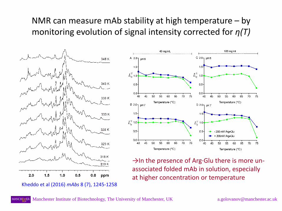
NMR can measure mAb stability at high temperature – by monitoring evolution of signal intensity corrected for η(T)
→In the presence of Arg·Glu there is more un-associated folded mAb in solution, especially at higher concentration or temperature
Kheddo et al (2016) mAbs 8 (7), 1245-1258
Manchester Institute of Biotechnology, The University of Manchester, UK [email protected]

NMR can measure diffusion of different components (mAb, and buffer/excipients) separately in the same sample: DOSY spectrum
Log D
Manchester Institute of Biotechnology, The University of Manchester, UK [email protected]
Diffusion of small molecules - can be used to obtain microscopic viscosity η

DOSY spectra of mAbs in different formulations was measured…
Manchester Institute of Biotechnology, The University of Manchester, UK [email protected]
Kheddo et al (2016) mAbs 8 (7), 1245-1258
Arg·Glu added

… and diffusion coefficients of mAbs in different formulations analysed
→ At high mAb concentrations self-diffusion is mostly determined by molecular crowding but also depends on microscopic viscosity => by itself not a very useful parameter for optimising formulations
Kheddo et al (2016) mAbs 8 (7), 1245-1258
Manchester Institute of Biotechnology, The University of Manchester, UK [email protected]

By measuring self-diffusion of small probe molecules present in the sample NMR can measure microscopic viscosity of buffer and mAbs solutions, and …
Kheddo et al (2016) mAbs 8 (7), 1245-1258
Manchester Institute of Biotechnology, The University of Manchester, UK [email protected]
Microscopic viscosity, NMR - derived
Macroscopic viscosity, measured using rheometry
→ Microscopic viscosity is generally lower than macroscopic one at higher mAbs concentrations

… knowing viscosity η and diffusion coefficient D we can estimate the change in the apparent “size” of mAb cluster (Rh) vs formulation conditions
Rh = 𝑘𝑇
6𝜋𝐷𝜂
→ At pH 7 which is closer to pI, or at higher mAb concentration, the apparent Rh of mAb is increased; addition of Arg·Glu reduces it in a concentration-dependent manner.
Consistent with observations from viscosity-corrected NMR signal intensities.
Kheddo et al (2016) mAbs 8 (7), 1245-1258
Manchester Institute of Biotechnology, The University of Manchester, UK [email protected]

Conclusions
• NMR signal intensity of mAb is a sensitive reporter for its state in solution: self-association, aggregation, loss of monomer, melting etc can be monitored, dependent on formulation conditions.
• Other NMR-derived parameters, such as diffusion coefficients of mAbs and excipients, provide further valuable information.
• NMR can work with non-diluted highly-concentrated mAb formulations.
• Increasing evidence that addition of relatively small concentrations of Arg·Glu (≤200 mM) often can stabilise mAb formulations better than Arg·HCl.
Manchester Institute of Biotechnology, The University of Manchester, UK [email protected]

Acknowledgements
Manchester’s team:
- Priscilla Kheddo (BBSRC BRIC CASE Studentship)
- Jack Bramham (BBSRC CASE Studentship)
- Matt Cliff
- Rebecca Dearman
MedImmune’s team (Cambridge, UK):
- Christopher van der Walle
- Shahid Uddin
- Malgorzata Tracka
- Jonathan Armer
Manchester Institute of Biotechnology, The University of Manchester, UK [email protected]