Gas Phase Structure Characterization Using Fourier Transform Ion Cyclotron Resonance Mass
APPLICATION OF FOURIER TRANSFORM ION CYCLOTRONRESONANCE MASS SPECTROMETRY TO OLIGOSACCHARIDES
Youmie Park and Carlito B. Lebrilla*Department of Chemistry, University of California, Davis, California 95616
Received 29 July 2003; revised 17 November 2003; accepted 19 November 2003
Published online in Wiley InterScience (www.interscience.wiley.com) DOI 10.1002/mas.20010
The application of Fourier transform ion cyclotron resonancemass spectrometry (FTICR MS) to the structural elucidation ofoligosaccharides is described. This review covers the analysesof oligosaccharides in the context of the unique features ofFTICR MS and the improvements in instrumentation that makeit possible to study this class of compounds. It consists of workperformed initially to understand the fundamental aspects ofoligosaccharide ionization and unimolecular fragmentation.More recent investigation includes the application of the tech-nique to samples of direct biological origin. Chemical andenzymatic degradation methods in conjunction with mass spec-trometry (MS) and the use front-end methods with FTICR MSare also discussed. The current applications including thecharacterization of bacterial lipooligosaccharides and phos-porylated carbohydrates are described. # 2004 WileyPeriodicals, Inc., Mass Spec Rev 24:232–264, 2005Keywords: Fourier transform ion cyclotron resonance massspectrometry; oligosaccharides; CID; mucin-type; glycosami-noglycans; N-linked; O-linked
I. INTRODUCTION
The development of mass spectrometric methods for the rapidand sensitive analysis of oligosaccharides has been a major topicof research in our laboratory. Oligosaccharides are in most casesconjugated with other biomolecules, which are known as glyco-conjugates. Protein glycosylation is the most common form ofpost-translational modification. It is estimated that more than50% of all proteins are glycosylated (Apweiler, Hermjakob, &Sharron, 1999). Oligosaccharides play a central role in manyfundamental cellular processes such as cell–cell recognition,adhesion, control of cell division, cellular differentiation, andmalignant transformation (Montreuil, 1980; Humphries et al.,1986; Ogier-Denis et al., 1989; Varki, 1993). For example,oligosaccharides that are present in mucin glycoproteins fromegg jellies play important roles in the process of sperm-eggrecognition and fertilization (Schmell, Gulyas, & Hedrick, 1983).Changes of O-linked oligosaccharide structures in cancer cellsare often associated with cancerous transformation (Brockhau-sen, 1999). In addition, oligosaccharides in glycoproteins also
play roles in maintaining protein structure and stability (Rudd &Dwek, 1997). They are responsible for some of the antigenicproperties of proteins, and involved in complement pathways(Lisowska, 2002; Ritchie et al., 2002). With recent syntheticadvances, glycoconjugates are emerging as targets for manytherapeutic applications such as cancer chemotherapy, diabetestherapy, antibiotics, antivirals, anti-inflammatories, and immu-nostimulants (McAuliffe & Hindsgaul, 1997; Yarema &Bertozzi, 1998; Koeller & Wong, 2000; Bertozzi & Kiessling,2001; Dove, 2001; Maeder, 2002). However, oligosaccharideanalyses such as sequencing and structure characterization stillremain very labor-intensive and complicated. Effective tools foroligosaccharide analyses will facilitate the understanding of theircellular functions as well as diseases. To this end, a researchprogram was developed in this laboratory that initially involvedthe construction and optimization of Fourier transform ioncyclotron resonance mass spectrometry (FTICR MS) instrumen-tation, the study of fundamental gas-phase decomposition pro-cesses, and the analysis of biological samples to develop massspectrometry (MS) as the primary tool for oligosaccharideanalysis. MS offers the potential of both high sensitivity andspeed, but to develop efficient schemes for sequencing oligo-saccharides, a thorough understanding of the gas-phase chem-istry of these compounds is also necessary.
Structural features of oligosaccharides contribute to makethe mass spectrometric analyses of these compounds consider-ably more difficult than that of other biopolymers such aspeptides and nucleotides. Knowing the sequence of an oligo-meric chain is not sufficient for full characterization. The linkage,degree of branching, and stereochemistry must also be deter-mined. Determination of the linkage alone, for example, isalready a formidable task. A glycosidic bond linking two cyclichexoses can be accomplished in one of five ways (neglectingstereochemistry) through the hydroxyl groups shown below. Incomparison, amino acids are linked in the primary chain of aprotein in only one way, by forming an amide bond. Branchingalso readily occurs with oligosaccharides but is seldom found inpeptides or proteins. In addition, oligosaccharides generally havepoorer MS sensitivities than, for example, peptides.
Mass Spectrometry Reviews, 2005, 24, 232– 264# 2004 by Wiley Periodicals, Inc.
————*Correspondence to: Carlito B. Lebrilla, Department of Chemistry,
University of California, One Shields Avenue, Davis, CA 95616.
E-mail: [email protected]
Several features of FTICR make it ideal for oligosaccharideanalysis. The high resolution and mass accuracy readily yieldsthe composition in terms of numbers of fucose, hexose, N-acetylhexose, sialic acid, as well as degree of sulfation, etc. Thesimultaneous implementation of both electrospray ionization(ESI) and matrix-assisted laser desorption/ionization (MALDI),the two most useful ionization methods for oligosaccharides,was performed first in FTICR. The availability of reliable andconsistent tandem MS events such as collision-induced dissocia-tion (CID) and more recently infrared multiphoton dissociation(IRMPD) and electron capture dissociation (ECD) with anyionization method in FTICR made it a rapid and key tool forstructural elucidation.
This review is not meant to be comprehensive in oligo-saccharide or carbohydrate analyses. There are already extensivereviews in this area (Harvey, 1999). Instead, it highlights the roleFTICR MS has played in this area. As the early FTICR MSanalyses of oligosaccharides were performed primarily in ourlaboratory, we begin with some historical perspectives includingsome early instrumentation development in our laboratory. Themore recent studies performed in this laboratory and elsewherewere performed generally on commercial instruments.
II. INSTRUMENTATION
A. Fourier Transform Ion Cyclotron ResonanceMass Spectrometry
FTICR (Wanczek, 1984; Buchanan, 1987; Wilkins et al., 1989;Dienes et al., 1996; Marshall, Hendrickson, & Jackson, 1998) isperformed by trapping ions in an analyzer cell composed ofelectrically conductive walls placed in a homogenous region ofa magnet (Comisarow & Marshall, 1974a,b; Grosshans &Marshall, 1990). Detection is performed by exciting all the ionsnearly simultaneously (depending on the excitation method, e.g.,chirp versus impulse) and coherently to the same cyclotron radius(Schweikhard & Marshall, 1993). The image current produced bythe motion of the coherent ions is digitized and Fourier trans-formed. The longer the ions remain in coherence, the longer thesignal persists. This provides not only stronger signals but alsobetter resolution (Grosshans & Marshall, 1990). Several factorsdetermine the performance of the instrument. The most impor-tant of these are pressure, homogeneity (electric and magneticfield), and magnetic field strength. Collisions decrease the lengthof the transient by dephasing the motion of the ions (Marshall,Hendrickson, & Jackson, 1998). For this reason, the lowestpressure provides the best performance. In our system, cryo-pumps are used to produce a base pressure of 10�11 torr withMALDI and 10�9 torr with electrospray. Electric field and, tosome extent, local magnetic field homogeneity is determined bythe cell design and material (Comisarow, 1981; Caravatti &Allemann, 1991; Beu & Laude, 1992). Several groups andinstrument manufacturers have implemented the cylindrical andopen-end design as this yields better performance (Beu & Laude,1992). We have implemented a double-cell design for the elec-trospray that consists of two complete analyzer cells in the samechamber. We used the second analyzer cell to monitor thetemperature for performing thermal dissociation experiments (as
in blackbody induced radiation dissociation or BIRD) (Price,Schnier, & Williams, 1996).
The magnetic field strength is important as ion retention,resolution, and high-mass capability all increase with the fieldstrength (Ledford, Rempel, & Gross, 1984; Marshall, Hendrick-son, & Jackson, 1998). The major limitation is that the price ofsuperconducting magnets increases exponentially with thestrength of the field. The overall cost of the instrument furtherincreases as many of the components that are normally ‘‘non-magnetic’’ such as stainless steel electrodes, ion guides, vacuumchambers, etc., become magnetic. Many have been replaced withtruly non-magnetic material such as aluminum or titanium. Turbopumps and cryopumps as well as monitors are also affectedunless the magnet is shielded. The majority of the studiesperformed in our laboratory have been on 4.7 and 5.0 T instru-ments up until now. These fields have provided an acceptablecompromise between performance and price.
Oligosaccharides, and other biomolecules, require lowvacuum or even atmospheric ionization sources to produce ions.The low vacuum interferes with the performance of FTICR.For this reason, most analytical instruments produce ions out-side the analyzer chamber (externally) and transport the ions intothe analyzer cell. The latest major advancement in FTICR is thedevelopment of ion guides to transport ions from a low vacuumsource to the ultra-high vacuum analyzer chamber. This labo-ratory and several others including McIver (Hunt et al., 1985;McIver, Hunter, & Bowers, 1985; Lebrilla, Amster, & McIver,1989) and Wanzek (Kofel et al., 1986) have proposed variousmethods of ion injection (Marshall & Schweikhard, 1992). Thesemethods now include but are not limited to quadrupole andoctupole ion guides and lens arrays whose purpose is to guideions through the inhomogeneous region of the field.
The capability of FTICR to provide high masses (Chen et al.,1995), high resolution/accurate mass (Li, McIver, & Hunter,1994; McIver, Li, & Hunter, 1994; McLafferty, 1994; Kelleheret al., 1995), and multiple stages of CID are well known. Lesswell known are differences between FTICR and other MSmethods. Knowing these differences is important if the methodsdeveloped in FTICR are to be applied to other types of massspectrometers. The time between ion formation and detection is3 to 6 orders of magnitude longer in FTICR than TOF and sectorinstruments. The large difference in detection times is an im-portant factor in ionization sources such as MALDI that producesa large fraction of ions as metastable species. However, themetastable ions can also be used to obtain more structurallyrelevant fragments. Delaying the detection time is an importantmethod in TOF (i.e., post-source decay) (Spengler et al., 1994;Lemoine, Chirat, & Domon, 1996; Naven et al., 1997) forproducing a greater number of structurally informative fragmentions. Differences in detection times are less of a factor whencomparing quadrupole ion traps and FTICR. In many ways, thesemethods are similar. The major difference being the former isfaster and less expensive, and the latter significantly higher inresolving power and mass accuracy.
At the beginning of this project, the laboratory was equippedwith an external fast atom bombardment (FAB) ionization sourcecoupled to a single set of quadrupole rods. The rods function as anion guide that leads to the analyzer cell housed in the homo-geneous region of a superconducting magnet (McCullough,
FTICR MS OF OLIGOSACCHARIDES &
233
Gard, & Lebrilla, 1991). The reported performance of ESI(Yamashita & Fenn, 1984; Fenn et al., 1989) and MALDI (Karas& Hillenkamp, 1988; Stahl et al., 1991) for producing ions oflarge biomolecules encouraged us to proceed on a major cons-truction effort to build ESI and MALDI source for our instrument.
We built ESI and MALDI sources that were interchangeablewith the existing FAB source. The utility of MALDI and later ESIfor the analysis of oligosaccharides was readily apparent butswitching sources, particularly between ESI and MALDI,became a major limitation. Exchanging ionization sourcesrequired venting, removing the source, installing the other,pump-down, and bake-out; the entire procedure took up to 2 days.The necessity of having both sources forced us to devise a newmethod for rapidly exchanging them. The original chamber hadbeen modified for electrospray with the addition of two additionalstages of pumping. We decided to build a completely separatechamber with its own ion optics and analyzer cell that wouldhouse a MALDI source. The new chamber would share a com-mon data system and magnet with the original chamber, resultingin considerable savings. This set-up had other advantages. Onechamber could be used even when the other required service(Gard et al., 1996).
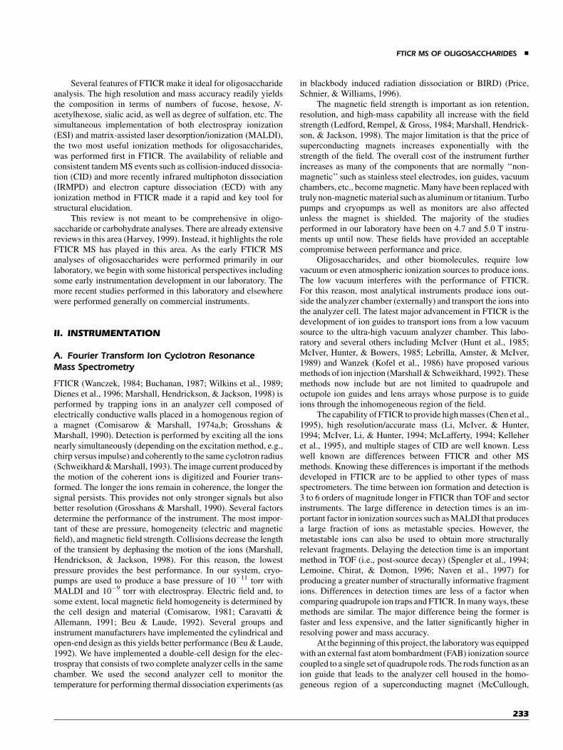
On the basis of our experience with the original chamber, weconcluded that the three stages of differential pumping wereunnecessary for either MALDI or FAB. We used a similar designfor the new chamber, that is, to incorporate the quadrupole ionguide that had performed so effectively. However, we limited thenumber of pumping stages to only two. The resulting instrumentcontained two independent vacuum chambers that were mountedon wheels (Fig. 1). Each chamber could be positioned into thesuperconducting magnet and connected to the data system whenneeded (Carroll et al., 1996; Gard et al., 1996). MALDI with thenew chamber worked as well as with the original one. Thedecreased pumping was not a limitation. Many of the results
discussed below are obtained with both this instrument and acommercial instrument.
B. Ionization Methods
1. Fast Atom Bombardment (FAB)
FAB or more appropriately liquid secondary ion MS-LSIMSmade it relatively straightforward to produce anionic and cationicspecies of oligosaccharides in the gas-phase with FTICR (Carrollet al., 1991; McCullough, Gard, & Lebrilla, 1991). Compoundswith masses as large as m/z¼ 1500 were readily obtained,albeit decreasing quasimolecular ion abundances accompaniedincreasing mass. The quasimolecular ion was usually the protoncoordinated species, although significant abundances of sodiatedspecies were at times observed. We found that FTICR providedthe same detection limits as other MS instruments for FAB(micromoles). The most notable difference between FTICR andsector, the most commonly used instrument with FAB at the time,was the nearly complete absence of matrix ions in the FTICRspectra. The absence of chemical background greatly simplifiedthe spectra allowing the observation of even weakly abundantfragment ions.
In both anion and cation modes, extensive fragmentationwas the norm with FAB in FTICR. Fragmentation was moreextensive in the cation mode, but in the anion mode we observedsignificant cross-ring cleavages that yielded linkage information(Carroll & Lebrilla, 1992; Carroll et al., 1993; Carroll, Willard, &Lebrilla, 1995). Significant effort was placed on increasingsensitivity and ion yield but the best detection limit that could beobtained remained in the microgram and sometimes sub-microgram range. This was not a limitation specifically ofFTICR but rather that of the ionization method.
FIGURE 1. Schematic of FTICR instrument with dual champers for ESI and MALDI. This configuration
provides rapid access to both ESI and MALDI. [Color figure can be viewed in the online issue, which is
available at www.interscience.wiley.com.]
& PARK AND LEBRILLA
234

Ions produced by FAB are sufficiently vibrationally excitedas to dissociate during the detection time scale of FTICR. Wepublished the first reported study on metastable decay in the ICR(Ngoka & Lebrilla, 1993). Metastable decay of FAB-producedions have been observed with other types of mass spectrometers(Grese, Cerny, & Gross, 1989; Thorne, Ballard, & Gaskell,1990), but it was commonly thought that metastable decay couldnot be observed in the ICR because of the rapid rates ofdissociation. However, the size of the molecules and the rela-tively low internal energy imparted by FAB, compared toelectron impact, for example, produced ions whose metastabledecay rates were in the order of milliseconds. Indeed, the absenceof significant chemical noise in the FAB FTICR MS spectra canbe attributed the metastable decay of matrix cluster ions. Morerecently, metastable decay of MALDI produced ions were alsoobserved with FTICR for peptides, namely RPPGFSPF andPKPQQFFGLM (Ho & Fenselau, 2000). Metastable decay rateswere dependent on a number of factors but especially laserfluences and MALDI matrices. They observed decay time con-stants on the order of 10 msec. Similar metastable decay may alsocontribute to the relatively lack of matrix ions observed in theMALDI spectra from FTICR MS.
The rates of metastable decay in FAB varied with thecharge carrier (proton and alkali metal) and the charge type(Ngoka, Gal, & Lebrilla, 1994). The decay rates decrease as afunction of cation ion size (i.e., Hþ>Liþ>Naþ>Kþ) for linearand cyclic oligosaccharides (Table 1). There are at least twoexplanations for this behavior. The cation affinity of oligosac-charides increases with decreasing cation size. Thus, the effectcould be because of the large internal energy associated withcationization. The second is that the small, densely chargedcation may promote dissociation, in this case, glycosidic bondcleavage.
Metastable decay initially showed great promise for use asan analytical tool. The molecular ion and structurally informativefragments can be obtained merely by varying the detectiontime. It also eliminated a significant amount of chemical noisefrom the matrix as these peaks were often metastable and di-minished in intensity at longer detection times. However,metastable decay proved to be source of several problems. Itbroadened the peaks severely thereby limiting mass resolution
and accuracy. It was also not highly predictable and depended onmany factors including the dissociation threshold and the internalenergy of the molecules that apparently differed between mole-cules. Therefore, oligosaccharides with labile groups, such assialic acids, fucose, and lipids, could not be produced intact in thegas phase. Furthermore, metastable decay limited the massrange as the rate appeared to scale with molecular size. The massrange limitation was the consequence of the internal energy thatincreased with molecular size. It became readily apparent, thatrather than being a useful analytical tool, metastable decay andthus LSIMS would make it impossible to study large oligosac-charides with complicated structures.
2. Electrospray Ionization (ESI)
This method employs a needle at a potential 2–3 kV above (orbelow depending on the desired polarity) the potential of theaperture to the first vacuum stage (Yamashita & Fenn, 1984; Fennet al., 1989). Ions are produced as liquid flows out of the needleproducing solvent clusters containing analyte. The specificmechanism for electrospray is still under contention. However,the desolvation of the droplets is the essential process (Kebarle &Tang, 1993). The clusters are desolvated with a heated capillaryin our home made instrument. Other methods are used such as acounter flow of gas to achieve the same results (Yamashita &Fenn, 1984; Fenn et al., 1989). Electrospray can be used toanalyze neutral and anionic oligosaccharide. ESI of neutrals isnot known to produce detection limits as high as MALDI (Duffinet al., 1992; Fura & Leary, 1993; Huddleston, Bean, & Carr,1993; Kohler & Leary, 1995; Viseux, Hoffmann, & Domon,1997). Derivatization with a basic group (Takao et al., 1996;Yoshino et al., 1995) or more effectively, a positively chargedgroup, such as Girards T (Naven & Harvey, 1996), increases thesensitivity of neutral oligosaccharides. Our experience with theESI FTICR has been that neutral oligosaccharides do not producestrong signals in the cation mode as Naþ ions were necessary toform the quasimolecular ions. Acidic oligosaccharides produceabundant signals with ESI (Reinhold, Reinhold, & Costello,1995). The deprotonated ion is easier to form than the alkali metalcoordinated species. Depending on the conditions, ESI can betuned to give essentially no fragmentation. This is importantwhen mixtures are analyzed and when it is not apparent whichsignals belong to fragments and which belong to quasimolecularions.
An ESI source was built in-house, and designed with the ionsimulation program SIMION. The design was based on one de-veloped by Chait that employed a heated capillary for desolvation(Chowdhury, Katta, & Chait, 1990). As we were constructing ourESI source, several groups had incorporated ESI with the FTICR(Henry, Quinn, & McLafferty, 1991; Cheng et al., 1995). How-ever, none have studied oligosaccharides. Other groups wereexploring the utility of ESI for oligosaccharide analysis withquadrupole analyzer equipped instruments (Reinhold, Reinhold,& Costello, 1995; Yoshino et al., 1995; Naven & Harvey, 1996;Viseux, Hoffmann, & Domon, 1997). For neutral oligosacchar-ides, ESI offered no major advantages over MALDI. For anionicor acidic oligosaccharides, however, it showed great utility(Reinhold, Reinhold, & Costello, 1995). A comparison ofgangliosides performed with ESI and MALDI on an FTICR
TABLE 1. First order decay rate constants of linear and
cyclic oligosaccharides coordinated to various cations
FTICR MS OF OLIGOSACCHARIDES &
235
mass spectrometer has been published (Penn et al., 1997).Gangliosides with multiple sialic acids are difficult to analyzewith MALDI but worked well with ESI-FTICR. When performedin the anion mode, ESI produced charge states that correspondeddirectly to the number of sialic acid. Other glycoconjugates withneutral oligosaccharides perform well with electrospray. Thepeptide moiety in glycopeptides may be capable of receiving thecharge yielding protonated species with ESI (Alving et al., 1998).ESI employing both ion trap and FTICR was used to obtainoligosaccharide sequence and linkage information. Partial acidhydrolysis of oligosaccharides was performed by using an acid-exchange resin as the acid catalyst, which minimized the decom-position of monosaccharides and deacetylation of N-acetylhex-osamines. In addition, Zn(dien)-Cl2 was used for derivatizationto determine stereochemistry and anomericity (Cancilla et al.,2000).
3. Matrix-Assisted Laser Desorption/Ionization (MALDI)
This method of ionization involves the co-crystallization of theanalyte with a matrix that acts as a chromophore for the laserradiation (Karas & Hillenkamp, 1988). The mechanism for ionformation is initiated by the electronic excitation of the media(Bencsura & Vertes, 1995; Ehring & Sundqvist, 1995). However,collision dynamics rapidly assume a substantial role as energypropagates in the condensed-phase medium. Ions may be pre-formed in the solid state (Liao & Allison, 1995; Lehmann,Knochenmuss, & Zenobi, 1997) or produced by ion–moleculereactions in the gas phase (Wang et al., 1993; Liao & Allison,1995). Furthermore, MALDI produces ions that are typically lessenergetic than FAB but more energetic than ESI (Demirev et al.,1987; Thorne, Ballard, & Gaskell, 1990; Spengler, Kirsch, &Kaufmann, 1991; Ngoka & Lebrilla, 1993; Ngoka, Gal, &Lebrilla, 1994; Karas et al., 1995).
Several groups including Hillenkamp showed the remark-able improvement of MALDI (coupled to a TOF instrument) overFAB for the analysis of oligosaccharides (Mock, Davey, &Cottrell, 1991; Stahl et al., 1991; Harvey, 1993). This encouragedus to immediately develop a MALDI source for our instrument.McIver was the first to implement MALDI to an external sourceFTICR instrument (Li, McIver, & Hunter, 1994; McIver, Li, &Hunter, 1994). They showed extraordinarily high mass and highresolution (106 FWHH) with peptides. Wilkins implementedMALDI in an internal source and showed the high-masscapability of the FTICR instrument (Castoro, Koester, & Wilkins,1992; Koster, Castoro, & Wilkins, 1992; Castoro & Wilkins,1993). Our FAB source was readily amenable to MALDI. Wesimply rotated the Csþ gun 90 degrees leaving an open area wherethe laser beam could enter and strike the probe. To obtain MALDIspectra, we placed a small nitrogen laser operating at 337 nm onthe vacuum cart and pointed it towards the FAB source. This wassufficient to obtain the first MALDI spectra of oligosaccharides inFTICR (Carroll et al., 1996). The quadrupole ion guide workedwell to collimate the ion beam. A nitrogen gas pulse trapped thetranslationally excited ions in the analyzer cell. The advantagesof MALDI-FTICR in the analysis of oligosaccharide wereimmediately apparent. The quasimolecular ions of neutral oligo-saccharides were always the base peak (Carroll et al., 1996).
Fragmentation was minimal and the detection limit wasincreased by over 5 orders of magnitude compared to FAB. Theions were produced significantly cooler than with FAB andmetastable decay was less of a problem. The MALDI spectrumobtained by FTICR was free of the chemical noise, because of thematrix, that is commonly observed in time-of-flight instruments(Cancilla et al., 1996; Carroll et al., 1996).
The choice of matrix is important as some produce moremetastable decay than others. There are dozens of matrices andco-matrices that have been investigated for their effectivenesswith oligosaccharides (Mohr, Boernsen, & Widmer, 1995; Kolli& Orlando, 1996; Krause, Stoeckli, & Schlunegger, 1996;Nonami, Fukui, & Erra-Balsells, 1997). An interesting study byNovotny used compounds (oxazone) that are structurally similarto oligosaccharides in the hope of inducing better co-crystal-lization (Chen, Baker, & Novotny, 1997). The best matrices inour hands are 2,5-dihydroxybenzoic acid (Strupat, Karas, &Hillenkamp, 1991) (DHB) and super-DHB (Karas et al., 1993)(a mixture composed of DHB and 2-hydroxy-5-methoxybenzoicacid) for neutral and 2,5-dihydroxyacetophenone (DHAP)(Krause, Stoeckli, & Schlunegger, 1996) for acidic oligosacchar-ides. We find that matrices that have been found to work well withTOF instruments do not necessarily work well with FTICR. Thelarge time scale difference again enhances the effects of matricesthat produce ions too energetically. Generally, MALDI worksbest for neutral oligosaccharides producing detection limits in thelow picomolar and sub-picomolar levels. To increase detectionlimits, several groups have employed derivatization (Dell, 1987;Naven & Harvey, 1996; Powell & Harvey, 1996; Pitt & Gorman,1997). Even a single derivative on the reducing terminus cansignificantly increase the sensitivity of MALDI. We introduced9-aminofluorene to label oligosaccharides by reductive amina-tion to the reducing terminus for MALDI-FTICR characteriza-tion with increased sensitivity (Franz, Molinski, & Lebrilla,2001). Y- and B-fragments were observed in the presence ofsodium dopant, and the protonated of the labeled compoundsshowed only Y-fragments, which allowed for complete sequenceanalysis. Interestingly, protonated benzylamine-labeled and9-aminofluorene-labeled lacto-N-fucopentaose I (LNFP I) andlacto-N-difucohexaose I (LNDFH I) showed a long-range gly-cosyl transfer reaction in CID-FTICR mass spectra (Franz &Lebrilla, 2002). L-Fucose was preferentially involved in thetransfer reaction and molecular modeling studies supported theproton-catalyzed mechanism for explaining the transfer reactionin gas phase.
We characterized the instrument further with respect toMALDI specifically for oligosaccharides by probing the limits ofdetection and the resolution. The limits of detection for a diversegroup of oligosaccharides were found to be in the pico- tofemtomole per liter range; similar to the detection limits reportedon TOF instruments (Mock, Davey, & Cottrell, 1991; Stahl et al.,1991; Mohr, Boernsen, & Widmer, 1995). Derivatization in-creased the sensitivity to the low femtomolar range as it does withTOF instruments (Lemoine, Chirat, & Domon, 1996; Naven &Harvey, 1996; Powell & Harvey, 1996; Takao et al., 1996). Theresolution on a routine analysis is in the order of 105 (FWHH)and, with additional effort, it is easily increased to 106. Inaddition, the mass accuracy for MALDI and ESI was unmatchedby any other types of mass analyzer. With external calibration,
& PARK AND LEBRILLA
236
McIver showed that mass accuracies of less than 20 ppm could bereliably obtained with on a relatively low field magnet (Li,McIver, & Hunter, 1994). We published mass accuracies of lessthan 5 ppm obtained routinely with internal calibration on asimilarly low field magnet (Wu et al., 1995).
To analyze neutral and acidic oligosaccharides simulta-neously, we developed a new anion dopant for neutral oligo-saccharides to detect in negative ion mode (Wong et al., 1999).Dilute H2SO4 solution was used as a dopant. A sulfate adduct[MþHSO4]� was formed under mild conditions, and a sulfatederivative [MþHSO4-H2O]� was produced under more concen-trated H2SO4 solutions (�10�2 M). This method allowedsimultaneous detection of neutral and acidic oligosaccharidesmixtures by the combination of complex formation (with neutraloligosaccharides) and the deprotonation of acidic oligosacchar-ides. Later, alkylsulfonates were examined as anion dopants toquantify desialylation reactions (Wong, Wang, & Lebrilla, 2000).They produced quasimolecular ions composed of the oligosac-charides and the deprotonated alkylsulfonates. It was found thatsulfated oligosaccharides suppressed sialylated oligosaccharidesin negative ion mode MALDI-FTICR (An & Lebrilla, 2001).These results were important as both oligosaccharides arefound together in many biological preparations. To minimizesuppression effects, sialylated oligosaccharides were convertedinto their methyl ester forms and sulfanilic acid was used as adopant. This allowed both sialylated and sulfated oligosacchar-ides to be successfully analyzed in negative ion mode, whileminimizing the effect of suppression. To characterize thefragmentation behavior of acidic oligosaccharide metal com-plexes, sialylated oligosaccharides were doped with two dif-ferent alkali metals (Csþ, Naþ, and Liþ) simultaneously. Thisformed gas-phase mixed metal complexes in MALDI-FTICR(Penn, Cancilla, & Lebrilla, 2000). It was determined that a sialicacid residue is capable of solvating two large alkali metals, suchas, cesium, simultaneously. However, two small alkali metals
could not be accommodated because of having higher chargedensities.
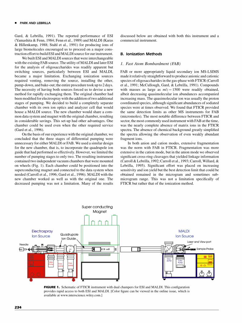
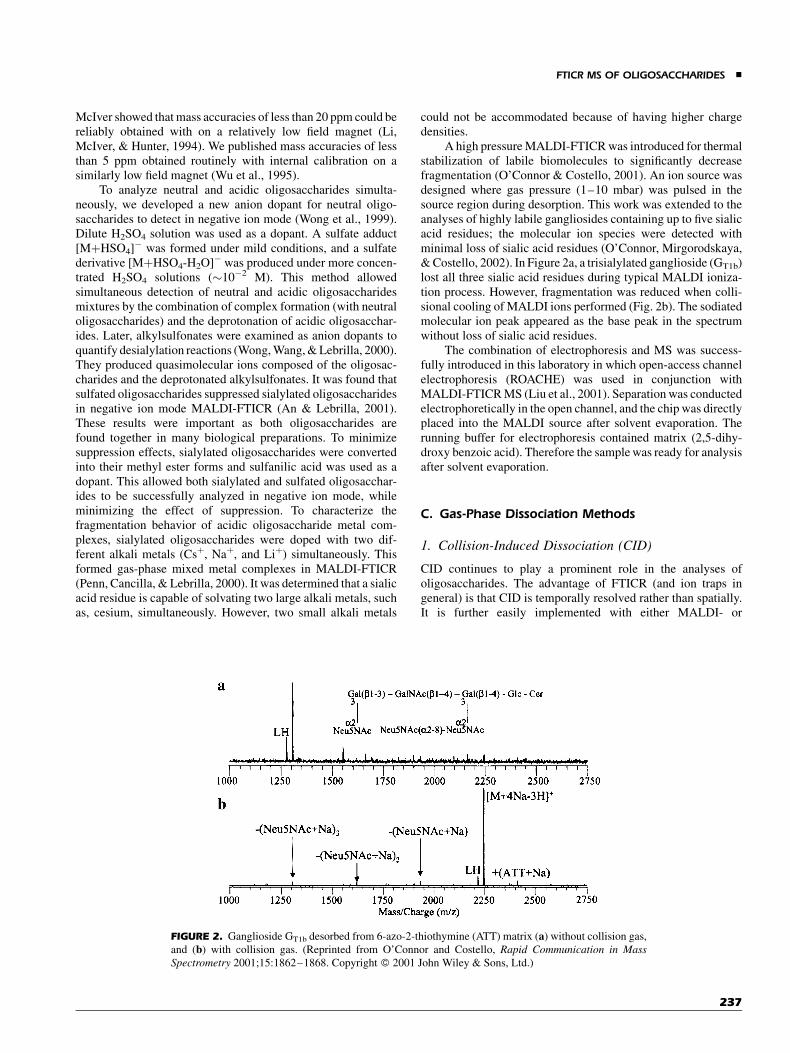
A high pressure MALDI-FTICR was introduced for thermalstabilization of labile biomolecules to significantly decreasefragmentation (O’Connor & Costello, 2001). An ion source wasdesigned where gas pressure (1–10 mbar) was pulsed in thesource region during desorption. This work was extended to theanalyses of highly labile gangliosides containing up to five sialicacid residues; the molecular ion species were detected withminimal loss of sialic acid residues (O’Connor, Mirgorodskaya,& Costello, 2002). In Figure 2a, a trisialylated ganglioside (GT1b)lost all three sialic acid residues during typical MALDI ioniza-tion process. However, fragmentation was reduced when colli-sional cooling of MALDI ions performed (Fig. 2b). The sodiatedmolecular ion peak appeared as the base peak in the spectrumwithout loss of sialic acid residues.
The combination of electrophoresis and MS was success-fully introduced in this laboratory in which open-access channelelectrophoresis (ROACHE) was used in conjunction withMALDI-FTICR MS (Liu et al., 2001). Separation was conductedelectrophoretically in the open channel, and the chip was directlyplaced into the MALDI source after solvent evaporation. Therunning buffer for electrophoresis contained matrix (2,5-dihy-droxy benzoic acid). Therefore the sample was ready for analysisafter solvent evaporation.
C. Gas-Phase Dissociation Methods
1. Collision-Induced Dissociation (CID)
CID continues to play a prominent role in the analyses ofoligosaccharides. The advantage of FTICR (and ion traps ingeneral) is that CID is temporally resolved rather than spatially.It is further easily implemented with either MALDI- or
FIGURE 2. Ganglioside GT1b desorbed from 6-azo-2-thiothymine (ATT) matrix (a) without collision gas,
and (b) with collision gas. (Reprinted from O’Connor and Costello, Rapid Communication in Mass
Spectrometry 2001;15:1862–1868. Copyright � 2001 John Wiley & Sons, Ltd.)
FTICR MS OF OLIGOSACCHARIDES &
237
ESI-produced ions. CID in FTICR is performed by isolating thedesired ion. This procedure involves the resonance excitation ofall other ions to the point where they leave the cell or collide withthe cell walls. In the past, ejection of unwanted ions was perfor-med with selective resonance ejection of individual ions. Moresophisticated methods have been developed using arbitrarywaveform generators that can be programmed for the retention ofdesired masses (Guan & Marshall, 1996).
Two methods of CID are currently used with FTICR. On-resonance CID uses an excitation frequency equal to the ion’scyclotron frequency to translationally excite the ion (Cody et al.,1982; Cody, Burnier, & Freiser, 1982; Cody & Freiser, 1982). Inthis method, the ions are translationally excited to a larger orbit.Collision gas is pulsed into the analyzer chamber producingcollisions, in which translational energy is converted into vibra-tional energy. Off-resonance excitation (or sustained off-reso-nance excitation SORI (Gauthier, Trautman, & Jacobson, 1991))employs a frequency slightly offset from the ion’s cyclotronfrequency. A constant RF level is applied to the excite electrodesthroughout the CID event. A constant background pressure ofcollision gas is typically employed. The advantage of this methodis that the ions are excited to a smaller radius and de-excited backto the cell center. Thousands of cycles are used and multiplecollisions occur throughout the process. The ions are vibration-ally excited continuously by relatively low energy collisions. Theprecursor and fragment ions hover near the center of the cellallowing additional stages of CID and stronger intensities duringdetection. SORI is the main mode of CID used in thisinvestigation.
2. Infrared-Multiphoton Dissociation (IRMPD)
Although CID is invaluable for structural analyses, it has inherentlimitations. The total energy imparted on the ion is limited byinstrumentation design. The conversion of translational energy tointernal energy is not uniform and varies considerably with thesize of the ion, which in turn leads to variable fragmentationefficiencies. Ion loss because of scattering decreases the sensi-tivity of the method (Williams, Furlong, & McLafferty, 1990).Multiple MS events that are often necessary to provide extensivefragmentation are rendered ineffective by the extensive materiallosses during each CID event.
In Fourier transform MS, CID further limits the accumula-tion time as each event adds an additional period to allow theremoval of the collision gas. IRMPD provides essentially thesame fragment ions as CID (Gauthier, Trautman, & Jacobson,1991), but the technique has distinct advantages. CID is limitedto a maximum kinetic energy that depends on the magneticfield strength and the geometry of the analyzer cell. The energyin IRMPD is limited primarily by the power supplied by thelaser. IRMPD experiments have higher duty cycles than CID asthe latter employs a collision gas that requires a pump-downperiod.
IRMPD is well suited for FTICR MS and other ion-storagemass analyzers, but it has not been widely applied for bio-analyses. IRMPD analyses of several small compounds have longago been reported (Morgenthaler & Eyler, 1979; Dunbar, 1984;Thorne & Beauchamp, 1984; Bensimon, Rapin, & Gaumann,
1986; Gauthier, Trautman, & Jacobson, 1991; Dunbar et al.,1995). Studies of IRMPD of proteins have also been reported(Little et al., 1994; Dufresne, Wood, & Hendrickson, 1998). Denovo sequencing of proteins, often with 100% sequence cover-age, was demonstrated with IRMPD (Little et al., 1994). Similarcapabilities with DNA and nucleotides were also demonstrated(Little & McLafferty, 1995, 1996; Little et al., 1996). Nucleotideswere also dissociated in an external ion reservoir of an FTICRmass spectrometer (Hofstadler, Sannes-Lowery, & Griffey,1999). Phosphorylated peptides were similarly examined in thenegative mode (Flora & Muddiman, 2001).
Costello and co-workers showed that IRMPD providesfragments of the glycan components of glycopeptides, leavingthe peptide portion intact under certain conditions (Hakanssonet al., 2001). An earlier study used IRMPD to validate the struc-tures of saccharomicins, which are heptadecaglycoside anti-biotics (Shi et al., 1999). These studies involved primarilyprotonated species of glycoconjugates. However, free oligosac-charides are typically coordinated to alkali metal ions forionization.
To perform IRMPD experiments, minor modifications maybe necessary on some commercial instruments. The modificationperformed on our instrument involved replacing an internalelectron impact source on the trapping plate. The trapping platewith the electron filament block was removed and replaced with acopper plate containing a 13-mm hole in the center. Four copperwires (0.24 gauge) were fixed by screws on the plate, twohorizontally and two vertically, over the hole and set 3.6-mmapart. The existing aluminum vacuum chamber was fitted with a70-mm diameter BaF2 window (Bicron Corp., Newbury, OH). AParallax continuous wave CO2 laser (Waltham, MA) with 20-Wmaximum power was mounted onto the magnet and aimeddirectly at the center of the analyzer cell.
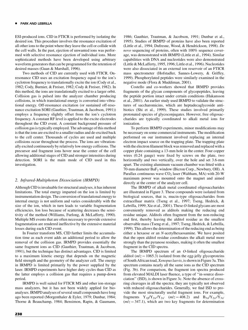
The IRMPD of alkali metal coordinated oligosaccharidesare illustrated in Figure 3. These compounds were isolated frombiological sources, that is, mucin-type oligosaccharides fromextracelluar matrix (Tseng et al., 1997; Tseng, Hedrick, &Lebrilla, 1999; Xie et al., 2001). TheseO-linked glycans are mostconveniently removed as alditols making the reducing endresidue unique. Alditols often fragment from the non-reducingend first, thereby leaving the alditol residue as the smallestobservable mass (Tseng et al., 1997; Tseng, Hedrick, & Lebrilla,1999). This allows the determination of the reducing end as beingeither a hexaose or an N-acetylhexaosamine. We have positedthat the open alditol residue coordinates the alkali metal morestrongly than the pyranose residues, making it often the smallestfragment in the CID spectra.
The IRMPD spectrum of an O-linked oligosaccharidealditol (m/z¼ 1065.3) isolated from the egg-jelly glycoproteinsof South African toad,Xenopus laevis, is shown in Figure 3a. Thisspectrum contains nearly all the same ions as the CID spectrum(Fig. 3b). For comparison, the fragment ion spectra producedfrom elevated MALDI laser fluence, a type of ‘‘in-source disso-ciation’’ (ISD), is shown in Figure 3c. Note the absence of cross-ring cleavages in all the spectra; they are typically not observedwith reduced oligosaccharides. Generally, we find ISD to pro-vide the most structurally useful fragment ions. For example,fragments Y1b/Y2a0/Y2a0 (m/z¼ 408.2) and B3a/Y2a0/Y3a0
(m/z¼ 347.1), which are two key fragments for determination
& PARK AND LEBRILLA
238
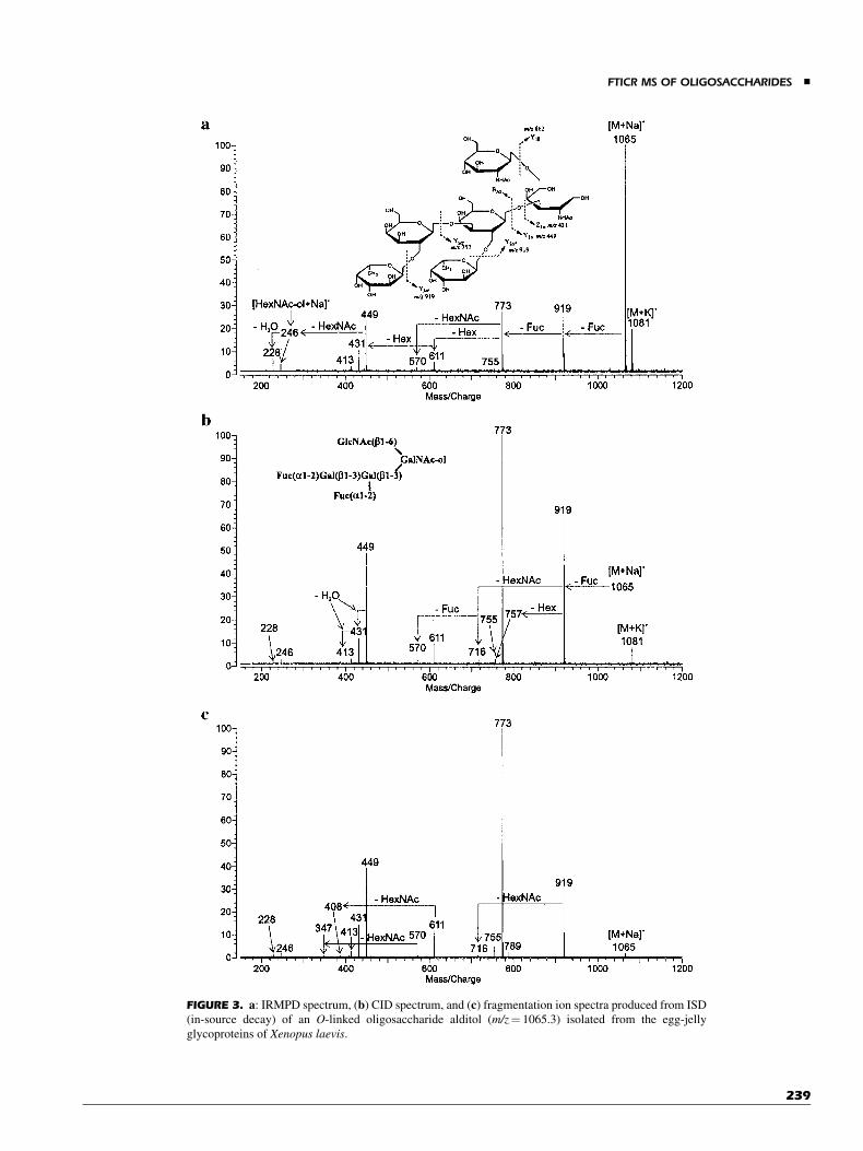
FIGURE 3. a: IRMPD spectrum, (b) CID spectrum, and (c) fragmentation ion spectra produced from ISD
(in-source decay) of an O-linked oligosaccharide alditol (m/z¼ 1065.3) isolated from the egg-jelly
glycoproteins of Xenopus laevis.
FTICR MS OF OLIGOSACCHARIDES &
239
of the structure, were missing in both IRMPD and CID spectra(Fig. 3a,b). A major limitation of the ISD spectra is that structuralelucidation is possible only with pure compounds.
A weakly abundant Y2a0 (m/z¼ 757.2) fragment, diagnosticof the Gal(b1-3)Gal linkage (Tseng, Hedrick, & Lebrilla, 1999),was not observed in the IRMPD but was observed as a weaksignal in the CID spectrum. However, small fragments, likealditol residue ions (m/z¼ 228.1 and 246.1) were clearly andconsistently observed in the IRMPD spectra but were often absentin the CID spectra. In our experience, we found that compoundswith molecular weight greater than 1,300 Da generally requiredmore than two MS stages of CID to yield small fragments thatcorrespond to the alditol residue. However, the IRMPD readilyyielded the monosaccharide alditol residue in a single MS/MSevent.
The two major reaction pathways under IRMPD are the lossof the metal (dissociation) and the fragmentation of theoligosaccharides. For the small alkali metals (Liþ and Naþ),the major reaction products were because of fragmentation. Forthe large alkali metal ions, Kþ, Rbþ, and Csþ, the prominentreaction products observed were the alkali metal ions, correspon-ding primarily to dissociation. Similar behavior was observed inan extensive CID investigation reported earlier (Cancilla et al.,1999). However, the differentiation between the two reactionchannels were not as distinct in CID. Fragmentation reactionswere observed prominently in CID even with Kþ, Rbþ, and Csþ,albeit the relative abundance of the bare metal ion increased withthe size of the alkali metal.
The binding energy of the alkali metal to oligosaccharidesdecrease with the order Liþ>Naþ>Kþ>Rbþ>Csþ (Cancillaet al., 1996). For this reason, alkali metal ion loss is greater forCsþ and less for Liþ. This order is also the same for the ability ofthe metal to catalyze charge-site fragmentation. Liþ and Naþ
produce more fragments than the larger alkali metal ions.The energy diagram in Figure 4 describes the two types ofbehavior. For Liþ and Naþ, the reaction threshold is higher formetal ion loss than for fragmentation (Fig. 4a). For the largeralkali metal ions, Kþ, Rbþ, and Csþ, the reverse is true. The metalion dissociation has a lower threshold than fragmentation(Fig. 4b).
There are mainly similarities in the CID and IRMPD spectraof oligosaccharides. However, there are minor but critical diffe-rences between the two dissociation methods. The larger varia-tions in energy transferred during collisions provide dissociationpathways that are not accessible to IRMPD. CID therefore mayprovide characteristic ions that allow better differentiation ofstereoisomers and linkages. However, for the oligosaccharides,we found very few variations in the ions produced. IRMPDprovides energy not only to the isolated ions but also to thefragment ions to produce a cascade of fragments down to the lastresidue. For the oligosaccharide alditols, it allows the rapiddetermination of the reducing end. This information is accessibleby CID often only through multiple tandem MS events. In thisregard, IRMPD and CID offer complementary information.IRMPD provides another method in the toolbox needed foroligosaccharide analyses.
In situations when high laser power is used, IRMPD can beused to isolate specific masses from the analyzer cell. Infraredlaser isolation (IRLI) was used to isolate ions by exciting the
desired ions to a radius larger than the beam radius (Xie, Schu-bothe, & Lebrilla, 2003). The ions in the center of the analyzercells decompose in the highly degradative beam to yield protonsor alkali metal ions that are readily lost through ion evaporationfrom the analyzer cell. This concept was illustrated for mixturesof oligosaccharides where the isolated species was furtherdissociated by CID and IRMPD.
3. Electron Capture Dissociation (ECD)
The use of low energy electron to yield fragmentation in multiplypositively charged analytes is the main concept behind ECD. Theprocess can in principle be applied to any trapped analyzer but ithas been primarily applied to FT ICRMS. The method has foundwide applications in the so-called ‘‘top-down’’ approach wherethe whole intact protein is simultaneously sequenced (Fridriks-son et al., 2000). A more intriguing use of this dissociativemethod is to search for post-translation modification such asglycosylation. Zubarev and co-workers used ECD to localizeO-glycosylation sites on peptides (Mirgorodskaya, Roepstorff, &Zubarev, 1999). Large glycopeptides derived from immunoglu-bilin were also analyzed in the similar manner (Fridriksson et al.,2000) with the site determined and the heterogeneity of five high-mannose glycoforms characterized.
FIGURE 4. Energy diagrams for (a) small alkali metal ions (Liþ and
Naþ), and (b) large alkali metal ions (Kþ, Rbþ, and Csþ).
& PARK AND LEBRILLA
240
Trypsin-derived glycopeptides from a lectin were analyzedby Nilsson and co-workers (Hakansson et al., 2001). IRMPD andECD were compared directly with ECD yielding predominantlyfragmentation along the peptide chain and IRMPD fragmenta-tion along the oligosaccharide chain. While ECD showsconsiderable promise for proteins and peptides, it may be oflimited use to oligosaccharides. Neutral oligosaccharides aretypically observed as sodium-coordinated species whereasmultiply anionic oligosaccharides, though multiply charged,are often observed as anions and would not be readily amenableto ECD.
III. FUNDAMENTAL STUDIES OFOLIGOSACCHARIDE FRAGMENTATION
A. Dissociation of Oligosaccharides
The study of metastable decay of FAB produced ions wasinformative and helped us realize the limitations associated withthis ionization method. An understanding based on fundamentalknowledge of ion dissociation mechanisms and processes are keytowards developing useful analytical tools. To this end, we havepursued greater understanding of the unimolecular dissociationof oligosaccharides with MALDI.
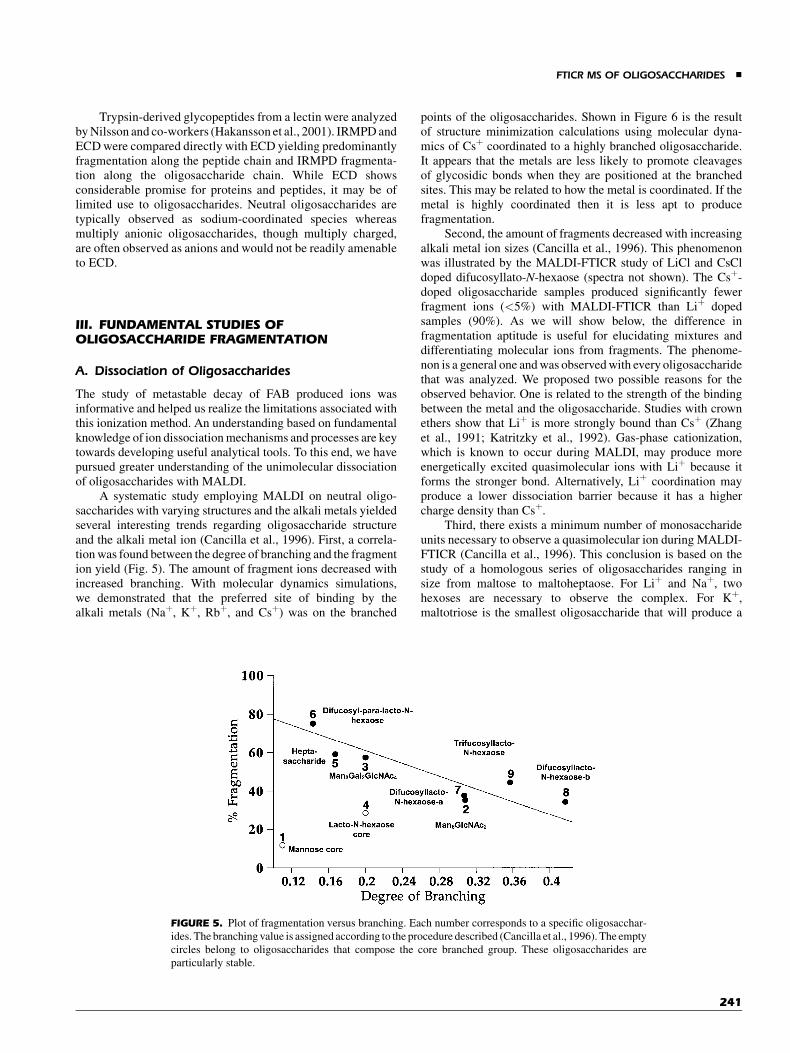
A systematic study employing MALDI on neutral oligo-saccharides with varying structures and the alkali metals yieldedseveral interesting trends regarding oligosaccharide structureand the alkali metal ion (Cancilla et al., 1996). First, a correla-tion was found between the degree of branching and the fragmention yield (Fig. 5). The amount of fragment ions decreased withincreased branching. With molecular dynamics simulations,we demonstrated that the preferred site of binding by thealkali metals (Naþ, Kþ, Rbþ, and Csþ) was on the branched
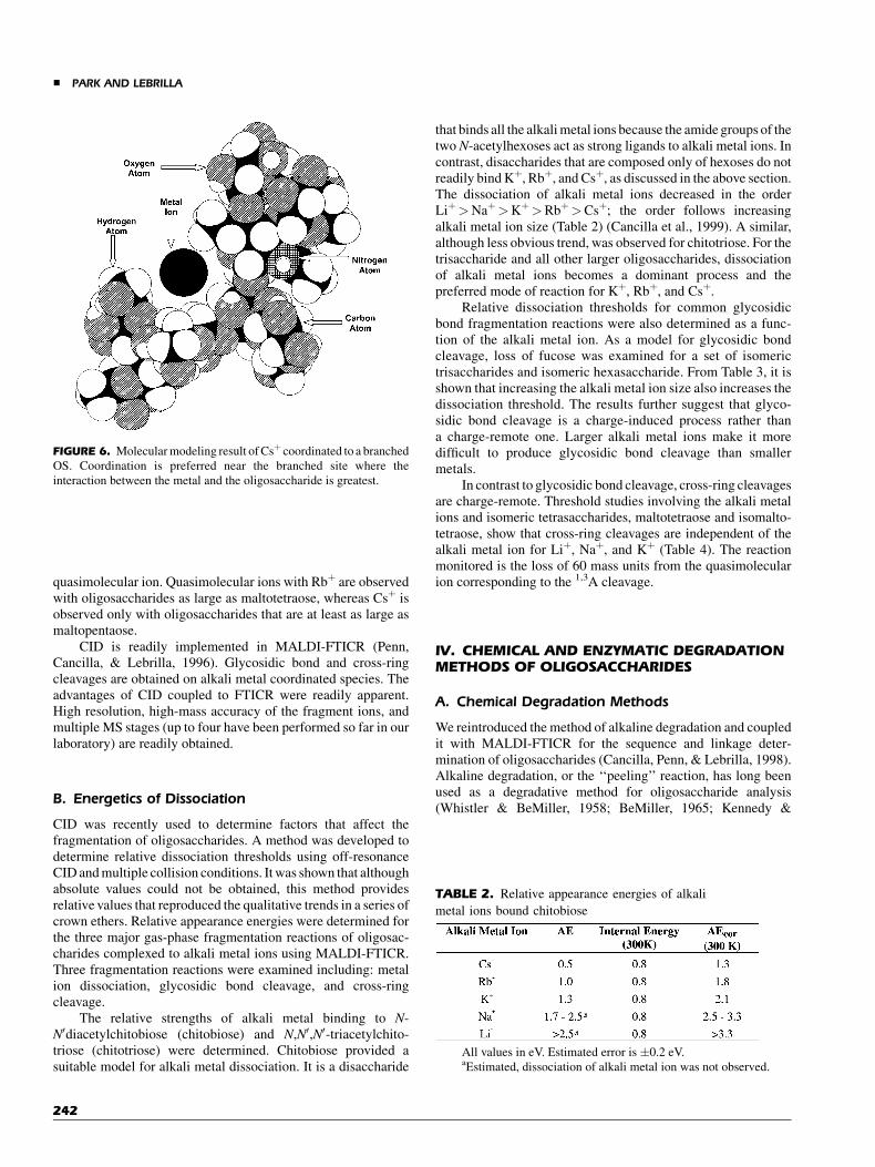
points of the oligosaccharides. Shown in Figure 6 is the resultof structure minimization calculations using molecular dyna-mics of Csþ coordinated to a highly branched oligosaccharide.It appears that the metals are less likely to promote cleavagesof glycosidic bonds when they are positioned at the branchedsites. This may be related to how the metal is coordinated. If themetal is highly coordinated then it is less apt to producefragmentation.
Second, the amount of fragments decreased with increasingalkali metal ion sizes (Cancilla et al., 1996). This phenomenonwas illustrated by the MALDI-FTICR study of LiCl and CsCldoped difucosyllato-N-hexaose (spectra not shown). The Csþ-doped oligosaccharide samples produced significantly fewerfragment ions (<5%) with MALDI-FTICR than Liþ dopedsamples (90%). As we will show below, the difference infragmentation aptitude is useful for elucidating mixtures anddifferentiating molecular ions from fragments. The phenome-non is a general one and was observed with every oligosaccharidethat was analyzed. We proposed two possible reasons for theobserved behavior. One is related to the strength of the bindingbetween the metal and the oligosaccharide. Studies with crownethers show that Liþ is more strongly bound than Csþ (Zhanget al., 1991; Katritzky et al., 1992). Gas-phase cationization,which is known to occur during MALDI, may produce moreenergetically excited quasimolecular ions with Liþ because itforms the stronger bond. Alternatively, Liþ coordination mayproduce a lower dissociation barrier because it has a highercharge density than Csþ.
Third, there exists a minimum number of monosaccharideunits necessary to observe a quasimolecular ion during MALDI-FTICR (Cancilla et al., 1996). This conclusion is based on thestudy of a homologous series of oligosaccharides ranging insize from maltose to maltoheptaose. For Liþ and Naþ, twohexoses are necessary to observe the complex. For Kþ,maltotriose is the smallest oligosaccharide that will produce a
FIGURE 5. Plot of fragmentation versus branching. Each number corresponds to a specific oligosacchar-
ides. The branching value is assigned according to the procedure described (Cancilla et al., 1996). The empty
circles belong to oligosaccharides that compose the core branched group. These oligosaccharides are
particularly stable.
FTICR MS OF OLIGOSACCHARIDES &
241
quasimolecular ion. Quasimolecular ions with Rbþ are observedwith oligosaccharides as large as maltotetraose, whereas Csþ isobserved only with oligosaccharides that are at least as large asmaltopentaose.
CID is readily implemented in MALDI-FTICR (Penn,Cancilla, & Lebrilla, 1996). Glycosidic bond and cross-ringcleavages are obtained on alkali metal coordinated species. Theadvantages of CID coupled to FTICR were readily apparent.High resolution, high-mass accuracy of the fragment ions, andmultiple MS stages (up to four have been performed so far in ourlaboratory) are readily obtained.
B. Energetics of Dissociation
CID was recently used to determine factors that affect thefragmentation of oligosaccharides. A method was developed todetermine relative dissociation thresholds using off-resonanceCID and multiple collision conditions. It was shown that althoughabsolute values could not be obtained, this method providesrelative values that reproduced the qualitative trends in a series ofcrown ethers. Relative appearance energies were determined forthe three major gas-phase fragmentation reactions of oligosac-charides complexed to alkali metal ions using MALDI-FTICR.Three fragmentation reactions were examined including: metalion dissociation, glycosidic bond cleavage, and cross-ringcleavage.
The relative strengths of alkali metal binding to N-N0diacetylchitobiose (chitobiose) and N,N0,N0-triacetylchito-triose (chitotriose) were determined. Chitobiose provided asuitable model for alkali metal dissociation. It is a disaccharide
that binds all the alkali metal ions because the amide groups of thetwo N-acetylhexoses act as strong ligands to alkali metal ions. Incontrast, disaccharides that are composed only of hexoses do notreadily bind Kþ, Rbþ, and Csþ, as discussed in the above section.The dissociation of alkali metal ions decreased in the orderLiþ>Naþ>Kþ>Rbþ>Csþ; the order follows increasingalkali metal ion size (Table 2) (Cancilla et al., 1999). A similar,although less obvious trend, was observed for chitotriose. For thetrisaccharide and all other larger oligosaccharides, dissociationof alkali metal ions becomes a dominant process and thepreferred mode of reaction for Kþ, Rbþ, and Csþ.
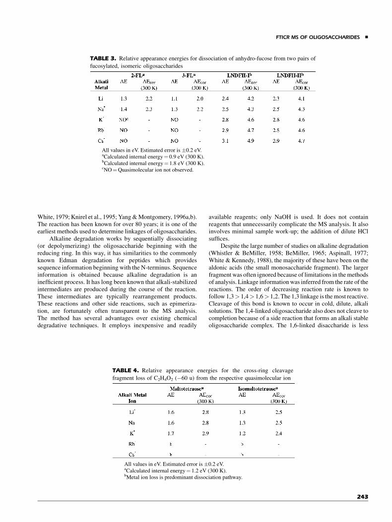
Relative dissociation thresholds for common glycosidicbond fragmentation reactions were also determined as a func-tion of the alkali metal ion. As a model for glycosidic bondcleavage, loss of fucose was examined for a set of isomerictrisaccharides and isomeric hexasaccharide. From Table 3, it isshown that increasing the alkali metal ion size also increases thedissociation threshold. The results further suggest that glyco-sidic bond cleavage is a charge-induced process rather thana charge-remote one. Larger alkali metal ions make it moredifficult to produce glycosidic bond cleavage than smallermetals.
In contrast to glycosidic bond cleavage, cross-ring cleavagesare charge-remote. Threshold studies involving the alkali metalions and isomeric tetrasaccharides, maltotetraose and isomalto-tetraose, show that cross-ring cleavages are independent of thealkali metal ion for Liþ, Naþ, and Kþ (Table 4). The reactionmonitored is the loss of 60 mass units from the quasimolecularion corresponding to the 1,3A cleavage.
IV. CHEMICAL AND ENZYMATIC DEGRADATIONMETHODS OF OLIGOSACCHARIDES
A. Chemical Degradation Methods
We reintroduced the method of alkaline degradation and coupledit with MALDI-FTICR for the sequence and linkage deter-mination of oligosaccharides (Cancilla, Penn, & Lebrilla, 1998).Alkaline degradation, or the ‘‘peeling’’ reaction, has long beenused as a degradative method for oligosaccharide analysis(Whistler & BeMiller, 1958; BeMiller, 1965; Kennedy &
FIGURE 6. Molecular modeling result of Csþ coordinated to a branched
OS. Coordination is preferred near the branched site where the
interaction between the metal and the oligosaccharide is greatest.
TABLE 2. Relative appearance energies of alkali
metal ions bound chitobiose
All values in eV. Estimated error is �0.2 eV.aEstimated, dissociation of alkali metal ion was not observed.
& PARK AND LEBRILLA
242
White, 1979; Knirel et al., 1995; Yang & Montgomery, 1996a,b).The reaction has been known for over 80 years; it is one of theearliest methods used to determine linkages of oligosaccharides.
Alkaline degradation works by sequentially dissociating(or depolymerizing) the oligosaccharide beginning with thereducing ring. In this way, it has similarities to the commonlyknown Edman degradation for peptides which providessequence information beginning with the N-terminus. Sequenceinformation is obtained because alkaline degradation is aninefficient process. It has long been known that alkali-stabilizedintermediates are produced during the course of the reaction.These intermediates are typically rearrangement products.These reactions and other side reactions, such as epimeriza-tion, are fortunately often transparent to the MS analysis.The method has several advantages over existing chemicaldegradative techniques. It employs inexpensive and readily
available reagents; only NaOH is used. It does not containreagents that unnecessarily complicate the MS analysis. It alsoinvolves minimal sample work-up; the addition of dilute HClsuffices.
Despite the large number of studies on alkaline degradation(Whistler & BeMiller, 1958; BeMiller, 1965; Aspinall, 1977;White & Kennedy, 1988), the majority of these have been on thealdonic acids (the small monosaccharide fragment). The largerfragment was often ignored because of limitations in the methodsof analysis. Linkage information was inferred from the rate of thereactions. The order of decreasing reaction rate is known tofollow 1,3> 1,4> 1,6> 1,2. The 1,3 linkage is the most reactive.Cleavage of this bond is known to occur in cold, dilute, alkalisolutions. The 1,4-linked oligosaccharide also does not cleave tocompletion because of a side reaction that forms an alkali stableoligosaccharide complex. The 1,6-linked disaccharide is less
TABLE 3. Relative appearance energies for dissociation of anhydro-fucose from two pairs of
fucosylated, isomeric oligosaccharides
All values in eV. Estimated error is �0.2 eV.aCalculated internal energy¼ 0.9 eV (300 K).bCalculated internal energy¼ 1.8 eV (300 K).cNO¼Quasimolecular ion not observed.
TABLE 4. Relative appearance energies for the cross-ring cleavage
fragment loss of C2H4O2 (�60 u) from the respective quasimolecular ion
All values in eV. Estimated error is �0.2 eV.aCalculated internal energy¼ 1.2 eV (300 K).bMetal ion loss is predominant dissociation pathway.
FTICR MS OF OLIGOSACCHARIDES &
243

reactive than 1,4-linked but also cleaves at elevated temperatures.The 1,2 linkage does not react even under the most vigorousconditions. With a branched oligosaccharide that has, forexample, both 1,3 and 1,6 linkage at the branch point, thereaction will proceed via the 1,3-linked antenna first. The mostimportant feature of this reaction to MS is that the degradedoligosaccharides contain nascent reducing rings. Cross-ringcleavages, whether with MALDI or CID, occur most readily onthe reducing ring. The method is summarized in Scheme 1.Sequential degradation is obtained to produce nascent reducingrings that are fragmented by MALDI or CID. Sequence isobtained because the reaction is inefficient and representativefragments corresponding to nearly all degradative cleavages areobserved.
For MALDI, the net result of alkaline degradation is thatglycosidic bonds are cleaved beginning with the reducing ring.Each cycle reveals a new reducing ring so that MALDI andMALDI-CID fragmentation can be employed to produce cross-ring cleavages at the nascent reducing end. Furthermore, becausethe quasimolecular ions are sodium coordinated, cross-ringcleavages are produced to yield the specific linkages (Coates &Wilkins, 1985; Garozzo et al., 1990; Spengler, Dolce, & Cotter,1990; Carroll & Lebrilla, 1992; Carroll, Willard, & Lebrilla,1995). Both linkage and sequence information are obtained withless than 100 pmol.
The result obtained with LNFP-1 illustrates in some detailthe utility of AD coupled to MALDI-FTICR. The MALDI-FTICR spectrum of the untreated compound shows only the
quasimolecular ion and the prominent loss of fucose (spectrumnot shown). No other structural information is obtained. CID ofthe quasimolecular ion produces only more fucose loss until theion is totally defucosylated.
After treatment of the oligosaccharide for 24 hr with sodiumhydroxide at 608C, there are sufficient fragment ions in theMALDI-FTICR spectrum to sequence the oligosaccharide anddetermine most of the linkages (Fig. 7). The cleavage pattern forLNFP-1 obtained with AD-MALDI is shown (Scheme 2). Fromthe mass spectrum, numerous glycosidic bond cleavages arereadily observed corresponding to C4 (m/z¼ 714.245), C3
(m/z¼ 552.190), and C2 (m/z¼ 349.109). In addition, thecomplementary fragments, such as B4 (m/z¼ 696.233) and B3
(m/z¼ 534.180), are also obtained. There are combinations ofcleavages such as m/z¼ 388.120 corresponding to a B3þY4
cleavage (designated as B3/Y4),m/z¼ 406.131 because of C3/Y4,and m/z¼ 568.186 because of C4/Y4. In addition, cross-ringcleavages such as 0,3A4 show the 1,3-linkage at the fourthsaccharide position. The advantages of AD for fucose containingoligosaccharides are further illustrated in this example. Theuninformative loss of fucose is still obtained, m/z¼ 730.238,during MALDI. However, the molecule remains sufficientlyintact so that the placement of the fucose is unambiguouslyidentified at the end of the chain. The C2 fragment, m/z¼349.109, is the disaccharide that makes up the non-reducing endand identifies the position of fucose.
CID is useful for obtaining cross-ring cleavages at thenascent reducing end when they are absent in the MALDI
SCHEME 1. Sequential alkaline degradation.
& PARK AND LEBRILLA
244

FIGURE 7. MALDI-MS spectrum of LNFP-1 after 24 hr under alkaline degradation conditions.
FTICR MS OF OLIGOSACCHARIDES &
245
spectrum. No cross-ring cleavages are observed for saccharides 2and 3 (1 is the non-reducing ring) in the MALDI spectrum. Thefragment because of C2 cleavage (m/z¼ 349.109) was subjectedto CID (spectrum not shown). From the spectrum we observe an0,2X3 (m/z¼ 229.069) cleavage yielding the 1,2 linkage at sac-charide 4. The saccharide 3 does not yield cross-ring cleavageseven with CID. This linkage combination (i.e., Glc1! 3GlcNAc)appears to be unique in this regard. Similar linkage combinations
in other oligosaccharides also do not produce cross-ringcleavages.
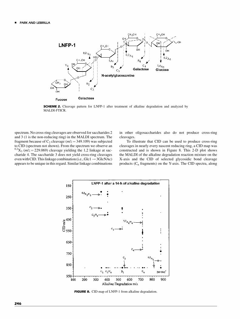
To illustrate that CID can be used to produce cross-ringcleavages in nearly every nascent reducing ring, a CID map wasconstructed and is shown in Figure 8. This 2-D plot showsthe MALDI of the alkaline degradation reaction mixture on theX-axis and the CID of selected glycosidic bond cleavageproducts (Cn fragments) on the Y-axis. The CID spectra, along
SCHEME 2. Cleavage pattern for LNFP-1 after treatment of alkaline degradation and analyzed by
MALDI-FTICR.
FIGURE 8. CID map of LNFP-1 from alkaline degradation.
& PARK AND LEBRILLA
246
the Y-axis, provide further supporting evidences for theMALDI assignments and confirm the identity of the proposedlinkages.
Based on the MS analysis of the alkaline degradationreaction mixture, one can predict the linkage and sequence forthis compound to be:
Fuc1 ! 2Hex1 ! ð2 or 3ÞHexNAc1 ! 3Hex1 ! 4Hex:
The only ambiguity is in the third saccharide residue wherethe linkage may be either 1-2 or 1-3. Both Hex1-2HexNAc andHex1-3HexNAc combinations do not appear to produce cross-ring cleavages.
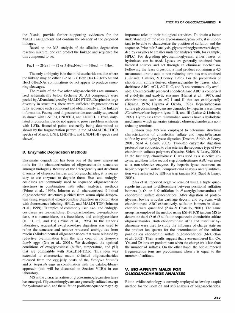
The results of the five other oligosaccharides are summar-ized schematically below (Scheme 3). All compounds werepeeled by AD and analyzed by MALDI-FTICR. Despite the largediversity in structures, there were sufficient fragmentations tofully sequence each compound and obtain nearly all the linkageinformation. Fucosylated oligosaccharides are readily sequencedas shown with LNFP-I, LNDFH-I, and LNDFH-II. Even sialy-lated oligosaccharides do not appear to pose a problem as shownwith LSTa. Branched points are easily being determined asshown by the fragmentation pattern in the AD-MALDI-FTICRspectra of Man-5, LNH, LNDFH-I, and LNDFH-II (spectra notshown).
B. Enzymatic Degradation Methods
Enzymatic degradation has been one of the most importanttools for the characterization of oligosaccharide structuresamongst biologists. Because of the heterogeneity and structuraldiversity of oligosaccharides and polysaccharides, it is neces-sary to use enzymes to degrade them. Exo- and endogly-cosidases are commonly used to sequence oligosaccharidestructures in combination with other analytical methods(Prime et al., 1996). Johnson et al. characterized O-linkedoligosaccharide structures purified from serum alpha-fetopro-tein using sequential exoglycosidase digestion in combinationwith fluorescence labeling, HPLC, and MALDI-TOF (Johnsonet al., 1999). Examples of commonly used exo- and endogly-cosidases are a-D-sialidase, b-D-galactosidase, a-D-galactosi-dase, a-D-mannosidase, a-L-fucosidase, and endoglycosidase(H, F1, F2, and F3) (Prime et al., 1996). In the author’slaboratory, sequential exoglycosidase digestion was used torefine the structure and remove structural ambiguities frommucin O-linked neutral oligosaccharides that were released byreductive b-elimination from the jelly coat of the Xenopuslaevis eggs (Xie et al., 2001). We developed the optimalconditions of exoglycosidase (buffer, temperature, and pH)that are compatible with MALDI-FTICR. This idea wasextended to characterize mucin O-linked oligosaccharidesreleased from the egg-jelly coats of the Xenopus borealisand X. tropicals eggs in combination with the catalog-libraryapproach (this will be discussed in Section VI(B)) in ourlaboratory.
MS in the characterization of glycosaminoglycan structureshas emerged. Glycosaminoglycans are generally sulfated exceptfor hyaluronic acid, and the sulfation position/sequence may play
important roles in their biological activities. To obtain a betterunderstanding of the roles glycosaminoglycan play, it is impor-tant to be able to characterize the position of sulfation and thesequence. Prior to MS analysis, glycosaminoglycans were degra-ded by enzymes to smaller units for analyses with, for example,HPLC. For degrading glycosaminoglycans, either lyases orhydrolases can be used. Lyases are generally obtained frombacterial sources and act through an eliminase mechanism.Following the lyase digestion, a final product containing a 4,5unsaturated uronic acid at non-reducing terminus was obtained(Linhardt, Galliher, & Cooney, 1986). For the preparation ofchondroitin sulfate-derived oligosaccharides by lyases, chon-droitinase ABC, AC I, AC II, C, and B are commercially avail-able. Commercially prepared chondroitinase ABC is comprisedof endolytic and exolytic enzymes (Hamai et al., 1997), andchondroitinase such as AC I and II that act endolytically(Hiyama, 1976; Hiyama & Okada, 1976). Heparin/heparansulfate glycosaminoglycans are degraded by heparin lyases fromFlavobacterium: heparin lyase I, II, and III (Lohse & Linhardt,1992). Hydrolases from mammalian sources have a hydrolyticmechanism which generates saturated oligosaccharides at a non-reducing terminus.
ESI-ion trap MS was employed to determine structuralcharacterization of chondroitin sulfate and heparin/heparansulfate by employing lyase digestion (Desaire, Sirich, & Leary,2001; Saad & Leary, 2003). Two-step enzymatic digestionprotocol was conducted to characterize the sequence type of twochondroitin sulfates polymers (Desaire, Sirich, & Leary, 2001).In the first step, chondroitnase C was used as a selective en-zyme, and then in the second step chondroitinase ABC was usedas a non-selective enzyme. By heparin lyase treatment onheparin/heparan sulfate, compositional analysis and quantifica-tion were achieved by ESI-ion trap tandem MS (Saad & Leary,2003).
Zaia et al. reported negative ion-ESI using a triple quad-rupole instrument to differentiate between positional sulfationisomers (4-O- or 6-O-sulfation in N-acetylgalactosamine) ofchondroitin sulfate disaccharides. After digestion of proteo-glycans, bovine articular cartilage decorin and biglycan, withchondroitinase ABC exhaustively, sulfation isomers in disac-charides were quantified (Zaia & Costello, 2001). The samegroup has employed the method using ESI-FTICR tandem MS todetermine the 4-O-/6-O-sulfation sequence in chondroitin sulfateoligosaccharides. Both chondroitinase AC I and testicular hy-aluronase were used to study the influence of charge state onthe product ion spectra for the determination of the sulfateposition on chondroitin sulfate oligosaccharides (McClellanet al., 2002). Their results suggest that even-numbered Bn, Cn,Yn, and Zn ions are predominant when the charge (z) is less thanthe number of sulfates. On the other hand, the odd-numberedfragmentation ions are predominant when z is equal to thenumber of sulfates.
V. BIO-AFFINITY MALDI FOROLIGOSACCHARIDE ANALYSES
Biotin-avidin technology is currently employed to develop a rapidmethod for the isolation and MS analysis of oligosaccharides.
FTICR MS OF OLIGOSACCHARIDES &
247
SCHEME 3. Five oligosaccharides (mannose 5, lacto-N-hexaose, LSTa, LNDFH-I, and LNDFH-II) after
treatment with alkaline degradation and analyzed by MALDI-FTICR.
& PARK AND LEBRILLA
248

The concept of biologically active probes for selective MALDIhas been present for several years. Hutchens has suggestedthe use of probe surfaces that are designed to extract specificmolecules from unfractionated biological fluids and extracts(Hutchens & Yip, 1993). In one application, agarose beads withattached single-strand DNAwere used to capture lactoferrin frompreterm infant urine and placed directly on the MALDI probefor analysis. Orlando developed probe affinity MS to immo-bilize monoclonal antibody IgG1 directly on the probe so thatbiotinylated insulin can be captured and analyzed (Brockman &Orlando, 1995). To determine the specific components of theprotein human basic fibroblast growth factor (bFGF) that interactwith mouse mAB 11.1, Chait used agarose beads with the antigento concentrate partially digested components of the protein (Zhaoet al., 1996). Li and co-workers used agarose beads containingavidin to extract biotinylated peptides and proteins (bradykininand insulin) (Schriemer & Li, 1996). The researchers furtherfound that the condition for MALDI preparation caused thedegradation of the complex so that the biotin-labeled compoundcan be deposited on the probe whereas the agarose beads arephysically removed. They noted that agarose beads on the probesurface produce deleterious effects on the MALDI spectrum(Nelson et al., 1995).
We produced a number of bioaffinity probes specifically toanalyze oligosaccharides to be examined with MALDI-FTICRMS. In the first series of bioaffinity probes, avidin was immobi-lized directly on the probe surface and the oligosaccharides were
biotinylated. In the second series, lectins (oligosaccharide bind-ing proteins) were directly immobilized to a microprous surfaceto concentrate oligosaccharides on the probe surface.
A. Avidin Affinity Probe
A simple method was developed to immobilize avidin on theMALDI probe, thereby eliminating the use of the agarose beads(Wang, Tseng, & Lebrilla, 1999). An avidin solution was simplyapplied to the microporous surface and dried. Drying the solutionfixes the protein on the surface. Research by Li and co-workersshowed that avidin dried on a surface remains active towardsbiotin (Schriemer & Li, 1996). Once the avidin is immobilized,MALDI preparation involves taking the solution containing thebiotin labeled compounds and depositing it on the probe. Thebiotin-avidin interaction was allowed to take place and the probewas washed to remove non-biotin labeled material. Matrix wasthen applied and the solvent evaporated.
Derivatization of oligosaccharides by biotin was relativelystraightforward. Several types of biotin derivatives are commer-cially available (Savage et al., 1992). They are functionalizedwith carboxylic acids or amines and can have long and shortspacers ranging in length from 10 to 24 A. The longer spacerarms were important for this study as they have been shownto allow greater access to avidin immobilized on surfaces(Schriemer & Li, 1996). We adapted a procedure developedby Takao and co-workers for attaching 4-aminobenzoic acid
SCHEME 3. (Continued )
FTICR MS OF OLIGOSACCHARIDES &
249
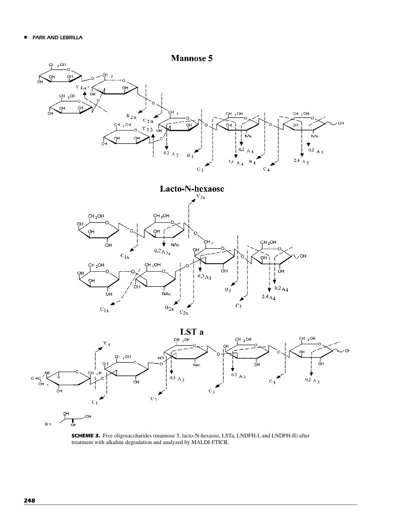
2-(diethylamino)ethyl ester to neutral oligosaccharides (Yoshinoet al., 1995; Takao et al., 1996). This involved forming the Schiffbase and reducing the intermediate with sodium cyanoborohy-dride to produce the corresponding amine (Scheme 4). The ‘‘X’’in Scheme 4 refers to a variably length spacer.
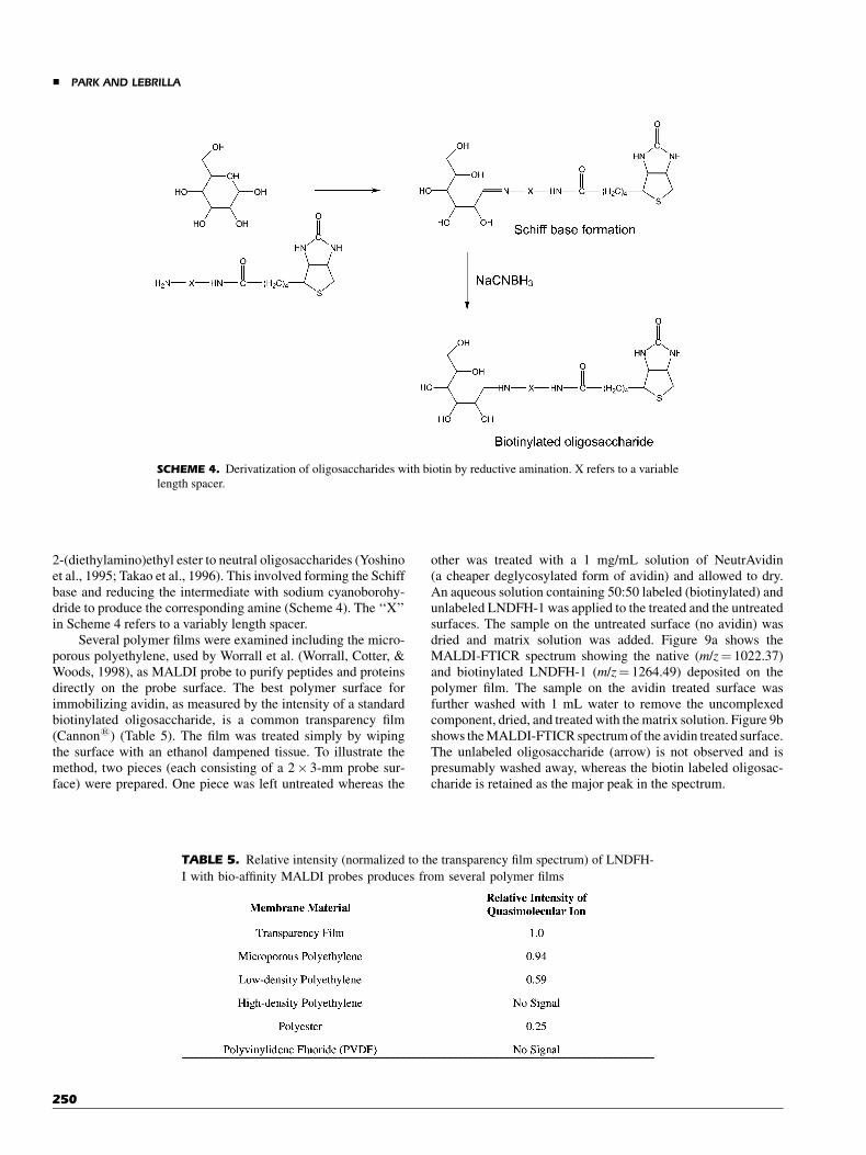
Several polymer films were examined including the micro-porous polyethylene, used by Worrall et al. (Worrall, Cotter, &Woods, 1998), as MALDI probe to purify peptides and proteinsdirectly on the probe surface. The best polymer surface forimmobilizing avidin, as measured by the intensity of a standardbiotinylated oligosaccharide, is a common transparency film(Cannon1) (Table 5). The film was treated simply by wipingthe surface with an ethanol dampened tissue. To illustrate themethod, two pieces (each consisting of a 2� 3-mm probe sur-face) were prepared. One piece was left untreated whereas the
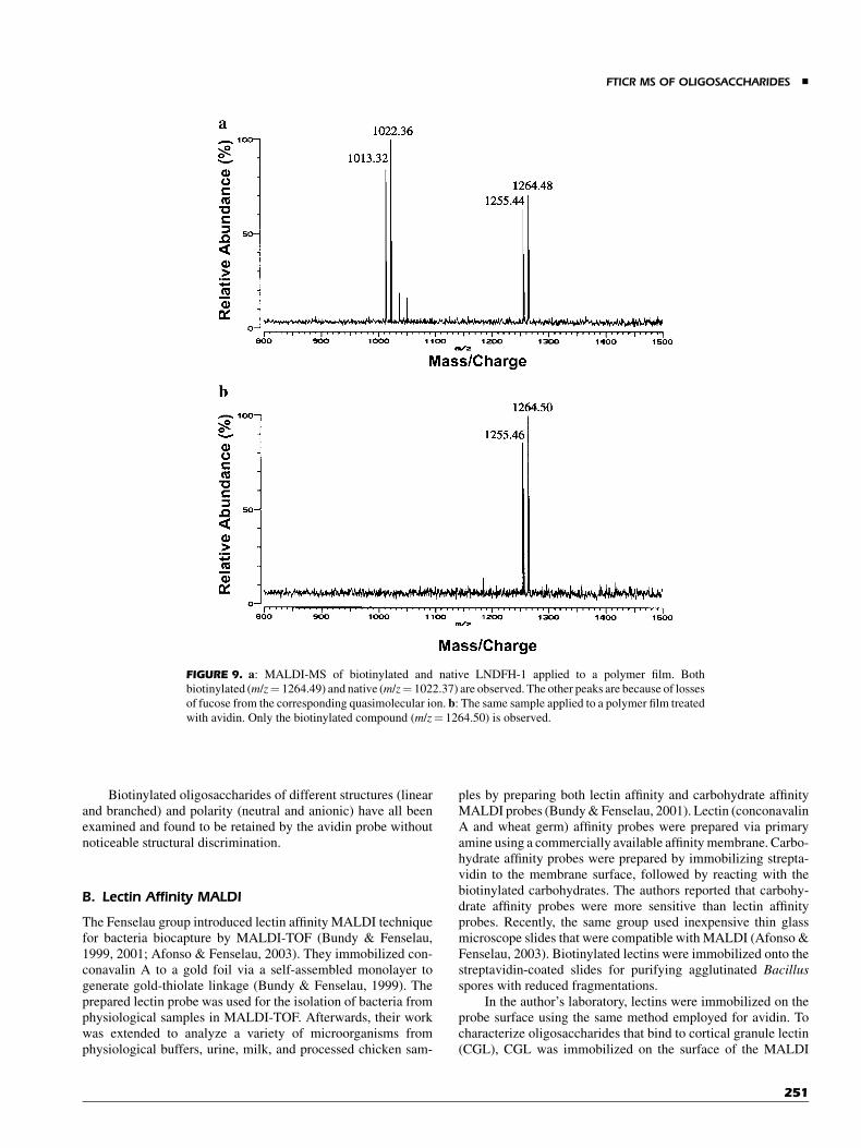
other was treated with a 1 mg/mL solution of NeutrAvidin(a cheaper deglycosylated form of avidin) and allowed to dry.An aqueous solution containing 50:50 labeled (biotinylated) andunlabeled LNDFH-1 was applied to the treated and the untreatedsurfaces. The sample on the untreated surface (no avidin) wasdried and matrix solution was added. Figure 9a shows theMALDI-FTICR spectrum showing the native (m/z¼ 1022.37)and biotinylated LNDFH-1 (m/z¼ 1264.49) deposited on thepolymer film. The sample on the avidin treated surface wasfurther washed with 1 mL water to remove the uncomplexedcomponent, dried, and treated with the matrix solution. Figure 9bshows the MALDI-FTICR spectrum of the avidin treated surface.The unlabeled oligosaccharide (arrow) is not observed and ispresumably washed away, whereas the biotin labeled oligosac-charide is retained as the major peak in the spectrum.
SCHEME 4. Derivatization of oligosaccharides with biotin by reductive amination. X refers to a variable
length spacer.
TABLE 5. Relative intensity (normalized to the transparency film spectrum) of LNDFH-
I with bio-affinity MALDI probes produces from several polymer films
& PARK AND LEBRILLA
250
Biotinylated oligosaccharides of different structures (linearand branched) and polarity (neutral and anionic) have all beenexamined and found to be retained by the avidin probe withoutnoticeable structural discrimination.
B. Lectin Affinity MALDI
The Fenselau group introduced lectin affinity MALDI techniquefor bacteria biocapture by MALDI-TOF (Bundy & Fenselau,1999, 2001; Afonso & Fenselau, 2003). They immobilized con-conavalin A to a gold foil via a self-assembled monolayer togenerate gold-thiolate linkage (Bundy & Fenselau, 1999). Theprepared lectin probe was used for the isolation of bacteria fromphysiological samples in MALDI-TOF. Afterwards, their workwas extended to analyze a variety of microorganisms fromphysiological buffers, urine, milk, and processed chicken sam-
ples by preparing both lectin affinity and carbohydrate affinityMALDI probes (Bundy & Fenselau, 2001). Lectin (conconavalinA and wheat germ) affinity probes were prepared via primaryamine using a commercially available affinity membrane. Carbo-hydrate affinity probes were prepared by immobilizing strepta-vidin to the membrane surface, followed by reacting with thebiotinylated carbohydrates. The authors reported that carbohy-drate affinity probes were more sensitive than lectin affinityprobes. Recently, the same group used inexpensive thin glassmicroscope slides that were compatible with MALDI (Afonso &Fenselau, 2003). Biotinylated lectins were immobilized onto thestreptavidin-coated slides for purifying agglutinated Bacillusspores with reduced fragmentations.
In the author’s laboratory, lectins were immobilized on theprobe surface using the same method employed for avidin. Tocharacterize oligosaccharides that bind to cortical granule lectin(CGL), CGL was immobilized on the surface of the MALDI
FIGURE 9. a: MALDI-MS of biotinylated and native LNDFH-1 applied to a polymer film. Both
biotinylated (m/z¼ 1264.49) and native (m/z¼ 1022.37) are observed. The other peaks are because of losses
of fucose from the corresponding quasimolecular ion. b: The same sample applied to a polymer film treated
with avidin. Only the biotinylated compound (m/z¼ 1264.50) is observed.
FTICR MS OF OLIGOSACCHARIDES &
251

FIGURE 10. HPLC of the jelly coat of Xenopus laevis. The sample contains the neutral oligosaccharides.
CHART 1. The structure of 12 oligosaccharides elucidated by NMR (Strecker et al., 1995). The structures
are confirmed by mass spectrometry (See Table 6).
& PARK AND LEBRILLA
252
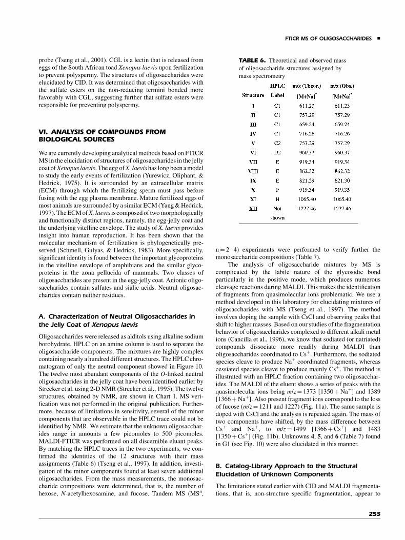
probe (Tseng et al., 2001). CGL is a lectin that is released fromeggs of the South African toad Xenopus laevis upon fertilizationto prevent polyspermy. The structures of oligosaccharides wereelucidated by CID. It was determined that oligosaccharides withthe sulfate esters on the non-reducing termini bonded morefavorably with CGL, suggesting further that sulfate esters wereresponsible for preventing polyspermy.
VI. ANALYSIS OF COMPOUNDS FROMBIOLOGICAL SOURCES
We are currently developing analytical methods based on FTICRMS in the elucidation of structures of oligosaccharides in the jellycoat ofXenopus laevis. The egg ofX. laevishas long been a modelto study the early events of fertilization (Yurewicz, Oliphant, &Hedrick, 1975). It is surrounded by an extracellular matrix(ECM) through which the fertilizing sperm must pass beforefusing with the egg plasma membrane. Mature fertilized eggs ofmost animals are surrounded by a similar ECM (Yang & Hedrick,1997). The ECM ofX. laevis is composed of two morphologicallyand functionally distinct regions, namely, the egg-jelly coat andthe underlying vitelline envelope. The study ofX. laevis providesinsight into human reproduction. It has been shown that themolecular mechanism of fertilization is phylogenetically pre-served (Schmell, Gulyas, & Hedrick, 1983). More specifically,significant identity is found between the important glycoproteinsin the vitelline envelope of amphibians and the similar glyco-proteins in the zona pellucida of mammals. Two classes ofoligosaccharides are present in the egg-jelly coat. Anionic oligo-saccharides contain sulfates and sialic acids. Neutral oligosac-charides contain neither residues.
A. Characterization of Neutral Oligosaccharides inthe Jelly Coat of Xenopus laevis
Oligosaccharides were released as alditols using alkaline sodiumborohydrate. HPLC on an amine column is used to separate theoligosaccharide components. The mixtures are highly complexcontaining nearly a hundred different structures. The HPLC chro-matogram of only the neutral component showed in Figure 10.The twelve most abundant components of the O-linked neutraloligosaccharides in the jelly coat have been identified earlier byStrecker et al. using 2-D NMR (Strecker et al., 1995). The twelvestructures, obtained by NMR, are shown in Chart 1. MS veri-fication was not performed in the original publication. Further-more, because of limitations in sensitivity, several of the minorcomponents that are observable in the HPLC trace could not beidentified by NMR. We estimate that the unknown oligosacchar-ides range in amounts a few picomoles to 500 picomoles.MALDI-FTICR was performed on all discernible eluant peaks.By matching the HPLC traces in the two experiments, we con-firmed the identities of the 12 structures with their massassignments (Table 6) (Tseng et al., 1997). In addition, investi-gation of the minor components found at least seven additionaloligosaccharides. From the mass measurements, the monosac-charide compositions were determined, that is, the number ofhexose, N-acetylhexosamine, and fucose. Tandem MS (MSn,
n¼ 2–4) experiments were performed to verify further themonosaccharide compositions (Table 7).
The analysis of oligosaccharide mixtures by MS iscomplicated by the labile nature of the glycosidic bondparticularly in the positive mode, which produces numerouscleavage reactions during MALDI. This makes the identificationof fragments from quasimolecular ions problematic. We use amethod developed in this laboratory for elucidating mixtures ofoligosaccharides with MS (Tseng et al., 1997). The methodinvolves doping the sample with CsCl and observing peaks thatshift to higher masses. Based on our studies of the fragmentationbehavior of oligosaccharides complexed to different alkali metalions (Cancilla et al., 1996), we know that sodiated (or natriated)compounds dissociate more readily during MALDI thanoligosaccharides coordinated to Csþ. Furthermore, the sodiatedspecies cleave to produce Naþ coordinated fragments, whereascessiated species cleave to produce mainly Csþ. The method isillustrated with an HPLC fraction containing two oligosacchar-ides. The MALDI of the eluent shows a series of peaks with thequasimolecular ions being m/z¼ 1373 [1350þNaþ] and 1389[1366þNaþ]. Also present fragment ions correspond to the lossof fucose (m/z¼ 1211 and 1227) (Fig. 11a). The same sample isdoped with CsCl and the analysis is repeated again. The mass oftwo components have shifted, by the mass difference betweenCsþ and Naþ, to m/z¼ 1499 [1366þCsþ] and 1483[1350þCsþ] (Fig. 11b). Unknowns 4, 5, and 6 (Table 7) foundin G1 (see Fig. 10) were also elucidated in this manner.
B. Catalog-Library Approach to the StructuralElucidation of Unknown Components
The limitations stated earlier with CID and MALDI fragmenta-tions, that is, non-structure specific fragmentation, appear to
TABLE 6. Theoretical and observed mass
of oligosaccharide structures assigned by
mass spectrometry
FTICR MS OF OLIGOSACCHARIDES &
253
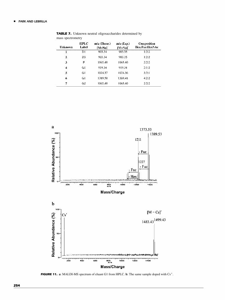
FIGURE 11. a: MALDI-MS spectrum of eluant G1 from HPLC. b: The same sample doped with Csþ.
TABLE 7. Unknown neutral oligosaccharides determined by
mass spectrometry
& PARK AND LEBRILLA
254
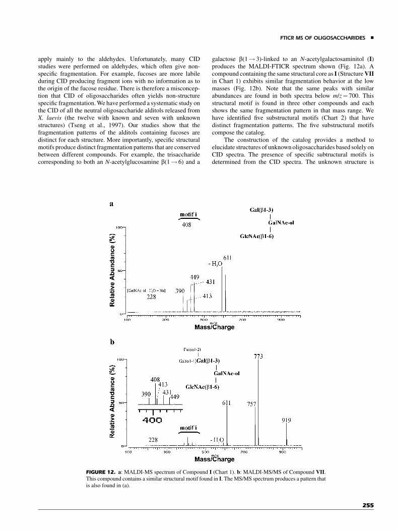
apply mainly to the aldehydes. Unfortunately, many CIDstudies were performed on aldehydes, which often give non-specific fragmentation. For example, fucoses are more labileduring CID producing fragment ions with no information as tothe origin of the fucose residue. There is therefore a misconcep-tion that CID of oligosaccharides often yields non-structurespecific fragmentation. We have performed a systematic study onthe CID of all the neutral oligosaccharide alditols released fromX. laevis (the twelve with known and seven with unknownstructures) (Tseng et al., 1997). Our studies show that thefragmentation patterns of the alditols containing fucoses aredistinct for each structure. More importantly, specific structuralmotifs produce distinct fragmentation patterns that are conservedbetween different compounds. For example, the trisaccharidecorresponding to both an N-acetylglucosamine b(1! 6) and a
galactose b(1! 3)-linked to an N-acetylgalactosaminitol (I)produces the MALDI-FTICR spectrum shown (Fig. 12a). Acompound containing the same structural core as I (StructureVIIin Chart 1) exhibits similar fragmentation behavior at the lowmasses (Fig. 12b). Note that the same peaks with similarabundances are found in both spectra below m/z¼ 700. Thisstructural motif is found in three other compounds and eachshows the same fragmentation pattern in that mass range. Wehave identified five substructural motifs (Chart 2) that havedistinct fragmentation patterns. The five substructural motifscompose the catalog.
The construction of the catalog provides a method toelucidate structures of unknown oligosaccharides based solely onCID spectra. The presence of specific subtructural motifs isdetermined from the CID spectra. The unknown structure is
FIGURE 12. a: MALDI-MS spectrum of Compound I (Chart 1). b: MALDI-MS/MS of Compound VII.This compound contains a similar structural motif found in I. The MS/MS spectrum produces a pattern that
is also found in (a).
FTICR MS OF OLIGOSACCHARIDES &
255

pieced together by combining the motifs. This is illustrated withan oligosaccharide that is a minor component of the mixture andwhose structure is unknown. Unknown 3 found in F (Fig. 10) ofthe HPLC trace produces the CID spectrum (MS2) shown inFigure 13a. The MS4 experiment was also performed (m/z¼1065! 773/757! 611) and is shown in Figure 13b. Notethe presence of motifs i and ii (See Chart 2) in the two spectra.We reconstruct a structure containing both motifs i and ii and wereconcile the difference in mass by the addition of fucose. Theresult is the structure summarized in Figure 13. The additionalfucose is placed on the Gal simply because it is not usually foundlinked to another fucose or to a GlcNAc. Note that this compoundis similar in structure to VII. The two differ by only a fucoseresidue.
This method has been used to elucidate at least nearly allthe remaining structures in the mixture of neutral oligosacchar-ides (Chart 3). The possibility of obtaining complete structuralelucidation with only a few picomoles of material (in this caseapproximate 10 picomoles), makes this method a potentiallypowerful tool for analysis. This method will have wideapplications in the study of other oligosaccharide libraries whereseveral of the structures are known by NMR. It can also be used todetermine the structures of less abundant component or changesin composition when an external stimulus is applied to theorganism.
C. Characterization of Bacterial Lipooligosaccharides
Recent events have placed renewed emphasis on the rapiddetermination and characterization of microorganisms. MALDImass spectrometry has been used for the detection and char-acterization of intact microorganisms since the first demonstra-tion in 1975 (Fenselau & Demirev, 2001). Intact proteinbiomarkers from Bacillus cereus T spores (Demirev, Ramirez,& Fenselau, 2001), and lipopeptide biomarkers from Bacillusglobigii (Williams, Hathout,& Fenselau, 2002) werecharacterizedbyMALDI-FTICR.Simpleandrapidmicroorganismidentificationmethod was reported by employing on-slide proteolytic digestionfollowed by MALDI-FTICR tandem mass spectrometry anddatabase searching (Yao, Afonso, & Fenselau, 2002).
Bacterial lipooligosaccharides play important roles invirulence and pathogenesis to the host. Therefore, elucidationof the precise structure of lipooligosaccharides is necessary forunderstanding the pathogenic process. Many mass spectrometricapplications have been developed to characterize structures ofbacterial lipooligosaccharides. In earlier studies, plasma deso-rption mass spectrometry provided the information regardingoligosaccharide sequence, molecular weight, and the location ofacyl residues (Jardine et al., 1986, 1989). Also ESI mass spec-trometry was introduced to investigate the structural hetero-geneity of lipooligosaccharides (Gibson et al., 1993; Muhleckeret al., 1999). In conjunction with capillary electrophoresis, thestructure of lipooligosaccharides from Moraxella catarrhaliswas characterized by ESI mass spectrometry and tandem massspectrometry (Kelly et al., 1996). Delayed extraction MALDIwith a TOF has been used to analyze bacterial lipooligosacchar-ide structures (Bradford et al., 1997). Delayed extraction impro-ved both mass resolution and stability of the molecular ions.
ESI-FTICR was used to characterize the structures oflipooligosaccharides (Gaucher et al., 2000; Leavell, Leary, &Yamasaki, 2002). Sequence and linkage information of unknownlipooligosaccharide glycoforms were obtained from Haemophi-lus influenza 2019 using tandem MS (MSn) analysis on FTICRand quadrupole ion trap instruments in combination with alkalimetal ion coordination without permethylation or peracetylation(Gaucher et al., 2000). To get structural information aboutoligosaccharide moiety, especially linkage and connectivity,MSn was performed on the sodiated complexes in the positivemode. Their results suggested that the lipooligosaccharidesextracts from Haemophilus influenza have extensive micro-hetrogeniety with structural isomers having different connec-tivities. As another application of FTICR, the structure oflipooligosaccharides from Neisseria gonorrhoeae 302 lipooli-gosaccharides has been reported (Leavell, Leary, & Yamasaki,2002). With the mass accuracy of FTICR, the lipooligosacchar-ide composition was confirmed through exact mass measure-ments. In addition, tandem mass spectrometry in the negative ionmode was conducted to determine the topology of the glycoform,and confirmed the topology using a computer program, sac-charide topology analysis tool (STAT).
D. Phosporylated Carbohydrates
The phosphate position of hexoses phosphate monosac-charides was determined by using ion/molecule reactions in
CHART 2. Five substructural motifs identified by CID. Each substruc-
tural motifs are identified with the CID fragmentation pattern. The
structures of unknown oligosaccharides are reconstructed based on the
presence of specific patterns in the CID spectrum.
& PARK AND LEBRILLA
256
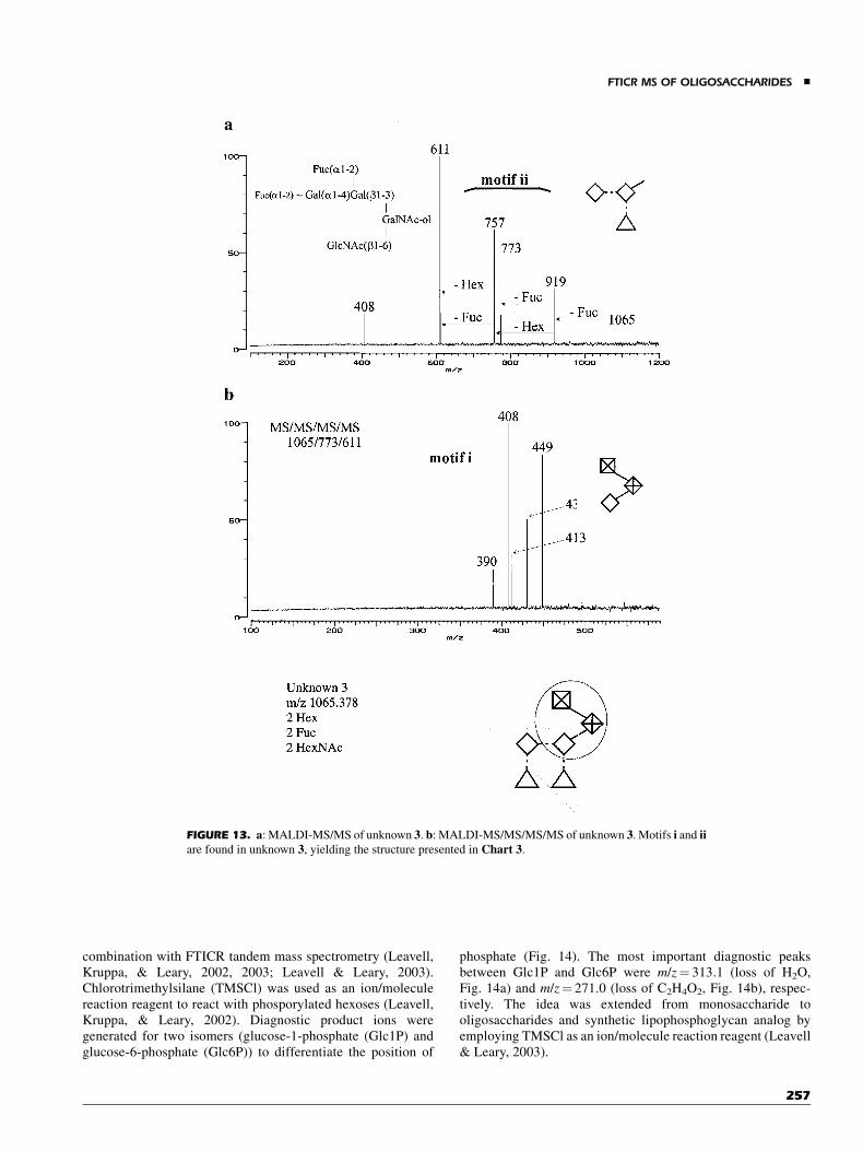
combination with FTICR tandem mass spectrometry (Leavell,Kruppa, & Leary, 2002, 2003; Leavell & Leary, 2003).Chlorotrimethylsilane (TMSCl) was used as an ion/moleculereaction reagent to react with phosporylated hexoses (Leavell,Kruppa, & Leary, 2002). Diagnostic product ions weregenerated for two isomers (glucose-1-phosphate (Glc1P) andglucose-6-phosphate (Glc6P)) to differentiate the position of
phosphate (Fig. 14). The most important diagnostic peaksbetween Glc1P and Glc6P were m/z¼ 313.1 (loss of H2O,Fig. 14a) and m/z¼ 271.0 (loss of C2H4O2, Fig. 14b), respec-tively. The idea was extended from monosaccharide tooligosaccharides and synthetic lipophosphoglycan analog byemploying TMSCl as an ion/molecule reaction reagent (Leavell& Leary, 2003).
FIGURE 13. a: MALDI-MS/MS of unknown 3. b: MALDI-MS/MS/MS/MS of unknown 3. Motifs i and iiare found in unknown 3, yielding the structure presented in Chart 3.
FTICR MS OF OLIGOSACCHARIDES &
257
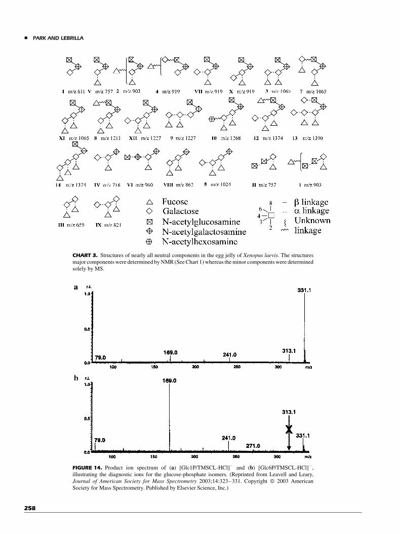
CHART 3. Structures of nearly all neutral components in the egg jelly of Xenopus laevis. The structures
major components were determined by NMR (See Chart 1) whereas the minor components were determined
solely by MS.
FIGURE 14. Product ion spectrum of (a) [Glc1P/TMSCL-HCl]� and (b) [Glc6P/TMSCL-HCl]�,
illustrating the diagnostic ions for the glucose-phosphate isomers. (Reprinted from Leavell and Leary,
Journal of American Society for Mass Spectrometry 2003;14:323–331. Copyright � 2003 American
Society for Mass Spectrometry. Published by Elsevier Science, Inc.)
& PARK AND LEBRILLA
258
VII. CONCLUSION
FTICR is highly suited for the structural elucidation of oligo-saccharides. Important features including high resolution, high-mass accuracy, and facile implementation of CID help toovercome many of the difficulties in analyzing this class of com-pounds. However, complete structural elucidation in the picomo-lar and even subpicomolar level may not be possible with massspectrometry alone. Several tools are also important includingNMR, chemical, and enzymatic methods. When used in com-bination with mass spectrometry, these methods will be able toelucidate structures in complex mixtures in amounts that arebiologically important. It is further clear that mass spectrometry,and specifically FTICR, will play a central role in the generalstructural analyses of these compounds.
REFERENCES
Afonso C, Fenselau C. 2003. Use of bioactive glass slides for matrix-assisted
laser desorption/ionization analysis: Application to microorganisms.
Anal Chem 75:694–697.
Alving K, Korner R, Paulsen H, Peter-Katalinic J. 1998. Nanospray-ESI low-
energy CID and MALDI post-source decay for determination of O-
glycosylation sites in MUC4 peptides. J Mass Spectrom 33:1124–1133.
An HJ, Lebrilla CB. 2001. Suppression of sialylated by sulfated oligosac-
charides in negative MALDI-FTMS. Israel J Chem 41:117–127.
Apweiler R, Hermjakob H, Sharron N. 1999. On the frequency of protein
glycosylation, as deduced from analysis of the SWISS-PROT database.
Biochim Biophys Acta 1473:4–8.
Aspinall GO. 1977. Selective degradation of carbohydrate polymers. Pure
Appl Chem 49:1105–1134.
BeMiller JN. 1965. Alkaline degradation of starch. In: Whistler RL, editor.
Starch: Chemistry and technology. New York: Academic Press. pp 521–
532.
Bencsura A, Vertes A. 1995. Dynamics of hydrogen bonding and energy trans-
fer in matrix-assisted laser desorption. Chem Phys Lett 247:142–148.
Bensimon M, Rapin J, Gaumann T. 1986. Comparison of infrared photo-
dissociation in a Fourier transform mass spectrometer with metastable
ion decay in a double-focusing mass spectrometer. Int J Mass Spectrom
Ion Proc 72:125–135.
Bertozzi CR, Kiessling LL. 2001. Chemical glycobiology. Science
291:2357–2364.
Beu SC, Laude DA. 1992. Open trapped ion cell geometries for Fourier
transform ion cyclotron resonance mass spectrometry. Int J Mass
Spectrom Ion Proc 112:215–230.
Bradford WG, Jeffrey JE, Constance MJ, Wade H, Arnold MF. 1997.
Characterization of bacterial lipooligosaccharides by delayed extrac-
tion matrix-assisted laser desorption ionization time-of-flight mass
spectrometry. J Am Soc Mass Spetrom 8:645–658.
Brockhausen I. 1999. Pathways of O-glycan biosynthesis in cancer cells.
Biochim Biophys Acta 1473:67–95.
Brockman AH, Orlando R. 1995. Probe-immobilized affinity chromatogra-
phy/mass spectrometry. Anal Chem 67:4581–4585.
Buchanan MV. 1987. Fourier Transform Mass Spectrometry: Evolution, Inno-
vation, and Applications. ACS Symposium Series 359.
Bundy J, Fenselau C. 1999. Lectin-based affinity capture for MALDI-MS
analysis of bacteria. Anal Chem 71:1460–1463.
Bundy JL, Fenselau C. 2001. Lectin and carbohydrate affinity capture
surfaces for mass spectrometric analysis of microorganisms. Anal
Chem 73:751–757.
Cancilla MT, Penn SG, Lebrilla CB. 1998. Alkaline degradation of oligo-
saccharides coupled with matrix-assisted laser desorption/ionization
Fourier transform mass spectrometry: A new method for sequencing
oligosaccharides. Anal Chem 70:663–672.
Cancilla MT, Penn SG, Carroll JA, Lebrilla CB. 1996. Coordination of alkali
metals to oligosaccharides dictates fragmentation behavior in matrix-
assited laser desorption/ionization Fourier transform mass spectro-
metry. J Am Chem Soc 118:6736–6745.
Cancilla MT, Wong AW, Voss LR, Lebrilla CB. 1999. Fragmentation
reactions in the mass spectrometry of neutral oligosaccharides. Anal
Chem 71:3206–3218.
Cancilla MT, Gaucher SP, Desaire H, Leary JA. 2000. Combined partial acid
hydrolysis and electrospray ionization—Mass spectrometry for the
structural determination of oligosaccharides. Anal Chem 72:2901–
2907.
Caravatti P, Allemann M. 1991. The infinity cell: A new trapped-ion cell with
radiofrequency covered trapping electrodes for Fourier transform ion
cyclotron resonance mass spectrometry. Org Mass Spectrom 26:514–
518.
Carroll JA, Willard D, Lebrilla CB. 1995. Energetics of cross-ring cleavages
and their relevance to the linkage determination of oligosaccharides.
Anal Chim Acta 307:431–447.
Carroll JA, Lebrilla CB. 1992. Fragmentation behavior of disaccharides
during desorption-ionization. Org Mass Spectrom 27:639–643.
Carroll JA, Ngoka L, McCullough S, Gard E, Jones AD, Lebrilla CB. 1991.
Quadrupole Fourier transform mass spectrometry of oligosaccharides.
Anal Chem 63:2526–2529.
Carroll J, Lebrilla CB, Ngoka L, Beggs CG. 1993. Liquid secondary ion mass
spectrometry/Fourier transform mass spectrometry of oligosaccharide
anions. Anal Chem 65:1582–1587.
Carroll JA, Penn SG, Fannin ST, Wu J, Cancilla MT, Green MK, Lebrilla CB.
1996. A dual vacuum chamber Fourier transform mass spectrometer
with rapidly interchangeable LSIMS, MALDI, and ESI sources: Initial
results with LSIMS and MALDI. Anal Chem 68:1798–1804.
Castoro JA, Koester C, Wilkins C. 1992. Matrix-assisted laser desorption/
ionization of high-mass molecules by Fourier-transform mass spectro-
metry. Rapid Commun Mass Spectrom 6:239–241.
Castoro JA, Wilkins CL. 1993. Ultrahigh resolution matrix-assisted laser
desorption ionization of small proteins by Fourier transform mass
spectrometry. Anal Chem 65:2621–2627.
Chen P, Baker AG, Novotny MV. 1997. The use of osazones as matrices for the
matrix-assisted laser desorption/ionization mass spectrometry of
carbohydrates. Anal Biochem 244:144–151.
Chen R, Cheng X, Mitchell DW, Hofstadler SA, Wu Q, Rockwood AL,
Sherman MG, Smith RD. 1995. Trapping, detection, and mass
determination of coliphage T4 DNA ions by electrospray ionization
Fourier transform ion cyclotron resonance mass spectrometry. Anal
Chem 67:1159–1163.
Cheng X, Chen R, Bruce JE, Schwartz BL, Anderson GA, Hofstadler SA,
Gale DC, Smith RD, Gao J, Sigal GB, Mammen M, Whitesides GM.
1995. Using electrospray ionization FTICR mass spectrometry to study
competitive bindng of inhibitors to carbonic anhydrase. JAm Chem Soc
117:8859–8860.
Chowdhury SK, Katta V, Chait BT. 1990. An electrospray-ionization mass
spectrometer with new features. Rapid Commun Mass Spectrom 4:
81–87.
Coates ML, Wilkins CL. 1985. Laser desorption Fourier transform mass
spectrometry of malto-oligosaccharides. Biomed Mass Spectrom 12:
424–428.
Cody RB, Burnier RC, Freiser BS. 1982. Collision-induced dissociation with
Fourier transform mass spectrometry. Anal Chem 54:96–101.
Cody RB, Freiser BS. 1982. Collision-induced dissociation in a Fourier-
transform mass spectrometer. Int J Mass Spectrom Ion Phys 41:199–
204.
FTICR MS OF OLIGOSACCHARIDES &
259
Cody RB, Burnier RC, Cassady CJ, Freiser BS. 1982. Consecutive collision-
induced dissociations in Fourier transform mass spectrometry. Anal
Chem 54:2225–2228.
Comisarow MB. 1981. Cubic trapped-ion cell for ion cyclotron resonance. Int
J Mass Spectrom Ion Phys 37:251–257.
Comisarow MB, Marshall AG. 1974a. Fourier transform ion cyclotron
resonance spectroscopy. Chem Phys Lett 25:282–283.
Comisarow MB, Marshall AG. 1974b. Frequency sweep Fourier transform
ion cyclotron resonance spectroscopy. Chem Phys Lett 26:489–
490.
Dell A. 1987. FAB-mass spectrometry of carbohydrates. Adv Carbohydr
Chem Biochem 45:19–72.
Demirev PA, Ramirez J, Fenselau C. 2001. Tandem mass spectrometry of
intact proteins for characterization of biomarkers from Bacillus cereus
T spores. Anal Chem 73.
Demirev P, Olthoff JK, Fenselau C, Cotter RJ. 1987. High-mass ion
fragmentation as a function of time and mass. Anal Chem 59:1951–
1954.
Desaire H, Sirich TL, Leary JA. 2001. Evidence of block and randomly
sequenced chondroitin polysaccharides: Sequential enzymatic diges-
tion and quantification using ion trap tandem mass spectrometry. Anal
Chem 73:3513–3520.
Dienes T, Pastor SJ, Schurch S, Scott JR, Yao J, Cui S, Wilkins CL. 1996.
Fourier transform mass spectrometry—Advancing years (1992–mid
1996). Mass Spectrom Rev 15:163–211.
Dove A. 2001. The bittersweet promise of glycobiology. Nat Biotechnol
19:913–917.
Duffin KL, Welply JK, Huang E, Henion JD. 1992. Characterization of N-
linked oligosaccharides by electrospray and tandem mass spectrometry.
Anal Chem 64:1440–1448.
Dufresne CP, Wood TD, Hendrickson CL. 1998. High-resolution electrospray
ionization Fourier transform mass spectrometry with infrared multi-
photon dissociation of glucokinase from Bacillus stearothermophilus.
J Am Soc Mass Spectrom 9:1222–1225.
Dunbar RC. 1984. Photodissociation in the ICR ion trap. In: Bowers MT,
editor. Gas phase ion chemistry. Orlando: Academic Press. pp 129–
166.
Dunbar RC, McMahon TB, Tholmann D, Tonner DS, Salahub DR, Wei D.
1995. Zero-pressure thermal-radiation-induced dissociation of gas-
phase cluster ions: Comparison of theory and experiment for (H2O)2Cl�
and (H2O)3Cl�. J Am Chem Soc 117:12819–12825.
Ehring H, Sundqvist BUR. 1995. Studies of the MALDI process by
luminescence spectroscopy. J Mass Spectrom 30:1303–1310.
Fenn JB, Mann M, Meng CK, Wong SF, Whitehouse CM. 1989. Electrospray
ionization for mass spectrometry of large biomolecules. Science 246:
64–71.
Fenselau C, Demirev PA. 2001. Characterization of intact microorganisms by
MALDI mass spectrometry. Mass Spectrom Rev 20:157–171.
Flora JW, Muddiman DC. 2001. Selective, sensitive, and rapid phosphopep-
tide identification in enzymatic digests using ESI-FTICR-MS with
infrared multiphoton dissociation. Anal Chem 73:3305–3311.
Franz AH, Molinski TF, Lebrilla CB. 2001. MALDI-FTMS characterization
of oligosaccharides labeled with 9-aminofluorene. J Am Soc Mass
Spetrom 12:1254–1261.
Franz AH, Lebrilla CB. 2002. Evidence for long-range glycosyl transfer
reactions in the gas phase. J Am Soc Mass Spectrom 13:325–337.
Fridriksson EK, Beavil A, Holowka D, Gould HJ, Baird B, McLafferty FW.
2000. Heterogeneous glycosylation of immunoglobulin E constructs
characterized by top-down high-resolution 2-D mass spectrometry.
Biochem 39:3369–3376.
Fura A, Leary JA. 1993. Differentiation of Ca2þ and Mg2þ coordinated
branched trisaccharide isomers: An electrospray ionization and tandem
mass spectrometry study. Anal Chem 65:2805–2811.
Gard EE, Green MK, Warren H, Camara EJO, He F, Penn SG, Lebrilla CB.
1996. A dual vacuum chamber Fourier transform mass spectrometer
with rapidly interchangeable FAB, MALDI, and ESI Sources:
Electrospray results. Int J Mass Spectrom Ion Proc 157/158:115–127.
Garozzo D, Giuffrida M, Impallomeni G, Ballistreri A, Montaudo G. 1990.
Determination of linkage position and identification of the reducing end
in linear oligosaccharides by negative ion fast atom bombardment mass
spectrometry. Anal Chem 62:279–286.
Gaucher SP, Cancilla MT, Phillips NJ, Gibson BW, Leary JA. 2000. Mass
spectral characterization of lipooligosaccharides from Haemophilus
influenza 2019. Biochem 39:12406–12414.
Gauthier JW, Trautman TR, Jacobson DB. 1991. Sustained off-resonance irra-
diation for collision-activated dissociation involving Fourier transform
mass spectrometry. Collision-activateddissociation technique thatemula-
tes infrared multiphoton dissociation. Anal Chim Acta 246:211–225.
Gibson BW, Melaugh W, Phillips NJ, Apicella MA, Campagnari AA, Griffiss
JM. 1993. Investigation of the structural heterogeneity of lipooligo-
saccharides from pathogenicHaemophilus andNeisseria species and of
the R-type lipooligosaccharides from Salmonella typhimurium by
electrospray mass spectrometry. J Bacteriol 175:2702–2712.
Grese RP, Cerny RL, Gross ML. 1989. Metal ion-peptide interactions in the
gas phase: A tandem mass spectrometry study of alkali metal cationized
peptides. J Am Chem Soc 111:2835–2842.
Grosshans PB, Marshall AG. 1990. Theory of ion cyclotron resonance mass
spectrometry: Resonant excitation and radial ejection in orthorhombic
and cylindrical ion traps. Int J Mass Spectrom Ion Proc 100:347–
379.
Guan S, Marshall AG. 1996. Stored waveform inverse Fourier transform
(SWIFT) ion excitation in trapped-ion mass spectrometry: Theory and
applications. Int J Mass Spectrom Ion Proc 157/158:5–37.
Hakansson K, Cooper HJ, Emmett MR, Costello CE, Marshall AG, Nilsson
CL. 2001. Electron capture dissociation and infrared multiphoton
dissociation MS/MS of an N-glycosylated tryptic peptide to yield
complementary sequence information. Anal Chem 73:4530–4536.
Hamai A, Hashmoto N, Mochizuki H, Kato F, Makiguchi Y, Horie K, Suzuki
S. 1997. Two distinct chondroitin sulfate ABC lyases. An endoelimi-
nase yielding tetrasaccharides and an exoeliminase preferentially acting
on oligosaccharides. J Biol Chem 272:9123–9130.
Harvey DJ. 1993. Quantitative aspects of the matrix-assisted laser desorption
mass spectrometry of complex oligosaccharides. Rapid Commun Mass
Spectrom 7:614–619.
Harvey DJ. 1999. Matrix-assisted laser desorption/ionization mass spectro-
metry of carbohydrates. Mass Spectrom Rev 18:349–450.
Henry KD, Quinn JP, McLafferty FW. 1991. High-resolution electrospray
mass spectra of large molecules. J Am Chem Soc 113:5447–5449.
Hiyama K. 1976. Action of chondroitinases II. Numerical calculation of the
degree of multiple attack. J Biochem 80:1209–1214.
Hiyama K, Okada S. 1976. Action of chondroitinases I. The mode of action of
two chondroitinase-AC preparations of different origin. J Biochem
80:1201–1207.
Ho Y-P, Fenselau C. 2000. Metastable dacay of peptide ions on a Fourier
transform mass spectrometer equipped with an external ion source.
J Mass Spectrom 35:183–188.
Hofstadler SA, Sannes-Lowery KA, Griffey RH. 1999. Infrared multipho-
ton dissociation in an external ion reservoir. Anal Chem 71:2067–
2070.
Huddleston MJ, Bean MF, Carr SA. 1993. Collisional fragmentation of
glycopeptides by electrospray ionization LC/MS and LC/MS/MS:
Methods for selective detection of glycopeptides in protein digests.
Anal Chem 65:877–884.
Humphries MJ, Matsumoto K, White SL, Olden K. 1986. Oligosaccharide
modification by Swainsonine treatment inhibits pulmonary coloniza-
tion by B16-F10 murine melanoma cells. Proc Natl Acad Sci USA
83:1752–1756.
& PARK AND LEBRILLA
260
Hunt DF, Shabanowitz J, Yates JR, McIver RT, Hunter RL. 1985. Tandem-
quadrupole Fourier transform mass spectrometry of oligopeptides. Anal
Chem 57:2728–2733.
Hutchens TW, Yip TT. 1993. New desorption strategies for the mass-
spectrometric analysis of macromolecules. Rapid Commun Mass
Spectrom 7:576–580.
Jardine I, Shirley W, Brennan PJ, McNeal CJ, Macfarlane RD. 1986.
Heterogeniety of bacterial antigenic lipooligosaccharides determined
by californium-252 plasma desorption mass spectrometry. Biomed
Environ Mass Spectrom 13:273–276.
Jardine I, Scanlan G, McNeil M, Brennan PJ, Patrick J. 1989. Plasma
desorption mass spectrometric analysis of mycobacterial glycolipids.
Anal Chem 61:416–422.
Johnson PJ, Poon TCW, Hjelm NM, Ho CS, Ho SKW, Welby C, Stevenson D,
Patel T, Parekh R, Townsend RR. 1999. Glycan composition of serum
alpha-fetoprotein in patients with hepatocellular carcinoma and non-
seminomatous germ cell tumour. Brit J Cancer 81:1188–1195.
Karas M, Hillenkamp F. 1988. Laser desorption ionization of proteins with
molecular masses exceeding 10,000 daltons. Anal Chem 60:2299–
2301.
Karas M, Ehring H, Nordhoff E, Stahl B, Strupat K, Hillenkamp F, Grehl M,
Krebs B. 1993. Matrix-assisted laser desorption/ionization mass
spectrometry with additives to 2,5-dihydroxybenzoic acid. Org Mass
Spectrom 28:1476–1481.
Karas M, Bahr U, Strupat K, Hillenkamp F, Tsabopoulos A, Pramanik BN.
1995. Matrix dependence of metastable fragmentation of glycoproteins
in MALDI TOF mass spectrometry. Anal Chem 67:675–679.
Katritzky AR, Malhotra N, Ramanathan R, Kemerait RC, Jr., Zimmerman JA,
Eyler JR. 1992. Measurement of gas-phase binding energies of crown
ethers with metal ions by Fourier-transform ion cyclotron resonance
mass spectrometry. Rapid Commun Mass Spectrom 6:25–27.
Kebarle P, Tang L. 1993. From ions in solution to ions in the gas phase. Anal
Chem 65:972A–986A.
Kelleher NL, Senko MW, Little DP, O’Connor PB, McLafferty FW. 1995.
Complete large-molelcule high-resolution mass spectra from 50-
femtomole microvolume injection. J Am Soc Mass Spectrom 6:220–
221.
Kelly J, Masoud H, Perry MB, Richards JC, Thibault P. 1996. Separation and
characterization of O-deacylated lipooligosaccharides and glycans
derived from Moraxella catarrhalis using capillary electrophoresis-
electrospray mass spectrometry and tandem mass spectrometry. Anal
Biochem 233:15–30.
Kennedy JF, White CA. 1979. Polysaccharides. In: Barton D, Ollis WD,
editors. Comprehensive organic chemistry. New York: Pergamon Press.
pp 755–815.
Knirel YA, Grosskurth H, Helbig JH, Zahringer U. 1995. Structures of
decasaccharide and tridecasaccharide tetraphosphates isolated by strong
alkaline degradation of O-deacylated lipopolysaccharide of pseudomonas
flourescens strain ATCC 49271. Carbohydr Res 279:215–226.
Koeller KM, Wong C-H. 2000. Emerging themes in medicinal glycoscience.
Nat Biotechnol 18:835–841.
Kofel P, Allemann M, Kellerhals H, Wanczek KP. 1986. Time-of-flight ICR
spectrometry. Int J Mass Spectrom Ion Proc 72:53–61.
Kohler M, Leary JA. 1995. LC/MS/MS of carbohydrates with postcolumn
addition of metal chlorides using a triaxial electrospray probe. Anal
Chem 67:3501–3508.
Kolli VSK, Orlando R. 1996. A new matrix for matrix-assisted laser
desorption/ionization on magnetic sector instruments with point detec-
tors. Rapid Commun Mass Spectrom 10:923–926.
Koster C, Castoro JA, Wilkins CL. 1992. High-resolution matrix-assisted
laser desorption/ionization of biomolecules by Fourier transform mass
spectrometry. J Am Chem Soc 114:7572–7574.
Krause J, Stoeckli M, Schlunegger UP. 1996. Studies on the selection of new
matrices for ultraviolet matrix-assited laser desorption/ionization time-
of-flight mass spectrometry. Rapid Commun Mass Spectrom 10:1927–
1933.
Leavell MD, Kruppa GH, Leary JA. 2002. Analysis of phosphate position in
hexose monosaccharides using ion-molecule reactions and SORI-CID
on an FT-ICR mass spectrometer. Anal Chem 74:2608–2611.
Leavell MD, Kruppa GH, Leary JA. 2003. Determination of phosphate
position in hexose monosaccharides using FTICR mass spectrometer:
Ion/molecule reactions, labeling studies, and dissociation mechanisms.
Int J Mass Spectrom 222:135–153.
Leavell MD, Leary JA, Yamasaki R. 2002. Mass spectrometric strategy for the
characterization of lipooligosaccharides from Neisseria gonorrhoeae
302 using FTICR. J Am Soc Mass Spectrom 13:571–576.
Leavell MD, Leary JA. 2003. Probing isomeric differences of phosphorylated
carbohydrates through the use of ion/molecule reactions and FT-ICR
MS. J Am Soc Mass Spectrom 14:323–331.
Lebrilla CB, Amster IJ, McIver RT, Jr. 1989. External ion source FTMS
instrument for analysis of high mass ions. Int J Mass Spectrom Ion Proc
87:R7–R13.
Ledford EB, Jr., Rempel DL, Gross ML. 1984. Space charge effects in Fourier
transform mass spectrometry. Mass calibration. Anal Chem 56:2744–
2748.
Lehmann E, Knochenmuss R, Zenobi R. 1997. Ionization mechanisms in
matrix-assisted laser desorption/ionization mass spectrometry: Con-
tribution of pre-formed ions. Rapid Commun Mass Spectrom 11:1483–
1492.
Lemoine J, Chirat F, Domon B. 1996. Structural analysis of derivatized oligo-
saccharides using post-source decay matrix-assisted laser desorption/
ionization mass spectrometry. J Mass Spectrom 31:908–912.
Li YZ, McIver RT, Jr., Hunter RL. 1994. High-accuracy molecular mass
determination for peptides and proteins by Fourier transform mass
spectrometry. Anal Chem 66:2077–2083.
Liao P-C, Allison J. 1995. Enhanced detection of peptides in matrix-assisted
laser desorption/ionization mass spectrometry through the use of
charge-localized derivatives. J Mass Spectrom 30:511–512.
Linhardt RJ, Galliher PM, Cooney CL. 1986. Polysaccharide lyases. Appl
Biochem Biotech 12:135–176.
Lisowska E. 2002. The role of glycosylation in protein antigenic properties.
Cell Mol Life Sci 59:445–455.
Little DP, McLafferty FW. 1995. Sequencing 50-mer DNAs using electro-
spray tandem mass spectrometry and complememtary fragmentation
methods. J Am Chem Soc 117:6783–6784.
Little DP, McLafferty FW. 1996. Infrared photodissociation of non-covalent
adducts of electrosprayed nucleotide ions. J Am Soc Mass Spectrom
7:209–210.
Little DP, Speir JP, Senko MW, O’Connor PW, McLafferty FW. 1994.
Infrared multiphoton dissociation of large multiply-charged ions for
biomolecule sequencing. Anal Chem 66:2809–2815.
Little DP, Aaserud DJ, Valaskovic GA, McLafferty FW. 1996. Sequence
information from 42-108-mer DNAs (complete for a 50-mer) by tandem
mass spectrometry. J Am Chem Soc 118:9352–9359.
Liu J, Tseng K, Carcia B, Lebrilla CB, Mukerjee E, Collins S, Smith R. 2001.
Electrophoresis separation in open microchannels. A method for
coupling electrophoresis with MALDI-MS. Anal Chem 73:2147–2151.
Lohse DL, Linhardt RJ. 1992. Purification and characterization of heparin
lyases from Flavobacterium heparinum. J Biol Chem 267:24347–
24355.
Maeder T. 2002. Sweet medicines. Sci Am 287:40–47.
Marshall AG, Hendrickson CL, Jackson GS. 1998. Fourier transform ion
cyclotron resonance mass spectrometry: A primer. Mass Spectrom Rev
17:1–35.
Marshall AG, Schweikhard L. 1992. Fourier transform ion cyclotron
resonance mass spectrometry: Technique developements. Int J Mass
Spectrom Ion Proc 118/119:37–70.
FTICR MS OF OLIGOSACCHARIDES &
261
McAuliffe JC, Hindsgaul O. 1997. Carbohydrate drugs-an ongoing challenge.
Chem Ind 5:170–174.
McClellan JE, Costello CE, O’Connor PB, Zaia J. 2002. Influence of charge
state on product ion mass spectra and the determination of 4S/6S
sulfation sequence of chondroitin sulfate oligosaccharides. Anal Chem
74:3760–3771.
McCullough SM, Gard E, Lebrilla CB. 1991. A 3T external source
quadrupole Fourier transform mass spectrometer for ion/molecule
reactions and analysis. Int J Mass Spectrom Ion Proc 107:91–102.
McIver RT, Jr., Hunter RL, Bowers WD. 1985. Coupling a quadrupole mass
spectrometer and Fourier transform mass spectrometer. Int J Mass
Spectrom Ion Proc 64:67–77.
McIver RT, Jr., Li YZ, Hunter RL. 1994. High-resolution laser desorption
mass spectrometry of peptides and small proteins. Proc Natl Acad Sci
USA 91:4801–4805.
McLafferty FW. 1994. High resolution tandem FTMS above 10KDa. Acc
Chem Res 27:379–386.
Mirgorodskaya E, Roepstorff P, Zubarev RA. 1999. Localization of O-
glycosylation sites in peptides by electron capture dissociation in a
Fourier transform mass spectrometer. Anal Chem 71:4431–4436.
Mock KK, Davey M, Cottrell JS. 1991. The analysis of underivatized
oligosaccharides by matrix-assisted laser desorption mass spectro-
metry. Biochem Biophys Res Commun 177:644–651.
Mohr MD, Boernsen KO, Widmer HM. 1995. Matrix-assisted laser desorp-
tion ionization mass spectrometry: Improved matrix for oligosacchar-
ides. Rapid Commun Mass Spectrom 9:809–814.
Montreuil J. 1980. Primary structure of glycoprotein glycans: Basis for the
molecular biology of glycoprotein. Adv Carbohydr Chem Biochem
37:157–223.
Morgenthaler LN, Eyler JR. 1979. Laser-induced photodissociation of
chloroethane molecular ions in the gas phase. J Chem Phys 71:1486–
1491.
Muhlecker W, Gulati S, McQuillen DP, Ram S, Rice PA, Reinhold VN. 1999.
An essential saccharide binding domain for the mAb 2C7 established
for Neisseria gonorrhoeae LOS by ES-MS and MSn. Glycobiology
9:157–171.
Naven TJP, Harvey DJ. 1996. Cationic derivatization of oligosaccharides with
Girard’s T ragent for improved performance in matrix-assisted laser
desorption/ionization and electrospray mass spectrometry. Rapid
Commun Mass Spectrom 10:829–834.
Naven TJP, Harvey DJ, Brown J, Critchley G. 1997. Fragmentation of
complex carbohydrates following ionization by matrix-assisted laser
desorption with an instrument fitted with time-lag focusing. Rapid
Commun Mass Spectrom 11:1681–1686.
Nelson RW, Krone JR, Bieber AL, Williams P. 1995. Mass spectrometry
immunoassay. Anal Chem 67:1153–1158.
Ngoka LC, Gal JF, Lebrilla CB. 1994. Effects of cations and charge types on
the metastable decay rates of oligosaccharides. Anal Chem 66:692–
698.
Ngoka L, Lebrilla CB. 1993. ‘‘Slow’’ metastable decomposition of oligo-
saccharide cations produced in an external source Fourier tranform
mass spectrometer. J Am Soc Mass Spectrom 4:210–215.
Nonami H, Fukui S, Erra-Balsells R. 1997. b-Carboline alkaloids as matrices
for matrix-assisted ultraviolet laser desorption time-of-flight mass
spectrometry of proteins and sulfated oligosacchairdes: A comparative
study using phenylcarbonyl compounds, carbazoles, and classical
matrices. J Mass Spectrom 32:287–296.
O’Connor PB, Mirgorodskaya E, Costello CE. 2002. High pressure matrix-
assisted laser desorption/ionization Fourier transform mass spectro-
metry for minimization of ganglioside fragmentation. J Am Soc Mass
Spectrom 13:402–407.
O’Connor PB, Costello CE. 2001. A high pressure matrix-assisted laser
desorption/ionization Fourier transform mass spectrometry ion source
for thermal stabilization of labile biomolecules. Rapid Commun Mass
Spectrom 15:1862–1868.
Ogier-Denis E, Bauvy C, Aubery M, Codogno P, Sapin C, Rousset M,
Zweibaum A, Trugnan G. 1989. Processing of asparigine-linked
oligosaccharides is an early biochemical marker of the enterocytic
differentiation of HT-29 cells. J Cell Biochem 41:13–23.
Penn SG, Cancilla MT, Lebrilla CB. 1996. Collision-induced dissociation of
branched oligosaccharide ions with analysis and calculation of relative
dissociation thresholds. Anal Chem 68:2331–2339.
Penn SG, Cancilla MT, Lebrilla CB. 2000. Fragmentation behavior of
multiple-metal-coordinated acidic oligosaccharides studied by matrix-
assisted laser desorption ionization Fourier transform mass spectro-
metry. Int J Mass Spectrom 195/196:259–269.
Penn SG, Cancilla MT, Green MK, Lebrilla CB. 1997. Direct comparison of
matrix-assisted laser desorption/ionization and electrospray ionization
in the analysis of gangliosides by Fourier transform mass spectrometry.
Eur Mass Spectrom 3:67–79.
Pitt JJ, Gorman JJ. 1997. Oligosaccharide characterization and quantitation
using 1-phenyl-3-methyl-5-pyrazolone derivatization and matrix-
assisted laser desorption/ionization time-of-flight mass spectrometry.
Anal Biochem 248:63–75.
Powell AK, Harvey DJ. 1996. Stabilization of sialic acids in N-linked
oligosaccharides and gangliosides for analysis by positive ion matrix-
assisted laser desorption/ionization mass spectrometry. Rapid Commun
Mass Spectrom 10:1027–1032.
Price WD, Schnier PD, Williams ER. 1996. Tandem mass spectrometry of
large biomolecule ions by blackbody infrared radiative dissociation.
Anal Chem 68:859–866.
Prime S, Dearnley J, Ventom AM, Parekh RB, Edge CJ. 1996. Oligosacchar-
ide sequencing based on exo- and endoglycosidase digestion and liquid
chromatographic analysis of the products. J Chromatogr A 720:263–
274.
Reinhold VN, Reinhold BB, Costello CE. 1995. Carbohydrate molecular
weight profiling, sequence, linkage, and branching data: ES-MS and
CID. Anal Chem 67:1772–1784.
Ritchie GE, Moffatt BE, Sim RB, Morgan BP, Dwek RA, Rudd PM. 2002.
Glycosylation and the complement system. Chem Rev 102:305–
319.
Rudd PM, Dwek RA. 1997. Glycosylation: Heterogeniety and the 3D
structure of proteins. Crit Rev Biochem Mol Biol 32:1–100.
Saad O, Leary JA. 2003. Compositional analysis and quantification of heparin
and heparan sulfate by electrospray ionization ion trap tandem mass
spectrometry. 51st American Society for Mass Spectrometry.
Savage MD, Mattson G, Desai S, Mielander GW, Morgensen S, Conklin
EJ. 1992. Avidin-Biotin Chemistry: A handbook. Rockford, IL:
Pierce.
Schmell ED, Gulyas BJ, Hedrick JL. 1983. Egg surface changes during
fertilization and the molecular mechanism of the block to polyspermy.
Mechanism and control of animal fertilization. New York: Academic
Press. pp 365–413.
Schriemer DS, Li L. 1996. Combining avidin-biotin chemistry with matrix-
assisted laser desorption/ionization mass spectrometry. Anal Chem
68:3382–3387.
Schweikhard L, Marshall AG. 1993. Excitation modes for Fourier transform-
ion cyclotron resonance mass spectrometry. J Am Soc Mass Spectrom
4:433–452.
Shi SD-H, Hendrickson CL, Marshall AG, Siegel MM, Kong F, Carter GT.
1999. Structural validation of saccharomicins by high resolution and
high mass accuracy Fourier transform-ion cyclotron resonance-mass
spectrometry and infrared multiphoton dissociation tandem mass
spectrometry. J Am Soc Mass Spetrom 10:1285–1290.
Spengler B, Dolce JW, Cotter RJ. 1990. Infrared laser desorption mass
spectrometry of oligosaccharides: Fragmentation mechanisms and
isomer analysis. Anal Chem 62:1731–1737.
& PARK AND LEBRILLA
262

Spengler B, Kirsch D, Kaufmann R. 1991. Metastable decay of peptides and
proteins in matrix-assisted laser-desorption mass spectrometry. Rapid
Commun Mass Spectrom 5:198–202.
Spengler B, Kirsch D, Kaufman R, Lemoine J. 1994. Structure analysis of
branched oligosacchiardes using post-source decay in matrix-assisted
laser desorption ionization mass spectrometry. Org Mass Spectrom
29:782–787.
Stahl B, Steup M, Karas M, Hillenkamp F. 1991. Analysis of neutral
oligosaccharides by matrix-assisted laser desoprtion/ionization mass
spectrometry. Anal Chem 63:1463–1466.
Strecker G, Wieruszeski JM, Plancke Y, Boilly B. 1995. Primary structure of
12 neutral oligosaccharide-alditols released from the jelly coats of the
anuran Xenopus laevis by reductive beta-elimination. Glycobiology
5:137–146.
Strupat K, Karas M, Hillenkamp F. 1991. 2,5-Dihydroxybenzoic acid: A new
matrix for laser desorption/ionization mass spectrometry. Int J Mass
Spectrom Ion Proc 111:89–102.
Takao T, Tambara Y, Nakamura A, Yoshino K, Fukuda H, Fukuda M,
Shimonishi Y. 1996. Sensitive analysis of oligosaccharides derivatized
with 4-aminobenzoic acid 2-(diethylamino)ethyl ester by matrix-
assisted laser desorption/ionization mass spectrometry. Rapid Commun
Mass Spectrom 10:637–640.
Thorne GC, Ballard KD, Gaskell SJ. 1990. Metastable decomposition of
peptide [MþH]þ ions via rearrangement involving loss of C-terminal
amino acid residue. J Am Soc Mass Spectrom 1:249–257.
Thorne LR, Beauchamp JL. 1984. Infrared photochemistry of gas phase ions.
In: Bowers MT, editor. Gas phase ion chemistry. Orlando: Academic
Press. pp 41–97.
Tseng K, Hedrick JL, Lebrilla CB. 1999. The catalog-library approach for the
rapid and sensitive structural elucidation of oligosaccharides. Anal
Chem 71:3747–3754.
Tseng K, Lindsay LL, Penn S, Hedrick JL, Lebrilla CB. 1997. Characteriza-
tion of neutral oligosaccharide-alditols from Xenopus laevis egg jelly
coats by matrix assisted laser desorption Fourier transform mass
spectrometry. Anal Biochem 250:18–28.
Tseng K, Wang H, Lebrilla CB, Bonnell B, Hedrick J. 2001. Identification and
structural elucidation of lectin-binding oligosaccharides by bio-affinity
matrix-assisted laser desorption/ionization Fourier transform mass
spectrometry. Anal Chem 73:3556–3561.
Varki A. 1993. Biological roles of oligosaccharides: All of the theories are
correct. Glycobiology 3:97–130.
Viseux N, Hoffmann Ed, Domon B. 1997. Structural analysis of perme-
thylated oligosaccharides by electrospray tandem mass spectrometry.
Anal Chem 69:3193–3198.
Wanczek KP. 1984. Ion cyclotron resonance spectrometry—A review. Int J
Mass Spectrom Ion Proc 60:11–60.
Wang H, Tseng K, Lebrilla CB. 1999. A general method for producing
bioaffinity MALDI probes. Anal Chem 71:2014–2020.
Wang BH, Dreisewerd K, Bahr U, Karas M, Hillenkamp F. 1993. Gas-phase
cationization and protonation of neutrals generated by maxtrix-assisted
laser desorption. J Am Soc Mass Spectrom 4:393–398.
Whistler RL, BeMiller JN. 1958. Alkaline degradation of polysaccharides. In:
Wolfrom ML, editor. Advances in carbohydrate chemistry. New York:
Academic Press. pp 289–329.
White CA, Kennedy JF. 1988. Identification and structural analysis of
monomeric and polymeric carbohydrates. In: Kennedy JF, editor.
Carbohydrate chemistry. Oxford: Clarendon Press. pp 42–72.
Wilkins CL, Chowdhury AK, Nuwaysir LM, Coates ML. 1989. Fourier trans-
form mass spectrometry: Current status. Mass Spectrom Rev 8:67–92.
Williams ER, Furlong JJP, McLafferty FW. 1990. Efficiency of collisionally-
activated dissociation and 193-nm photodissociation of peptide ions in
Fourier-transform mass spectrometry. JAm Soc Mass Spectrom 1:288–
294.
Williams BH, Hathout Y, Fenselau C. 2002. Structural characterization of
lipopeptide biomarkers isolated from Bacillus globigii. J Mass
Spectrom 37:259–264.
Wong AW, Wang H, Lebrilla CB. 2000. Selection of anionic dopant for
quantifying desialylation reactions with MALDI-FTMS. Anal Chem
72:1419–1425.
Wong AW, Cancilla MT, Voss LR, Lebrilla CB. 1999. Anion dopant for
oligosaccharides in matrix-assisted laser desorption/ionization mass
spectrometry. Anal Chem 71:205–211.
Worrall TA, Cotter RJ, Woods AS. 1998. Purification of contaminated
peptides and proteins on synthetic membrane surfaces for matrix-
assisted laser desorption/ionization mass spectrometry. Anal Chem
70:750–756.
Wu J, Fannin ST, Franklin MA, Molinski TF, Lebrilla CB. 1995. Exact mass
determinination for elemental analysis of ions produced by matrix-
assisted laser desorption. Anal Chem 67:3788–3792.
Xie Y, Schubothe KM, Lebrilla CB. 2003. Infrared laser isolation of
ions in Fourier transform mass spectrometry. Anal Chem 75:160–164.
Xie Y, Tseng K, Lebrilla CB, Hedrick JL. 2001. Targeted use of
exoglycosidase digestion for the structural elucidation of neutral O-
linked oligosaccharides. J Am Soc Mass Spetrom 12:877–884.
Yamashita M, Fenn JB. 1984. Electrospray ion source. Another variation on
the free-jet theme. J Phys Chem 88:4451–4459.
Yang JC, Hedrick JL. 1997. cDNA cloning and sequence analysis of the
Xenopus laevis egg envelop glycoprotein gp43. Dev Growth Differ
38:457–467.
Yang BY, Montgomery R. 1996a. Alkaline degradation of fructofuranosides.
Carbohydr Res 280:47–57.
Yang BY, Montgomery R. 1996b. Alkaline degradation of glucose: Effect of
intial concentration of reactants. Carbohydr Res 280:27–45.
Yao Z-P, Afonso C, Fenselau C. 2002. Rapid microorganism identification
with on-slide proteolytic digestion followed by matrix-assisted laser
desorption/ionization tandem mass spectrometry and database search-
ing. Rapid Commun Mass Spectrom 16:1953–1956.
Yarema KJ, Bertozzi CR. 1998. Chemical approaches to glycobiology and
emerging carbohydrate-based therapeutic agents. Curr Opin Chem Biol
2:49–61.
Yoshino K, Takao T, Murata H, Shimonishi Y. 1995. Use of derivatizing agent
4-aminobenzoic acid 2-(diethylamino)ethyl ester for high-sensitivity
detection of oligosaccharides by electrospray ionization mass spectro-
metry. Anal Chem 67:4028–4031.
Yurewicz EC, Oliphant G, Hedrick JL. 1975. The macromolecular compo-
sition of Xenopus laevis egg jelly coat. Biochem 14:3101–3107.
Zaia J, Costello CE. 2001. Compositional analysis of glycosaminoglycans by
electrospray mass spectrometry. Anal Chem 73:233–239.
Zhang H, Chu IH, Leming S, Dearden DV. 1991. Gas-phase molecular
recognition: Gas-phase crown ether-alkali metal ion complexes and
their reactions with neutral crowns. J Am Chem Soc 113:7415–7417.
Zhao Y, Muir TW, Kent SBH, Tischer E, Scardina JM, Chait BT. 1996.
Mapping protein-protein interactions by affinity-directed mass spec-
trometry. Proc Natl Acad Sci USA 93:4020–4024.
Carlito B. Lebrillawas born in San Jose, Philippines. At age 7 he and his family emigrated
to the U.S. and settled in California. He received his BS from U.C. Irvine doing
undergraduate research with Prof. R.W. Taft and his Ph.D. from U.C. Berkeley under Prof.
FTICR MS OF OLIGOSACCHARIDES &
263
Wilhelm Maier. He was a NATO-NSF and a Humboldt Postdoctoral Fellow at the Technical
University working with Prof. Helmut Schwarz and a U.C. President’s Fellow with Prof.
R.T. McIver, Jr. at U.C. Irvine. He has been at U.C. Davis since 1989 and is currently in the
Department of Chemistry and Medicine: Biological Chemistry.
Youmie Park received her B.Sc. (1988) and M.Sc. (1991) degrees in Pharmacy from Seoul
National University, Seoul, South Korea. She received her Ph.D. degree in Medicinal and
Natural Products Chemistry from the University of Iowa (1998). There she worked in
Professor Linhardt’s research group studying purification and characterization of a new
heparan sulfate proteoglycan and a new glycosaminoglycan from natural sources. During
two postdoctoral studies at the University of Michigan and Seoul National University, she
focused on the synthesis of bioconjugates for targeted gene delivery and natural products
chemistry, respectively. She joined Professor Lebrilla’s research group at the University of
California in 2003 and is currently working on MALDI-FTICR MS of oligosaccharides
from biological sources.
& PARK AND LEBRILLA
264