
Apoptosis in the developing rat cochlea and its related structures
9
Ž . Developmental Brain Research 119 2000 75–83 www.elsevier.comrlocaterbres Research report Apoptosis in the developing rat cochlea and its related structures Predrag Nikolic, Leif E. Jarlebark, Tania E. Billett, Peter R. Thorne ) ¨ Department of Physiology, Faculty of Medicine and Health Science, UniÕersity of Auckland, Auckland, New Zealand Accepted 12 October 1999 Abstract Ž . Mammalian development involves proliferation and programmed cell death apoptosis . This study was undertaken to analyse the spatial and temporal organisation of apoptosis in developing rat cochlear and associated tissues using in situ terminal deoxynucleotidyl Ž . Ž . transferase TdT -mediated dUTP nick end-labelling of DNA fragments TUNEL , and light and electron microscopy. Embryonic Ž . Ž . E12–E19 days and postnatal rats P0–P21 days were studied. Fixed tissues were stained for apoptosis using TUNEL technique and the cytomorphology of apoptosis was confirmed by light and electron microscopy. Apoptotic cells were detected predominantly during the embryonic and early postnatal development of the cochlea. Apoptosis occurred in embryonic precursors of the cochlear duct epithelium, mainly in the region of its outgrowth between E12 and E16. In the periotic mesenchyme, apoptosis occurred in areas committed to Ž . Ž . develop into the middle ear cavity peaking at E16 and perilymphatic compartments peaking around E18–E19 . Apoptosis in the VIIIth Ž . nerve statoacoustic ganglion was detected throughout the embryonic and early postnatal periods, peaking at E18–E19, around the time when the cochlear neural connections are being established. At later postnatal days, apoptosis was seen only occasionally in cochlear tissues, predominantly in tissues lining the middle ear cavity and sporadically in cells of the otic capsule. Therefore, apoptosis appears to occur in areas of remodeling, in areas of cavitation and in areas of differentiation. These findings provide a template for studying the molecular mechanisms involved in the development of the rat inner ear. q 2000 Elsevier Science B.V. All rights reserved. Keywords: Apoptosis; Rat; Development; Cochlea; TUNEL; Otocyst 1. Introduction Ž . Programmed cell death PCD is essential for the nor- mal development and differentiation of organs and tissues and occurs in a spatiotemporally reproducible fashion. It is executed by activation of specific genes and their products as part of an intrinsic suicidal programme during which w x cells die by a process known as apoptosis 9,16 . An apoptotic cell exhibits stereotypical morphological changes which include cellular shrinkage, chromatin condensation along the nuclear envelope and the formation of membrane blebs that contain intact organelles. The cell is then in- gested by neighbouring cells or specialised phagocytic Ž w x. cells reviewed in Refs. 13,15,23 . At the molecular level, common changes include proteolysis by caspases ) Corresponding author. Fax: q 64-9-3737499; e-mail: [email protected] and fragmentation of cellular DNA by an apoptosis-specific w x enzyme caspase-activated DNase 19 into oligonucleotide fragments approximately 180–200 bp in size. PCD may serve several functions during ontogeny. Neural and other cells die unless they adequately interact with other cells which, in turn, provide limited amounts of Ž. w x extracellular survival signal s 28 . Less selective, but often massive cell death may serve to sculpt the organs, w x especially hollow structures 31 , to remove transient struc- tures past their usefulness, such as the tadpole tail during w x metamorphosis 12,24 and to delete self-reactive T-cells w x 34 . The development of the ear represents a remarkable sequence of events in which its delicate structures are organised into the three-dimensional array of sensory, secretory and neural structures and fluid systems of the inner ear and the small bones of the middle ear. Whilst the morphological sequence of events in the development of the ear have been well described, the molecular mecha- nisms underlying the organisation of cellular structures of the ear are still undefined. 0165-3806r00r$ - see front matter q 2000 Elsevier Science B.V. All rights reserved. Ž . PII: S0165-3806 99 00161-3
-
Upload
predrag-nikolic -
Category
Documents
-
view
212 -
download
0
Transcript of Apoptosis in the developing rat cochlea and its related structures

Ž .Developmental Brain Research 119 2000 75–83www.elsevier.comrlocaterbres
Research report
Apoptosis in the developing rat cochlea and its related structures
Predrag Nikolic, Leif E. Jarlebark, Tania E. Billett, Peter R. Thorne )¨Department of Physiology, Faculty of Medicine and Health Science, UniÕersity of Auckland, Auckland, New Zealand
Accepted 12 October 1999
Abstract
Ž .Mammalian development involves proliferation and programmed cell death apoptosis . This study was undertaken to analyse thespatial and temporal organisation of apoptosis in developing rat cochlear and associated tissues using in situ terminal deoxynucleotidyl
Ž . Ž .transferase TdT -mediated dUTP nick end-labelling of DNA fragments TUNEL , and light and electron microscopy. EmbryonicŽ . Ž .E12–E19 days and postnatal rats P0–P21 days were studied. Fixed tissues were stained for apoptosis using TUNEL technique and thecytomorphology of apoptosis was confirmed by light and electron microscopy. Apoptotic cells were detected predominantly during theembryonic and early postnatal development of the cochlea. Apoptosis occurred in embryonic precursors of the cochlear duct epithelium,mainly in the region of its outgrowth between E12 and E16. In the periotic mesenchyme, apoptosis occurred in areas committed to
Ž . Ž .develop into the middle ear cavity peaking at E16 and perilymphatic compartments peaking around E18–E19 . Apoptosis in the VIIIthŽ .nerve statoacoustic ganglion was detected throughout the embryonic and early postnatal periods, peaking at E18–E19, around the time
when the cochlear neural connections are being established. At later postnatal days, apoptosis was seen only occasionally in cochleartissues, predominantly in tissues lining the middle ear cavity and sporadically in cells of the otic capsule. Therefore, apoptosis appears tooccur in areas of remodeling, in areas of cavitation and in areas of differentiation. These findings provide a template for studying themolecular mechanisms involved in the development of the rat inner ear. q 2000 Elsevier Science B.V. All rights reserved.
Keywords: Apoptosis; Rat; Development; Cochlea; TUNEL; Otocyst
1. Introduction
Ž .Programmed cell death PCD is essential for the nor-mal development and differentiation of organs and tissuesand occurs in a spatiotemporally reproducible fashion. It isexecuted by activation of specific genes and their productsas part of an intrinsic suicidal programme during which
w xcells die by a process known as apoptosis 9,16 . Anapoptotic cell exhibits stereotypical morphological changeswhich include cellular shrinkage, chromatin condensationalong the nuclear envelope and the formation of membraneblebs that contain intact organelles. The cell is then in-gested by neighbouring cells or specialised phagocytic
Ž w x.cells reviewed in Refs. 13,15,23 . At the molecularlevel, common changes include proteolysis by caspases
) Corresponding author. Fax: q 64-9-3737499; e-mail:[email protected]
and fragmentation of cellular DNA by an apoptosis-specificw xenzyme caspase-activated DNase 19 into oligonucleotide
fragments approximately 180–200 bp in size.PCD may serve several functions during ontogeny.
Neural and other cells die unless they adequately interactwith other cells which, in turn, provide limited amounts of
Ž . w xextracellular survival signal s 28 . Less selective, butoften massive cell death may serve to sculpt the organs,
w xespecially hollow structures 31 , to remove transient struc-tures past their usefulness, such as the tadpole tail during
w xmetamorphosis 12,24 and to delete self-reactive T-cellsw x34 .
The development of the ear represents a remarkablesequence of events in which its delicate structures areorganised into the three-dimensional array of sensory,secretory and neural structures and fluid systems of theinner ear and the small bones of the middle ear. Whilst themorphological sequence of events in the development ofthe ear have been well described, the molecular mecha-nisms underlying the organisation of cellular structures ofthe ear are still undefined.
0165-3806r00r$ - see front matter q 2000 Elsevier Science B.V. All rights reserved.Ž .PII: S0165-3806 99 00161-3

( )P. Nikolic et al.rDeÕelopmental Brain Research 119 2000 75–8376
PCD in the developing inner ear has been reported inw x w xthe rat 20,22,32 and in the human 29 ; however, those
studies were limited to microscopical analysis. Recently,attention has been directed at defining the spatiotemporalfeatures of apoptosis in the developing ear with studies of
w x w x w xPCD in the chick 6 , the mouse 25,26,30 and the rat 35 .In this study we have investigated the spatiotemporal
occurrence of apoptosis during ear development in ratsusing a modification of the in situ terminal deoxynu-
Ž .cleotidyl transferase TdT -mediated dUTP nick-end la-Ž .belling TUNEL technique for visualisation of DNA frag-
w xmentation 8 , a hallmark of apoptosis, as well as usingŽ .light and transmission electron microscopy TEM for
evaluation of cytomorphological changes.This is the first extensive study of apoptosis in the
developing rat inner ear which is frequently used as ananimal model in studies of the molecular mechanismsinvolved in development. The pattern of PCD we demon-strate here is a part of a series of morphogenetic eventswhich shape the sensory hearing organ from the simpleotocyst to an intricate and complex sensory structure.
2. Materials and methods
2.1. Animals
All procedures in this study were approved by theUniversity of Auckland Animal Ethics Committee. Theinner ears of embryonic and postnatal Wistar rats of
Ždifferent developmental ages were used. Embryos E12,.E14, E16, E18 and E19 were obtained by Caesarean
section from euthanised, timed pregnant dams. Animalswere paired overnight, and the success of copulation wasjudged by presence of sperm in a vaginal smear the nextmorning. The stage of embryonic development was thusbased on the post-coital timing and the first 24 h followingthe commencement of pairing was called E1. Postnatal
Ž .P0, P3, P6, P9, P12, P15, P18 and P21 pups wereŽeuthanised by an overdose of Nembutal 300 mgrkg,
.Abbott Laboratories, New Zealand . For TUNEL, blocksof inner ear tissues were dissected out, fixed by immersionovernight at 48C in 4% formaldehyde and 0.25% glu-
Žtaraldehyde in 0.01 M phosphate-buffered saline PBS, pH.7.4 . Postnatal rats P3 and older were first perfused tran-
scardially with the above fixative. For pups P6 and older,decalcification was carried out in 8% ethylenediaminete-
Ž .tra-acetic acid disodium salt EDTA in 4% formaldehydeŽ .fixative pH 8.0 at 48C for 4 to 8 days, depending on the
age. At least two animals were used for each age.Specimens for morphological study by light and elec-
tron microscopy were prepared in the same manner, exceptthat the tissues were fixed in 2.5% glutaraldehyde in 0.08
Ž .M phosphate buffer pH 7.4 . Two animals for each agewere used.
2.2. TUNEL
Tissues fixed in 4% formaldehyde and 0.25% glu-taraldehyde were embedded in paraffin, then serially sec-tioned at 5 mm and mounted onto glass slides coated with
Ž .3-aminopropyl-triethoxysilane Sigma, USA . Followingdeparaffinisation and rehydration, alternate sections werestained for apoptosis-associated DNA fragmentation by amodification of the in situ TdT-mediated TUNEL tech-
w xnique 8 or with toluidine blue for morphological evalua-tion. Briefly, for TUNEL staining, sections were incubated
Žin 20 mgrml proteinase K Boehringer Mannheim, Ger-.many for 40 min. All reactions were carried out at room
temperature unless otherwise stated. Endogenous peroxi-dases were quenched by immersion in 3% H O for 52 2
Žmin. After incubation in TdT buffer Life Technologies,.USA for 10 min, sections were incubated with TdTrbio-
tinylated ATP labelling mixture in a humidity chamber atŽ378C for 60 min. The mixture contained 2 ml TdT 15
. Ž .Urml Life Technologies and 2 ml of 0.4 mM biotinyl-
Ž . Ž .Fig. 1. Controls for the TUNEL technique. A Negative control TdT excluded from labelling reaction with absence of labelling in the cochlear duct andŽ . Ž . Ž .adjacent structures at E16. B Positive control pretreated with DNase showing general DAB staining of the nuclei. C Section of an E12 otocyst stained
Ž .with toluidine blue showing pyknotic nuclei arrowheads . Abbreviations: cd, cochlear duct; sag, statoacoustic ganglion; e, epithelium; m, mesenchyme; o,Ž . Ž .otocyst. Orientation of dorsal d and caudal c is shown. Scale bars in A and Bs200 mm; scale bar in Cs15 mm.

( )P. Nikolic et al.rDeÕelopmental Brain Research 119 2000 75–83 77
Ž .ated ATP Life Technologies in 100 ml TdT buffer. TdTwas omitted from the above mixture in negative controlsŽ .Fig. 1A , and positive controls were incubated with DNaseŽ . ŽI Boehringer Mannheim prior to the labelling step Fig..1B The reaction was stopped by immersion in 2=SSC
Ž .300 mM NaCl and 30 mM sodium citrate, pH 8.0 for 15min. After washing in PBS, and blocking with 2% bovineserum albumin in PBS 10 min, the slides were incubated in
Ž . Žstandard ABC peroxidase labelling mixture Vector Lab-.oratories, USA for 40 min, developed with nickel-en-
X Ž .hanced 3,3 -diaminobenzidine DAB, Sigma and verylightly counterstained with toluidine blue. Slides weredehydrated through an ethanol series, cleared in xylene,
Žmounted with Hystomount Hughes & Hughes, New.Zealand and coverslipped.
TUNEL staining was considered positive when seen asdark brown staining of nuclei, which were typically py-knotic and shrunken. Because apoptotic cells tend to breakdown some of the very small TUNEL-positive stainingprofiles most likely represent fragments of a dying cell.Thus it is difficult to accurately quantify the extent ofapoptosis by counting all TUNEL-positive profiles. In this
study the level of apoptosis was qualitatively estimated toindicate the relative extent in the tissue and localisation ofcell death during development. To do this each structure inthe tissue was ascribed a score depending on the average
Žnumber of TUNEL-positive profiles except those that. Žappeared as fragments present per section studied, 0sno
profiles, qs1–5, qqs6–10, qqqs11 or more.profiles .
2.3. Microscopy
Light and TEM were used to evaluate apoptosis byexamining morphological changes in cells of the inner ear.Tissues fixed in glutaraldehyde were post-fixed in 1%aqueous solution of OsO for 1 h, washed 3 times in 0.1 M4
phosphate buffer and dehydrated in a graded series ofŽ .ethanol 50–100% . Specimens were then cleared in
Žpropylene oxide and embedded in resin Procure 812,.ProSciTech, Australia , followed by polymerisation at 608C
Ž .for 12–24 h. Thick sections 2 mm were stained with 1%toluidine blue in 1% borax and studied by light mi-
Ž .croscopy. Thin sections 0.1 mm were taken from areas of
Ž .Fig. 2. Development of the rat cochlea. Toluidine blue-stained parasagittal sections of the developing embryonic inner ear A–C and mid-modiolarŽ . Ž . Ž .sections of the E19 and postnatal ear D–F at the same magnification. A At E12 the otocyst o is a simple epithelial sac and the statoacoustic ganglion
Ž . Ž . Ž . Ž . Ž .sag is observed positioned between this and the rhombencephalon r . B At E14 the otocyst o is elongated and the cochlear duct cd protrudes fromŽ .its ventromedial epithelium. The vestibular structures v are forming from the dorsolateral portion of the otocyst. Other abbreviations: r, rhombencephalon;
Ž . Ž .t, tongue. C The cochlear duct is coiled at E16 and the cochlear ganglion has begun to differentiate from the statoacoustic ganglion sag . The tympanicŽ . Ž . Ž . ŽU .cavity tc is forming, and the mesenchyme m around the cochlear duct shows compaction at this stage. D At E19 the cochlear duct of the cochlea
Ž . Ž .has formed the basal, middle and apical turns and the cochlear fluid spaces, scala vestibuli sv and scala tympani st have begun to open. The otic capsuleŽ . Ž . Ž . Ž .cap is formed of cartilage. E The differentiating sensory epithelium the organ of Corti oc is obvious at P0 and by P6 F the cochlea has an almost
Ž . Ž .adult size and appearance. Orientation of dorsal d and caudal c is shown. Scale bar in A for all imagess200 mm.
( )P. Nikolic et al.rDeÕelopmental Brain Research 119 2000 75–8378
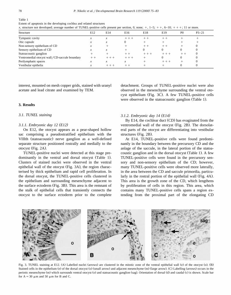
Table 1Extent of apoptosis in the developing cochlea and related structuresx, structure not developed; average number of TUNEL-positive cells present per section, 0, none; q, 1–5; qq, 6–10; qqq; 11 or more.
Structure E12 E14 E16 E18 E19 P0 P3–21
Tympanic cavity x x qqq qq qq q qOtic capsule x x 0 q q q qNon-sensory epithelium of CD x q q qq qq q 0Sensory epithelium of CD x x q 0 0 0 0Statoacoustic ganglion q q qq qqq qqq qq 0Ventromedial otocyst wallrCD-saccule boundary qq qqq qqq q 0 0 0Perilymphatic spaces x x x q qqq q 0Vestibular epithelia x qqq qq q q 0 0
interest, mounted on mesh copper grids, stained with uranylacetate and lead citrate and examined by TEM.
3. Results
3.1. TUNEL staining
( )3.1.1. Embryonic day 12 E12On E12, the otocyst appears as a pear-shaped hollow
sac comprising a pseudostratified epithelium with theŽ .VIIIth statoacoustic nerve ganglion as a well-defined
separate structure positioned rostrally and medially to theŽ .otocyst Fig. 2A .
TUNEL-positive nuclei were detected at this stage pre-Ž .dominantly in the ventral and dorsal otocyst Table 1 .
Clusters of stained nuclei were observed in the ventralŽ .epithelial wall of the otocyst Fig. 3A , the region charac-
terised by thick epithelium and rapid cell proliferation. Inthe dorsal otocyst, the TUNEL-positive cells clustered inthe epithelium and surrounding mesenchyme adjacent to
Ž .the surface ectoderm Fig. 3B . This area is the remnant ofthe stalk of epithelial cells that transiently connects theotocyst to the surface ectoderm prior to the complete
detachment. Groups of TUNEL-positive nuclei were alsoobserved in the mesenchyme surrounding the ventral oto-
Ž .cyst epithelium Fig. 3C . A few TUNEL-positive cellsŽ .were observed in the statoacoustic ganglion Table 1 .
( )3.1.2. Embryonic day 14 E14Ž .By E14, the cochlear duct CD has evaginated from the
Ž .ventromedial wall of the otocyst Fig. 2B . The dorsolat-eral parts of the otocyst are differentiating into vestibular
Ž .structures Fig. 2B .At E14, TUNEL-positive cells were found predomi-
nantly in the boundary between the precursory CD and theanlage of the saccule, in the lateral portion of the statoa-
Ž .coustic ganglion and in the dorsal otocyst Table 1 . A fewTUNEL-positive cells were found in the precursory sen-sory and non-sensory epithelium of the CD; however,many TUNEL-positive cells were observed more laterally,in the area between the CD and saccule primordia, particu-
Ž .larly in the rostral portion of the epithelial wall Fig. 4A .This area is the growth zone of the CD, which lengthensby proliferation of cells in this region. This area, whichcontains many TUNEL-positive cells spans a region ex-tending from the proximal part of the elongating CD
Ž . Ž . Ž . Ž . Ž .Fig. 3. TUNEL staining at E12. A Labelled nuclei arrows are clustered in the mitotic zone of the ventral epithelial wall e of the otocyst o . BŽ . Ž . Ž . Ž . Ž . Ž . Ž .Stained cells in the epithelium e of the dorsal otocyst o small arrow and adjacent mesenchyme m large arrow . C Labelling arrows occurs in the
Ž . Ž . Ž . Ž . Ž .periotic mesenchyme m which surrounds ventral otocyst o and statoacoustic ganglion sag . Orientation of dorsal d and caudal c is shown. Scale barfor As30 mm and 50 mm for B and C.

( )P. Nikolic et al.rDeÕelopmental Brain Research 119 2000 75–83 79
Ž . Ž . Ž . Ž .Fig. 4. TUNEL staining at E14. A Labelled nuclei small arrows in the epithelium e of the ventromedial otocyst o occur in the region between theŽ . Ž . Ž . Ž . Ž .primordium of the cochlear duct cd and the future saccule s . There is some staining large arrow in the periotic mesenchyme m . B Staining in the
Ž . Ž . Ž . Ž . Ž .periphery of the statoacoustic ganglion sag arrowheads and in the epithelium e of the otocyst o small arrow , more laterally from the section in A.Ž . Ž . Ž . Ž . Ž .C Labelling in the epithelium e of the dorsal side of the otocyst o ; m, mesenchyme. Orientation of dorsal d and caudal c is shown. Scale bars in Aand Cs40 mm and scale bar in Bs50 mm.
through to the anlage of the saccule. More laterally, thestatoacoustic ganglion displayed a few TUNEL-positive
Žcells mainly located in the periphery of the ganglion Fig..4B . In the close vicinity of the VIIIth ganglion, many
TUNEL-positive cells were seen in the otocyst epithelialŽ .wall Fig. 4B . Many TUNEL-positive cells were observed
in the dorsal otocyst, in the precursory sensory epithelia ofŽ .the equilibrium organ Fig. 4C although it was not possi-
ble at this stage to distinguish between a forming maculaor crista.
( )3.1.3. Embryonic day 16 E16By this stage of development, the CD coils to achieve
almost one full turn, and surrounding mesenchyme hascompacted to form the otic capsule. The tympanic cavityhas also begun to form with the regression of cells and the
Ž .cochlear ganglion CG has differentiated from the statoa-Ž .coustic ganglion SAG and separated from the vestibular
Žportion of this ganglion, and coils alongside the CD Fig. 2.C .
TUNEL-positive cells were mostly found in the CD-saccule boundary zone, in the tympanic cavity, in the
Ž .non-sensory epithelium of the CD and in the CG Table 1 .Notably, many TUNEL-positive cells were still present inthe CD growth zone, that is in the epithelium between the
Ž .CD and the saccule Fig. 5A . TUNEL-positive mesenchy-mal cells were scattered around the CD, usually in thevicinity of the TUNEL-positive epithelial or ganglioniccells. A few TUNEL-positive nuclei were observed in theprecursory non-sensory epithelium of the CD, mainly onthe modiolar side of the duct, at the point of inflexion. Afew cells localised in the CG showed DNA fragmentationŽ .Fig. 5B . Some of the TUNEL-positive cells were foundalong the fibres, but it was not possible to distinguishwhether these were neurones or glial cells. There weremany TUNEL-positive cells lining the tympanic cavity of
Ž . Ž . Ž . Ž .Fig. 5. TUNEL staining at E16. A Numerous labelled nuclei small arrow occur in the epithelial e growth zone between the cochlear duct cd and theŽ . Ž . Ž . Ž . Ž . Ž .saccule s . Staining also occurs in the adjacent mesenchyme m large arrow . B Stained cells arrowheads in the statoacoustic ganglion sag are
Ž . Ž . Ž .mainly in its periphery. C Labelled cells arrows line the tympanic cavity tc of the future middle ear. Other abbreviations: cd, cochlear duct; e,Ž . Ž .epithelium; m, mesenchyme. Dorsal d and caudal c orientation shown in A is same for B and C. Scale bar in C indicates 100 mm in A, and 50 mm in B
and C.

( )P. Nikolic et al.rDeÕelopmental Brain Research 119 2000 75–8380
Ž . Ž . Ž .Fig. 6. TUNEL staining at E19. A In the basal turn, stained cells occur in the cochlear ganglion cg arrowhead and on the modiolar side of the cochlearŽ . Ž . Ž . Ž . Ž . Ž . Ž .duct cd epithelium e small arrow . Labelled mesenchymal m cells large arrows line the cavity of the scala tympani st . B The labelling pattern in
Ž . Ž . Ž . Ž . Ž .the cochlear ganglion cg arrowhead and the cochlear duct cd small arrow extends to the middle turn. Numerous TUNEL-stained mesenchymal mŽ . Ž . Ž . Ž .cells large arrow occur in the area which eventually cavitates into scala tympani. C Labelled cells in the mesenchyme m large arrow and in the
Ž . Ž . Ž .epithelium small arrow which lines the tympanic cavity. The dorsal d and caudal c orientation shown in A is same for A–C. Scale bar indicates 100mm in A and B and 50 mm in C.
Ž .the future middle ear cavity Fig. 5C , presumably under-going cell death to allow enlargement of the cavity. ManyTUNEL-positive cells were also observed along the walls
Ž .of the forming Eustachian tube data not shown .
( )3.1.4. Embryonic day 18 E18At this stage, the CD has grown in size and formed
basal, middle and apical turns, coiled around the modiolaraxis. The cartilaginous otic capsule is well defined by thisstage.
Most of the TUNEL-labelling was observed in the basaland middle turns of the cochlea and in the cochlear
Ž .supporting structures Table 1 . In the cochlear ganglion,many TUNEL-positive cells were found in the basal andmiddle turns. TUNEL-positive cells were principally posi-tioned in the periphery of the ganglion and many TUNEL-positive cells were observed along the vestibulocochlearnerve and in the vestibular ganglion. Another area where afew TUNEL-positive cells were detected was in the CD
epithelium in the basal and middle turns. These werepredominantly found in the non-sensory epithelium on themodiolar side of the CD. A few TUNEL-positive cellswere also observed in clusters in the mesenchyme of themiddle ear and many TUNEL-positive cells were observed
Ž .in the epithelium lining the tympanic cavity Table 1 .
( )3.1.5. Embryonic day 19 E19The most striking feature of cochlear differentiation at
this stage is the opening of the perilymphatic spacesŽ .beginning at the basal turn Fig. 2D .
TUNEL-positive cells were mainly observed in the CG,CD and perilymphatic spaces of basal and middle turns, as
Ž .well as in the middle ear Table 1 . In the basal and middleturns of CD, small clusters of few TUNEL-positive cellswere detected in the non-sensory epithelium and weregrouped on the modiolar side of the duct. Similar to E18,many TUNEL-positive cells were observed in the CG in
Ž .the basal and the middle turns Fig. 6A and B with a
Ž . Ž . Ž .Fig. 7. TUNEL staining at P0. A Stained nuclei in the middle cochlear turn occur in the cochlear ganglion cg arrowhead and around the scala tympaniŽ . Ž . Ž . Ž . Ž .st large arrows . B Labelling in the apical turn occurs in the cochlear ganglion cg arrowheads and in a group of cells on the modiolar side of the
Ž . Ž . Ž . Ž .cochlear duct cd epithelium arrow . C Stained cells arrows lining the tympanic cavity. Abbreviations: sv, scala vestibuli; oc, organ of Corti; cd,Ž . Ž .cochlear duct; cap, otic capsule; m, mesenchyme. The dorsal d and caudal c orientation shown in A is same for A–C. Scale bars100 mm for A–C.

( )P. Nikolic et al.rDeÕelopmental Brain Research 119 2000 75–83 81
Ž . Ž .Fig. 8. Electron microscopy of apoptotic cells in the E12 otocyst. A A typical cell in an early stage of apoptosis arrow shows condensation of theŽ . Ž . Ž .nucleus n and cytoplasm and detachment from normal neighbouring epithelial cells. An adjacent apoptotic cell has been phagocytosed arrowhead . B A
Ž . Ž . Ž .periotic mesenchymal cell undergoing fragmentation into apoptotic bodies arrows , each containing a part of the condensed nucleus n and organelles. CŽ .A phagocytosed fragment of an apoptotic cell arrowhead observed as an electron-dense inclusion in a normal epithelial cell. Scale barss1 mm.
somewhat larger number in the middle turn. Only a fewTUNEL-positive cells were observed in the apical turn ofthe CG. Another area displaying many TUNEL-positivecells was the anlage of the scala tympani. Since this spacewas already open in the basal turn, TUNEL-positive cells
Ž .were observed lining its edge Fig. 6A . On the other hand,many TUNEL-positive mesenchymal cells were detectedin the area that begins to cavitate into scala tympani in the
Ž .middle turn Fig. 6B . A few TUNEL-positive cells weredetected in the middle ear, in the mesenchyme and tym-
Ž .panic cavity Fig. 6C .
( )3.1.6. Postnatal day 0 P0The cochlea has acquired almost adult appearance, ex-
cept the organ of Corti which has not yet achieved aŽ .mature configuration Fig. 2E .
The number of TUNEL-positive cells was smaller thanat earlier stages. The pattern of TUNEL-labelling in thecochlear ganglion and duct is more towards the apex. Afew TUNEL-positive cells were localised principally in themiddle turn and also in the apical turn of CG. In the CD, afew TUNEL-positive cells were detected in the apical turn,
Ž .on the modiolar side of the epithelium Fig. 7A and B . Afew TUNEL-positive cells were scattered in the mes-
Ž .enchyme in the tympanic cavity Fig. 7C , as well as in theŽ .cartilaginous otic capsule Table 1 .
( ) ( )3.1.7. Postnatal day 3 P3 to postnatal day 21 P21The number of TUNEL-positive cells declined further.
They could be observed only occasionally, and almostexclusively in the ossifying cartilaginous structures of the
Ž .cochlea, mainly in the otic capsule Table 1 .
3.2. Morphological eÕaluation by light and electron mi-croscopy
Apoptosis in the developing rat cochlea was confirmedby light and electron microscopy. By light microscopy,apoptotic cells appeared as more densely toluidine blue
Ž .stained bodies, often engulfed by other cells Fig. 1C .
They were smaller than the surrounding healthy cells andsometimes had a clear halo around them. This pattern ofstaining is most likely a consequence of nuclear andcytoplasmic condensation and the detachment of theshrinking apoptotic cell from the adjacent cells. A numberof cells which were clearly apoptotic were observed byelctron microscopy in the corresponding areas of develop-ing cochlea where TUNEL-positive cells were observed.Apoptotic cells displayed a number of morphological fea-tures including reduced size of the cell body, increasedelectron density of the nucleus and cytoplasm, cytoplasmicinclusions and condensation of chromatin close to the
Ž .edges of nuclear envelope Fig. 8A . The electron densebodies, or cellular fragments, were common in areas withabundant apoptosis. These bodies were typically round oroval, with electron-dense cytoplasm and darker centralarea of nuclear condensation. The organelles could some-
Ž .times be observed in the cytoplasm Fig. 8B . However,the apoptotic bodies were usually observed engulfed byapparently normal cells, which did not look like spe-
Ž .cialised phagocytes Fig. 8C .
4. Discussion
The spatiotemporal pattern of apoptosis in the develop-ing rat cochlea and related structures has been mappedusing biochemical and morphological criteria. Apoptosis ismost apparent during the embryonic and very early postna-tal periods, and then subsides substantially. Apoptosis wasobserved mainly in areas undergoing cavitation, in struc-tures that are removed, in areas of rapid proliferation, andin the tissues undergoing differentiation.
It seems that apoptosis plays an important role inclearing the areas of cavitation in the rat ear. Apoptosisoccurs substantially during the formation of hollow struc-
Ž .tures, notably the perilymphatic spaces E18 and E19 andŽ .the tympanic cavity after E16 . The cavitation of perilym-
phatic spaces progresses from the basal turn towards theapex, and we found that mesenchymal cells in these areas

( )P. Nikolic et al.rDeÕelopmental Brain Research 119 2000 75–8382
die by apoptosis following the same base-to-apex gradient.A role for apoptosis in opening the perilymphatic spaces
w xhas also been suggested in the mouse 25 . Our findingsthat apoptosis occurs in the middle ear mesenchyme andepithelium which lines the tympanic cavity and plays arole in the process of cavitation of the hollow tympanum
w xare in agreement with those of Roberts and Miller 30 inthe murine middle ear. However, in contrast to the pres-ence of apoptosis limited to P1 in the mouse, our findingsshow that in the rat, apoptosis occurs in the middle ear fora longer period, from E16 and gradually disappears duringthe postnatal period. This is probably due to the differ-ences in developmental sequences in the rat and the mouse.
One of the main areas where apoptosis is detected in theearly stages of the otocyst development is the dorsalepithelium of the otocyst and the surrounding mesen-chyme, in agreement with the earlier reports of the location
w xof cell death in the rat 20,22 , and in the human otocystw x29 . The otocyst is formed when the otic placode invagi-nates and its walls join to form the fluid-filled cyst whichthen sinks in the mesenchyme. On E12 there is still astalk-like vestige of epithelium and mesoderm between theotocyst and the surface epithelium. It seems that apoptosisparticipates in elimination of this stalk.
We observed a large number of apoptotic cells in thezones of rapid proliferation. A key example is the CD–sac-cule boundary and its precursor, the ventromedial epithe-lial wall of the otocyst, the zone where the CD outpouchesfrom the otocyst and which continues to be the growth
w xzone for the CD 21 . This is a region of abundant apop-Ž .totic cell death from the earliest day E12 of development
we examined to E16. This is in agreement with previouslyw x w x w xreported findings in rat 20,22 , human 29 , mouse 25
w xand chick 6 . The high apoptotic rate coincides spatiallyand temporally with the high mitotic rate in this area. Thepeak of mitotic activity occurs in this area between E13and E15 in the rat, as determined by autoradiographic
w xstudies 17 . It appears, therefore, that the new cells areproduced in larger numbers than required, and that thesupernumerary cells are removed by apoptosis.
The coincidence of apoptosis and differentiation sug-gests that apoptosis also plays a major role in the differen-tiation of cochlea. Apoptosis in the rat statoacoustic gan-glion was observed from E12. Its rate peaked from E18 to
w xE19, somewhat later than in the mouse 26 and occurswith a base-to-apex temporal gradient. This is analogous to
Ž .the temporal pattern of terminal mitosis E17 and E18 , asshown by autoradiography of tritiated thymidine uptakew x17 . Also, the peak of apoptosis in the cochlear ganglioncoincides with the pattern of the ingrowth of neurofila-ment-positive nerve fibers into the sensory epitheliumw x10,27 and precedes the expression of adult-likeŽ .axodendritic efferent synapses with radial afferent fibresw x Ž .18 . At this time E16–P0 , apoptosis occurs substantiallyin the cochlear ganglion and scarcely in the developingsensory epithelium. This would imply that the ganglion
cells are initially overproduced and that their survivaldepends on provision of trophic factors by the sensory
w xcells, as suggested by in vitro studies 33 and geneticw xmanipulation 5 . Consequently, the lack of trophic factors
would induce apoptosis in neurons. In addition to apoptoticcells in the ganglion, we detected apoptotic cells along theneural fibres. Since our study was not designed to deter-mine the exact type of ganglion cells that undergo apopto-sis, it is possible that some of the dying cells we observedare supporting cells, rather than neurones. In particular,E15–E17 is the time in rat embryonic development whenSchwann cells are generated from their precursors and are
w xprone to PCD 14 . The lack of TUNEL-positive cells inthe spiral ganglion of the postnatal rat implies there is littlecell death in this structure at this time of development.This is in contrast to the dramatic reduction observed inthe number of spiral ganglion neurones around P5 and P6
w xin the rat 32 at a time when there is a change from anafferent to a mainly efferent pattern of innervation of outer
w xhair cells 18 . The difference with our study is probablyŽdue to the fact that our selection of postnatal times P3 and
.P6 straddles the time over which this neuronal loss isoccurring. TUNEL staining is a marker of DNA fragmen-tation, which occurs in a narrow time window during thecourse of cell death. Thus, it would identify only thosecells which are undergoing cell death at the moment oftissue preparation.
Apoptosis has been studied in the developing equilib-w x w xrium sensory organs of the chick 6 , rat 35 and mouse
w x25 . Although the objective of the current study was todescribe the pattern of apoptosis in the cochlea, whereapoptosis was observed in the rat vestibular structures,particularly in the early, otocyst stages of development, it
w xshowed a similar pattern to that described in rat 35 andw xmouse 25 .
The understanding of the molecular mechanisms whichregulate PCD in the inner ear is still fragmentary. Forinstance, bcl-2 is believed to play a role in protecting cells
w xfrom PCD 1 . Overexpression of bcl-2 in the chick innerear causes a reduction of the number of apoptotic cells anddisruptions of normal development of the semicircular
w xcanals 6 . Even though developmental differences betweenthe mouse and the rat have to be considered, the paucity ofapoptosis in the sensory epithelium of the cochlea maypartly be ascribed to the findings that, in mice, bcl-2mRNA is expressed in this region during embryogenesis
w xand the early postnatal period 11 . On the other hand, themouse spiral ganglion does not express bcl-2 mRNA untilP1, around the time when we could no longer detectapoptosis in rat cochlear ganglion.
Our findings show that apoptotic cells commonly haveŽ .a tendency to cluster to be detected in a group , which is
consistent with the previous observations indicating thatw xthe apoptotic cells mainly occur in groups 29 or ‘‘hot
w xspots’’ 6 . In addition, there is often evidence that in acluster of apoptotic cells, there are cells of different types,

( )P. Nikolic et al.rDeÕelopmental Brain Research 119 2000 75–83 83
for example epithelial and mesenchymal cells. Therefore,the apoptotic cascade may be triggered within a cell if aneighbouring cell is undergoing apoptosis. Further cell–cellsignals other than withdrawal of trophic factors may havean important role in inducing apoptosis, and the signalling
Ž .molecule s may not be restricted to a certain cell type. Aubiquitous molecule, extracellular adenosine 5X-triphos-
Ž .phate ATP plays a role in cell death signalling in thew xthymus and other tissues 2–4,7 . The spatiotemporal or-
ganisation of apoptosis presented here provides a basis forour current investigation whether ATP acting through spe-cific P2X receptors might play an analogous role in thedeveloping cochlea.
Acknowledgements
We thank Drs. Gary Housley and Srdjan Vlajkovic forvery helpful suggestions on the manuscript. This work wassupported by the Marsden Fund and Health ResearchCouncil of New Zealand.
References
w x1 J.M. Adams, S. Cory, The Bcl-2 protein family — arbiters of cellŽ .survival, Science 281 1998 1322–1326.
w x2 S.C. Chow, G.E.N. Kass, S. Orrenius, Purines and their roles inŽ .apoptosis, Neuropharmacology 36 1997 1149–1156.
w x3 Y. Chvatchko, S. Valera, J.P. Aubry, T. Renno, G. Buell, J.Y.Bonnefoy, The involvement of an ATP-Gated ion channel, P in2= 1
Ž .thymocyte apoptosis, Immunity 5 1996 275–283.w x4 F. Di Virgilio, P. Chiozzi, S. Falzoni, D. Ferrari, J.M. Sanz, V.
Venketaraman, O.R. Baricordi, Cytolytic P2X purinoceptors, CellŽ .Death Diff. 5 1998 191–199.
w x5 P. Ernfors, T. Van De Water, J. Loring, R. Jaenisch, Complementaryroles of BDNF and NT-3 in vestibular and auditory development,
Ž .Neuron 14 1995 1153–1164.w x6 D.M. Fekete, S.A. Homburger, M.T. Waring, A.E. Riedl, L.F.
Garcia, Involvement of programmed cell death in morphogenesis ofŽ .the vertebrate inner ear, Development 124 1997 2451–2461.
w x7 C. Franceschi, M.P. Abbracchio, D. Barbieri, S. Ceruti, D. Ferrari,J.P. Iliou, S. Rounds, P. Schubert, E. Schulze-Lohoff, F.A. Rassen-dren, M. Staub, C. Volonte, A.R. Wakade, G. Burnstock, Purines`
Ž .and cell death, Drug Dev. Res. 39 1996 442–449.w x8 Y. Gavrieli, Y. Sherman, S.A. Ben-Sasson, Identification of pro-
grammed cell death in situ via specific labeling of nuclear DNAŽ .fragmentation, J. Cell Biol. 119 1992 493–501.
w x Ž .9 D.R. Green, Apoptotic pathways: the roads to ruin, Cell 94 1998695–698.
w x10 A. Hafidi, G. Despres, R. Romand, Cochlear innervation in thedeveloping rat: an immunocytochemical study of neurofilament and
Ž .spectrin proteins, J. Comp. Neurol. 300 1990 153–161.w x11 N. Ishii, A. Wanaka, K. Ohno, K. Matsumoto, Y. Eguchi, T. Mori,
Y. Tsujimoto, M. Tohyama, Localization of Bcl-2, Bax, and Bcl-XmRNAs in the developing inner ear of the mouse, Brain Res. 726Ž .1996 123–128.
w x12 A. Ishizuyaoka, Apoptosis of larval cells during amphibian meta-Ž .morphosis, Microsc. Res. Techn. 34 1996 228–235.
w x13 M.D. Jacobson, M. Weil, M.C. Raff, Programmed cell death inŽ .animal development, Cell 88 1997 347–354.
w x14 K.R. Jessen, R. Mirsky, Embryonic Schwann cell development —
the biology of Schwann cell precursors and early Schwann cells, J.Ž .Anat. 191 1997 501–505.
w x15 E.M. Johnson Jr., T.L. Deckwerth, M. Deshmukh, Neuronal death indevelopmental models: possible implications in neuropathology,
Ž .Brain Pathol. 6 1996 397–409.w x16 J.F. Kerr, A.H. Wyllie, A.R. Currie, Apoptosis: a basic biological
phenomenon with wide-ranging implications in tissue kinetics, Br. J.Ž .Cancer 26 1972 239–257.
w x17 K.M. Khan, W.F. Marovitz, DNA content, mitotic activity, andincorporation of tritiated thymidine in the developing inner ear of
Ž .the rat, Anat. Rec. 202 1982 501–509.w x18 M. Lenoir, A. Shnerson, R. Pujol, Cochlear receptor development in
Ž .the rat with emphasis on synaptogenesis, Anat. Embryol. 160 1980253–262.
w x19 X. Liu, P. Li, P. Widlak, H. Zou, X. Luo, W.T. Garrard, X. Wang,The 40-kDa subunit of DNA fragmentation factor induces DNAfragmentation and chromatin condensation during apoptosis, Proc.
Ž .Natl. Acad. Sci. U.S.A. 95 1998 8461–8466.w x20 W.F. Marovitz, K.M. Khan, T. Schulte, Ultrastructural development
of the early rat otocyst, Ann. Otol. Rhinol. Laryngol. — Suppl. 86Ž .1977 9–28.
w x21 W.F. Marovitz, J.M. Shugar, Single mitotic center for rodent cochlearŽ .duct, Ann. Otol. Rhinol. Laryngol. 85 1976 225–233.
w x22 W.F. Marovitz, J.M. Shugar, K.M. Khan, The role of cellularŽ .degeneration in the normal development of rat otocyst, Laryngo-
Ž .scope 86 1976 1413–1425.w x23 C.E. Milligan, L.M. Schwartz, Programmed cell death during animal
Ž .development, Br. Med. Bull. 53 1997 570–590.w x24 A. Nishikawa, H. Hayashi, Spatial, temporal and hormonal regula-
tion of programmed muscle cell death during metamorphosis of theŽ .frog Xenopus laevis, Differentiation 59 1995 207–214.
w x25 K. Nishizaki, M. Anniko, Y. Orita, K. Karita, Y. Masuda, T.Yoshino, Programmed cell death in the developing epithelium of the
Ž .mouse inner ear, Acta Oto-Laryngol. 118 1998 96–100.w x26 K. Nishizaki, M. Anniko, Y. Orita, Y. Masuda, T. Yoshino, Pro-
grammed cell death in the mouse cochleovestibular ganglion duringŽ .development, Orl, J. Oto-Rhino-Laryngol. Rel. Spec. 60 1998
267–271.w x27 U. Pirvola, E. Lehtonen, J. Ylikoski, Spatiotemporal development of
cochlear innervation and hair cell differentiation in the rat, Hear.Ž .Res. 52 1991 345–355.
w x28 M.C. Raff, B.A. Barres, J.F. Burne, H.S. Coles, Y. Ishizaki, M.D.Jacobson, Programmed cell death and the control of cell survival:
Ž .lessons from the nervous system, Science 262 1993 695–700.w x29 J. Represa, J.A. Moro, A. Gato, F. Pastor, E. Barbosa, Patterns of
epithelial cell death during early development of the human innerŽ .ear, Ann. Otol. Rhinol. Laryngol. 99 1990 482–488.
w x30 D.S. Roberts, S.A. Miller, Apoptosis in cavitation of middle earŽ .space, Anat. Rec. 251 1998 286–289.
w x31 I. Rodriguez, K. Araki, K. Khatib, J.C. Martinou, P. Vassalli, Mousevaginal opening is an apoptosis-dependent process which can be
Ž .prevented by the overexpression of Bcl2, Dev. Biol. 184 1997115–121.
w x32 J. Rueda, C. de la Sen, J.M. Juiz, J.A. Merchan, Neuronal loss in the´Ž .spiral ganglion of young rats, Acta Oto-Laryngol. 104 1987 417–
421.w x33 H. Staecker, T.R. Van de Water, P.P. Lefebvre, W. Liu, M.
Moghadassi, V. Galinovic-Schwartz, B. Malgrange, G. Moonen,NGF, BDNF and NT-3 play unique roles in the in vitro developmentand patterning of innervation of the mammalian inner ear, Dev.
Ž .Brain Res. 92 1996 49–60.w x34 G.T. Williams, Role of apoptosis in the immune system, Biochem.
Ž .Cell Biol. 72 1994 447–450.w x35 J.L. Zheng, W.Q. Gao, Analysis of rat vestibular hair cell develop-
ment and regeneration using calretinin as an early marker, J. Neu-Ž .rosci. 17 1997 8270–8282.

![Automatic Cochlea Multi-modal Images Segmentation · 2018-04-03 · Automatic Cochlea Multi-modal Images Segmentation Al-Dhamari, CI2018 Methods: Cochlea Model 9 [5] Gerber et al,](https://static.fdocuments.in/doc/165x107/5f8e42f1fe0c2a0180250f2a/automatic-cochlea-multi-modal-images-segmentation-2018-04-03-automatic-cochlea.jpg)


