Apomixis in plant reproduction: a novel perspective on an ... · 1 seeds will be harvested. Farmers...
21
REVIEW Apomixis in plant reproduction: a novel perspective on an old dilemma Gianni Barcaccia • Emidio Albertini Received: 9 May 2013 / Accepted: 23 June 2013 / Published online: 14 July 2013 Ó The Author(s) 2013. This article is published with open access at Springerlink.com Abstract Seed is one of the key factors of crop produc- tivity. Therefore, a comprehension of the mechanisms underlying seed formation in cultivated plants is crucial for the quantitative and qualitative progress of agricultural production. In angiosperms, two pathways of reproduction through seed exist: sexual or amphimictic, and asexual or apomictic; the former is largely exploited by seed com- panies for breeding new varieties, whereas the latter is receiving continuously increasing attention from both sci- entific and industrial sectors in basic research projects. If apomixis is engineered into sexual crops in a controlled manner, its impact on agriculture will be broad and pro- found. In fact, apomixis will allow clonal seed production and thus enable efficient and consistent yields of high- quality seeds, fruits, and vegetables at lower costs. The development of apomixis technology is expected to have a revolutionary impact on agricultural and food production by reducing cost and breeding time, and avoiding the complications that are typical of sexual reproduction (e.g., incompatibility barriers) and vegetative propagation (e.g., viral transfer). However, the development of apomixis technology in agriculture requires a deeper knowledge of the mechanisms that regulate reproductive development in plants. This knowledge is a necessary prerequisite to understanding the genetic control of the apomictic process and its deviations from the sexual process. Our molecular understanding of apomixis will be greatly advanced when genes that are specifically or differentially expressed dur- ing embryo and embryo sac formation are discovered. In our review, we report the main findings on this subject by examining two approaches: i) analysis of the apomictic process in natural apomictic species to search for genes controlling apomixis and ii) analysis of gene mutations resembling apomixis or its components in species that normally reproduce sexually. In fact, our opinion is that a novel perspective on this old dilemma pertaining to the molecular control of apomixis can emerge from a cross- check among candidate genes in natural apomicts and a high-throughput analysis of sexual mutants. Keywords Apomixis Á Plant reproduction Á Hybrids Á Seed production Introduction One of the greatest success stories in modern agriculture has been the tremendous yield increase achieved by coupling high-yield varieties with high-input agronomic systems, creating the so-called Green Revolution. Approximately one-third of the world’s seed supply comes from the com- mercial seed market, another one-third is provided by publicly funded institutions, and the seed saved by farmers accounts for the remainder. Over the centuries, crop plants have followed the gen- eral pattern of introduction, selection, and hybridization. Crop introduction has been crucial for agriculture because Communicated by T. Dresselhaus. A contribution to the Special Issue ‘‘HAPRECI—Plant Reproduction Research in Europe.’’ G. Barcaccia Laboratory of Genetics and Genomics, DAFNAE, University of Padova, Campus of Agripolis, Viale dell’Universita ` 16, 35020 Legnaro, Italy E. Albertini (&) Department of Applied Biology, University of Perugia, Borgo XX Giugno 74, 06121 Perugia, Italy e-mail: [email protected] 123 Plant Reprod (2013) 26:159–179 DOI 10.1007/s00497-013-0222-y
Transcript of Apomixis in plant reproduction: a novel perspective on an ... · 1 seeds will be harvested. Farmers...

REVIEW
Apomixis in plant reproduction: a novel perspective on an olddilemma
Gianni Barcaccia • Emidio Albertini
Received: 9 May 2013 / Accepted: 23 June 2013 / Published online: 14 July 2013
� The Author(s) 2013. This article is published with open access at Springerlink.com
Abstract Seed is one of the key factors of crop produc-
tivity. Therefore, a comprehension of the mechanisms
underlying seed formation in cultivated plants is crucial for
the quantitative and qualitative progress of agricultural
production. In angiosperms, two pathways of reproduction
through seed exist: sexual or amphimictic, and asexual or
apomictic; the former is largely exploited by seed com-
panies for breeding new varieties, whereas the latter is
receiving continuously increasing attention from both sci-
entific and industrial sectors in basic research projects. If
apomixis is engineered into sexual crops in a controlled
manner, its impact on agriculture will be broad and pro-
found. In fact, apomixis will allow clonal seed production
and thus enable efficient and consistent yields of high-
quality seeds, fruits, and vegetables at lower costs. The
development of apomixis technology is expected to have a
revolutionary impact on agricultural and food production
by reducing cost and breeding time, and avoiding the
complications that are typical of sexual reproduction (e.g.,
incompatibility barriers) and vegetative propagation (e.g.,
viral transfer). However, the development of apomixis
technology in agriculture requires a deeper knowledge of
the mechanisms that regulate reproductive development in
plants. This knowledge is a necessary prerequisite to
understanding the genetic control of the apomictic process
and its deviations from the sexual process. Our molecular
understanding of apomixis will be greatly advanced when
genes that are specifically or differentially expressed dur-
ing embryo and embryo sac formation are discovered. In
our review, we report the main findings on this subject by
examining two approaches: i) analysis of the apomictic
process in natural apomictic species to search for genes
controlling apomixis and ii) analysis of gene mutations
resembling apomixis or its components in species that
normally reproduce sexually. In fact, our opinion is that a
novel perspective on this old dilemma pertaining to the
molecular control of apomixis can emerge from a cross-
check among candidate genes in natural apomicts and a
high-throughput analysis of sexual mutants.
Keywords Apomixis � Plant reproduction � Hybrids �Seed production
Introduction
One of the greatest success stories in modern agriculture has
been the tremendous yield increase achieved by coupling
high-yield varieties with high-input agronomic systems,
creating the so-called Green Revolution. Approximately
one-third of the world’s seed supply comes from the com-
mercial seed market, another one-third is provided by publicly
funded institutions, and the seed saved by farmers accounts
for the remainder.
Over the centuries, crop plants have followed the gen-
eral pattern of introduction, selection, and hybridization.
Crop introduction has been crucial for agriculture because
Communicated by T. Dresselhaus.
A contribution to the Special Issue ‘‘HAPRECI—Plant Reproduction
Research in Europe.’’
G. Barcaccia
Laboratory of Genetics and Genomics, DAFNAE,
University of Padova, Campus of Agripolis,
Viale dell’Universita 16, 35020 Legnaro, Italy
E. Albertini (&)
Department of Applied Biology, University of Perugia,
Borgo XX Giugno 74, 06121 Perugia, Italy
e-mail: [email protected]
123
Plant Reprod (2013) 26:159–179
DOI 10.1007/s00497-013-0222-y

many of the world’s crops are produced outside their
region of domestication. Once introgressed, selection and
breeding strategies have led to the development of new
cultivars with improved yield and adaptation. Plant
breeders are working to extend the Green Revolution by
intensifying selection, developing more hybrid varieties in
more crops, and increasing the range of plant functions
through mutation and transgenic breeding. Hence, plant
breeding will continue to play a crucial role in crop
improvement because the needs are many, the techniques
are expanding, the new genetic combinations are limitless,
and the successes of the past illuminate the potential of the
future. In outcrossing species, alleles disseminate in the
offspring; thus, the optimal genotype is lost together with
the desired trait. Exact copies of a superior genotype can be
made via vegetative propagation; however, this technique
is usually not applicable to annual crops such as maize,
rice, and wheat. The fixation of a given genotype occurs
naturally in species that exhibit an asexual type of seed
production termed apomixis. This trait by itself is highly
valuable for agriculture; however, despite many efforts, it
has not been possible to introduce apomixis into modern
domesticated crop species.
As a reproductive strategy for cloning plants via seeds,
apomixis is a highly desirable trait in modern agriculture.
In fact, apomixis results in offspring that are exact genetic
replices of the female parent because embryos are derived
from the parthenogenic development of apomeiotic egg
cells (for reviews on apomixis, see Bicknell and Koltunow
2004; Ozias-Akins 2006; Albertini et al. 2010; Pupilli and
Barcaccia 2012; Koltunow et al. 2013). From an evolu-
tionary point of view, apomixis may be regarded as a
consequence of sexual failure rather than as a recipe for
clonal success (Silvertown 2008).
Introgression of apomixis from wild relatives into crop
species and transformation of sexual genotypes into apo-
mictically reproducing genotypes are long-held goals of
plant breeding. Breeders believe that the introduction of
apomixis into agronomically important crops will have
revolutionary implications for agriculture. The potential
benefits of harnessing apomixis are many and vary from
full exploitation of heterosis by reseeding the best hybrids
to clonal propagation of the superior genotypes in seed-
propagated outcrossing crops. The impact of apomictic
crops in agriculture would be massive in both developed
and developing countries. Unfortunately, barring a few
exceptions in some forage grasses and fruit trees, apomixis
is not a common feature among crop species.
The fixation of hybrid vigor through apomixis is a
desirable objective for breeders and farmers alike and is
expected to have a revolutionary impact on food and
agriculture production. The stabilization of heterozygous
genotypes via apomixis would make breeding programs
faster and cheaper (Fig. 1). The impact of apomictic crops
in agriculture would be comparable to, or even greater
than, the impact of the Green Revolution, especially in
Third World countries (Vielle-Calzada et al. 1996; Pupilli
and Barcaccia 2012). In fact, it has been estimated that the
use of apomixis technology in the production of hybrid rice
alone could provide benefits exceeding 1,800 million Eu-
ros per year (Spillane et al. 2004; Albertini et al. 2010).
Apomixis technology could also provide benefits for
clonally propagated crops. Clonal crop yields are limited
by pathogens (mainly viral and endophytic), which accu-
mulate over successive rounds of vegetative propagation
and seriously limit the yield and exchange of germplasms
between countries. The use of apomixis technology in these
crops would provide the additional option and benefit of
propagation via clonal seeds and thus generating disease-
free material that can be more easily stored and trans-
ported. The use of apomictic seed as an alternative to
vegetative propagules would provide similar benefits (e.g.,
lower costs and higher yields) over the current use of true
seed of such crops. For example, apomixis technology
could make true potato seeds a more attractive option for
potato breeders and cultivators and would return benefits to
growers of as much as 2.3 billion Euros per year (Spillane
et al. 2004).
The development of apomixis technology in agriculture
will require a deeper knowledge of the mechanisms regu-
lating reproductive development in plants. Our molecular
understanding of apomixis would be greatly increased if
genes that are specifically or differentially expressed dur-
ing the formation of the embryo and embryo sac could be
identified. Over the last two decades, many scientists have
speculated about the isolation of gene/s controlling key
steps of the apomictic pathway, and many papers have
postulated the production of engineered plants exhibiting
apomictic-like phenotypes. In fact, none of the major crop
plants have been bred for apomixis, and only some features
of apomixis have been genetically engineered in model
species. Consequently, even in the era of genomics,
achieving an understanding of the genetic control and
molecular regulation of apomixis appears much more
complicated than expected. Large amounts of cytological
and ecological information, along with genetic and
molecular data, have been collected mainly from model
species (i.e., Boechera holboellii, Hieracium spp., Hyper-
icum perforatum, Paspalum spp., Poa pratensis, Ranun-
culus spp., and Taraxacum officinale) and have often been
tested in Arabidopsis thaliana (Arabidopsis) to elucidate
the mechanisms of apomeiosis, parthenogenesis, and apo-
mixis. Several genes involved in the formation of unre-
duced embryo sacs and egg cells, in addition to genes
responsible for the autonomous development of the embryo
and endosperm, have been cloned and characterized;
160 Plant Reprod (2013) 26:159–179
123
however, none of these genes are capable of miming the
apomictic pathway as a whole in crop plants. Hence, after
two decades of substantial studies conducted in several
laboratories and model plants, the asexual reproductive
strategy termed ‘‘gametophytic apomixis’’ by Nogler
(1984) still appears to be an unsolved puzzle. As a result,
seed companies have lost interest in this research, and it
has been difficult to acquire funds for conducting research
on apomixis.
Currently, novel views and original concepts are
emerging from the fog, including a link between apomixis
and gene-specific silencing mechanisms (likely based on
Fig. 1 Comparison between conventional and apomixis-mediated
methods for breeding F1 hybrid varieties. In traditional breeding,
within a segregating population (e.g., F2 population) some genotypes
are selected and after some generation of selfing followed by
phenotypic selection, tested for their specific combining ability in
order to be used as parental lines for the constitution of heterotic F1
hybrid seeds. The best performing inbred lines are selected, multi-
plied in isolated fields, and crossed in pairwise combinations to obtain
uniform, vigorous, and high-yield F1 hybrids. This scheme, however,
requires a series of actions: the two inbred lines must be kept pure and
multiplied in separate fields. Then, to obtain the hybrid seed, it is
necessary to establish a dedicated field where about one quarter of the
plants is used as pollinator (i.e., pollen donor inbred) and on the
remaining plants (i.e., seed parent inbred) the hybrid F1 seeds will be
harvested. Farmers cannot re-use seeds collected from F1 hybrids as
these seeds will give rise to highly variable populations because of
genetic segregation and recombination. Using apomictic lines,
however, the situation would be much simpler. Once superior inbred
lines to be used as seed parent are selected, they can be crossed with
clonal lines as pollen donors carrying the gene for apomixis, in order
to obtain F1 hybrid seeds sharing a highly heterozygous genotype.
From this moment on, each F1 hybrid variety can be maintained for
several generations with permanently fixed heterosis
Plant Reprod (2013) 26:159–179 161
123
chromatin remodeling factors or trans-acting and hetero-
chromatic interfering RNAs involved in both transcrip-
tional and post-transcriptional gene regulation) and the
parallel between the Y chromosome and apomixis-bearing
chromosomes from the most primitive to the most
advanced in evolutionary terms (comparative genomic
analyses revealed common features such as few recombi-
nation events, accumulation of transposable elements, and
degeneration of genes). More recently, merging lines of
evidence regarding the role of auxin in cell fate specifi-
cation of the embryo sac and egg cell development have
been reported in Arabidopsis.
Mechanisms of apomixis
Because apomictic reproduction entails the development of
an embryo from a cell with a somatic chromosome number,
there are several ways to produce embryos of apomictic
origin. The simplest pathway avoids the production of an
embryo sac, and the maternal embryo originates from one
or more somatic cells of the ovule. Among the agricul-
turally important species, adventitious embryony (i.e.,
sporophytic apomixis) has been noted in mango (Mangif-
era indica), several Citrus species, and orchids. The most
comprehensive treatise on adventitious embryony has been
published by Naumova (1992).
When the maternal embryo originates from a diploid egg
cell differentiated in an unreduced embryo sac, the apo-
mictic pathway is referred to as gametophytic apomixis
(Nogler 1984).
In gametophytic apomixis, the unreduced embryo sac
may arise from a somatic nucellar cell that acquires the
developmental program of a functional megaspore, a
mechanism referred to as apospory. Alternatively, if the
embryo sac forms from a megaspore mother cell with
suppressed or modified meiosis, the pathway is referred to
as diplospory. It is worth emphasizing that apomictic plants
may or may not change meiosis itself, but in any case they
do activate the gametic cell fate either in a somatic cell
(apospory) or in an unreduced megaspore (diplospory) as
surrogate for meiotic products (Albertini and Barcaccia
2007). Once 2n female gametophytes and gametes are
formed (apomeiosis), they subsequently undergo embryo-
genesis autonomously without fertilization by a male
gamete (somatic parthenogenesis). Endosperm formation
may be fertilization-independent (autonomous endosperm)
or may require fertilization (pseudogamous endosperm).
Among others, apospory has been reported in Beta, Bra-
chiaria, Cenchrus, Chloris, Compositae, Eriochloa, Het-
eropogon, Hieracium, Hyparrhenia, Hypericum, Panicum,
Paspalum, Pennisetum, Poaceae, Ranunculus, Sorghum,
Themeda, and Urochloa, whereas diplospory has been
noted in Agropyrum, Allium, Antennaria, Boechera (for-
merly Arabis), Datura, Eragrostis, Erigeron, Eupatorium,
Ixeris, Parthenium, Paspalum, Poa, Taraxacum, and
Tripsacum (Table 1).
Inheritance of apomixis: genetic control
and recombination potentials
In both aposporic and diplosporic species, robust models
have been postulated and eventually validated for
inspecting the genetic basis of apomixis and its compo-
nents (i.e., apomeiosis and parthenogenesis). The inheri-
tance fundamentals include a divergence in the number of
genes, gene functions, and relationships among alleles, as
well as dominance of apomixis over sexuality (Asker and
Jerling 1992; Carman 1997; Savidan 2000; Grimanelli
et al. 2001; Koltunow and Grossniklaus 2003). Genetic
analysis in several species has consistently demonstrated
that a simple inheritance system involving a few Mendelian
genes controls the expression of apomixis or its compo-
nents. In contrast, molecular and cytogenetic analyses of
chromosomal region(s) carrying the determinants of apo-
mixis in several species have revealed a complex genetic
control mechanism that is likely based on a system of
polygenes in addition to mechanisms involving a lack of
recombination, trans-acting elements for gamete elimina-
tion, supernumerary chromatin structures, and DNA rear-
rangements (see a review by Pupilli and Barcaccia 2012
and references therein).
Currently, gametophytic apomixis is thought to rely on
three genetically independent Mendelian loci, each
exerting control over a key developmental component,
including apomeiotic megaspores, parthenogenic unre-
duced egg cells, and modified endosperms (Grossniklaus
et al. 2001a; Koltunow and Grossniklaus 2003; Bicknell
and Koltunow 2004; Vijverberg and van Dijk 2007; see
also a review by Albertini et al. 2010 and references
therein). A single regulatory gene was originally proposed
as being sufficient for promoting apomixis (Peacock
1992). Although simple genetic control seems to support
this hypothesis, molecular evidence suggests that a more
complex inheritance system directs the entire process of
apomixis. In some species, linkage groups typically
transmitted with apomixis contain large blocks of
sequence that lack genetic recombination between
molecular markers, leading to speculation that adapted
gene complexes within supernumerary chromatin might
be required for the expression of apomixis (Ozias-Akins
et al. 1998; Roche et al. 2001; Akiyama et al. 2004). A
close relationship between apomictic mechanisms and
heterochromatic regions of the genome that are rich in
retrotransposons has raised the intriguing possibility that
162 Plant Reprod (2013) 26:159–179
123

Ta
ble
1B
asic
info
rmat
ion
on
inh
erit
ance
mo
del
s,g
enet
icre
com
bin
atio
np
ote
nti
als,
and
mo
lecu
lar
map
pin
gst
ud
ies
of
apo
spo
ryan
dd
iplo
spo
ryin
apo
mic
tic
spec
ies
Ap
om
ixis
syst
em
Sp
ecie
s
En
do
sper
m
dev
elo
pm
ent
Su
pp
ress
ion
of
reco
mb
inat
ion
Ref
eren
ces
for
gen
etic
map
pin
gan
din
her
itan
ce
Can
did
ate
gen
es
Ref
eren
ces
for
can
did
ate
gen
es
Ap
osp
ory
Bra
chia
ria
bri
zan
tha
Pse
ud
og
amo
us
Yes
do
Val
lean
dS
avid
an(1
99
6),
Pes
sin
oet
al.
(19
97
,1
99
8)
RP
S8
-RP
S1
5a-R
PL
41
Sti
1;
Hel
ic
Lac
erd
aet
al.
(20
13
),
Sil
vei
raet
al.
(20
12
)
C.
cili
ari
sP
seu
do
gam
ou
sN
oS
her
wo
od
etal
.(1
99
4),
Ro
che
etal
.(1
99
9),
Jess
up
etal
.(2
00
2),
Dw
ived
iet
al.
(20
07),
Co
nn
eret
al.
(20
13
)
BB
M-l
ike
Co
nn
eret
al.
(20
08
)
Hie
raci
um
spp
.A
uto
no
mo
us
No
Bic
kn
ell
etal
.(2
00
0),
Cat
anac
het
al.
(20
06),
Ko
ltu
no
wet
al.
(20
11
),T
uck
eret
al.
(20
12
)
HF
IER
od
rig
ues
etal
.(2
00
8,
20
10
a)
H.
per
fora
tum
Pse
ud
og
amo
us
No
Mat
zket
al.(2
00
1),
Bar
cacc
iaet
al.(2
00
6,
20
07)
AR
IAD
NE
7(A
RI7
)
HA
PP
Ylo
cus
Sch
alla
uet
al.
(20
10
),G
alla
etal
.(p
erso
nal
com
mu
nic
atio
n)
P.
ma
xim
um
Pse
ud
og
amo
us
No
Eb
ina
etal
.(2
00
5),
Kau
shal
etal
.(2
00
8)
AS
G-1
Ch
enet
al.
(19
99
,2
00
5)
P.
no
tatu
mP
seu
do
gam
ou
sY
esM
arti
nez
etal
.(2
00
3),
Ste
inet
al.
(20
07)
LO
RE
LE
IF
elit
tiet
al.
(20
11
)
P.
sim
ple
xP
seu
do
gam
ou
sY
esP
up
illi
etal
.(2
00
1),
Lab
om
bar
da
etal
.(2
00
2)
Un
spec
ified
(AC
R)
Cal
der
ini
etal
.(2
01
2),
Pu
pil
li(p
erso
nal
com
mu
nic
atio
n)
P.
cili
are
Pse
ud
og
amo
us
Yes
Jess
up
etal
.(2
00
2,
20
03
)P
ca2
1,
Pca
24
Sin
gh
etal
.(2
00
7)
P.
squ
am
ula
tum
Pse
ud
og
amo
us
Yes
Ozi
as-A
kin
set
al.
(19
98
),G
oel
etal
.(2
00
6),
Hu
oet
al.
(20
09
)
BB
M-l
ike
Co
nn
eret
al.
(20
08
)
P.
pra
ten
sis
Pse
ud
og
amo
us
No
Bar
cacc
iaet
al.
(19
98
),A
lber
tin
iet
al.
(20
01
),
Mat
zket
al.
(20
05
)
SE
RK
,A
PO
ST
AR
T1
-2A
lber
tin
iet
al.
(20
04
,2
00
5)
Ra
nu
ncu
lus
au
rico
mu
sP
seu
do
gam
ou
sN
oN
og
ler
(19
84,
19
95)
ND
Dip
losp
ory
Bo
ech
era
spp
.P
seu
do
gam
ou
sY
esS
chra
nz
etal
.(2
00
6),
Lo
vel
let
al.
(20
13)
AP
OL
LO
Co
rral
etal
.(2
01
3)
Eri
ger
on
an
nu
us
Au
ton
om
ou
sN
oN
oy
esan
dR
iese
ber
g(2
00
0),
No
yes
etal
.(2
00
7)
ND
T.
offi
cin
ale
Au
ton
om
ou
sN
oT
asan
dv
anD
ijk
(19
99),
van
Dij
ket
al.
(19
99
,
20
03
),V
ijv
erb
erg
etal
.(2
00
4,
20
10
)
ND
T.
da
ctyl
oid
esP
seu
do
gam
ou
sY
esL
ebla
nc
etal
.(1
99
5),
Gri
man
elli
etal
.(1
99
8)
ND
Can
did
ate
gen
esfo
rap
om
ixis
are
also
giv
enal
on
gw
ith
refe
ren
ces
Plant Reprod (2013) 26:159–179 163
123
DNA structure and/or RNA interference could play a role
in regulating the expression of apomixis-related genes
(Pupilli and Barcaccia 2012). A well-characterized class
of small regulatory RNAs that are widespread in
eukaryotes appears to regulate gamete function and fer-
tilization in plants by altering gene expression through
post-transcriptional gene silencing, translational inhibi-
tion, and heterochromatin modification (Ron et al. 2010).
Increasing experimental evidence suggests that genetic
recombination can either be suppressed or allowed in
chromosomal regions surrounding the master locus for
apomixis depending on the evolutionary pathway of the
genome. This is a phenomenon that has been well docu-
mented for plant chromosomes carrying sex-determining
genes (Vyskot and Hobza 2004). From an evolutionary
point of view, we expect that relatively young and simple
genetic systems of apomixis determination should include
a narrow euchromatic region where genetic recombination
between apomeiosis and parthenogenesis loci, and their
linked genes, is possible (Table 1), as in Poa (Barcaccia
et al. 2000; Albertini et al. 2001), Taraxacum (van Dijk and
Bakx-Schotman 2004), Hypericum (Schallau et al. 2010),
Erigeron (Noyes and Rieseberg 2000), Hieracium (Cat-
anach et al. 2006), and Panicum maximum (Kaushal et al.
2008). In contrast, a degenerate heterochromatic block
carrying apomixis factors should represent evolutionarily
advanced genetic systems of apomixis determination with
large non-recombining regions surrounding the apomixis
locus (Table 1) as in Pennisetum/Cenchrus (Ozias-Akins
et al. 1998; Roche et al. 1999), Brachiaria (Pessino et al.
1998), Paspalum (Labombarda et al. 2002; Stein et al.
2007; Podio et al. 2012), and Tripsacum (Grimanelli et al.
1998). Just recently, Conner et al. (2013) found recombi-
nation between apospory and parthenogenesis loci in C.
ciliaris (Table 1). Based on the experimental data collected
thus far, Pupilli and Barcaccia (2012) have recently
hypothesized that a relatively simple genetic system con-
trols apomixis in terms of the number of genes involved in
the expression of its components (i.e., genes controlling
apomeiosis and parthenogenesis and eventually autono-
mous endosperm development). However, elements within
the chromosome block carrying the apomixis genes (e.g.,
transposable elements, repetitive elements, and pseudo-
genes) make it a complex genetic system, with loci that
vary from elementary and primitive to evolutionarily
advanced. According to recent findings, the first type
reflects a chromosome pair showing tightly linked genetic
determinants for apomixis in a narrow euchromatic region
where genetic recombination is not suppressed, whereas
the other type includes a chromosome pair that possesses a
degenerate gene block with a large non-recombining region
surrounding the apomixis locus (for details see Pupilli and
Barcaccia 2012).
Searching for genes controlling apomixis in natural
apomicts
Although many years of descriptive studies have provided
a solid documentation of the types of apomictic processes
that occur in a wide variety of plant species, molecular
studies aimed at understanding the basis of apomixis have
failed to adequately elucidate its central mystery, partly
because the majority of apomicts do not constitute agri-
culturally important crops and, with a few exceptions (e.g.,
Tripsacum and maize), do not have agriculturally important
relatives (Bicknell and Koltunow 2004; Albertini et al.
2010). An early theory regarding genetic control of apo-
mixis proposed that the trait is regulated by ‘‘a delicate
gene balance’’ (Muntzing 1940) of recessive genes and that
this balance might be disturbed after crosses. Currently,
basic inheritance is usually thought to depend on a single
master regulatory gene or a few dominant key genes, which
allow a megaspore mother cell or a somatic nucellar cell to
form an embryo sac without meiotic reduction and an
embryo to develop from an unreduced egg cell without
fertilization (Asker and Jerling 1992; Koltunow et al. 1995;
Savidan 2000; Grossniklaus et al. 2001b). Once apomictic
genes initiate embryo development and the initial cell
forms and divides, the genes controlling embryo cell for-
mation and patterning are most likely the same as those
required for sexual embryo development. Whether the
products of apomictic genes are proteins that are not pro-
duced in sexually reproducing plants (i.e., gain of function)
or proteins that normally function to initiate events in
sexual reproduction but have become altered with respect
to their activity or spatial and temporal distribution during
development (i.e., loss of function) is still not well
understood. Currently, a number of researchers support the
hypothesis that zygotic embryogenesis and apomictic par-
thenogenesis follow similar pathways during embryo and
seed production (Bicknell and Koltunow 2004; Albertini
et al. 2004; Sharbel et al. 2010). Specific genes are acti-
vated, modulated, or silenced in the primary steps of plant
reproduction to ensure that functioning embryo sacs
develop from meiotic spores and/or apomictic cells.
Because additional genes may be specifically or differen-
tially expressed in sexually versus apomictically repro-
ducing plants, and these genes may operate during embryo
development, we would be better equipped to understand
apomixis if the genes responsible for controlling specific
and differential expression during embryo and embryo sac
formation could be identified.
Some scientists believe that apomixis is controlled by
specific genes encoding new proteins with a novel initiat-
ing function not observed in sexually reproducing plants,
and these scientists have performed experiments based on
either differential display or subtractive hybridization of a
164 Plant Reprod (2013) 26:159–179
123
single flower stage. This led to the identification of a
number of candidate genes. For example, in Brachiaria
species, among the 12 candidates isolated by Leblanc et al.
(1997), only two proved to be specifically expressed in
mature ovaries containing unreduced (aposporic) embryo
sacs. Instead of genes specifically expressed either in
apomictic or sexual genotypes, Rodrigues et al. (2003)
searched for candidates expressed in both genotypes of the
same species and identified 11 genes that were differen-
tially expressed between apomictic and sexual genotypes.
In Paspalum notatum, three distinct gene transcripts
showed differential expression between apomictic and
sexual F1 individuals after apospory initiation in flowers
(Pessino et al. 2001). An additional 65 genes that were
differentially expressed between apomictic and sexual
genotypes at the meiotic stage were identified by Laspina
et al. (2008). A large subset of these candidates mapped in
silico to a genomic region on rice chromosome 2 that was
previously associated with apospory (Pessino et al. 1998;
Pupilli et al. 2004), and one of these genes showed high
similarity to lorelei, a gene associated with male gamete
delivery to the egg cell in Arabidopsis (Felitti et al. 2011).
Candidate genes specifically expressed in either apo-
mictic or sexual ovules were also identified in P. maximum
(Chen et al. 1999, 2005; Yamada-Akiyama et al. 2009) and
Pennisetum ciliare (Vielle-Calzada et al. 1996). Additional
genes differentially expressed between apomictic and
sexual samples were isolated in P. maximum (Yamada-
Akiyama et al. 2009), P. ciliare (Vielle-Calzada et al.
1996; Singh et al. 2007), Hieracium pilosella (Guerin et al.
2000), and Eragrostis curvula (Cervigni et al. 2008; Selva
et al. 2012).
Although a large number of candidate genes exhibiting
differences in spatial and temporal expression levels and
patterns have been identified with these approaches, both
their function and their involvement in the control of
apomixis remain largely speculative (Ozias-Akins 2006).
Moreover, although negative in context, these studies
added support to the second theory, which proposes that
apomixis is controlled by proteins that normally function to
initiate events in sexual reproduction but may be altered
with respect to their activity or spatial and temporal dis-
tribution during development (Bicknell and Koltunow
2004). One of the first supporters of this hypothesis was
Carman (1997), who suggested that apomixis is a result of
the deregulation of sex-related genes with respect to spatial
and temporal expression as a consequence of their heter-
ochronic expression due to hybridization (Singh et al.
2007; Sharbel et al. 2009; reviewed by Albertini et al.
2010). Research carried out in species that reproduce
through distinct pathways seemed to confirm that apomixis
relies upon either spatial or temporal misexpression of
genes acting during female sexual reproduction
(Grimanelli et al. 2003; Tucker et al. 2003; Albertini et al.
2004; Curtis and Grossniklaus 2007; Sharbel et al. 2009).
Attempts to isolate asynchronously regulated genes were
carried out by comparing the transcriptional profiles of
apomictic and sexual ovules over several developmental
stages in several species because careful staging was
thought to be critical for the interpretation of the results,
particularly if misexpression, rather than unique expres-
sion, was responsible for the switch in reproduction mode
(Ozias-Akins 2006). Recently, careful staging of ovary
development has led to the identification of differentially
expressed transcripts in P. pratensis (Albertini et al. 2004,
2005; Marconi et al. 2013), B. holboellii (Sharbel et al.
2009, 2010), Paspalum simplex (Polegri et al. 2010), and
H. perforatum (Galla and Barcaccia 2012; Galla et al.,
personal communication).
In P. pratensis, Albertini et al. (2004) isolated as many
as 179 fragments that were differentially expressed
between apomictic and sexual genotypes. Importantly,
most of the transcripts were not specifically associated with
apomictic or sexual genotypes; instead, expression was
differentially modulated or quantitatively different (Al-
bertini et al. 2004, 2005), supporting the hypothesis that
apomixis may result from a deregulated sexual pathway. In
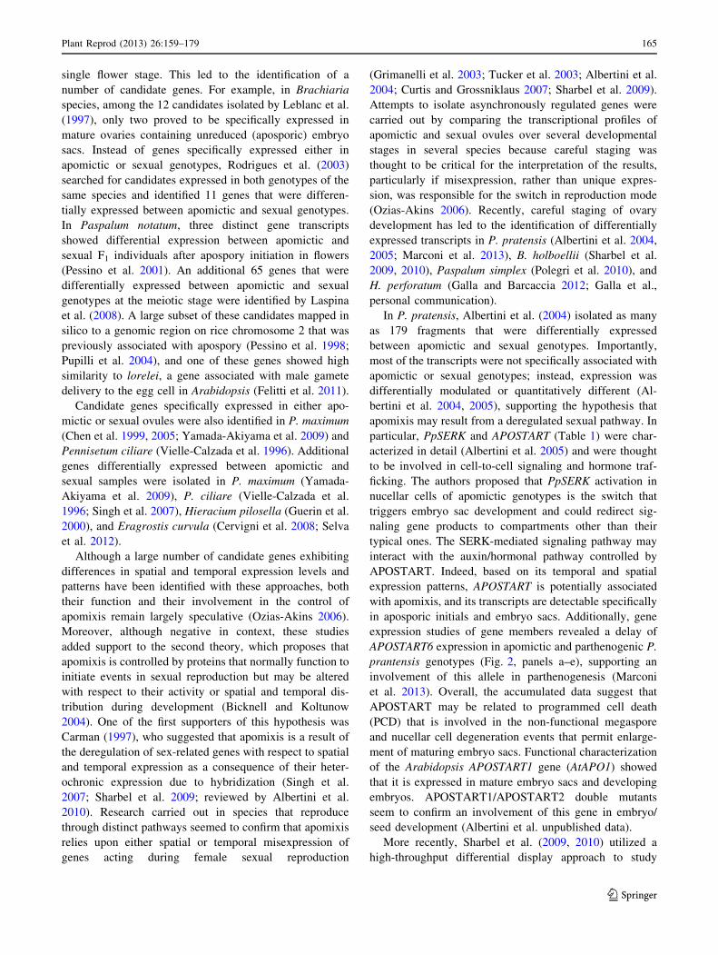
particular, PpSERK and APOSTART (Table 1) were char-
acterized in detail (Albertini et al. 2005) and were thought
to be involved in cell-to-cell signaling and hormone traf-
ficking. The authors proposed that PpSERK activation in
nucellar cells of apomictic genotypes is the switch that
triggers embryo sac development and could redirect sig-
naling gene products to compartments other than their
typical ones. The SERK-mediated signaling pathway may
interact with the auxin/hormonal pathway controlled by
APOSTART. Indeed, based on its temporal and spatial
expression patterns, APOSTART is potentially associated
with apomixis, and its transcripts are detectable specifically
in aposporic initials and embryo sacs. Additionally, gene
expression studies of gene members revealed a delay of
APOSTART6 expression in apomictic and parthenogenic P.
prantensis genotypes (Fig. 2, panels a–e), supporting an
involvement of this allele in parthenogenesis (Marconi
et al. 2013). Overall, the accumulated data suggest that
APOSTART may be related to programmed cell death
(PCD) that is involved in the non-functional megaspore
and nucellar cell degeneration events that permit enlarge-
ment of maturing embryo sacs. Functional characterization
of the Arabidopsis APOSTART1 gene (AtAPO1) showed
that it is expressed in mature embryo sacs and developing
embryos. APOSTART1/APOSTART2 double mutants
seem to confirm an involvement of this gene in embryo/
seed development (Albertini et al. unpublished data).
More recently, Sharbel et al. (2009, 2010) utilized a
high-throughput differential display approach to study
Plant Reprod (2013) 26:159–179 165
123
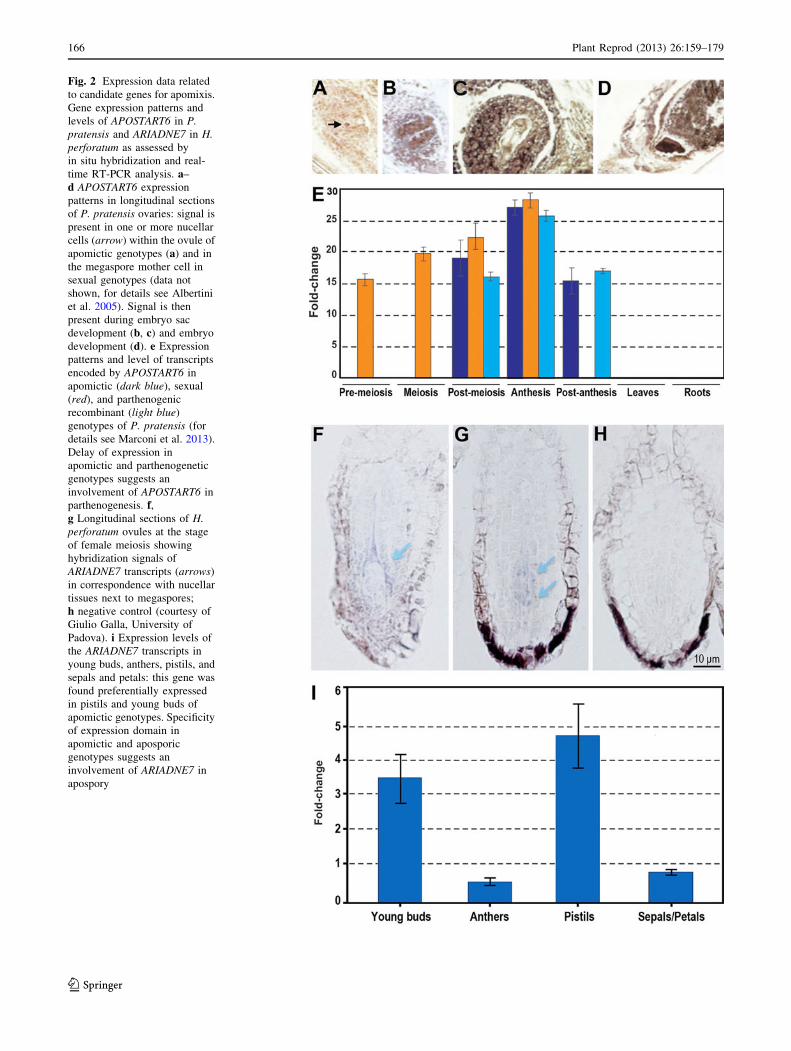
Fig. 2 Expression data related
to candidate genes for apomixis.
Gene expression patterns and
levels of APOSTART6 in P.
pratensis and ARIADNE7 in H.
perforatum as assessed by
in situ hybridization and real-
time RT-PCR analysis. a–
d APOSTART6 expression
patterns in longitudinal sections
of P. pratensis ovaries: signal is
present in one or more nucellar
cells (arrow) within the ovule of
apomictic genotypes (a) and in
the megaspore mother cell in
sexual genotypes (data not
shown, for details see Albertini
et al. 2005). Signal is then
present during embryo sac
development (b, c) and embryo
development (d). e Expression
patterns and level of transcripts
encoded by APOSTART6 in
apomictic (dark blue), sexual
(red), and parthenogenic
recombinant (light blue)
genotypes of P. pratensis (for
details see Marconi et al. 2013).
Delay of expression in
apomictic and parthenogenetic
genotypes suggests an
involvement of APOSTART6 in
parthenogenesis. f,g Longitudinal sections of H.
perforatum ovules at the stage
of female meiosis showing
hybridization signals of
ARIADNE7 transcripts (arrows)
in correspondence with nucellar
tissues next to megaspores;
h negative control (courtesy of
Giulio Galla, University of
Padova). i Expression levels of
the ARIADNE7 transcripts in
young buds, anthers, pistils, and
sepals and petals: this gene was
found preferentially expressed
in pistils and young buds of
apomictic genotypes. Specificity
of expression domain in
apomictic and aposporic
genotypes suggests an
involvement of ARIADNE7 in
apospory
166 Plant Reprod (2013) 26:159–179
123

naturally occurring quantitative variations in gene expres-
sion between ovules of apomictic and sexual B. holboellii
genotypes and identified 543 genes exhibiting a develop-
mental shift in expression between the sexual and apome-
iotic ovules. A carefully devised experiment was also
undertaken by Polegri et al. (2010), leading to the identi-
fication of a set of genes in P. simplex with stage- and/or
phenotype-specific expression. In particular, a class of
alleles that showed a characteristic specificity of expression
was fully linked to apomixis based on mapping data.
In H. perforatum, a differential display analysis of
sporogenesis and gametogenesis led to the isolation of
several transcripts specifically expressed in the pistils of a
highly apomictic ecotype, including an EST showing
similarity to a gene coding for an ATPase RNA helicase
responsible for an embryo defective phenotype in Arabid-
opsis (MEE29, maternal effect embryo). This gene, termed
HpMEE29-like, was differentially expressed between
aposporic and meiotic H. perforatum plants (Barcaccia
et al. 2007). More recently, Galla and Barcaccia (2012)
adopted high-throughput 454 technology to sequence the
entire Hypericum flower transcriptome using single verti-
cils collected from apomictic and sexual genotypes.
Computational procedures were used to assemble and
annotate more than 25,000 transcripts exclusively from
anthers and carpels at different developmental stages. Galla
et al. (2013) identified dozens of genes related to sporo-
genesis and gametogenesis, with particular reference to the
formation of embryo sacs, embryos, and seeds. Interest-
ingly, some of the transcripts showed sequence homology
with candidate genes for apomixis that were cloned in
apomictic species and mutants, including components of
the HAPPY locus (Schallau et al. 2010). Notably, many
small transcripts of genes specifically expressed in apo-
mictic genotypes exhibited high similarity to microRNA
precursors that target specific transcription factors. In
particular, the most represented and conserved families
were miR156, miR166, miR390, miR394, miR396, and
miR414, which have dozens of potential target genes with
a wide range of molecular functions including metabolism,
response to stress, flower development, and plant repro-
duction Galla et al. (2013).
Different approaches were chosen in Pennisetum squa-
mulatum (Conner et al. 2008) and H. perforatum (Schallau
et al. 2010). In P. squamulatum, by sequencing an aposp-
ory-specific genomic region (ASGR), Conner et al. (2008)
isolated a gene sharing similarity with BABY BOOM
(BBM), which was named ASGR–BBM (Table 1). This
gene encodes a protein containing two AP2 domains that
are 96 % similar to the AP2 regions of BnBBM (outside the
AP2 domains, the similarity of ASGR-BBM to BnBBM
declines significantly to 35 and 27 % in the upstream and
downstream regions, respectively). In H. perforatum,
Schallau et al. (2010) screened genomic clones using an
apospory-linked SCAR marker as probe and identified a
142 kb BAC clone containing a gene homologous to
Arabidopsis ARIADNE7 (ARI7, Table 1), which is anno-
tated as a nucleic acid binding protein. In particular, both
aposporic- and sexual-specific HpARI7 alleles were found
co-expressed in the pistils at different developmental
stages, whereas the gene product of the apomictic allele
was specifically expressed in pistils of the apomictically
reproducing individuals (Schallau et al. 2010). More
recently, Galla et al. (personal communication) demon-
strated that HpARI7 gene is preferentially expressed in
pistils at different developmental stages and that HpARI7
transcripts are specifically detectable in nucellar tissues of
the ovule next to megaspores in apomictic H. perforatum
genotypes (Fig. 2, panels f–i). Specificity of the expression
domain in apomictic and aposporic genotypes suggests an
involvement of this gene belonging to the HAPPY locus in
apospory.
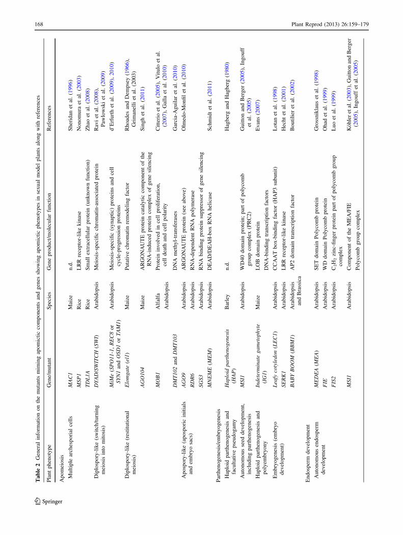
Functional analysis of genes miming apomixis in sexual
model plants
If it is true that apomixis is a consequence of sexual failure,
rather than a means for clonal success, from an evolu-
tionary point of view (Silvertown 2008), it is also true that
apomixis, as a biological process of seed formation, rep-
resents an altered form of sexuality rather than a new
developmental program (Koltunow and Grossniklaus
2003). Carman (1997) hypothesized that apomixis is a
result of the spatial and temporal deregulation of sex-
related gene expression as a consequence of heterochronic
expression due to hybridization. This hypothesis was con-
firmed experimentally by cytological observations in
Tripsacum (Grimanelli et al. 2003) and more recently by
transcriptome analyses in Boechera (Sharbel et al. 2009,
2010). Additionally, Tucker et al. (2003) provided evi-
dence that marker genes related to embryo sac develop-
ment are similarly expressed in sexual and apomictic
Hieracium genotypes.
The idea that apomixis is an altered form of sexuality
that results from temporal and spatial alterations in the
action of the sexual program suggests that a synthetic
apomixis system using variant alleles of genes isolated
from sexual model species, such as Arabidopsis, can be
developed (Chaudhury and Peacock 1993). By identifying
and combining the genes involved in apomixis, the
assembly of an asexual system of seed formation in a
sexual plant should be possible. In particular, some of the
genes isolated and characterized from sexual species may
have roles in apomixis (Table 2). For example, the recent
findings regarding the molecular mechanisms controlling
Plant Reprod (2013) 26:159–179 167
123
Ta
ble
2G
ener
alin
form
atio
no
nth
em
uta
nts
mim
ing
apo
mic
tic
com
po
nen
tsan
dg
enes
sho
win
gap
om
icti
cp
hen
oty
pes
inse
xu
alm
od
elp
lan
tsal
on
gw
ith
refe
ren
ces
Pla
nt
ph
eno
typ
eG
ene/
mu
tan
tS
pec
ies
Gen
ep
rod
uct
/mo
lecu
lar
fun
ctio
nR
efer
ence
s
Ap
om
eio
sis
Mu
ltip
lear
ches
po
rial
cell
sM
AC
1M
aize
n.d
.S
her
idan
etal
.(1
99
6)
MS
P1
Ric
eL
RR
rece
pto
r-li
ke
kin
ase
No
no
mu
raet
al.
(20
03
)
TD
L1
AR
ice
Sm
all
extr
acel
lula
rp
rote
in(u
nk
no
wn
fun
ctio
n)
Zh
aoet
al.
(20
08
)
Dip
losp
ory
-lik
e(s
wit
ch/t
urn
ing
mei
osi
sin
tom
ito
sis)
DY
AD
/SW
ITC
H(S
WI)
Ara
bid
op
sis
Mei
osi
s-sp
ecifi
cch
rom
atin
-ass
oci
ated
pro
tein
Rav
iet
al.
(20
08
),
Paw
low
ski
etal
.(2
00
9)
MiM
e(S
PO
11-1
,R
EC
8o
r
SY
N1
and
OS
D1
or
TA
M1)
Ara
bid
op
sis
Mei
osi
s-sp
ecifi
c(s
yn
apti
c)p
rote
ins
and
cell
cycl
e-p
rog
ress
ion
pro
tein
s
d’E
rfu
rth
etal
.(2
00
9),
20
10
)
Dip
losp
ory
-lik
e(r
esti
tuti
on
al
mei
osi
s)
Elo
mg
ate
(el1
)M
aize
Pu
tati
ve
chro
mat
inre
mo
del
ing
fact
or
Rh
oad
esan
dD
emp
sey
(19
66
),
Gri
man
elli
etal
.(2
00
3)
AG
O1
04
Mai
zeA
RG
ON
AU
TE
pro
tein
cata
lyti
cco
mp
on
ent
of
the
RN
A-i
nd
uce
dp
rote
inco
mp
lex
of
gen
esi
len
cin
g
Sin
gh
etal
.(2
01
1)
MO
B1
Alf
alfa
Ara
bid
op
sis
Pro
tein
inv
olv
edin
cell
pro
life
rati
on
,
cell
dea
than
dce
llp
ola
rity
Cit
teri
oet
al.
(20
05
),V
itu
loet
al.
(20
07
),G
alla
etal
.(2
01
0)
DM
T1
02
and
DM
T1
03
DN
Am
eth
yl-
tran
sfer
ases
Gar
cia-
Ag
uil
aret
al.
(20
10)
Ap
osp
ory
-lik
e(a
po
spo
ric
init
ials
and
emb
ryo
sacs
)
AG
O9
Ara
bid
op
sis
AR
GO
NA
UT
Ep
rote
in(s
eeab
ov
e)O
lmed
o-M
on
fil
etal
.(2
01
0)
RD
R6
Ara
bid
op
sis
RN
A-d
epen
den
tR
NA
po
lym
eras
e
SG
S3
Ara
bid
op
sis
RN
Ab
ind
ing
pro
tein
sup
pre
sso
ro
fg
ene
sile
nci
ng
MN
EM
E(M
EM
)A
rab
ido
psi
sD
EA
D/D
EA
H-b
ox
RN
Ah
elic
ase
Sch
mid
tet
al.
(20
11
)
Par
then
og
enes
is/e
mb
ryo
gen
esis
Hap
loid
par
then
og
enes
isan
d
facu
ltat
ive
pse
ud
og
amy
Ha
plo
idp
art
hen
og
enes
is
(HA
P)
Bar
ley
n.d
.H
agb
erg
and
Hag
ber
g(1
98
0)
Au
ton
om
ou
sse
edd
evel
op
men
t,
incl
ud
ing
par
then
og
enes
is
MS
I1A
rab
ido
psi
sW
D4
0d
om
ain
pro
tein
;p
art
of
po
lyco
mb
gro
up
com
ple
x(P
RC
2)
Gu
itto
nan
dB
erg
er(2
00
5),
Ing
ou
ff
etal
.(2
00
5)
Hap
loid
par
then
og
enes
isan
d
po
lyem
bry
on
y
Ind
eter
min
ate
ga
met
op
hyt
e
(IG
1)
Mai
zeL
OB
do
mai
np
rote
in
DN
A-b
ind
ing
tran
scri
pti
on
fact
ors
Ev
ans
(20
07
)
Em
bry
og
enes
is(e
mb
ryo
dev
elo
pm
ent)
Lea
fyco
tyle
do
n(L
EC
1)
Ara
bid
op
sis
CC
AA
Tb
ox
-bin
din
gfa
cto
r(H
AP
3su
bu
nit
)L
ota
net
al.
(19
98
)
SE
RK
1A
rab
ido
psi
sL
RR
rece
pto
r-li
ke
kin
ase
Hec
ht
etal
.(2
00
1)
BA
BY
BO
OM
(BB
M1
)A
rab
ido
psi
s
and
Bra
ssic
a
AP
2d
om
ain
tran
scri
pti
on
fact
or
Bo
uti
lier
etal
.(2
00
2)
En
do
sper
md
evel
op
men
t
Au
ton
om
ou
sen
do
sper
m
dev
elo
pm
ent
ME
DE
A(M
EA
)A
rab
ido
psi
sS
ET
do
mai
nP
oly
com
bp
rote
inG
ross
nik
lau
set
al.
(19
98
)
FIE
Ara
bid
op
sis
WD
do
mai
nP
oly
com
bp
rote
inO
had
etal
.(1
99
9)
FIS
2A
rab
ido
psi
sC
2H
2zi
nc-
fin
ger
pro
tein
par
to
fp
oly
com
bg
rou
p
com
ple
x
Lu
oet
al.
(19
99
)
MS
I1A
rab
ido
psi
sC
om
po
nen
to
fth
eM
EA
/FIE
Po
lyco
mb
gro
up
com
ple
x
Ko
hle
ret
al.(2
00
3),
Gu
itto
nan
dB
erg
er
(20
05
),In
go
uff
etal
.(2
00
5)
168 Plant Reprod (2013) 26:159–179
123
embryo sac development, fertilization, and endosperm
development may be useful for determining genetic links
with apomeiosis, parthenogenesis, and autonomous or
pseudogamous endosperm development. It is believed that
basic structural and functional analyses of these candidate
genes are crucial for engineering apomixis in sexual crops.
Genes that are expressed during embryo sac develop-
ment, and thus putatively involved in differentiating sexual
from apomictic pathways, are of particular interest. An
embryo sac develops through megaspore mother cell
(MMC) differentiation, meiosis, determination of the
functional megaspore, and embryo and endosperm devel-
opment. Because meiosis is either completely bypassed
(apospory) or extremely altered (diplospory) in apomixis,
genes related to female sporogenesis are thought to be
more specifically involved in diplosporic apomixis, and
genes involved in embryo sac cell identity are presumably
crucial for aposporic apomixis. Overall, genes associated
with female gametogenesis and egg cell development are
likely shared between sexual and apomictic pathways (for
details on these aspects see reviews by Tucker et al. 2003;
Koltunow and Grossniklaus 2003; Ozias-Akins and van
Dijk 2007; Tucker and Koltunow 2009; Albertini et al.
2010; Dwivedi et al. 2010).
Regarding the determination of the megasporocyte, an
analysis of mutations affecting MMC differentiation may
be crucial for understanding the specification of aposporic
initials during female sporogenesis. The Arabidopsis
mutant sporocyteless/nozzle (spl) is unable to develop a
functional MMC and shows defects in nucellar cell identity
(Schiefthaler et al. 1999; Sieber et al. 2004). Moreover,
mutations in the Arabidopsis gene WUSCHEL (WUS), a
regulator of stem cell identity in the shoot apical meristem,
also result in defects in MMC specification (Gross-Hardt
et al. 2002). WUS acts by modulating the expression of
WINDHOSE1 (WIH1) and WINDHOSE2 (WIH2), which
control female sporogenesis in conjunction with TOR-
NADO2 (Lieber et al. 2011). Additionally, the MULTIPLE
ARCHESPORIAL CELLS1 (MAC1) gene of maize (Sheri-
dan et al. 1996, 1999) and the MULTIPLE SPOROCYTES1
(MSP1) and TAPETUM DETERMINANT LIKE1A
(TDL1A) genes in rice (Nonomura et al. 2003; Zhao et al.
2008) are required for the production of only one me-
gasporocyte per single ovule, indicating that these genes
are involved in MMC determination. Loss of gene function
results in multiple MMCs, indicating that these genes
negatively regulate the sporogenous cell fate in the ovule.
Zhao et al. (2002) identified the ortholog of MSP1 in
Arabidopsis (EXTRA SPOROGENOUS CELLS7 EXCESS
MICROSPOROCYTES1, EXS7EMS1); however, its role in
regulating MMC number is not known. MSP1 encodes a
leucine-rich repeat-like kinase (LRR–RLK) (Nonomura
et al. 2003) that has been identified in plants as a
transmembrane protein involved in a complex array of
signaling pathways related to cell differentiation and
developmental events (Dievart and Clark 2004).
In Arabidopsis, an alternative route for restricting the
specification of embryo sac precursors has been proposed
through the action of ARGONAUTE (AGO) genes (Olme-
do-Monfil et al. 2010; Tucker et al. 2012). AGO proteins
are known to be involved in post-transcriptional gene
silencing mediated by short RNAs (either microRNAs or
short interfering RNAs) (Baumberger and Baulcombe
2005). Small RNAs (sRNAs) have recently been studied in
different model systems, and it is now known that muta-
tions in the molecular pathways that generate sRNAs may
dramatically affect fertility (Van Ex et al. 2011; Tucker
et al. 2012). Previous research has demonstrated that strong
mutant alleles of genes involved in the formation and
activity of miRNAs, such as AGO1, DCL1, HEN1, and
HYL1, disrupt reproductive development (reviewed by Van
Ex et al. 2011). However, interpreting these phenotypes is
frequently difficult because such mutations have ectopic
effects and influence different aspects of plant development
(Axtell 2013). Two members of the ARGONAUTE protein
family, AGO5 and AGO9, which are involved in the reg-
ulatory pathway of sRNAs in plants, have been associated
with cell specification and embryo sac development
(Olmedo-Monfil et al. 2010; Tucker et al. 2012). An AGO5
ortholog in rice was reported to be essential for the pro-
gression of pre-meiotic mitosis and meiosis (Nonomura
et al. 2007), and the production of viable gametes without
meiosis was reported in maize lacking the ortholog of
AGO9 (Singh et al. 2011). Some of the genes isolated and
characterized from sexual species may play roles in the
framework of apomixis, and it is possible that sRNAs act
by silencing master genes directly involved in differenti-
ating apomictic from sexual pathways.
Particular Arabidopsis mutants have revealed that
gametogenesis can be uncoupled from meiosis. For
example, loss of certain ARGONAUTE (i.e., AGO9) genes
and other genes in the small RNA pathway, such as RNA-
DEPENDENT RNA POLYMERASE6 (RDR6) and SUP-
PRESSOR OF GENE SILENCING3 (SGS3), resulted in
loss of restriction in gametic cell identity and fate in the
ovule and gain of expression in multiple somatic initials in
the nucellar tissue that can differentiate into gametic cells
without undergoing meiosis and can initiate female
gametogenesis through the activation of TEs (Olmedo-
Monfil et al. 2010). Notably, these TEs are normally
silenced in both developing and fully differentiated Ara-
bidopsis ovules (Slotkin et al. 2009). AGO9 silences TEs in
the embryo sac, and hence, its function resembles that of
the PIWI (P-element-induced wimpy testis in Drosophila)
regulatory proteins responsible for maintaining incomplete
differentiation in stem cells and preserving stable cell
Plant Reprod (2013) 26:159–179 169
123
division rates in the germ line lineage of invertebrates and
mammals (Klattenhoff and Theurkauf 2008). A phenotype
similar to that generated by AGO9 loss-of-function alleles
has also been observed in maize by repressing DMT103
and DMT102, which are homologs of Arabidopsis
DOMAINS REARRANGED METHYLTRANSFERASE
(DRM2) and CHROMOMETHYLASE 3 (CMT3) genes,
respectively (Garcia-Aguilar et al. 2010). These genes are
involved in new DNA methylation in DRM2 sequences and
in the retention of DNA methylation in non-CG sites in
Arabidopsis (Cao and Jacobsen 2002). Although the
function of these genes is unknown in maize, they could
play a role in ensuring that a single embryo sac develops
within each ovule. This mechanism could be epigenetically
regulated through DNA methylation (Pillot et al. 2010a, b).
Evidence regarding the role of auxin in the cell fate
specification of embryo sac development has recently been
obtained. In Arabidopsis, two YUCCA (YUC) genes
encoding proteins that are crucial for local auxin biosyn-
thesis were shown to be expressed in the ovule, which is
consistent with the role of auxin as a cell fate determinant
(Pagnussat et al. 2009). Owing to the high concentrations
of auxin detected in the distal tip of the nucellus at early
stages of ovule development (Pagnussat et al. 2009) and
because SPOROCYTELESS/NOZZLE (SPL) was demon-
strated to repress the expression of YUCCA genes (Li et al.
2008), it is likely that auxin plays a key role in the cell
specification machinery that regulates differentiation of the
MMC and/or maintains the undifferentiated state of
nucellar cells once the MMC is formed.
Proper MMC formation involves two distinct pathways:
the first involves a commitment to the specification of a
MMC from a somatic cell (i.e., cellular identity determi-
nation), and the second involves a commitment to the dif-
ferentiation of an MMC from a single cell (i.e., cellular type
specialization). Knowledge of key genes involved in both
pathways should provide the molecular tools necessary for
developing an artificial apomictic system in sexual plants.
In Arabidopsis, as in most angiosperms, the MMC
undergoes regular meiosis and gives rise to a tetrad of
haploid megaspores, of which three usually degenerate and
one becomes the functional megaspore. Several loss-of-
function phenotypes related to megasporogenesis were
recently discovered in Arabidopsis and monocots such as
rice and maize, with each showing some features of dip-
lospory. For example, in the rice mutant meiosis arrested at
leptotene1 (mel1), the MMCs arrested megasporogenesis at
pre-meiotic or eventually at meiotic stages (Nonomura
et al. 2007). The gene controlling the mel1 phenotype
belongs to the ARGONAUTE (AGO) gene family, which is
involved in several developmental processes in plants via
the action of sRNAs (Vaucheret 2008). Another gene that
is critical for proper megasporogenesis in Arabidopsis is
DYAD/SWITCH1 (SWI1), which is responsible for sister
chromatid cohesion and centromere organization at meio-
sis. An allelic variant of this gene, dyad, proved to be
responsible for the production of few unreduced egg cells
(Ravi et al. 2008). Fully penetrant diplospory-like pheno-
types were induced in Arabidopsis by replacing meiosis
with mitosis in MiMe genotypes (d’Erfurth et al. 2009,
2010). In particular, these genotypes combine mutations in
the two genes SPO11-1, which prevents chromosome
pairing and recombination, and REC8 (also known as
SYN1), which modifies chromatid segregation along with
OSD1 (MiMe-1 mutant; d’Erfurth et al. 2009) or CYCA1-2/
TAM (MiMe-2 mutant; d’Erfurth et al. 2010). Similar to
dyad, these mutants were shown to form unreduced egg
cells because they did not undergo a second meiotic divi-
sion. Singh et al. (2011) uncovered a maize mutant,
Dominant non-reduction 4 (Dnr4), that manifested defects
in chromatin condensation during meiosis with subsequent
failure of chromosome complement segregation, suggest-
ing that this mutation affects the function of chromatin
remodeling factors. After substitution of a meiosis division
for a mitosis-like division, these mutants formed functional
unreduced egg cells that exhibited phenotypes resembling
diplospory, which is similar to what was observed in the
maize elongate (el1) mutant (Rhoades and Dempsey 1966)
and the natural apomict Tripsacum (Grimanelli et al. 2003).
In maize, the Dnr4 mutant phenotype arises due to
lesions in the AGO104 gene, which is the ortholog of
Arabidopsis AGO9. Together with MEL1, these genes
belong to the ARGONAUTE gene family. The abnormal
patterns of cell specification mediated by the lack of AGO9
and AGO104 are reminiscent of aposporic and diplosporic
apomixis, respectively, suggesting that these natural
apomeiotic variants might rely on similar regulatory
pathways although they are characterized by fundamental
differences.
Concerning the selection of the functional megaspore, in
Arabidopsis as in most of Angiosperms, the MMC under-
goes regular meiosis and gives rise to a linear tetrad of
haploid megaspores. Three of these megaspores (usually
those located in the micropylar area) degenerate, and only
one becomes the functional megaspore that undergoes
meiosis. A distinctive process that occurs in aposporic
species (e.g., Paspalum spp., H. perforatum) is the
degeneration of all four megasporocytes while the already
advanced aposporic embryo sacs develop further; in dip-
losporic species, unreduced megaspores originate via re-
stitutional meiosis (i.e., Taraxacum-type, which is also
widespread in Boechera spp.) or via a complete bypass of
meiosis (i.e., Antennaria-type, also common in Tripsacum
dactyloides).
In diplospory, abnormal meiosis is largely asynaptic
because of the absence of pairing between homologous
170 Plant Reprod (2013) 26:159–179
123
chromosomes and therefore results in a restitution nucleus
in the first division and the formation of a dyad of unre-
duced megaspores (for a review, see Albertini et al. 2010).
In addition to diplospory, several molecular mechanisms
associated with apospory may also explain the specificity
of megasporocyte degeneration, including position-depen-
dent degeneration, cell polarization changes, and PCD
(reviewed by Pupilli and Barcaccia 2012).
The easiest explanation for the occurrence of apomeiosis
could be the fact that the chalazal megaspore has prefer-
ential access to nutritional elements or regulatory factors
because it is closest to the maternal tissues. In aposporic
ovules, the hierarchical position of the linear tetrad of
megaspores could be perturbed by the growth of multiple
embryo sacs that compete for nutrients and growth factors
(Pupilli and Barcaccia 2012). The question remains, what
happens in diplosporic ovules when meiosis is altered or
bypassed? Molecular studies in Arabidopsis have sug-
gested that the degeneration and death of the micropylar
megaspore and the identity determination of the functional
megaspore may be controlled by a positional signaling
pathway (Yang and Sundaresan 2000) and/or a polarity
effect, as manifested by the distribution pattern of organ-
elles and microtubules of the cytoskeleton during mega-
sporogenesis (Bajon et al. 1999).
Among the characteristic features of cells undergoing
PCD, DNA degradation and changes in Ca2? accumulation
dynamics have been observed in degenerating megaspores
(as reviewed by Tucker and Koltunow 2009). In lettuce,
differences between Ca2? accumulation pathways in the
degenerating micropylar megaspore and the chalazal
megaspore were observed, and the latter retained a sig-
nificantly higher concentration of calcium (Qiu et al. 2008).
Some members of the MPS-ONE-BINDER (MOB1) gene
family in diplosporic mutants of Medicago sativa were
specifically expressed in degenerating megaspores of nor-
mal ovules and in enlarged megaspore mother cells and
embryo sacs of apomeiotic ovules (Citterio et al. 2005,
2006). MOB1 gene products were also found in microspore
tetrads at the beginning of pollen development and in the
tapetum cells of anthers undergoing PCD to allow pollen
dispersal at maturity. Overall, the results suggest that
MOB1 genes can play a key role in the reproductive
pathway in plants. MOB proteins are involved in cell cycle
progression and PCD in Drosophila and mammals (Hira-
bayashi et al. 2008; Vitulo et al. 2007).
In Arabidopsis and some apomictic species, one factor
putatively associated with the selective degeneration of
meiotic megaspores is regulated by callose boundaries and
plates that can accumulate at one pole or around the MMC
and in the transverse cell walls between the functional
megaspore and its degenerated sister megaspores (Olmedo-
Monfil et al. 2010); these boundaries and plates can also
form next to the unreduced megaspore of apomeiotic dyads
in Medicago diplosporic mutants (Albertini and Barcaccia
2007). Another factor is the ANTIKEVORKIAN (AKV)
gene that, when mutated, allowed for the survival of all
four megaspores in 10 % of Arabidopsis ovules (Yang and
Sundaresan 2000). However, in Paspalum and other
aposporic grasses, complete suppression of the sexual
pathways does not always occur (Hojsgaard et al. 2008,
2013). As a consequence, different degeneration patterns of
megaspores in apomictic and sexual lineages cannot be
governed by specific apomixis-related genes. This process
is more likely to be pleiotropically affected by a gene
cascade triggering the apomictic pathway; alternatively,
the sexual megaspores could be mechanically disrupted by
the overgrowing aposporic embryo sacs.
Another crucial step in apomictic reproduction is the
initiation and autonomous development of the embryo
combined with the formation of the endosperm. Parthe-
nogenesis, as a fertilization-independent form of embryo
development, is a key component of aposporic embryo
development, and it is the only feature that aposp-
oric development has in common with diplosporic embryo
development. Parthenogenesis has been widely observed in
nature; however, in sexual plants, it generally occurs at a
low rate in haploid egg cells (Lacadena 1974). A general
rule in zygotic development is that the activation and
consequent initiation of embryogenesis is trigged by the
fertilization of an egg cell. In animals, egg cell fertilization
induces an increase in Ca2? levels at the site of sperm cell
entry that propagates throughout the egg cell, inducing a
downstream cascade of signals required to initiate
embryogenesis (Miyazaki and Ito 2006). In plants, an
increase in intracellular Ca2? was observed after egg cell
fertilization (Antoine et al. 2000); however, this was not
sufficient to trigger parthenogenesis (Curtis and Gross-
niklaus 2008).
Parallel mutant screens for apomixis enabled the iden-
tification of genes controlling the fertilization-independent
initiation of seed development in Arabidopsis. These
genes, termed FERTILIZATION-INDEPENDENT SEEDS
(FIS) genes, encode protein members of the Polycomb-
related complex (Koltunow and Grossniklaus 2003). The
fis mutants are known to initiate endosperm development
without fertilization to varying extents. However, the fre-
quency of embryo initiation by division of an egg cell is
either low or does not occur at all in most fis mutants.
Several natural mutations are known to induce the par-
thenogenic development of embryos in plants when the
zygote is forced to begin its growth in a haploid environ-
ment. The ‘‘Salmon’’ system in wheat produces high
numbers of haploid parthenogenic embryos (Matzk 1996),
and the haploid inducer (hap) mutant of barley is associ-
ated with parthenogenesis (Hagberg and Hagberg 1980).
Plant Reprod (2013) 26:159–179 171
123
More recently, it was shown that parthenogenic embryos
can be generated at a relatively high frequency in trans-
genic lines of Arabidopsis expressing a modified centro-
mere-specific histone CENH3 protein (Ravi and Chan
2010). However, the question of whether these findings
could be ascribable to apomictic features in wild apomicts
is unclear because these mutations are related to the hap-
loid condition of the zygote. In Arabidopsis, the ubiquitous
overexpression of several transcription factors induces
ectopic formation of embryo-like structures. LEAFY
COTYLEDON (LEC1 and LEC2) genes induce the
expression of embryo-specific genes and trigger the
development of embryo-like structures (Stone et al. 2001).
PICKLE (PKL) is an upstream regulator of LEC genes that
acts as repressor of embryogenesis (Henderson et al. 2004),
and BABY BOOM (BBM) is an AP2-domain transcription
factor that when overexpressed leads to the development of
embryos and cotyledons from vegetative tissues (Boutilier
et al. 2002). Moreover, the homeodomain protein encoded
by WUSCHEL (WUS) is involved in promoting the vege-
tative to embryogenic transition and/or maintaining the
identity of embryonic stem cells (Zuo et al. 2002). How-
ever, no experimental evidence supports similar functions
for these genes in the egg cell or zygote. Of the genes
proven to promote somatic embryogenesis in vegetative
tissues, only the Arabidopsis ortholog of carrot SOMATIC
EMBRYOGENESIS RECEPTOR KINASE (SERK) was
expressed during gametogenesis and early zygotic
embryogenesis (Hecht et al. 2001; Albertini et al. 2005).
However, a clear phenotype could not be observed when
the SERK gene was ectopically expressed in Arabidopsis
(Hecht et al. 2001).
Currently, it is well known that the female gametophyte
controls embryo and/or endosperm development at two
different levels: (1) repression of embryo/endosperm
development in the absence of fertilization through
imprinting and (2) expression of factors that are required
after fertilization. FERTILIZATION-INDEPENDENT
ENDOSPERM (FIE) (Ohad et al. 1999), MEDEA (MEA)
(Grossniklaus et al. 1998), and FERTILIZATION-INDE-
PENDENT SEED2 (FIS2) (Chaudhury et al. 1997) repress
endosperm development in the absence of fertilization.
Mutations in another member of this group, MULTICOPY
SUPPRESSOR OF IRA (MSI1), induce the formation of
rudimentary parthenogenic embryos from growing and
dividing egg cells and lead to the repressive FIS gene
phenotype. Nevertheless, these parthenogenic embryos
abort at an early stage (Guitton and Berger 2005). All these
genes demonstrate homology with the Polycomb group
(PcG) of genes that are involved in repressing development
in Drosophila through chromatin remodeling mechanisms
(Schwartz and Pirrotta 2007). The FIE/FIS2/MEA complex
(FIS complex) acts by repressing the transcription of target
genes involved directly in endosperm development. One of
these genes, PHERES1 (PHE1), encodes a MADS domain-
containing protein. MEA and FIE proteins interact directly
with the PHE1 promoter (Kohler et al. 2003). All fis
mutations show aberrant embryo and endosperm develop-
ment if fertilized and exhibit autonomous endosperm
development if unfertilized; however, the seeds abort
irrespective of the paternal contribution.
Imprinting, or parent-specific expression of genes, is a
mechanism by which the embryo sac controls the early
stages of seed development. MEA was the first documented
example of an imprinted gene involved in seed formation
(Grossniklaus et al. 1998) in which the paternal allele is
silenced and the maternal allele is expressed in the central
cell. Both parental alleles are then silenced, and the mod-
ifications are removed in the central cell by a protein
encoded by DEMETER (DME), a DNA glycosylase/lyase
related to the DNA repair protein family (Choi et al. 2002).
All genes of the FIS complex act through an epigenetically
regulated mechanism to maintain the quiescent status of the
central cell in the absence of fertilization. MULTICOPY
SUPPRESSOR OF IRA (MSI1), another PcG gene prod-
uct, participates together with its interacting cell cycle
control protein RETINOBLASTOMA-RELATED (RBR)
in the molecular machinery that represses autonomous
endosperm development (Ebel et al. 2004). The MSI1/RBR
complex regulates the methylation state of DNA during
gametogenesis by repressing the transcription of a DNA
methylase that normally controls methylation during
embryo sac formation in Arabidopsis (Johnston et al. 2008;
Jullien et al. 2008).
In plants, two main strategies have been adopted to
demethylate DNA during gamete formation in ovules. One
of these strategies, mediated by the MEA/DME system, is
specific to the central cell, whereas the other strategy,
controlled by the MSI1/RBR complex, is genomewide,
affecting both the egg cell and the central cell (Sundaresan
and Alandete-Saez 2010). Both mechanisms are likely to
play key roles in maintaining the parent-of-origin-depen-
dent expression of imprinted genes during gametogenesis.
Therefore, it is possible that complete maternal control of
reproduction (i.e., apomixis) could be achieved by modi-
fying the epigenetic program that controls imprinting
(Bicknell and Koltunow 2004). This assumption, together
with the fact that down-regulation of imprinted genes in the
FIS complex mimics aspects of apomictic reproduction,
such as autonomous embryo and endosperm development,
prompted research aimed at linking their expression to
apomictic reproduction. For example, the expression pro-
files of FIS complex genes were proven to be similar in
apomictic and sexual lines of Hieracium (Tucker et al.
2003). Moreover, down-regulation of the Arabidopsis FIE
ortholog in Hieracium did not result in any autonomous
172 Plant Reprod (2013) 26:159–179
123
endosperm development, although its activity was neces-
sary for regular seed formation in both apomictic and
sexual lines (Rodrigues et al. 2008). Similarly, gene
expression studies of the Hieracium homolog of Arabid-
opsis, MSI1, in germ lineages of apomictic and sexual lines
demonstrated that it is not associated with autonomous
seed formation in this species (Rodrigues et al. 2010a). In
fact, autonomous endosperm development was not induced
in sexual species such as rice and maize by genetic trans-
formation of OsFIE1 (Luo et al. 2009) or by down-regu-
lation of ZmFIE1 and ZmFIE2 (Rodrigues et al. 2010b).
Overall, the current findings indicate that the repressive
function of the FIS complex is not conserved beyond
Arabidopsis and that FIS genes are not directly involved in
triggering apomixis in sexual species.
In conclusion, factors delivered by paternal sperm cells
could provide molecular cues that link egg cell fertilization
to the first zygotic divisions. This scenario leads to the
hypothesis that paternal–maternal triggering mechanisms
could have a role in the repression of embryo development
in the absence of fertilization. Moreover, epigenetic
mechanisms of inactivation or down-regulation of genes by
DNA methylation could represent another level of control
by which the egg cell is maintained in a quiescent state in
the absence of fertilization.
Concluding remarks
Recent molecular studies aimed at understanding the basis
of apomixis have failed to properly elucidate its central
mystery because most apomicts are not agriculturally
important crops and do not have agriculturally important
relatives. Additionally, no apomicts have been sequenced
so far, therefore genome annotation information is still not
available. If it is true that zygotic embryogenesis (sexual-
ity) and apomeiotic parthenogenesis (apomixis) follow
similar pathways during embryo and seed development, it
is also true that specific genes have to be activated, mod-
ulated, or silenced in the primary steps of plant reproduc-
tion to ensure that functioning embryo sacs develop from
apomeiotic rather than meiotic cells. Other genes could be
specifically or differentially expressed in sexual and apo-
mictic plants during embryo and endosperm development.
The main approaches that have been followed to study
the molecular basis of apomixis address the isolation of
genes that prime the expression of apomixis in natural
apomicts and/or the identification of genes that mime the
features of apomictic pathways when they are deregulated
in model sexual systems. To take advantage of this fun-
damental knowledge of apomixis with the aim of trans-
ferring it into sexual crops, three main strategies have been
adopted to date: (1) the direct introgression of apomixis
into crop plants by means of conventional breeding
schemes; (2) the genetic transformation of crop plants by
transferring exogenous genes that control the expression of
apomixis; and (3) the genetic transformation of crop plants
by deregulating the endogenous genes that trigger the
expression of apomixis.
Although traditional breeding has taken advantage of
crop species with close apomictic relatives, transferring
apomixis to sexual plants has been unsuccessful to date
(Savidan 2000). The introgression of apomixis into crop
species from wild relatives failed mainly because natural
apomicts are characterized by hybridity and polyploidy, and
the loci controlling apomixis usually have a simple inheri-
tance but a complex structure. In fact, these loci are
apparently located in very large chromosomal regions,
which make them recalcitrant to recombination-based
genetic mapping strategies in addition to complicating their
physical cloning. In the case of Tripsacum, the current
evidence suggests the presence of barriers in the transfer of
apomixis to maize (Leblanc et al. 2009). The greatest pro-
gress has been made with the introgression of apomixis into
pearl millet from Pennisetum, where a single alien chro-
mosome persists in the most advanced apomictic lines
(Singh et al. 2010; Zeng et al. 2011). In the sexual model
plant Arabidopsis, for each element of apomixis identified
by mutation so far, the penetrance was shown to be gener-
ally low and the related candidate genes were never found to
be associated with any feature of apomixis in natural
apomicts. Most importantly, the three main components of
apomixis could not be successfully combined into a single
model plant to date. Consequently, although findings in
Arabidopsis represent the first steps toward the synthesis of
an artificial apomixis system, the engineering of asexual
reproduction in Arabidopsis has not yet been accomplished.
Because there are a large number of candidates, it is
likely that a cross-check between apomictic species should
be carried out to assess both analogous and unique genes.
In fact, if some of these genes are truly involved in apo-
mixis, their functions should be conserved in other species.
For example, Laspina et al. (2008) carried out a bioinfor-
matics comparison among their own sequences and those
reported by Albertini et al. (2004, 2005), identifying five
genes with identical annotations. Several of the genes that
are differentially expressed in both Paspalum spp. and P.
pratensis appear to be involved in an ERK signal trans-
duction cascade, with deviations controlled by a Ras
ortholog and phospholipase C. Moreover, to further con-
firm the involvement of APOSTART in apomixis, partial/
complete cDNA fragments showing sequence homology
with this gene were isolated in P. squamulatum, Cenchrus
ciliaris and H. perforatum. In all three species, an APO-
START member was found expressed specifically in par-
thenogenic individuals at the stage when embryo develops
Plant Reprod (2013) 26:159–179 173
123

(Marconi et al. 2013). A similar bioinformatics comparison
was also performed between the aposporic species P.
simplex and H. perforatum, revealing good candidates for
apomixis (located on the ACR, apomixis controlling region
of Paspalum, and linked to the HAPPY locus of Hyperi-
cum) shared by these two model systems (Schallau et al.
2010; Calderini et al. 2012). In particular, this gene, known
in Arabidopsis to be involved in the initiation of DNA
replication and to be regulated transcriptionally during cell
cycle, is currently under investigations in both species
(Galla and Pupilli, personal communication).
We are confident that a novel perspective on the old
dilemma of apomixis can emerge not only from a rigorous
cross-check among apomixis candidate genes cloned in
natural apomictic species but also from a high-throughput
analysis of sexual mutants in which pivotal genes control
the expression of apomictic components, such as apomei-
osis (both apospory and diplospory), parthenogenesis, and/
or autonomous endosperm development. The link between
apomixis and gene-specific silencing mechanisms, includ-
ing chromatin remodeling factors or trans-acting and het-
erochromatic small interfering RNAs involved in both
transcriptional and post-transcriptional gene regulation, is
beginning to become clear. In fact, merging lines of evi-
dence regarding the role of microRNAs in the control of
transcription factors, which act on genes directly involved
in the development of embryo sacs, embryos, and seeds,
have been reported in Arabidopsis.
Our opinion is that the final steps toward understanding
the genetic system controlling apomixis are now being
taken, and upcoming research programs will be crucial for
reaching a turning point that could actually represent the
‘‘year zero’’ from which apomixis begins to be successfully
introduced or mimed in sexual species and hence utilized in
the most important crop species to support agricultural and
food interests worldwide.
Acknowledgments Authors want to thank COST Action FA0903
‘‘Harnessing Plant Reproduction for Crop Improvement.’’ Gianni
Barcaccia was supported by the following grant: Research Project of
the University of Padova (year 2012), ‘‘Transcriptomics of repro-
ductive organs in model species for comparative analysis of the
genetic-molecular factors characterizing sexual and apomictic pro-
cesses’’ (code: CPDA128282/12).
Open Access This article is distributed under the terms of the
Creative Commons Attribution License which permits any use, dis-
tribution, and reproduction in any medium, provided the original
author(s) and the source are credited.
References
Akiyama Y, Conner JA, Goel S, Morishige DT, Mullet JE, Hanna
WW, Ozias-Akins P (2004) High-resolution physical mapping in
Pennisetum squamulatum reveals extensive chromosomal het-
eromorphism of the genomic region associated with apomixis.
Plant Physiol 134:1733–1741
Albertini E, Barcaccia G (2007) Apomeiosis in Poa pratensis L. and
Medicago sativa L. In: Horandl E, Grossniklaus U, Sharbel T,
van Dijk P (eds) Apomixis: evolution, mechanisms and
perspectives. Gantner, Ruggel [Regnum Veg. 147], pp 93–116
Albertini E, Porceddu A, Ferranti F, Reale L, Barcaccia G, Falcinelli
M (2001) Apospory and parthenogenesis may be uncoupled in
Poa pratensis L: cytological and genetic evidences. Sex Plant
Reprod 14:213–217
Albertini E, Marconi G, Barcaccia G, Raggi L, Falcinelli M (2004)
Isolation of candidate genes for apomixis in Poa pratensis L.
Plant Mol Biol 56:879–894
Albertini E, Marconi G, Reale L, Barcaccia G, Porceddu A, Ferranti
F, Falcinelli M (2005) SERK and APOSTART: candidate genes
for apomixis in Poa pratensis L. Plant Physiol 138:2185–2199
Albertini E, Barcaccia G, Mazzucato A, Sharbel TF, Falcinelli M
(2010) Apomixis in the era of biotechnology. In: Pua EC, Davey
MR (eds) Plant developmental biology—biotechnological per-
spectives, vol 1. Springer, Berlin, pp 405–436
Antoine AF, Faure J-E, Cordeiro C, Dumas C, Rougier M, Feijo JA
(2000) A calcium influx is triggered and propagates in the zygote
as a warfront during in vitro fertilization of flowering plants.
Proc Natl Acad Sci USA 97:10643–10648
Asker SE, Jerling L (1992) Apomixis in plants. CRC Press, Boca
Raton, FL
Axtell MJ (2013) Classification and comparison of small RNAs from
plants. Annu Rev Plant Biol 64:137–159
Bajon C, Horlow C, Motamayor J, Sauvanet A, Robert D (1999)
Megasporogenesis in Arabidopsis thaliana L: an ultrastructural
study. Sex Plant Reprod 12:99–109
Barcaccia G, Mazzucato A, Albertini E, Zethof J, Pezzotti M, Gerats
A, Falcinelli M (1998) Inheritance of parthenogenesis in Poa
pratensis L: auxin test and AFLP linkage analyses support
monogenic control. Theor Appl Genet 97:74–82
Barcaccia G, Mazzucato A, Falcinelli M (2000) Inheritance of
apomictic seed production in Kentucky bluegrass (Poa pratensis
L). J New Seeds 2:43–58
Barcaccia G, Arzenton F, Sharbel TF, Varotto S, Parrini P, Lucchin M
(2006) Genetic diversity and reproductive biology in ecotypes of
the facultative apomict Hypericum perforatum L. Heredity
96:322–334
Barcaccia G, Baumlein H, Sharbel TF (2007) Apomixis in St. John’s
wort: an overview and glimpse towards the future. In: Horandl E,
Grossniklaus U, Van Dijk P, Sharbel TF (eds) Apomixis.
Mechanisms and perspectives. International Association of Plant
Taxonomy, Koeltz Scientific Books, Vienna, pp 259–280
Baumberger N, Baulcombe DC (2005) Arabidopsis ARGONA-
UTE1 is an RNA Slicer that selectively recruits microRNAs
and short interfering RNAs. Proc Natl Acad Sci USA
102:11928–11933
Bicknell RA, Koltunow AM (2004) Understanding apomixis: recent
advances and remaining conundrums. Plant Cell 16:S228–S245
Bicknell RA, Borst NK, Koltunow AM (2000) Monogenic inheritance
of apomixis in two Hieracium species with distinct develop-
mental mechanisms. Heredity 84:228–237
Boutilier K, Offringa R, Sharma VK, Kieft H, Ouellet T, Zhang L,
Hattori J, Liu CM, van Lammeren AA, Miki BL, Custers JB, van
Lookeren Campagne MM (2002) Ectopic expression of BABY
BOOM triggers a conversion from vegetative to embryonic
growth. Plant Cell 14:1737–1749
Calderini O, Martinez E, Hojsgaard D, Quarin C, Paolocci F, Caceres
ME, Donnison I, Baumlein H, Pupilli F (2012) Comparative
genomic analysis of the apomixis locus in Paspalum spp. J Basic
Appl Genet 23:61–62
174 Plant Reprod (2013) 26:159–179
123
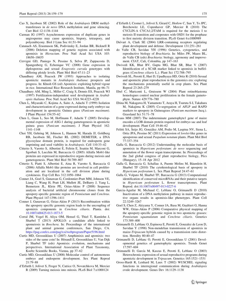
Cao X, Jacobsen SE (2002) Role of the Arabidopsis DRM methyl-
transferases in de novo DNA methylation and gene silencing.
Curr Biol 12:1138–1144
Carman JG (1997) Asynchronous expression of duplicate genes in
angiosperms may cause apomixis, bispory, tetraspory, and
polyembryony. Biol J Linn Soc 61:51–94
Catanach AS, Erasmuson SK, Podivinsky E, Jordan BR, Bicknell R
(2006) Deletion mapping of genetic regions associated with
apomixis in Hieracium. Proc Natl Acad Sci USA 103:
18650–18655
Cervigni GD, Paniego N, Pessino S, Selva JP, Zappacosta D,
Spangenberg G, Echenique VC (2008) Gene expression in
diplosporous and sexual Eragrostis curvula genotypes with
differing ploidy levels. Plant Mol Biol 67:11–23
Chaudhury AM, Peacock JW (1993) Approaches to isolating
apomictic mutants in Arabidopsis thaliana: prospects and
progress. In: Khush GS (ed) Apomixis: exploiting hybrid vigour
in rice. International Rice Research Institute, Manila, pp 66–71
Chaudhury AM, Ming L, Miller C, Craig S, Dennis ES, Peacock WJ
(1997) Fertilization-independent seed development in Arabid-
opsis thaliana. Proc Natl Acad Sci USA 94:4223–4228
Chen L, Miyazaki C, Kojima A, Saito A, Adachi T (1999) Isolation
and characterization of a gene expressed during early embryo sac
development in apomictic Guinea grass (Panicum maximum).
J Plant Physiol 154:55–62
Chen L, Guan L, Seo M, Hoffmann F, Adachi T (2005) Develop-
mental expression of ASG-1 during gametogenesis in apomictic
guinea grass (Panicum maximum). J Plant Physiol 162:
1141–1148
Choi YH, Gehring M, Johnson L, Hannon M, Harada JJ, Goldberg
RB, Jacobsen SE, Fischer RL (2002) DEMETER, a DNA
glycosylase domain protein, is required for endosperm gene
imprinting and seed viability in Arabidopsis. Cell 110:33–42
Citterio S, Varotto S, Albertini E, Feltrin E, Soattin M, Marconi G,
Sgorbati S, Lucchin M, Barcaccia G (2005) Alfalfa Mob1-like
proteins are expressed in reproductive organs during meiosis and
gametogenesis. Plant Mol Biol 58:789–807
Citterio S, Piatti S, Albertini E, Aina R, Varotto S, Barcaccia G
(2006) Alfalfa Mob1-like proteins are involved in cell prolifer-
ation and are localized in the cell division plane during
cytokinesis. Exp Cell Res 312:1050–1064
Conner JA, Goel S, Gunawan G, Cordonnier-Pratt MM, Johnson VE,
Liang C, Wang H, Pratt LH, Mullett JE, DeBarry J, Yang L,
Bennetzen JL, Klein PE, Ozias-Akins P (2008) Sequence
analysis of bacterial artificial chromosome clones from the
apospory-specific genomic region of Pennisetum and Cenchrus.
Plant Physiol 147:1396–1411
Conner J, Gunawan G, Ozias-Akins P (2013) Recombination within
the apospory specific genomic region leads to the uncoupling of
apomixis components in Cenchrus ciliaris. Planta. doi:
10.1007/s00425-013-1873-5
Corral JM, Vogel H, Aliyu OM, Hensel G, Thiel T, Kumlehn J,
Sharbel T (2013) APOLLO: a candidate allele linked to
apomeiosis in Boechera. In: Proceedings of the international
plant and animal genome conferences, San Diego, CA.
https://pag.confex.com/pag/xxi/webprogram/Paper5196.html
Curtis MD, Grossniklaus U (2007) Amphimixis and apomixis: two
sides of the same coin! In: Horandl E, Grossniklaus U, Van Dijk
P, Sharbel TF (eds) Apomixis: evolution, mechanisms and
perspectives. International Association of Plant Taxonomy,
Koeltz Scientific Books, Vienna, pp 37–62
Curtis MD, Grossniklaus U (2008) Molecular control of autonomous
embryo and endosperm development. Sex Plant Reprod
21:79–88
d’Erfurth I, Jolivet S, Froger N, Catrice O, Novatchkova M, Mercier
R (2009) Turning meiosis into mitosis. PLoS Biol 7:e1000124
d’Erfurth I, Cromer L, Jolivet S, Girard C, Horlow C, Sun Y, To JPC,
Berchowitz LE, Copenhaver GP, Mercier R (2010) The
CYCLIN-A CYCA1;2/TAM is required for the meiosis I to
meiosis II transition and cooperates with OSD1 for the prophase
to first meiotic division transition. PLoS Genet 6:e1000989
Dievart A, Clark SE (2004) LRR-containing receptors regulating
plant development and defense. Development 131:251–261
do Valle CB, Savidan YH (1996) Genetics, cytogenetics, and
reproductive biology of Brachiaria. In: Miles JW, Maass BL,
do Valle CB (eds) Brachiaria: biology, agronomy and improve-
ment. CIAT, Cali, Colombia, pp 147–163
Dwivedi KK, Bhat BV, Gupta MG, Bhat SR, Bhat V (2007)
Identification of a SCAR marker linked to apomixis in buffel-
grass (Cenchrus ciliaris L.). Plant Sci 172:788–795
Dwivedi SL, Perotti E, Hari D, Upadhyaya HD, Ortiz R (2010) Sexual
and apomictic plant reproduction in the genomics era: exploring
the mechanisms potentially useful in crop plants. Sex Plant
Reprod 23:265–279
Ebel C, Mariconti L, Gruissem W (2004) Plant retinoblastoma
homologues control nuclear proliferation in the female gameto-
phyte. Nature 429:776–780
Ebina M, Nakagawa H, Yamamoto T, Araya H, Tsuruta S-I, Takahara
M, Nakajima K (2005) Co-segregation of AFLP and RAPD
markers to apospory in Guineagrass (Panicum maximum Jacq.).
Grassland Sci 51:71–78
Evans MM (2007) The indeterminate gametophyte1 gene of maize
encodes a LOB domain protein required for embryo sac and leaf
development. Plant Cell 19:46–62
Felitti SA, Seijo JG, Gonzalez AM, Podio M, Laspina NV, Siena L,
Ortiz JPA, Pessino SC (2011) Expression of lorelei-like genes in
aposporous and sexual Paspalum notatum plants. Plant Mol Biol
77:337–354
Galla G, Barcaccia G (2012) Understanding the molecular basis of
apomixis in Hypericum perforatum: de novo sequencing and
annotation of the flower organ transcriptome. In: Proceedings of
the 2nd global congress of plant reproductive biology, Pecs
(Hungary), 15–18 Apr 2012
Galla G, Barcaccia G, Schallau A, Puente Molins M, Baumlein H,
Sharbel TF (2010) The cytohistological basis of apospory in
Hypericum perforatum L. Sex Plant Reprod 24:47–61
Galla G, Volpato M, Sharbel TF, Barcaccia G (2013) Computational
identification of conserved microRNAs and their putative targets
in Hypericum perforatum L. flower transcriptomes. Plant
Reprod. doi:10.1007/s00497-013-0227-6
Garcia-Aguilar M, Michaud C, Leblanc O, Grimanelli D (2010)
Inactivation of a DNA methylation pathway in maize reproduc-
tive organs results in apomixis-like phenotypes. Plant Cell
22:3249–3267
Goel S, Chen Z, Akiyama Y, Conner JA, Basu M, Gualtieri G, Hanna
WW, Ozias-Akins P (2006) Comparative physical mapping of
the apospory-specific genomic region in two apomictic grasses:
Pennisetum squamulatum and Cenchrus ciliaris. Genetics
173:389–400
Grimanelli D, Leblanc O, Espinosa E, Perotti E, Gonzalez de Leon D,
Savidan Y (1998) Non-mendelian transmission of apomixis in
maize-Tripsacum hybrids caused by a transmission ratio distor-
tion. Heredity 80:40–47
Grimanelli D, Leblanc O, Perotti E, Grossniklaus U (2001) Devel-
opmental genetics of gametophytic apomixis. Trends Genet
17:597–604
Grimanelli D, Garcıa M, Kaszas E, Perotti E, Leblanc O (2003)
Heterochronic expression of sexual reproductive programs during
apomictic development in Tripsacum. Genetics 165:1521–1531
Gross-Hardt R, Lenhard M, Laux T (2002) WUSCHEL signaling
functions in interregional communication during Arabidopsis
ovule development. Genes Dev 16:1129–1138
Plant Reprod (2013) 26:159–179 175
123

Grossniklaus U, Vielle-Calzada JP, Hoeppner MA, Gagliano WB
(1998) Maternal control of embryogenesis by MEDEA, a
polycomb group gene in Arabidopsis. Science 280:446–450
Grossniklaus U, Spillane C, Page DR, Kohler C (2001a) Genomic
imprinting and seed development: endosperm formation with
and without sex. Curr Opin Plant Biol 4:21–27
Grossniklaus U, Nogler GA, van Dijk PJ (2001b) The genetic control
of gametophytic apomixis. Plant Cell 13:1491–1498
Guerin J, Rossel JB, Robert S, Tsuchiya T, Koltunow A (2000) A
DEFICIENS homologue is down-regulated during apomictic
initiation in ovules of Hieracium. Planta 210:914–920
Guitton EA, Berger F (2005) Loss of function of MULTICOPY
SUPPRESSOR OF IRA 1 produces nonviable parthenogenic
embryos in Arabidopsis. Curr Biol 15:750–754
Hagberg A, Hagberg G (1980) High frequency of spontaneous
haploids in the progeny of an induced mutation in barley.
Hereditas 93:341–343
Hecht V, Vielle-Calzada J-P, Hartog MV, Schmidt EDL, Boutilier K,
Grossniklaus U, de Vries SC (2001) The Arabidopsis somatic
embryogenesis receptor kinase 1 (SERK1) gene is expressed in
developing ovules and embryos and enhances embryogenic
competence in culture. Plant Physiol 127:803–816
Henderson JT, Li H-C, Rider SD, Mordhorst AP, Romero-Severson J,
Cheng J-C, Robey J, Sung ZR, de Vries SC, Ogas J (2004)
PICKLE acts throughout the plant to repress expression of
embryonic traits and may play a role in gibberellin-dependent
responses. Plant Physiol 134:995–1005
Hirabayashi S, Nakagawa K, Sumita K, Hidaka S, Kawai T, Ikeda M,
Kawata A, Ohno K, Hata Y (2008) Threonine 74 of MOB1 is a
putative key phosphorylation site by MST2 to form the scaffold to
activate nuclear Dbf2-related kinase 1. Oncogene 27:4281–4292
Hojsgaard DH, Schegg E, Valls JFM, Martinez EJ, Quarin CL (2008)
Sexuality, apomixis, ploidy levels, and genomic relationships
among four Paspalum species of the subgenus Anachyris
(Poaceae). Flora 203:535–547
Hojsgaard DH, Martınez EJ, Quarin CL (2013) Competition between
meiotic and apomictic pathways during ovule and seed devel-
opment results in clonality. New Phytol 197:336–347
Huo H, Conner JA, Ozias-Akins P (2009) Genetic mapping of the
apospory-specific genomic region (ASGR) in Pennisetum squa-
mulatum using retrotransposon-based molecular markers. Theor
Appl Genet 119:199–212
Ingouff M, Haseloff J, Berger F (2005) Polycomb group genes control
developmental timing of endosperm. Plant J 42:663–674
Jessup RW, Burson BL, Burow GB, Wang YWCC, Li Z, Paterson AH,
Hussey MA (2002) Disomic inheritance, suppressed recombina-
tion, and allelic interactions govern apospory in buffelgrass as
revealed by genome mapping. Crop Sci 42:1688–1694
Jessup RW, Burson BL, Burow G, Wang Y-W, Chang C, Li Z,
Paterson AH, Hussey MA (2003) Segmental allotetraploidy and
allelic interactions in buffelgrass (Pennisetum ciliare (L.) Link
syn. Cenchrus ciliaris L.) as revealed by genome mapping.
Genome 46:304–313
Johnston AJ, Matveeva E, Kirioukhova O, Grossniklaus U, Gruissem
W (2008) A dynamic reciprocal RBR-PRC2 regulatory circuit
controls Arabidopsis gametophyte development. Curr Biol
18:1680–1686
Jullien PE, Mosquna A, Ingouff M, Sakata T, Ohad N, Berger F
(2008) Retinoblastoma and its binding partner MSI1 control
imprinting in Arabidopsis. PLoS Biol 6:1693–1705
Kaushal P, Malaviya DR, Roy AK, Pathak S, Agrawal A, Khare A,
Siddiqui SA (2008) Reproductive pathways of seed development
in apomictic guinea grass (Panicum maximum Jacq.) reveal
uncoupling of apomixis components. Euphytica 164:81–92
Klattenhoff C, Theurkauf W (2008) Biogenesis and germline
functions of piRNAs. Development 135:3–9
Kohler C, Henning L, Spillane C, Pien S, Gruissem W, Grossniklaus
U (2003) The polycomb-group protein MEDEA regulates seed
development by controlling expression of the MADS-box gene
PHERES1. Genes Dev 17:1540–1553
Koltunow AM, Grossniklaus U (2003) Apomixis: a developmental
perspective. Annu Rev Plant Biol 54:547–574
Koltunow AM, Bicknell RA, Chaudhury AM (1995) Apomixis:
molecular strategies for the generation of genetically identical
seeds without fertilization. Plant Physiol 108:1345–1352
Koltunow AMG, Johnson SD, Rodrigues JCM, Okada T, Hu Y,
Tsuchiya T, Wilson S, Fletcher P, Ito K, Suzuki G et al (2011)
Sexual reproduction is the default mode in apomictic Hieracium
subgenus Pilosella, in which two dominant loci function to
enable apomixis. Plant J 66:890–902
Koltunow AMG, Ozias-Akins P, Siddiqi I (2013) Apomixis. In: Becraft
PW (ed) Seed genomics. Wiley-Blackwell, Oxford, pp 83–110
Labombarda P, Busti A, Caceres ME, Quarın CL, Pupilli F, Arcioni S
(2002) An AFLP marker tightly linked to apomixis reveals
hemizygosity in a portion of the apomixis-controlling locus in
Paspalum simplex. Genome 45:513–519
Lacadena J-R (1974) Spontaneous and induced parthenogenesis and
androgenesis. In: Kasha KJ (ed) Haploids in higher plants:
advances and potential. The University of Guelph, Guelph,
pp 13–32
Lacerda ALM, Alencar Dusi DM, Alves ER, Rodrigues JCM, Gomes
ACMM, Carneiro VTC (2013) Expression analyses of Brachi-
aria brizantha genes encoding ribosomal proteins BbrizRPS8,
BbrizRPS15a, and BbrizRPL41 during development of ovaries
and anthers. Protoplasma 250:505–514
Laspina NV, Vega T, Seijo G, Gonzalez AM, Martelotto LG, Stein J,
Podio M, Ortiz JPA, Echenique VC, Quarin CL, Pessino SC
(2008) Gene expression analysis at the onset of aposporous
apomixis in Paspalum notatum. Plant Mol Biol 67:615–628
Leblanc O, Grimanelli D, Gonzalez-de-Leon D, Savidan Y (1995)
Detection of the apomictic mode of reproduction in maize-
Tripsacum hybrids using maize RFLP markers. Theor Appl
Genet 90:1198–1203
Leblanc O, Armstead I, Pessino S, Ortiz JPA, Evans C, do Valle C,
Hayward MD (1997) Nonradioactive mRNA fingerprinting to
visualize gene expression in mature ovaries of Brachiaria
hybrids derived from B. brizantha, an apomictic tropical forage.
Plant Sci 126:49–58
Leblanc O, Grimanelli D, Hernandez-Rodriguez M, Galindo PA,
Soriano-Martinez AM, Perotti E (2009) Seed development and
inheritance studies in apomictic maize-Tripsacum hybrids reveal
barriers for the transfer of apomixis into sexual crops. Intl J Dev
Biol 53:585–596
Li L-C, Qin G-J, Tsuge T, Hou X-H, Ding M-Y, Aoyama T, Oka A,
Chen Z, Gu H, de Zhao Y, Qu L-J (2008) SPOROCYTELESS
modulates YUCCA expression to regulate the development of
lateral organs in Arabidopsis. New Phytol 179:751–764
Lieber D, Lora J, Schrempp S, Lenhard M, Laux T (2011)
Arabidopsis WIH1 and WIH2 genes act in the transition from
somatic to reproductive cell fate. Curr Biol 21:1009–1017
Lotan T, Ohto M, Yee KM, West MA, Lo R, Kwong RW, Yamagishi
K, Fischer RL, Goldberg RB, Harada JJ (1998) Arabidopsis
LEAFY COTYLEDON1 is sufficient to induce embryo devel-
opment in vegetative cells. Cell 93:1195–1205
Lovell JT, Aliyu OM, Mau M, Schranz ME, Koch M, Kiefer C, Song
B-H, Mitchell-Olds T, Sharbel TF (2013) On the origin and
evolution of apomixis in Boechera. Plant Reprod. doi:
10.1007/s00497-013-0218-7
Luo M, Bilodeau P, Koltunow A, Dennis ES, Peacock WJ, Chaudhury
AM (1999) Genes controlling fertilization-independent seed
development in Arabidopsis thaliana. Proc Natl Acad Sci USA
96:296–301
176 Plant Reprod (2013) 26:159–179
123

Luo M, Platten D, Chaudhury AM, Peacock WJ, Dennis ES (2009)
Expression, imprinting, and evolution of rice homologs of the
polycomb group genes. Mol Plant 2:711–723
Marconi G, Sharbel T, Colombo L, Masiero S, Barcaccia G, Galla G,
Conner J, Ozias-Akins P, Albertini E (2013) Differential
expression in sexual and apomictic genotypes of three model
species strengthens a crucial role of APOSTART in the
formation of seeds by apomixis. In: International plant and
animal genome conferences, San Diego, CA, USA, 11–16 Jan
2013, Abstract W031. https://pag.confex.com/pag/xxi/webpro
gram/Paper5196.html
Martinez EJ, Hopp HE, Stein J, Ortiz JPA, Quarin CL (2003) Genetic
characterization of apospory in tetraploid Paspalum notatum
based on the identification of linked molecular markers. Mol
Breed 12:319–327
Matzk F (1996) The ‘Salmon System’ of wheat—a suitable model for
apomixis research. Hereditas 125:299–304
Matzk F, Meister A, Brutovska R, Schubert I (2001) Reconstruction
of reproductive diversity in Hypericum perforatum L. opens
novel strategies to manage apomixis. Plant J 26:275–282
Matzk F, Prodanovic S, Baumlein H, Schubert I (2005) The
inheritance of apomixis in Poa pratensis confirms a five locus
model with differences in gene expressivity and penetrance.
Plant Cell 17:13–24
Miyazaki S, Ito M (2006) Calcium signals for egg activation in
mammals. J Pharmacol Sci 100:545–552
Muntzing A (1940) Further studies on apoximis and sexuality in Poa.
Hereditas 27:115–190
Naumova T (1992) Apomixis in angiosperms: nucellar and integu-
mentary embryony. CRC Press, Boca Raton, FL
Nogler GA (1984) Gametophytic apomixis. In: Johri BM (ed)
Embryology of angiosperms. Springer, Berlin, pp 475–518
Nogler GA (1995) Genetics of apomixis in Ranunculus auricomus.
VI. Epilogue. Bot Helv 105:111–115
Nonomura K, Miyoshi K, Eiguchi M, Suzuki T, Miyao A,
Hirochika H, Kurata N (2003) The MSP1 gene is necessary
to restrict the number of cells entering into male and female
sporogenesis and to initiate anther wall formation in rice. Plant
Cell 15:1728–1739
Nonomura K, Morohoshi A, Nakano M, Eiguchi M, Miyao A,
Hirochika H, Kurata N (2007) A germ cell specific gene of the
ARGONAUTE family is essential for the progression of
premeiotic mitosis and meiosis during the male and female
sporogenesis in rice. Plant Cell 19:2583–2594
Nonomura K, Morohoshi A, Nakano M, Eiguchi M, Miyao A,
Hirochika H, Kurata N (2008) A germ cell specific gene of the
ARGONAUTE family is essential for the progression of
premeiotis mitosis and meiosis during the male and female
sporogenesis in rice. Plant Cell 19:2583–2594
Noyes RD, Rieseberg LH (2000) Two independent loci control
agamospermy (apomixis) in the triploid flowering plant Erigeron
annuus. Genetics 155:379–390
Noyes RD, Baker R, Mai B (2007) Mendelian segregation for two-
factor apomixis in Erigeron annuus (Asteraceae). Heredity
98:92–98
Ohad N, Yadegari R, Margossian L, Hannon M, Michaeli D, Harada
JJ, Goldberg RB, Fischer RL (1999) Mutations in FIE, a WD
polycomb group gene, allow endosperm development without
fertilization. Plant Cell 11:407–416
Olmedo-Monfil V, Duran-Figueroa N, Arteaga-Vazquez M, Demesa-
Arevalo E, Autran D, Grimanelli D, Slotkin RK, Martienssen
RA, Vielle-Calzada JP (2010) Control of female gamete
formation by a small RNA pathway in Arabidopsis. Nature
464:628–632
Ozias-Akins P (2006) Apomixis: developmental characteristics and
genetics. Crit Rev Plant Sci 25:199–214
Ozias-Akins P, van Dijk PJ (2007) Mendelian genetics of apomixis.
Annu Rev Genet 41:509–537
Ozias-Akins P, Roche D, Hanna WW (1998) Tight clustering and
hemizygosity of apomxis-linked markers in Pennisetum squa-
mulatum implies genetic control of apospory by a divergent
locus that may have no allelic form in sexual genotypes. Proc
Natl Acad Sci USA 95:5127–5132
Pagnussat GC, Alandete-Saez M, Bowman JL, Sundaresan V (2009)
Auxin-dependent patterning and gamete specification in the
Arabidopsis female gametophyte. Science 324:1684–1689
Pawlowski WP, Wang C-JR, Golubovskaya IN, Szymaniak JM, Shi
L, Hamant O, Zhu T, Harper L, Sheridan WF, Cande WZ (2009)
Maize AMEIOTIC1 is essential for multiple early meiotic
processes and likely required for the initiation of meiosis. Proc
Natl Acad Sci USA 106:3603–3608
Peacock JP (1992) Genetic engineering and mutagenesis for apomixis
in rice. Apomixis Newsl 4:3–7
Pessino SC, Ortiz JPA, Leblanc O, do Valle CB, Hayward MD (1997)
Identification of a maize linkage group related to apomixis in
Brachiaria. Theor Appl Genet 94:439–444
Pessino SC, Evans C, Ortiz JPA, Armstead I, do Valle CB, Hayward
MD (1998) A genetic map of the apospory-region in Brachiaria
hybrids: identification of two markers closely associated with the
trait. Hereditas 128:153–158
Pessino SC, Espinoza F, Martinez EJ, Ortiz JPA, Valle EM, Quarin
CL (2001) Isolation of cDNA clones differentially expressed in
flowers of apomictic and sexual Paspalum notatum. Hereditas
134:35–42
Pillot M, Autran D, Leblanc O, Grimanelli D (2010a) A role for
CHROMOMETHYLASE3 in mediating transposon and euchro-
matin silencing during egg cell reprogramming in Arabidopsis.
Plant Signal Behav 5:1167–1170
Pillot M, Baroux C, Vazquez MA, Autran D, Leblanc O, Vielle-
Calzada JP, Grossniklaus U, Grimanelli D (2010b) Embryo and
endosperm inherit distinct chromatin and transcriptional states
from the female gametes in Arabidopsis. Plant Cell 22:307–320
Podio M, Rodriguez MP, Felitti S, Stein J, Martinez E, Siena LA,
Quarin CL, Pessino SC, Ortiz JPA (2012) Sequence character-
ization, in silico mapping and cytosine methylation analysis of
markers linked to apospory in Paspalum notatum. Genet Mol
Biol 35:827–837
Polegri L, Calderini O, Arcioni S, Pupilli F (2010) Specific expression
of apomixis-linked alleles revealed by comparative transcrip-
tomic analysis of sexual and apomictic Paspalum simplex
Morong flowers. J Exp Bot 61:1869–1883
Pupilli F, Barcaccia G (2012) Cloning plants by seeds: inheritance
models and candidate genes to increase fundamental knowledge
for engineering apomixis in sexual crops. J Biotechnol
159:291–311
Pupilli F, Labombarda P, Caceres ME, Quarin CL, Arcioni S (2001)
The chromosome segment related to apomixis in Paspalum
simplex is homoeologous to the telomeric region of the long arm
of rice chromosome 12. Mol Breed 8:53–61
Pupilli F, Martinez EJ, Busti A, Calderini O, Quarın CL, Arcioni S
(2004) Comparative mapping reveals partial conservation of
synteny at the apomixis locus in Paspalum spp. Mol Genet
Genomics 270:539–548
Qiu Y, Liu R, Xie C, Russell S, Tian H (2008) Calcium changes
during megasporogenesis and megaspore degeneration in lettuce
(Lactuca sativa L). Sex Plant Reprod 21:197–204
Ravi M, Chan SWL (2010) Haploid plants produced by centromere-
mediated genome elimination. Nature 464:615–618
Ravi M, Marimuthu MP, Siddiqi I (2008) Gamete formation without
meiosis in Arabidopsis. Nature 451:1121–1124
Rhoades MM, Dempsey E (1966) Induction of chromosome doubling
at meiosis by the elongate gene in maize. Genetics 54:505–522
Plant Reprod (2013) 26:159–179 177
123
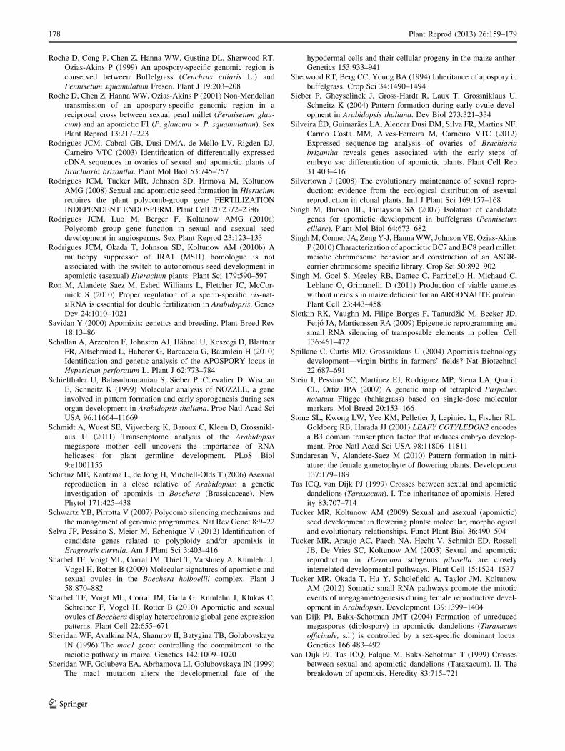
Roche D, Cong P, Chen Z, Hanna WW, Gustine DL, Sherwood RT,
Ozias-Akins P (1999) An apospory-specific genomic region is
conserved between Buffelgrass (Cenchrus ciliaris L.) and
Pennisetum squamulatum Fresen. Plant J 19:203–208
Roche D, Chen Z, Hanna WW, Ozias-Akins P (2001) Non-Mendelian
transmission of an apospory-specific genomic region in a
reciprocal cross between sexual pearl millet (Pennisetum glau-
cum) and an apomictic F1 (P. glaucum 9 P. squamulatum). Sex
Plant Reprod 13:217–223
Rodrigues JCM, Cabral GB, Dusi DMA, de Mello LV, Rigden DJ,
Carneiro VTC (2003) Identification of differentially expressed
cDNA sequences in ovaries of sexual and apomictic plants of
Brachiaria brizantha. Plant Mol Biol 53:745–757
Rodrigues JCM, Tucker MR, Johnson SD, Hrmova M, Koltunow
AMG (2008) Sexual and apomictic seed formation in Hieracium
requires the plant polycomb-group gene FERTILIZATION
INDEPENDENT ENDOSPERM. Plant Cell 20:2372–2386
Rodrigues JCM, Luo M, Berger F, Koltunow AMG (2010a)
Polycomb group gene function in sexual and asexual seed
development in angiosperms. Sex Plant Reprod 23:123–133
Rodrigues JCM, Okada T, Johnson SD, Koltunow AM (2010b) A
multicopy suppressor of IRA1 (MSI1) homologue is not
associated with the switch to autonomous seed development in
apomictic (asexual) Hieracium plants. Plant Sci 179:590–597
Ron M, Alandete Saez M, Eshed Williams L, Fletcher JC, McCor-
mick S (2010) Proper regulation of a sperm-specific cis-nat-
siRNA is essential for double fertilization in Arabidopsis. Genes
Dev 24:1010–1021
Savidan Y (2000) Apomixis: genetics and breeding. Plant Breed Rev
18:13–86
Schallau A, Arzenton F, Johnston AJ, Hahnel U, Koszegi D, Blattner
FR, Altschmied L, Haberer G, Barcaccia G, Baumlein H (2010)
Identification and genetic analysis of the APOSPORY locus in
Hypericum perforatum L. Plant J 62:773–784
Schiefthaler U, Balasubramanian S, Sieber P, Chevalier D, Wisman
E, Schneitz K (1999) Molecular analysis of NOZZLE, a gene
involved in pattern formation and early sporogenesis during sex
organ development in Arabidopsis thaliana. Proc Natl Acad Sci
USA 96:11664–11669
Schmidt A, Wuest SE, Vijverberg K, Baroux C, Kleen D, Grossnikl-
aus U (2011) Transcriptome analysis of the Arabidopsis
megaspore mother cell uncovers the importance of RNA
helicases for plant germline development. PLoS Biol
9:e1001155
Schranz ME, Kantama L, de Jong H, Mitchell-Olds T (2006) Asexual
reproduction in a close relative of Arabidopsis: a genetic
investigation of apomixis in Boechera (Brassicaceae). New
Phytol 171:425–438
Schwartz YB, Pirrotta V (2007) Polycomb silencing mechanisms and
the management of genomic programmes. Nat Rev Genet 8:9–22
Selva JP, Pessino S, Meier M, Echenique V (2012) Identification of
candidate genes related to polyploidy and/or apomixis in
Eragrostis curvula. Am J Plant Sci 3:403–416
Sharbel TF, Voigt ML, Corral JM, Thiel T, Varshney A, Kumlehn J,
Vogel H, Rotter B (2009) Molecular signatures of apomictic and
sexual ovules in the Boechera holboellii complex. Plant J
58:870–882
Sharbel TF, Voigt ML, Corral JM, Galla G, Kumlehn J, Klukas C,
Schreiber F, Vogel H, Rotter B (2010) Apomictic and sexual
ovules of Boechera display heterochronic global gene expression
patterns. Plant Cell 22:655–671
Sheridan WF, Avalkina NA, Shamrov II, Batygina TB, Golubovskaya
IN (1996) The mac1 gene: controlling the commitment to the
meiotic pathway in maize. Genetics 142:1009–1020
Sheridan WF, Golubeva EA, Abrhamova LI, Golubovskaya IN (1999)
The mac1 mutation alters the developmental fate of the
hypodermal cells and their cellular progeny in the maize anther.
Genetics 153:933–941
Sherwood RT, Berg CC, Young BA (1994) Inheritance of apospory in
buffelgrass. Crop Sci 34:1490–1494
Sieber P, Gheyselinck J, Gross-Hardt R, Laux T, Grossniklaus U,
Schneitz K (2004) Pattern formation during early ovule devel-
opment in Arabidopsis thaliana. Dev Biol 273:321–334
Silveira ED, Guimaraes LA, Alencar Dusi DM, Silva FR, Martins NF,
Carmo Costa MM, Alves-Ferreira M, Carneiro VTC (2012)
Expressed sequence-tag analysis of ovaries of Brachiaria
brizantha reveals genes associated with the early steps of
embryo sac differentiation of apomictic plants. Plant Cell Rep
31:403–416
Silvertown J (2008) The evolutionary maintenance of sexual repro-
duction: evidence from the ecological distribution of asexual
reproduction in clonal plants. Intl J Plant Sci 169:157–168
Singh M, Burson BL, Finlayson SA (2007) Isolation of candidate
genes for apomictic development in buffelgrass (Pennisetum
ciliare). Plant Mol Biol 64:673–682
Singh M, Conner JA, Zeng Y-J, Hanna WW, Johnson VE, Ozias-Akins
P (2010) Characterization of apomictic BC7 and BC8 pearl millet:
meiotic chromosome behavior and construction of an ASGR-
carrier chromosome-specific library. Crop Sci 50:892–902
Singh M, Goel S, Meeley RB, Dantec C, Parrinello H, Michaud C,
Leblanc O, Grimanelli D (2011) Production of viable gametes
without meiosis in maize deficient for an ARGONAUTE protein.
Plant Cell 23:443–458
Slotkin RK, Vaughn M, Filipe Borges F, Tanurdzic M, Becker JD,
Feijo JA, Martienssen RA (2009) Epigenetic reprogramming and
small RNA silencing of transposable elements in pollen. Cell
136:461–472
Spillane C, Curtis MD, Grossniklaus U (2004) Apomixis technology
development—virgin births in farmers’ fields? Nat Biotechnol
22:687–691
Stein J, Pessino SC, Martınez EJ, Rodriguez MP, Siena LA, Quarın
CL, Ortiz JPA (2007) A genetic map of tetraploid Paspalum
notatum Flugge (bahiagrass) based on single-dose molecular
markers. Mol Breed 20:153–166
Stone SL, Kwong LW, Yee KM, Pelletier J, Lepiniec L, Fischer RL,
Goldberg RB, Harada JJ (2001) LEAFY COTYLEDON2 encodes
a B3 domain transcription factor that induces embryo develop-
ment. Proc Natl Acad Sci USA 98:11806–11811
Sundaresan V, Alandete-Saez M (2010) Pattern formation in mini-
ature: the female gametophyte of flowering plants. Development
137:179–189
Tas ICQ, van Dijk PJ (1999) Crosses between sexual and apomictic
dandelions (Taraxacum). I. The inheritance of apomixis. Hered-
ity 83:707–714
Tucker MR, Koltunow AM (2009) Sexual and asexual (apomictic)
seed development in flowering plants: molecular, morphological
and evolutionary relationships. Funct Plant Biol 36:490–504
Tucker MR, Araujo AC, Paech NA, Hecht V, Schmidt ED, Rossell
JB, De Vries SC, Koltunow AM (2003) Sexual and apomictic
reproduction in Hieracium subgenus pilosella are closely
interrelated developmental pathways. Plant Cell 15:1524–1537
Tucker MR, Okada T, Hu Y, Scholefield A, Taylor JM, Koltunow
AM (2012) Somatic small RNA pathways promote the mitotic
events of megagametogenesis during female reproductive devel-
opment in Arabidopsis. Development 139:1399–1404
van Dijk PJ, Bakx-Schotman JMT (2004) Formation of unreduced
megaspores (diplospory) in apomictic dandelions (Taraxacum
officinale, s.l.) is controlled by a sex-specific dominant locus.
Genetics 166:483–492
van Dijk PJ, Tas ICQ, Falque M, Bakx-Schotman T (1999) Crosses
between sexual and apomictic dandelions (Taraxacum). II. The
breakdown of apomixis. Heredity 83:715–721
178 Plant Reprod (2013) 26:159–179
123
van Dijk PJ, van Baarlen P, de Jong H (2003) The occurrence of
phenotypically complementary apomixis-recombinants in
crosses between sexual and apomictic dandelions (Taraxacum
officinale). Sex Plant Reprod 16:71–76
Van Ex F, Jacob Y, Martienssen RA (2011) Multiple roles for small
RNAs during plant reproduction. Curr Opin Plant Biol
14:588–593
Vaucheret H (2008) Plant argonautes. Trends Plant Sci 13:350–358
Vielle-Calzada JP, Crane CF, Stelly DM (1996) Apomixis: the
asexual revolution. Science 274:1322–1323
Vijverberg K, van Dijk PJ (2007) Genetic linkage mapping of
apomixis loci. In: Horandl E, Grossniklaus U, van Dijk P,
Sharbel TF (eds) Apomixis. Evolution, mechanisms and per-
spectives. International Association of Plant Taxonomy, Koeltz
Scientific Books, Vienna, pp 137–158
Vijverberg K, Van der Hulst RGM, Lindhout P, van Dijk PJ (2004) A
genetic linkage map of the diplosporous chromosomal region in
Taraxacum officinale (common dandelion; Asteraceae). Theor
Appl Genet 108:725–732
Vijverberg K, Milanovic S, Bakx-Schotman T, van Dijk PJ (2010)
Genetic fine mapping of DIPLOSPOROUS in Taraxucum
(dandelion; Asteraceae) indicates a duplicated DIP-gene. BMC
Plant Biol 10:154
Vitulo N, Vezzi A, Galla G, Citterio S, Marino G, Ruperti B,
Zermiani M, Albertini E, Valle G, Barcaccia G (2007) Charac-
terization and evolution of the cell cycle-associated Mob
domain-containing proteins in eukaryotes. Evol Bioinform
3:121–158
Vyskot B, Hobza R (2004) Gender in plants: sex chromosomes are
emerging from the fog. Trends Genet 20:432–438
Yamada-Akiyama H, Akiyama Y, Ebina M, Xu Q, Tsuruta S, Yazaki
J, Kishimoto N, Kikuchi S, Takahara M, Takamizo T, Sugita S,
Nakagawa H (2009) Analysis of expressed sequence tags in
apomictic guineagrass (Panicum maximum). J Plant Physiol
166:750–761
Yang W, Sundaresan V (2000) Genetics of gametophyte biogenesis in
Arabidopsis. Curr Opin Plant Biol 3:53–57
Zeng Y, Conner J, Ozias-Akins P (2011) Identification of ovule
transcripts from the Apospory-Specific Genomic Region
(ASGR)-carrier chromosome. BMC Genomics 12:206
Zhao DZ, Wang GF, Speal B, Ma H (2002) The excess microsporo-
cytes1 gene encodes a putative leucine-rich repeat receptor
protein kinase that controls somatic and reproductive cell fates in
the Arabidopsis anther. Genes Dev 16:2021–2031
Zhao X, de Palma J, Oane R, Gamuyao R, Luo M, Chaudhury A,
Herve P, Xue Q, Bennett J (2008) OsTDL1A binds to the LRR
domain of rice receptor kinase MSP1, and is required to limit
sporocyte numbers. Plant J 54:375–387
Zuo J, Niu QW, Frugis G, Chua NH (2002) The WUSCHEL gene
promotes vegetative-to-embryonic transition in Arabidopsis.
Plant J 30:349–359
Plant Reprod (2013) 26:159–179 179
123