AOP 42: Inhibition of Thyroperoxidase and Subsequent ...
126
Organisation for Economic Co-operation and Development DOCUMENT CODE For Official Use English - Or. English 1 January 1990 AOP 42: Inhibition of Thyroperoxidase and Subsequent Adverse Neurodevelopmental Outcomes in Mammals Short Title: TPO Inhibition and Altered Neurodevelopment This document was approved by the Extended Advisory Group on Molecular Screening and Toxicogenomics in June 2018. The Working Group of the National Coordinators of the Test Guidelines Programme and the Working Party on Hazard Assessment are invited to review and endorse the AOP by 29 March 2019. Magdalini Sachana, Administrator, Hazard Assessment, [email protected], +(33- 1) 85 55 64 23 Nathalie Delrue, Administrator, Test Guidelines, [email protected], +(33-1) 45 24 98 44 This document, as well as any data and map included herein, are without prejudice to the status of or sovereignty over any territory, to the delimitation of international frontiers and boundaries and to the name of any territory, city or area.
Transcript of AOP 42: Inhibition of Thyroperoxidase and Subsequent ...

Organisation for Economic Co-operation and Development
DOCUMENT CODE
For Official Use English - Or. English
1 January 1990
AOP 42: Inhibition of Thyroperoxidase and Subsequent Adverse
Neurodevelopmental Outcomes in Mammals
Short Title: TPO Inhibition and Altered Neurodevelopment
This document was approved by the Extended Advisory Group on Molecular Screening and
Toxicogenomics in June 2018.
The Working Group of the National Coordinators of the Test Guidelines Programme and the
Working Party on Hazard Assessment are invited to review and endorse the AOP by 29 March
2019.
Magdalini Sachana, Administrator, Hazard Assessment, [email protected], +(33-
1) 85 55 64 23
Nathalie Delrue, Administrator, Test Guidelines, [email protected], +(33-1) 45 24 98
44
This document, as well as any data and map included herein, are without prejudice to the status of or sovereignty over any territory, to the
delimitation of international frontiers and boundaries and to the name of any territory, city or area.

2 │
Foreword
This Adverse Outcome Pathway (AOP) on Inhibition of Thyroperoxidase and Subsequent
Adverse Neurodevelopmental Outcomes in Mammals, has been developed under the
auspices of the OECD AOP Development Programme, overseen by the Extended Advisory
Group on Molecular Screening and Toxicogenomics (EAGMST), which is an advisory
group under the Working Group of the National Coordinators for the Test Guidelines
Programme (WNT). The AOP has been reviewed internally by the EAGMST, externally
by experts nominated by the WNT, and has been endorsed by the WNT and the Working
Party on Hazard Assessment (WPHA) in xxx.
Through endorsement of this AOP, the WNT and the WPHA express confidence in the
scientific review process that the AOP has undergone and accept the recommendation of
the EAGMST that the AOP be disseminated publicly. Endorsement does not necessarily
indicate that the AOP is now considered a tool for direct regulatory application.
The Joint Meeting of the Chemicals Committee and the Working Party on Chemicals,
Pesticides and Biotechnology agreed to declassification of this AOP on xxxx.
This document is being published under the responsibility of the Joint Meeting of the
Chemicals Committee and the Working Party on Chemicals, Pesticides and Biotechnology.
The outcome of the internal and external reviews are publicly available respectively in the
AOP Wiki and the eAOP Portal of the AOP Knowledge Base at the following links:
[internal review] [external review].

│ 3
Table of contents
Foreword ................................................................................................................................................ 2
Adverse Outcome Pathway on Inhibition of Thyroperoxidase and Subsequent Adverse
Neurodevelopmental Outcomes in Mammals ..................................................................................... 5
Short Title: TPO Inhibition and Altered Neurodevelopment ............................................................... 5 Authors ............................................................................................................................................. 5
Abstract .................................................................................................................................................. 6
Background ............................................................................................................................................ 7
Graphical Representation ..................................................................................................................... 7
Summary of the AOP ............................................................................................................................ 8
Molecular Initiating Events (MIE), Key Events (KE), Adverse Outcomes (AO) ............................... 8 Key Event Relationships ...................................................................................................................... 8 Stressors ............................................................................................................................................... 9
Overall Assessment of the AOP ............................................................................................................ 9
Domain of Applicability .................................................................................................................... 10 Life Stage Applicability ................................................................................................................. 10 Taxonomic Applicability ................................................................................................................ 10 Sex Applicability ............................................................................................................................ 10
Essentiality of the Key Events ............................................................................................................ 11
Weight of Evidence Summary ............................................................................................................ 12
Biological plausibility: ....................................................................................................................... 12 Concordance of dose-response relationships: .................................................................................... 13 Temporal concordance among the key events and adverse effect: .................................................... 14 Consistency: ....................................................................................................................................... 14 Uncertainties, inconsistencies, and data gaps: ................................................................................... 15
Quantitative Consideration ................................................................................................................ 16
References ............................................................................................................................................ 17
Appendix 1 ........................................................................................................................................... 21
List of MIEs in this AOP ................................................................................................................... 21 Event: 279: Thyroperoxidase, Inhibition........................................................................................ 21
List of Key Events in the AOP .......................................................................................................... 27 Event: 277: Thyroid hormone synthesis, Decreased ...................................................................... 27 Event: 281: Thyroxine (T4) in serum, Decreased .......................................................................... 33 Event: 280: Thyroxine (T4) in neuronal tissue, Decreased ............................................................ 39 Event: 756: Hippocampal gene expression, Altered ...................................................................... 45 Event: 757: Hippocampal anatomy, Altered .................................................................................. 49 Event: 758: Hippocampal Physiology, Altered .............................................................................. 53
List of Adverse Outcomes in this AOP .............................................................................................. 58
Appendix 2 ........................................................................................................................................... 64

4 │
List of Adjacent Key Event Relationships in the AOP ...................................................................... 64 Relationship: 309: Thyroperoxidase, Inhibition leads to TH synthesis, Decreased ....................... 64 Relationship: 305: TH synthesis, Decreased leads to T4 in serum, Decreased .............................. 68 Relationship: 312: T4 in serum, Decreased leads to T4 in neuronal tissue, Decreased ................. 72 Relationship: 746: T4 in neuronal tissue, Decreased leads to Hippocampal gene expression,
Altered ............................................................................................................................................ 78 Relationship: 747: Hippocampal gene expression, Altered leads to Hippocampal anatomy,
Altered ............................................................................................................................................ 82 Relationship: 749: Hippocampal anatomy, Altered leads to Hippocampal Physiology, Altered ... 87 Relationship: 748: Hippocampal Physiology, Altered leads to Cognitive Function, Decreased ... 93
List of Non Adjacent Key Event Relationships in this AOP ............................................................. 98 Relationship: 366: Thyroperoxidase, Inhibition leads to T4 in serum, Decreased ......................... 98 Relationship: 1388: T4 in serum, Decreased leads to Hippocampal anatomy, Altered ............... 111 Relationship: 1389: T4 in serum, Decreased leads to Hippocampal Physiology, Altered ........... 116 Relationship: 403: T4 in serum, Decreased leads to Cognitive Function, Decreased .................. 121

│ 5
Adverse Outcome Pathway on Inhibition of Thyroperoxidase
and Subsequent Adverse Neurodevelopmental Outcomes in
Mammals
Short Title: TPO Inhibition and Altered Neurodevelopment
Authors
Kevin M. Crofton, R3Fellows LLC, Durham, NC USA <[email protected]>
Mary Gilbert, National Health and Environmental Effects Research Laboratory, US EPA,
RTP, NC USA <[email protected]>
Katie Paul Friedman, National Center for Computational Toxicology, US EPA, RTP, NC
USA <[email protected]>
Barbara Demeneix, UMR MNHN/CNRS 7221 Evolution of Endocrine Regulations,
National History Museum, Paris, France <[email protected]>
Mary Sue Marty, Toxicol. Environ. Res. Consult, Dow Chemical Company, Midland,
Michigan; <[email protected]>
R. Thomas Zoeller, Biology Department, University of Massachusetts, Amherst, MA

6 │
Abstract
This AOP describes one adverse outcome that may result from the inhibition of
thyroperoxidase (TPO) during mammalian development. Chemical inhibition of TPO, the
molecular-initiating event (MIE), results in decreased thyroid hormone (TH) synthesis, and
subsequent reduction in circulating concentrations of THs. THs are essential for normal
human brain development, both prenatally and postnatally, modulating genes critical for a
normal neuroanatomical development, with subsequent effects on neurophysiology, and
finally neurological function. Therefore, chemicals that interfere with TH synthesis have
the potential to cause TH insufficiency that may result in adverse neurodevelopmental
effects in offspring. Herein, we discuss the implications of developmental TPO inhibition
for hippocampal anatomy, function, and ultimately neural function controlled by the
hippocampus. The biochemistry of TPO and its essentiality for TH synthesis is well known
across species. The hippocampus is known to be critically involved in cognitive, emotional,
and memory function. The adverse consequences of TH insufficiency depend both on
severity and developmental timing, indicating that exposure to TPO inhibitors may produce
different effects at different developmental windows of exposure. It is important to note
that thyroid stimulating hormone (TSH) is not a KE in this AOP. While TSH may play a
role in feedback-driven compensatory processes, it is not directly involved in brain
development. The overall weight of evidence for this AOP is strong. Gaps in our
understanding include the relationship of TH-dependent gene expression and complexities
of brain development. Although quantitative information at all levels of KERs is limited a
number of applications of this AOP have been identified.

│ 7
Background
This AOP was originally started on the Chemical Mode of Action WIKI sponsored by
WHO/IPCS. The MOA was originally described and published by Zoeller and Crofton
(Crit Rev Toxicol 2005). Thanks to the following contributors whose work on the MOA-
WIKI fostered further development on the AOP wiki: Michelle Embry, Richard Judson,
Vicki Dellarco, Chihae Yang, Kevin Crofton.
Zoeller RT, Crofton KM. Mode of action: developmental thyroid hormone insufficiency--
neurological abnormalities resulting from exposure to propylthiouracil. Crit Rev Toxicol.
2005 Oct-Nov;35(8-9):771-81
Graphical Representation

8 │
Summary of the AOP
Molecular Initiating Events (MIE), Key Events (KE), Adverse Outcomes (AO)
Sequence Type Event
ID Title Short name
1 MIE 279 Thyroperoxidase, Inhibition Thyroperoxidase,
Inhibition
2 KE 277 Thyroid hormone synthesis,
Decreased
TH synthesis, Decreased
3 KE 281 Thyroxine (T4) in serum,
Decreased
T4 in serum, Decreased
4 KE 280 Thyroxine (T4) in neuronal
tissue, Decreased
T4 in neuronal tissue,
Decreased
5 KE 756 Hippocampal gene
expression, Altered
Hippocampal gene
expression, Altered
6 KE 757 Hippocampal anatomy,
Altered
Hippocampal anatomy,
Altered
7 KE 758 Hippocampal Physiology,
Altered
Hippocampal Physiology,
Altered
8 AO 402 Cognitive Function,
Decreased
Cognitive Function,
Decreased
Key Event Relationships
Upstream Event Relationship
Type
Downstream
Event Evidence
Quantitative
Understanding
Thyroperoxidase,
Inhibition
adjacent Thyroid
hormone
synthesis,
Decreased
High Low
Thyroid hormone
synthesis,
Decreased
adjacent Thyroxine (T4)
in serum,
Decreased
High Moderate
Thyroxine (T4) in
serum, Decreased
adjacent Thyroxine (T4)
in neuronal
tissue,
Decreased
Moderate Moderate
Thyroxine (T4) in
neuronal tissue,
Decreased
adjacent Hippocampal
gene expression,
Altered
Moderate Low
Hippocampal gene
expression, Altered
adjacent Hippocampal
anatomy,
Altered
Moderate Low

│ 9
Hippocampal
anatomy, Altered
adjacent Hippocampal
Physiology,
Altered
Moderate Low
Hippocampal
Physiology, Altered
adjacent Cognitive
Function,
Decreased
High Moderate
Thyroperoxidase,
Inhibition
non-adjacent Thyroxine (T4)
in serum,
Decreased
High Moderate
Thyroxine (T4) in
serum, Decreased
non-adjacent Hippocampal
gene expression,
Altered
High Low
Thyroxine (T4) in
serum, Decreased
non-adjacent Hippocampal
anatomy,
Altered
High Low
Thyroxine (T4) in
serum, Decreased
non-adjacent Hippocampal
Physiology,
Altered
Moderate Low
Thyroxine (T4) in
serum, Decreased
non-adjacent Cognitive
Function,
Decreased
High Moderate
Stressors
Name Evidence
Methimazole High
Propylthiouracil High
Overall Assessment of the AOP
The following summary tables for:
1.Support for Biological Plausibility of KERS
2. Support for Essentiality of KEs
3. Empirical Support for KERs
Can be downloaded at:
https://aopwiki.org/system/dragonfly/production/2018/08/10/46w2o2kkl4_TPO_AOP_Su
mmary_Tables_20180602.pdf

10 │
Domain of Applicability
Life Stage Applicability
Life Stage Evidence
During brain development High
Development High
Taxonomic Applicability
Term Scientific Term Evidence Links
human Homo sapiens High NCBI
rat Rattus norvegicus High NCBI
mouse Mus musculus Moderate NCBI
Sex Applicability
Sex Evidence
Male High
Female High
Chemicals: This AOP applies to a wide range of chemicals structures that inhibit
TPO either in vivo or in vitro. Well recognized positive controls include
propylthiouracil (PTU) and methimazole (MMI). There are 100s of other
chemicals known to inhibit TPO in vitro (e.g., Paul-Friedman et al., 2016).
Sex: This AOP applies to males and females. Disruption of thyroid hormone
regulation during fetal and early postnatal develop, as well as the subsequent
adverse impacts on nervous system development are similar in both sexes. There
are no compelling data to suggest sex differences in susceptibility to TH disruption
mediated by inhibition of TPO during development.
Life stages: The relevant life stages for this AOP are fetal and early postnatal ages
during critical windows of nervous system development where thyroid hormones
guide normal development of the brain. There are clear windows of developmental
susceptibility and different brain regions show distinct ontogenetic profiles for TH
requirements. Distinct phenotypes have been described in both humans and animal
models for different periods of TH insufficiency. The influence of maternal thyroid
status prior to onset of fetal thyroid function is an important consideration. This
AOP does not apply to adult life states.
Taxonomic: Based on the majority of the available evidence the taxonomic
applicability domains of this AOP is mammals. Most evidence for this AOP has
been gathered primarily from laboratory rodents and humans. However, there are
supporting data from amphibians and birds for TPO inhibition leading to altered
TH profiles. Due to the conserved nature of TH synthesis, transport, metabolism
and transcriptional activity, this AOP is likely to be applicable to other classes of
vertebrates where thyroid hormones drive development of the nervous system (e.g.,
birds, fish, reptiles). However, species-specific differences in development, ADME

│ 11
(adsorption, distribution, metabolism, and elimination), compensatory endocrine
responses may influence the outcomes, particularly from a quantitative standpoint.
Essentiality of the Key Events
It is widely accepted that each of the key events is essential.
Molecular Initiating Event: The molecular initiating event, i.e. inhibition of TPO,
is the essential event to initiate this AOP, as supported by in vitro and in vivo
evidence. TPO is the only enzyme capable of de novo TH synthesis (Taurog, 2005).
TPO is classically defined as a complex enzyme with multiple catalytic cycles
capable of iodinating multiple species (Divi et al., 1997). However, in the context
of this AOP we are using TPO inhibiton not in the classical sense, but instead to
refer to the results derived from the assays commonly used assays to investigate
environmental chemicals (e.g., guiacol oxidation). A number of studies have
demonstrated that cessation of exposure is known to result in a return to normal
levels of TH synthesis and circulatory hormone levels (Cooper et al., 1983). Many
in vivo and in vitro studies consistently demonstrate enzyme inhibition with similar
chemicals for multiple species (Taurog, 1999; Paul et al., 2013; Vickers et al.,
2012).
Thyroid hormone synthesis, Decreased. A number of studies have demonstrated a
correlation between TPO activity and decreased TH synthesis (e.g., Vickers et al.,
2012). Thyroid gland T4 concentrations, as well as serum TH, are decreased in
response to thyroidectomy and recover when in-vitro derived follicles are grafted
in athyroid mice (Antonica et al., 2012).
Thyroxine (T4) in serum, Decreased. Inhibition of TPO is widely accepted as
resulting in decreased TH synthesis in the thyroid gland, which results in decreased
serum T4 concentration (Taurog, 2005). Stop/recovery experiments demonstrate
recovery of serum thyroxine concentrations due to cessation of developmental
exposure to chemical stressors (e.g., Crofton et al., 2000), with similar findings in
adult rats (Cooper et al.,1984). Studies in adult animals show a similar recovery
after cessation of dosing (e.g., Hill et al., 1998).
Thyroxine (T4) in neuronal tissue, decreased: Mulitple studies have demonstrated
that fetal brain TH levels, previously decreased by maternal exposure to TPO
inhibitors or thyroidectomy, recovered following maternal dosing with T4 (e.g.,
Calvo et al., 1990). In addition, upregulation of deiodinase has been shown
compensate for some loss of neuronal T3 (Escobar-Morreale et al., 1997). Indirect
evidence shows that T4 replacement that bring circulating T4 concentration back
to normal, leads to recovery of brain TH and prevents downstream effects including
alterations in gene expression in the developing brain.
Hippocampal Gene Expression, Altered: It is well established specific genomic
pathways underlie the progression of a number of neurodevelopmental processes
in the hippocampus. There is some evidence from ex vivo studies that

12 │
administration of growth factors will reverse the hippocampal dysplasia seen in
Jacob/Nsfm knockout mice (Spilker et al., 2016). Less is known about the impact
of hormone replacement on TH-responsive gene expression and the qualitative and
quantitative relationships between altered TH-dependent gene expression in this
brain region and altered hippocampal cytoarchitectural anatomy.
Hippocampal anatomy, altered: It is well accepted that normal hippocampal
anatomy is critical for hippocampal physiological function, and that alterations in
anatomy lead to altered neuronal activity in the hippocampus (Lee et al., 2015;
Grant et al., 1992; Spilker et al., 2016).
Hippocampal physiology, altered: It is a well-accepted assertion that hippocampal
synaptic integrity and neuronal plasticity are essential for spatial information
processing in animals and spatial and episodic memory in humans. However, other
brain regions also can influence these complex behaviors. Limited data from studies
in BDNF knockout animals demonstrate that deficits in hippocampal synaptic
transmission and plasticity, and downstream behaviors can be rescued with
recombinant BDNF (Aarse et al., 2016; Andero et al., 2014).
Cognitive function, decreased: It is a well-known fact that TH are critical for
normal nervous system development (Williams et al., 2008). And this includes
development of the hippocampus which plays a major role in spatial, temporal, and
contextual memory. Indeed, most developed countries check for childhood
hypothyroidism at birth to immediately begin replacement therapy. This has been
shown to alleviate most adverse impacts of hypothyroidism in congenitally
hypothyroid children (Derksen-Lubsen and Verkerk 1996; Zoeller and Rovet,
2004). The essentiality of the relationship between decreased TH levels and this
adverse outcome is well accepted. Decreased cognitive function specific to the
hippocampal region are particularly associated with decrements in memory and
learning domains of cognition.
Weight of Evidence Summary
Biological plausibility:
Biological plausibility refers to the structural or functional relationship between the key
events based on our fundamental understanding of "normal biology". In general, the
biological plausibility and coherence linking TPO inhibition through decreases in
circulating concentrations of THs, to adverse impacts in the developing hippocampus and
subsequent cognitive behaviors is very solid. That thyroidal TPO is the sole enzyme
capable of de novo TH synthesis and the only source of circulating T4, is beyond doubt. It
is also widely accepted that circulating T4 is the only source of nervous system T4 that is
converted to the biological active T3. The direct link between reduced brain TH
concentrations and reduced expression of TR regulated genes is supported by a plethora of
literature. However, the direct connection between exactly which genes are regulated and
at which developmental periods is not as clear. Similarly, the precise relationships between

│ 13
gene expression and hippocampal anatomy is not completely known. A lot of the work in
this area has been done for a limited number of genes and specific hippocampal anatomical
anomalies that are known to alter both the physiological and function of the hippocampus,
and subsequent cognitive function. That said, it is widely acknowledged that abnormal TH
levels during fetal and early development lead to adverse hippocampally-driven cognitive
function in humans and laboratory animals.
1. The biochemistry of TPO and its essentiality for TH synthesis is well known across
species, with the evidence across vertebrate species, including amphibians, birds,
rodents, pigs, and humans.
2. The relationship between TH synthesis and serum TH concentrations is well
accepted scientific dogma. There are no other pathways in mammals that will
maintain homeostatic serum TH concentrations.
3. Serum is the only source of thyroxine for the brain. In the brain, deiodinases
convert T4 to T3, the more biologically active moiety. Some serum T3 may also
contribute to total brain T3. These are well accepted scientific facts.
4. It is well established that T3 binding to thyroid receptors controls critical
transcriptional and translational processes in the developing brain, including the
hippocampus. Lack of TH results in abnormal development of the structure and
physiological function in the hippocampus. What is not well known is exactly
which genes, at what fetal and postnatal ages, are responsible for the development
of the complexity of hippocampal anatomy and function.
5. Lastly, the biological plausibility that changes in brain structure and physiology,
and specifically aberrations in the hippocampus, lead to abnormal cognitive
function is well accepted.
Concordance of dose-response relationships:
There are a large number of studies that include correlative evidence between exposure to
TPO inhibitors and downstream KEs, as well as the AO. In addition, there are also studies
with dose-response relationships that indirectly link KEs, especially from serum TH
concentrations to downstream KEs and the AO. There is a more limited set of studies in
which two directly linked key events were considered in the same study following exposure
to TPO inhibitors or other stressors (e.g., thyroidectomy, gene knockouts). These later
studies, while providing critical data for causatively linking the key events, provide less
information on the concordance of the dose-response relationship, especially for the latter
KEs. For earlier KEs, Zoeller and Crofton (2005) provide good dose response concordance
for data derived from the TPO inhibitor PTU. While limited in number, in general these
studies provide moderate confidence that downstream key events occurred at
concentrations equal to or greater than those directly upstream. In addition, there are several
quantitiative models that, based on empirical data, can predict dose relationships between
many of the early KEs up to and including serum hormone concentrations (e.g., Degon et
al., 2008; Fisher et al 2013; Ekerot et al., 2012; Leonard et al., 2016). A more recent model
predicts neuroanatomical anomalies based on serum and brain T4 concentrations (Hassan
et al., 2017).
All this information taken together, provide strong concordance of the dose-relationships
for all KEs.

14 │
Temporal concordance among the key events and adverse effect:
There are two aspects of the temporal concordance of the key events in a developmental
AOP. The first is the temporal concordance refers to the degree to which the data support
the hypothesized sequence of the key events; i.e., the effect on KE1 is observed before the
effect on KE2, which is observed before the effect on KE3, and so on. This translates to
the temporal concordance of the AOP from TPO inhibition to decreased TH synthesis,
reduced circulating TH concentrations, decreased nervous system TH, altered gene
expression and anatomy in the hippocampus, and subsequent alterations in hippocampal
physiology that result in decrements in cognition. The strength of the temporal concordance
between these KEs varies from weak to strong (see Appendix Tables and individual KEs
for detailed information). There is strong evidence for the early direct KEs from both
empirical and modeling studies, and for many of the later KEs via the indirect KERs. The
temporal concordance between TPO inhibition and TH synthesis is clearly evidenced by
data from ex vivo and in vitro studies, as well as computational models (Leonard et al.,
2016; Degon et al., 2008; Zoeller and Crofton, 2005; Cooper et al., 1983; Goldey et al.
1985; Christenson et al 1995). Data supporting the temporal concordance for the later KEs,
i.e., from serum TH to changes in hippocampal physiology are limited or lacking.
The second aspect of temporal concordance for developmental AOPs is evidenced by
demonstrations for critical windows of development where key events are perturbed, for
which the effects are permanent and found during early development and throughout
adulthood (Seed et al., 2005). It is a well-recognized fact that there are critical
developmental windows for disruption of serum THs that result in subsequent alterations
in all downstream KEs including the AO cognitive function later in development and
adulthood. Indeed, the literature is replete with studies that demonstrate critical windows
of susceptibity to thyroid disruption and adverse impacts on the developing brain. For
reviews see: Morreale de Escobar (2001); Howdshell (2002). There are also many studies
in which downstream direct and indirect consequences of TPO inhibition and other
stressors (e.g., iodine deficiency, thyroidectomy, gene knockouts) have been ameliorated
by administration of thyroxine. For example, based on the indirect link between serum TH
hormone concentrations and decrements in hippocampally-mediated spatial behaviors, it
commonly accepted dogma that there are critical windows of development in which
exposure and hormone reduction lead to permanent effect on cognitive functions. Indeed,
most developed countries have mandatory screening for congenital hypothyroidism, so that
hormone replacement therapy can begin immediately, and thus prevent declines in IQ in
childhood. (e.g., the temporal concordance between the MIE, KEs and AO. Overall, all
available data are consistent with the temporal concordance of this AOP.
Consistency:
There is no data that we are aware of that does not support the pattern of key events
described in this AOP. A limited number of studies with measurements of directly linked
KEs within the same study, the fact that the majority of the data was generated with single-
stressor studies (e.g., one chemical dose, knockout, or thyroidectomy), coupled with likely
differences in sensitivity of many of the measured endpoints (e.g., gene expression), make
it difficult to determine quantitative consistency between studies. Nonetheless, the
occurrence of the final AO, when upstream key events are observed is extremely consistent.
It is also very important to note that the AO, alterations in cognitive function, is not likely
to be specific solely to this AOP. Many of the key events included in this AOP overlap with

│ 15
AOPs linking other molecular initiating events to alterations in hippocampally-driven
cognitive behaviors such as spatial learning in rats and IQ in humans.
Uncertainties, inconsistencies, and data gaps:
There are several areas of uncertainty and data gaps in the current AOP:
There is a lack of quantitative information for several the KERs. These gaps hamper
development of quantitative models that will allow linkages between the MIE and
AO. Quantitative models are needed to facilitate efficient use of data on ~1000
chemicals from in vitro TPO assays (e.g., Paul-Friedman et al., 2016) to predict
potential adverse outcomes. Computational models are needed to describe
relationships between serum and brain TH as a critical KER. With an additional
metric of TH action in brain, this may be sufficient for application to computational
prediction in the regulatory arena. These gaps include:
o Insufficient information exists to quantitatively link the degree of in vivo TPO
inhibition required to elicit specific decrements in circulating T4
concentrations; Genistein is an example of where a very large degree of
inhibition may be required to have an impact on serum TH;
o There is a lack of data to quantitatively associate serum TH concentrations with
TH concentrations in specific brain regions;
o Presently TH-responsive gene expression in hippocampus has not been
quantitatively linked to changes in hippocampal anatomy, hippocampal
function, and subsequent adverse cognitive effects. Neither has this AOP
considered the nongenomic actions of TH on cell signaling in brain.
There is limited available data that inform a quantitative relationship between in
vitro and in vivo inhibition of TPO (but see Vickers et al., 2012).
Compensatory feedback systems are not included in this AOP. For example, it is
well known that with chemicals that inhibit TPO (e.g., PTU) decrease circulating
TH concentrations which activates the hypothalamic-pituitary feedback system
(Capen, 1997). This leads to increased secretion of TSH, which upregulates TH
synthesis in the thyroid gland (e.g., McCain, 1995; Capen, 1997; Hill et al., 1998).
There is also compensation within the developing nervous system where low tissue
T4 concentrations upregulates deiodinases in an attempt to maintain proper levels
of T3 (e.g., Morse et al., 1996; Sharlin et al 2010). These and other compensatory
systems are likely to be differentially active across different developmental ages
and in different brain regions
Lastly, there is some uncertainty in the literature about the role of thyroid
stimulating hormone (TSH) in thyroid hormone based adverse outcome pathways
and the relevance of rodent data for humans. It is clear that TSH is a key event in
the AOP for rat thyroid follicular tumors (McCain, 1995; Hill et al., 1998) and this
pathway is not deemed relevant to humans (Axelrad et al., 2005). However, it is
critically important to note that the current AOP does not contain TSH as a KE.
This is because, while TSH may play a role in feedback-driven compensatory
processes to maintain peripheral hormone concentrations, it is not directly involved
in brain development. In this AOP, TSH may be used as a supporting biomarker
for alterations in circulating THs, however, it is not a perfect surrogate. There are
also numerous examples of pharmaceutical and industrial chemicals that alter

16 │
circulating THs in rats without any measurable change in TSH (NTP, 1990;
O’Connor et al., 1998 2000; Liu et al., 1994; Zoeller et al., 2005; Morse et al., 1996;
Goldey et al., 1995; Lau et al 2003; Schneider et al., 2011). In the absence of TSH
changes, exposure to some of these chemicals do result in adverse neurological
outcomes (e.g., Goldey and Crofton, 1998; Crofton, 2004; Zoeller et al., 2005;
Cope et al., 2015). Therefore, stressor-induced changes in TH, not in TSH, are
responsible for adverse neurological outcomes.
Quantitative Consideration
Assessment of quantitative understanding of the AOP: Currently, there are quantitative
models for the early KERs from TPO inhibiton to serum hormone concentrations, but none
for later KERs. And only one of these models the KERs during early development (Fisher
et al., 2013). A recent study by Hassan et al. (2017) quantitatively linked PTU-induced TH
synthesis declines in the dam and the fetus to decrements in serum and brain TH
concentrations to a structural malformation in the postnatal brain. In this study, estimates
of TPO inhibition were derived from glandular and serum PTU and TH concentrations. For
the rest of the KERs in this AOP, there is a varying amount of data from dose-response
studies that demonstrate increasing impact with increasing chemical dose for all the KEs,
and the direct and indirect KERs. At present, the overall quantitative understanding of the
AOP is insufficient to directly link a measure of chemical potency as a TPO inhibitor to a
quantitative prediction of effect on cognitive function (e.g., IQ in humans, learning deficits
in rodents). Empirical information on dose-response relationships for the intermediate KEs,
currently unavailable, would inform a computational, predictive model for thyroid
disruption via TPO inhibition.

│ 17
References
Aarse J, Herlitze S, Manahan-Vaughan D. The requirement of BDNF for hippocampal
synaptic plasticity is experience-dependent. Hippocampus. 2016 Jun;26(6):739-51.
Andero R, Choi DC, Ressler KJ. BDNF-TrkB receptor regulation of distributed adult
neural plasticity, memory formation, and psychiatric disorders. Prog Mol Biol Transl Sci.
2014;122:169-92.
Antonica F, Kasprzyk DF, Opitz R, Iacovino M, Liao XH, Dumitrescu AM, Refetoff S,
Peremans K, Manto M, Kyba M, Costagliola S. Generation of functional thyroid from
embryonic stem cells. Nature. 2012 491(7422):66-71.
Axelrad DA, Baetcke K, Dockins C, Griffiths CW, Hill RN, Murphy PA, Owens N, Simon
NB, Teuschler LK. Risk assessment for benefits analysis: framework for analysis of a
thyroid-disrupting chemical. J Toxicol Environ Health A. 2005 68(11-12):837-55.
Calvo R, Obregón MJ, Ruiz de Oña C, Escobar del Rey F, Morreale de Escobar
G. Congenital hypothyroidism, as studied in rats. Crucial role of maternal thyroxine but
not of 3,5,3'-triiodothyronine in the protection of the fetal brain. J Clin Invest. 1990
Sep;86(3):889-99.
Capen CC Mechanistic data and risk assessment of selected toxic end points of the thyroid
gland. Toxicol Pathol. 1997 Jan-Feb;25(1):39-48. Review.
Cooper, D.S., Kieffer, J.D., Halpern, R., Saxe, V., Mover, H., Maloof, F., and Ridgway,
E.C. (1983). Propylthiouracil (PTU) pharmacology in the rat. II. Effects of PTU on thyroid
function. Endocrinology 113:921–928.
Cooper DS, Kieffer JD, Saxe V, Mover H, Maloof F, Ridgway EC. Methimazole
pharmacology in the rat: studies using a newly developed radioimmunoassay for
methimazole. Endocrinology. 1984 Mar;114(3):786-93.
Cope RB, Kacew S, Dourson M. A reproductive, developmental and neurobehavioral study
following oral exposure of tetrabromobisphenol A on Sprague-Dawley rats. Toxicology.
2015 329:49-59.
Crofton KM, Kodavanti PR, Derr-Yellin EC, Casey AC, Kehn LS. PCBs, thyroid
hormones, and ototoxicity in rats: cross-fostering experiments demonstrate the impact of
postnatal lactation exposure. Toxicol Sci. 2000 57(1):131-40.
Crofton KM. Developmental disruption of thyroid hormone: correlations with hearing
dysfunction in rats. Risk Anal. 2004 Dec;24(6):1665-71.
Degon, M., Chipkin, S.R., Hollot, C.V., Zoeller, R.T., and Chait, Y. (2008). A
computational model of the human thyroid. Mathematical Biosciences 212, 22–53
Derksen-Lubsen, G. and P. H. Verkerk (1996). "Neuropsychologic development in early
treated congenital hypothyroidism: analysis of literature data." Pediatr Res 39(3): 561-6.
Ekerot P, Ferguson D, Glämsta EL, Nilsson LB, Andersson H, Rosqvist S, Visser SA.
Systems pharmacology modeling of drug-induced modulation of thyroid hormones in dogs
and translation to human. Pharm Res. 2013 30(6):1513-24.

18 │
Escobar-Morreale HF, Obregón MJ, Hernandez A, Escobar del Rey F, Morreale de Escobar
G. Regulation of iodothyronine deiodinase activity as studied in thyroidectomized rats
infused with thyroxine or triiodothyronine. Endocrinology. 1997 Jun;138(6):2559-68.
Fisher JW, Li S, Crofton K, Zoeller RT, McLanahan ED, Lumen A, Gilbert
ME. Evaluation of iodide deficiency in the lactating rat and pup using a biologically based
dose-response model. Toxicol Sci. 2013 132(1):75-86.
Goldey ES, Crofton KM. Thyroxine replacement attenuates hypothyroxinemia, hearing
loss, and motor deficits following developmental exposure to Aroclor 1254 in rats. Toxicol
Sci. 1998 45(1):94-10
Goldey ES, Kehn LS, Lau C, Rehnberg GL, Crofton KM. Developmental exposure to
polychlorinated biphenyls (Aroclor 1254) reduces circulating thyroid hormone
concentrations and causes hearing deficits in rats. Toxicol Appl Pharmacol. 1995;
135(1):77-88.
Grant SG, O'Dell TJ, Karl KA, Stein PL, Soriano P, Kandel ER. Impaired long-term
potentiation, spatial learning, and hippocampal development in fyn mutant mice. Science.
1992 Dec 18;258(5090):1903-10.
Hassan, I, El-Masri, H., Kosian, PA, Ford, J, Degitz, SJ and Gilbert, ME. Quantitative
Adverse Outcome Pathway for Neurodevelopmental Effects of Thyroid Peroxidase-
Induced Thyroid Hormone Synthesis Inhibition. Toxicol Sci. 2017 Nov 1;160(1):57-73.
Howdeshell, K.L. A Model of the Development of the Brain as a Construct of the Thyroid
System. Env Hlth Perpect. 2002. 100(suppl 3)337-348.
Hill RN, Crisp TM, Hurley PM, Rosenthal SL, Singh DV. Risk assessment of thyroid
follicular cell tumors. Environ Health Perspect. 1998 Aug;106(8):447-57.
Lau C, Thibodeaux JR, Hanson RG, Rogers JM, Grey BE, Stanton ME, Butenhoff JL,
Stevenson LA. Exposure to perfluorooctane sulfonate during pregnancy in rat and mouse.
II: postnatal evaluation. Toxicol Sci. 2003 Aug;74(2):382-92
Lee KH, Lee H, Yang CH, Ko JS, Park CH, Woo RS, Kim JY, Sun W, Kim JH, Ho WK,
Lee SH. Bidirectional Signaling of Neuregulin-2 Mediates Formation of
GABAergicSynapses and Maturation of Glutamatergic Synapses in Newborn Granule
Cells ofPostnatal Hippocampus. J Neurosci. 2015 Dec 16;35(50):16479-93.
Liu J, Liu Y, Barter RA, Klaassen CD.: Alteration of thyroid homeostasis by UDP-
glucuronosyltransferase inducers in rats: a dose-response study. J Pharmacol Exp Ther 273,
977-85, 1994.
McClain RM. Mechanistic considerations for the relevance of animal data on thyroid
neoplasia to human risk assessment. Mutat Res. 1995 Dec;333(1-2):131-42
Morreale de Escobar, G. The role of thyroid hormone in fetal neurodevelopment. J Pediat
Endocrinol Metabol. 2001. 14; 1453-62.
Morse DC, Wehler EK, Wesseling W, Koeman JH, Brouwer A. Alterations in rat brain
thyroid hormone status following pre- and postnatal exposure to polychlorinated biphenyls
(Aroclor 1254). Toxicol Appl Pharmacol. 1996 Feb;136(2):269-79.
NTP National Toxicology Program.: NTP toxicology and carcinogenesis studies of 3,3'-
dimethylbenzidine dihydrochloride (CAS no. 612-82-8) in F344/N rats (drinking water
studies). Natl Toxicol Program Tech Rep Ser 390, 1-238, 1991.

│ 19
O'Connor, J. C., J. C. Cook, et al. (1998). "An ongoing validation of a Tier I screening
battery for detecting endocrine-active compounds (EACs)." Toxicol Sci 46(1): 45-60.
O'Connor, J. C., L. G. Davis, et al. (2000). "Detection of dopaminergic modulators in a tier
I screening battery for identifying endocrine-active compounds (EACs)." Reprod Toxicol
14(3): 193-205.
Paul KB, Hedge JM, Rotroff DM, Hornung MW, Crofton KM, Simmons SO. Development
of a thyroperoxidase inhibition assay for high-throughput screening. Chem Res Toxicol.
2014 Mar 17;27(3):387-99
Paul KB, Hedge JM, Macherla C, Filer DL, Burgess E, Simmons SO, Crofton KM,
Hornung MW. Cross-species analysis of thyroperoxidase inhibition by xenobiotics
demonstrates conservation of response between pig and rat. Toxicology. 2013. 312:97-107
Paul-Friedman K, Watt ED, Hornung MW, Hedge JM, Judson RS, Crofton KM, Houck
KA, Simmons SO. 2016. Tiered High-Throughput Screening Approach to Identify
Thyroperoxidase Inhibitors Within the ToxCast Phase I and II Chemical Libraries. Toxicol
Sci. 151:160-80.
Schneider S, Kaufmann W, Strauss V, van Ravenzwaay B. Vinclozolin: a feasibility and
sensitivity study of the ILSI-HESI F1-extended one-generation rat reproduction protocol.
Regul Toxicol Pharmacol. 2011 Feb;59(1):91-100.
Sharlin DS, Gilbert ME, Taylor MA, Ferguson DC, Zoeller RT. The nature of the
compensatory response to low thyroid hormone in the developing brain. J Neuroendocrinol.
2010 Mar;22(3):153-65. d
Spilker C, Nullmeier S, Grochowska KM, Schumacher A, Butnaru I, Macharadze T,
Gomes GM, Yuanxiang P, Bayraktar G, Rodenstein C, Geiseler C, Kolodziej A, Lopez-
Rojas J, Montag D, Angenstein F, Bär J, D'Hanis W, Roskoden T, MikhaylovaM, Budinger
E, Ohl FW, Stork O, Zenclussen AC, Karpova A, Schwegler H, Kreutz MR.A Jacob/Nsmf
Gene Knockout Results in Hippocampal Dysplasia and Impared BDNFSignaling in
Dendritogenesis. PLoS Genet. 2016. 12(3):e1005907
Taurog A. Molecular evolution of thyroid peroxidase. Biochimie. 1999 May;81(5):557-62
Taurog A. 2005. Hormone synthesis. In: Werner and Ingbar’s The Thyroid: A Fundamental
and Clinical Text (Braverman LE, Utiger RD, eds). Philadelphia:Lippincott, Williams and
Wilkins, 47–81
US EPA (2011) FIFRA Scintific Advisory Panel Consultation. Integrated Approaches to
Testing and Assessment Strategy: Use of New Computational and Molecular Tools, US.
May 24, 26, 2011, US Environmental Protection Agency, Office of Pesticide Programs,
Washington DC
Vickers AE, Heale J, Sinclair JR, Morris S, Rowe JM, Fisher RL Thyroid organotypic rat
and human cultures used to investigate drug effects on thyroid function, hormone synthesis
and release pathways. Toxicol Appl Pharmacol. 2012 260(1):81-8.
Zoeller RT, Crofton KM. Mode of action: developmental thyroid hormone insufficiency--
neurological abnormalities resulting from exposure to propylthiouracil. Crit Rev Toxicol.
2005 35(8-9):771-81. Review. PubMed PMID: 16417044.
Zoeller, R. T., R. Bansal, et al. (2005). "Bisphenol-A, an environmental contaminant that
acts as a thyroid hormone receptor antagonist in vitro, increases serum thyroxine, and alters
RC3/neurogranin expression in the developing rat brain." Endocrinology 146(2): 607-612.

20 │
Zoeller RT, Rovet J. Timing of thyroid hormone action in the developing brain: clinical
observations and experimental findings. J Neuroendocrinol. 2004 Oct;16(10):809-18

│ 21
Appendix 1
List of MIEs in this AOP
Event: 279: Thyroperoxidase, Inhibition
Short Name: Thyroperoxidase, Inhibition
Key Event Component
Process Object Action
iodide peroxidase activity thyroid peroxidase decreased
AOPs Including This Key Event
AOP ID and Name Event
Type
Aop:42 - Inhibition of Thyroperoxidase and Subsequent Adverse
Neurodevelopmental Outcomes in Mammals
MIE
Aop:119 - Inhibition of thyroid peroxidase leading to follicular cell
adenomas and carcinomas (in rat and mouse)
MIE
Aop:159 - Thyroperoxidase inhibition leading to reduced young of year
survival via anterior swim bladder inflation
MIE
Aop:175 - Thyroperoxidase inhibition leading to altered amphibian
metamorphosis
MIE
Stressors
Name
2(3H)-Benzothiazolethione
2-mercaptobenzothiazole
Ethylene thiourea
Mercaptobenzothiazole
Methimazole
Propylthiouracil
Resorcinol
Thiouracil
Ethylenethiourea
Amitrole
131-55-5
2,2',4,4'-Tetrahydroxybenzophenone
Daidzein
Genistein
4-Nonylphenol
4-propoxyphenol
Sulfamethazine

22 │
Biological Context
Level of Biological Organization
Molecular
Cell term
Cell term
thyroid follicular cell
Organ term
Organ term
thyroid follicle
Evidence for Perturbation by Stressor
Overview for Molecular Initiating Event
There is a wealth of information on the inhibition of TPO by drugs such as MMI and PTU,
as well as environmental xenobiotics. In the landmark paper on thyroid disruption by
environmental chemicals, Brucker-Davis (1998) identified environmental chemicals that
depressed TH synthesis by inhibiting TPO. Hurley (1998) listed TPO as a major target for
thyroid tumor inducing pesticides. More recent work has tested over 1000 chemicals using
a high-throughput screening assay (Paul-Friedman et al., 2016).
Domain of Applicability
Taxonomic Applicability
Term Scientific Term Evidence Links
rat Rattus norvegicus High NCBI
humans Homo sapiens High NCBI
pigs Sus scrofa High NCBI
Xenopus laevis Xenopus laevis High NCBI
chicken Gallus gallus High NCBI
zebrafish Danio rerio Moderate NCBI
fathead minnow Pimephales promelas High NCBI
Life Stage Applicability
Life Stage Evidence
All life stages High

│ 23
Sex Applicability
Sex Evidence
Female High
Male High
TPO inhibition is a MIE conserved across taxa, with supporting data from experimental
models and human clinical testing. This conservation is likely a function of the high degree
of protein sequence similarity in the catalytic domain of mammalian peroxidases (Taurog,
1999). Ample data available for human, rat, and porcine TPO inhibition demonstrate
qualitative concordance across these species (Schmultzer et al., 2007; Paul et al., 2013;
Hornung et al., 2010). A comparison of rat TPO and pig TPO, bovine lactoperoxidase, and
human TPO inhibition by genistein demonstrated good qualitative and quantitative (40–
66%) inhibition across species, as indicated by quantification of MIT and DIT production
(Doerge and Chang, 2002). Ealey et al. (1984) demonstrated peroxidase activity in guinea
pig thyroid tissue using 3,3'-diaminobenzidine tetrahydrochloride (DAB) as a substrate that
is oxidized by the peroxidase to form a brown insoluble reaction product. Formation of this
reaction product was inhibited by 3-amino-1,2,4-triazole and the TPO inhibitor,
methimazole (MMI). A comparative analysis of this action of MMI between rat- and
human-derived TPO indicates concordance of qualitative response. Data also suggest an
increased quantitative sensitivity to MMI in rat compared to human (Vickers et al., 2012).
Paul et al. (2013) tested 12 chemicals using the guaiacol assay using both porcine and rat
thyroid microsomes. The authors concluded that there was an excellent qualitative
concordance between rat and porcine TPO inhibition, as all chemicals that inhibited TPO
in porcine thyroid microsomes also inhibited TPO in rat thyroid microsomes when tested
within the same concentration range. In addition, these authors noted a qualitative
concordance that ranged from 1.5 to 50-fold differences estimated by relative potency.
Similary, Takayama et al. (1986) found a very large species difference in potency for
sulfamonomethoxine between cynomologus monkeys and rats.
Key Event Description
Thyroperoxidase (TPO) is a heme-containing apical membrane protein within the follicular
lumen of thyrocytes that acts as the enzymatic catalyst for thyroid hormone (TH) synthesis.
TPO catalyzes several reactions in the thyroid gland, including: the oxidation of iodide;
nonspecific iodination of tyrosyl residues of thyroglobulin (Tg); and, the coupling of
iodotyrosyls to produce Tg-bound monoiodotyrosine (MIT) and diiodotyrosine (DIT) (Divi
et al., 1997; Kessler et al., 2008; Ruf et al., 2006; Taurog et al., 1996). The outcome of TPO
inhibition is decreased synthesis of thyroxine (T4) and triiodothyronine (T3), a decrease in
release of these hormones from the gland into circulation, and unless compensated, a
consequent decrease in systemic concentrations of T4, and possibly T3. The primary
product of TPO-catalyzed TH synthesis is T4 (Taurog et al., 1996; Zoeller et al., 2007) that
would be peripherally or centrally deiodinated to T3.
It is important to note that TPO is a complex enzyme and that has two catalytic cycles and
is capable of iodinating multiple species (Divi et al., 1997). Alterations in all of these events
are not covered by some of the commonly used assays that measure “TPO inhibition” (e.g.,
guicacol and AmplexUltraRed, see below). Therefore, in the context of this AOP we are
using TPO inhibiton not in the classical sense, but instead to refer to the empirical data
derived from the assays commonly used assays to investigate environmental chemicals.

24 │
Figure 1 below illustrates the enzymatic and nonenzymatic reactions mediated by TPO that
result in the synthesis of thyroxine (T4).
Inhibition of TPO can be reversible, with transient interaction between the enzyme and the
chemical, or irreversible, whereby suicide substrates permanently inactivate the enzyme.
Reversible and irreversible TPO inhibition may be determined by the chemical structure,
may be concentration dependent, or may be influenced by other conditions, including the
availability of iodine (Doerge and Chang, 2002).
The ontogeny of TPO has been determined using both direct and indirect
evidence. Available evidence suggests the 11th to 12th fetal week as the beginning of
functional TPO in humans. In rodents, TPO function begins late in the second fetal week,
with the first evidence of T4 secretion on gestational day 17 (Remy et al., 1980). Thyroid-
specific genes appear in the thyroid gland according to a specific temporal pattern;
thyroglobulin (Tg), TPO (Tpo), and TSH receptor (Tshr) genes are expressed by gestational
day 14 in rats, and the sodium iodide symporter, NIS (Nis), is expressed by gestational day
16 in rats. Maturation to adult function is thought to occur within a few weeks after
parturition in rats and mice, and within the first few months in neonatal humans
(Santisteban and Bernal, 2005). Tg is first detected in human fetuses starting at 5th week
of gestation and rises throughout gestation (Thorpe-Beeston et al., 1992), but iodine
trapping and T4 production does not occur until around 10-12 weeks. Also, the
dimerization of Tg, a characteristic of adult TH storage, is not found until much later in
human gestation (Pintar, 2000). In rats, Tg immunoreactivity does not appear until day 15
of gestation (Fukiishi et al., 1982; Brown et al., 2000). The vast majority of research and
knowledge on Tg is from mammals, although genomic orthologs are known for a variety
of other species (Holzer et al., 2016). It is important to note that prior to the onset of fetal
thyroid function, TH are still required by the developing fetus which until that time relies
solely on maternal sources. Chemical-induced TPO inhibition can affect synthesis in the
maternal gland and in the fetal gland.
How it is Measured or Detected
There are no approved OECD or EPA guideline study protocols for measurement of TPO
inhibition.. However, there is an OECD scoping document on identification of chemicals
that modulate TH signaling that provides details on a TPO assay (OECD, 2017).

│ 25
From the early 1960's, microsomal fractions prepared from porcine thyroid glands and
isolated porcine follicles were used as a source of TPO for inhibition experiments (Taurog,
2005). Limited information has been published using microsomes from human goiter
samples (Vickers et al., 2012) and rat thyroid glands (Paul et al., 2013; 2014; Paul-
Friedman et al., 2016).
TPO activity has been measured for decades via indirect assessment by kinetic
measurement of the oxidation of guaiacol (Chang & Doerge 2000; Hornung et al., 2010;
Schmutzler et al., 2007). This method is a low-throughput assay due to the very rapid
kinetics of the guaiacol oxidation reaction. More recently, higher-throughput methods
using commercial fluorescent and luminescent substrates with rodent, porcine, and human
microsomal TPO have been developed (Vickers et al., 2012; Paul et al., 2013; 2014; Kaczur
et al., 1997). This assay substitutes a pre-fluorescent substrate (Amplex UltraRed) for
guaiacol, that when incubated with a source of peroxidase and excess hydrogen peroxidase,
results in a stable fluorescent product proportional to TPO activity (Vickers et al.,
2012). The stability of the fluorescent reaction product allows this assay to be used in a
higher throughput format (Paul-Friedman et al., 2016). This approach is appropriate for
high-throughput screening but does not elucidate the specific mechanism by which a
chemical may inhibit TPO (Paul-Friedman et al., 2016), and as with most in vitro assays,
is subject to various sources of assay interference (Thorne et al., 2010).
HPLC has been used to measure of the activity of TPO via formation of the precursors
monoiodotyrosine (MIT), diiodotyrosine (DIT), and both T3 and T4, in a reaction mixture
containing TPO, or a surrogate enzyme such as lactoperoxidase (Divi & Doerge 1994). The
tools and reagents for this method are all available. However, HPLC or other analytical
chemistry techniques make this a low throughput assay, depending on the level of
automation. A primary advantage of this in vitro method is that it directly informs
hypotheses regarding the specific mechanism by which a chemical may impact thyroid
hormone synthesis in vitro.
References
Brown RS, Shalhoub V, Coulter S, Alex S, Joris I, De Vito W, Lian J, Stein
GS. Developmental regulation of thyrotropin receptor gene expression in the fetal and
neonatal rat thyroid: relation to thyroid morphology and to thyroid-specific gene
expression. Endocrinology. 2000 Jan;141(1):340-5.
Brucker-Davis F. 1998. Effects of environmental synthetic chemicals on thyroid function.
Thyroid 8:827-856.
Chang, H. C. and D. R. Doerge (2000) Dietary genistein inactivates rat thyroid peroxidase
in vivo without an apparent hypothyroid effect. Toxicol Appl Pharmacol. 168:244-252.
Divi, R. L., & Doerge, D. R. (1994). Mechanism-based inactivation of lactoperoxidase and
thyroid peroxidase by resorcinol derivatives. Biochemistry 33(32), 9668–9674.
Divi, R. L., Chang, H. C., & Doerge, D. R. (1997). Anti-Thyroid Isoflavones from Soybean.
Biochem. Pharmacol. 54(10), 1087–1096.
Doerge DR, Chang HC. Inactivation of thyroid peroxidase by soy isoflavones, in vitro and
in vivo. J Chromatogr B Analy Technol Biomed Life Sci. 2002 Sep 25;777(1-2):269-79.

26 │
Ealey PA, Henderson B, Loveridge N.A quantitative study of peroxidase activity in unfixed
tissue sections of the guinea-pig thyroid gland. Histochem J. 1984 Feb;16(2):111-22.
Fukiishi Y, Harauchi T, Yoshizaki T, Hasegawa Y, Eguchi Y. Ontogeny of thyroid
peroxidase activity in perinatal rats. Acta Endocrinol (Copenh). 1982 101(3):397-402.
Holzer G, Morishita Y, Fini JB, Lorin T, Gillet B, Hughes S, Tohmé M, Deléage G,
Demeneix B, Arvan P, Laudet V. Thyroglobulin Represents a Novel Molecular
Architecture of Vertebrates. J Biol Chem. 2016 Jun 16.
Hornung, M. W., Degitz, S. J., Korte, L. M., Olson, J. M., Kosian, P. a, Linnum, A. L., &
Tietge, J. E. (2010). Inhibition of thyroid hormone release from cultured amphibian thyroid
glands by methimazole, 6-propylthiouracil, and perchlorate. Toxicol Sci 118(1), 42–51.
Hurley PM. 1998. Mode of carcinogenic action of pesticides inducing thyroid follicular
cell tumors in rodents. Environ Health Perspect 106:437-445.
Kaczur, V., Vereb, G., Molnár, I., Krajczár, G., Kiss, E., Farid, N. R., & Balázs, C. (1997).
Effect of anti-thyroid peroxidase (TPO) antibodies on TPO activity measured by
chemiluminescence assay. Clin. Chem 43(8 Pt 1), 1392–6.
Kessler, J., Obinger, C., Eales, G., 2008. Factors influencing the study of peroxidase-
generated iodine species and implications for thyroglobulin synthesis. Thyroid 18, 769–
774.
OECD (2017) New Scoping Document on in vitro and ex vivo Assays for the Identification
of Modulators of Thyroid Hormone Signalling. Series on Testing and Assessment. No.
207. ISSN: 20777876 (online) http://dx.doi.org/10.1787/20777876
Paul KB, Hedge JM, Macherla C, Filer DL, Burgess E, Simmons SO, Crofton KM,
Hornung MW. Cross-species analysis of thyroperoxidase inhibition by xenobiotics
demonstrates conservation of response between pig and rat. Toxicology. 2013. 312:97-107
Paul, K.B., Hedge, J.M., Rotroff, D.M., Hornung, M.W., Crofton, K.M., Simmons, S.O.
2014. Development of a thyroperoxidase inhibition assay for high-throughput screening.
Chem. Res. Toxicol. 27(3), 387-399.
Paul-Friedman K, Watt ED, Hornung MW, Hedge JM, Judson RS, Crofton KM, Houck
KA, Simmons SO. 2016. Tiered High-Throughput Screening Approach to Identify
Thyroperoxidase Inhibitors Within the ToxCast Phase I and II Chemical Libraries. Toxicol
Sci. 151:160-80.
Pintar, J.E. (2000) Normal development of the hypothalamic-pituitary-thyroid axis. In.
Werner & Ingbar’s The Thyroid. (8th ed), Braverman. L.E. and Utiger, R.D. (eds)
Lippincott Williams and Wilkins, Philadelphia.
Remy L, Michel-Bechet M, Athouel-Haon AM, Magre S. Critical study of endogenous
peroxidase activity: its role in the morphofunctional setting of the thyroid follicle in the rat
fetus. Acta Histochem. 1980;67(2):159-72.
Ruf, J., & Carayon, P. (2006). Structural and functional aspects of thyroid peroxidase.
Archives of Biochemistry and Biophysics, 445(2), 269–77.
Santisteban P, Bernal J. Thyroid development and effect on the nervous system. Rev
Endocr Metab Disord. 2005 Aug;6(3):217-28.
Schmutzler, C., Bacinski, A., Gotthardt, I., Huhne, K., Ambrugger, P., Klammer, H.,
Schlecht, C., Hoang-Vu, C., Gruters, A., Wuttke, W., Jarry, H., Kohrle, J., 2007a. The

│ 27
ultraviolet filter benzophenone 2 interferes with the thyroid hormone axis in rats and is a
potent in vitro inhibitor of human recombinant thyroid peroxidase. Endocrinology 148,
2835–2844.
Taurog A. 2005. Hormone synthesis. In: Werner and Ingbar’s The Thyroid: A Fundamental
and Clinical Text (Braverman LE, Utiger RD, eds). Philadelphia:Lippincott, Williams and
Wilkins, 47–81
Taurog, a, Dorris, M. L., & Doerge, D. R. (1996). Mechanism of simultaneous iodination
and coupling catalyzed by thyroid peroxidase. Archives of Biochemistry and Biophysics,
Taurog A. Molecular evolution of thyroid peroxidase. Biochimie. 1999 May;81(5):557-62
Takayama S, Aihara K, Onodera T, Akimoto T. Antithyroid effects of propylthiouracil and
sulfamonomethoxine in rats and monkeys. Toxicol Appl Pharmacol. 1986 Feb;82(2):191-
9.
Thorne N, Auld DS, Inglese J. Apparent activity in high-throughput screening: origins of
compound-dependent assay interference. Curr Opin Chem Biol. 2010 Jun;14(3):315-24.
Thorpe-Beeston JG, Nicolaides KH, McGregor AM. Fetal thyroid function. Thyroid. 1992
Fall;2(3):207-17. Review.
Vickers AE, Heale J, Sinclair JR, Morris S, Rowe JM, Fisher RL. Thyroid organotypic rat
and human cultures used to investigate drug effects on thyroid function, hormone synthesis
and release pathways. Toxicol Appl Pharmacol. 2012 Apr 1;260(1):81-8.
Zoeller, R. T., Tan, S. W., & Tyl, R. W. (2007). General background on the hypothalamic-
pituitary-thyroid (HPT) axis. Critical Reviews in Toxicology, 37(1-2), 11–53.
List of Key Events in the AOP
Event: 277: Thyroid hormone synthesis, Decreased
Short Name: TH synthesis, Decreased
Key Event Component
Process Object Action
thyroid hormone generation thyroid hormone decreased
AOPs Including This Key Event
AOP ID and Name Event Type
Aop:42 - Inhibition of Thyroperoxidase and Subsequent
Adverse Neurodevelopmental Outcomes in Mammals
KeyEvent
Aop:65 - XX Inhibition of Sodium Iodide Symporter and
Subsequent Adverse Neurodevelopmental Outcomes in
Mammals
KeyEvent
Aop:128 - Kidney dysfunction by decreased thyroid
hormone
MolecularInitiatingEvent

28 │
Aop:134 - Sodium Iodide Symporter (NIS) Inhibition and
Subsequent Adverse Neurodevelopmental Outcomes in
Mammals
KeyEvent
Aop:54 - Inhibition of Na+/I- symporter (NIS) leads to
learning and memory impairment
KeyEvent
Aop:159 - Thyroperoxidase inhibition leading to reduced
young of year survival via anterior swim bladder inflation
KeyEvent
Aop:175 - Thyroperoxidase inhibition leading to altered
amphibian metamorphosis
KeyEvent
Aop:176 - Sodium Iodide Symporter (NIS) Inhibition
leading to altered amphibian metamorphosis
KeyEvent
Aop:188 - Iodotyrosine deiodinase (IYD) inhibition leading
to altered amphibian metamorphosis
KeyEvent
Aop:192 - Pendrin inhibition leading to altered amphibian
metamorphosis
KeyEvent
Aop:193 - Dual oxidase (DUOX) inhibition leading to
altered amphibian metamorphosis
KeyEvent
Stressors
Name
Propylthiouracil
Methimazole
Biological Context
Level of Biological Organization
Cellular
Cell term
Cell term
thyroid follicular cell
Organ term
Organ term
thyroid gland
Evidence for Perturbation by Stressor
Overview for Molecular Initiating Event
not applicable as this KE is not an MIE

│ 29
Propylthiouracil
6-n-proylthiouracil is a common positive control
Methimazole
Methimazole is a very common positve control
Domain of Applicability
Taxonomic Applicability
Term Scientific Term Evidence Links
rat Rattus norvegicus High NCBI
human Homo sapiens High NCBI
Pig Pig High NCBI
Xenopus laevis Xenopus laevis Moderate NCBI
Life Stage Applicability
Life Stage Evidence
All life stages High
Sex Applicability
Sex Evidence
Male High
Female High
Decreased TH synthesis resulting from TPO or NIS inhibition is conserved across taxa,
with in vivo evidence from humans, rats, amphibians, some fish specis, and birds, and in
vitro evidence from rat and porcine microsomes. Indeed, TPO and NIS mutations result in
congenital hypothyroidism in humans (Bakker et al., 2000; Spitzweg and Morris, 2010),
demonstrating the essentiality of TPO and NIS function toward maintaining euthyroid
status. Though decreased serum T4 is used as a surrogate measure to indicate chemical-
mediated decreases in TH synthesis, clinical and veterinary management of
hyperthyroidism and Grave's disease using propylthiouracil and methimazole, known to
decrease TH synthesis, indicates strong medical evidence for chemical inhibition of TPO
(Zoeller and Crofton, 2005).
Key Event Description
The thyroid hormones (TH), triiodothyronine (T3) and thyroxine (T4) are thyrosine based
hormones. Synthesis of TH is regulated by thyroid-stimulating hormone (TSH) binding to

30 │
its receptor and thyroidal availability of iodine via the sodium iodide symporter (NIS).
Other proteins contributing to TH production in the thyroid gland, including
thyroperoxidase (TPO), dual oxidase enzymes (DUOX), and pendrin are also necessary for
iodothyronine production (Zoeller et al., 2007).
The production of THs in the thyroid gland and resulting serum concentrations are
controlled by a negatively regulated feedback mechanism. Decreased T4 and T3 serum
concentrations activates the hypothalamus-pituitary-thyroid (HPT) axis which upregulates
thyroid-stimulating hormone (TSH) that acts to increase production of additional THs
(Zoeller and Tan, 2007). This regulatory system includes: 1) the hypothalamic secretion of
the thyrotropin-releasing hormone (TRH); 2) the thyroid-stimulating hormone (TSH)
secretion from the anterior pituitary; 3) hormonal transport by the plasma binding proteins;
4) cellular uptake mechanisms at the tissue level; 5) intracellular control of TH
concentration by deiodinating mechanisms; 6) transcriptional function of the nuclear TH
receptor; and 7) in the fetus, the transplacental passage of T4 and T3 (Zoeller et al., 2007).
TRH and the TSH primarily regulate the production of T4, often considered a “pro-
hormone,” and to a lesser extent of T3, the transcriptionally active TH. Most of the hormone
released from the thyroid gland into circulation is in the form of T4, while peripheral
deiodination of T4 is responsible for the majority of circulating T3. Outer ring deiodination
of T4 to T3 is catalyzed by the deiodinases 1 and 2 (DIO1 and DIO2), with DIO1 expressed
mainly in liver and kidney, and DIO2 expressed in several tissues including the brain
(Bianco et al., 2006). Conversion of T4 to T3 takes place mainly in liver and kidney, but
also in other target organs such as in the brain, the anterior pituitary, brown adipose tissue,
thyroid and skeletal muscle (Gereben et al., 2008; Larsen, 2009).
Most evidence for the ontogeny of TH synthesis comes from measurements of serum
hormone concentrations. And, importantly, the impact of xenobiotics on fetal hormones
must include the influence of the maternal compartment since a majority of fetal THs are
derived from maternal blood early in fetal life, with a transition during mid-late gestation
to fetal production of THs that is still supplemented by maternal THs. In humans, THs can
be found in the fetus as early as gestational weeks 10-12, and concentations rise
continuously until birth. At term, fetal T4 is similar to maternal levels, but T3 remains 2-3
fold lower than maternal levels. In rats, THs can be detected in the fetus as early as the
second gestational week, but fetal synthesis does not start until gestational day 17 with birth
at gestational day 22-23. Maternal THs continue to supplement fetal production until
parturition. (see Howdeshell, 2002; Santisteban and Bernal, 2005 for review). The
ontogeny of TPO inhibition during development by environmental chemicals is a data gap.
Decreased TH synthesis in the thyroid gland may result from several possible molecular-
initiating events (MIEs) including: 1) Disruption of key catalytic enzymes or cofactors
needed for TH synthesis, including TPO, NIS, or dietary iodine insufficiency.
Theoretically, decreased synthesis of Tg could also affect TH production (Kessler et al.,
2008; Yi et al., 1997). Mutations in genes that encode requisite proteins in the thyroid may
also lead to impaired TH synthesis, including mutations in pendrin associated with Pendred
Syndrome (Dossena et al., 2011), mutations in TPO and Tg (Huang and Jap 2015), and
mutations in NIS (Spitzweg and Morris, 2010). 2) Decreased TH synthesis in cases of
clinical hypothyroidism may be due to Hashimoto's thyroiditis or other forms of thyroiditis,
or physical destruction of the thyroid gland as in radioablation or surgical treatment of
thyroid lymphoma. 3) It is possible that TH synthesis may also be reduced subsequent to
disruption of the negative feedback mechanism governing TH homeostasis, e.g. pituitary

│ 31
gland dysfunction may result in a decreased TSH signal with concomitant T3 and T4
decreases. 4) More rarely, hypothalamic dysfunction can result in decreased TH synthesis.
Increased fetal thyroid levels are also possible. Maternal Graves disease, which results in
fetal thyrotoxicosis (hyperthyroidism and increased serum T4 levels), has been successfully
treated by maternal administration of TPO inhibitors (c.f., Sato et al., 2014).
It should be noted that different species and different lifestages store different amounts of
TH precursor and iodine within the thyroid gland. Thus, decreased TH synthesis via
transient iodine insufficiency or inhibition of TPO may not affect TH release from the
thyroid gland until depletion of stored iodinated Tg. Adult humans may store sufficient Tg-
DIT residues to serve for several months to a year of TH demand (Greer et al., 2002;
Zoeller, 2004). Neonates and infants have a much more limited supply of less than a week.
How it is Measured or Detected
Decreased TH synthesis is often implied by measurement of TPO and NIS inhibition
measured clinically and in laboratory models as these enzymes are essential for TH
synthesis. Rarely is decreased TH synthesis measured directly, but rather the impact of
chemicals on the quantity of T4 produced in the thyroid gland, or the amount of T4 present
in serum is used as a marker of decreased T4 release from the thyroid gland (e.g., Romaldini
et al., 1988). Methods used to assess TH synthesis include, incorporation of radiolabel
tracer compounds, radioimmunoassay, ELISA, and analytical detection.
Recently, amphibian thyroid explant cultures have been used to demonstrate direct effects
of chemicals on TH synthesis, as this model contains all necessary synthesis enzymes
including TPO and NIS (Hornung et al., 2010). For this work THs was measured by
HPLC/ICP-mass spectometry. Decreased TH synthesis and release, using T4 release as the
endpoint, has been shown for thiouracil antihyperthyroidism drugs including MMI, PTU,
and the NIS inhibitor perchlorate (Hornung et al., 2010).
TIQDT (Thyroxine-immunofluorescence quantitative disruption test) is a method that
provides an immunofluorescent based estimate of thyroxine in the gland of zebrafish
(Thienpont et al 2011). This method has been used for ~25 xenobiotics (e.g., amitrole,
perchlorate, methimazole, PTU, DDT, PCBs). The method detected changes for all
chemicals known to directly impact TH synthesis in the thyroid gland (e.g., NIS and TPO
inbibitors), but not those that upregulate hepatic catabolism of T4.
References
Bakker B, Bikker H, Vulsma T, de Randamie JS, Wiedijk BM, De Vijlder JJ. 2000. Two
decades of screening for congenital hypothyroidism in The Netherlands: TPO gene
mutations in total iodide organification defects (an update). The Journal of clinical
endocrinology and metabolism. 85:3708-3712.
Bianco AC, Kim BW. (2006). Deiodinases: implications of the local control of thyroid
hormone action. J Clin Invest. 116: 2571–2579.
Dossena S, Nofziger C, Brownstein Z, Kanaan M, Avraham KB, Paulmichl M. (2011).
Functional characterization of pendrin mutations found in the Israeli and Palestinian
populations. Cell Physiol Biochem. 28: 477-484.Gereben B, Zavacki AM, Ribich S, Kim

32 │
BW, Huang SA, Simonides WS, Zeöld A, Bianco AC. (2008). Cellular and molecular basis
of deiodinase-regulated thyroid hormone signalling. Endocr Rev. 29:898–938.
Gereben B, Zeöld A, Dentice M, Salvatore D, Bianco AC. Activation and inactivation of
thyroid hormone by deiodinases: local action with general consequences. Cell Mol Life
Sci. 2008 Feb;65(4):570-90
Greer MA, Goodman G, Pleus RC, Greer SE. Health effects assessment for environmental
perchlorate contamination: the dose response for inhibition of thyroidal radioiodine uptake
in humans. Environ Health Perspect. 2002. 110:927-937.
Howdeshell KL. 2002. A model of the development of the brain as a construct of the thyroid
system. Environ Health Perspect. 110 Suppl 3:337-48.
Hornung MW, Degitz SJ, Korte LM, Olson JM, Kosian PA, Linnum AL, Tietge JE. 2010.
Inhibition of thyroid hormone release from cultured amphibian thyroid glands by
methimazole, 6-propylthiouracil, and perchlorate. Toxicol Sci 118:42-51.
Huang CJ and Jap TS. 2015. A systematic review of genetic studies of thyroid disorders in
Taiwan. J Chin Med Assoc. 78: 145-153.
Kessler J, Obinger C, Eales G. Factors influencing the study of peroxidase-generated iodine
species and implications for thyroglobulin synthesis. Thyroid. 2008 Jul;18(7):769-74. doi:
10.1089/thy.2007.0310
Larsen PR. (2009). Type 2 iodothyronine deiodinase in human skeletal muscle: new
insights into its physiological role and regulation. J Clin Endocrinol Metab. 94:1893-1895.
Romaldini JH, Farah CS, Werner RS, Dall'Antonia Júnior RP, Camargo RS. 1988. "In
vitro" study on release of cyclic AMP and thyroid hormone in autonomously functioning
thyroid nodules. Horm Metab Res.20:510-2.
Santisteban P, Bernal J. Thyroid development and effect on the nervous system. Rev
Endocr Metab Disord. 2005 Aug;6(3):217-28.
Spitzweg C, Morris JC. 2010. Genetics and phenomics of hypothyroidism and goiter due
to NIS mutations. Molecular and cellular endocrinology. 322:56-63.
Thienpont B, Tingaud-Sequeira A, Prats E, Barata C, Babin PJ, Raldúa D. Zebrafish
eleutheroembryos provide a suitable vertebrate model for screening chemicals that impair
thyroid hormone synthesis. Environ Sci Technol. 2011. 45(17):7525-32.
Yi X, Yamamoto K, Shu L, Katoh R, Kawaoi A. Effects of Propyithiouracil (PTU)
Administration on the Synthesis and Secretion of Thyroglobulin in the Rat Thyroid Gland:
A Quantitative Immuno-electron Microscopic Study Using Immunogold Technique.
Endocr Pathol. 1997 Winter;8(4):315-325.
Zoeller RT. Interspecies differences in susceptibility to perturbation of thyroid hormone
homeostasis requires a definition of "sensitivity" that is informative for risk analysis. Regul
Toxicol Pharmacol. 2004 Dec;40(3):380.
Zoeller RT, Crofton KM. 2005. Mode of action: developmental thyroid hormone
insufficiency--neurological abnormalities resulting from exposure to propylthiouracil. Crit
Rev Toxicol. 35:771-81
Zoeller RT, Tan SW, Tyl RW. 2007. General background on the hypothalamic-pituitary-
thyroid (HPT) axis. Critical reviews in toxicology. 37:11-53.
│ 33
Event: 281: Thyroxine (T4) in serum, Decreased
Short Name: T4 in serum, Decreased
Key Event Component
Process Object Action
regulation of hormone levels thyroxine decreased
AOPs Including This Key Event
AOP ID and Name Event
Type
Aop:42 - Inhibition of Thyroperoxidase and Subsequent Adverse
Neurodevelopmental Outcomes in Mammals
KeyEvent
Aop:54 - Inhibition of Na+/I- symporter (NIS) leads to learning and
memory impairment
KeyEvent
Aop:8 - Upregulation of Thyroid Hormone Catabolism via Activation of
Hepatic Nuclear Receptors, and Subsequent Adverse Neurodevelopmental
Outcomes in Mammals
KeyEvent
Aop:65 - XX Inhibition of Sodium Iodide Symporter and Subsequent
Adverse Neurodevelopmental Outcomes in Mammals
KeyEvent
Aop:134 - Sodium Iodide Symporter (NIS) Inhibition and Subsequent
Adverse Neurodevelopmental Outcomes in Mammals
KeyEvent
Aop:152 - Interference with thyroid serum binding protein transthyretin
and subsequent adverse human neurodevelopmental toxicity
KeyEvent
Aop:159 - Thyroperoxidase inhibition leading to reduced young of year
survival via anterior swim bladder inflation
KeyEvent
Aop:175 - Thyroperoxidase inhibition leading to altered amphibian
metamorphosis
KeyEvent
Aop:176 - Sodium Iodide Symporter (NIS) Inhibition leading to altered
amphibian metamorphosis
KeyEvent
Aop:194 - Hepatic nuclear receptor activation leading to altered amphibian
metamorphosis
KeyEvent
Stressors
Name
Propylthiouracil
Methimazole
Biological Context
Level of Biological Organization
Tissue

34 │
Organ term
Organ term
serum
Evidence for Perturbation by Stressor
Propylthiouracil
6-n-propylthouracil is a classic positive control for inhibition of TPO
Methimazole
Classic positive control
Domain of Applicability
Taxonomic Applicability
Term Scientific Term Evidence Links
human Homo sapiens High NCBI
rat Rattus norvegicus High NCBI
mouse Mus musculus High NCBI
chicken Gallus gallus Moderate NCBI
Xenopus laevis Xenopus laevis Moderate NCBI
Pig Pig High NCBI
Life Stage Applicability
Life Stage Evidence
All life stages High
Sex Applicability
Sex Evidence
Female High
Male High
The overall evidence supporting taxonomic applicability is strong. THs are evolutionarily
conserved molecules present in all vertebrate species (Hulbert, 2000; Yen, 2001).
Moreover, their crucial role in zebra fish (Thienpont et al., 2011), amphibian and lamprey
metamorphoses is well established (Manzon and Youson, 1997; Yaoita and Brown, 1990;
Furlow and Neff, 2006). Their existence and importance has also been described in many
different animal and plant kingdoms (Eales, 1997; Heyland and Moroz, 2005), while their

│ 35
role as environmental messenger via exogenous routes in echinoderms confirms the
hypothesis that these molecules are widely distributed among the living organisms
(Heyland and Hodin, 2004). However, the role of TH in the different species depends on
the expression and function of specific proteins (e.g receptors or enzymes) under TH
control and may vary across species and tissues. As such extrapolation regarding TH action
across species should be done with caution.
With few exceptions, vertebrate species have circulating T4 (and T3) that are bound to
transport proteins in blood. Clear species differences exist in serum transport proteins
(Dohler et al., 1979; Yamauchi and Isihara, 2009). There are three major transport proteins
in mammals; thyroid binding globulin (TBG), transthyretin (TTR), and albumin. In adult
humans, the percent bound to these proteins is about 75, 15 and 10 percent, respectively
(Schussler 2000). In contrast, in adult rats the majority of THs are bound to TTR. Thyroid
binding proteins are developmentally regulated in rats. TBG is expressed in rats until
approximately postnatal day (PND) 60, with peak expression occurring during weaning
(Savu et al., 1989). However, low levels of TBG persist into adult ages in rats and can be
experimentally induced by hypothyroidism, malnutrition, or caloric restriction (Rouaze-
Romet et al., 1992). While these species differences impact TH half-life (Capen, 1997) and
possibly regulatory feedback mechanisms, there is little information on quantitative dose-
response relationships of binding proteins and serum hormones during development across
different species. Serum THs are still regarded as the most robust measurable key event
causally linked to downstream adverse outcomes.
Key Event Description
All iodothyronines are derived from the modification of tyrosine molecules (Taurog, 2000).
There are two biologically active thyroid hormones (THs) in serum, triiodothyronine (T3)
and T4, and a few inactive iodothyronines (rT3, 3,5-T2). T4 is the predominant TH in
circulation, comprising approximately 80% of the TH excreted from the thyroid gland and
is the pool from which the majority of T3 in serum is generated (Zoeller et al., 2007). As
such, serum T4 changes usually precede changes in other serum THs. Decreased thyroxine
(T4) in serum results result from one or more MIEs upstream and is considered a key
biomarker of altered TH homeostasis (DeVito et al., 1999).
Serum T4 is used as a biomarker of TH status because the circulatory system serves as the
major transport and delivery system for TH delivery to tissues. The majority of THs in the
blood are bound to transport proteins (Bartalena and Robbins, 1993). In serum, it is the
unbound, or ‘free’ form of the hormone that is thought to be available for transport into
tissues. Free hormones are approximately 0.03 and 0.3 percent for T4 and T3, respectively.
There are major species differences in the predominant binding proteins and their affinities
for THs (see below). However, there is broad agreement that changes in serum
concentrations of THs is diagnostic of thyroid disease or chemical-induced disruption of
thyroid homeostasis (DeVito et al., 1999; Miller et al., 2009; Zoeller et al., 2007).
Normal serum T4 reference ranges can be species and lifestage specific. In rodents, serum
THs are low in the fetal circulation, increasing as the fetal thyroid gland becomes functional
on gestational day 17, just a few days prior to birth. After birth serum hormones increase
steadily, peaking at two weeks, and falling slightly to adult levels by postnatal day 21
(Walker et al., 1980; Harris et al., 1978; Goldey et al., 1995; Lau et al., 2003). Similarly,
in humans, adult reference ranges for THs do not reflect the normal ranges for children at
different developmental stages, with TH concentrations highest in infants, still increased in

36 │
childhood, prior to a decline to adult levels coincident with pubertal development
(Corcoran et al. 1977; Kapelari et al., 2008). In some frog species, there is an analogous
peak in thyroid hormones in tadpoles that starts around NF stage 56, peaks at Stage 62 and
the declines to lower levels by Stage 56 (Sternberg et al., 2011; Leloup and Buscaglia,
1977).
How it is Measured or Detected
Serum T3 and T4 can be measured as free (unbound) or total (bound + unbound). Free
hormone concentrations are clinically considered more direct indicators of T4 and T3
activities in the body, but in animal studies, total T3 and T4 are typically measured.
Historically, the most widely used method in toxicology is radioimmunoassay (RIA). The
method is routinely used in rodent endocrine and toxicity studies. The ELISA method is a
commonly used as a human clinical test method. Analytical determination of
iodothyronines (T3, T4, rT3, T2) and their conjugates, though methods employing HLPC,
liquid chromatography, immuno luminescence, and mass spectrometry are less common,
but are becoming increasingly available (Hornung et al., 2015; DeVito et al., 1999; Baret
and Fert, 1989; Spencer, 2013). It is important to note that thyroid hormones
concentrations can be influenced by a number of intrinsic and extrinsic factors (e.g.,
circadian rhythms, stress, food intake, housing, noise) (see for example, Döhler et al.,
1979).
Any of these measurements should be evaluated for the relationship to the actual endpoint
of interest, repeatability, reproducibility, and lower limits of quantification using a fit-for-
purpose approach (i.e., different regulatory needs will require different levels of confidence
in the AOP). This is of particular significance when assessing the very low levels of TH
present in fetal serum. Detection limits of the assay must be compatible with the levels in
the biological sample. All three of the methods summarized above would be fit-for-
purpose, depending on the number of samples to be evaluated and the associated costs of
each method. Both RIA and ELISA measure THs by an indirect methodology, whereas
analytical determination is the most direct measurement available. All these methods,
particularly RIA, are repeatable and reproducible.
References
Axelrad DA, Baetcke K, Dockins C, Griffiths CW, Hill RN, Murphy PA, Owens N, Simon
NB, Teuschler LK. Risk assessment for benefits analysis: framework for analysis of a
thyroid-disrupting chemical. J Toxicol Environ Health A. 2005 68(11-12):837-55.
Baret A. and Fert V. T4 and ultrasensitive TSH immunoassays using luminescent
enhanced xanthine oxidase assay. J Biolumin Chemilumin. 1989. 4(1):149-153
Bartalena L, Robbins J. Thyroid hormone transport proteins. Clin Lab Med. 1993
Sep;13(3):583-98. Bassett JH, Harvey CB, Williams GR. (2003). Mechanisms of thyroid
hormone receptor-specific nuclear and extra nuclear actions. Mol Cell Endocrinol. 213:1-
11.
Capen CC. Mechanistic data and risk assessment of selected toxic end points of the thyroid
gland. Toxicol Pathol. 1997 25(1):39-48.

│ 37
Cope RB, Kacew S, Dourson M. A reproductive, developmental and neurobehavioral study
following oral exposure of tetrabromobisphenol A on Sprague-Dawley rats. Toxicology.
2015 329:49-59.
Corcoran JM, Eastman CJ, Carter JN, Lazarus L. (1977). Circulating thyroid hormone
levels in children. Arch Dis Child. 52: 716-720.
Crofton KM. Developmental disruption of thyroid hormone: correlations with hearing
dysfunction in rats. Risk Anal. 2004 Dec;24(6):1665-71.
DeVito M, Biegel L, Brouwer A, Brown S, Brucker-Davis F, Cheek AO, Christensen R,
Colborn T, Cooke P, Crissman J, Crofton K, Doerge D, Gray E, Hauser P, Hurley P, Kohn
M, Lazar J, McMaster S, McClain M, McConnell E, Meier C, Miller R, Tietge J, Tyl R.
(1999). Screening methods for thyroid hormone disruptors. Environ Health Perspect.
107:407-415.
Döhler KD, Wong CC, von zur Mühlen A (1979). The rat as model for the study of drug
effects on thyroid function: consideration of methodological problems. Pharmacol Ther B.
5:305-18.
Eales JG. (1997). Iodine metabolism and thyroid related functions in organisms lacking
thyroid follicles: Are thyroid hormones also vitaminsProc Soc Exp Biol Med. 214:302-317.
Furlow JD, Neff ES. (2006). A developmental switch induced by thyroid hormone:
Xenopus laevis metamorphosis. Trends Endocrinol Metab. 17:40–47.
Goldey ES, Crofton KM. Thyroxine replacement attenuates hypothyroxinemia, hearing
loss, and motor deficits following developmental exposure to Aroclor 1254 in rats. Toxicol
Sci. 1998 45(1):94-10
Goldey ES, Kehn LS, Lau C, Rehnberg GL, Crofton KM. Developmental exposure to
polychlorinated biphenyls (Aroclor 1254) reduces circulating thyroid hormone
concentrations and causes hearing deficits in rats. Tox Appl Pharmacol. 1995 135(1):77-
88.
Harris AR, Fang SL, Prosky J, Braverman LE, Vagenakis AG. Decreased outer ring
monodeiodination of thyroxine and reverse triiodothyronine in the fetal and neonatal
rat. Endocrinology. 1978 Dec;103(6):2216-22
Heyland A, Hodin J. (2004). Heterochronic developmental shift caused by thyroid hormone
in larval sand dollars and its implications for phenotypic plasticity and the evolution of
non-feeding development. Evolution. 58: 524-538.
Heyland A, Moroz LL. (2005). Cross-kingdom hormonal signaling: an insight from thyroid
hormone functions in marine larvae. J Exp Biol. 208:4355-4361.
Hill RN, Crisp TM, Hurley PM, Rosenthal SL, Singh DV. Risk assessment of thyroid
follicular cell tumors. Environ Health Perspect. 1998 Aug;106(8):447-57.
Hornung MW, Kosian P, Haselman J, Korte J, Challis K, Macherla C, Nevalainen E, Degitz
S (2015) In vitro, ex vivo and in vivo determination of thyroid hormone modulating activity
of benzothiazoles. Toxicol Sci 146:254-264.
Hulbert AJ. Thyroid hormones and their effects: a new perspective. Biol Rev Camb Philos
Soc. 2000 Nov;75(4):519-631. Review.

38 │
Kapelari K, Kirchlechner C, Högler W, Schweitzer K, Virgolini I, Moncayo R. 2008.
Pediatric reference intervals for thyroid hormone levels from birth to adulthood: a
retrospective study. BMC Endocr Disord. 8: 15.
Lau C, Thibodeaux JR, Hanson RG, Rogers JM, Grey BE, Stanton ME, Butenhoff JL,
Stevenson LA. Exposure to perfluorooctane sulfonate during pregnancy in rat and mouse.
II: postnatal evaluation. Toxicol Sci. 2003 Aug;74(2):382-92.
Leloup, J., and M. Buscaglia. La triiodothyronine: hormone de la métamorphose des
amphibiens. CR Acad Sci 284 (1977): 2261-2263.
Liu J, Liu Y, Barter RA, Klaassen CD.: Alteration of thyroid homeostasis by UDP-
glucuronosyltransferase inducers in rats: a dose-response study. J Pharmacol Exp Ther 273,
977-85, 1994
Manzon RG, Youson JH. (1997). The effects of exogenous thyroxine (T4) or
triiodothyronine (T3), in the presence and absence of potassium perchlorate, on the
incidence of metamorphosis and on serum T4 and T3 concentrations in larval sea lampreys
(Petromyzon marinus L.). Gen Comp Endocrinol. 106:211-220.
McClain RM. Mechanistic considerations for the relevance of animal data on thyroid
neoplasia to human risk assessment. Mutat Res. 1995 Dec;333(1-2):131-42
Miller MD, Crofton KM, Rice DC, Zoeller RT. Thyroid-disrupting chemicals: interpreting
upstream biomarkers of adverse outcomes. Environ Health Perspect. 2009 117(7):1033-41
Morse DC, Wehler EK, Wesseling W, Koeman JH, Brouwer A. Alterations in rat brain
thyroid hormone status following pre- and postnatal exposure to polychlorinated biphenyls
(Aroclor 1254). Toxicol Appl Pharmacol. 1996 Feb;136(2):269-79.
NTP National Toxicology Program.: NTP toxicology and carcinogenesis studies of 3,3'-
dimethylbenzidine dihydrochloride (CAS no. 612-82-8) in F344/N rats (drinking water
studies). Natl Toxicol Program Tech Rep Ser 390, 1-238, 1991.
O'Connor, J. C., J. C. Cook, et al. (1998). "An ongoing validation of a Tier I screening
battery for detecting endocrine-active compounds (EACs)." Toxicol Sci 46(1): 45-60.
O'Connor, J. C., L. G. Davis, et al. (2000). "Detection of dopaminergic modulators in a tier
I screening battery for identifying endocrine-active compounds (EACs)." Reprod Toxicol
14(3): 193-205.
Rouaze-Romet M, Savu L, Vranckx R, Bleiberg-Daniel F, Le Moullac B, Gouache P,
Nunez EA. 1992. Reexpression of thyroxine-binding globulin in postweaning rats during
protein or energy malnutrition. Acta Endocrinol (Copenh).127:441-448.
Savu L, Vranckx R, Maya M, Gripois D, Blouquit MF, Nunez EA. 1989. Thyroxine-
binding globulin and thyroxinebinding prealbumin in hypothyroid and hyperthyroid
developing rats. BiochimBiophys Acta. 992:379-384.
Schneider S, Kaufmann W, Strauss V, van Ravenzwaay B. Vinclozolin: a feasibility and
sensitivity study of the ILSI-HESI F1-extended one-generation rat reproduction protocol.
Regul Toxicol Pharmacol. 2011 Feb;59(1):91-100.
Schussler, G.C. (2000). The thyroxine-binding proteins. Thyroid 10:141–149.
Spencer, CA. (2013). Assay of thyroid hormone and related substances. In De Groot, LJ et
al. (Eds). Endotext. South Dartmouth, MA

│ 39
Sternberg RM, Thoemke KR, Korte JJ, Moen SM, Olson JM, Korte L, Tietge JE, Degitz
SJ Jr. Control of pituitary thyroid-stimulating hormone synthesis and secretion by thyroid
hormones during Xenopus metamorphosis. Gen Comp Endocrinol. 2011. 173(3):428-37
Taurog A. 2005. Hormone synthesis. In: Werner and Ingbar’s The Thyroid: A Fundamental
and Clinical Text (Braverman LE, Utiger RD, eds). Philadelphia:Lippincott, Williams and
Wilkins, 47–81Walker P, Dubois JD, Dussault JH. Free thyroid hormone concentrations
during postnatal development in the rat. Pediatr Res. 1980 Mar;14(3):247-9.
Thienpont B, Tingaud-Sequeira A, Prats E, Barata C, Babin PJ, Raldúa D. Zebrafish
eleutheroembryos provide a suitable vertebrate model for screening chemicals that impair
thyroid hormone synthesis. Environ Sci Technol. 2011 Sep 1;45(17):7525-32.
Yamauchi K1, Ishihara A. Evolutionary changes to transthyretin: developmentally
regulated and tissue-specific gene expression. FEBS J. 2009. 276(19):5357-66.
Yaoita Y, Brown DD. (1990). A correlation of thyroid hormone receptor gene expression
with amphibian metamorphosis. Genes Dev. 4:1917-1924.
Yen PM. (2001). Physiological and molecular basis of thyroid hormone action. Physiol
Rev. 81:1097-1142.
Zoeller, R. T., R. Bansal, et al. (2005). "Bisphenol-A, an environmental contaminant that
acts as a thyroid hormone receptor antagonist in vitro, increases serum thyroxine, and alters
RC3/neurogranin expression in the developing rat brain." Endocrinology 146(2): 607-612.
Zoeller RT, Tan SW, Tyl RW. General background on the hypothalamic-pituitary-thyroid
(HPT) axis. Crit Rev Toxicol. 2007 Jan-Feb;37(1-2):11-53
Event: 280: Thyroxine (T4) in neuronal tissue, Decreased
Short Name: T4 in neuronal tissue, Decreased
Key Event Component
Process Object Action
regulation of hormone levels thyroxine decreased
AOPs Including This Key Event
AOP ID and Name Event
Type
Aop:42 - Inhibition of Thyroperoxidase and Subsequent Adverse
Neurodevelopmental Outcomes in Mammals
KeyEvent
Aop:54 - Inhibition of Na+/I- symporter (NIS) leads to learning and
memory impairment
KeyEvent
Aop:8 - Upregulation of Thyroid Hormone Catabolism via Activation of
Hepatic Nuclear Receptors, and Subsequent Adverse Neurodevelopmental
Outcomes in Mammals
KeyEvent
Aop:65 - XX Inhibition of Sodium Iodide Symporter and Subsequent
Adverse Neurodevelopmental Outcomes in Mammals
KeyEvent

40 │
Aop:134 - Sodium Iodide Symporter (NIS) Inhibition and Subsequent
Adverse Neurodevelopmental Outcomes in Mammals
KeyEvent
Aop:152 - Interference with thyroid serum binding protein transthyretin
and subsequent adverse human neurodevelopmental toxicity
KeyEvent
Stressors
Name
Methimazole
Propylthiouracil
Biological Context
Level of Biological Organization
Organ
Organ term
Organ term
brain
Domain of Applicability
Taxonomic Applicability
Term Scientific Term Evidence Links
human Homo sapiens High NCBI
rat Rattus norvegicus High NCBI
chicken Gallus gallus Low NCBI
Life Stage Applicability
Life Stage Evidence
During brain development High
Sex Applicability
Sex Evidence
Female High
Male High
THs are critical for normal brain development in most vertebrates, primarily documented
empirically in mammalian species (Bernal, 2013). However, there is compelling data that

│ 41
demonstrates the need for TH in brain development for many other taxa, including: birds,
fish and frogs (Van Herck et al., 2013; Denver, 1998; Power et al., 2001). The most well
known non-mammalian action of TH is to induce metamorphosis in amphibians and some
fish species. However, there is a fundamental difference in the mechanisms by which T3
affects amphibian metamorphosis vs its role in mammalian brain development (Galton,
1983). In the rat, brain development proceeds, even if defective, despite the absence of TH.
By contrast, TH administration to tadpoles induces early metamorphosis, whereas in its
absence, tadpoles grow to extremely large size, but the metamorphosis program is never
activated (Galton, 1983).
Key Event Description
Thyroid hormones (TH) are present in brain tissue of most vertebrate species, and thyroxine
(T4) is converted to triiodothyronine locally in this tissue. The amount of THs in brain is
known to vary during development and to differ among brain regions (Calvo et al., 1990;
Kester et al., 2004; Tu et al., 1999). In human cerebral cortex, T3 increases steadily from
13-weeks, reaching adult levels by 20 weeks post conception. This occurs despite very low
and unchanging levels in fetal serum T3, when fetal serum T4 increases 3-fold over the
same period. This indicates that T3 in fetal brain is locally generated from serum-derived
T4 via the activity of deiodinases, primarily DIO2. DIO2 serves to convert T4 to T3. During
this time in fetal development DIO3 activity, which converts T3 to the inactive reverse T3
(rT3), remains very low in cortex. In contrast, in other brain regions including
hippocampus and cerebellum, T3 remains low throughout early and mid-gestation and
corresponds with high activity of DIO3 in these brain regions. In late gestation and after
birth, DIO3 levels drop in hippocampus and cerebellum with a corresponding increase in
T3 concentrations (Kester et al., 2004).
A similar spatial and temporal profile of deiodinase activity and corresponding brain
hormone concentrations has been observed in rodent brain (Calvo et al., 1990; Tu et al.,
1999). In the rat, either whole brain or cortex have been preferentially assessed due to the
low levels of hormones present and the small tissue volumes make quantitification difficult.
Brain T3 and T4 rise in parallel from gestational day 10 to gestational day 20 in rat. They
are typically both quite low until gestational 17 with steep increases between GD18 and
GD20 corresponding to the onset of fetal thyroid function (Calvo et al., 1990; Ruiz de Ono
et al., 1988; Obergon et al., 1981). Just before birth, brain T3 and T4 concentrations are
about one-third to one-half that of adult brain. Brain development in the early postnatal
period in rat is roughly equivalent to the 3rd trimester in humans such that adult levels of
T3 and T4 in brain are not reached in rodents until the 2nd-3rd postnatal week.
For THs to gain access to brain tissue they need to cross the blood brain barrier (BBB)
which regulates the active transport of TH into neurons. Many transporter proteins have
been identified, and the monocarboxylate transporters (Mct8, Mct10) and anion-
transporting polypeptide (OATP1c1) show the highest degree of affinity towards TH and
are prevalent in brain (Jansen et al., 2007; Mayer et al., 2014). Transporters express a
distinct distribution pattern that varies by tissue and age (Friesema et al., 2005; Henneman
et al., 2001; Visser et al., 2007; Heuer et al., 2005; Muller and Heuer, 2007). Although
several transporters have been identified, current knowledge of cell specific profile of
transporters is limited.
Most of the hormone transported across the blood brain barrier is in the form of T4,
primarily though the cellular membrane transporters (e.g., OATP1c1 transporter) into the

42 │
astrocyte (Visser and Visser, 2012; Sugiyama et al., 2003; Tohyama et al., 2004). Within
the astrocyte, T4 is converted into T3 via the local activity of deiodinase 2 (DIO2)
(Guadano-Ferraz et al., 1997). A small amount of T3 may cross the blood brain barrier
directly via the T3-specific transporter, MCT8 (Heuer et al., 2005). Although in mature
brain T3 derives partially from the circulation and from the deiodination of T4, in the fetal
brain T3 is exclusively a product of T4 deiodination (Calvo et al., 1990; Grijota-Martinez
et al., 2011). In both cases, only the required amount of T3 is utilized in neurons and the
excess is degraded by the neuron-specific deiodinase DIO3 (Tu et al., 1999; St. Germain et
al., 2009; Hernandez et al., 2010).
Both deiodinase and transporter expression in brain peak in different brain regions at
different times in fetal and neonatal life (Kester et al., 2004; Bates et al., 1999; Muller and
Heuer, 2014; Heuer, 2007). Collectively, these spatial and temporal patterns of transporter
expression and deiodinase activity provide exquisite control of brain T3 available for
nuclear receptor activation and regulated gene expression.
How it is Measured or Detected
Radioimmunoassays (RIAs) are commonly used to detect TH in the brain (e.g., Obregon
et al., 1982; Calvo et al., 1990; Morse et al., 1996; Bansal et al., 2005; Gilbert et al., 2013).
The method (and minor variants) is well established in the published literature. However,
it is not available in a simple 'kit' and requires technical knowledge of RIAs, thus has not
been used in most routine toxicology studies. Evaluations in neuronal tissue are
complicated by the difficulty of the fatty matrix, heterogeneity of regions within the brain,
and low tissue concentrations and small tissue amounts especially in immature brain. Most
often whole brain homogenates are assessed, obfuscating the known temporal and regional
differences in brain hormone present. Two analytical techniques, LC- and HPLC-
inductively coupled plasma–mass spectrometry have recently been used to measure brain
concentrations of TH. These techniques have proven capable of measuring very low levels
in whole-body homogenates of frog tadpoles at different developmental stages (e.g., Simon
et al., 2002; Tietge et al., 2010). The assay detects I–, MIT, DIT, T4, T3, and rT3. More
recently, Wang and Stapleton (2010) and Donzelli et al. (2016) used liquid
chromatography-tandem mass spectrometry for the simultaneous analysis of five THs
including thyroxine (T4), 3,3′,5-triidothyronine (T3), 3,3′,5′-triiodothyronine (rT3; reverse
T3), 3,3′-diiodothyronine (3,3′-T2), and 3,5-diiodothyronine (3,5-T2) in serum and a
variety of tissues including brain. These analytical methods require expensive equipment
and technical expertise and as such are not routinely used.
References
Bansal R, You SH, Herzig CT, Zoeller RT (2005). Maternal thyroid hormone increases
HES expression in the fetal rat brain: an effect mimicked by exposure to a mixture of
polychlorinated biphenyls (PCBs). Brain Res Dev Brain Res 156:13-22.
Bates JM, St Germain DL, Galton VA. Expression profiles of the three iodothyronine
deiodinases, D1, D2, and D3, in the developing rat. Endocrinology. 1999 Feb;140(2):844-
51.
Bernal J. (2013). Thyroid Hormones in Brain Development and Function. In: De Groot LJ,
Chrousos G, Dungan K, Feingold KR, Grossman A, Hershman JM, Koch C, Korbonits M,

│ 43
McLachlan R, New M, Purnell J, Rebar R, Singer F, Vinik A, editors. Endotext [Internet].
South Dartmouth (MA): MDText.com, Inc.; 2000-2015. www.thyroidmanager.org
Calvo R, Obregón MJ, Ruiz de Oña C, Escobar del Rey F, Morreale de Escobar G. (1990).
Congenital hypothyroidism, as studied in rats. Crucial role of maternal thyroxine but not of
3,5,3′-triiodothyronine in the protection of the fetal brain. J. Clin. Invest. 86:889-899.
Chatonnet F., Picou F., Fauquier T., and Flamant F., (2011). Thyroid Hormone Action in
Cerebellum and Cerebral Cortex Development, Journal of Thyroid Research, Volume
2011, Article ID 145762, 8 pages http://dx.doi.org/10.4061/2011/145762)
Denver, RJ 1998 The molecular basis of thyroid hormone-dependent central nervous
system remodeling during amphibian metamorphosis. Comparative Biochemistry and
Physiology Part C: Pharmacology, Toxicology and Endocrinology, 119:219-228.
Donzelli R, Colligiani D, Kusmic C, Sabatini M, Lorenzini L, Accorroni A, Nannipieri M,
Saba A, Iervasi G, Zucchi R. Effect of Hypothyroidism and Hyperthyroidism on Tissue
Thyroid Hormone Concentrations in Rat. Eur Thyroid J. 2016 Mar;5(1):27-34.
Friesema EC, Jansen J, Milici C, Visser TJ (2005) Thyroid hormone transporters. Vitam
Horm 70:137-167.
Galton VH 1983 Thyroid hormone action in amphibian metamorphosis. In: Oppenheimer
JH, Samuels HH (eds) Molecular Basis of Thyroid Hormone Action. Academic Press, New
York, pp 445–483.
Gilbert ME, Hedge JM, Valentin-Blasini L, Blount BC, Kannan K, Tietge J, Zoeller RT,
Crofton KM, Jarrett JM, Fisher JW (2013) An animal model of marginal iodine deficiency
during development: the thyroid axis and neurodevelopmental outcome. Toxicol Sci
132:177-195.
Grijota-Martinez C, Diez D, Morreale de Escobar G, Bernal J, Morte B. (2011). Lack of
action of exogenously administered T3 on the fetal rat brain despite expression of the
monocarboxylate transporter 8. Endocrinology. 152:1713-1721.
Guadano-Ferraz A, Obregon MJ, St Germain DL, Bernal J. (1997). The type 2
iodothyronine deiodinase is expressed primarily in glial cells in the neonatal rat brain. Proc
Natl Acad Sci USA. 94: 10391–10396.
Hennemann G, Docter R, Friesema EC, de Jong M, Krenning EP, Visser TJ. (2001). Plasma
membrane transport of thyroid hormones and its role in thyroid hormone metabolism and
bioavailability. Endocr Rev. 22:451-476.
Hernandez A, Quignodon L, Martinez ME, Flamant F, St Germain DL. Type 3 deiodinase
deficiency causes spatial and temporal alterations in brain T3 signaling that are dissociated
from serum thyroid hormone levels. Endocrinology. 2010 Nov;151(11):5550-8.
Heuer H. (2007). The importance of thyroid hormone transporters for brain development
and function. Best Pract Res Clin Endocrinol Metab. 21:265–276.
Heuer H, Maier MK, Iden S, Mittag J, Friesema EC, Visser TJ, Bauer K. (2005). The
monocarboxylate transporter 8 linked to human psychomotor retardation is highly
expressed in thyroid hormone-sensitive neuron populations. Endocrinology 146:1701–
1706.
Jansen J, Friesema EC, Kester MH, Milici C, Reeser M, Gruters A, Barrett TG, Mancilla
EE, Svensson J, Wemeau JL, Busi da Silva Canalli MH, Lundgren J, McEntagart ME,
Hopper N, Arts WF, Visser TJ (2007) Functional analysis of monocarboxylate transporter

44 │
8 mutations identified in patients with X-linked psychomotor retardation and elevated
serum triiodothyronine. J Clin Endocrinol Metab 92:2378-2381.
Kester MH, Martinez de Mena R, Obregon MJ, Marinkovic D, Howatson A, Visser TJ,
Hume R, Morreale de Escobar G. (2004). Iodothyronine levels in the human developing
brain: major regulatory roles of iodothyronine deiodinases in different areas. J Clin
Endocrinol Metab 89:3117–3128.
Mayer S, Müller J, Bauer R, Richert S, Kassmann CM, Darras VM, Buder K, Boelen A,
Visser TJ, Heuer H. Transporters MCT8 and OATP1C1 maintain murine brain thyroid
hormone homeostasis. J Clin Invest. 2014 May 1;124(5):1987-99.
Moog N.K., Entringer S., Heim Ch., Wadhwa PD., Kathmann N., Buss C. (2017). Influence
of maternal thyroid hormones during gestation on fetal brain development. Neuroscience,
2017, 342: 68–100. doi:10.1016/j.neuroscience.2015.09.0
Morse DC, Wehler EK, Wesseling W, Koeman JH, Brouwer A. Alterations in rat brain
thyroid hormone status following pre- and postnatal exposure to polychlorinated biphenyls
(Aroclor 1254). Toxicol Appl Pharmacol. 1996 Feb;136(2):269-79
Müller J, Heuer H. Expression pattern of thyroid hormone transporters in the postnatal
mouse brain. Front Endocrinol (Lausanne). 2014 Jun 18:5:92.
Obregon MJ, Mallol J, Escobar del Rey F, Morreale de Escobar G. (1981). Presence of l-
thyroxine and 3,5,3-triiodo-l-thyronine in tissues from thyroidectomised rats.
Endocrinology 109:908-913.
Power DM, Llewellyn L, Faustino M, Nowell MA, Björnsson BT, Einarsdottir IE, Canario
AV, Sweeney GE. Thyroid hormones in growth and development of fish. Comp Biochem
Physiol C Toxicol Pharmacol. 2001 Dec;130(4):447-59.
Ruiz de Oña C, Obregón MJ, Escobar del Rey F, Morreale de Escobar G. Developmental
changes in rat brain 5'-deiodinase and thyroid hormones during the fetal period: the effects
of fetal hypothyroidism and maternal thyroid hormones. Pediatr Res. 1988 Nov;24(5):588-
94.
Simon R, Tietge JE, Michalke B, Degitz S, Schramm KW. Iodine species and the endocrine
system: thyroid hormone levels in adult Danio rerio and developing Xenopus laevis. Anal
Bioanal Chem. 2002 Feb;372(3):481-5.
St Germain DL, Galton VA, Hernandez A. (2009). Minireview: Defining the roles of the
iodothyronine deiodinases: current concepts and challenges. Endocrinology. 150:1097-
107.
Sugiyama D, Kusuhara H, Taniguchi H, Ishikawa S, Nozaki Y, Aburatani H, Sugiyama Y.
(2003). Functional characterization of rat brain-specific organic anion transporter (Oatp14)
at the blood–brain barrier: high affinity transporter for thyroxine. J Biol Chem. 278:43489–
43495.
Tietge JE, Butterworth BC, Haselman JT, Holcombe GW, Hornung MW, Korte JJ, Kosian
PA, Wolfe M, Degitz SJ. Early temporal effects of three thyroid hormone synthesis
inhibitors in Xenopus laevis. Aquat Toxicol. 2010 Jun 1;98(1):44-50.
Tohyama K, Kusuhara H, Sugiyama Y. (2004). Involvement of multispecific organic anion
transporter, Oatp14 (Slc21a14), in the transport of thyroxine across the blood-brain barrier.
Endocrinology. 145: 4384–4391.

│ 45
Tu HM, Legradi G, Bartha T, Salvatore D, Lechan RM, Larsen PR. (1999). Regional
expression of the type 3 iodothyronine deiodinase messenger ribonucleic acid in the rat
central nervous system and its regulation by thyroid hormone. Endocrinology. 140: 784–
790.
Van Herck SL, Geysens S, Delbaere J, Darras VM. Regulators of thyroid hormone
availability and action in embryonic chicken brain development. Gen Comp Endocrinol.
2013.190:96-104.
Visser EW, Visser TJ. (2012). Finding the way into the brain without MCT8. J Clin
Enodcrinol Metab. 97:4362-4365.
Visser WE, Friesema EC, Jansen J, Visser TJ. (2007). Thyroid hormone transport by
monocarboxylate transporters. Best Pract Res Clin Endocrinol Metab. 21:223–236.
Wang, D. and Stapleton, HM. (2010) Analysis of thyroid hormones in serum by liquid
chromatography -tandem mass spectrometry. Anal Bioanal Chem. 2010 Jul; 397(5): 1831–
1839
Event: 756: Hippocampal gene expression, Altered
Short Name: Hippocampal gene expression, Altered
Key Event Component
Process Object Action
regulation of gene expression hippocampal formation abnormal
AOPs Including This Key Event
AOP ID and Name Event
Type
Aop:42 - Inhibition of Thyroperoxidase and Subsequent Adverse
Neurodevelopmental Outcomes in Mammals
KeyEvent
Aop:8 - Upregulation of Thyroid Hormone Catabolism via Activation of
Hepatic Nuclear Receptors, and Subsequent Adverse Neurodevelopmental
Outcomes in Mammals
KeyEvent
Aop:134 - Sodium Iodide Symporter (NIS) Inhibition and Subsequent
Adverse Neurodevelopmental Outcomes in Mammals
KeyEvent
Aop:152 - Interference with thyroid serum binding protein transthyretin
and subsequent adverse human neurodevelopmental toxicity
KeyEvent
Stressors
Name
Methimazole
Propylthiouracil

46 │
Biological Context
Level of Biological Organization
Tissue
Organ term
Organ term
brain
Domain of Applicability
Taxonomic Applicability
Term Scientific Term Evidence Links
mouse Mus musculus High NCBI
rats Rattus norvegicus High NCBI
human Homo sapiens Moderate NCBI
Life Stage Applicability
Life Stage Evidence
During brain development High
Sex Applicability
Sex Evidence
Female High
Male High
Gene expression in the developing brain in general is analogous across most mammalian
species (Kempermann, 2012). Most of the empirical data on gene expression in
hippocampus is from rat, mouse and human studies.
Key Event Description
Thyroid hormones control genes in the developing brain by classical ligand (T3) activation
of thyroid receptors which leads to DNA binding and subsequent transcription and
translation (for a review of TH rols in brain development see, Bernal 2015). Gene
expression profiles have been published for the developing human and rodent hippocampus
(Zhang et al., 2002; Mody et al., 2001). In both humans and rodents, the hippocampus
undergoes typical stages of neurodevelopment found in most brain regions, including: cell
proliferation, migration, differentiation, synapse formation, and the maturation of synaptic

│ 47
function. In the rodent, peak windows during pre- and post-natal periods have been
identified during which major cellular and physiological events occur (see Figure 1). Each
window expresses distinct patterns of gene transcription and clusters of genes increase their
expression corresponding to the progression of events of hippocampal ontogeny (see Mody
et al., 2001). Tables of gene clusters associated with these phases can be found in
Supplementary Tables of Mody et al. (2001).
During the very early prenatal period, genes corresponding to general cellular function are
prominent (Mody et al., 2001). These are followed in time by genes regulating neuronal
differentiation and migration in the mid to late gestational period. From late gestation
(gestational day 15) until birth almost all the cells in the CA fields switch from a highly
active proliferation state to a postmitotic state, and then undergo differentiation and
migration. Expression of proliferative genes involved in cell cycle progression are highly
expressed at gestational day 16, then subsequently are silent immediately after birth when
genes directing neuronal growth switch on. The pyramidal neurons of the CA fields in the
hippocampus proper develop in advance of the granule cells that comprise the principal
cells of the dentate gyrus. As such, the genes controlling the distinct phases of
neurodevelopment are expressed at different times in these two hippocampal subregions
(Altman and Bayer, 1990a; b). In both subregions, however, many phenotypic changes
within the hippocampal neuron occur in the period immediately after birth (postnatal day 1
to 7). Almost all neurons show extensive growth and differentiation during the first
postnatal week. These cellular changes are marked by rapid cytoskeletal changes,
production of cell adhesion molecules, and extracellular matrix formation. The gene
families involved in these processes include actins, tubulins, and chaperonin proteins

48 │
essential for promoting correct protein folding of cytoskeletal components. Cell adhesion
and extracellular matrix proteins are also upregulated during this period as these genes are
critical for differentiation and synaptogenesis.
During late postnatal hippocampal development (postnatal day 16-30), hippocampal
circuits become more active and exhibit increased synaptic plasticity. Many genes
upregulated during this phase of development are involved in synaptic function and include
genes regulating vesicle associated proteins and calcium-mediated transmitter release,
neurotrophins, and neurotransmitter receptors. Efficient energy utilization is essential
during this period of increased synaptic activity, events mirrored by an upregulation of
enzymes involved in glucose and oxidative metabolism.
How it is Measured or Detected
Measurement of genomic profiles in developing brain use methods that are well established
and accepted in the published literature. Microarray studies with expression profile
analyses have been conducted in cortex and hippocampus of humans (Zhang et al., 2002),
non-human primates, and rodent brains of various ages (Mody et al., 2001; Royland et al.,
2008; Dong et al., 2015). More commonly, quantitative rtPCR or in situ hybridization have
been used to probe individual gene transcripts (Dowling et al., 2000, Morte et al., 2010) or
their protein products (Alvarez-Dolado et al., 1994; Gilbert et al., 2007). Recently RNA-
Seq technology was applied to T3-treated primary mouse cortical cells and gene targets
enriched in astrocytes and neurons to identify TH-responsive genes (Gil-Ibanez et al, 2015).
References
Altman J, Bayer SA. Migration and distribution of two populations of hippocampal granule
cell precursors during the perinatal and postnatal periods. J Comp Neurol. 1990a Nov
15;301(3):365-81.
Altman J, Bayer SA. Prolonged sojourn of developing pyramidal cells in the intermediate
zone of the hippocampus and their settling in the stratum pyramidale. J Comp Neurol.
1990b Nov 15;301(3):343-64.
Alvarez-Dolado M, Ruiz M, Del Rio JA, Alcantara S, Burgaya F, Sheldon M, Nakajima
K, Bernal J, Howell BW, Curran T, Soriano E, Munoz A (1999) Thyroid hormone regulates
reelin and dab1 expression during brain development. J Neurosci 19:6979-6993.
Bernal J. (2105) Thyroid Hormones in Brain Development and Function. In: De Groot LJ,
Chrousos G, Dungan K, Feingold KR, Grossman A, Hershman JM, Koch C, Korbonits M,
McLachlan R, New M, Purnell J, Rebar R, Singer F, Vinik A, editors. Endotext [Internet].
South Dartmouth (MA): MDText.com, Inc..
Dong H, You SH, Williams A, Wade MG, Yauk CL, Thomas Zoeller R (2015) Transient
Maternal Hypothyroxinemia Potentiates the Transcriptional Response to Exogenous
Thyroid Hormone in the Fetal Cerebral Cortex Before the Onset of Fetal Thyroid Function:
A Messenger and MicroRNA Profiling Study. Cereb Cortex 25:1735-1745.
Dowling AL, Zoeller RT. 2000. Thyroid hormone of maternal origin regulates the
expression of RC3/neurogranin mRNA in the fetal rat brain. Brain Res: Molec Brain
Res. 82:126-132.

│ 49
Gilbert ME, Sui L, Walker MJ, Anderson W, Thomas S, Smoller SN, Schon JP, Phani S,
Goodman JH (2007) Thyroid hormone insufficiency during brain development reduces
parvalbumin immunoreactivity and inhibitory function in the hippocampus. Endocrinology
148:92-102.
Gil-Ibanez P, Garcia-Garcia F, Dopazo J, Bernal J, Morte B. 2015. Global Transcriptome
Analysis of Primary Cerebrocortical Cells: Identification of Genes Regulated by
Triiodothyronine in Specific Cell Types. Cerebral cortex. Nov 2.
Kempermann G. New neurons for 'survival of the fittest'. Nat Rev Neurosci. 2012
Oct;13(10):727-36.
Mody M, Cao Y, Cui Z, Tay KY, Shyong A, Shimizu E, Pham K, Schultz P, Welsh D,
Tsien JZ. Genome-wide gene expression profiles of the developing mouse hippocampus.
Proc Natl Acad Sci U S A. 2001 Jul 17;98(15):8862-7.
Morte B, Ceballos A, Diez D, Grijota-Martinez C, Dumitrescu AM, Di Cosmo C, Galton
VA, Refetoff S, Bernal J. Thyroid hormone-regulated mouse cerebral cortex genes are
differentially dependent on the source of the hormone: a study in monocarboxylate
transporter-8- and deiodinase-2-deficient mice. Endocrinology. 2010. 151:2381-2387.
Royland JE, Parker JS, Gilbert ME. A genomic analysis of subclinical hypothyroidism in
hippocampus and neocortex of the developing rat brain. J Neuroendocrinol. 2008
Dec;20(12):1319-38.
Zhang Y, Mei P, Lou R, Zhang MQ, Wu G, Qiang B, Zhang Z, Shen Y. Gene expression
profiling in developing human hippocampus. J Neurosci Res. 2002 Oct 15;70(2):200-8.
Event: 757: Hippocampal anatomy, Altered
Short Name: Hippocampal anatomy, Altered
Key Event Component
Process Object Action
brain development hippocampal formation morphological change
AOPs Including This Key Event
AOP ID and Name Event
Type
Aop:42 - Inhibition of Thyroperoxidase and Subsequent Adverse
Neurodevelopmental Outcomes in Mammals
KeyEvent
Aop:8 - Upregulation of Thyroid Hormone Catabolism via Activation of
Hepatic Nuclear Receptors, and Subsequent Adverse Neurodevelopmental
Outcomes in Mammals
KeyEvent
Aop:134 - Sodium Iodide Symporter (NIS) Inhibition and Subsequent
Adverse Neurodevelopmental Outcomes in Mammals
KeyEvent
Aop:152 - Interference with thyroid serum binding protein transthyretin
and subsequent adverse human neurodevelopmental toxicity
KeyEvent

50 │
Stressors
Name
Propylthiouracil
Methimazole
Biological Context
Level of Biological Organization
Tissue
Organ term
Organ term
brain
Domain of Applicability
Taxonomic Applicability
Term Scientific Term Evidence Links
mouse Mus musculus High NCBI
rat Rattus norvegicus High NCBI
human Homo sapiens High NCBI
Life Stage Applicability
Life Stage Evidence
During brain development High
Sex Applicability
Sex Evidence
Male High
Female High
The hippocampus is generally similar in structure function across most mammalian species
(West, 1990). The vast majority of information on the structure of the hippocampus is from
mice, rats and primates including humans.
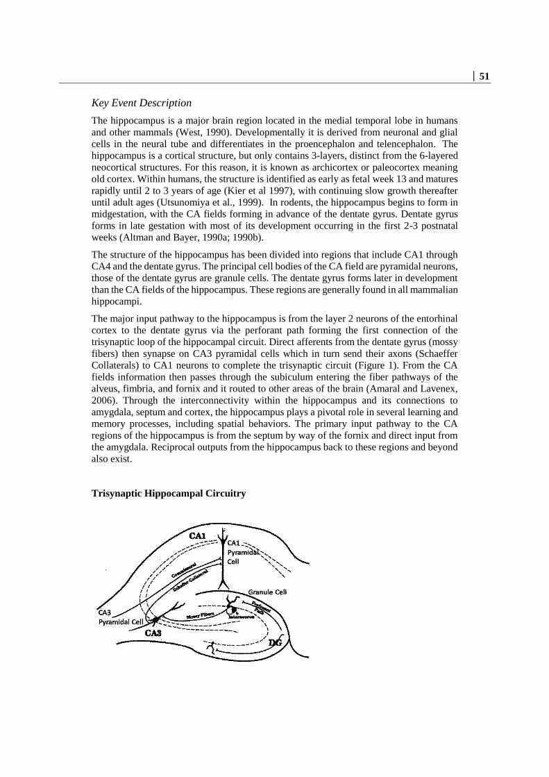
│ 51
Key Event Description
The hippocampus is a major brain region located in the medial temporal lobe in humans
and other mammals (West, 1990). Developmentally it is derived from neuronal and glial
cells in the neural tube and differentiates in the proencephalon and telencephalon. The
hippocampus is a cortical structure, but only contains 3-layers, distinct from the 6-layered
neocortical structures. For this reason, it is known as archicortex or paleocortex meaning
old cortex. Within humans, the structure is identified as early as fetal week 13 and matures
rapidly until 2 to 3 years of age (Kier et al 1997), with continuing slow growth thereafter
until adult ages (Utsunomiya et al., 1999). In rodents, the hippocampus begins to form in
midgestation, with the CA fields forming in advance of the dentate gyrus. Dentate gyrus
forms in late gestation with most of its development occurring in the first 2-3 postnatal
weeks (Altman and Bayer, 1990a; 1990b).
The structure of the hippocampus has been divided into regions that include CA1 through
CA4 and the dentate gyrus. The principal cell bodies of the CA field are pyramidal neurons,
those of the dentate gyrus are granule cells. The dentate gyrus forms later in development
than the CA fields of the hippocampus. These regions are generally found in all mammalian
hippocampi.
The major input pathway to the hippocampus is from the layer 2 neurons of the entorhinal
cortex to the dentate gyrus via the perforant path forming the first connection of the
trisynaptic loop of the hippocampal circuit. Direct afferents from the dentate gyrus (mossy
fibers) then synapse on CA3 pyramidal cells which in turn send their axons (Schaeffer
Collaterals) to CA1 neurons to complete the trisynaptic circuit (Figure 1). From the CA
fields information then passes through the subiculum entering the fiber pathways of the
alveus, fimbria, and fornix and it routed to other areas of the brain (Amaral and Lavenex,
2006). Through the interconnectivity within the hippocampus and its connections to
amygdala, septum and cortex, the hippocampus plays a pivotal role in several learning and
memory processes, including spatial behaviors. The primary input pathway to the CA
regions of the hippocampus is from the septum by way of the fornix and direct input from
the amygdala. Reciprocal outputs from the hippocampus back to these regions and beyond
also exist.
Trisynaptic Hippocampal Circuitry

52 │
How it is Measured or Detected
Data in support of this key event have been collected using a wide variety of standard
biochemical, histological and anatomical methods (e.g., morphometrics,
immunohistochemical staining, in situ hybridization and imaging procedures). Many of
methods applied to reveal anatomical abnormalities are routine neurohistopathology
procedures similar to those recommended in EPA and OECD developmental neurotoxicity
guidelines (US EPA, 1998; OCED, 2007). Subtle cytoarchitectural features depend on
more specialized birth dating procedures and staining techniques. It is essential to consider
the timing of events during development for detection to occur, as well as the timing for
detection (Hevner, 2007; Garman et al., 2001; Zgraggen et al., 2012). Similar techniques
used in rodent stydies have been applied to postmortem tissue in humans.
In humans, structural neuroimaging techniques are used to assess hippocampal volume with
an analysis technique known as voxel-based morphometry (VBM). Volume of brain
regions is measured by drawing regions of interest (ROIs) on images from brain scans
obtained from magnetic resonance imaging (MRI) or positron emission tomography (PET)
scans and calculating the volume enclosed. (Mechelli et al., 2005). Similar imaging
techniques can be applied in rodent models (Powell et al., 2009; Hasegawa et al., 2010;
Pirko et al., 2005; Pirko and Johnson, 2008).
References
Altman J, Bayer SA. Migration and distribution of two populations of hippocampal granule
cell precursors during the perinatal and postnatal periods. J Comp Neurol. 1990a Nov
15;301(3):365-81.
Altman J, Bayer SA. Prolonged sojourn of developing pyramidal cells in the intermediate
zone of the hippocampus and their settling in the stratum pyramidale. J Comp Neurol.
1990b Nov 15;301(3):343-64.
Amaral D, Lavenex P (2006). "Ch 3. Hippocampal Neuroanatomy". In Andersen P, Morris
R, Amaral D, Bliss T, O'Keefe J. The Hippocampus Book. Oxford University Press. ISBN
978-0-19-510027-3.
Garman RH, Fix AS, Jortner BS, Jensen KF, Hardisty JF, Claudio L, Ferenc S. Methods to
identify and characterize developmental neurotoxicity for human health risk assessment.
II: neuropathology. Environ Health Perspect. 2001 Mar;109 Suppl 1:93-100.
Hasegawa M, Kida I, Wada H. A volumetric analysis of the brain and hippocampus of rats
rendered perinatal hypothyroid. Neurosci Lett. 2010 Aug 2;479(3):240-4.
Hevner RF. Layer-specific markers as probes for neuron type identity in human neocortex
and malformations of cortical development. J Neuropathol Exp Neurol. 2007 66(2):101-9.
Kier, EL, Kim, JH, Fulbright, K, Bronen, RA. Embryology of the human fetal
hippocampus: MR imaging, anatomy, and histology. AJNR Am J Neuroradiol: 1997,
18(3);525-32.
Mechelli A, Price C, Friston K, Ashburner J (2005) Voxel-Based Morphometry of the
Human Brain: Methods and Applications. Curr Med Imaging Rev 1:105-113.

│ 53
OECD. 2007. OECD guidelines for the testing of chemicals/ section 4: Health effects. Test
no. 426: Developmental neurotoxicity study.
http://www.oecd.org/dataoecd/20/52/37622194.
Pirko I, Fricke ST, Johnson AJ, Rodriguez M, Macura SI. Magnetic resonance imaging,
microscopy, and spectroscopy of the central nervous system in experimental animals.
NeuroRx. 2005 Apr;2(2):250-64.
Pirko I, Johnson AJ. Neuroimaging of demyelination and remyelination models. Curr Top
Microbiol Immunol. 2008; 318:241-66.
Powell MH, Nguyen HV, Gilbert M, Parekh M, Colon-Perez LM, Mareci TH, Montie E.
Magnetic resonance imaging and volumetric analysis: novel tools to study the effects of
thyroid hormone disruption on white matter development. Neurotoxicology. 2012
Oct;33(5):1322-9.
U.S.EPA. 1998. Health effects guidelines OPPTS 870.6300 developmental neurotoxicity
study. EPA Document 712-C-98-239.Office of Prevention Pesticides and Toxic
Substances.
Utsunomiya, H., K Takano, M Okazaki, A Mitsudome Development of the temporal lobe
in infants and children: analysis by MR-based volumetry. AJNR Am J Neuroradiol: 1999,
20(4);717-23.
West MJ (1990). "Stereological studies of the hippocampus: a comparison of the
hippocampal subdivisions of diverse species including hedgehogs, laboratory rodents, wild
mice and men". Progress in Brain Research. Progress in Brain Research 83: 13–36.
Zgraggen E, Boitard M, Roman I, Kanemitsu M, Potter G, Salmon P, Vutskits L, Dayer
AG, Kiss JZ. Early postnatal migration and development of layer II pyramidal neurons in
the rodent cingulate/retrosplenial cortex. Cereb Cortex. 2012 Jan;22(1):144-57.
Event: 758: Hippocampal Physiology, Altered
Short Name: Hippocampal Physiology, Altered
Key Event Component
Process Object Action
chemical synaptic transmission synapse abnormal
AOPs Including This Key Event
AOP ID and Name Event
Type
Aop:42 - Inhibition of Thyroperoxidase and Subsequent Adverse
Neurodevelopmental Outcomes in Mammals
KeyEvent
Aop:8 - Upregulation of Thyroid Hormone Catabolism via Activation of
Hepatic Nuclear Receptors, and Subsequent Adverse Neurodevelopmental
Outcomes in Mammals
KeyEvent
Aop:134 - Sodium Iodide Symporter (NIS) Inhibition and Subsequent
Adverse Neurodevelopmental Outcomes in Mammals
KeyEvent

54 │
Aop:152 - Interference with thyroid serum binding protein transthyretin
and subsequent adverse human neurodevelopmental toxicity
KeyEvent
Stressors
Name
Propylthiouracil
Iodine deficiency
Methimazole
Biological Context
Level of Biological Organization
Tissue
Organ term
Organ term
brain
Domain of Applicability
Taxonomic Applicability
Term Scientific Term Evidence Links
human Homo sapiens Moderate NCBI
rat Rattus norvegicus High NCBI
mouse Mus musculus High NCBI
Life Stage Applicability
Life Stage Evidence
During brain development High
Sex Applicability
Sex Evidence
Female High
Male High

│ 55
The majority of evidence for this key event come from work in rodent species (i.e., rat,
mouse). There is a moderate amount of evidence from other species, including humans
(Clapp et al., 2012).
Key Event Description
The hippocampus functions as a highly integrated and organized communication and
information processing network with millions of interconnections among its constitutive
neurons. Neurons in the hippocampus and throughout the brain transmit and receive
information largely through chemical transmission across the synaptic cleft, the space
where the specialized ending of the presynaptic axon terminus of the transmitting neuron
meets the specialized postsynaptic region of the neuron that is receiving that information
(Kandell et al., 2012).
During development (see KE: Hippocampal anatomy, Altered), as neurons reach their final
destination and extend axonal processes, early patterns of electrical synaptic activity
emerge in the hippocampus. These are large fields of axonal innervation of broad synaptic
target sites that are replaced by more elaborate but highly targeted and refined axonal
projections brought about by activity-dependent synaptic pruning and synapse
elimination. This is a classic case of the interaction between physiological and anatomical
development, where anatomy develops first, and can be ‘reshaped’ by physiological
function (Kutsarova et al., 2017).
In the rat, excitatory processes are fully mature in area CA1 of hippocampus within 2 weeks
of birth with inhibitory processes lagging begin by several weeks (Muller et al., 1989;
Michelson and Lothman, 1988; Harris and Teyler, 1984). In hippocampal slices, inhibitory
function in areaCA1s is first seen on postnatal day 5 and increases in strength at postnatal
day 12 through 15. In vivo studies fail to detect inhibition until postnatal day 18 with steady
increase thereafter to adult levels by postnatal day 28. Synaptic plasticity in the form of
long-term potentiation (LTP) is absent in the very young animal, only emerging about
postnatal day 14, appearing to require the stability of both excitatory and inhibitory function
to be established (Muller et al., 1989; Bekenstein and Lothman, 1991). These features of
the maturation of hippocampal physiology are paralleled in dentate gyrus, but as with
anatomical indices in the rat, the development of these physiological parameters lag behind
the CA1 by about 1 week.
How it is Measured or Detected
In animals, synaptic function in the hippocampus has been examined with imaging
techniques, but more routinely, electrical field potentials recorded in two subregions of the
hippocampus, area CA1 and dentate gyrus, have been assessed in vivo or in vitro from
slices taken from naive or exposed animals. Field potentials reflect the summed synaptic
response of a population of neurons following direct stimulation of input pathways across
a monosynaptic connection. Changes in response amplitude due to chemical perturbations
and other stressors (e.g., iodine deficiency, thyroidectomy, gene knockouts) is evidence of
altered synaptic function. This can be measured in vitro, in vivo, or in hippocampal slices
taken from treated animals (Gilbert and Burdette, 1995). The most common physiological
measurements used to assess function of the hippocampus are excitatory synaptic

56 │
transmission, inhibitory synaptic transmission, and synaptic plasticity in the form of long-
term potentiation (LTP).
Excitatory Synaptic Transmission: Two measures, the excitatory postsynaptic potential
(EPSP) and the population spike are derived from the compound field potential at
increasing stimulus strengths. The function described by the relationship of current strength
(input, I) and evoked response (output, O), the I-O curve is the measure of excitatory
synaptic transmission (Gilbert and Burdette, 1995).
Inhibitory Synaptic Transmission: Pairs of stimulus pulses delivered in close temporal
proximity is used to probe the integrity of inhibitory synaptic transmission. The response
evoked by the second pulse of the pair at brief intervals (<30 msec) arrives during the
activation of feedback inhibitory loops in the hippocampus. An alteration in the degree of
suppression to the 2nd pulse of the pair reflects altered inhibitory synaptic function (Gilbert
and Burdette, 1995).
Long Term Potentiation (LTP): LTP is widely accepted to be a major component of the
cellular processes that underlie learning and memory (Malenka and Bear, 2004; Bramham
and Messaoudi, 2005). LTP represents, at the synapse and molecular level, the coincident
firing of large numbers of neurons that are engaged during a learning event. The persistence
of LTP emulates the duration of the memory. Synaptic plasticity in the form of LTP is
assessed by delivering trains of high frequency stimulation to induce a prolonged
augmentation of synaptic response. Probe stimuli at midrange stimulus strengths are
delivered before and after application of LTP-inducing trains. The degree of increase in
EPSP and PS amplitude to the probe stimulus after train application, and the duration of
the induced synaptic enhancement are metrics of LTP. Additionally, contrasting I-O
functions of excitatory synaptic transmission before and after (hours to days) LTP is
induced is also a common measure of LTP maintanence (Bramham and Messaoudi, 2005;
Kandell et al., 2012; Malenka and Bear, 2004).
Synaptic function in the human hippocampus has been assessed using
electroencephalography (EEG) and functional neuroimaging techniques (Clapp et al.,
2012). EEG is a measure of electrical activity over many brain regions but primarily from
the cortex using small flat metal discs (electrodes) placed over the surface of the skull. It is
a readily available test that provides evidence of how the brain functions over time.
Functional magnetic resonance imaging or functional MRI (fMRI) uses MRI technology
to measure brain activity by detecting associated changes in blood flow. This technique
relies on the fact that cerebral blood flow and neuronal activation are coupled. Positron
emission tomography (PET) is a functional imaging technique that detects pairs of gamma
rays emitted indirectly by a radionuclide (tracer) injected into the body (Tietze, 2012;
McCarthy, 1995). Like fMRI, PET scans indirectly measure blood flow to different parts
of the brain – the higher the blood flow, the greater the activation (McCarthy, 1995). These
techniques have been widely applied in clinical and research settings to assess learning and
memory in humans and can provide information targeted to hippocampal functionality
(McCarthy, 1995; Smith and Jonides, 1997; Willoughby et al., 2014; Wheeler et al., 2015;
Gilbert et al., 1998).
Assays of this type are fit for purpose, have been well accepted in the literature, and are
reproducible across laboratories. The assay directly measures the key event of altered
neurophysiological function.

│ 57
References
Bekenstein JW, Lothman EW. An in vivo study of the ontogeny of long-term potentiation
(LTP) in the CA1 region and in the dentate gyrus of the rat hippocampal formation. Brain
Res Dev Brain Res. 1991 Nov 19;63(1-2):245-
Bramham CR, Messaoudi E (2005) BDNF function in adult synaptic plasticity: the synaptic
consolidation hypothesis. Prog Neurobiol 76:99-125.
Clapp WC, Hamm JP, Kirk IJ, Teyler TJ. Translating long-term potentiation from animals
to humans: a novel method for noninvasive assessment of cortical plasticity. Biol
Psychiatry. 2012 Mar 15;71(6):496-502.
Gilbert, M.E. and Burdette, L.J. (1995). Hippocampal Field Potentials: A Model System to
Characterize Neurotoxicity. In Neurotoxicology: Approaches and Methods. L.W Chang
and W. Slikker (Eds). Academic Press:New York, 183-204.
Gilbert ME, Mack CM. Chronic lead exposure accelerates decay of long-term potentiation
in rat dentate gyrus in vivo. Brain Res. 1998 Apr 6;789(1):139-49.
Harris KM, Teyler TJ. Developmental onset of long-term potentiation in area CA1 of the
rat hippocampus. J Physiol. 1984. 346:27-48.
Kandell, E., Schwartz, J., Siegelbaum, A. and Hudspeth, A.J. (2012) Principles of Neural
Science, 5th Edition. Elsevier, North Holland.
Kutsarova E, Munz M, Ruthazer ES. Rules for Shaping Neural Connections in the
Developing Brain. Front Neural Circuits. 2017 Jan 10;10:111. doi:
10.3389/fncir.2016.00111.
Malenka RC, Bear MF (2004) LTP and LTD: an embarrassment of riches. Neuron 44:5-
21.
McCarthy, G. (1995) Review: Functional Neuroimaging and Memory. The Neuroscientist,
1:155-163.
Michelson HB, Lothman EW. An in vivo electrophysiological study of the ontogeny of
excitatory and inhibitory processes in the rat hippocampus. Brain Res Dev Brain Res. 1989
May 1;47(1):113-22.
Muller D, Oliver M, Lynch G. Developmental changes in synaptic properties in
hippocampus of neonatal rats. Brain Res Dev Brain Res. 1989 Sep 1;49(1):105-14.
Smith, E and Jonides, J. (1997). Working Memory: A View from Neuroimaging. Cognitive
Psychology, 33:5-42.
Tietze, KJ. (2012). Review of Laboratory and Diagnostic Tests- Positron Emission
Tomography. In Clinical Sills for Pharmacists, 3rd Edition, pp 86-122.
Wheeler SM, McLelland VC, Sheard E, McAndrews MP, Rovet JF (2015) Hippocampal
Functioning and Verbal Associative Memory in Adolescents with Congenital
Hypothyroidism. Front Endocrinol (Lausanne) 6:163.
Willoughby KA, McAndrews MP, Rovet JF (2014) Effects of maternal hypothyroidism on
offspring hippocampus and memory. Thyroid 24:576-584.

58 │
List of Adverse Outcomes in this AOP
Event: 402: Cognitive Function, Decreased
Short Name: Cognitive Function, Decreased
Key Event Component
Process Object Action
learning or memory decreased
cognition decreased
AOPs Including This Key Event
AOP ID and Name Event Type
Aop:42 - Inhibition of Thyroperoxidase and Subsequent Adverse
Neurodevelopmental Outcomes in Mammals
AdverseOutcome
Aop:65 - XX Inhibition of Sodium Iodide Symporter and
Subsequent Adverse Neurodevelopmental Outcomes in Mammals
AdverseOutcome
Aop:134 - Sodium Iodide Symporter (NIS) Inhibition and
Subsequent Adverse Neurodevelopmental Outcomes in Mammals
AdverseOutcome
Aop:152 - Interference with thyroid serum binding protein
transthyretin and subsequent adverse human neurodevelopmental
toxicity
AdverseOutcome
Stressors
Name
Methimazole
Propylthiouracil
Iodine deficiency
Biological Context
Level of Biological Organization
Individual
Domain of Applicability
Taxonomic Applicability
Term Scientific Term Evidence Links
human Homo sapiens High NCBI
rat Rattus norvegicus High NCBI
mouse Mus musculus High NCBI

│ 59
Life Stage Applicability
Life Stage Evidence
During brain development High
Sex Applicability
Sex Evidence
Male High
Female High
Basic forms of learning behavior such as habituation have been found in many taxa from
worms to humans (Alexander, 1990). More complex cognitive processes such as executive
function likely reside only in higher mammalian species such as non-human primates and
humans.
Key Event Description
Learning and memory depend upon the coordinated action of different brain regions and
neurotransmitter systems constituting functionally integrated neural networks (D’Hooge
and DeDeyn, 2001). Among the many brain areas engaged in the acquisition of, or retrieval
of, a learned event, the hippocampal-based memory systems have received the most study.
The main learning areas and pathways are similar in rodents and primates, including man
(Eichenbaum, 2000; Stanton and Spear, 1990; Squire, 2004).
In humans, the hippocampus is involved in recollection of an event’s rich spatial-temporal
contexts and distinguished from simple semantic memory which is memory of a list of facts
(Burgess et al., 2000). Hemispheric specialization has occurred in humans, with the left
hippocampus specializing in verbal and narrative memories (i.e., context-dependent
episodic or autobiographical memory) and the right hippocampus, more prominently
engaged in visuo-spatial memory (i.e., memory for locations within an environment). The
hippocampus is particularly critical for the formation of episodic memory, and
autobiographical memory tasks have been developed to specifically probe these functions
(Eichenbaun, 2000; Willoughby et al., 2014). In rodents, there is obviously no verbal
component in hippocampal memory, but reliance on the hippocampus for spatial, temporal
and contextual memory function has been well documented. Spatial memory deficits and
fear-based context learning paradigms engage the hippocampus, amygdala, and prefrontal
cortex (Eichenbaum, 2000; Shors et al., 2001; Samuels et al., 2011; Vorhees and Williams,
2014; D’Hooge and DeDeyn, 2001; Lynch, 2004; O’Keefe and Nadal, 1978). These tasks
are impaired in animals with hippocampal dysfunction (O’Keefe and Nadal, 1978; Morris
and Frey, 1987; Gilbert et al., 2016).

60 │
How it is Measured or Detected
In rodents, a variety of tests of learning and memory have been used to probe the integrity
of hippocampal function. These include tests of spatial learning like the radial arm maze
(RAM), the Barnes maze, and most commonly, the Morris water maze (MWM). Test of
novelty such as novel object recognition, and fear based context learning are also sensitive
to hippocampal disruption. Finally, trace fear conditioning which incorporates a temporal
component upon traditional amygdala-based fear learning engages the hippocampus. A
brief description of these tasks follows.
1) RAM, Barnes, MWM are examples of spatial tasks in which animals are required to
learn: the location of a food reward (RAM); an escape hole to enter a preferred dark tunnel
from a brightly lit open field area (Barnes maze); or a hidden platform submerged below
the surface of the water in a large tank of water (MWM) (Vorhees and Williams, 2014).
2) Novel Object recognition. This is a simpler task that can be used to probe recognition
memory. Two objects are presented to animal in an open field on trial 1, and these are
explored. On trial 2, one object is replaced with a novel object and time spent interacting
with the novel object is taken evidence of memory retention (i.e., I have seen one of these
objects before, but not this one. Cohen and Stackman, 2015).
3) Contextual Fear conditioning is a hippocampal based learning task in which animals are
placed in a novel environment and allowed to explore for several minutes before delivery
of an aversive stimulus, typically a mild foot shock. Upon reintroduction to this same
environment in the future (typically 24-48 hours after original training), animals will limit
their exploration, the context of this chamber being associated with an aversive event. The
degree of suppression of activity after training is taken as evidence of retention, i.e.,
memory (Curzon et al., 2009).
4) Trace fear conditioning. Standard fear conditioning paradigms require animals to make
an association between a neutral conditioning stimulus (CS, a light or a tone) and an
aversive stimulus (US, a footshock). The unconditioned response (CR) that is elicited upon
delivery of the footshock US is freezing behavior. With repetition of CS/US delivery, the
previously neutral stimulus comes to elicit the freezing response. This type of learning is
dependent on the amygdala, a brain region associated with, but distinct from the
hippocampus. Introducing a brief delay between presentation of the neutral CS and the
aversive US, a trace period, requires the engagement of the amygdala and the hippocampus
(Shors et al., 2004).
Most methods are well established in the published literature and many have been engaged
to evaluate the effects of developmental thyroid disruption. The US EPA and OECD
Developmental Neurotoxicity (DNT) Guidelines (OCSPP 870.6300 or OECD 426) both
require testing of learning and memory (USEPA, 1998; OECD, 2007). These DNT
Guidelines have been deemed valid to identify developmental neurotoxicity and adverse
neurodevelopmental outcomes (Makris et al., 2009).
A variety of standardized learning and memory tests have been developed for human
neuropsychological testing. These include episodic autobiographical memory, word pair
recognition memory; object location recognition memory. Some components of these tests
have been incorporated in general tests of adult intelligence (IQ) such as the WAIS and the
Wechsler. Modifications have been made and norms developed for incorporating of tests
of learning and memory in children. Examples of some of these tests include:

│ 61
1) Rey Osterieth Complex Figure (RCFT) which probes a variety of functions including as
visuospatial abilities, memory, attention, planning, and working memory (Shin et al.,
2006).
2) Children’s Auditory Verbal Learning Test (CAVLT) is a free recall of presented word
lists that yields measures of Immediate Memory Span, Level of Learning, Immediate
Recall, Delayed Recall, Recognition Accuracy, and Total Intrusions. (Lezak 1995; Talley,
1986).
3) Continuous Visual Memory Test (CVMT) measures visual learning and memory. It is a
free recall of presented pictures/objects rather than words but that yields similar measures
of Immediate Memory Span, Level of Learning, Immediate Recall, Delayed Recall,
Recognition Accuracy, and Total Intrusions. (Lezak, 1984; 1994).
4) Story Recall from Wechsler Memory Scale (WMS) Logical Memory Test Battery, a
standardized neurospychological test designed to measure memory functions (Lezak, 1994;
Talley, 1986).
5) Autobiographical memory (AM) is the recollection of specific personal events in a
multifaceted higher order cognitive process. It includes episodic memory- remembering of
past events specific in time and place, in contrast to semantic autobiographical memory is
the recollection of personal facts, traits, and general knowledge. Episodic AM is associated
with greater activation of the hippocampus and a later and more gradual developmental
trajectory. Absence of episodic memory in early life (infantile amnesia) is thought to reflect
immature hippocampal function (Herold et al., 2015; Fivush, 2015).
6) Staged Autobiographical Memory Task. In this version of the AM test, children
participate in a staged event involving a tour of the hospital, perform a series of tasks
(counting footprints in the hall, identifying objects in wall display, buy lunch, watched a
video). It is designed to contain unique event happenings, place, time,
visual/sensory/perceptual details. Four to five months later, interviews are conducted using
Children’s Autobiographical Interview and scored according to standardized scheme
(Willoughby et al., 2014).
Regulatory Significance of the AO
A prime example of impairments in cognitive function as the adverse outcome for
regulatory action is developmental lead exposure and IQ function in children (Bellinger,
2012). In addition, testing for the impact of chemical expsoures on cognitive function, often
including spatially-mediated behaviors, is an intergral part of both EPA and OECD
developmental neurotoxicity guidelines (USEPA, 1998; OECD, 2007).
References
Alexander RD (1990) Epigenetic rules and Darwinian algorithms: The adaptive study of
learning and development. Ethology and Sociobiology 11:241-303.
Bellinger DC (2012) A strategy for comparing the contributions of environmental
chemicals and other risk factors to neurodevelopment of children. Environ Health Perspect
120:501-507.

62 │
Burgess N (2002) The hippocampus, space, and viewpoints in episodic memory. Q J Exp
Psychol A 55:1057-1080.
Cohen, SJ and Stackman, RW. (2015). Assessing rodent hippocampal involvement in the
novel object recognition task. A review. Behav. Brain Res. 285: 105-1176.
Curzon P, Rustay NR, Browman KE. Cued and Contextual Fear Conditioning for Rodents.
In: Buccafusco JJ, editor. Methods of Behavior Analysis in Neuroscience. 2nd edition.
Boca Raton (FL): CRC Press/Taylor & Francis; 2009
D'Hooge R, De Deyn PP (2001) Applications of the Morris water maze in the study of
learning and memory. Brain Res Brain Res Rev 36:60-90.
Eichenbaum H (2000) A cortical-hippocampal system for declarative memory. Nat Rev
Neurosci 1:41-50.
Fivush R. The development of autobiographical memory. Annu Rev Psychol. 2011.
62:559-82.
Gilbert ME, Sanchez-Huerta K, Wood C (2016) Mild Thyroid Hormone Insufficiency
During Development Compromises Activity-Dependent Neuroplasticity in the
Hippocampus of Adult Male Rats. Endocrinology 157:774-787.
Gilbert ME, Sui L (2006) Dose-dependent reductions in spatial learning and synaptic
function in the dentate gyrus of adult rats following developmental thyroid hormone
insufficiency. Brain Res 1069:10-22.
Herold, C, Lässer, MM, Schmid, LA, Seidl, U, Kong, L, Fellhauer, I, Thomann, PA, Essig,
M and Schröder, J. (2015). Neuropsychology, Autobiographical Memory, and
Hippocampal Volume in “Younger” and “Older” Patients with Chronic Schizophrenia.
Front. Psychiatry, 6: 53.
Lezak MD (1984) Neuropsychological assessment in behavioral toxicology--developing
techniques and interpretative issues. Scand J Work Environ Health 10 Suppl 1:25-29.
Lezak MD (1994) Domains of behavior from a neuropsychological perspective: the whole
story. Nebr Symp Motiv 41:23-55.
Lynch, M.A. (2004). Long-Term Potentiation and Memory. Physiological Reviews. 84:87-
136.
Makris SL, Raffaele K, Allen S, Bowers WJ, Hass U, Alleva E, Calamandrei G, Sheets L,
Amcoff P, Delrue N, Crofton KM. A retrospective performance assessment of the
developmental neurotoxicity study in support of OECD test guideline 426. Environ Health
Perspect. 2009 Jan;117(1):17-25.
Morris RG, Frey U. Hippocampal synaptic plasticity: role in spatial learning or the
automaticrecording of attended experience? Philos Trans R Soc Lond B Biol Sci. 1997 Oct
29;352(1360):1489-503. Review
O’Keefe, J. and Nadel, L. (1978). The Hippocampus as a Cognitive Map. Oxford: Oxford
University Press.
OECD. 2007. OECD guidelines for the testing of chemicals/ section 4: Health effects. Test
no. 426: Developmental neurotoxicity
study. www.Oecd.Org/dataoecd/20/52/37622194.Pdf [accessed May 21, 2012].
Samuels BA, Hen R (2011) Neurogenesis and affective disorders. Eur J Neurosci 33:1152-
1159.

│ 63
Shin, MS, Park, SY, Park, SR, Oeol, SH and Kwon, JS. (2006). Clinical and empirical
appliations fo the Rey-Osterrieth complex figure test. Nature Protocols, 1: 892-899.
Shors TJ, Miesegaes G, Beylin A, Zhao M, Rydel T, Gould E (2001) Neurogenesis in the
adult is involved in the formation of trace memories. Nature 410:372-376.Squire LR (2004)
Memory systems of the brain: a brief history and current perspective. Neurobiol Learn
Mem 82:171-177.
Stanton ME, Spear LP (1990) Workshop on the qualitative and quantitative comparability
of human and animal developmental neurotoxicity, Work Group I report: comparability of
measures of developmental neurotoxicity in humans and laboratory animals. Neurotoxicol
Teratol 12:261-267.
Talley, JL. (1986). Memory in learning disabled children: Digit span and eh Rey Auditory
verbal learning test. Archives of Clinical Neuropsychology, Elseiver.
U.S.EPA. 1998. Health effects guidelines OPPTS 870.6300 developmental neurotoxicity
study. EPA Document 712-C-98-239.Office of Prevention Pesticides and Toxic
Substances.
Vorhees CV, Williams MT (2014) Assessing spatial learning and memory in rodents. ILAR
J 55:310-332.
Willoughby KA, McAndrews MP, Rovet JF. Accuracy of episodic autobiographical
memory in children with early thyroid hormone deficiency using a staged event. Dev Cogn
Neurosci. 2014. 9:1-11.

64 │
Appendix 2
List of Adjacent Key Event Relationships in the AOP
Relationship: 309: Thyroperoxidase, Inhibition leads to TH synthesis,
Decreased
AOPs Referencing Relationship
AOP Name Adjacency Weight of
Evidence
Quantitative
Understanding Inhibition of Thyroperoxidase and
Subsequent Adverse
Neurodevelopmental Outcomes in
Mammals
adjacent High Low
Thyroperoxidase inhibition leading to
reduced young of year survival via
anterior swim bladder inflation
adjacent
Evidence Supporting Applicability of this Relationship
Taxonomic Applicability
Term Scientific Term Evidence Links
human Homo sapiens High NCBI
rat Rattus norvegicus High NCBI
Xenopus laevis Xenopus laevis High NCBI
Life Stage Applicability
Life Stage Evidence
All life stages High
Sex Applicability
Sex Evidence
Male High
Female High
Inhibition of TPO activity is widely accepted to directly impact TH synthesis. This is true
for both rats and humans, as well as some fishes, frogs and birds. Most of the data
supporting a causative relationship between TPO inhibition and altered TH synthesis is
derived from animal studies, in vitro thyroid microsomes from rats or pigs, and a limited

│ 65
number of human ex vivo (Nagasaka and Hidaka, 1976; Vickers et al., 2012) and clinical
studies. There are data to support that gene mutations in TPO result in congenital
hypothyroidism, underscoring the essential role of TPO in human thyroid hormone
synthesis.
Key Event Relationship Description
Thyroperoxidase (TPO) is a heme-containing apical membrane protein within the follicular
lumen of thyrocytes that acts as the enzymatic catalyst for thyroid hormone (TH) synthesis
(Taurog, 2005). Two commonly used reference chemicals, propylthiouracil (PTU) and
methimazole (MMI), are drugs that inhibit the ability of TPO to: a) activate iodine and
transfer it to thyroglobulin (Tg) (Davidson et al., 1978); and, b) couple thyroglobulin (Tg)-
bound iodotyrosyls to produce Tg-bound thyroxine (T4) and triiodothyronine (T3) (Taurog,
2005).
Evidence Supporting this KER
The weight of evidence supporting a direct linkage between the MIE, TPO inhibition, and
the KE of decreased TH synthesis, is strong and supported by more than three decades of
research in animals, including humans (Cooper et al., 1982; Cooper et al.,1983; Divi and
Doerge, 1994).
Biological Plausibility
The biological plausibility for this KER is rated Strong. TPO is the only enzyme capable
of de novo systhesis of TH. TPO catalyzes several reactions, including the oxidation of
iodide, nonspecific iodination of tyrosyl residues of thyroglobulin (Tg) to form
monoiodotyrosyl (MIT) or diiodotyrosyl (DIT) residues, and the coupling of these Tg-
bound iodotyrosyls to produce Tg-bound T3 and T4 (Divi and Doerge, 1994; Kessler et al.,
2008; Ruf et al., 2006; Taurog et al., 1996, 2005). Therefore, inhibition of TPO activity is
widely accepted to directly impact TH synthesis.
Empirical Evidence
Empirical support for this KER is strong. There are several papers that have measured
alterations in TPO and subsequent effects on TH synthesisTaurog et al. (1996) showed
decreased guicaol activity, decreased bound I125, and subsequent decreases in newly formed
T3 and T4 per molecule of Tg, following exposure to PTU, MMI and some
antibiotics. Following in vivo exposure to PTU in rats (Cooper et al., 1982; 1983), there
are concentration and time-dependent decreases in thyroid protein bound iodine and serum
T4 and T3 that recovered one month after cessation of PTU exposure. In addition,
measures of thyroidal iodine content were highly correlated with intra-thyroidal PTU
concentration. Vickers et al. (2012) demonstrated dose- and time- dependent inhibition of
TPO activity in both human and rat thyroid homogenates exposed to MMI. Tietge et al
(2010) recently showed decreases in thyroidal T4 following MMI exposure in
Xenopus. Doerge et al (1998) showed that a tryphenylmethane dye, malachite green,
inhibited TPO and lowered thyroxine production. A recent paper used a series of
benzothiazoles and showed TPO inhibition (guicaol assay) and inhibition of TSH
stimulated thyroxine release from Xenopus thyroid gland explant cultures (Hornung et al.,
2015).

66 │
Temporal Evidence: The temporal nature of this KER is applicable to all life stages,
including development (Seed et al., 2005). The impact of decreased TPO activity on
thyroidal hormone synthesis is similar across all ages. Good evidence for the temporal
relationship of the KER comes from thyroid system modeling (e.g., Degon et al., 2008;
Fisher et al., 2013) using data using data from studies of iodine deficiency and chemicals
that inhibit NIS. In addition, there is ample evidence of the temporal impacts of TPO
inhibition on TH synthesis, using ex vivo and in vitro measures that demonstrate the time
course of inhibition following chemical exposures, including some data from human
thyroid microsomes and ex vivo thyroid slices (Vickers et al., 2012). Future work is needed
that measures both TPO inhibition and TH production during development.
Dose-Response Evidence: Dose-response data is available from a number of studies that
correlate TPO inhibition with decreased TH production measured using a variety of
endpoints including iodine organification (e.g., Taurog et al., 1996), inhibition of guicaol
oxidation in thyroid microsomes (e.g., Doerge and Chang, 2002), and direct measure of
thyroid gland T4 concentrations (e.g., Hornung et al., 2015). However, there is a lack of
dose-response data from developmental studies showing direct linkages from TPO
inhibition to thyroidal TH synthesis.
Uncertainties and Inconsistencies
While it is clear that TPO inhibition will lead to altered hormone synthesis, there is a need
for data that will inform quantitative modeling of the relationship between TPO inhibition
and the magnitude of effects on thyroid hormone synthesis.
It is important to note that data from studies on genistein highlight this uncertainty. Doerge
and colleagues have demonstrated that for this compound up to 80% TPO inhibition did
not result in decreased serum T4 in rats (Doerge and Chang, 2002). This is not consistent
with other prototypical TPO inhibitors (e.g., PTU, MMI). It remains to be determined, if
for some presently unknown reason, that genistein is an outlier or not. This again points to
the need for quantitative modeling of the relationship between TPO inhibtion and
downstream KEs.
Quantitative Understanding of the Linkage
Response-response relationship
There are only a limited number of studies where both TPO inhibition and iodine
organification have been measured in vivo, and there are not enough data available to make
any definitive quantitative correlations. One in vivo study in rats exposed to the TPO
inhibitor genistein found no in vivo impact on serum thyroid hormone concentrations, even
when TPO was inhibited up to 80% (Chang and Doerge, 2000).
References
Chang HC, Doerge DR. Dietary genistein inactivates rat thyroid peroxidase in vivo without
an apparent hypothyroid effect. Toxicol Appl Pharmacol 168:244–252 (2000).
Cooper DS, Kieffer JD, Halpern R, Saxe V, Mover H, Maloof F, Ridgway EC (1983)
Propylthiouracil (PTU) pharmacology in the rat. II. Effects of PTU on thyroid function.
Endocrinology 113:921-928.

│ 67
Cooper DS, Saxe VC, Meskell M, Maloof F, Ridgway EC. Acute effects of propylthiouracil
(PTU) on thyroidal iodide organification and peripheral iodothyronine deiodination:
correlation with serum PTU levels measured by radioimmunoassay. J Clin Endocrinol
Metab. 1982 54(1):101-7.
Davidson, B., Soodak, M., Neary, J.T., Strout, H.V., and Kieffer, J.D. (1978). The
irreversible inactivation of thyroid peroxidase by methylmercaptoimidazole, thiouracil, and
propylthiouracil in vitro and its relationship to in vivo findings. Endocrinology 103:871–
882.
Divi, R. L., and Doerge, D. R. (1994). Mechanism-based inactivation of lactoperoxidase
and thyroid peroxidase by resorcinol derivatives. Biochemistry 33(32), 9668-74.
Doerge DR, Chang HC, Divi RL, Churchwell Mechanism for inhibition of thyroid
peroxidase by leucomalachite green. Chem Res Toxicol. 1998 11(9):1098-104.
Doerge DR, Chang HC. Inactivation of thyroid peroxidase by soy isoflavones, in vitro and
in vivo. J Chromatogr B Analyt Technol Biomed Life Sci. 2002 Sep 25;777(1-2):269-79
Hornung MW, Kosian PA, Haselman JT, Korte JJ, Challis K, Macherla C, Nevalainen E,
Degitz SJ. In Vitro, Ex Vivo, and In Vivo Determination of Thyroid Hormone Modulating
Activity of Benzothiazoles.Toxicol Sci. 2015 146(2):254-64.
Kessler, J., Obinger, C., and Eales, G. (2008). Factors influencing the study of peroxidase-
generated iodine species and implications for thyroglobulin synthesis. Thyroid 18(7), 769-
74, 10.1089/thy.2007.0310.
Nagasaka, A., and Hidaka, H. (1976). Effect of antithyroid agents 6-propyl-2-thiouracil and
1-mehtyl-2-mercaptoimidazole on human thyroid iodine peroxidase. J. Clin. Endocrinol.
Metab. 43:152–158.
Ruf, J., and Carayon, P. (2006). Structural and functional aspects of thyroid peroxidase.
Archives of biochemistry and biophysics 445(2), 269-77, 10.1016/j.abb.2005.06.023.
Taurog, A., Dorris, M. L., and Doerge, D. R. (1996). Mechanism of simultaneous
iodination and coupling catalyzed by thyroid peroxidase. Archives of biochemistry and
biophysics 330(1), 24-32,
Taurog A. 2005. Hormone synthesis. In: Werner and Ingbar’s The Thyroid: A Fundamental
and Clinical Text (Braverman LE, Utiger RD, eds). Philadelphia:Lippincott, Williams and
Wilkins, 47–81.
Tietge JE, Butterworth BC, Haselman JT, Holcombe GW, Hornung MW, Korte JJ, Kosian
PA, Wolfe M, Degitz SJ. Early temporal effects of three thyroid hormone synthesis
inhibitors in Xenopus laevis. Aquat Toxicol. 2010 98(1):44-50
Vickers AE, Heale J, Sinclair JR, Morris S, Rowe JM, Fisher RL. Thyroid organotypic rat
and human cultures used to investigate drug effects on thyroid function, hormone synthesis
and release pathways. Toxicol Appl Pharmacol. 2012 260(1):81-8.

68 │
Relationship: 305: TH synthesis, Decreased leads to T4 in serum, Decreased
AOPs Referencing Relationship
AOP Name Adjacency Weight of
Evidence
Quantitative
Understanding Inhibition of Thyroperoxidase and
Subsequent Adverse
Neurodevelopmental Outcomes in
Mammals
adjacent High Moderate
XX Inhibition of Sodium Iodide
Symporter and Subsequent Adverse
Neurodevelopmental Outcomes in
Mammals
adjacent High Moderate
Sodium Iodide Symporter (NIS)
Inhibition and Subsequent Adverse
Neurodevelopmental Outcomes in
Mammals
adjacent High High
Inhibition of Na+/I- symporter (NIS)
leads to learning and memory
impairment
adjacent High Moderate
Thyroperoxidase inhibition leading to
reduced young of year survival via
anterior swim bladder inflation
adjacent
Evidence Supporting Applicability of this Relationship
Taxonomic Applicability
Term Scientific Term Evidence Links
human Homo sapiens High NCBI
rat Rattus norvegicus High NCBI
mouse Mus musculus High NCBI
Xenopus laevis Xenopus laevis High NCBI
Life Stage Applicability
Life Stage Evidence
All life stages High
Sex Applicability
Sex Evidence
Male High
Female High

│ 69
While a majority of the empirical evidence comes from work with laboratory rodents, there
is a large amount of supporting data from humans (with anti-hyperthyroidism drugs
including propylthiouracil and methimazole), some amphibian species (e.g., frog), and
some avian species (e.g, chicken). The following ares samples from a large literature that
supports this concept: Cooper et al. (1982; 1983); Hornung et al. (2010); Van Herck et al.
(2013); Paul et al. (2013); Alexander et al. (2017).
Key Event Relationship Description
Thyroid hormones (THs), thyroxine (T4) and triiodothyronine (T3) are synthesized by NIS
and TPO in the thyroid gland as iodinated thyroglobulin (Tg) and stored in the colloid of
thyroid follicles. Secretion from the follicle into serum is a multi-step process. The first
involves thyroid stimulating hormone (TSH) stimulation of the separation of the peptide
linkage between Tg and TH. The next steps involve endocytosis of colloid, fusion of the
endosome with the basolateral membrane of the thyrocyte, and finally release of TH into
blood. More detailed descriptions of this process can be found in reviews by Braverman
and Utiger (2012) and Zoeller et al. (2007).
Evidence Supporting this KER
The weight of evidence linking these two KEs of decreased TH synthesis and decreased T4
in serum is strong. It is commonly accepted dogma that decreased synthesis in the thyroid
gland will result in decreased circulating TH (serum T4).
Biological Plausibility
The biological relationship between two KEs in this KER is well understood and
documented fact within the scientific community.
Empirical Evidence
It is widely accepted that TPO inhibition leads to declines in serum T4 levels in adult
mammals. This is due to the fact that the sole source for circulating T4 derives from
hormone synthesis in the thyroid gland. Indeed, it has been known for decades that
insufficient dietary iodine will lead to decreased serum TH concentrations due to
inadequate synthesis. Strong qualitative and quantitative relationships exist between
reduced TH synthesis and reduced serum T4 (Ekerot et al., 2013; Degon et al., 2008;
Cooper et al., 1982; 1983; Leonard et al., 2016; Zoeller and Tan, 2007). There is more
limited evidence supporting the relationship between decreased TH synthesis and lowered
circulating hormone levels during development. Lu and Anderson (1994) followed the
time course of TH synthesis, measured as thyroxine secretion rate, in non-treated pregnant
rats and correlated it with serum T4 levels More recently, modeling of TH in the rat fetus
demonstrates the quantitative relationship between TH synthesis and serum T4
concentrations (Hassan et al., 2017). Furthermore, a wide variety of drugs and chemicals
that inhibit TPO are known to result in decreased release of TH from the thyroid gland, as
well as decreased circulating TH concentrations. This is evidenced by a very large number
of studies that employed a wide variety of techniques, including thyroid gland explant
cultures, tracing organification of 131-I and in vivo treatment of a variety of animal species

70 │
with known TPO inhibitors (King and May,1984; Atterwill et al., 1990; Brown et al., 1986;
Brucker-Davis, 1998; Hornung et al., 2010; Hurley et al., 1998; Kohrle, 2008).
Temporal Evidence: The temporal nature of this KER is applicable to all life stages,
including development (Seed et al., 2005). There are currently no studies that measured
both TPO synthesis and TH production during development. However, the impact
decreased TH synthesis on serum hormones is similar across all ages. Good evidence for
the temporal relationship comes from thyroid system modeling of the impacts of iodine
deficiency and NIS inhibition (e.g., Degon et al., 2008; Fisher et al., 2013). In addition,
recovery experiments have demonstrated that serum thyroid hormones recovered in
athyroid mice following grafting of in-vitro derived follicles (Antonica et al., 2012).
Dose-response Evidence: Dose-response data is lacking from studies that include
concurrent measures of both TH synthesis and serum TH concentrations. However, data is
available demonstrating correlations between thyroidal TH and serum TH concentrations
during gestation and lactation during development (Gilbert et al., 2013). This data was
used to develop a rat quantitative biologically-based dose-response model for iodine
deficiency (Fisher et al., 2013).
Uncertainties and Inconsistencies
There are no inconsistencies in this KER, but there are some uncertainties. The first
uncertainty stems from the paucity of data for quantitative modeling of the relationship
between the degree of synthesis decrease and resulting changes in circulating T4
concentrations. In addition, most of the data supporting this KER comes from inhibition of
TPO, and there are a number of other processes (e.g., endocytosis, lysosomal fusion,
basolateral fusion and release) that are not as well studied.
Quantitative Understanding of the Linkage
Response-response relationship
Fisher et al. (2013) published a quantitative biologically-based dose-response model for
iodine deficiency in the rat. This model provides quantitative relationships for thyroidal T4
synthesis (iodine organification) and predictions of serum T4 concentrations in developing
rats. There are other computational models that include thyroid hormone synthesis. Ekerot
et al. (2012) modeled TPO, T3, T4 and TSH in dogs and humans based on exposure to
myeloperoxidase inhibitors that also inhibit TPO. This model was recently adapted for rat
(Leonard et al., 2016) and Hassan et al (2017) have extended it to include the pregnant rat
dam in response to TPO inhibition induced by PTU. While the original model predicted
serum TH and TSH levels as a function of oral dose, it was not used to explicitly predict
the relationship between serum hormones and TPO inhibition, or thyroidal hormone
synthesis. Leonard et al. (2016) recently incorporated TPO inhibition into the model.
Degon et al (2008) developed a human thyroid model that includes TPO, but does not make
quantitative prediction of organification changes due to inhibition of the TPO enzyme.
References
Alexander EK, Pearce EN, Brent GA, Brown RS, Chen H, Dosiou C, Grobman WA,
Laurberg P, Lazarus JH, Mandel SJ, Peeters RP, Sullivan S. 2017 Guidelines of the

│ 71
American Thyroid Association for the Diagnosis and Management of Thyroid Disease
During Pregnancy and the Postpartum. Thyroid. 2017 Mar;27(3):315-389.
Antonica F, Kasprzyk DF, Opitz R, Iacovino M, Liao XH, Dumitrescu AM, Refetoff S,
Peremans K, Manto M, Kyba M, Costagliola S. Generation of functional thyroid from
embryonic stem cells. Nature. 2012 491(7422):66-71.
Atterwill CK, Fowler KF. A comparison of cultured rat FRTL-5 and porcine thyroid cells
for predicting the thyroid toxicity of xenobiotics. Toxicol In Vitro. 1990. 4(4-5):369-74.
Braverman, L.E. and Utiger, R.D. (2012). Werner and Ingbar's The Thyroid: A
Fundamental and Clinical Text (10 ed.). Philadelphia, PA: Lippincott Williams & Wilkins.
pp. 775-786. ISBN 978-1451120639.
Brown CG, Fowler KL, Nicholls PJ, Atterwill C. Assessment of thyrotoxicity using in vitro
cell culture systems. Food Chem Toxicol. 1986 24(6-7):557-62.
Brucker-Davis F. Effects of environmental synthetic chemicals on thyroid function.
Thyroid. 1998 8(9):827-56.
Cooper DS, Kieffer JD, Halpern R, Saxe V, Mover H, Maloof F, Ridgway EC (1983)
Propylthiouracil (PTU) pharmacology in the rat. II. Effects of PTU on thyroid function.
Endocrinology 113:921-928.
Cooper DS, Saxe VC, Meskell M, Maloof F, Ridgway EC.Acute effects of propylthiouracil
(PTU) on thyroidal iodide organification and peripheral iodothyronine deiodination:
correlation with serum PTU levels measured by radioimmunoassay. J Clin Endocrinol
Metab. 1982 54(1):101-7.
Degon, M., Chipkin, S.R., Hollot, C.V., Zoeller, R.T., and Chait, Y. (2008). A
computational model of the human thyroid. Mathematical Biosciences 212, 22–53
Ekerot P, Ferguson D, Glämsta EL, Nilsson LB, Andersson H, Rosqvist S, Visser SA.
Systems pharmacology modeling of drug-induced modulation of thyroid hormones in dogs
and translation to human. Pharm Res. 2013 30(6):1513-24.
Fisher JW, Li S, Crofton K, Zoeller RT, McLanahan ED, Lumen A, Gilbert
ME. Evaluation of iodide deficiency in the lactating rat and pup using a biologically based
dose-response model. Toxicol Sci. 2013 132(1):75-86.
Gilbert ME, Hedge JM, Valentín-Blasini L, Blount BC, Kannan K, Tietge J, Zoeller RT,
Crofton KM, Jarrett JM, Fisher JW. An animal model of marginal iodine deficiency during
development: the thyroid axis and neurodevelopmental outcome. Toxicol Sci. 2013
132(1):177-95.
Hassan, I, El-Masri, H., Kosian, PA, Ford, J, Degitz, SJ and Gilbert, ME. Quantitative
Adverse Outcome Pathway for Neurodevelopmental Effects of Thyroid Peroxidase-
Induced Thyroid Hormone Synthesis Inhibition. Toxicol Sci. 2017 Nov 1;160(1):57-73
Hornung MW, Degitz SJ, Korte LM, Olson JM, Kosian PA, Linnum AL, Tietge JE.
Inhibition of thyroid hormone release from cultured amphibian thyroid glands by
methimazole, 6-propylthiouracil, and perchlorate. Toxicol Sci. 2010 118(1):42-51.
Hurley PM. Mode of carcinogenic action of pesticides inducing thyroid follicular cell
tumors in rodents. Environ Health Perspect. 1998 106(8):437-45.
King DB, May JD. Thyroidal influence on body growth. J Exp Zool. 1984 Dec;232(3):453-
60.

72 │
Köhrle J. Environment and endocrinology: the case of thyroidology. Ann Endocrinol
(Paris). 2008 69(2):116-22.
Leonard JA, Tan YM, Gilbert M, Isaacs K, El-Masri H. Estimating margin of exposure to
thyroid peroxidase inhibitors using high-throughput in vitro data, high-throughput
exposure modeling, and physiologically based pharmacokinetic/pharmacodynamic
modeling. Toxicol Sci. 2016 151(1):57-70.
Lu, M-H, and Anderson, RR. Thyroxine secretion rats during pregnancy in the rat. Endo
Res. 1994. 20(4):343-364.
Paul KB, Hedge JM, Macherla C, Filer DL, Burgess E, Simmons SO, Crofton KM,
Hornung MW. Cross-species analysis of thyroperoxidase inhibition by xenobiotics
demonstrates conservation of response between pig and rat. Toxicology. 2013. 312:97-107.
Van Herck SL, Geysens S, Delbaere J, Darras VM. Regulators of thyroid hormone
availability and action in embryonic chicken brain development. Gen Comp Endocrinol.
2013. 190:96-104.
Zoeller, R. T., Tan, S. W., and Tyl, R. W. (2007). General background on the hypothalamic-
pituitary-thyroid (HPT) axis. Critical reviews in toxicology 37(1-2), 11-53.
Relationship: 312: T4 in serum, Decreased leads to T4 in neuronal tissue,
Decreased
AOPs Referencing Relationship
AOP Name Adjacency Weight of
Evidence
Quantitative
Understanding Inhibition of Thyroperoxidase and
Subsequent Adverse
Neurodevelopmental Outcomes in
Mammals
adjacent Moderate Moderate
XX Inhibition of Sodium Iodide
Symporter and Subsequent Adverse
Neurodevelopmental Outcomes in
Mammals
adjacent Moderate Low
Sodium Iodide Symporter (NIS)
Inhibition and Subsequent Adverse
Neurodevelopmental Outcomes in
Mammals
adjacent High Low
Inhibition of Na+/I- symporter (NIS)
leads to learning and memory
impairment
adjacent Moderate Low

│ 73
Evidence Supporting Applicability of this Relationship
Taxonomic Applicability
Term Scientific Term Evidence Links
rat Rattus norvegicus Moderate NCBI
mouse Mus musculus Moderate NCBI
human Homo sapiens Moderate NCBI
Life Stage Applicability
Life Stage Evidence
During brain development High
All life stages Moderate
Sex Applicability
Sex Evidence
Male Moderate
Female Moderate
The majority of the information on this KER comes from in vivo studies with rodents
(mainly MCT8 knock-out mice and thyroidectomized rats) and histopathological analyses
of human brain tissues derived from patients affected by AHDS (Allan-Herndon-Dudley
syndrome). The evoluationary conservation of the transport of TH from circulation to the
developing brain suggests, with some uncertainty, that this KER is also applicable to other
mammalian species.
Key Event Relationship Description
In mammals, thyroxine (T4) in brain tissue is derived almost entirely from the circulating
pool of T4 in blood. Transfer of free T4 (and to a lesser extent, T3) from serum binding
proteins (thyroid binding globulin (TBG), transthyretin (TTR) and albumin; see McLean
et al., 2017, for a recent review) into the brain requires transport across the blood brain
barrier (BBB) and /or indirect transport from the cerebral spinal fluid (CSF) into the brain
through the blood-CSF-barrier. The blood vessels in rodents and humans expresses the
main T4 transporter, MCT8, (Roberts et al. 2008), as does the choroid plexus which also
expresses TTR and secretes the protein into the CSF (Alshehri et al. 2015).
T4 entering the brain through the BBB is taken up into astrocytes via cell membrane
iodothyronine transporters (e.g., organic anion-transporting polypeptides OATP),
monocarboxylate transporter 8 (MCT8) (Visser et al., 2011). In astrocytes, T4 is then
deiodinated by Type II deiodinase to triiodothyronine (T3) (St Germain and Galton, 1997),
which is then transported via other iodothyronine transporters (MCT8) into neurons (Visser
et al., 2011). While some circulating T3 may be taken up into brain tissue directly from
blood (Dratman et al., 1991), the majority of neuronal T3 comes from deiodination of T4

74 │
in astrocytes. Decreases in circulating T4 will eventually result in decreased brain T3 tissue
concentrations. It is also known that Type II deiodinase can be up-regulated in response to
decreased T4 concentrations to maintain tissue concentrations of T3 (Pedraza et al., 2007;
Lavado-Autric et al., 2013; Morse et al., 1986), except in tanycytes of the paraventricular
nucleus (Fekete and Lechan, 2014).
Evidence Supporting this KER
The weight of evidence linking reductions in circulating serum TH and reduced brain
concentrations of TH is moderate. Many studies support this basic linkage. However, there
are compensatory mechanisms (e.g., upregulation of deiodinases, transporters) that may
alter the relationship between hormones in the periphery and hormone concentrations in
the brain. There is limited information available on the quantitative relationship between
circulating levels of TH, these compensatory processes, and neuronal T4 concentrations,
especially during development. Furthermore, in certain conditions, such as iodine
deficiency, the decreases in circulating hormone might have greater impacts on tissue levels
of TH (see for instance, Escobar del Rey, et al., 1989).
Biological Plausibility
The biological relationship between these two KEs is strong as it is well accepted dogma
within the scientific community. There is no doubt that decreased circulating T4 leads to
declines in tissue concentrations of T4 and T3 in a variety of tissues, including brain.
However, compensatory mechanisms (e.g., increased expression of Type 2 deiodinase)
may differ during different lifestages and across different tissues, especially in different
brain regions. Similarly, the degree to which serum TH must drop to overwhelm these
compensatory responses has not been established.
Empirical Evidence
Several studies have shown that tissue levels, including brain, of TH are proportional to
serum hormone level (Oppenheimer, 1983; Morreale de Escobar et al., 1987; 1990; Calvo
et al., 1992; Porterfield and Hendrich, 1992, 1993; Broedel et al., 2003). In
thyroidectomized rats, brain concentrations of T4 were decreased and Type II deiodinase
(DII) activity was increased. Both brain T3 and T4 as well as DII activity returned to normal
following infusion of T4 (Escobar-Morreale et al., 1995; 1997). Animals treated with PTU,
MMI, or iodine deficiency during development demonstrate both lower serum and lower
brain TH concentrations (Escobar-Morreale et al 1995; 1997; Taylor et al., 2008; Bastian
et al., 2012; 2014; Gilbert et al., 2013). Compared to the wildtype, a mouse MCT8
knockout model has was shown to have decreased plasma T4, decreased uptake of T4 into
the brain, and decreased brian T3 concentrations, as well as increased cortical diodinase
Type 2 activity and increased plasma T3 concentrations (Mayerl et al., 2014; Barez-Lopez
et al., 2016).
Temporal Evidence: The temporal relationship between serum T4 and T4 in growing
neuronal tissue described in this KER is developmental (Seed et al., 2005). While all brain
regions will be impacted by changes in serum hormones, brain concentrations will be a
function of development stage and brain region. Data are available from thyroid hormone
replacement studies that demonstrate recovery of fetal brain T3 and T4 levels (following
low iodine diets or MMI exposure) to control levels after maternal thyroid hormone
replacement or iodine supplementation (e.g., Calvo et al.,1990; Obregon et al., 1991). For
example, Calvo et al. (1990) carried out a detailed study of the effects of TPO inhibition

│ 75
on serum and tissues levels of TH in gestating rats. Clear dose-dependent effects of T4
replacement, but not T3 replacement were seen in all maternal tissues. However, for fetal
tissues, neither T4 nor T3, at any dose, could completely restore tissue TH levels to control
levels.
Dose-Response Evidence: There is good evidence, albeit from a limited number of studies
of the correlative relationship between circulating thyroid hormone concentrations and
brain tissue concentrations during fetal and early postnatal development following maternal
iodine deficient diets or chemical treatments that depress serum THs (c.f., Calvo et al.,
1990; Obregon et al., 1991; Morse et al.,1996).
Uncertainties and Inconsistencies
The fact that decreased serum TH results in lower brain TH concentrations is well
accepted. However, the ability of the developing brain to compensate for insuffiencies in
serum TH has not been well studied. Limited data is available that demonstrates that
changes in local deiodination in the developing brain can compensate for chemical-induced
alterations in TH concentrations (e.g., Calvo et al., 1990; Morse et al., 1996; Sharlin et al.,
2010). And, there are likely different quantitative relationships between these two KEs
depending on the compensatory ability based on both developmental stage and specific
brain region (Sharlin et al., 2010). For these reasons, the empirical support for this linkage
is rated as moderate
The role of cellular transporters represents an additional uncertainly. In addition, future
work on cellular transport mechanisms and deiodinase activity is likley to inform addition
of new KEs and KERs between serum and brain T4.
Quantitative Understanding of the Linkage
Response-response relationship
While it is well established that decreased in serum TH levels result in decreased brain TH
concentrations, particularly fetal brain concentrations, a major gap is the lack of empirical
data that allow direct quantification of this relationship (Hassan et al., 2018). Recently,
serum TH and brain TH were measured in fetal cortex and postnatal day 14 offspring
following graded degrees of hypothyroidism induced by PTU (O’Shaughnessy et al., 2018).
Results showed that brain levels TH levels at both ages were quantitatively related to serum
T4 levels. Additional dose-response information is necessary to confirm these findings, and
standardization of analysis for the measurements in these distinct matrices is crucial to
allow comparisons to be made between independent experiments.
References
Alshehri B, D'Souza DG, Lee JY, Petratos S, Richardson SJ. The diversity of mechanisms
influenced by transthyretin in neurobiology: development, disease and endocrine
disruption. J Neuroendocrinol. 2015 May;27(5):303- 23.
Bárez-López S, Obregon MJ, Martínez-de-Mena R, Bernal J, Guadaño-Ferraz A, Morte
B. Effect of Triiodothyroacetic Acid Treatment in Mct8 Deficiency: A Word of Caution.
Thyroid. 2016 May;26(5):618-26.

76 │
Bastian TW, Anderson JA, Fretham SJ, Prohaska JR, Georgieff MK, Anderson GW (2012),
Fetal and neonatal iron deficiency reduces thyroid hormone-responsive gene mRNA levels
in the neonatal rat hippocampus and cerebral cortex. Endocrinology 153:5668-5680.
Bastian TW, Prohaska JR, Georgieff MK, Anderson GW (2014) Fetal and neonatal iron
deficiency exacerbates mild thyroid hormone insufficiency effects on male thyroid
hormone levels and brain thyroid hormone-responsive gene expression. Endocrinology
55:1157-1167.
Broedel, O., Eravci, M., Fuxius, S., Smolarz, T., Jeitner, A., Grau, H., Stoltenburg-
Didinger, G., Plueckhan, H., Meinhold, H., and Baumgartner, A. (2003). Effects of hyper
and hypothyroidism on thyroid hormone concentrations in regions of the rat brain. Am. J.
Physiol. Endocrinol. Metab. 285:E470–480.
Calvo, R., Obregon, M.J., Escobar del Rey, F., and Morreale de Escobar, G. (1992). The
rat placenta and the transfer of thyroid hormones from the mother to the fetus. Effects of
maternal thyroid status. Endocrinology 131:357–365.
Calvo R, Obregon MJ, Ruiz de Ona C, Escobar del Rey F, Morreale de Escobar G 1990
Congenital hypothyroidism, as studied in rats. Crucial role of maternal thyroxine but not of
3,5,3’-triiodothyronine in the protection of the fetal brain. J Clin Invest 86:889–899.
Denver, RJ. (1998). The molecular basis of thyroid hormone-dependent central nervous
system remodeling during amphibian metamorphosis. Comparative Biochemistry and
Physiology Part C: Pharmacology, Toxicology and Endocrinology, 119:219-228.
Dratman MB, Crutchfield FL, Schoenhoff MB. Transport of iodothyronines from
bloodstream to brain: contributions by blood:brain and choroid plexus:cerebrospinal fluid
barriers. Brain Res. 1991 Jul 19;554(1-2):229-36.
Gilbert ME, Hedge JM, Valentin-Blasini L, Blount BC, Kannan K, Tietge J, Zoeller RT,
Crofton KM, Jarrett JM, Fisher JW (2013) An animal model of marginal iodine deficiency
during development: the thyroid axis and neurodevelopmental outcome. Toxicol Sci
132:177-195.
Escobar del Rey F, Ruiz de Oña C, Bernal J, Obregón MJ, Morreale de Escobar G.
Generalized deficiency of 3,5,3'-triiodo-L-thyronine (T3) in tissues from rats on a low
iodine intake, despite normal circulating T3 levels. Acta Endocrinol (Copenh). 1989
Apr;120(4):490-8.
Escobar-Morreale HF, Obregón MJ, Escobar del Rey F, Morreale de Escobar G.
Replacement therapy for hypothyroidism with thyroxine alone does not ensure
euthyroidism in all tissues, as studied in thyroidectomized rats. J Clin Invest. 1995
Dec;96(6):2828-38.
Escobar-Morreale HF1, Obregón MJ, Hernandez A, Escobar del Rey F, Morreale de
Escobar G. Regulation of iodothyronine deiodinase activity as studied in thyroidectomized
rats infused with thyroxine or triiodothyronine. Endocrinology. 1997 Jun;138(6):2559-68.
Fekete C, Lechan RM. Central regulation of hypothalamic-pituitary-thyroid axis under
physiological and pathophysiological conditions. Endocr Rev. 2014 Apr;35(2):159-94
Lavado-Autric R, Calvo RM, de Mena RM, de Escobar GM, Obregon MJ. Deiodinase
activities in thyroids and tissues of iodine-deficient female rats. Endocrinology. 2013
Jan;154(1):529-36.

│ 77
Mayerl S, Müller J, Bauer R, Richert S, Kassmann CM, Darras VM, Buder K, Boelen A,
Visser TJ, Heuer H. Transporters MCT8 and OATP1C1 maintain murine brain thyroid
hormone homeostasis. J Clin Invest. 2014 May;124(5):1987-99.
McLean TR, Rank MM, Smooker PM, Richardson SJ. Evolution of thyroid hormone
distributor proteins. Mol Cell Endocrinol. 2017 Feb 27. pii: S0303-7207(17)30151-X. doi:
10.1016/j.mce.2017.02.038. [Epub ahead of print]
Morreale de Escobar, G., Obregon, M.J., and Escobar del Ray, F. (1987). Fetal and
maternal thyroid hormones. Hormone Res. 26:12–27.
Morreale de Escobar, G., Calvo, R., Obregon, M.J., and Escobar del Rey, F. (1990).
Contribution of maternal thyroxine to fetal thyroxine pools in normal rats near term.
Endocrinology 126:2765–2767.
Morse DC, Wehler EK, Wesseling W, Koeman JH, Brouwer A. Alterations in rat brain
thyroid hormone status following pre- and postnatal exposure to polychlorinated biphenyls
(Aroclor 1254). Toxicol Appl Pharmacol. 1996 Feb;136(2):269-79
Obregon MJ, Ruiz de Ona C, Calvo R, Escobar del Rey F, Morreale de Escobar G 1991
Outer ring iodothyronine deiodinases and thyroid hormone economy: Responses to iodine
deficiency in the rat fetus and neonate. Endocrinology 129:2663–2673.
Oppenheimer, J.H. (1983). The nuclear Receptor-triiodothyronine complex: Relationship
to thyroid hormone distribution, metabolism, and biological action. In: Molecular Basis of
Thyroid Hormone Action, eds. J.H. Oppenheimer and H.H. Samuels, pp. 1–35. New York:
Academic Press.
O'Shaughnessy KL, Wood, C, Ford RL, Kosian, PA, Hotchkiss, MG, Degitz SJ, Gilbert
ME. Thyroid hormone disruption in the fetal and neonatal rat: Predictive hormone
measures and bioindicators of hormone action in the developing cortex. Toxicol Sci. 2018
Aug 6. doi: 10.1093/toxsci/kfy190. [Epub ahead of print]
Pedraza PE, Obregon MJ, Escobar-Morreale HF, del Rey FE, de Escobar GM (2006)
Mechanisms of adaptation to iodine deficiency in rats: thyroid status is tissue specific. Its
relevance for man. Endocrinology 147:2098-2108.
Porterfield, S.P. and Hendrich, C.E. (1992). Tissue iodothyronine levels in fetuses of
control and hypothyroid rats at 13 and 16 days gestation. Endocrinology 131:195–200.
Porterfield, S.P. and Hendrich, C.E. (1993). The role of thyroid hormones in prenatal
neonatal neurological development-current perspectives. Endocrine Rev. 14:94–106.
Power DM, Llewellyn L, Faustino M, Nowell MA, Björnsson BT, Einarsdottir IE, Canario
AV, Sweeney GE. (2001). Thyroid hormones in growth and development of fish. Comp
Biochem Physiol C Toxicol Pharmacol. Dec;130(4):447-59.
Roberts LM, Woodford K, Zhou M, Black DS, Haggerty JE, Tate EH, Grindstaff KK,
Mengesha W, Raman C, Zerangue N. Expression of the thyroid hormone transporters
monocarboxylate transporter-8 (SLC16A2) and organic ion transporter-14 (SLCO1C1) at
the blood-brain barrier. Endocrinology. 2008 Dec;149(12):6251-61.
Seed J, Carney EW, Corley RA, Crofton KM, DeSesso JM, Foster PM, Kavlock R, Kimmel
G, Klaunig J, Meek ME, Preston RJ, Slikker W Jr, Tabacova S, Williams GM, Wiltse J,
Zoeller RT, Fenner-Crisp P, Patton DE. Overview: Using mode of action and life stage
information to evaluate the human relevance of animal toxicity data. Crit Rev Toxicol.
2005 35:664-72.

78 │
Sharlin DS, Gilbert ME, Taylor MA, Ferguson DC, Zoeller RT. (2010).The nature of the
compensatory response to low thyroid hormone in the developing brain. J
Neuroendocrinol. Mar;22(3):153-65.
St. Germain, D.L. and Galton, V.A. (1997). The deiodinase family of selenoproteins.
Thyroid 7:655–668.
Taylor MA, Swant J, Wagner JJ, Fisher JW, Ferguson DC (2008) Lower thyroid
compensatory reserve of rat pups after maternal hypothyroidism: correlation of thyroid,
hepatic, and cerebrocortical biomarkers with hippocampal neurophysiology.
Endocrinology 149:3521-3530.
Van Herck SL, Geysens S, Delbaere J, Darras VM. (2013). Regulators of thyroid hormone
availability and action in embryonic chicken brain development. Gen Comp
Endocrinol.190:96-104.
Visser WE, Friesema EC, Visser TJ. Minireview: thyroid hormone transporters: the
knowns and the unknowns. Mol Endocrinol. 2011 Jan;25(1):1-14.
Relationship: 746: T4 in neuronal tissue, Decreased leads to Hippocampal gene
expression, Altered
AOPs Referencing Relationship
AOP Name Adjacency Weight of
Evidence
Quantitative
Understanding
Inhibition of Thyroperoxidase
and Subsequent Adverse
Neurodevelopmental
Outcomes in Mammals
adjacent Moderate Low
Sodium Iodide Symporter
(NIS) Inhibition and
Subsequent Adverse
Neurodevelopmental
Outcomes in Mammals
adjacent Moderate Low
Evidence Supporting Applicability of this Relationship
Taxonomic Applicability
Term Scientific Term Evidence Links
rat Rattus norvegicus Moderate NCBI
mouse Mus musculus Moderate NCBI
Life Stage Applicability
Life Stage Evidence
During brain development High

│ 79
Sex Applicability
Sex Evidence
Male High
Female High
Most of the data available has come from rodent models. The evolutionary conservation of
thyroid receptors (Holzer et al., 2017) coupled with their role in TR regulated gene
transcription in neurodevelopment, suggests that this KER may also be applicable to other
species (see text above).
Key Event Relationship Description
Many cellular and biochemical effects of thyroid hormones (TH) are mediated through
regulation of gene expression (Oppenheimer, 1983; Bernal, 2007). Thyroxine (T4) is
transferred from the serum to the brain (see KER: Thyroxine (T4) in Serum, Decreased
leads to Thyroxine (T4) in Neuronal Tissue, Decreased), where it converted to
triiodothyronine (T3), the level of which is highly controlled by deiodinases. T3 binds to
thyroid receptors (TR) in the nucleus of neuronal and glial cells to control gene expression.
It is generally accepted that the modulation of TR gene expression in the hippocampus, or
any other brain region, must therefore depend on the presence of hormone in these tissues.
Evidence Supporting this KER
The weight of evidence is moderate for TH concentrations affecting gene expression in the
developing brain is (Oppenheimer and Schwartz, 1997; Oppenheimer, 1983; Bernal, 2007;
Morte et al., 2010a; 2010b; Williams, 2008). Direct measurement of TH in brain tissue,
and in hippocampus in particular, has shown correlations with gene expression. Therefore,
it is assumed that reductions in TH-responsive genes in the hippocampus stem from
reduced availability of hormone in the brain from the serum. However, studies in which
there are simultaneous assessments of hippocampal concentrations of thyroid hormone and
hippocampal gene expression is limited.
Biological Plausibility
The biological relationship between these two KEs is strong. It is a generally accepted fact
that TH produce their actions on brain development by binding to nuclear receptors to affect
gene transcription. See KER (1387): "T4 in serum, Decreased leads (non-adjacently) to
Hippocampal gene expression, Altered" for more information on TR regulated genes. As
the primary means whereby TH promotes its action is by binding to TR in brain, TH must
be present in brain to affect this action. Circulating levels of T4 represent the primary
source of T4 in the brain, which is then converted to the active hormone T3 by deiodinases
within neuronal tissue.

80 │
Empirical Evidence
The empirical support for this KER is moderate. Many in vitro studies have demonstrated
a relationship between hormone concentraionsTH and the induction of gene expression in
brain cells, including hippocampal neurons in culture (Gil-Ibanez et al., 2015; Morte et al.,
2010b). However, there are a limited number of studies investigating TH concentrations in
the hippocampus and hippocampal gene expression. This is the case because thyroid
hormone is difficult to measure in hippocampus and TH-induced gene expression changes
can be subtle. We are aware of only four in vivo studies in which both thyroid hormones in
the brain and gene expression in brain were simultaneously measured (Bastian et al., 2012;
2014; Hernandez et al., 2010; Sharlin et al., 2008). Only two of these reports, stemming
from the same laboratory, specifically assessed thyroid hormone and gene expression in
hippocampus. In these studies, Bastian et al., (2012; 2014) measured decrements in
hippocampal T3 using RIA and correlated these reductions with alterations in the
expression of myelin associated genes (Mbp, Plp), the neurotrophin, Ngf, the calcium
binding protein Parv, a TH-dependent transcription factor, Hr, and Agt.
Temporal Evidence: The temporal nature of this KER on TH dependent gene regulation is
developmental (Seed et al., 2005). The impact of brain TH concentrations on regulation of
TR regulated genes is age-dependent for a number of genes critical for normal hippocampal
development. It is widely accepted that different genes are altered dependent upon the
window of exposure in the fetal, neonatal or adult brain (c.f., Pathak et al, 2011; Mohan et
al., 2012; Quignodon et al., 2004; Williams, 2008). Thyroid hormone supplementation has
been shown to reverse some of the effects on gene expression (Mohan et al., 2012; Liu et
al., 2010; Pathak et al., 2011).
Dose-Response Evidence: Dose-response data exists but is limited to a small number of
studies and a small number of genes (Bastian et al., 2012; 2014; Sharlin et al., 2008).
Uncertainties and Inconsistencies
There are no inconsistencies in this KER, but there are uncertainties. Uncertainties remain
in the relationship of neuronal TH concentrations and gene expression in the brain because
of the lack of studies simultaneously examining brain hormone and gene expression in the
same study. This stems from the technological challenges associated with measuring brain
hormone and the sometimes-subtle changes in brain gene expression induced by
manipulations of the thyroid system. In addition, there are also some physiological actions
of T4 that are mediated non-genomically at the cell membrane (Davis et al.,
2016). However, the exact role for the non-genomic effects is not well accepted or
understood (Galton, 2017).
Quantitative Understanding of the Linkage
Response-response relationship
There is only one study available to date that provides empirical data on both TH
concentrations and measures of gene expression changes in brain. O'Shaughnessy et al
(2018) demostrates dose-response relationships between brain T4 and T3 concentrations
and changes in a variety of genes (e.g., Parv, Col11a2, Hr, Ngf) that were "statistically
significant at doses that decreased brain t4 and/or T3". There was no quantitation of this
relationship reported.

│ 81
References
Bastian TW, Anderson JA, Fretham SJ, Prohaska JR, Georgieff MK, Anderson GW (2012),
Fetal and neonatal iron deficiency reduces thyroid hormone-responsive gene mRNA levels
in the neonatal rat hippocampus and cerebral cortex. Endocrinology 153:5668-5680.
Bastian TW, Prohaska JR, Georgieff MK, Anderson GW (2014) Fetal and neonatal iron
deficiency exacerbates mild thyroid hormone insufficiency effects on male thyroid
hormone levels and brain thyroid hormone-responsive gene expression. Endocrinology
155:1157-1167.
Bernal J (2007) Thyroid hormone receptors in brain development and function. Nat Clin
Pract Endocrinol Metab 3:249-259.
Davis, P.J., Goglia, F., Leonard, J.L., 2016. Nongenomic actions of thyroid hormone. Nat.
Rev. Endocrinol. 12, 111-121.
Galton VA. The ups and downs of the thyroxine pro-hormone hypothesis. Mol Cell
Endocrinol. 2017 Jan 24. pii: S0303-7207(17)30042-4. doi: 10.1016/j.mce.2017.01.029.
Gil-Ibañez P, García-García F, Dopazo J, Bernal J, Morte B. Global Transcriptome
Analysis of Primary Cerebrocortical Cells: Identification of Genes Regulated by
Triiodothyronine in Specific Cell Types. Cereb Cortex. 2017 Jan 1;27(1):706-717.
Hernandez A, Quignodon L, Martinez ME, Flamant F, St Germain DL (2010), Type 3
deiodinase deficiency causes spatial and temporal alterations in brain T3 signaling that are
dissociated from serum thyroid hormone levels. Endocrinology 151:5550-5558.
Holzer G, Roux N, Laudet V. Evolution of ligands, receptors and metabolizing enzymes of
thyroid signaling. Mol Cell Endocrinol. 2017 Mar 22. pii: S0303-7207(17)30191-0. doi:
10.1016/j.mce.2017.03.021.
Liu D, Teng W, Shan Z, Yu X, Gao Y, Wang S, Fan C, Wang H, Zhang H. The effect of
maternal subclinical hypothyroidism during pregnancy on brain development in rat
offspring. Thyroid. 2010 Aug;20(8):909-15.
Mohan V, Sinha RA, Pathak A, Rastogi L, Kumar P, Pal A, Godbole MM (2012) Maternal
thyroid hormone deficiency affects the fetal neocorticogenesis by reducing the proliferating
pool, rate of neurogenesis and indirect neurogenesis. Exp Neurol 237:477-488.
Morte B, Diez D, Auso E, Belinchon MM, Gil-Ibanez P, Grijota-Martinez C, Navarro D,
de Escobar GM, Berbel P, Bernal J (2010a) Thyroid hormone regulation of gene expression
in the developing rat fetal cerebral cortex: prominent role of the Ca2+/calmodulin-
dependent protein kinase IV pathway. Endocrinology 151:810-820.
Morte B, Ceballos A, Diez D, Grijota-Martínez C, Dumitrescu AM, Di Cosmo C, Galton
VA, Refetoff S, Bernal J. (2010b) Thyroid hormone-regulated mouse cerebral cortex genes
are differentially dependent on the source of the hormone: a study in monocarboxylate
transporter-8- and deiodinase-2-deficient mice. Endocrinology. 2010 May;151(5):2381-7
Oppenheimer, J. (1983). The nuclear-receptor-triiodothyronine complex: Relationship to
thyroid hormone distribution, metabolism, and biological action. Molecular Basis of
Thyroid Hormone Action. J. O. a. H. Samuels. New York, Academic Press: 1-34.

82 │
Oppenheimer, J. H. and H. L. Schwartz (1997). "Molecular basis of thyroid hormone-
dependent brain development." Endocr Rev 18(4): 462-75.
O'Shaughnessy KL, Wood, C, Ford RL, Kosian, PA, Hotchkiss, MG, Degitz SJ, Gilbert
ME. Thyroid hormone disruption in the fetal and neonatal rat: Predictive hormone
measures and bioindicators of hormone action in the developing cortex. Toxicol Sci. 2018
Aug 6. doi: 10.1093/toxsci/kfy190. [Epub ahead of print]
Pathak A, Sinha RA, Mohan V, Mitra K, Godbole MM. 2011. Maternal thyroid hormone
before the onset of fetal thyroid function regulates reelin and downstream signaling cascade
affecting neocortical neuronal migration. Cerebral cortex. 21:11-21.
Quignodon L, Legrand C, Allioli N, Guadano-Ferraz A, Bernal J, Samarut J, Flamant F
(2004) Thyroid hormone signaling is highly heterogeneous during pre- and postnatal brain
development. J Mol Endocrinol 33:467-476.
Seed J, Carney EW, Corley RA, Crofton KM, DeSesso JM, Foster PM, Kavlock R, Kimmel
G, Klaunig J, Meek ME, Preston RJ, Slikker W Jr, Tabacova S, Williams GM, Wiltse J,
Zoeller RT, Fenner-Crisp P, Patton DE. Overview: Using mode of action and life stage
information to evaluate the human relevance of animal toxicity data. Crit Rev Toxicol.
2005 35:664-72.
Sharlin DS, Tighe D, Gilbert ME, Zoeller RT (2008) The balance between oligodendrocyte
and astrocyte production in major white matter tracts is linearly related to serum total
thyroxine. Endocrinology 149:2527-2536.
Williams GR (2008), Neurodevelopmental and neurophysiological actions of thyroid
hormone. J Neuroendocrinol. 2008 Jun;20(6):784-94
Relationship: 747: Hippocampal gene expression, Altered leads to Hippocampal
anatomy, Altered
AOPs Referencing Relationship
AOP Name Adjacency Weight of
Evidence
Quantitative
Understanding
Inhibition of Thyroperoxidase
and Subsequent Adverse
Neurodevelopmental
Outcomes in Mammals
adjacent Moderate Low
Sodium Iodide Symporter
(NIS) Inhibition and
Subsequent Adverse
Neurodevelopmental
Outcomes in Mammals
adjacent Moderate Low

│ 83
Evidence Supporting Applicability of this Relationship
Taxonomic Applicability
Term Scientific Term Evidence Links
mouse Mus musculus Moderate NCBI
rat Rattus norvegicus High NCBI
Life Stage Applicability
Life Stage Evidence
During brain development Moderate
Sex Applicability
Sex Evidence
Male High
Female High
The majority of data in support of this KER is from rodent models. The evolutionary
conservation of thyroid receptors (Holzer et al., 2017) coupled with their role in TR
regulated gene transcription in neurodevelopment, suggests that this KER may also be
applicable to other species.
Key Event Relationship Description
The basic biological processes that link gene regulation in the structural formation and
function of all organs of the body are similar throughout the developing organism. In the
developing brain, genes encode proteins critical for developmental events intrinsic to
structural development (e.g., neurogenesis, neuronal migration, synaptogenesis,
myelination). The development of the hippocampus is no exception to this general rule of
biology.
Evidence Supporting this KER
The overall weight of evidence is moderate for a direct linkage between perturbation of the
expression of genes in brain (and in hippocampus specifically) and neuroanatomical
abnormalities. It is widely acknowledged that the development of the structure of the
hippocampus is under the control of hippocampal gene expression. However, while an
extensive body of literature exists linking some genes to hippocampal structure, there is no
complete compendium on the total number of genes involved, nor direct causative links
between the myriad of genes and the intricate development (both timing and location) of
the majority of hippocampal structure.

84 │
Biological Plausibility
The biological plausibility of this KER is rated as strong. It is well established that gene
regulation controls brain development. This also applies to the development of the
hippocampus, where nuclear thyroid receptors that regulate gene transcription, directly or
indirectly via transcription factor regulation, to control translation.
Empirical Evidence
Empirical support for this KER is rated as strong. The number of publications in this area
is extensive. A few examples are: Strange et al. (2014); Takei et al. (2016); and Shin et al.
(2015). Work supporting the relationship includes use of a variety of animal models (i.e.,
nutritional deficiencies, chromosome abnormalities, gene deletions, knock out animals,
toxicant exposures and developmental hormonal imbalance) (e.g., Frotscher, 2010; Castren
and Castren, 2014; Spilker et al., 2016; Skucas et al., 2011; Lessman et al., 2011). Mutant
mouse lines generated for genes involved in human cortical malformations such as
doublecortin, reelin, Lis1 and Tuba1a also show gross disorganization within the
hippocampus (Khalaf-Nazzal et al., 2013). Collectively, data from these studies clearly
support the link between alterations in hippocampal gene expression and structural changes
in hippocampal volume, cell number, and/or cytoarchitecture. A direct linkage between
some specific gene targets and structural change in the hippocampus has been demonstrated
using knock out and mutant mouse models (e.g., Grant et al., 1992; Lee et al., 2000;
Frotscher, 2010; Castren and Castren, 2014; Spilker et al., 2016; Skucas et al., 2011;
Lessman et al., 2011; Khalaf-Nazzal et al., 2013).
Temporal Evidence: The temporal nature of this KER is developmental (Seed et al., 2005).
It is a well-recognized fact that there are critical developmental windows for disruption of
TR-regulated genes and subsequent formation of the anatomy of the hippocampus. This
has been demonstrated in multiple studies. Many of the gene-anatomy relationships critical
to brain development only exist during development, or exist only to a very limited extent
in the adult brain. For example, genes controlling neuronal proliferation and migration are
critically essential in hippocampal development, and their disruption results in abnormal
hippocampal anatomy. Whereas, in the adult brain the genes are largely without effect as
these processes are completed in the early neonatal period. In support of this, a limited
number of studies have defined critical periods for the interaction of some genes and
resulting neuroanatomical organization of the hippocampus (Lee et al., 2015; Favaro et al.,
2009; Lee et al., 2000). In addition, there are some ‘rescue’ experiments for a select
number of genes (eg., Lee et al., 2015; Spilker et al., 2016). Several examples are described
below:
In the Jacob/Nsfm knockout model, hippocampal dysplasia is seen in hippocampal areas
CA1 and CA3, characterized by reduced complexity of the synapto-dendritic
cytoarchitecture, shorter dendrites and fewer branches (Spilker et al., 2016). Simplified
dendritic trees and reduced synaptogenesis were also observed in hippocampal primary
neurons cultured from these knock out mice relative to cultures from wild type mice. The
protein product of Jacob/Nsfm regulates activity-dependent brain-derived neurotrophic
factor (Bdnf) transcription. Lower BDNF levels were seen in area CA1 of knock out mice
on postnatal day 10. The dysplasia seen in hippocampal neuronal cultures from knock mice
could be reversed by BDNF supplementation if administered in early (2-4 days in vitro)
but not later (15 days in vitro) in development.
Neuroregluin-2 (Nrg2) contributes to synaptogenesis of the granule cell layer of the
hippocampus. In hippocampal slice cultures, inducible microRNA targeting strategies have

│ 85
demonstrated early suppression of Nrg2 (4 days in vitro) but not late suppression (7 days
in vitro) reduced synaptogenesis of inhibitory neurons. On the other hand, late treatment
impaired the dendritic outgrowth of excitatory synaptic connections. These effects could
be eliminated with overexpression of Nrg2 (Lee et al., 2015).
Many of the gene-regulated processes involved in hippocampal development are also
present in the developing cortex. In models of prenatal hypothyroidism, altered expression
patterns of many genes involved in neuronal migration and apoptosis are associated with
disruptions in hippocampal organization and cytoarchitecture of the cerebral cortex (Pathak
et al, 2011; Mohan et al., 2012; Lui et al., 2010). Structural changes in hippocampus and
cerebral cortex are dependent on time of exposure (Auso et al., 2003; Berbel et al., 2010;
Pathak et al., 2011) and can be reversed with TH supplementation (Mohan et al., 2012;
Pathak et al., 2011; Berbel et al., 2010).
Dose-Response Evidence: Dose-response data is lacking for this KER. Papers that utilize
knock-out and mutant models do not provide ‘dose-response’ information for gene-
anatomy relationships. Studies in which genes and anatomy were reported following
developmental hypothyroidism were single high-dose studies that focused on varying the
developmental window of exposure, but not necessarily the dose.
Uncertainties and Inconsistencies
There are no inconsistencies in this KER, but there are some uncertainties. Few studies
exist that report both gene expression changes and structural changes in the hippocampus
in same study to provide direct causative evidence for this KER. Lacking also is the specific
suite of genes that are altered in the hippocampus at particular developmental times that are
causal to the structural defects reported. For future research, it is critical to generate data in
which the upstream KE is modulated in a ‘dose-response’ manner to better support the
causative relationship. Significant data gaps also exist for basic fetal hippocampal
development.
Quantitative Understanding of the Linkage
Response-response relationship
There are no data on the quantitative linkages between gene expression changes and altered
hippocampal anatomy.
References
Auso E, Lavado-Autric R, Cuevas E, Del Rey FE, Morreale De Escobar G, Berbel P (2004)
A moderate and transient deficiency of maternal thyroid function at the beginning of fetal
neocorticogenesis alters neuronal migration. Endocrinology 145:4037-4047.
Berbel P, Navarro D, Ausó E, Varea E, Rodríguez AE, Ballesta JJ, Salinas M, Flores E,
Faura CC, de Escobar GM. Role of late maternal thyroid hormones in cerebral cortex
development: an experimental model for human prematurity. Cereb Cortex. 2010
20(6):1462-75.
Castrén ML, Castrén E. BDNF in fragile X syndrome. Neuropharmacology. 2014 76:729-
36.

86 │
Favaro R, Valotta M, Ferri AL, Latorre E, Mariani J, Giachino C, Lancini C, Tosetti V,
Ottolenghi S, Taylor V, Nicolis SK. Hippocampal development and neural stem cell
maintenance require Sox2-dependent regulation of Shh. Nat Neurosci. 2009 12(10):1248-
56.
Frotscher M. Role for Reelin in stabilizing cortical architecture. Trends Neurosci. 2010
Sep;33(9):407-14.
Grant SG, O'Dell TJ, Karl KA, Stein PL, Soriano P, Kandel ER. Impaired long-term
potentiation, spatial learning, and hippocampal development in fyn mutant mice. Science.
1992 Dec 18;258(5090):1903-10.
Holzer G, Roux N, Laudet V. Evolution of ligands, receptors and metabolizing enzymes of
thyroid signaling. Mol Cell Endocrinol. 2017 Mar 22. pii: S0303-7207(17)30191-0. doi:
10.1016/j.mce.2017.03.021. [Epub ahead of print]
Khalaf-Nazzal R, Bruel-Jungerman E, Rio JP, Bureau J, Irinopoulou T, Sumia I,
Roumegous A, Martin E, Olaso R, Parras C, Cifuentes-Diaz C, Francis F. Organelle and
cellular abnormalities associated with hippocampal heterotopia in neonatal doublecortin
knockout mice. PLoS One. 2013 Sep 2;8(9):e72622.
Lee KH, Lee H, Yang CH, Ko JS, Park CH, Woo RS, Kim JY, Sun W, Kim JH, Ho WK,
Lee SH. Bidirectional Signaling of Neuregulin-2 Mediates Formation of
GABAergicSynapses and Maturation of Glutamatergic Synapses in Newborn Granule
Cells ofPostnatal Hippocampus. J Neurosci. 2015 Dec 16;35(50):16479-93.
Lee SM, Tole S, Grove E, McMahon AP. A local Wnt-3a signal is required fordevelopment
of the mammalian hippocampus. Development. 2000 Feb;127(3):457-67.
Lessmann V, Stroh-Kaffei S, Steinbrecher V, Edelmann E, Brigadski T, Kilb W, Luhmann
HJ. The expression mechanism of the residual LTP in the CA1 region of BDNF k.o. mice
is insensitive to NO synthase inhibition. Brain Res. 2011 1391:14-23.
Liu D, Teng W, Shan Z, Yu X, Gao Y, Wang S, Fan C, Wang H, Zhang H.The effect of
maternal subclinical hypothyroidism during pregnancy on brain development in rat
offspring. Thyroid. 2010 Aug;20(8):909-15.
Mohan V, Sinha RA, Pathak A, Rastogi L, Kumar P, Pal A, Godbole MM (2012) Maternal
thyroid hormone deficiency affects the fetal neocorticogenesis by reducing the proliferating
pool, rate of neurogenesis and indirect neurogenesis. Exp Neurol 237:477-488.
Pathak A, Sinha RA, Mohan V, Mitra K, Godbole MM. 2011. Maternal thyroid hormone
before the onset of fetal thyroid function regulates reelin and downstream signaling cascade
affecting neocortical neuronal migration. Cerebral Cortex.11-21.
Seed J, Carney EW, Corley RA, Crofton KM, DeSesso JM, Foster PM, Kavlock R, Kimmel
G, Klaunig J, Meek ME, Preston RJ, Slikker W Jr, Tabacova S, Williams GM, Wiltse J,
Zoeller RT, Fenner-Crisp P, Patton DE. Overview: Using mode of action and life stage
information to evaluate the human relevance of animal toxicity data. Crit Rev Toxicol.
2005 35(8-9):664-72.
Shin JH, Kim YN, Kim IY, Choi DH, Yi SS, Seong JK. Increased Cell Proliferations and
Neurogenesis in the Hippocampal Dentate Gyrus of Ahnak Deficient Mice. Neurochem
Res. 2015 Jul;40(7):1457-62.
Skucas VA, Mathews IB, Yang J, Cheng Q, Treister A, Duffy AM, Verkman AS,
Hempstead BL, Wood MA, Binder DK, Scharfman HE. Impairment of select forms

│ 87
ofspatial memory and neurotrophin-dependent synaptic plasticity by deletion ofglial
aquaporin-4. J Neurosci. 2011 31(17):6392-7.
Spilker C, Nullmeier S, Grochowska KM, Schumacher A, Butnaru I, Macharadze T,
Gomes GM, Yuanxiang P, Bayraktar G, Rodenstein C, Geiseler C, Kolodziej A,Lopez-
Rojas J, Montag D, Angenstein F, Bär J, D'Hanis W, Roskoden T, MikhaylovaM, Budinger
E, Ohl FW, Stork O, Zenclussen AC, Karpova A, Schwegler H, Kreutz MR.A Jacob/Nsmf
Gene Knockout Results in Hippocampal Dysplasia and Impared BDNFSignaling in
Dendritogenesis. PLoS Genet. 2016 Mar 15;12(3):e1005907
Strange BA, Witter MP, Lein ES, Moser EI. Functional organization of the hippocampal
longitudinal axis. Nat Rev Neurosci. 2014 Oct;15(10):655-69.
Takei Y, Kikkawa YS, Atapour N, Hensch TK, Hirokawa N. Defects in Synaptic Plasticity,
Reduced NMDA-Receptor Transport, and Instability of Postsynaptic Density Proteins in
Mice Lacking Microtubule-Associated Protein 1A. J Neurosci. 2015 35(47):15539-54.
Relationship: 749: Hippocampal anatomy, Altered leads to Hippocampal
Physiology, Altered
AOPs Referencing Relationship
AOP Name Adjacency Weight of
Evidence
Quantitative
Understanding
Inhibition of Thyroperoxidase and
Subsequent Adverse
Neurodevelopmental Outcomes in
Mammals
adjacent Moderate Low
Sodium Iodide Symporter (NIS) Inhibition and Subsequent Adverse Neurodevelopmental Outcomes in Mammals
adjacent Moderate Low
Evidence Supporting Applicability of this Relationship
Taxonomic Applicability
Term Scientific Term Evidence Links
rat Rattus norvegicus Moderate NCBI
mouse Mus musculus Moderate NCBI
human Homo sapiens Not Specified NCBI
Life Stage Applicability
Life Stage Evidence
During brain development High

88 │
Sex Applicability
Sex Evidence
Male High
Female High
The majority of data in support of this KER is from rodent models. The evolutionary
conservation of hippocampal anatomy in mammals, birds, and reptiles (see Hevner, 2016;
Streidter, 2015) suggests, with some uncertainty, that this KER is also applicable to
multiple species.
Key Event Relationship Description
The hippocampus is a highly integrated and organized communication and information
processing network with millions of interconnections among its constitutive neurons (see
Andersen et al, 2006). The neuronal spine is the primary site of action for synaptic interface
between neurons. Although difficult to measure due to their small size, large number and
variable shapes, changes in the frequency and structure of dendritic spines of hippocampal
neurons has dramatic effects on synaptic physiology and plasticity (Harris et al., 1992).
Anatomical integrity at a more macro-level is also essential for physiological function. The
connectivity of axons emanating from one set of cells that synapse on the dendrites of the
receiving cells must be intact for effective communication between neurons to be possible.
Synaptogenesis is a critical step for neurons to be integrated into neural networks during
development. Changes in the placement of cells within the network due to delays or
alterations in neuronal migration, the absence of a full proliferation of dendritic arbors and
spine upon which synaptic contacts are made, and the lagging of transmission of electrical
impulses due to insufficient myelination will independently and cumulatively impair
synaptic function.
Evidence Supporting this KER
The weight of evidence supporting the relationship between structural abnormalities in
brain induced and altered synaptic function is moderate. There is no doubt that altered
structure can lead to altered function. Many examples from knock out models, genetic
mutations, prenatal alcohol, nutritional deficits demonstrate a correlative link between
altered structure and impaired synaptic function within the hippocampus (Gil-Mohapel et
al., 2010; Berman and Hannigan, 2000; Grant et al., 1992; Palop et al., 2010; Ieraci and
Herrera, 2007). However, the scientific understanding of the causative and quantitative
relationship between the two KEs is incomplete.
Biological Plausibility
The biological plausibility of alterations in hippocampal structure having an impact on
synaptic function and plasticity in brain is strong. Because synaptic transmission in the
hippocampus relies on the integrity of contacts and the reliability of electrical and chemical
transmission between pre- and post-synaptic neurons, it is well accepted that interference
on the anatomical levels will very much impact the functional output on the
neurophysiological level (Knowles, 1992; Schultz and Engelhardt, 2014).

│ 89
Empirical Evidence
Empirical support for this KER is rated as moderate. Numerous examples of a direct
linkage between hippocampal anatomy and hippocampal physiology are evident in knock
out or transgenic mouse models (eg., Lessman et al., 2011). Other data is derived from
nutritional deficiencies, alcohol exposure, and hippocampal slice culture models (Berman
and Hannigan, 2000; Ieraci and Herrera, 2007; Gilbert et al., 2016). Although several
examples are evident to demonstrate direct linkages between alterations in hippocampal
anatomy and disruptions in hippocampal physiology, there is not a mechanism, anatomical
insult, or signature pattern of synaptic impairment that accompanies each of these
treatments.
Below are a few examples where direct linkages have been reported and they serve to bear
witness to a direct relationship between altered hippocampal anatomy and altered
hippocampal physiology.
Fyn is a tyrosine kinase gene involved in synaptic plasticity. Mutations of this gene lead to
a lack of expression during development and result in an increase in the number of neurons
in the dentate gyrus and CA subfields of the hippocampus. Fyn mutant mice also exhibited
impairments in long term potentiation in hippocampal CA1 whereas two other forms of
short-term plasticity remained intact (Grant et al., 1992).
Neuroregluin-2 (NRG2) is a growth factor and is highly expressed in the hippocampal
dentate where it contributes to synaptogenesis of newborn granule cells. In hippocampal
slice cultures, inducible microRNA targeting strategies have demonstrated suppression of
NRG2 reduced synaptogenesis of inhibitory neurons and impaired dendritic outgrowth and
maturation of glutamatergic synapses. These anatomical alterations were accompanied by
reductions in the amplitude of excitatory synaptic currents. The magnitude of the
impairment was dependent on the timing of the infection and could be eliminated with
overexpression of NRG2 in this in vitro model (Lee et al., 2015).
Brain-derived neurotrophic factor (BDNF) activation of CREB-activated gene expression
plays a documented role in hippocampal synapogenesis, dendrite formation, and synaptic
plasticity in the developing and adult nervous system (Lessmann et al., 2011; Panja and
Bramham, 2014). Jacob is a protein that translocates to the nucleus upon activation of
BDNF-dependent pathways and is involved in both neuronal plasticity and
neurodegeneration. Hippocampal neurons in culture derived from Jacob/Nsmf knockout
mice exhibit shorter neurite length, reduced branching, and a few synaptic contacts. This
effect was specific to hippocampal neurons, as cortical cells derived from the same animals
did not display these abnormalities. In vivo, these animals exhibited a reduction of dendritic
complexity of CA1 neurons, lower number of branches, decreased spine density. Deficits
in synaptic plasticity in the form of LTP accompanied these structural impairments (Spilker
et al., 2016).
In Alzheimer’s Disease, amyloid-b protein accumulates in the hippocampus and leads to
the formation of amyloid plaques, neuritic dystrophy and aberrant sprouting of axon
terminals of the hippocampus. In a developmental germ-line knockout mouse model, high
levels of amyloid-b induced aberrant neuronal network excitability and altered innervation
of inhibitory interneurons. Deficits in hippocampal plasticity were seen in the dentate
gyrus without change in basal levels of synaptic transmission. In contrast, in area CA1,
synaptic transmission was impaired while measures of synaptic plasticity remained intact
(Palop et al., 2007).

90 │
Other evidence for a direct linkage between hippocampal anatomy and hippocampal
physiology comes from the area of adult neurogenesis. The neurogenesis process refers to
the acquisition of new neurons on the hippocampus of the adult brain and is associated with
enhanced hippocampal synaptic function and learning ability (Deng et al., 2010).
Manipulations such as caloric restriction, exercise and hormones can enhance neurogenesis
and increase synaptic transmission and plasticity (Kapoor et al., 2015; Trivino-Paredes et
al., 2016; Deng et al., 2010). A reciprocal relationship also exists whereby increases in
hippocampal neural activity serves to increase neurogenesis (Bruel-Jungerman et al., 2007,
Bruel-Jungerman et al., 2009, Kameda et al., 2012). Manipulations that decrease
hippocampal neurogenesis including exposure to antidepressants, hormone disruption,
stress, and alcohol are associated with impaired synaptic function (Herrera et al., 2003;
Saxe et al., 2006; Gilbert et al., 2016; Montero-Pedrazuela et al., 2006; Gil-Mohapel et al.,
2006; Sofroniew et al., 2006).
Temporal Evidence: The temporal nature of this KER is developmental (Seed et al., 2005).
This has been demonstrated in multiple studies. A few examples detailed above defined
critical periods for the manipulation that alters the structural development of the
hippocampus that persists to adulthood to disrupt the synaptic physiology measured in the
hippocampus in adulthood (Lee et al., 2015; Grant et al., 1992). A more limited number of
‘rescue’ experiments have been reported. Lee et al (2015), using an in vitro model,
demonstrated impaired synaptogenesis that was dependent on the timing of the infection
and could be eliminated with overexpression of NRG2. In Spliker et al (2016), BDNF
application rescued the morphological deficits in hippocampal pyramidal neurons from
Jacob/Nsmf mice.
Dose-Response Evidence: Dose-response data is lacking for this KER. For future research,
it is critical to generate data in which the upstream KE is modulated in a ‘dose-response’
manner to better support the causative relationship.
Uncertainties and Inconsistencies
There are no inconsistencies in this KER, but there are uncertainties. Although several
examples are evident to demonstrate direct linkages between alterations in hippocampal
anatomy and disruptions in hippocampal physiology, there is not a common cellular
mechanism, anatomical insult, or signature pattern of synaptic impairment that defines a
common anatomically driven physiological phenotype. In addition, it is also known that
there is an interaction between physiological and anatomical development, where anatomy
develops first, and can be ‘reshaped’ by the ongoing maturation of physiological function
(e.g., Kutsarova et al., 2017)
Quantitative Understanding of the Linkage
Response-response relationship
Information does not exist to develop quantitative relationships between the KEs in this
KER. Papers that utilize knock-out and mutant models have not provided ‘dose-response’
information for anatomy-physiology relationships.

│ 91
References
Andersen, P., Morris,R., Amaral,D., Bliss,T., O'Keefe, J. (Editors). The Hippocampus
Book. Oxford University Press, 2006. ISBN: 9780195100273
Berman RF, Hannigan JH. Effects of prenatal alcohol exposure on the hippocampus: spatial
behavior, electrophysiology, and neuroanatomy. Hippocampus. 2000;10(1):94-110.
Bruel-Jungerman E, Davis S, Laroche S (2007) Brain plasticity mechanisms and memory:
a party of four. Neuroscientist 13:492-505.
Bruel-Jungerman E, Veyrac A, Dufour F, Horwood J, Laroche S, Davis S (2009) Inhibition
of PI3K-Akt signaling blocks exercise-mediated enhancement of adult neurogenesis and
synaptic plasticity in the dentate gyrus. PLoS One 4:e7901.
Deng W, Aimone JB, Gage FH (2010) New neurons and new memories: how does adult
hippocampal neurogenesis affect learning and memory Nat Rev Neurosci 11:339-350.
Gil-Mohapel J, Boehme F, Kainer L, Christie BR. Hippocampal cell loss and neurogenesis
after fetal alcohol exposure: insights from different rodent models.Brain Res Rev. 2010
Sep 24;64(2):283-303.
Gilbert ME, Goodman JH, Gomez J, Johnstone AF, Ramos RL. Adult hippocampal
neurogenesis is impaired by transient and moderate developmental thyroid hormone
disruption. Neurotoxicology. 2016 Dec 31;59:9-21.
Grant SG, O'Dell TJ, Karl KA, Stein PL, Soriano P, Kandel ER. Impaired long-term
potentiation, spatial learning, and hippocampal development in fyn mutant mice. Science.
1992 Dec 18;258(5090):1903-10.
Harris KM, Teyler TJ. Developmental onset of long-term potentiation in area CA1 of the
rat hippocampus. J Physiol. 1984 Jan;346:27-48.
Herrera DG, Yague AG, Johnsen-Soriano S, Bosch-Morell F, Collado-Morente L, Muriach
M, Romero FJ, Garcia-Verdugo JM (2003) Selective impairment of hippocampal
neurogenesis by chronic alcoholism: protective effects of an antioxidant. Proc Natl Acad
Sci U S A 100:7919-7924.
Hevner RF. Evolution of the mammalian dentate gyrus. J Comp Neurol. 2016 524(3):578-
94.
Ieraci A, Herrera DG. Single alcohol exposure in early life damages hippocampal
stem/progenitor cells and reduces adult neurogenesis. Neurobiol Dis. 2007 Jun;26(3):597-
605.
Kameda M, Taylor CJ, Walker TL, Black DM, Abraham WC, Bartlett PF (2012) Activation
of latent precursors in the hippocampus is dependent on long-term potentiation. Transl
Psychiatry 2:e72.
Kapoor R, Fanibunda SE, Desouza LA, Guha SK, Vaidya VA (2015) Perspectives on
thyroid hormone action in adult neurogenesis. J Neurochem 133:599-616.
Knowles WD, Normal anatomy and neurophysiology of the hippocampal formation. J Clin
Neurophysiol. 1992 Apr;9(2):252-63.
Kutsarova E, Munz M, Ruthazer ES. Rules for Shaping Neural Connections in the
Developing Brain. Front Neural Circuits. 2017. 10:111. doi: 10.3389/fncir.2016.00111.

92 │
Lee KH, Lee H, Yang CH, Ko JS, Park CH, Woo RS, Kim JY, Sun W, Kim JH, Ho WK,
Lee SH. Bidirectional Signaling of Neuregulin-2 Mediates Formation of
GABAergicSynapses and Maturation of Glutamatergic Synapses in Newborn Granule
Cells ofPostnatal Hippocampus. J Neurosci. 2015 Dec 16;35(50):16479-93.
Lessmann V, Stroh-Kaffei S, Steinbrecher V, Edelmann E, Brigadski T, Kilb W, Luhmann
HJ. The expression mechanism of the residual LTP in the CA1 region ofBDNF k.o. mice
is insensitive to NO synthase inhibition. Brain Res. 2011. 1391:14-23.
Montero-Pedrazuela A, Venero C, Lavado-Autric R, Fernandez-Lamo I, Garcia-Verdugo
JM, Bernal J, Guadano-Ferraz A (2006) Modulation of adult hippocampal neurogenesis by
thyroid hormones: implications in depressive-like behavior. Mol Psychiatry 11:361-371.
Palop JJ, Chin J, Roberson ED, Wang J, Thwin MT, Bien-Ly N, Yoo J, Ho KO, Yu GQ,
Kreitzer A, Finkbeiner S, Noebels JL, Mucke L. Aberrant excitatory neuronal activity and
compensatory remodeling of inhibitory hippocampal circuits in mouse models of
Alzheimer's disease. Neuron. 2007 Sep 6;55(5):697-711.
Panja, D. and C. R. Bramham (2014). "BDNF mechanisms in late LTP formation: A
synthesis and breakdown." Neuropharmacology 76 Pt C: 664-676.Schultz C, Engelhardt
M. Anatomy of the hippocampal formation. Front Neurol Neurosci. 2014. 34:6-17
Saxe MD, Battaglia F, Wang JW, Malleret G, David DJ, Monckton JE, Garcia AD,
Sofroniew MV, Kandel ER, Santarelli L, Hen R, Drew MR. Ablation of hippocampal
neurogenesis impairs contextual fear conditioning and synaptic plasticity in the dentate
gyrus. Proc Natl Acad Sci U S A. 2006 Nov 14;103(46):17501-6.
Seed J, Carney EW, Corley RA, Crofton KM, DeSesso JM, Foster PM, Kavlock R, Kimmel
G, Klaunig J, Meek ME, Preston RJ, Slikker W Jr, Tabacova S, Williams GM, Wiltse J,
Zoeller RT, Fenner-Crisp P, Patton DE. Overview: Using mode of action and life stage
information to evaluate the human relevance of animal toxicity data. Crit Rev Toxicol.
2005 35:664-72.
Schultz C, Engelhardt M. Anatomy of the hippocampal formation. Front Neurol Neurosci.
2014. 4:6-17.
Sofroniew et al., 2006
Spilker C, Nullmeier S, Grochowska KM, Schumacher A, Butnaru I, Macharadze T,
Gomes GM, Yuanxiang P, Bayraktar G, Rodenstein C, Geiseler C, Kolodziej A, Lopez-
Rojas J, Montag D, Angenstein F, Bär J, D'Hanis W, Roskoden T, MikhaylovaM, Budinger
E, Ohl FW, Stork O, Zenclussen AC, Karpova A, Schwegler H, Kreutz MR. A Jacob/Nsmf
Gene Knockout Results in Hippocampal Dysplasia and Impaired BDNFSignaling in
Dendritogenesis. PLoS Genet. 2016 Mar 15;12(3):e1005907
Striedter GF. Evolution of the hippocampus in reptiles and birds. J Comp Neurol. 2016
Feb 15;524(3):496-517
Triviño-Paredes J, Patten AR, Gil-Mohapel J, Christie BR. The effects of hormones and
physical exercise on hippocampal structural plasticity. Front Neuroendocrinol. 2016.
41:23-43.

│ 93
Relationship: 748: Hippocampal Physiology, Altered leads to Cognitive
Function, Decreased
AOPs Referencing Relationship
AOP Name Adjacency Weight of
Evidence
Quantitative
Understanding
Inhibition of Thyroperoxidase and
Subsequent Adverse
Neurodevelopmental Outcomes in
Mammals
adjacent High Moderate
Sodium Iodide Symporter (NIS) Inhibition and Subsequent Adverse Neurodevelopmental Outcomes in Mammals
adjacent Moderate Low
Evidence Supporting Applicability of this Relationship
Taxonomic Applicability
Term Scientific Term Evidence Links
rat Rattus norvegicus High NCBI
mouse Mus musculus High NCBI
humans Homo sapiens High NCBI
Life Stage Applicability
Life Stage Evidence
During brain development High
Sex Applicability
Sex Evidence
Male High
Female High
The majority of data in support of this KER is from rodent models. The evolutionary
conservation of the role of the hippocampus in spatial cognitive functions suggests, with
some uncertainty, that this KER is also applicable to other mammalian species.
Key Event Relationship Description
It is a well-accepted assertion that hippocampal synaptic integrity and plasticity are
essential for spatial information processing in animals and spatial and episodic memory in
humans (Burgess, 2002; Martin et al., 2000; Sweatt, 2016). A large number of studies with
a variety of techniques and approaches have linked hippocampal functional deficits to

94 │
decreased spatial ability, context learning, and fear learning. Study of human disease states
and conditions where hippocampal function is impaired (i.e., brain trauma, Alzheimer’s
disease, temporal lobe epilepsy, Down’s Syndrome), and imaging studies of hippocampal
activation during memory challenge, makes itirrefutable that the hippocampus is essential
for specific types of cognition abilities. Decades of animal research has reinforced this
assertion.
There are many forms of synaptic plasticity and numerous ways in which physiological
function of neural circuits can be assessed. Similarly, there are many forms of learning and
memory and multiple tasks and specifics associated with these tasks that vary from
laboratory to laboratory. An emerging field of computational cognitive neuroscience lies
at the intersection of computational neuroscience, machine learning and neural network
theory. These computational and theoretical frameworks support the participation of the
hippocampal synaptic transmission and plasticity in learning and memory in animals and
humans (for review see: Ashby and Helie, 2012).
Evidence Supporting this KER
The weight of evidence for proper hippocampal function and episodic memory in humans
and the animal analogue, spatial and fear-based context learning, is strong. Seminal studies
over the past 60 years firmly established the cellular basis of behavior with synaptic
plasticity (LTP and LTD). And recent work has provided details on the local hippocampal
circuitry needed for memory formation and behavioral change (Sweatt, 2016). In humans,
virtual reality experiments in large-scale spatial contexts demonstrate the convergence of
spatial memory performance in normal patients with fMRI of the hippocampus clearly
demonstrating the essentiality of hippocampal function to spatial learning (Burgess, 2002).
This assertion is consistent with a wealth of animal data on hippocampal learning and
memory. In rodent models, functional impairment of the hippocampus assessed using
electrophysiological techniques is correlated with deficits in spatial memory typically
assessed using mazes, and memory for context often assessed in fear-based learning
paradigms (O’Keefe and Nadel, 1978; Clark et al., 2000; Squire, 2004; Eichenbaum, 2000;
Panjo and Bramham, 2014).
Biological Plausibility
The biological plausibility of the KER is rated as strong. It is well accepted that the normal
hippocampal function is critical for the acquisition and memory of context and spatially
mediated tasks in rodents and humans (Sweatt, 2016).
Empirical Evidence
Empirical support for this KER is strong. The requisite of hippocampal integrity to optimal
visuo-spatial context learning (i.e., episodic memory) in humans and spatial learning in
rodents is well documented. In vivo recording in conscious behaving animals has
demonstrated activity-dependent neural changes taking place in the hippocampus during
spatial learning (Gruart and Delgado-Garcia, 2007). Impairments in hippocampal function
induced by drugs, chemicals, lesions, mutant or knock out models that cause changes in
synaptic transmission, plasticity, and hippocampal network activity, are coincident with
deficits in spatial and context-based fear learning (O’Keefe and Nadel, 1978; Bannerman
et al., 2014; Lynch, 2004; Verret et al., 2012). Similarly, treatments found to enhance or
facilitate hippocampal synaptic transmission and plasticity are associated with improved

│ 95
learning and memory (Deng et al., 2010; Novkovic et al., 2015; Andrade et al., 2015;
Trivino-Paredes et al., 2016). For example, n-methyl-d-aspartate (NMDA)-mediated
glutamatergic synaptic transmission is essential for the induction of hippocampal synaptic
plasticity in the form of LTP. Blockade of this form of plasticity by selective NMDA-
receptors blockers impairs LTP and hippocampal tests of learning and memory (reviewed
in Sweatt, 2016). Perturbation of hippocampal plasticity and impaired spatial learning have
been reported in adult offspring following prenatal ethanol exposure (An and Zhang,
2015). The fyn mutant mouse (fyn is a tyrosine kinase pathway) displays impairments in
hippocampal synaptic transmission and plasticity, as well asspatial learning deficits (Grant
et al., 1992). Brain-derived neurotrophic factor (BDNF) knock out animals exhibit synaptic
plasticity deficits and learning impairments (Aarse et al., 2016; Panja and Bramham, 2014).
In the Jacob/Nfsm model which also exhibits pronounced alterations in BDNF-mediated
signaling, hippocampal synaptic transmission and plasticity impairments were
accompanied by deficits in contextual fear conditioning and novel location recognition
tasks (Spilker et al., 2016). Finally, in rodent models of developmental TH insufficiency,
impairments in hippocampal synaptic transmission and plasticity are coincident with
deficits in learning tasks that require the hippocampus (Opazo et al., 2008; Gilbert and Sui,
2006, Gilbert, 2011, Gilbert et al., 2016).
In humans, hippocampal physiology assessed using neuroimaging reveals activation of
hippocampus upon engagement in spatial learning and episodic memory providing a direct
linkage of these two specific KEs (Burgess, 2002). In fMRI studies of congenitally
hypothyroid children, or children born to women with altered thyroid function during
pregnancy, changes in hippocampal activity patterns during memory encoding and
retention were observed and associated with memory impairments (Wheeler et al., 2012;
2015; Willoughby et al., 2013; 2014).
Temporal Evidence: The temporal nature of this KER is developmental (Seed et al., 2005).
This has been demonstrated in multiple studies. It is well-recognized that there are critical
developmental windows for disruption of the functional development of the hippocampus
and the integrity of this structure is essential for later development of spatial ability, context
learning, and fear learning. A wealth of studies have shown correlation between
hippocampal LTP and spatial learning performance, as well as the role of glutamatergic
synaptic transmission and BDNF-mediated signaling pathways in these processes
(Bramham, 2007; Andero et al., 2014; Morris et al., 1986; Sweatt, 2016; Migaud et al.,
1998). Although studies on reversibility are rare, deficits in hippocampal synaptic
transmission and plasticity in slices from BDNF knockout animals can be rescued
with recombinant BDNF (Patterson et al., 1996).
Dose-Response Evidence: Limited dose-response information is available. Studies have
investigated dose-dependency of both electrophysiological and behavioral impairments in
animals suffering from developmental TH insufficiency (e.g., Gilbert and Sui, 2006;
Gilbert, 2011; Gilbert et al., 2016).
Uncertainties and Inconsistencies
There are no inconsistencies in this KER, but there are some uncertainties. It is a widely-
held assertion that synaptic transmission and plasticity in the hippocampus underlie spatial
learning (Martin et al., 2000; Gruart and Delgado-Garcia, 2007; Bramham, 2007).
However, the causative relationship of which specific alterations in synaptic function are
associated with specific cognitive deficits is difficult to ascertain given the many forms of

96 │
learning and memory, and the complexity of synaptic interactions in even the simplest brain
circuit.
Quantitative Understanding of the Linkage
Response-response relationship
Information does not exist to develop quantitative relationships between the KEs in this
KER.
References
Aarse J, Herlitze S, Manahan-Vaughan D. The requirement of BDNF for hippocampal
synaptic plasticity is experience-dependent. Hippocampus. 2016 Jun;26(6):739-51.
An L, Zhang T. Prenatal ethanol exposure impairs spatial cognition and synaptic plasticity
in female rats. Alcohol. 2015 Sep;49(6):581-8.
Andero R, Choi DC, Ressler KJ. BDNF-TrkB receptor regulation of distributed adult
neural plasticity, memory formation, and psychiatric disorders. Prog Mol Biol Transl Sci.
2014. 122:169-92.
Andrade-Talavera Y, Benito I, Casañas JJ, Rodríguez-Moreno A, Montesinos
ML. Rapamycin restores BDNF-LTP and the persistence of long-term memory in a model
of Down's syndrome. Neurobiol Dis. 2015. 82:516-25
Ashby FG, Helie S. The Neurodynamics of Cognition: A Tutorial on Computational
Cognitive Neuroscience. J Math Psychol. 2011 Aug 1;55(4):273-289.
Bannerman DM, Sprengel R, Sanderson DJ, McHugh SB, Rawlins JNP, Monyer H,
Seeburg PH (2014) Hippocampal synaptic plasticity, spatial memory and anxiety. Nat Rev
Neurosci 15:181-192.
Bramham CR. Control of synaptic consolidation in the dentate gyrus: mechanisms,
functions, and therapeutic implications. Prog Brain Res. 2007. 163:453-71.
Burgess N (2002) The hippocampus, space, and viewpoints in episodic memory. Q J Exp
Psychol A 55:1057-1080. Clark RE, Zola SM, Squire LR. Impaired recognition memory in
rats after damage to the hippocampus. J Neurosci. 2000 Dec 1;20(23):8853-60.
Deng W, Aimone JB, Gage FH (2010) New neurons and new memories: how does adult
hippocampal neurogenesis affect learning and memory Nat Rev Neurosci 11:339-350.
Gilbert ME (2011) Impact of low-level thyroid hormone disruption induced by
propylthiouracil on brain development and function. Toxicol Sci 124:432-445.
Gilbert ME, Sanchez-Huerta K, Wood C (2016) Mild Thyroid Hormone Insufficiency
During Development Compromises Activity-Dependent Neuroplasticity in the
Hippocampus of Adult Male Rats. Endocrinology 157:774-787.
Gilbert ME, Sui L (2006) Dose-dependent reductions in spatial learning and synaptic
function in the dentate gyrus of adult rats following developmental thyroid hormone
insufficiency. Brain Res 1069:10-22.

│ 97
Grant SG, O'Dell TJ, Karl KA, Stein PL, Soriano P, Kandel ER. Impaired long-term
potentiation, spatial learning, and hippocampal development in fyn mutant mice. Science.
1992 Dec 18;258(5090):1903-10.
Gruart A, Delgado-García JM. Activity-dependent changes of the hippocampal CA3-CA1
synapse during the acquisition of associative learning in conscious mice. Genes Brain
Behav. 2007 Jun;6 Suppl 1:24-31.
Lynch, M.A. (2004). Long-Term Potentiation and Memory. Physiological Reviews. 84:87-
136.
Martin SJ, Grimwood PD, Morris RG. Synaptic plasticity and memory: an evaluation of
the hypothesis. Annu Rev Neurosci. 2000. 23:649-711.
Migaud M, Charlesworth P, Dempster M, Webster LC, Watabe AM, Makhinson M, He Y,
Ramsay MF, Morris RG, Morrison JH, O'Dell TJ, Grant SG. Enhanced long-term
potentiation and impaired learning in mice with mutant postsynaptic density-95 protein.
Nature. 1998 Dec 3;396(6710):433-9.
Morris RG, Frey U. Hippocampal synaptic plasticity: role in spatial learning or the
automaticrecording of attended experience? Philos Trans R Soc Lond B Biol Sci. 1997 Oct
29;352(1360):1489-503. Review
Novkovic T, Mittmann T, Manahan-Vaughan D. BDNF contributes to the facilitation of
hippocampal synaptic plasticity and learning enabled by environmental enrichment.
Hippocampus. 2015 Jan;25(1):1-15.
O’Keefe, J. and Nadel, L. (1978). The Hippocampus as a Cognitive Map. Oxford: Oxford
University Press.
Opazo MC, Gianini A, Pancetti F, Azkcona G, Alarcón L, Lizana R, Noches V, Gonzalez
PA, Marassi MP, Mora S, Rosenthal D, Eugenin E, Naranjo D, Bueno SM, Kalergis AM,
Riedel CA (2008), Maternal hypothyroxinemia impairs spatial learning and synaptic nature
and function in the offspring. Endocrinology 149:5097-5106
Panja, D. and C. R. Bramham (2014). "BDNF mechanisms in late LTP formation: A
synthesis and breakdown." Neuropharmacology 76 Pt C: 664-676.
Schultz C, Engelhardt M, Anatomy of the hippocampal formation. Front Neurol Neurosci.
2014. 34:6-17
Patterson SL, Abel T, Deuel TA, Martin KC, Rose JC, Kandel ER. Recombinant BDNF
rescues deficits in basal synaptic transmission and hippocampal LTP in BDNF knockout
mice. Neuron. 1996 Jun;16(6):1137-45.
Seed J, Carney EW, Corley RA, Crofton KM, DeSesso JM, Foster PM, Kavlock R, Kimmel
G, Klaunig J, Meek ME, Preston RJ, Slikker W Jr, Tabacova S, Williams GM, Wiltse J,
Zoeller RT, Fenner-Crisp P, Patton DE. Overview: Using mode of action and life stage
information to evaluate the human relevance of animal toxicity data. Crit Rev Toxicol.
2005 35:664-72.
Spilker C, Nullmeier S, Grochowska KM, Schumacher A, Butnaru I, Macharadze T,
Gomes GM, Yuanxiang P, Bayraktar G, Rodenstein C, Geiseler C, Kolodziej A, Lopez-
Rojas J, Montag D, Angenstein F, Bär J, D'Hanis W, Roskoden T, Mikhaylova M, Budinger
E, Ohl FW, Stork O, Zenclussen AC, Karpova A, Schwegler H, Kreutz MR. A Jacob/Nsmf
Gene Knockout Results in Hippocampal Dysplasia and Impaired BDNF Signaling in
Dendritogenesis. PLoS Genet. 2016 Mar 15;12(3):e1005907Squire LR 2004. Memory

98 │
systems of the brain: A brief history and current perspective. Neurobiology of Learning
and Memory, 82: 171-177
Sweatt JD. Neural plasticity and behavior - sixty years of conceptual advances. J
Neurochem. 2016 Oct;139 Suppl 2:179-199. doi: 10.1111/jnc.13580. Review. PubMed
PMID: 26875778.
Triviño-Paredes J, Patten AR, Gil-Mohapel J, Christie BR. The effects of hormones and
physical exercise on hippocampal structural plasticity. Front Neuroendocrinol. 2016.
41:23-43.
Verret L, Mann EO, Hang GB, Barth AM, Cobos I, Ho K, Devidze N, Masliah E, Kreitzer
AC, Mody I, Mucke L, Palop JJ. Inhibitory interneuron deficit links altered network
activity and cognitive dysfunction in Alzheimer model. Cell. 2012Apr 27;149(3):708-21.
Wheeler SM, McAndrews MP, Sheard ED, Rovet J (2012) Visuospatial associative
memory and hippocampal functioning in congenital hypothyroidism. J Int Neuropsychol
Soc 18:49-56.
Wheeler SM, McLelland VC, Sheard E, McAndrews MP, Rovet JF (2015) Hippocampal
Functioning and Verbal Associative Memory in Adolescents with Congenital
Hypothyroidism. Front Endocrinol (Lausanne) 6:163.
Willoughby KA, McAndrews MP, Rovet JF (2014) Effects of maternal hypothyroidism on
offspring hippocampus and memory. Thyroid 24:576-584.
Willoughby KA, McAndrews MP, Rovet J (2013) Effects of early thyroid hormone
deficiency on children's autobiographical memory performance. J Int Neuropsychol Soc
19:419-429.
List of Non Adjacent Key Event Relationships in this AOP
Relationship: 366: Thyroperoxidase, Inhibition leads to T4 in serum, Decreased
AOPs Referencing Relationship
AOP Name Adjacency Weight of
Evidence
Quantitative
Understanding Inhibition of Thyroperoxidase and
Subsequent Adverse
Neurodevelopmental Outcomes in
Mammals
non-
adjacent
High Moderate
Thyroperoxidase inhibition leading to
reduced young of year survival via
anterior swim bladder inflation
non-
adjacent
Evidence Supporting Applicability of this Relationship
Taxonomic Applicability
Term Scientific Term Evidence Links
Xenopus laevis Xenopus laevis High NCBI

│ 99
rat Rattus norvegicus High NCBI
chicken Gallus gallus Moderate NCBI
human Homo sapiens High NCBI
Life Stage Applicability
Life Stage Evidence
All life stages High
Sex Applicability
Sex Evidence
Male High
Female High
Use of TPO inhibitors as anti-hyperthyroidism drugs, in humans and pets (Emiliano et al.,
2010; Trepanier, 2006), in amphibian and avian species (Coady et al., 2010; Grommen et
al., 2011; Rosebrough et al., 2006; Tietge et al., 2012), demonstrate decreased serum TH
concentrations in vivo in rats (US EPA, 2005) and strongly supports a causative linkage
between inhibition of TPO and decreased serum T4 across species.
Key Event Relationship Description
Thyroperoxidase (TPO) is the enzyme that catalyzes iodine organification of thyroglobulin
to produce thyroglobulin (Tg)-bound T3 and T4 in the lumen of thyroid follicles. Tg-bound
THs are endocytosed across the apical lumen-follicular cell membrane, undergo
thyroglobulin proteolysis, followed by hormone section into the blood stream (see Taurog,
2005 for review). This indirect KER describes the relationship of TPO inhibition to reduced
circulating levels of thyroid hormone (TH) in the serum.
Evidence Supporting this KER
The weight of evidence linking thyroperoxidase inhibition to reductions in circulating
serum TH is strong. Many studies support this basic linkage. There is no inconsistent data.
Biological Plausibility
It is a well-accepted fact that inhibition of the only enzyme capable of synthesizing THs,
TPO, results in subsequent decrease in serum TH concentrations. A large amount of
evidence from clinical and animal studies clearly support the commonly accepted dogma
that inhibition of TPO leads to decreased serum THs.

100 │
Empirical Evidence
The majority of research in support of this KER involve exposure to known TPO inhibitors
and measurement of serum hormones. There are a many in vivo studies that link decreases
in serum TH concentrations with exposure to xenobiotics that inhibit thyroperoxidase
(TPO) (Brucker-Davis, 1998; Hurley, 1998; Boas et al., 2006; Crofton, 2008; Kohrle, 2008;
Pearce and Braverman, 2009; Murk et al., 2013).
While these studies support the connection between exposure to a known TPO inhibitor
and decreased TH, many of these studies do not empirically measure TPO inhibition or
decreased TH synthesis. Thus, many studies support the indirect linkage between TPO
inhibition (for chemicals identified as TPO inhibitors in in vivo or ex vivo studies) and
decreased TH, with the well accepted theory that these proceed via decreased TH synthesis.
That exposure to TPO inhibitors leads to decreased serum TH concentrations, via decreased
TH synthesis is strongly supported by decades of mechanistic research in a variety of
species.
This indirect relationship is also evidenced by the use of clinically-relevant anti-
hyperthyroidism drugs, MMI and PTU (Laurberg & Anderson, 2014; Sundaresh et al.,
2013). These drugs are both recognized TPO inhibitors and are part of a standard drug-
based regimen of care for clinically hyperthyroid patients including those with Grave's
disease. Serum THs are measured as the bioindicator of successful treatment with anti-
hyperthyroidism drugs; the actual decrease in TH synthesis in the thyroid gland is implied
in the efficacious use of these drugs (Trepanier, 2006).
In rats, MMI and PTU are often used as control chemicals to decrease serum THs to study
biological phenomena related to disruption of TH homeostasis (many examples, including
Zoeller and Crofton, 2005; Morreale de Escobar et al, 2004; Schwartz et al., 1997; Herwig
et al., 2014; Wu et al., 2013; Pathak et al., 2011). Further, MMI is recommended as a
positive control for use in the Amphibian Metamorphosis (Frog) Assay within Tier 1 of the
U.S. EPA Endocrine Disruptor Screening Program (US EPA, 2009; Coady et al., 2010), an
assay used to evaluate the potential for chemicals to disrupt TH homeostasis. PTU has been
suggested a positive control chemical in the guidance for the Comparative Developmental
Thyroid Assay (US EPA, 2005), a non-guideline assay used to evaluate the potential for
chemicals to disrupt TH homeostasis during gestation and early neonatal development.
Thus, an indirect key event relationship between TPO inhibition and decreased serum THs
is strongly supported by a large database of clinical medicine and investigative research
with whole animals (with a great deal of supporting evidence in rats and frogs).
Temporal Evidence: The temporal nature of this KER is applicable to all life stages,
including development (Seed et al., 2005). The qualitative impact of thyroperoxidase
inhibition on serum hormones is similar across all ages. The temporal nature of the impact
on serum THs by TPO inhibitors in developmental exposure studies is evidenced by the
duration of exposure and developmental age (Goldey et al., 1995; Ahmed et al., 2010;
Tietge et al., 2010), as well as recovery after cessation of exposure (Cooke et al., 1993;
Goldey et al., 1995; Sawin et al., 1998; Axelstad et al., 2008; Shibutani et al., 2009; Lasley
and Gilbert, 2011). The temporal relationship between TPO inhibitor exposure duration
and serum hormone decreases in adult organisms has been widely demonstrated (e.g., Hood
et al., 1999; Mannisto et al., 1979). In addition, MMI and PTU induced decreases in serum
T4 are alleviated by thyroid hormone replacement in both fetal and postnatal age rats
(Calvo et al., 1990; Sack et al., 1995; Goldey and Crofton, 1998). Computational modeling

│ 101
of the thyroid also provides evidence for the indirect temporal relationship between these
two KEs (e.g., Degon et al., 2008; Fisher et a., 2013).
Dose-Response Evidence: Empirical data is available from enough studies in animals
treated with TPO inhibitors during development to make it readily accepted dogma that a
dose-response relationship exists between TPO inhibition and serum TH
concentrations. Again, these studies do not empirically measure TPO inhibition or
decreased TH synthesis, but rely on the strong support of decades of mechanistic research
in a variety of species of the causative relationship between these KEs. Examples of dose-
responsive changes in TH concentrations following developmental exposure to TPO
inhibitors include studies a variety of species, including: rodents (Blake and Henning, 1985;
Goldey et al., 1995; Sawin et al., 1998); frogs (Tietge et al., 2013); fish tissue levels
(Elsalini and Rohr, 2003.); and, chickens (Wishe et al., 1979). Computational modeling of
the thyroid also provides evidence for the indirect dose-response relationship between these
two KEs (e.g., Leonard et al., 2016; Fisher et a., 2013).
Uncertainties and Inconsistencies
There are no inconsistencies in this KER, but there are some uncertainties. The
predominant uncertainty regarding the indirect key event relationship between inhibition
of TPO activity and decreased serum T4 is the quantitative nature of this relationship, i.e.,
to what degree must TPO be inhibited in order to decrease serum T4 by a certain magnitude.
Many animal (rat) studies typically employ relatively high exposures of TPO-inhibiting
chemicals that result in hypothyroidism (severe decrements in T4 and T3). Thus, a dose-
response relationship between TPO inhibition and decreased serum T4 is not typically
defined. However, there are numerous publications demonstrating clear dose- and duration-
dependent relationships between TPO inhibitors dose and reduced serum T3 and T4 in
rodent models (see for example: Cooper et al., 1983; Hood et al., 1999; Goldey et al., 2005;
Gilbert, 2011). The relationship between maternal and fetal levels of hormone following
chemically-induced TPO inhibiton has not been well characterized and may differ based
on kinetics. Reductions in serum TH in the fetus, in rat and human is derived a chemical’s
effect on the maternal thyroid gland as well as the fetal thyroid gland.
Quantitative Understanding of the Linkage
Response-response relationship
The indirect linkage between exposure to known TPO inhibitors and decreased serum TH
has not been defined quantitatively. The two key event relationships that mediate this
relationship (TPO inhibition leading to decreased TH synthesis, and decreased TH
synthesis leading to decreased serum TH) have been incorporated into some quantitative
models. A quantitative biologically-based dose-response model for iodine deficiency in the
rat includes relationships between thyroidal T4 synthesis and serum T4 concentrations in
developing rats Fisher et al. (2013). Ekerot et al. (2012) modeled TPO, T3, T4 and TSH in
dogs and humans based on exposure to myeloperoxidase inhibitors that also inhibit TPO
and was has recently adapted for rat (Leonard et al., 2016). While the original model
predicted serum TH and TSH levels as a function of oral dose, it was not used to explicitly
predict the relationship between serum hormones and TPO inhibition, or thyroidal hormone
synthesis. Leonard et al. (2016) recently incorporated TPO inhibition into the model.

102 │
Degon et al (2008) developed a human thyroid model that includes TPO but does not make
quantitative prediction of organification changes due to inhibition of the TPO enzyme.
References
Ahmed OM, Abd El-Tawab SM, Ahmed RG. Effects of experimentally induced maternal
hypothyroidism and hyperthyroidism on the development of rat offspring: I. The
development of the thyroid hormones-neurotransmitters and adenosinergic system
interactions. Int J Dev Neurosci. 2010 28(6):437-54
Axelstad M, Hansen PR, Boberg J, Bonnichsen M, Nellemann C, Lund SP, Hougaard KS,
Hass U. Developmental neurotoxicity of propylthiouracil (PTU) in rats: relationship
between transient hypothyroxinemia during development and long-lasting behavioural and
functional changes. Toxicol Appl Pharmacol. 2008 232(1):1-13.
Blake HH, Henning SJ. Effect of propylthiouracil dose on serum thyroxine, growth, and
weaning in young rats. Am J Physiol. 1985 248(5 Pt 2):R524-30.
Boas M, Feldt-Rasmussen U, Skakkebaek NE, Main KM. Environmental chemicals and
thyroid function. Eur J Endocrinol. 2006 154:599-611.
Brucker-Davis F. Effects of environmental synthetic chemicals on thyroid function.
Thyroid. 1998 8:827-56.
Calvo R, Obregón MJ, Ruiz de Oña C, Escobar del Rey F, Morreale de Escobar
G. Congenital hypothyroidism, as studied in rats. Crucial role of maternal thyroxine but
not of 3,5,3'-triiodothyronine in the protection of the fetal brain. J Clin Invest. 1990
Sep;86(3):889-99.
Coady K, Marino T, Thomas J, Currie R, Hancock G, Crofoot J, McNalley L, McFadden
L, Geter D, Klecka G. 2010. Evaluation of the amphibian metamorphosis assay: exposure
to the goitrogen methimazole and the endogenous thyroid hormone L-thyroxine.
Environmental toxicology and chemistry / SETAC. Apr;29:869-880.
Cooke PS, Kirby JD, Porcelli J. Increased testis growth and sperm production in adult rats
following transient neonatal goitrogen treatment: optimization of the propylthiouracil dose
and effects of methimazole. J Reprod Fertil. 1993 97(2):493-9
Cooper DS, Kieffer JD, Halpern R, Saxe V, Mover H, Maloof F, Ridgway EC (1983)
Propylthiouracil (PTU) pharmacology in the rat. II. Effects of PTU on thyroid function.
Endocrinology 113:921-928.
Crofton KM Thyroid disrupting chemicals: mechanisms and mixtures. Int J Androl. 2008
31:209-23
Degon, M., Chipkin, S.R., Hollot, C.V., Zoeller, R.T., and Chait, Y. (2008). A
computational model of the human thyroid. Mathematical Biosciences 212: 22–53.
Ekerot P, Ferguson D, Glämsta EL, Nilsson LB, Andersson H, Rosqvist S, Visser SA.
Systems pharmacology modeling of drug-induced modulation of thyroid hormones in dogs
and translation to human. Pharm Res. 2013 Jun;30(6):1513-24.
Fisher JW, Li S, Crofton K, Zoeller RT, McLanahan ED, Lumen A, Gilbert
ME. Evaluation of iodide deficiency in the lactating rat and pup using a biologically based
dose-response model. Toxicol Sci. 2013 132(1):75-86.

│ 103
Elsalini OA, Rohr KB.: Phenylthiourea disrupts thyroid function in developing zebrafish.
Dev Genes Evol 212, 593-8, 2003.
Emiliano, A.B., Governale, L., Parks, M., Cooper, D.S., 2010. Shifts in propylthiouracil
and methimazole prescribing practices: antithyroid drug use in the United States from 1991
to 2008. J. Clin. Endocrinol. Metab. 95, 2227–2233.
Gilbert ME. 2011. Impact of low-level thyroid hormone disruption induced by
propylthiouracil on brain development and function. Toxicol Sci. 124:432-445.
Goldey ES, Kehn LS, Rehnberg GL, Crofton KM. Effects of developmental
hypothyroidism on auditory and motor function in the rat. Toxicol Appl Pharmacol. 1995
135(1):67-76.
Goldey ES, Crofton KM. Thyroxine replacement attenuates hypothyroxinemia, hearing
loss, and motor deficits following developmental exposure to Aroclor 1254 in rats. Toxicol
Sci. 1998 Sep;45(1):94-105.
Grommen, S.V., Iwasawa, A., Beck, V., Darras, V.M., De Groef, B., 2011. Ontogenic
expression profiles of thyroid-specific genes in embryonic and hatching chicks. Domest.
Anim. Endocrinol. 40, 10–18.
Herwig A, Campbell G, Mayer CD, Boelen A, Anderson RA, Ross AW, Mercer JG, Barrett
P. 2014. A thyroid hormone challenge in hypothyroid rats identifies T3 regulated genes in
the hypothalamus and in models with altered energy balance and glucose homeostasis.
Thyroid: Nov;24:1575-1593.
Hood A, Liu YP, Gattone VH, 2nd, Klaassen CD (1999) Sensitivity of thyroid gland growth
to thyroid stimulating hormone (TSH) in rats treated with antithyroid drugs. Toxicol Sci
49:263-271.
Hurley PM. Mode of carcinogenic action of pesticides inducing thyroid follicular cell
tumors in rodents. Environ Health Perspect. 1998 106:437-45.
Köhrle J. Environment and endocrinology: the case of thyroidology. Ann Endocrinol
(Paris). 2008 69:116-22.
Lasley SM, Gilbert ME. Developmental thyroid hormone insufficiency reduces expression
of brain-derived neurotrophic factor (BDNF) in adults but not in neonates. Neurotoxicol
Teratol. 2011 33(4):464-72
Laurberg P, Andersen SL. 2014. Therapy of endocrine disease: antithyroid drug use in early
pregnancy and birth defects: time windows of relative safety and high risk? Eur J
Endocrinol. 2014 Jul;171(1):R13-20.
Leonard JA, Tan YM, Gilbert M, Isaacs K, El-Masri H. Estimating margin of exposure to
thyroid peroxidase inhibitors using high-throughput in vitro data, high-throughput
exposure modeling, and physiologically based pharmacokinetic/pharmacodynamic
modeling. Toxicol Sci. 2016 151(1):57-70.
Männistö PT, Ranta T, Leppäluoto J. Effects of methylmercaptoimidazole (MMI),
propylthiouracil (PTU), potassium perchlorate (KClO4) and potassium iodide (KI) on the
serum concentrations of thyrotrophin (TSH) and thyroid hormones in the rat. Acta
Endocrinol (Copenh). 1979 91(2):271-81.
Morreale de Escobar G, Obregon MJ, Escobar del Rey F (2004) Role of thyroid hormone
during early brain development. Eur J Endocrinol 151 Suppl 3:U25-37.

104 │
Murk AJ, Rijntjes E, Blaauboer BJ, Clewell R, Crofton KM, Dingemans MM, Furlow JD,
Kavlock R, Köhrle J, Opitz R, Traas T, Visser TJ, Xia M, Gutleb AC. Mechanism-based
testing strategy using in vitro approaches for identification of thyroid hormone disrupting
chemicals. Toxicol In Vitro. 2013 27:1320-46.
Pathak A, Sinha RA, Mohan V, Mitra K, Godbole MM. 2011. Maternal thyroid hormone
before the onset of fetal thyroid function regulates reelin and downstream signaling cascade
affecting neocortical neuronal migration. Cerebral Cortex. 21:11-21.
Pearce EN, Braverman LE. Environmental pollutants and the thyroid. Best Pract Res Clin
Endocrinol Metab. 2009 23:801-1.
Rosebrough, R.W., Russell, B.A., McMurtry, J.P., 2006. Studies on doses of methimazole
(MMI) and its administration regimen on broiler metabolism. Comp. Biochem. Physiol. A:
Mol. Integr. Physiol. 143, 35–41.
Sack J, Weller A, Rigler O, Rozin A.A simple model for studying the correction of in utero
hypothyroidism in the rat. Pediatr Res. 1995 37(4 Pt 1):497-501.
Sawin S, Brodish P, Carter CS, Stanton ME, Lau C. Development of cholinergic neurons
in rat brain regions: dose-dependent effects of propylthiouracil-induced hypothyroidism.
Neurotoxicol Teratol. 1998 20(6):627-35
Schwartz HL, Ross ME, Oppenheimer JH (1997) Lack of effect of thyroid hormone on late
fetal rat brain development. Endocrinology 138:3119-3124.
Seed J, Carney EW, Corley RA, Crofton KM, DeSesso JM, Foster PM, Kavlock R, Kimmel
G, Klaunig J, Meek ME, Preston RJ, Slikker W Jr, Tabacova S, Williams GM, Wiltse J,
Zoeller RT, Fenner-Crisp P, Patton DE. Overview: Using mode of action and life stage
information to evaluate the human relevance of animal toxicity data. Crit Rev Toxicol.
2005 35(8-9):664-72.
Shibutani M, Woo GH, Fujimoto H, Saegusa Y, Takahashi M, Inoue K, Hirose M,
Nishikawa A. Assessment of developmental effects of hypothyroidism in rats from in utero
and lactation exposure to anti-thyroid agents. Reprod Toxicol. 2009 Nov;28(3):297-307
Sundaresh V, Brito JP, Wang Z, Prokop LJ, Stan MN, Murad MH, Bahn RS. 2013.
Comparative effectiveness of therapies for Graves' hyperthyroidism: a systematic review
and network meta-analysis. The Journal of clinical endocrinology and metabolism.
98:3671-3677.
Taurog A. 2005. Hormone synthesis. In: Werner and Ingbar’s The Thyroid: A Fundamental
and Clinical Text (Braverman LE, Utiger RD, eds). Philadelphia:Lippincott, Williams and
Wilkins, 47–81
Taurog, a, Dorris, M. L., & Doerge, D. R. (1996). Mechanism of simultaneous iodination
and coupling catalyzed by thyroid peroxidase. Archives of Biochemistry and Biophysics,
Taurog A. Molecular evolution of thyroid peroxidase. Biochimie. 1999 May;81(5):557-62
Tietge JE, Butterworth BC, Haselman JT, Holcombe GW, Hornung MW, Korte JJ, Kosian
PA, Wolfe M, Degitz SJ. Early temporal effects of three thyroid hormone synthesis
inhibitors in Xenopus laevis. Aquat Toxicol. 2010 Jun 1;98(1):44-50
Tietge, J.E., Degitz, S.J., Haselman, J.T., Butterworth, B.C., Korte, J.J., Kosian, P.A.,
Lindberg-Livingston, A.J., Burgess, E.M., Blackshear, P.E., Hornung, M.W., 2012.
Inhibition of the thyroid hormone pathway in Xenopus laevis by 2- mercaptobenzothiazole.
Aquat. Toxicol. 126C, 128–136.

│ 105
Tietge JE, Degitz SJ, Haselman JT, Butterworth BC, Korte JJ, Kosian PA, Lindberg-
Livingston AJ, Burgess EM, Blackshear PE, Hornung MW. Inhibition of the thyroid
hormone pathway in Xenopus laevis by 2-mercaptobenzothiazole. Aquat Toxicol. 2013
15;126:128-36
Trepanier, L.A., 2006. Medical management of hyperthyroidism. Clin. Tech. Small Anim.
Pract. 21, 22–28.
U.S. Environmental Protection Agency. 2005. Guidance for Thyroid Assays in Pregnant
Animals, Fetuses, and Postnatal Animals, and Adult Animals. Office of Pesticide
Programs, Health Effects Division, Washington, DC
U.S. Environmental Protection Agency. 2009. Endocrine Disruptor Screening Program
Test Guidelines OPPTS 890.1100: Amphibian Metamorphosis (Frog). Washington, DC.
Wishe H I, Rolle-Getz G K, and Goldsmith E D.: The effects of aminotriazole (ATZ) on
the thyroid gland and the development of the white leghorn chick. Growth 43, 238-251,
1979
Wu S, Tan G, Dong X, Zhu Z, Li W, Lou Z, Chai Y. 2013. Metabolic profiling provides a
system understanding of hypothyroidism in rats and its application. PloS one.8:e55599.
Zoeller RT, Crofton KM (2005) Mode of action: developmental thyroid hormone
insufficiency--neurological abnormalities resulting from exposure to propylthiouracil. Crit
Rev Toxicol 35:771-781.
Relationship: 1387: T4 in serum, Decreased leads to Hippocampal gene
expression, Altered
AOPs Referencing Relationship
AOP Name Adjacency Weight of
Evidence
Quantitative
Understanding Inhibition of Thyroperoxidase and
Subsequent Adverse
Neurodevelopmental Outcomes in
Mammals
non-
adjacent
High Low
Evidence Supporting Applicability of this Relationship
Taxonomic Applicability
Term Scientific Term Evidence Links
rat Rattus norvegicus High NCBI
mouse Mus musculus High NCBI

106 │
Life Stage Applicability
Life Stage Evidence
During brain development High
Sex Applicability
Sex Evidence
Male High
Female High
Most of the data available has come from rodent models.
Key Event Relationship Description
Many of the physiological effects of thyroid hormones (THs) are mediated through
regulation of gene expression by zinc finger nuclear receptor proteins that are encoded by
thyroid hormone genes alpha (Thra) and beta (Thrb). It is widely accepted that TH regulates
gene transcription during brain development (Bernal, 2007; Anderson et al., 2003). The
sole source of TH to the brain is from the circulating levels of the prohormone, thyroxine
(T4). Once taken up from the serum to reach the brain, T4 is converted to triiodothyronine
(T3) which binds to TH nuclear receptors (TRα and TRβ). On binding, and in the presence
of regulatory cofactors, transcription of certain genes is either up- or down-regulated
(Oppenheimer, 1983). However, only a small number of genes have been shown to be
directly influenced by TH receptor binding, and of these, most are transcription factors
(Quignodon et al., 2008; Thompson and Potter, 2000; Horn and Heuer, 2010). In this
manner, THs do influence a wide variety of genes.
Evidence Supporting this KER
The weight of evidence for this indirect relationship is strong. It is well established that
serum TH is the primary source of brain T4 from which neuronal T3, the active hormone,
is locally generated and presented to the receptors in the nucleus of neurons to control gene
transcription.
Biological Plausibility
The biological plausibility of this KER is rated as strong. This is consistent with the known
biology of the relationship between serum TH concentrations and brain TH concentrations,
and the known action of TH to mediate gene transcription in brain and many other tissues.
Empirical Evidence
The empirical support for this KER is strong. A global transcriptome analysis of primary
cerebrocoritical cells was recently published in which a number of genes regulated by T3
were identified (Gil-Ibanez et al., 2015). Although the bulk of literature in which serum TH
reductions have been associated with gene expression changes in the brain have been
focused on the cortex, several reports in hippocampus are available. Genes directly

│ 107
regulated by TH include the transcription factors Hr and Klf9 (Bteb) (Thompson and Potter,
2000; Cayrou et al., 2002; Denver and Williamson, 2009). The expression of a number of
genes modulated by TH are expressed in the hippocampus. Many of genes that regulate
processes involved in hippocampal development are also present in the developing cortex.
Thus, Table 1 lists TH responsive genes whose expression in either area are altered by TH
reduction. This list is not meant to be exhaustive, just exemplary.
Gene Name Tissue Model Age Reference
FETAL
Klf9 (Bteb) Rat- Cortex MMI+CLO4 Fetus-GD17 Dong et al., 2015
Nurr1 Mouse-cortex Thyroidectomy, MMI +
ClO4
Fetal GD17;
PN90 Navarro et al., 2014
Bdnf Rat- Cortex MMI Fetus GD14-18 Pathak et al., 2011
Trkb Rat- Cortex MMI Fetus GD14-18 Pathak et al., 2011
MCT8 Rat- Cortex MMI Fetus GD14-18 Mohan et al, 2012
Dio2 Rat -Cortex MMI Fetus GD14-18 Mohan et al, 2012
CyclinD1 Rat- Cortex MMI Fetus GD14-18 Mohan et al, 2012
Cyclin D2 Rat- Cortex MMi Fetus GD14-18 Mohan et al, 2012
Pax6 Rat- Cortex MMI Fetus GD14 Mohan et al, 2012
Hr Mouse- cortex MMI + ClO4 Fetal GD17 Morte et al., 2010
Sema7a Mouse- Cortex MMI + ClO4 Fetal GD17 Morte et al., 2010
RC3
(Neurogranin)
Rat- Hippocampus,
Cortex MMI Fetus-GD16
Dowling and Zoeller,
2001
Camk4 Mouse-cortex Thyroidectomy, MMI +
ClO4
Fetal GD17;
PN90
Morte et al., 2010;
Navarro et al., 2014
NEONATAL
Klf9 (Bteb) Rat, Mouse- Cortex PTU Neonate-PN14
Royland et al., 2008;
Bastian et al., 2012;
Denver and Williamson,
2009; Denver et al., 1999
Hr
Rat- Cortex,
Hippocampus,
Cerebellum
PTU, MMI Neonate-PN14
Royland et al., 2008;
Bastian et al., 2012;
Thompson and Potter,
2000; Morte et al., 2010
Parv Rat- cortex PTU Neonate-
PN14/21
Royland et al., 2008;
Bastian et al., 2012;
2014, Shiraki et al., 2014
Ngf Rat- Hippocampus,
cortex PTU
Neonate-PN14,
PN90
Royland et al., 2008;
Bastian et al., 2012;
Gilbert et al., 2016
Agt Rat- Cortex PTU Neonate, PN14 Royland et al., 2008;
Bastian et al., 2012; 2014
Col11a2 Rat- Cortex PTU Neonate, PN14 Royland et al., 2008
Itih2 Rat- Cortex PTU Neonate, PN14 Royland et al., 2008
Sema7a Rat- Cortex PTU Neonate-PN14 Royland et al., 2008

108 │
Reelin Rat- Hippocampus,
cortex, cerebellum Thyroidectomy, PTU
Alvarez-Dolado et al.
1999; Shiraki et al., 2014
Mbp Rat- Hippocampus,
Cortex, Cerebellum PTU, MMI
Neonate-
PN14/21
Ibarrola et al., 1997;
Royland et al., 2008;
Bastian et al., 2012;
2014, Shiraki et al, 2014
Plp2 Hippocampus,
Cortex PTU, MMI
Royland et al., 2008;
Bastian et al., 2012
Camk4 Rat/Mouse- cortex PTU Neonate-PN14 Royland et al., 2008
RC3
(Neurogranin) Rat-hippocampus Thyroidectomy + MMI
Neonate-PN5,
PN21
Iniquez et al., 1993;
Dong et al., 2010
Temporal Evidence: The temporal nature of this KER is developmental (Seed et al., 2005).
It is a well-recognized fact that there are critical developmental windows for disruption of
the serum THs that result in altered gene expression in the developing brain, including the
hippocampus. Rescue experiments for this endpoint of gene expression in hypothyroid
models are limited. In one, a combination of T3 and T4 treatment delivered on the last day
of a 3-day gestational MMI hypothyroxinemia mouse model altered the pattern of gene
expression observed in the cortex of offspring relative to euthyroid controls and MMI alone
(Dong et al., 2015).
Dose-Response Evidence: There are a limited number of studies that have reported on the
dose-dependent nature of the correlation between serum THs and hippocampal gene
expression (Bastian et al., 2012; 2014; Royland et al., 2008).
Uncertainties and Inconsistencies
There are no inconsistencies in this KER, but there are some uncertainties. It is widely
accepted that changes in serum THs will result in alterations in hippocampal gene
expression. Several different animal models have been used to manipulate serum TH
concentrations that also measure gene expression changes. Varying windows of exposure
to TH disruption and developmental sample time and region examined have also varied
across studies. However, dose-response data is lacking. Most investigations of
hippocampal gene expression have employed treatments that induce severe hormone
reductions induced by PTU or MMI, or by thyroidectomy. In addition, few reports have
studied the genes in the hippocampus, the cortex being more accessible in young animals.
Finally, when the hippocampus is the target, different genes at different ages are reported,
making it difficult to compare findings.
Quantitative Understanding of the Linkage
Response-response relationship
There are no quantitative models that predict the degree of serum TH reduction that is
required to alter hippocampal gene transcription. Most investigations for hippocampus
have been conducted in the neonate after severe hormone reductions. Only four
publications have reported dose-dependent effects on gene expression in at less than
maximal hormone depletion (Bastian et al., 2012; 2014; O'Shaughnessy et al., 2018;

│ 109
Royland et al., 2008). O'Shaughnessy et al (2018) demostrates dose-response relationships
between cortical T4 and T3 concentrations and changes in a variety of neocortical genes
(e.g., Parv, Col11a2, Hr, Ngf) that were "statistically significant at doses that decreased
brain t4 and/or T3". There was no quantitation of this relationship reported.
In addition, there is very little known about whether compensatory processes are available
in the developing hippocampus that may modulate the impact of serum levels on
hippocampal gene transcription. These available data suggest that a 40-50% decrement in
serum T4 in the pup, is sufficient to observe changes in hippocampal gene expression. This
is similar to finding for loss of hearing function in rats following postnatal chemical-
induced hypothyroxinemia (Crofton, 2004).
References
Alvarez-Dolado M, Ruiz M, Del Rio JA, Alcantara S, Burgaya F, Sheldon M, Nakajima
K, Bernal J, Howell BW, Curran T, Soriano E, Munoz A (1999) Thyroid hormone regulates
reelin and dab1 expression during brain development. J Neurosci 19:6979-6993.
Anderson GW, Schoonover CM, Jones SA (2003) Control of thyroid hormone action in the
developing rat brain. Thyroid 13:1039-56.
Bastian TW, Anderson JA, Fretham SJ, Prohaska JR, Georgieff MK, Anderson GW (2012),
Fetal and neonatal iron deficiency reduces thyroid hormone-responsive gene mRNA levels
in the neonatal rat hippocampus and cerebral cortex. Endocrinology 153:5668-5680.
Bastian TW, Prohaska JR, Georgieff MK, Anderson GW. 2014. Fetal and neonatal iron
deficiency exacerbates mild thyroid hormone insufficiency effects on male thyroid
hormone levels and brain thyroid hormone-responsive gene expression. Endocrinology.
155:1157-1167.
Bernal J. 2007. Thyroid hormone receptors in brain development and function. Nature
clinical practice Endocrinology & metabolism. 3:249-259.
Cayrou C, Denver RJ, Puymirat J. Suppression of the basic transcription element-binding
protein in brain neuronal cultures inhibits thyroid hormone-induced neurite branching.
Endocrinology. 2002 Jun;143(6):2242-9.
Crofton KM. Developmental disruption of thyroid hormone: correlations with hearing
dysfunction in rats. Risk Anal. 2004 Dec;24(6):1665-71.
Denver RJ, Ouellet L, Furling D, Kobayashi A, Fujii-Kuriyama Y, Puymirat J. Basic
transcription element binding protein (BTEB) is a thyroid hormone-regulated gene in the
developing central nervous system. Evidence for a role in neurite outgrowth. J Biol Chem.
1999 Aug 13;274(33):23128-34.
Denver RJ, Williamson KE (2009) Identification of a thyroid hormone response element
in the mouse Kruppel-like factor 9 gene to explain its postnatal expression in the brain.
Endocrinology 150:3935-3943.
Dong J, Liu W, Wang Y, Xi Q, Chen J. 2010. Hypothyroidism following developmental
iodine deficiency reduces hippocampal neurogranin, CaMK II and calmodulin and elevates
calcineurin in lactational rats. International journal of developmental neuroscience 28:589-
596.

110 │
Dong H, You SH, Williams A, Wade MG, Yauk CL, Thomas Zoeller R (2015) Transient
Maternal Hypothyroxinemia Potentiates the Transcriptional Response to Exogenous
Thyroid Hormone in the Fetal Cerebral Cortex Before the Onset of Fetal Thyroid Function:
A Messenger and MicroRNA Profiling Study. Cereb Cortex 25:1735-1745.
Dowling AL, Zoeller RT. Thyroid hormone of maternal origin regulates the expression of
RC3/neurogranin mRNA in the fetal rat brain. Brain research Molecular brain research.
2000. 82:126-132.
Gil-Ibanez P, Garcia-Garcia F, Dopazo J, Bernal J, Morte B. 2015. Global Transcriptome
Analysis of Primary Cerebrocortical Cells: Identification of Genes Regulated by
Triiodothyronine in Specific Cell Types. Cerebral cortex. Nov 2.
Horn S. and Heuer H. Thyroid hormone action during brain development: more questions
than answers. Mol Cell Endocrinol. 2010 Feb 5;315(1-2):19-26.
Ibarrola N, Rodriguez-Pena A (1997) Hypothyroidism coordinately and transiently affects
myelin protein gene expression in most rat brain regions during postnatal development.
Brain Res 752:285-293.
Iñiguez MA, Rodriguez-Peña A, Ibarrola N, Aguilera M, Muñoz A, Bernal J. Thyroid
hormone regulation of RC3, a brain-specific gene encoding a protein kinase-C substrate.
Endocrinology. 1993 Aug;133(2):467-73.
Mohan V, Sinha RA, Pathak A, Rastogi L, Kumar P, Pal A, Godbole MM (2012) Maternal
thyroid hormone deficiency affects the fetal neocorticogenesis by reducing the proliferating
pool, rate of neurogenesis and indirect neurogenesis. Exp Neurol 237:477-488.
Morte B, Diez D, Auso E, Belinchon MM, Gil-Ibanez P, Grijota-Martinez C, Navarro D,
de Escobar GM, Berbel P, Bernal J) Thyroid hormone regulation of gene expression in the
developing rat fetal cerebral cortex: prominent role of the Ca2+/calmodulin-dependent
protein kinase IV pathway. Endocrinology 2010a. 151:810-820.
Morte B, Ceballos A, Diez D, Grijota-Martinez C, Dumitrescu AM, Di Cosmo C, Galton
VA, Refetoff S, Bernal J. Thyroid hormone-regulated mouse cerebral cortex genes are
differentially dependent on the source of the hormone: a study in monocarboxylate
transporter-8- and deiodinase-2-deficient mice. Endocrinology. 2010b. 151:2381-2387.
Navarro D, Alvarado M, Morte B, Berbel D, Sesma J, Pacheco P, Morreale de Escobar G,
Bernal J, Berbel P. Late Maternal Hypothyroidism Alters the Expression of Camk4 in
Neocortical Subplate Neurons: A Comparison with Nurr1 Labeling. Cereb Cortex 2014.
10:2694-2706.
Oppenheimer J. The nuclear-receptor-triiodothyronine complex: Relationship to thyroid
hormone distribution, metabolism, and biological action, In: Samuels HH, eds: Molecular
Basis of Thyroid Hormone Action. Academic Press: New York. 1983: 1-34.
Pathak A, Sinha RA, Mohan V, Mitra K, Godbole MM (2011). Maternal thyroid hormone
before the onset of fetal thyroid function regulates reelin and downstream signaling cascade
affecting neocortical neuronal migration. Cereb Cortex 21:11-21.
Quignodon L, et al. Thyroid hormone signaling is highly heterogeneous during pre- and
postnatal brain development. J Mol Endocrinol 2004, 33(2), 467-476.
Royland JE, Parker JS, Gilbert ME. A genomic analysis of subclinical hypothyroidism in
hippocampus and neocortex of the developing rat brain. J Neuroendocrinol. 2008
Dec;20(12):1319-38.

│ 111
Seed J, Carney EW, Corley RA, Crofton KM, DeSesso JM, Foster PM, Kavlock R, Kimmel
G, Klaunig J, Meek ME, Preston RJ, Slikker W Jr, Tabacova S, Williams GM, Wiltse J,
Zoeller RT, Fenner-Crisp P, Patton DE. Overview: Using mode of action and life stage
information to evaluate the human relevance of animal toxicity data. Crit Rev Toxicol.
2005 35:664-72.
Shiraki A, Saito F, Akane H, Takeyoshi M, Imatanaka N, Itahashi M, Yoshida T, Shibutani
M (2014) Expression alterations of genes on both neuronal and glial development in rats
after developmental exposure to 6-propyl-2-thiouracil. Toxicol Lett 228:225-234.
Thompson CC, Potter GB. Thyroid hormone action in neural development. Cereb Cortex
2000, 10(10), 939-945.
Relationship: 1388: T4 in serum, Decreased leads to Hippocampal anatomy,
Altered
AOPs Referencing Relationship
AOP Name Adjacency Weight of
Evidence
Quantitative
Understanding Inhibition of Thyroperoxidase and
Subsequent Adverse
Neurodevelopmental Outcomes in
Mammals
non-
adjacent
High Low
Evidence Supporting Applicability of this Relationship
Taxonomic Applicability
Term Scientific Term Evidence Links
rat Rattus norvegicus High NCBI
mouse Mus musculus High NCBI
Life Stage Applicability
Life Stage Evidence
During brain development High
Sex Applicability
Sex Evidence
Male High
Female High

112 │
Most of the available data has come from rodent models. Human clinincal studies have
documented changes in hippocampal volume in children with congenital hypothyroidism
(Wheeler et al., 2011).
Key Event Relationship Description
The vast majority of brain thyroxine (T4) is from the serum. Once taken up from the serum,
T4 is converted to triiodothyronine (T3) which binds to the nuclear receptors (TRα and
TRβ) to control thyroid-mediated gene expression (Oppenheimer, 1983). It is well
established that TH regulates genes critical for brain development (Bernal, 2007; Anderson
et al., 2003). As such, the structural development of the hippocampus is modulated by TR-
mediated gene transcription, and alterations in serum TH can adversely impact
hippocampal neuroanatomy.
Evidence Supporting this KER
The weight of evidence for this indirect relationship is strong. There is a vast amount of
literature that supports this KER in multiple species.
Biological Plausibility
The biological plausibility of this KER is rated as strong. The relationship is consistent
with the known biology of the regulation of serum TH concentrations, brain TH
concentrations, and the known action of TH to modulate genes critical for developmental
processes that control structural development of the brain in general, including the
hippocampus.
Empirical Evidence
The empirical support for this KER is strong. In humans, untreated congenital
hypothyroidism and severe iodine deficiency are accompanied by reductions in circulating
levels of TH, and result in severe structural alterations in brain size, including hippocampus
(Wheeler et al., 2011). The tie to serum TH has been amply demonstrated in clinical therapy
of hypothyroidism during pregnancy and in congenitaly hypothyroid children born to
euthroid mothers. In addition, there is a vast amount of data from animal studies that
support this relationship. Gross structural changes in the hippocampus following severe TH
insufficiency are widely reported (Hasegawa et al., 2010; Powell et al.,2010; Madiera et
al., 1991; 1992; Rami et al. 1986a 1986b; Madeira and Paula-Barbosa 1993; Rabie et al.,
1980; Berbel et al., 1996). Other studies reveal more subtle changes in hippocampal
structure such as reductions in a specific subregions of the hippocampus or of a cell type
(eg. parvalbumin expressing inhibitory neurons) or synaptic component (ie synapsin,
postsynaptic density proteins) or misplacement of cells within the hippocampal cell layers
(Berbel et al., 1997; 2010; Gilbert et al., 2007; Auso et al., 2003; Gilbert et al., 2016; Cattani
et al., 2013). These observations at the histological level are correlated with reductions in
serum T4. The most profound structural impairments are typically seen with severe
reductions in both hormones.
Additional evidence for a relationship between serum TH and hippocampal anatomy
comes from the study of adult neurogenesis. The propensity of the hippocampus to generate
new neurons throughout the lifetime of the organism occurs in only two brain regions, the

│ 113
olfactory bulb and the hippocampus. Severe reductions in circulating levels of TH in
adulthood reduces both neuroprogenitor cell proliferation and survival of newly generated
neurons in the neurogenic niche of the hippocampal dentate gyrus (Ambrogini et al., 2005,
Montero-Pedrazuela et al., 2006, Kapoor et al., 2015). These same effects on neurogenesis
also occur during development. However, the impact of developmental TH disruption on
neurogenesis in adult offspring shows that the developing brain is more sensitive to these
persistent effects. For example, a reduction in the capacity for neurogenesis was recently
demonstrated in adult euthyroid offspring of developmentally TH compromised dams
(Gilbert et al., 2016). These data indicate a permanent deficit in the capacity for
neurogenesis, a process that controls dentate gyrus volume and cell number, following
moderate reductions in serum TH in the fetus/neonate.
Finally, from in vitro studies, T3 stimulation accelerates the formation of GABAergic
boutons and alters the distribution of GABAergic axons among growing neurons in culture.
This growth is dependent on both activity within the network and the presence of T3. It can
be blocked by the T3 nuclear receptor antagonist, 1-850, or pharmacological block of
synaptic activity (Westerholz et al., 2010; 2013). T3 is believed to have this effect by it
action on synaptic pruning. This example reveals the dynamic interplay between synaptic
activity and neuroanatomy in the developing nervous system (Kozorovitskiy 2012).
Temporal Evidence: The temporal nature of this KER is developmental (Seed et al., 2005).
It is a well-recognized fact that there are critical developmental windows for disruption of
the serum THs that result in altered hippocampal anatomy. Reductions in serum TH in the
neonate produced alterations in hippocampal parvalbumin-expressing neurons while the
same treatment in adulthood is without effect (Gilbert et al., 2007). In a rodent model of
prenatal TH deficiency, decreased length and number of radial glial cells which are critical
for neuronal migration was reversed by hormone replacement treatment to the dam (Pathak
et al., 2011). Reversibility of cortical layering defects with thyroxine treatment have also
been reported in models of maternal hypothyroidism (Pathak et al., 2011; Berbel et al.,
2010; Mohan et al., 2012). In in vitro studies, temporal specificity of the influence of T3
on GABAergic synapses and synaptic pruning has also been demonstrated (Westerholz et
al., 2013). In addition, clinical therapy of hypothyroidism during pregnancy, and in
congenitally hypothyroid children born to euthroid mothers ameliorates most of the adverse
impacts on the developing human brain.
Dose-Response Evidence: There are limited data available to inform the dose-dependent
nature of the correlation between serum THs and changes in hippocampal anatomy. Gilbert
et al (2007) demonstrated dose-dependent declines in the expression of protein marker
inhibitory neurons in both hippocampus and neocortex with graded exposures to PTU and
resultant serum T4. Shiraki et al. (2014; 2016) report dose-dependent alterations in the
expression patterns of several neuronal and glial protein markers in the hippocampus after
developmental exposure to different doses of PTU or MMI. Gilbert et al. (2016) report
dose-dependent reductions in linear morphometry and volume of hippocampal subfields
following developmental exposure to the PTU.
Uncertainties and Inconsistencies
This has been repeatedly demonstrated. However, with some studies noted above, most
investigations have been conducted in the neonate after severe hormone reductions induced
by PTU, MMI or thyroidectomy. These severe changes alter a wide variety of general
growth and developmental processes. In one of the few dose-response studies assessing
hippocampal anatomy, alterations in simple guidenline metrics of linear morphometry and

114 │
volume of hippocampal subfields following developmental exposure to the PTU were
largely restricted to the high dose group, despite alterations in downstream KEs of
hippocampal physiology and cognitive function. This may result from inadequacy of the
assessment tools or the timing of the observations. Similarly, in chemically induced serum
hormone reductions of comparable magnitude as those induced by PTU or MMI,
observations of hippocampal morphology are not always seen (PTU vs ETU or mancozeb,
European Commission, 2017). Consideration of the sensitivity of neuroanatomical and
neurobehavioral method used, as well as chemical kinetics that drive the reduction of
maternal, fetal, or neonatal TH reduction, may be key to understanding these discrepancies.
More data is needed that link more limited decrements in serum TH to specific hippocampal
anatomical changes. The role of direct fetal TPO inhibition contribution to fetal TH and
subsequent changes to hippocampal structure and subsequent downstream KEs in humans
is a knowledge gap.
Quantitative Understanding of the Linkage
Response-response relationship
Most investigations for hippocampal anatomy have been conducted in the neonate after
severe hormone reductions. There is currently insufficient data for quantitative analysis of
serum T4 and hippocampal neuroanatomy.
References
Ambrogini P, Cuppini R, Ferri P, Mancini C, Ciaroni S, Voci A, Gerdoni E, Gallo G (2005)
Thyroid hormones affect neurogenesis in the dentate gyrus of adult rat.
Neuroendocrinology 81:244-253.
Anderson GW, Schoonover CM, Jones SA (2003) Control of thyroid hormone action in the
developing rat brain. Thyroid 13:1039-56.
Auso E, Lavado-Autric R, Cuevas E, Del Rey FE, Morreale De Escobar G, Berbel P (2004)
A moderate and transient deficiency of maternal thyroid function at the beginning of fetal
neocorticogenesis alters neuronal migration. Endocrinology 145:4037-4047.
Berbel P, Marco P, Cerezo JR, DeFelipe J (1996) Distribution of parvalbumin
immunoreactivity in the neocortex of hypothyroid adult rats. Neurosci Lett 204:65-68.
Berbel P, Navarro D, Ausó E, Varea E, Rodríguez AE, Ballesta JJ, Salinas M, Flores E,
Faura CC, de Escobar GM. Role of late maternal thyroid hormones in cerebral cortex
development: an experimental model for human prematurity. Cereb Cortex. 2010
Jun;20(6):1462-75.
Bernal J. 2007. Thyroid hormone receptors in brain development and function. Nature
clinical practice Endocrinology & metabolism. 3:249-259.
Cattani D, Goulart PB, Cavalli VL, Winkelmann-Duarte E, Dos Santos AQ,
Pierozan P, de Souza DF, Woehl VM, Fernandes MC, Silva FR, Gonçalves CA, Pessoa-
Pureur R, Zamoner A. Congenital hypothyroidism alters the oxidative status, enzyme
activities and morphological parameters in the hippocampus of developing rats. Mol Cell
Endocrinol. 2013 Aug 15;375(1-2):14-26.

│ 115
Gilbert ME, Goodman JH, Gomez J, Johnstone AF, Ramos RL. Adult hippocampal
neurogenesis is impaired by transient and moderate developmental thyroid hormone
disruption. Neurotoxicology. 2016. 59:9-21.
Gilbert ME, Sui L, Walker MJ, Anderson W, Thomas S, Smoller SN, Schon JP, Phani S,
Goodman JH (2007) Thyroid hormone insufficiency during brain development reduces
parvalbumin immunoreactivity and inhibitory function in the hippocampus. Endocrinology
148:92-102.
Hasegawa M, Kida I, Wada H (2010) A volumetric analysis of the brain and hippocampus
of rats rendered perinatal hypothyroid. Neurosci Lett 479:240-244.
Kapoor R, Fanibunda SE, Desouza LA, Guha SK, Vaidya VA (2015) Perspectives on
thyroid hormone action in adult neurogenesis. J Neurochem 133:599-616.
Kozorovitskiy Y, Saunders A, Johnson CA, Lowell BB, Sabatini BL. Recurrent network
activity drives striatal synaptogenesis. Nature. 2012 May 13;485(7400):646-50.
Madeira MD, Cadete-Leite A, Andrade JP, Paula-Barbosa MM (1991) Effects of
hypothyroidism upon the granular layer of the dentate gyrus in male and female adult rats:
a morphometric study. J Comp Neurol 314:171-186.
Madeira MD, Paula-Barbosa MM (1993) Reorganization of mossy fiber synapses in male
and female hypothyroid rats: a stereological study. J Comp Neurol 337:334-352.
Madeira MD, Sousa N, Lima-Andrade MT, Calheiros F, Cadete-Leite A, Paula-Barbosa
MM (1992) Selective vulnerability of the hippocampal pyramidal neurons to
hypothyroidism in male and female rats. J Comp Neurol 322:501-518.
Mohan V, Sinha RA, Pathak A, Rastogi L, Kumar P, Pal A, Godbole MM (2012) Maternal
thyroid hormone deficiency affects the fetal neocorticogenesis by reducing the proliferating
pool, rate of neurogenesis and indirect neurogenesis. Exp Neurol 237:477-488.
Montero-Pedrazuela A, Venero C, Lavado-Autric R, Fernandez-Lamo I, Garcia-Verdugo
JM, Bernal J, Guadano-Ferraz A (2006) Modulation of adult hippocampal neurogenesis by
thyroid hormones: implications in depressive-like behavior. Mol Psychiatry 11:361-371.
Oppenheimer J. The nuclear-receptor-triiodothyronine complex: Relationship to thyroid
hormone distribution, metabolism, and biological action, In: Samuels HH, eds: Molecular
Basis of Thyroid Hormone Action. Academic Press: New York. 1983: 1-34.
Pathak A, Sinha RA, Mohan V, Mitra K, Godbole MM (2011) Maternal thyroid hormone
before the onset of fetal thyroid function regulates reelin and downstream signaling cascade
affecting neocortical neuronal migration. Cereb Cortex 21:11-21.
Powell MH, Nguyen HV, Gilbert M, Parekh M, Colon-Perez LM, Mareci TH, Montie E
(2012) Magnetic resonance imaging and volumetric analysis: novel tools to study the
effects of thyroid hormone disruption on white matter development. Neurotoxicology
33:1322-1329.
Rabie A, Clavel MC, Legrand J (1980) Analysis of the mechanisms underlying increased
histogenetic cell death in developing cerebellum of the hypothyroid rat: determination of
the time required for granule cell death. Brain Res 190:409-414.
Rami A, Patel AJ, Rabie A (1986a) Thyroid hormone and development of the rat
hippocampus: morphological alterations in granule and pyramidal cells. Neuroscience
19:1217-1226.

116 │
Rami A, Rabie A, Patel AJ (1986b) Thyroid hormone and development of the rat
hippocampus: cell acquisition in the dentate gyrus. Neuroscience 19:1207-1216.
Seed J, Carney EW, Corley RA, Crofton KM, DeSesso JM, Foster PM, Kavlock R, Kimmel
G, Klaunig J, Meek ME, Preston RJ, Slikker W Jr, Tabacova S, Williams GM, Wiltse J,
Zoeller RT, Fenner-Crisp P, Patton DE. Overview: Using mode of action and life stage
information to evaluate the human relevance of animal toxicity data. Crit Rev Toxicol.
2005 35:664-72.
Shiraki A, Saito F, Akane H, Takeyoshi M, Imatanaka N, Itahashi M, Yoshida T, Shibutani
M (2014) Expression alterations of genes on both neuronal and glial development in rats
after developmental exposure to 6-propyl-2-thiouracil. Toxicol Lett 228:225-234.
Shiraki A, Saito F, Akane H, Akahori Y, Imatanaka N, Itahashi M, Yoshida T, Shibutani
M. Gene expression profiling of the hippocampal dentate gyrus in an adult toxicity study
captures a variety of neurodevelopmental dysfunctions in rat models of hypothyroidism. J
Appl Toxicol. 2016 Jan;36(1):24-34.
Westerholz S, de Lima AD, Voigt T. Thyroid hormone-dependent development of early
cortical networks: temporal specificity and the contribution of trkB and mTOR pathways.
Front Cell Neurosci. 2013. 7:121.
Westerholz S, de Lima AD, Voigt T. Regulation of early spontaneous network activity and
GABAergic neurons development by thyroid hormone. Neuroscience. 2010 Jun
30;168(2):573-89.
Wheeler SM, Willoughby KA, McAndrews MP, Rovet JF. Hippocampal size and memory
functioning in children and adolescents with congenital hypothyroidism. J Clin Endocrinol
Metab. 2011. 96(9):E1427-34
Relationship: 1389: T4 in serum, Decreased leads to Hippocampal Physiology,
Altered
AOPs Referencing Relationship
AOP Name Adjacency Weight of
Evidence
Quantitative
Understanding
Inhibition of Thyroperoxidase and
Subsequent Adverse
Neurodevelopmental Outcomes in
Mammals
non-
adjacent
Moderate Low
Evidence Supporting Applicability of this Relationship
Taxonomic Applicability
Term Scientific Term Evidence Links
rat Rattus norvegicus High NCBI
mouse Mus musculus High NCBI
human Homo sapiens Moderate NCBI

│ 117
Life Stage Applicability
Life Stage Evidence
During brain development High
Sex Applicability
Sex Evidence
Male High
Female High
Most of the data to support this KER are derived from rodent studies.
Key Event Relationship Description
Thyroid hormones are critical for normal development of the structure and function of the
brain, including the hippocampus (Anderson et al., 2003; Bernal, 2007). Brain
concentrations of T4 are dependent on transport of primarily T4 from serum, with
subsequent conversion to T3 in the astrocytes by deiodinase and transfer to nuclear
receptors within the neuron. This is followed by TH dependent gene transcription that
influences hippocampal structural development and subsequent physiological function.
Evidence Supporting this KER
The weight of evidence for this indirect relationship is moderate. A wide variety studies
have been performed in several labs in which thyroid hormone reductions in serum induced
by chemicals/treatments, acting at a variety of target sites to disrupt hormonal status, is
coincident with altered hippocampal physiology and/or plasticity. These include inhibition
of TPO, NIS, dietary insufficiencies of iodine, and upregulation of liver catabolism, NIS
inhibition, or dietary manipulation of iodine. Most of the data available is from the model
TPO inhibitors, PTU and MMI, and this data documents enduring hippocampal
physiological impairments in adult offspring following a period of transient serum TH
insufficiencies in the pre- and post-natal period. Serum hormones are reported for the
neonate and the dam at the termination of exposure, and recovery of hormonal status in the
adult has been demonstrated in a number of studies despite the persistence of the
hippocampal deficit. A few laboratories have reported dose-dependent effects at less than
maximal hormone depletion.
Biological Plausibility
The biological plausibility of this KER is rated as strong. The relationship is consistent
with the known biology of how TH control development of hippocampal physiology.
Empirical Evidence
Empirical support for this indirect KER is rated as strong. Empirical data from studies that
measure serum TH concentrations and then assess alterations in synaptic function in the

118 │
hippocampus have come from several laboratories. This work has employed in vivo, ex
vivo and in vitro preparations from developmentally exposed animals.
Most of the in vivo neurophysiological assessments have been performed in the dentate
gyrus. Excitatory and inhibitory synaptic transmission were reduced by PTU in a dose-
dependent fashion (Gilbert and Sui, 2006; Gilbert et al., 2007; Gilbert, 2011). Serum T4
decrements in dams and pups were positively correlated with the synaptic
impairments. Serum T4 and hippocampal excitatory transmission were also reduced in
pups from dams exposed to perchlorate (Gilbert et al., 2008) and iodine deficiency (Gilbert
et al., 2013). However, serum T4 reductions induced by the complex PCB mixture, A1254,
were associated with increases not decrements in excitatory response amplitudes (Gilbert
et al., 2003).
Impaired synaptic transmission and plasticity in the form of long-term potentiation (LTP)
and long-term depression (LTD) have been reported using in vitro and ex vivo preparations
(Sui and Gilbert, 2003; Sui et al., 2005; 2007; Gilbert and Sui, 2006; Gilbert and
Paczkowski, 2003; Gilbert, 2011; Taylor et al., 2008; Vara et al., 2002), Dong et al., 2005;
Gilbert, 2003; 2004; 2011; Gilbert et al., 2016; ), .
In many studies these observations have been reported under conditions of severe
hypothyroidism induced primarily by TPO-inhibitors MMI and PTU or severe iodine
deficiency (Vara et al., 2002; Dong et al., 2005). In others, researchers produced graded
degrees of TH insufficiency in dams and pups by administering varying doses of PTU,
perchlorate, or dietary iodine deficiency, and reported dose-dependency of the observed
effects. This work has provided increased confidence in the relationship between TH
insufficiency and functional impairment of the hippocampus, and the specificity of the
observed effects to be mediated by TH insufficiency (Gilbert and Sui, 2006; Gilbert, 2011;
Gilbert et al., 2013; 2016).
As described in the KER entitled “Hippocampal anatomy, altered leads to Hippocampal
Physiology, Altered”, there is dynamic reciprocal interplay between neuroanatomy and
physiology, particularly evident in the developing nervous sytem, making it difficult to
parse the effects of one independently of the other (Kozorovitsky et al., 2012). In the in
vitro studies of Westerholz et al (2010; 2013), T3-induced increase in GABAgeric synapses
is activity-dependent, in that the anatomical changes described required both spontaneous
electical activity in the network in addition to thyroid hormone. The electrophysiological
competence of that emerging synaptic network was similarly dependent upon the hormone
stimulation in addition to the growth of the GABAergic neurons. In this manner, TH can
directly influence the formation of emerging cortical networks. Although demontrated
using cortical neurons, it is expected that very similar processes occur in the developing
neural networks of the hippocampus.
Temporal Evidence: The temporal nature of this KER is developmental (Seed et al., 2005).
It is a well-recognized fact that there are critical developmental windows for disruption of
the serum THs that result in altered physiological function in the dentate gyrus (Gilbert
2011; Sanchez-Huerta et al., 2015). Rescue experiments have not been performed in
developmental hypothyroid models. In in vitro studies, temporal specificity of the influence
of T3 on network activity has been demonstrated (Westerholz et al., 2013).
Dose-Response Evidence: There are several reports on the dose-dependent nature of the
correlation between serum THs and changes in hippocampal physiology albeit from a
limited number of laboratories (Taylor et al., 2008; Gilbert et al., 2007; 2016; Gilbert 2011;
Gilbert and Sui, 2006).

│ 119
Uncertainties and Inconsistencies
There are no inconsistencies in this KER, but there are some remaining uncertainties. It is
widely accepted that changes in serum THs during development will result in alterations in
behavior controlled by the hippocampus. This has been repeatedly demonstrated in animal
models and in humans. However, most studies have been performed under conditions of
severe hypothyroidism induced primarily by TPO-inhibitors MMI and PTU, or severe
iodine deficiency. In addition, it is also known that there is an interaction between
physiological and anatomical development, where anatomy develops first, and can be
‘reshaped’ by the ongoing maturation of physiological function (e.g., Kutsarova et al.,
2017).
Quantitative Understanding of the Linkage
Response-response relationship
Insufficient data exist to date that could be used to develop a quantitative predictive model
of neurophysiological in hippocampus from serum TH concentrations. The dynamic range
over which neurophysiological endpoints can vary is small complicating the development
of quantitative relationships between degree of TH insufficiency and magnitude of
neurophysiological impairment.
References
Anderson GW, Schoonover CM, Jones SA (2003) Control of thyroid hormone action in the
developing rat brain. Thyroid 13:1039-56.
Bernal J. 2007. Thyroid hormone receptors in brain development and function. Nature
clinical practice Endocrinology & metabolism. 3:249-259.
Dong J, Yin H, Liu W, Wang P, Jiang Y, Chen J. Congenital iodine deficiency and
hypothyroidism impair LTP and decrease C-fos and C-jun expression in rat hippocampus.
Neurotoxicol 2005; 26:417-426.
Gilbert ME. Perinatal exposure to polychlorinated biphenyls alters excitatory synaptic
transmission and short-term plasticity in the hippocampus of the adult rat.
Neurotoxicology. 2003 Dec;24(6):851-60.
Gilbert ME. Alterations in synaptic transmission and plasticity in area CA1 of adult
hippocampus following developmental hypothyroidism. Brain Res Dev 2004 Jan
31;148(1):11-8
Gilbert ME. Impact of low-level thyroid hormone disruption induced by propylthiouracil
on brain development and function. Toxicol Sci. 2011;124(2):432-445.
Gilbert ME, Hedge JM, Valentín-Blasini L, Blount BC, Kannan K, Tietge J, Zoeller RT,
Crofton KM, Jarrett JM, Fisher JW. An animal model of marginal iodine deficiency during
development: the thyroid axis and neurodevelopmental outcome. Toxicol Sci. 2013
Mar;132(1):177-95.

120 │
Gilbert ME, Paczkowski C. Propylthiouracil (PTU)-induced hypothyroidism in the
developing rat impairs synaptic transmission and plasticity in the dentate gyrus of the adult
hippocampus. Brain Res Dev Brain Res. 2003 Oct 10;145(1):19-29
Gilbert ME, Sanchez-Huerta K, Wood C. Mild Thyroid Hormone Insufficiency During
Development Compromises Activity-Dependent Neuroplasticity in the Hippocampus of
Adult Male Rats. Endocrinology. 2016 Feb;157(2):774-87.
Gilbert ME, Sui L. Dose-dependent reductions in spatial learning and synaptic function in
the dentate gyrus of adult rats following developmental thyroid hormone insufficiency.
Brain Res. 2006 Jan 19;1069(1):10-2
Gilbert ME, Sui L. Developmental exposure to perchlorate alters synaptic transmission in
hippocampus of the adult rat. Environ Health Perspect. 2008 Jun;116(6):752-60.
Gilbert ME, Sui L, Walker MJ, Anderson W, Thomas S, Smoller SN, Schon JP, Phani S,
Goodman JH. Thyroid hormone insufficiency during brain development reduces
parvalbumin immunoreactivity and inhibitory function in the hippocampus.
Endocrinology. 2007 Jan;148(1):92-102. PubMed PMID: 17008398.
Kozorovitskiy Y, Saunders A, Johnson CA, Lowell BB, Sabatini BL. Recurrent network
activity drives striatal synaptogenesis. Nature. 2012 May 13;485(7400):646-50.
Sánchez-Huerta K, Pacheco-Rosado J, Gilbert ME. Adult onset-hypothyroidism:
alterations in hippocampal field potentials in the dentate gyrus are largely associated with
anaesthesia-induced hypothermia. J Neuroendocrinol. 2015 Jan;27(1):8-19.
Seed J, Carney EW, Corley RA, Crofton KM, DeSesso JM, Foster PM, Kavlock R, Kimmel
G, Klaunig J, Meek ME, Preston RJ, Slikker W Jr, Tabacova S, Williams GM, Wiltse J,
Zoeller RT, Fenner-Crisp P, Patton DE. Overview: Using mode of action and life stage
information to evaluate the human relevance of animal toxicity data. Crit Rev Toxicol.
2005 35:664-72.
Sui L, Anderson WL, Gilbert ME. Impairment in short-term but enhanced long-term
synaptic potentiation and ERK activation in adult hippocampal area CA1 following
developmental thyroid hormone insufficiency. Toxicol Sci. 2005 May;85(1):647-56.
Sui L, Gilbert ME. Pre- and postnatal propylthiouracil-induced hypothyroidism impairs
synaptic transmission and plasticity in area CA1 of the neonatal rat hippocampus.
Endocrinology. 2003 Sep;144(9):4195-203.
Taylor MA, Swant J, Wagner JJ, Fisher JW, Ferguson DC. Lower thyroid compensatory
reserve of rat pups after maternal hypothyroidism: correlation of thyroid, hepatic, and
cerebrocortical biomarkers with hippocampal neurophysiology. Endocrinology. 2008
Jul;149(7):3521-30. doi: 10.1210/en.2008-0020. PubMed PMID: 18372327.
Vara H, Muñoz-Cuevas J, Colino A. Age-dependent alterations of long-term synaptic
plasticityin thyroid-deficient rats. Hippocampus. 2003;13(7):816-25.
Westerholz S, de Lima AD, Voigt T. Thyroid hormone-dependent development of early
cortical networks: temporal specificity and the contribution of trkB and mTOR pathways.
Front Cell Neurosci. 2013 Aug 6;7:121.
Westerholz S, de Lima AD, Voigt T. Regulation of early spontaneous network activity and
GABAergic neurons development by thyroid hormone. Neuroscience. 2010Jun
30;168(2):573-89.

│ 121
Relationship: 403: T4 in serum, Decreased leads to Cognitive Function,
Decreased
AOPs Referencing Relationship
AOP Name Adjacency Weight of
Evidence
Quantitative
Understanding Inhibition of Thyroperoxidase and
Subsequent Adverse
Neurodevelopmental Outcomes in
Mammals
non-
adjacent
High Moderate
XX Inhibition of Sodium Iodide
Symporter and Subsequent Adverse
Neurodevelopmental Outcomes in
Mammals
non-
adjacent
High Moderate
Sodium Iodide Symporter (NIS)
Inhibition and Subsequent Adverse
Neurodevelopmental Outcomes in
Mammals
non-
adjacent
High Low
Evidence Supporting Applicability of this Relationship
Taxonomic Applicability
Term Scientific Term Evidence Links
rat Rattus norvegicus High NCBI
mouse Mus musculus High NCBI
human Homo sapiens High NCBI
Life Stage Applicability
Life Stage Evidence
During brain development High
Sex Applicability
Sex Evidence
Male High
Female High
There is a plethora of data supporting this KER in rats, mice, and humans.
Key Event Relationship Description
Thyroid hormones (TH) are critical for normal development of the structure and function
of the brain, including hippocampal development and cognitive function (Anderson et al.,

122 │
2003; Bernal, 2007; Willoughby et al., 2014). Brain concentrations of T4 are dependent
on transfer of T4 from serum, through the vascular endothelia, into astrocytes. In
astrocytes, T4 is converted to T3 by deiodinase and subsequently transferred to neurons
cellular membrane transporters. In the brain T3 controls transcription and translation of
genes responsible for normal hippocampal structural and functional development. Clearly
the brain circuitry controlling cognitive function is complex and is not solely accomplished
by the functionality of the hippocampus. However, it is well documented that normal
hippocampal structure and physiology are critical for the development of cognitive
function. Thus, there is an indisputable indirect link between serum T4 and cognitive
function.
Evidence Supporting this KER
The weight of evidence for this indirect relationship is strong. Alterations in serum TH
concentrations are very well correlated with adverse impacts on cognitive behaviors such
as learning and memory. This includes a large amount of literature, from more than four
decades of research, that links hypothyroidism and/or hypothyroxinemia with alterations
in spatial cognitive function, a hippocampal dependent behavior. A number of reviews are
cited below that are primarily from humans and rodents, but this indirect relationship has
also been shown for a number of other species.
In humans, severe serum TH reductions that accompany congenital hypothyroidism
dramatically impair brain function and lead to severe mental retardation. Lower global IQ
scores, language delays and weak verbal skills, motor weakness, attentional deficits and
learning impairments accompany low serum TH in children (Derksen-Lubsen and Verkerk
1996). Standard tests of IQ function in children born to mothers with even marginal
hypothyoidism during pregnancy or in children with a defective thyroid gland who are then
treated remain approximately 6 points below expected values. Selective deficits on visual
spatial, motor, language, memory and attention tests are observed, the exact phenotype
largely dependent on the developmental window over which the insufficiency occurred and
the severity of the hormone deficit (Mirabella et al. 2000; Rovet 2002; Zoeller and Rovet
2004; Willoughby et al 2014). Indeed, this link is recognized as being so clinically
important that T4 and TSH are monitored in all newborns in the US.
In rodent models, reductions in serum TH induced by TPO inhibitors such as MMI and
PTU, when induced during development, lead to a variety of neurobehavioral impairments.
These impairments can occur in the sensory, motor, and cognitive domains. The specific
phenotype is dependent on both the window of exposure, the duration of exposure, and the
severity of the hormone reduction (Zoeller and Rovet, 2004). This includes more than four
decades of work linking serum TH changes to alterations in hippocampal-dependent spatial
behaviors (Akaike et al., 2004; Axelstand etal., 2008; Brosvic et al; Kawada et al, 1988;
Friedhoff et al, 2000; Gilbert and Sui, 2006; Gilbert et al., 2016; Gilbert, 2011).
Biological Plausibility
The biological plausibility of this KER is rated as strong. The relationship is consistent
with the known biology of how the relationship between serum TH concentrations, brain
TH concentrations, and TH control of brain development.

│ 123
Empirical Evidence
Empirical support for this KER is rated as strong. Empirical data from studies that measure
serum TH concentrations and then assess alterations in cognitive function, including
hippocampal dependent behaviors, is vast. The qualitative relationship between reduced
serum hormone levels and adverse cognitive outcomes is well accepted in endocrinology,
as well as developmental neuroendocrinology. Indeed, the relationship between serum T4
and T3 levels and adverse neurodevelopmental outcomes (e.g., IQ loss in children) is
beyond reproach.
Temporal Evidence: The temporal nature of this KER is developmental (Seed et al., 2005).
It is a well-recognized fact that there are critical developmental windows for disruption of
the serum THs that result in cognitive function. In humans, hormone insufficiency that
occurs in mid-pregnancy due to maternal drops in serum hormone, and that which occurs
in late pregnancy due to disruptions in the fetal thyroid gland lead to different patterns of
cognitive impairment (Zoeller and Rovet, 2004). In animal models, deficits in
hippocampal-dependent cognitive tasks result from developmental, but not adult hormone
deprivation (Gilbert and Sui, 2006; Gilbert et al., 2016; Axelstad et al, 2009; Gilbert, 2011;
Opazo et al., 2008). Replacement studies have demonstrated that varying adverse
neurobehavioral outcomes, including cognitive function, can be reduced or eliminated if
T4 (and/or T3) treatment is given during the critical windows (e.g., Kawada et al., 1988;
Goldey and Crofton, 1998; Reid et al., 2007).
Dose-Response Evidence: An increasing amount of literature is now available that provides
clear evidence of the ‘dose-response’ nature of this KER. Most research over that last 40
years has employed high doses of chemicals, or chemicals plus thyroidectomies, that results
in severe depletion of circulating thyroid hormones. More recently, researchers produced
graded degrees of TH insufficiency in dams and pups by administering varying doses of
chemicals and have correlated them to the dose-dependency of the observed effects. This
work has provided increased confidence in the relationship between serum TH decrements
and a variety of neurodevelopmental impairments, and also to the specificity of the
observed effects on brain development that is directly mediated by TH insufficiency
(Goldey et al., 1995; Crofton, 2004; Gilbert and Sui, 2006; Gilbert, 2011; Bastian et al.,
2014; Royland et al., 2008; Sharlin et al., 2008).
Uncertainties and Inconsistencies
There are no inconsistencies in this KER, but there are some remaining uncertainties. It is
widely accepted that changes in serum THs during development will result in alterations in
behavior controlled by the hippocampus. This has been repeatedly demonstrated in animal
models and in humans. A major uncertainty is the precise relationship between the degree,
timing and duration of serum TH changes that leads to these behavioral deficits.
Inconsistencies may also exist for chemicals other than classical TPO inhibitors that may
reduce serum TH and induce impairments in cognitive function, but through action on other
endocrine systems, or via direct action on the brain in the absence of an intervening
endocrine action.

124 │
Quantitative Understanding of the Linkage
Response-response relationship
Except for a quantitative relationship between serum T4 and hearing loss in rodents
(Crofton, 2004), there are no other reports of development of quantitative predictive models
linking serum TH and adverse neurological outcomes. Insufficient data exist to develop a
quantitative predictive model of adverse cognitive outcomes from serum TH
concentrations. However, evidence from human studies suggests that decreases as low as
25% in serum T4 in pregnant women will yield small decrements in IQ in children (e.g.,
Haddow et al., 1995). Since publication of this seminal paper, several reports have appeared
providing supportive if not direct confirmatory data on the association of reductions in
maternal or early postnatal serum TH and adverse neurodevelopmental outcomes (e.g.,
Rovet and Willoughby, 2010, Wheeler et al., 2011, Willoughby et al., 2014, Wheeler et al.,
2015; Pop et al., 1999, Pop et al., 2003, Kooistra et al., 2006, Henrichs et al., 2010,
Korevaar et al., 2016). Based on these data, regulatory authorities have used 10 and/or 20%
changes in serum T4 as a point of departure for hazard assessments in rodent studies (EPA,
2011).
References
Akaike M, Kato N, Ohno H, Kobayashi T (1991) Hyperactivity and spatial maze learning
impairment of adult rats with temporary neonatal hypothyroidism. Neurotoxicol Teratol
13:317-322.
Anderson GW, Schoonover CM, Jones SA (2003) Control of thyroid hormone action in the
developing rat brain. Thyroid 13:1039-56.
Axelstad M, Hansen PR, Boberg J, Bonnichsen M, Nellemann C, Lund SP, Hougaard KS,
Hass U. Developmental neurotoxicity of propylthiouracil (PTU) in rats: relationship
between transient hypothyroxinemia during development and long-lasting behavioural and
functional changes. Toxicol Appl Pharmacol. 2008 Oct 1;232(1):1-13
Bastian TW, Prohaska JR, Georgieff MK, Anderson GW (2014) Fetal and neonatal iron
deficiency exacerbates mild thyroid hormone insufficiency effects on male thyroid
hormone levels and brain thyroid hormone-responsive gene expression. Endocrinology
155:1157-1167.
Bernal J. 2007. Thyroid hormone receptors in brain development and function. Nature
clinical practice Endocrinology & metabolism. 3:249-259.
Brosvic GM, Taylor JN, Dihoff RE. (2002). Influences of early thyroid hormone
manipulations: delays in pup motor and exploratory behavior are evident in adult operant
performance. Physiol Behav. Apr 15;75(5):697-715.
Crofton KM. Developmental disruption of thyroid hormone: correlations with hearing
dysfunction in rats. Risk Anal. 2004 Dec;24(6):1665-71.
Derksen-Lubsen, G. and P. H. Verkerk (1996). "Neuropsychologic development in early
treated congenital hypothyroidism: analysis of literature data." Pediatr Res 39(3): 561-6.
EPA (2011) FIFRA Scientific Advisory Panel Consultation, Integrated Approaches to
Testing and Assessment Strategy:Use of New Computational and Molecular Tools, US

│ 125
Environmental Protection Agency, Office of Pesticide Programs, Washington DC May 24-
26, 2011.
Friedhoff AJ, Miller JC, Armour M, Schweitzer JW, Mohan S. Role of maternal
biochemistry in fetal brain development: effect of maternal thyroidectomy on behaviour
and biogenic amine metabolism in rat progeny. Int J Neuropsychopharmacol. 2000
Jun;3(2):89-97.
Gilbert ME. Impact of low-level thyroid hormone disruption induced by propylthiouracil
on brain development and function. Toxicol Sci. 2011;124(2):432-445.
Gilbert ME, Sui L. Dose-dependent reductions in spatial learning and synaptic function in
the dentate gyrus of adult rats following developmental thyroid hormone insufficiency.
Brain Res. 2006 Jan 19;1069(1):10-2
Gilbert ME, Sanchez-Huerta K, Wood C (2016) Mild Thyroid Hormone Insufficiency
During Development Compromises Activity-Dependent Neuroplasticity in the
Hippocampus of Adult Male Rats. Endocrinology 157:774-787.
Goldey ES, Kehn LS, Rehnberg GL, Crofton KM. Effects of developmental
hypothyroidism on auditory and motor function in the rat. Toxicol Appl Pharmacol. 1995
Nov;135(1):67-76.
Goldey ES, Crofton KM. (1998) Thyroxine replacement attenuates hypothyroxinemia,
hearing loss, and motor deficits following developmental exposure to Aroclor 1254 in
rats. Toxicol Sci. 45:94-105.
Haddow, J. E., G. E. Palomaki, et al. (1999). "Maternal thyroid deficiency during
pregnancy and subsequent neuropsychological development of the child." N Engl J Med
341(8): 549-55.
Henrichs J, Bongers-Schokking JJ, Schenk JJ, Ghassabian A, Schmidt HG, Visser TJ,
Hooijkaas H, de Muinck Keizer-Schrama SM, Hofman A, Jaddoe VV, Visser W, Steegers
EA, Verhulst FC, de Rijke YB, Tiemeier H (2010) Maternal thyroid function during early
pregnancy and cognitive functioning in early childhood: the generation R study. J Clin
Endocrinol Metab 95:4227-4234.
Kawada J, Mino H, Nishida M, Yoshimura Y. (1988) An appropriate model for congenital
hypothyroidism in the rat induced by neonatal treatment with propylthiouracil and surgical
thyroidectomy: studies on learning ability and biochemical
parameters. Neuroendocrinology. 47:424-30.
Kooistra L, Crawford S, van Baar AL, Brouwers EP, Pop VJ (2006) Neonatal effects of
maternal hypothyroxinemia during early pregnancy. Pediatrics 117:161-167.
Korevaar TI, Muetzel R, Medici M, Chaker L, Jaddoe VW, de Rijke YB, Steegers EA,
Visser TJ, White T, Tiemeier H, Peeters RP (2016) Association of maternal thyroid
function during early pregnancy with offspring IQ and brain morphology in childhood: a
population-based prospective cohort study. Lancet Diabetes Endocrinol 4:35-43.
Mirabella, G., D. Feig, et al. (2000). "The effect of abnormal intrauterine thyroid hormone
economies on infant cognitive abilities." J Pediatr Endocrinol Metab 13(2): 191-4.
Opazo MC, Gianini A, Pancetti F, Azkcona G, Alarcón L, Lizana R, Noches V, Gonzalez
PA, Marassi MP, Mora S, Rosenthal D, Eugenin E, Naranjo D, Bueno SM, Kalergis AM,
Riedel CA (2008), Maternal hypothyroxinemia impairs spatial learning and synaptic nature
and function in the offspring. Endocrinology 149:5097-5106.

126 │
Pop VJ, Brouwers EP, Vader HL, Vulsma T, van Baar AL, de Vijlder JJ (2003) Maternal
hypothyroxinaemia during early pregnancy and subsequent child development: a 3-year
follow-up study. Clin Endocrinol (Oxf) 59:282-288.
Pop VJ, Kuijpens JL, van Baar AL, Verkerk G, van Son MM, de Vijlder JJ, Vulsma T,
Wiersinga WM, Drexhage HA, Vader HL (1999) Low maternal free thyroxine
concentrations during early pregnancy are associated with impaired psychomotor
development in infancy. Clin Endocrinol (Oxf) 50:149-155
Reid RE, Kim EM, Page D, O'Mara SM, O'Hare E. Thyroxine replacement in an animal
model of congenital hypothyroidism.Physiol Behav. 2007 91(2-3):299-303. Epub 2007
Mar 15.
Rovet, J. F. (2002). Congenital hypothyroidism: an analysis of persisting deficits and
associated factors." Child Neuropsychol 8(3): 150-62.
Royland JE, Parker JS, Gilbert ME. 2008a. A genomic analysis of subclinical
hypothyroidism in hippocampus and neocortex of the developing rat brain. Journal of
neuroendocrinology. Dec;20:1319-1338.
Sharlin DS, Tighe D, Gilbert ME, Zoeller RT (2008) The balance between oligodendrocyte
and astrocyte production in major white matter tracts is linearly related to serum total
thyroxine. Endocrinology 149:2527-2536.
Willoughby KA, McAndrews MP, Rovet J. Effects of maternal hypothyroidism on
offspring hippocampus and memory. Thyroid, 2014;24:576-584.
Zoeller RT, Rovet J. Timing of thyroid hormone action in the developing brain: clinical
observations and experimental findings. J Neuroendocrinol. 2004 Oct;16(10):809-18.
![Adverse outcome pathways to bridge the gap between ...€¦ · 24 h MPP/Rotenone c M]] 0 50 100 y ] *** *** *** Positioning of proteasome inhibition within the AOP . Triggering of](https://static.fdocuments.in/doc/165x107/5f829704fe27b20dc1013407/adverse-outcome-pathways-to-bridge-the-gap-between-24-h-mpprotenone-c-m-0.jpg)