Cellulose-Based Hydrogels as Sustained Drug-Delivery Systems
International Journal of Biological Macromolecules 162 (2020) 1869–1879
Contents lists available at ScienceDirect
International Journal of Biological Macromolecules
j ourna l homepage: ht tp : / /www.e lsev ie r .com/ locate / i jb iomac
Anti-staphylococcal hydrogels based on bacterial cellulose and theantimicrobial biopolyester poly(3-hydroxy-acetylthioalkanoate-co-3-hydroxyalkanoate)
Virginia Rivero-Buceta a,e, María Rosa Aguilar b,c,e,⁎, Ana María Hernández-Arriaga a,e, Francisco G. Blanco a,e,Antonia Rojas d,e, Marta Tortajada d,e, Rosa Ana Ramírez-Jiménez b,c,e,Blanca Vázquez-Lasa b,c,e, Auxiliadora Prieto a,e,⁎a Polymer Biotechnology Group, Biological Research Center (CIB-CSIC), CSIC, 28040 Madrid, Spainb Biomaterials Group, Institute of Polymer Science and Technology (ICTP-CSIC), Spainc Networking Biomedical Research Centre in Bioengineering, Biomaterials and Nanomedicine (CIBER-BBN), Spaind ADM-Biopolis Parque Científico Universidad de Valencia, edf. 2 C/Catedrático Agustín Escardino, 9, 46980 Paterna, Valencia, Spaine Interdisciplinary Platform for Sustainable Plastics towards a Circular Economy-Spanish National Research Council (SusPlast-CSIC), Madrid, Spain
⁎ Corresponding authors at: Interdisciplinary Platform foCircular Economy-Spanish National Research Council (Sus
E-mail addresses: [email protected] (M.R. Aguilar
https://doi.org/10.1016/j.ijbiomac.2020.07.2890141-8130/© 2020 The Authors. Published by Elsevier B.V
a b s t r a c t
a r t i c l e i n f oArticle history:Received 15 April 2020Received in revised form 23 July 2020Accepted 26 July 2020Available online 07 August 2020
Keywords:Bacterial cellulosePolyhydroxyalkanoatesAntimicrobial polyester
Polymeric hydrogels from bacterial cellulose (BC) have been widely used for the development of wound dress-ings due to itswater holding capacity, its high tensile strength andflexibility, its permeability to gases and liquids,but lacks antibacterial activity. In this work, we have developed novel antimicrobial hydrogels composed of BCand the antimicrobial poly(3-hydroxy-acetylthioalkanoate-co-3-hydroxyalkanoate) (PHACOS). Hydrogelsbased on different PHACOS contents (20 and 50wt%)were generated and analysed through different techniques(IR, DSC, TGA, rheology, SEM and EDX) and their bactericidal activity was studied against Staphylococcus aureus.PHACOS20 (BC 80%-PHACOS 20%) hydrogel shows mechanical and thermal properties in the range of humanskin and anti-staphylococcal activity (kills 1.8 logs) demonstrating a huge potential for wound healing applica-tions. Furthermore, the cytotoxicity assay using fibroblast cells showed that it keeps cell viability over 85% inall the cases after seven days.
© 2020 The Authors. Published by Elsevier B.V. This is an open access article under the CC BY license (http://creativecommons.org/licenses/by/4.0/).
1. Introduction
Some microorganisms naturally produce a huge variety of biopoly-mers such as polysaccharides, polyesters, and polyamides. These bio-polymers present a renewable character, intrinsic biocompatibility,biodegradability and specific features [1], which make them goodcandidates for the development of biomaterials. In this work, bacterialcellulose (BC) and poly(3-hydroxy-acetylthioalkanoate-co-3-hydroxyalkanoate) biopolymer (PHACOS) were specifically chosen forthe development of gels with antimicrobial properties for woundhealing [2].
BC is a homopolymer produced by some bacterial strains [3] whosechemical structure is composed of linear chains of β-1,4-glucan that, incontrast with plant-derived cellulose, BC is free of lignin and hemicellu-lose. In addition, the degree of polymerization, is higher in BC [4] whichhas also influence on its mechanical properties. It has been
r Sustainable Plastics towards aPlast-CSIC), Madrid, Spain.), [email protected] (A. Prieto).
. This is an open access article under
demonstrated that BC regenerated from ionic liquids (e.g. BMIMCI)maintains the degree of polymerization, and polydispersity respect tothose of the initial one. However, its morphology is significantlychanged and its microfibrils are fused into a relatively homogeneousmacrostructure. It has been demonstrated that the degree of crystallin-ity of the cellulose can change during its regeneration and, dependingon the different regeneration conditions, the crystallinity of cellulosevaries from crystalline to amorphous states [5]. Other advantage of BCis that it is a renovable polymer that can be produced by some bacterialstrains [3].The highly pure 3-D structure of the nanofibers (3–8 nm) isstabilized by inter- and intra-fibrillar hydrogen bonds providing BCwith uniquemechanical characteristics [6]. It has a high tensile strength,mechanical stability, non-toxicity, high crystallinity, highwater-holdingcapacity, a remarkable permeability to gases and liquids and a greatcompatibility with living tissues [7]. These intrinsic features make BCan ideal candidate for the preparation of wound dressings, especiallyfor burn wounds, tissue regeneration and as temporary skin substitutes[8], as it provides a moist environment, an adequate gaseous exchangeand thermal insulation, it is biocompatible, stable and presents low ad-herence to the skin [6]. Specifically, for wound healing applications,
the CC BY license (http://creativecommons.org/licenses/by/4.0/).

1870 V. Rivero-Buceta et al. / International Journal of Biological Macromolecules 162 (2020) 1869–1879
biocompatibility and water holding capacity have been modified in dif-ferent ways [7]. Moreover, since BC lacks antimicrobial capacity, a veryimportant property when considering skin burns treatments, most ofthe modifications have been driven to prevent microbial infections.Portela et al. reviewed themarketed BC-basedwound dressing productsand the materials used to reinforced BC, such as, among others, poly(vinyl alcohol) (PVA), that improves the mechanical properties, or chi-tosan and alginate, that enhance elongation, rehydration, swelling ratiosandwater vapour transmission, or silver nanoparticles, that confer anti-microbial activity [8].
Skin regeneration and wound healing involves several complex bio-logical processes that attempt to restore the skin barrier function.Healing processes are specially delayed or impaired in those patientswith underlying disorders that lead to chronic inflammation [9], orwhen the injure is infected, being Staphylococcus aureus andmethicillin-resistant Staphylococcus aureus (MRSA) the most commoncause of wound infection [10]. A review published in 2010 estimatedthat 150,000 patients were affected annually by MRSA infections inthe European Union (EU), resulting in additional hospital attributablecosts of EUR 380million for EU healthcare systems [11].Wound healingwill be favoured if re-epithelization, connective tissue fibre regenera-tion and angiogenesis are promoted and infection is prevented [12].
Other bacterial biopolymer of biomedical interest are the poly-hydroxyalkanoates (PHA) [13]. PHAs are hydrophobic polyesters of 3-hydroxyalkanoic acids stored as inclusions in the bacterial cytoplasmunder nutrient limitations [14]. The side chain varies in length and com-position, and depending on its length, PHA are classified as short chainlength (scl) and medium chain length (mcl). Mcl-PHA can be tailoredto the needs of specific applications by metabolic engineering and feed-ing specific bacterial species with structurally related carbon sourcesthat are processed through the β-oxidation pathway [15]. PHACOS is afunctionalized mcl-PHA produced in Pseudomonas putida [16]. Thispolymer contains monomers with thioester groups in the side chainthat confer antibacterial activity to PHACOS specifically against S. aureusisolates including MRSA. PHACOS cellular toxicity in terms of viabilityand metabolic functions of mammalian cells were deeply studied byDinjaski et al. Inflammatory activity was also analysed in vitro andin vivo by PHACOS implantation subcutaneously inmice. The results in-dicated minimal inflammation associated with this polymer [17].
Bactericidal hydrogels based on the combination of BC andmcl-PHAhave not been reported so far. In this work, two biopolymers, BC andPHACOSwere specifically chosen for the development of gels with anti-microbial properties for potential application in wound healing [2]. Thegood antimicrobial activity of PHACOS remained in the blend of thesetwo biopolymers obtained from renewable sources. One of the mainchallenges of the present work was the dissolution of the BC and the in-compatibility of both polymers, which was solved using ionic liquid asdirect solvent. BC/PHACOS hydrogels with different proportions of theantimicrobial agent (20 and 50 wt%) were prepared, and the influenceof PHACOS content on the structural features, morphology, and thermalandmechanical propertieswere deeply studied. Resultswere comparedand discussed with non-bactericidal hydrogels based on BC and mcl-PHA containing 3-hydroxyoctanoic acid as major monomer (PHO)which were prepared under the same experimental conditions. Finally,the antimicrobial activity of the BC/PHACOS hydrogels was evaluatedin vitro using S. aureus and their cytotoxicity assessed using fibroblastsof human embryonic skin.
2. Experimental
2.1. Materials
Poly(3-hydroxyoctanoate-co-3-hexanoate) copolymer with 5% ofpoly(3-hydroxyhexanoate) (hereafter called PHO), was a productfrom Bioplastech (Ltd Ireland). PHACOS was a bacterial functionalizedpolyester obtained from ADM-Biopolis, which monomer content was
40% of non-functionalized monomers (3-hydroxyoctanoate, 3-hydroxydecanoate, and 3-hydroxyhexanoate monomers) and 60% offunctionalized monomers (3-hydroxy-6-acetylthiohexanoate and 3-hydroxy-4-acetylthiobutanoate monomers). BC pellicles were pro-duced in static culture using the bacterial strain Komagataeibactermedellinensis ID13488 as previously described [18]. 1-Butyl-3-methylimidazolium chloride (BMIMCl) was purchased from Sigma-Aldrich and abcr GmbH.
2.2. Preparation of samples
Hydrogels were prepared by combination of different ratios of BCand PHACOS using a 1 wt/v-% of the total polymer concentration. Thesolubilization of the polymers was carried out following a protocol de-scribed by Hameed and col. [19] for plant cellulose and poly(3-hydroxybutyrate) (PHB) with the following modifications: To dissolvethe BC, 235 mg of BC were mixed with 25 mL of BMIMCl (melted byheating at 70 °C), heated and stirred at 100 °C for 16 h to get a completeand homogeneous solution. To obtain a solution of PHACOS, about235mgwere dispersed into 25mL of BMIMCl. Both BC and PHACOS so-lutions were mixed in the desired proportions and stirred at 100 °C foranother 16 h. The mixtures were poured into teflon moulds and coagu-lated in deionized water. The BMIMIC was removed by dialysis againstwater for 72 h. The dialysis time was established after verifying thatthe ionic liquid was completely removed from the sample using spec-troscopic techniques. Finally, samples were frozen and lyophilized.Hydrogels of BC with PHO were obtained using the same methodologyand tested as control as non-bactericidal hydrogels. Hydrogel sampleswith BC/PHACOS or BC/PHO 80/20 and 50/50 wt% ratios were obtainedwhich were named as PHACOS20, PHO20, PHACOS50 and PHO50, re-spectively. BC was dissolved separately in the BMIMCl (melted byheating at 70 °C), heated and stirred at 100 °C for 48 h mimicking thehydrogels preparation conditions. Then, BC was subsequently regener-ated in deionized water giving the product named as rBC(regenerated BC).
2.3. Characterization
2.3.1. ATR-FTIR analysisAttenuated total internal reflectance Fourier transform infrared
(ATR-FTIR) characterization of the hydrogels was carried out in aPerkin–Elmer (Spectrum One) spectrometer equipped with a ATR ac-cessory. Spectra were recorded in the range from 4000 to 400 cm−1
by 32 scans and with a resolution of 4 cm−1.
2.3.2. Thermogravimetric analysis (TGA)Thermogravimetric analyses (TGA) of the hydrogels was performed
on TGA Q500 (TA Instruments) thermogravimetric analyser, under dy-namic nitrogen at a heating rate of 10 °C/min working in a range of25–600 °C. Approximately, 10mg of each dried hydrogel were weighedand the weight loss was recorded over temperature. The extrapolatedonset temperature (Tonset), that denotes the temperature at which theweight loss starts, and temperature at peak maximum (Tmax) in theDTG curve, that shows the maximum decomposition rate of a compo-nent of the material, were measured.
2.3.3. Differential scanning calorimetry (DSC)Differential Scanning Calorimetry (DSC) was performed using a
DSC8500 Perkin Elmer calorimeter. 5–10 mg of each dried samplewere sealed in the pan and cooled to −70 °C at 20 °C/min, held for5 min, and heated to 150 °C at 20 °C/min, and held at that temperaturefor 2min to remove the thermal history. Then, the samples were cooledto−70 °C at 20 °C/min, held for 5 min and, finally, heated to 150 °C at20 °C/min (second scan). As reference, an empty pan was employed.DSCwas used to determine the glass transition temperature (Tg) values

1871V. Rivero-Buceta et al. / International Journal of Biological Macromolecules 162 (2020) 1869–1879
which were taken as the midpoint of transition in the second scan ofDSC thermograms.
2.3.4. Mechanical testsThe tensile behaviour of the hydrogels was analysed using a stress-
controlled oscillatory rheometer ARG2 TA Instruments using parallelplate geometry. Hydrogels samples were measured using a crosshatched steel parallel plate (20mmof diameter). The viscoelastic prop-erties of the hydrogels were analysed examining their storage (G′)(elastic behaviour) and loss (G″) (viscous behaviour) moduli obtainedin the dynamic mechanical analysis (stress-strain tests).
Strain sweeps were performed between 0.01 and 1000% strain, set-ting the normal force at 0.05 N with a frequency of 0.5 Hz. Frequencysweeps were done at 0.2% strain, 0.05 N normal force, from 0.1 to100 Hz.
All hydrogels (80/20 and 50/50 BC/PHACOS and BC/PHO) weretested swollen in distilled water at 37 °C, using a rBC as control.
2.3.5. Scanning electron microscopy (SEM) and Energy dispersivespectroscopy (EDS)
The morphology of the freeze-dried hydrogels was examined byscanning electronmicroscopy (SEM)using aHitachi SU8000 Field Emis-sion Scanning Electron Microscope (FE-SEM) (Hitachi High-Technologies Corporation, Tokyo, Japan) with an accelerating voltageof 1 keV. Previous to the analysis, all samples were sputter-coatedwith gold (Polaron SC7640, QuorumTechnologies Ltd., England). The el-emental composition was determined by energy dispersive spectros-copy (EDS; Quantax 200, Bruker) coupled to SEM and data wasprocessed and reported using the tools offered by ESPRIT software(Bruker, Germany).
2.3.6. Swelling studiesThe swelling behaviour of hydrogelswas studied in distilledwater at
room temperature (25 °C) and at different times. Firstly, the dried sam-ples were weighed and, next, immersed in 10 mL of distilled water atroom temperature. Then, the samples were taken from the solution atdifferent time periods, wiped carefully with a filter paper and weighed.All the sampleweights were taken in triplicate. The results are shown aspercentage of swelling (%S) and calculated by the formula:
%S ¼ Wt−Wdð ÞWd
� 100
where Wd is the initial weight of the dried sample andWt is the weightof the swollen sample at time t.
2.4. Antimicrobial assay
We design a new antimicrobial assay based on the JIS L 1902:2008-Absorptionmethod [20] for testing the antimicrobial capacity of the bio-materials. It considers themicroorganisms included in thematrix of thehydrogels and consists in the enzymatic depolymerisation of the mate-rial for releasing the bacterial cells just before the survival analysis.
S. aureus CECT 86 was grown in NB medium (Nutrient BrothDifcto™). Isolated bacterial colonies were subsequently inoculatedinto liquid NB medium and cultured at 37 °C under shaking conditionsfor 16 h. Bacterial concentration was estimated by optical density at600 nm wavelength (OD600), using a spectrophotometer (Ultrospec 10cell density meter; Amersham Biosciencies). Cells in stationary growthphase were collected by centrifugation (20 min, 2906 ×g, 4 °C) and di-luted to a final OD600 of 1, in 1/500 diluted NB medium (dilution ofthe NB with sterilized water to a 500-fold volume). These conditionsallow bacterial survival, but avoid cell proliferation during the assay.
BC-based hydrogels (rBC, PHACOS20, PHACOS50, PHO20, PHO50)were cut in slices (0.9 cm3) and sterilized by UV light for 15 min oneach side, in a GelDoc™ Molecular Imager® (BioRad). These sterilized
hydrogel slices were introduced into the wells of a 24 well-plate(Nunclon, Thermo-Scientific), and covered with 200 μL of the lattercell suspension in 1/500 NB, with a final estimated concentration perwell of 1 × 10⁸ CFUs. Cell suspensions in the absence of contact materialwere used as bacterial growth controls (t0 and t24 S. aureus samples).Growth control at t0 was plated in agar-LB plates and total viable cellswere counted after 16 h of incubation at 37 °C. The 24 well-plate withthe samples was incubated for 24 h at 37 °C and saturated atmosphere(>90%).
To release absorbed cells inside the hydrogel, a mix of two enzymeswasused. Commercially available cellulase (C2730, Sigma-Aldrich); andthe mcl-PHA depolymerase (PhaZ), from Streptomyces exfoliatus K10DSMZ 41693 PhaZSex2. The number 2 means that the strain S. exfoliatusK10 has two depolymerases: # 1 is PHB depolymerase and # 2, which isthe mcl-PHA depolymerase used in this work, produced in Rodococcussp. T104 (pENVO) and purified as described previously [21]. An enzy-matic mix solution (DepolMS) was formulated for total depolymeriza-tion and solubilization of the hydrogels. Optimal conditions forDepolMS resulted to be 35 μg of cellulase to 1 μg of PhaZSex2, in a finalvolume of 500 μL Tris-HCl pH 6.0 and 1 h 30 min incubation time at30 °C with soft shaking. Although these are not the optimal conditionsfor the enzymatic catalyst, they were established to maintain cell sur-vival while materials were hydrolysed.
DepolMS was added to the wells containing the hydrogels and to acell suspension without any in-contact material as control. Subse-quently, the resulting suspensions were recovered and plated in agar-LB plates and total viable cells were counted after 16 h of incubationat 37 °C. The reported data were the averages of 3–5 independent sam-ples performed in duplicates.
A schematic representation of the antimicrobial test is displayed inFig. S1.
2.5. Cytotoxicity
Cytotoxicity was assessed using fibroblasts of human embryonicskin (HFB, Innoprot). The culture medium was Dulbecco's modifiedEagle's medium with HEPES enriched with 4500 mg/L of glucose(DMEM, Sigma) and supplementedwith 10% FBS, 200mM L-glutamine,100 μg/mL penicillin and 100 μg/mL streptomycin (complete medium).Thermanox® (TMX) discs were used as negative control.
Following the ISO 10993–5 standard Indirect, cytotoxicity ofhydrogels was analysed using Alamar Blue assay (AB, Biorad), [22].This assaymeasured the response of cells to hydrogel extracts collectedat different times. Briefly, sterilized hydrogels were set in 5 mL of FBS-free supplemented DMEM at 37 °C. Aliquots of medium extracts weretaken at 4 h, 1, 4 and 7 days under sterile conditions. Fibroblasts wereseeded at a density of 90,000 cells/mL in complete medium in a 96-well culture plate. After 24 h of incubation, the medium was replacedwith the corresponding extract and further incubated for 24 h. After-wards, extracts were replaced with 100 μL of AB dye (10% AB solutionin phenol red free DMEM medium) and plates were incubated at37 °C for 3 h in the dark. Finally, absorbance was monitored at 570 nmand 600 nm using a microplate reader (Biotek Synergy HT). Mediumwithout hydrogel extracts was used as control. Resultswere normalizedto the control (TMX) and given as mean ± SD (n = 16).
2.6. Statistical analysis
All data from antimicrobial assay and cytotoxicity are presented asmean± SD. One-way ANOVAwas used to determine the significant dif-ference at which statistical significance was reported when the p-valuewas less than 0.05. Statistical analysis was performed using GraphPadInStat version 7.0 (GraphPad software, San Diego, CA).

Table 1Assignments of ATR-FTIR bands of K.medellinensis BC; abbreviations: ν: stretching; β: in-plane bending; ω: wagging; as: asymmetric; s: symmetric.
Frequency (cm−1) Assignment Cellulose I Cellulose II
3341 ν(OH) ✓ ✓
2901 ν(CH) ✓ ✓
1642 OH of water absorbed from cellulose ✓ ✓
1466 β(OH) ✓ ✓
1427 βs(CH2) ✓
1335 β(OH) ✓
1315 ω(CH2) ✓
1281 β (CH2) ✓
1205 β(OH) ✓
1162 νas(COC) ✓
1056 ν(CO) ✓ ✓
1031 ν(CO) ✓ ✓
897 Group C1 frequency ✓
745 Iα708 Iβ
1872 V. Rivero-Buceta et al. / International Journal of Biological Macromolecules 162 (2020) 1869–1879
3. Results and discussion
3.1. Preparation of samples
Ionic liquids have been widely used during the last decade in thepreparation of materials for different biomedical applications [23].They are used to dissolve and functionalize, for example, simple sugars,cyclodextrins, cellulose and polysaccharides, such as starch, chitin/chi-tosan, xylan, etc., or proteins, like silk [24,25]. The toxicity of this typeof solvents has been questioned and a debate on their use on biomate-rials science was open [26]. In recent years alternative new non-toxicand biodegradable bio-ionic liquids are being developed [27,28]. Tradi-tional ionic liquids are also used on the preparation of biomaterials aslong as the purification of the final product ensures the complete re-moval of the solvent [23,29–31].
In this work, BC and PHA blends were obtained by the dissolution ofthe polymers in BMIMCl and coagulation of the mixture in water.BMIMClwas used due to its ability to dissolve polymers of very differenthydrophilicity (i.e., hydrophilic BC and hydrophobic PHACOS) and todisrupt intra- and intermolecular bonds of BC. [2,19,32–34]. Its highchloride concentration is highly effective in breaking the extensive net-work of hydrogen bonds, allowing the dissolution of higher concentra-tions of BC than other traditional systems, including non-derivatizing(organic liquid/inorganic salt, like N,N-dimethylacetamide/LiCl; or or-ganic liquid/amine/SO2, such as dimethylsulfoxide/trimethylamine/SO2; or ammonia/ammonium salt; or aqueous bases, like 10% NaOH)and derivatizing solvents (CF3COOH, HCOOH, N,N-dimethylformamide/N2O4) [35–37]. Dissolved BC in BMIMCl instantlycoagulates in the presence of anti-solvents, such as water, methanol,ethanol, acetone, dichloromethane, acetonitrile, or mixtures of them[38]. Usually, higher degree of crystallinity is obtained when the BC iscoagulated in deionizedwater, resulting in higher strength of the regen-erated matrix [32]. Thus, application of this methodology allowed thepreparation of bacterial biopolymers-based hydrogels containing thePHACOS polyhydroxyalkanoate. Purification of the systems was con-firmed by ATR-FTIR spectroscopy. Fig. S2 shows the ATR-FTIR spectrafor both PHA containing hydrogels along with that of ionic liquid. Itcan be clearly observed that the signals of BMIMCI at 3090 cm−1,assigned to –C–H stretching band of the –CH2 and –CH3 stretching vi-brations of the alkyl groups located at the nitrogen atoms of theimidazolium ring, and the bands at 1560 and 1466 cm−1, that corre-sponds to the skeleton vibrations of the imidazole ring, disappeared inthe spectra of all hydrogels [39]. Hydrogels of BC/PHACOS and BC/PHOblends with compositions 100/0, 80/20 and 50/50 (wt/wt) were ob-tained and thoroughly characterized as described in the followingsections.
Fig. 1.ATR-FTIRbands of BC and rBC produced byKomagataeibactermedellinensis ID13488.
3.2. Structural characterization
FTIR was applied for the identification of BC polymorphs in cellu-loses of different origin. Nelson and O'Connor identified two forms ofthe cellulose (I and II) with specific infrared spectral bands dependingon the contribution of the polymorphs of crystalline cellulose [40].More recently, it was reported two forms of cellulose I (Iα and Iβ) inBC produced by the bacteria Komagataeibacter medellinensis by FTIR[41–45]. Cellulose II was also identified in the BC produced byAcetobacter xylinum [46] that is able to produce the two forms of cellu-lose, cellulose I and cellulose II.
FTIR was applied for the characterization of the Komagataeibactermedellinensis ID13488 cellulose [45]. Moreover, the effect of the treat-ment with ionic liquids on the cellulose structure was also studied.Table 1 summarizes the assignment of most important ATR-FTIRpeaks of BC and rBC samples whose spectra are shown in Fig. 1.
The bands at 1427, 1281, 1205, 1157, 897, 745 and 708 cm−1 wereattributed to cellulose I. The peak at 1427 cm−1 is normally reported
at 1420 for cellulose II, but moves to higher wavenumbers dependingon the content of cellulose I.
Bands assigned to cellulose II were also identified in theKomagataeibacter medellinensis ID13488 BC. The high intensity bandsat 1335 and 1315 cm−1 and the peak at 1162 cm−1, that correspondto the C-O-C asymmetric stretching, are characteristics of cellulose II.
The peaks at 1056 cm−1 and 1031 cm−1 are associated to the C\\Ostretching and common to both polymorphs. These peaks are ascribedto the glycosidic C\\H deformation with ring vibration contributionand O\\H bending, which is typical from β-glycosidic linkages betweencellulose in glucose.
The peaks at 745 and 708 cm−1 indicated the presence of bothallomorphs (Iα and Iβ), respectively [41,42].
BC and rBC ATR-FTIR spectra were rather similar however, the rBCpeaks were smoother. The main differences were observed in peaks at1427, 1335, 1315, 1157, 1059, 1035 and 897 cm−1. The peak at1427 cm−1 of BC was less defined in the rBC spectrum, indicating an in-crease in amorphous cellulose. The bands of BC at 1335 and 1315 cm−1,were not observed in the rBC spectrum in which only one peak ap-peared at 1318 cm−1, that indicates again the presence of amorphouscellulose. The band assigned to the C-O-C asymmetric stretchingmoved from 1162 cm−1 to 1157 cm−1 and the two peaks at 1059 and1035 cm−1 of the BC, which are associated to the C\\O stretching,

1873V. Rivero-Buceta et al. / International Journal of Biological Macromolecules 162 (2020) 1869–1879
moved to a unique band at 1017 cm−1 in the case of the rBC. Moreover,the band at 897 cm−1 is higher in the case of rBC, which is associatedwith cellulose I. These changes corroborate that the treatment withthe ionic liquid affects the crystallinity of the cellulose, as described pre-viously [44], observing a higher contribution of cellulose I and amor-phous cellulose in rBC.
Analysing the PHACOS hydrogels ATR-FTIR spectra (Fig. 2a), twopeaks were observed for the carbonyl groups, one at 1734 and theother at 1691 cm−1. The peak at 1734 cm−1 can be assigned to the car-bonyl of the ester group (Fig. S3), and the band at 1691 cm−1 can be at-tributed to the thioester group. In both compositions a displacement tohigher wavenumbers of these bands was observed with PHACOS con-tent (1738 and 1693 cm−1 for PHACOS50, and at 1741 and1694 cm−1 for PHACOS20), indicating that both carbonyl groupsforms hydrogen bonds with BC in the hydrogels. In Fig. 2b, a schematicrepresentation of the hydrogen interactions between BC and PHACOS isshown as previously reported by Hammed et al. for blends of celluloseand poly(3-hydroxybutyrate-co-3-hydroxyvalerate) (PHBV) [19].
Accordingly, in the FTIR spectra of non-bactericidal PHO hydrogels(Fig. S3) the carbonyl band at 1726 cm−1moved to higherwavenumberin the spectra of PHO20 andPHO50 (1738 cm−1 and1739 cm−1, respec-tively) respect to PHO, due to intermolecular hydrogen bonding
Fig. 2. a) ATR-FTIR bands of rBC, biopolymer PHACOS and BC/PHACOS blends of differentcomposition. b) Possible hydrogen bonding interaction between BC and PHACOS.
between cellulose and PHO [19]. The intensity of this band also in-creased as a function of PHO content.
Elemental analysis of PHACOS hydrogels was obtained by EDS andspectra of PHACOS20 and PHACOS50 are shown in Fig. S3. In both spec-tra, a peak at 2.15 keV corresponding to sulphur was shown that in-creased with PHACOS content. The absence of the band due tochlorine confirmed the proper elimination of the ionic liquid. The ele-ments composition was 54.19 wt% of C, 44.11 wt% of O and 1.71 wt%of S in the case of PHACOS50, and 49.79 wt% of C, 49.95 wt% of O and0.26 wt% of S for PHACOS20. These results confirmed the purificationof the samples and corroborated the incorporation and entrapment ofPHACOS within both hydrogel compositions.
3.3. Morphology
The morphology of lyophilized hydrogels was examined using SEM.Firstly, the surfaces of BC and the rBC were analysed and compared(Fig. 3a and b). As described previously [34,42,44], SEM micrographsof BC showed fibers of 50 nm diameter. However, this morphology sig-nificantly changed when analysing rBC; 50 nm fibers were still ob-served, but most of them were fused [19].
SEM micrographs of the PHA hydrogels blends showed characteris-tic phase separation structure of granules homogeneously dispersed ina porous BC matrix. The amount of granules increased as a function ofPHA concentration. Surfaces with low content of PHA (20%) presentedspherical beadlikemicrophasewhereas those with higher PHA contents(50%) reflected smaller microdomains which appeared fused or veryclose to one others. Previous studies on blends of cellulose and PHBV re-ported similar findings [19]; the authors also observed that when PHBVwas dissolved alone in BMIMCI it regenerated into a beadlike structurein the coagulation process. Accordingly, in our work, BC and PHAswere both completely dissolved in the ionic liquid forming a homoge-neous solution in which the intermolecular hydrogen bonds of BC arebroken and in turn, new hydrogen interactions between the polysac-charide and the corresponding PHA are formed as observed in theFTIR spectra of the lyophilized PHACOS hydrogels. Then, it can be as-sumed that phase separation takes place during the coagulation processin water and themicrodomains are due to the corresponding PHA pres-ent in the formulation. These microdomains will participate in the hy-drogen bonds with the hydroxyl groups of BC previously formed inthe mixing solution. These type of interactions do not ensure a robustcrosslinking, fact that is beneficial for the antimicrobial action ofPHACOS. The exposure and availability of the PHACOS granules on thesurfacewill favour the contact of the polyhydroxyalkanoate with bacte-ria wall, factor that is crucial to guarantee the antimicrobial activity ofthe system specially in infected wound healing.
Another relevant issue in tissue regeneration is porosity. Likewise,porosity of a hydrogel scaffold is very important for application inwound healing processes since it is associated with transport of nutri-ents and small molecules, and communication with biological moieties[47]. In this work, porosity of hydrogels showed an inhomogeneouspore distribution throughout the surface and it somewhat dependedon PHA content. The influence of porosity of BC hydrogels on cell prolif-eration has been deeply investigated. Some studies supported that theinhomogeneous distribution of pores in the range 20–100 nm is notan advantage for cell proliferation. However, other recent papers re-ported a better performance of BC in wound dressing when the systemwas applied by its porous side compared to the more compact side [7].In this context, some studies have been focused to optimizing the mi-croporosity of bacterial cellulose scaffold using different kinds ofporogens [48]. In our case, porosity of developed hydrogels was ob-tained by lyophilisation applying a conventional cycle. However, ifneeded, porosity could be adjusted by applying different freezing cycles[49]. Taking in overall the features of the PHACOS hydrogels, on the oneside, porous bacterial cellulosewill allow transport functions and on the
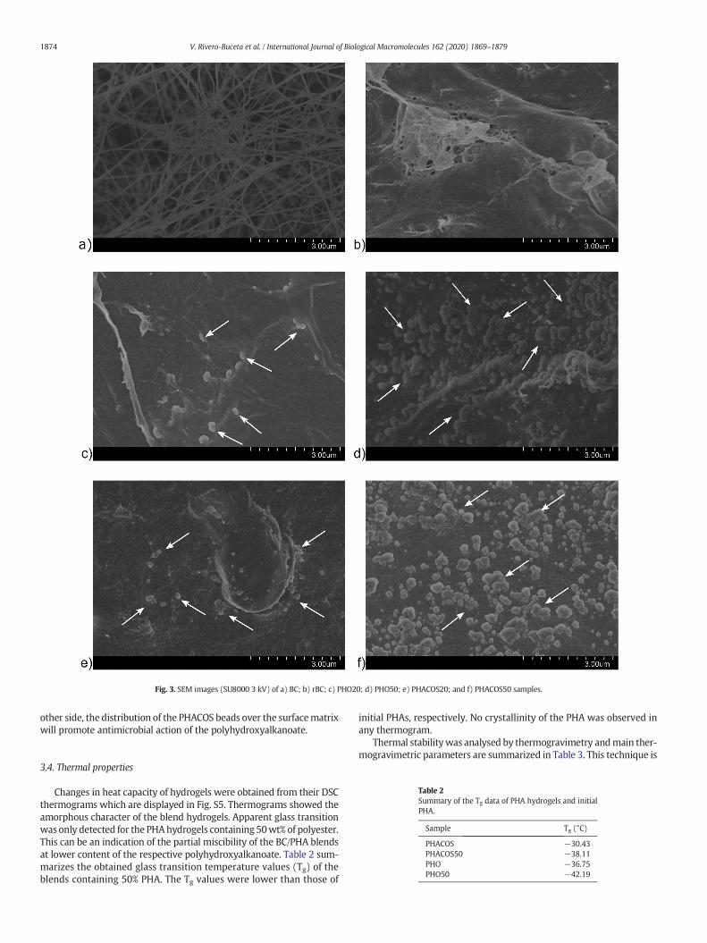
Fig. 3. SEM images (SU8000 3 kV) of a) BC; b) rBC; c) PHO20; d) PHO50; e) PHACOS20; and f) PHACOS50 samples.
1874 V. Rivero-Buceta et al. / International Journal of Biological Macromolecules 162 (2020) 1869–1879
other side, the distribution of the PHACOS beads over the surfacematrixwill promote antimicrobial action of the polyhydroxyalkanoate.
Table 2Summary of the Tg data of PHA hydrogels and initialPHA.
Sample Tg (°C)
PHACOS −30.43PHACOS50 −38.11PHO −36.75PHO50 −42.19
3.4. Thermal properties
Changes in heat capacity of hydrogels were obtained from their DSCthermograms which are displayed in Fig. S5. Thermograms showed theamorphous character of the blend hydrogels. Apparent glass transitionwas only detected for the PHAhydrogels containing 50wt% of polyester.This can be an indication of the partial miscibility of the BC/PHA blendsat lower content of the respective polyhydroxyalkanoate. Table 2 sum-marizes the obtained glass transition temperature values (Tg) of theblends containing 50% PHA. The Tg values were lower than those of
initial PHAs, respectively. No crystallinity of the PHA was observed inany thermogram.
Thermal stabilitywas analysed by thermogravimetry andmain ther-mogravimetric parameters are summarized in Table 3. This technique is

Table 3TGA and DTG results of the PHA hydrogels and control samples.
T onset (°C) T max1 (°C) T max2 (°C)
BC 303 – 355rBC 309 – 337PHACOS 277 287 –PHO 256 285 –PHACOS20 269 288 325PHACOS50 262 281 326PHO20 261 280 342PHO50 270 286 342
Fig. 4. a) Variation of storage modulus of hydrogels applying strain sweeps, andb) frequency sweeps.
Table 4Values of storage modulus (G') at three representative physiological angular frequencies.
G' (Pa)
0.628 rad/s 0.99 (rad/s) 79.10 (rad/s)
rBC 1061.5 ± 77.1 1090.5 ± 79.9 1688.5 ± 53.0PHO20 867.4 ± 36.1 908.0 ± 39.9 1427. 7 ± 41.1PHO50 1571.0 ± 46.5 1663.7 ± 49.9 2689.3 ± 93.41PHACOS20 1682.7 ± 80.2 1793.5 ± 100.2 3006.3 ± 396.91PHACOS50 1056.0 ± 129.7 1111.6 ± 145.6 1694.5 ± 242.61
1875V. Rivero-Buceta et al. / International Journal of Biological Macromolecules 162 (2020) 1869–1879
commonly used in the characterization of polymeric systems since itprovides useful information on the intrinsic structure of the system.Thermal stability of rBC decreased if compared with native BC, asshown in Table 3. That is, Tmax value decreased after the treatmentwith BMIMCl which can be attributed to a decrease in polymorph IIand an increase in amorphous cellulose [34,44].
Thermal degradation of all hydrogel blends, underwent in two steps,presenting twomaximum temperature (Tmax) values in theDTG curves.The first weight loss was attributed to the PHAs thermal degradationwhereas the second onewas ascribed to the rBCpyrolysis. This degrada-tion pattern indicates the not formation of interpolymericmatrix, it cor-relates with the segregation of the corresponding PHA within thecellulose matrix as observed in SEM examination and it is coherentwith the amorphous state of the PHA within the hydrogels observedin DSC study.
On the other hand, since the proposed hydrogel system is intendedto be used as a biomaterial for wound healing which is carried out atphysiological temperature 37 °C, it can be said that the polymerichydrogels will be thermally stable at that temperature.
3.5. Mechanical performance
Mimicking the mechanical properties of the skin is necessary inorder to obtain a suitable hydrogel that do not damage the edges ofthe ulcer to be treated. Therefore, the mechanical properties of thenew hydrogel must be adjusted in the same range as those of the skinto ensure better compatibility.
Viscoelastic properties of hydrogels swollen inwaterwas carried outby means of rheological measurements.
The linear viscoelastic range (LVR) was determined between 0.01and 5% strain (Fig. 4a); in this range the viscoelastic properties of allhydrogels were independent of the imposed strain levels but thePHACOS20 and PHO20 presented broader LVR. For strains valueslower than 5%, G' was over G" in all hydrogels, which indicates thatthe samples have a gel-like structure. At strain levels higher that 10%the value of G' exceeds G", indicating that hydrogel structure was bro-ken at that point.
rBC provided the highest storage modulus value, followed byPHACOS20 and PHO20 samples and the PHACOS50 and PHO50 gavethe lowest. Therefore, adding polyesters to the bacterial cellulose pro-duced a reduction in the storage modulus that can be attributed to theformation of newhydrogen interactions between both types of biopoly-mers, decreasing the intrinsic hydrogen bonds interactions of BC [50].
The frequency sweeps results revealed the dependence of the threedimensional structure of the hydrogels on the angular frequency (ω) asit is shown in Fig. 4b and Table 4. In the Fig. 4b it can also be appreciatedthat at high angular frequency values, between 100 and 300 rad/s, thestructure of the hydrogels is not stable [51–53].
The highest G' value corresponds to the PHACOS20 hydrogel,followed by PHO50. The lowest values of G' were registered forPHACOS50 and PHO20. That is, PHACOS20 and PHO50have a similar be-haviour. Thus, in the case of the PHO, a 50 wt% of this polymer is neces-sary for getting a stable structure, but in the case of the PHACOS, only a20% of the polymer is required. Analysing the structure of both PHAs,
PHACOS contains thioester groups at the end of the side chain that con-tribute to the formation of hydrogen bonds to stabilize the frameworkstructure. However, as this type of carbonyl groups does not exist inPHO, higher amount of this polymer is needed to obtain a comparablestorage modulus.
There is a wide range of elastic (G') and viscous (G") moduli re-ported in literature through different measurements (torsional, uni- orbi-axial). Holt et al. measured the viscoelastic response of wholehuman skin and dermis-only subjected to low-magnitude shear loadsover a range of physiological frequencies (0.628 to 75.39 rad/s), similarto our conditions. They found that G' in whole skin increased over thisfrequency range from 325.0 ± 93.7 Pa to 1227.9 ± 498.8 Pa anddermis-only showed a similar trend with mean G' values increasingfrom 434.9 ± 122.1 to 6620.0 ± 849.5 Pa. Taking into considerationthis work and analysing results of Fig. 4b and Table 4, our data showan increase of G' with ω less marked than that reported by Holt, how-ever, G' values still lie in the range of those reported by skin anddermis-only samples [54],making thesematerials promising for the de-velopment of new wound dressings with antibacterial properties.

1876 V. Rivero-Buceta et al. / International Journal of Biological Macromolecules 162 (2020) 1869–1879
3.6. Swelling studies
The swelling capacity of a hydrogel is a critical and effective param-eter for wound infection control because during the healing process fastand large water uptake capacity is needed to absorb exudates. In thissense, BC and its composites have been widely used in wound healingapplications due to their high capability of water absorption.
Fig. 5 shows the water absorption capacity of each hydrogel devel-oped in this work. It was observed that the swelling ability increasedwith time, first quickly and then gradually, reaching a constant swelling.
As it has been reported, BC has a strong swelling capacity in waterdue to the formation of multiple hydrogen bonds predominantly withthe accessible hydroxyls groups in the amorphous regions of the poly-saccharide [55]. The dissolution of BC in ionic liquid and subsequent re-generation to obtain rBC reduces significantly the crystallinity of theoriginal material (as explained in Subsection 3.2), being the amorphousregions more accessible to water and leading to a higher swelling ratio.
PHACOS20 and PHO50 showed rather similar and remarkably highswelling potential compared to rBC alone. It could be ascribed to thepresence of hydrophilic groups (-OH, -COO-, -SCOC3H) that facilitateshydrogen bonding formation and favours water absorption. A similarparallelism between samples of these compositions was previously ob-served in their mechanical behaviour. PHACOS20 included 20% of PHAwhile PHO50 included 50%. PHACOS presents thioester groups at theend of the side chain that contribute to the formation of hydrogenbonds that further stabilize the structure if compared with PHO thatneeded to be included in higher concentration to achieve similar results.
In contrast, PHACOS50 resulted in the lowest water content, and itcould be due to the formation of a more rigid hydrogel structure andthe presence of a high amount of PHACOS granules at the surface (seeFig. 3) that hinders water absorption. Therefore, the amount ofPHACOS present in the hydrogel is crucial to control the swelling ratio.
3.7. Antimicrobial activity
An effective wound dressing should ensure drainage and epithelialgrowth. During the wound healing process, dead tissue covers thewound and serves as a medium for bacterial growth, reduces the host'sresistance to infection, delays the formation of granulation tissue andthe re-epithelialization [56]. In this sense, the main goal of the presentwork was the development of novel bactericidal BC hydrogels that in-corporate the polyhydroxyalkanoate PHACOS which has antimicrobialproperties against S. aureus.
Fig. 5. Swelling profiles of hydrogels at different time periods.
The minimal inhibitory concentration (MIC) and the antimicrobialcapacity of PHACOS has been reported in our previous articles [17,57].We demonstrated the PHACOS antibacterial activity according to theISO 22196:2011 for measurement the antibacterial activity on the plas-tic surface. We also calculated the MIC for the soluble oligomers andmonomers released for the enzymatic hydrolysis of PHACOS and PHOas control, resulting in a much lower value for PHACOS (40 μM) thanthat of PHO (3 mM) [57]. In addition, the specificity of PHACOS againstS. aureus strains, including MRSA clinical isolates, was fully demon-strated when compared to PHO in antimicrobial assays against a panelof 7 Gram-positive strains and 2 Gram-negative strains (includingE. coli) [17].
The bactericidal behaviour of all prepared hydrogels in this workwas analysed consequently against S. aureus.
Due to the great variety of antimicrobial applications, several stan-dard methods to assess their efficiency have been developed. Themost important standard antimicrobial methods for antibacterialhydrogels includes both qualitative (i.e. AATCC 147:2004, ISO20645:2004) and quantitative (i.e. AATCC 100:2004, JIS L 1902:2008-Absorption method) methods. However, there is no consensus on themost adequate method to be used, and it is difficult to compare thedata among them. BC-based hydrogels are porous materials with highwater content. Taking into consideration that bacteria can colonize thewhole structure, in this work the bactericidal assay was performed byapplying a tailored methodology based on JIS L 1902:2008-Absorptionmethod but suitable to assess the total bacterial load, including thatabsorbed or adhered inside the hydrogel in each tested condition. Thismethod is based on the enzymatic hydrolysis of the biological polymersBC, PHACOS and PHO (for details see M&M 2.4, Fig. S1).
The bactericidal activity results against S. aureus is shown as the log-arithmic difference between the number of bacteria in a control cell sus-pension (S. aureus t24) and each hydrogel-treated sample. Controlshows that the viability of S. aureus was not significantly reduced dueto incubation time and growth conditions, or when exposed to cellulaseand PHA depolymerase (DepolMS) (Fig. 6a). Neither regenerated cellu-lose, nor hydrogels of BC-PHO, PHO20 and PHO50 displayed antibacte-rial activity, conversely to hydrogels containing PHACOS (Fig. 6a).PHACOS20 resulted in a cell viability decrease from 108 to 1.6 × 10⁶, a1.8 logarithmic unit reduction, and PHACOS50 decreased CFU from108 to 1.6 × 10⁵, a 2.8 logarithmic unit reduction, similarly to the antimi-crobial activity reported in the literature for other hydrogels [58].Theseresults show that the antibacterial behaviour of these BC-basedhydrogels is due exclusively to the PHACOS component. Taking into ac-count that direct contact is needed for the antibacterial activity,hydrogels may enhance bactericidal activity of PHACOS. It is importantto notice that the specificity of PHACOS against S. aureus, far frombeing a drawback, restricted antibacterial activity over these importantskin infecting bacteria. This is, in fact, considered an advantage, as itwould leave unharmed commensal bacteria from the skin. S. aureus isa mayor skin pathogen. It is themain cause of impetigo, which accountsfor 50–60% of all bacterial skin infections, and is also the main cause ofcellulitis and folliculitis (skin infections). Moreover, PHACOS is activeagainst MRSA [17] that may be responsible for up to 60% of skin infec-tions seen in US emergency departments, reasons why we havetargeted this microorganism. Our work also paves the way for furtherdevelopments to design synergetic effective hydrogels containing forinstance metal nanoparticles, biological extracts or antibiotics specificfor other relevant skin infective bacteria (i.e. Pseudomonas aeruginosa).
Different strategies have been traditionally applied for conferringantimicrobial activity to hydrogels for wound healing [8]. Themost con-ventional consists of loading antibiotics into the hydrogel as deliverysystem. Antibiotic treatment is one of the main approaches of modernmedicine which is used to combat infections [60]. However, the emer-gence, spread, and persistence of multidrug-resistant (MDR) bacteriasuch as MRSA, advise against the misuse of antibiotics and raise de-mands for safe and efficacious agents that were less prone to

Fig. 6. Antimicrobial activity of the blended hydrogels. a) Logarithmic reduction of bacteria count for the suspension incubatedwith each hydrogel, regarding a cell suspension of S. aureuscontrol. DepolMS refers to the suspension incubated with the enzymes; rBC with the regenerated cellulose; PHACOS20, PHACOS50, PHO20 and PHA50 with the corresponding hydrogel.Statistically significant differences from a one way ANOVA test are indicated with ** (p < 0.01) **** (p < 0.0001). b) Viable cell count from panel A.
1877V. Rivero-Buceta et al. / International Journal of Biological Macromolecules 162 (2020) 1869–1879
stimulatingdevelopment of resistance. In recent years, alternatives suchas antimicrobial nanoparticles (e.g., Ag, ZnO or TiO2) and antimicrobialpeptides (AMPs), have increased their attention [61], but thesemethod-ologies have some important limitations such as themaximum concen-tration that can be loaded within the polymer network. Naturalpolymers with inherent antibacterial properties like chitosan can over-come this issue, but its efficacy as pure polymers against certain impor-tant clinical isolated pathogen is scarcely reported [62]. In this work wehave developed a new hydrogel with efficient antimicrobial activityagainst MRSA, using two natural bacterial biopolymers of different na-ture that can be produced by sustainable processes following the princi-ples of circular economy including the reusability of the solvent appliedfor blending (ionic liquid).
3.8. Biocompatibility of hydrogels
Biocompatibility of BC with living tissues has been previously stud-ied in repeated occasions and is comparable to other biomaterials com-monly used in tissue engineering, like polyglycolic acid and
polytetrafluorethylene [7]. BC is widely used in in vivo applications be-cause it lacks proteins or polymers, a requirement of the United Statesfood and drug administration (FDA) legislations for implants in directcontact with blood. In fact, several BC-based materials approved bythe FDA are used as tissue surgical sheets, reinforcing matrixes andmeshes, because of the absence of skin irritation and the lower coagula-tion rates compared to other materials [61]. Skin tolerance and in vivoresponse to BC applying skin lesion model, demonstrated that the ery-thema clinical scorewas zero at 2 and 24h after patch removal in almostall volunteers [63. In vivo cytotoxicity of BC has been studied byimplanting BC nanofibers subcutaneously in BALB/c mice, withoutshowing changes in the normal development of animals [64].
PHACOS cellular toxicity in terms of viability and metabolic func-tions of mammalian cells were deeply studied by Dinjaski et al. Inflam-matory activity was also analysed in vitro and in vivo by PHACOSimplantation subcutaneously in mice. The results indicated minimal in-flammation associated with this polymer [17].
Based on these antecedents, in this work, the cytotoxicity of antimi-crobial BC/PHACOS hydrogels were evaluated according to ISO 10993-5

Fig. 7.Biocompatibility assay: Indirect cytotoxicity showing cell viability in the presence ofextracts at different time points. Statistically significant differences between studiedsamples and control from a one way ANOVA test are indicated with * (p < 0.05).
1878 V. Rivero-Buceta et al. / International Journal of Biological Macromolecules 162 (2020) 1869–1879
standard using human dermal fibroblasts. Thus, the effects of materialextracts on themetabolic activity of the cells were evaluated at differenttime periods. According to the standard, if the relative cell viability ofthe highest concentration of the sample extract (100% extract) is ≥70%of the control group, then the material shall be considered non-cytotoxic.
Fig. 7 shows cell viability (CV) values of human fibroblasts culturedin the presence of extracts of the PHACOS containing hydrogels samplestaken at different time periods. It can be observed that cell viability inthe presence of extracts of rBC and PHACOS hydrogels samples at 4 hwas statistically lower than the control, however cell viability recoveredin the presence of extracts taken in the following time points (1, 4 and7) giving not statistically different values over the studied period exceptfor some cases. Taking the results in overall, we can say that no sampleshowed cytotoxicity according to ISO 10993-5 standard since cell viabil-ity was over 85% in all studied samples.
4. Conclusion
Antimicrobial hydrogels based on BC and PHACOSwere successfullyprepared using the ionic liquid BMIMCl and were cytocompatibleagainst fibroblasts of human embryonic skin. Hydrogels microstructureconsisted in a BC matrix having PHACOS granules homogeneously dis-tributed and stabilized by hydrogen bonding interactions betweenboth types of polymers. The PHACOS20 had elastic properties compara-ble to the skin features, optimum swelling properties for absorbingfluids in wounds, and besides, presented the highest significant anti-staphylococcal activity (reduction of viability in 1.8 logarithmic units).20% of PHACOS was enough to provide BC-based hydrogel with antimi-crobial activity versus S. aureus, using low amount of polymer concen-tration in the synthesis (1 w/v-%). The hydrogel compositionPHACOS20 is selected as the best candidate formulation and proposedfor further developments in wound healing applications.
Supplementary data to this article can be found online at https://doi.org/10.1016/j.ijbiomac.2020.07.289.
Credit author statement
Virginia Rivero-Buceta: Conceptualization, Investigation, Writing -Original Draft, Visualization.
María Rosa Aguilar: Conceptualization, Supervision, Writing - Re-view & Editing. Corresponding author (2).
Ana María Hernández-Arriaga: Supervision, Writing - OriginalDraft.
Francisco G. Blanco: Investigation, Writing - Original Draft.Antonia Rojas: Resources.
Marta Tortajada: Resources.Rosa Ana Ramírez-Jiménez: Methodology.Blanca Vázquez-Lasa: Supervision, Writing - Review & Editing.Auxiliadora Prieto: Conceptualization, Supervision, Writing - Re-
view& Editing Funding acquisition, Project administration. Correspond-ing author (1).
Acknowledgements
Authors thank financial support to thank the Spanish Ministry ofScience, Innovation and Universities [MAT2017-84277-R and Bio2017-8344-8-R], the European Union's Horizon 2020 Research and Innova-tion Programme [grant agreement no 870294 (Mix-Up)] and the Com-munity of Madrid [P2018/NMT4389] for the financial support of thisproject. The kind support by David Gómez in the SEM experiments isgreatly appreciated.
References
[1] S.C.M. Fernandes, P. Sadocco, A. Alonso-Varona, T. Palomares, A. Eceiza, A.J.D.Silvestre, I. Mondragon, C.S.R. Freire, Bioinspired antimicrobial and biocompatiblebacterial cellulose membranes obtained by surface functionalization withaminoalkyl groups, ACS Appl. Mater. Interfaces 5 (2013) 3290–3297, https://doi.org/10.1021/am400338n.
[2] J. Yang, J. Li, Self-assembled cellulose materials for biomedicine: a review,Carbohydr. Polym. 181 (2018) 264–274, https://doi.org/10.1016/j.carbpol.2017.10.067.
[3] A.F. Jozala, L.C. de Lencastre-Novaes, A.M. Lopes, V. de Carvalho Santos-Ebinuma,P.G. Mazzola, A. Pessoa-Jr, D. Grotto, M. Gerenutti, M.V. Chaud, Bacterialnanocellulose production and application: a 10-year overview, Appl. Microbiol.Biotechnol. 100 (2016) 2063–2072, https://doi.org/10.1007/s00253-015-7243-4.
[4] M. Tabuchi, K. Watanabe, Y. Morinaga, F. Yoshinaga, Acetylation of Bacterial Cellu-lose: Preparation of Cellulose Acetate Having a High Degree of Polymerization, vol.42, 1998 1451–1454.
[5] S. Zhu, Y. Wu, Q. Chen, Z. Yu, C. Wang, S. Jin, Y. Ding, G. Wu, Dissolution of cellulosewith ionic liquids and its application: a mini-review, Green Chem. 8 (2006)325–327, https://doi.org/10.1039/b601395c.
[6] W. Czaja, A. Krystynowicz, S. Bielecki, R.M. Brown, Microbial cellulose - the naturalpower to heal wounds, Biomaterials 27 (2006) 145–151, https://doi.org/10.1016/j.biomaterials.2005.07.035.
[7] I. Sulaeva, U. Henniges, T. Rosenau, A. Potthast, Bacterial cellulose as a material forwound treatment: properties and modifications: a review, Biotechnol. Adv. 33(2015) 1547–1571, https://doi.org/10.1016/j.biotechadv.2015.07.009.
[8] R. Portela, C.R. Leal, P.L. Almeida, R.G. Sobral, Bacterial cellulose: a versatile biopoly-mer for wound dressing applications, Microb. Biotechnol. 2019 (2019)https://doi.org/10.1111/1751-7915.13392.
[9] M.P. Rowan, L.C. Cancio, E.A. Elster, D.M. Burmeister, L.F. Rose, S. Natesan, R.K. Chan,R.J. Christy, K.K. Chung, Burn wound healing and treatment: review and advance-ments, Crit. Care 19 (2015) 1–12, https://doi.org/10.1186/s13054-015-0961-2.
[10] J.V. Vayalumkal, T. Jadavji, Children hospitalized with skin and soft tissue infections,Pediatr. Drugs 8 (2006) 99–111, https://doi.org/10.2165/00148581-200608020-00003.
[11] R. Köck, K. Becker, B. Cookson, S. Harbarth, J. Kluytmans, M. Mielke, G. Peters, R.L.Skov, Methicillin-resistant Staphylococcus aureus (MRSA): burden of disease andcontrol challenges in Europe, Euro Surveill 15 (2010), 19688https://doi.org/10.2807/ese.15.41.19688-en.
[12] M.E. Aljghami, S. Saboor, S. Amini-Nik, Emerging innovative wound dressings, Ann.Biomed. Eng. 47 (2019) 659–675, https://doi.org/10.1007/s10439-018-02186-w.
[13] M. Zinn, B. Witholt, T. Egli, Occurrence, Synthesis and Medical Application of Bacte-rial Polyhydroxyalkanoate, vol. 53, 2001 5–21.
[14] A. Prieto, I.F. Escapa, V. Martínez, N. Dinjaski, C. Herencias, F. de la Peña, N. Tarazona,O. Revelles, A holistic view of polyhydroxyalkanoate metabolism in Pseudomonasputida, Environ. Microbiol. 18 (2016) 341–357, https://doi.org/10.1111/1462-2920.12760.
[15] M. Tortajada, L.F. da Silva, M.A. Prieto, Second-generation functionalizedmediumchain- length polyhydroxyalkanoates: the gateway to high-value bioplasticapplications, Int. Microbiol. 16 (2013) 1–15, https://doi.org/10.2436/20.1501.01.175.
[16] I.F. Escapa, V. Morales, V.P. Martino, E. Pollet, L. Avérous, J.L. García, M.A. Prieto, Dis-ruption of β-oxidation pathway in Pseudomonas putida KT2442 to produce newfunctionalized PHAs with thioester groups, Appl. Microbiol. Biotechnol. 89 (2011)1583–1598, https://doi.org/10.1007/s00253-011-3099-4.
[17] N. Dinjaski, M. Fernández-Gutiérrez, S. Selvam, F.J. Parra-Ruiz, S.M. Lehman, J. SanRomán, E. García, J.L. García, A.J. García, M.A. Prieto, PHACOS, a functionalized bacte-rial polyester with bactericidal activity against methicillin-resistant Staphylococcusaureus, Biomaterials 35 (2014) 14–24, https://doi.org/10.1016/j.biomaterials.2013.09.059.
[18] A.M. Hernández-Arriaga, C. del Cerro, L. Urbina, A. Eceiza, M.A. Corcuera, A. Retegi,M. Auxiliadora Prieto, Genome sequence and characterization of the bcs clustersfor the production of nanocellulose from the low pH resistant strain

1879V. Rivero-Buceta et al. / International Journal of Biological Macromolecules 162 (2020) 1869–1879
Komagataeibacter medellinensis ID13488, Microb. Biotechnol. 12 (2019) 620–632,https://doi.org/10.1111/1751-7915.13376.
[19] N. Hameed, Q. Guo, F.H. Tay, S.G. Kazarian, Blends of cellulose and poly(3-hydroxybutyrate-co-3-hydroxyvalerate) prepared from the ionic liquid 1-butyl-3-methylimidazolium chloride, Carbohydr. Polym. 86 (2011) 94–104, https://doi.org/10.1016/j.carbpol.2011.04.016.
[20] E. Pinho, L. Magalhães, M. Henriques, Antimicrobial Activity Assessment of Textiles:Standard Methods Comparison, 2011 493–498, https://doi.org/10.1007/s13213-010-0163-8.
[21] V. Martínez, P.G. de Santos, J. García-Hidalgo, D. Hormigo, M.A. Prieto, M. Arroyo, I.de la Mata, Novel extracellular medium-chain-length polyhydroxyalkanoatedepolymerase from Streptomyces exfoliatus K10 DSMZ 41693: a promising biocat-alyst for the efficient degradation of natural and functionalized mcl-PHAs, Appl.Microbiol. Biotechnol. 99 (2015) 9605–9615, https://doi.org/10.1007/s00253-015-6780-1.
[22] C.S.A. (CSA), Iso 10993-5, Int. Organ, vol. 2007, 2009 1–11, https://doi.org/10.1021/es0620181.
[23] S.S. Silva, T.C. Santos, M.T. Cerqueira, A.P. Marques, L.L. Reys, T.H. Silva, S.G. Caridade,J.F. Mano, R.L. Reis, The use of ionic liquids in the processing of chitosan/silkhydrogels for biomedical applications, Green Chem. 14 (2012) 1463–1470, https://doi.org/10.1039/c2gc16535j.
[24] O.A. El Seoud, A. Koschella, L.C. Fidale, S. Dorn, T. Heinze, Applications of ionic liquidsin carbohydrate chemistry: a window of opportunities, Biomacromolecules 8(2007) 2629–2647, https://doi.org/10.1021/bm070062i.
[25] J. Zhang, J. Wu, J. Yu, X. Zhang, J. He, J. Zhang, Application of ionic liquids for dissolv-ing cellulose and fabricating cellulose-based materials: state of the art and futuretrends, Mater. Chem. Front. 1 (2017) 1273–1290, https://doi.org/10.1039/c6qm00348f.
[26] I.M. Marrucho, L.C. Branco, L.P.N. Rebelo, Ionic liquids in pharmaceutical applica-tions, Annu. Rev. Chem. Biomol. Eng. 5 (2014) 527–546, https://doi.org/10.1146/annurev-chembioeng-060713-040024.
[27] T. Santos de Almeida, A. Júlio, N. Saraiva, A.S. Fernandes, M.E.M. Araújo, A.R. Baby, C.Rosado, J.P. Mota, Choline- versus imidazole-based ionic liquids as functional ingre-dients in topical delivery systems: cytotoxicity, solubility, and skin permeation stud-ies, Drug Dev. Ind. Pharm. 43 (2017) 1858–1865, https://doi.org/10.1080/03639045.2017.1349788.
[28] J.M. Gomes, S.S. Silva, R.L. Reis, Biocompatible ionic liquids: fundamental behavioursand applications, Chem. Soc. Rev. 48 (2019) 4317–4335, https://doi.org/10.1039/c9cs00016j.
[29] R.F.P. Pereira, K. Zehbe, C. Günter, T. Dos Santos, S.C. Nunes, F.A.A. Paz, M.M. Silva,P.L. Granja, A. Taubert, V. De Zea Bermudez, Ionic liquid-assisted synthesis of Meso-porous silk fibroin/silica hybrids for biomedical applications, ACS Omega 3 (2018)10811–10822, https://doi.org/10.1021/acsomega.8b02051.
[30] F. Lv, C. Wang, P. Zhu, C. Zhang, Characterization of chitosan microparticles rein-forced cellulose biocomposite sponges regenerated from ionic liquid, Cellulose 21(2014) 4405–4418, https://doi.org/10.1007/s10570-014-0440-y.
[31] S. Islam, L. Arnold, R. Padhye, Comparison and characterisation of regenerated chito-san from 1-butyl-3-methylimidazolium chloride and chitosan from crab shells,Biomed. Res. Int. 2015 (2015) 1–6, https://doi.org/10.1155/2015/874316.
[32] Z. Liu, X. Sun, M. Hao, C. Huang, Z. Xue, T. Mu, Preparation and characterization ofregenerated cellulose from ionic liquid using different methods, Carbohydr. Polym.117 (2015) 54–62, https://doi.org/10.1016/j.carbpol.2014.09.053.
[33] M.T. Clough, K. Geyer, P.A. Hunt, S. Son, U. Vagt, T. Welton, Ionic liquids: not alwaysinnocent solvents for cellulose, Green Chem. 17 (2015) 231–243, https://doi.org/10.1039/c4gc01955e.
[34] R.P. Swatloski, S.K. Spear, J.D. Holbrey, R.D. Rogers, Dissolution of cellose with ionicliquids, J. Am. Chem. Soc. 124 (2002) 4974–4975, https://doi.org/10.1021/ja025790m.
[35] M. Isik, H. Sardon, D. Mecerreyes, Ionic liquids and cellulose: dissolution, chemicalmodification and preparation of new cellulosic materials, Int. J. Mol. Sci. 15 (2014)11922–11940, https://doi.org/10.3390/ijms150711922.
[36] T. Heinze, A. Koschella, Solvents applied in the field of cellulose chemistry: a minireview, Polímeros. 15 (2006) 84–90, https://doi.org/10.1590/s0104-14282005000200005.
[37] S. Wang, A. Lu, L. Zhang, Recent advances in regenerated cellulose materials, Prog.Polym. Sci. 53 (2016) 169–206, https://doi.org/10.1016/j.progpolymsci.2015.07.003.
[38] A.P. Dadi, S. Varanasi, C.A. Schall, Enhancement of cellulose saccharification kineticsusing an ionic liquid pretreatment step, Biotechnol. Bioeng. 95 (2006) 904–910,https://doi.org/10.1002/bit.21047.
[39] F.A. Yassin, F.Y. El Kady, H.S. Ahmed, L.K. Mohamed, S.A. Shaban, A.K. Elfadaly, Highlyeffective ionic liquids for biodiesel production from waste vegetable oils, Egypt. J.Pet. 24 (2015) 103–111, https://doi.org/10.1016/j.ejpe.2015.02.011.
[40] M.L. Nelson, R.T. O’Connor, Relation of certain infrared bands to cellulose crystallin-ity and crystal lattice type. Part II. A new infrared ratio for estimation of crystallinityin celluloses I and II, J. Appl. Polym. Sci. 8 (1964) 1325–1341, https://doi.org/10.1002/app.1964.070080323.
[41] C. Molina-Ramírez, C. Castro, R. Zuluaga, P. Gañán, Physical characterization of bac-terial cellulose produced by Komagataeibacter medellinensis using food supplychain waste and agricultural by-products as alternative low-cost feedstocks, J.Polym. Environ. 26 (2018) 830–837, https://doi.org/10.1007/s10924-017-0993-6.
[42] C. Molina-Ramírez, M. Castro, M. Osorio, M. Torres-Taborda, B. Gómez, R. Zuluaga, C.Gómez, P. Gañán, O.J. Rojas, C. Castro, Effect of different carbon sources on bacterialnanocellulose production and structure using the low pH resistant strainKomagataeibacter medellinensis, Materials (Basel) 10 (2017)https://doi.org/10.3390/ma10060639.
[43] F. Carrillo, X. Colom, J.J. Suñol, J. Saurina, Structural FTIR analysis and thermal char-acterisation of lyocell and viscose-type fibres, Eur. Polym. J. 40 (2004) 2229–2234,https://doi.org/10.1016/j.eurpolymj.2004.05.003.
[44] J.-H. Pang, X. Liu, M.Wu, Y.-Y. Wu, X.-M. Zhang, R.-C. Sun, Fabrication and character-ization of regenerated cellulose films using different ionic liquids, J. Spectrosc. 2014(2014) 1–8, https://doi.org/10.1155/2014/214057.
[45] L. Urbina, A.M. Hernández-Arriaga, A. Eceiza, N. Gabilondo, M.A. Corcuera, M.A.Prieto, A. Retegi, By-products of the cider production: an alternative source of nutri-ents to produce bacterial cellulose, Cellulose 24 (2017) 2071–2082, https://doi.org/10.1007/s10570-017-1263-4.
[46] J.R. Malcom Brown, Cellulose: structural and functional aspects (1989) 145–151.[47] X. Zhang, Z. Li, X. Che, L. Yu, W. Jia, R. Shen, J. Chen, Y. Ma, G.Q. Chen, Synthesis and
characterization of polyhydroxyalkanoate organo/hydrogels, Biomacromolecules 20(2019) 3303–3312, https://doi.org/10.1021/acs.biomac.9b00479.
[48] M. Zaborowska, A. Bodin, H. Bäckdahl, J. Popp, A. Goldstein, P. Gatenholm, Micropo-rous bacterial cellulose as a potential scaffold for bone regeneration, Acta Biomater.6 (2010) 2540–2547, https://doi.org/10.1016/j.actbio.2010.01.004.
[49] S.V. Madihally, H.W.T. Matthew, Porous chitosan scaffolds for tissue engineering,Biomaterials 20 (1999) 1133–1142, https://doi.org/10.1016/S0142-9612(99)00011-3.
[50] J.D. Conrad, G.M. Harrison, The rheology and processing of renewable resource poly-mers, AIP Conf. Proc. 1027 (2008) 114–116, https://doi.org/10.1063/1.2964497.
[51] R. Shah, R. Vyroubal, H. Fei, N. Saha, T. Kitano, P. Saha, Preparation of bacterial cellu-lose based hydrogels and their viscoelastic behavior, AIP Conf. Proc. 1662 (2015)1–8, https://doi.org/10.1063/1.4918895.
[52] P. Basu, N. Saha, P. Saha, Swelling and rheological study of calcium phosphate filledbacterial cellulose-based hydrogel scaffold, J. Appl. Polym. Sci. 48522 (2019) 48522,https://doi.org/10.1002/app.48522.
[53] Y. Numata, H. Kono, A.Mori, R. Kishimoto, K. Tajima, Structural and rheological char-acterization of bacterial cellulose gels obtained from Gluconacetobacter genus, FoodHydrocoll. 92 (2019) 233–239, https://doi.org/10.1016/j.foodhyd.2019.01.060.
[54] B. Holt, A. Tripathi, J. Morgan, Viscoelastic response of human skin to lowmagnitudephysiologically relevant shear, J. Biomech. 41 (2008) 2689–2695, https://doi.org/10.1016/j.jbiomech.2008.06.008.
[55] S. Rongpipi, D. Ye, E.D. Gomez, E.W. Gomez, Progress and opportunities in the char-acterization of cellulose – an important regulator of cell wall growth andmechanics,Front. Plant Sci. 9 (2019) 1–28, https://doi.org/10.3389/fpls.2018.01894.
[56] I.S. Savitskaya, D.H. Shokatayeva, A.S. Kistaubayeva, L.V. Ignatova, I.E. Digel, Antimi-crobial andwound healing properties of a bacterial cellulose basedmaterial contain-ing B. subtilis cells, Heliyon 5 (2019)https://doi.org/10.1016/j.heliyon.2019.e02592.
[57] V. Martínez, N. Dinjaski, L.I. de Eugenio, F. de la Peña, M.A. Prieto, Cell system engi-neering to produce extracellular polyhydroxyalkanoate depolymerasewith targetedapplications, Int. J. Biol. Macromol. 71 (2014) 28–33, https://doi.org/10.1016/j.ijbiomac.2014.04.013.
[58] M.S. Kim, G.W. Oh, Y.M. Jang, S.C. Ko, W.S. Park, I.W. Choi, Y.M. Kim, W.K. Jung, An-timicrobial hydrogels based on PVA and diphlorethohydroxycarmalol (DPHC) de-rived from brown alga Ishige okamurae: an in vitro and in vivo study for wounddressing application, Mater. Sci. Eng. C. 107 (2020), 110352https://doi.org/10.1016/j.msec.2019.110352.
[60] W. Liu, H. Du, M. Zhang, K. Liu, H. Liu, H. Xie, X. Zhang, C. Si, Bacterial cellulose-basedcomposite scaffolds for biomedical applications: a review, ACS Sustain. Chem. Eng. 8(2020) 7536–7562, https://doi.org/10.1021/acssuschemeng.0c00125.
[61] G.F. Picheth, C.L. Pirich, M.R. Sierakowski, M.A. Woehl, C.N. Sakakibara, C.F. de Souza,A.A. Martin, R. da Silva, R.A. de Freitas, Bacterial cellulose in biomedical applications:a review, Int. J. Biol. Macromol. 104 ( (2017) 97–106, https://doi.org/10.1016/j.ijbiomac.2017.05.171.
[62] E.M. Costa, S. Silva, F.K. Tavaria, M.M. Pintado, Insights into chitosan antibiofilm ac-tivity against methicillin-resistant Staphylococcus aureus, J. Appl. Microbiol. 122(2017) 1547–1557, https://doi.org/10.1111/jam.13457.
[63] I.F. Almeida, T. Pereira, N.H.C.S. Silva, F.P. Gomes, A.J.D. Silvestre, C.S.R. Freire, J.M.Sousa Lobo, P.C. Costa, Bacterial cellulose membranes as drug delivery systems: anin vivo skin compatibility study, Eur. J. Pharm. Biopharm. 86 (2014) 332–336,https://doi.org/10.1016/j.ejpb.2013.08.008.
[64] R.A.N. Pértile, S. Moreira, R.M. Gil Da Costa, A. Correia, L. Guardão, F. Gartner, M.Vilanova, M. Gama, Bacterial cellulose: long-term biocompatibility studies, J.Biomater. Sci. Polym. Ed. 23 (2012) 1339–1354, https://doi.org/10.1163/092050611X581516.