Anti-HBV potential of PD-1/PD-L1 blockade in chronic HBV...
26
Anti-HBV potential of PD-1/PD-L1 blockade in chronic HBV infection Daniel Verdon 1 , Anna E. Brooks 1,4 , Anuj Gaggar 2 , Jacky Woo 2 , Mani Subramanian 2 , Christian Schwabe 3 , Edward J. Gane 3 , Rod Dunbar 1,4 . 1 University of Auckland, Auckland, New Zealand, 2 Gilead Sciences, Foster City, CA, United States, 3 Auckland Clinical Studies, Auckland, New Zealand, 4 Maurice Wilkins Centre for Molecular Biodiscovery, Auckland, New Zealand
Transcript of Anti-HBV potential of PD-1/PD-L1 blockade in chronic HBV...

Anti-HBV potential of PD-1/PD-L1 blockade in chronic HBV infection
Daniel Verdon1, Anna E. Brooks1,4, Anuj Gaggar2, Jacky Woo2, Mani Subramanian2, Christian Schwabe3, Edward J. Gane3, Rod Dunbar 1,4.
1University of Auckland, Auckland, New Zealand, 2Gilead Sciences, Foster City, CA, United States, 3Auckland Clinical Studies, Auckland, New Zealand, 4Maurice Wilkins Centre for Molecular Biodiscovery, Auckland, New Zealand
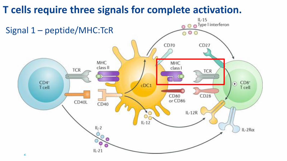
T cells require three signals for complete activation.
2
Signal 1 – peptide/MHC:TcR

T cells require three signals for complete activation.
3
Signal 2 – Co-stimulation
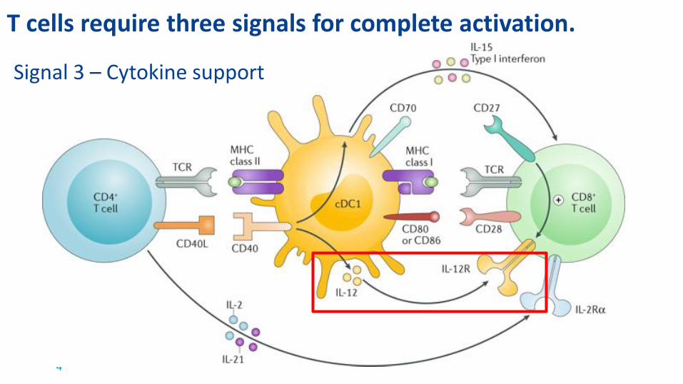
T cells require three signals for complete activation.
4
Signal 3 – Cytokine support

CHB Background
5
o Viral clearance in acute HBV is associated with a broad, robust CD4+ and CD8+ T cell response and seroconversion.
o An insufficient T-cell response to HBV antigens is characteristic of chronic HBV and limits durable viral control and clearance.
o T-cell exhaustion is a major pathway mediating an impaired in situ response.
Ye, Cell Death Disase 2015.

Co-inhibitory signaling drives progressive T cell exhaustion
6 T cellAPC
• T cell exhaustion is characterised by progressive loss of proliferative capacity and effector cytokine production, and increased susceptibility to apoptosis.
• Exhausted T cells progressively acquire expression of co-inhibitory surface receptors.
• PD-1/PD-L1 is a dominant co-inhibitory axis mediating T cell exhaustion in CHB.

PD-1 Expression and signalling
7
• IgSF/CD28 transmembrane protein. Binds PD-L1 (B7-H1) and PD-L2 (B7-DC).
• Important for normal tissue homeostasis and T cell memory formation.
• Expressed on activated and memory T cells - PD-1 signalling drives SHP-1/2 recruitment and impairs both TCR/CD3 and CD28 signal transduction.
• Expressed on B cells, NK, macrophages and dendritic cells.
• Expressed on TREG – signalling enhances function and drives proliferation.
• Expressed on MDSC – signalling drives IL-10 production. Gianchecchi AI Rev 2013; Hui Science 2013; Yulefpolskiy JI 2014; Huang JI 2014; Schumacher Immunity 2018
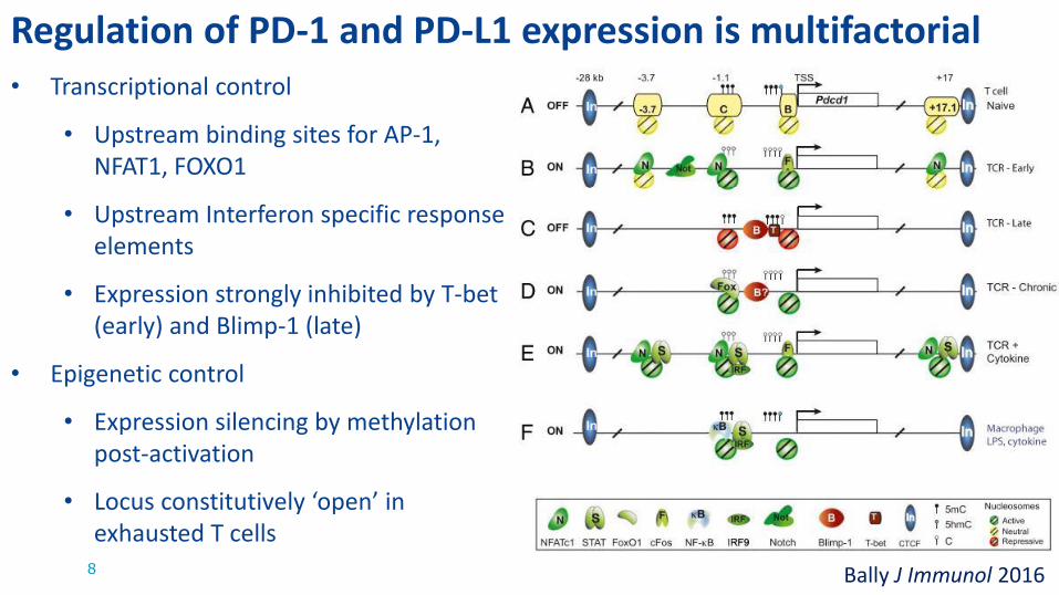
Regulation of PD-1 and PD-L1 expression is multifactorial
8 Bally J Immunol 2016
• Transcriptional control
• Upstream binding sites for AP-1, NFAT1, FOXO1
• Upstream Interferon specific response elements
• Expression strongly inhibited by T-bet (early) and Blimp-1 (late)
• Epigenetic control
• Expression silencing by methylation post-activation
• Locus constitutively ‘open’ in exhausted T cells
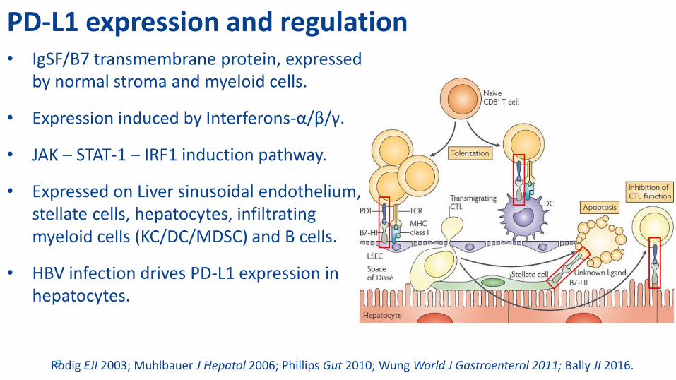
PD-L1 expression and regulation
9
• IgSF/B7 transmembrane protein, expressed by normal stroma and myeloid cells.
• Expression induced by Interferons-α/β/γ.
• JAK – STAT-1 – IRF1 induction pathway.
• Expressed on Liver sinusoidal endothelium, stellate cells, hepatocytes, infiltrating myeloid cells (KC/DC/MDSC) and B cells.
• HBV infection drives PD-L1 expression in hepatocytes.
Rodig EJI 2003; Muhlbauer J Hepatol 2006; Phillips Gut 2010; Wung World J Gastroenterol 2011; Bally JI 2016.

PD-1 and PD-L1 blockade in Solid Tumours
10Roberts NEJM 2015 Brahmer NEJM 2012
• Melanoma-specific TIL also exist in an immunosuppressive microenvironment and exhibit T cell exhaustion.
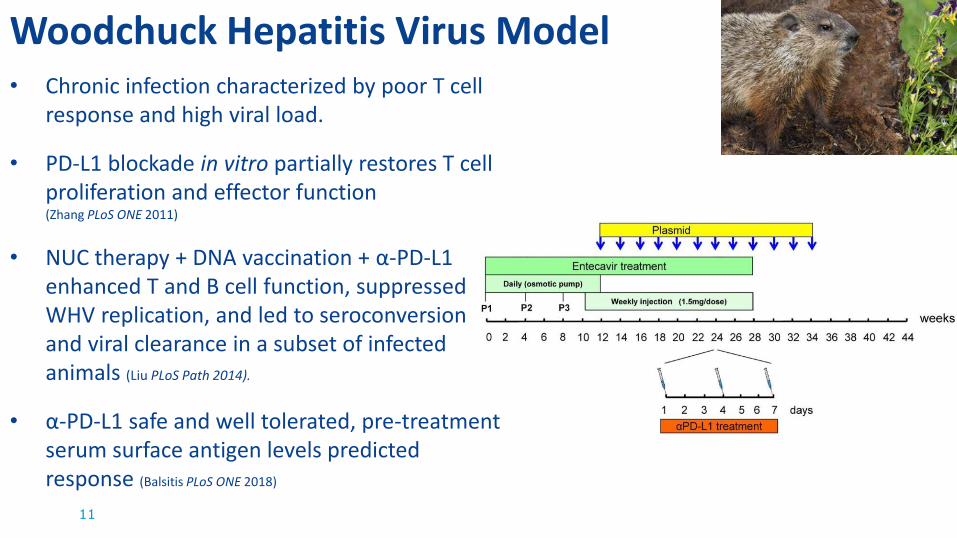
Woodchuck Hepatitis Virus Model
11
• Chronic infection characterized by poor T cell response and high viral load.
• PD-L1 blockade in vitro partially restores T cell proliferation and effector function (Zhang PLoS ONE 2011)
• NUC therapy + DNA vaccination + α-PD-L1 enhanced T and B cell function, suppressed WHV replication, and led to seroconversion and viral clearance in a subset of infected animals (Liu PLoS Path 2014).
• α-PD-L1 safe and well tolerated, pre-treatment serum surface antigen levels predicted response (Balsitis PLoS ONE 2018)
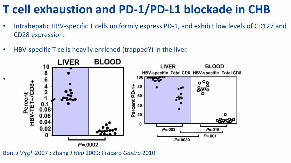
T cell exhaustion and PD-1/PD-L1 blockade in CHB
12
• Intrahepatic HBV-specific T cells uniformly express PD-1, and exhibit low levels of CD127 and CD28 expression.
• HBV-specific T cells heavily enriched (trapped?) in the liver.
•
Boni J Virol 2007 ; Zhang J Hep 2009; Fisicaro Gastro 2010.
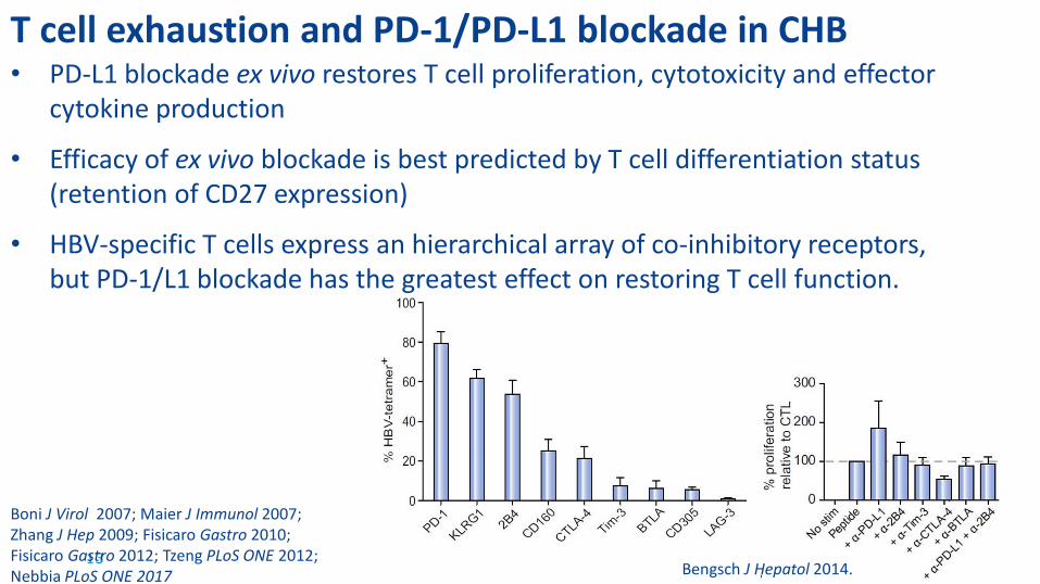
T cell exhaustion and PD-1/PD-L1 blockade in CHB
13
Boni J Virol 2007; Maier J Immunol 2007; Zhang J Hep 2009; Fisicaro Gastro 2010; Fisicaro Gastro 2012; Tzeng PLoS ONE 2012; Nebbia PLoS ONE 2017 Bengsch J Hepatol 2014.
• PD-L1 blockade ex vivo restores T cell proliferation, cytotoxicity and effector cytokine production
• Efficacy of ex vivo blockade is best predicted by T cell differentiation status (retention of CD27 expression)
• HBV-specific T cells express an hierarchical array of co-inhibitory receptors, but PD-1/L1 blockade has the greatest effect on restoring T cell function.

T cell exhaustion and PD-1/PD-L1 blockade in CHB
14
At 3 mg/kg, evaluation in HBV-infected patients with advanced HCC demonstrated declines in HBsAg of >1 log10 in 3/51 (6%) of patients
(Sangro et al, The Liver Meeting 2016, Checkmate-040)

Nivolumab +/- GS-4774
15
• Monoclonal antibody against PD-1
• Approved for solid organ tumors and lymphomas1
• Human IgG4, half-life 12 days (<3mg/kg) to 20 days (10mg/kg)2.
• Previous studies had demonstrated vaccine GS-4774 to be immunogenic in CHB but to lack therapeutic efficacy3,4
1. Topalian NEJM 2012; 2. Glassman Cancer Biol Med. 2014;
3. Gaggar Vaccine 2014; 4. Lok J Hep 2016.

Study Design
16
• Primary efficacy endpoint: Change in HBsAg log10 IU/mL levels 12 weeks post-Nivo
• Clinically approved dose 3mg/kg (melanoma, NSCLC, RCC).
Gane J Hep 2017; Verdon The Liver Meeting 2017.

PD-1 is primarily expressed on memory and effector T cells
17 Gane J Hep 2017; Verdon The Liver Meeting 2017.
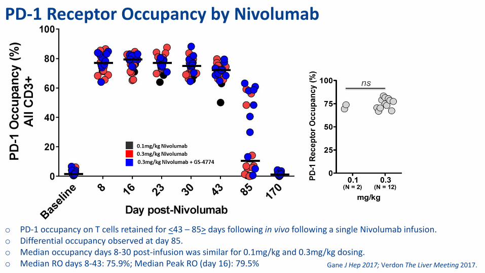
PD-1 Receptor Occupancy by Nivolumab
o PD-1 occupancy on T cells retained for <43 – 85> days following in vivo following a single Nivolumab infusion.o Differential occupancy observed at day 85.o Median occupancy days 8-30 post-infusion was similar for 0.1mg/kg and 0.3mg/kg dosing. o Median RO days 8-43: 75.9%; Median Peak RO (day 16): 79.5% Gane J Hep 2017; Verdon The Liver Meeting 2017.
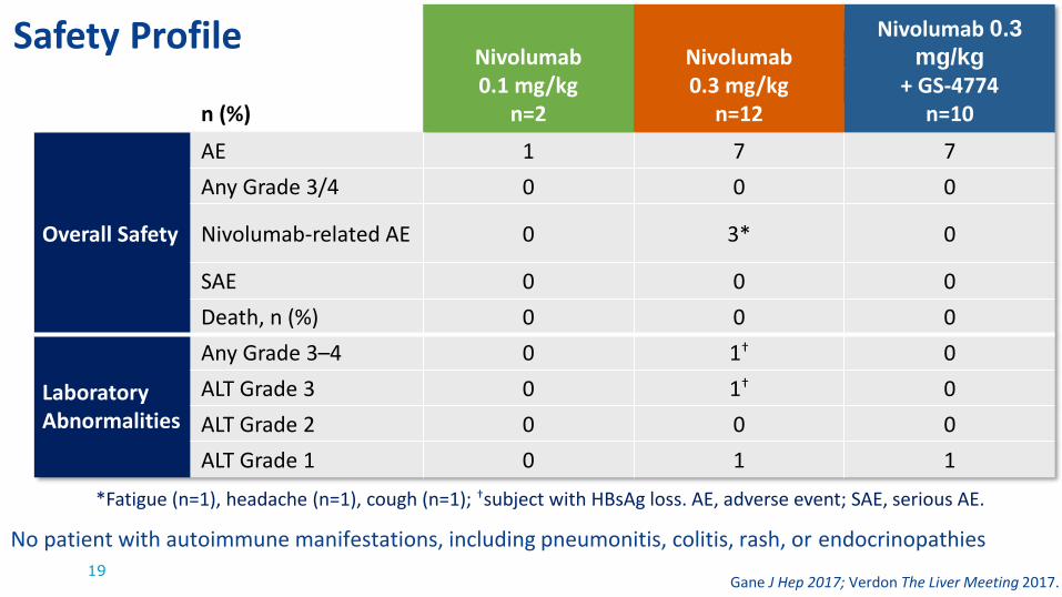
Safety Profile
19
n (%)
Nivolumab0.1 mg/kg
n=2
Nivolumab0.3 mg/kg
n=12
Nivolumab 0.3
mg/kg
+ GS-4774 n=10
Overall Safety
AE 1 7 7
Any Grade 3/4 0 0 0
Nivolumab-related AE 0 3* 0
SAE 0 0 0
Death, n (%) 0 0 0
Laboratory Abnormalities
Any Grade 3–4 0 1† 0
ALT Grade 3 0 1† 0
ALT Grade 2 0 0 0
ALT Grade 1 0 1 1
*Fatigue (n=1), headache (n=1), cough (n=1); †subject with HBsAg loss. AE, adverse event; SAE, serious AE.
19
No patient with autoimmune manifestations, including pneumonitis, colitis, rash, or endocrinopathies
Gane J Hep 2017; Verdon The Liver Meeting 2017.

HBSAg Changes from Baseline
20
• 2/22 (9%) at Week 12 and 3/22 (14%) at Week 24 with a >0.5 log10 reduction in HBsAg
– Only one patient with >1 log10 reduction in HBsAg at either clinical timepoint
• No baseline demographic feature associated with >0.5 log10 reduction in HBsAg
Gane J Hep 2017; Verdon The Liver Meeting 2017.

Case Study: Clinical observations
21
Gane J Hep 2017; Verdon The Liver Meeting 2017.

Case Study: Lymphocyte subset redistribution
22
Verdon, The Liver Meeting 2017.
Summary and Conclusions
23
• Peripheral PD-1 receptor occupancy is maintained in CHB patients for up to 85 days following a single 0.3mg/kg IV dose of Nivolumab, without evidence of immune-mediated AE.
• 20/22 (90%) of patients treated with 0.3mg/kg Nivolumab exhibited some decline in serum HBsAg at week 12.
• One patient experienced complete and sustained HBsAg loss and anti-HBS seroconversion, and remains disease-free at 29mo.

Future directions
24
• Anti-PD-L1 monoclonal therapy in CHB
• siRNA inhibition of PD-L1 expression (transient?)
• Small molecule inhibitors of PD-1 and/or PD-L1 (more predictable half-life and clearance?)
• Combination checkpoint blockade therapies – both multiple checkpoints (e.g. Tim-3, CD244) and/or agonistic targets (e.g. CD137).
Nebbia PLoS ONE 2017; Fisicaro Gastro 2012

Future directions
25
• Adoptive Cell Transfer supported by anti-PD1/L1 – ex vivo rescue of HBV-specific T cells.
• Tetramer/Dextramer or activation marker-based isolation (PepMix™stimulation).
• Expansion of HBV-specific T cells under ‘restorative’ conditions that modulate cell phenotype and metabolism before reinfusion.
• Agonistic microbeads (αCD3/28)
• Cytokine support (e.g. IL-12)
• Costimulation (e.g. CD137L or agonistic αCD137)
Verdon et al. unpublished data
Schurich PLoS Path 2013; Schurich Cell Rep 2016.

We extend our thanks to the patients, their families, and participating investigators.
This study was funded by Gilead Sciences, Inc.