Anhang A: Evidenztabellen Folgende Bewertungvorlagen nach SIGN wurden entsprechend dem Studientyp...
146
Methodikbericht Anhang A: Evidenztabellen Folgende Bewertungvorlagen nach SIGN wurden entsprechend dem Studientyp verwendet, um die Evidenzlevel (EL) zu ermitteln: Methodology Checklist 1: Systematic Reviews and Meta-analyses Methodology Checklist 2: Controlled Trials Methodology Checklist 5: Diagnostic studies Methodology Checklist 6: Economic studies Quelle: Scottish Intercollegiate Guidelines Network (SIGN). SIGN 50: a guideline developer’s handbook. Edinburgh: SIGN; 2015. (SIGN publication no. 50). [November 2015]. Available from URL: http://www.sign.ac.uk] Schlüsselfrage: 01. What factors contribute to increased fracture risk/increased number of fractures? EL Literaturstelle Systematic Reviews and Meta-analyses 1++ Abramowitz, J. et al. Adverse Event Reporting for Proton Pump Inhibitor Therapy: An Overview of Systematic Reviews. Otolaryngol Head Neck Surg. . . 2016 1++ Bolland, M. J. et al. Calcium intake and risk of fracture: systematic review. Bmj. 351. h4580. 2015 1++ Su, B. et al. Risk of bone fractures associated with glucagon-like peptide-1 receptor agonists' treatment: a meta-analysis of randomized controlled trials. Endocrine. 48. 107-15. 2015 1+ Mamza, J. et al. DPP-4 inhibitor therapy and bone fractures in people with Type 2 diabetes - A systematic review and meta-analysis. Diabetes Res Clin Pract. 116. 288-98. 2016 1- Wang, Z. et al. Effects of Statins on Bone Mineral Density and Fracture Risk: A PRISMA-compliant Systematic Review and Meta-Analysis. Medicine (Baltimore). 95. e3042. 2016 2++ Solmi, M. et al. Bone mineral density, osteoporosis, and fractures among people with eating disorders: a systematic review and meta-analysis. Acta Psychiatr Scand. 133. 341-51. 2016 2++ Yan, Z. et al. Relationship between subclinical thyroid dysfunction and the risk of fracture: a meta-analysis of prospective cohort studies. Osteoporos Int. 27. 115-25. 2016 2++ Yang, L. et al. Metabolic syndrome and the risk of bone fractures: A Meta-analysis of prospective cohort studies. Bone. 84. 52-6. 2016 2++ Lopez, L. M. et al. Steroidal contraceptives and bone fractures in women: evidence from observational studies. Cochrane Database Syst Rev. . Cd009849. 2015 2++ Lv, Q. B. et al. The relationship between weight change and risk of hip fracture: meta-analysis of prospective studies. Sci Rep. 5. 16030. 2015 2++ Shen, G. S. et al. Cigarette smoking and risk of hip fracture in women: a meta-analysis of prospective cohort studies. Injury. 46. 1333-40. 2015 2++ Wu, Z. J. et al. C-reactive protein and risk of fracture: a systematic review and dose-response meta-analysis of prospective cohort studies. Osteoporos Int. 26. 49-57. 2015 2++ Xiao, F. et al. Association between loop diuretic use and fracture risk. Osteoporos Int. 26. 775-84. 2015 2++ Yin, X. H. et al. Exposure to fluoride in drinking water and hip fracture risk: a meta-analysis of observational studies. PLoS One. 10. e0126488. 2015 2+ Cheng, X. et al. Cadmium Exposure and Risk of Any Fracture: A PRISMA-Compliant Systematic Review and Meta-Analysis. Medicine (Baltimore). 95. e2932. 2016 2+ Fan, Y. et al. Diabetes mellitus and risk of hip fractures: a meta-analysis. Osteoporos Int. 27. 219-28. 2016 2+ Kojima, G. Frailty as a predictor of fractures among community-dwelling older people: A systematic review and meta-analysis. Bone. 90. 116-122. 2016 2+ Wang, J. et al. Increased risk of vertebral fracture in patients with diabetes: a meta-analysis of cohort studies. Int Orthop. 40. 1299-307. 2016 2+ Wang, Q. et al. Parity and osteoporotic fracture risk in postmenopausal women: a dose-response meta-analysis of prospective studies. Osteoporos Int. 27. 319- 30. 2016 2+ Cortes, Y. I. et al. Bone Density and Fractures in HIV-infected Postmenopausal Women: A Systematic Review. J Assoc Nurses AIDS Care. 26. 387-98. 2015 2+ Fraser, L. A. et al. Enzyme-inducing antiepileptic drugs and fractures in people with epilepsy: A systematic review. Epilepsy Res. 116. 59-66. 2015 2+ Stubbs, B. et al. Schizophrenia and the risk of fractures: a systematic review and comparative meta-analysis. Gen Hosp Psychiatry. 37. 126-33. 2015 2+ Yang, S. D. et al. Bone fracture and the interaction between bisphosphonates and proton pump inhibitors: a meta-analysis. Int J Clin Exp Med. 8. 4899-910. 2015 2- Ping, F. et al. Opioids increase hip fracture risk: a meta-analysis. J Bone Miner Metab. 2016 2- Walenkamp, M. M. et al. Predictors of unstable distal radius fractures: a systematic review and meta-analysis. J Hand Surg Eur Vol. 41. 501-15. 2016 2- Yuan, Z. C. et al. Risk of hip fracture following stroke, a meta-analysis of 13 cohort studies. Osteoporos Int. 2016 2- Bang, C. S. et al. Osteoporosis and bone fractures in alcoholic liver disease: a meta-analysis. World J Gastroenterol. 21. 4038-47. 2015
Transcript of Anhang A: Evidenztabellen Folgende Bewertungvorlagen nach SIGN wurden entsprechend dem Studientyp...
Methodikbericht Anhang A: Evidenztabellen Folgende Bewertungvorlagen nach SIGN wurden entsprechend dem Studientyp verwendet, um die Evidenzlevel (EL) zu ermitteln: Methodology Checklist 1: Systematic Reviews and Meta-analyses Methodology Checklist 2: Controlled Trials Methodology Checklist 5: Diagnostic studies Methodology Checklist 6: Economic studies Quelle:
Scottish Intercollegiate Guidelines Network (SIGN). SIGN 50: a guideline developer’s handbook. Edinburgh: SIGN; 2015. (SIGN publication no. 50). [November 2015]. Available from URL: http://www.sign.ac.uk]
Schlüsselfrage: 01. What factors contribute to increased fracture risk/increased number of fractures?
EL Literaturstelle Systematic Reviews and Meta-analyses
1++ Abramowitz, J. et al. Adverse Event Reporting for Proton Pump Inhibitor Therapy: An Overview of Systematic Reviews. Otolaryngol Head Neck Surg. . . 2016
1++ Bolland, M. J. et al. Calcium intake and risk of fracture: systematic review. Bmj. 351. h4580. 2015
1++ Su, B. et al. Risk of bone fractures associated with glucagon-like peptide-1 receptor agonists' treatment: a meta-analysis of randomized controlled trials. Endocrine. 48. 107-15. 2015
1+ Mamza, J. et al. DPP-4 inhibitor therapy and bone fractures in people with Type 2 diabetes - A systematic review and meta-analysis. Diabetes Res Clin Pract. 116. 288-98. 2016
1- Wang, Z. et al. Effects of Statins on Bone Mineral Density and Fracture Risk: A PRISMA-compliant Systematic Review and Meta-Analysis. Medicine (Baltimore). 95. e3042. 2016
2++ Solmi, M. et al. Bone mineral density, osteoporosis, and fractures among people with eating disorders: a systematic review and meta-analysis. Acta Psychiatr Scand. 133. 341-51. 2016
2++ Yan, Z. et al. Relationship between subclinical thyroid dysfunction and the risk of fracture: a meta-analysis of prospective cohort studies. Osteoporos Int. 27. 115-25. 2016
2++ Yang, L. et al. Metabolic syndrome and the risk of bone fractures: A Meta-analysis of prospective cohort studies. Bone. 84. 52-6. 2016
2++ Lopez, L. M. et al. Steroidal contraceptives and bone fractures in women: evidence from observational studies. Cochrane Database Syst Rev. . Cd009849. 2015
2++ Lv, Q. B. et al. The relationship between weight change and risk of hip fracture: meta-analysis of prospective studies. Sci Rep. 5. 16030. 2015
2++ Shen, G. S. et al. Cigarette smoking and risk of hip fracture in women: a meta-analysis of prospective cohort studies. Injury. 46. 1333-40. 2015
2++ Wu, Z. J. et al. C-reactive protein and risk of fracture: a systematic review and dose-response meta-analysis of prospective cohort studies. Osteoporos Int. 26. 49-57. 2015
2++ Xiao, F. et al. Association between loop diuretic use and fracture risk. Osteoporos Int. 26. 775-84. 2015
2++ Yin, X. H. et al. Exposure to fluoride in drinking water and hip fracture risk: a meta-analysis of observational studies. PLoS One. 10. e0126488. 2015
2+ Cheng, X. et al. Cadmium Exposure and Risk of Any Fracture: A PRISMA-Compliant Systematic Review and Meta-Analysis. Medicine (Baltimore). 95. e2932. 2016
2+ Fan, Y. et al. Diabetes mellitus and risk of hip fractures: a meta-analysis. Osteoporos Int. 27. 219-28. 2016
2+ Kojima, G. Frailty as a predictor of fractures among community-dwelling older people: A systematic review and meta-analysis. Bone. 90. 116-122. 2016
2+ Wang, J. et al. Increased risk of vertebral fracture in patients with diabetes: a meta-analysis of cohort studies. Int Orthop. 40. 1299-307. 2016
2+ Wang, Q. et al. Parity and osteoporotic fracture risk in postmenopausal women: a dose-response meta-analysis of prospective studies. Osteoporos Int. 27. 319-30. 2016
2+ Cortes, Y. I. et al. Bone Density and Fractures in HIV-infected Postmenopausal Women: A Systematic Review. J Assoc Nurses AIDS Care. 26. 387-98. 2015
2+ Fraser, L. A. et al. Enzyme-inducing antiepileptic drugs and fractures in people with epilepsy: A systematic review. Epilepsy Res. 116. 59-66. 2015
2+ Stubbs, B. et al. Schizophrenia and the risk of fractures: a systematic review and comparative meta-analysis. Gen Hosp Psychiatry. 37. 126-33. 2015
2+ Yang, S. D. et al. Bone fracture and the interaction between bisphosphonates and proton pump inhibitors: a meta-analysis. Int J Clin Exp Med. 8. 4899-910. 2015
2- Ping, F. et al. Opioids increase hip fracture risk: a meta-analysis. J Bone Miner Metab. 2016
2- Walenkamp, M. M. et al. Predictors of unstable distal radius fractures: a systematic review and meta-analysis. J Hand Surg Eur Vol. 41. 501-15. 2016
2- Yuan, Z. C. et al. Risk of hip fracture following stroke, a meta-analysis of 13 cohort studies. Osteoporos Int. 2016
2- Bang, C. S. et al. Osteoporosis and bone fractures in alcoholic liver disease: a meta-analysis. World J Gastroenterol. 21. 4038-47. 2015
Ulrike
Textfeld
publiziert bei
Ulrike
Stempel
2- Heikkila, K. et al. Celiac disease and bone fractures: a systematic review and meta-analysis. J Clin Endocrinol Metab. 100. 25-34. 2015
2- Liu, S. et al. Risk factors for the second contralateral hip fracture in elderly patients: a systematic review and meta-analysis. Clin Rehabil. 29. 285-94. 2015
2- Teng, Z. et al. Opioids contribute to fracture risk: a meta-analysis of 8 cohort studies. PLoS One. 10. e0128232. 2015
2- Veronese, N. et al. Vitamin K antagonists' use and fracture risk: results from a systematic review and meta-analysis. J Thromb Haemost. 13. 1665-75. 2015
2- Wu, A. M. et al. The relationship between dietary protein consumption and risk of fracture: a subgroup and dose-response meta-analysis of prospective cohort studies. Sci Rep. 5. 9151. 2015
2- Yan, A. et al. Does tea consumption correlate to risk of fracture? A meta-analysis. Int J Clin Exp Med. 8. 8347-57. 2015
2- Zhang, X. et al. Alcohol consumption and hip fracture risk. Osteoporos Int. 26. 531-42. 2015
Schlüsselfrage: 02. Which diagnostic measurements or tools are effective in identifying increased risk of fracture?
EL Literaturstelle Systematic Reviews and Meta-analyses
2++ Burch, J. et al. Systematic review of the use of bone turnover markers for monitoring the response to osteoporosis treatment: the secondary prevention of fractures, and primary prevention of fractures in high-risk groups. Health Technol Assess. 18. 1-180. 2014
2- Devlin, H. et al. Can mandibular bone resorption predict hip fracture in elderly women? A systematic review of diagnostic test accuracy. Gerodontology. 32. 163-8. 2015
2- Marques, A. et al. The accuracy of osteoporotic fracture risk prediction tools: a systematic review and meta-analysis. Ann Rheum Dis. 74. 1958-67. 2015
2- Nayak, S. et al. Performance of risk assessment instruments for predicting osteoporotic fracture risk: a systematic review. Osteoporos Int. 25. 23-49. 2014
Studies of Diagnostic Accuracy
- Graeff, Christian et al. High resolution quantitative computed tomography-based assessment of trabecular microstructure and strength estimates by finite-element analysis of the spine, but not DXA, reflects vertebral fracture status in men with glucocorticoid-induced osteoporosis. Bone. 52. 568-77. 2013
Schlüsselfrage: 03. Which diagnostic measurements or tools best predict response to pharmacological treatment?
EL Literaturstelle Systematic Reviews and Meta-analyses 2++ Burch, J. et al. Systematic review of the use of bone turnover markers for monitoring the response to osteoporosis treatment: the secondary prevention of
fractures, and primary prevention of fractures in high-risk groups. Health Technol Assess. 18. 1-180. 2014
Controlled Trials 1- Naylor, K E et al. Response of bone turnover markers to raloxifene treatment in postmenopausal women with osteopenia. Osteoporos Int. 2016
Schlüsselfrage: 04. Which pharmacological interventions are effective in fracture prevention? Population: Über 50-Jährige
EL Literaturstelle Systematic Reviews and Meta-analyses 2+ Wright, E. et al. Bisphosphonates and evidence for association with esophageal and gastric cancer: a systematic review and meta-analysis. BMJ Open. 5.
e007133. 2015
Controlled Trials 1++ Black, D. M. et al. The effect of 6 versus 9 years of zoledronic acid treatment in osteoporosis: a randomized second extension to the HORIZON-Pivotal Fracture
Trial (PFT). Journal of bone and mineral research : the official journal of the American Society for Bone and Mineral Research. 30. 934-44. 2015
1++ Greenspan, S. L. et al. Efficacy and safety of single-dose zoledronic acid for osteoporosis in frail elderly women: a randomized clinical trial. JAMA internal medicine. 175. 913-21. 2015
1++ Palacios, S. et al. A 7-year randomized, placebo-controlled trial assessing the long-term efficacy and safety of bazedoxifene in postmenopausal women with osteoporosis: effects on bone density and fracture. Menopause. 22. 806-13. 2015
1++ Zhang, Z. L. et al. Alendronate sodium/vitamin D3 combination tablet versus calcitriol for osteoporosis in Chinese postmenopausal women: a 6-month, randomized, open-label, active-comparator-controlled study with a 6-month extension. Osteoporos Int. 26. 2365-74. 2015
1++ Brown, J. P. et al. Denosumab significantly increases bone mineral density and reduces bone turnover compared with monthly oral ibandronate and risedronate in postmenopausal women who remained at higher risk for fracture despite previous suboptimal treatment with an oral bisp. Osteoporos Int. 25. 1953-61. 2014
1++ Nakano, Tetsuo et al. Once-weekly teriparatide reduces the risk of vertebral fracture in patients with various fracture risks: subgroup analysis of the Teriparatide Once-Weekly Efficacy Research (TOWER) trial. J. Bone Miner. Metab. 32. 441-6. 2014
1++ Roux, C. et al. Denosumab compared with risedronate in postmenopausal women suboptimally adherent to alendronate therapy: efficacy and safety results from a randomized open-label study. Bone. 58. 48-54. 2014
1++ Paggiosi, M. A. et al. Comparison of the effects of three oral bisphosphonate therapies on the peripheral skeleton in postmenopausal osteoporosis: the TRIO study. Osteoporos Int. 25. 2729-41. 2014
1++ Rajatanavin, R. et al. The efficacy of calcium supplementation alone in elderly Thai women over a 2-year period: a randomized controlled trial. Osteoporos Int. 24. 2871-7. 2013
Population: Patienten mit chronischem Nierenversagen
EL Literaturstelle Systematic Reviews and Meta-analyses 1++ Hahn, Deirdre et al. Interventions for metabolic bone disease in children with chronic kidney disease. Cochrane Database Syst Rev. . CD008327. 2015
Controlled Trials 1+ Moe, Sharon M et al. Effects of Cinacalcet on Fracture Events in Patients Receiving Hemodialysis: The EVOLVE Trial. J. Am. Soc. Nephrol. 26. 1466-75. 2015
Population: Männer
EL Literaturstelle Systematic Reviews and Meta-analyses 1++ Chen, L. X. et al. Comparison of Bone Mineral Density in Lumbar Spine and Fracture Rate among Eight Drugs in Treatments of Osteoporosis in Men: A
Network Meta-Analysis. PLoS One. 10. e0128032. 2015
1++ Pinzone, Marilia Rita et al. Is there enough evidence to use bisphosphonates in HIV-infected patients? A systematic review and meta- analysis. AIDS Rev. 16. 213-22. 2014
2++ Crandall, C. J. et al. Comparative effectiveness of pharmacologic treatments to prevent fractures: an updated systematic review. Ann Intern Med. 161. 711-23. 2014
2+ Wright, E. et al. Bisphosphonates and evidence for association with esophageal and gastric cancer: a systematic review and meta-analysis. BMJ Open. 5. e007133. 2015
Controlled Trials 1++ Langdahl, B. L. et al. A 24-month study evaluating the efficacy and safety of denosumab for the treatment of men with low bone mineral density: results from the
ADAMO trial. The Journal of clinical endocrinology and metabolism. 100. 1335-42. 2015
1++ Chen, Z. G. et al. Effects of atorvastatin on bone mineral density (BMD) and bone metabolism in elderly males with osteopenia and mild dyslipidemia: a 1-year randomized trial. Archives of gerontology and geriatrics. 59. 515-21. 2014
1++ Kachnic, L. A. et al. RTOG 0518: randomized phase III trial to evaluate zoledronic acid for prevention of osteoporosis and associated fractures in prostate cancer patients. Prostate Cancer Prostatic Dis. 16. 382-6. 2013
1- Glüer, Claus-C et al. Comparative effects of teriparatide and risedronate in glucocorticoid-induced osteoporosis in men: 18-month results of the EuroGIOPs trial. J. Bone Miner. Res. 28. 1355-68. 2013
Population: Patienten mit Osteogenesis Imperfecta
EL Literaturstelle Systematic Reviews and Meta-analyses 1++ Dwan, Kerry et al. Bisphosphonate therapy for osteogenesis imperfecta. Cochrane Database Syst Rev. CD005088. 2014
2+ Rijks, E. B. et al. Efficacy and Safety of Bisphosphonate Therapy in Children with Osteogenesis Imperfecta: A Systematic Review. Horm Res Paediatr. 84. 26-42. 2015
Controlled Trials 1++ Plante, Laura et al. Effect of high-dose vitamin D supplementation on bone density in youth with osteogenesis imperfecta: A randomized controlled trial. Bone.
86. 36-42. 2016
Population: Postmenopausale Frauen
EL Literaturstelle Systematic Reviews and Meta-analyses 1++ Beaudoin, C. et al. Denosumab compared to other treatments to prevent or treat osteoporosis in individuals at risk of fracture: a systematic review and
meta-analysis. Osteoporos Int. . . 2016
2+ Crandall, C. J. et al. Comparative effectiveness of pharmacologic treatments to prevent fractures: an updated systematic review. Ann Intern Med. 161. 711-23. 2014
2+ Wright, E. et al. Bisphosphonates and evidence for association with esophageal and gastric cancer: a systematic review and meta-analysis. BMJ Open. 5. e007133. 2015
Controlled Trials 1++ Bristow, S. M. et al. Acute effects of calcium supplements on blood pressure and blood coagulation: secondary analysis of a randomised controlled trial in
post-menopausal women. Br J Nutr. 114. 1868-74. 2015
1++ Gu, J. M. et al. The efficacy and safety of weekly 35-mg risedronate dosing regimen for Chinese postmenopausal women with osteoporosis or osteopenia: 1-year data. Acta Pharmacol Sin. 36. 841-6. 2015
1++ Leder, B. Z. et al. Denosumab and teriparatide transitions in postmenopausal osteoporosis (the DATA-Switch study): extension of a randomised controlled trial. Lancet (London, England). 386. 1147-55. 2015
1++ Recker, R. R. et al. A randomized, doubleblind phase 2 clinical trial of blosozumab, a sclerostin antibody, in postmenopausal women with low bone mineral density. J Bone Miner Res. 30. 21624. 2015
1++ Sugimoto, T. et al. Three-year denosumab treatment in postmenopausal Japanese women and men with osteoporosis: results from a 1-year open- label extension of the Denosumab Fracture Intervention Randomized Placebo Controlled Trial (DIRECT). Osteoporosis international : a journal established as result of cooperation between the European Foundation for Osteoporosis and the National Osteoporosis Foundation of the USA. 26. 765-74. 2015
1++ Binkley, N. et al. Efficacy and safety of oral recombinant calcitonin tablets in postmenopausal women with low bone mass and increased fracture risk: a randomized, placebo-controlled trial. Osteoporosis international : a journal established as result of cooperation between the European Foundation for Osteoporosis and the National Osteoporosis Foundation of the USA. 25. 2649-56. 2014
1++ Kim, K. J. et al. Efficacy and safety of weekly alendronate plus vitamin D(3) 5600 IU versus weekly alendronate alone in Korean osteoporotic women: 16-week randomized trial. Yonsei Med J. 55. 715-24. 2014
1++ Leder, B. Z. et al. Two years of Denosumab and teriparatide administration in postmenopausal women with osteoporosis (The DATA Extension Study): a randomized controlled trial. The Journal of clinical endocrinology and metabolism. 99. 1694-700. 2014
1++ McClung, M. R. et al. Romosozumab in postmenopausal women with low bone mineral density. The New England journal of medicine. 370. 412-20. 2014
1++ Miller, P D et al. Long-term fracture rates seen with continued ibandronate treatment: pooled analysis of DIVA and MOBILE long-term extension studies. Osteoporos Int. 25. 349-57. 2014
1++ Paggiosi, M. A. et al. Comparison of the effects of three oral bisphosphonate therapies on the peripheral skeleton in postmenopausal osteoporosis: the TRIO study. Osteoporos Int. 25. 2729-41. 2014
1++ Bonnick, S. et al. Effects of odanacatib on BMD and safety in the treatment of osteoporosis in postmenopausal women previously treated with alendronate: a randomized placebo-controlled trial. J Clin Endocrinol Metab. 98. 4727-35. 2013
1++ Horwitz, M. J. et al. A comparison of parathyroid hormone-related protein (1-36) and parathyroid hormone (1-34) on markers of bone turnover and bone density in postmenopausal women: the PrOP study. J Bone Miner Res. 28. 2266-76. 2013
1++ Li, S. et al. Long-term calcium supplementation may have adverse effects on serum cholesterol and carotid intima-media thickness in postmenopausal women: a double-blind, randomized, placebo-controlled trial. Am J Clin Nutr. 98. 1353-9. 2013
1+ McColm, J. et al. Single- and multiple-dose randomized studies of blosozumab, a monoclonal antibody against sclerostin, in healthy postmenopausal women. Journal of bone and mineral research : the official journal of the American Society for Bone and Mineral Research. 29. 935- 43. 2014
Population: Prämenopausale Frauen EL Literaturstelle Systematic Reviews and Meta-analyses 2+ Wright, E. et al. Bisphosphonates and evidence for association with esophageal and gastric cancer: a systematic review and meta-analysis. BMJ Open. 5.
e007133. 2015
Controlled Trials 1+ Hadji, P. et al. Effects of zoledronic acid on bone mineral density in premenopausal women receiving neoadjuvant or adjuvant therapies for HR+ breast cancer:
the ProBONE II study. Osteoporosis international : a journal established as result of cooperation between the European Foundation for Osteoporosis and the National Osteoporosis Foundation of the USA. 25. 1369-78. 2014
1+ Kalder, M. et al. Effects of zoledronic acid versus placebo on bone mineral density and bone texture analysis assessed by the trabecular bone score in premenopausal women with breast cancer treatment-induced bone loss: results of the ProBONE II substudy. Osteoporosis international : a journal established as result of cooperation between the European Foundation for Osteoporosis and the National Osteoporosis Foundation of the USA. 26. 353-60. 2015
Population: Sekundäre Osteoporose EL Literaturstelle Systematic Reviews and Meta-analyses 1++ Conwell Louise, S. et al. Bisphosphonates for osteoporosis in people with cystic fibrosis. Cochrane Database of Systematic Reviews. 2014
1++ Melek, J. et al. Efficacy and safety of medical therapy for low bone mineral density in patients with inflammatory bowel disease: a meta- analysis and systematic review. Clin Gastroenterol Hepatol. 12. 32-44.e5. 2014
1+ Feng, Z. et al. Bisphosphonates for the prevention and treatment of osteoporosis in patients with rheumatic diseases: a systematic review and meta-analysis. PLoS One. 8. e80890. 2013
Controlled Trials 1- Glüer, Claus-C et al. Comparative effects of teriparatide and risedronate in glucocorticoid-induced osteoporosis in men: 18-month results of the EuroGIOPs trial.
J. Bone Miner. Res. 28. 1355-68. 2013
Schlüsselfrage: 06. What monitoring should be conducted in individuals taking pharmacological interventions?
EL Literaturstelle Systematic Reviews and Meta-analyses 2++ Burch, J. et al. Systematic review of the use of bone turnover markers for monitoring the response to osteoporosis treatment: the secondary prevention of
fractures, and primary prevention of fractures in high-risk groups. Health Technol Assess. 18. 1-180. 2014
Controlled Trials
1- Bell, K. et al. Relative Utility of BMD and Bone Turnover Monitoring of Zoledronic Acid Therapy among Women with Osteoporosis: Secondary Analysis of Randomised Controlled Trial Data. J Bone Miner Res. 2016
1- Naylor, K E et al. Response of bone turnover markers to raloxifene treatment in postmenopausal women with osteopenia. Osteoporos Int. 2016
1- Farahmand, P. et al. Early changes in biochemical markers of bone formation during teriparatide therapy correlate with improvements in vertebral strength in men with glucocorticoid-induced osteoporosis. Osteoporos Int. 24. 2971-81. 2013
Schlüsselfrage: 07. What interventions are effective in improving concordance with pharmacological interventions for fracture prevention?
EL Literaturstelle Systematic Reviews and Meta-analyses 2+ Hiligsmann, M. et al. Interventions to improve osteoporosis medication adherence and persistence: a systematic review and literature appraisal by the ISPOR
Medication Adherence & Persistence Special Interest Group. Osteoporos Int. 24. 2907-18. 2013
2- Iglay, K. et al. Systematic Literature Review and Meta-analysis of Medication Adherence With Once-weekly Versus Once-daily Therapy. Clin Ther. 37. 1813-21.e1. 2015
Controlled Trials 1+ Cizmic, A. D. et al. Impact of interactive voice response technology on primary adherence to bisphosphonate therapy: a randomized controlled trial. Osteoporos
Int. 26. 2131-6. 2015
1- Gonnelli, S. et al. How the knowledge of fracture risk might influence adherence to oral therapy of osteoporosis in Italy: the ADEOST study. Aging Clin Exp Res. 28. 459-68. 2016
1- LeBlanc, A. et al. Encounter Decision Aid vs. Clinical Decision Support or Usual Care to Support Patient-Centered Treatment Decisions in Osteoporosis: The Osteoporosis Choice Randomized Trial II. PLoS One. 10. e0128063. 2015
1- Ganda, K. et al. Compliance and persistence to oral bisphosphonate therapy following initiation within a secondary fracture prevention program: a randomised controlled trial of specialist vs. non-specialist management. Osteoporos Int. 25. 1345-55. 2014
Schlüsselfrage: 08. What exercise interventions are effective in reducing the risk of fracture or improving BMD levels? Population: Über 50-Jährige
EL Literaturstelle Systematic Reviews and Meta-analyses 1++ Kendrick, Denise et al. Exercise for reducing fear of falling in older people living in the community. Cochrane Database of Systematic Reviews. 2014
2++ Bolam, K. A. et al. The effect of physical exercise on bone density in middle-aged and older men: a systematic review. Osteoporos Int. 24. 2749-62. 2013
2- Rong, K. et al. Increasing Level of Leisure Physical Activity Could Reduce the Risk of Hip Fracture in Older Women: A Dose-Response Meta- analysis of Prospective Cohort Studies. Medicine (Baltimore). 95. e2984. 2016
Controlled Trials 1++ Uusi-Rasi, K. et al. Exercise and vitamin D in fall prevention among older women: a randomized clinical trial. JAMA Intern Med. 175. 703-11. 2015
1++ Palvanen, M. et al. Effectiveness of the Chaos Falls Clinic in preventing falls and injuries of home-dwelling older adults: a randomised controlled trial. Injury. 45. 265-71. 2014
1+ Leung, K. S. et al. Effects of 18-month low-magnitude high-frequency vibration on fall rate and fracture risks in 710 community elderly--a cluster-randomized controlled trial. Osteoporos Int. 25. 1785-95. 2014
1- Halvarsson, A. et al. Balance training with multi-task exercises improves fall-related self-efficacy, gait, balance performance and physical function in older adults with osteoporosis: a randomized controlled trial. Clin Rehabil. 29. 365-75. 2015
1- Gianoudis, J. et al. Effects of a targeted multimodal exercise program incorporating high-speed power training on falls and fracture risk factors in older adults: a community-based randomized controlled trial. J Bone Miner Res. 29. 182-91. 2014
Population: Männer EL Literaturstelle Systematic Reviews and Meta-analyses 2++ Bolam, K. A. et al. The effect of physical exercise on bone density in middle-aged and older men: a systematic review. Osteoporos Int. 24. 2749-62. 2013
Population: Postmenopausale Frauen
EL Literaturstelle Systematic Reviews and Meta-analyses 1++ Melek, J. et al. Efficacy and safety of medical therapy for low bone mineral density in patients with inflammatory bowel disease: a meta- analysis and systematic
review. Clin Gastroenterol Hepatol. 12. 32-44.e5. 2014
2++ Ma, D. et al. Effects of walking on the preservation of bone mineral density in perimenopausal and postmenopausal women: a systematic review and meta-analysis. Menopause. 20. 1216-26. 2013
2+ Zhao, R. et al. The effects of differing resistance training modes on the preservation of bone mineral density in postmenopausal women: a meta-analysis. Osteoporos Int. 26. 1605-18. 2015
2- Rong, K. et al. Increasing Level of Leisure Physical Activity Could Reduce the Risk of Hip Fracture in Older Women: A Dose-Response Meta- analysis of Prospective Cohort Studies. Medicine (Baltimore). 95. e2984. 2016
Controlled Trials 1++ Uusi-Rasi, K. et al. Exercise and vitamin D in fall prevention among older women: a randomized clinical trial. JAMA Intern Med. 175. 703-11. 2015
1++ Kim, K. J. et al. Efficacy and safety of weekly alendronate plus vitamin D(3) 5600 IU versus weekly alendronate alone in Korean osteoporotic women: 16-week randomized trial. Yonsei Med J. 55. 715-24. 2014
1++ Slatkovska, L. et al. Effect of whole-body vibration on calcaneal quantitative ultrasound measurements in postmenopausal women: a randomized controlled trial. Calcif Tissue Int. 95. 547-56. 2014
1++ Bonnick, S. et al. Effects of odanacatib on BMD and safety in the treatment of osteoporosis in postmenopausal women previously treated with alendronate: a randomized placebo-controlled trial. J Clin Endocrinol Metab. 98. 4727-35. 2013
1+ Leung, K. S. et al. Effects of 18-month low-magnitude high-frequency vibration on fall rate and fracture risks in 710 community elderly--a cluster-randomized controlled trial. Osteoporos Int. 25. 1785-95. 2014
1+ Multanen, J. et al. Effects of high-impact training on bone and articular cartilage: 12-month randomized controlled quantitative MRI study. J Bone Miner Res. 29. 192-201. 2014
1+ Paolucci, T. et al. Efficacy of group-adapted physical exercises in reducing back pain in women with postmenopausal osteoporosis. Aging Clin Exp Res. 26. 395-402. 2014
1+ Stolzenberg, N. et al. Vibration or balance training on neuromuscular performance in osteopenic women. Int J Sports Med. 34. 956-62. 2013
1- Halvarsson, A. et al. Balance training with multi-task exercises improves fall-related self-efficacy, gait, balance performance and physical function in older adults with osteoporosis: a randomized controlled trial. Clin Rehabil. 29. 365-75. 2015
1- Liu, B. X. et al. The Effect of the Modified Eighth Section of Eight-Section Brocade on Osteoporosis in Postmenopausal Women: A Prospective Randomized Trial. Medicine (Baltimore). 94. e991. 2015
1- Winklmayr, M. et al. Radon balneotherapy and physical activity for osteoporosis prevention: a randomized, placebo-controlled intervention study. Radiat Environ Biophys. 54. 123-36. 2015
Population: Prämenopausale Frauen
EL Literaturstelle Systematic Reviews and Meta-analyses 1+ Zhao, R. et al. Efficiency of jumping exercise in improving bone mineral density among premenopausal women: a meta-analysis. Database of Abstracts of
Reviews of Effects. . 1393-1402. 2014
Schlüsselfrage: 09. What dietary interventions are effective in reducing the risk of fracture or improving BMD levels?
EL Literaturstelle Systematic Reviews and Meta-analyses 1++ Avenell, Alison et al. Vitamin D and vitamin D analogues for preventing fractures in post-menopausal women and older men. Cochrane Database of Systematic
Reviews. 2014
1- Ruan, J. et al. Effect of B vitamin (folate, B6, and B12) supplementation on osteoporotic fracture and bone turnover markers: a meta- analysis. Med Sci Monit. 21. 875-81. 2015
2- Lambert, H. et al. The effect of supplementation with alkaline potassium salts on bone metabolism: a metaanalysis. Osteoporos Int. 26. 1311-8. 2015
Controlled Trials 1+ Dostal, A. M. et al. Long-Term Supplementation of Green Tea Extract Does Not Modify Adiposity or Bone Mineral Density in a Randomized Trial of Overweight
and Obese Postmenopausal Women. J Nutr. 146. 256-64. 2016
1- Enneman, A. W. et al. Effect of Vitamin B12 and Folic Acid Supplementation on Bone Mineral Density and Quantitative Ultrasound Parameters in Older People with an Elevated Plasma Homocysteine Level: B-PROOF, a Randomized Controlled Trial. Calcif Tissue Int. 96. 401-9. 2015
Schlüsselfrage: 10. What other (nondietary, nonexercise) interventions are effective in reducing the risk of fracture or improving BMD levels?
EL Literaturstelle Systematic Reviews and Meta-analyses 1+ Liu, Yunxia et al. Chinese herbal medicines for treating osteoporosis. Cochrane Database Syst Rev. CD005467. 2014
Controlled Trials 1- Winklmayr, M. et al. Radon balneotherapy and physical activity for osteoporosis prevention: a randomized, placebo-controlled intervention study. Radiat Environ
Biophys. 54. 123-36. 2015
Schlüsselfrage: 11. What is the clinical and cost effectiveness of integrated models of care (which include assessment, identification, treatment and follow up) compared with standalone elements for the primary and secondary prevention of fragility fracture?
EL Literaturstelle Economic Evaluations ++ Ghimire, E. et al. Effects of a Community-Based Fall Management Program on Medicare Cost Savings. Am J Prev Med. 49. e109-16. 2015
+ Yates, C. J. et al. Bridging the osteoporosis treatment gap: performance and cost-effectiveness of a fracture liaison service. J Clin Densitom. 18. 150-6. 2015
+ Milte, R. et al. Cost-effectiveness of individualized nutrition and exercise therapy for rehabilitation following hip fracture. J Rehabil Med. 48. 378-85. 2016
12. Which interventions reduce pain, improve deformity, mobility, and quality of life in patients suffering from vertebral fractures as well as fractures of the pelvis or sacrum due to osteoporosis?
EL Literaturstelle Systematic Reviews and Meta-analyses 1++ Buchbinder, R. et al. Percutaneous vertebroplasty for osteoporotic vertebral compression fracture. Cochrane Database Syst Rev. Cd006349. 2015
1++ Rzewuska, M. et al. The efficacy of conservative treatment of osteoporotic compression fractures on acute pain relief: a systematic review with meta-analysis. Eur Spine J. 24. 702-14. 2015
1++ Stevenson, M. et al. Percutaneous vertebroplasty and percutaneous balloon kyphoplasty for the treatment of osteoporotic vertebral fractures: a systematic review and cost-effectiveness analysis. Health Technol Assess. 18. 1-290. 2014
1+ Mattie, R. et al. Comparing Percutaneous Vertebroplasty and Conservative Therapy for Treating Osteoporotic Compression Fractures in the Thoracic and Lumbar Spine: A Systematic Review and Meta-Analysis. J Bone Joint Surg Am. 98. 1041-51. 2016
1- Bouza, C. et al. Safety of balloon kyphoplasty in the treatment of osteoporotic vertebral compression fractures in Europe: a meta-analysis of randomized controlled trials. Eur Spine J. 24. 715-23. 2015
2++ Newman, M. et al. Spinal Orthoses for Vertebral Osteoporosis and Osteoporotic Vertebral Fracture: A Systematic Review. Arch Phys Med Rehabil. 97. 1013-25. 2016
2++ Han, S. L. et al. Is vertebroplasty a risk factor for subsequent vertebral fracture, meta-analysis of published evidence?. Osteoporos Int. 26. 113-22. 2015
2+ Goodwin, V. A. et al. Orthotics and taping in the management of vertebral fractures in people with osteoporosis: a systematic review. BMJ Open. 6. e010657. 2016
2+ Feng, H. et al. Unilateral versus bilateral percutaneous kyphoplasty for osteoporotic vertebral compression fractures: A systematic review and meta-analysis of RCTs. J Orthop Res. 33. 1713-23. 2015
2+ Xiao, H. et al. Comparing complications of vertebroplasty and kyphoplasty for treating osteoporotic vertebral compression fractures: a meta-analysis of the randomized and non-randomized controlled studies. Eur J Orthop Surg Traumatol. 25 Suppl 1. S77-85. 2015
2- Cheng, X. et al. Comparison of unilateral versus bilateral percutaneous kyphoplasty for the treatment of patients with osteoporosis vertebral compression fracture (OVCF): a systematic review and meta-analysis. Eur Spine J. 2016
2- Gu, C. N. et al. Outcomes of vertebroplasty compared with kyphoplasty: a systematic review and meta-analysis. J Neurointerv Surg. 8. 636- 42. 2016
2- Chang, X. et al. Vertebroplasty versus kyphoplasty in osteoporotic vertebral compression fracture: a meta-analysis of prospective comparative studies. Int Orthop. 39. 491-500. 2015
2- Song, D. et al. The incidence of secondary vertebral fracture of vertebral augmentation techniques versus conservative treatment for painful osteoporotic vertebral fractures: a systematic review and meta-analysis. Acta Radiol. 56. 970-9. 2015
2- Wang, C. H. et al. Comparison of high-viscosity cement vertebroplasty and balloon kyphoplasty for the treatment of osteoporotic vertebral compression fractures. Pain Physician. 18. E187-94. 2015
2- Huang, Z. et al. Is unilateral kyphoplasty as effective and safe as bilateral kyphoplasties for osteoporotic vertebral compression fractures? A meta-analysis. Clin Orthop Relat Res. 472. 2833-42. 2014
2- Yang, H. et al. Kyphoplasty versus vertebroplasty for painful osteoporotic vertebral compression fractures-which one is better? A systematic review and meta-analysis. Int J Spine Surg. 7. e45-57. 2013
Evidenztabellen
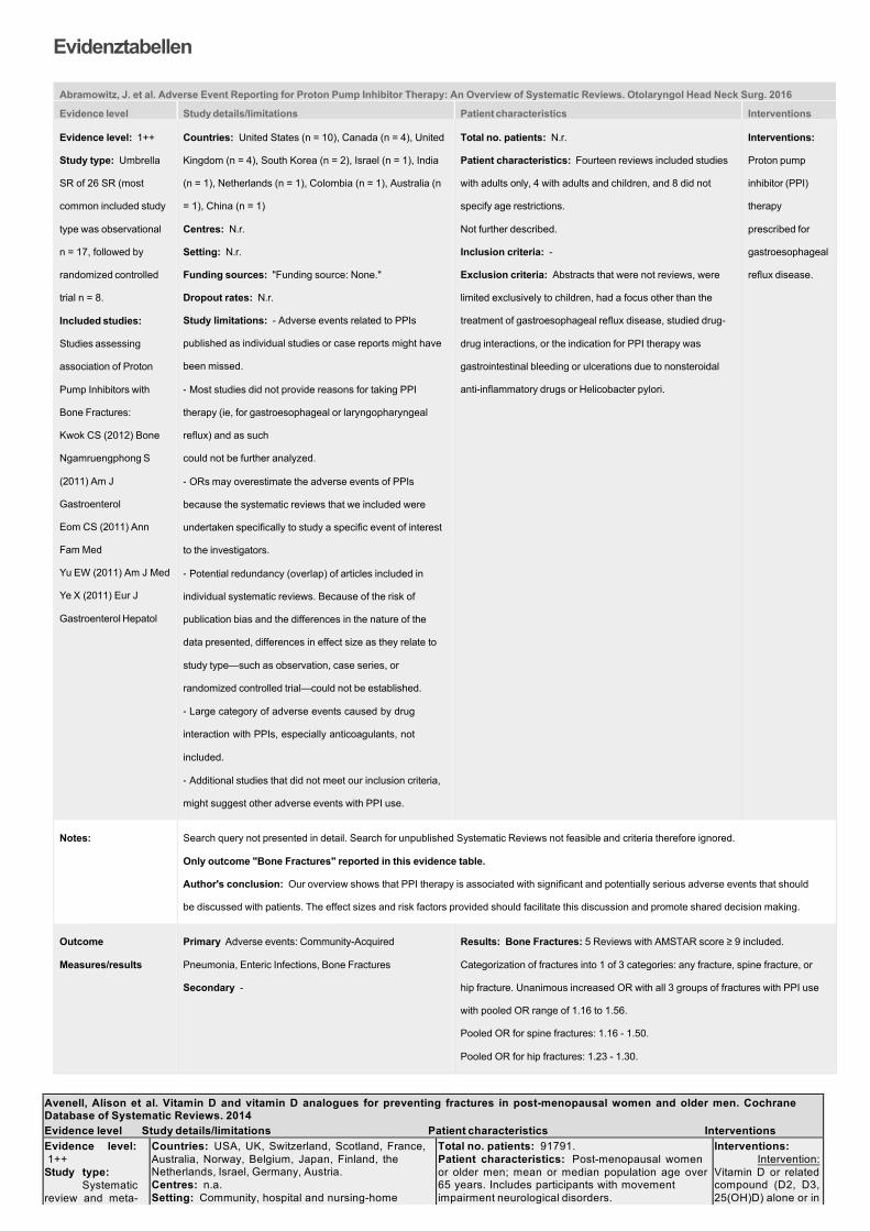
Abramowitz, J. et al. Adverse Event Reporting for Proton Pump Inhibitor Therapy: An Overview of Systematic Reviews. Otolaryngol Head Neck Surg. 2016
Evidence level Study details/limitations Patient characteristics Interventions Evidence level: 1++
Study type: Umbrella
SR of 26 SR (most
common included study
type was observational
n = 17, followed by
randomized controlled
trial n = 8.
Included studies:
Studies assessing
association of Proton
Pump Inhibitors with
Bone Fractures:
Kwok CS (2012) Bone
Ngamruengphong S
(2011) Am J
Gastroenterol Eom CS (2011) Ann
Fam Med
Yu EW (2011) Am J Med
Ye X (2011) Eur J
Gastroenterol Hepatol
Countries: United States (n = 10), Canada (n = 4), United
Kingdom (n = 4), South Korea (n = 2), Israel (n = 1), India
(n = 1), Netherlands (n = 1), Colombia (n = 1), Australia (n
= 1), China (n = 1) Centres: N.r.
Setting: N.r.
Funding sources: "Funding source: None."
Dropout rates: N.r.
Study limitations: - Adverse events related to PPIs
published as individual studies or case reports might have
been missed.
- Most studies did not provide reasons for taking PPI
therapy (ie, for gastroesophageal or laryngopharyngeal
reflux) and as such
could not be further analyzed. - ORs may overestimate the adverse events of PPIs
because the systematic reviews that we included were
undertaken specifically to study a specific event of interest
to the investigators.
- Potential redundancy (overlap) of articles included in
individual systematic reviews. Because of the risk of
publication bias and the differences in the nature of the
data presented, differences in effect size as they relate to
study type—such as observation, case series, or
randomized controlled trial—could not be established.
- Large category of adverse events caused by drug
interaction with PPIs, especially anticoagulants, not
included.
- Additional studies that did not meet our inclusion criteria,
might suggest other adverse events with PPI use.
Total no. patients: N.r.
Patient characteristics: Fourteen reviews included studies
with adults only, 4 with adults and children, and 8 did not
specify age restrictions.
Not further described. Inclusion criteria: -
Exclusion criteria: Abstracts that were not reviews, were
limited exclusively to children, had a focus other than the
treatment of gastroesophageal reflux disease, studied drug-
drug interactions, or the indication for PPI therapy was
gastrointestinal bleeding or ulcerations due to nonsteroidal
anti-inflammatory drugs or Helicobacter pylori.
Interventions:
Proton pump
inhibitor (PPI)
therapy
prescribed for
gastroesophageal
reflux disease.
Notes:
Search query not presented in detail. Search for unpublished Systematic Reviews not feasible and criteria therefore ignored.
Only outcome "Bone Fractures" reported in this evidence table.
Author's conclusion: Our overview shows that PPI therapy is associated with significant and potentially serious adverse events that should
be discussed with patients. The effect sizes and risk factors provided should facilitate this discussion and promote shared decision making.
Outcome
Measures/results
Primary Adverse events: Community-Acquired
Pneumonia, Enteric Infections, Bone Fractures
Secondary -
Results: Bone Fractures: 5 Reviews with AMSTAR score ≥ 9 included.
Categorization of fractures into 1 of 3 categories: any fracture, spine fracture, or
hip fracture. Unanimous increased OR with all 3 groups of fractures with PPI use
with pooled OR range of 1.16 to 1.56.
Pooled OR for spine fractures: 1.16 - 1.50.
Pooled OR for hip fractures: 1.23 - 1.30.
Avenell, Alison et al. Vitamin D and vitamin D analogues for preventing fractures in post-menopausal women and older men. Cochrane Database of Systematic Reviews. 2014 Evidence level Study details/limitations Patient characteristics Interventions Evidence level: 1++
Study type: Systematic
review and meta-
Countries: USA, UK, Switzerland, Scotland, France, Australia, Norway, Belgium, Japan, Finland, the Netherlands, Israel, Germany, Austria. Centres: n.a. Setting: Community, hospital and nursing-home
Total no. patients: 91791. Patient characteristics: Post-menopausal women or older men; mean or median population age over 65 years. Includes participants with movement impairment neurological disorders.
Interventions: Intervention:
Vitamin D or related compound (D2, D3, 25(OH)D) alone or in

analysis: 52 studies in the meta-analysis (RCT and quasi- RCT). Included studies:
Aloia JF, et al. (1988) Am J Med Arthur RS, et al. (1990) Mineral Electrol Metab Avenell A, et al. (2004) Clin Trials J Bischoff HA, et al. (2003) J Bone Minerl Res Bolton-Smith C, et al. (2007) J Bone Minerl Res Burleigh E, et al. (2007) Age Ageing Caniggia A, et al. (1984) Acta Vitaminologica et Enzymologica Chapuy MC, et al. (1992) N Engl J Med Chapuy MC, et al. (2002) Osteoporosis Int Dawson-Hughes B, et al. (1997) N Engl J Med Dukas L, et al. (2004) J Am Geriatr Soc Ebeling PR, et al. (2001) J Clin Endocr Metab Falch JA, et al. (1987) Acta Med Scand Flicker L, et al. (2005) J Am Geriatr Soc Gallagher JC, et al. (1989) P Soc Exp Biol Med Gallagher JC & Goldgar D. (1990) Ann Inter Med Gallagher JC, et al. (2001) J Clin Endocr Metab Garay Lillo J, et a. (1997) Geriátrika Geusens P & Dequeker J. (1986) Bone and Mineral Glendenning P, et al. (2012) J Bone Miner Res Gorai I, et al. (1999) Calcified Tissue International Harwood RH, et al. (2004) Age Ageing Hayashi Y, et al. (1992) J Bone Miner Res Ishida Y & Kawai S. (2004) Am J Med Janssen HC, et al. (2010) Aging Clinical and Experimental Research Komulainen MH, et al. (1998) Maturitas Law M, et al. (2006) Age Ageing Lips P, et al. (1996) Ann Intern Med Lyons RA, et al. (2007)
settings. Funding sources: We acknowledge the internal financial support for earlier versions of the review from: University of Aberdeen, UK; University of Edinburgh, UK; University of Hull, UK; University of Newcastle, Australia; University of Otago and Healthcare Otago Endowment Trust, New Zealand; University of York, UK. We received external financial support for earlier versions from: Australian Institute of Health and Welfare; Health Research Council of New Zealand, Medical Research Council, UK. Dropout rates: n.a. Study limitations: Incomplete information was available to us on the number of drop outs from intervention and control groups in a number of trials. Thus, it is possible that our analyses, based on the principles of intention-to-treat, might have under- estimated the number of outcome events in the intervention or control groups, or both.
Inclusion criteria: Study type: Any randomised trial or quasi-randomised trials. Participants: Post-menopausal women or older men (mean or median population age over 65 years), or both. Participants with neurological diseases impairing mobility were included. Intervention: Vitamin D or related compound, alone or in combination with calcium supplementation. Comparison: Placebo, no intervention, or the administration of calcium supplements. Outcomes: Primary: 1. Hip fracture; Secondary: 1. Any non-vertebral fracture. 2. Vertebral fracture. 3. Any new fracture. 4. Adverse effects. Exclusion criteria: Studies focused on participants on corticosteroid therapy; studies where vitamin D was given to patients selected on the basis of renal failure; interventions incorporating treatments other than vitamin D and calcium were not considered, e.g. vitamin D and hormone replacement therapy (HRT) compared with HRT alone. Interventions examining eldecalcitol (ED-71, 1alpha, 25-dihydroxy-2beta- (3-hydroxypropoxy) vitamin D3) were also not included. In defining a comparison, advice only on dietary modification to increase calcium intake was not considered as supplementation.
combination with calcium supplementation. Comparison: Placebo, no intervention, or the administration of calcium supplements.

Osteoporosis Int Menczel J, et al. (1994) Clinical Orthop Relat R Mitri J, et al. (2011) Am J Clin Nutr Nakatsuka K, et al. (1997) Nippon Ronen Igakkai Zasshi - Japanese Journal of Geriatrics Ones K, et al. (2007) Internet Journal of Epidemiology Orimo H, et al. (1994) Calcified Tissue Int Salovaara K, et al. (2010) J Bone Miner Res Ott SM & Chesnut CH. (1989) Ann Intern Med Peacock M, et al. (2000) J Clin Endocr Metab Pfeifer M, et al. (2000) J Bone Miner Res Pfeifer M, et al. (2004) J Bone Miner Res Porthouse J, et al. (2005) BMJ Prince RL, et al. (2008) Arch Intern Med Zhu K, et al. (2008) J Bone Miner Res Zhu K, et al. (2008) J Clin Endocr Metab Grant AM, et al. (2005) Lancet Shiraki M, et al. (1996) Endocrine Journal Smith H, et al. (2007) Rheumatology Tilyard MW, et al. (1992) N Engl J Med Trivedi DP, et al. (2003) BMJ Ushiroyama T, et al. (2001) Maturitas Sanders KM, et al. (2010) JAMA Jackson RD, et al. (2006) N Engl J Med Witham MD, et al. (2010) Circulation: Heart Failure Witham MD, et al. (2013) JAMA Intern Med Notes: Only two items on the Cochrane risk of bias tool were assessed (for selection bias) instead of the usual eight. The reason is likely
that this article is an update and the original versions predates the Rob tool by Higgins 2011 and the articles were not graded once again. Additional tests for publication bias such as Egger, Beggs test could have been performed. Author's conclusion: Vitamin D alone, in the doses and formulations that have been used, appears unlikely to be effective in fracture prevention in older people. Vitamin D plus calcium can help prevent hip fracture or any type of fracture. The benefits need to be balanced against the risk of kidney stones, kidney disease, gastrointestinal disease or heart disease. Vitamin D and calcium together are not associated with an increased risk of dying. Alphacalcidol may protect against vertebral fractures. Calcitriol appears to be associated with an increased incidence of adverse effects such as hypercalcaemia.
Outcome Measures/results
Primary Hip fracture Secondary Any non-vertebral fracture: Non-vertebral fractures were defined as all fractures except those of the vertebrae, but including hip fractures. 2. Vertebral fracture: (two outcomes were sought: clinical fracture events, and new vertebral deformity identified by radiological morphometry or semi- quantitative reading by a radiologist, using routine radiographs, according to a defined experimental
Results: Included studies: We included 53 trials with a total of 91,791 participants. 31 trials examined vitamin D (including 25-hydroxy vitamin D) with or without calcium in the prevention of fractures. 22 smaller trials examined calcitriol or alfacalcidol (1-alphahydroxyvitamin D3), mostly with participants who had established osteoporosis. Vitamin D only: There is high quality evidence that vitamin D alone, in the formats and doses tested, is unlikely to be effective in preventing hip fracture (11 trials, 27,693 participants; risk ratio (RR) 1.12, 95% confidence

protocol. Either of these methods appear to provide a valid approach to defining vertebral deformity) 3. Any new fracture: In previous versions of the review, the category ’any new fracture’ was classified as fractures not covered by hip, vertebral or non-vertebral categories, or where the site of fracture was unclear. This meant that some of the very large community trials were not analysed together, because they chose to report ’non-vertebral fractures’ or ’hip fractures’ or ’vertebral fractures’ but not ’all fractures’ and these numbers were not available or could not be calculated from the data without risk of double counting. As a new feature in this version of the review for Analyses 1 to 4 (comparisons involving vitamin D), the category ’all fractures’ includes data from non-vertebral fractures (or hip or vertebral fractures if not given), if the data for ’all fractures’ are not available (see Differences between protocol and review). 4. Adverse effects: (hypercalcaemia, renal disease, gastrointestinal symptoms, all as defined by the investigators; death).
intervals (CI) 0.98 to 1.29) or any new fracture (15 trials, 28,271 participants; RR 1.03, 95% CI 0.96 to 1.11). Vitamin D +Calcium: Hip fracture risk: There is high quality evidence that vitamin D plus calcium results in a small reduction in hip fracture risk (nine trials, 49,853 participants; RR 0.84, 95% confidence interval (CI) 0.74 to 0.96; P 0.01). In low-risk populations (residents in the community: with an estimated eight hip fractures per 1000 per year), this equates to one fewer hip fracture per 1000 older adults per year (95% CI 0 to 2). In high risk populations (residents in institutions: with an estimated 54 hip fractures per 1000 per year), this equates to nine fewer hip fractures per 1000 older adults per year (95% CI 2 to 14). Non vertebral fractures: There is high quality evidence that vitamin D plus calcium is associated with a statistically significant reduction in incidence of new non-vertebral fractures. However, there is only moderate quality evidence of an absence of a statistically significant preventive effect on clinical vertebral fractures. Any fracture: There is high quality evidence that vitamin D plus calcium reduces the risk of any type of fracture (10 trials, 49,976 participants; RR 0.95, 95% CI 0.90 to 0.99). Adverse effects: mortality was not adversely affected by either vitamin D or vitamin D plus calcium supplementation (29 trials, 71,032 participants, RR 0.97, 95% CI 0.93 to 1.01). Hypercalcaemia, which was usually mild (2.6 to 2.8 mmol/L), was more common in people receiving vitamin D or an analogue, with or without calcium (21 trials, 17,124 participants, RR 2.28, 95% CI 1.57 to 3.31), especially for calcitriol (four trials, 988 participants, RR 4.41, 95% CI 2.14 to 9.09), than in people receiving placebo or control. There was also a small increased risk of gastrointestinal symptoms (15 trials, 47,761 participants, RR 1.04, 95% CI 1.00 to 1.08), especially for calcium plus vitamin D (four trials, 40,524 participants, RR 1.05, 95% CI 1.01 to 1.09), and a significant increase in renal disease (11 trials, 46,548 participants, RR 1.16, 95% CI 1.02 to 1.33).
Bang, C. S. et al. Osteoporosis and bone fractures in alcoholic liver disease: a meta-analysis. World J Gastroenterol. 21. 4038-47. 2015
Evidence level Study details/limitations Patient characteristics Interventions Evidence level:
2-
Study type: MA
of 15 studies (10
cohort studies, 5
case-control
studies)
Included studies:
Lindsell DR
(1982) Br Med J
Diamond T (1990)
Gut
Bonkovsky HL
(1990) Hepatology
Ninkovic M (2000)
Eur J
Gastroenterol
Hepatol
Ninkovic M (2001)
Calcif Tissue Int
Carey EJ (2003)
Liver Transpl
Kim MJ (2003) Korean J Intern
Med
Sokhi RP (2004)
Liver Transpl
Alvisa-Negrín J
(2009) Alcohol
Alcohol
González-Reimers
Countries: Europe (n = 8), the United States (n = 3), Canada
(n = 1), Australia (n = 1), India (n = 1), and Korea (n = 1)
Centres: N.r.
Setting: N.r. Funding sources: N.r.
Dropout rates: N.r.
Study limitations: - Substantial methodological
heterogeneity between the enrolled studies.
- Quality of enrolled studies. Included studies classified as
high-quality (≥ 7 stars) or low-quality (< 7 stars). Average
number of stars awarded in 15 studies was 6.4. 8 studies with
zero stars in section of comparability.
- A significant outlier observed. In analysis of association
between ALD and bone fractures, effect size of outlier (RR =
33.725) was more than 10 times the adjusted effect size (RR =
1.944). Reason for large effect size of outlier is postulated as
methodological problem. In this study, presence of fracture
was recorded by anamnesis and chest X-ray film and could
overestimate rate of bone fractures. NOS Quality low (6 stars)
in this study.
- No consideration given to any potential confounders, which
could be influential to bone fractures and osteoporosis.
- Newcastle-Ottawa scale (NOS) used to assess the
methodological quality of studies. Scale has been criticized for
low agreement between authors and reviewers.
Total no. patients: 726 participants in analysis of bone fractures,
470 participants in analysis of osteoporosis, and 769 participants
in analysis of BMD.
Patient characteristics: 4 studies had ALD groups that
consisted only of cirrhotic patients, 2 studies included no alcoholic
cirrhotic patients, and in 5 studies, the presence or absence of
cirrhosis was not specified.
Men and women with a mean age ranging from 47 ± 1.1 - 65.2 ±
11 years.
Inclusion criteria: Case-control or cohort studies that (1) were designed to evaluate ALD in the target or control group;
and
(2) included at least one outcome (prevalence of any bone
fractures, prevalence of osteoporosis, or BMD) that enabled
comparisons of osteodystrophy between patients with ALD and
the control group.
Exclusion criteria: (1) Incomplete data; (2) review article; or
(3) abstract only (study not published as full-text article).
Interventions:
-

E (2011) Alcohol
Mitchell R (2011)
World J Hepatol
Mahmoudi A
(2011) Clin Res
Hepatol
Gastroenterol
Wibaux C (2011)
Joint Bone Spine
Choudhary NS
(2011) Dig Dis Sci
González-Reimers
E (2011) Alcohol
Alcohol
Notes: Search query not stated. No search for grey/ unpublished literature. Unclear, if 2 people extracted data. The authors provide no declaration of
conflicts of interest. Unclear, if Newcastle-Ottawa Scale (for Case-Control and Cohort studies) were applied correctly.
Unclear, which requirements for diagnosis of alcoholic liver disease (ALD) were implemented in selection of studies. Author's conclusion: In conclusion, current publications indicate a significant association between bone fractures and ALD, independent of BMD or
the presence of osteoporosis. Due to the qualitative and quantitative heterogeneity among studies, further studies utilizing homogenous populations
and controlling for confounding risk factors for fractures are needed to elucidate the underlying mechanism of bone fractures in ALD. Outcome
Measures/results
Primary Relationship between ALD and bone fractures or
osteoporosis using adjusted risk ratios (RRs) and
standardized mean difference (SMD).
Prevalence of any fractures or osteoporosis assessed by
radiologic examinations and BMD assessed by dual-energy X-
ray absorptiometry (DXA) or dual-photon absorptiometry
(DPA). Osteoporosis defined as a value for BMD: 2.5 standard
deviations or more below the reference range.
Secondary -
Results: Association between ALD and bone fractures or osteoporosis: 6 studies
included. ALD showed significant RR of 1.944 (95% CI; 1.354, 2.791, P < 0.001) for
development of bone fractures. ALD showed not significant RR of 0.849 (95% CI;
0.523, 1.380, P = 0.509) for development of osteoporosis. BMD not significantly
different between ALD and control groups (SMD in femoral neck BMD: -0.172, 95%
CI; -0.453, 0.110, P = 0.233; SMD in spine BMD: -0.169, 95% CI; -0.476, 0.138, P =
0.282).
Beaudoin, C. et al. Denosumab compared to other treatments to prevent or treat osteoporosis in individuals at risk of fracture: a systematic review and meta-analysis. Osteoporos Int. . . 2016 Evidence level Study details/limitations Patient characteristics Interventions
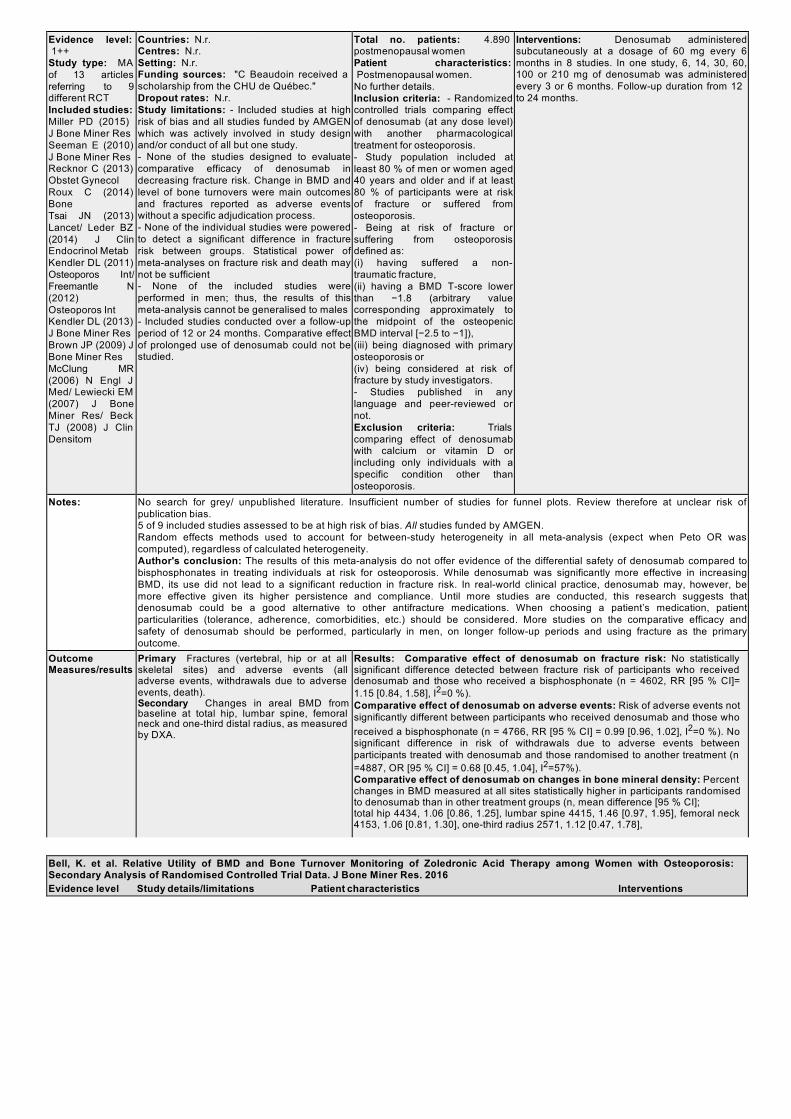
Evidence level: 1++
Study type: MA of 13 articles referring to 9 different RCT Included studies: Miller PD (2015) J Bone Miner Res Seeman E (2010) J Bone Miner Res Recknor C (2013) Obstet Gynecol Roux C (2014) Bone Tsai JN (2013) Lancet/ Leder BZ (2014) J Clin Endocrinol Metab Kendler DL (2011) Osteoporos Int/ Freemantle N (2012) Osteoporos Int Kendler DL (2013) J Bone Miner Res Brown JP (2009) J Bone Miner Res McClung MR (2006) N Engl J Med/ Lewiecki EM (2007) J Bone Miner Res/ Beck TJ (2008) J Clin Densitom
Countries: N.r. Centres: N.r. Setting: N.r. Funding sources: "C Beaudoin received a scholarship from the CHU de Québec." Dropout rates: N.r. Study limitations: - Included studies at high risk of bias and all studies funded by AMGEN which was actively involved in study design and/or conduct of all but one study. - None of the studies designed to evaluate comparative efficacy of denosumab in decreasing fracture risk. Change in BMD and level of bone turnovers were main outcomes and fractures reported as adverse events without a specific adjudication process. - None of the individual studies were powered to detect a significant difference in fracture risk between groups. Statistical power of meta-analyses on fracture risk and death may not be sufficient - None of the included studies were performed in men; thus, the results of this meta-analysis cannot be generalised to males - Included studies conducted over a follow-up period of 12 or 24 months. Comparative effect of prolonged use of denosumab could not be studied.
Total no. patients: 4.890 postmenopausal women Patient characteristics: Postmenopausal women.
No further details. Inclusion criteria: - Randomized controlled trials comparing effect of denosumab (at any dose level) with another pharmacological treatment for osteoporosis. - Study population included at least 80 % of men or women aged 40 years and older and if at least 80 % of participants were at risk of fracture or suffered from osteoporosis. - Being at risk of fracture or suffering from osteoporosis defined as: (i) having suffered a non- traumatic fracture, (ii) having a BMD T-score lower than −1.8 (arbitrary value corresponding approximately to the midpoint of the osteopenic BMD interval [−2.5 to −1]), (iii) being diagnosed with primary osteoporosis or (iv) being considered at risk of fracture by study investigators. - Studies published in any language and peer-reviewed or not. Exclusion criteria: Trials comparing effect of denosumab with calcium or vitamin D or including only individuals with a specific condition other than osteoporosis.
Interventions: Denosumab administered subcutaneously at a dosage of 60 mg every 6 months in 8 studies. In one study, 6, 14, 30, 60, 100 or 210 mg of denosumab was administered every 3 or 6 months. Follow-up duration from 12 to 24 months.
Notes: No search for grey/ unpublished literature. Insufficient number of studies for funnel plots. Review therefore at unclear risk of publication bias. 5 of 9 included studies assessed to be at high risk of bias. All studies funded by AMGEN. Random effects methods used to account for between-study heterogeneity in all meta-analysis (expect when Peto OR was computed), regardless of calculated heterogeneity. Author's conclusion: The results of this meta-analysis do not offer evidence of the differential safety of denosumab compared to bisphosphonates in treating individuals at risk for osteoporosis. While denosumab was significantly more effective in increasing BMD, its use did not lead to a significant reduction in fracture risk. In real-world clinical practice, denosumab may, however, be more effective given its higher persistence and compliance. Until more studies are conducted, this research suggests that denosumab could be a good alternative to other antifracture medications. When choosing a patient’s medication, patient particularities (tolerance, adherence, comorbidities, etc.) should be considered. More studies on the comparative efficacy and safety of denosumab should be performed, particularly in men, on longer follow-up periods and using fracture as the primary outcome.
Outcome Measures/results
Primary Fractures (vertebral, hip or at all skeletal sites) and adverse events (all adverse events, withdrawals due to adverse events, death). Secondary Changes in areal BMD from baseline at total hip, lumbar spine, femoral neck and one-third distal radius, as measured by DXA.
Results: Comparative effect of denosumab on fracture risk: No statistically significant difference detected between fracture risk of participants who received denosumab and those who received a bisphosphonate (n = 4602, RR [95 % CI]= 1.15 [0.84, 1.58], I2=0 %). Comparative effect of denosumab on adverse events: Risk of adverse events not significantly different between participants who received denosumab and those who received a bisphosphonate (n = 4766, RR [95 % CI] = 0.99 [0.96, 1.02], I2=0 %). No significant difference in risk of withdrawals due to adverse events between participants treated with denosumab and those randomised to another treatment (n =4887, OR [95 % CI] = 0.68 [0.45, 1.04], I2=57%). Comparative effect of denosumab on changes in bone mineral density: Percent changes in BMD measured at all sites statistically higher in participants randomised to denosumab than in other treatment groups (n, mean difference [95 % CI]; total hip 4434, 1.06 [0.86, 1.25], lumbar spine 4415, 1.46 [0.97, 1.95], femoral neck 4153, 1.06 [0.81, 1.30], one-third radius 2571, 1.12 [0.47, 1.78],
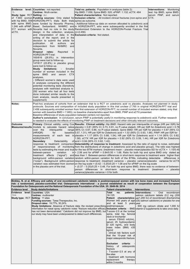
Bell, K. et al. Relative Utility of BMD and Bone Turnover Monitoring of Zoledronic Acid Therapy among Women with Osteoporosis: Secondary Analysis of Randomised Controlled Trial Data. J Bone Miner Res. 2016 Evidence level Study details/limitations Patient characteristics Interventions
Evidence level: 1-
Study type: RCT of 7.683 women with hip BMD, 558 with spine BMD, 1.246 with sPINP, 517 women with sCTX.
Countries: not reported. Centres: Multi-center. Setting: n.r. Funding sources: Only stated for HORIZON(-PFT) trials: Both trials were funded by Novartis Pharma, Basel, Switzerland. NHMRC and Novartis had no role in the study design; in the collection, analysis, and interpretation of data; in the writing of the report; and in the decision to submit the article for publication. All authors are independent from NHMRC and Novartis. Dropout rates: Reported in HORIZON-PFT trial: 165/616 (26.8%) in intervention group were lost to follow-up. 147/617 (23.8%) in placebo group were lost to follow-up. Study limitations: - Smaller number of women included in the spine BMD and serum CTX analyses, - Different women’s data were used in analyses comparing the different potential monitoring tests (Sensitivity analyses with restricted analysis to 292 women who had all four tests done indicated similar results to the main analysis, results presented in supplement)
Total no. patients: Population in analyzes (table 2): Hip BMD: 7.056; Spine BMD: 520; sPINP: 1.132; sCTX: 484 Patient characteristics: Not reported. Inclusion criteria: - All incident clinical fractures (non-spine and spine) as outcome. - Additional follow-up data on women allocated to zoledronic acid in HORIZON-PFT, who were subsequently enrolled in the first Randomized Extension to the HORIZON-Pivotal Fracture Trial (n=2.456) Exclusion criteria: Not stated.
Interventions: Monitoring test: hip BMD, spine BMD, serum PINP, and serum CTX.
Notes: Post-hoc analyses of cohorts from an extension trial to a RCT on zoledronic acid vs placebo. Analyses not planned in study protocols. Sources and composition of included study population in this trial unclear (7.765 in original HORIZON-PFT trial and 2.456 subsequently enrolled women from first extension of HORIZON-PFT; no overall number of included women stated, only that women with baseline BMD = 7.683); no baseline characteristics apart from BMD, PINP and sCTX presented. Baseline differences of study population between centers not reported. Author's conclusion: In conclusion, serum PINP is potentially useful in monitoring response to zoledronic acid. Further research is needed to evaluate the effects of monitoring PINP on treatment decisions and other clinically relevant outcomes.
Outcome Measures/results
Primary Clinical validity: using Cox models to calculate hazard ratios over the interquartile range (HR/IQR) for baseline measurements of each test in HORIZON-PFT. Secondary Detectability of response to treatment: comparison of responsiveness of monitoring tests by estimating the mean and true between-person variation in treatment effects (“signal”) and background within-person variation (“noise”). Background within-person variation was estimated from women in the placebo arm of the trial.
Results: Clinical validity: Hip BMD: Hazard ratio per interquartile of range (HR per IQR) for placebo = 0.81 (95% CI; 0.73, 0.91, no P-value stated). HR per IQR for Zoledronic acid = 0.76 (95% CI; 0.67, 0.86, no P-value stated). Spine BMD: HR per IQR for placebo = 0.87 (95% CI; 0.57, 1.31). HR per IQR for Zoledronic acid = 1.02 (95% CI; 0.63, 1.66). PINP: HR per IQR for placebo = 1.17 (95% CI; 0.88, 1.54). HR per IQR for Zoledronic acid = 1.14 (95% CI; 0.82, 1.58). sCTX: HR per IQR for placebo = 0.92 (95% CI; 0.59, 1.44). HR per IQR for Zoledronic acid = 0.94 (95% CI; 0.62, 1.42). Detectability of response to treatment: Assessed by the ratio of signal to noise, estimated from the distributions of change in zoledronic acid and placebo groups. The ratio was highest for the two BTM markers: (treatment mean – placebo mean)/placebo-SD for sCTX = 1.15/0.45 = 2.56 and for sPINP = 0.84/0.50 = 1.68. Ratio for both hip and spine BMD only stated as less than 1. Between-person differences in short-term response to treatment were higher than random within-person variation for both of the BTMs, indicating detectable differences in response to treatment: (treatment variance – placebo variance)/placebo variance for sCTX on the log scale = (0.42 – 0.20)/0.20 = 1.08 and for sPINP on the log scale = (0.37 – 0.25)/0.25 = 0.48. For both hip and spine BMD, there was no evidence of between- person differences in short-term response to treatment: (treatment – placebo variance)/placebo variance = 0 for both.
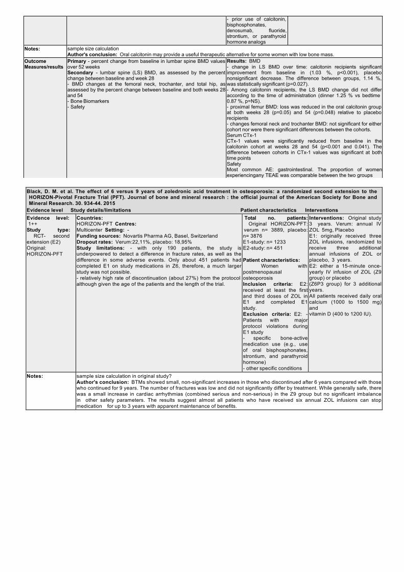
Binkley, N. et al. Efficacy and safety of oral recombinant calcitonin tablets in postmenopausal women with low bone mass and increased fracture risk: a randomized, placebo-controlled trial. Osteoporosis international : a journal established as result of cooperation between the European Foundation for Osteoporosis and the National Osteoporosis Foundation of the USA. 25. 2649-56. 2014 Evidence level Study details/limitations Patient characteristics Interventions Evidence level: 1++
Study type: RCT
Countries: USA Centres: multicenter Setting: 11 sites Funding sources: Tarsa Therapeutics, Inc. Dropout rates: 19,77%, 30,24% Study limitations: Absence of fracture data; the revised prescribing information for nasal spray calcitonin notes “fracture reduction efficacy has not been demonstrated.” Calcitonin did not improve hip BMD, but our study may have been underpowered to detect such differences.
Total no. patients: N=125
Patient characteristics: Women ≥45 years of age and at least 5 years postmenopause Inclusion criteria: - BMD T-score−2.5 at the lumbar spine, total hip, femoral neck or trochanter - serum FSH≥0 mIU/mL, serum 25 hydroxyvitamin D ≥20 ng/mL, and body mass index (BMI) ≤35 kg/m2 - clinical risk factors such that the 10-year risk of major osteoporotic fracture Exclusion criteria: - history of osteoporotic fracture - T-score≤−2.5 at any of the sites - treatment with hormone replacement therapy within 3 months
Interventions: - Oral recombinant salmon calcitonin tablets (0.2 mg (200 μg/1,200 IU) of recombinant (Escherichia coli) salmon calcitonin) or placebo for one year - 600 mg calcium citrate and 1,000 IU vitamin D supplements to take once daily
- prior use of calcitonin, bisphosphonates, denosumab, fluoride, strontium, or parathyroid hormone analogs
Notes: sample size calculation Author's conclusion: Oral calcitonin may provide a useful therapeutic alternative for some women with low bone mass.
Outcome Measures/results
Primary - percent change from baseline in lumbar spine BMD values over 52 weeks Secondary - lumbar spine (LS) BMD, as assessed by the percent change between baseline and week 28 - BMD changes at the femoral neck, trochanter, and total hip, as assessed by the percent change between baseline and both weeks 28 and 54 - Bone Biomarkers - Safety
Results: BMD - change in LS BMD over time: calcitonin recipients significant improvement from baseline in (1.03 %, p<0.001), placebo nonsignificant decrease. The difference between groups, 1.14 %, was statistically significant (p=0.027). - Among calcitonin recipients, the LS BMD change did not differ according to the time of administration (dinner 1.25 % vs bedtime 0.87 %, p=NS). - proximal femur BMD: loss was reduced in the oral calcitonin group at both weeks 28 (p=0.05) and 54 (p=0.048) relative to placebo recipients - changes femoral neck and trochanter BMD: not significant for either cohort nor were there significant differences between the cohorts. Serum CTx-1 CTx-1 values were significantly reduced from baseline in the calcitonin cohort at weeks 28 and 54 (p<0.001 and 0.041). The difference between cohorts in CTx-1 values was significant at both time points Safety Most common AE: gastrointestinal. The proportion of women experiencingany TEAE was comparable between the two groups
Black, D. M. et al. The effect of 6 versus 9 years of zoledronic acid treatment in osteoporosis: a randomized second extension to the HORIZON-Pivotal Fracture Trial (PFT). Journal of bone and mineral research : the official journal of the American Society for Bone and Mineral Research. 30. 934-44. 2015
Evidence level Study details/limitations Patient characteristics Interventions Evidence level: 1++
Study type: RCT- second
extension (E2) Original: HORIZON-PFT
Countries: HORIZON-PFT Centres: Multicenter Setting: - Funding sources: Novartis Pharma AG, Basel, Switzerland Dropout rates: Verum:22,11%, placebo: 18,95% Study limitations: - with only 190 patients, the study is underpowered to detect a difference in fracture rates, as well as the difference in some adverse events. Only about 451 patients had completed E1 on study medications in Z6, therefore, a much larger study was not possible. - relatively high rate of discontinuation (about 27%) from the protocol although given the age of the patients and the length of the trial.
Total no. patients: Original HORIZON-PFT:
verum n= 3889, placebo: n= 3876 E1-study: n= 1233 E2-study: n= 451 Patient characteristics:
Women with postmenopausal osteoporosis Inclusion criteria: E2: received at least the first and third doses of ZOL in E1 and completed E1 study. Exclusion criteria: E2: - Patients with major protocol violations during E1 study - specific bone-active medication use (e.g., use of oral bisphosphonates, strontium, and parathyroid hormone) - other specific conditions
Interventions: Original study 3 years. Verum: annual IV ZOL 5mg, Placebo E1: originally received three ZOL infusions, randomized to receive three additional annual infusions of ZOL or placebo, 3 years. E2: either a 15-minute once- yearly IV infusion of ZOL (Z9 group) or placebo (Z6P3 group) for 3 additional years. All patients received daily oral calcium (1000 to 1500 mg) and vitamin D (400 to 1200 IU).
Notes: sample size calculation in original study? Author's conclusion: BTMs showed small, non-significant increases in those who discontinued after 6 years compared with those who continued for 9 years. The number of fractures was low and did not significantly differ by treatment. While generally safe, there was a small increase in cardiac arrhythmias (combined serious and non-serious) in the Z9 group but no significant imbalance in other safety parameters. The results suggest almost all patients who have received six annual ZOL infusions can stop medication for up to 3 years with apparent maintenance of benefits.

Outcome Measures/results
Primary Percentage change in total hip BMD at year 9 relative to year 6 in Z9 compared with Z6P3. Secondary - Change in hip BMD (total and femoral neck) at years 7 and 8 vs. year 6 and at years 7, 8, and 9 vs. year 0. - Incidence of fractures (morphometric vertebral and clinical fractures) at year 9 relative to year 6. - change in BTMs (bone tumor marker) at years 7, 8, and 9 relative to year 6. - Safety
Results: Bone mineral density - mean change from year 6 to 9 in total hip BMD was 0.54% in Z9 compared with 1.31% in Z6P3 producing a mean between-group difference of 0.78% (95% confidence interval [CI]: 0.37%, 1.93%; p=0.183). - Compared to the core study baseline (year 0) to year 9, there was no significant difference between the treatments in total hip BMD changes. Bone turnover markers - mean serum levels of PINP and the other BTMs remained within the premenopausal reference range in both the groups. - For b-CTX, there was no evidence of a difference by treatment. - For BSAP, a statistically significant difference was observed between groups at year 9. - Within the Z9, the mean BTM values increased from year 1. The within-group increase was significant for both PINP (p=0.004) and b-CTX (p=0.002). Fractures - Fractures were too few for meaningful comparison. - no significant difference in the risk of all clinical fractures. Height No significant differences in change between groups at year 7, 8, 9 vs year 6. Safety Overall incidences of AEs and SAEs were similar in the two treatment groups.
Bolam, K. A. et al. The effect of physical exercise on bone density in middle-aged and older men: a systematic review. Osteoporos Int. 24. 2749-62. 2013 Evidence level Study details/limitations Patient characteristics Interventions Evidence level: 2++
Study type: SR of 7 RCT and 2 CT Included studies: Braith RW (1996) J Am Coll Cardiol Huuskonen J (2001) Osteoporos Int Kukuljan S (2009) Osteoporos Int Kukuljan S (2011) J Clin Endocrinol Metab Menkes A (1993) J Appl Physiol Paillard T (2004) Int J Sports Med Ryan AS (1994) J Appl Physiol Whiteford J (2010) Osteoporos Int Woo J (2007) Age Ageing
Countries: 3 USA, 1 Finland, 3 Australia, 1 France, 1 China Centres: Not reported. Setting: Not reported. Funding sources: Not stated. Dropout rates: Of the trials that did report dropout, the average rate was 3.3 %. For the four studies who reported dropout by group, the overall dropout rates were 6.8 and 2.1 % for the exercise and control groups, respectively. Study limitations: - Analysis includes only data from published studies and the possibility exists of missing relevant unpublished trials. - Relatively small number of participants may limit ability to detect a statistically significant difference between intervention and control groups, and this should be considered when interpreting the results of these studies. As a result, it is strongly suggested that statistical power calculations be included in reports of future intervention studies. - Many of the studies included in this review did not report the post-intervention scores, or post-intervention data were not available. In addition to this, due to the heterogeneous nature of the included exercise protocols and the variation in BMD measurement sites, a meta-analysis was not performed. - Using BMD as measured by DXA is a further limitation of this review due to concerns regarding the inherent inaccuracies of this method of measurement and its inability to provide information regarding important determinants of bone strength (size, shape and structure). Consequently, there is growing interest in using quantitative computed tomography (QCT) to assess bone strength, and researchers should aim to use this method to assess the effects of exercise interventions on whole bone strength where possible.
Total no. patients: 559 men Patient characteristics: 1 trial with middle aged
heart transplant patients on glucocorticoid treatment (Mean Age±SD: 56.0±6.0 and 52.0±10). Remaining studies with men aged from 53-62 years (range) to 65-74 years (range) from nonclinical populations. Inclusion criteria: 1) design: randomised controlled trials (RCT) or controlled trials (CT); (2) population: middle- aged or older men (45 years and older). Studies in middle-aged and older men and studies including men and women in which results for men and women were reported separately were eligible for inclusion; (3) intervention: any exercise protocol involving resistance training only, impact loading exercise only, weight-bearing aerobic exercise only or a combination of these types of exercise; and (4) outcome: BMD (g/cm2) of the lumbar spine, Ward's triangle, trochanter, proximal femur, femoral neck or total hip measured by DXA. Only full-text articles were included, and no restrictions were placed on the language of the article. Exclusion criteria: -
Interventions: 8 exercise programmes: 2 walking only, 3 resistance training only, 1 walking and resistance training, 1 resistance training and impact loading activities and 1 resistance training and Tai Chi. Majority of programmes with 3 exercise sessions a week (ranging from 2–5 each week). Intensities of interventions varied greatly (all but 1 trials with resistance exercises used individualised intensities; percentage of 1-RM, where RM or repetition maximum is the maximum amount that can be moved or lifted one time only, 8-RM or 5– 15-RM and 1 trial who did not report intensity but instead the participants were supplied with a medium strength elastic band).
Notes: No search for grey literature possibly introducing publication bias. Author's conclusion: Results from this systematic review indicate that resistance training alone or in combination with impact- loading activities is safe and may assist in the prevention of osteoporosis in middle-aged and older men. However, due to the variation among studies as well as in study quality, additional high-quality randomised controlled trials in this population are required in order to establish evidence-based guidelines for the optimal exercise prescription. Nevertheless, for those individuals willing and able to perform physical exercise, regular resistance training and impact-loading activities should be considered as an effective strategy to prevent osteoporosis in middle-aged and older men.
Outcome Measures/results
Primary Not stated. Reported findings: effects of interventions on BMD of the lumbar spine, femoral neck BMD, total hip BMD trochanteric BMD, both Ward's triangle
Results: BMD: Effects of exercise varied greatly among studies, with six interventions having a positive effect on BMD and two interventions having no significant effect. It appears that resistance training alone

and proximal femur BMD. Secondary Not stated. Reported finding: Dropout, attendance and adverse events
or in combination with impact-loading activities are most osteogenic for this population, whereas the walking trials had limited effect on BMD.
Bolland, M. J. et al. Calcium intake and risk of fracture: systematic review. Bmj. 351. h4580. 2015 Evidence level Study details/limitations Patient characteristics Interventions
Evidence level: 1++ Study type: SR/MA of 18 RCT Included studies: Chevalley T (1994) Osteoporos Int Lau EM (2001) J Bone Miner Res Inkovaara J (1983) Age Ageing Hansson T (1987) Calcif Tissue Int Chapuy MC (1994) BMJ Reid IR (1995) Am J Med Recker RR (1996) J Bone Miner Res Dawson-Hughes B (1997) N Engl J Med Riggs BL (1998) Bone Miner Res Baron JA (1999) N Engl J Med Ruml LA (1999) Am J Ther Chapuy MC (2002) Osteoporos Int Avenell A (2004) Clin Trials Fujita T (2004) Bone Miner Metab Harwood RH (2004) Age Ageing Larsen ER (2004) J Bone Miner Res Grant AM (2005) Lancet Porthouse J (2005) BMJ Jackson RD (2006) N Engl J Med Prince RL (2006) Arch Intern Med Reid IR (2006) Am J Med Bolton-Smith C (2007) J Bone Miner Res (2007 Curr Med
Res Opin
Reid IR (2008) Arch Intern Med
Salovaara K
(2010) J Bone
Countries: N.r. Centres: N.r. Setting: Mostly community care setting, 5 studies with residential care setting. Funding sources: "The study was funded by the Health Research Council (HRC) of New Zealand. MJB is the recipient of a Sir Charles Hercus Health Research Fellowship. The authors are independent of the HRC. The HRC had no role in study design, the collection, analysis, and interpretation of data, the writing of the article, or the decision to submit it for publication." Dropout rates: N.r. Study limitations: - No meta-regression analyses performed because there were few studies that reported sufficient data for such an analysis. - Many of randomised controlled trials of short duration and did not have fracture as primary endpoint. - Trials generally carried out in healthy populations or those at risk of osteoporosis, findings might not apply to other population groups.
Total no. patients: n = 69.269 Patient characteristics: Analyses of dietary sources of calcium: 2 studies with mean age (SD) 57 (6) and 72 (6) years. 88% and 100% of participants female. Analyses of calcium supplements: 26 studies with mean age ≥70 years. Participants mostly female. Inclusion criteria: - Randomised controlled trials and cohort, case-control, or cross sectional studies with fracture as an outcome with participants aged >50 at baseline, or for cohort studies, where most follow-up occurred in participants aged >50. - Studies of calcium supplements used in combination with other treatment provided that the other treatment was given to both arms and studies of co-administered calcium and vitamin D supplements (CaD). Exclusion criteria: Studies where most participants had a major systemic pathology at baseline other than osteoporosis, such as renal failure or malignancy.
Interventions: Dietary sources of calcium and calcium supplements.
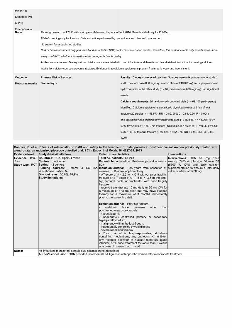
Miner Res
Sambrook PN
(2012)
Osteoporos Int Notes: Thorough search until 2013 with a simple update search query in Sept 2014. Search stated only for PubMed.
Ti/ab-Screening only by 1 author. Data extraction performed by one authors and checked by a second.
No search for unpublished studies. Risk of bias assessment only performed and reported for RCT, not for included cohort studies. Therefore, this evidence table only reports results from
analysis of RCT, all other information must be regarded as 2- quality.
Author's conclusion: Dietary calcium intake is not associated with risk of fracture, and there is no clinical trial evidence that increasing calcium
intake from dietary sources prevents fractures. Evidence that calcium supplements prevent fractures is weak and inconsistent.
Outcome
Measures/results
Primary Risk of fractures.
Secondary -
Results: Dietary sources of calcium: Sources were milk powder in one study (n
= 200, calcium dose 800 mg/day, vitamin D dose 240 IU/day) and a preparation of
hydroxyapatite in the other study (n = 62, calcium dose 800 mg/day). No significant
results.
Calcium supplements: 26 randomised controlled trials (n = 69.107 participants)
identified. Calcium supplements statistically significantly reduced risk of total
fracture (20 studies, n = 58.573; RR = 0.89, 95% CI; 0.81, 0.96; P = 0.004)
and statistically non-significantly vertebral fracture (12 studies, n = 48.967; RR =
0.86, 95% CI; 0.74, 1.00), hip fracture (13 studies, n = 56.648; RR = 0.95, 95% CI;
0.76, 1.18) or forearm fracture (8 studies, n = 51.775; RR = 0.96, 95% CI; 0.85, 1.09).
Bonnick, S. et al. Effects of odanacatib on BMD and safety in the treatment of osteoporosis in postmenopausal women previously treated with alendronate: a randomized placebo-controlled trial. J Clin Endocrinol Metab. 98. 4727-35. 2013 Evidence level Study details/limitations Patient characteristics Interventions Evidence level: 1++
Study type: RCT
Countries: USA, Spain, France Centres: multicenter Setting: 42 centers Funding sources: Merck & Co, Inc, Whitehouse Station, NJ Dropout rates: 30,6%, 18,9% Study limitations: -
Total no. patients: n= 243 Patient characteristics: Postmenopausal women ≥ 60 y Inclusion criteria: - ≥5 years from cessation of menses, or Bilateral oophorectomy - AT-score of ≤ - 2.5 to > -3.5 without prior fragility fracture or a T-score of ≤ - 1.5 to > -3.5 at the total hip, femoral neck, or trochanter with prior fragility fracture - received alendronate 10 mg daily or 70 mg OW for a minimum of 3 years prior, but may have stopped therapy for a maximum of 3 months immediately prior to the screening visit. Exclusion criteria: - Prior hip fracture - metabolic bone diseases other than postmenopausal osteoporosis - hypocalcaemia - inadequately controlled primary or secondary hyperparathyroidism, - malignancy within the last 5 years - inadequately controlled thyroid disease - severe renal insufficiency - Prior use of iv bisphosphonates, strontium- containing medications, any cathepsin K inhibitor, any receptor activator of nuclear factor-kB ligand inhibitor, or fluoride treatment for more than 2 weeks at a dose of greater than 1 mg/d
Interventions: ODN 50 mg once weekly (OW) or placebo. Vitamin D3 (5600 IU OW) and daily calcium supplementation to ensure a total daily calcium intake of 1200 mg.
Notes: no limitations mentioned, sample size calculation not described Author's conclusion: ODN provided incremental BMD gains in osteoporotic women after alendronate treatment.

Outcome Measures/results
Primary Percentage change from baseline in the femoral neck BMD compared with placebo at the end of 24 months Secondary - percentage change from baseline in BMD at the femoral neck, trochanter, total hip, lumbar spine, and one third radius at 12 and 24 months within each group - percentage change from baseline in biochemical markers of bone resorption [serum C-telopeptides of type 1 collagen (sCTx), urinary N-telopeptides of type 1 collagen (uNTx) to creatinine (Cr) ratio] - biochemical markers of bone formation [serum bone specific alkaline phosphatase (sBSAP), serum N-terminal propeptide of type 1 collagen (sP1NP)] - Percentage change from baseline in total body BMD - bone resorption marker urine total deoxypyridinoline (uDPyr) - target engagement marker serum cross- linked carboxyterminal telopeptides of type 1 collagen (s1CTP) - osteoclast number marker serum tartrate resistant acid phosphatase isoform (TRAP)-5b - indices of calcium and mineral homeostasis compared with placebo at the end of 12 and 24 months - Safety and tolerability
Results: BMD - Femoral neck BMD was significantly increased in the ODN group compared with placebo at month 24 (P < 0.001) at total hip , trochanter, and lumbar spine - treatment with ODN significantly increased BMDcompared with placebo by month 24 - No statistically significant difference in total body or one third radius BMD was noted between treatment groups or vs baseline at month 24 - one third radius BMD declined significantly from baseline in the placebo group Other markers: s1CTP and TRAP-5b - s1CTP increased in the ODN group from baseline to month 12, remaining unchanged in the placebo group over the 2-year study. Differences between the groups were statistically significant month 12 and 24. - sTRAP-5b increased over the first 18 months in the ODN group but it did not increase significantly in the placebo group. The increase in sTRAP-5b at both months 12 and 24 was - significantly greater with ODN than placebo Safety The overall incidence of clinical and laboratory AEs and serious AEs (SAE) were comparable between the two treatment groups
Bouza, C. et al. Safety of balloon kyphoplasty in the treatment of osteoporotic vertebral compression fractures in Europe: a meta-analysis of randomized controlled trials. Eur Spine J. 24. 715-23. 2015 Evidence level Study details/limitations Patient characteristics Interventions Evidence level: 1-
Study type: Systematic
review and meta- analysis: 7 RCTs Included studies: Bastian L, et al. (2013) AJNR Am J Neuroradiol Becker S, et al. (2007) Clin Orthop Relat Res Blattert TR, et al. (2009) Spine (Phila Pa 1976) Boonen S, et al. (2011) J Bone Miner Res Korovessis P, et al. (2013) Spine (Phila Pa 1976) Van Meirhaeghe J, et al. (2013) Spine Werner CM, et al. (2013) J Bone Joint Surg Am
Countries: Germany, Greece, Switzerland. Centres: n.a. Setting: n.a. Austria, Belgium,
Funding sources: No corporate/industry funds were received in support of this work. None of the authors have a financial interest in any of the devices discussed in this report. Dropout rates: Attrition bias was judged as low in all 7 studies. Study limitations: We recognize that our meta-analysis shows significant heterogeneity across studies which, as the sensitivity analysis reveals, is associated with their different designs (unicentre vs. multicentre). As heterogeneity disappears when selecting uni-centre studies, we can hypothesize that these discrepancies in individual studies may reflect disparity, for example, in the technical performance of the procedure, though our study design does not allow us to identify specific features.
Total no. patients: 424 Patient characteristics: 424 Participants with symptomatic OVCF. Mean age range 70-74 years. Follow up time ranged from 1-24 months. Inclusion criteria: We included any RCT performed in Europe that met the following predetermined criteria: (a) population: studies conducted on adults with symptomatic OVCFs; (b) intervention: balloon kyphoplasty performed with injection of polymethylmethacrylate (PMMA); (c) results: including the description of clinical outcomes with respect to at least one of the following primary variables: cement leakage, serious clinical complications and new vertebral fractures. Given that our objective was not to compare BKP with other techniques but to assess BKP safety, no restrictions were made regarding comparator, nor were these analysed. In addition, no restrictions regarding the length of follow-up were established. Exclusion criteria: Inclusion criteria unmet.
Interventions: Balloon kyphoplasty performed with injection of polymethylmethacrylate (PMMA).
Notes: Baseline characteristics contain no information on gender. Only one forest plot is displayed despite several meta-analyses that were performed. Very high heterogeneity between studies (I2>95%) with not mention of significance or display of forest plots (for new vertebral fractures and serious complications). This is likely caused by apparent differences in follow ups (1-24months) and differences in the procedure of the intervention (amount of cement used f.e.). Conflicts of interest from primary studies are not reported, instead the industry support was reported in table 1 as present or absent. Publication bias was not investigated. Author's conclusion: The safety profile and associated complications of balloon kyphoplasty shown in this analysis, based on the evidence provided by existing RCTs, can be of help to the practicing clinician who must contrast them with the potential benefits of the technique. These data represent an important step towards a balanced evaluation of the intervention though, a better reporting and more reliable data on long-term assessment of potential sequelae are needed.
Outcome Measures/results
Primary Cement leaks, serious clinical complication, new vertebral fractures. Secondary n.a.
Results: Six randomized controlled trials fulfilled the inclusion criteria. These studies included data on 525 treated levels in 424 patients. Cement leakages: were detected in 18.3 % (95 % CI 11.6, 23.0) of fractures intervened. In about 0.5 % (95 % CI 0.1, 1.1) of fractures leakages proved to be symptomatic. Serious clinical complications: were recorded in 11.5 % (95 % CI 1.1, 21.7) of patients treated with balloon kyphoplasty with several of these cases requiring intensive treatment or post-operative surgery. New vertebral fractures: were detected in 20.7 % (95 % CI 0.4, 40.9) of patients treated but rates showed an upward pattern when the follow- up period increased. In 54 % of such cases, the fractures were located in regions adjacent to the treated level.
Bristow, S. M. et al. Acute effects of calcium supplements on blood pressure and blood coagulation: secondary analysis of a randomised controlled trial in post-menopausal women. Br J Nutr. 114. 1868-74. 2015
Evidence level Study details/limitations Patient Interventions characteristics

Evidence level: 1++
Study type: RCT- exploratory study
Countries: RCT- exploratory study Centres: Single Center Setting: Research Clinic, Department of Medicine, University of Auckland Funding sources: Health Research Council of New Zealand Dropout rates: 3% Study limitations: Relatively small size of the control group (n 20), which was due to the primary outcome (change in markers of bone turnover) being a comparison between five groups. The differences in blood pressure and blood coagulation only reached significance at some time points.
Total no. patients: n= 100 Patient characteristics:
Women, at least 5 years postmenopausal Inclusion criteria: - Exclusion criteria: - history of CVD, if their 5-year cardiovascular risk was >15 % - major ongoing systemic illness - medication known to affect Ca or bone metabolism in the past year - >2000 IU/d (50 μg/d) of vitamin D
Interventions: 5 groups: Calcium Citrate and Calcium Carbonate and MCH A and MCH B and Placebo. 1 g/d of Ca or a placebo Blood pressure measured at baseline and every 2 h up to 8 h after their first dose and after 3 months of supplementation. Blood coagulation measured by thromboelastography (TEG) in a subgroup (n 40) up to 8 h only.
Notes: no sample size calculation mentioned. Author's conclusion: In conclusion, this study has identified two putative mechanisms by which Ca supplements might impact on cardiovascular risk – through acute increases in both blood pressure and in blood coagulation immediately following dosing. The potential importance of these findings and the fact that the changes are only significant at some time points both suggest that confirmation by independent studies is required. However, these preliminary observations could be important in helping us understand the adverse effects of Ca supplements on vascular health.
Outcome Measures/results
Primary Original Study: RCT in postmenopausal women, comparing the acute and 3- month effects of different Ca supplements on serum Ca and bone turnover. Serum Ca was elevated for at least 8 h following 1000 mg of Ca supplementation. Examination of Ca supplements on blood pressure and acute effects on blood coagulation problematic: the commonly used laboratory measures unsuitable for examining the influence of physiological changes in serum Ca on whole-blood coagulation. Therefore: blood coagulation measured by thromboelastography (TEG) Secondary -
Results: All four Ca treatment arms were pooled for the comparison of blood pressure effects with placebo. The comparison of coagulation factors was performed for the calcium citrate and placebo arms only. Blood pressure - Systolic and diastolic blood pressures were significantly lower than baseline at all time points between 2 and 8 h in both the control and the Ca groups (all time points P<0·001. - Changes from baseline at individual time points, the reductions in systolic blood pressure were smaller in the Ca group vs placebo at 2 (P=0·003), 4 (P=0·02) and 6h (P=0·01). - reductions in diastolic blood pressure were smaller in the Ca v. placebo group at 2 h (P=0·004). Blood coagulation - Blood coagulability tended to increase over 8 h in both the groups (increase in the coagulation index between 2 and 8 h) in the Ca (all time points P< 0·007) and control (all P<0·042) groups - The increase from baseline in the coagulation index was significantly greater in the Ca group compared with the control group at 4 h (P=0·03) - reduction in the time to clot initiation, significantly shorter in the Ca group va control at 4 h (P= 0·03) - other TEG variables not different
Brown, J. P. et al. Denosumab significantly increases bone mineral density and reduces bone turnover compared with monthly oral ibandronate and risedronate in postmenopausal women who remained at higher risk for fracture despite previous suboptimal treatment with an oral bisp. Osteoporos Int. 25. 1953-61. 2014 Evidence level Study details/limitations Patient characteristics Interventions Evidence level: 1++
Study type: Post-hoc analysis of 2 RCT´s
Countries: USA, Europe, Australia, Canada Centres: multicenter Setting: Study 1: 74 centers , Study 2: 82 centers Funding sources: Amgen Inc. Dropout rates: - Study limitations: -
Total no. patients: Study 1: n= 833 (transition to ibandronate TTI) Study 2: n= 870 (transition to risedronate TTR) Patient characteristics: Postmenopausal women with low bone density who had been treated previously with oral bisphosphonate BP therapy ≥55 years Inclusion criteria: - either stopped oral daily or weekly BP treatment (any oral BP for study TTI and alendronate for TTR) - remained on treatment but had low adherence Exclusion criteria: - Other medications affecting bone metabolism within 3 months prior to screening - used or using osteoporosis medications other than daily or weekly oral BP (TTI) or alendronate (TTR) - raloxifene, calcitonin, calcium and vitamin D, or hormone replacement therapy.
Interventions: - 60 mg denosumab subcutaneously every 6 months - once monthly for 12 months: TTI Study 150 mg oral ibandronate, TTR study 150 mg oral risedronate daily calcium (500 mg or more) and vitamin D (800 international units or more) supplements
Notes: Post-Hoc analysis, no sample size calculation. Author's conclusion: Transitioning to denosumab was well tolerated and more effective in increasing BMD and reducing bone turnover than cycling to a monthly oral BP treatment in subjects who remained at higher fracture risk despite suboptimal BP treatment.
Outcome
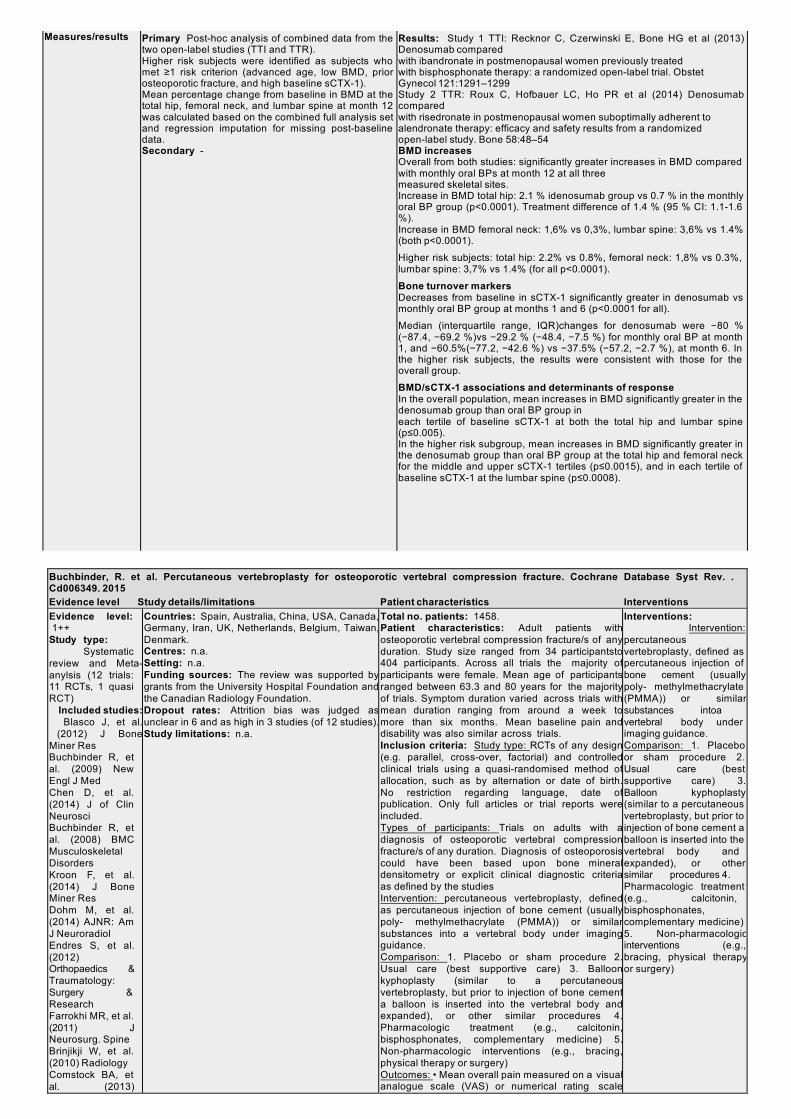
Measures/results Primary Post-hoc analysis of combined data from the two open-label studies (TTI and TTR). Higher risk subjects were identified as subjects who met ≥1 risk criterion (advanced age, low BMD, prior osteoporotic fracture, and high baseline sCTX-1). Mean percentage change from baseline in BMD at the total hip, femoral neck, and lumbar spine at month 12 was calculated based on the combined full analysis set and regression imputation for missing post-baseline data. Secondary -
Results: Study 1 TTI: Recknor C, Czerwinski E, Bone HG et al (2013) Denosumab compared with ibandronate in postmenopausal women previously treated with bisphosphonate therapy: a randomized open-label trial. Obstet Gynecol 121:1291–1299 Study 2 TTR: Roux C, Hofbauer LC, Ho PR et al (2014) Denosumab compared with risedronate in postmenopausal women suboptimally adherent to alendronate therapy: efficacy and safety results from a randomized open-label study. Bone 58:48–54 BMD increases Overall from both studies: significantly greater increases in BMD compared with monthly oral BPs at month 12 at all three measured skeletal sites. Increase in BMD total hip: 2.1 % idenosumab group vs 0.7 % in the monthly oral BP group (p<0.0001). Treatment difference of 1.4 % (95 % CI: 1.1-1.6 %). Increase in BMD femoral neck: 1,6% vs 0,3%, lumbar spine: 3,6% vs 1.4% (both p<0.0001).
Higher risk subjects: total hip: 2.2% vs 0.8%, femoral neck: 1,8% vs 0.3%, lumbar spine: 3,7% vs 1.4% (for all p<0.0001).
Bone turnover markers Decreases from baseline in sCTX-1 significantly greater in denosumab vs monthly oral BP group at months 1 and 6 (p<0.0001 for all).
Median (interquartile range, IQR)changes for denosumab were −80 % (−87.4, −69.2 %)vs −29.2 % (−48.4, −7.5 %) for monthly oral BP at month 1, and −60.5%(−77.2, −42.6 %) vs −37.5% (−57.2, −2.7 %), at month 6. In the higher risk subjects, the results were consistent with those for the overall group.
BMD/sCTX-1 associations and determinants of response In the overall population, mean increases in BMD significantly greater in the denosumab group than oral BP group in each tertile of baseline sCTX-1 at both the total hip and lumbar spine (p≤0.005). In the higher risk subgroup, mean increases in BMD significantly greater in the denosumab group than oral BP group at the total hip and femoral neck for the middle and upper sCTX-1 tertiles (p≤0.0015), and in each tertile of baseline sCTX-1 at the lumbar spine (p≤0.0008).
Buchbinder, R. et al. Percutaneous vertebroplasty for osteoporotic vertebral compression fracture. Cochrane Database Syst Rev. . Cd006349. 2015 Evidence level Study details/limitations Patient characteristics Interventions Evidence level: 1++
Study type: Systematic
review and Meta- anylsis (12 trials: 11 RCTs, 1 quasi RCT)
Included studies: Blasco J, et al.
(2012) J Bone Miner Res Buchbinder R, et al. (2009) New Engl J Med Chen D, et al. (2014) J of Clin Neurosci Buchbinder R, et al. (2008) BMC Musculoskeletal Disorders Kroon F, et al. (2014) J Bone Miner Res Dohm M, et al. (2014) AJNR: Am J Neuroradiol Endres S, et al. (2012) Orthopaedics & Traumatology: Surgery & Research Farrokhi MR, et al. (2011) J Neurosurg. Spine Brinjikji W, et al. (2010) Radiology Comstock BA, et al. (2013)
Countries: Spain, Australia, China, USA, Canada, Germany, Iran, UK, Netherlands, Belgium, Taiwan, Denmark. Centres: n.a. Setting: n.a. Funding sources: The review was supported by grants from the University Hospital Foundation and the Canadian Radiology Foundation. Dropout rates: Attrition bias was judged as unclear in 6 and as high in 3 studies (of 12 studies). Study limitations: n.a.
Total no. patients: 1458. Patient characteristics: Adult patients with osteoporotic vertebral compression fracture/s of any duration. Study size ranged from 34 participantsto 404 participants. Across all trials the majority of participants were female. Mean age of participants ranged between 63.3 and 80 years for the majority of trials. Symptom duration varied across trials with mean duration ranging from around a week to more than six months. Mean baseline pain and disability was also similar across trials. Inclusion criteria: Study type: RCTs of any design (e.g. parallel, cross-over, factorial) and controlled clinical trials using a quasi-randomised method of allocation, such as by alternation or date of birth. No restriction regarding language, date of publication. Only full articles or trial reports were included. Types of participants: Trials on adults with a diagnosis of osteoporotic vertebral compression fracture/s of any duration. Diagnosis of osteoporosis could have been based upon bone mineral densitometry or explicit clinical diagnostic criteria as defined by the studies Intervention: percutaneous vertebroplasty, defined as percutaneous injection of bone cement (usually poly- methylmethacrylate (PMMA)) or similar substances into a vertebral body under imaging guidance. Comparison: 1. Placebo or sham procedure 2. Usual care (best supportive care) 3. Balloon kyphoplasty (similar to a percutaneous vertebroplasty, but prior to injection of bone cement a balloon is inserted into the vertebral body and expanded), or other similar procedures 4. Pharmacologic treatment (e.g., calcitonin, bisphosphonates, complementary medicine) 5. Non-pharmacologic interventions (e.g., bracing, physical therapy or surgery) Outcomes: • Mean overall pain measured on a visual analogue scale (VAS) or numerical rating scale
Interventions: Intervention:
percutaneous vertebroplasty, defined as percutaneous injection of bone cement (usually poly- methylmethacrylate (PMMA)) or similar substances into a vertebral body under imaging guidance. Comparison: 1. Placebo or sham procedure 2. Usual care (best supportive care) 3. Balloon kyphoplasty (similar to a percutaneous vertebroplasty, but prior to injection of bone cement a balloon is inserted into the vertebral body and expanded), or other similar procedures 4. Pharmacologic treatment (e.g., calcitonin, bisphosphonates, complementary medicine) 5. Non-pharmacologic interventions (e.g., bracing, physical therapy or surgery)

Radiology Gray LA, et al. (2007) BMC Musculoskelet Disorders Kallmes DF, et al. (2009) Amn J Neuroradiol Kallmes DF, et al. (2009) New Engl J Med Staples MP, et al. (2011) BMJ
(NRS) • Disability measured by the Roland Morris Disability Questionnaire (RMDQ) or other disability measure • Vertebral fracture and/or osteoporosis-specific health- related quality of life, e.g., the Quality of Life Questionnaire of the European Foundation for Osteoporosis (QUALEFFO) • Overall health-related quality of life, e.g., European Quality of Life with 5 Dimensions (EQ-5D) or Assessment of Quality of Life (AQoL) questionnaire • Treatment success measured by a participant-reported global impression of clinical change (much or very much improved), or similar measure • New (incident) symptomatic vertebral fractures (the denominator was the number of participants, but the numerator could include more than one new fracture per participant) • Number of serious other adverse events judged to be due to the procedure (e.g. infection, clinical complications arising from cement leakage) Exclusion criteria: Trials enrolling participants with vertebral fractures due to another cause such as major trauma and malignancy were excluded.
Notes: Author's conclusion: "Based upon the currently available moderate quality evidence, our review does not support a role of vertebroplasty for the treatment of osteoporotic vertebral fractures in routine clinical care. There were no demonstrable clinically important benefits compared with a sham procedure and subgroup analyses indicated that results did not differ according to duration of pain ≤ 6 weeks versus > 6 weeks. Sensitivity analyses confirmed that open trials that compared vertebroplasty with usual care are likely to have overestimated any benefit of vertebroplasty. Correcting for these biases would likely drive any observed improvements seen with vertebroplasty over usual care, towards the null, in keeping with the findings from the placebo- controlled trials. Although we did not demonstrate an increased risk of incident symptomatic vertebral fractures or other serious adverse events associated with vertebroplasty, a clinically important increased risk cannot be excluded based upon our review due to the small number of events. Furthermore, serious adverse events related to vertebroplasty were reported in several trials. Patients should be informed about the lack of high quality evidence supporting benefits of vertebroplasty and its potential for harm.
Outcome Measures/results
Primary • Mean overall pain measured on a visual analogue scale (VAS) or numerical rating scale (NRS) • Disability measured by the Roland Morris Disability Questionnaire (RMDQ) or other disability measure • Vertebral fracture and/or osteoporosis- specific health- related quality of life, e.g., the Quality of Life Questionnaire of the European Foundation for Osteoporosis (QUALEFFO) • Overall health-related quality of life, e.g., European Quality of Life with 5 Dimensions (EQ-5D) or Assessment of Quality of Life (AQoL) questionnaire • Treatment success measured by a participant- reported global impression of clinical change (much or very much improved), or similar measure • New (incident) symptomatic vertebral fractures (the denominator was the number of participants, but the numerator could include more than one new fracture per participant) • Number of serious other adverse events judged to be due to the procedure (e.g. infection, clinical complications arising from cement leakage). Secondary • Proportion of participants with pain improved by a clinically relevant amount, for example improvement of at least 2.5 units or 30% on a 0 or 1 to 10 scale. • New (incident) radiographic vertebral fractures (the denominator was the number of participants, but the numerator could include more than one new fracture per participant) • Other adverse events.
Results: • Eleven RCTs and one quasi-RCT conducted in various countries were included. Two trials compared vertebroplasty with placebo (209 randomised participants), six compared vertebroplasty with usual care (566 randomised participants) and four compared vertebroplasty with kyphoplasty (545 randomised participants). • Both placebo-controlled trials were judged to be at low overall risk of bias while other included trials were generally considered to be at high risk of bias across a range of criteria, most seriously due to lack of participant and study personnel blinding. • Compared with placebo, there was moderate quality evidence based upon two trials that vertebroplasty provides no demonstrable benefits with respect to pain, disability, disease-specific or overall quality of life or treatment success. Mean pain (on a scale 0 to 10, higher scores indicate more pain) was 5 points with placebo and 0.7 points better (1.5 better to 0.15 worse) with vertebroplasty, an absolute pain reduction of 7% (15% better to 1.5% worse) and relative reduction of 10% (21% better to 2% worse) (two trials, 201 participants). At one month, mean disability measured by the Roland Morris Disability Questionnaire (scale range 0 to 23, higher scores indicate worse disability) was 13.6 points in the placebo group and 1.1 points better (2.9 better to 0.8 worse) in the vertebroplasty group, absolute improvement in disability 4.8% (12.8% better to 3.3% worse), relative change 6.3% better (17.0% better to 4.4% worse) (two trials, 201 participants). • Disease-specific quality of life measured by the QUALEFFO (scale 0 to 100, higher scores indicating worse quality of life) was 2.4 points in the placebo group and 0.40 points worse (4.58 better to 5.38 worse) in the vertebroplasty group, absolute change: 0.4% worse (5% worse to 5% better), relative change 0.7% worse (9% worse to 8% better (based upon one trial, 73 participants). At one month overall quality of life measured by the EQ5D (0 = death to 1 = perfect health, higher scores indicate greater quality of life at one month was 0.27 points in the placebo group and 0.05 points better (0.01 worse to 0.11 better) in the vertebroplasty group, absolute improvement in quality of life 5% (1% worse to 11% better), relative change 18% better (4% worse to 39% better) (two trials, 201 participants). Based upon one trial (78 participants) at one month, 9/40 (or 225 per 1000) people perceived that treatment was successful in the placebo group compared with 12/38 (or 315 per 1000; range 150 to 664) in the vertebroplasty group, RR 1.40 (95% CI 0.67 to 2.95), absolute risk difference 9% more reported success (11% fewer to 29% more); relative change 40% more reported success (33% fewer to 195% more). • Based upon moderate quality evidence from three trials (one placebo, two usual care, 281 participants) with up to 12 months follow-up, we are uncertain whether or not vertebroplasty increases the risk of new symptomatic vertebral fractures (28/143 observed in the vertebroplasty group compared with 19/138 in the control group; RR 1.47 (95% CI 0.39 to 5.50). • Adverse effects: based upon moderate quality evidence from two placebo- controlled trials (209 participants), we are uncertain about the exact risk of other adverse events (3/106 were observed in the vertebroplasty group compared with 3/103 in the placebo group; RR 1.01 (95% CI 0.21 to 4.85)). Notably, serious adverse events reported with vertebroplasty included osteomyelitis, cord compression, thecal sac injury and respiratory failure.
Burch, J. et al. Systematic review of the use of bone turnover markers for monitoring the response to osteoporosis treatment: the secondary prevention of fractures, and primary prevention of fractures in high-risk groups. Health Technol Assess. 18. 1-180. 2014
Evidence level Study Patient characteristics Interventions details/limitations
Evidence level: 2++ Countries: Total no. patients: No overall number stated. Most studies Interventions:

Study type: SR of 42 studies (21 controlled and 16 uncontrolled cohort studies and 5 RCTs) Included studies: Armstrong DJ (2007) Rheumatology Bauer DC (2004) J Bone Miner Res Bjarnason NH (2001) Osteoporos Int Blumsohn A (2011) Osteoporos Int Bruyere O (2010) Osteoporos Int Burshell AL (2010) Bone Chen P (2005) J Bone Miner Res Clowes JA (2003) J Bone Miner Res Delmas PD (2007) Bone Delmas PD (2009) J Bone Miner Res Dobnig H (2005) J Clin Endocrinol Metab Dobnig H (2006) Osteoporos Int Eastell R (2003) J Bone Miner Res Eastell R (2011) J Bone Miner Res Garnero P (2008) Clin Chem Heaney RP (2011) Osteoporos Int Hochberg MC (2010) J Clin Densitom Imai K (2009) Osteoporos Int Ishijima M (2009) Calcif Tissue Int Iwamoto J (2004) Yonsei Med J Iwamoto J (2005) J Bone Miner Metab Kim SW (2005) Endocr J Kitatani K (2003) J Bone Miner Metab Kung AW-C (2009) Int J Rheum Dis Kyd PA (1998) Ann Clin Biochem Kyd PA (1999) Ann Clin Biochem Lane NE (2000) Osteoporos Int Majima T (2008) J Bone Miner Metab Masaryk P (2002) Vnitr Lek Miller P (2008) Bone Moro-Alvarez MJ (2010) J Bone Miner Res Reginster JY (2004) Bone Reyes-Garcia R (2010) Menopause Roche (2007) URL: www.roche- trials.com/trialDetailsGet.action? studyNumber=ML19930 Hoffmann-La Roche (2009) URL: www.clinicaltrials.gov/ct2/show/NCT00545909 Hoffmann-La Roche (2009) URL: http://ClinicalTrials.gov/show/NCT00545480 Shiraki M (2011) J Bone Miner Metab Siddiqi M (2010) Osteoporos Int Stepan JJ (2008) Calcif Tissue Int Tsujimoto M (2011) Bone Watts NB (2001) Osteoporos Int
Multinational, Asia, UK, Western Europe,Austria, Belgium, Greece, Ireland, Luxembourg, South America, Eastern Europe, USA/Canada Centres: Not reported. Setting: Not reported. Funding sources: The National Institute for Health Research Health Technology Assessment programme. Dropout rates:
Dropout rates not reported. None of the 5 RCTs reported characteristics of patients lost to followup, only 3 reported reasons for the losses. 10 non- RCT had no loss to follow-up (some were retrospective analyses). Reasons for losses to follow-up reported in further 11 non-RCT Study limitations: All RCTs considered to be at high or uncertain risk of bias, therefore of low quality. All non- randomised studies considered at high or unclear risk of bias, therefore of low quality. No RCTs identified that addressed one of the primary aims of the review (evaluation of clinical effectiveness of monitoring regimens that included at least one bone turnover marker, over and above monitoring regimens where a bone turnover marker was not used). Very high proportion of patients in RCT were adherent to medication; this is unlikely to be representative of clinical practice. Most of the evidence available relating to the accuracy of bone turnover markers was in the form of correlations with BMD, measured using DXA. A statistically significant correlation coefficient is not indicative of a strong association between two variables. Furthermore, BMD is
were small (22 - 3.105 patients). 2 studies did not report the number recruited; 21 studies had fewer than 100 participants. Patient characteristics: Majority of studies conducted primarily (or entirely) in post-menopausal women; only 4 studies reported including men. Where reported, mean age ranging from 56.1 - 73.9 years. Inclusion criteria: (1) a randomised controlled trial (RCT) comparing a monitoring regimen that included at least one bone turnover marker test with a monitoring regimen without bone turnover marker testing, or a different bone turnover marker, and reporting either change in patient management strategies and/or treatment adherence rates; (2) a study evaluating the impact of bone turnover marker test results on the decision-making process, that also reported the subsequent rate of fracture in the population; (3) a prospective study that compared the results of bone turnover marker tests with the results of bone biopsy or a composite reference standard of BMD and subsequent fracture outcome; (4) a prospective study that reported at least a p-value for the association between changes in bone turnover markers and BMD, biopsy, and/or the incidence of fractures from correlation or multivariate regression analyses; (5) a prospective study reporting inter- and/or intrapatient variability on bone turnover marker test results for patients receiving one of the treatments being evaluated; or (6) a cost-effectiveness analysis of bone turnover marker monitoring strategies. Non-effectiveness prospective studies had to recruit at least 20 patients with osteoporosis who were receiving one of the treatments of interest Exclusion criteria: (1) Studies assessing the effectiveness of treatments for osteoporosis using changes in bone turnover markers solely as an outcome were excluded. (2) Prognostic studies using a bone turnover marker to identify patients at risk of osteoporosis and fracture at baseline, prior to commencing treatment, were excluded, as were studies that included fewer than 20 patients, meeting the population inclusion criteria, in analyses of outcomes applicable to this review.
Bone turnover markers

only one factor that impacts on bone strength and therefore on fracture risk and the association between bone turnover markers and BMD may vary depending on the population and on the menopausal status in women. Extreme heterogeneity ruling out meta-analysis (heterogeneity in terms of patient populations, definitions used to diagnose osteoporosis, treatment regimens, bone turnover marker tests used and their timing, DXA sites used and timing of DXA scans).
Notes: No investigation of publication bias. Author's conclusion: Implications for service provision The lack of evidence of clinical effectiveness and the heterogeneity and poor quality of the available evidence on the accuracy, reliability and reproducibility of bone turnover markers for monitoring response to osteoporosis treatment precluded the possibility of making any recommendations on the choice of bone turnover marker being used in routine clinical practice for its superiority to monitor osteoporosis treatment response. In addition, the evidence to support the use of bone turnover marker feedback results to improve patient adherence to osteoporosis treatment was not convincing. Suggested research priorities In order to determine whether or not bone turnover marker monitoring improves treatment management decisions and ultimately impacts on patient outcomes in terms of reduced incidence of fracture, RCTs are required. The predictive accuracy of bone turnover markers for future fracture outcomes in patients receiving osteoporosis treatment could be investigated using prospective, long-term observational studies with large sample sizes. However, in view of the large number of potential patient population– treatment–test combinations, the most promising combinations would need to be identified in order to ensure the more costly and time-consuming studies, such as RCTs, focus on evaluating those strategies. Therefore, we consider the research priority to identify these promising treatment–test combinations. This can be achieved by either conducting small variability studies or initiating a patient registry to collect standardised data. The former would be quicker, easier and less costly, but the quality of the data would be poorer. Further, prior to establishing the latter it is likely that a more widespread use of bone turnover markers in clinical practice would be required. Once the most promising treatment–test combinations have been identified, well-designed RCTs can be conducted to evaluate the effectiveness of those monitoring regimens; this would include measuring outcomes such as the proportion of non-responders, adherence rates, treatment management decisions and fracture outcome. Data from these RCTs along with other sources can then be included in a decision-analytic model in order to investigate cost- effectiveness.
Outcome Measures/results Primary Effectiveness of bone
turnover markers in monitoring treatment response: change in patient management strategies, incidence of fracture, treatment adherence rates Secondary Test accuracy: diagnostic accuracy, correlation coefficient for the association between a bone turnover marker and bone density and/or the incidence of fracture, risk/incidence of fracture associated with the bone turnover marker test results, p- value for a bone turnover marker. Reliability and reproducibility: intra- and/or interpatient variability in bone turnover marker test results
Results: Clinical effectiveness: Reported in 5 RCTs and 1 post hoc analysis of a subgroup of patients from a previous RCT. High variability regarding definition of adherence, compliance and persistence. None of the RCTs reported on impact of bone turnover marker monitoring on treatment management. Adherence: Reported in 1 study that found significantly greater average daily proportion of patients who were persistent and compliant to 5 mg/day risedronate in those receiving feedback after the first reinforcement visit at 13 weeks (p = 0.01). Decrease in adherence observed over time attributed to increasing number of patients who did not persist with treatment. Compliance: Reported in 5 studies. Very high and not representative rate of compliance with bisphosphonate. No significant difference between feedback and no-feedback arms where ORs could be calculated; most studies with insufficient data. Persistence: Reported in 1 study with high and not representative baseline rate for 5 mg/day risedronate. No significant impact of uNTX on persistence in the no-feedback arm (p = 0.71); feedback of uNTX results significantly affected persistence in the feedback group (p = 0.0029). Significant impact of feedback of uNTX results on discontinuation (p = 0.017); Where message of good uNTX was given(> 30% decrease), HR for discontinuation was 0.71 [95% CI 0.53, 0.95]; Where message of poor uNTX response was given, HR for discontinuation was 2.22 (95% CI 1.27, 3.89). Where message of stable uNTX response was given, no significant difference in discontinuation between those receiving feedback and those who were not. Quality of life: Osteoporosis Patient Perception Survey (OPPS) QoL questionnaire reported in 2 studies. 1 study with statistically significant differences of at least 3.8% favouring feedback for all domains and the composite score (p ≤ 0.021) in women aged between 55 and 85 years (with the exception of the motivation domain [p >0.05]). 1 study with significant increases in feeling informed, satisfaction and the overall composite score. Although statistically significant, it is unclear whether or not the small absolute changes are clinically significant. Test accuracy: Bisphosphonates: Reported in 18 studies. No 2 studies assessed the same combination of variables and patient population. Of the 54 r- values reported for correlations between changes in bone turnover marker and BMD, 19 were statistically significant (p < 0.05); however, all of the correlations
were weak (r < 0.5). Insufficient data for any combination of bone turnover marker, DXA site, time points at which tests were conducted, and patient population to identify any patterns. Limited data from studies conducting multiple regression analyses indicated that there may be a significant association between changes in bone turnover markers and either BMD or incidence of hip or vertebral fractures. However, only 1 study adjusted for all of the confounders considered important by the review authors. Teriparatide: Reported in 10 studies (7 studies with 20 μg/day, 2 studies with 40 μg/day, 2 studies with combined population that received either 20 or 40 μg/day, 1 study did not report administered dose). Across 71 reported r-values for correlations between changes in bone turnover and BMD in patients treated with 20 μg/day teriparatide, 22 were statistically significant; however, all of them were weak (r < 0.5). In patients being treated with 40 μg/day, 9 of 21 reported r-values indicated statistically significant, but weak, correlations. No studies were identified that reported the results of multiple regression analyses in patients being treated with teriparatide. Raloxifene: Reported in 3 studies. Of the 12 reported r- values, none was statistically significant and all correlations were weak. Strontium ranelate: Reported in 2 studies. Of the 7 reported r-values, 6 were statistically significant; sP1NP and sCTX showed moderate correlations with absolute changes in BMD (r = 0.615 and 0.56, respectively). Denosumab: Reported in 1 study. Of the 6 reported r-values, 5 were statistically significant; all correlations weak (r < 0.50). Test reliability and reproducibility: Four studies reported S/N ratio for a bone turnover marker in those being treated with either etidronate, teriparatide or raloxifene. Each study calculated the S/N ratio differently. Within-study comparisons show that sP1NP at 2 weeks had a lower S/N ratio than sCTX at 2 weeks but a higher S/N ratio at 25 weeks, and sP1NP had a greater S/N ratio than sBALP when measured at 6 months.
Chang, X. et al. Vertebroplasty versus kyphoplasty in osteoporotic vertebral compression fracture: a meta-analysis of prospective comparative studies. Int Orthop. 39. 491-500. 2015 Evidence level Study details/limitations Patient characteristics Interventions Evidence level: 2-
Study type: Systematic
review and Meta analysis of 20 studies (6 RCTs, 14 prospective cohort studies). Included studies: YiX, et al. (2014) Arch Orthop Trauma Surg Bae H, et al. (2010) Spine De Negri P, et al. (2007) Clin J Pain Dong R, et al. (2009) Int Orthop Endres S, et al. (2012) Orthop Traumatol Surg Res Folman Y, et al. (2011) Isr Med Assoc J Grohs JG, et al. (2005) J Spinal Disord Tech Kumar K, et al. (2010) Neurosurgery Li X, et al. (2012) J Spinal Disord Tech Liu JT, et al. (2010) Osteoporos Int Lovi A, et al. (2009) Eur Spine J Movrin I, et al. (2010) Arch Orthop Trauma Surg Omidi-Kashani F, et al. (2013) J Osteoporos Pflugmacher R, et al. (2005) RoFo Qian J, et al. (2012) PloS One Roellinghoff M, et al. (2009) Minim Invasive Neurosurg
Countries: n.a. Centres: n.a. Setting: n.a. Funding sources: n.a. Dropout rates: Dropout rates and attrition bias were judged as low in 5 studies, and unclear in 15 studies. Study limitations: Our article still has limitations. First, only six RCTs out of 20 in all finally met our criteria and were included. Second, the quality assessment result has shown some unclear bias such as in random sequence generation. Third, the trials that were included in our study were all English literature, which might lead to insufficient data inclusion.
Total no. patients: 1429. Patient characteristics: 1429 participants with diagnosed osteoporotic vertebral compression fracture (OVCF) of both genders. All participants were older than 18 years. The article contains no information on baseline characteristics. Inclusion criteria: Prospective comparative trials (PCTs) or randomized controlled trials (RCTs) with no language restrictions that compared PVP with PKP, all genders, age 18 or above, who had been diagnosed as osteoporotic vertebral compression fractures (OVCF) caused by osteoporosis. The interventions in the experimental group and control group were PKP and PVP respectively. Exclusion criteria: Non-PCT trials or RCT trials, retrospective study, bone tumors, hemangioma and other reasons for PKP/PVP surgical treatment.
Interventions: The interventions in the experimental group and control group were percutaneous kyphoplasty (PKP) (n=732) and percutaneous vetebroplasty (PVP) (n= 697) respectively.

Ruiz Santiago F, et al. (2010) Eur J Radiol Schofer MD, et al. (2009) Arch Orthop Trauma Surg Vogl TJ, et al. (2013) Spine Zhou JL, et al. (2008) Chin J Traumatol Notes: Unlcear how search terms were combined with operators and whether MesH terms were used. Unclear whether two investigators
selected articles and extracted data. No consideration of grey literature. Outcomes were not specified before the search and lack definitions in general. No baseline study characteristics are provided. No conflict of interest declarations (neither from primary studies, nor the authors of the article). Author's conclusion: According to the statistical results, the conclusion can be drawn that the PKP technique reduced the rate of cement leakage and significantly improves the safety compared with PVP. For the relief of pain, the main clinical symptom caused by OVCF, both are effective methods.
Outcome Measures/results
Primary Age of patients, fracture time before surgery, surgical time, short time visual analog scale (VAS), long time VAS, short time Oswestry Disability Index (ODI), long term ODI, Cobb angle short, Cobb angle long, volume of the cement, cement leakage, adjacent level fracture. Secondary n.a.
Results: Comparing these two methods, the PKP group took more operation time [SMD = 0.66, 95 % CI (0.28, 1.03), p=0.0006] with higher anterior vertebral body height [SMD = 1.40, 95 % CI (0.49, 2.32), p=0.003], greatly reduced Cobb angle in the long run [SMD = −0.61, 95 % CI (−1.04, −0.19), p=0.005] and had lower risk of cement leakage. However, in VAS scores and ODI scores after the surgery whether for the short-term efficacy (no more than 1 week after the surgery) or long-term efficacy (more than six months), Cobb angle in the short run and new fracture in the adjacent level, no statistically differences were found between the two groups.
Chen, L. X. et al. Comparison of Bone Mineral Density in Lumbar Spine and Fracture Rate among Eight Drugs in Treatments of Osteoporosis in Men: A Network Meta-Analysis. PLoS One. 10. e0128032. 2015
Evidence level Study Patient characteristics Interventions details/limitations
Evidence level: 1++ Study type: Systematic Review and Meta Analysis of 13 RCTs Included studies:
Boonen S, 2012, The New England journal of medicine. Boonen S, 2009, Journal of bone and mineral research. Kaufman JM, 2013, The Journal of clinical endocrinology and metabolism. Kaufman JM, 2005, Osteoporosis international. Kurland ES, 2000, The Journal of clinical endocrinology and metabolism Orwoll ES, 2010, Journal of bone and mineral research. Orwoll ES, 2010, Bone. Orwoll E, 2000, The New England journal of medicine. Ringe JD, 2010, Arzneimittel-Forschung. Ringe JD, 2009, Rheumatology international. Ringe JD, 2004, Rheumatology international. Ringe JD, 2001, The Journal of clinical endocrinology and metabolism. Walker MD, 2013; Endocrine.
Countries: Centres: Setting: Funding sources: Dropout rates: Study limitations:
Total no. patients: n= 3647 patients Patient characteristics: Men with primary or idiopathic osteoporosis Inclusion criteria: (1) Participants: Men with primary or idiopathic osteoporosis. (2) Interventions and comparisons: Therapy regimens that included two of the following drugs or one drug and a placebo: alfacalcidol, alendronate, ibandronate, risedronate, zoledronate, strontium ranelate, teriparatide and parathyroid hormone; (3) Outcomes: the BMD in LS and the fracture rate; (4) Study design: randomized controlled trials (RCTs) or quasi-RCTs. Exclusion criteria: (1) abstracts, letters, or meeting proceedings; (2) repeated data or did not report the outcomes of interest; (3) duration of follow-up was < 12 months.
Interventions: Zoledronate (20mg) vs Placebo
Notes: Author's conclusion: Zoledronate might be the best choice to increase the BMD in LS and teriparatide (20mg) might lead

to the lowest fracture rate. Outcome Measures/results
Primary - Secondary BMD in LS and fracture rate
Results: BMD in LS compared with placebo - network of comparisons - zoledronate (SMDs 13.48, 95%CrI 11.88–15.08) yielded the most significant effect on increasing the BMD - followed by alendronate (11.04, 9.68–12.41) - teriparatide (20mcg) + risedronate (10.98, 8.55–13.48), risedronate (10.33, 8.68–12.01), teriparatide (20mcg) (9.33, 6.87–11.76), strontium ranelate (8.88, 7.51–10.24), ibandronate (5.49, 3.82–7.16), parathyroid hormone (4.89, 3.12–6.62) and alfacalcidol (3.42, 1.7–5.2). BMD in LS compared with other active therapies - network of comparisons - teriparatide (20mcg) + risedronate therapy better than zoledronate(2.5, -0.41–5.47), - zoledronate better than all other therapies: 2.44 (0.36–4.55)for alendronate, 10.06 (7.66–12.46) for alfacalcidol, 3.14 (0.83–5.49) for risedronate, 7.98 (5.73–10.28) for ibandronate, 4.6 (2.51–6.72) for strontium ranelate, 4.15 (1.23–7.11) for teriparatide (20mcg) and 8.58 (6.27–10.93) for parathyroid hormone Fracture rate - network of comparisons - placebo resulted in a significantly higher fracture rate vs risedronate (OR 2.51, 95% CrI 1.23–4.24) or zoledronate (2.92, 1.29–5.62) or teriparatide (20mcg) (4.04, 1.36–8.49) or teriparatide (40mcg) (3.5, 1.14–8.34). - Alfacalcidol significantly increased the fracture rate vs risedronate (7.66, 1.74–19.27) or zoledronate (8.41, 2.12–20.03) or strontium ranelate (5.21, 1.32–11.88) or teriparatide (20mcg) (12.12, 2.17– 33.84) or teriparatide (40mcg) (10.49, 1.83–30.47) no significant differences between other therapies
Chen, Z. G. et al. Effects of atorvastatin on bone mineral density (BMD) and bone metabolism in elderly males with osteopenia and mild dyslipidemia: a 1-year randomized trial. Archives of gerontology and geriatrics. 59. 515-21. 2014 Evidence level Study details/limitations Patient characteristics Interventions Evidence level: 1++
Study type: RCT
Countries: China Centres: single center Setting: Geriatric Department of Renji Hospital, Shanghai Funding sources: National Nature Science Foundation of China Dropout rates: 6,25%, 6,25% Study limitations: An exclusive focus on male subjects may partially reduce the impact of hormone changes. However, our findings should be interpreted prudently due to the relatively small sample size, non-placebo-controlled design and a short 1-year follow-up. In addition, it will be better if we had pre-test estimation for the sample size. Further, due to ethical considerations, the subjects recruited were osteopenic with mild dyslipidemia. Thus, the results might be relevant to similar subjects but not for patients afflicted with osteoporosis and severe dyslipidemia or nondyslipidemia subjects.
Total no. patients: n=64 Patient characteristics: male subjects, aged above 60 years Inclusion criteria: - mild dyslipidemia - osteopenia before recruitment - not treated routinely with lipid- lowering drugs in the last 3 months before recruitment Exclusion criteria: - history of medications for diseases - affecting bone metabolism; - therapy for osteoporosis with diphosphonate and calcitriol within the last 6 months; - other medications including diuretics, testosterones, thyroid hormones, glucocorticoids, or immunosuppressants within the last 1 year; - relevant diseases: malignant tumors, severe hepatic or renal dysfunction, hyperthyroidism, and hyperparathyroidism.
Interventions: 1-year 10 mg/day atorvastatin and lifestyle guidance, vs lifestyle guidance only
Notes: no sample size calculation. No ITT. Allocation concealment not clear. Author's conclusion: Our findings suggest that in elderly male patients with osteopenia and mild dyslipidemia, therapeutic doses of atorvastatin were associated with positive effects on BMD, probably mediated by suppressed bone resorption.
Outcome Measures/results
Primary comparison of BMD at total hip, lumbar spine and femoral neck between atorvastatin and control groups from baseline to month 12 Secondary comparison of bone metabolic parameters from baseline to month 12 - bone resorptive marker CTx, - bone formative marker OC, - other bone metabolism markers including 25(OH)D, iPTH, - testosterone
Results: Lipid changes at month 12 - atorvastatin vs control: TG: significant decreases (−7.9 ±10.6%, P < 0.01) LDL: sign. decreases (−7.3 ±7.6%, P < 0.01) - no statistical significance between groups: TC, HDL-C BMD changes after 6 months - No statistical significances between groups in total hip, lumbar spine, femoral neck BMD changes after 12 months - statistical significances in total hip BMD between groups (P < 0.01), but not in BMDs at femoral neck and lumbar spine
Bone metabolic parameters - CTx showed significant changes (P = 0.04) atorvastatin vs control - Bone formative markers OC and other bone metabolism markers including 25(OH)D, iPTH, and testosterone did not differ significantly between atorvastatin treatment group and control group
Correlation between changes in efficacy values and lipids - No statistically significant correlation between BMD levels of total hip, and low density lipoprotein (LDL)or TG levels at month 12 - No statistically significant differences were found in the correlation between CTx, and LDL-C and TG levels at month 6, 12
Cheng, X. et al. Cadmium Exposure and Risk of Any Fracture: A PRISMA-Compliant Systematic Review and Meta-Analysis. Medicine (Baltimore). 95. e2932. 2016
Evidence level Study details/limitations Patient characteristics Interventions Evidence level:
Countries: Belgium, Sweden (5 studies),
Total no. patients:
Interventions: Cd exposure: urinary Cd (U-Cd) as biomarkers for

2+ Study type: MA
of 7/ 8 studies (6
cohorts and 2
case-control
studies)
Included studies:
Staessen JA
(1999) Lancet
Alfven T (2004) J
Bone Miner Res
Zhu G (2004)
Biometals
Engstrom A
(2011) J Bone
Miner Res Thomas LD (2011)
J Bone Miner Res
Engstrom A
(2012) Bone Dahl C (2014) Biol
Trace Elem Res
Sommar JN
(2014) Calcif
Tissue Int
China, Norway
Centres: N.r. Setting: N.r.
Funding sources: None.
Dropout rates: N.r.
Study limitations: - Observational studies
have inherent limitations, such as selective
bias and recall or memory bias.
- Evidence of publication bias observed; - Small number of studies included in meta-
analysis limits ability to draw a reliable
conclusion, especially in the subgroup
analyses.
- Between-study heterogeneity is common in
meta-analysis, and exploring the potential
sources of between-study heterogeneity is
essential. Sensitivity and subgroup analyses
indicated the existence of heterogeneity.
- Included studies used different methods to
assess and categorize Cd exposure, findings
likely influenced by misclassification of
exposure. Potential for misclassification of
exposure to Cd may contribute to
heterogeneity among all studies in summary
analysis.
504.346 Patient characteristics:
Age, Median (Range):
44.05 (NA) years - 64.5
(50–85) years. NA in 1
study.
Inclusion criteria: -
Study design was either
cohort, case–control, or
cross-sectional;
- the exposure was Cd
exposure;
- the outcome was
fracture risk; and hazard
ratio (HR) or RR and
odds ratio (OR), with
corresponding 95% CI (or
data to calculate these)
were reported.
Exclusion criteria: - Editorial letters, historical
reviews, and descriptive
studies, such as case
reports and case series,
or laboratory studies;
- they did not contain
enough data for
calculating RR;
- when multiple
publications covered the
same study population,
only the study with the
larger sample was
included.
long-term exposures to Cd in 4 articles, 2 articles evaluated Cd
exposure levels by estimating the dietary Cd (D-Cd) using food
frequency questionnaire; 1 article examined Cd exposure levels in
drinking water, 1 article investigated Cd exposure levels in erythrocytes
(Ery-Cd). 1 article combined assessment of dietary and U-Cd.
Notes:
No search for grey/ unpublished literature. Funnel plot and Begg and Egger test both indicate the existence of publication bias, possibly leading to
overestimation of effect or/and its direction. However, funnel plots futile due to insufficient number of studies.
Reported number of included studies varying (8 articles in flow diagram, 10 studies from 7 articles in text and abstract).
Subgroup analyses not preplanned. Patient characteristics not described in detail (e.g. male/ female ratio).
Author's conclusion: In summary, the current meta-analysis demonstrates that a high Cd exposure may be a risk factor for any fracture. However,
this result should be interpreted cautiously because of the heterogeneity among studies and existence of publication bias. Additional large, high-
quality prospective studies are needed to evaluate the association between Cd exposure and the risk of development of fracture.
Outcome
Measures/results
Primary Fracture risk
Secondary -
Results: Pooled relative risk of any fracture for highest vs lowest category of cadmium
concentration: RR = 1.30 (95% CI; 1.13, 1.49). Significant association remained consistent in
subgroup analyses when stratified by study type, geographical region, method of cadmium exposure
assessment, and gender.
Cheng, X. et al. Comparison of unilateral versus bilateral percutaneous kyphoplasty for the treatment of patients with osteoporosis vertebral compression fracture (OVCF): a systematic review and meta-analysis. Eur Spine J. 2016 Evidence level Study details/limitations Patient characteristics Interventions Evidence level: 2-
Study type: Systematic
Countries: China, Korea, USA. Centres: n.a. Setting: n.a. Funding sources: n.a.
Total no. patients: 428 Patient characteristics: Numer of patients per study 15-28 (unilateral) and 21-40 bi-lateral; 0- Inclusion criteria: (1) study design: randomized
Interventions: Intervention:
unilateral and bilateral

review and meta- analysis (8 studies, observational studies and RCTs). Included studies:
Chen C, et al. (2010) Injury Song BK, et al. (2009) Osteoporos Int Chen CM, et al. (2011) J Spinal Disord Tech Chung HJ, et al. (2008) Int Orthop Wang Z, et al. (2012) J Clin Neurosci Wang H, et al. (2015) J Clin Neurosci Chen L, et al. (2011) Spine Rebolledo BJ, et al. (2013) Bone Joint J
Dropout rates: 3 out of 8 studies included in the quality assessment were considered to inadequately describe drop- outs. Study limitations: "We consider that our result of meta- analysis is influ- enced by the following reasons: first, the description of randomization process is not sufficient in most of the studies. Although aggregate data had larger statistical power, the included studies still had various types of bias. Eight of studies have moderate quality and one has a high risk of bias. It is necessary that amounts of high-quality randomized controlled trials should be required. Second, there were only cement leakage and postoperative adja- cent-level fractures in our analysis. More important com- plications should also be considered, such as intraoperative pulmonary cement embolism, intercostal neuralgia and cerebrospinal fluid leakage. And these complications might affect the preponderance of the two approaches and could be included by long-term follow-up. Third, the clinical outcomes of the included studies were limited, although data pooling. Thus, our meta-analysis might be limited to answer some important clinical questions. Studies contained large outcomes are required."
or non-randomized controlled study (2) study population: participants were patients with osteoporosis vertebral compression fracture (OVCF)s (3) purpose of interventions: to compare clinical outcome differences between unilateral and bilateral kyphoplasties; (4) outcome measurements: one of the following outcomes: operation time, cement dosage, VAS, ODI, restoration rate, cement leakage, X-ray exposure frequency and postoperative adjacent- level fractures. Exclusion criteria: Studies that did not meet the above criteria were excluded from selection.
kyphoplasty.
Notes: Study characteristics provides incorrect data on included participants of one study (280 instead of 28). Despite some high heterogeneity (I2=96% p<0.000001) fixed effects models were used for some meta-analyses. Causes for high heterogeneity are not investigated using sensitivity analysis or meta-regression. Some outcomes (adverse events) are simply to rare to be meta-analyzed in this context (n=4 for cement leakage). No investigation of publication bias. Conflicts of interest for the primary studies was not reported. Author's conclusion: "In conclusion, our meta-analysis suggested that a definitive verdict could not be reached regarding which surgical approach is better for OVCF. Although unilateral PKP was associated with shorter operative time, less X-ray exposure frequency and dosage of PMMA than bilateral PKP in the treatment of OVCF. There was no apparent difference in the short- and long-term clinical outcomes and complications between the two approaches. However, bilateral PKP approaches were higher than unilateral PKP approaches in terms of the restoration rate. But on account of lack of some high-quality evidence, we consider that more high- quality randomized controlled trials are required and more complications should be analysed to resolve which surgical approach is better for OVCF treatment in the future."
Outcome Measures/results
Primary Operation time, short-term and long-term visual analogue scale (VAS), short term Oswestry Disability index (ODI), restoration rate, dosage of polymehtylmeth-acrylate (PMMA) injected, cement leakage, X-ray exposure frequency, postoperative adjacent-level fractures. Secondary n.a.
Results: 8 studies involving 428 patients were included in the meta-analysis. Mean operative time was shorter in the unilateral groups compared with the bilateral groups [P<0.05, weighted mean difference WMD) -19.74 (-30.56, -8.92)]. VAS: There was no significant difference in the short-term postoperative VAS scores [P>0.05, WMD 0.03 (-0.34, 0.40)], the long-term postoperative VAS scores between them [P>0.05, WMD 0.01 (-0.42, 0.45)] and the short-term postoperative ODI [P>0.05, WMD -0.33 (-2.36, 1.69)] between the two groups. PMMA dosage: The unilateral approaches required significantly less dosage of PMMA than the bipedicular approaches did [P<0.05, WMD -1.56 (-1.59, -1.16)]. Restoration rate: in the bilateral groups was higher than the unilateral groups [P<0.05, WMD -7.82 (-12.23, -3.41)]. Cement leakage: There was no significant difference in the risk ratio of cement leakage [P>0.05, RR 0.86 (0.36, 2.06)] and postoperative adjacent-level fractures [P>0.05, RR 0.91 (0.25, 3.26)] between the two methods. The mean X-ray exposure frequency in the unilateral groups was greater than the bilateral groups [P<0.05, WMD -5.69 (-10.67, -0.70)].
Cizmic, A. D. et al. Impact of interactive voice response technology on primary adherence to bisphosphonate therapy: a randomized controlled trial. Osteoporos Int. 26. 2131-6. 2015 Evidence level Study details/limitations Patient characteristics Interventions
Evidence level: 1+
Study type: RCT
Countries: USA Centres: Kaiser Permanente Colorado Setting: Outpatient setting. Funding sources: "This study was funded by Kaiser Permanente Colorado. The institution had no role in the design and conduct of the study; collection,management, analysis, and interpretation of the data; and preparation, review, or approval of the manuscript." Dropout rates: 1 patient in control group excluded from statistical analysis (died before 25 day timeframe) Study limitations: - Lack of standardized definition that includes a timeframe after which a patient is considered primary non-adherent. - IVR phone call only available in English.
Total no. patients: 246 randomized, 245 analyzed. Patient characteristics: Both groups: mean age 71.5 years. 93 % of patients female. Alendronate most commonly prescribed bisphosphonate, 86% of all study patients had a diagnosis of osteoporosis. Inclusion criteria: Adults, aged 18 years and older, with a new oral bisphosphonate prescription for osteoporosis or osteopenia, which was not purchased within 14–20 days of being ordered.
Exclusion criteria: Patients with
prescriptions sent to a non-KPCO pharmacy, residing in a skilled nursing facility, or
without continuous KPCO membership 12 months before and 25 days after enrollment.
Interventions: Intervention group: Intervention patients were contacted on the day of enrollment by an automated interactive voice response (IVR) phone call. If no one answered, a voicemail message instructed the patient to contact a toll-free number to retrieve the message. If medication was still not purchased 7 days after receiving the IVR phone call, a letter reminding the patient of prescription and explaining benefits and risks of bisphosphonate therapy was mailed. Control group: Did not receive phone call or letter.
Notes: Patients with an integrated health plan by Kaiser Permanente Colorado recruited. External validity/ applicability of results for guideline population questionable. 1 patient in each group had a normal "T-score". Only internally filled prescriptions at Kaiser Permanente pharmacies included in calculation for adherence but no. of patients filling their prescription at external pharmacies shouldn't differ between randomized groups theoretically. No allocation concealment mentioned. No blinding described but outcomes not observer-dependent. No test for baseline differences described. Groups stated as 1:1 randomized but group sizes differ and calculated group size not reached. Author's conclusion: An IVR phone call plus a follow-up letter represents a practical intervention to improve primary adherence to bisphosphonates effectively and efficiently while also positively impacting secondary adherence. Application of this process may be useful to improve primary adherence to other medications, especially those for chronic disease states.
Outcome Measures/results
Primary Proportion of patients who purchased their prescription within 25 days of study enrollment: 25 days determined as enrollment and IVR phone call made on day 0; letter sent on day 7, if indicated; letter to be delivered by day 11; and up to 14 days follow- up after receiving the letter, which was day 25. Secondary (1) proportion of intervention patients meeting the primary outcome who purchased their prescription before and after the expected letter arrival date, (2) identifying factors other than the intervention that were independently associated with primary adherence, and (3) medication possession ratio (MPR) for 6 months in patients in both groups who met the primary outcome.
Results: Primary: Percent of patients who purchased their prescription within 25 days of study enrollment significantly greater in intervention group compared to control group (62/127, 48.8 % vs. 36/118, 30.5 %, unadjusted OR=2.17, 95 % CI=1.29–3.67). After adjusting for age, sex, history of bone mineral density (BMD) test, and history of a fracture, OR for primary adherence in the intervention versus control group was 2.30 (95 % CI 1.34–3.94). OR should have been only adjusted for fractures because only this factor showed significant relation - see later. Secondary: Majority of intervention patients with primary adherence to bisphosphonate therapy (n=62) purchased bisphosphonate before letter arrival date (11 days after randomization) compared to after (76 vs. 24 %, respectively). Similar pattern in control group (64 % purchasing their prescription before day 11 versus 36 % after, p=0.208 for difference between the two groups). Sex, age or history of BMD test not significantly associated with primary adherence. Patients with a history of fracture significantly more likely to have primary adherence to bisphosphonate therapy (OR=2.21, 95 % CI 1.11–4.40). Mean MPR for intervention patients was 69 % (95 % CI 61–77) vs 60 % (95 % CI 49–71) in control patients (p<0.001). Percent of patients in the intervention and control groups who had a MPR ≥80 % was 43.6 and 41.7 % (p=0.856).
Conwell Louise, S. et al. Bisphosphonates for osteoporosis in people with cystic fibrosis. Cochrane Database of Systematic Reviews. 2014 Evidence level Study details/limitations Patient characteristics Interventions Evidence level: 1++
Study type: MA of 7 RCT Included studies: Aris RM (2000) Am J Respir Crit Care Med Aris RM (2004) Am J Respir Crit Care Med Boyle MP (2005) Pediatr Pulmonol Chapman I (2009) J Clin Endocrinol Metab Haworth CS (2001) Thorax Haworth CS (2011) J Cyst Fibros
Countries: If stated: 2 studies from USA, 1 from Australia, 1 from UK, 1 from UK and Ireland, 1 from Canada Centres: If stated: 3 Single centre studies, 3 Multi-centre studies (2 sites, 4 sites and 6 sites) Setting: If stated: 1 university hospital, 1 adult CF centre, 2 studies with CF (specialty) clinics Funding sources: Not stated for review. Funding for 1 included trial provided by grants from CF Foundation and Verne S. Caviness General Center for Clinical Research. 1 trial funded by U.S. Food and Drug administration, Merck
Total no. patients: 237 adult participants Patient characteristics: Overall distribution/ pooled value of sex, age, BMD scores in studies not described. 1 study with 34 post-transplant adults with CF, all other studies with non-transplanted CF patients. 6 trials with supplemented participants (Vitamin D + Calcium) Inclusion criteria: Types of studies: - Randomised controlled trials, - Published papers and abstracts, - Trials published in all languages. Types of participants: - People of all ages and of both sexes with CF diagnosed clinically or by sweat and genetic testing, - All degrees of disease severity and bone density. Types of interventions: - All trials examining bisphosphonates compared to controls (other bisphosphonates, placebo or usual treatment) for treating or preventing
Interventions: Types of bisphosphonate: intravenous pamidronate in two trials; oral alendronate in two trials; oral risedronate; intravenous zoledronate; intravenous zoledronate.
Papaioannou A (2008) Chest
and Co, Inc., Clinical Nutrition Research Unit, Verne S. Caviness General Center for Clinical Research at University of North Carolina, CF Foundation and NIDDK. 1 trial partly funded by Novartis Pharmaceuticals Pty Ltd, Australia. 1 trial funded by Merck Frosst Canada. 1 trial funded by CF Trust UK. A later study by the same authors funded by unrestricted educational grants from Proctor & Gamble (Norwich, USA) and Cystic Fibrosis Trust, with support to investigators from the UK National Institute of Health Research. Dropout rates: 5 studies described withdrawals from the study: Significantly more withdrawals due to adverse events at 12 and 24 months in the treatment group compared to the control group (OR = 4.07 [95% CI 1.11 to 14.90]; OR = 16.34 [95% CI 1.98 to 134.89]). No significant difference between treatment or control groups for total withdrawals for any reason at 6, 12 and 24 months. 1 patient in 2 studies stated as lost to follow-up, respectively. Study limitations: Significant clinical heterogeneity between the studies but in the majority, only low statistical heterogeneity. Only one trial assessed quality of life, with no significant effect of intervention on physical or mental components of the score. Fever was not reported in association with oral bisphosphonates. Lack of significant differences in the rate of fractures, gastrointestinal adverse effects, withdrawals or survival may relate to small numbers of participants involved and short duration of the trials. Risk of bias due to blinding was judged as present in 2 trials, unclear in 3 trials and low in 2 trials. When participants are aware of the treatment they are receiving, they may be more or less likely to report adverse events. The judgment of individuals who collect and interpret patient data may be affected when the assessor is aware of the treatment a participant is receiving. Lack of blinding may result in biased results.
osteoporosis in people with CF, - All doses and routes of administration, - Trials with a minimum duration of six months. Types of outcome measures: Primary outcome: Fractures Secondary outcomes: Bone density, Quality of life, Adverse events, Withdrawals, Survival Exclusion criteria: N.r.
Notes: No investigation of publication bias but thorough search of published and unpublished literature. No summary table with study characteristics provided. No Cochrane risk of bias summary table. Author's conclusion: Implications for practice Based on seven small trials, oral and intravenous bisphosphonates cause a rapid increase in spine and hip or femoral BMD in adults with CF. Only two trials assessed the effect on distal radial BMD, with only the study of longer duration reporting an improvement. Severe bone pain is common with the use of intravenous bisphosphonates in participants with CF not receiving oral corticosteroids, and may limit tolerability. Severe bone pain was also observed in one of three trials of oral bisphosphonates. Oral bisphosphonate treatment did not cause increased gastrointestinal symptoms. Only one trial assessed quality of life and did not show a benefit. These short-term trials (maximum of two years) did not show fracture reduction or survival benefit. Currently, no other options for the treatment of CF-related osteoporosis have been reported in the public domain. Thus, although bisphosphonates increase BMD, no recommendation can be made concerning the use of bisphosphonates in participants with CF as an effect on fractures could not be demonstrated. Implications for research This area of research would benefit from a large multicentre RCT of bisphosphonates (oral and intravenous) with separate analyses
for participants with and without lung transplantation to measure the effectiveness of these therapies on outcomes important to people with CF, such as fractures and survival. These outcomes would require long-term studies. Data on adverse events such as gastrointestinal events, bone pain and fever, in addition to the use of corticosteroids should also be recorded. Studies assessing pretreatment with corticosteroids are required.
Outcome Measures/results
Primary Fractures (number of participants with any fracture and number of fractures at all sites, spine, hip, wrist) Secondary 1. Bone density: measured by dual-energy X- ray absorptiometry (DXA), reported as per cent change from baseline. Data reported using other methods of bone density measurement such as single energy X-ray absorptiometry (SXA) and quantitative tomography (QCT) would be used if available but analysed separately: i) lumbar spine ii) hip or femur iii) radius iv) total body 2. Quality of life (QoL): CF- related, osteoporosis-related or general QoL measures 3. Adverse events: bone pain, hypocalcaemia and gastrointestinal adverse events (number of participants, number of adverse events) 4. Withdrawals: i) withdrawals due to adverse events ii) total withdrawals 5. Survival
Results: Trials of participants who have not received lung transplant New fractures: Fractures were not reported as an outcome measure in two trials. No statistically significant difference in the number of participants with new non-vertebral fractures in the treatment group compared to controls after 12 months of intervention: OR = 0.19 (95% CI 0.01 to 4.21). 2 trials showd no vertebral fractures in either treatment or control group after 24 months. Combining data for both vertebral and non-vertebral fractures from the available studies, there was no significant difference between groups at 12 months: OR = 0.72 (95% CI 0.13 to 3.80). No fractures reported at 24 months. Per cent change in BMD: a. Lumbar spine: Reported in all six studies. 4 studies showed significant increase in BMD measured at the lumbar spine in the treatment group after 6 months: MD = 4.61 (95% CI 3.90 to 5.32, P < 0.00001). Low statistical heterogeneity between studies; I2 = 39%. 4 studies showed a significant increase in BMD in favour of bisphosphonates after 12 months: MD = 6.10 (95% CI 5.10 to 7.10, P < 0.00001). Low statistical heterogeneity; I2 = 0%. 2 trials reported significant effect after 24 months in favour of the treatment group: MD = 5.49 (95% CI 4.38 to 6.60, P < 0.00001). Moderate statistical heterogeneity; I2 = 40%. b. Total hip or femur: Reported in 5 studies. Combined data at six months showed significant result in favour of bisphosphonates for total hip or femur BMD: MD = 3.35 (95% CI 1.63 to 5.07, P = 0.00013). Data from 4 trials showed a significant increase in total hip or femur BMD after 12 months: MD= 4.35 (95% 2.99 to 5.70, P < 0.00001). 2 studies showed significant effect in favour of intervention after 24 months: MD = 6.05 (CI 95% 3.74 to 8.36, P < 0.00001). Substantial heterogeneity between studies for total hip or femur (I2 = 73% at six months) and low statistical heterogeneity between studies at the 12-month and 24-month time points; I2 = 38% and 39%, respectively. c. Radius: 2 studies reported data for distal radius and 1 reported data for ultra distal radius. No significant result for distal radius at six-month time-point: MD = -0.49 (95% CI -2.42 to 1.45). Substantial statistical heterogeneity between studies (I2 = 71%). Non- significant decrease in BMD as measured by SXA of the ultra distal forearm in participants receiving pamidronate at six months: MD = -2.70 (95% CI -5.59 to 0.19). Quality of life: Reported in 1 trial measured with SF-36v2. No significant changes in the physical component score (MD = 2.51 [95%CI -1.38 to 6.40]); mental component score (MD = -5.93 [95% CI -11.73 to -0.13]; total score MD = -0.11 [95% CI -3.35 to 3.12]). Adverse events: Reported in 6 trials. a. Bone Pain: Combining study results for all routes of administration, bisphosphonates were associated with significantly higher occurrence of bone pain: OR = 18.52 (95% CI 5.39 to 63.57, (P < 0.00001). Heterogeneity: P = 0.22; I2 = 32%. b. Fever: Combined data from the trials of intravenous bisphosphonates show participants in the treatment groups were significantly more likely to experience fever: OR= 12.64 (95% CI 2.31 to 69.11, P = 0.0034). Heterogeneity: P = 0.44; I2 =0.0%. c. Gastrointestinal adverse effects: Described in 3 trials assessing oral bisphosphonates. 1 study reported three cases of diarrhoea (1 participant receiving alendronate, 2 receiving placebo). Participants continued to experience diarrhoea after discontinuing the study medication. 1 participant with dysphagia but unclear study group allocation. 1 trial of oral alendronate with 10 gastrointestinal-related adverse events in the treatment group (three events of nausea or vomiting or both and one event of each of reflux, difficulty swallowing, oesophagitis, constipation, gastrointestinal upset, intestinal obstruction and stomach pain or burn) and 7 gastrointestinal-related adverse events in the control group (four events of nausea or vomiting or both and one event of each of constipation, intestinal obstruction and stomach pain or burn). 42 gastrointestinal symptoms reported in oral risedronate trial (26 in the treatment group, 16 in the placebo group). d. Other adverse effects: 1 study reported musculoskeletal side effects following 27 of 63 zoledronate infusions, but after only 4 of 73 placebo infusions. In the 24-month trial of oral risedronate, two participants discontinued the study medication in the first 12 months due to muscle aches or generalised pain. Within two years of randomisation 346 adverse events were recorded; most events were classed as respiratory exacerbations (n = 230). No difference in incidence between the two groups. 1 study reported SAEs in the alendronate group as exacerbation of CF (n = 3), bronchial superinfection (n = 1), hypoglycaemic seizure (n = 1), gastrointestinal obstruction (n = 1) and intestinal obstruction (n = 1). SAEs reported for the control group included exacerbation of CF (n = 3), with two of these participants experiencing additional gastrointestinal complaints. Withdrawals: Reported in 5 studies. Significantly more withdrawals due to adverse events in the treatment group compared to the control group at 12 and 24 months: OR = 4.07 (95% CI 1.11 to 14.90, P = 0.034) and 16.34 (95% CI 1.98 to 134.89, P = 0.0095). At 6, 12 and 24 months, data from these trials do not show any significant difference between treatment or control groups for total withdrawals for any reason. Survival: Combining 12-month outcome data of oral risedronate trial with alendronate trial did not show a statistically significant difference: OR = 2.98 (95% CI 0.30 to 29.84). Trials of participants who have received lung transplant Only one trial reported on this comparison. New fractures: No statistically significant difference in the number of participants with new vertebral fractures in the bisphosphonate (pamidronate) group (3 out of 16 participants) versus the control group (1 out of 18 participants): OR = 3.92 (95% CI 0.36 to 42.20). Per cent change in BMD: a. Lumbar spine: Significantly higher after 24 months in the treatment group than in the control group at the lumbar spine: MD = 6.20 (95% CI 4.28 to 8.12). b. Hip or femur: Significantly higher after 24 months in the treatment group than in the control group at the femur: MD = 7.90 (95% CI 5.78 to 10.02). Quality of life: Not reported. Adverse events: None reported. Withdrawals: No withdrawals other than those due to death in either treatment group. Survival: Three participants died before the first BMD data could be collected at6 months and were excluded from further analysis. No other participants died during the course of the trial.
Cortes, Y. I. et al. Bone Density and Fractures in HIV-infected Postmenopausal Women: A Systematic Review. J Assoc Nurses AIDS Care. 26. 387-98. 2015
Evidence level Study details/limitations Patient characteristics Interventions Evidence level:
2+
Countries: n.a.
Centres: n.a.
Total no. patients: 9926, of which 713 were HIV positive and
in their menopausal period.
Interventions:
Non
Study type:
Systematic
review: 10 studies
(6 cross sectional studies, 4
longitudinal cohort
studies)
Included studies:
Anastos K, et al.
(2007) Antiviral
Therapy
Cazanave C, et al.
(2008) AIDS
Gomes DC, et al.
(2014)
Menopause
Jacobson DL, et
al. (2008) Journal
of Acquired
Immune
Deficiency
Syndromes
Li Vecchi V, et al.
(2012) BMC
Infectious
Diseases
Pinto Neto LFS, et
al. (2011) Journal
of Clinical
Densitometry
Sharma A, et al.
(2011) Maturitas
Yin MT, et al.
(2012) AIDS
Yin MT, et al.
(2010) AIDS
Yin MT, et al.
(2005)
Osteoporosis
International
Setting: n.a.
Funding sources: Yamnia Cort es was funded as a predoctoral
trainee by the National Institute of Nursing Research, National
Institutes of Health, Training in Interdisciplinary Research to
Prevent Infections (TIRI), 5T32NR013454.
Dropout rates: Study limitations: This review has several limitations. Included
studies were restricted to English-language articles published in
peer-reviewed journals. Only studies that assessed BMD by DXA
were included, so it is possible that other less rigorous studies
were missed. Given the significant heterogeneity, a meta-analysis
was not conducted.
Patient characteristics: Age is not available for studies, but
mean age ranges between 39 and 56 years. Exposure to
antiretroviral therapy between 61 and 100%. 3 out of 10
studies have a mixed ethnical background.
Inclusion criteria: Studies were eligible if they included a
sample of HIV-infected post-menopausal women, and reported
bone mineral density (BMD) or fracture data. Studies were
included regardless of how postmenopausal was defined (e.g.,
amenorrhea for ≥12 consecutive months without confirmational
hormone measures).
Exclusion criteria: Studies were excluded if they did not
report any BMD or fracture data specifically for HIV-infected
postmenopausal women.
interventional
studies.
Notes: Study selection and data extraction were not carried out by two investigators. Grey literature was not considered. Excluded studies are not listed.
Author's conclusion: Although there is sufficient evidence to support menopause as an important predictor of bone loss and low BMD in HIV-
infected postmenopausal women, there is still inadequate evidence to suggest a role in fracture rates at this time.
Future studies that follow women across the menopause transition, using confirmatory endocrine biomarkers, are needed to gain a better
understanding of the impact of menopause on HIV disease progression and bone loss. Longer-than-usual follow-up of HIV-infected women may be
necessary to determine risk for fractures in this population, given their earlier age of menopause and age at which fractures commonly occur. In
addition, a greater understanding of the effect of reproductive hormones on HIV-associated non-AIDS conditions, such as low BMD, is important to
prevent and treat these conditions more effectively in the growing pop- ulation of older women with HIV (High et al., 2012). Findings in our review are
relevant to health care pro- viders with an HIV-infected patient population because they highlight the need to accurately assess postmenopausal
status and modifiable risk factors for osteoporosis in all older HIV-infected women.
Primary OR or PR for low BMD or osteoporosis.

Outcome
Measures/results
Secondary Change in BMD, HR of fractures.
Results: The prevalence of osteoporosis reported in these studies ranged from
7.3% to 84% and 0.7% to 23% in HIV-infected and uninfected postmenopausal
women, respectively. In the two qualifying studies, postmenopausal status was
not a predictor of fractures in HIV-infected women.
BMD studies: Risk factors for lower BMD were: older age, lower BMI, lowest
historical weight, corticosteroid use. Behavioral predictors of low BMD included
strength training, substance abuse.
HIV characteristics independently associated with low BMD are: ART with or
without protease inhibitor, time on ART, low nadir CD4+ cell count, higher viral
load and hepatitis C co-infection.
Fracture studies: Older age, white race, corticosteroid use and current smoking
were independent predictors of fractures. While HIV characteristics were not
independently associated with fractures, both studies found that hepatitis C co-
infection was a predictor of fractures for HIV-infected men and women.
Crandall, C. J. et al. Comparative effectiveness of pharmacologic treatments to prevent fractures: an updated systematic review. Ann Intern Med. 161. 711-23. 2014 Evidence level Study details/limitations Patient characteristics Interventions Evidence level: 2++
Study type: updated systematic review of 315 articles Included studies: -
Countries: - Centres: - Setting: - Funding sources: Agency for Healthcare Research and Quality (AHRQ) Dropout rates: - Study limitations: The dearth of head-to-head comparisons of the benefits and harms of the agents. This has led several investigators to estimate comparative effectiveness using indirect methods. Although no consistent differences in efficacy have been found, this does not constitute proof that they do not exist. The lack of data in men leaves clinicians and policymakers to try to extrapolate from data in women, which may not be valid. Additional limitations common to all systematic reviews are the possibility of publication bias and heterogeneity in the definition of outcomes and adverse events. Limitations of this review include our reliance on English- language publications and no assessment of costs.
Total no. patients: - Patient characteristics: for women or men with osteoporosis that was not due to a secondary cause (such as glucocorticoid therapy and androgen-deprivation therapy) Inclusion criteria: systematic reviews and randomized,controlled trials (RCTs): - FDA-approved pharmacotherapy (excluding calcitonin and etidronate) and also measured fractures as an outcome at a minimum follow- up of 6 months. observational studies with more than 1000 participants for adverse events and case reports for rare events. only Englishlanguage studies Exclusion criteria: -
Interventions: Pharmacologic
treatments to prevent fractures in adults at risk
Notes: Mixture between narrative and systematic Review Author's conclusion: Good-quality evidence supports that several medications for bone density in osteoporotic range and/or preexisting hip or vertebral fracture reduce fracture risk. Side effects vary among drugs, and the comparative effectiveness of the drugs is unclear.
Outcome Measures/results
Primary - Secondary Fracture Prevention Comparative Effectiveness Adverse Events
Results: Previous review: MacLean C, Newberry S, Maglione M, McMahon M, Ranganath V, Suttorp M, et al. Systematic review: comparative effectiveness of treatments to prevent fractures in men and women with low bone density or osteoporosis. Ann Intern Med. 2008;148:197-213. - 76 randomized trials and 24 meta-analyses: good-quality evidence that alendronate, etidronate, ibandronate, risedronate, zoledronic acid, estrogen, parathyroid hormone, and raloxifene prevented osteoporotic fractures, although not all of these agents prevented hip fractures. Update: - additional data about zoledronic acid - data about a new agent, denosumab zoledronic acid - 6 placebo-controlled studies of various doses in postmenopausal women. - The 2 largest (n= 7.230 and n=2127): statistically significant reductions in nearly all types of fractures assessed, with relative risk reductions ranging from 0.23 to 0.73 at time points from 24 to 36 months after initiation of treatment Denosumab - 2 placebo-controlled trials in postmenopausal women (n= 332, n= 7521). - the second study: significant reductions in each anatomical fracture type measured (hip, nonvertebral, vertebral, and new clinical vertebral), with hazard ratios of 0.31 to 0.80. Bisphosphonates (alendronate, ibandronate, risedronate, and zoledronic acid), denosumab, teriparatide) - reduce fractures compared with placebo in postmenopausal women
with osteoporosis, with relative risks for fractures generally in the range of 0.40 to 0.60 for vertebral fractures and 0.60 to 0.80 for nonvertebral fractures. Number needed to treat of 60 to 89 to prevent 1 vertebral fracture and 50 to 67 to prevent 1 hip fracture over 1 to 3 years of treatment. - effect of ibandronate on hip fracture risk reduction is unclear because not a separately reported outcome - raloxifene reduces only vertebral fractures Males with osteoporosis - one RCT: n=1200 zoledronic acid for 2 years. 1.6% new radiologically detected vertebral fractures, vs placebo 4.9% , relative risk of 0.33 (95% CI, 0.16 to 0.70). 1.0% vs 1.8% placebo, had a clinical vertebral or nonvertebral fracture (hazard ratio, 0.6 [CI, 0.2 to 1.5]) Comparative Effectiveness Head-to-head comparative effectiveness studies assessing fracture outcomes are rare. Several attempts to estimate comparative effectiveness (network meta-analysis and indirect or mixed treatment comparisons) 1. network meta-analysis of 116 placebo-controlled or head-to-head trials (AMSTAR 10): - assessing alendronate, risedronate, ibandronate, zoledronic acid, raloxifene, denosumab, teriparatide, vitamin D, and calcium concluded that any of the drugs were likely more effective than vitamin D or calcium. - evidence supporting raloxifene was not as strong as the evidence for the other drugs - differences in vertebral and nonvertebral fracture risk reduction among any of the bisphosphonates, denosumab, or teriparatide were not consistent or statistically significant 2. Network Meta Analysis of 30 RCTs (AMSTAR 6): - no significant differences in nonvertebral fracture risk in the indirect comparisons among alendronate, risedronate, etidronate, ibandronate, zoledronic acid, raloxifene, denosumab, teriparatide, or strontium - etidronate, ibandronate, and raloxifene lack direct evidence of superiority to placebo in preventing nonvertebral fractures 3. Network Meta Analysis (sponsored by manufacturer) of 21 RCTs (AMSTAR 7( - no statistically significant difference in indirect or mixed treatment comparisons in nonvertebral fracture risk reduction among alendronate, risedronate, etidronate, ibandronate, zoledronic acid, raloxifene, denosumab, teriparatide, or strontium. - etidronate, raloxifene, and ibandronate did not have direct evidence of a reduction in nonvertebral fractures relative to placebo 4. Network Meta Analyis with 9 RCTs(AMSTAR 3) - alendronate, risedronate, ibandronate, zoledronic acid, and denosumab - inclusion to studies that reported clinical and morphometric vertebral fractures and had a treatment period of at least 3 years - no statistically significant differences among drugs 5. Network Meta Analysis (sponsored by manufacturer) with 8 RCTs (AMSTAR 6) - Other than the sponsor’s drug and the outcome of morphometric vertebral fractures, this analysis did not find any consistent significant differences among drugs for the various fracture outcomes All of these network meta-analyses are limited by the dearth of head- to-head studies; nevertheless, their conclusions are consistent with our narrative synthesis of the evidence. Adverse Events Atypical Subtrochanteric Fractures - atypical subtrochanteric fractures seen in patients treated with bisphosphonates. These associations come entirely from observational studies, and results are not completely consistent. - An increased risk has not been seen in clinical trials, although even an analysis of data aggregated from 3 large trials (a total of 14 195 women) was underpowered to detect an effect (pooled relative risk, 1.33 [CI, 0.12 to 14.7]) - a recent meta-analysis of 5 case– control studies and 6 cohort studies (AMSTAR score 10) found an overall pooled risk ratio of 1.70 (CI, 1.22 to 2.37) Cancer - low-strength signals of potential associations with various types of cancer, but additional data are needed. - possible association between the use of bisphosphonates and esophageal cancer - observational studies found that bisphosphonate use was
associated with either no increased risk or, in some cases, a statistically significant decrease in the risk for all types of cancer in general Cardiac Risks - no increased risk after bisphosphonate prescription
- Risk increased with risedronate use and decreased with alendronate use Gastrointestinal Side Effects update: risk for mild upper gastrointestinal side effects with use of alendronate (OR, 1.07 [CI, 1.01 to 1.14]), teriparatide (OR, 3.26 [CI, 2.82 to 3.78]), and denosumab (OR, 1.74 [CI, 1.29 to 2.38]). Infection - increased risk Osteonecrosis of the Jaw - nearly all associated with the use of intravenous bisphosphonates - Systematic Review (AMSTAR 6): 12 Studies: limitations in case definition and the identification of the denominator led to wide variation in the reported incidence, from 0.028% to 4.3%. No pooling - Systematic Review (AMSTAR 6): 12 studies with high heterogeneity and found an OR of 2.32 (CI, 1.30 to 3.91; I2 41%) - Osteonecrosis of the jaw has also been reported with denosumab use Other - teriparatide to be associated with an increased risk for hypercalcemia (OR, 12.90 [CI, 10.49 to 16.00]) - zoledronic acid (OR, 7.22 [CI, 1.81 to 42.70]) to be associated with an increased risk for hypocalcemia - raloxifene use: Hot flashes (OR, 1.58 [CI, 1.35 to 1.84]), thromboembolic events (OR, 1.63 [CI, 1.36 to 1.98]), pulmonary embolism (OR, 1.82 [CI, 1.16 to 2.92]), fatal strokes (OR, 1.56 [CI, 1.04 to 2.39] Treatment Duration Only 2 large RCTs have compared shorter with longer durations (10 years) of therapy with alendronate vs placebo: - the cumulative risk for nonvertebral fractures was not significantly different between those continuing (19%) and discontinuing (19%) alendronate. - among women who continued alendronate, there was a significantly lower risk for clinically recognized vertebral fractures (5.3% for placebo vs. 2.4% for alendronate; RR, 0.45 [CI, 0.24 to 0.85]) Dual-Energy X-Ray Absorptiometry Monitoring Little direct research need for more studies about the appropriate use of dual energy x-ray absorptiometry scans
Devlin, H. et al. Can mandibular bone resorption predict hip fracture in elderly women? A systematic review of diagnostic test accuracy. Gerodontology. 32. 163-8. 2015 Evidence level Study details/limitations Patient characteristics Interventions Evidence level: 2-
Study type: Systematic
review (7 obeservational diagnostic studies) Included studies:
Passos JS, (2012) Menopause Gulsahi A, (2010) Dentomaxillofac Radiol GulsahiA, (2009) Oral Radiol Devlin H, (2007) Bone Ishii K, (2007) Dentomaxillofac Radiol White SC, (2005) Osteoporos Int Drozdzowska B, (2002) Dentomaxillofac Radiol
Countries: n.a. Centres: n.a. Setting: n.a. Funding sources: n.a. Dropout rates: n.a. Study limitations: This systematic review of the literature has shown a wide variation in the sensitivity and specificity values for the MCI between the two research groups. Two studies recruited men and women and one enlisted only women in their study group. The incidence of hip fractures (and therefore cortical erosion) is known to be higher in women than in men. This may explain the differences in sensitivity. Two large studies tested the diagnostic accuracy of MCW in detecting hip osteoporosis. One study used a sample with a prevalence of hip osteoporosis of about 10%, whereas the sample another study had a prevalence of 14.7%. In British women aged 65 years, the mean prevalence of femoral neck osteoporosis was 8.1% (2.7% in men). Therefore, the results from these two reviewed studies are applicable to a post- menopausal population only, as they recruited more osteoporotic patients than would be expected in a sample of adult patients attending for routine treatment at the dental office.
Total no. patients: 1434 Patient characteristics: Age range 33-84. Gender distribution unclear. Inclusion criteria: Studies were selected if they compared any measurements on the dental radiographs with a reference standard diagnosis of low bone mineral density in the hip, that is, central dual energy X-ray absorptiom- etry (DXA). Osteoporosis of the femoral neck was defined by the World Health Organization (WHO) as a bone mineral density that is 2.5 standard deviations or more below the young adult mean value. Exclusion criteria: Not the interventions of interest (MCW, MCI), non-adhesion to WHO standard of osteoporosis.
Interventions: Dental radiographs: Mandibular cortical width (MCW) and mandibular cortical index (MCI), were compared with the reference standard. The MCW was measured at the cortex defined as that region where a line perpendicular to the lower border of the mandible also passes through the mental foramen. Klemetti et al. described the MCI as a method of classifying the basal mandibular porosity: C1: the endosteal cortical margin of the basal mandibular bone was even and regular. C2: mild-to-moderate porosity as indicated by some resorption of the endosteal margin. C3: severe resorption with cortical porosity and heavy residues or islands of bone.
Notes: Unclear wether article selection and data extraction was carried out by more than one investigator. No inclusion of gray literature. Scientific quality was assessed but not reported. The displayed results are largely incomparable due to differences in gender and measurement, which explains the range of the observed specificity and sensitivity for both MCW and MCI. No analysis stratified by gender was performed. Results are presented without confidence intervals for MCI. No COI declaration for the article authors or the included studies. Author's conclusion: In view of the moderate diagnostic accuracy of both MCW and MCI tests, patients should not receive a panoramic radiograph for the purpose of diagnosis of osteoporosis but rather any referral for further investigation should be on the basis of a chance finding on the radiograph. In deciding whether to refer the patient for further investigation, the dentist must also take a detailed medical and medication history and enquire into lifestyle factors as they are also important risk factors for osteoporosis.
Outcome Measures/results
Primary Sensitivity, Specificity, AUC, of MCW and MCI in predicting hip osteoporosis. Osteoporosis of the femoral neck was defined by the World Health Organization (WHO) as a bone mineral density that is 2.5 standard deviations or more below the young adult mean value. Secondary n.a.
Results: Seven studies were identified. Meta-analysis was not possible because of the heterogeneity of the studies. The studies all demonstrated moderate diagnostic accuracy. MCW: sensitivity range (20-95%), specificity 43-84%, AUC 0.779-0,835. MCI sensitivity 71-93%, specificity 31-71%.
Dostal, A. M. et al. Long-Term Supplementation of Green Tea Extract Does Not Modify Adiposity or Bone Mineral Density in a Randomized Trial of Overweight and Obese Postmenopausal Women. J Nutr. 146. 256-64. 2016 Evidence level Study details/limitations Patient characteristics Interventions Evidence level: 1+
Study type: Randomized,
parallel-arm, double-blinded, placebo-controlled study. Multicentric design.
Countries: USA. Centres: n.a. Setting: Clinical setting. Funding sources: he funding sources and study supplement provider did not contribute to the design or conduct of the study or to the writing or submission of this manuscript. Dropout rates: Intervention group: 15 out of 76 (20%); control group: 10 out of 70 (14%). Study limitations: Limitations of the current study include the lack of measurements of resting energy expenditure and respiratory quotient, which would have allowed us to form relations between the observed changes in central adiposity, energy, expenditure, and substrate utilization. Last, our genotypic analysis was limited to just 1 polymorphism of 1 enzyme involved in GTC metabolism. It is possible that other enzymes and physiologic pathways may play a role in green tea's effect on body composition.
Total no. patients: 121. (61 intervention, 60 control). Patient characteristics: Postmenopausal women, Mean age at baseline 60 an 61 years in intervention and comparison group. Mean BMI was 27.9 and 27.6 kg/m2 in the two groups. Patients were randomized according to genotype of catechol-O-methyltransferase (COMT), which is considered to drive the antiobesity effects behind green tea consumption using block randomization. This was done in two strata high COMT activity (G/G genotype) and low COMT activity (A/G and A/A genotype). Inclusion criteria: 50–70 years old; generally healthy postmenopausal women; heterogeneously (51–75% glandular) or extremely (>75% glandular) dense breasts; planning to reside in or near Minnesota for study duration; willingness to give written informed consent. Exclusion criteria: Regular green tea intake (i.e. more than one cup per week); Hepatitis B or C viral infection indicated by the presence of hepatitis B surface antigen or antibodies to hepatitis C virus, respectively. Elevated liver enzymes level > 1.5 times the upper limit of normal; Current or prior (within last 6 months) use of hormone therapy, including systemic postmenopausal hormone therapy (such as oral pills, patch, or gel), chemopreventive agents such as selective estrogen receptor modulators (tamoxifen, raloxifene) or aromatase inhibitors; Previous diagnosis of breast cancer, proliferative breast disease, or ovarian cancer; history of any other malignancy in the past 5 years (apart from non-melanoma skin cancer); presence of breast implants; currently taking methotrexate or etanercept; BMI <18.5 or >40 kg/m2; Ongoing enrollment in a weight loss or weight gain program; Weight change > 10 lbs during the previous year; Alcohol intake >7 drinks per week; Current smoker of cigarettes or other tobacco products.
Interventions: Intervention:
Decaffeinated Green Tea Extract Catechin Complex (GTE). Mean daily catechin content was 1315±116 mg/d (843 6 44 mg as EGCG), which is approximately equivalent to five 240-mL servings of brewed green tea (30). GTE ingredients were analyzed by HPLC to demonstrate comparability with the stated catechin contents of the manufacturer. 2 Capsules were ingested twice daily. Comparison: Placebo capsules were supplied by Corban Laboratories, which also provided the GTE capsules (Eniva Nutraceutics, Plymouth, Minnesota) in 8 batches. Placebo capsules were identical in appearance to GTE and contained 816 mg maltodextrin, 808 mg cellulose, and 8 mg magnesium stearate (flow agent). 2 Capsules were ingested twice daily for 12 months.
Notes: No description of allocation concealment is provided, which is a potential source of bias. No intention to treat analysis was performed and 25 participants were randomized but not analyzed. 5 withdrawals are attributed to adverse effects, and withdrawal was higher in the intervention group (20% vs 14%), which would indicate potential attrition bias if the numbers were larger. The authors claim that the baseline characteristics did not differ between participants who completed the study and those who withdrew. It is unclear if there are group differences between centers. Author's conclusion: In conclusion, the daily consumption of decaffeinated GTE containing 843 mg EGCG for 12 mo was not associated with changes in adiposity, BMD, or obesity-associated hormones. The COMT genotype did not modify these results. However, GTE may be beneficial for reduction in tissue and gynoid %fat in individuals with a higher BMI.

Outcome Measures/results
Primary Anthropometric and body-composition measurements. BMI: BMI was calculated by dividing weight in kilograms by height in meters squared (kg/m2). DXA: DXA scans were completed at the University of Minnesota!s Delaware Clinical Research Unit by using a GE Healthcare Lunar iDXA (GE Healthcare) and analyzed with the use of Encore software version 13.6, revision 2. Body fat: Total body fat was expressed as the percentage of total body mass, and android and gynoid fat were expressed as the percentage of total body fat. Subcutaneous fat was determined by using an algorithm and measurements of total abdominal thickness and the width of the subcutaneous fat layer along the lateral extent of the abdomen along with empirically derived geometric constants to estimate the subcutaneous fat in the android region. Visceral adipose tissue (VAT) was determined in the android region by subtracting subcutaneous fat from total fat. Tissue percentage fat was calculated as follows: fat mass/(total mass – BMC). The android region was defined as the caudal limit placed at the top of the iliac crest and its height set to 20% of the distance from the top of the iliac crest to the base of the skull. The upper limit of the gynoid region was set below the iliac crest a distance 1.5 times the height of the android region. The lower limit was set a distance of 2 times the height of the android region. Central fat distribution was assessed by the android: gynoid fat ratio, calculated as android fat divided by gynoid fat. BMD: Areal BMD was expressed as grams per centimeters squared (g/cm2). T-scores were expressed in SDs by using the peak bone mass from the manufacturer!s reference population. z scores were measured as the deviation from the normal age- and sex-matched means and SDs. Osteoporosis was diagnosed, in accordance with the WHO definition, as a T- score of less than 22.5 as assessed by DXA; osteopenia was diagnosed as BMD of 21.0 to 22.5. Secondary Obesity-associated hormone quantification: Blood samples for obesity-associated hormone and glucose assessment were collected at the prespecified time points of baseline and month 12, to correspond with the scheduled annual DXA scan, after an overnight fast of >10 h. Plasma leptin, adiponectin, and ghrelin were measured by using RIA kits manufactured by EMD Millipore (interassay %CV: leptin = 7.1%, adiponectin = 8.9%, ghrelin = 7.8%; intra-assay %CV: leptin = 6.6%, adiponectin = 7.4%, ghrelin = 5.5%). Glucose hemostasis analysis: Serum insulin was measured by using a simultaneous 1-step immunoenzymatic chemiluminescent assay (Access Ultrasensitive Insulin assay; Quest Diagnostics; intra-assay %CV: 3–5.0%; interassay %CV = 3.9%). Serum glucose concentrations were measured by using a hexokinase enzymatic reference method (Quest Diagnostics; monthly %CV = 1.4%). HOMA-IR was calculated as fasting insulin (mIU/mL) 3 fasting glucose (mg/dL)/405.
Results: There were no differences in changes in BMI (-0.13±0.11 compared with -0.05±0.11; P=0.61), total fat mass (-0.30±0.16 compared with -0.12±0.15 kg; P=0.40), percentage of body fat (-0.15%±0.17% compared with -0.15%±0.16%; P=0.99), or BMD (-0.006±0.002 compared with -0.003±0.002 g/cm2; P=0.49) over 12 mo between women taking GTE (n=61) and those taking a placebo (n=60). Interactions were observed between treatment and time for gynoid percentage of fat (%fat) and tissue %fat. Gynoid %fat increased from baseline to month 12 in the placebo group as baseline BMI increased and decreased over time as baseline BMI increased in the GTE group (P-interaction=0.02). Tissue %fat increased from baseline to month 12 in the placebo group as baseline BMI increased. In the GTE group, tissue %fat decreased during the intervention as baseline BMI increased (P-interaction = 0.04). No changes were seen in circulating leptin, ghrelin, adiponectin, or insulin concentrations. COMT genotype did not modify the effect of GTE on any variable.
Dwan, Kerry et al. Bisphosphonate therapy for osteogenesis imperfecta. Cochrane Database Syst Rev. CD005088. 2014
Evidence level Study Patient characteristics Interventions details/limitations
Evidence level: 1++ Study type: MA of 14 RCT Included studies: Adami S (2003) J Bone Miner Res Barros ER (2012) J Pediatr Endocrinol Metab Bishop N (2010) J Bone Miner Res Bishop N (2013) Lancet Chevrel G (2006) J Bone Miner Res DiMeglio LA (2006) J Bone Miner Res Gatti D (2005) J Bone Miner Res Letocha AD (2005) J Bone Miner Res Rauch F (2009) J Bone Miner Res Sakkers R (2004) Lancet Seikaly MG (2005) J Pediatr Orthop Senthilnathan S (2008) Arch Dis Child Ward LM (2011) J Bone Miner Res Zoledronic Acid (2008) http://clinicaltrials.gov/show/NCT00063479
Countries: Not stated. Centres: Single- and multicentre Setting: Not stated. Funding sources: Not stated.
Dropout rates: Not stated. Study limitations:
- Majority of trials not powered to show a statistically significant difference in many outcome measures. - Meta-analysis of the available data limited due to the different agents used (oral versus IV bisphosphonates), different outcome measures, different populations (adults versus children), different reporting indices (z score versus t score versus total BMD),
Total no. patients: 819 participants Patient characteristics: 12 trials enrolled 709 children (1 trial with participants up to 19 years). 2 trials with 110 adults (Mean age [range]: 34.9 years [21 to 50 years] and Mean age [SD]: 37 [12] intervention group/ 36 [12] placebo group) Inclusion criteria: Types of studies: Published or unpublished randomised controlled trials (RCTs) and quasi-randomised trials. Types of participants: Children (defined as age 0 to 18 years) and adults with all types of OI diagnosed using accepted diagnostic criteria, based on clinical or laboratory findings, or both. Types of interventions: Bisphosphonates to improve BMD in OI compared to placebo, no treatment control group, or comparator interventions, such as sodium fluoride; testosterone; vitamin C; vitamin D; flavonoids; calcitonin; growth hormone; parathyroid hormone; and different formulations or treatment regimens of bisphosphonates. Types of outcome measures: 1. Fracture reduction 2. Change in BMD as assessed 3. Change in biochemical markers of bone and mineral metabolism 4. Growth 5. Bone pain 6. Quality of life 7. Lung function Exclusion criteria: -
Interventions: 6 trials compared an oral bisphosphonate to placebo, 3 trials compared an IV bisphosphonate to placebo, 1 trial compared different doses of oral bisphosphonates, 1 trial compared different doses of IV bisphosphonates 1 trial compared oral to IV bisphosphonates, 2 trials compared different IV bisphosphonates.

variable inclusion of a placebo or control group.
Notes: 6 of 14 RCT also included in Rijks et al. 2015. Search in "Inborn Errors of Metabolism Trial Register" (Compiled from CENTRAL, MEDLINE and handsearch of Journal of Inherited Metabolic Disease. Inclusion of conference abstracts for unpublished trials. No investigation of publication bias. Author's conclusion: Implications for practice: The results from the included trials provide evidence, for statistically significant improvement in BMDin individuals affected with OI when treated with either oral or IV bisphosphonates. It remains to be seen whether this increase in BMD is a surrogate marker for fracture reduction and clinical functional improvement. At this time, comparative data remains inconclusive though several trials reported here individually show improvements in fracture incidence. Additionally, the long-term safety of bisphosphonates in OI, particularly when used in children, and the risk of bisphosphonates to cause atypical femur fractures, impair perioperative or fracture bone healing (or both), as well as increased bone density if unmonitored, have not been sufficiently evaluated. The duration of use once BMDis improved requires thoughtful study. Effects of bisphosphonates on growth, bone deformity, mobility, and pain have been reviewed but absolute benefits remain inconclusive.
Outcome Measures/results Primary 1. Fracture reduction (numbers and rates) 2. Change in BMD as assessed by DEXA Secondary 1. Change in biochemical markers of bone and mineral metabolism (e.g., bone alkaline phosphatase measurements) 2. Growth (z scores; vertebral heights)
3. Bone pain (as assessed by self- reported questionnaires of pain and analgesic use) 4. Quality of life (e.g., functional changes in mobility, strength, well-being and completion of activities of daily living (ADLs)) 5. Lung function (e.g., pulmonary function testing)
Results: Oral bisphosphonates compared to placebo or no treatment control group: 6 trials included. Fracture incidence: 1 trial with Hazard ratio = 0.69 (95% CI 0.52, 0.91). 1 trial with HR = 0.58 (95% CI: 0.37, 0.92) (P = 0.0416) at 12 months in favour of oral risendronate. Change in BMD as assessed by DEXA: 3 studies reported significant difference at 2 years MD = 17.31 (95% CI 5.01, 29.62; P = 0.0058); considerable heterogeneity I2 = 77%. Change in biochemical markers of bone and mineral metabolism and bone histology: No significant changes in most studies reported. No pooling possible. Growth: Inconclusive results from 5 trials. No pooling possible. Bone pain: 1 trial with statistically significant decreases in pain scores and analgesic use at 12 months, MD =-3.63 (95% CI; -5.17, -2.09) and MD =-2.00 (95% CI; -3.57, -0.43). Non-significant changes in the other trials. Quality of life: 1 trial with statistically significant increase in well-being as assessed by scored participant recall, MD = 3.19 (95% CI; 2.25, 4.13) and non-significant changes in the other trials. Lung function: Not reported in included trials. IV Bisphosphonates compared to placebo or no treatment control group: 3 trials included. Fracture incidence: No statistically significant effect in any of included trials. Change in BMD as assessed by DEXA: Pooled analysis of 2 studies for spine BMD at 6 months = MD 9.96 (95% CI -2.51, 22.43) (I2 = 89%); at 12 months, MD = 14.68 (95% CI; -6.08, 35.45) (I2 = 95%). 1 trial investigated mean per cent change in spine BMD z score: statistically significant increases with IV pamidronate at six months, MD = 21.59 (95% CI; 5.79, 37.39) and 12 months, MD = 25.60 (95% CI; 11.48 to 39.72). Change in biochemical markers of bone and mineral metabolism and bone histology: Changes narratively reported in 2 trials. Growth: Reported in 1 trial with no significant changes. Bone pain: Reported in 1 trial with no significant changes. Quality of life: Reported in 1 trial with no significant changes. Lung function: Not reported in included trials. Oral versus IV bisphosphonates: 1 trial included with no statistically significant effects on any outcome. Different doses of oral or IV bisphosphonates: 2 trials included. Fracture incidence: No statistically significant effect in included trials. Change in BMD as assessed by DEXA: 1 trial with statistically significant difference in lumbar spine z score BMD adjusting for baseline value of each outcome and age when comparing 0.2 mg/kg vs 2 mg/kg, MD -1.18 (95% CI; -1.97, -0.39). No difference at remaining two dose groups: 0.2 mg/kg/week vs 1 mg/kg/week and 1 mg/kg/week vs 2 mg/kg/week.Change in biochemical markers of bone and mineral metabolism and bone histology: In 1 trial declined mean (SD) serum bone specific ALP from 235 (49) IU/l at baseline to 170 (46) IU/l at 12 months. NTx declined from 2089 (1612) to 551 (120) nmol BCE/mmol creatinine over the same period. In the other study, bone specific alkaline phosphatase activity declined by 1% from 81 ± 28 to 80 ± 39 IU/l and NTx by 21% from 2575 ± 1618 to 2044 ± 1031 nmol/mmol BCE (maximum decline for any child 70%) over 2 year period. Growth: No statistically significant changes. Bone pain: Reported in 1 trial with no significant changes. Quality of life: Reported in 1 trial with no significant changes. Lung function: Not reported in included trials. IV versus IV bisphosphonates: 2 trials included.Fracture incidence: 1 trial without statistically significant effects and 1 trial with significant effects but presented no data apart from P-value (P=0.025 and P=0.048). Change in BMD as assessed by DEXA: The zoledronic trial reported statistically significant change in lumbar spine BMD, MD 8.06 (95% CI; 0.48 , 15.64) favouring zoledronic acid. The other trial appears to favour pamidronate in the analysis, MD=-1.50 (95% CI; -2.86, -0.14) but trial report indicates that compared with the pamidronate group, the zoledronic acid group had significant increase in lumbar spine z score at one year (P = 0.053). Change in biochemical markers of bone and mineral metabolism and bone histology: 1 trial with statistically significant reduction in CTx , P1NP and BSALP in the zoledronic group compared to the pamidronate group at both six and 12 months. Growth: No statistically significant changes. Bone pain: Reported in 1 trial with no significant changes. Quality of life: Not reported in trials. Lung function: Not reported in included trials.
Enneman, A. W. et al. Effect of Vitamin B12 and Folic Acid Supplementation on Bone Mineral Density and Quantitative Ultrasound Parameters in Older People with an Elevated Plasma Homocysteine Level: B-PROOF, a Randomized Controlled Trial. Calcif Tissue Int. 96. 401-9. 2015 Evidence level Study details/limitations Patient characteristics Interventions
Evidence level: 1-
Study type: Double-blind,
randomized, placebo-controlled multicenter trial.
Countries: The Netherlands. Centres: 3 Centers: VUmc, Wageningen UR, Erasmus MC. Setting: n.a. Funding sources: B-PROOF is supported and funded by The Netherlands Organization for Health Research and Development (ZonMw, Grant 6130.0031), the Hague; unrestricted grant from NZO (Dutch Dairy Association), Zoetermeer; Orthica, Almere; NCHA (Netherlands Consortium Healthy Ageing) Leiden/Rotterdam; Ministry of Economic Affairs, Agriculture, and Innovation (Project KB- 15-004-003), the Hague; Wageningen University, Wageningen; VU University Medical Center, Amsterdam; Erasmus Medical Center, Rotterdam. All organizations are based in the Netherlands. The sponsors do not have any role in the design or implementation of the study, data collection, data management, data analysis, data inter- pretation, or in the preparation, review, or approval of the manuscript. Dropout rates: Controls: 308 out of 1458 randomized (21%); Intervention: 335 out of 1461 randomized (23%). Study limitations: It could be regarded as a limitation that baseline levels of BMD in this randomized controlled trial differed significantly between the intervention and placebo group. However, we adjusted for baseline BMD, and therefore we assume that this did not influence the results of the analyses. Another limitation of the study is the fact that all participants received 600 IU vitamin D daily, which is in line with the guidelines of the Dutch Health Council. In the past, vitamin D supplementation with 400 IU daily has been shown to influence BMD up to 2.6 %. Effects of vitamin D may therefore have masked the possibly small effects of vitamin B12 and folic acid on BMD.
Total no. patients: 1458 Controls; 1461 interventions were randomized. Patient characteristics: Mildly hypercysteinemic elderly population; mean age 72-73 years, 48-57% female participants per group. BMI range between 26.8 and 27kg/m2. Inclusion criteria: Age ≥ 65 years at baseline; a plasma Homocysteine level between 12.0 and 50.0 µmol/L. Exclusion criteria: Serum creatinine level >150 µmol/L; the presence of cancer in the past 5 years (excluding non-melanoma skin cancer); use of high doses of B- vitamins (intramuscular injections of vitamin B12 and/or folic acid intake > 300 µg/day); permanent use of a wheel chair. For BMD measurements, participants had to be able to visit one of the study centers.
Interventions: Intervention:
Tablet with 500 µg vitamin
B12 and 400 µg folic acid OD for 2 years. Control: Placebo tablet OD for 2 years. Both tablets contain 15 µg (600 IU) of vitamin D3 to ensure a normal vitamin D status.
Notes: Randomization sequence is not specified. Concealment is not described. The baseline characteristics were significantly different between the two groups regarding lumbar spine BMD (primary outcome). The study centers used different machinery, known to yield unequal results. ITT analysis data is not shown - the authors claim it is not different from the results of the per protocol analysis. Author's conclusion: In conclusion, this study showed no overall effect of treatment with vitamin B12 and folic acid on BMD or QUS parameters in elderly, mildly hyperhomocysteinemic persons, but suggests a small beneficial effect on BUA in persons >80 years who were compliant in taking the supplement.
Outcome Measures/results
Primary BMD: Using DXA a scan of the femur was made to determine the BMD at the femoral neck (FN). The left hip was scanned, but in case of a prosthesis, the right hip was scanned. A scan of the lumbar spine (LS) was made to assess BMD in the vertebrae L1 to L4. Calcaneal quantitative ultrasound (QUS): Measurements of both the left and right calcaneus were performed in duplo. Outcome parameters of QUS are broadband ultrasound attenuation (BUA) and speed of sound (SOS). Secondary Compliance: Participants were regarded as compliant to the study treatment when at least 80 % of the tablets had been taken during the intervention period, as indicated by the number of returned tablets; Adverse Events.
Results: After 2 years, FN-BMD and BUA had significantly decreased, while LS-BMD significantly increased (all p < 0.01) and SOS did not change in either treatment arm. No statistically significant differences between the intervention and placebo group were present for FN-BMD (p = 0.24), LS-BMD (p = 0.16), SOS (p = 0.67), and BUA (p = 0.96). However, exploratory subgroup analyses revealed a small positive effect of the intervention on BUA at follow-up among compliant persons >80 years (estimated marginal mean 64.4 dB/MHz for the intervention group and 61.0 dB/MHz for the placebo group, p = 0.04 for difference).
Fan, Y. et al. Diabetes mellitus and risk of hip fractures: a meta-analysis. Osteoporos Int. 27. 219-28. 2016
Evidence level Study details/limitations Patient characteristics Interventions Evidence level: 2+ Study type: Systematic review and meta- analysis: 21 observational studies. Included studies: Nicodemus KK & Folsom AR (2001) Diabetes Care Janghorbani M, et al. (2006) et al. Diabetes Care Holmberg AH, et al. (2006) Osteoporos Int Dobnig H, et al. (2006) J Clin Endocrinol Metab Forsen L, et al. (1999) Diabetologia Ahmed LA, (2006) Osteoporos Int Schwartz AV, et al. (2001) J Clin
Endocrinol Metab
Meyer HE, et al.
Countries: USA, Canada, Netherlands, UK, Spain, Norway, Australia, Sweden, Denmark, Austria, Taiwan, Singapore. Centres: n.a. Setting: n.a. Funding sources: n.a. Dropout rates: n.a. 15 out of 21 studies were considered adequate in the quality assessment of follow-ups. Study limitations: 8 of the included studies did not distinguish between type 1 and type 2 diabetes. Given that type 1 diabetes was strongly associated with hip fracture risk, misclassifications of type 1 diabetes as type 2 diabetes might overestimate the real risk of type 2 diabetes.
Second, significant heterogeneity (I2=97 %) was found
in the overall analysis. As indicated by subgroup
analysis, the wide range of follow-up duration, type of
diabetes, and gender likely contributed to the observed
heterogeneity.
Third, uncontrolled confounding may exist, and these
residual confounding factors, such as previous history of
falls, use of corticosteroids, and serum vitamin D status,
could have confounded the study results.
Fourth, adjustment of bone mineral density was only
performed in 3 studies, so the effect of prevalent
osteoporosis on the relation between diabetes and
fracture risk could not be determined. Finally, the study
Total no. patients: 6,995,272 participants, Patient characteristics: 1,199,698 Diabetic patients and 82,293 hip fracture events. Age range (where available) 20-98 years. 33-100% female participants. Follow up range (where available) 3-22 years. Inclusion criteria: (1) case–control and cohort studies that investigated the relationship between diabetes mellitus and risk of hip fractures; (2) hip fractures could be ascertained by self-reports, review of radiologic reports, radiographs, or medical records; and (3) they provided at least age-adjusted risk ratio (RR) or odds ratio (OR) and its 95 % confidence interval (CI) of hip fractures among diabetes and non-diabetes individuals. Exclusion criteria: Studies were ruled out if they did not provide an adjusted risk estimate and if they were a duplicate publication. For multiple publications from the same population, we only included the study that provided the longest duration of follow-up results. To estimate the female-to-male RR, we also excluded studies conducted among single-sex populations.
Interventions: Non- interventional study design.

(1993) Am J
Epidemiol
Vestergaard P, et
al. (2005)
Diabetologia
Miao J, et al.
(2005) Diabetes
Care
Ivers RQ, et al.
(2001) Diabetes
Care
Ottenbacher KJ,
et al. (2002) J
Gerontol A Biol
Sci Med Sci
Leslie WD, et al.
(2007) Bone
Lipscombe LL, et
al.(2007) Diabetes
Care
Chen HF, et al.
(2008) Diabetes
Care
Koh WP, et al.
(2010) Diabetes
Care
Strotmeyer ES, et
al. (2011) Curr
Gerontol Geriatr
Res
Giangregorio LM,
et al. (2012) J
Bone Miner Res
Oei L, et al. (2013)
Diabetes Care
Hothersall EJ, et
al. (2014) J Bone
Miner Res
Martinez-Laguna
D, et al. (2015)
Osteoporos Int
participants were mainly from Europe and America;
therefore, our findings may not be applicable to other
broader populations.
Notes: No use of Mesh terms and incomplete reporting of search strategy. All analyses display high and significant Heterogeneity. (I2=97% for the main
result; or 92 and 86% if diabetes types are investigated in subgroups). This should have consequences, i.e. investigation of heterogeneity, exclusion
of studies or refraining from meta-analysis. The results should be interpreted with caution due to substantial differences between individual studies.
Author's conclusion: Individuals with diabetes mellitus have an excessive risk of hip fractures compared with non-diabetes. Moreover, the in-
creased risks are higher in type 1 diabetes. The association between diabetes and hip fracture risk is similar in men and women. Targeted hip
fracture prevention in diabetic populations is likely to yield considerable benefits. Outcome
Measures/results
Primary Hip fractures: RR between diabetic and non-
diabetics (OR was treated as RR in case control
studies).
Subgroup analysis for type of diabetes (where
Results: Diabetes mellitus was associated with an increased risk of hip fractures (RR
2.07; 95 % CI 1.83–2.33) in a random effects model. Subgroup analysis indicated that
excess risk of hip fracture was more pronounced in type 1 diabetes (RR 5.76; 95 % CI
3.66–9.07) than that in type 2 diabetes (RR 1.34; 95 % CI 1.19–1.51). The pooled female-

available).
Secondary Gender specific risk estimate for hip
fractures.
to-male RR of hip fractures was 1.09 (95 % CI 0.93–1.28).
Farahmand, P. et al. Early changes in biochemical markers of bone formation during teriparatide therapy correlate with improvements in vertebral strength in men with glucocorticoid-induced osteoporosis. Osteoporos Int. 24. 2971-81. 2013 Evidence level Study details/limitations Patient characteristics Interventions Evidence level: 1-
Study type: RCT
Countries: Germany, Greece, Italy, Spain Centres: 16 centers Setting: Not reported. Funding sources: "Funding was provided by Lilly Research Centre, Europe." Dropout rates: 15 patients discontinued the study prematurely. 7 patients (15.6%; 2 deaths, 1 protocol violation, 4 patient decision) in teriparatide group; 8 (17.0%; 1 death, 2 patient decision, 3 Adverse events, 2 lost to follow-up) in risedronate group. Study limitations: - Analysis restricted to only one vertebra (T12), but vertebral strength may vary along the spine. - FE analysis assumes that bone tissue properties are constant for all patients during longitudinal treatment. - Duration of treatment was for 18 months only and limited sample size.
Total no. patients: 38 patients in the teriparatide and 39 in the risedronate group completed treatment duration. N in analysis for teriparatide ranges from 25-36 patients, in risendronate from 31-37 patients. Patient characteristics: Men with GIO. Unclear, which patients were included in presented analysis. Characteristics for complete study population: Mean age: 56.3 years (range, 25–82 years). Both study groups combined: 36 patients (39.1%) with previous fracture, 31 patients (33.7%) received osteoporosis therapy prior to study. Median GC dose at baseline: 8.8 mg/d, GCs mainly taken for rheumatoid arthritis (22.7% of GC requiring disorders), Crohn’s disease (14.5%), asthma (10.0%), and chronic obstructive pulmonary disease (8.2%), for a median duration of 6.4 years. Inclusion criteria: - Ambulatory men 25 years of age and older presenting to Visit 1 with a bone mineral density (BMD) of at least 1.5 standard deviation (SD) below the corresponding normal young adult men average BMD (T score of -1.5 or lower), as determined from the manufacturer's database at any of the following regions of interest: total hip, femoral neck, or lumbar spine, - Have received glucocorticoid therapy at an average dose of at least 5.0 milligrams (mg) per day of prednisone or its equivalent for a minimum of 3 consecutive months immediately preceding screening (Visit 1), as determined by medical history, - A minimum of 2 lumbar vertebrae (L) in the L-1 through L-3 region must be evaluable by quantitative computerized tomography, - Normal or clinically insignificant abnormal laboratory values (as determined by the investigator) including serum calcium, parathyroid hormone (PTH) (1 84), and 25 hydroxyvitamin D concentrations, and alkaline phosphatase activity. Exclusion criteria: - Presence of a mild, moderate, or severe spinal fracture in both the twelfth thoracic vertebra (T-12) and first lumbar vertebra (L-1), as determined by the central reading facility using the semiquantitative technique, - Abnormal albumin-corrected serum calcium levels, - History of unresolved skeletal diseases that affect bone metabolism other than glucocorticoid-induced osteoporosis, - History of malignant neoplasms in the 5 years prior to Visit 2, with the exception of superficial basal cell or squamous cell carcinomas of the skin that have been definitively treated. Increased baseline risk of osteosarcoma; this includes patients with Paget's disease of the bone, previous primary skeletal malignancy, or skeletal exposure to therapeutic irradiation, - Abnormal thyroid function not corrected by therapy, - Past and/or current treatment with certain medications.
Interventions: Fasting blood samples (10 ml), prepared as serum samples. High resolution quantitative CT (HRQCT protocol)
Notes: Pre-planned analyses of cohorts from a RCT on teriparatide vs risedronate. Randomisation is mentioned but method not specified (nor in trial registry). No concealment method described. Study could have been completely blinded, but was restricted to investigators. No test for baseline differences between groups described. Differences reported regarding frequency of Anti-TNF therapy. Unclear, if distribution of GC-requiring disorders varied significantly between groups (e.g. 5 cases [8.6%] of Crohn's Disease in Teriparatide group and 11 cases [21.2%] in Risendronate group). Subjects are not analysed in the groups to which they were randomly allocated (ITT). Unclear, which study participants are included in this analyses (38 patients in the teriparatide and 39 in the risedronate group completed the 18-month treatment duration, but n for teriparatide ranges from 25-36 patients, in risendronate from 31-37 patients). No site specific data is given - unclear, if data from 16 centers were comparable. Author's conclusion: In conclusion, teriparatide at 20 μg/day demonstrated superior efficacy compared to risedronate 35 mg/week in the effects on biomechanical indices estimated by HRQCT-based FEA at the 12th thoracic vertebra in male patients with GIO. The changes from baseline in PINP revealed significant positive correlations with the changes in vertebral strength in all the loading modes at 18 months in the teriparatide group only. Changes in serum CTx showed fewer correlations. Serial spine QCT involves exposure to significant levels of radiation and considerable costs, which will limit its widespread use in normal clinical practice as an indicator of vertebral bone strength. Because bone strength estimated using this method was correlated significantly with serum levels of the bone formation marker PINP during teriparatide treatment, this suggests that monitoring of PINP may be clinically useful as a surrogate marker of biomechanical properties in GIO patients treated with teriparatide, but further studies with larger study populations and correlations with fracture outcomes are needed.
Outcome Measures/results
Primary Bone marker changes: Serum concentrations of PINP (by Intact UniQ RIA assay, Orion Diagnostica, Espoo, Finland) and C-terminal cross-linked telopeptides of type I collagen (CTx, by Serum Crosslaps® ELISA, Nordic Bioscience Diagnostics, Herlev, Denmark) measured at baseline and at 3, 6 and 18 months of treatment Secondary Bone strength: CT scans performed at baseline and at 6 and 18 months of treatment. Thin-
Results: Bone turnover markers: Significant differences between treatments at 3, 6 and 18 months of therapy in the teriparatide and risedronate groups (p<0.001) with exception of CTx at month 18 (p=0.105). Not further quantified. Changes in FEA variables during treatment: Significantly stronger increases in teriparatide group vs risendronate group after 18 months in Anterior bending (kN mm): Mean (SD) = 140.2 (58.8) vs. 112.8 (40.8), p = 0.012; Axial compression (kN): Mean (SD) = 7.08 (3.48) vs. 5.95 (2.2), p = 0.015; Axial torsion (kN mm): Mean (SD) = 71.0 (31.8) vs. 58.2 (19.2), p = 0.005; Normalised axial compression strength (N/mm2): Mean (SD) = 6.13
slice spiral CT of 12th thoracic vertebra (T12). If T12 was fractured, HRQCT performed on intact L1 vertebra. 2 images reconstructed (1st with FOV, 2nd as complete vertebral body segmented using a semiautomatic algorithm). HRQCT-based FEA used to estimate the effects of treatment on bone strength and stiffness at T12 using the technique described by Graeff et al.
(3.29) vs. 5.38 (20.8), p = 0.021. Correlations between changes in BTM and changes in FEA variables: Significant positive correlations between change in PINP at 3, 6 and 18 months with changes in finite element strength and stiffness in all loading modes at 18 months (anterior bending, axial compression, and axial torsion) and in change in normalized axial compression strength in teriparatide group (r=0.422 to r=0.563). No significant correlation in risedronate group stated (Table 2; p-values not shown).
Feng, Z. et al. Bisphosphonates for the prevention and treatment of osteoporosis in patients with rheumatic diseases: a systematic review and meta-analysis. PLoS One. 8. e80890. 2013
Evidence level Study Patient characteristics Interventions details/limitations
Evidence level: 1+
Study type: MA of 20 RCT Included studies:
Nakayamada S (2004) J Rheumatol Tascioglu F (2005) Rheumatol Int Lems WF (2006) Osteoporos Int de Nijs RN (2006) N Engl J Med Yamada S (2007) Yakugaku Zasshi Yeap S (2008) J Rheumatol Takeda S (2008) Mod Rheumatol Okada Y (2008) J Rheumatol Sato S (2008) J Rheumatol Benucci M (2009) Clin Exp Rheumatol Hakala M (2012) Scand J Rheumatol
Countries: Not stated, only that studies with "patients from more than 10 countries" were included. Centres: Not stated. Setting: Not stated. Funding sources:
No current external funding
sources for this study. Dropout rates: Not stated.
Study limitations: - Relatively small
sample sizes - No investigation regarding change of biochemical markers of bone turnover - No investigation of efficacy of special BPs in treating osteoporosis in rheumatic patients - Only one trial enrolling rheumatic patients without GC treatment. Conclusion may be relatively restricted to rheumatic patients who did receive glucocorticoid treatment.
Total no. patients: 1.422 patients Patient characteristics: Included patients were restricted to RA in 5 trials. Average age of patients more than 50 years in 14 trials, younger than 50 years in 6 trials. 8 prevention trials (starting BPs treatment in the first three months of GC therapy); 11 treatment trials (BPs given for long-term GC user; mean dosage of GC consumption greater than 7.5 mg/day [prednisone equivalent] in 6 trials)less than 7.5 mg/day in 5 trials). No GC usage in 1 trial. Calcium + vitamin D given to patients in 10 trials, only calcium in 5 trials and only placebo in 5 trials. Inclusion criteria: (1) Types of study: Only RCTs were selected for further assessment because observational studies are more likely to have confounding bias. Trials focusing on the comparison of different BPs, or between BPs and denosumab or teriparatide were not included. (2) Participants: Only ambulatory rheumatic patients older than 18 years were included, regardless of gender and menopausal status. (3) Intervention: The intervention was the use of any generation of BPs, alone or together with calcium and/or vitamin D, irrespective of administered approach. The intervention in control group was placebo, alone or together with calcium, vitamin D, and calcitonin. (4) Outcomes: Incidence of vertebral and non-vertebral fractures was collected as the primary outcome. Percent change of BMD measured by dual- energy X-ray absorptiometry (DXA) at lumbar spine, total hip and femoral neck at 6, 12, 24, and 36 months were the secondary outcome. Exclusion criteria: -
Interventions: Intervention were - oral cyclical etidronate (200mg or 400 mg), - i.m. neridronate 25mg/month, - oral pamidronate 300 mg/d, - daily (oral) risedronate 2.5 mg/d, - i.v. 90 mg pamidronate, - i.m. clodronate 100 mg/week, plus calcium and vitamin D, - oral cyclical etidronate 200 mg, - oral alendronate (10mg/d or 70mg/week) (plus placebo) or oral alendronate 5 mg/d plus alfacalcidol (1 µg/d), - oral ibandronate 150 mg/month. Controls were - oral (cyclical) placebo (150 mg/d or 1 µg/d or 200mg or 400 mg or 150 mg/month), - only prednisolone, - cyclical risedronate 15 mg/w, - i.v. 90 mg pamidronate first time, then 30 mg once every 3 months, - alfacalcidol 1 mg/d - calcium (0.5g twice/d or 0.8g/d or 1 g/d) (plus vitamin D 800 UI/d), - (alfa)calcitridol (0.25 µg twice/d or 0.5 µg/d or 0.75 µg/d or 1µg/d) (plus calcium 3.0 g/d for every 90 day or plus placebo), - calcitonin 200IU/d.
Notes: Process of study selection not described (unclear if performed by 2 people). Quality assessed with Jadad: 2 trials scored 5 points (high quality), 6 scored 4 points, 5 trials scored 3 points, 5 trials scored 2 points, 2 trials scored 1 points (low quality). P-value of less than 0.05 stated as requirement for statistical significance but later results with P-value of 0.05 interpreted as statistical significant. Analyzes on Adverse Reactions futile: Calculated on basis of Fixed-Effects-Model despite significant heterogeneity and despite earlier determination of using Random-Effects-Model in this case. Significant more withdrawals due to side effects in BPs group ignored in conclusion and only presented in supplement. Author's conclusion: In both short- and middle-term therapy, BPs are effective agents in preserving bone mass from loss for patients with rheumatic diseases, mainly for those who have GC consumption. BPs can prevent bone loss at both lumbar spine and hip, and can further reduce the risk of vertebral fractures. Moreover, the efficacy of BPs is better when using BPs to prevent rather than to treat osteoporosis in rheumatic patients. There is, however, no robust evidence to suggest that BPs can prevent non-vertebral fractures and that continuous therapy is better than intermittent therapy. RCTs of large sample size trials with long-term follow-up are needed to further determine the efficacy and optimal usage of BPs in managing osteoporosis in patients with rheumatic diseases.
Outcome Measures/results
Primary Incidence of vertebral and non- vertebral fractures (symptomatic and asymptomatic combinded) Secondary Percent change of BMD measured by dual-energy X-ray absorptiometry (DXA) at lumbar spine, total hip and femoral neck at 6, 12, 24, and 36 months
Results: Vertebral Fractures: 10 studies with 903 people (all treated by GC) included. Funnel plots suggested no statistically significant publication bias reporting new fractures incidence (Egger’ test P =0.50, Begg’s test P = 0.28). No statistically significant heterogeneity observed (x2 = 7.21, df= 9, P =0.62, I2 = 0%). Combined data: RR = 0.61 (95% CI; 0.44, 0.83, P = 0.002). Subgroup analyzes or prevention and treatment trials: RR = 0.43 (95%CI; 0.22, 0.84, P = 0.01) and RR = 0.69 (95%CI; 0.49, 0.98, P = 0.04). Subgroup analyzes for follow-up period: RR for 18-month follow-up in prevention group stated as significant despite non- significant results (RR = 0.31 [95% CI; 0.10, 1.00]; P= 0.05). RR in treatment group significant for follow-up of 36 months and longer (RR = 0.58 [95% CI; 0.40, 0.83]; P = 0.003). Non-vertebral Fractures: 5 trials with 634 patients (all treated by GC) included. RR of combined data = 0.49 (95% CI; 0.23, 1.02, P = 0.06). No statistically significant heterogeneity observed (x2= 0.3, df =4, P= 0.99, I2= 0%). Percent Change of BMD: Significant publication bias (Egger’ test P = 0.01, Begg’s test P = 0.03). Trim and fill analysis found 6 potentially unpublished trials but the overall effects were not substantially influenced. Lumbar spine BMD: 11 trials with n= 764 with data at 6 months: WMD = 3.72% (95% CI; 2.72, 4.72, P < 0.001); Substantial heterogeneity (x2= 36.96 df= 10, P<0.01, I2 = 73%); 19 trials with n= 1.317 with data at 12 months: WMD = 3.67% (95% CI; 2.84, 4.50, P < 0.001); Substantial heterogeneity (x2= 73.37 df= 18, P<0.01, I2 = 75%); 6 trials with n= 431 with data at 24 months: WMD = 3.64% (95% CI; 2.59, 4.69, P < 0.001); no significant
heterogeneity; 4 trials with n = 386 with data at 36 months: WMD = 5.87% (95% CI; 4.59, 7.15, P < 0.001). No significant heterogeneity. Hip BMD: 4 trials with n= 449 with data at 6 months: WMD = 0.81% (95% CI; 0.22, 1.39, P < 0.01); No significant heterogeneity. 7 trials with n= 716 with data at 12 months: WMD = 2.23% (95% CI; 1.29, 3.17, P < 0.001); Substantial heterogeneity (x2= 21.68 df= 6, P=0.001, I2 = 72%); 2 trials with n= 221 with data at 24 months: WMD = 5.9% (95% CI; 5.31, 6.19, P < 0.001); No significant heterogeneity. 1 trial with n = 144 with data at 36 months: WMD = 7.48% (95% CI; 7.14, 7.82, P < 0.001). No significant heterogeneity. Femur Neck BMD: 6 trials with n= 529 with data at 6 months: WMD = 1.36% (95% CI; 0.74, 1.99, P < 0.01); No significant heterogeneity. 10 trials with n= 715 with data at 12 months: WMD = 2.46% (95% CI; 1.75, 3.18, P < 0.01); No significant heterogeneity. 4 trials with n= 281 with data at 24 months: WMD = 3.58% (95% CI; 2.59, 4.69, P < 0.01); Substantial heterogeneity (x2= 10.6, df= 3, P=0.01, I2 = 72%); 2 trial with n = 158 with data at 36 months: WMD = 4.15% (95% CI; -0.38, 8.67, P = 0.07). Substantial heterogeneity (x2= 4.15 df= 1, P=0.04, I2 = 76%). Adverse Reaction: Incidence of adverse events not different between BPs group and control group. RR = 1.02 (95%CI; 0.85, 1.22, P = 0.87). Calculated on basis of Fixed-Effects-Model despite significant heterogeneity (x2= 18.19, df= 7, P=0.01, I2 = 62%) and despite earlier determination of using Random-Effects-Model when heterogeneity greater than 50%. Significant more withdrawals due to side effects in BPs group relative to control group: RR = 1.44 (95% CI; 1.05, 1.97, P = 0.02).
Fraser, L. A. et al. Enzyme-inducing antiepileptic drugs and fractures in people with epilepsy: A systematic review. Epilepsy Res. 116. 59-66. 2015
Evidence level Study details/limitations Patient characteristics Interventions Evidence level: 2+
Study type: SR of 13
studies (7 cross-sectional
studies, 4 cohort studies, 1
case-control study, 1 nested
case-control study)
Included studies:
Espinosa et al. (2011)
Fuleihan et al. (2008)
Heo et al. (2011)
Nicholas et al. (2013)
Pack et al. (2008)
Phabphal et al. (2009)
Phabphal et al. (2013)
Salimipour et al. (2013)
Sato et al. (2001)
Sivaraaman and Jacobson
(2009)
Souverein et al. (2006b)
Srivastava and Jain (2001)
Stephen et al. (1999)
Countries: 7 Asia/ Middle East, 3 North America, 3
Europe
Centres: N.r. Setting: N.r.
Funding sources: "This study received no
sponsorship or funding".
Dropout rates: N.r. Study limitations: - Studies inconsistent in their
catego-rization of topiramate as an EIAED or a non-
EIAED.
- Possibility of publication bias: 75% of relevant
conference abstracts vs 54% of published papers
were negative studies; i.e., showeing no differences
in BMD or fracture incidence for EIAEDs compared
to non-EIAEDs.
- Most of identified studies were of poor
methodological quality and failed to control for
important fracture risk factors between groups.
- Over half of included studies were cross sectional
analyses.
Total no. patients: 68,973 adult People with epilepsy
Patient characteristics: 2 studies only available as
conference abstracts with limited information on study
participants.
2 studies performed in females only, 9 studies with
patients of male and female sex, unclear in 2 studies.
Review includes 35.365 males, 32.016 females and 1.592
persons of not-specified sex.
Mean age oranged from 25 to 76 years. Duration of anti-epileptic drugs (AEDs) use ranging from 1
to 36 years.
Inclusion criteria: - Epilepsy defined as disorder of
recurrent seizures (partial or generalized) requiring
ongoing AED therapy.
- All comparatives studies of PWE on an EIAED . - Participants of any age or sex, diagnosed with epilepsy,
who were prescribed an enzyme-inducing anti-epileptic
drugs (EIAED, including carbamazepine, oxcarbazepine,
phenobarbital, phenytoin, primidone, or topiramate) for at
least one year.
Exclusion criteria: Studies without a comparison group
taking a non-EIAED (for at least one year).
Interventions:
Enzyme-inducing
anti-epileptic drugs
(EIAED) and non-
EIAED for at least one
year.
Notes:
Extensive search for unpublished/ grey literature mentioned in "Discussion" but no search in trial registry database described. Quality
Score from NOS categorized in poor, moderate and high quality and reported in these categories on study level.
Publication Bias not investigated. Author's conclusion: The literature is divided regarding the bone effects of EIAEDs; however, current best evidence supports an
increased fracture risk in PWE treated with an EIAED compared to those treated with non-EIAEDs. A single article dominated our review,
and other large methodologically rigorous studies are needed to confirm or refute its results. Further small studies, with limited power to
control for multiple potentially confounding variables, are not likely to help.
Outcome Measures/results Primary Fracture (at any bone site) and change in
BMD (at any site).
Secondary Mortality, seizure control, and reported
adverse events.
Results: BMD: 10 studies included. 5 studies with decreased (or decreasing) BMD
values in PWE on EIAEDs compared to non-EIAEDs and 5 studies without effect.
Fractures: 3 studies included. No difference in fractures was identified between
EIAED and non-EIAED users in 2 studies. 1 study found EIAEDs to be associated
with more fractures than non-EIAEDs in females (adjusted hazard ratio [HR] for all
fracture sites: 1.22, 95% CI; 1.12, 1.34 and HR for hip fractures: 1.49, 95% CI; 1.15,
1.94). Secondary outcomes: Insufficient data available to report on effects of EIAEDs
(compared to non-EIAEDs) on mortality, seizure control, or reported adverse events.
Ganda, K. et al. Compliance and persistence to oral bisphosphonate therapy following initiation within a secondary fracture prevention program: a randomised controlled trial of specialist vs. non-specialist management. Osteoporos Int. 25. 1345-55. 2014 Evidence level Study details/limitations Patient characteristics Interventions Evidence level: 1-
Study type: RCT
Countries: Australia Centres: Concord Hospital secondary fracture prevention (SFP) program Setting: Not reported. Funding sources: Not reported. Dropout rates: Group A (Specialist service): 10/45 (22.2%) Group B (Primary Care Physician management): 10/49 (20.4%) Study limitations: - Dropout rate over 2 years was 24 %. Whilst this number seems comparable to dropout rates in other randomised trials and was equal between groups A and B, it clearly has affected the power of the study. - At least a 40 % improvement in compliance and persistence in group A compared to group B expected. At study end, it was surprising to see that the difference between groups A and B was much smaller than expected. Thus, although our study may have been inadequately powered to detect smaller differences between groups, the results still indicate that treatment initiation should be the primary role of SFP programs. - Potential for self-selection bias in that patients who consented to be randomised may have been more compliant and persistent, although this bias would affect both arms of the study equally. - Any analysis of claims data assumes that patients take the medication after dispensing. However, it is unlikely that patients who regularly collect their medication and are obliged to make certain co-payments would then not use the drug.
Total no. patients: 102 randomized, 94 analyzed Patient characteristics: Majority of participants were female (88.9% in group A and 79.6% in Group B). Approximately half of subjects in both groups had sustained a minimal trauma fracture prior to the fracture that led to inclusion into the current study (“index fracture”). Time from the index fracture to first clinic visit, major risk factors for osteoporosis, body weight and BMI, bone mineral density at any site, socioeconomic status (IRSAD) and ethnicity similar between groups. Proportion of subjects currently consuming alcohol lower, and mean uDPD/cr ratio higher in group A compared to group B. Inclusion criteria: Men and women aged 45 years and over who had sustained a symptomatic fracture due to minimal trauma (defined as a fall from a standing height or lesser impact). Exclusion criteria: Patients excluded if: - they were unable to provide informed consent, - resided in a nursing home or hostel at the time of the incident fracture, - had a life expectancy of less than 3 years, - did not have a local medical practitioner, - had malignant or metabolic bone disease, - were diagnosed with gastrointestinal malabsorption syndromes or - had contra-indications to oral anti-resorptive therapy.
Interventions: Patients were prescribed oral weekly bisphosphonate therapy in the form of either risedronate 35 mg or alendronate 70 mg. If required, patients were also given calcium (600–1,200 mg/day) and vitamin D supplements (1,000 IU/day). Therapy with oral bisphosphonates was subsidised in all patients under the auspices of PBS. Group A: Continued management by specialist SFP service at Concord Hospital, with clinic visits at baseline and at 3, 6, 12, 18 and 24 months. Group B: Seen by SFP service at baseline and again at 3 months but then managed by their primary care physician until a final study visit at 24 months post-randomisation.
Notes: Simple randomisation sequence generation (coin flip). No allocation concealment described. Data analysts could have been blinded but were not. Exclusion criteria "metabolic bone disease" contradicting with secondary fracture prevention in bisphosphonate users. 4 participants in each group excluded from ITT analyses after randomisation because they had incomplete Australian Pharmaceutical Benefits Scheme (PBS) data (no true ITT). Author's conclusion: Compliance and persistence to oral bisphosphonate therapy remain high amongst patients initiated within an SFP program, with community-based and SFP program management being equally effective in maintaining therapeutic compliance and persistence over 2 years. These results indicate that one of the main functions of an SFP program may be the initiation of therapy rather than continuous patient monitoring.
Outcome Measures/results
Primary Patient compliance at 24 months measured by calculating medication possession ratio (MPR), Persistence defined as the number of days a patient was in possession of a medication to the first gap in therapy of greater than 90 days after completion of the previous refill. Both assessed by PBS data. Secondary Changes in bone mineral density and bone turnover markers, self-reported compliance.
Results: Compliance: Median medication possession ratio at 24 months 0.78 (IQR, 0.50–0.93) in group A and 0.79 (IQR, 0.48–0.96) in group B (p=0.68). Adjusted OR: 1.06 (95% CI; 0.46, 2.47), p = 0.89. Persistence: Adjusted time to non-persistence at 24 months: 0.71 (95% CI; 0.29, 1.76), p = 0.46. Time-based changes in BMD, bone turnover or self-reported compliance were not associated with compliance or persistence.
Ghimire, E. et al. Effects of a Community-Based Fall Management Program on Medicare Cost Savings. Am J Prev Med. 49. e109-16. 2015 Evidence level Study details/limitations Patient characteristics Interventions
Evidence level: ++
Study type: Controlled
retrospective cohort study
Countries: USA Centres: Centers for Medicare and Medicaid Services Setting: Services provided by physicians across inpatient and outpatient settings, and other non- institutional providers including physician assistants, clinical social workers, nurse practitioners, independent clinical laboratories, ambulance providers, and stand-alone ambulatory surgical centers. Funding sources: This study was funded by Centers for Medicare & Medicaid Services (Contract No. HHSM-500-2011-00012I). Dropout rates: N.r. Study limitations: - Limitations resulting from a nonrandomized study design: self-selecting to enroll in MOB may have differed from controls in their baseline motivations, behaviors, or abilities to participate in MOB. Retrospective data collected for administrative purposes may not contain all variables reflecting important differences between participant and control populations. Selection bias may have thus influenced the results.
Total no. patients: 6.136 Patient characteristics: Medicare beneficiaries. Participants and comparison groups closely matched on sex (83% female); age (79.1–79.6 years); race (92%–93% white); Medicaid eligibility (15%); and disability at Medicare enrollment (9%) for analyses of outcomes. The incidence rates of arthritis, hypertension, ischemic heart disease, diabetes, and peripheral vascular disease also comparable in participant and comparison groups for analyses of all outcomes. Inclusion criteria: MOB participants identified as Medicare beneficiaries and linked to their Medicare data (if there was a unique one-to-one match based on all demographic identifiers available in the program data [i.e., date of birth, gender, and ZIP code]). Non-MOB Participants who were continuously receiving Medicare fee-for-service benefits during the 1 year before program enrollment (pre-enrollment period), and during the 1 year after MOB program enrollment (outcome period). Exclusion criteria: Participants with end-stage renal disease or in hospice and long-term institutional care.
Interventions: A Matter of Balance (MOB), an intervention aimed at reducing the fear of falling and increasing physical and social activity in older adults.
Notes: No discussion of possible assumptions that may have been made. Additional analysis for a subset of participants and controls who survived through the end of the outcome period and for the impact of different levels of program exposure on total medical costs. Reasons for study design chosen not provided but resulting study limitations discussed. Author's conclusion: This study suggests that MOB and similar prevention programs have the potential to reduce Medicare costs. Further research accounting for program delivery costs would help inform the development of Medicare-covered preventive benefits.
Outcome Measures/results
Outcome Measures Medical costs, health service utilization, physical therapy use, and falls or fall-related fractures in the year after program enrollment. All cost measures included Medicare payments, secondary payer costs, and beneficiary liabilities. Health service utilization categories: planned hospitalizations, unplanned hospitalizations, outpatient emergency department use, and physician office services, all measured as the average number of visits or admissions per person. Physical therapy (PT) measured in terms of percentage of beneficiaries with any PT visits, and average number of total PT visits per beneficiary. Effects on incidence of falls or fall-related fractures in year after program participation.
Results: MOB participation associated with total medical cost savings of $938 per person (95% CI%; $379, $1.498) at 1 year. Savings per person amounted to $517 (95% CI;$265, $769) for unplanned hospitalizations; $81 for home health care (95% CI; $20, $141); $234 (95% CI; $55, $413) for skilled nursing facility care. No significant changes in incidence of falls or fall-related fractures not detected, suggesting that cost savings accrue through other mechanisms.
Gianoudis, J. et al. Effects of a targeted multimodal exercise program incorporating high-speed power training on falls and fracture risk factors in older adults: a community-based randomized controlled trial. J Bone Miner Res. 29. 182-91. 2014 Evidence level Study details/limitations Patient characteristics Interventions Evidence level: 1-
Study type: RCT
Countries: Australia Centres: Not reported. Setting: Community. Funding sources: This study was financially supported by a grant from the JO & JR Wicking Trust, which is managed by ANZ Trustees. This project was under the auspices of Osteoporosis Australia and the Australian and New Zealand Bone and Mineral Society (ANZBMS). These are both charitable organizations that provide grants for medical research. RMD was supported by a National Health and Medical Research Council (NHMRC) Career Development Award (ID 425849). We thank the following for their support of the project: Arthritis Victoria, YMCA Victoria, Belgravia Leisure, and Brimbank and Maribyrnong City Councils. We also thank Blackmores for providing the calcium and vitamin D3 supplements used in this study and all the participants and exercise trainers who were involved in the study. Dropout rates: 5/81 in Osteo-cise group (6.2%) 7/81 in control group (8.6%) Study limitations: Not explicitly discussed. Points mentioned: - Reason for the lack of a significant net gain in total hip aBMD in our study is unclear, - Study not designed or powered to detect an effect of the Osteo‑cise program on falls, but participants in Osteo‑cise group tended to experience more falls than controls but there were no significant differences in falls rate between groups,
- Compliance with exercise program was modest, averaging 59% over the 12 months, despite the use of established behavioral strategies to maximize adoption and adherence such as goal setting and
Total no. patients: 162 randomized, 150 analyzed Patient characteristics: Osteo‑cise group: Women, n (%): 60 (74); Age (years): 67.7±6.5; Body mass index (kg/m2): 27.2±3.8; Current HRT use (%): 6.8. Control group: Women, n (%): 59 (73); Age (years): 67.2±5.5; Body mass index (kg/m2): 27.6±4.8; Current HRT use (%): 5.1. Inclusion criteria: Men and women aged 60 years and over living independently in the community in the Western suburbs and surrounding regions of Melbourne, Australia, and with a total hip, femoral neck or lumbar spine T‑ score between –1.0 and < –2.5 standard deviations (SD), or classified as at increased risk for falls or fracture based on a questionnaire adapted from Sanders and colleagues. Exclusion criteria: People aged <60 years or people who had a body mass index (BMI) >40 kg/m2, had a history of osteoporosis or a recent (past 6 months) low‑trauma fracture, had participated in resistance training or a structured weight‑ bearing exercise program more than once a week in the past 3 months, were current smokers, had any medical condition or used medication known to influence bone metabolism or fracture risk, had initiated calcium or vitamin D supplementation in the preceding 6 months, were expecting to travel for more than 6 weeks throughout the intervention, and, for women, were currently using or in the previous 6 months had used hormone replacement therapy (>0.625 mg/d
Interventions: Control group: requested to continue their usual care and were provided with general consumer information about osteoporosis, available from Osteoporosis Australia (www.osteoporosis.org.au), to enable them to actively take charge of their own musculoskeletal health. Intervention group: Community-based multifaceted program "Osteo‑cise: Strong Bones for Life" with 4 key components: (1) Osteo‑cise: a multimodal targeted osteoporosis and falls prevention exercise program; (2) Osteo‑Adopt: behavioral change strategies designed to encourage adoption and maintenance of lifelong exercise participation; (3) Osteo‑ Ed: a series of community‑based osteoporosis education/awareness seminars aimed at improving participants’ knowledge and understanding of osteoporosis risk factors, exercise, and nutrition so that they could actively take charge of their bone health; and (4) Osteo‑Instruct: a “train‑the‑trainers” workshop designed to instruct the exercise trainers implementing the program on the aims and structure of the program and to update them on the latest osteoporosis prevention and management strategies.
identification of barriers to physical activity, - Over 40% of participants in Osteo‑cise program reported one or more musculoskeletal complaints over 12‑month intervention.
Premarin or equivalent estrogen).
Notes: No allocation concealment described. Investigators/ data analyst could have been blinded but were not. No true ITT analysis (missing data not imputed and consequently, not all randomized participants analyzed). Results of test on differences at baseline not shown but participants stated as equal. Unclear, whether results were comparable between study sites. Author's conclusion: In conclusion, this study demonstrates that the Osteo‑cise: Strong Bones for Life community‑based, multimodal exercise program represents an effective approach to improve multiple musculoskeletal and functional performance measures in older adults with risk factors for falls and/or low BMD. Although this did not translate into a reduction in the rate of falls, further large‑scale trials are needed to evaluate the efficacy of this multimodal approach on reducing falls and fracture.
Outcome Measures/results
Primary Outcomes not defined as primary or secondary outcomes. In order of mention: Adherence (exercise cards completed by participants and checked regularly by trainers, tablet count for supplements) Secondary Adverse events (patient- and trainer-reported), Changes in physical activity and diet (by 24‑hour food diary and Community Healthy Activities Model Program for Seniors (CHAMPS) physical activity questionnaire), Changes in BMD, Changes in body composition (Lumbar spine [L2–L4] and proximal femur aBMD [g/cm2], total body lean mass [kg] and fat mass [kg] assessed using DXA), muscle strength and functional performance (three‑repetition maximum [3‑ RM] testing, Timed Stair Climb Test, 30 Second Sit‑to‑Stand, Four Square Step Test, and the Timed Up and Go Test with a secondary cognitive task), Falls and fractures (monthly “falls calendar”)
Results: Adherence: Mean±SD compliance to exercise program: 59%±32% (median 74%), equivalent to approx. 2 sessions/ week. 23.5% of participants attended on average less than 1 session/ week, 21.0% attended between 1.0 and 1.9 sessions, 55.5% attended 2 to 3 sessions per week. Mean compliance with calcium and vitamin D supplements similar in each group (calcium: Osteo‑cise, 89.2%; controls, 89.9%; vitamin D: Osteo‑cise, 93.2%; controls, 93.5%). Adverse events: 34 of 81 Osteo‑cise participants reported 40 musculoskeletal complaints or injuries (63% in first 6 months). 6 participants withdrew from training as a result of their injury. Not reported for control group. Changes in physical activity and diet: No significant within‑group changes or between‑group differences for changes in total leisure time physical activity, participation in weight‑bearing exercise (outside of the intervention) or diet. Daily carbohydrate intake decreased in the Osteo‑cise group significantly (214.1±105 g/d at baseline vs 192.6±87.4 g/d at 12 months) and was at the same level as in the control group (207.5±78 g/d at baseline vs 192.06±70.2 g/d at 12 months). Changes in BMD: After 12 months, significant 1.1% (95% CI; 0.3%, 2.0%; p<0.05) greater increase in lumbar spine aBMD in Osteo‑cise group compared with controls. Significant 1.5% (95% CI; 0.8%, 2.1%; c) increase in aBMD in Osteo‑cise group but no marked change in controls (0.3%; 95% CI, 0.3% to 1.0%). Significant 1.0% (95% CI; 0.2%, 1.9%; p<0.05) greater increase at femoral neck in aBMD in Osteo‑cise group compared with controls, non‑significant 0.6% (95% CI, 0.0% to 1.2%; p=0.06) increase in the Osteo‑cise group and 0.4% (95% CI, 1.0% to 0.2%; p=0.21) non‑significant loss in controls. No significant between‑group differences for change in total hip aBMD. Changes in body composition, muscle strength, and functional performance: No significant changes in weight, total body lean mass or fat mass after 12 months in either group. Greater gain for leg and back muscle strength in Osteo‑cise group compared to controls after 6 months (net difference: bilateral leg extension: 9.5%, 95% CI, 3.7% to 15.2%; seated row: 13.3%, 95% CI, 6.3% to 20.3%; both p<0.001). 4.5% (95% CI, 0.6% to 8.3%; p<0.05) greater gain in functional muscle power in Osteo‑cise group compared with controls after 6 months, which persisted after 12 months (4.8%; 95% CI, 0.9% to 8.6%; p<0.05). Significant exercise‑induced net gains after 6 months for 30 Second Sit‑to‑Stand (11.4%, 95% CI, 5.5% to 17.3%; p<0.001) and Four Square Step Test (5.6%; 95% CI, 0.9% to 10.3%; p<0.05). No between‑group differences for Timed Up and Go Test after 6 or 12 months. Falls and fractures: No significant differences in falls incidence between groups or number of participants sustaining one or more falls or multiple falls. Over 12 months, 46 falls (in 29 participants) in he Osteo‑cise group and 34 falls (in 25 participants) in control group.
Glüer, Claus-C et al. Comparative effects of teriparatide and risedronate in glucocorticoid-induced osteoporosis in men: 18-month results of the EuroGIOPs trial. J. Bone Miner. Res. 28. 1355-68. 2013 Evidence level Study details/limitations Patient characteristics Interventions Evidence level: 1-
Study type: RCT
Countries: Germany, Greece, Italy, Spain Centres: 16 centers Setting: Not reported. Funding sources: "This work was supported by Lilly Research Center, Europe. The EuroGIOPS study was funded by Lilly Research Center, Europe. (ClinicalTrials.gov identifier: NCT00503399)." Dropout rates: 15 patients discontinued the study prematurely. 7 patients (15.6%; 2 deaths, 1 protocol violation, 4 patient decision) in teriparatide group; 8 (17.0%; 1 death, 2 patient decision, 3 Adverse events, 2 lost to follow-up) in risedronate group. Study limitations: - Number of valid post baseline HRQCT evaluations smaller compared with QCT analyzed cases due to more frequent violations of HRQCT scan protocol. Negative impact on statistical power to show differences between study groups in HRQCT structural variables. - Bone tissue properties in FE analysis assumed to be constant for all patients and over the course of pharmacological treatment. - Treatment modifies average level TMD, and fracture risk at a given level of BMD is higher for GIO compared to primary postmenopausal osteoporosis. Rapid onset of fractures after initiation of treatment and similarly rapid return to pretreatment levels of fracture risk after termination of treatment cannot be explained by changes in BMD alone. Magnitude of these effects remains unclear and further refinement of HRQCT may help to address this issue. - Duration of treatment was for 18 months only. Longer treatment may offer more pronounced advantages.
Total no. patients: 92 patients randomized, 77 analyzed. Patient characteristics: Mean age: 56.3 years (range, 25–82 years). Both study groups combined: 36 patients (39.1%) with previous fracture, 31 patients (33.7%) received osteoporosis therapy prior to study. Median GC dose at baseline: 8.8 mg/d, GCs mainly taken for rheumatoid arthritis (22.7% of GC requiring disorders), Crohn’s disease (14.5%), asthma (10.0%), and chronic obstructive pulmonary disease (8.2%), for a median duration of 6.4 years. Inclusion criteria: - Ambulatory men 25 years of age and older presenting to Visit 1 with a bone mineral density (BMD) of at least 1.5 standard deviation (SD) below the corresponding normal young adult men average BMD (T score of -1.5 or lower), as determined from the manufacturer's database at any of the following regions of interest: total hip, femoral neck, or lumbar spine, - Have received glucocorticoid therapy at an average dose of at least 5.0 milligrams (mg) per day of prednisone or its equivalent for a minimum of 3 consecutive months immediately preceding screening (Visit 1), as determined by medical history, - A minimum of 2 lumbar vertebrae (L) in the L-1 through L-3 region must be evaluable by quantitative computerized tomography, - Normal or clinically insignificant abnormal laboratory values (as determined by the investigator) including serum calcium, parathyroid hormone (PTH) (1 84),
Interventions: Intervention
group: 20 μg teriparatide once a day as a subcutaneous injection Control group: 35mg risedronate once weekly orally as tablet

- Imbalance in anti‑TNF use between groups at baseline may reflect distribution of underlying disorders between groups, with subjects in the risedronate group more frequently reporting musculoskeletal and gastrointestinal disorders than subjects in the teriparatide group. Post hoc analysis that included the underlying disorder category as a covariate in the fully‑adjusted model showed almost identical results to the predefined model.
and 25 hydroxyvitamin D concentrations, and alkaline phosphatase activity. Exclusion criteria: - Presence of a mild, moderate, or severe spinal fracture in both the twelfth thoracic vertebra (T-12) and first lumbar vertebra (L-1), as determined by the central reading facility using the semiquantitative technique, - Abnormal albumin-corrected serum calcium levels, - History of unresolved skeletal diseases that affect bone metabolism other than glucocorticoid-induced osteoporosis,
- History of malignant neoplasms in the 5 years prior to Visit 2, with the exception of superficial basal ell or squamous cell carcinomas of the skin that have c been definitively treated. Increased baseline risk of osteosarcoma; this includes patients with Paget's disease of the bone, previous primary skeletal malignancy, or skeletal exposure to therapeutic irradiation, - Abnormal thyroid function not corrected by therapy, - Past and/or current treatment with certain medications.
Notes: Randomisation is mentioned but method not specified (nor in trial registry). No concealment method described. Study could have been completely blinded, but was restricted to investigators. No test for baseline differences between groups described. Differences reported regarding frequency of Anti-TNF therapy. Unclear, if distribution of GC-requiring disorders varied significantly between groups (e.g. 5 cases [8.6%] of Crohn's Disease in Teriparatide group and 11 cases [21.2%] in Risendronate group). Different dose and application regimen between groups possible confounder. Subjects are not analysed in the groups to which they were randomly allocated (ITT). Particularly ex post exclusion of "patients with missing data on confounders" does not mirror setting in actual practice. Analyses with these patients included as comparison would have strengthened trust in results. No site specific data is given - unclear, if data from 16 centers were comparable. Author's conclusion: In summary, in this study of men with GIO, treatment for 18 months with teriparatide—a bone forming drug —was more efficacious than risedronate — a potent antiresorptive — in improving vertebral bone competence with regard to QCT and HRQCT‑based trabecular BMD, HRQCT‑based integral BMD, FE-derived bone strength, and bone surface‑to‑volume ratio as a microstructural variable. Both teriparatide and risedronate were generally well tolerated. Additional studies are needed to elucidate the clinical consequences of these results, and the place of advanced radiologic imaging techniques in the assessment of GIO.
Outcome Measures/results
Primary Change in trabecular BMD (Tb.BMD) of L1–L3 at month 18: Assessed with continuous spiral computed tomography (CT) covering all of L1–L3 was acquired at 120 kV and 100mA. 3 mm slice thickness; 0.6 mm pixel size, but could vary between clinical sites. Quality assurance and BMD calibration carried out using the dedicated phantoms and procedures provided by Mindways, Inc. (Austin, TX, USA). All scans centrally evaluated in a volume of interest (VOI) with elliptical cross‑section with software QCTPRO version 4.1.3 (Mindways, Inc.) following the guidelines of the manufacturer. Tb.BMD calculated for each vertebra and average of all lumbar vertebrae measured. Vertebrae judged fractured by central radiologist not included in the analysis. Secondary Change in trabecular BMD (Tb.BMD) of L1–L3 at month 6; HRQCT variables: Acquired via thin‑slice spiral CT scan of T12 at 120 kV and 360 mA. If fracture in T12, HRQCT on intact L1 vertebra. Density-calibrated images using the same phantoms and methods as for QCT. Strength (failure load), stiffness at T12 as estimated by FE analysis, aBMD of the lumbar spine, total hip, and femoral neck measured by DXA, and biochemical markers of bone turnover including serum aminoterminal propeptide of type I procollagen (P1NP) and serum beta C‑terminal cross‑ linking telopeptide of type I collagen (b‑CTx)
Results: QCT analysis: No significant differences after 6 months. Significant increase at 18 months in LS mean (SE) trabecular BMD at L1–L3 from baseline in teriparatide group than patients in the risedronate group (12.3±3.2 mg/cm3 vs 2.9±3.1 mg/cm3; p=0.004). HRQCT analysis: Results with nonmissing covariates data available on a subset of 51 patients (23 on teriparatide, 28 on risedronate) at 6 months, 56 subjects (28 on risedronate, 28 on teriparatide) at 18 months. Mean changes from baseline to 18 months stated as statistically significant for all HRQCT variables for both treatment groups (with exception of change in cross‑sectional area of vertebra in risedronate group) but not quantified. HRQCT‑based increases in T12 integral and trabecular BMD significantly larger for teriparatide compared to risedronate at 18 months. Integral BMD (mg/cm3): LS mean change 15.24±4.86 in teriparatide group, LS mean change 4.16±5.34 in risedronate group, p = 0.028. Trabecular BMD (mg/cm3): LS mean change 12.62±4.14 in teriparatide group, LS mean change 1.64±4.57 in risedronate group, p = 0.011. FE analysis: Statistically significant increases in both treatment groups after 18 months stated but not quantified. Biochemical markes of bone turnover: Differences between treatments in change from baseline stated as statistically significant at all time points (p<0.001) with the exception of b‑CTx at month 18 (p = 0.105) but not quantified. DXA analysis: Significantly increases in aBMD at lumbar spine + total hip after 18 months (p<0.05). Changes statistically significantly higher for teriparatide at lumbar spine (LS mean ± SE: 0.060 ± 0.015 g/cm2 for teriparatide vs 0.030 ± 0.015 g/cm2 for risedronate; p=0.045), and at femoral neck (0.011 ± 0.009 g/cm2 for teriparatide vs 0.009 ± 0.009 g/cm2 for risedronate; p=0.026). Non-significant group differences at total hip (0.017 ± 0.008 g/cm2 for teriparatide vs 0.008 ± 0.008 g/cm2 for risedronate; p=0.256). Safety and tolerability findings: Duration of GC therapy (Q1, Q3): median in teriparatide group: 17.8 months (16.1, 18.0 months) vs median 17.5 months (8.9, 18.0 months; p=0.341) in risedronate group. Cumulative GC dose (Q1, Q3): median in teriparatide group: 4.1 g (2.7, 5.6 g) vs median 3.1 g (2.4, 5.9 g; p=0.376) in risedronate group. No significant between‑treatment differences regarding TEAEs and SAEs.
Goodwin, V. A. et al. Orthotics and taping in the management of vertebral fractures in people with osteoporosis: a systematic review. BMJ Open. 6. e010657. 2016 Evidence level Study details/limitations Patient characteristics Interventions Evidence level: 2+
Study type: Systematic
review: 9 studies
Countries: n.a. Centres: n.a. Setting: n.a. Funding sources: This review was supported by a research grant from AGILE (Chartered Physiotherapists working with Older People) and also by the National Institute for Health Research (NIHR) Collaboration for
Total no. patients: 468. Patient characteristics: The studies included 468 participants of which 404 (86%) were women. Where reported, participants had a mean (SD) age of 72.1 (7.9) years and had sustained an OVF
Interventions: External support,
such as a spinal brace, orthosis (thoracolumbosacral
(2 parallel group RCTs, 4 crossover RCTs, 2 before- after study, 1 observational study). Included studies:
Valentin GH, et al. (2014) Prosthet
Orthot Int Talic A, et al. (2012) Mater Sociomed Pfeifer M, et al. (2011) Am J Phys Med Rehabil Pfeifer M, et al. (2004) Am J Phys Med Rehabil Murata K, et al. (2012) J Neurosurg Spine Liaw MY, et al. (2009) J Back Musculoskelet Rehabil Li M, et al. (2015) Prosthet Orthot Int Kim HJ, et al. (2014) J Bone Joint Surg Am Greig AM, et al. (2008) Man Ther
Leadership in Applied Health Research and Care South West Peninsula. The views expressed are those of the authors and not necessarily those of the NHS, the 22. NIHR or the Department of Health. Dropout rates: Incomplete reporting of outcome data was considered at low risk in 6 and at unclear risk in 3 studies. Study limitations: One limitation was the exclusion of non-English full-text papers, however, we are confident from our extensive search (that was not restricted to English) and screening process that any potential papers would have failed to meet other selection criteria and therefore would not have been included in the review. It could also be suggested that including non-randomised and non-controlled studies is a limitation. However, the purpose of our review was to establish the current evidence base, and not establish effectiveness, and therefore we considered it important to include all study designs. In addition, the use of the risk of bias tool enabled a judgement on the overall quality of the included studies, and we found that even the included randomised trials were not without limitations.
between 3 days and 2 years previously. Six studies did not report time since fracture. The number of participants in each study varied from 13 to 108 (Mean=52). Studies took place in Europe, Asia and Australia. The majority of participants were ambulatory and community-dwelling. Inclusion criteria: We included primary studies that used quantitative or qualitative methods evaluating the impact of using an external support, such as a spinal brace, orthosis or postural tape, with adults with OVF. We were interested in outcomes relating to the WHO International Classification of Functioning, Disability and Health (ICF) domains of body structure and function, activities and participation. We were also interested in the experiences and perceptions of users of the external support. Exclusion criteria: We excluded studies that involved traumatic vertebral fractures, non- vertebral fractures and those involving children, reviews and opinion papers, studies published only as an abstract and those where full text was not available in English. We also excluded controlled studies where the intervention also included surgical, pharmacological and rehabilitation interventions, except where these were provided to intervention and comparator participants. For non-controlled studies, only those where the evaluation related to the orthotic device/tape were included.
orthosis TLSO) or postural tape.
Notes: Weak definition of research question, intervention and relevant outcomes. No COI for the primary studies was reported. Author's conclusion: Spinomed appears to have some benefit in terms of increasing back strength and reducing disability, it does not necessarily offer better outcomes when compared with other devices, such as soft braces. The quality of studies examining the effectiveness of orthotics or taping for the management of OVF is generally limited and therefore we would err on the side of caution when considering their use in clinical practice. Overall, there is limited evidence for the use of orthotics or taping either in the acute or long-term management of those with OVF. Further studies using high-quality methods and reporting are required to determine whether taping or orthotics are effective and cost-effectiveness.
Outcome Measures/results
Primary WHO International Classification of Functioning, Disability and Health (ICF) domains of body structure and function, activities and participation. Secondary Experiences and perceptions of users of the external support.
Results: Impairments: Pain: 2 studies targeted those with acute fractures using TLSO or soft brace and found no benefit, whereas those studies with those with longer term fractures reported mixed findings. Pfeifer et al reported the Spinomed device reduced pain measured using Milner’s rating scale (1=low pain to 4=very severe pain). When compared with no brace they found an Absolute Difference=−1.6 (95% CIs −2.1 to −1.1), with similar findings from an earlier study. Valentin et al, however, did not find improvements in pain on a 0–10 scale (with higher scores indicting worse pain) at 3 months when using Spinomed (median difference (range)=−1 (−4.7 to 1.7); p=0.06). Li et al reported that Spinomed was no better than a soft brace after 3 weeks using a 0–10 pain scale (mean pain (SD) 4.0 (2.0) vs 4.5 (2.1)). Postural stability: A range of different methods were used to assess postural stability such as a force plate, computerised dynamic posturography and a sway metre. Each assessment method reported multiple complex components of postural stability but there were no consistent findings within or across the studies. Back strength: Three studies assessed the use of Spinomed on isometric back strength. Pfeifer et al reported a mean increase (SD) of 180 (152) Newtons when wearing the device and with an absolute difference of 182 (95% CI 125.1 to 238.9) Newtons compared with wearing no brace. Angle of kyphosis: Five studies reported angle of kyphosis using a range of techniques, including an inclinometer, radiographs and three- dimensional photomorphometry. Postural taping was found to have immediate improvements in thoracic kyphosis when compared with placebo or no tape (Mean angle in degrees (SD) 55.3 (13.5); 57.2 (13.8); and, 58.2 (12.3) respectively; p=0.024).27 Li et al only assessed this outcome on 10/51 participants and it was unclear as to how they were selected. Two studies were unclear as to whether the findings represented an improvement or deterioration in kyphosis. Activities: Pfeifer et al reported reduced disability associated with using Spinomed when compared with no brace using limitations in everyday life questionnaire (absolute difference −2.3 (95% CI −1.7 to −2.9), although it is unclear whether this is superior to a soft brace. Kim et al found no between group differences in Oswestry Disability Index (ODI), for those with an acute OVF when comparing TLSO (mean difference −1.88; 95% CI −7.02 to 9.38) or soft bracing (mean difference 2.41; −7.86 to 9.27) with no brace. Participation: Participation in relation to quality of life and well-being, was evaluated in four studies. The SF36 domains were not improved by using a TLSO or soft brace when compared with no brace (Mean Physical component score (PCS) 32, 35 and 30 respectively; p=0.716) or Spinomed (median within-group difference in Physical component score=6.5, range −9.2 to 13.3; p=0.07) whereas the Hobi well-being scale did improve after wearing Spinomed (absolute differ- ence=12.7; 95% CI 9.7 to 15.7). There was no indica- tion that either outcome was moderated by population characteristics.

Feng, H. et al. Unilateral versus bilateral percutaneous kyphoplasty for osteoporotic vertebral compression fractures: A systematic review and meta-analysis of RCTs. J Orthop Res. 33. 1713-23. 2015 Evidence level Study details/limitations Patient characteristics Interventions Evidence level: 2+
Study type: Systematic
review and Meta- analysis: 12 studies (including observational studies). Included studies: Chung HJ, et al. (2008) Int Orthop Chen C, et al. (2010) Injury Chen C, et al (2011) J Spinal Disord Tech Chen L, et al. (2011) Spine (Phila Pa 1976) Rebolledo BJ, et al. (2013) Bone Joint J Feng J, et al. (2013) China Medical Herald Mao JG, et al. (2013) Yiyao Qianyan Zhai HL, et al. (2013) Inner Mongolia Medical J He CJ, et al. (2014) Chin J Bone Joint Injury Lin XL, et al. (2014) China Medical Herald Liu XJ, et al. (2014) J Cervicod Lumbod Yan L, et al. (2014) Spine 39:B19–B26.
Countries: n.a. Centres: n.a. Setting: n.a. Funding sources: No funds were received in support of this work. Dropout rates: Dropout rates were described in 10 out of 12 studies. Study limitations: As occurs with all systematic reviews, there is the possibility that some relevant studies may not have been identified, thus their potential impact on the results cannot be estimated. To reduce this risk, however, we conducted a wide- ranging bibliographic search that included various electronic databases, we carried out an intense manual search of references, therefore we feel that the risk has been minimized. Moreover, owing to the weakness of the study design, various types of biases such as selection bias, performance bias, publication bias, and detection bias, which would lower the quality of evidence. More high-quality RCTs with long-term follow-up that address efficacy, safety, and complications are needed.
Total no. patients: 1030. Patient characteristics: Participants with primary osteoporotic vertebral fractures. (OVF). 35-100% females in the study population. Mean age between 63 and 78 in group 1 and 61-79 years in group 2. Inclusion criteria: In order to be included, a study had to be a therapeutic study with a randomized controlled and prospective comparative design (Level of Evidence I, II). Furthermore, each study had to meet three inclusion criteria related to (1) the population that was considered to be acceptable for study (patients with primary osteoporotic vertebral compression fractures), (2) the intervention being studied (unilateral compared with bilateral percutaneous kyphoplasty), (3) the outcomes being evaluated (including any clinically relevant outcome, such as pain relief, surgery time, vertebral body height restoration, local kyphosis angle, cement dosage, cement leakage, recurrent adjacent vertebral fractures, and other complications). Exclusion criteria: Studies were excluded if (1) non-PCT trials or RCT trials, retrospective study, (2) secondary osteoporosis (corticosteroids, endocrine disorders and an inflammatory process), inability to give informed consent, uncorrected coagulopathy, general poor physical state, painless OVCF, spinal metastatic cancer, and neurological symptoms.
Interventions: Unilateral
compared with bilateral percutaneous balloon kyphoplasty (PVP).
Notes: No consideration of grey literature. No description wether Mesh terms were used and how the keywords were combined. Conflicts of interest for primary studies are not reported. The main results are not significant with CI reaching the threshold of no effect (-0.36, 0.00). Author's conclusion: This research confirmed that both unilateral and bilateral percutaneous kyphoplasty are relatively safe and effective treatment for patients with osteoporotic vertebral compression fractures. Both procedures demonstrated similar good clinical and radiological outcomes, but unilateral PKP received less operation time and cement dosage, and it offered a higher degree of pain relief and general health (GH) benefit with short-term follow-up after operation. We suggest that a unilateral percutaneous kyphoplasty is advantageous.
Outcome Measures/results
Primary Pain, as reflected in a VAS, and incidence of adjacent vertebral fracture and bone cement leakage. Secondary Surgery time, cement dosage, postoperative kyphosis angle change, the height loss rate, neurological deficits, nerve root irritation, and lung embolism.
Results: Primary:Pain: The results indicate that unilateral percutaneous balloon kyphoplasty (PKP) had a better degree of pain relief (visual analog scale) than bilateral PKP (p=0.04; 95% CI -0.36 to 0.00) with short-term follow-up (within 4 weeks) after operation. Secondary: Height loss: The radiological outcome assessment with short-term follow-up after operation indicates bilateral PKP had a better degree of anterior vertebral height restoration (p=0.03; 95% CI 2.58 to 0.14). Operation time, cement dosage: Operation time and cement dosage were less for unilateral PKP (p<0.05). Complications: There were no differences in complications such as cement leakage and adjacent vertebral fractures between two approaches (p=0.06 and p=0.97).Qol: Life quality assessment (SF-36) indicates unilateral PKP had a better result of bodily pain relief (p<0.05; 95% CI 3.93 to 7.48) and general health benefit (p<0.05; 95%CI 0.02 to 2.93) with short-term follow-up after operation.
Gonnelli, S. et al. How the knowledge of fracture risk might influence adherence to oral therapy of osteoporosis in Italy: the ADEOST study. Aging Clin Exp Res. 28. 459-68. 2016 Evidence level Study details/limitations Patient characteristics Interventions Evidence level: 1-
Study type: RCT
Countries: Italy Centres: 34 Centres (Departments of Internal Medicine, Rheumatology, Rehabilitation and Geriatrics) located in both academic and nonacademic general hospitals Setting: Outpatient setting Funding sources: Not stated. Dropout rates: Not calculated by the authors and group difference not investigated. Group 1: 40/414 lost to follow-up (9.7%) Group 2: 45/402 lost to follow-up (11.2%) Study limitations: (1) Use of an indirect method for
Total no. patients: 816 randomizied, 731 analyzed Patient characteristics: Only available for per-protocol population: Age (years), median (interquartile range [IQR]): Group 1: 65 (59–73); Group 2: 66 (60–73) BMI (Kg/m2), median (IQR): Group 1: 24.2 (22.0–27.4); Group 2: 23.4 (21.5–27.3)
Interventions: Group 1: Control group. Patients received drug prescription with usual explanations and recommendations by physician; Group 2: Intervention group. In addition to standard clinical practice, patients received detailed information about their individual fracture risk along with a leaflet containing the individual 10-year absolute risk of major osteoporotic fracture (calculated by using DeFRA algorithm).

assessing compliance which is not the most reliable; (2) All of the information was obtained from participants with no additional information from other sources, such as primary care physicians; (3) Prospective study indicated a follow-up visit after 12-months, even though this reflects the Italian practice, more frequent visits could have represented a strong reinforcement of compliance and persistence; finally, the cost of medications and socio-economic status have not been taken into consideration.
With fractures, n/total n (%): Group 1: 136/374 (36.4); Group 2: 135/357 (37.8) Inclusion criteria: Osteoporotic women, aged 50 years or over, receiving a prescription of an oral osteoporosis medication (bisphosphonates, strontium ranelate and SERMs) for the first time Exclusion criteria: Not reported.
Notes: Randomization mentioned but method not specified. No allocation concealment described. Investigators/ outcome assessors not blinded. Outcomes are observer-dependent and therefore potentially biased. Baseline characteristics presented only for per-protocol population with significant differences in baseline characteristics regarding weight, hip fractures, vertebral fractures, other fractures and other comorbidities. Unclear, if original groups were comparable. Unclear, how persistence was measured. No ITT-analyses. Differences between centers not investigated. Author's conclusion: Although providing the patients with their individual fracture risk information was not statistically effective, further studies on additional interventions able to improve the patients’ perceived risk of fracture are warranted.
Outcome Measures/results
Primary Compliance: assessed with 4-item Morisky Medication Adherence Scale (MMAS). Two or more inadequate responses considered indicative of low compliance. Secondary Persistance: Calculated on basis of information taken from case reports; individuals with persistence <30 % categorized as having low persistence, individuals with persistence >75 % categorized as having high persistence.
Results: Compliance: High compliance (MMAS = 0) found in 217 of 374 patients in group 1 (58.1 %) and 229 of 357 patients in group 2 (64.2 %). Test for statistical significant differences not presented, only stated that it was not significant. Persistance: High persistance >75% found in 234 of 374 patients in group 1 (62.6 %) and 238 of 357 patients in group 2 (66.8 %). Test for statistical significant differences not presented, only stated that it was not significant.
Graeff, Christian et al. High resolution quantitative computed tomography-based assessment of trabecular microstructure and strength estimates by finite-element analysis of the spine, but not DXA, reflects vertebral fracture status in men with glucocorticoid-induced osteoporosis. Bone. 52. 568-77. 2013 Evidence level Study details/limitations Patient characteristics Interventions Evidence level: - Study type: Participants taken from a randomized, open-label, active comparator controlled study EUROGIOPS, multinational (15 European sites).
Countries: Germany, Greece, Italy, Spain. Centres: n.a. Setting: n.a. Funding sources: The EuroGIOPS study was funded by Eli Lilly & Company. Dropout rates: No dropouts described, but 19 patients were excluded due to protocol violations or lack of both measurements available. Study limitations: An intrinsic limitation of all estimators of bone strength, including FEA is the missing information about individual load- ing history that complements bone strength in the assessment of frac- ture risk.
Total no. patients: 73 Patient characteristics: Male participants (54,6 +-14 years old) with glucocorticoid-induced osteoporosis (GIO). Inclusion criteria: Male participants, age 25 years or above; were ambulatory, had normal laboratory values for serum calcium, alkaline phosphatase, 25- hydroxyvitamin D and parathyroid hormone (PTH). They had a lumbar spine (L1–L4), femoral neck, or total hip BMD T-score equal to or less than − 1.5 SD. At least two lumbar vertebrae were required to be without artifacts, fractures, and/or other abnormalities that would interfere with the DXA and CT assessments. Patients had received GC therapy at an average dose of at least 5.0 mg/day of prednisone or its equivalent for a minimum of 3 consecutive months immediately preceding the screening visit. Exclusion criteria: Exclusion criteria included unresolved skeletal diseases other than GIO, presence of a spinal fracture in both T-12 and L-1, impaired renal function (creatinine clearance < 30 mL/min/1.73 m2), abnormal thyroid function not corrected by therapy, history of symptomatic nephro- or urolithiasis in the year prior to randomization, malignant neoplasms in the 5 years prior to randomization, and any contraindication to therapy with teriparatide and risedronate. Excluded medications included intravenous bisphosphonates within 12 months prior to the screening visit, any use of strontium ranelate or fluoride at therapeutic doses (≥20 mg/day) for more than 3 months in the 2 years prior to randomization, or for more than a total of 2 years, or at any dosages within the 6 months prior to randomization. Previous treatment for any duration with calcitonin, oral bisphosphonates or active vitamin D3 analogues that had been stopped by the randomization visit was allowed.
Interventions: High-resolution
quantitative computed tomography (HRQCT); finite element analysis (FEA) generated from the HQRCT images; dual- energy X-ray absorptiometry (DXA) as the reference method.
Notes: No clear hypothesis or aim is described. Measures were not objective but independently assessed by 2 investigators. in case of the DXA vertebral fracture status. Importantly, blinding of the investigators is not described, causing observer bias. "Conflicts between the two readers regarding fracture severity were resolved by using the more severe grade." - likely leads to overestimation. "Conflicts in fracture etiology were excluded if one reviewer reported a cause other than osteoporosis" - could cause inappropriate inclusion methods and spectrum bias. One measure used T12 and the other used L1-3 vertebra as measurements - ideally the same both measures are applied to the same location. No information regarding the interval between both measurements. Not all recruited patients were included in the study due to CT protocol violations. Author's conclusion: In conclusion, we have demonstrated that QCT or HRQCT-based BMD as well as microstructural and FEA variables derived from HRQCT images of human vertebrae measured noninvasively in vivo showed a substantially superior performance compared with DXA in discriminating between subjects with and without prevalent vertebral fractures in a population of men with GIO. In particular, Tb.BMD as a single measure and multivariate models combining the microstructure of trabecular and cortical bone were good discriminators between these two groups. Additionally, the same microstructural model was the one most closely associated with fracture severity. Thus, compared with DXA, QCT and HRQCT methods appear to be better suited for case finding strategies to identify subjects at highest risk for fracture in GIO. Prospective studies to evaluate incident vertebral fractures are needed to confirm these results.
Outcome Measures/results
Outcome Measures Cortical BMD (Ct.BMD) and thickness (Ct.Th), trabecular BMD (Tb.BMD), apparent trabecular bone volume fraction (app.BV/TV), and apparent trabecular separation (app.Tb.Sp) were analyzed by HRQCT. Stiffness and strength of T12 were computed by HRQCT- based nonlinear FEA for axial compression, anterior bending and axial torsion.
Results: Thirty-one (42.4%) subjects had prevalent vertebral fractures. In logistic regressions adjusted for age, glucocorticoid dose and osteoporosis treatment, Tb.BMD was most closely associated with vertebral fracture status (standardized odds ratio [sOR]: Tb.BMD T12: 4.05 [95% CI: 1.8–9.0], Tb.BMD L1–L3: 3.95 [1.8–8.9]). Strength divided by cross-sectional area for axial compression showed the most significant association with spine fracture status among FEA variables (2.56 [1.29–5.07]). SDI was best predicted by a micro-structural model using Ct.Th and app.Tb.Sp (r2=0.57, p<0.001). Spinal or hip DXA measurements did not show significant associations with fracture status or severity.

Greenspan, S. L. et al. Efficacy and safety of single-dose zoledronic acid for osteoporosis in frail elderly women: a randomized clinical trial. JAMA internal medicine. 175. 913-21. 2015 Evidence level Study details/limitations Patient characteristics Interventions Evidence level: 1++
Study type: RCT
Countries: USA Centres: Single Center Setting: Nursing homes and other assisted-living facilities Funding sources: NIH grants Study medication and matching placebo were provided free of charge by Novartis Pharmaceuticals, East Hanover, NJ Dropout rates: ITT Zoledronic acid group: 15,73%, Placebo group: 9,78% Study limitations: First, despite randomization,the treatment group contained more participants with frailty, falls, diabetes, and anticonvulsant use. Despite the baseline differences, however, the adjusted analyses were similar, confirming the robustness of the findings. Second,our study was neither designed nor powered to examine fracture reduction, although we did gather fracture data from adverse events and vertebral fracture assessment examinations. In such a debilitated population, there were a number of dropouts by 24 months.
Total no. patients: n= 181 Patient characteristics: Frail women 65 years or older who resided in a nursing home or assisted-living facility Inclusion criteria: - Not receiving a bisphosphonate - either a history of vertebral or hip fracture - a measured BMD below the treatment cutoff for osteoporosis - cognitive and functional impairment,immobility, multiple medical conditions, and who were prescribed multiple medications (including glucocorticoids and antiseizure medications) Exclusion criteria: - projected life expectancy of less than 2 years - estimated glomerular filtration rate below 30 mL/min
Interventions: - infusion with 5mg of intravenous zoledronic acid - placebo Immediate adverse reactions 3 days after infusion. Follow-up at 6, 12, and 24 months
Notes: sample size calculation Author's conclusion: In summary,we found that a single infusion of zoledronic acid in frail, cognitively challenged, less mobile elderlywomen improvedbonedensityandreducedboneturnover for 2years. This suggests thatevenaveryfrail cohortmaybenefit.However, prior to changing practice, larger trials are needed to determine whether improvement in these surrogate measureswill translate into fracture reduction for vulnerable elderly persons.
Outcome Measures/results
Primary Percentage change in BMD of the total hip and spine at 12 monthsµ Secondary - adverse events - bone turnover markers - change in BMD through 24 months at other skeletal sites - bone turnover markers - physicalandcognitive function - comorbidity - survival - exploratory assessment of fragility fractures at 12 and 24 months
Results: Bone Mineral Density Hip: 12 months: Zoledronic acid 2.8% [0.5%] vs placebo −0.5% [0.4%]; P < .001 24 months 2.6%[0.6%] vs −1.5% [0.7%];P < .001 Adjusted mean(SE) difference was 3.9(0.7)% points at 24months (P < .001) Femoral neck: mean (SE) 2.7 (1.0) % points at 24months (P = .01) Spine: 3.6 (0.7) % points at 24months (P < .001) Biochemical Markers of Bone Turnover Serum C–telopeptide cross-links type I collagen, bone resorption decreased in the treatment group (by 0.095 nmol/L month 12 and 0.087 month 24) (P = .01), increased in the placebo group (by 0.068 and 0.070 nmol/L) (P < .05) The adjusted mean(SE) between-group difference was 0.135 (0.035) nmol/L bone collagen equivalent at 24months (P < .001).
P1NP decreased in the treatment group at 12 and 24months by 21.9 and 20.4 μg/L,(P < .01). The mean(SE) adjusted between-group difference was 16.95 (3.15) μg/L at 24 months (P < .001).
Function and Mental Status Both cognitive and physical function declined significantly, but no group differences. Adverse Events 97% had an adverse event, 64% had a serious adverse event, but no group differences.
Gu, C. N. et al. Outcomes of vertebroplasty compared with kyphoplasty: a systematic review and meta-analysis. J Neurointerv Surg. 8. 636- 42. 2016 Evidence level Study details/limitations Patient characteristics Interventions Evidence level: 2-
Study type: Systematic
review and meta- analysis (29 studies: 3 RCTs, 16 prospective non-randomized, 10 retrospective comparative studies) Included studies:
Countries: n.a. Centres: n.a. Setting: n.a. Funding sources: n.a. Dropout rates: n.a. Study limitations: The majority of the studies comparing vertebroplasty and kyphoplasty published in the current literature and included in our meta-analysis are non-RCTs or retrospective studies and are limited by selection and publication biases. At the time of our literature review, data from only four RCTs comparing vertebroplasty and kyphoplasty were available. This lack of RCTs may explain the high degree of heterogeneity in our results. Our study is also limited by the variable reporting of pain and disability. By including studies which reported pain using the VAS scale and disability using the ODI scale, we excluded studies that did not use these scales which may have altered our results. The range of follow-up data reported varied between studies, and our
Total no. patients: 2838 participants (1384 kyphoblasty, 1454 vertebroplasty) Patient characteristics: 1384 kyphoblasty, 1454 vertebroplasty. No information on age, sex. Inclusion criteria: Inclusion criteria included any retrospective, prospective, non-randomized, or randomized studies with separate arms comparing outcomes of vertebroplasty and kyphoplasty. Exclusion criteria: Exclusion
Interventions: Kyphoplasty
or Vertebroplasty

Bae et al, (2010) Spine Bozkurt et al, (2014) Asian Spine J De Negri et al, (2007) Clin J Pain Dohm et al, (2014) Am J Neuroradiol Dong et al, (2009) International Orthopaedics Dong et al, (2012) International Orthopaedics Folman et al, (2011) Isr Med Assoc J Foo et al, (2006) International Osteoporosis Foundation Frankel et al, (2007) Spine J Gan et al, (2013) Acta Radiol Garnier et al, (2012) Orthop Traumatol-Sur Grohs et al, (2005) Journal of Spinal Disorders and Techniques Hiwatashi et al, (2009) American Journal of Neuroradiology Kim et al, (2012) Journal of Spinal Disorders and Techniques Kumar et al, (2010) Operative Neurosurgery Li et al, (2010) Journal of Spinal Disorders and Techniques Liu et al, (2010) Osteoporosis Int Lovi et al, (2009) European Spine Journal Movrin et al, (2010) Archives of Orthopaedic and Trauma Surgery Omidi-Kashani et al, (2013) Journal of Osteoporosis Rollinghoff et al, (2009) Minimally Invasive Neurosurgery Santiago et al, Eur J Radiol Schofer et al, (2009) Arch Orthop Traum Su Tang et al, J Int Med Res Yan et al, (2011) Arch Orthop Traum Su Yi et al, (2007) J Korean Neurosurg S Yokoyama, (2013) Am J Neuroradiol Zhou et al, (2008) Chinese Journal of Traumatology Evans et al, (2014) under review
attempt to use standardized time frames may not be a true representation of the reported VAS values. We were unable to reliably evaluate for publication bias. Finally, our literature search included only published English articles which can result in the exclusion of relevant non-English articles and selection and publication bias.
criteria included: (1) case reports; (2) studies not separating outcomes by intervention type (ie, studies that grouped kyphoplasty and vertebroplasty into one treatment group); (3) non- comparative studies (ie, studies with only one group— kyphoplasty or vertebroplasty); and (4) studies which included the treatment of pathologic and non-osteoporotic vertebral body compression fractures (ie, fractures due to cancer or other bone pathology).
Notes: Heterogeneity is assessed using I2, but not Cochran Q. Heterogeneity is high (>65%) for 3/4 of the outcomes. The high amount of heterogeneity and high risk of bias (11 of 29 studies) should have had consequences for the analysis (f.e. investigation of heterogeneity, exclusion of studies & sensitivity analysis or meta-regression analysis). Forrest plots do not display weight of the studies, nor heterogeneity. Funnel plots for publication bias could have been presented. Baseline characteristics provide no
information on age or gender. Author's conclusion: This systematic review and meta-analysis demonstrated no significant differences between vertebroplasty and kyphoplasty in short- and long-term pain and disability outcomes. While kyphoplasty was associated with improved anatomic outcomes and lower odds of new fracture, both had similar pain and disability outcomes. Further studies are needed to better determine if any particular subgroups of patients would benefit more from vertebroplasty or kyphoplasty in the treatment of vertebral body compression fractures.
Outcome Measures/results
Primary (1) procedure characteristics including mean cement volume injected and mean operation time (2) pain improvement as reported by the visual analog pain scale (VAS) (3) disability improvement as reported by the Oswestry Disability Index (ODI) (4) complications including adjacent level fracture, overall extraosseous cement leakage, symptomatic extraosseous cement leakage, new vertebral fractures and refractures; (5) anatomic outcomes including compression ratio, kyphotic angle, and vertebral body height (anterior, middle and posterior body heights). The reporting of pain was grouped into eight time frames: initial, postoperative, 3–14 days, 15–30 days, 1–3 months, 4–6 months, 7–12 months and 12+ months. The reporting of ODI was grouped into seven time frames: initial, postoperative, 15–30 days, 1–3 months, 4– 6 months, 7–12 months and 12+ months. Secondary n.a.
Results: 29 studies enrolling 2838 patients were included. Pain scores: No significant differences were found in mean pain scores between the two groups postoperatively (2.9±1.5 kyphoplasty vs 2.9±1.7 vertebroplasty, p=0.39) and at 12 months (2.7±1.8 kyphoplasty vs 3.2±1.8 vertebroplasty, p=0.64). Disability: No significant differences were found in disability postoperatively (34.7±7.1 kyphoplasty group vs 36.3 ±7.8 vertebroplasty group, p=0.74) or at 12 months (28.3±16 kyphoplasty group vs 29.6±13.9 vertebroplasty group, p=0.70). Kyphoplasty was associated with lower odds of new fractures (p=0.06), less extraosseous cement leakage (p<0.01), and greater reduction in kyphotic angle (p<0.01).
Gu, J. M. et al. The efficacy and safety of weekly 35-mg risedronate dosing regimen for Chinese postmenopausal women with osteoporosis or osteopenia: 1-year data. Acta Pharmacol Sin. 36. 841-6. 2015 Evidence level Study details/limitations Patient characteristics Interventions Evidence level: 1++
Study type: RCT
Countries: China Centres: multicenter Setting: Seven investigational sites: University and other Hospitals in different cities Funding sources: Kunming Jida Pharmaceutical Co, Ltd, Kunming, China Dropout rates: weekly groups 6.9% (10/145 patients) and daily groups 5.6% (8/144 patients) Study limitations: A limitation of this study was the open-label design which may have introduced bias. Additionally, although BMD was measured by either Hologic (HOLOGIC Corporation) or GE Lunar (GE Medical Systems) DXA machines and individual sites followed quality-control procedures recommended by the manufacturers, there was no independent central BMD quality- control/quality-assessment program. However, because the study was not large enough to compare fracture incidence, the central adjudication of events such as fractures was not performed by either an internal or an external committee.
Total no. patients: N= 289 Patient characteristics: Postmenopausal, generally healthy women with BMI between 18-30. Inclusion criteria: - no severe lumbar anatomical abnormalities that could affect DXA bone densitometry, such as severe scoliosis - BMD: (1) Osteopenia (a femoral neck, lumbar spine 1– 4, or total hip BMD T-score between -1 and -2.5SD). They also had to meet at least one of the following two risk factors: advanced age (≥65 years) or more than 10 years since the last menses. The number of cases of osteopenia comprised one-third of the total number of patients. (2) Osteoporosis: a femoral neck, lumbar spine 1–4, or total hip BMD T-score of less than -2.5; a T-score of less than -1.0 at any of the three sites above and with at least one fragility fracture Exclusion criteria: - serious heart disease - serious gastrointestinal diseases - diabetes uncontrolled with medications and fasting blood glucose >7.0 mmol/L were also - serious organic diseases of the nervous and endocrine systems - disorders of the bones and joints - other chronic diseases - high blood pressure >160/95 mmHg, - liver enzyme levels 1.5 times the upper limits of normal for serum alanine transaminase (ALT) or serum aspartate aminotransferase (AST); serum creatinine >133 μmol/L; white blood cell count <3.5×10⁹/L, or hemoglobin <100 g/L - Subjects who had used bisphosphonates, fluoride, or glucocorticoids in the preceding 12 months - estrogen, calcitonin, ipriflavone, selective estrogen receptor modulators, strontium salt, or active vitamin D in the preceding 6 months; - Xianlinggubao, Gusongkang, or Qianggujiaonang in the preceding 3 months - allergic to bisphosphonates
Interventions: Weekly group or daily group (n=145 for each) that received oral risedronate 35 mg once a week or 5 mg daily, for 1 year.
Notes: Sample size calculaction described. No ITT, but similar analysis ([…] In the full analysis set FAS, the population is a trial population, which is as close as possible to the general population for which a test treatment is intended. The FAS population can include individuals who fail to comply with the treatment protocol.) Author's conclusion: The weekly 35-mg and daily 5-mg risedronate dosing regimens during 1 year of follow-up show similar efficacy in improving BMDs and biochemical markers of bone turnover in Chinese postmenopausal women with osteoporosis or osteopenia. Moreover, the two dosing regimens exhibit similar safety and tolerability.
Outcome Measures/results
Primary - test of non-inferiority, comparing the least squares mean percent change from baseline in the lumbar spine BMD in the 35-mg once-a-week and 5-mg-daily groups after 12 months. Secondary - percent changes from baseline in the BMDs of the total hip and the femoral neck at 12 months - percent changes from baseline in biochemical markers of bone turnover (β-CTX and P1NP) at 6 and 12 months
Results: BMD: no statistically significant differences between the two treatment groups in terms of mean percent changes in BMD at the lumbar spine, femoral neck, total hip at any time point. Lumbar Spine BMD - mean percent changes from baseline were statistically significant in both treatment groups. In the per protocol set PPS analysis, the mean percent changes at 12 months (95% CI) were 4.87% (3.92%–5.81%) in the 35-mg-weekly group and 4.35% (3.31%–5.39%) in the 5-mg-daily group. Femoral Neck BMD - mean percent changes from baseline were also statistically significant in both treatment groups; in the PPS analysis, the mean percent changes at 12 months (95% CI) were 1.67% (0.79%–2.55%) in the 35- mg-weekly group and 2.74% (1.97%–3.51%) in the 5-mg-daily group. Total Hip BMD - mean percent changes from baseline were statistically significant in both treatment groups; in the PPS analysis, the mean percent changes at 12 months (95% CI) were 2.07% (1.38%–2.76%) in the 35-mg-weekly group and 2.26% (1.54%– 2.97%) in the 5-mg-daily group. P1NP and ß-CTX Significant decreases from baseline at 6 and 12 months in both
treatment groups (P<0.05). In the PPS analysis, the geometric mean percent changes at 6 months in the 35-mg-weekly group and 5-mg-daily groups were -55.3% and -59.7% for β-CTX and -55.6% and -59.5% for P1NP. There was no significant difference between the treatment groups at the endpoint for any of the biochemical markers of bone turnover in either PPS or FAS. Safety The incidences of clinical AEs were 48.3% (70/145 patients) in the weekly group and 54.2% (78/144 patients) in the daily group, with no significant difference between the treatment groups.
Hadji, P. et al. Effects of zoledronic acid on bone mineral density in premenopausal women receiving neoadjuvant or adjuvant therapies for HR+ breast cancer: the ProBONE II study. Osteoporosis international : a journal established as result of cooperation between the European Foundation for Osteoporosis and the National Osteoporosis Foundation of the USA. 25. 1369-78. 2014 Evidence level Study details/limitations Patient characteristics Interventions Evidence level: 1+
Study type: RCT – Pro Bone II Study Marburg
Countries: Germany Centres: Single Center Setting: University Funding sources: Novartis Pharmaceuticals Corporation Dropout rates: none Study limitations: First, its relatively small size and short duration could hamper evaluation of the anticipated protective effect of ZOL in preventing CTIBL, and the study is underpowered to evaluate differences in disease recurrence and rare events such as pathologic fractures. In addition, the utility of adjuvant bisphosphonates in premenopausal women is currently unclear.
Total no. patients: N= 70 Patient characteristics:
Premenopausal women with early HR+ Breast Cancer Inclusion criteria: - no more than four positive lymph nodes - free of nodal involvement - hormone receptor- positive (HR+: ER+ and/or progesterone receptor-positive [PgR+]) disease - bone density T-score of ≥−2.5 (DXA) at study entry Exclusion criteria: - history of treatment or disease affecting bone metabolism - known visceral metastasis or bone metastases - known prior treatment with or hypersensitivity to bisphosphonates - abnormal renal function - current, active dental problems - current/prior diagnosis of osteonecrosis of the jaw (ONJ) or recent (within 6 weeks)/planned dental or jaw surgery
Interventions: 8 cycles of adjuvant treatment with ZOL over 24 months (4 mg IV every 3 months) or Placebo plus neoadjuvant chemotherapy and/or endocrine therapy based on disease characteristics. Participants also received 500 mg calcium and 400 International Units of vitamin D.
Notes: No sample size calculation described. Risk of Bias unclear because study-design not well described. ITT. Endpoints not definded under “methods”, only described under “results”. Author's conclusion: Adding ZOL to adjuvant therapy improved BMD, reduced BTM levels, and was well tolerated in premenopausal women with early HR+ BC receiving adjuvant chemotherapy and/or endocrine therapy.
Outcome Measures/results
Primary Absolute change in lumbar spine BMD from baseline to 24 months Secondary Absolute change in femoral neck and total hip BMD from baseline to 24 months
Results: Lumbar Spine BMD - increase of 0.038 g/cm2 calcium hydroxyapatite (CaHA) in ZOL- treated study participants (3.14 %; range −3.20 to 9.55 %) versus a substantial 0.075 g/cm2 CaHA (6.43 %; range −12.10 to 3.64 %) decrease in placebo-treated participants (P<0.0001 for between- group differences at month 24) - unadjusted mean changes in Z- and T-scores, with statistically significant differences between the ZOL and placebo arms for each parameter (P<0.0001) - no difference in the effect of ZOL versus placebo on the outcomes in endocrine and chemoendocrine subgroups Femoral Neck/ Total Hip - Similar increase in ZOL-treated participants in BMD (and T- and Z- scores). - BMD, T-scores, and Z-scores were decreased in placebo group. - Differences between the ZOL and placebo arms for change in femoral neck (both sides) and total femoral BMD (both sides) were statistically significant (P<0.0001). Bone Resorption Marker C TX - mean increase of CTX (59 %) levels in the placebo arm: +3 % at month 3, +36 % at month 6, +56 % at months 9 and 12, and +65 % at month 24 versus baseline at each month.
- mean CTX values decreased by approximately 50 % among participants treated with ZOL: –58%atmonth 3, –60% at month 6, – 50% at month 9, –55% at month 12, and −53% at month 24 versus baseline at each month. - The between-group differences in CTX levels at all assessed timepoints in the ITT population were statistically significant (P<0.0001) and consistent with the antiresorptive effect of ZOL. P1NP - mean P1NP levels also increased markedly in the placebo arm (−14 % at month 3, +20 % at month 6, +36 % at month 9, +39 % at month 12, and +45 % at month 24 versus baseline at each month) versus significant, rapid, and sustained decreases in participants treated with ZOL (−56 % at months 3 and 6, –55 % at month 9, –59 % at month 12, and −57 % at month 24 versus baseline at each month). - The between-group differences in P1NP levels at all assessed timepoints in the ITT population were statistically significant (P<0.0001). Safety Zoledronic acid was generally well tolerated, and the AEs were consistent with the known safety profile of ZOL.
Hahn, Deirdre et al. Interventions for metabolic bone disease in children with chronic kidney disease. Cochrane Database Syst Rev. . CD008327. 2015 Evidence level Study details/limitations Patient Interventions
characteristics Evidence level: 1++
Study type: MA of 18 RCT
Included studies: Ardissino G
(2000) Pediatr Nephrol Chan JC (1994) J Pediatr Eke (1983) Arch Dis Child Greenbaum LA (2005) Pediatr Nephrol Greenbaum LA (2007) Am J Kidney Dis Gulati A (2010) Geriatr Nephrol Urol Hodson EM (1985) Clin Nephrol Jones CL (1994) Clin Nephrol Klaus G (1995) Pediatr Nephrol Mak RH (1985) Br Med J (Clin Res Ed) Pieper AK (2006) Am J Kidney Dis Rianthavorn P (2013) Pediatr Nephrol Salusky IB (1991) N Engl J Salusky IB (1998) Kidney Int
Countries: Europe, UK, USA, Poland, India, Canada, Germany, Thailand Centres: Single centre and multi- centre studies (national and international) Setting: 6 studies at university teaching hospitals Funding sources: Not reported. Dropout rates: 7 studies reported loss of follow-up or exclusion from data analysis (attrition bias) exceeding 10%. Study limitations: - No study provided data on patient-centred outcomes such as fractures, deformities and bone pain with only three studies providing numerical data on changes in height - Criteria for diagnosis of CKD-MBD, examined interventions, reported outcomes and measurement of outcomes varied between studies (no meta-analyses possible) - Many studies reported point estimate of results but not SD or 95% CI - Cross-over studies only presented the combined data for both arms (no inclusion in meta-analysis possible) - Included studies commonly reported incompletely and of poor methodological quality - Many studies too small to detect any differences (under-reporting)
Total no. patients: 576 children Patient characteristics:
No pooled analyses. If reported, min and max of mean age ± SD (years) were 5 ± 3 and 16.4 ± 14.0 If reported, Sex (M/F) between 13/2 and 4/10 Inclusion criteria: Types of studies - All RCT + quasi-RCT examining treatments for prevention and treatment of CKD-MBD in children and adolescents Types of participants - Studies involving children with CKD stages 2 to 5D (glomerular filtration rate < 90 mL/min/1.73 m2) - Childhood was defined according to the
Interventions: Intraperitoneal versus oral calcitriol (2 studies, 40 children), Intermittent oral versus daily oral calcitriol (3 studies, 104 children), Active vitamin D preparations versus placebo or no specific treatment (4 studies, 85 children), calcitriol versus dihydrotachysterol or ergocalciferol (2 studies, 97 children), Ergocalciferol (replacement doses) versus placebo or no treatment (2 studies, 100 children), Phosphate binders: calcium carbonate versus aluminium hydroxide (2 studies, 29 children), Phosphate binders: sevelamer compared with calcium carbonate or calcium acetate (3 studies, 77 children), Calcitriol versus doxercalciferol (2 studies, 51 children)

Salusky IB (2005) Clin J Am Soc Nephrol Wesseling-Perry K (2011) Kidney Int Schmitt CP (2003) Pediatr Nephrol Shroff R (2012) Clin J Am Soc Nephrol Watson AR (1988) Child Nephrol Urol
definitions applied in the included studies, but did not exceed 21 years of age Types of interventions - Dietary - Pharmacological - specifically vitamin D or metabolites, calcimimetic and phosphate binding agents - Surgical - Herbal or alternative treatments - Changes in dialysis prescription Considered comparisons for each of these interventions: - Intervention vs placebo
- Intervention A vs intervention B - Frequency and mode of administration (e.g. oral or IV) - Dose and duration of treatment Types of outcome measures - Patient-centred outcome measures - Surrogate outcomes - Adverse events Exclusion criteria: Types of participants - Studies of children with CKD secondary to primary tubulopathies, e.g. cystinosis, or with diseases known to directly affect bones e.g. primary hyperoxaluria, - Studies in children following kidney transplant - Studies of recombinant human growth hormone in children with CKD
Notes: No methodological limitations despite missing table 1 with characteristics of included studies. Author's conclusion: Bone disease, assessed by changes in PTH levels, is improved by all vitamin D preparations. However, no consistent differences between routes of administration, frequencies of dosing or vitamin D preparations were demonstrated. Although fewer episodes of high calcium levels occurred with the non-calcium-containing phosphate binder, sevelamer, compared with calcium-containing binders, there were no differences in serum phosphorus and calcium overall and phosphorus values were reduced to similar extents. All studies were small with few data available on patient-centred outcomes (growth, bone deformities) and limited data on biochemical parameters or bone histology resulting in considerable imprecision of results thus limiting the applicability to the care of children with CKD.
Outcome Measures/results
Primary Primary, patient-centred outcome measures: Growth, Bone fractures, Bone deformities, Symptoms related to hypercalcaemia, Parathyroidectomy Secondary Secondary, patient- centred outcome measures: Commencing dialysis treatment, Dialysis-related clinical events, Parathyroidectomy. Surrogate outcomes: Change in bone histology, Changes in radiological abnormalities, Changes in PTH levels, Changes in alkaline
Results: Intraperitoneal (IP) vs oral calcitriol: PTH-levels significantly lower with IP compared with oral calcitriol (1 study: MD -501.00 pg/mL, 95% CI -721.54 to -280.46) but number of children with abnormal bone histology did not differ between treatments. Intermittent vs daily oral calcitriol: Change in mean height SDS (1 study: MD 0.13, 95% CI -0.22 to 0.48) and percentage fall in PTH levels at eight weeks (1 study: MD -5.50%, 95% CI -32.37 to 21.37) and 12 months (1 study: MD -6.00% 95% CI -25.27 to 13.27) did not differ between treatments. Active vitamin D preparations (calcitriol, paricalcitol, 1-hydroxyvitamin D) vs placebo or no specific treatment: 1 study reported vitamin D preparations significantly reduced PTH levels (-55.00 pmol/L, 95% CI -83.03 to -26.97). No significant difference in hypercalcaemia risk with vitamin D preparations compared with placebo or no specific treatment (4 studies, 103 children: RD 0.08 mg/dL, 95% CI -0.08 to 0.24). Considerable heterogeneity (I2 = 55%). 2 studies (97 children) compared calcitriol with other vitamin D preparations and both found no
phosphatase (ALP) levels, Changes in serum calcium, phosphorus and calciumphosphorus product, Changes in FGF23 levels Adverse events: Vascular or extraosseous calcifications, Deterioration of kidney function, Hyperphosphataemia, Hypercalcaemia, Elevation of calcium-phosphorus product, Radiological deterioration of CKD- MBD, Development of adynamic bone disease on bone histomorphometry, Hypertension or hypotension, Aggravation of anaemia
significant differences in growth between preparations. Ergocalciferol in patients with CKD and vitamin D deficiency: 2 studies reported significantly later development of elevated PTH levels in ergocalciferol treated children (1 study: hazard ratio 0.30, 95% CI 0.09 to 0.93) though the number with elevated PTH levels did not differ between groups (1 study, 40 children: RR 0.33, 95% CI 0.11 to 1.05). Calcium carbonate vs aluminium hydroxide: 1 study (17 children: MD -0.86 SDS, 95% CI -2.24 to 0.52) reported no significant difference in mean final height SDS between treatments. Sevelamer vs calcium-containing phosphate binders: In 3 studies no significant differences in final calcium, phosphorus or PTH levels between binders.
Halvarsson, A. et al. Balance training with multi-task exercises improves fall-related self-efficacy, gait, balance performance and physical function in older adults with osteoporosis: a randomized controlled trial. Clin Rehabil. 29. 365-75. 2015
Evidence level Study details/limitations Patient Interventions characteristics Evidence level: 1-
Study type: RCT
Countries: Sweden Centres: Not reported. Setting: Not reported. Funding sources: The study was supported by grants through the Regional Agreement on Medical Training and Clinical Research between Stockholm County Council and Karolinska Institutet (ALF), as well as from the Swedish Research Council (grant numbers 521-2010-2483 and 521-2013-2525) and the Health Care Sciences Postgraduate School at Karolinska Institutet. Dropout rates: Balance training: Overall: 52.9%; Lost to follow-up: 26.5%; Due to illness: 17.7%; Declining interest: 8.8%. Balance training + physical activity: Overall: 83.9%; Lost to follow-up: 41.9%; Due to illness: 19.4%; Declining interest: 9.7%; Unknown reason: 6.5%; Bad compliance: 6.5%. Control group: Overall: 32.3%; Lost to follow-up: 16.1%; Due to illness: 9.7%; Declining interest: 3.2%; Personal reason: 3.2%. Study limitations: - Sample was highly skewed in gender distribution (two men) and might be seen as a limitation of the generalizability of results, and results may not be valid for men. - The lack of blinding of the assessors after baseline testing. - Only analyses of short-term effects of balance training program for those who fulfilled the study period. - 27 participants dropped out, mostly in the training groups. By observing the descriptive data, the Training+Physical activity group seems to have had more difficulties with their balance performance and physical function in comparison with the other groups.
Total no. patients: 96 randomized, 69 analyzed Patient characteristics:
Mean age: 76 years (range: 66- 87), 78% experienced fall during previous year, No significant differences (p ≤ 0.05) at baseline between three groups, except for physical activity level (p = 0.045), with a significant difference between Training group and Training+Physical activity group (p = 0.008). Inclusion criteria: (1) age ≥65 years with diagnosed osteoporosis, (2) being afraid of falling and/or having experienced at least one fall in the last 12 months, and (3) independence in ambulation. Exclusion criteria: Participants who (1) had experienced fractures during the last year, (2) had a Mini- Mental State Examination (MMSE) score < 24, (3) had severely decreased vision, (4) had other diseases or constraints that might interfere with participation in the exercise program.
Interventions: Training group (TG): performed in groups of 6 to 10 participants, 45 minutes/session, and 3 times/week for 12 weeks. 2-3 physiotherapists present at each session. Training comprised exercises targeting various systems for postural control (stability limits, sensory orientation, gait, and postural responses). Exercises performed on 3 different levels (basic, moderate, and advanced). Training+Physical activity group (TPAG): Training program + participants received instructions to walk for at least 30 minutes, 3 times/week (preferably with poles, i.e., Nordic walking). Control group (CG): described one time with "using Web- based software", another time with "encouraged to live their regular lives and were offered the same balance training at the end of the study".
Notes: No allocation concealment described. Blinding not consistently implemented (some investigators were blinded at baseline but later also involved in balance training and consequently not longer blind to group allocation). Significant differences between groups at baseline regarding physical activity level (p = 0.008). Outcome "Reduced Fear of falling [Time Frame: Short-term]" added in trial register after data collection was finished. Time frames of other outcomes adapted from up to 15 months to "short-term". Intervention in control group unclear: Described as "using Web-based software", later as "encouraged to live their regular lives and were offered the same balance training at the end of the study".
No ITT-analysis despite heavy drop-out of up to 83%. While some results (e.g. regarding gait speed) achieved statistical significance, their clinical significance remains questionable. Author's conclusion: Clinical messages • Balance training with dual- and multitask successfully improves fall-related self-efficacy, gait speed, balance performance and physical function in older adults with osteoporosis. • It is unclear if added physical activity gives additional benefits to the balance training program.
Outcome Measures/results
Primary Fall-related self-efficacy (measured by Falls Efficacy Scale – International [FES-I]) Secondary Effects for fear of falling, gait, balance, physical function, and physical activity level (measured with a single-item question – ‘In general, are you afraid of falling?’ – with possible responses being ‘not at all’, ‘a little’, ‘quite a bit’, and ‘very much’)
Results: Fall-related self-efficacy: Significantly improved in all three groups: FES-I median (min-max) in TG: from 26 (20-47) to 22 (17-32); p<0.001. FES-I median (min-max) in TPAG: from 26 (20-46) to 22 (17-38); p=0.001. FES-I median (min-max) in CG: from 27.5 (21-39) to 25 (17-41); p=0.015. Both training groups had significantly larger improvement (Kruskall Wallis p-value=0.044). Fear of falling: Significantly improved in intervention groups: Median (min- max) in TG: from 1 (1-3) to 1 (0-3); p=0.044. Median (min-max) in TPAG: from 2 (1-3) to 4 (0-3); p=0.046. Median (min-max) in CG: from 2 (1-3) to 2 (0-3); p=0.771. No significant change between groups (Kruskall Wallis p- value=0.243). Preferred Walking Speed: Significantly improved in intervention groups in preferred speed single-task (m/s): Mean (SD) in TG: from 1.21 (0.21) to 1.28 (0.22); p=0.025. Mean (SD) in TPAG: from 1.09 (0.21) to 1.18 (0.19); p=0.01. Mean (SD) in CG: from 1.17 (0.21) to 1.16 (0.21); p=0.929. No significant change between groups (ANOVA p-value=0.005). Balance: No significant differences from baseline to follow-up for both one- leg stance and modified figure-of-eight when comparing the three groups (Kruskall Wallis p-value=0.099). Both training groups increased their time (s) in one-leg stance from baseline to follow-up: Median (min-max) in TG: from 6.2 (1.8-30.0) to 18.1 (1.7-30.0); p=0.019. Median (min-max) in TPAG: from 4.7 (0.5-30.0) to 10.2 (1.9-30.0); p=0.020. Median (min-max) in CG: from 8.9 (1.1-30.0) to 9.0 (0.6-30.0); p=0.977.
Han, S. L. et al. Is vertebroplasty a risk factor for subsequent vertebral fracture, meta-analysis of published evidence?. Osteoporos Int. 26. 113-22. 2015 Evidence level Study details/limitations Patient characteristics Interventions Evidence level: 2++
Study type: Systematic
review and Meta- analysis 13 studies: (6 RCTs, 2 nRCTs, 5 cohort studies) Included studies: Buchbinder R, et al. (2009) N Engl J Med Klazen CA, et al. (2010) Lancet Diamond TH, et al. (2006) Med J Aust Blasco J, et al. (2012) J Bone Miner Res Rousing R, et al. (2009) Spine Diamond TH, et al. (2003) Am J Med Firanescu C, et al. (2011) Trials Rad AE, et al. (2010) J Vasc Interv Radiol Voormolen MH, et al. (2007) Am J Neuroradiol Ishiguro S, et al. (2010) J Orthop Surg Wang HK, et al. (2010) Pain Med Rousing R, et al. (2010) Spine Chosa K, et al. (2011) Jpn J Radiol Farrokhi MR, et al. (2011) J Neurosurg-Spine Alvarez L, et al. (2006) Spine
Countries: Netherlands, USA, Australia, Japan, Taiwan, Denmark, Iran, Spain. Centres: n.a. Setting: n.a. Funding sources: hi-Liang Han, Shuang-Lin Wan, Qing- Tao Li, Dong-Tan Xu, Hong-Min Zang, Ning-Jie Chen, Li- Ye Chen, Wen-Peng Zhang, Chong Luan, Fei Yang, and Zhao-Wan Xu declare that this manuscript is supported by Office of Education in Zhejiang Province, Grant Number: Y201121005. Dropout rates: Incomplete outcome reporting and attrition bias was judged as low (4) and unclear 9 (out of 13 studies) using the Cochrane risk of bias tool. Study limitations: First, only studies evaluating the fracture risk after PVP were included, outcome of which cannot be generalized to patients undergoing PKP, since it is cement augmentation that is blamed for the induction of subsequent vertebral fracture. And clinical studies had dem- onstrated discrete morbidity of vertebral fracture across PVP and PKP. Therefore, although a broad review scope provides us with a larger sample size and finally adequate statistical power to detect a treatment effect, we excluded trials if they aimed to evaluate the incidence of vertebral fracture after PKP. Similarly, we restricted our topic on primary oste- oporosis; the outcome of which could not be extrapolated to other patients, such as individual with a malignant tumor and so on. Secondly, although time-to-event outcomes are the most appropriate data for addressing the core point of our review, only three studies meet this criterion, two of which are non-randomized trial or retrospective study with rather small simple size. To be honest, the level of evidence obtained by retrospective studies is lower than that provided by ran- domized controlled trials. Although we included studies in which the result was reported in dichotomous way for further testing of our outcome and we carried out subgroup analysis according to study design for further testing the likelihood of false negative results, better understanding of the relation still requires further larger simple size RCT with high quality. Thirdly, the overall pooled incidence of new-onset vertebral fracture might be underestimated. Specially, mean duration varied greatly across the studies included, which might bring about many other confounding factors affecting patients’ final outcome. Meanwhile, most of the eligible studies only included symptomatic incident fractures and did not have systematic radiologic follow-up. However, previous literature demonstrated that almost two third of vertebral fractures are asymptomatic. Thus, it is necessary for surgeons to interpret our outcome carefully.
Total no. patients: 2551 Patient characteristics: 2,551 individuals with osteoporotic vertebral compression fractures (OVCF) were identified (1,631 in PVP group and 920 in control group); most of whom were women (n = 1,813 [71.1 %]). Mean age ranged between 71 -20 years between studies. All participants received anti-osteoporotic medications such as bisphosphonates, calcium supplementation, and vitamin D. Inclusion criteria: 1. Types of studies: any experimental studies or observational studies evaluating the subsequent vertebral fracture risk of vertebroplasty for treatment of osteoporotic VCFs were considered; 2. Types of participants: primary osteoporosis VCF patients without coexisting medical conditions or treatments that affect bone or calcium metabolism. 3. Types of interventions: comparisons of percutaneous vertebroplasty versus conservative treatment (CT), optimal medical therapy (OMT), or sham procedure for treating VCF in osteoporosis patients. Vertebroplasty can be conducted either through uni-pedicular approach or through double-pedicular approach. Cement materials can be polymethyl methacrylate (PMMA) or calcium phosphate cement (CPC). 4. The main outcomes of interest was data on new-onset vertebral fracture (i.e., adjacent and total). Exclusion criteria: Studies were excluded if the population consisted of patients with vertebral compression fractures due to metastasis, benign or malignant tumor, infection, or osteonecrosis (Kummels’ disease); Articles were excluded if they were case reports or case serials without a control group.
Interventions: Intervention: Percutaneous
vertebroplasty. Vertebroplasty can be conducted either through uni-pedicular approach or through double-pedicular approach. Cement materials can be polymethylmethacrylate (PMMA) or calcium phosphate cement (CPC). Comparison: conservative treatment (CT), optimal medical therapy (OMT), or sham procedure for treating VCF in osteoporosis patients.
Notes: No inclusion of grey literature. Unclear if data were extracted by more than one investigator. No listing of excluded studies. Funnel plots with 2 studies are of limited use. Author's conclusion: Our results do not support the hypothesis that vertebroplasty leads to increased risk of subsequent vertebral fracture, neither adjacent nor total vertebral fracture. And in light of this negative relationship between PVP and subsequent fracture, we recommend that the procedure should not necessarily be abandoned until the dispute on the efficacy of
vertebroplasty is definitely resolved. However, due to the inherent limitation in Cochrane systematic review method, closer understanding this relation still requires further prospective random- ized controlled studies with larger sample size, adequate follow-up period, and systematic radiologic follow-up.
Outcome Measures/results
Primary New-onset vertebral fracture (i.e., adjacent and total). Secondary n.a.
Results: Thirteen studies with a number of 2,551 individuals (1,631 in vertebroplasty group and 920 in control group) were suitable for this meta-analysis. Adjacent vertebral fractures In trials that reported adjacent vertebral fracture as time-to-event data (two trials, n = 328), we found a similar incidence of vertebral fracture in percutaneous vertebroplasty (PVP) group compared to conservative therapy (HR 0.60, 95 % confidence interval 0.29 to 1.26; P = 0.18). Overall vertebral fracture: In trials that reported overall vertebral fracture as time-to-event data (three trials, n = 704), vertebroplasty was associated with a slightly increased but non-significant risk for vertebral fracture (HR 1.14, 95 % confidence interval 0.65 to 2.00; P= 0.65). The outcome was further confirmed in the secondary meta-analysis of studies that reported vertebral fracture as dichotomous data. Subgroup analysis according to study design revealed no difference either.
Horwitz, M. J. et al. A comparison of parathyroid hormone-related protein (1-36) and parathyroid hormone (1-34) on markers of bone turnover and bone density in postmenopausal women: the PrOP study. J Bone Miner Res. 28. 2266-76. 2013 Evidence level Study details/limitations Patient characteristics Interventions Evidence level: 1++
Study type: RCT
Countries: USA Centres: Single center Setting: University of Pittsburgh Medical Center and Endocrinology Clinic Funding sources: NIH grants Dropout rates: 14,29% Study limitations: -
Total no. patients: N=105, 3 groups Patient characteristics: Healthy postmenopausal women 45 to 75 years. Inclusion criteria: - lumbar spine (LS), total hip (TH), or femoral neck (FN) bone densitometry DXA T- score of between -2.0 and -4.0, with at least two vertebrae evaluable by DXA Exclusion criteria: - being black - smoking - body mass index (BMI) >30 - use of bisphosphonates previous 6 months - use of any estrogen, raloxifene, or calcitonin in the previous year - use of PTHrP(1-36), PTH, or PTH analog within the past - fracture within the past 12 months - significant active cardiac, vascular, renal, pulmonary, hepatic, hematologic, rheumatologic, or endocrine disorder - any medication that could interfere with skeletal turnover - serum calcium >10.5 mg/dL, 25(OH)D <20 ng/mL - intact PTH >65 pg/mL at screening
Interventions: 90 days, 3 groups, daily subcutaneous PTHrP(1-36) 400 µg/day and PTHrP(1-36) 6µ00 µg/day and PTH(1-34) 20 µg/day (self-injection).
Notes: no study limitations described, no sample size calculation. Author's conclusion: PTHrP(1-36) appears to be similar to PTH(1-34) in terms of efficacy, safety, and tolerability in patients with postmenopausal low bone density or osteoporosis. Future studies with PTHrP(1-36) are needed to determine the optimal doses, dosing intervals, and duration of use to optimize efficacy while eliminating the incidence of hypercalcemia. In addition, it may be of interest to compare PTHrP(1-36) with non injectable forms of PTH under development to define its role as a potential future therapy for osteoporosis.
Outcome Measures/results
Primary - changes in amino-terminal telopeptides of procollagen 1 (PINP) - changes in carboxy-terminal telopeptides of collagen 1 (CTX) Secondary - Safety parameters (serum total and ionized calcium, phosphorous, creatinine, urine calcium and phosphorous, blood pressure, and pulse) - 1,25(OH)2D - BMD changes at the LS, TH, FN, and forearm.
Results: Effects on markers of bone turnover - There was an early (day 15) and significant (p<0.0005) increase in PINP in all three groups, which was sustained through the 90 days of the study - The increase in PINP in the PTH(1-34) group was significantly greater than both doses of PTHrP(1‑36) at all times, and by completion of the study was two- to fourfold greater
- The increases in CTX occurred later [day 60 for PTH(1-34) and day 90 for both PTHrP(1-36) groups]. The change at day 90 was threefold greater for the PTH(1-34) group than either of the PTHrP(1-36) (p<0.05) groups, which were not different from each other Effects on bone mineral density - There was no significant difference in BMD change among the three groups - LS BMD increased equivalently and significantly by day 90 in all three groups (p<0.05 vs baseline for all) - TH: increase significant only for the two PTHrP(1-36) groups (p<0.05 vs baseline) - FN: increase significant only for the PTHrP(1-36) 400 group (p<0.05 vs baseline) - no significant change in the forearm BMD in any group Effects on serum and urine minerals - Serum calcium: significantly greater in the PTHrP(1-36) 400 group when compared with the PTH(1-34) group at day 15 (p<0.0005) and day 30 (p<0.05) but did not differ from the PTHrP(1-36) 600 group. This increase was significant on both day 15 and day 30 compared with baseline (p<0.0005 for both). - Serum Calcium: increased on day 15 in the PTHrP(1-36) 600 group and was significantly greater than the PTH(1-34) group at this time point (p<0.05) but no different from the other PTHrP(1-36) group - Serum phosphorus and TmP/GFR were minimally but significantly higher in the PTH(1‑34) group compared with the PTHrP(1‑36) groups on days 15, 30, and 60, but not different at baseline or day 90 - Creatinine did not change substantially during the study - Urine calcium excretion: no difference on day 90 among the three groups, significant increase in the PTHrP(1-36) 400 mg/d group (p<0.005) and a nonsignificant upward trend in the other two groups - FECa: significantly greater in the PTHrP(1-36) 400 and 600 groups on day 15 compared with the PTH(1-34) group (p<0.05 and p<0.0005). FECa remained elevated in the PTHrP(1-36) 400 group vs baseline (p<0.005) throughout the study and in the PTHrP(1-36) 600 mg/day group only at days 15 and 30 (p<0.05). FECa increased in the PTH(1-34) group at day 90 (p<0.005). Effects on 1,25 (OH)2D All three groups significant increase in 1,25(OH)2D, at day 15 and was greatest for the PTHrP(1-36) 400mg/day group vs PTHrP(136) 600 and PTH(1-34) group on days 15, 60, and 90 Adverse events There were no serious adverse events. Greater number of terminations in the two PTHrP(1-36) groups compared with the PTH(1-34) group.
Heikkila, K. et al. Celiac disease and bone fractures: a systematic review and meta-analysis. J Clin Endocrinol Metab. 100. 25-34. 2015

Evidence level Study details/limitations Patient characteristics Interventions Evidence level:
2-
Study type: SR
of 16 case-control
and cross-
sectional studies
MA of 14 studies
Included studies:
Vazquez H (2000)
Am J
Gastroenterol
Fickling WE
(2001) Postgrad
Med J
Vestergaard P
(2002) AmJ
Epidemiol
Thomason K
(2003) Gut
West J (2003) Gastroenterology
Moreno ML (2004)
Clin Gastroenterol
Hepatol
Davie MW (2005)
Osteoporos Int
Ludvigsson JF
(2007) Aliment
Pharmacol Ther
Kubincova L
(2007) Vnitr Lek
Lewis NR (2008)
Aliment
Pharmacol Ther
Jafri MR (2008)
Dig Dis Sci
Agardh D (2009)
Scand J
Gastroenterol
Sanchez MI
(2011) World J
Gastroenterol
Vilppula A (2011)
BMC
Gastroenterol
Dennison EM
(2012) Bone
Stobaugh DJ
(2013)
Osteoporos Int
Countries: Argentina, United Kingdom, Denmark, Sweden, Czech Republic,
United States, Finland
Centres: N.r. Setting: N.r.
Funding sources: "This work was supported by grants to the Celiac Disease
Study Group from the Competitive State Research Financing of the Expert
Responsibility Area of Tampere University Hospital and Seinäjoki Central
Hospital (VTR16), the Academy of Finland and Sigrid Juselius Foundation. The
funding bodies had no role in the study design, data collection and analysis,
decision to publish, or preparation of the manuscript."
Dropout rates: N.r. Study limitations: - No search for unpublished papers possibly introducing
publication bias towards studies with positive or large associations.
- No clear evidence for participant or study characteristics having introduced
heterogeneity to meta-analyses, but as tests of heterogeneity similarly have low
power, differences between studies may have pulled overall estimates in either
direction.
- Many included studies with prevalent design, which does not allow for causal
inference on the relationship between celiac disease and bone fractures.
- Data on bone fractures self-reported in 9 studies (possibly suffering from recall
bias).
- Previous studies have shown that celiac disease patients’ bone density
improves with beginning a gluten-free diet. Half of included studies with cases
following a gluten-free diet, the other half without information on a gluten-free
diet. Therefore, review unable to determine role of a gluten-free diet in the
association between clinically diagnosed or serologically identified celiac
disease and bone fractures.
- 7 sets of analyses adjusted for age and sex only and one for age and body
mass index (BMI) only. In one study, only age
was adjusted for, and two sets of analyses unadjusted).
Total no. patients: No overall No. stated.
Patient characteristics: Age (range): 13 - 86
years.
Participants mainly women, ranging from 52% -
100%.
Inclusion criteria: (1) Articles containing data
on prevalence and/or incidence of bone
fractures in individuals with and without coeliac
disease. (2) Papers describing observational
studies of any design, conducted in any
population and published in any language.
Exclusion criteria: (1) Articles describing
studies of exposures or outcomes other than
coeliac disease and bone fractures.
(2) Studies in which all participants had coeliac
disease.
(3) Case reports, case-series and articles that
did not contain original data (letters, editorials
and reviews).
Interventions:
Exposure:
Celiac disease

Notes: No search for unpublished studies. No quality assessment of included studies. Author's conclusion: Our findings suggest that clinically diagnosed celiac disease and bone fractures cooccur and that celiac disease was
associated with an increased risk of hip fractures as well as fractures in general. Further research would be needed to determine whether
unrecognized celiac disease is associated with the risk of bone fractures.
Outcome
Measures/results
Primary Risk of any bone fractures.
Secondary Risk of hip fracture.
Results: Any fracture:
Meta-analyses of case-control and cross-sectional studies: Bone
fractures almost twice as common in individuals with a clinically
diagnosed celiac disease as in those without the disease (RR =
1.92; 95% CI; 1.29, 2.84).
Meta-analyses of prospective studies: Celiac disease at baseline
associated with a 30% increase (RR = 1.30; 95% CI; 1.14, 1.50).
Hip fracture:
Meta-analyses of case-control and cross-sectional studies: No
significant association (RR = 1.75; 95% CI; 0.78, 3.89).
Meta-analyses of prospective studies: 69% increase in risk of hip
fracture (RR = 1.69; 95% CI; 1.10, 2.59).
2 studies of unrecognized celiac disease (elevated circulating
concentrations of celiac disease-specific autoantibodies but no
celiac disease diagnosis) with contradicting findings.
Hiligsmann, M. et al. Interventions to improve osteoporosis medication adherence and persistence: a systematic review and literature appraisal by the ISPOR Medication Adherence & Persistence Special Interest Group. Osteoporos Int. 24. 2907-18. 2013 Evidence level Study details/limitations Patient characteristics Interventions Evidence level: 2+
Study type: SR of 15 RCT, 2 cross-over trials, 5 nonrandomized uncontrolled studies Included studies: Blalock SJ (2002) Am J Health Promot Clowes JA (2004) J Clin Endocrinol Metab Cook PF (2007) Am J Med Qual Cooper S (2006) Int J Clin Pract Delmas PD (2007) J Clin Endocrinol Metab Freemantle N (2012) Osteoporos Int Guilera M (2006) Osteoporos Int Heilmann RM (2012) Am J Health Syst Pharm Hill DA (2010) Am J Obstet Gynecol Kendler DL (2011) Osteoporos Int Lai PS (2011) J Clin Pharm Ther Montori VM (2011) Am J Med Nielsen D (2010) Patient Educ Couns Ojeda-Bruno S (2011) Osteoporos Int Robbins B (2004) J Gerontol Nurs Roux C (2011) Joint Bone Spine Shu aD (2009) Am J Manag Care Silverman SL (2012) Osteoporos Int Solomon DH (2012) Arch Intern Med
Countries: Not reported. Centres: Not reported Setting: Not reported. Funding sources: Not stated. Two of seven authors are affiliated to Pfizer Inc, one of them additionally to AstraZeneca Inc. Dropout rates: See results. Study limitations: - Studies of limited quality included. Improvement in quality of studies observed over time, but well-designed RCT needed to assess efficacy of enhancing adherence to osteoporosis medications. - Definitions and measurement of medication adherence and persistence inconsistent, precluding any quantitative synthesis. - Information reported by authors of each article limited making it difficult to calculate standardized effect size. - No studies examined impact of interventions on clinical outcomes (e.g. fractures). - Durability of intervention effect and appropriate period of follow-up are important considerations which are largely overlooked - Problems with internal and external validity of presented data and potential biases could limit usefulness of some studies.
Total no. patients: 14.662 patients (Intervention arm: 9.420; control arm: 5.242) Patient characteristics: 64 % of patients from 4 trials. Follow-up ranged from 4 to 48 months. Patients included "mainly postmenopausal women"; no further details on study population given. Inclusion criteria: Interventional studies of adult users of osteoporosis medications (not limited to bisphosphonates) or calcium and vitamin D supplements that tested any intervention for an improvement in adherence or persistence and reported quantitative measures of adherence and/or persistence. Exclusion criteria: Non-English studies, observational.
Interventions: 11 studies: educational program varying in content and method of delivery (group-based, face-to-face, telehealth program, telephone counseling, written information), 4 studies: monitoring/ supervision, 2 studies: drug regimen, 1 study: drug regimens and patient support, 1 study: pharmacist intervention, 1 study: electronic prescription.

Yuksel N (2010) Osteoporos Int Notes: No search for grey/ unpublished literature. Process of study selection not described sufficiently. No investigation of publication bias.
Author's conclusion: In conclusion, this review found that simplification of dosing regimens, decision aids, electronic prescription, or patient education may help to improve adherence or persistence to osteoporosis medications. We identified wide variation of quality of studies in the osteoporosis area. The efficacy of patient education was variable across studies, while monitoring/supervision does not seem an effective way to enhance medication adherence or persistence.
Outcome Measures/results
Primary Adherence: Where an explicit cutoff was used to define adherence (e.g., >80%of doses taken), this was applied to calculate the proportion of patients adhering to therapy at time T, proportion of patients taking their medicine correctly, and medication possession ratio. In other cases, patients were labeled as adherent without an explicit cutoff. Secondary Persistence: proportion of patients continuing treatments at a given follow-up time
Results: Adherence: 9 studies showed statistically significant (P=0.05) improvement in medication adherence by intervention compared to control group. 2 small studies found significant impact of patient education and information on medication adherence, but 2 large-scale randomized studies did not confirm this results. Decision aid had significant impact on number of adherent patients (23 patients vs. 14 patients, P=0.009), and pharmaceutical care shown to improve adherence in 2 studies, but significant improvement was only found using self- report questionnaire and not using pill count in one of these studies. The simplification of dosing regimens shown to significantly influence medication adherence in 3 studies. Persistance: 5 of 13 studies showed significant improvement. Simplification of dosing regimens had significant impact on medication persistence in 3 studies. Patients most persistent with those having the least frequent dosing regimens. Electronic prescription increased persistence with calcium in 1 study (57 vs. 22 %, study vs. control groups, P=0.001) and education by pharmacists increased calcium intake at 4 months in 1 study (30 vs. 19 %, study vs. control groups, P=0.011). None of the 4 studies assessing monitoring/supervision intervention showed impact on persistence. Most education programs had no significant impact on persistence (3 studies), including the patient support program.
Huang, Z. et al. Is unilateral kyphoplasty as effective and safe as bilateral kyphoplasties for osteoporotic vertebral compression fractures? A meta-analysis. Clin Orthop Relat Res. 472. 2833-42. 2014 Evidence level Study details/limitations Patient characteristics Interventions Evidence level: 2-
Study type: Systematic
review and Meta- analysis (5 prospective cohort studies). Included studies:
Chen C, et al. (2010) Injury Chen C, et al. (2011) J Spinal Disord Tech Chen L, et al. (2011) Spine (Phila Pa 1976) Rebolledo BJ, et al. (2013) Bone Joint J
Countries: n.a. Centres: n.a. Setting: n.a. Funding sources: Funding for this study was received from the Office of Education in Zhejiang Province, Grant No. Y201121005 (SLW, ZBH) and by the Science and Technology Agency in Zhejiang Province, Grant No. 2013C33237 (SLW, ZBH). Dropout rates: Risk of attrition bias by incomplete outcome data was considered to be high for all 5 included studies. Study limitations: First, the sample size of the included studies was small, so despite data pooling, our meta-analysis might be underpowered to answer some important questions. Larger studies are needed. Second, although aggregate data contained larger statistical power, the included prospective randomized studies had various types of biases, such as selection bias, performance bias, and detection bias, which would lower the quality of evidence. Third, the SD data for the VAS were not reported in one study; by converting the reported data to SD, statistical biases were produced in this meta- analysis. Fourth, the overall GRADE quality of evidence was very low, which lowers confidence in any subsequent recommendations. Moreover, owing to the limited number of included studies, publication bias was difficult to evaluate and sensitivity was impossible to evaluate even with obvious heterogeneity.
Total no. patients: 253. Patient characteristics: Participants were diagnosed with primary osteoporotic vertebral compression fractures (OVCF). Participants were predominantly female (17 males were included in total). No information on age is provided. Follow- ups range between 0.5 to 36 months. Inclusion criteria: Studies were considered eligible for inclusion if they met the following criteria: (1) randomized controlled trials (RCTs) and prospective comparative studies; (2) had patients with primary osteoporotic vertebral compression fractures; (3) unilateral and bilateral percutaneous kyphoplasties were performed; and (4) reported at least one of the following results: VAS, Oswestry Disability Index, incidence of adjacent vertebral fractures, cement leakage, vertebral body height restoration, operative time, or local kyphosis angle. Exclusion criteria: Studies were excluded if (1) they were retrospective studies, case reports, or reviews; (2) they enrolled patients with neo- plastic etiology (ie, metastasis or myeloma), infection, neural compression, invasive disease, traumatic fracture, neurologic deficits, spinal stenosis, previous surgery at the vertebral body in question, or long-term use of steroids; (3) they did not report any result mentioned in the inclusion criteria; (4) if they were not published in English.
Interventions: Unilateral and bilateral percutaneous kyphoplasties.
Notes: Baseline characteristics do not contain information on participants' age. The conclusion drawn by the authors is not adequate: "This meta-analysis comparing unilateral and bilateral percutaneous kyphoplasties for treating OVCF showed that unilateral and bilateral percutaneous kyphoplasties are safe and effective surgical procedures". The methods were NOT compared to standard treatment but to each other, which does not make them safe but rather equally safe or unsafe to use. Conflicts of interest for study authors are mentioned but not stated; for the primary data they are not mentioned. The differences in procedures and follow-up times represent an additional source of bias. Author's conclusion: This meta-analysis comparing unilateral and bilateral percutaneous kyphoplasties for treating osteoporotic vertebral compression fractures showed that unilateral and bilateral percutaneous kyphoplasties are safe and effective surgical procedures. Considering less operation time and less cost, we suggest that a unilateral percutaneous kyphoplasty is advantageous, but because of the poor quality of the evidence, high-quality randomized controlled trials are required to resolve this issue definitively.
Outcome Measures/results
Primary VAS, Oswestry Disability Index, incidence of adjacent vertebral fractures, cement leakage, vertebral body height restoration, operative time, or local kyphosis angle. Secondary n.a.
Results: The short- and long-term clinical outcomes as assessed by the VAS and Oswestry Disability Index showed no differences between unilateral and bilateral kyphoplasties (p=0.41, p=0.60 for VAS; p=0.10, p=0.36 for Oswestry Disability Index). There were no differences in complications such as cement leakage and adjacent vertebral fractures associated with the two approaches (p=0.43 and p=0.95). The kyphosis angle reduction and anterior vertebral height restoration showed no difference between the two approaches (p=0.34 and p=0.46). The unilateral approach was shorter in terms of surgical time (mean difference, -24.98; p<0.0001). The overall GRADE system evidence quality was very low, with only high evidence for operation time, which lessens our confidence in recommendations.
Iglay, K. et al. Systematic Literature Review and Meta-analysis of Medication Adherence With Once-weekly Versus Once-daily Therapy. Clin Ther. 37. 1813-21.e1. 2015 Evidence level Study details/limitations Patient characteristics Interventions Evidence level: 2-
Study type: SR of 7 cohort studies. Included studies: Brankin E (2006) Curr Med Res Opin Cramer JA (2005) Curr Med Res Opin Cramer JA (2006) Clin Ther Downey TW (2006) South Med J Kertes J (2008) Isr Med Assoc J Rabenda V (2008) Osteoporos Int Rabenda V (2008) J Bone Joint Surg Am
Countries: USA, UK, Belgium, France, Israel Centres: Not reported. Setting: Not reported. Funding sources: "This study was funded by Merck & Co.,Inc., Kenilworth ,NJ ,USA." Dropout rates: Not reported. Study limitations: - Measures of adherence based on MPR, which only indicates that prescription was filled. It is unknown whether medication was taken. - Potential for publication bias. - All included articles were in English and limited to those published between 2002 and 2013. Studies might not represent larger pool of all adherence studies available. - No adjustment for potential confounding factors or investigation of these possible due to small number of included studies - Review focused on QD and QW therapy only and is not generalizable to other osteoporosis dosing regimens. - Significant heterogeneity between observational studies for endpoint of MD in MPR. Random-effects modeling used but caution should be used when interpreting results.
Total no. patients: 65.679 women initiating treatment with bisphosphonates. Patient characteristics: Mean (SD) Age: when reported between 64.4 (10.4) - 72.9 (10.3) years. Follow-up: 6 studies with 12 months and 1 study with 13 months. Inclusion criteria: Studies must (1) be conducted in adults with non-infectious chronic disease, (2) a primary publication, (3) quantify adherence or compliance and contain an adequate description of the methods used, and (4) compare QD and QW dosing of pharmacologic treatments. Exclusion criteria: (1) Studies that assessed adherence to contraception or vitamin supplements, (2) Studies with patients with cancer, (3) animal studies, (4) full-text article not available and the abstract did not provide sufficient information on the methods, (5) randomized clinical trials (RCT) because of the stringent follow-up implemented in RCTs (no real-world assessment of adherence).
Interventions: Alendronate/ risedronate in 5 studies. Alendronate only in 2 studies.
Notes: Study population not framed and unclear. Only studies until 2012 included. No search for grey/ unpublished literature. No assessment of publication bias. 14 articles excluded during data extraction (fulfilled all inclusion criteria but e.g. defined MPR differently). 1 study was excluded "to maintain homogenous sample for meta-analysis" because it included patients without osteoporosis although population of interest was not specified beforehand. Author's conclusion: In summary, a QW dosing regimen was associated with improved medication adherence compared with the QD dosing regimen in patients with osteoporosis. It is important for physicians to consider the impact of dosing regimens when making treatment decisions, especially in patient subgroups that experience lower levels of adherence.
Outcome Measures/results
Primary Adherence Assessment: medication possession ratio (MPR) Secondary Adherence: Medication possession ratio ≥80% defined as adherence
Results: Mean Difference in MPR: Assessed in 9 studies. Significant substantial heterogeneity: (Q = 48.2, P < 0.0001, I2 = 83.4%). Significantly greater MPR with once-weekly dosing regiment (QW) compared with once-daily (QD) dosing (pooled MD = 12.29%; 95% CI; 10.76%, 13.82%; no p-value stated). After exclusion of study which results could not be verified: pooled MD = 11.69% (95%CI, 9.35%–14.03%; no p-value stated). OR for Adherence: MPR ≥80% present in 5 studies. Low heterogeneity (Q = 4.3, P < 0.36, I2 = 7.9%). Pooled OR = 1.92 (95% CI; 1.84, 1.99; no p-value stated) favoring QW dosing.
Kachnic, L. A. et al. RTOG 0518: randomized phase III trial to evaluate zoledronic acid for prevention of osteoporosis and associated fractures in prostate cancer patients. Prostate Cancer Prostatic Dis. 16. 382-6. 2013
Evidence level Study details/limitations Patient Interventions characteristics
Evidence level: 1++
Study type: RCT
Countries: USA, Canada Centres: multi center Setting: no information Funding sources: Radiation Therapy Oncology Group (RTOG) and National Cancer Institute(NCI)grants Dropout rates: 11,93% Study limitations: Due to the early closure, the trial results are limited by small patient numbers. This likely resulted in decreased statistical power to detect any differences in the incidence of bone fractures or QOL.
Total no. patients: n= 109
Patient characteristics:
confirmed adenocarcinoma of the prostate Inclusion criteria: - ≥ T3 disease
- or < T3 with Gleason’s score (GS) > 8, - or < T3 with GS 7 and PSA ≥ 15, - or < T3 with GS < 7 and PSA ≥ 20 - any N stage, - negative bone scan - Zubrod performance status 0–1 - age ≥ 18 years; - normal calcium levels
- T-scores of the hip ( −2.5 vs. ≥ −1.0) - planned duration of LHRH therapy (≥ 1 year and ≤ 2.5 years vs. >2.5 year Exclusion criteria: -
Interventions: 36 months Arm 1 first dose of zoledronic acid (4 mg, infusion) concurrently with the start of RT and then every six months (6 infusions). Dosage adjustment was required for those with renal impairment. Arm 2 Observation All patients Vitamin D dose was 400 IU and calcium dose was 500 mg, both taken orally
Notes: sample size calculation (n= 1272 patients), but study was stopped after 3 years because lack of patients: n= 109. Author's conclusion: For patients with advanced, non-metastatic prostate cancer receiving LHRH agonist and RT, the use of zoledronic acid was associated with statistically improved BMD percent changes. The small number of accrued patients resulted in decreased statistical power to detect any differences in the incidence of bone fractures or QOL.
Outcome Measures/results
Primary freedom from any bone fracture (FABF Secondary Qualtiy of Life QoL BMD
Results: Baseline- 36 Months Fractures Only two patients experienced a bone fracture (1 in each arm)
BMD Lumbar Spine - percent changes statistically improved with zoledronic acid vs observation (6% [6%, 6%] vs. −5% [−6%, −4%], p=<0.0001) Left total hip 1% [1%, 1%] vs. −8% [−9%, −7%], p=0.0002 Left femoral neck (3% [3%, 3%] vs. −8% [−10%, −6%], p=0.0007) Right hip, right femoral neck not statistically significant
Adverse Events There was one patient death (grade 5 myocardial ischemia) in Arm 1 that was reported as possibly related to treatment, but no autopsy was performed and one death in Arm 2 (grade 5 cardiac) not related to
treatment.
QoL no differences
Kalder, M. et al. Effects of zoledronic acid versus placebo on bone mineral density and bone texture analysis assessed by the trabecular bone score in premenopausal women with breast cancer treatment-induced bone loss: results of the ProBONE II substudy. Osteoporosis international : a journal established as result of cooperation between the European Foundation for Osteoporosis and the National Osteoporosis Foundation of the USA. 26. 353-60. 2015 Evidence level Study details/limitations Patient characteristics Interventions Evidence level: 1+
Study type: RCT . ProBONE II Marburg substudy for details see Hadji, 2014
Countries: Germany Centres: Setting: Funding sources: Dropout rates: Study limitations:
Total no. patients: Patient characteristics: Inclusion criteria: Exclusion criteria:
Interventions:
Notes: see Hadji 2014 Author's conclusion: In premenopausal women with BC, 24 months of intravenous ZOL treatment significantly increased the LS BMD as well as the TBS.
Outcome Measures/results
Primary Secondary lumbar spine (LS) bone mineral density (BMD) and the trabecular bone score (TBS)Results
Results: - changes in the BMD were of significantly larger magnitude with the PLB than with ZOL, from the baseline to 24 months at all time points, reaching −6.43 % versus 3.14 % at 24 months (p<0. 001). - Changes in the TBS were also significantly greater with the PLB compared to ZOL at 24 months (−2.16 % versus 0.75 %, p<0.005). - BMD increased significantly with ZOL at 12 and 24 months (2.17 %, p<0.001 and 3.14 %, p<0.001, respectively), whereas the BMD significantly decreased with the PLB at 12 and 24 months (−5.02 %, p<0.001 and −6.43 %, p<0.001, respectively). - TBS, a significant increase with ZOL from the baseline to 12 months followed by a decrease to 24 months was observed (2.41 %,p<0.006 and 0.75 %, p=0.357), while with the PLB, the TBS showed a slight decrease from baseline to12 months but a significant decrease from baseline to 24 months (−0.52 %, p=0.298 and −2.16 %, p<0.002). - 24-month follow-up, the LS BMD and the TBS showed only a moderate correlation, ranging from 0.28 (p=0.087) to 0.47 (p=0.003). - No significant correlations between the changes in the BMD and the TBS from the baseline at any time during the follow-up period.
Kendrick, Denise et al. Exercise for reducing fear of falling in older people living in the community. Cochrane Database of Systematic Reviews. 2014 Evidence level Study details/limitations Patient characteristics Interventions Evidence level: 1++
Study type: MA of 30 studies (29 RCT and 1 quasi- RCT) Included studies: Barnett A (2003)
Age Ageing Campbell AJ (1997) Age Ageing Clemson L (2010) Aust Occup Ther J Freiberger E (2012) J Am Geriatr Soc Haines TP (2009) Clin Rehabil Halvarsson A (2011) Clin Rehabil Hinman MR (2002) J Geriatr Phys Ther Karinkanta S (2012) J Aging Phys Act Lai C (2013) Gait Posture Lajoie Y (2004) Aging Clin Exp Res Lin M (2007) J Am Geriatr Soc Logghe IHJ (2009) J Am Geriatr Soc McCormack G (2004) Australas J Ageing Nguyen MH (2012) Clin Interv Aging Reinsch S (1992) Gerontologist Rendon A (2012) Age Ageing Sihvonen S (2004) Gerontology Tiedemann A (2012) J Gerontol A Biol Sci Med Sci Ullmann G (2010) J Altern Complement Med Vogler CM (2009) Arch Phys Med Rehabil Vrantsidis F (2009) J Aging Phys Act Wallsten SM (2006) J Appl Gerontol Weerdesteyn V (2006) Gerontology Westlake KP (2007) Phys Ther
Countries: 8 studies conducted in Australia, 7 in the USA, 3 in the Netherlands, 2 in Canada, 2 in Finland, 2 in Taiwan, 1 in China, 1 in Germany, 1 in New Zealand, 1 in South Korea, 1 in Sweden, 1 in Vietnam. 2012). Centres: Not reported. Setting: Community setting Funding sources: Internal sources Salary support for authors and library resources by University of Nottingham, UK, University College London, UK, Glasgow Caledonian University, UK, University of Surrey, UK, St George’s, University of London, UK, Nottingham University Hospitals NHS Trust, UK. External sources No sources of support supplied Dropout rates: Not reported. Study limitations: - Since many studies included in our review did not measure fear of falling as a primary outcome, it is likely that our searches failed to identify some studies that measured but did not report fear of falling. - Upon reconsideration of our study selection judgements subsequent to editorial feedback on our review, we identified 7 studies (695 participants) where it was unclear whether the treatment in the comparator group may have increased exercise. These are currently awaiting assessment as more details are required on the content of the comparator treatment to decide on inclusion of these studies and to consider the implications of the current decision not to include these studies in terms of introducing bias into the review.
Total no. patients: 2.878 participants Patient characteristics: Older people living in the community. 4 studies recruited only women. Of the 26 studies that recruited both sexes, 25 reported the sex of participants, with most of these including a majority of women (ranging from 51% to 89%). The mean age of participants ranged from 68 years to 85 years. In 12 studies the mean age was less than 75 years; in all other studies the mean age was 75 years or older. Two studies specifically recruited people aged over 75 and over 80. 12 studies recruited participants who were at increased risk of falls (either because they had previously fallen or because they had other risk factors for falls). Three of these studies recruited participants who had fear of falling in addition to being at increased risk of falls. Inclusion criteria: Types of studies - Randomised and quasi-randomised controlled trials. Studies with cluster allocation, but excluded those with only one or two clusters in each intervention group. Types of participants - Trials where majority of study population comprised people aged 65 years and over who were ’community - dwelling’, i.e. living either at home or in places of residence that do not rovide nursing care or rehabilitation. - People who had recorded fear of falling or had a history of falling. Types of interventions - Studies reporting on effect of exercise interventions, whether an exercise ’prescription’ or a recommendation, done in a group or individually and supervised or not. - Varying content of the exercises: for example, gait, balance and functional training; strength or resistance training; flexibility training; 3D training such as Tai Chi, Qigong and dance; and endurance training, which comprised aerobic exercise aimed at cardiovascular fitness. Types of outcome measures - Trials that measured fear of falling as a primary or secondary outcome. Exclusion criteria: - Trials including combined populations (i.e. community-dwelling older people and those receiving nursing or rehabilitation care) if they did not report separate results for community-dwelling adults. We also excluded trials whose populations were restricted to individuals with a specific condition (e.g. stroke or hip fracture). - Multi-component interventions that combined exercises with other intervention components (such as medication reviews, home hazard assessments, screening for and managing visual impairment, cognitive behavioural interventions and other interventions that aim to address risk factors for falling) due to the difficulty of attributing observed effects to the exercise.
Interventions: 36 interventions reported from 30 included studies. Classified according to the fall prevention classification taxonomy developed by ProFaNE, with nine (25%) classified as 3D (Tai Chi, Yoga); 19 (53%) classified as gait, balance, coordination, functional tasks; and eight (22%) classified as strength and resistance based interventions. Interventions were with supervised activities (n = 27; 75%) or unsupervised activities (n = 9; 25%). Interventions delivered in group settings (n = 20; 56%) or individually (n = 16; 44%). Duration of intervention up to 12 weeks (n = 22; 61%), 13 to 26 weeks (n = 7; 19%) and more than 26 weeks (n = 7; 19%). Majority (n = 32; 89%) of exercise interventions performed between 1-3 times per week, 4 (11%) exercise interventions performed 4 or more times a week.
Wolf SL (1996) J Am Geriatr So Wolf B (2001) Clin Rehabil Yang XJ (2012) Phys Ther Yoo EJ (2010) Res Sports Med Zhang JG (2006) Arch Gerontol Geriatr Notes: Date of literature search not stated. No summary table of study characteristics.
Poor reporting of allocation methods in trials made assessment of selection bias difficult in most studies. All of the studies at high risk of performance + detection biases. 12 studies at high risk of attrition bias. Author's conclusion: Implications for practice Exercise interventions in community-dwelling older people probably reduce fear of falling to a limited extent immediately after the intervention, without increasing the risk or frequency of falls. There is insufficient evidence from the included studies to determine whether exercise interventions reduce fear of falling beyond the end of the intervention or their effect on other outcomes.
Outcome Measures/results
Primary Fear of falling (included measurement using single-item measures and measures of falls efficacy, balance confidence, and concern or worry about falling): • Fear of falling as measured by a single-item question. • Falls efficacy as measured by the Falls Efficacy Scale (FES) and its associated versions, FmES, rFES, and FES-UK. • Balance confidence as measured by the Activities-specific Balance Confidence scale for older adults (ABC) (and its UK version ABC-UK). • Concern about falling as measured by the international version of the FES in its long (FES-I) and short forms (Short FES-I), Mobility Efficacy Scale (MES), aFES, and amFES, and worry about falling measured using the Survey of Activities and Fear of Falling in the Elderly (SAFFE). Secondary • Occurrence of falls: number or rate of falls, or participants experiencing at least one fall (fallers). Falls were included as a secondary outcome as there may be beneficial effects on falls from exercise but they may also occur as adverse events during exercise. • Activity avoidance or restriction: the modified SAFFE, or single questions asking about activity avoidance. • Depression: the depression subscale of the Hospital Anxiety and Depression Scale (HADS), the Geriatric Depression Scale (GDS), GDS-5, GDS-20, or the Cornell Scale for Depression in Dementia (CSDD). • Anxiety: Beck Anxiety Inventory, the anxiety subscale of the Hospital Anxiety and Depression Scale (HADS), Penn State Worry Questionnaire and Geriatric Mental State Examination. These scales have been validated for use in older adults. • Compliance with or adherence to exercise interventions, including measures of physical activity. • Cost.
Results: Primary: Exercise interventions associated with a small to moderate reduction in fear of falling immediately post intervention (SMD 0.37 favouring exercise, 95% confidence interval (CI) 0.18 to 0.56; 24 studies; 1692 participants, low quality evidence). Pooled effect sizes did not differ significantly between the different scales used to measure fear of falling. Although none of the sensitivity analyses changed the direction of effect, the greatest reduction in the size of the effect was on removal of an extreme outlier study with 73 participants (SMD 0.24 favouring exercise, 95% CI 0.12 to 0.36). Subgroup analyses did not provide robust evidence of differences in effect in terms of either the study primary aim (reduction of fear of falling or other aim), the study population (recruitment on the basis of increased falls risk or not), the characteristics of the study exercise intervention or the study control intervention (no treatment or alternative intervention). Secondary: Very low quality evidence that exercise interventions may be associated with a small reduction in fear of falling up to six months post intervention (SMD = 0.17, 95%CI -0.05 to 0.38; four studies, 356 participants) and more than 6 months post intervention (SMD 0.20, 95% CI -0.01 to 0.41; three studies, 386 participants). Very low quality evidence suggests exercise interventions in these studies that also reported on fear of falling reduced the risk of falling measured either as participants incurring at least one fall during follow-up or the number of falls during follow-up. Very low quality evidence from four studies indicated that exercise interventions did not appear to reduce symptoms of depression or increase physical activity. The only study reporting the effects of exercise interventions on anxiety found no difference between groups. No studies reported the effects of exercise interventions on activity avoidance or costs. It is important to remember that our included studies do not represent the totality of the evidence of the effect of exercise interventions on falls, depression, anxiety or physical activity as our review only includes studies that reported fear of falling.
Kim, K. J. et al. Efficacy and safety of weekly alendronate plus vitamin D(3) 5600 IU versus weekly alendronate alone in Korean osteoporotic women: 16-week randomized trial. Yonsei Med J. 55. 715-24. 2014
Evidence level Study Patient characteristics Interventions details/limitations
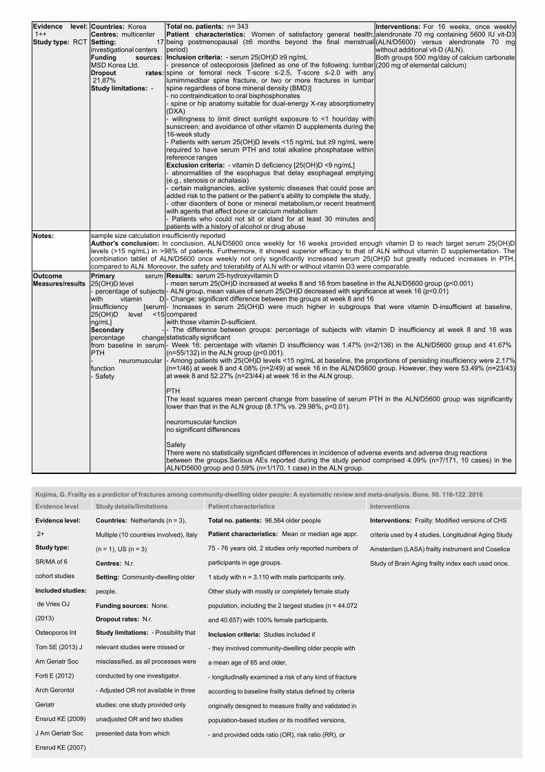
Evidence level: 1++
Study type: RCT
Countries: Korea Centres: multicenter Setting: 17 investigational centers Funding sources: MSD Korea Ltd. Dropout rates: 21,87%
Study limitations: -
Total no. patients: n= 343 Patient characteristics: Women of satisfactory general health; being postmenopausal (≥6 months beyond the final menstrual period) Inclusion criteria: - serum 25(OH)D ≥9 ng/mL - presence of osteoporosis [defined as one of the following: lumbar spine or femoral neck T-score ≤-2.5, T-score ≤-2.0 with any lumimmedibar spine fracture, or two or more fractures in lumbar spine regardless of bone mineral density (BMD)] - no contraindication to oral bisphosphonates - spine or hip anatomy suitable for dual-energy X-ray absorptiometry (DXA) - willingness to limit direct sunlight exposure to <1 hour/day with sunscreen; and avoidance of other vitamin D supplements during the 16-week study - Patients with serum 25(OH)D levels <15 ng/mL but ≥9 ng/mL were required to have serum PTH and total alkaline phosphatase within reference ranges Exclusion criteria: - vitamin D deficiency [25(OH)D <9 ng/mL] - abnormalities of the esophagus that delay esophageal emptying (e.g., stenosis or achalasia) - certain malignancies, active systemic diseases that could pose an added risk to the patient or the patient’s ability to complete the study, - other disorders of bone or mineral metabolism,or recent treatment with agents that affect bone or calcium metabolism - Patients who could not sit or stand for at least 30 minutes and patients with a history of alcohol or drug abuse
Interventions: For 16 weeks, once weekly alendronate 70 mg containing 5600 IU vit-D3 (ALN/D5600) versus alendronate 70 mg without additional vit-D (ALN). Both groups 500 mg/day of calcium carbonate (200 mg of elemental calcium)
Notes: sample size calculation insufficiently reported Author's conclusion: In conclusion, ALN/D5600 once weekly for 16 weeks provided enough vitamin D to reach target serum 25(OH)D levels (>15 ng/mL) in >98% of patients. Furthermore, it showed superior efficacy to that of ALN without vitamin D supplementation. The combination tablet of ALN/D5600 once weekly not only significantly increased serum 25(OH)D but greatly reduced increases in PTH, compared to ALN. Moreover, the safety and tolerability of ALN with or without vitamin D3 were comparable.
Outcome Measures/results
Primary serum 25(OH)D level - percentage of subjects with vitamin D insufficiency [serum 25(OH)D level <15 ng/mL] Secondary - percentage change from baseline in serum PTH - neuromuscular function - Safety
Results: serum 25-hydroxyvitamin D - mean serum 25(OH)D increased at weeks 8 and 16 from baseline in the ALN/D5600 group (p<0.001) - ALN group, mean values of serum 25(OH)D decreased with significance at week 16 (p<0.01) - Change: significant difference between the groups at week 8 and 16 - Increases in serum 25(OH)D were much higher in subgroups that were vitamin D-insufficient at baseline, compared with those vitamin D-sufficient. - The difference between groups: percentage of subjects with vitamin D insufficiency at week 8 and 16 was statistically significant - Week 16: percentage with vitamin D insufficiency was 1.47% (n=2/136) in the ALN/D5600 group and 41.67% (n=55/132) in the ALN group (p<0.001). - Among patients with 25(OH)D levels <15 ng/mL at baseline, the proportions of persisting insufficiency were 2.17% (n=1/46) at week 8 and 4.08% (n=2/49) at week 16 in the ALN/D5600 group. However, they were 53.49% (n=23/43) at week 8 and 52.27% (n=23/44) at week 16 in the ALN group. PTH The least squares mean percent change from baseline of serum PTH in the ALN/D5600 group was significantly lower than that in the ALN group (8.17% vs. 29.98%, p<0.01). neuromuscular function no significant differences Safety There were no statistically significant differences in incidence of adverse events and adverse drug reactions between the groups.Serious AEs reported during the study period comprised 4.09% (n=7/171, 10 cases) in the ALN/D5600 group and 0.59% (n=1/170, 1 case) in the ALN group.
Kojima, G. Frailty as a predictor of fractures among community-dwelling older people: A systematic review and meta-analysis. Bone. 90. 116-122. 2016
Evidence level Study details/limitations Patient characteristics Interventions Evidence level:
2+
Study type:
SR/MA of 6
cohort studies
Included studies:
de Vries OJ
(2013)
Osteoporos Int
Tom SE (2013) J
Am Geriatr Soc
Forti E (2012)
Arch Gerontol
Geriatr
Ensrud KE (2009)
J Am Geriatr Soc
Ensrud KE (2007)
Countries: Netherlands (n = 3),
Multiple (10 countries involved), Italy
(n = 1), US (n = 3)
Centres: N.r. Setting: Community-dwelling older
people.
Funding sources: None. Dropout rates: N.r.
Study limitations: - Possibility that
relevant studies were missed or
misclassified, as all processes were
conducted by one investigator.
- Adjusted OR not available in three
studies: one study provided only
unadjusted OR and two studies
presented data from which
Total no. patients: 96,564 older people
Patient characteristics: Mean or median age appr.
75 - 76 years old, 2 studies only reported numbers of
participants in age groups.
1 study with n = 3.110 with male participants only.
Other study with mostly or completely female study
population, including the 2 largest studies (n = 44.072
and 40.657) with 100% female participants.
Inclusion criteria: Studies included if - they involved community-dwelling older people with
a mean age of 65 and older,
- longitudinally examined a risk of any kind of fracture
according to baseline frailty status defined by criteria
originally designed to measure frailty and validated in
population-based studies or its modified versions,
- and provided odds ratio (OR), risk ratio (RR), or
Interventions: Frailty: Modified versions of CHS
criteria used by 4 studies, Longitudinal Aging Study
Amsterdam (LASA) frailty instrument and Coselice
Study of Brain Aging frailty index each used once.
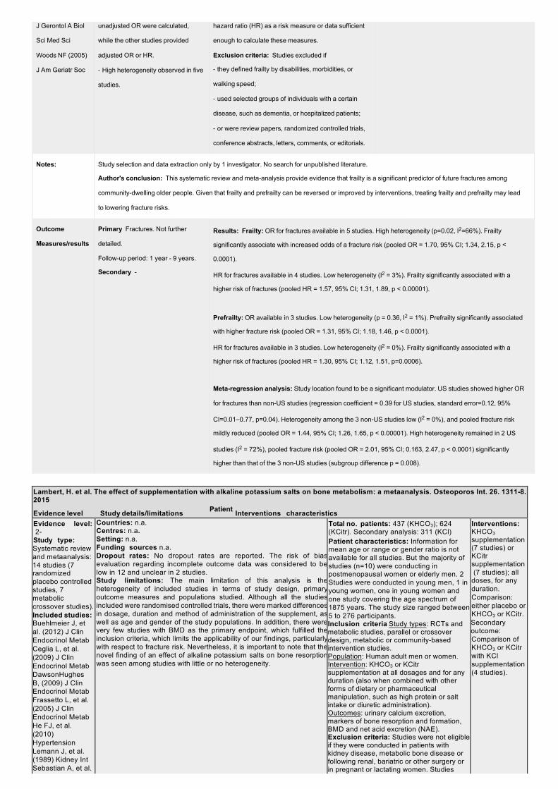
J Gerontol A Biol
Sci Med Sci
Woods NF (2005)
J Am Geriatr Soc
unadjusted OR were calculated,
while the other studies provided
adjusted OR or HR.
- High heterogeneity observed in five
studies.
hazard ratio (HR) as a risk measure or data sufficient
enough to calculate these measures.
Exclusion criteria: Studies excluded if - they defined frailty by disabilities, morbidities, or
walking speed;
- used selected groups of individuals with a certain
disease, such as dementia, or hospitalized patients;
- or were review papers, randomized controlled trials,
conference abstracts, letters, comments, or editorials.
Notes:
Study selection and data extraction only by 1 investigator. No search for unpublished literature.
Author's conclusion: This systematic review and meta-analysis provide evidence that frailty is a significant predictor of future fractures among
community-dwelling older people. Given that frailty and prefrailty can be reversed or improved by interventions, treating frailty and prefrailty may lead
to lowering fracture risks.
Outcome
Measures/results
Primary Fractures. Not further
detailed.
Follow-up period: 1 year - 9 years. Secondary -
Results: Frailty: OR for fractures available in 5 studies. High heterogeneity (p=0.02, I2=66%). Frailty
significantly associate with increased odds of a fracture risk (pooled OR = 1.70, 95% CI; 1.34, 2.15, p <
0.0001).
HR for fractures available in 4 studies. Low heterogeneity (I2 = 3%). Frailty significantly associated with a
higher risk of fractures (pooled HR = 1.57, 95% CI; 1.31, 1.89, p < 0.00001).
Prefrailty: OR available in 3 studies. Low heterogeneity (p = 0.36, I2 = 1%). Prefrailty significantly associated
with higher fracture risk (pooled OR = 1.31, 95% CI; 1.18, 1.46, p < 0.0001).
HR for fractures available in 3 studies. Low heterogeneity (I2 = 0%). Frailty significantly associated with a
higher risk of fractures (pooled HR = 1.30, 95% CI; 1.12, 1.51, p=0.0006).
Meta-regression analysis: Study location found to be a significant modulator. US studies showed higher OR
for fractures than non-US studies (regression coefficient = 0.39 for US studies, standard error=0.12, 95%
CI=0.01–0.77, p=0.04). Heterogeneity among the 3 non-US studies low (I2 = 0%), and pooled fracture risk
mildly reduced (pooled OR = 1.44, 95% CI; 1.26, 1.65, p < 0.00001). High heterogeneity remained in 2 US
studies (I2 = 72%), pooled fracture risk (pooled OR = 2.01, 95% CI; 0.163, 2.47, p < 0.0001) significantly
higher than that of the 3 non-US studies (subgroup difference p = 0.008).
Lambert, H. et al. The effect of supplementation with alkaline potassium salts on bone metabolism: a metaanalysis. Osteoporos Int. 26. 1311-8. 2015
Evidence level Study details/limitations Patient Interventions characteristics Evidence level: 2-
Study type: Systematic review and metaanalysis: 14 studies (7 randomized placebo controlled studies, 7 metabolic crossover studies). Included studies: Buehlmeier J, et al. (2012) J Clin Endocrinol Metab Ceglia L, et al. (2009) J Clin Endocrinol Metab DawsonHughes B, (2009) J Clin Endocrinol Metab Frassetto L, et al. (2005) J Clin Endocrinol Metab He FJ, et al. (2010) Hypertension Lemann J, et al. (1989) Kidney Int Sebastian A, et al.
Countries: n.a. Centres: n.a. Setting: n.a. Funding sources n.a. Dropout rates: No dropout rates are reported. The risk of bias evaluation regarding incomplete outcome data was considered to be low in 12 and unclear in 2 studies. Study limitations: The main limitation of this analysis is the heterogeneity of included studies in terms of study design, primary outcome measures and populations studied. Although all the studies included were randomised controlled trials, there were marked differences in dosage, duration and method of administration of the supplement, as well as age and gender of the study populations. In addition, there were very few studies with BMD as the primary endpoint, which fulfilled the inclusion criteria, which limits the applicability of our findings, particularly with respect to fracture risk. Nevertheless, it is important to note that the novel finding of an effect of alkaline potassium salts on bone resorption was seen among studies with little or no heterogeneity.
Total no. patients: 437 (KHCO3); 624 (KCitr). Secondary analysis: 311 (KCl) Patient characteristics: Information for mean age or range or gender ratio is not available for all studies. But the majority of studies (n=10) were conducting in postmenopausal women or elderly men. 2 Studies were conducted in young men, 1 in young women, one in young women and one study covering the age spectrum of 1875 years. The study size ranged between 5 to 276 participants. Inclusion criteria Study types: RCTs and metabolic studies, parallel or crossover design, metabolic or community-based intervention studies. Population: Human adult men or women. Intervention: KHCO3 or KCitr supplementation at all dosages and for any duration (also when combined with other forms of dietary or pharmaceutical manipulation, such as high protein or salt intake or diuretic administration). Outcomes: urinary calcium excretion, markers of bone resorption and formation, BMD and net acid excretion (NAE). Exclusion criteria: Studies were not eligible if they were conducted in patients with kidney disease, metabolic bone disease or following renal, bariatric or other surgery or in pregnant or lactating women. Studies
Interventions: KHCO3 supplementation (7 studies) or KCitr supplementation (7 studies); all doses, for any duration. Comparison: either placebo or KHCO3 or KCitr. Secondary outcome: Comparison of KHCO3 or KCitr with KCl supplementation (4 studies).
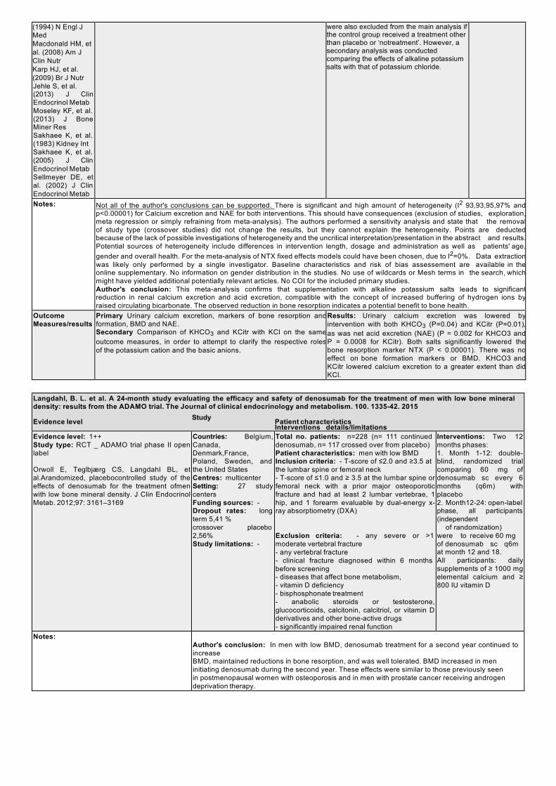
(1994) N Engl J Med Macdonald HM, et al. (2008) Am J Clin Nutr Karp HJ, et al. (2009) Br J Nutr Jehle S, et al. (2013) J Clin Endocrinol Metab Moseley KF, et al. (2013) J Bone Miner Res Sakhaee K, et al. (1983) Kidney Int Sakhaee K, et al. (2005) J Clin Endocrinol Metab Sellmeyer DE, et al. (2002) J Clin Endocrinol Metab
were also excluded from the main analysis if the control group received a treatment other than placebo or ‘notreatment’. However, a secondary analysis was conducted comparing the effects of alkaline potassium salts with that of potassium chloride.
Notes: Not all of the author's conclusions can be supported. There is significant and high amount of heterogeneity (I2 93,93,95,97% and p<0.00001) for Calcium excretion and NAE for both interventions. This should have consequences (exclusion of studies, exploration, meta regression or simply refraining from meta-analysis). The authors performed a sensitivity analysis and state that the removal of study type (crossover studies) did not change the results, but they cannot explain the heterogeneity. Points are deducted because of the lack of possible investigations of heterogeneity and the uncritical interpretation/presentation in the abstract and results. Potential sources of heterogeneity include differences in intervention length, dosage and administration as well as patients' age, gender and overall health. For the meta-analysis of NTX fixed effects models could have been chosen, due to I2=0%. Data extraction was likely only performed by a single investigator. Baseline characteristics and risk of bias assessement are available in the online supplementary. No information on gender distribution in the studies. No use of wildcards or Mesh terms in the search, which might have yielded additional potentially relevant articles. No COI for the included primary studies. Author's conclusion: This meta-analysis confirms that supplementation with alkaline potassium salts leads to significant reduction in renal calcium excretion and acid excretion, compatible with the concept of increased buffering of hydrogen ions by raised circulating bicarbonate. The observed reduction in bone resorption indicates a potential benefit to bone health.
Outcome Measures/results
Primary Urinary calcium excretion, markers of bone resorption and formation, BMD and NAE. Secondary Comparison of KHCO3 and KCitr with KCl on the same outcome measures, in order to attempt to clarify the respective roles of the potassium cation and the basic anions.
Results: Urinary calcium excretion was lowered by intervention with both KHCO3 (P=0.04) and KCitr (P=0.01), as was net acid excretion (NAE) (P = 0.002 for KHCO3 and P = 0.0008 for KCitr). Both salts significantly lowered the bone resorption marker NTX (P < 0.00001). There was no effect on bone formation markers or BMD. KHCO3 and KCitr lowered calcium excretion to a greater extent than did KCl.
Langdahl, B. L. et al. A 24-month study evaluating the efficacy and safety of denosumab for the treatment of men with low bone mineral density: results from the ADAMO trial. The Journal of clinical endocrinology and metabolism. 100. 1335-42. 2015
Evidence level Study Patient characteristics Interventions details/limitations
Evidence level: 1++ Study type: RCT _ ADAMO trial phase II open label Orwoll E, Teglbjærg CS, Langdahl BL, et al.Arandomized, placebocontrolled study of the effects of denosumab for the treatment ofmen with low bone mineral density. J Clin Endocrinol Metab. 2012;97: 3161–3169
Countries: Belgium, Canada, Denmark,France, Poland, Sweden, and the United States Centres: multicenter Setting: 27 study centers Funding sources: - Dropout rates: long term 5,41 % crossover placebo 2,56% Study limitations: -
Total no. patients: n=228 (n= 111 continued denosumab, n= 117 crossed over from placebo) Patient characteristics: men with low BMD Inclusion criteria: - T-score of ≤2.0 and ≥3.5 at the lumbar spine or femoral neck - T-score of ≤1.0 and ≥ 3.5 at the lumbar spine or femoral neck with a prior major osteoporotic fracture and had at least 2 lumbar vertebrae, 1 hip, and 1 forearm evaluable by dual-energy x- ray absorptiometry (DXA) Exclusion criteria: - any severe or >1 moderate vertebral fracture - any vertebral fracture - clinical fracture diagnosed within 6 months before screening - diseases that affect bone metabolism, - vitamin D deficiency - bisphosphonate treatment - anabolic steroids or testosterone, glucocorticoids, calcitonin, calcitriol, or vitamin D derivatives and other bone-active drugs - significantly impaired renal function
Interventions: Two 12 months phases: 1. Month 1-12: double- blind, randomized trial comparing 60 mg of denosumab sc every 6 months (q6m) with placebo 2. Month12-24: open-label phase, all participants (independent of randomization) were to receive 60 mg of denosumab sc q6m at month 12 and 18. All participants: daily supplements of ≥ 1000 mg elemental calcium and ≥ 800 IU vitamin D
Notes: Author's conclusion: In men with low BMD, denosumab treatment for a second year continued to increase BMD, maintained reductions in bone resorption, and was well tolerated. BMD increased in men initiating denosumab during the second year. These effects were similar to those previously seen in postmenopausal women with osteoporosis and in men with prostate cancer receiving androgen deprivation therapy.

Outcome Measures/results Primary primary, secondary not defined Secondary - percent changes in BMD of the lumbar spine, total hip, femoral neck, trochanter, and 1/3 radius - percent change from baseline in serum type I collagen C-telopeptide [sCTX] - safety
Results: BMD long term denosumab group: - month 12-24: increases of BMD continued at all skeletal sites evaluated (2.2% lumbar spine, 0.9% total hip, 1.3% femoral neck, 1.3% trochanter, and 0.2% 1/3 radius) - baseline-month 24: 8.0% (lumbar spine), 3.4% (total hip), 3.4% (femoral neck), 4.6% (trochanter), and 0.7% (1/3 radius) - (all P < .01) Placebo Crossover Group: - month 12-24: significant gains in BMD at the lumbar spine (4.9%), total hip (1.7%), femoral neck (1.9%), trochanter (2.0%), and 1/3 radius (1.0%) (all P < .01) - month 1-24: similar to those observed in the long-term denosumab group during the first 12 months (lumbar spine, 5.8%; total hip, 2.3%; femoral neck, 2.2%; trochanter, 3.2%; and 1/3 radius, 0.6%).
Bone turnover markers long term denosumab group: - month 12: 60% reduction in sCTX(P < .0001). - months 18 and 24, sCTX levels remained lower than baseline (57% and 50%)(P < .0001) Placebo Crossover Group: - month 1-12: no change - months 12-24: median decrease in sCTX of 68% at month 18 and 59% at month 24 (P < .0001)
Anti-denosumab antibody assays Anti-denosumab binding antibodies were not detected at anytime point
Fractures During phase II, clinical fractures in 4 men (2 rib and 2 foot, (3.6%))and Clinical osteoporotic fractures were reported in 2 men (2 rib) in the long- term denosumab treatment group (1.8%). No new vertebral fractures.
SAFETY During the open-label phase, the participant incidence of overall AEs was 63% in the long-term group and 52% in the crossover group. Most AEs were mild or moderate in severity in both groups.
LeBlanc, A. et al. Encounter Decision Aid vs. Clinical Decision Support or Usual Care to Support Patient-Centered Treatment Decisions in Osteoporosis: The Osteoporosis Choice Randomized Trial II. PLoS One. 10. e0128063. 2015 Evidence level Study details/limitations Patient characteristics Interventions Evidence level: 1-
Study type: RCT
Countries: USA Centres: Family Medicine, Preventive Medicine, Primary Care Internal Medicine and General Internal Medicine Setting: Primary care Funding sources: This study was supported by a grant from the Foundation for Informed Medical Decision Making (Now the Informed Medical Decisions Foundation, http://www.informedmedicaldecisions.org/). The funders had no role in study design, data collection and analysis, decision to publish, or preparation of the manuscript. Dropout rates: 1 post hoc exclusion after randomization in "usual care" arm because patient was already on bisphosphonates (7.1%). 1 post hoc exclusion after randomization in "FRAX" arm because patient withdrew consent. 6 patients lost to follow-up in "usual care" arm (42.9%), 11 patients and 5 clinicians lost to follow-up in "FRAX" arm (48.5%), 10 patients and 4 clinicians lost to follow- up in "Decision aid" arm (43.8%). Study limitations: Inability to determine impact of intervention on patient adherence to bisphosphonates, a central issue in osteoporosis care. Results too imprecise to make a judgment. Clinicians had very limited experience in using the decision aid with their patients.
Total no. patients: 79 women randomized, 77 analyzed Patient characteristics: Mean Age (SD): 69 (8)
years in Decision Aid arm; 66 (10) years in Frax/Usual Care arm. Mean BMI (SD): 27 (6) years in Decision Aid arm; 28 (5) years in Frax/Usual Care arm. Mean Frax Score (SD): 14 (8) % in Decision Aid arm; 13 (7) % in Frax/Usual Care arm. Inclusion criteria:
Clinicians (i.e., physicians, nurse
practitioners): from participating primary care practices affiliated with Mayo Clinic and provided care for patients with osteopenia or osteoporosis. Patients: English- speaking women over 50, with a diagnosis of osteopenia or osteoporosis, not taking bisphosphonates or other prescription medications to treat their condition, identified by their clinician as potentially eligible for bisphosphonates, available for a six month follow up after randomization, and no major learning barriers. Exclusion criteria: -
Interventions: Decision aid arm: Use of the Osteoporosis Choice decision aid by clinician and patient during the clinical encounter: included (a) individualized 10-year risk of having a bone fracture (by FRAX calculator) with and without use of bisphosphonates (i.e., showing the absolute reduction with bisphosphonates) represented using an evidence-based pictograph and assuming a treatment-related reduction in overall fractures of 40%; and (b) potential harms and other downsides of using bisphosphonates. The study coordinator prepared each decision aid according to each patient’s characteristics prior to the clinical encounter. Shared decision-making with patient about whether to start bisphosphonates and whether to start at that time or at a later time. FRAX arm: consisted of giving clinicians a copy of patient’s individualized 10-year risk of having a bone fracture estimated using FRAX calculator before visit for use during clinical encounter. Usual Care: Clinicians discussed risk of fractures and treatment as usual without any research-related intervention.
Notes: Allocation concealment mentioned but not specified. Study planned and started with 2 intervention arm. One additional arm ("usual care") was added 5 months after enrollment had already started. Reasons not stated comprehensively and indicates deficiencies in study planning). Consequently, sample size calculation is invalid and study underpowered. This is further compounded as original sample size of 150 participants was not reached since study funding run out beforehand. Unclear, if 6 months follow-up could be maintained for patients in "usual care" arm. Obtainment of pharmacy records 9 months after enrollment impossible for this intervention arm because study ended before this point.
Patient's characteristics for "usual care" arm and "Frax" arm not separably reported, only that there was "no difference between the FRAX and usual care arms, nor were overall results significantly impacted by analyses comparing the three arms versus only two arms [...]. Therefore, the results comparing the FRAX arm and the Usual Care arm were combined and all subsequent results are presented as Decision Aid vs. FRAX/Usual Care arm". Separate analysis of arms would have improved trust in results, especially since follow-up proceedings vary. Post hoc exclusion of patients after randomization in analyses (no true ITT). Author's conclusion: Compared to usual care with or without the FRAX calculation, use of the Osteoporosis Choice decision aid during the consultation efficiently improves shared decision making about therapy for osteoporosis between primary care clinicians and at-risk women.
Outcome Measures/results
Primary Patients’ knowledge, adherence to medication, involvement in decision making by the clinician, fidelity to intended intervention, acceptability, satisfaction, and quality of life. Secondary Decisional conflict, decision to start medication.
Results: Patient knowledge: Significant increase in patient knowledge in the Decision Aid arm about matters covered in the decision aid (Knowledge of Risk without medication: RR = 2.0 [95% CI; 1.2, 3.2], p=0.01; Knowledge of Post-treatment risk: RR = 2.6 [95% CI; 1.6, 4.4], p<0.0001, and no difference in knowledge of matters not covered in the decision aid (Overall: Median = 7.0 [IQR: 4.5, 9.0] for Decision Aid; Median = 5.5 [IQR: 2.5, 8.0] for FRAX/ Usual Care, p=0.11). Prescription rate: No significant differences between groups. Adherence and initial prescription filled: No significant differences between groups. Patient involvement in decision: Significantly higher in Decision aid arm: OPTION score: 57% (95%CI; 50, 64) vs 43% (95% CI; 37, 48), t-test p = 0.001. Patient satisfaction: No significant differences between groups.
Leder, B. Z. et al. Denosumab and teriparatide transitions in postmenopausal osteoporosis (the DATA-Switch study): extension of a randomised controlled trial. Lancet (London, England). 386. 1147-55. 2015 Evidence level Study Patient characteristics Interventions
details/limitations Evidence level: 1++
Study type: RCT – Extension of DATA study: DATA-Switch
Countries: USA Centres: USA Setting: - Funding sources:
Amgen, Eli Lilly, and National Institutes of Health Dropout rates: 7,23% Study limitations: First, the size of the study precludes an assessment of the relative safety or antifracture efficacy of the three assigned treatment regimens. Bone mineral density, however, has proven to be a reliable, although imperfect, predictor of antifracture efficacy in patients treated with osteoporosis drugs, including denosumab and teriparatide. Additionally, the specific clinical impact of the transient bone loss that occurs in women switching from denosumab to teriparatide cannot be precisely estimated.
Total no. patients: N=83 Patient characteristics: Postmenopausal women aged 45 years or older Inclusion criteria: DATA study: At least 36 months beyond their last menses (or 36 months after hysterectomy) and high fracture risk - BMD T score ≤–2,5 at the spine, hip, or femoral neck - BMD T score ≤–2,0 with at least one BMD independent risk factor - BMD T score ≤–1,0 with a history of a fragility fracture DATA-Switch - 1) dual x-ray absorptiometry spine or hip T score
Exclusion criteria: DATA study:
‑ evidence of hyperparathyroidism
‑ vitamin D deficiency (serum level <20 ng/mL)
‑ other congenital or acquired bone disease
‑ history of malignancy<br>
Interventions: Original DATA study: 24 months. Teriparatide 20 μg subcutaneously daily, denosumab 60 mg subcutaneously every 6 months, or both drugs. DATA-Switch: After completion of DATA study, women originally assigned to teriparatide received 24 months denosumab (teriparatide to denosumab group), those originally assigned to denosumab received 24 months teriparatide (denosumab to teriparatide group), and those originally assigned to both received 24 months of denosumab alone (combination to denosumab group).
Notes: no sample size calculation Author's conclusion: In postmenopausal osteoporotic women switching from teriparatide to denosumab, bone mineral density continued to increase, whereas switching from denosumab to teriparatide results in progressive or transient bone loss. These results should be considered when choosing the initial and subsequent management of postmenopausal osteoporotic patients.
Outcome Measures/results
Primary Percent change in posterioranterior spine bone mineral density over 4 years Secondary percent change in total hip, femoral neck, and radius shaft BMD percent change in serum osteocalcin and Ctelopeptide concentrations
Results: Original DATA study: after 24 months of the originally assigned drugs, mean lumbar spine bone mineral density had increased significantly in all treatment groups relative to baseline with the greatest increases in women treated with both drugs together. DATASwitch: Lumbar Spine BMD: After 48 months: no significant difference between any of the treatment groups increased more after the treatment transition (between months 24 and 48) in the teriparatide to denosumab group (8·6 [SD 5·0]) than in either the denosumab to teriparatide group (4·8 [5·6]; between group p=0·0203) or the combination to denosumab group (3·4 [3·5]; betweengroup p=0·0005). Total hip BMD: After 48 months: increased more in the combination to denosumab group than in either the teriparatide to denosumab group (betweengroup p=0·0446) or the denosumab to teriparatide group (betweengroup p<0·0001). After 48 months: increased more in the teriparatide to denosumab group than in the denosumab to teriparatide group (betweengroup p=0·0002). After transition, months 24-48: increased more in the teriparatide to denosumab group (4·7% [SD 2·6]) than in both the combination to denosumab group (2·2% [1·8]; p=0·0008) and the denosumab to teriparatide group (–0·7% [3·1]; p<0·0001). Femoral neck BMD:
After 48 months: increased by 8·3% (SD 5·6) in the teriparatide to denosumab group, 4·9% (6·0) in the denosumab to teriparatide group, and 9·1% (6·1) in the combination to denosumab group (denosumab to teriparatide vs teriparatide to denosumab p=0·0447; denosumab to teriparatide vs combination to denosumab p=0·0336). After transition, months 24-48: increases in the teriparatide to denosumab group (5·6% [SD 4·5]) were larger than in either the combination to denosumab group (2·1% [4·9]; p=0·0156) or the denosumab to teriparatide group (1·2% [4·9]; p=0·0019) Distal radius BMD After 48 months: patients originally treated with denosumab switched to teriparatide, radius bone mineral density progressively decreased resulting in net 0–48month decrease of –1·8% (SD 5·9). After 48 months: increase by 2·8% (SD 3·2) in the combination to denosumab group and reverted to the original baseline in the teriparatide to denosumab group (0·0% [2·9]). After 48 months: increases at the distal 1/3 radius in the combination to denosumab group were significantly larger than those either in the teriparatide to denosumab group (p=0·0075) or the denosumab to teriparatide group (p=0·0099). Osteocalcin After 48 months: treated with denosumab followed by teriparatide, median osteocalcin increased by 275% (IQR 176–410) above the original baseline after 6 months of teriparatide (month 30) and remained 159% (81–302) over the original baseline even after 24 months of teriparatide. Ctelopeptide concentrations increased by 183% (IQR 80–343) at month 30 and were 42% (22–120) above baseline After 48 months: treated with teriparatide followed by denosumab, bone resorption (Ctelopeptide) was maximally suppressed after 1 month of denosumab, whereas bone formation (osteocalcin) was not maximally suppressed until 12–24 months of denosumab treatment. After 48 months: treated with combination therapy followed by denosumab monotherapy, however, both markers were maximally suppressed at all postswitch timepoints. After 48 months: comparing women in the teriparatide to denosumab group vs the combination to denosumab group (both of which are receiving the same treatment from month 24–48), serum osteocalcin was significantly higher in those switching from teriparatide monotherapy to denosumab monotherapy than in those switching from combination therapy to denosumab monotherapy at 1 months and 6 months after the transition (months 25 and 30; p<0·0001 at month 25 and p<0·0023 at month 30). Conversely, Ctelopeptide in these two groups did not differ at any timepoint after the treatment transitions. Leder BZ, Tsai JN, Uihlein AV, et al. Two years of Denosumab and teriparatide administration in postmenopausal women with osteoporosis (The DATA Extension Study): a randomized controlled trial. J Clin Endocrinol Metab 2014; 99: 1694– 700. Tsai JN, Uihlein AV, Lee H, et al. Teriparatide and denosumab, alone or combined, in women with postmenopausal osteoporosis: the DATA study randomised trial. Lancet 2013; 382: 50–56.
Leder, B. Z. et al. Two years of Denosumab and teriparatide administration in postmenopausal women with osteoporosis (The DATA Extension Study): a randomized controlled trial. The Journal of clinical endocrinology and metabolism. 99. 1694-700. 2014
Evidence level Study details/limitations Patient Interventions characteristics
Evidence level: 1++
Study type: RCT, DATA extension study
Countries: USA Centres: - Setting: - Funding sources: Eli Lilly Inc, Amgen Inc, and by the Harvard Clinical and Translational Science Center, the National Center for Research Resources. Dropout rates: Teriparatide 9,68%, Desonumab 6,06%, Combination 20% Study limitations: First, the study was powered to detect changes in BMD but not changes in fracture rates. A study aimed to assess comparative fracture risk reduction would require a very large sample size and would not be feasible in the context of an investigator-initiated trial. Nonetheless, treatment-induced increases in BMD at the various anatomic sites have proven to be reliable, although not perfect, surrogates for efficacy in fracture prevention and the larger increases in BMD in the combination group compared with those in the monotherapy groups (ranging from 3.4% to 5.6% at the spine and 3.1% to 4.3% at the hip) are very likely to be clinically significant. In addition, although treatment induced improvements in the non-BMD determinants of bone strength may be important, BMD changes in patients treated with both denosumab and teriparatide monotherapy have been shown to predict fracture reduction. Furthermore, our cohort size was limited and made up predominately of white women, potentially limiting generalizability and making subgroup analysis not feasible.
Total no. patients: N= 94
Patient characteristics:
postmenopausal women aged 45 or older Inclusion criteria: - at least 36 months since last menses (or since hysterectomy if the FSH level was >40 U/L) - at high fracture risk Exclusion criteria: - evidence of hyperparathyroidism, - vitamin D deficiency (serum level less than 20 ng/mL)
- other congenital or acquired bone disease - history of malignancy (with the exception of nonmelanoma skin cancer) - history of ionizing radiation therapy - significant cardiopulmonary, liver, or renal disease - major psychiatric disease - excessive alcohol intake - ever taken parenteral bisphosphonates, teriparatide, or strontium ranelate - glucocorticoids or oral bisphosphonates within 6 months of enrollment - estrogen, selective estrogen receptor modulators within 3 months of enrollment - calcitonin within 3 months of enrollment
Interventions: The DATA extension study is a 12-month extension to the original 12-month, open-label, randomized controlled trial. 60 mg of denosumab sc every 6 months (Prolia; Amgen, Inc), 20 µg of teriparatide sc daily (Forteo; Eli Lilly, Inc), or both medications for 12 months. Continuation to the assigned treatment for an additional 12 months (24 months of total therapy). Before the initial randomization, subjects were stratified based on age (<65 or ≥65 years) and prior bisphosphonate use (yes/no).
Notes: Sample size calculation no information. Modified intention-to-treat analysis, which included all data from subjects completing at least 1 postbaseline visit Author's conclusion: Two years of concomitant teriparatide and denosumab therapy increases BMD more than therapy with either medication alone and more than has been reported with any current therapy. The combination of these agents may prove to be an important treatment option in patients at high risk of fracture.
Outcome Measures/results
Primary Percent change in PA spine BMD at 24 months. Secondary - percent change in total hip, femoral neck, and radius shaft BMD - overall change in serum OC, P1NP, and CTX
Results: BMD 0- 24 months SPINE - mean PA spine BMD increased significantly in all treatment groups relative to the baseline (P<.0001 for all within-group comparisons increased more in the combination group than teriparatide or denosumab groups .01 vs p .008 increase did not differ significantly between and femoral neck months relative to baseline .0008 within .003 no significant difference .23 total hip .001 .08 distal radius by both small decrease was statistically magnitude of change similar .95 although differed compared with .004 bmd there were between-group differences changes second year therapy. however increases. spine .007 .09 biochemical markers bone turnover women treated alone mean serum oc p1np ctx at time points whereas they decreased other net auc each individual point which had month remained similarly suppressed slowly. overall lower monotherapy furthermore levels despite progressively greater suppression thereafter respectively as it measured finally comparison safety calcium>10.8 mg/dL were observed. All serious adverse events, including those occurring in year 1, were considered unrelated to the study therapy.
Leung, K. S. et al. Effects of 18-month low-magnitude high-frequency vibration on fall rate and fracture risks in 710 community elderly--a cluster-randomized controlled trial. Osteoporos Int. 25. 1785-95. 2014
Evidence level Study details/limitations Patient Interventions characteristics Evidence level: 1+
Study type: RCT
Countries: China Centres: Elderly community centers Setting: Standardized settings (sharing the same scope of service, operating hours, manpower, and target groups) Funding sources: This study was supported by General Research Fund (Ref: 469508) of University Grants Committee (www.ugc.edu.hk), Hong Kong SAR government. Dropout rates: Control group: 50 participant lost to follow-up (14.5%) Vibration group: 108 participants lost to follow-up (29.7%). Main reason in both groups: "Declined follow-up" Study limitations: - Ideal design of study would have been individual randomized double-blinded. → Blinding of subjects difficult if not impossible because vibration though in low magnitude can easily be felt → Would have been most efficient to have the elderly grouped together in centers where vibration platforms were housed and closely monitored by the center staff during vibration sessions - Low incidence rate of fracture resulted in low power to detect effects on fracture prevention - Difference in dropout rates due to demands for daily vibration intervention in centers, while control group remained as participants in regular program in community centers.
Total no. patients: 710 women Patient characteristics:
Vibration group: Age (years): 74.5 (7.1); Body mass index (kg/m2): 24.1 (3.6); Hypertension: 192 (53 %); Diabetes: 53 (15 %). Control group: Age (years): 71.3 (7.2); Body mass index (kg/m2): 24.0 (3.7); Hypertension: 167 (48 %); Diabetes: 50 (14 %). Inclusion criteria: Healthy females aged 60 years or above, independent and active in the community. Exclusion criteria: (1) Women taking any medications or had medical conditions which affected metabolism of the musculoskeletal system, e.g., bisphosphonates, (2) Women who participated in supervised regular exercise for twice a week or more, (3) Women with pacemaker in situ, (4) Women with malignancy, (5) Women with a history of smoking or excessive alcohol use (more than seven drinks per week).
Interventions: Control group: Subjects remained in their habitual life style and participated in the normal interest group activities (e.g., card games, drama) organized by the community centers. Vibration group: Women received LMHFV by standing upright without knee bending on a specially designed vibration platform that provided vertical synchronous vibration at 35 Hz, 0.3 g (peak-to-peak magnitude), displacement of <0.1mm, 20 min/day, 5 days/week for 18 months, in addition to the normal activities in the centers. Safety issues and operative procedures of LMHFV instructed by research staff. On-site validation/ calibration of platforms monthly by technician.
Notes: Randomisation method acceptable although prone to subvertion (Center-based simple randomization by envelop drawing). Investigators blinded. Subjects not blinded "[...] because the vibration signal from the treatment platform could easily be felt [...]". No test for baseline differences stated. Not stated whether subjects had significant differences at baseline. Cluster randomisation of centers (not individuals) might compound confounding influence of centers. Analyses of fall incidence based on subjects who reported their fall records (no true ITT-analyses). No data/ differences regarding centers reported. Author's conclusion: In conclusion, this is the first study to show the beneficial effects of LMHFV in the prevention of falls among
the elderly in the community. The LMHFV is well accepted with negligible adverse effects. This noninvasive intervention should be promoted in the community as an effective fall prevention program and to decrease fall-related injuries.
Outcome Measures/results
Primary Composite occurrence of fall or fracture after 18 month (self-reported and confirmed with clinical information including radiographs obtained from electronic patient record of Hong Kong Hospital Authority) Secondary Compliance rate (built-in data logger in vibration platform), Balancing ability (Basic Balance Master System; NeuroCom International Inc, OR, USA), Quadriceps muscle (dynamometer attached at malleoli level), Areal bone mineral density (BMD, g/cm2)
Results: Fall or fracture incidence: Based on 334 vibration group subjects and 327 control subjects who reported their fall records. 62 (18.6 % of 334) subjects with falls or fractures in vibration group and 94 (28.7 % of 327) in control group. Adjusted hazard ratio of falls or fractures in vibration group compared with control: 0.56 (95% CI; 0.40, 0.78, p=0.001). Adjusted incidence rate of falls in compared with control group: 0.54 (95% CI; 0.37, 0.78, p=0.001). Rate of fracture in vibration group: 1.1%; Rate of fracture in control group: 2.31 %. No statistically significant difference (adjusted HR=0.42, 95%CI; 0.12, 1.45, p=0.171). Compliance: Average of 3.3 vibration sessions per week, 66 % vibration in the study period (18-month). Balancing ability: Balancing ability in vibration group significantly improved compared with control group from baseline to 18 months. Improvement in reaction time: −0.38 s (95% CI; −0.55, −0.21, p<0.001). Improvement in movement velocity: 0.94 °/s (95% CI; 0.63, 1.26, p<0.001). Improvement in maximum point excursion: 10.72 % (95%CI; 7.05, 14.39, p<0.001). No significant difference of directional control. Quadriceps muscle: At 18 month significant improvement in vibration group: Mean between-group difference in dominant leg = 2.46 kg (95% CI; 1.70, 3.22, p<0.001); Mean between-group difference in nondominant leg = 2.43 kg (95% CI; 1.59, 3.27, p<0.001). Areal bone mineral density: Lumbar spine: Mean change in vibration group 0.08% compared with −0.64% in control group at 18 months (mean between-group difference = 0.72 %, 95%CI; −0.12, 1.56, p=0.089). Hip BMD: Decrease in both groups. Mean change of total hip BMD in vibration group −1.86 % compared with −1.89 % in control group. Health-related quality of life: No significant difference between groups after 18 months (Total score in vibration group = 2.45 [95% CI; 0.01, 4.90] vs Total score in control group = 1.62 [95% CI; -1.18, 4.43)]
Li, S. et al. Long-term calcium supplementation may have adverse effects on serum cholesterol and carotid intima-media thickness in postmenopausal women: a double-blind, randomized, placebo-controlled trial. Am J Clin Nutr. 98. 1353-9. 2013 Evidence level Study details/limitations Patient characteristics Interventions Evidence level: 1++
Study type: RCT
Countries: China Centres: single center Setting: Harbin Medical University, Harbin Funding sources: National Natural Science Foundation of China Dropout rates: 2,63%, 4,4% Study limitations: The follow-up period was only 2 y as a result of the negative calcium effects. Therefore, there was only one post randomization measurement, which prevented the use of intention-to-treat analyses. Instead, per-protocol analyses were used. Per-protocol analyses may overestimate effects compared with intention-to-treat analyses. Therefore, we could not directly evaluate whether an increase in serum cholesterol concentrations would increase risk of CVD because of the relatively short intervention period.
Total no. patients: see below Patient characteristics: 190 premenopausal women (30–40 y old) and 182 postmenopausal women (50–60 y old) with dyslipidemia Inclusion criteria: Dyslipidemia: fasting triglyceride concentration >1.7 mmol/L, fasting total cholesterol concentration >5.18 mmol/L, fasting LDL cholesterol >3.37 mmol/L, or fasting HDL cholesterol concentration <1.04 mmol/L Exclusion criteria: - Any major diseases - regular medication use, which affected calcium or lipid metabolism - serum 25-hydroxyvitamin D3 concentration ,20 nmol/L - use bisphosphonates or hormone therapy in the previous year, - calcium or vitamin D3 supplementation in the previous year, - pregnant women, breastfeeding women, hysterectomized women, or perimenopausal period
Interventions: 800 mg Ca/d or a placebo for 2 years
Notes: No sample size calculation, no ITT Author's conclusion: Calcium supplementation in postmenopausal women with dyslipidemia increases serum total cholesterol concentrations and CIMT. In postmenopausal women with dyslipidemia, calcium supplements should be prescribed with caution
Outcome Measures/results
Primary No outcome measures defined. - Total cholesterol, - HDL-cholesterol, - LDL-cholesterol, - triglyceride, - glucose, - calcium - blood pressure - Serum estradiol - Carotid intima-media thickness (CIMT) Secondary -
Results: - Serum total cholesterol concentration after 2 y increased significantly in the calcium group compared with that in the placebo group (P <0.001) - Serum triglyceride, LDL cholesterol, and HDL cholesterol concentrations and CIMT were not significantly affected after 2 y. - There was a significant interaction between calcium supplementation and menopausal status on serum cholesterol concentrations (P < 0.001) and CIMT (P = 0.017). The change of total cholesterol in premenopausal women and postmenopausal women after calcium supplementation was 0.04 ± 0.07 compared with 0.61 ± 0.21 mmol/L (P < 0.001). - The change of CIMT in premenopausal women and postmenopausal women after calcium supplementation was 0.0238 ±0.0348 compared with 0.0615 ± 0.134 mm (P = 0.003), respectively.
Liu, B. X. et al. The Effect of the Modified Eighth Section of Eight-Section Brocade on Osteoporosis in Postmenopausal Women: A Prospective Randomized Trial. Medicine (Baltimore). 94. e991. 2015 Evidence level Study details/limitations Patient characteristics Interventions

Evidence level: 1-
Study type: RCT
Countries: China Centres: Liuzhou traditional Chinese Medical Hospital and Guangzhou Hospital of Traditional Chinese Medicine Setting: Outpatient Funding sources: This study was supported by the National Science Foundation Committee (NSFC) of China (grant no. 81201971) and by the Natural Science Foundation of Guangdong (grant no. 2014A030313107) and by the Medical Scientific Research Foundation of Guangdong Province (grant no. B2012126) and also by Grant [2013]163 from Key Laboratory of Malignant Tumor Molecular Mechanism and Translational Medicine of Guangzhou Bureau of Science and Information Technology. Dropout rates: 14 of 198 women (7.1%). Control n=6; Ca n=5; MESE n=2; MESE+Ca n=1. Reasons: Constipation related to the study (n=3), serious back pain (n=5), fracture (n=4), and illness unrelated to study (n=2). Study limitations: "Possible, that delayed response is a function of mineralization, a process known to continue long after new bone tissue (osteoid) has been secreted by osteoblasts." No further limitations discussed.
Total no. patients: 198 women randomised, 184 analysed. Patient characteristics: Control group: Mean age (year): 61.87±8.29; Height: 156.6±4.04 Ca group: Mean age (year): 62.29±6.47; Height: 156.0±4.36 WESE group: Mean age (year): 63.23±7.56; Height: 154.27±4.34 WESE+Ca group: Mean age (year): 61.45±5.89; Height: 155.25±3.3 Inclusion criteria: (1) BMD in L2 to L4 and hip identified in women to be inadequate or BMD < –2.5 SD (T score -2.5) indicating osteoporosis, (2) Postmenopausal women without traumatic fracture at any sites and typical menopausal symptoms (3) Age from 50 to 75 years. Exclusion criteria: Women with cardiovascular or cerebrovascular disease, women with blood pressure higher than 160/110 mmHg on medication, women with systolic blood pressure less than 90 mmHg, women with heart stents or body implantation, women with a history of thrombosis within the past 6 months, women unrehabilitated from surgical operation, women which have been treated with drugs for osteoporosis or other agents which affect bone metabolism, women unrecovered from muscle strain joint injuries or fractures, women with symptoms of vertigo and in poor health, women with spinal nerve canal stenosis, spondylolisthesis, or lumbar disc herniation, and women with epileptics.
Interventions: Control group
(n=48); Ca alone (n=50; 500mg of calcium and 200 IU of vitamin D3, thrice daily); MESE (modified eighth section of Eight- Section Brocade) exercise alone (n=50; repetitions per time, thrice daily); MESE+Ca (n=50)
Notes: Method of randomisation not described. No allocation concealment mentioned. Investigators could have been blinded but were not. Placebo should have been applied for groups without Ca supplement for improved comparability. Adverse events (e.g. fractures) not included as outcomes, but as reasons for drop-out. No ITT-analyses. Author's conclusion: Our study reported greater improvement in the MESE and MESE+Ca group. We found that the treatment of MESE exercise was the most effective for alleviating the chronic pain, increasing the left femoral neck and lumbar BMD and balance in posture, which are associated with the increased bone fragility and higher risk of fracture. Importantly, the low attrition and exercise compliance coupled with the low number of adverse events indicates that this type of exercise program is safe, feasible, and well tolerated by previously untrained, but otherwise healthy older men [sic!]. Certainly, clearly more research is needed in order to better understand the specific therapeutic potential of MESE as an exercise model. Moreover, there seems to be a certain need for studies to assess any potential long-term risks. However, with appropriate introduction to the exercise modality, MESE exercise seems to be reasonably safe for most people.
Outcome Measures/results
Primary Bone and body composotion Secondary Balance and motility performance measures
Results: Pain Visual Analogue Scale Scores: Results not quantified/ data not shown. No significant difference for VAS in Ca group from 6 months to 12 months (P>0.05). VAS decreased significantly in MESE and MESE+Ca groups to control group at each time point. Improvement in chronic back pain in MESE+Ca group and group MESE group better than in Ca group after 12 months (P<0.05, P<0.001). Bone Mineral Density: Results not quantified/ data not shown. MESE resulted in 1.9% net gain in left femural neck BMD (not significant). Compared to control, significant higher increase after 12 months (P<0.001). Significant 1.4% to 1.9% within-group increase in both exercise groups for left femoral neck BMD. No significant difference in lumbar BMD in both MESE and MESE+Ca groups after treatment for 12 months (P>0.05). Change in Balance Capacity: Results not quantified/ data not shown. Significant better results in 3-feet Up and Go Test (3'UG) in MESE and MESE+Ca groups compared to control group after the treatment for 6 to 12 months (P<0.001 and P<0.001). Results in OLS test significantly longer (1.75, 2.23, and 3.21 seconds) in Ca, MESE, and MESE+Ca groups compared to control group after the treatment for 3 weeks (P<0.05, P<0.001, P<0.001).
Liu, S. et al. Risk factors for the second contralateral hip fracture in elderly patients: a systematic review and meta-analysis. Clin Rehabil. 29. 285-94. 2015
Evidence level Study details/limitations Patient characteristics Interventions Evidence level:
2-
Study type:
SR/MA of 13
case-control
studies.
Included studies:
Saxena P (2000)
Injury
Angthong C
(2009) Acta
Orthop Traumatol
Turc
Countries: Japan (n = 3), UK (n = 2), South
Korea (n = 2), Thailand (n = 1), Greece (n =
1), China (n = 1), Netherlands (n = 1), Italy (n
= 1) Centres: N.r.
Setting: N.r.
Funding sources: "This research received
no specific grant from any funding agency in
the public, commercial, or not for-profit
sectors."
Dropout rates: N.r. Study limitations: - All included studies with
case-control design. Recall and interviewer
biases unavoidable. Reported ORs mainly
Total no. patients: 1.544 cases and 15.541 controls
Patient characteristics: Age: 1 study did not provide information. All other
studies with participants at least ≥ 50 years.
Inclusion criteria: (1) only full-text articles with language restriction of
English;
(2) a study was performed to examine the association between the second
contralateral hip fracture and the risk factors, cases and controls were defined
based on the number of hip fractures sustained (bilateral and unilateral
respectively);
(3) sufficient data was published for estimating an odds ratio (OR) or
standardized mean difference (SMD) with 95% confidence intervals (CIs).
Exclusion criteria: -
Interventions:
Exposure:
Unilateral vs.
Bilateral hip
fractures
Sawalha S (2012)
J Bone Joint Surg
Brit
Chiu K (1992) Journal Trauma
Acute Care Sur
Yamanashi A
(2005) Osteop Int
Fukushima T
(2006) J
Orthop Sci
Vochteloo AJ
(2012) Arch
Orthop Trauma
Surg
Mitani S (2010) J
Orthop Sci
Rodaro E (2004)
Europa
Medicophysica
Dretakis K (1998)
Calcified Tissue
Int
Souder CD (2012)
J Bone Joint Surg
Lee SH (2012)
Yonsei Med J
Chang J-D (2013)
Injury
unadjusted.
- Significant heterogeneity unavoidable owing
to various measurement forms of some risk
factors and widely ranged follow-up periods.
- All subjects in the 13 articles were Asian,
European and US; conclusions might not
extrapolate to African and Australasian
populations.
Notes:
No search for unpublished literature. Not all relevant characteristics from study population of included studies presented (e.g. sex).
Results from sensitivity analysis not presented.
High risk of recall bias due to case-control design and confounding. Random-effects analysis performed even though defined threshold value of I2 ≥ 50 from chapter "Statistical analysis" was not reached (Fig.2: A -
Female).
References and in text citations partly not correct. Author's conclusion: Patients involved with female, living in institutions, osteoporosis, low vision, dizziness, dementia, respiration diseases and
cardiac diseases were at risk for a second contralateral hip fracture after the initial hip fracture.
Outcome
Measures/results
Primary Risk factors. No information on
measurement of factors provided.
Secondary -
Results: Significant risk factors identified:
- Female sex (OR = 1.30; 95% CI, 1.02, 1.64; P = 0.031),
- living in institutions (OR = 2.53; 95% CI; 1.33, 4.85; P = 0.005),
- osteoporosis (OR = 10.02; 95% CI; 5.41, 18.57; P <0.001),
- low vision (OR = 2.09; 95% CI; 1.06, 4.12; P = 0.034),
- dementia (OR = 2.02; 95% CI; 1.54, 2.65; P = 0.001),
- dizziness (OR = 2.87; 95% CI; 1.42, 5.87; P = 0.003)
- respiration diseases (OR = 2.58; 95% CI; 1.22, 5.47; P = 0.013).
Liu, Yunxia et al. Chinese herbal medicines for treating osteoporosis. Cochrane Database Syst Rev. CD005467. 2014 Evidence level Study details/limitations Patient characteristics Interventions Evidence level: 1+
Study type: MA of 109 RCT Included studies: See review.
Countries: China Centres: 3 multicentre randomised trials Setting: Inpatients and outpatients Funding sources: Internal sources of Support • School of Public Health, Shandong University, China.
Total no. patients: 10,655 people with osteoporosis Patient characteristics: 65 trials included postmenopausal osteoporosis patients, 8 trials included senile osteoporosis patients and 35 trials included primary osteoporosis patients unspecified as to life-stage or co- morbidity. Race/ethnicity of all patients was Chinese in all 108 trials.
Interventions: 99 different Chinese herbal medicines tested. 8 Chinese herbal medicines tested twice or more (Kanggusong granule in 2 trials,

• Beijing University of Chinese Medicine, China. External sources of Support • National Basic Research Program (’973’ program, grant number 2006CB504602), China. • International Science & Technology Cooperation Project (grant number 2009DFA31460), China. • The Program for Innovative Research Team of Beijing University of Chinese Medicine (no. 2011-CXTD-09), China. Dropout rates: Not reported in majority of trials. If reported, between 1% - 32.3% Study limitations: - Due to poor study quality and limited data on the individual herbal interventions, no definitive conclusions can be drawn from this review on the effectiveness of any Chinese herbal medicine intervention tested to date. - Most of Chinese herbal medicines evaluated were mixtures of different herbs and most of them were prescribed by the investigators without reporting of the quality standards for the preparations. Interpretation of these findings should therefore be cautious considering that along with the other methodological limitations in the study designs, these studies cannot be replicated. There is a risk that statistically significant findings from a handful of studies at questionable risk of bias are given more prominence than the lack of data for relevant endpoints available from the vast majority of included studies in the review. - It cannot be concluded that using Chinese herbal medicines in osteoporosis patients is safe, because adverse effects were not sufficiently and adequately reported in all the included trials. - Evidence is limited: potential bias may have occurred in the selection of participants, the administration of treatment and the assessment of outcomes in the included trials, which may obscure or exaggerate treatment effects. Therefore, the implications of our findings for clinical practice are very limited. - All identified trials conducted in Chinese participants: publication bias might therefore be an issue. Furthermore, few of included studies actually reported the right outcomes and were downgraded for selective reporting. - Authors did not find any unpublished studies or studies published in languages other than Chinese or English. - No attempt to contact original authors to verify unclear information. - Almost all Chinese herbal medicines in included trials were tested only once and on small samples.
105 RCT included adults with a mean age of 61.3 years. 3 trials did not report data on gender or age or both. Average number of participants in trials was 98, ranging from 20 to 600 patients per trial. Inclusion criteria: Types of studies RCT irrespective of blinding, publication status or language. Cohort studies for evaluation of adverse effects/ safety. Types of participants Study participants with primary osteoporosis or osteopenia regardless of age, gender or ethnic origin. Diagnosis on basis of one of following criteria for bone mineral density (BMD) levels: single photon absorptiometry (SPA), dual photon absorptiometry (DPA), quantitative computed tomography (QCT), dual energy X-ray absorptiometry (DXA) or peripheral dual energy X-ray absorptiometry (pDXA). World Health Organization (WHO) recommended criteria for primary osteoporosis based on BMD levels: • Normal: BMD is within +1 or -1 standard deviations (SD) of the young adult mean. • Osteopenia (low bone mass): BMD is between -1 and -2.5 SD below the young adult mean. • Osteoporosis: BMD is -2.5 SD or more than the young adult mean. • Severe (established) osteoporosis: BMD is more than -2.5 SD and one or more osteoporotic fractures have occurred. The Chinese diagnostic criteria for primary osteoporosis based on BMD levels: • Normal: BMD is within +1 or -1 SD of peak bone mass of the same race, gender and region. • Osteopenia (low bone mass): BMD is between -1 and -2 standard deviations below peak bone mass of the same race, gender and region. • Osteoporosis: BMD is -2 SD or more than the peak bone mass of the same race, gender and region. • Severe (established) osteoporosis: BMD is more than -2 SD and one or more osteoporotic fractures have occurred. Types of interventions Single herbs, combinations of herbs or Chinese proprietary medicines. Chinese herbal medicines classified into 2 major types: a) Chinese proprietary medicine which was approved by the China State Food and Drug Administration (SFDA) and produced by pharmaceutical companies with good manufacture practices; b) herbal formulae prescribed by Chinese medicine practitioners based on a Chinese medicine diagnosis (i.e. pattern differentiation of symptoms). Control intervention could be no treatment, placebo or conventional pharmaceutical medicine (such as hormone replacement therapy, bisphosphonate, calcitonin, calcium and vitamin D), as well as non-pharmaceutical interventions such as exercise. Co-intervention was allowed as long as all groups from the randomised allocation received the same co-intervention. Types of outcome measures: Major outcome measures at the end of treatment or at maximal follow-up: 1. Number of individuals with fractures and type of fractures (lumbar spine, radius, femoral neck). 2. Quality of life (QoL) or symptoms including pain, muscle fatigue and limited mobility. 3. Death directly or indirectly attributed to osteoporosis. 4. Adverse effects including serious adverse events and withdrawals: Reports of adverse effects associated with the herbs used in the treatment of osteoporosis from any type of study in order to evaluate harms. Minor outcomes at the end of treatment or at maximal follow-up: 1. Bone mineral density (BMD). 2. Biochemical indicators: serum calcium (Ca), phosphorus (P), alkaline phosphatase (ALP), oestradiol (E2), parathyroid hormone (PTH), calcitonin (CT), bone Gla protein (BGP), interleukin-6 (IL-6). Exclusion criteria: - Patients with secondary osteoporosis including corticosteroid-induced osteoporosis due to diseases affecting metabolism of bone and the liver, kidney and haematopoietic system, and disability of the heart and cerebral vessels. - Studies in which duration of herbal treatment was less than three months.
Bushen Qiangshen pill in 2 trials, Migu tablets in 2 trials, Gukang oral liquid in 3 trials,Gushukang granule in 3 trials, Qianggu capsule in 3 trials, Liuwei Dihuang pills in 2 trials and Xianlinggubao capsule in 7 trials. Different control interventions in each trial of these trials. 2 trials tested single herbs and remaining trials tested compounds of multiple herbs. 45 trials tested Chinese proprietary medicine and the others tested prescribed herbal formulae. The composition and treatment regimens of the Chinese herbal medicines varied. Median duration of treatment was 5.7 months (ranging from 3 to 12 months). Control interventions included placebo in 3 trials and no treatment in 5 trials. In the other trials the control interventions were conventional pharmaceutical medicine or Chinese herbal medicines plus co- intervention versus the same co-intervention.
Notes: No search for grey/ unpublished literature. No summary table for characteristics of included studies. No assessment of publication bias "[d]ue to the small number of trials testing the same intervention and same outcome". All trials at unclear risk of bias. Broad range of included interventions/ outcomes ruling out pooling could have been counteracted with stricter inclusion criteria (e.g. only controls with conventional therapy). Applicability of results on European patients unclear (all 109 trials were conducted in Chinese populations). Author's conclusion: Based on this systematic review, some Chinese herbal medicines may have beneficial effects on the bone mineral density at anatomical sites in people with osteoporosis. However, a large number of the included studies have an uncertain risk of bias, there is a lack of relevant clinical endpoints in the available literature and the evidence is of low quality. Further rigorous studies are needed to demonstrate whether these effects are clinically meaningful and to detect other effects.
Outcome Measures/result
Primary Fractures, Adverse Events, QoL Secondary BMD, Biochemical indicators
Results: Primary: 7 trials reported fracture incidence, but were small in sample size, suffered from various biases and tested different Chinese herbal medicines. These
s trials compared Kanggusong capsules versus placebo, Kanggusong granule versus Caltrate or ipriflavone plus Caltrate, Yigu capsule plus calcium versus placebo plus calcium, Xianlinggubao capsule plus Caltrate versus placebo plus Caltrate, Bushen Zhuanggu granules plus Caltrate versus placebo granules plus Caltrate, Kanggusong soup plus Caltrate versus Caltrate, Zhuangguqiangjin tablets and Shujinbogu tablets plus calcitonin ampoule versus calcitonin ampoule. Results were inconsistent. No trial reported death or serious adverse events of Chinese herbal medicines, while some trials reported minor adverse effects such as nausea, diarrhoea, etc. 1 trial showed that Bushenhuoxue therapy plus calcium carbonate tablets and alfacalcidol had a better effect on quality of life score (scale 0 to 100, higher is better) than calcium carbonate tablets and alfacalcidol (mean difference (MD) 5.30; 95% confidence interval (CI) 3.67 to 6.93). Secondary: Compared with placebo in three separate trials, Chinese herbal medicines (Migu decoction, Bushen Yigu soft extract, Kanggusong capsules) showed a statistically significant increase in bone mineral density (BMD) (e.g. Kanggusong capsules, MD 0.06 g/cm3; 95% CI 0.02 to 0.10). Compared with no intervention in five trials, only two showed that Chinese herbal medicines had a statistically significant effect on increase in BMD (e.g. Shigu yin, MD 0.08 g/cm3; 95% CI 0.03 to 0.13). Compared with conventional medicine in 61 trials, 23 showed that Chinese herbal medicines had a statistically significant effect on increase in BMD. In 48 trials evaluating Chinese herbal medicines plus western medication against western medication, 26 showed better effects of the combination therapy on increase in BMD.
Lopez, L. M. et al. Steroidal contraceptives and bone fractures in women: evidence from observational studies. Cochrane Database Syst Rev. . Cd009849. 2015
Evidence level Study details/limitations
Patient characteristics Interventions
Evidence level:
2++
Study type:
Systematic
review and Meta-
analysis: 14
observational
studies (7 case-
control and 7
cohort studies)
Included studies:
Barad D, et al. (2005) Fertil Steril Cooper C, et al. (1993) Bone Kaunitz AM, et al. (2006) Contraception Lanza LL, et al. (2013) Obstet Gynecol La Vecchia C, et al. (1999) Lancet Mallmin H, et al. (1994) Osteoporosis Int Meier C, et al. (2010) J Clin Endocr Metab Memon S, et al. (2011) Contraception Michaelsson K, et al. (1999) Lancet O’Neill TW, et al. (1996) J Epidemiol Comm H Tuppurainen M, et al. (1994) Osteoporosis Int Vessey M, et al.
Countries: n.a.
Centres: n.a.
Setting: Hospital
or clinical setting.
Funding sources:
• National Institute
of Child Health and
Human
Development,
USA. Funding to
FHI 360 for
conducting the review (LML, SM, MC) • United States Agency for International Development, USA. Funding to FHI 360 for conducting the review (LML, SM, MC) Dropout rates: Follow up adequacy was considered adequate in 2 out of 7 studies (NOS scale). Study limitations: "Due to varied study designs, we did not conduct meta-analysis."
Total no. patients: 445979 for the main comparison, 6 studies of moderate or high quality.
Patient characteristics: Use or non-use of steroidal contraceptives during reproductive stage,
not as a post-menopausal hormone treatment. Patient age range from 20-87 years.
Inclusion criteria: Study types: Cohort studies of contraceptive users as well as case-control
studies. Post hoc analysis from such studies was also considered.
Population: Women who used steroidal contraceptives during their reproductive years or women
in a comparison group who did not use hormonal contraceptives during their reproductive years.
Intervention: Comparisons of a hormonal contraceptive with a non-hormonal contraceptive, no
contraceptive, or another hormonal contraceptive. The contraceptive must have been intended
for contraception and not as treatment for another health condition, such as hormone
replacement therapy for post- menopausal women. Interventions could also include a
supplement for one group, such as another hormone or a vitamin or mineral preparation. Outcome: The primary outcome was the risk of fracture, particularly fracture of the spine, hip, or wrist. We did not examine data on bone mineral density, which is considered a surrogate marker for fracture. Exclusion criteria: Study types: RCTs were excluded, as they were reviewed elsewhere (Lopez 2014) and no RCT had fracture as an outcome. Population: Studies that focused on women with specific conditions or situations that can affect bone health, such as epilepsy because some medications have a negative influence and athletes given that exercise can have a positive influence. Intervention: Studies in which the contraceptive was not intended for contraception, but as treatment for another health condition, such as hormone replacement therapy for postmenopausal women.
Interventions:
Intervention: Oral
contraceptives (OC);
Depot
medroxyprogesterone
acetate (DMPA);
Intrauterine devices (IUD)
use.
Comparison: Non-use of
contraceptives.

(1998) Contraception Vestergaard P, et al. (2008) Contraception Wei S, et al. (2011) Contraception
Notes:
No conflict of interest statements for the included studies. The heterogeneities in study design did not allow meta-analysis of data.
Author's conclusion: Evidence from observational studies does not indicate an overall association between oral contraceptive use and fracture
risk. Some studies found increased risk for specific user subgroups. For depot medroxyprogesterone acetate (DMPA), users may have an increased
fracture risk. Hormonal IUD use could be associated with decreased fracture risk.
Outcome
Measures/results
Primary Total risk
of fracture, first
fracture, risk of
fracture in the
spine, hip or wrist.
Secondary n.a.
Results: 14 studies met inclusion criteria, 6 were considered to provide moderate to high-level evidence.
Oral OC: (6 studies) We noted few associations with fracture risk. One cohort study reported OC ever-users had increased risk
for all fractures (RR 1.20, 95% CI 1.08 to 1.34). However, a case-control study reported no association except for those with 10
years or more since use (OR 1.55, 95% CI 1.03 to 2.33). Another case-control study reported increased risk only for those who
had 10 or more prescriptions (OR 1.09, 95% CI 1.03 to 1.16). A cohort study of postmenopausal women found no increased
fracture risk for OC use after excluding women with prior fracture. Two other studies found little evidence of association between
OC use and fracture risk. A cohort study noted increased risk for subgroups, such as those with longer use or specific intervals
since use. A case-control study reported increased risk for any fracture only among young women with less than average
use.
DPMA: (2 studies) One reported increased fracture risk for DMPA ever-use (OR 1.44, 95% CI 1.01 to 2.06), more than four
years of use (OR 2.16, 95% CI 1.32 to 3.53), and women over 50 years old. The other reported increased risk for any past use,
including one or two prescriptions (OR 1.17, 95% CI 1.07 to 1.29) and for current use of 3 to 9 prescriptions (OR 1.36, 95% CI
1.15 to 1.60) or 10 or more (OR 1.54, 95% CI 1.33 to 1.78).
IUD: (1 study) For the levonorgestrel-releasing IUD, one study reported reduced fracture risk for ever-use (OR 0.75, 95% CI 0.64 to 0.87) and for longer use.
Lv, Q. B. et al. The relationship between weight change and risk of hip fracture: meta-analysis of prospective studies. Sci Rep. 5. 16030. 2015
Evidence level Study details/limitations Patient characteristics Interventions Evidence level:
2++
Study type: MA
of 8 prospective
cohort studies
Included studies:
Langlois JA
(1998) Arch Intern
Med
Langlois JA
(2001) Osteoporis
Int
Meyer HE (1998)
Osteoporis Int
Amador LF (2006)
Aging Clin Exp
Res
Langlois JA
(1996) Arch Intern
Med
Ensrud KE (2003)
J Am Geriatr Soc
Mussolino ME
Countries: USA (7 studies), Norway (1 study)
Centres: N.r.
Setting: N.r.
Funding sources: "This work was funded by the National Natural Science Foundation of
China (81372014). The funders had no role in the design, execution, and reporting of the
study."
Dropout rates: N.r. Study limitations: - No subgroup analysis by gender and age performed.
- “Pre-study weight” (overweight or non-overweight) and willingness to “try to lose weight”
(intentional vs unintentional weight
loss) may influence risk of fracture. Findings from another study suggest that intentional
weight loss in non-overweight population (BMI < 25.9 kg/m2) will not increase risk of hip
fracture, while unintentional weight loss in non-overweight population (BMI < 25.9 kg/m2)
and both intentional and unintentional weight loss in overweight population (BMI > 25.9
kg/m2) will increase risk of hip fracture. Only 1 study provided RR data for intentional and
unintentional weight loss.
- Some of confounding factors that were inherent in included studies could not be
estimated. Statistically significant evidence of heterogeneity found in weight loss studies.
- Due to a lack of critical data not all of the studies met requirement for dose-response
meta-analysis.
- Measurement of weight change in most previous studies did not consider relative fat and
lean mass change, though the gain in fat mass may have a negative effect on BMD, thus
further influencing the risk of hip fracture.
Total no. patients: Weight loss
studies included 85.592 participants
Weight gain studies included 80.768
participants
Patient characteristics: Age (range):
37 - 104 years. No mean age
estimated.
4 studies included exclusively women,
whereas the largest study with 39.089
participants included both women
(19.938) and men (19.151), followed
by the second largest study with only
women (33.834).
Inclusion criteria: (1) Designed as a
prospective cohort study;
(2) exposure of interest included either
weight variability, weight change,
weight loss, or weight gain;
(3) primary outcome of interest was
hip fracture; and
(4) relative risk (RR) estimates with
95% CI (confidence interval) were
reported or could be calculated using
Interventions:
Weight
change, not
further
detailed.

(1998) J Bone
Miner Res French SA (1996)
Int J Obes
the reported data.
Exclusion criteria: -
Notes:
No search for grey/ unpublished literature.
Author's conclusion: This meta-analysis of prospective studies finds that weight loss may increase the risk of hip fracture but that weight gain may
decrease the risk of hip fracture.
Outcome Measures/results
Primary Hip fracture. Secondary -
Results: Weight loss: Investigated in 8 studies. Statistically significant increased risk for hip fractures. RR = 1.84 (95 CI; 1.45, 2.33). Statistically significant evidence of heterogeneity (I2 = 67.4%, P = 0.001) (I2 = 0.0%, P = 0.636) Weight gain: Investigated in 8 studies. Statistically significant decreased risk for hip fractures. RR = 0.73 (95 CI; 0.61, 0.89). No significant heterogeneity (I2 = 0.0%, P = 0.636).
Ma, D. et al. Effects of walking on the preservation of bone mineral density in perimenopausal and postmenopausal women: a systematic review and meta-analysis. Menopause. 20. 1216-26. 2013 Evidence level Study details/limitations Patient characteristics Interventions Evidence level: 2++
Study type: MA of 10 trials (7 RCT and 3 CT) Included studies:
Nelson ME (1991) Am J Clin
Nutr Yoo EJ (2010) Res Sports Med Brooke-Wavell K (1997) Clin Sci (Lond) Ebrahim S (1997) Age Ageing Hatori M (1993) Calcif Tissue Int Silverman NE (2009) Calcif Tissue Int Wu J (2006) Metabolism Yamazaki S (2004) J Bone Miner Metab Martin D (1993) J Bone Miner Res Gao H (2006) Peking Union Medical College
Countries: USA, Korea, UK, Japan, China Centres: Not reported. Setting: Not reported. Funding sources: "None". Dropout rates: Intervention group: Compliance between 60.5-100% Control group: Compliance between 57.1-97.6% Study limitations: - Recruitment of a small, highly selected sample of women, - Trials variable for study design and walking intervention (some authors did not describe the walking program in details) - Initial purpose to evaluate the effects of walking on perimenopausal and postmenopausal women, could not be undertaken because most included trials were only conducted in postmenopausal women.
Total no. patients: Invention group: 332 women; control group: 290 women Patient characteristics: Age, mean (range): 51.1 (4- 60) - 71.0 years. 1 trial included both perimenopausal and postmenopausal women. 7 trials excluded hormone therapy (HT) users, 1 trial did not describe the details regarding HT use, 2 studies reported recruiting 4 and 7 HT users. Nutritional supplements 2 studies supplied all participants with calcium or calcium and vitamin D14 for the duration of the intervention. 1 trial instructed all participants to eat a hypocaloric diet to lose weight. 1 trial investigated effects of isoflavone. 1 trial investigated effects of high or moderate dietary calcium. Inclusion criteria: (1) Studies reported as peer- reviewed articles and dissertations (2) Studies published in English or Chinese. (3) Randomized and nonrandomized controlled studies on walking interventions in perimenopausal and postmenopausal women who were not undertaking regular physical activities Exclusion criteria: (1) Studies in which intervention group or control group included men only or in which the data for women only were not presented or were provided when requested from the authors. (2) Interventions that combined walking with other forms of exercises.
Interventions: Walking
intervention 8 trials reported walking for 40 to 60 minutes per session 1 trial with two levels of 45 and 30 minutes of walking per session 1 trial with 30 minutes of walking per session
Notes: Final search query not presented. No search for grey literature. Author's conclusion: Walking as a singular exercise therapy has no significant effects on BMD at the lumbar spine, at the radius, or for the whole body in perimenopausal and postmenopausal women. Although significant and positive effects on femoral neck BMD in this population are evident with an intervention duration of more than 6 months, the effects of walking might be less clinically significant for fracture prevention.
Outcome Measures/results
Primary BMD at lumbar spine, femoral neck, radius, and for the whole body. Measured by single-photon absorptiometry (SPA), dual- photon absorptiometry (DPA), or dual x-ray absorptiometry (DXA). Secondary -
Results: BMD at the lumbar spine: Assessed in all included trials. Combined weighted mean difference (WMD) in BMD = 0.01 g/cm2 (WMD [fixed effects]; 95% CI; -0.00, 0.02; P = 0.05; I2 = 47%). BMD at the femoral neck: Assessed in 7 included trials. Combined weighted mean difference (WMD) in BMD = 0.01 g/cm2 (WMD [fixed effects]; 95% CI; -0.00, 0.01; P = 0.07; I2 = 0%). BMD at radius: Assessed in 2 included trials. Combined weighted mean difference (WMD) in BMD = -0.01 g/cm2 (WMD [random effects]; 95% CI; -0.06, 0.04; P = 0.71; I2 = 54%). BMD for the whole body: Assessed in 2 included trials. Combined weighted mean difference (WMD) in BMD = 0.04 g/cm2 (WMD [fixed effects]; 95% CI; -0.00, 0.08; P = 0.06; I2 = 0%).
Mamza, J. et al. DPP-4 inhibitor therapy and bone fractures in people with Type 2 diabetes - A systematic review and meta-analysis. Diabetes Res Clin Pract. 116. 288-98. 2016
Evidence level Study details/limitations Patient characteristics Interventions Evidence level:
1+
Study type:
Systematic
review and meta-
analysis: 51 RCTs.
Included studies:
Rosenstock J, et
al. (2006) Clin
Ther Henry RR, et al.
(2014) Diabetes
Obes Metab
Arechavaleta R, et
al. (2011)
Diabetes Obes
Metab
Charbonnel B, et
al. (2013)
Diabetologia
Goke B, et al.
(2013) Int J Clin
Pract
Schernthaner G,
et al.(2013)
Diabetes Care
Wainstein J, et al.
(2012) Diabetes
Obes Metab
Iwamoto Y, et al.
(2010) Diabetes
Obes Metab
Alba M, et al.
(2013) Diabetes
Obes Metab
DeFronzo RA, et
al. (2012) J Clin
Endocrinol Metab
Scherbaum WA,
et al. (2008)
Diabetes Obes
Metab
Arjona Ferreira
JC, et al. (2013)
Am J Kidney Dis
Arjona Ferreira
JC, et al. (2013)
Diabetes Care
Aschner P, et al.
(2010) Diabetes
Countries: international, not specified.
Centres: n.a.
Setting: n.a.
Funding sources: n.a.
Dropout rates: n.a.
Study limitations: Combination therapy was also included
within the intervention, active comparator and placebo arms.
As a result, the inclusion of these combination therapies may
have exerted an effect on the outcome (the number of
fractures). In future, a meta-regression could be performed,
in order to identify the significance of this effect.
Nonetheless, the presence of combination therapies in many
patients accurately reflects the reality that single drug
therapy is often insufficient to control glycaemic levels.
Another limitation of this review was the unavailability of
some data, especially within unpublished, yet disclosed trials
from the clinicaltrials.gov website.
Total no. patients: 36,402
participants.
Patient characteristics: 37
studies (n=23974) compared
a DPP-4 inhibitor with a
placebo. 14 studies
(n=12,428) investigated DPP-
4 inhibitor against an active
comparator.
The mean age of patients was
57.5±5.4 years and 47% of
the entire population was
female. Participants had a
mean body mass index (BMI)
of 30.2±2.0 kg/m2. The
average HbA1c was 8.2%
and the percentage of
patients who experienced a
hypoglycaemic event was
lower in the intervention
compared to the comparator
group (4.3% vs 5.3%).
The durations of the studies
ranged from 12 to 205 weeks,
with an average of 47.8
weeks. There were 29 RCTs
with a duration <52 weeks,
and 22 studies were >52
weeks.
Inclusion criteria: Study
type: RCTs Population: T2DM
patients Intervention: The
intervention had to be either a
DPP-4
inhibitor Comparison: Number
of fractures. There was no
restriction on the publication
date of articles, as all studies
up until June, 2015 were
included.
Exclusion criteria: No
fractures occured during trial.
Non-English articles. Non-
human studies.
Interventions: Placebo studies: The DPP-4
inhibitors investigated in each RCT are as follows: 5
studies with alogliptin, 3 with linagliptin, 12 studies
with saxagliptin, 29 with sitagliptin and two studies
with vildagliptin. Considering the placebo groups, a
total of 28 studies (55%) had placebo in combination
with another drug.
Active comparator studies: Five studies (35%) were
combination therapies. 8 studies involved the use of
metformin, and another eight studies involved the
use of a sulfonylurea. 2 studies included a
thiazolidinedione, while one study included a
glucagon-like peptide-1 (GLP-1) receptor agonists
and one study involved voglibose, an alpha-
glucosidase inhibitor (AGI). Overall, there were 86
cases of fracture in the intervention group, and 64
cases with the comparators. The most common
types of fracture recorded include lower limb (12
cases), ankle (10 cases) and rib (8 cases).
Obes Metab
Barnett AH, et al.
(2013) Lancet
Chacra AR, et al.
(2009) Int J Clin
Pract
Dobs AS, et al.
(2013) J Diabetes
Fonseca V, et al.
(2007)
Diabetologia
Fonseca V, et al.
(2013) J Diabetes
Complications
Goldstein BJ, et
al. (2007)
Diabetes Care
Hollander PL, et
al. (2011) Diab
Vasc Dis Res
Jadzinsky M, et al.
(2009) Diabetes
Obes Metab
Nauck MA, et al.
(2007) Diabetes
Obes Metab
Vilsbøll T, et al.
(2010) Diabetes
Obes Metab
Yang W, et al.
(2011) Diabetes
Res Clin Pract
Yoon KH, et al.
(2011) Int J Clin
Pract
Notes:
Unclear whether articles selection and data extraction were carried out by multiple investigators. No listing of excluded studies, consideration of grey
literature or COI of the included studies.
Author's conclusion: The meta-analysis showed that there was no significant association between DPP-4 inhibitor use and the incidence of
fractures.
Outcome
Measures/results
Primary Fracture incidence.
Secondary n.a.
Results: 51 eligible RCTs were selected for meta-analysis (n = 36,402). 37 studies
compared DPP-4 inhibitor with placebo (n = 23,974), while 14 studies (n = 12,428)
compared DPP-4 inhibitor with an active comparator.
Fracture incidence: There was no significant association of fracture events with the
use of DPP-4 inhibitor when compared with placebo (OR; 0.82, 95% CI 0.57–1.16, P
= 0.9) or when DPP-4 inhibitor was compared against an active comparator (OR;
1.59, 95% CI 0.91– 2.80, P = 0.9).
Marques, A. et al. The accuracy of osteoporotic fracture risk prediction tools: a systematic review and meta-analysis. Ann Rheum Dis. 74. 1958-67. 2015 Evidence level Study details/limitations Patient characteristics Interventions Evidence level: Countries: n.a. Total no. patients: Women with BMD Interventions: Risk
2- Study type:
Systematic review and Meta- Analysis (13 cohort studies). Included studies:
Langsetmo L, (2011) CMAJ Bolland MJ, (2011) J Bone Miner Res Ensrud KE, (2009) Arch Intern Med Pressman AR, (2011) J Clin Densitom Cheung EYN, (2012) Osteoporos Int Sund R, (2014) Calcif Tissue Int Hippisley-Cox J, (2009) BMJ (Online) Collins GS, (2011) BMJ Cummins NM, (2011) Calcif Tissue Int Donaldson MG, (2009) J Bone Miner Res Gonzalez-Macias J, (2012) Bone Friis-Holmberg T, (2014) J Clin Densitom Ettinger B, (2013) Osteoporos Int
Centres: n.a. Setting: n.a. Funding sources: AM was supported for this study by an educational grant from the Portuguese Health Directorate. Dropout rates: n.a. Study limitations: Assessing the quality of the studies with QUADAS-2 proved a difficult task, mostly due to poor reporting, and may be contro- versial as regards some points. Concerning the meta-analysis, we frequently had to calculate the SE based on other parameters, which may have led to slightly different results. We did not ask authors to provide data on age when this was missing from the publications. This may have slightly influenced the results of meta-analysis, as age may affect the AUC. The only way to adjust our meta-analyses by age was to include studies with similar age bands or to stratify. We did the first but not the second as it was not possible to stratify with the published data. Using AUC as the outcome for the meta-analysis could also be seen as a limitation, given its weaknesses as discussed above.
(GARVAN 5574; FRAX 115611); Women without BMD (QFracture 1779154; FRAX 131224); Men without BMD (QFracture 1741983; FRAX 11199). Patient characteristics: Age range 35- 116 years. Inclusion criteria: A. Population— general adult population, both men and women B. Intervention/test—any fracture risk prediction tool, score, algorithm or other instruments available to predict risk of fracture (with or without BMD measurement) C. Comparator/control—because we wished to evaluate the performance of prediction tests, we defined the observed occurrence of the event of interest—osteoporotic fracture— as the ‘gold standard’ D. Outcome/performance—the primary outcome measure was the area under the curve (AUC) of the fracture risk predic- tion and its SE, obtained from receiver operating characteris- tic (ROC) analysis, in the predetermined prediction time-interval. This was chosen as the primary outcome because the AUC represents the accuracy of the predictive model, that is, the probability that a randomly chosen subject with fracture is correctly rated or ranked with greater risk than a randomly chosen individual without fracture E. Design—cohort studies (either prospective or retrospective) and case– control studies if past data were available for all subjects. Exclusion criteria: Studies that included only specific disease populations (eg, chronic renal failure or rheumatoid arthritis patients). Studies that considered the performance of single variables, such as weight or age. Published before 2003, languages other than English, Spanish, French, Italian or Portuguese, non Human studies.
prediction tools for the outcome: ‘Computer model for osteoporotic fracture risk’; FRAMO; FRAX; FRISC; FRISK; GARVAN-GRX; FRC; QFracture; Updated QFracture (2012); Score for estimating the long- term risk of fracture in post-menopausal women; Simplified fracture risk system; SOF; WHI.
Notes: No evaluation of gray literature. Data was only extracted by a single investigator. Modified QADAS2 was used to assess study quality, but no results for individual studies are available. No information of weighting of studies is given in the forest plots. Heterogeneity is very high (I2>80%) in most investigations despite random effects models, but neither exploration, exclusion of studies, meta regression or refraining from meta analysis was done. Publication bias was not investigated. Author's conclusion: Most of the 13 tools are feasible in clinical practice. FRAX has the largest number of externally validated and independent studies. The overall accuracy of the different tools is satisfactory (>0.70), with QFracture reaching 0.89 (95% CI 0.88 to 0.89). Significant methodological limitations were observed in many studies, suggesting caution when comparing tools based solely on the AUC.
Outcome Measures/results
Primary Osteoporotic fracture risk by area under the curve (AUC). Secondary n.a.
Results: 45 studies met inclusion criteria, corresponding to 13 different tools. Only three tools had been tested more than once in a population-based setting: FRAX (26 studies in 9 countries), GARVAN (6 studies in 3 countries) and QFracture (3 studies in the UK, 1 also including Irish participants). Twenty studies with these three tools were included in a total of 17 meta-analyses (for hip or major osteoporotic fractures; men or women; with or without bone mineral density). Overall, QFracture obtained the highest AUCs, being above 0.80 in three out of four studies. The three meta-analyses of GARVAN resulted in AUCs of around 0.70. Meta-analyses of studies with FRAX resulted in AUCs of between 0.61 and 0.79.
Mattie, R. et al. Comparing Percutaneous Vertebroplasty and Conservative Therapy for Treating Osteoporotic Compression Fractures in the Thoracic and Lumbar Spine: A Systematic Review and Meta-Analysis. J Bone Joint Surg Am. 98. 1041-51. 2016
Evidence level Study details/limitations Patient Interventions characteristics
Evidence level: 1+
Study type: Systematic
review and Meta- Analysis (11 RCTs) Included studies: Buchbinder R, et al. (2009) N Engl J Med Kroon F, et al. (2014) J Bone Miner Res Kallmes DF, et al.
Countries: Spain, Australia, China, US, UK, Iran, Netherlands, Belgium, Denmark. Centres: n.a. Setting: n.a. Funding sources: No external funding was received for this study. Dropout rates: Incomplete outcome data and attrition bias was judged as low in 10 studies and high in 1 of the included studies. Study limitations: First, the treatment methods in the control groups varied from no treatment to sham surgery, and there may have been other clinically relevant but unreported confounding factors such as medications administered, variation in the methods of non- operative treatment, and lack of investigation into alternative causes of pain after PVP was performed. Additionally, not all studies required confirmation of OVCFs with MRI (magnetic resonance imaging) for all patients enrolled in the trial. Several studies also allowed patients to cross over to the treatment group, potentially
Total no. patients: 1048 (531
vertebroplasty (PVP) participants; 517 controls) Patient characteristics: Adults with 1 r more osteoporotic compression fractures (OVCF)s in the lumbar and/or thoracic spine that were not caused by a neoplasm, trauma, or
Interventions: Intervention: Percutatneous vertebroplasty (PVP) applied alone or in combination with any other treatment; PVP was defined as a medical spinal procedure in which polymethylmethacrylate (PMMA) was injected percutaneously into a fractured vertebra with the goal of relieving back pain caused by an osteoporotic vertebral compression fractures (OVCF).
(2009) N Engl J Med Comstock BA, et al. (2013) Radiology Rousing R, et al. (2009) Spine Rousing R, et al. (2010) Spine Blasco J, et al. (2012) J Bone Miner Res Chen D, et al. (2014) J Clin Neurosci Farrokhi MR, et al. (2011) J Neurosurg Spine Klazen CA, et al. (2010) Lancet Voormolen MH, et al. (2007) AJNR Am J Neuroradiol
leading to biased results. Future studies with limited patient crossover would help to determine whether or not the long-term outcome is similar between PVP and control groups.
any other specific condition. Cases: 18-101 cases per study (69-80% female) mean age 65- 80. Controls: 16-101 controls per study (69-88% female) mean age 66-80.
Inclusion criteria: (P)opulation: Adults
with 1 or more osteoporotic vertebral compression fractures
(OVCF)s in the lumbar and/or
thoracic spine that were not caused by a neoplasm, trauma, or any other specific
condition. (I)ntervention: was Percutatneous vertebroplasty (PVP) applied alone or in combination with any other treatment; PVP was defined as a medical spinal procedure in which PMMA was injected percutaneously into a fractured vertebra with the goal of relieving back pain caused by an OVCF. (C)omparison: was any other treatment, placebo, or sham treatment. (O)utcome: was the change in pain level as measured by any scale; assessed at 1 to 2 weeks, 2 to 3 months, and 12 months. Exclusion criteria:
Conference proceedings, theses, reviews, expert opinions, and articles written in a language other than English.
Comparison: Other treatment, placebo, or sham treatment,
Notes: Gray or unpublished literature was not considered in the review. There are differences in the study design in the control groups consisted of sham surgery to no treatment or conservative therapy, which were still included and the authors are aware of the fact. One study was considered to be at high risk of bias, but sensitivity analysis showed that results are unaffected by removal of any single study. Forrest plots provide no information on heterogeneity. Heterogeneity was high at all three timepoints, despite random- effects models. This is likely due to differences in study design (especially comparison) and how outcomes were calculated from different scores. Author's conclusion: Up to 1 year postoperatively, the effect of PVP exceeded the effect of conservative therapy with respect to pain relief in patients with osteoporotic compression fractures. The effect size was significant and close to the minimal clinically important difference.
Outcome Measures/results
Primary Changes in pain level: "We calculated the nonstandardized “raw” means of difference in the change in pain level measured on a VAS (visual analog scale) or on an NRS (numeric rating scale) from 0 to 10. The MCID (minimal clinically important difference) was identified through the literature and was used to determine the degree to which the difference in pain scores between the 2 groups was clinically meaningful. Ostelo et al. previously proposed that a change of 15 on a VAS from 0 to 100, or a change of 2 on an NRS from 0 to 10, could represent an MCID in pain level. According to that study, taking into account the baseline score, a 30% improvement from an individual patient’s baseline score could also be considered a useful threshold to classify a change as clinically meaningful. Of the studies in this analysis that referenced an MCID, most listed a change of 1.5 points on an NRS or VAS as the threshold for a clinically meaningful difference; therefore, we considered a change of 1.5 points as the MCID in this study as well. Secondary n.a.
Results: Of 209 records found, 11 were considered relevant, involving 1,048 participants. The risk of bias was considered low in 10 studies and high in 1. The 531 patients treated with PVP had a significantly lower pain level compared with the control group at 1 to 2 weeks, 2 to 3 months, and 12 months. The 95% CI (confidence interval) of the pooled effect size at every time interval included the score of 1.5, considered to be the minimal clinically important difference. The largest pooled effect size of 21.4 (95% CI, 22.3 to 20.5) was found during the first 1 to 2 weeks. The heterogeneity was high at all 3 time points (I2, 71% to 96%). No significant publication bias was detected.
McClung, M. R. et al. Romosozumab in postmenopausal women with low bone mineral density. The New England journal of medicine. 370. 412-20. 2014 Evidence level Study details/limitations Patient characteristics Interventions Evidence level: 1 + + Study type: RCT
Countries: Belgium, Canada, Spain, Denmark,USA, Centres: multicenter Setting: 28 centers in Argentina, Austria, Belgium Denmark, Spain, USA, Canada
Total no. patients: N= 419 Patient characteristics:
Ambulatory postmenopausal women, 55 to 85 years of age Inclusion criteria: low bone
Interventions: 12 months subcutaneous romosozumab monthly (at a dose of 70 mg, 140 mg, or 210 mg) or every 3 months (140 mg or 210 mg), subcutaneous placebo, or an open-label active comparator — oral alendronate (70 mg weekly) or subcutaneous teriparatide (20 μg daily).
Funding sources: Amgen and UCB Pharma Dropout rates: Romosozumab Groups: 7,77%, 3,77%, 10,2%, 4,17%, 2%. Alendronate Group: 7,84%, Teriparatide: 6,12%, Placebo: 6% Study limitations: -
mineral density (a T score of −2.0 or less at the lumbar spine, total hip, or femoral neck and −3.5 or more at each of the three sites). Exclusion criteria: - history of vertebral fracture or a fragility fracture of the wrist, humerus, hip, or pelvis after 50 years of age - history of metabolic bone disease; - a serum level of 25- hydroxyvitamin D of less than 20 ng per milliliter; - untreated hyperthyroidism or hypothyroidism; - current hyperparathyroidism or hypoparathyroidism; - an elevated aminotransferase level; - substantially impaired renal function - current hypercalcemia or hypocalcemia - cancer, HIV, Hep C, B - history of spinal stenosis - facial-nerve paralysis - solid-organ or bone marrow transplantation - use of any agents affecting bone metabolism
Notes: Sample size calculation described. Author's conclusion: In postmenopausal women with low bone mass, romosozumab was associated with increased bone mineral density and bone formation and with decreased bone resorption.
Outcome Measures/results
Primary Percentage change from baseline in bone mineral density at the lumbar spine at month 12 in the individual romosozumab groups and the pooled placebo group Secondary Percentage change from baseline in bone mineral density - at the lumbar spine at month 6, - at the total hip and femoral neck at months 6 and 12, - at the distal third of the radius at month 12 Percentage change from baseline in the measures of bone metabolism at months 1, 3, 6, 9, and 12 in the individual romosozumab groups and pooled placebo group. Effect of romosozumab as compared with alendronate or teriparatide on the percentage change from baseline in bone mineral density at the lumbar spine, total hip, femoral neck, and distal third of the radius over the 12-month period. Safety
Results: Bone Mineral Density at month 12, - participants in the pooled romosozumab group, as compared with the pooled placebo group, had a significant increase in bone mineral density at the lumbar spine (primary endpoint; P<0.001), regardless of dose frequency (monthly or every 3 months) and dose level (140 mg or 210 mg). - each of the five romosozumab groups, as compared with the pooled placebo group, had a significant increase in bone mineral density at the lumbar spine, as well as at the total hip and femoral neck (P<0.001 for all comparisons). - The largest gains were observed with the 210- mg monthly dose of romosozumab, with mean increases from baseline to 12 months of 11.3% at the lumbar spine, 4.1% at the total hip, and 3.7% at the femoral neck. These increases were significantly greater than those observed in the alendronate and teriparatide groups (P<0.001 for all three comparisons). - No noteworthy differences in bone mineral density at the distal third of the radius were observed at 12 months between any of the romosozumab groups and the pooled placebo group, the alendronate group, or the teriparatide group. - The bone mineral density at the lumbar spine and total hip was also significantly increased at month 6 in all the romosozumab groups as compared with the pooled placebo group (P≤0.006). - The increases in bone mineral density at the femoral neck were significantly greater at month 6 in the groups that received romosozumab in doses of 140 mg monthly, 210 mg monthly, and 210 mg every 3 months than in the pooled placebo group (P<0.02). - Increases in bone mineral density at the lumbar spine, total hip, and femoral neck at month 6 were also significantly greater in the groups that received the two highest doses of romosozumab (140 mg monthly and 210 mg monthly) than in the groups that received alendronate or teriparatide (P≤0.01). Markers of Bone Turnover - In all the romosozumab groups, increases in bone-formation markers were transitory. Increases were noted 1 week after the initial dose was administered and were greatest at month 1. The levels returned to baseline values or fell below baseline values between months 2 and 9, depending on the dose and the marker. - In all the romosozumab groups, the level of the bone-resorption marker serum β-CTX initially decreased from baseline, with the largest median decrease apparent in the first week. In the groups that received monthly doses of romosozumab, the levels of serum β- CTX remained below baseline values at month 12. Biochemical Analyses Treatment with romosozumab was associated with a decrease from baseline in the serum calcium level. The mean nadir was observed by month 1, at which time the change from baseline ranged from −0.03 to −0.07 mmol per liter (−0.12 to −0.28 mg per deciliter), representing a decrease of 1.30 to 2.68% from baseline, following a dose–response pattern. The serum calcium levels returned to baseline values at subsequent visits. ADVERSE EVENTS AND SAFETY The proportions of participants reporting adverse events and serious adverse events were similar in the pooled placebo group and the romosozumab groups. No apparent relationship between dose and adverse events was observed.
McColm, J. et al. Single- and multiple-dose randomized studies of blosozumab, a monoclonal antibody against sclerostin, in healthy postmenopausal women. Journal of bone and mineral research : the official journal of the American Society for Bone and Mineral Research. 29. 935- 43. 2014
Evidence level Study Patient characteristics Interventions details/limitations
Evidence level: 1+
Study type: RCT, Single‑ and Multiple‑Dose
Countries: UK, USA, Singapore Centres: mulitcenter Setting: US commercial phase 1 units Funding sources:
Total no. patients: single‑dose study, 60 subjects (placebo n=2 per cohort 1-6, blosozumab n= 6 per cohort) multiple‑dose study, 59 subjects (n=12 placebo)
Interventions: The total study duration for each subject was approximately 16 weeks (4‑week entry period; single study day; 12‑week follow‑up) for the single‑dose study and 24 weeks (4‑week entry period; 8‑week treatment period; 12‑week follow‑up) for the multiple‑dose study. The single‑dose study (first 5 cohorts): dose range of 7.5 to 750mg as a slow iv infusion (7.5, 25, 75, 225, and 750 mg). At each dose level during the dose‑ escalation phase, subjects received blosozumab or placebo, according to the randomization schedule. In addition, a single cohort of subjects received blosozumab 150mg or
Eli Lilly and Company Dropout rates:
Single dose: 1,67% Multiple dose: 5,08 % Study limitations: -
Patient characteristics: Overtly healthy postmenopausal females, between the ages of 45 and 70 years (single‑dose study), or between 45 and 80 years (multiple‑dose study), with a body mass index between 19.0 and 32.0 kg/m2 Inclusion criteria: - Normal laboratory parameters at study entry - no biochemical evidence of abnormal renal or hepatic function - For cohorts 7 and 8 in the single‑dose study, subjects were required to be taking or recently discontinued alendronate, having taken alendronate for at least 12 of the previous 18 months Exclusion criteria: Exclusion criteria - Paget’s disease - parathyroid disease - thyroid disease - fracture of a long bone within 12 weeks of screening - current use of therapies for osteoporosis - use of hormone-replacement therapy within the previous 12 months - bisphosphonate BP during the previous 24 months (with the exception of the BP groups in the single-dose study)
placebo administered by sc injection (cohort 6). Two additional cohorts (cohorts 7 and 8) of postmenopausal women with prior exposure to bisphosphonate (BP) were assigned to 225mg or 750mg blosozumab iv, respectively, in an unblinded manner. After study drug administration, all subjects were followed for 12 weeks for safety, PK, and PD assessments. The multiple‑dose study: a once every 2 weeks (Q2W) and a once every 4 weeks (Q4W) dose regimen. 6 treatment arms, consisting of 3 sc dosing arms (180mg Q4W, 270mg Q4W, and 270mg Q2W), and 2 iv dosing arms (540mg Q4W and 750mg Q2W) of blosozumab and a placebo arm (iv and sc study drug [blosozumab or placebo]) was dosed Q2W for all dosing arms (placebo alternating with blosozumab for the Q4W regimens) to maintain the blinding, with the final dose administered at week 8. Subjects were followed for an additional 12 weeks after the final dose of study drug.
Notes: No sample size calculation. Small group sizes! No ITT . Study design not well described. Author's conclusion: In summary, blosozumab was well tolerated and exhibited anabolic effects on bone. These findings support further investigation of blosozumab as a potential anabolic therapy for osteoporosis.
Outcome Measures/results
Primary not defined Secondary not defined safety, tolerability, pharmacokinetics (PK), and pharmacodynamics (PD)
Results: Safety Blosozumab was generally well tolerated after single or multiple administrations (up to 750 mg). The majority of AEs were mild. The most common (reported in ≥4 subjects) AEs (reported in ≥4 subjects) across the 2 studies, regardless of causality, were arthralgia, back pain, fatigue, headache, injection site reactions, nasal congestion, nausea, upper respiratory tract infection, and vomiting. During the course of the studies, there were no clinically significant alterations in hematology, clinical chemistry, vital signs, urinalysis, or electrocardiogram measurements. In the multiple‑dose study, there was a dose‑related trend for increased mean intact parathyroid hormone (iPTH) at day 57 (iPTH not measured in the single‑dose study); this trend was also apparent at day 4 for the 750‑mg Q2W group. Mean iPTH levels were above the upper limit of normal for the 750‑mg Q2W group from day 4 onward but trended upward within the normal range for the other dose groups. The increase in iPTH was associated with a dose‑related reduction in mean urinary calcium levels, which was returning to baseline by day 113 for the lower doses but still reduced on day 113 for the 540‑mg Q4W and 750‑mg Q2W groups. There was no clinically significant impact on serum calcium levels. Antibodies against blosozumab were detected after single (23% of subjects) and multiple administrations (36% of subjects) with low titers in general. Bone mineral density The study was not powered to detect BMD differences! Lumbar Spine: Single-dose-study at day 85: After the single sc or iv dose of blosozumab, dose‑dependent lumbar spine BMD increases were observed - mean BMD increases were statistically different from baseline for BP (mean=2.40%, p=0.021) and naive subjects (mean=3.41%, p=0.002) after a 750‑mg iv dose. - No statistically significant difference between these two groups of subjects (p=0.820 for 225 mg, p=0.554 for 750 mg). Multiple-dose study, day 85: - Across the 5 blosozumab dosing regimens tested, the mean percent increase of total lumbar spine BMD from baseline was between 2.52% (180mg sc Q4W) and 7.71% (750mg iv Q2W), showed a dose‑dependent pattern. - mean BMD change for the 540‑mg iv Q4W dosing group (4.83%) was less than that for the 270‑mg sc Q2W (5.61%) dosing group, although the total monthly exposure to blosozumab was lower in the latter group because of the bioavailability associated with sc administration (the monthly area under the concentration versus time curve of 270mg sc Q2W was approximately 50% of 540mg iv Q4W post the last dose). - Compared with placebo, statistically significant increases in lumbar spine BMD were observed in the 540‑mg iv Q4W (p=0.003), 270‑mg sc Q2W (p<0.001), and 750‑mg iv Q2W (p<0.001) blosozumab dosing groups. Hip The mean total hip BMD percent change from baseline on day 85 after blosozumab administration ranged from –2.9% to 2.4% in the single‑dose study, and from –0.8% to 1.7% in the multiple‑dose study. The increases were not statistically significant from placebo in either study. Biomarker responses - After blosozumab administration at all dose levels investigated, there were dose‑dependent increases in serum levels of total sclerostin. Although the assay detects both free sclerostin and blosozumab‑bound sclerostin, the decline of the total sclerostin profiles was always in parallel to the decline of the blosozumab concentration profiles, implying blosozumab‑bound sclerostin was the predominant component measured. Therefore, the dose‑dependent elevation of total sclerostin indicates the target engagement of blosozumab. - Dose‑dependent biomarker responses were also observed in OC, BSAP, and P1NP. The biomarker changes seen post‑dose in both the single‑ and multiple‑dose studies were statistically significant at multiple dose levels and time points and a clear dose‑response relationship was detected. In the multiple‑dose study, the magnitude of the P1NP response appears to be reduced with successive doses of blosozumab. - After a single sc or iv dose of blosozumab, CTx levels decreased by approximately 30% to 50%, in a relatively dose- dependent manner, then gradually returned to baseline. With repeated doses, CTx gradually returned to baseline, which was not obviously dose-dependent. The only exception was 750mg iv Q2W, where the CTx level stayed depressed for a longer period of time during the treatment, and a clear rebound effect after the last dose (with mean of approximately 100% increase from baseline) was observed.

Melek, J. et al. Efficacy and safety of medical therapy for low bone mineral density in patients with inflammatory bowel disease: a meta- analysis and systematic review. Clin Gastroenterol Hepatol. 12. 32-44.e5. 2014 Evidence level Study details/limitations Patient characteristics Interventions Evidence level: 1++
Study type: MA of 19 RCT Included studies: Abibtol V (2002) Aliment Pharmacol Ther Abitbol V (2007) Clin Gastroenterol Hepatol Bartram SA (2003) Aliment Pharmacol Ther Benchimol EL (2007) J Pediatr Gastroenterol Nutr Bernstein CN (1996) Aliment Pharmacol Ther Haderslev KV (2000) Gastroenterology Henderson S (2006) Am J Gastroenterol Kitazaki S (2009) Aliment Pharmacol Ther Klaus J (2011) Am J Gastroenterol Klaus J (2011) World J Gastroenterol Klaus J (2011) J Gastrointest Liver Dis Kriel MH (2010) Osteoporos Int Palomba S (2005) Osteoporos Int Pappa HM (2011) Am J Gastroenterol Robinson RJ (1998) Gastroenterology Sbrocchi AM (2010) Pediatr Int Siffledeen JS (2005) Clin Gastroenterol Hepatol von Tripitz C (2000) Eur J Gastroenterol Hepatol von Tripitz C (2003) Aliment Pharmacol Ther
Countries: USA (3 trials), Denmark, UK (3 trials), Germany (7 trials), Italy, Australia, France (2 trials), Japan, Canada (2 trials) Centres: Single center (15 trials) and multi- center (6 trials) Setting: Not reported. Funding sources: Not stated. Dropout rates: Not stated. Study limitations: - Primary end point was BMD, prevalence and incidence of fractures were generally not evaluated. Pooled results were drawn from analysis of 4 studies, where the largest trial showed most significant effect. Future studies should be designed to include incidence of fractures as the outcome. - Relatively small number of included studies did not allow comparison of drugs within the bisphosphonate class. - Included trials were mostly heterogeneous (variations regarding study duration and size, patient characteristics and criteria (age, type and activity of disease, geographic location), and type of intervention. Some studies aimed to prevent bone loss, whereas others aimed to treat established osteopenia. - Study quality varied greatly and meta- regression analysis showed that it largely affected the outcome and degree of heterogeneity. - Therapies such as teriparatide and denosumab have not yet been evaluated in IBD. Only a limited number of studies that we analyzed included patients treated with biologics.
Total no. patients: No overall number stated. No. of patients adds up to 1.575 Patient characteristics: No pooled analyzes of baseline characteristics stated. Patients with UC, CD or IC (quiescent and active, partly on GC, partly with osteopenia or osteoporosis) Inclusion criteria: Randomized controlled trials in patients with IBD (ie, Crohn’s disease [CD], ulcerative colitis [UC], or indeterminate colitis) that compared outcomes between intervention and controlled arms (placebo, no treatment, other drug). No restrictions regarding age, sex, duration of study, geographic region or language. Exclusion criteria: -
Interventions: Calcium and Vitamin D, Bisphosphonate, Sodium Fluoride, Calcitonin, Low- impact Exercise
Notes: Last search ended 3 years before publication (2011). Thorough search in 5 databases and hand search of conference proceedings. 19 trials with 21 medical therapies included (3 studies compared multiple arms within the same study). Scientific quality assessed with Jadad (9 trials scored 4-5 points indicating high quality, 6 trials scored 3 points and 6 trials scores 1-2 points indicating low quality). Systematic review on medical therapies but also includes 1 trial on low-impact exercise (no reason given). Author's conclusion: On the basis of a meta-analysis, bisphosphonate is effective and well tolerated for the treatment of low BMD in patients with IBD and reduces the risk of vertebral fractures. There are insufficient data to support the efficacy of calcium and vitamin D, fluoride, calcitonin, or low-impact exercise. However, the small number of randomized controlled trials limited our meta-analysis.
Outcome Measures/results
Primary Improvement of BMD at the lumbar spine Secondary Improvement of BMD at hip, incidence of fractures, adverse effects of therapy, gastrointestinal side effects
Results: Efficacy and Safety of Calcium and Vitamin D: 1 study compared effect of calcium plus vitamin D vs placebo, 1 study compared effect of calcium plus vitamin D vs no treatment. No statistically significant effect in both studies. No pooling possible due to different study designs. Efficacy and Safety of Bisphosphonate: Assessed in 13 trials (11 studies vs placebo or no treatment, 1 study vs vitamin, 1 study vs fluoride). Pooled overall effect by mixed-effect analysis showed significant superiority of bisphosphonates to control in improving lumbar spine BMD (SDm, 0.51; 95% CI, 0.29, 0.72; P < 0.01). High heterogeneity (I2 = 84.7%). Funnel plot revealed "some asymmetry"; Begg test (P = 0.059) and Egger tests (P = 0.071) showed nonsignificant trend toward publication bias. Changes in the hip BMD reported in 7 studies. Pooled overall effect by mixed- effect showed significant superiority of bisphosphonates to control in improving improving BMD of the hip (SDm = 0.26; 95% CI, 0.04, 0.49; P = 0.02). Low heterogeneity (I2 = 16.7%). Funnel plot revealed asymmetry; no statistical evidence of publication bias by Begg or Egger test (P = 0.54 and 0.70, respectively). 3 studies reported incidences of non-vertebral fractures. Pooled OR in favor for Bisphosphonate = 0.35 (95% CI, 0.06, 1.95; P = 0.23) 4 studies reported rate of

vertebral fractures. Pooled OR = 0.38 (95% CI, 0.15, 0.96; P = 0.04) Incidence of adverse effects reported in 12 studies. Pooled OR of adverse effects = 1.24 (95% CI, 0.83, 1.85; P = 0.29). Pooled OR of gastrointestinal side effects = 0.96 (95% CI, 0.54, 1.72; P = 0.90). Efficacy and Safety of Sodium Fluoride: Assessed in 4 studies (1 placebo- controlled study, 3 vs no treatment). Pooled overall effect showed superiority of fluoride to placebo/no treatment in improving lumbar spine BMD: SDm = 1.18 (95% CI, 0.10, 2.26; P = 0.03). High heterogeneity (I2 = 90.3%). Funnel plot revealed asymmetry; no statistical evidence of publication bias by Begg or Egger test (P = 0.31 and 0.09, respectively). All studies reported incidence of adverse effects, but only 1 study referred to gastrointestinal side effects. Pooled OR of adverse effects = 0.79 (95% CI, 0.41, 1.50; P = 0.47). Efficacy and Safety of Calcitonin: Assessed in 1 study including children and adolescents with active or quiescent CD or UC with osteopenia or osteoporosis. No significant effect of Calcitonin vs placebo. Efficacy and Safety of Low-impact Exercise: Assessed in 1 study with adult with active or quiescent CD. Baseline proportions of these patients not reported. Low-impact exercise was not superior to control in improving BMD at lumbar spine or hip (SDm = 0.17 [95% CI, -0.19, 0.53; P = 0.36] and SDm = 0.11 [95% CI, -0.25, 0.47; P = 0.55]). Incidence of fractures or adverse effects not reported.
Miller, P D et al. Long-term fracture rates seen with continued ibandronate treatment: pooled analysis of DIVA and MOBILE long-term extension studies. Osteoporos Int. 25. 349-57. 2014 Evidence level Study details/limitations Patient characteristics Interventions Evidence level: 1++
Study type: RCT - Post-hoc pooled analysis
Countries: USA, Belgium, Germany, Switzerland Centres: Multicenter Setting: Analysis of four studies: MOBILE LTE, DIVA LTE, BONE, IV Fracture Prevention study Funding sources: Genentech and GlaxoSmithKline Dropout rates: - Study limitations: This was a post-hoc pooled meta-analysis, not a randomized prospective clinical trial. However, the analysis does use individual patient data, which may be preferable to the use of summary data. The studies pooled in this analysis had generally similar study designs and patient populations, and the analysis was adjusted for differences in baseline characteristics; however, it is possible that unmeasured or uncontrolled differences between groups or residual confounding could have resulted in bias in our findings. P values were not adjusted for multiple comparisons, and since MOBILE and DIVA were not placebo-controlled clinical trials, the placebo data used in this analysis were derived from the 3-year BONE and IV fracture trials. Fractures were not an endpoint in MOBILE and DIVA; however, clinical fractures and NVFs were collected as AEs across all the trials, and X-ray confirmation was required. Findings in the populations included in randomized clinical trials may not be generalizable to the general population with osteoporosis.
Total no. patients: 692 patients, n= 1924 Placebo data Patient characteristics:
692 patients, n= 1924 Placebo data Inclusion criteria:
Patients treated for 2 years in MOBILE with monthly oral ibandronate 150 mg (n =176) and in DIVA with IV ibandronate every 2 months 2 mg (n =253) or quarterly 3 mg (n =263) who continued on the same regimens for 3 additional years in the LTEs were included. - Women included in MOBILE and DIVA had LS BMD T-score of −2.5 to −5.0; these studies did not require participants to have a prevalent vertebral fracture. - Women included in BONE and the IV Fracture Prevention study had 1–4 prevalent vertebral fractures, and LS BMD T-score −2.0 to −5.0. Exclusion criteria: -
Interventions: 5 years with
either oral (monthly 150 mg) or IV (q2mo 2 mg or quarterly 3 mg) ibandronate. Participants in all four studies received daily supplementation with 400 IU vitamin D and 500 mg calcium.
Notes: Post-hoc pooled analysis Check-list difficult to apply Author's conclusion: In women with postmenopausal osteoporosis, continuous treatment with ibandronate over 5 years results in low sustained clinical fracture rate.
Outcome Measures/results
Primary Fracture rate Secondary Secondary - Time to fracture (number of days from day 1 to the date of onset of the first fracture event) - Time to first all clinical fracture was also analyzed separately for the monthly oral ibandronate 150 mg and IV ibandronate (q2mo 2 mg and quarterly 3 mg; ACE 12 mg) groups. - Data from the monthly oral 150 mg, q2mo IV 2 mg, and quarterly IV 3mg arms were combined as a single high-ACE group (ACE ≥10.8 mg)
Results: - The rate of all clinical fractures was comparable with all ibandronate treatments in MOBILE LTE and DIVA LTE and for the pooled high-dose ACE group - A lower rate of vertebral fractures was observed in the monthly oral 150 mg group than the IV treatment groups (1.70 % compared with 5.53 % and 5.32 % for the IV q2mo 2 mg and quarterly 3 mg groups - The Kaplan–Meier analysis found that time to fracture for all clinical fractures (Fig. 1), NVFs, and clinical vertebral fractures were all significantly longer with ibandronate treatments with ACE≥ 10.8 mg compared with placebo (P=0.001, P =0.036, and P =0.003). - Time to fracture for key NVFs was longer with ibandronate than placebo treatment, but the difference was not statistically significant (P =0.073) - For all clinical fractures, the time to first fracture was significantly longer compared with placebo for monthly oral ibandronate 150 mg (P =0.0174) and IV ibandronate (q2mo 2 mg and quarterly 3 mg, ACE 12 mg, P=0.002)
Milte, R. et al. Cost-effectiveness of individualized nutrition and exercise therapy for rehabilitation following hip fracture. J Rehabil Med. 48. 378-85. 2016 Evidence level Study details/limitations Patient characteristics Interventions Evidence level: +
Study type: RCT
Countries: Australia Centres: N.r. Setting: Patients recruited from acute care and rehabilitation setting. Unclear, if intervention was performed in these or an outpatient setting. Funding sources: The trial was supported by a grant from the National Health and Medical Research Council (426758). Rachel Milte was supported by an Australian Postgraduate Award and Flinders University Research Scholarship. Dropout rates: 10 withdrawals and 8 deaths. Study limitations: - Wide variability in the cost data in our sample, with the probabilistic sensitivity analysis indicating that the true value for the ICER for QALY probably lies somewhere between the intervention dominating (i.e. provides both an improvement in QoL and reduction in costs) ranging up to a cost of $AUD 51,000 per QALY gained, which is just above the accepted upper threshold of cost-effectiveness in Australia. - Use of imputed values for QoL at baseline. Unclear, whether groups differed at baseline in this aspect.
Total no. patients: 175 Patient characteristics: Patients with a fall-related hip fracture. Mean Age (SD): 82.4 (5.7) years in interventions group and 83.0 (6.2) years in control group. 86.5% of participants were women in control group and 67.4% in intervention group. Inclusion criteria: Age 70 years or above, absence of severe cognitive impairment (Mini-Mental State Examination (MMSE) score of 18 or above out of 30), a body mass index (BMI) of between 18.5 and 35 kg/m2. Exclusion criteria: Presence of a pathological fracture, admitted from a residential aged care facility, unable to communicate with staff in English, non- ambulatory pre-fracture or limited to stand transfers only post-surgery, or not deemed to be medically stable within 14 days postsurgery.
Interventions: Intervention group (commenced within 14 days following survey and lasting 6 months): involved a coordinated and individualized care plan for each participant, focusing on strength and balance exercises and nutritional therapy. Exercises based on Otago exercise programme, combining strength, balance, and walking training undertaken 3 times per week. Participants visited by trial physical therapist every 14 days to progress exercises and visited by trial dietitian every 14 days (alternately to physical therapist visits) to review dietary intake and modify strategies. Control group: received access to usual rehabilitation programmes recommended during hospitalization, social visits weekly from trial staff and generic nutrition, exercise and falls prevention information.
Notes: Potential weaknesses resulting from study design not discussed (Results likely unrepresentative of real practice. More resources are likely to be available in a RCT study setting than in normal practice; patient compliance may be higher than normal; there is unlikely to be scope for economies of scale; etc. The overall result is likely to be higher costs and better outcomes in the trial than are achievable once the treatment is provided on a broader basis.) Significantly more women in control group (86.5% vs 67.4%, p=0.002). Author's conclusion: A comprehensive 6-month programme of therapy from dietitians and physical therapists could be provided at a relatively low additional cost in this group of frail older adults, and the incremental cost-effectiveness ratio indicates likely cost- effectiveness, although there was a very high level of uncertainty in the findings.
Outcome Measures/results
Outcome Measures Healthcare costs (combining a measurement of healthcare- related costs associated with intervention with a measure of benefits over 6 months’ study time-period), Health-related quality of life (HRQoL) (measured with AQoL-4D), Cost-utility analysis (incremental cost- effectiveness ratio [ICER] of cost per QALY gained)
Results: Healthcare costs: No significant difference in overall healthcare costs between groups, mean difference of $AUD 575 (95% CI –5,876 to 7,025, p = 0.861). HRQoL: Mean difference in utility score recollected prior to surgery between groups extremely small (0.003), indicating that both groups recollect their health states prior to fracture as similar. Cost-utility analysis: At 6 months programme associated with small additional cost and a gain in QALY relative to usual care with social visits. ICER for intervention positive, with a mean of $AUD 28,350 at 6 months.
Nakano, Tetsuo et al. Once-weekly teriparatide reduces the risk of vertebral fracture in patients with various fracture risks: subgroup analysis of the Teriparatide Once-Weekly Efficacy Research (TOWER) trial. J. Bone Miner. Metab. 32. 441-6. 2014 Evidence level Study details/limitations Patient characteristics Interventions

Evidence level: 1++
Study type: RCT- subgroup
analysis TOWER trial Nakamura T, Sugimoto T, Nakano T, Kishimoto H, Ito M, Fukunaga M, Hagino H, Sone T, Yoshikawa H, Nishizawa Y, Fujita T, Shiraki M (2012) Randomized Teriparatide [Human Parathyroid Hormone (PTH) 1- 34] Once-Weekly Efficacy Research (TOWER) trial for examining the reduction in new vertebral fractures in subjects with primary osteoporosis and high fracture risk. J Clin Endocrinol Metab 97:3097– 3106
Countries: Japan Centres: Multi Center Setting: 65 study sites Funding sources: Asahi Kasei Pharma Corporation Dropout rates: Verum: 31,03 Placebo: 15,97 Study limitations: TOWER Trial: The trial included subjects at least 65 yr of age who were at high risk of fracture; the effect of treatment on the risk of fractures might differ in younger patients with osteoporosis. The trial was not powered to detect the efficacy on the risk of nonvertebral fracture in terms of the numbers of subjects. Significant difference between the two groups in the numbers of the subjects who discontinued the study could be another limitation. Subgroup Analysis Subgroup analyses inherently have limited power to detect interactions in a small subgroup with few events. This produces a statistical limitation for our study. In this analysis, statistically significant reduction was not observed in the subgroup with prevalent vertebral fracture number equal to 0, prevalent vertebral fracture grade 0– 2, and lumbar BMD C-2.5 SD, and there may have been too few subjects in each subgroup. However, the incidence of new vertebral fracture in the teriparatide groups was 0. Therefore, we feel that these results can be generalized to the clinical setting.
Total no. patients: n=578 Patient characteristics:
Generally healthy men, postmenopausal women with primary osteoporosis
between 65 and 91 years Inclusion criteria: - one to five vertebral fractures - low BMD (T-score\-1.67) at the lumbar spine (L2–L4), femoral neck, total hip, or distal radius Exclusion criteria: - diseases other than osteoporosis that may affect bone metabolism and decrease BMD - conditions that could reduce vertebral bone strength and interfere with bone mineral densitometry - serum calcium levels of 11.0 mg/dl or greate - evidence of serious kidney, liver, or heart disease - intake of bisphosphonates within the past 52 wk or calcitonin, activated form of vitamin D3, vitamin K, estrogen, selective estrogen- receptor modulators, ipriflavone, or anabolic steroids within the past 8 wk
Interventions: for 72 weeks weekly subcutaneous injections - placebo (n = 290) - verum 56.5 lg of teriparatide (n = 288) All subjects received daily oral supplements of calcium 610 mg, vitamin D 400 IU, and magnesium 30 mg.
Notes: no sample size calculation Author's conclusion: TOWER Trial: In conclusion, weekly teriparatide injection at a dose of 56.5 g reduced the incidence of vertebral fracture in patients with primary osteoporosis, including older men and postmenopausal women. Itmayprovide another option of anabolic treatment for osteoporosis. Subgroup Analyisis In conclusion, our results indicate that once-weekly injections of 56.5 lg teriparatide reduce the vertebral fracture risk in various subgroups including: age, vertebral fracture number and grade, bone turnover level, and renal function level. These results indicate the consistent antifracture efficacy of teriparatide in patients with varying degrees of fracture risk.

Outcome Measures/results
Primary - incidence of new, radiographically confirmed vertebral fractures (Th4 to L4) Secondary - incidence of clinical vertebral and nonvertebral fractures - changes in BMD at the lumbar spine (L2 to L4), total hip, and femoral neck - differences in the levels of biochemical markers of bone formation and bone resorption
Results: Incident vertebral fracture: 2.7 % (7/261) of cases in the teriparatide group and 13.2 % (37/281) in placebo group. Adverse events No differences between the placebo and teriparatide groups in the frequencies of any AE, deaths, or SAE. Subgroup analysis Incident vertebral fracutures: Verum Group: n= 2: Multiple fractures Placebo group: n=4: Two fractures , n=1: Three , n= 1: Four Total: 9 incident vertebral fractures in the teriparatide group vs 46 in the placebo group A higher peak in lower spinal locations (T11– L4). The most frequent fracture sites in the placebo group were T11, L1, and L3 (n = 7). Risk Ratio RRs observed in the subset of exclusively female patients were similar to those observed in all patients. The overall RR of incident vertebral fracture: teriparatide vs placebo group was 0.20. Once-weekly teriparatide significantly reduced the risk of incident vertebral fracture for all patients in both the younger (\75 years: RR = 0.06, p = 0.007) and older (C75 years: RR = 0.32, p = 0.015)patients. RR in patients without prevalent vertebral fracture was 0.00 (because no incident in the verum group). A significant fracture risk reduction in patients with one and two or more prevalent vertebral fractures, with RRs of 0.08 (p = 0.015) and 0.29 (p = 0.009). Once-weekly teriparatide significantly reduced the risk of incident fracture in patients with the most severe (grade 3) vertebral deformity (RR = 0.26, p = 0.003). RR in patients with low lumbar BMD (\-2.5 SD) was 0.25 (p = 0.035), and RR with high BMD (C-2.5 SD) was 0.00. The teriparatide group had a significantly reduced incident fracture risk in both high bone marker turnover (Cmedian) and low turnover (
Moe, Sharon M et al. Effects of Cinacalcet on Fracture Events in Patients Receiving Hemodialysis: The EVOLVE Trial. J. Am. Soc. Nephrol. 26. 1466-75. 2015 Evidence level Study details/limitations Patient characteristics Interventions Evidence level: 1+
Study type: RCT
Countries: 22 countries, including the USA, Canada, Australia, three Latin American nations, Russia and 15 European nations. Centres: Multicenter trial with approximately 500 sites Setting: Not reported. Funding sources: Sponsored by Amgen Inc. Dropout rates: Discontinuation of study drug: 66.7% in Cinacacet group and 70.5% in placebo group. Discontinuation of study: 7.7% in Cinacacet group and 8.2% in placebo group. Study limitations: Initially relatively low statistical power to detect a clinically meaningful difference in all clinical fractures which was further hampered by very high rates of discontinuation of study drug in both groups, high rates of kidney transplantation in younger patients, and differential application of parathyroidectomy and commercial (off-protocol) cinacalcet in patients randomized to placebo compared with cinacalcet. Large number of participants stopped taking cinacalcet for both protocol-specified and nonspecified reasons. Highly likely that observed treatment effect was attenuated by reduced adherence to treatment. Only patient-reported clinical fractures identified (no screening for asymptomatic fractures). No distinction between traumatic and atraumatic fractures. Participants might have taken other medications at baseline or throughout the trial which were not accounted for.
Total no. patients: 1.948 Cincalcet group and 1.935 Placebo group Patient characteristics: Cincalcet group: Median age: 55.0 (35.0, 74.0); Female sex: 41.5%; Median BMI: 26.3 (20.4, 36.4); Median duration of dialysis: 45.4 months (8.5, 142.0); Diabetes: 33.6%; Cardiovascular disease: 95.4%. Placebo group: Median age: 54.0 (35.0, 73.0); Female sex: 39.7%; Median BMI: 26.4 (20.6, 36.7); Median duration of dialysis: 45.1 months (9.8, 149.6); Diabetes: 33.5%; Cardiovascular disease: 94.6% Inclusion criteria: - Men or women ≥ 18 years of age at screening. - Treated with maintenance hemodialysis 3 times a week for ≥ 3 months before randomization. - PTH obtained from the central laboratory must be ≥ 300 pg/mL (31.8 pmol/L). - Serum calcium obtained from the central laboratory must be ≥ 8.4
Interventions: Treatment with cinacalcet versus placebo (in addition to optimal standard therapy in both groups). Starting dose of 30 mg daily. Patients were eligible for dose escalation once every 4 weeks during a 20-week escalation phase (to 60 mg, 90 mg, 120 mg, or 180 mg daily) or every 8 weeks during follow-up, depending on levels of plasma parathyroid hormone and serum calcium. Dialysis, phosphate binders, vitamin D sterols, calcium supplements, and other medications were prescribed at the discretion of the treating

mg/dL (2.1 mmol/L). - Ca x P obtained from the central laboratory must be ≥ 45 mg/dL (3.63 mmol/L). - In the investigator‟s opinion, the subject is likely to be available during the follow up phase of the study. - Agree to be followed for study endpoints until the end of study. - Ethical - Before any study-specific procedure, the appropriate written informed consent must be obtained. Exclusion criteria: - Have an unstable medical condition in the judgment of the investigator. - Parathyroidectomy in the 12 weeks before the date of informed consent. - Severe (ie, life-limiting) concomitant disease, including life- threatening malignancy or acquired immune deficiency syndrome (if likely to reduce life expectancy to less than 5 years), or any other life- threatening concomitant disease. Subjects with excised superficial lesions (eg, basal cell and squamous cell carcinoma of the skin) may be included. - Received therapy with cinacalcet within 3 months of randomization. - Hospitalization within 12 weeks of randomization for any of the following events: o Myocardial infarction o Unstable angina o Heart failure (including any unplanned presentation to a health care facility that would require mechanical intervention [ie, unplanned dialysis treatment]) o Peripheral vascular disease (other than for dialysis vascular access revision) o Stroke - History of seizure within 12 weeks prior to randomization. - Scheduled date for kidney transplant from a known living donor. - Anticipated parathyroidectomy within 6 months after randomization. - General: o Other investigational procedures are excluded. o Subject is currently enrolled in or has not yet completed at least 30 days since ending other investigational device or drug trial(s), or subject is receiving other investigational agent(s). o Subject has known sensitivity or intolerance to any of the products to be administered for the purpose of this study. o Subject has any kind of disorder that compromises the ability of the subject to give written informed consent and/or to comply with study procedures. o Subject is pregnant, is breast feeding, or is of child-bearing potential and not using adequate contraceptive precautions.
physicians, who were encouraged to adhere to published clinical-practice guidelines.
Notes: Trial stated as randomized but no randomization procedure or allocation concealment in this or earlier publications described. Extremly high rates of study drug discontinuation in both groups (66.7% in Cinacacet group and 70.5% in placebo group). Reasons for early discontinuation included adverse events (15.8% of patients receiving cinacalcet, 11.8% of those receiving placebo), "Other administrative decision/subject request" (12.9% and 19.7%), kidney transplantation (13.3% and 11.9%). Author's conclusion: In conclusion, using an unadjusted intention-to-treat analysis, cinacalcet did not reduce the rate of clinical fracture. However, when accounting for differences in baseline characteristics, multiple fractures, and/or events prompting discontinuation of study drug, cinacalcet reduced the rate of clinical fracture by 16%–29%.
Outcome Measures/results
Primary Bone Fractures: A bone fracture of any skeletal site confirmed by local definitive radiological diagnosis (a report is acceptable) Secondary -
Results: Fracture Events during the Study: 493 patients experienced at least one confirmed fracture (255 of 1935 [13.2%] randomized to placebo and 238 of 1948 [12.2%] randomized to cinacalcet). Unadjusted ITT-analyses: Relative hazard = 0.89 (95% CI; 0.75, 1.07). Adjusted ITT-analyses: Relative hazard = 0.84 (95% CI; 0.69, 1.01; P=0.07). Lag-censoring analysis (censoring time >6 months after stopping the study drug): Relative hazard = 0.72 (95% CI; 0.58, 0.90; P=0.003).
Censoring at Cointerventions (censored at time of the cointerventions of parathyroidectomy, kidney transplant, or provision of commercial cinacalcet): Relative hazard = 0.71 (95%CI; 0.58, 0.87, P=0.001). Fracture Sites: Cinacalcet Incidence rate for hip fractures: 0.6 (95% CI; 0.5, 0.8). Placebo Incidence rate hip fractures: 0.8 (95% CI; 0.6, 1.0). Cinacalcet Incidence rate for vertebral fractures: 0.4 (95% CI; 0.2, 0.5). Placebo Incidence rate vertebral fractures: 0.4 (95% CI; 0.3, 0.6). Clinical Fracture Rates: Cinacalcet Incidence rate per 100 patients-years for all sites: 4.3 (95% CI; 3.8, 4.8). Placebo Incidence rate per 100 patients-years for all sites: 4.7 (95% CI; 4.2, 5.3).
Multanen, J. et al. Effects of high-impact training on bone and articular cartilage: 12-month randomized controlled quantitative MRI study. J Bone Miner Res. 29. 192-201. 2014
Evidence level Study details/limitations Patient Interventions characteristics Evidence level: 1+
Study type: RCT
Countries: Finland Centres: Not stated. Setting: Not stated. Funding sources: This study was supported by the Academy of Finland, the Finnish Ministry of Education and Culture, and the Yrjö Jahnsson Foundation. JM has been compensated for his work by the grants from the Finnish Cultural Foundation, the Finnish Rheumatism Foundation, the Juho Vainio Foundation, the Emil Aaltonen Foundation, the Central Finland Health Care District, and the Finnish Doctoral Programme of Musculoskeletal Disorders and Biomaterials (TBDP). Dropout rates: 2/40 of participants randomized to exercise group lost to follow-up. 0/40 in control group. Study limitations: The study was limited by single‑slice MRI data from the load‑bearing area of each condyle; however, it is unlikely that three‑ dimensional MRI sequences would have revealed a difference between the exercise groups because load‑bearing cartilage was evaluated from the MRI images. Furthermore, occasionally thinned or deteriorated cartilage prevented reliable segmentation of cartilage into superficial and deep layers, which might have provided more information about exercise induced focal cartilage alterations. Nonetheless, the previous clinical studies have clearly demonstrated the relationship between dGEMRIC index measured in full‑thickness cartilage and early signs of OA, reflecting the biochemical differentiation of disease.
Total no. patients: 80 women Patient characteristics:
Exercise group (n=38): Mean age (SD): 58 (4) years; Mean body mass index (kg/m2): 26.7 (4.2) Control group (n=40): Mean age (SD): 59 (4) years; Mean body mass index (kg/m2): 26.7 (4.2) Inclusion criteria:
(1) Postmenopausal women aged 50 to 65 years, (2) knee pain on most days, (3) regular intensive exercise no more than twice a week, (4) no illnesses that contraindicated exercise or would limit participation in the exercise program and (5) a Kellgren/Lawrence (K/L) radiographic grading of tibiofemoral joint of OA 1 or 2. Exclusion criteria: (1) T‑ score for femoral neck bone mineral density (BMD, g/cm2) lower than –2.5 (ie, indicating osteoporosis), (2) body mass index (BMI) 35 kg/m2, (3) knee instability or surgery of the knee due to trauma, inflammatory joint disease, intraarticular steroid injections in the knee during the preceding 12 months, (4) contraindications to MRI, allergies to contrast agents, or renal insufficiency.
Interventions: Exercise group: High‑impact, multidirectional (1) aerobic and (2) step‑aerobic jumping exercise programs alternated every 2 weeks. Supervised group exercise classes lasting 55 minutes carried out 3 times a week for 12 months. Loading gradually increased after 3 months by progressively raising height of foam fences and step benches to 20 cm in aerobic exercises and in jumping exercises. Control group: Asked to maintain their usual activities and offered possibility of participating in a social group meeting every third month.
Notes: No allocation concealment described. No results for test on baseline differences stated. One outcome assessor not blinded regarding knee cartilage segmentation without further explanation. Outcomes/ endpoints of interest not stated.
Analyses stated as ITT but tables don't included all randomised participants. Control group was also offered to participate in a social group meeting every third month which is not further mentioned. Author's conclusion: In summary, progressively implemented high‑impact training, which increased bone mass and physical function, did not have effects on the biochemical composition of cartilage and may be feasible in the prevention of OP and physical performance–related risk factors of falling in postmenopausal women.
Outcome Measures/results
Primary Program feasibility Secondary Daily physical activity, Bone measurements, Cartilage measurements, Physical function, Perceived pain, Knee stiffness, and Self-rated physical functioning
Results: Program feasibility: Mean training compliance: 68%; Mean training frequency: 2.1 (SD 0.9) per week (with lost trainees included). Average RPE of the exercise program: 13.9 (SD 0.5). Number of visits to attending physician did not differ statistically between groups (p=0.15). Daily physical activity: Mean DISLog outside intervention sessions was 163 (SD 43) in exercise group (n=34) and 168 (SD 46) in the control group (n=40) (p=0.64). Bone measurements: After 12 months, BMC at femoral neck was significantly greater (1.6%) in exercise group than control group (not further quantified, no p-value stated). No significant intergroup differences in trochanter or lumbar spine. No relationship between femoral neck BMC change and training compliance (data not shown). Cartilage measurements: No significant changes between groups at any dGEMRIC index or T2 after corrected multiple comparisons after 12 months. No relationships between changes of femoral neck BMC and changes in dGEMRIC index segments in medial (r=0.03; 95% CI; –0.14, 0.20) or lateral condyles (r=–0.07; 95% CI; –0.29; 0.16) of posterior central femur, or in medial (r=–0.02; 95% CI; –0.19, 0.16) or lateral condyles (r=0.14; 95% CI; –0.08, 0.35) of central tibial cartilage. Physical function, Perceived pain, Knee stiffness, and Self-rated physical functioning: Exercise group improved isometric leg extension force by 11% (p=0.009), dynamic balance by 3% (p=0.022), and cardiorespiratory fitness by 4% (p=0.027) when compared to the control group. No intergroup differences in knee pain, stiffness, or self‑rated physical functioning.
Nayak, S. et al. Performance of risk assessment instruments for predicting osteoporotic fracture risk: a systematic review. Osteoporos Int. 25. 23-49. 2014 Evidence level Study details/limitations Patient characteristics Interventions Evidence level: 2-
Study type: Systematic
review (14 cohort studies). Included studies:
van Staa TP, (2006) QJM Leslie WD, (2011) Osteoporos Int Bolland MJ, (2011) J Bone Miner Res Tamaki J, (2011) Osteoporos Int Lo JC, (2011) Am J Manag Care LeslieWD, (2010) J Bone Miner Res Langsetmo L, (2011) CMAJ Hippisley-Cox J, (2009) BMJ Ettinger B, (2005) J Womens Health Leslie WD, (2010) J Clin Densitom Abrahamsen B, (2006) J Bone Miner Res Leslie WD, (2008) Bone Collins GS, (2011) BMJ Czerwinski E, (2011) Osteoporos Int
Countries: New Zealand, Poland, Canada, Japan, Denmark, United Kingdom, USA. Centres: n.a. Setting: n.a. Funding sources: Smita Nayak was supported by grant 7R01AR060809-03 from the National Institute of Arthritis and Musculoskeletal and Skin Diseases; Susan L. Greenspan was supported by NIH grants P30AG024827 and R01AG028068-01A2. Dropout rates: n.a. Study limitations: Our study has several limitations. Although we performed an exhaustive literature search, we may have missed some relevant citations, particularly those that were published after our literature search was performed. Additionally, we were unable to perform a meta-analysis of the included studies due to considerable study heterogeneity in key characteristics. Furthermore, the studies included in our systematic review assessed the performance of risk assessment instruments in cohorts from which data on fracture outcomes was primarily collected in the 1990s and the early 2000s, and may not reflect the exact performance of these risk assessment instruments today—as osteoporotic fracture rates are changing over time, the performance of risk assessment instruments would be expected to change, and the calibration of the instruments would need to be reassessed. In addition, we included studies that included patients on osteoporosis therapy that can reduce fracture risk, which may alter the observed risk. Our study also had several notable strengths. We performed an exhaustive literature search for studies that evaluated the performance of fracture risk assessment instruments by comparing predicted fracture probability to observed fracture probability. We are not aware of any other comprehensive systematic review of the calibration of fracture risk assessment instruments.
Total no. patients: n.a. Patient characteristics: 9 studies included female participants only; and 4 of the other 6 studies had predominantly female participants. The average age of study participants ranged from 47 to 74, and sample sizes ranged from 501 to over two million participants. The majority of studies did not report the race/ethnicity of their participants; however, all but one of the studies were conducted in countries with predominantly white populations. Inclusion criteria: Adult populations; original data; assessment of the performance of a clinical fracture risk assessment instrument to predict absolute future fracture risk; and predicted and observed fracture probabilities (or sufficient information to determine these probabilities) for different risk thresholds or categories of the instrument evaluated. Studies of risk assessment instruments that contained a BMD component in addition to clinical risk factors, as long as the instrument had a clinical risk factor component. Studies published in any language, in any type of adult patient population, in any type of publication (e.g., journal article, abstract, government report, dissertation) with no exclusions for any study participant characteristics. Exclusion criteria: Populations that were composed of solely individuals with osteoporosis or osteopenia; studies that did not provide both predicted and observed fracture probabilities (or sufficient data to determine these probabilities) for specific risk instrument thresholds or categories over a period of time; studies that only provided “average” predicted and observed probabilities of fracture over time for an entire study population rather than reporting separate performance data for several different risk thresholds or categories, as the “average” performance of an instrument. Studies that presented findings only in units of rate per person-years rather than as a probability over a fixed period of follow-up time. We required that included studies of the performance of risk assessment instruments were done in populations other than the instrument derivation cohort, as the focus of this review was determining how these instruments perform in patients other than those whose data was used to develop the risk assessment instrument. Thus, we did not
Interventions: Clinical
fracture risk assessment tools, including WHO fracture risk assessment (FRAX).
include studies in which the instruments were developed and validated in the same patients, or in which data from the study population was used in the development of the risk assessment instrument. Studies with a split-cohort design, in which the instrument was developed in one group of participants within a cohort and tested in a separate group of participants within that cohort.
Notes: Selection of articles and extraction of data was performed by a single investigator. No evaluation of study quality. But risks of bias that were deemed relevant to the particular investigation are reported (f.e. authorship by risk instrument developer, pharmacological industry funding/ author disclosures etc.). No evaluation of publication bias. A table listing the risk assessment tools investigated is not provided. Author's conclusion: In conclusion, our systematic review found that relatively few studies have been done that assess the calibration of fracture risk assessment instruments in populations separate from their development cohorts; of the studies that have been done on this topic, findings are mixed with respect to risk assessment instrument performance, and many studies have methodological limitations that make susceptibility to bias a concern. Further high-quality studies to assess the calibration of risk assessment instruments in populations in which they may be used are needed before the widespread use of individual risk assessment instruments can be recommended.
Outcome Measures/results
Primary Osteoporosis absolute fracture risk prediction over a given time period; comparison of predicted fracture with observed fracture probabilities. Secondary n.a.
Results: 14 studies met inclusion criteria. There was substantial heterogeneity among included studies. Six studies assessed the WHO’s Fracture Risk Assessment (FRAX) instrument in five separate cohorts, and a variety of risk assessment instruments were evaluated in the remainder of the studies. Approximately half found good instrument calibration, with observed fracture probabilities being close to predicted probabilities for different risk categories. Studies that assessed the calibration of FRAX found mixed performance in different populations. A similar proportion of studies that evaluated simple risk assessment instruments (≤5 variables) found good calibration when compared with studies that assessed complex instruments (>5 variables). Many studies had methodological features making them susceptible to bias.
Naylor, K E et al. Response of bone turnover markers to raloxifene treatment in postmenopausal women with osteopenia. Osteoporos Int. 2016 Evidence level Study details/limitations Patient characteristics Interventions Evidence level: 1-
Study type: Open-label,
parallel randomized control intervention study.
Countries: Centres: Setting: Funding sources: This study was funded by Warner Chilcott, the bone turnover marker measurements were funded by Immunodiagnostics Systems. Professor Richard Eastell (Academic Unit of Bone Metabolism, The University of Sheffield) is a National Institute for Health Research (NIHR) Senior Investigator. Dropout rates: n.a. Study limitations: The two methods of assessing BTM response have limita- tions. The LSC approach requires that a sample be taken before starting therapy. This could be overlooked or the patient may have low turnover due to previous treatment. In these cases, then the RI approach is particularly useful. However, this also has its limitations. Some untreated women were already in the lower half of the RI for some of the bone markers at baseline. These women will likely have a lesser BTM response. If the baseline marker (e.g. CTX) is low, then another bone marker (e.g. PINP) could be measured for comparison.
Total no. patients: 259 in total; 57,57,58 per intervention, 87 controls. Patient characteristics: Mean age ± SD 66.9 (7.2), 67.8 (7.8), 66.8 (6.7), 37.9 (1.7) (control). Inclusion criteria: Intervention group: Postmenopausal women with osteoporosis defined by dual-energy X-ray absorptiometry (DXA) bone mineral density (BMD) at the lumbar spine or proximal femur of (i) T-score ≤ −2.5 or (ii) T-score ≤ −1.0 plus a prevalent non-traumatic fracture. The participants were recruited through a hospital metabolic bone clinic and from general practice registers. Inclusion criteria were that the women were ambulatory, less than 85 years old, more than 5 years postmenopausal and able to give informed consent. Control group: Healthy premenopausal women ages 35 to 40 years, regular menstrual cycles and non-use of hormonal contraception. Exclusion criteria: Exclusion criteria for both groups were fracture in the previous 12 months, the use of medications or diagnosis of any disease or medical condition known to affect bone or a BMI outside the range of 18 to 35 kg/m2.
Interventions: Three orally administered bisphosphonates. The women with osteoporosis were randomised to receive one of three oral bisphosphonates at the licensed dose: (i) ibandronate (Bonviva, Roche, 150 mg once a month), (ii) alendronate (Fosamax, Merck, 70 mg once a week) or (iii) risedronate (Actonel, Warner Chilcott, 35 mg once a week). Participants also received calcium carbonate 3 g (1200 mg elemental calcium) and cholecalciferol 20 micrograms (800 IU) per day (Adcal D3, two tablets daily, ProStrakan) which was initiated 1 week before the bisphosphonate at the baseline one visit. The healthy premenopausal women were not prescribed any medications throughout the study.
Notes: Intervention groups could have easily been double blinded in order to reduce bias. The control group is not age-matched, which limits comparability. No statistical tests to identify differences in baseline characteristics were performed. Dropouts are not adequately described; instead a different publication is referenced which is not openly accessible. No ITT analysis was carried out. Author's conclusion: Bone turnover markers can be a useful tool to evaluate response to bisphosphonate treatment. Both least significant change (LSC) and premenopausal reference interval (RI) approaches can identify those that reach the target for response and are associated with BMD change. Bone turnover markers reflect different aspects of the bone remodelling cycle and so response to treatment differs between markers, which needs to be considered in their interpretation.
Outcome Measures/res Evidence level Study details/limitations ults
Primary Biochemical markers were collected at (-1,0,1,2,4,12,13,48 and 96 weeks) from the intervention group and at week 0 in the control group. The C- telopeptide of type I collagen (CTX), N-mid osteocalcin (OC), intact pro-collagen I N-propeptide (PINP), bone alkaline phosphatase (Bone ALP) and 25 hydroxyvitamin D (25OHD) were measured using the IDS-iSYS automated immunoassays . The N- telopeptide of type I collagen (NTX) was measured in urine by an automated competitive immunoassay. The NTX was expressed as a ratio to urinary creatinine concentration measured by the dry slide method. Change in biochemical markers is calculated as least significant change (LSC) or reference interval (RI). "The LSC was calculated using measurements from the 12 and 13 week visits of the treatment group (n = 147). This represents the within person variability for measurements a week apart for women on treatment. Participants who met the target response (responders) were defined as those in whom the
Results: Over 70 % of women achieved a target response for serum CTX and PINP, irrespective of the approach used (RI or LSC). The percentage decrease at 12 weeks was greater for women with baseline PINP above the RI −63 % (difference 13 %, 95 % CI 0 to 27.1, P = 0.049) and good compliance −67 % (difference 15.9 %, 95 % CI 6.3 to 25.5, P=0.001). Responders had a greater increase in spine bone density compared to non-responders; for example 6.2 vs. 2.3 % (difference 3.9 %, 95 % CI 1.6 to 6.3, P = 0.0011) for PINP LSC. The magnitude of change in bone markers was greater with ibandronate and alendronate than risedronate.
percentage decrease from baseline in the concentration of a specific bone marker was greater than the LSC" "We established RIs from 87 premenopausal women who were vitamin D replete (25OHD >50 nmol/L). The reference data for each BTM was presented as the geometric mean and 95 % RI. All data were log10 transformed. The 95 % RI was calculated for each marker as the mean ± 1.96 SD. The target for response to treatment using RIs was defined as a result below the mean value for premenopausal women, and this was assessed using the available BTM result from the time points on treatment mentioned above, with the baseline one visit BTM used as the pretreatment value. A chi- squared test was used to compare the effect of treatment group on the number of participants reaching the target for response. Secondary Drug accountability was recorded using medical events monitoring system (MEMS) bottle caps (AARDEX, Zurich, Switzerland). Good compliance was defined as more than 80 % compliance over the first 48 weeks of the study (because 48 weeks was when the maximum bone marker response was reached).
Newman, M. et al. Spinal Orthoses for Vertebral Osteoporosis and Osteoporotic Vertebral Fracture: A Systematic Review. Arch Phys Med Rehabil. 97. 1013-25. 2016 Evidence level Study details/limitations Patient characteristics Interventions Evidence level: 2++
Study type: Systematic
review: 12 studies (8 RCTs, 4 non- randomized studies). Included studies: Kaplan RS, et al. (1996) Mayo Clin Proc Pfeifer M, et al. (2004) Am J Phys Med Rehabil Meccariello L, et al. (2013) Eur Spine J Li M, et al. (2015) Prosthet Orthot Int Dionyssiotis Y, et al. (2015) Prosthet Orthot Int Pfeifer M, et al. (2011) Am J Phys Med Rehabil Talic A, et al. (2012) Mater Sociomed Gündogdu M, et al. (2013) Turk J Geriatr Sinaki M & Lynn SG. (2002) Am J Phys Med Rehabil Raeissadat SA, et al. (2014) J Aging Res
Countries: Germany, Turkey, North America, Iran, Italy, Bosnia and Herzegovina, China, Greece. Centres: n.a. Setting: Community, outpatients, inpatients. Funding sources: Supported by the National Institute for Health Research Health Technology Assessment program under its commissioned research program (grant no. HTA 10/99/01); the National Institute for Health Research Biomedical Research Unit at the Nuffield Department of Orthopaedics, Rheumatology and Musculoskeletal Sciences, University of Oxford; and the Clinical Trials Unit at the University of Warwick. Dropout rates: Incomplete outcome data was considered to be of low risk of bias in 6, high risk of bias in 2, and unclear risk of bias in 4 studies. Study limitations: The main limitation of this review was the methodological quality of the included studies. This is a common problem in systematic reviews of rehabilitation interventions and to some extent is explained by the relatively pragmatic nature of the contributing studies that seek to mimic clinical practice. Uncertainty about or lack of blinded outcome assessment was a particular issue that should be addressed. It is particularly relevant to studies of orthoses where blinding participants and intervention providers is impracticable. Incomplete outcome data and lack of an intention-to- treat analysis was also a problem. Assessing outcomes based on compliance even with low rates of incomplete data can alter estimates of the treatment effect and the conclusions of a trial. It also introduces selection bias, making it uncertain whether findings are representative. In this review, 84% of participants were women, and only studies of acute OVF involved men. This is despite the fact that osteoporosis in men is not rare, and the significant morbidity associated with vertebral fracture is likely to affect men and women equally. This review can provide little insight into whether men with vertebral osteoporosis find orthoses acceptable and beneficial. In addition, the relatively small numbers and lack of information about how sample size was determined in 9 studies makes it possible that these studies are not adequately powered. In some, the effects of orthotic interventions may have been underestimated. Another aspect was outcome assessment. Measurement packages were not always comprehensive and the outcomes varied, making comparison between studies complex. Evaluating the likely multidimensional effects of an orthosis with a core set of standardized measures relevant to people with vertebral osteoporosis could provide greater insight and aid synthesis. For instance, evaluating effects on body structures, activity, and participation in relevant areas would include vertebral deformity, spinal curvature, pain, trunk muscle strength, balance, physical function, and quality of life along with data about complications and compliance. In addition, no study analyzed longer-term outcomes between groups. This seems necessary to understanding whether treatment effects persist in the medium- and long-term and the impact of orthoses on outcomes such as the progression of vertebral deformity, falls, and future fractures. It is also impossible to draw conclusions about the optimal duration of an orthotic intervention or to recommend when monitoring should occur in the context of long-term use, since no study addressed these issues. Because osteoporosis is a long-term, progressive condition, the needs of individuals are likely to change over time, making considerations around follow-up and monitoring pertinent.
Total no. patients: 626. Patient characteristics: Adults with a diagnosis of vertebral osteoporosis or osteopenia with or without OVF. Mean age range 62-82 years. 84% female participants in total. Inclusion criteria: Study type: Randomized controlled trials (RCTs) and pilot RCTs, controlled clinical trials without true randomization, and experimental studies with prospective control groups were included. Abstracts and working articles from such studies were also included where there was sufficient information to permit data extraction. Population: Adults with a diagnosis of vertebral osteoporosis or osteopenia with or without OVF. Intervention: Studies that compared spinal orthoses with inactive control groups were included, as were studies in which orthoses were used in addition to an intervention received by all participants (eg, exercise). Outcomes: primary outcomes: body structure and impairment related to fracture consolidation, pain, spinal muscle strength and endurance, spinal curvature, and height, as well as measures of physical activity, function, and quality of life. Secondary outcomes were related to adverse events and treatment compliance. Exclusion criteria: Studies of other designs, or involving other populations or interventions were excluded (eg, observational studies, studies of people with traumatic vertebral fractures, camptocormia, or hyperkyphosis of different etiology). Studies of product development or those that applied orthoses for single sessions were excluded because they were not thought to be reflective of clinical practice. Studies that compared spinal orthoses with medication or surgery without a control group were excluded.
Interventions: Intervention:
Spinal orthoses, with or without exercise intervention. Types included TLSO (rigid), TLO (rigid and semi-rigid), Kypho-orthoses (semi-rigid, flexible), Lumbosacral orthoses (rigid, semi-rigid, flexible) garment orthoses (flexible). Comparison: Inactive control groups.
Notes: Author's conclusion: Current evidence surrounding spinal orthoses is mixed and inconclusive, with evidence about orthotic care
after acute OVFs being particularly sparse. This review found more evidence to support the use of a semirigid backpack TLO for women with vertebral osteoporosis, OVF, and significant hyperkyphosis and promise in the use of a WKO as part of a program of exercise and postural reeducation. However, further studies of sufficient size and methodological quality are needed to provide support for any orthoses to be recommended. Any future studies should include both men and women and collect more detailed information about the participants. They should measure both immediate and longer-term outcomes, and measure a comprehensive set of outcomes that includes outcomes of key importance such as pain, vertebral deformity, adverse events, orthosis tolerability, functional mobility, quality of life, falls, and fractures.
Outcome Measures/results
Primary Body structure and impairment related to fracture consolidation, pain, spinal muscle strength and endurance, spinal curvature, and height, as well as measures of physical activity, function, and quality of life. Secondary Adverse events and treatment compliance.
Results: Complications: Three studies (n=153) evaluated orthoses after acute OVF; none were of high quality. Complications were highest with rigid orthoses. Evidence that orthoses could affect vertebral deformity was lacking. Pain, posture, Qol: Nine studies (n=473) of varying quality considered orthoses in subacute and longer rehabilitation. Three suggested a semirigid backpack thoracolumbar orthosis (TLO) could benefit strength, pain, posture, and quality of life. One found a weighted kypho-orthosis (WKO) improved balance.
Paggiosi, M. A. et al. Comparison of the effects of three oral bisphosphonate therapies on the peripheral skeleton in postmenopausal osteoporosis: the TRIO study. Osteoporos Int. 25. 2729-41. 2014 Evidence level Study details/limitations Patient characteristics Interventions Evidence level: 1++
Study type: RCT
Countries: UK Centres: Single center Setting: Sheffield Teaching Hospitals NHS Foundation Trust Funding sources: - Dropout rates: after 2 years: 45,35% Study limitations: Problem with the device with which we were measuring primary endpoint, stiffness index, and so, the results were not interpretable. Also, trial was prolonged from 1 to 2 years, but because of delays, 39 patients discontinued. No postmenopausal control group due to ethical reasons. Current licensed bisphosphonate doses were used, but these differ from the doses that were used for the original clinical trials that showed fracture risk reduction.
Total no. patients: n=172 Patient characteristics: 172 postmenopausal women (53–84 years) 226 Premenopausal women (33–40 years) to monitor device stability Inclusion criteria: - BMDT- score of ≤−2.5 at the spine and/or total hip - BMD T-score of 5 years postmenopausal but aged <85 years Exclusion criteria: - morbidly obese (body mass index (BMI)> 35 kg/m2) - underweight (BMI<18 kg/m2) - fracture in the past year
Interventions: (i) ibandronate 150 mg/month, (ii) alendronate 70 mg/week o (iii) risedronate 35 mg/week, plus calcium (1,200 mg/day) and vitamin D (800 IU/day)
Notes: Endpoints weren´t defined, only mentioned as such in the Results. Sample size calculation. Author's conclusion: The increases in lumbar spine and total body BMD were greater with ibandronate and alendronate than with risedronate. Treatment effects on peripheral measurements did not differ between the three bisphosphonates.
Outcome Measures/results
Primary Changes in calcaneus stiffness index Secondary - Changes in lumbar spine, total hip and total body BMD - Changes in densitometric measures of the upper extremities - Adverse events
Results: - Calcaneus stiffness index decreased in all Bisphosphonate-groups (p<0.001), with no difference between agents, but a decrease was also apparent for the premenopausal women (p<0.001) - Lumbar spine, total hip, femoral neck and total body BMD increased in all gropus(p<0.001), no change in premenopausal women. Lumbar spine and total body BMD statistically significant smaller increase in the risedronate group vs ibandronate and alendronate. - Densitometric measures of the upper extremities unchanged (total, ultradistal (UD) and 1/3 radius and ulna BMD (by DXA), estimated average phalangeal BMD (by RA), and Ad-SoS (by QUS) - Total radius vBMD (by pQCT at the 33 % site) increased (p=0.048). No change premenopausal women. - serious adverse events were 7, 2 and 8 in the ibandronate, alendronate and risedronate groups, but not related to drug
Palacios, S. et al. A 7-year randomized, placebo-controlled trial assessing the long-term efficacy and safety of bazedoxifene in
postmenopausal women with osteoporosis: effects on bone density and fracture. Menopause. 22. 806-13. 2015 Evidence level Study details/limitations Patient characteristics Interventions Evidence level: 1++
Study type: RCT
Countries: Asia-Pacific countries, Canada, Europe,Latin America, South Africa, and the United States Centres: 206 sites Setting: - Funding sources: Wyeth Research (Pfizer) Dropout rates: 74 % in BZA20 Group, 74% in BZA40 Group, 73% in Placebo Group Study limitations: Only 23% of women completed extension II. Women were enrolled in extension I and II on a self-selected basis, and so the baseline-characteristics of the extension population may differ from those of the core-study. This may limit the ability to make comparisons between treatment groups and the abiltiy to relate findings from extension II to findings from the core-study population.
Total no. patients: n=7492, Extension II: n= 1530
Patient characteristics: Generally healthy women
between the ages of 55 and 85 yr. Inclusion criteria: - At least 2 yr postmenopausal and osteoporosis, - Low BMD or radiographically confirmed vertebral fractures Subjects without prevalent vertebral fracture were required to have lumbar spine or femoral neck BMD T-scores between −2.5 and −4.0 (inclusive). Subjects with prevalent
Interventions: Originally 4 Groups: Bazedoxifene(BZA) 20mg, BZD 40 mg, Raloxifene(RLX) 60 mg, Placebo. Extension II, 6-7 years: 3 Groups (no RLX), all BZA- treated women had BZA 20mg.

vertebral fracture (at least one mild vertebral fracture) were required to have lumbar spine and femoral neck BMD T- scores not worse than −4.0. Exclusion criteria: - diseases that may affect bone metabolism - conditions that could interfere with bone mineral densitometry - pathologic vertebral fractures - vasomotor symptoms requiring treatment - serious conditions
Notes: sample size calculation Author's conclusion: Efficacy and safety of bezedoxifene are sustained across 7 years in postmenopausal women with
osteoporosis.
Outcome Measures/results
Primary Incidence of new radiographically confirmed vertebral fractures (T4–L4) among women in the bazedoxifene and placebo groups after 84 mo. Secondary - Incidence of clinical vertebral fractures - worsening vertebral fractures - nonvertebral fractures - changes from baseline in BMD of the lumbar spine, total hip, and femoral neck - safety, adverse events
Results: New vertebral fractures: lower in the original BZA40 and BZA20 groups vs placebo: 6,4%, 7,6%, 9,9%. Relative Risk reduction 36,5% and 30,4% (statistically significant). Overall incidence of nonvertebral fractures: no effect Mean changes lumbar spine and total hip BMD from baseline: lumbar spine not significant, total hip: significantly smaller in the BZA40 (-1,15%) and BZA20 (-1,19%) vs Placebo (-2,53%) Safety: Favorable Safety: similar adverse events in all groups.
Palvanen, M. et al. Effectiveness of the Chaos Falls Clinic in preventing falls and injuries of home-dwelling older adults: a randomised controlled trial. Injury. 45. 265-71. 2014 Evidence level Study details/limitations Patient characteristics Interventions Evidence level: 1++
Study type: RCT
Countries: Finland Centres: Falls prevention clinics entitled the Chaos Clinics Setting: Home-dwelling setting Funding sources: This study was funded by the Competitive Research Funding of the Pirkanmaa Hospital District, Tampere University Hospital, Tampere, Finland (Grants 9M073, 9K087, 9H057, 9H189, 9J085, 9F024, 9G053, 9E049, 9E153, 9F053, 9B061); The Finnish Ministry of Social Affairs and Health; the State Provincial Office of Western Finland; City of Tampere; the State Provincial Office of Southern Finland; City of Lappeenranta; Finland’s Slot Machine Association; The Central Union for the Welfare of the Aged; Lappeenranta Service Centre Foundation; and Juho Vainio Foundation. These organisations had no role in the design and conduct of the study, in the collection, analysis, and interpretation of the data, or in the preparation, review, or approval of the manuscript. Dropout rates: 72 withdrawals in intervention group (10.9%) 97 withdrawals in control group (14.9%) Study limitations: - Not all high-risk elderly people in the Chaos Clinic communities could be recruited. Regional health care professionals could find only those persons who already had contacted Finnish health care system for some reason. - Adherence to interventions and recommendations could be recorded at general level only. This was due to the study protocol according to which the participants were contacted in three-month intervals. - Study was not large enough to show statistically significant difference in the number of fractures between the groups. The reason for not reaching the originally planned sample size of 3200 participants was that due to financial limitations (grant under-funding) only two Chaos falls clinics (instead of the planned six) could be realized. - The non-blinded falls follow-up procedure in three months intervals was sub-optimal when the currently recommended procedure is weekly or monthly calendars. - Cost calculations were not built in the study, so it was not possible to assess the cost-effectiveness of the Chaos Clinic Falls Prevention Programme.
Total no. patients: 1.314 participants Patient characteristics: Age, mean (SD), years: 77.5 (5.6) in intervention group and 77.7 (5.7) in control group. Female, no. (%): 567 (85.8) in intervention group and 563 (86.2) in control group. Previous fall, no. (%) within 1 month: 94 (14.2) in intervention group and 74 (11.3) in control group. Inclusion criteria: Home-dwelling persons aged 70 years or more with increased risk for falling and fall-induced injuries. The main inclusion criterion was age 70 years or more. In addition, the person had to have at least one of the following independent risk factors for falls and injuries: problems in mobility and everyday function, 3 or more falls during the last 12 months, a previous fracture after the age 50, an osteoporotic fracture (hip fracture) in a close relative (mother or father), osteoporosis (diagnosed or a strong clinical suspicion such as thoracic kyphosis), low body weight (BMI < 19), and sickness or illness essentially increasing the risk for osteoporosis, falls and fractures. Exclusion criteria: Inability to give informed consent (for example, because of severe dementia or handicap), disabilities or illnesses preventing physical activity and training, inability to move (bedridden individuals), and terminal illness (predicted lifetime less than 12 months).
Interventions: Control group: received a general injury prevention brochure of the Finnish Prevention of Home Accidents Campaign. , Intervention group: received additionally individually tailored preventive measures judged necessary at the baseline assessment: Strength and balance training, Hip protectors and mobility assistive devices, Advice to increase general physical activity, Nutrition advice, Medical review and referrals, Medication review, Advice on alcohol and smoking, Home hazard assessment and modification. Execution of the intervention measures was supervised by the personnel of the Chaos Clinic.
Notes: Blinding of participants not possible but researchers blinded to group allocations. No statistical test on differences in baseline characteristics between groups described and no p-values shown. Groups stated as equal. Unclear, whether results were comparable between participating clinics. Author's conclusion: In conclusion, a multifactorial centre-based Chaos Clinic Falls Prevention Programme is effective in
preventing falls and fall-induced injuries of high-risk older adults living at home. The rate of falls and related injuries can be reduced by almost 30%. Such clinics are relatively easy and quick to establish although proper education of the staff is needed before initiation. Although the results are very promising further research is needed to compare different types of falls prevention protocols with each other and to assess the costs per prevented injury.
Outcome Measures/results
Primary Adherence Secondary Rate of falls and fallers, Rate of fall-induced injuries
Results: Adherence: Adherence at 6 months highest at nutrition advice (89%), Medical review and referrals (84%) and Exercise prescription (82%) and lowest at Hip protectors (14%), Reduction of alcohol use (17%) and Cessation of smoking (27%). Same segmentation order after 12 months. Rate of falls: Rate of falls significantly lower in intervention group (95 falls per 100 person-years) than in controls (131 falls per 100 person- years), (incidence rate ratio [IRR] 0.72; 95% CI; 0.61, 0.86; p < 0.001). Number needed to treat (NNT) to prevent one fall: 3. Rate of fallers: Rates of fallers (per 100 person-years): 63 in intervention group and 81 in control group. Hazard ratio (HR) of fallers in intervention group compared with control group: 0.78 (95% CI; 0.67, 0.91; p = 0.001; NNT = 6). Rate of fall-induced injuries: Number of fall-induced injuries in intervention group during one-year follow-up: 351 (55 per 100 person- years) and 468 (75 per 100 person-years) in control group. Incidence rate ratio (IRR) of fall-induced injuries in intervention group compared with control group: 0.74 (95% CI; 0.61, 0.89; p = 0.002; NNT = 5). IRR of fractures in intervention vs control group: 0.77 (95% CI; 0.48, 1.23; p = 0.276).
Paolucci, T. et al. Efficacy of group-adapted physical exercises in reducing back pain in women with postmenopausal osteoporosis. Aging Clin Exp Res. 26. 395-402. 2014 Evidence level Study details/limitations Patient characteristics Interventions Evidence level: 1+
Study type: RCT
Countries: Presumably Italy Centres: Academic ambulatory outpatient rehabilitation facility for Osteoporosis Setting: Outpatient Funding sources: Not stated. Dropout rates: Treatment group: 4/45 (11.1%) Control group: 20/45 (44.4%) Study limitations: - Many patients dropped out in CG, resulting in a higher dropout rate versus the TG suggesting that there was poor compliance during the entire trial in control subjects. Rehabilitative treatments should focus on patient compliance. In-group rehabilitative exercises might improve long-term compliance. - Short treatment duration could have facilitated high agreement with rehabilitation. Low adherence by individuals in CG to the home training regimen might have impaired the outcomes. Behavioural interventions might be needed to reduce psychological impairments that impact quality of life — consistent with biopsychosocial model of treating chronic musculoskeletal pain. - Due to chronic pathophysiology of OP, rehabilitative treatments should be repeated. Other studies should perform longer follow-ups to maintain results. - Lack of assessments of posture, muscle strength, gait, and balance. Future studies should examine how rehabilitative treatments affect these features to achieve their goals and determine the ideal type and intensity of exercise. - Protocol should be tested against other interventions to determine which method is most effective in improving functional status and bone mass and preventing falls and the consequent fractures.
Total no. patients: 90 randomized, 60 analyzed Patient characteristics: Treatment group: Age (years): 65.6
± 5.8; Weight (kg): 66.1 ± 9.7; Height (m): 1.59 ± 0.07; Body mass index (kg/m2): 26.0 ± 3.5 Control group:Age (years): 65.6 ± 5.3; Weight (kg): 64.9 ± 7.4; Height (m): 1.63 ± 0.06; Body mass index (kg/m2): 25.1 ± 3.0 Inclusion criteria: - Osteoporotic postmenopausal women with back pain (VAS > 3) for more than 6 months, menopause for at least 1 year, - Age 45–75 years, - No evidence or history of fracture. - OP defined per the WHO as BMD>2.5 standard deviations below BMD in young adult women, measured by dual-energy X-ray absorptiometry in this trial Exclusion criteria: - Male gender, - Presence of severe medical conditions that could have interfered with the outcomes of the trial (rheumatological, orthopaedic, or oncological diseases) or poorly controlled hypertension or cardiopulmonary disease (for safety reasons), concomitant medication that affected calcium and bone metabolism, - Previous lumbar spine surgery, and low back pain for sciatica or chronic nonspecific low back pain, - Patients who required other types of drugs, such as antidepressants and antiepileptics.
Interventions: Treatment group: Exercises performed at low intensity (60 - 70 % of maximum heart rate), for 1 h, 3 times/ week in groups of 5 patients. First session: educational content about ergonomic use of spine in daily activities and recreation presented by physiatrist and an experienced physiotherapist conducted motor training component. Subsequent sessions consisted of various aerobic physical activities and resistance training based on individual ability. Control group: Physiatrist delivered instructional booklet describing and depicting exercises that were to be performed at home for 1 month to reinforce the exercise habituation 3 times per week and at least once per week during the follow-up period.
Notes: No allocation concealment described. Trial stated as single-blinded, but not explained. No ITT-analyses. Length of intervention not stated, only that "Recent trials have demonstrated that a course of 10 sessions of rehabilitation has positive effects on back pain, postural control, minor disabilities, and quality of life in low back pain patients". Author's conclusion: Our group exercise-based rehabilitative programme reduces back pain and improves functional status and quality of life in women with postmenopausal OP.

Outcome Measures/results
Primary Back pain measured by Visual Analogue Scale (VAS) and McGill Pain Questionnaire (MPQ) at end of rehabilitation (T1), and at the 6-month follow-up (T2). Secondary Quality of life and functional impairments measured with Shortened Osteoporosis Quality of Life Questionnaire (SOQLQ) and Oswestry Disability Questionnaire (ODQ) at end of rehabilitation (T1), and at the 6-month follow-up (T2).
Results: Back pain measured by VAS: Significant lower scores at T1 and T2 for TG vs CG: 36 (18) vs 66 (19) at T1 (p<0.001) and 29 (16) vs 66 (17) at T2 (p<0.001). Back pain measured by MPQ: Significant lower scores at T2 for TG vs CG: 33 (20) vs 44 (12) (p=0.018). Quality of life: Significant lower scores at T2 for TG vs CG: 56 (13) vs 42 (16) (p=0.005). Functional impairments: Significant lower scores at T1 and T2 for TG vs CG: 20 (19) vs 38 (12) at T1 (p<0.001) and 20 (13) vs 27 (8) at T2 (p<0.001).
Ping, F. et al. Opioids increase hip fracture risk: a meta-analysis. J Bone Miner Metab. 2016
Evidence level Study details/limitations Patient characteristics Interventions Evidence level: 2-
Study type:
Systematic review
and meta-analysis:
10 observational
studies (4 case- control, 4
prospective-, one
retrospective and
one case-crossover
study).
Included studies:
Thorell K, et al.
(2014) BMC
Geriatr Guo Z, et
al. (1998) Am J
Epidemiol Card T,
et al. (2004) Gut
Li L, et al. (2013)
Am J Epidemiol
Leach MJ, et al.
(2015)
Pharmacoepidemiol
Drug Saf
Ensrud KE, et al.
(2003) Arch Intern
Med
Jensen J, et al.
(1991) J Intern Med
Shorr RI, et al.
(1992) J Gerontol
Vestergaard P, et al.
(2006) J Intern Med
Kamal-Bahl SJ, et
al. (2006) Am J
Geriatr
Pharmacother
Countries: USA, Denmark, Sweden, UK, Canada,
Australia.
Centres: n.a. Setting: n.a.
Funding sources: n.a.
Dropout rates: n.a.
Study limitations: First, although we searched for all
possible studies describing the relationship between
opioids and hip fractures, study eligibility was limited to
studies published in English.Additionally, we may have
missed studies that were excluded from the computer
databases used in our search.
Second, although we did not observe publication bias,
publication bias could not be completely ruled out.
Therefore, the pooled effect measure may not have been
absolutely precise. Third, although the sources of the
heterogeneity were identified, several other factors may
have contributed to its development, including ethnicity,
opioid dose, participant education level, socioeconomic
class and confounding variables.
Fourth, as was the case with the results of the previous
study, these results cannot be generalized to other
populations, particularly Asian populations. Fifth, some
limitations described in the study by Teng et al. persisted
in this study. For example, we were unable to determine
hip fracture timing. Hence, additional research on hip
fracture timing is needed. Finally, the quality of the
included studies was not evaluated, and no high-quality
original studies, such as randomized controlled trials, have
been published.
Total no. patients: 697011 participants.
Patient characteristics: Age where provided 18-80 years; and
43±27 years. Follow up time ranged between 8 months to 8 years.
Inclusion criteria: Studies where included if they (1) presented
original data from a cohort study or a case–control study or a case-
crossover study, (2) evaluated the relationship between opioid use
and hip fracture incidence, (3) studied opioids as the exposure of
interest, and (4) provided relative risks, odds ratios, hazard ratios
(HRs) or adjusted relative risks (RRs) and corresponding 95 %
confidence intervals (CIs). If the data were duplicated or the
population was studied in more than one study, we included the
study with the largest sample size and the most comprehensive
outcome evaluation. Exclusion criteria: n.a.
Interventions:
Opioid use.
Notes:
No information on participants gender. Quality of the included studies was not assessed. No COI of included studies. High heterogeneity was
observed in the primary studies, which indicates substantial differences in study design (not caused by a single study as assessed by sensitivity
analysis).
Author's conclusion: In conclusion, our meta-analysis demonstrates that opioid use may signifcantly increase hip fracture risk. Further higher
quality studies, such as randomized controlled trials, especially dose–response studies that research opioids and hip fracture, are urgently required.
Outcome
Measures/results
Primary Hip fracture risk.
Secondary n.a.
Results: Primary: The meta-analysis of the ten studies, which included 697,011
individuals, demonstrated a significant, positive correlation between opioid use and hip
fracture risk (RR 1.54, 95 % CI 1.34–1.77). Additionally, substantial heterogeneity was
observed (P=0.000, I2=84.6 %).
Pinzone, Marilia Rita et al. Is there enough evidence to use bisphosphonates in HIV-infected patients? A systematic review and meta- analysis. AIDS Rev. 16. 213-22. 2014 Evidence level Study details/limitations Patient characteristics Interventions Evidence level: 1++
Study type: Systematic
Review and Meta Analysis of 8 RCTs Included studies: Bolland M, 2007. J Clin Endocrinol Metab. Bolland M, 2012. J Clin Endocrinol Metab. Guaraldi G, 2004, HIV Clin Trials Huang J, 2009. AIDS Mondy K, 2005, J Acquir Immune Defic Syndr. McComsey G, 2007. AIDS Negredo E, 2005. AIDS Negredo E, 2014. Poster, 21st CROI Abstracts. Rozenberg S, 2012. AIDS Res
Countries: USA, France, Italy, Spain, New Zealand Centres: Setting: Funding sources: none Dropout rates: - Study limitations: The Meta Analysis was based on a limited number of trials with a small sample size and a short length of follow-up. None of the trials were adequately powered to detect the impact of bisphosphonates on a clinically important outcome such as fracture risk. For Quantitative outcomes there was considerable statistical heterogeneity among trials . Considering the small number of included studies, we were not able to conduct a sensitivity analysis with exclusion of trials at high risk of bias. We could not access whether differences in HAART regimes across studies might have contributed to the observed changes in BMD.
Total no. patients: 328 participants Patient characteristics: Adults with HIV-infection Inclusion criteria: - RCT - comparing bisphosphonates vs controls for the treatment of low BMD - HIV-infection - adults Exclusion criteria: -
Interventions: Bisphosphonates vs
controls for the treatment of low BMD 5 Trials: Alendronate vs Placebo or no treatment 3 Trials: Zoledronate vs Placebo or no treatment
Notes: Supplementary data not available online. Author's conclusion: In conclusion, administration of oral and intravenous bisphosphonates was associated with increased bone mineral density at the lumbar spine and total hip over two years in HIV-positive patients. However, none of the included trials were long enough to detect the impact of bisphosphonates on a clinically important outcome such as fracture risk. Larger studies with extended follow-up are warranted.
Outcome Measures/results
Primary not defined: BMD changes at lumbar spine, femoral neck, total hip adverse events Secondary -
Results: BMD percentage change after 48 weeks Lumbar Spine 4 trials: n= 198 patients, Alendronate vs placebo or no treatment. (MD: 2.45%; 95% CI: 1.64- 3.26)Heterogeneity: I2: 71%, p < 0.00001 3 trials: n= 194 patients, Zoledronate vs Placebo or no treatment (MD: 4.60%; 95% CI: 2.89- 6.32)Heterogeneity: I2: 33%, p < 0.00001 Total: (MD: 2.84%; 95% CI: 2.11-3.57) Heterogeneity: I2: 67%, p < 0.00001. Total after 96 weeks (MD: 6.76%; 95% CI: 4.98-8.54)
Femoral neck One Trial: BMD at 96 weeks, no significant differences between groups
Total hip after 48 weeks 3 trials: n= 157 patients, Alendronate vs placebo or no treatment. (MD: 2.26%; 95% CI: 1.45- 3.07)Heterogeneity: I2: 36%, p < 0.00001 3 trials: n= 104 patients, Zoledronate vs Placebo or no treatment (MD: 1.74%; 95% CI: 0.43- 3.06)Heterogeneity: I2: 42%, p < 0.00001 Total: (MD: 2.12%; 95% CI: 1.43-2.81)Heterogeneity: I2: 28%, p < 0.00001. Total after 96 weeks (MD: 3.2%; 95% CI: 1.52-4.88).
Adverse Events No drug-related withdrawals were reported in the five randomized controlled trials assessing alendronate, whereas two patients out of 104 receiving zoledronate experienced acute-phase reactions.
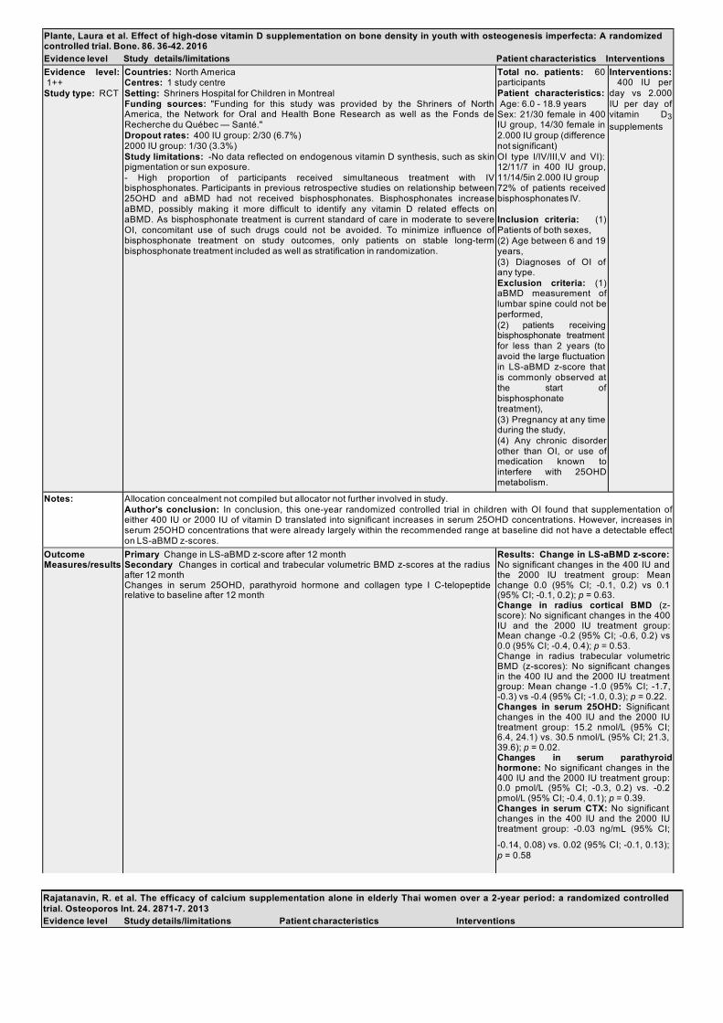
Plante, Laura et al. Effect of high-dose vitamin D supplementation on bone density in youth with osteogenesis imperfecta: A randomized controlled trial. Bone. 86. 36-42. 2016 Evidence level Study details/limitations Patient characteristics Interventions Evidence level: 1++
Study type: RCT
Countries: North America Centres: 1 study centre Setting: Shriners Hospital for Children in Montreal Funding sources: "Funding for this study was provided by the Shriners of North America, the Network for Oral and Health Bone Research as well as the Fonds de Recherche du Québec — Santé." Dropout rates: 400 IU group: 2/30 (6.7%) 2000 IU group: 1/30 (3.3%) Study limitations: -No data reflected on endogenous vitamin D synthesis, such as skin pigmentation or sun exposure. - High proportion of participants received simultaneous treatment with IV bisphosphonates. Participants in previous retrospective studies on relationship between 25OHD and aBMD had not received bisphosphonates. Bisphosphonates increase aBMD, possibly making it more difficult to identify any vitamin D related effects on aBMD. As bisphosphonate treatment is current standard of care in moderate to severe OI, concomitant use of such drugs could not be avoided. To minimize influence of bisphosphonate treatment on study outcomes, only patients on stable long-term bisphosphonate treatment included as well as stratification in randomization.
Total no. patients: 60 participants Patient characteristics: Age: 6.0 - 18.9 years
Sex: 21/30 female in 400 IU group, 14/30 female in 2.000 IU group (difference not significant) OI type I/IV/III,V and VI): 12/11/7 in 400 IU group, 11/14/5in 2.000 IU group 72% of patients received bisphosphonates IV. Inclusion criteria: (1) Patients of both sexes, (2) Age between 6 and 19 years, (3) Diagnoses of OI of any type. Exclusion criteria: (1) aBMD measurement of lumbar spine could not be performed, (2) patients receiving bisphosphonate treatment for less than 2 years (to avoid the large fluctuation in LS-aBMD z-score that is commonly observed at the start of bisphosphonate treatment), (3) Pregnancy at any time during the study, (4) Any chronic disorder other than OI, or use of medication known to interfere with 25OHD metabolism.
Interventions: 400 IU per
day vs 2.000 IU per day of vitamin D3 supplements
Notes: Allocation concealment not compiled but allocator not further involved in study. Author's conclusion: In conclusion, this one-year randomized controlled trial in children with OI found that supplementation of either 400 IU or 2000 IU of vitamin D translated into significant increases in serum 25OHD concentrations. However, increases in serum 25OHD concentrations that were already largely within the recommended range at baseline did not have a detectable effect on LS-aBMD z-scores.
Outcome Measures/results
Primary Change in LS-aBMD z-score after 12 month Secondary Changes in cortical and trabecular volumetric BMD z-scores at the radius after 12 month Changes in serum 25OHD, parathyroid hormone and collagen type I C-telopeptide relative to baseline after 12 month
Results: Change in LS-aBMD z-score: No significant changes in the 400 IU and the 2000 IU treatment group: Mean change 0.0 (95% CI; -0.1, 0.2) vs 0.1 (95% CI; -0.1, 0.2); p = 0.63. Change in radius cortical BMD (z- score): No significant changes in the 400 IU and the 2000 IU treatment group: Mean change -0.2 (95% CI; -0.6, 0.2) vs 0.0 (95% CI; -0.4, 0.4); p = 0.53. Change in radius trabecular volumetric BMD (z-scores): No significant changes in the 400 IU and the 2000 IU treatment group: Mean change -1.0 (95% CI; -1.7, -0.3) vs -0.4 (95% CI; -1.0, 0.3); p = 0.22. Changes in serum 25OHD: Significant changes in the 400 IU and the 2000 IU treatment group: 15.2 nmol/L (95% CI; 6.4, 24.1) vs. 30.5 nmol/L (95% CI; 21.3, 39.6); p = 0.02. Changes in serum parathyroid hormone: No significant changes in the 400 IU and the 2000 IU treatment group: 0.0 pmol/L (95% CI; -0.3, 0.2) vs. -0.2 pmol/L (95% CI; -0.4, 0.1); p = 0.39. Changes in serum CTX: No significant changes in the 400 IU and the 2000 IU treatment group: -0.03 ng/mL (95% CI;
-0.14, 0.08) vs. 0.02 (95% CI; -0.1, 0.13); p = 0.58
Rajatanavin, R. et al. The efficacy of calcium supplementation alone in elderly Thai women over a 2-year period: a randomized controlled trial. Osteoporos Int. 24. 2871-7. 2013 Evidence level Study details/limitations Patient characteristics Interventions
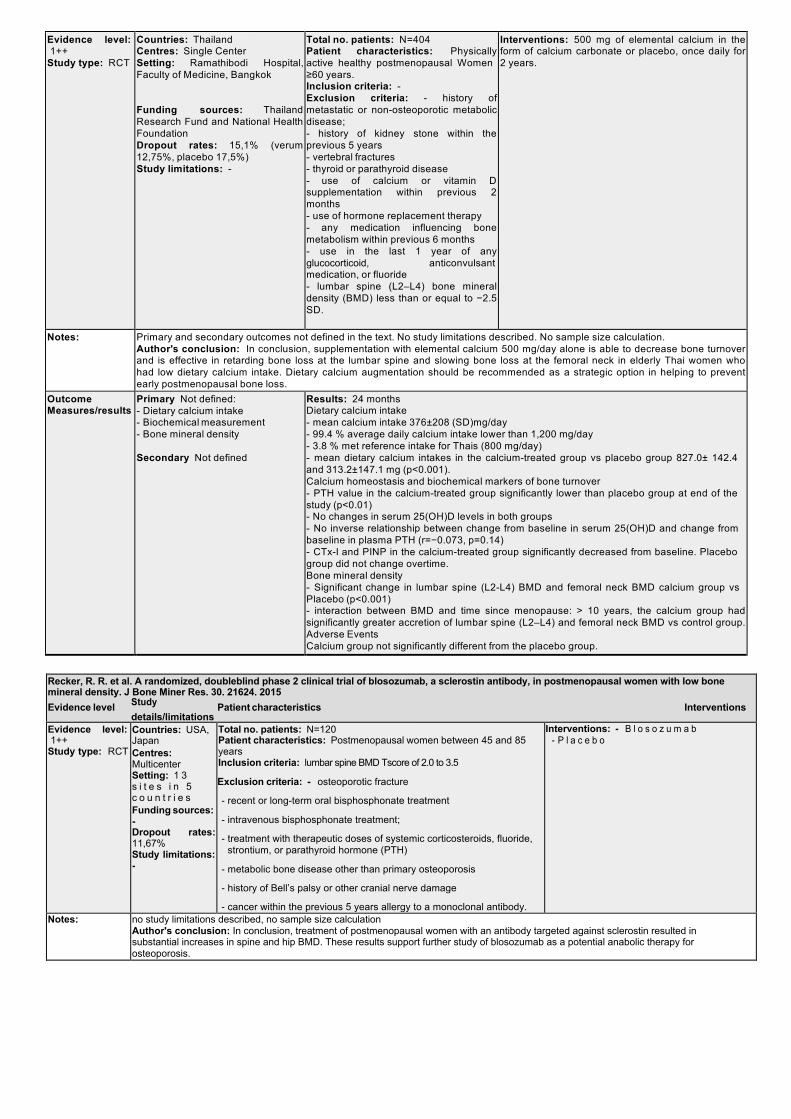
Evidence level: 1++
Study type: RCT
Countries: Thailand Centres: Single Center Setting: Ramathibodi Hospital, Faculty of Medicine, Bangkok Funding sources: Thailand Research Fund and National Health Foundation Dropout rates: 15,1% (verum 12,75%, placebo 17,5%) Study limitations: -
Total no. patients: N=404 Patient characteristics: Physically active healthy postmenopausal Women ≥60 years. Inclusion criteria: - Exclusion criteria: - history of metastatic or non-osteoporotic metabolic disease; - history of kidney stone within the previous 5 years - vertebral fractures - thyroid or parathyroid disease - use of calcium or vitamin D supplementation within previous 2 months - use of hormone replacement therapy - any medication influencing bone metabolism within previous 6 months - use in the last 1 year of any glucocorticoid, anticonvulsant medication, or fluoride - lumbar spine (L2–L4) bone mineral density (BMD) less than or equal to −2.5 SD.
Interventions: 500 mg of elemental calcium in the form of calcium carbonate or placebo, once daily for 2 years.
Notes: Primary and secondary outcomes not defined in the text. No study limitations described. No sample size calculation. Author's conclusion: In conclusion, supplementation with elemental calcium 500 mg/day alone is able to decrease bone turnover and is effective in retarding bone loss at the lumbar spine and slowing bone loss at the femoral neck in elderly Thai women who had low dietary calcium intake. Dietary calcium augmentation should be recommended as a strategic option in helping to prevent early postmenopausal bone loss.
Outcome Measures/results
Primary Not defined: - Dietary calcium intake - Biochemical measurement - Bone mineral density Secondary Not defined
Results: 24 months Dietary calcium intake - mean calcium intake 376±208 (SD)mg/day - 99.4 % average daily calcium intake lower than 1,200 mg/day - 3.8 % met reference intake for Thais (800 mg/day) - mean dietary calcium intakes in the calcium-treated group vs placebo group 827.0± 142.4 and 313.2±147.1 mg (p<0.001). Calcium homeostasis and biochemical markers of bone turnover - PTH value in the calcium-treated group significantly lower than placebo group at end of the study (p<0.01) - No changes in serum 25(OH)D levels in both groups - No inverse relationship between change from baseline in serum 25(OH)D and change from baseline in plasma PTH (r=−0.073, p=0.14) - CTx-I and PINP in the calcium-treated group significantly decreased from baseline. Placebo group did not change overtime. Bone mineral density - Significant change in lumbar spine (L2-L4) BMD and femoral neck BMD calcium group vs Placebo (p<0.001) - interaction between BMD and time since menopause: > 10 years, the calcium group had significantly greater accretion of lumbar spine (L2–L4) and femoral neck BMD vs control group. Adverse Events Calcium group not significantly different from the placebo group.
Recker, R. R. et al. A randomized, doubleblind phase 2 clinical trial of blosozumab, a sclerostin antibody, in postmenopausal women with low bone mineral density. J Bone Miner Res. 30. 21624. 2015 Evidence level Study Patient characteristics Interventions
details/limitations Evidence level: 1++
Study type: RCT
Countries: USA, Japan Centres: Multicenter Setting: 1 3 s i t e s i n 5 c o u n t r i e s Funding sources: - Dropout rates: 11,67% Study limitations: -
Total no. patients: N=120 Patient characteristics: Postmenopausal women between 45 and 85 years Inclusion criteria: lumbar spine BMD Tscore of 2.0 to 3.5
Exclusion criteria: - osteoporotic fracture
- recent or long-term oral bisphosphonate treatment
- intravenous bisphosphonate treatment;
- treatment with therapeutic doses of systemic corticosteroids, fluoride, strontium, or parathyroid hormone (PTH)
- metabolic bone disease other than primary osteoporosis
- history of Bell’s palsy or other cranial nerve damage
- cancer within the previous 5 years allergy to a monoclonal antibody.
Interventions: - B l o s o z u m a b - P l a c e b o
Notes: no study limitations described, no sample size calculation Author's conclusion: In conclusion, treatment of postmenopausal women with an antibody targeted against sclerostin resulted in substantial increases in spine and hip BMD. These results support further study of blosozumab as a potential anabolic therapy for osteoporosis.
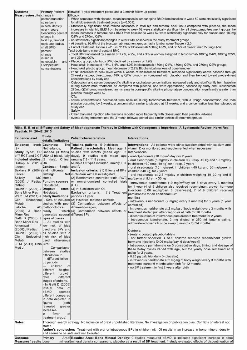
Outcome Measures/results
Primary Percent change in posterioranterior spine bone mineral density over 4 years Secondary percent change in total hip, femoral neck, and radius shaft BMD percent change in serum osteocalcin and Ctelopeptide concentrations
Results: 1 year treatment period and a 3 month follow-up period. Efficacy - When compared with placebo, mean increases in lumbar spine BMD from baseline to week 52 were statistically significant
for all blosozumab treatment groups (p<0.001). - Statistically significant dose-related increases in total hip and femoral neck BMD compared with placebo, the mean
increases in total hip BMD from baseline to week 52 were statistically significant for all blosozumab treatment groups the mean increases in femoral neck BMD from baseline to week 52 were statistically significant only for blosozumab 180mg Q2W and 270mg Q2W
- no statistically significant changes in wrist BMD observed in the study treatment groups - At baseline, 95.6% of the blosozumab treatment groups had a lumbar spine Tscore ≤ 2.0. - End of treatment, Tscore > –2.0 in 72.4% of blosozumab 180mg Q2W, and 88.5% of blosozumab 270mg Q2W
Total body bone mineral content BMC - Total BMC increased by a mean of 1.7%, 4.2%, and 7.3% in women assigned to blosozumab 180mg Q4W, 180mg Q2W,
and 270mg Q2W. - Placebo group, total body BMC declined by a mean of 1.9% - Head skull: increase of 1.6%, 1.4%, and 4.0% in blosozumab 180mg Q4W, 180mg Q2W, and 270mg Q2W groups - Head skull placbo group: mean decrease of 2.2% Biochemical markers of bone turnover - P1NP increased to peak level within 4 weeks of blosozumab treatment, remained significantly above baseline through
24weeks (except blosozumab 180mg Q4W group), as compared with placebo, and then trended toward pretreatment concentrations by study end
- Osteocalcin and serum bonespecific alkaline phosphatase concentrations increased early and significantly from baseline during blosozumab treatment, as compared with placebo, and were approaching baseline by study end. Blosozumab 270mg Q2W group maintained an increase in bonespecific alkaline phosphatase concentration significantly greater than placebo through week 52
- CTx - serum concentrations decreased from baseline during blosozumab treatment, with a trough concentration less than
placebo occurring by 2 weeks, a concentration similar to placebo at 12 weeks, and a concentration less than placebo at study end
- Safety - Other than mild injection site reactions reported more frequently with blosozumab than placebo, adverse - events during treatment and the 3 month followup period was similar across all treatment groups.
Rijks, E. B. et al. Efficacy and Safety of Bisphosphonate Therapy in Children with Osteogenesis Imperfecta: A Systematic Review. Horm Res Paediatr. 84. 26-42. 2015
Evidence level Study Patient characteristics Interventions details/limitations
Evidence level: 2+
Study type: SR of 7 RCT and 3 CT Included studies: Bishop N (2013) Lancet Sakkers R (2004) Lancet Seikaly MG (2005) J Pediatr Orthop Rauch F (2009) J Bone Miner Res Ward LM (2011) J Clin Endocrinol Metab Letocha AD (2005) J Bone Miner Res Gatti D (2005) J Bone Miner Res Antoniazzi F (2006) J Pediatr Rauch F (2006) J Clin Endocrinol Metab Li M (2011) Chin Med J
Countries: The Netherlands, Canada (3 trials), USA (2 trials), Italy (2 trials), China, UK Centres: Single- and multicenter Setting: Not stated. Funding sources: Not stated.
Dropout rates: Not stated. Study limitations: - 60% of included
studies with poor methodological quality, which generates several types of biases. → All studies with low risk of bias used oral BPs and all studies with a high risk of bias used intravenous BPs. - Comparisons between studies difficult due to → different follow- up periods → children of different heights, different growth rates, different stages of puberty. - In Gatti D (2005) textual data of aBMD seemed different compared to data depicted in figures (both revealed greater increases in favor of treatment group)
Total no. patients: 519 children Patient characteristics: Mean age: 1 studies with infants (mean age: 33 days), 9 studies with mean age ranging 7.9 - 11.9 years. Multiple OI types included: mainly I, III and IV Inclusion criteria: (1) Effects of BPs in children with OI investigated, (2) Randomized controlled trials (RCT) or nonrandomized controlled trials (CT), (3) >15 children with OI. Exclusion criteria: (1) Treatment periods <1 year, (2) Historical matched controls, (3) Comparison between effects of different dosages, (4) Comparison between effects of different BPs.
Interventions: All patients were either supplemented with calcium and vitamin D or monitored and supplemented when necessary. Interventions: - oral olpadronate (10 mg/m2/day) for 2 years - oral alendronate (5 mg/day in children <30 resp. 40 kg and 10 mg/day in children >30 resp. 40 kg) for 1 resp. 2 years - oral risedronate (15 mg/week in children <40 kg and 30 mg/week in children >40 kg) for 2 years - oral risedronate at 2.5 mg/day in children weighing 10–30 kg and 5 mg/day in children > 30 kg - intravenous pamidronate (10 mg/m2/day for 3 days every 3 months) for 1 year (4 of 9 children also received recombinant growth hormone injections [0.06 mg/kg/day, 6 days/week], 7 of 9 children received pamidronate for an additional 6–21 months) - intravenous neridronate (2 mg/kg every 3 months) for 3 years (1 year controlled) - intravenous neridronate at 2 mg/kg of body weight every 3 months with treatment started just after diagnosis at birth for 18 months - discontinuation of intravenous pamidronate treatment for 2 years - intravenous ibandronate, 2 mg diluted in 250 ml isotonic saline, administered over 2 h once every 3 months for 24 months Controls: - (enteric coated) placebo tablets - not further specified (4 of 9 children received recombinant growth hormone injections [0.06 mg/kg/day, 6 days/week]) - Intravenous pamidronate on 3 consecutive days, timing and dosage of these 3-day cycles varied with age, but the yearly dose remained at 9 ml/kg for 2 years. - 0.25 μg calcitriol daily (= placebo) - intravenous neridronate at 2 mg/kg of body weight every 3 months with treatment started 6 months after birth for 12 months - no BP treatment in first 2 years after birth
Notes: Thorough search strategy. No inclusion of grey/ unpublished literature. No investigation of publication bias. Conflicts of interest not stated. Author's conclusion: Treatment with oral or intravenous BPs in children with OI results in an increase in bone mineral density and seems to be safe and well tolerated.
Outcome Measures/results
Primary Areal bone mineral
Results: Areal Bone Mineral Density: 9 studies measured aBMD, 8 indicated significant increase in bone mineral density compared to placebo as a result of BP treatment. 1 study evaluated effects of discontinuation of
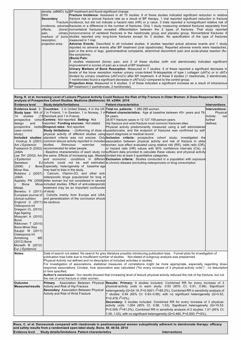
density (aBMD) by DXA Secondary
Fracture incidence, adverse effects, (bone) pain, urinary markers of bone resorption, projection area.
BP treatment and found significant change. Fracture Incidence: Assessed in all 10 studies. 6 of these studies indicated significant reduction in relative fracture risk or annual fracture rate as a result of BP therapy. 1 trial reported significant reduction in fracture incidence; but did not indicate a hazard ratio (HR) or p value. 3 trials reported a nonsignificant relative risk of fractures or a difference in the number of fractures. Only 1 study measuring vertebral fractures separately from nonvertebral fractures revealed differentiation between the 2 types of fractures. That study reported nonoccurrence of vertebral fractures in the risedronate group and placebo group. Nonvertebral fractures: 6 studies reported only long-bone fractures except for 3 studies. No specification of the type of fractures measured in 1 trial. Adverse Events: Described in all included studies. 9 studies reported actual adverse events and 1 study reported no adverse events after BP treatment (oral olpadronate). Reported adverse events were headaches, pain in the arms or legs, gastrointestinal complaints, abdominal discomfort/ pain and acute-phase reaction (flu- like symptoms). (Bone) Pain: 5 studies measured (bone) pain, and 2 of these studies (with oral alendronate) indicated significant improvement in scores of pain as a result of BP treatment. Urinary Markers of Bone Resorption: Measured in 7 studies. 5 of these reported a significant decrease in levels of the bone resorption marker urinary cross-linked N-telopeptide of type I collagen (uNTx) or in uNTx divided by urinary creatinine (uNTx/uCr) after BP treatment. 4 of these 5 studies (1 risedronate, 2 alendronate, 1 neridronate) found a significant decrease in uNTx/uCr compared to the control group. Projection Area: Measured in 6 studies. 3 of these indicated a significant increase as a result of intravenous BP treatment (1 pamidronate, 2 neridronate).
Rong, K. et al. Increasing Level of Leisure Physical Activity Could Reduce the Risk of Hip Fracture in Older Women: A Dose-Response Meta- analysis of Prospective Cohort Studies. Medicine (Baltimore). 95. e2984. 2016 Evidence level Study details/limitations Patient characteristics Interventions Evidence level: 2- Study type: MA of 14 studies (13 prospective cohort design and 1 prospective nested case–control study). Included studies:
Hoidrup S (2001) Am J Epidemiol Feskanich D (2002) JAMA Lee SH (2002) Am J Epidemiol Samelson EJ (2006) J Bone Miner Res Robbins J (2007) JAMA Appleby PN (2008) J Bone Miner Metab. Benetou V (2011) European journal of clinical nutrition Englund U (2011) Osteoporos Int Gregson CL (2010) Age Ageing Moayyeri A (2010) Bone Rikkonen T (2010) Bone Miner Res Kauppi M (2011) Osteoporos Int Armstrong ME (2012) Bone Morseth B (2012) Eur J Epidemiol
Countries: 3 in United States, 4 in the UK, 2 in Finland, 1 in Sweden, 1 in Norway, 1 in Denmark and 1 in France. Centres: Not reported. Setting: Not reported. Funding sources: Not stated. Dropout rates: Not reported. Study limitations: - Uniforming of dose of physical activity of different studies using standard criteria was not precise. Only common leisure activity reported in included studies. Strenuous exercise not recommended for elder people. - Baseline characteristics of each study not the same. Effects of increasing age, lifestyle and economic conditions in different cohorts could not be well estimated. Especially, heterogeneity of baseline age may lead to bias in the study. - Calcium, Vitamin-D3, and other anti- osteoporotic drugs popularized for long in elder women but not considered in several included studies. Effect of anti-osteoporotic treatment may be an important confounder of study. - Cohorts mainly from Europe and USA, and generalization of the conclusion should be cautious.
Total no. patients: 1.380.285 women Patient characteristics: Age at baseline between 45+ years and 76- 84 years. 28,871 fracture cases in 12.127.159 person-years. Hip fracture and wrist fracture most common fractures sites. Physical activity predominantly measured using a self administered questionnaire, and the endpoint of fractures was confirmed by self- report diagnosis or medical record. Inclusion criteria: prospective cohort study; investigated the association between physical activity and risk of fracture in older women; size effect evaluated using relative risk (RR), odds ratio (OR), or hazard ratio (HR) values with 95% confidence intervals (CIs), or sufficient data provided to calculate these values; and physical activity divided into at least 3 quantitative categories. Exclusion criteria: Studies conducted in a population with exposure to chronic disease (excluding osteoporosis) or drug consumption
Interventions: Physical
Activity: not further defined.
Notes: Date of search not stated. No search for grey literature possibly introducing publication bias. Funnel plots for investigation of publication bias futile due to insufficient number of studies. Not stated of subgroup analysis was preplanned. Physical Activity not defined and no description of included activities in studies. For investigation of associations, statistical measures of correlations might be more appropriate, especially regarding dose- response associations. Unclear, how association was calculated ("for every increase of 3 physical-activity units") - no description or time specified. Author's conclusion: Our results showed that increasing level of leisure physical activity reduced the risk of hip fracture, but not the risk of wrist fracture in older women.
Outcome Measures/results
Primary Association Between Physical Activity and Risk of Hip Fracture Secondary Association Between Physical Activity and Risk of Wrist Fracture
Results: Primary: 9 studies included. Combined RR for every increase of 3 physical-activity units in each study: 0.93 (95% CI; 0.91, 0.96). Significant heterogeneity (Q=54.19, P<0.0001; I2=85.2%). Combined RR in sensitivity analysis of 7 studies: 0.94 (95% CI: 0.93–0.96), with no significant heterogeneity (Q=5.52, P<0.479; I2=0%). Secondary: 3 studies included. Combined RR for every increase of 3 physical- activity units: 1.004 (95% CI; 0.98, 1.03). Significant heterogeneity (Q=10.53, P<0.005; I2=81.0%). Combined RR in sensitivity analysis of 2 studies: 1.01 (95% CI; 1.00, 1.03), with no significant heterogeneity (Q=0.460, P<0.500; I2=0%).
Roux, C. et al. Denosumab compared with risedronate in postmenopausal women suboptimally adherent to alendronate therapy: efficacy and safety results from a randomized open-label study. Bone. 58. 48-54. 2014 Evidence level Study details/limitations Patient characteristics Interventions

Evidence level: 1++
Study type: RCT
Countries: Europe, Canada, Australia Centres: Mulitcenter Setting: 82 centres Funding sources: Amgen Inc. Dropout rates: 5,3% Study limitations: -
Total no. patients: n=870 Patient characteristics: postmenopausal women aged ≥55 years Inclusion criteria: - previously been prescribed alendronate therapy (stopped ≥1 month or demonstrating suboptimal adherence) Exclusion criteria: - prior or current treatment with osteoporosis medication other than daily or weekly oral alendronate therapy - hormone replacement therapy - calcium and vitamin D (use of raloxifene or calcitonin prior to initiation of alendronate therapy was allowed)
Interventions: 12 months - denosumab 60 mg subcutaneously (SC) every 6months (Q6M) - risedronate orally (PO)150mg once monthly (QM, one 75mg tablet on each of 2 consecutive days) both groups: ≥1000 mg elemental calcium and ≥800 IU vitamin D per day
Notes: Sample size calcuation Author's conclusion: In conclusion, in postmenopausalwomen whowere previously taking alendronatewith suboptimal adherence, transitioning to denosumab was well tolerated and more effective to increase BMD and lower bone turnover than switching to risedronate.
Outcome Primary - Bone mineral density: mean percentage change from baseline in total hip BMD at month 12 Secondary - mean percentage change in femoral neck and lumbar spine BMD at month 12 - median percentage change from baseline in sCTX-1 at month 1 - median percentage change from baseline in sCTX-1 at month 6 - Biochemical markers - Adverse Events
Results: Bone mineral density - Denosumab significantly increased BMD at the total hip at month 12 with a mean percentage change from baseline of 2.0% (95% CI: 1.8%,2.3%); the difference in mean percentage change from risedronate was 1.6% (95% CI: 1.2%, 2.0%; p < 0.0001). - Denosumab also significantly increased BMD at the femoral neck and lumbar spine at month 12 with a mean percentage change from baseline of 1.4% (95% CI: 1.0%,1.7%) and 3.4% (95% CI: 3.1%, 3.8%). Compared with risedronate, a difference in mean percentage change between the treatment groups of 1.4% (95% CI: 0.9%, 1.8%; p < 0.0001) and 2.3% (95% CI: 1.8%, 2.8%; p < 0.0001). - DXA scans for the least significant change (LSC) in BMD: month 12,BMD gains denosumab- vs risedronate-treated subjects ≥LSC at total hip (49% vs 20%, p < 0.0001), femoral neck (24% vs 14%, p < 0.0001), lumbar spine (64% vs 32%, p < 0.0001) sCTX-1 - significant decrease in both treatment groups at month 1. Significantly greater reduction denosumab vs risedronate: median (IQR) percentage change of −77.7% (−85.9%,−67.6%) vs −17.0% (−36.8%, −1.6%) (p < 0.0001) - Median reductions in sCTX-1 at month 6 greater in the denosumab vs risedronate group: median (IQR) percentage change of −60.6% (−77.0%, −48.8%) vs –22.5% (−41.9%, 11.4%) (p < 0.0001) - Subjects treated with denosumab significantly greater gains in lumbar spine BMD at month 12 at each tertile of baseline sCTX-1 (p < 0.01). - At all sites the magnitude of the BMD gain was significantly more pronounced in the middle and highest sCTX-1 tertiles (treatment-bysCTX-1 tertile interaction p-values < 0.01)
Adverse Events Overall, the subject incidences of AEs were 293 subjects (68.3%) in the risedronate group and 269 subjects (62.7%) in the denosumab group. SAEs were reported for 8.2% of risedronate-treated subjects and 7.7% of denosumab-treated subjects.
Measures/results
Ruan, J. et al. Effect of B vitamin (folate, B6, and B12) supplementation on osteoporotic fracture and bone turnover markers: a meta- analysis. Med Sci Monit. 21. 875-81. 2015
Evidence level Study details/limitations Patient Interventions characteristics
Evidence level: 1-
Study type: Systematic
review and meta analysis: 8 RCTs. Included studies: Green TJ, et al. (2007) Am J Clin Nutr Shahab-Ferdows S, et al. (2012) J Nutr Sato Y, et al. (2008) JAMA Sawka AM, et al. (2007) Arch Intern Med Study of the Effectiveness of Additional Reductions in Cholesterol and Homocysteine (SEARCH) Collaborative Group, et al (2010) JAMA Gommans J, et al. (2013) BMC Geriatr Keser I, et al. (2013) Nutr Res Salari P, et al. (2014) Daru
Countries: n.a. Centres: n.a. Setting: n.a. Funding sources: This study was supported by the 2014 General Medical and Health Research Plan of Zhejiang Province, China, Grant No. 2014KYA229. Dropout rates: Dropout rates not reported in the study characteristics. Attrition bias was judged as low in all 9 studies. Study limitations: Firstly, the baseline of the patients included varied significantly in some studies. For example, the mean baseline folate level of the participants in the HOPE2 study was approximately 5 times higher than the participants in Sato’s study in Japan. Therefore, it might be possible that B vitamin supplementation might not generate additional benefits for the patients who already have high serum B vitamin level. Among the 4 RCTs that assessed B vitamin supplementation and fractures, only Sato’s study observed a risk-reduction effect. In fact, the study population was highly selected and characterized as very high plasma Hcy level, severe disability, and an unusually (10-fold) high fracture risk rate of the control cases compared to the average Japanese population of the same age. However, a recent meta-analysis observed that fortification with certain B vitamins, such as folate, exert protective effects on Hcy- related cerebrovascular risks and additional supplementation thus had no additive effect. Therefore, we cannot exclude the possibility that B vitamin supplementation might have some protective effects on bone health due to the homocysteine-lowering effects in countries without folate fortification, such as Japan.
Total no. patients: 26378. Patient characteristics:
Mean age range 39-75 and 36-75 (I/C). 17- 100% of participants were female. Inclusion criteria: Study type: RCTs. Population: not defined. Intervention: vitamin B supple- mentation, (type, dosage, duration not described). Comparison: Placebo. Outcome: fracture risk or bone turnover indicators. Exclusion criteria: Case report, animal study, or review; studies with detailed, but unextract-able outcome data (RR of fracture or changes of
Interventions: Intervention: Vitamin B supplementation (type and dose heterogeneous in the included studies): Folate + B6 + B12; or folate + B12; or folate only, or B6 + B12; Duration of treatment between 3 months and 5 years. Comparison: Placebo.
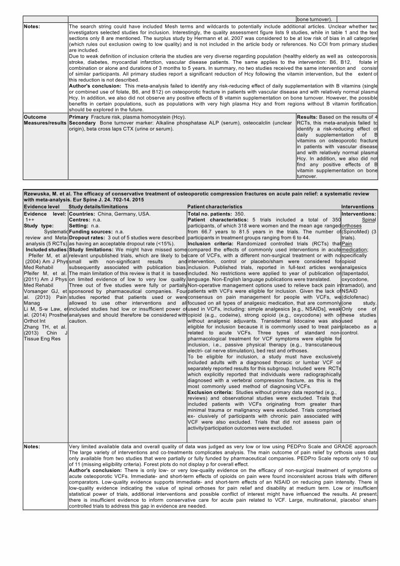
bone turnover). Notes: The search string could have included Mesh terms and wildcards to potentially include additional articles. Unclear whether two
investigators selected studies for inclusion. Interestingly, the quality assessment figure lists 9 studies, while in table 1 and the text sections only 8 are mentioned. The surplus study by Hermann et al. 2007 was considered to be at low risk of bias in all categories (which rules out exclusion owing to low quality) and is not included in the article body or references. No COI from primary studies are included. Due to weak definition of inclusion criteria the studies are very diverse regarding population (healthy elderly as well as osteoporosis, stroke, diabetes, myocardial infarction, vascular disease patients. The same applies to the intervention: B6, B12, folate in combination or alone and durations of 3 months to 5 years. In summary, no two studies received the same intervention and consist of similar participants. All primary studies report a significant reduction of Hcy following the vitamin intervention, but the extent of this reduction is not described. Author's conclusion: This meta-analysis failed to identify any risk-reducing effect of daily supplementation with B vitamins (single or combined use of folate, B6, and B12) on osteoporotic fracture in patients with vascular disease and with relatively normal plasma Hcy. In addition, we also did not observe any positive effects of B vitamin supplementation on bone turnover. However, the possible benefits in certain populations, such as populations with very high plasma Hcy and from regions without B vitamin fortification, should be explored in the future.
Outcome Measures/results
Primary Fracture risk, plasma homocystein (Hcy). Secondary Bone turnover marker: Alkaline phosphatase ALP (serum), osteocalclin (unclear origin), beta cross laps CTX (urine or serum).
Results: Based on the results of 4 RCTs, this meta-analysis failed to identify a risk-reducing effect of daily supplementation of B vitamins on osteoporotic fracture in patients with vascular disease and with relatively normal plasma Hcy. In addition, we also did not find any positive effects of B vitamin supplementation on bone turnover.
Rzewuska, M. et al. The efficacy of conservative treatment of osteoporotic compression fractures on acute pain relief: a systematic review with meta-analysis. Eur Spine J. 24. 702-14. 2015 Evidence level Study details/limitations Patient characteristics Interventions Evidence level: 1++
Study type: Systematic
review and Meta- analysis (5 RCTs). Included studies: Pfeifer M, et al.
(2004) Am J Phys Med Rehabil Pfeifer M, et al. (2011) Am J Phys Med Rehabil Vorsanger GJ, et al. (2013) Pain Manag Li M, S-w Law, et al. (2014) Prosthet Orthot Int Zhang TH, et al. (2013) Chin J Tissue Eng Res
Countries: China, Germany, USA. Centres: n.a. Setting: n.a. Funding sources: n.a. Dropout rates: 3 out of 5 studies were described as having an acceptable dropout rate (<15%). Study limitations: We might have missed some relevant unpublished trials, which are likely to be small with non-significant results and subsequently associated with publication bias. The main limitation of this review is that it is based on limited evidence of low to very low quality. Three out of five studies were fully or partially sponsored by pharmaceutical companies. Four studies reported that patients used or were allowed to use other interventions and all included studies had low or insufficient power of analyses and should therefore be considered with caution.
Total no. patients: 350. Patient characteristics: 5 trials included a total of 350 participants, of which 318 were women and the mean age ranged from 66.7 years to 81.5 years in the trials. The number of participants in treatment groups ranging from 6 to 44. Inclusion criteria: Randomized controlled trials (RCTs) that compared the effects of commonly used interventions in acute care of VCFs, with a different non-surgical treatment or with no intervention, control or placebo/sham were considered for inclusion. Published trials, reported in full-text articles were included. No restrictions were applied to year of publication or language. Non-English language publications were translated. Non-operative management options used to relieve back pain in patients with VCFs were eligible for inclusion. Given the lack of consensus on pain management for people with VCFs, we focused on all types of analgesic medication, that are commonly used in VCFs, including: simple analgesics [e.g., NSAIDs], weak opioid (e.g., codeine), strong opioid (e.g., oxycodone) with or without analgesic adjuvants. Transdermal lidocaine was also eligible for inclusion because it is commonly used to treat pain related to acute VCFs. Three types of standard non- pharmacological treatment for VCF symptoms were eligible for inclusion, i.e., passive physical therapy (e.g., transcutaneous electri- cal nerve stimulation), bed rest and orthoses. To be eligible for inclusion, a study must have exclusively included adults with a diagnosed thoracic or lumbar VCF or separately reported results for this subgroup. Included were RCTs which explicitly reported that individuals were radiographically diagnosed with a vertebral compression fracture, as this is the most commonly used method of diagnosing VCFs. Exclusion criteria: Studies without primary data reported (e.g., reviews) and observational studies were excluded. Trials that included patients with VCFs originating from greater than minimal trauma or malignancy were excluded. Trials comprised ex- clusively of participants with chronic pain associated with VCF were also excluded. Trials that did not assess pain or activity/participation outcomes were excluded.
Interventions: Spinal
orthoses (SpinoMed) (3 trials). Pain medication; specifically opioid analgesics (tapentadol, oxycodone, tramadol), and NSAID (diclofenac) (one study. Only one of these studies used a placebo as a control.
Notes: Very limited available data and overall quality of data was judged as very low or low using PEDPro Scale and GRADE approach. The large variety of interventions and co-treatments complicates analysis. The main outcome of pain relief by orthosis uses data only available from two studies that were partially or fully funded by pharmaceutical companies. PEDPro Scale reports only 10 out of 11 (missing eligibility criteria). Forest plots do not display p for overall effect. Author's conclusion: There is only low- or very low-quality evidence on the efficacy of non-surgical treatment of symptoms of acute osteoporotic VCFs. Immediate- and short-term effects of opioids on pain were found inconsistent across trials with different comparators. Low-quality evidence supports immediate- and short-term effects of an NSAID on reducing pain intensity. There is low-quality evidence indicating the value of spinal orthoses for pain relief and disability at medium term. Low or insufficient statistical power of trials, additional interventions and possible conflict of interest might have influenced the results. At present, there is insufficient evidence to inform conservative care for acute pain related to VCF. Large, multinational, placebo/ sham- controlled trials to address this gap in evidence are needed.
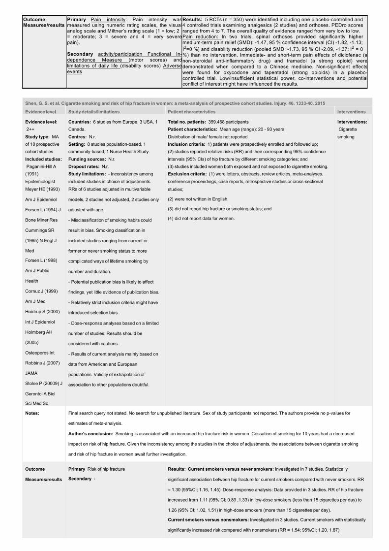
Outcome Measures/results
Primary Pain intensity: Pain intensity was measured using numeric rating scales, the visual analog scale and Miltner’s rating scale (1 = low; 2 = moderate; 3 = severe and 4 = very severe pain). Secondary activity/participation Functional In- dependence Measure (motor scores) and limitations of daily life (disability scores) Adverse events
Results: 5 RCTs (n = 350) were identified including one placebo-controlled and 4 controlled trials examining analgesics (2 studies) and orthoses. PEDro scores ranged from 4 to 7. The overall quality of evidence ranged from very low to low. Pain reduction: In two trials, spinal orthoses provided significantly higher medium-term pain relief (SMD): -1.47, 95 % confidence interval (CI) -1.82, -1.13; I2=0 %] and disability reduction (pooled SMD: -1.73, 95 % CI -2.09, -1.37; I2 = 0 %) than no intervention. Immediate- and short-term pain effects of diclofenac (a non-steroidal anti-inflammatory drug) and tramadol (a strong opioid) were demonstrated when compared to a Chinese medicine. Non-significant effects were found for oxycodone and tapentadol (strong opioids) in a placebo- controlled trial. Low/insufficient statistical power, co-interventions and potential conflict of interest might have influenced the results.
Shen, G. S. et al. Cigarette smoking and risk of hip fracture in women: a meta-analysis of prospective cohort studies. Injury. 46. 1333-40. 2015
Evidence level Study details/limitations Patient characteristics Interventions Evidence level: 2++ Study type: MA of 10 prospective cohort studies Included studies: Paganini-Hill A (1991) Epidemiologist Meyer HE (1993)
Am J Epidemiol
Forsen L (1994) J
Bone Miner Res
Cummings SR
(1995) N Engl J
Med Forsen L (1998)
Am J Public
Health
Cornuz J (1999)
Am J Med
Hoidrup S (2000)
Int J Epidemiol
Holmberg AH
(2005)
Osteoporos Int
Robbins J (2007)
JAMA
Stolee P (20009) J
Gerontol A Biol
Sci Med Sc
Countries: 6 studies from Europe, 3 USA, 1 Canada. Centres: N.r. Setting: 8 studies population-based, 1 community-based, 1 Nurse Health Study. Funding sources: N.r. Dropout rates: N.r. Study limitations: - Inconsistency among included studies in choice of adjustments. RRs of 6 studies adjusted in multivariable
models, 2 studies not adjusted, 2 studies only
adjusted with age.
- Misclassification of smoking habits could
result in bias. Smoking classification in
included studies ranging from current or
former or never smoking status to more
complicated ways of lifetime smoking by
number and duration.
- Potential publication bias is likely to affect
findings, yet little evidence of publication bias.
- Relatively strict inclusion criteria might have
introduced selection bias.
- Dose-response analyses based on a limited
number of studies. Results should be
considered with cautions.
- Results of current analysis mainly based on
data from American and European
populations. Validity of extrapolation of
association to other populations doubtful.
Total no. patients: 359.468 participants Patient characteristics: Mean age (range): 20 - 93 years. Distribution of male/ female not reported. Inclusion criteria: 1) patients were prospectively enrolled and followed up; (2) studies reported relative risks (RR) and their corresponding 95% confidence intervals (95% CIs) of hip fracture by different smoking categories; and (3) studies included women both exposed and not exposed to cigarette smoking. Exclusion criteria: (1) were letters, abstracts, review articles, meta-analyses, conference proceedings, case reports, retrospective studies or cross-sectional
studies; (2) were not written in English;
(3) did not report hip fracture or smoking status; and
(4) did not report data for women.
Interventions: Cigarette smoking
Notes:
Final search query not stated. No search for unpublished literature. Sex of study participants not reported. The authors provide no p-values for
estimates of meta-analysis.
Author's conclusion: Smoking is associated with an increased hip fracture risk in women. Cessation of smoking for 10 years had a decreased
impact on risk of hip fracture. Given the inconsistency among the studies in the choice of adjustments, the associations between cigarette smoking
and risk of hip fracture in women await further investigation.
Outcome
Measures/results
Primary Risk of hip fracture
Secondary -
Results: Current smokers versus never smokers: Investigated in 7 studies. Statistically
significant association between hip fracture for current smokers compared with never smokers. RR
= 1.30 (95%CI; 1.16, 1.45). Dose-response analysis: Data provided in 3 studies. RR of hip fracture
increased from 1.11 (95% CI; 0.89 ,1.33) in low-dose smokers (less than 15 cigarettes per day) to
1.26 (95% CI; 1.02, 1.51) in high-dose smokers (more than 15 cigarettes per day). Current smokers versus nonsmokers: Investigated in 3 studies. Current smokers with statistically
significantly increased risk compared with nonsmokers (RR = 1.54; 95%CI; 1.20, 1.87)

No statistically significant difference between former smokers versus never smokers (RR = 1.02;
95%CI; 0.93, 1.11) and former smokers versus current smokers (cessation <5 years RR = 1.01;
95%CI; 0.76, 1.26; cessation for 5–9 years RR = 1.10; 95%CI; 0.60, 1.60 and cessation for 10 years
RR = 0.70; 95%CI; 0.50, 0.90).
Slatkovska, L. et al. Effect of whole-body vibration on calcaneal quantitative ultrasound measurements in postmenopausal women: a randomized controlled trial. Calcif Tissue Int. 95. 547-56. 2014 Evidence level Study details/limitations Patient characteristics Interventions Evidence level: 1++
Study type: RCT
Countries: Canada Centres: Not reported. Setting: Not reported. Funding sources: A peer-reviewed grant from the Physicians’ Services Incorporated Foundation funded this trial. Juvent Inc. supplied the WBV platforms and Jamieson Laboratories provided calcium and vitamin D supplements. None of these sources were involved in the study design, conduct, analysis, interpretation of the data, preparation of this manuscript, or decision to submit the manuscript for publication. Dropout rates: 90-Hz WBV Group: 2 Lost to follow-up. 1 Started hormone therapy. 6 Missing QUS measurements. 30-Hz WBV Group: 3 Lost to follow-up. 5 Missing QUS measurements. Control group: 2 Lost to follow-up. 1 Started hormone therapy. 7 Missing QUS measurements. Study limitations: (1) Sample size and power calculations not based on calcaneal QUS outcomes, because they were collected as secondary endpoints. (2) Women and outcome assessor not blinded to control intervention, since sham WBV was not provided due to limited funding and a lack of effective masking of true WBV by a sham platform. (3) Control group participants were not instructed to stand still for 20 consecutive minutes every day, so it is unclear whether just standing on the vibration platform, regardless of WBV, could have contributed to the between-group differences in 12-month changes in BUA. (4) QUS outcomes missing in 12 % of participants and required replacements by use of multiple imputation models in primary analysis. (5) Numerous between-group comparisons performed during various statistical approaches, thus increasing the likelihood of chance findings.
Total no. patients: 202 women
Patient characteristics: 90-Hz WBV group (n =
67): Age (years), mean (SD): 60.5 (7.0); Body
mass index (kg m-2), mean (SD):24.9 (4.0) 30-Hz WBV group (n = 68): Age (years), mean (SD): 59.6 (6.0); Body mass index (kg m-2), mean (SD): 24.5 (3.6) Control group (n = 67): Age (years), mean (SD): 60.8 (5.5); Body mass index (kg m-2), mean (SD): 24.2 (3.4) Inclusion criteria: Women were eligible if
(1) they had experienced cessation of menses 1 or more years prior and (2) their lowest BMD T score at the lumbar spine, femoral neck, or total hip was between -1.0 and -2.5. Exclusion criteria: (1) BMD T score greater than -1.0, because previous research has shown that less-dense bones may have a greater response to WBV. (2) Osteoporosis (BMD T score ≤-2.5); fragility fracture after age 40; secondary causes of bone loss; other metabolic bone diseases or diseases affecting bone metabolism; history of active cancer in the past 5 years; body mass of C90 kg; knee or hip joint replacements; spinal implants; use of hormone therapy in the past 12 months, raloxifene or teriparatide in the past 6 months, or bisphosphonates for C3 months or within the past 3 months; chronic glucocorticoid, anticoagulant or anticonvulsant therapy; (3) inability to tolerate WBV for 20 consecutive minutes at screening; and expected changes in physical activity levels or out-of-town travels for more than four consecutive weeks.
Interventions: 90- and 30-Hz groups: Whole-body vibration (WBV) platforms synchronously oscillating at a frequency of 90- or 30-Hz with a peak acceleration or magnitude of 0.3 g (peak-to-peak displacement of\50 lm), provided by Juvent Regenerative Technologies Corporation, Riveria Beach, Florida. Participants instructed to stand erect on oscillating platform at home for 20 consecutive minutes daily for 12 months, with neutral posture at the neck, lumbar spine, and knees, wearing socks or barefoot, and without excessivefoot or body movements. Control group: Participants asked not to use WBV therapies. Calcium and vitamin D supplements provided to all participants at baseline + 6 months.
Notes: Investigators could have been blinded, but were not. Test for baseline differences not described. Author's conclusion: In conclusion, we found no beneficial effect of WBV on calcaneal QUS measurements in community- dwelling postmenopausal women receiving 0.3 WBV at 90 or 30 Hz, but instead a small but statistically significant decrease in calcaneal BUA in two out of four multiple imputation models, per protocol analysis and subgroup analysis of 80 % adherent participants. This potential negative effect needs to be confirmed in future research. In the absence of any clear beneficial bone effects at hip, spine, distal tibia, and calcaneus, we do not recommend WBV therapy at this time for the prevention of bone loss in postmenopausal women with low bone density.
Outcome Measures/results
Primary Calcaneal Quantitative Ultrasound Outcomes Secondary Adverse Events
Results: Calcaneal Quantitative Ultrasound Outcomes: No improvement in BUA, SOS, or QUI in any of the WBV groups when compared to controls after 12 months. ITT-analyses showed significant decreases in BUA in the 30-Hz WBV group when compared to the control group in 2 out of 4 multiple imputation models analyses. Not statistically significant trend also for 90-Hz group. Adverse Events: No SAE related to WBV. Various adverse events reported regarding minor foot-related problems (including plantar foot pain, foot numbness, toe cramping) but no significant between-group differences.
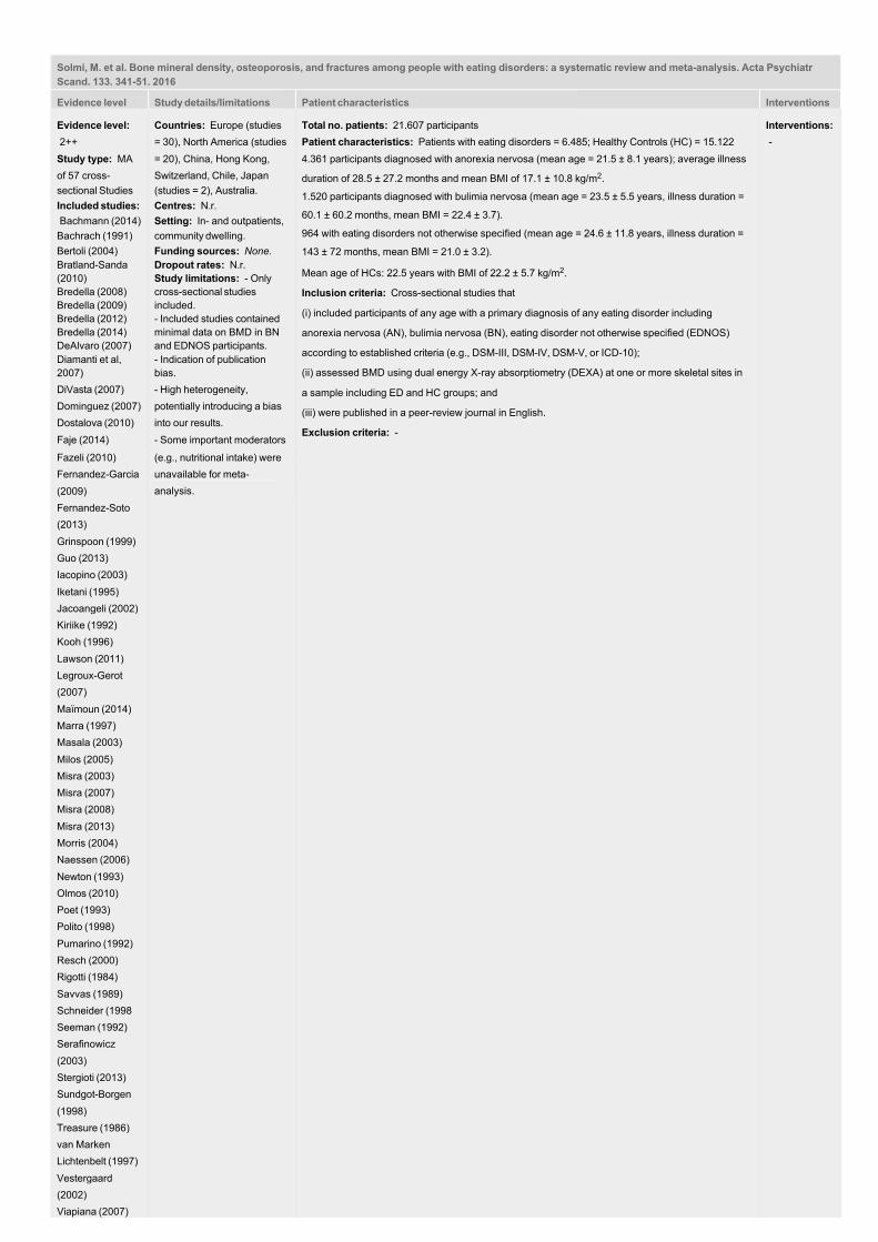
Solmi, M. et al. Bone mineral density, osteoporosis, and fractures among people with eating disorders: a systematic review and meta-analysis. Acta Psychiatr Scand. 133. 341-51. 2016
Evidence level Study details/limitations Patient characteristics Interventions Evidence level: 2++ Study type: MA of 57 cross- sectional Studies Included studies: Bachmann (2014) Bachrach (1991) Bertoli (2004) Bratland-Sanda (2010) Bredella (2008) Bredella (2009) Bredella (2012) Bredella (2014) DeAlvaro (2007) Diamanti et al, 2007) DiVasta (2007) Dominguez (2007) Dostalova (2010) Faje (2014)
Fazeli (2010) Fernandez-Garcia (2009) Fernandez-Soto (2013) Grinspoon (1999) Guo (2013) Iacopino (2003) Iketani (1995) Jacoangeli (2002) Kiriike (1992) Kooh (1996) Lawson (2011) Legroux-Gerot (2007) Maïmoun (2014) Marra (1997) Masala (2003) Milos (2005) Misra (2003) Misra (2007) Misra (2008) Misra (2013) Morris (2004) Naessen (2006) Newton (1993) Olmos (2010) Poet (1993) Polito (1998) Pumarino (1992) Resch (2000) Rigotti (1984) Savvas (1989) Schneider (1998 Seeman (1992) Serafinowicz (2003) Stergioti (2013) Sundgot-Borgen (1998) Treasure (1986) van Marken Lichtenbelt (1997) Vestergaard (2002) Viapiana (2007)
Countries: Europe (studies = 30), North America (studies = 20), China, Hong Kong, Switzerland, Chile, Japan (studies = 2), Australia. Centres: N.r. Setting: In- and outpatients, community dwelling. Funding sources: None. Dropout rates: N.r. Study limitations: - Only cross-sectional studies included. - Included studies contained minimal data on BMD in BN and EDNOS participants. - Indication of publication bias. - High heterogeneity, potentially introducing a bias into our results. - Some important moderators
(e.g., nutritional intake) were unavailable for meta-
Total no. patients: 21.607 participants Patient characteristics: Patients with eating disorders = 6.485; Healthy Controls (HC) = 15.122 4.361 participants diagnosed with anorexia nervosa (mean age = 21.5 ± 8.1 years); average illness
duration of 28.5 ± 27.2 months and mean BMI of 17.1 ± 10.8 kg/m2.
1.520 participants diagnosed with bulimia nervosa (mean age = 23.5 ± 5.5 years, illness duration =
60.1 ± 60.2 months, mean BMI = 22.4 ± 3.7).
964 with eating disorders not otherwise specified (mean age = 24.6 ± 11.8 years, illness duration =
143 ± 72 months, mean BMI = 21.0 ± 3.2).
Mean age of HCs: 22.5 years with BMI of 22.2 ± 5.7 kg/m2.
Inclusion criteria: Cross-sectional studies that
(i) included participants of any age with a primary diagnosis of any eating disorder including
anorexia nervosa (AN), bulimia nervosa (BN), eating disorder not otherwise specified (EDNOS)
according to established criteria (e.g., DSM-III, DSM-IV, DSM-V, or ICD-10);
(ii) assessed BMD using dual energy X-ray absorptiometry (DEXA) at one or more skeletal sites in
a sample including ED and HC groups; and
(iii) were published in a peer-review journal in English.
Exclusion criteria: -
Interventions: -
analysis.
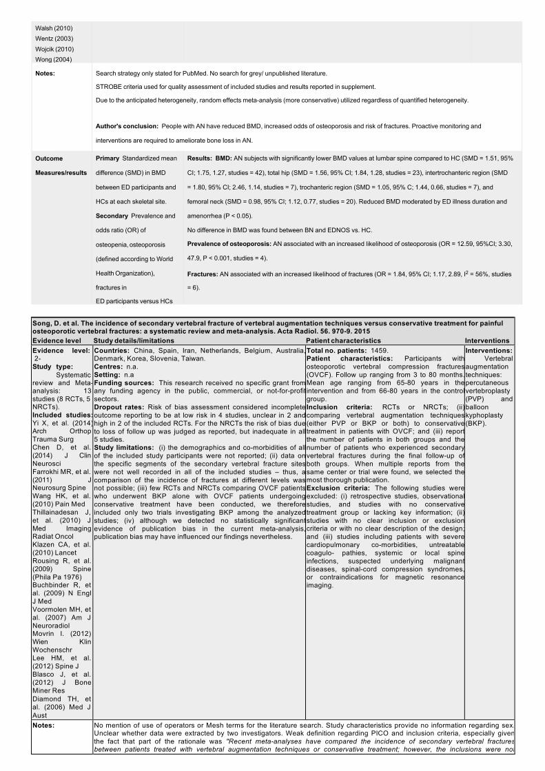
Walsh (2010) Wentz (2003) Wojcik (2010) Wong (2004)
Notes:
Search strategy only stated for PubMed. No search for grey/ unpublished literature.
STROBE criteria used for quality assessment of included studies and results reported in supplement.
Due to the anticipated heterogeneity, random effects meta-analysis (more conservative) utilized regardless of quantified heterogeneity.
Author's conclusion: People with AN have reduced BMD, increased odds of osteoporosis and risk of fractures. Proactive monitoring and
interventions are required to ameliorate bone loss in AN.
Outcome
Measures/results Primary Standardized mean
difference (SMD) in BMD
between ED participants and
HCs at each skeletal site.
Secondary Prevalence and
odds ratio (OR) of
osteopenia, osteoporosis
(defined according to World
Health Organization),
fractures in
ED participants versus HCs
Results: BMD: AN subjects with significantly lower BMD values at lumbar spine compared to HC (SMD = 1.51, 95%
CI; 1.75, 1.27, studies = 42), total hip (SMD = 1.56, 95% CI; 1.84, 1.28, studies = 23), intertrochanteric region (SMD
= 1.80, 95% CI; 2.46, 1.14, studies = 7), trochanteric region (SMD = 1.05, 95% C; 1.44, 0.66, studies = 7), and
femoral neck (SMD = 0.98, 95% CI; 1.12, 0.77, studies = 20). Reduced BMD moderated by ED illness duration and
amenorrhea (P < 0.05).
No difference in BMD was found between BN and EDNOS vs. HC. Prevalence of osteoporosis: AN associated with an increased likelihood of osteoporosis (OR = 12.59, 95%CI; 3.30,
47.9, P < 0.001, studies = 4).
Fractures: AN associated with an increased likelihood of fractures (OR = 1.84, 95% CI; 1.17, 2.89, I2 = 56%, studies
= 6).
Song, D. et al. The incidence of secondary vertebral fracture of vertebral augmentation techniques versus conservative treatment for painful osteoporotic vertebral fractures: a systematic review and meta-analysis. Acta Radiol. 56. 970-9. 2015 Evidence level Study details/limitations Patient characteristics Interventions Evidence level: 2-
Study type: Systematic
review and Meta- analysis: 13 studies (8 RCTs, 5 NRCTs). Included studies: Yi X, et al. (2014) Arch Orthop Trauma Surg Chen D, et al. (2014) J Clin Neurosci Farrokhi MR, et al. (2011) J Neurosurg Spine Wang HK, et al. (2010) Pain Med Thillainadesan J, et al. (2010) J Med Imaging Radiat Oncol Klazen CA, et al. (2010) Lancet Rousing R, et al. (2009) Spine (Phila Pa 1976) Buchbinder R, et al. (2009) N Engl J Med Voormolen MH, et al. (2007) Am J Neuroradiol Movrin I. (2012) Wien Klin Wochenschr Lee HM, et al. (2012) Spine J Blasco J, et al. (2012) J Bone Miner Res Diamond TH, et al. (2006) Med J Aust
Countries: China, Spain, Iran, Netherlands, Belgium, Australia, Denmark, Korea, Slovenia, Taiwan. Centres: n.a. Setting: n.a Funding sources: This research received no specific grant from any funding agency in the public, commercial, or not-for-profit sectors. Dropout rates: Risk of bias assessment considered incomplete outcome reporting to be at low risk in 4 studies, unclear in 2 and high in 2 of the included RCTs. For the NRCTs the risk of bias due to loss of follow up was judged as reported, but inadequate in all 5 studies. Study limitations: (i) the demographics and co-morbidities of all of the included study participants were not reported; (ii) data on the specific segments of the secondary vertebral fracture sites were not well recorded in all of the included studies – thus, a comparison of the incidence of fractures at different levels was not possible; (iii) few RCTs and NRCTs comparing OVCF patients who underwent BKP alone with OVCF patients undergoing conservative treatment have been conducted, we therefore included only two trials investigating BKP among the analyzed studies; (iv) although we detected no statistically significant evidence of publication bias in the current meta-analysis, publication bias may have influenced our findings nevertheless.
Total no. patients: 1459. Patient characteristics: Participants with osteoporotic vertebral compression fractures (OVCF). Follow up ranging from 3 to 80 months. Mean age ranging from 65-80 years in the intervention and from 66-80 years in the control group. Inclusion criteria: RCTs or NRCTs; (ii) comparing vertebral augmentation techniques (either PVP or BKP or both) to conservative treatment in patients with OVCF; and (iii) report the number of patients in both groups and the number of patients who experienced secondary vertebral fractures during the final follow-up of both groups. When multiple reports from the same center or trial were found, we selected the most thorough publication. Exclusion criteria: The following studies were excluded: (i) retrospective studies, observational studies, and studies with no conservative treatment group or lacking key information; (ii) studies with no clear inclusion or exclusion criteria or with no clear description of the design; and (iii) studies including patients with severe cardiopulmonary co-morbidities, untreatable coagulo- pathies, systemic or local spine infections, suspected underlying malignant diseases, spinal-cord compression syndromes, or contraindications for magnetic resonance imaging.
Interventions: Vertebral
augmentation techniques: percutaneous vertebroplasty (PVP) and balloon kyphoplasty (BKP).
Notes: No mention of use of operators or Mesh terms for the literature search. Study characteristics provide no information regarding sex. Unclear whether data were extracted by two investigators. Weak definition regarding PICO and inclusion criteria, especially given the fact that part of the rationale was "Recent meta-analyses have compared the incidence of secondary vertebral fractures between patients treated with vertebral augmentation techniques or conservative treatment; however, the inclusions were not
thorough and rigorous enough". E.g. the comparison (conservative treatment) was not specified. Importantly, no time period for incidence is specified, and the follow-ups of the included studies show a wide range of follow up times (6-80 months). No COI from primary studies. Author's conclusion: In conclusion, no differences in the incidence of secondary fractures in patients who underwent vertebral augmentation techniques and patients who underwent conservative treatment were observed. Additional RCTs, especially trials comparing the incidence of secondary vertebral fractures at different levels, trials comparing BKP and conservative treatment and trials identifying risk factors for secondary fractures, should be conducted.
Outcome Measures/results
Primary Incidence of secondary vertebral fractures. Secondary Subgroup analyses by study design (RCT and NRCT), geographic region (western countries and eastern countries, as the ethnicity of patients was not clearly stated in every study), duration of symptoms before enrollment (acute and not acute), and specific techniques (PVP, BKP and PVP+BKP) were also applied. A cumulative meta-analysis was also applied to investigate the influence of the publication date and sample size
Results: The pooled results from included studies showed no statistically significant differences in the incidence of secondary vertebral fractures between patients treated with vertebral augmentation techniques and conservative treatment. Subgroup analysis comparing different study designs, durations of symptoms, follow-up times, races of patients, and techniques were conducted, and no significant differences in the incidence of secondary fractures were identified (P>0.05). No obvious publication bias was detected by either Begg’s test (P=0.360 > 0.05) or Egger’s test (P=0.373 > 0.05).
Su, B. et al. Risk of bone fractures associated with glucagon-like peptide-1 receptor agonists' treatment: a meta-analysis of randomized controlled trials. Endocrine. 48. 107-15. 2015
Evidence level Study details/limitations Patient characteristics Interventions Evidence level: 1++
Study type: SR/MA of 16 RCT
Included studies: Buse JB (2009) Lancet
Buse JB (2011) Ann Intern Med
Buse JB (2013) Lancet
Charbonnel B (2013) Diabetologia
Gallwitz B (2011) Diabetes Care
Gallwitz B (2012) Lancet
Garber A (2011) Diabetes Obes Metab
Gill A (2010) Cardiovasc Diabetol
Heine RJ (2005) Ann Intern Med
Inagaki N (2012) Clin Ther
Lean ME (2013) Int J Obes
Nauck M (2007) Diabetologia
Nauck M (2013) Diabetes Obes Metab
NCT00614120
http://www.clinicaltrials.gov/show/NCT00614120
NCT00960661
http://www.clinicaltrials.gov/ct2/show/NCT00960661
Pratley R (2011) Int J Clin Pract
Countries: All studies but 2
conducted in multi-country
populations. 2 remaining studies
conducted in Germany and Japan.
Centres: N.r.
Setting: N.r. Funding sources: This work was
supported by Grants from the
National Natural Science
Foundation of China (NO:
81101444) and Shanghai Municipal
Natural Science Foundation (NO:
13ZR1432100).
Dropout rates: N.r. Study limitations: - Conclusion
needs to be interpreted with caution
because a relatively low number of
fracture cases were identified in
meta-analysis and because that the
durations of most trials were not
sufficiently long to study the effects
on bone fractures.
- Age of study populations relatively younger than typical osteoporosis
population, which might be an
important bias in evaluation of drug
effect on bone fractures.
- Risk of GLP-1 RA on specific
fracture sites could not be
determined due to limited number of
studies.
- Risk of bone quality might be
modified by patients’ concomitant
medications such as statins,
corticosteroids, estrogens, yet such
information were generally not
reported for individual studies.
- Risk observed in meta-analysis
Total no. patients: 11.206 patients
Patient characteristics: Middle-aged and
elderly study populations, with mean age
ranging from 45.9 years old to 59.5 years
old.
Proportion of female participants ranging
from 32.1 - 76.2 %.
Inclusion criteria: (1) patients without pre-
existing skeletal diseases such as
osteoporosis;
(2) randomized controlled human trials
(studies with well-designed open-label
extension were also included);
(2) with a duration of at least 12 weeks; (3) comparing the effects of exenatide or
liraglutide with placebo, or other active
drugs; and
(4) reporting fracture cases during follow-
up.
Exclusion criteria: Trials with duration
less than 12 weeks because they could not
provide relevant information on incidence of
fractures.
Interventions: Glucagon-like
peptide-1 receptor agonists
(GLP-1 RA): Exenatide or
liraglutide

could be affected by effects of
comparators.
Notes:
Unclear, whether data extraction was performed by two investigators.
Result of quality assessment with Jadad not presented one-to-one to studies, but as overall result instead.
Author's conclusion: Our study demonstrated a divergent risk of bone fractures associated with different GLP-1
RA treatments. The current findings need to be confirmed by future well-designed prospective or RCT studies.
Outcome Measures/results
Primary Risk of bone fractures
Results: Overall GLP-1 RA and risk of bone fractures: 14 studies included with 5.040 patients (23 fracture cases) in intervention group and 3.410 (15 fracture cases) in comparator group. No significant association:
OR = 1.05, 95 % CI; 0.59, 1.87). No significant heterogeneity: I2 = 0 %, P = 0.739. Observed risk effects not modified by age (P = 0.411), BMI (P = 0.291), and HbA1c (P = 0.786) level at baseline. Liraglutide and risk of bone fractures: 8 studies included with 3.737 patients (8 fracture cases) in intervention group and 3.175 (13 fracture cases) in comparator group. Treatment with liraglutide associated with a significant reduced risk of bone fractures, compared with placebo or other active drugs (OR = 0.38, 95 % CI; 0.17, 0.87, no p-value stated). No
significant heterogeneity: I2 = 0 %, P = 0.864. Excluding two studies using exenatide as comparators led risk effect to be non-significant (OR = 0.49, 95 % CI; 0.19, 1.22). Exenatide and risk of bone fractures: 10 studies included with 2.681 patients (20 fracture cases) in intervention group and 2.613 (7 fracture cases) in comparator group. Treatment with exenatide associated with a significant increased risk of bone fractures, compared with placebo or other active drugs (OR = 2.09, 95 % CI; 1.03, 4.21, no p-value stated). No significant
heterogeneity: I2 = 0 %, P = 0.745. After exclusion of two studies with liraglutide as comparator arm, the pooled MHOR was attenuated to 1.71 (95 % CI; 0.80, 3.67).
Secondary -
Stevenson, M. et al. Percutaneous vertebroplasty and percutaneous balloon kyphoplasty for the treatment of osteoporotic vertebral fractures: a systematic review and cost-effectiveness analysis. Health Technol Assess. 18. 1-290. 2014 Evidence level Study details/limitations Patient characteristics Interventions Evidence level: 1++
Study type: Systematic
review and Meta- analysis: 9 RCTs. Included studies:
Klazen CAH, et al. (2010) Lancet Buchbinder R, et al. (2009) N Engl J Med Kallmes DF, et al. (2009) N Engl J Med Blasco JA, et al. (2012) J Bone Miner Res Farrokhi MR, et al. (2011) J Neurosurg Spine Wardlaw D, et al. (2009) Lancet Liu JT, et al. (2010) Osteoporos Int Rousing R, et al. (2009) Spine Voormolen MHJ, et al. (2007) Am J Neuroradiol
Countries: Spain, Australia, Iran, Austria, Belgium, France, Germany, Italy, Netherlands, Sweden, UK, Taiwan. Centres: n.a. Setting: n.a. Funding sources: The research reported in this issue of the journal was commissioned and funded by the HTA programme on behalf of NICE as project number 10/122/01. The protocol was agreed in October 2011. The assessment report began editorial review in August 2012 and was accepted for publication in December 2012. The authors have been wholly responsible for all data collection, analysis and interpretation, and for writing up their work. The HTA editors and publisher have tried to ensure the accuracy of the authors’ report and would like to thank the reviewers for their constructive comments on the draft document. However, they do not accept liability for damages or losses arising from material published in this report. This report presents independent research funded by the National Institute for Health Research (NIHR). The views and opinions expressed by authors in this publication are those of the authors and do not necessarily reflect those of the NHS, the NIHR, NETSCC, the HTA programme or the Department of Health. If there are verbatim quotations included in this publication the views and opinions expressed by the interviewees are those of the interviewees and do not necessarily reflect those of the authors, those of the NHS, the NIHR, NETSCC, the HTA programme or the Department of Health. Dropout rates: Participants lost to follow up range between 5 and 26%. In 5 out of 9 studies, at least 80% of the included subjects were included (low risk of bias), in 3 studies more than 20% were lost to follow up (high risk of bias) and in one study this was not reported (unclear risk of bias). Reason for withdrawal were stated in 8 out 9 studies. Study limitations: The most serious
Total no. patients: 1114 (not stated in the article). Patient characteristics: Patients with osteoporotic vertebral compression fracture or non-osteoporotic vertebral fracture. 72- 83% of the participants were female. Mean age range 72-80 years. Inclusion criteria: Study type: RCT Population: people of any age and either gender with osteoporotic vertebral compression fractures. Studies which also include participants with non-osteoporotic vertebral fractures of other aetiologies (e.g. fractures associated with trauma, myeloma, or metastatic cancer) will be included if data relating to participants with osteoporotic fractures can be extracted separately. Intervention: Percutaneous vertebroplasty or percutaneous balloon kyphoplasty. Comparators: The comparators will be the interventions themselves, non-invasive management, a sham procedure, or no treatment. Outcomes: Pain/analgesic use; Back-specific functional status/mobility; Vertebral body height and angular deformity l Progression of treated fracture; Incidence of new vertebral fractures; Health-related quality of life Secondary outcomes: All-cause mortality; Symptomatic and asymptomatic leakage of cement (egg into adjacent intervertebral discs); Periprocedural balloon rupture; Post- operative complications (including infection); Other adverse events; Resource utilization; Cost utility. Exclusion criteria: Animal models; Preclinical and biological studies; narrative reviews, editorials, opinions; Reports published as meeting abstracts only, where insufficient methodological details are reported to allow critical appraisal of study quality.
Interventions: Intervention:
Percutaneous vertebroplasty or percutaneous balloon kyphoplasty. Comparison: The interventions themselves, non- invasive management, a sham procedure, or no treatment.

methodological problem was the lack of blinding in all studies except 2. Unblinding in surgical studies has been linked to a 25% overestimation of treatment effect. As Buchbinder and Kallmes have pointed out, the improvement in the treatment groups of the blinded trials was not dissimilar to that seen in the treatment groups of the unblinded trials.The assessment of BKP’s clinical effectiveness was particularly limited, as only one open-label RCT comparing BKP with non-surgical management was available, while the only study to compare BKP with PVP showed a number of potential sources of bias. A further limitation of these findings was the use of pain as a primary outcome. As others have argued, pain measurement may be confounded by a number of factors, including pain threshold, analgesia and level of activity. Back pain-related disability and quality of life may provide more objective and clinically meaningful measures. However, these outcomes were measured in heterogeneous ways among the trials, precluding statistical aggregation of the data. Measures of vertebral body height and angular deformity may also be more useful clinical outcomes than pain, insofar as improvements could enhance mobility and stave off deterioration of cardiopulmonary function. 4 studies reported these outcomes, but it was not possible to aggregate their findings owing to the heterogeneous approaches that were taken to wedge angle and vertebral body height measurement.
Notes: Article selection was not carried out by two investigators. Grey literature was not considered. Author's conclusion: For people with painful osteoporotic VCFs refractory to analgesic treatment, PVP and BKP perform significantly better in unblinded trials than optimal pain management (OPM) in terms of improving quality of life and reducing pain and disability. However, there is as yet no convincing evidence that either procedure performs better than OPLA. The uncertainty in the evidence base means that no definitive conclusion on the cost-effectiveness of PVP or BKP can be provided. Further research should focus on establishing whether or not BKP and PVP have a mortality advantage compared with OPLA and on whether or not these provide any utility gain compared with OPLA.
Outcome Measures/results
Primary Pain/analgesic use; Back-specific functional status/mobility; Vertebral body height (VBH) and angular deformity; Progression of treated fracture; Incidence of new vertebral fractures; Health-related quality of life. Secondary All-cause mortality; Symptomatic and asymptomatic leakage of cement (egg into adjacent intervertebral discs); Periprocedural balloon rupture; Post-operative complications (including infection); Other adverse events; Resource utilisation; Cost utility.
Results: Quality of evidence: This body of literature was of variable quality, with the two double-blind, OPLA-controlled trials being at the least risk of bias. The most significant methodological issue among the remaining trials was lack of blinding for both study participants and outcome assessors. The volume of available evidence of clinical effectiveness is greater for PVP than for BKP, and the methodological quality of some of that evidence is also higher than that of any study of BKP. Clinical effectiveness: In relation to PVP, the studies least at risk of bias found no significant differences between treatment groups in terms of change from baseline in HRQoL other than in terms of the total QUALEFFO score at 1 week in the Buchbinder study: this favoured PVP. No significant differences were observed in any measure of functional status or pain. Although the INVEST study reported a trend towards a greater number of patients in the PVP group reporting a clinically meaningful improvement in pain at 1 month, pooled data from the Buchbinder et al. indicate that, after adjusting for baseline opioid use, patients randomised to PVP were more likely than those randomised to the OPLA to be taking opioids at 1 month. Consequently, it is impossible to exclude the possibility that PVP was associated with worse outcomes which were masked by greater opioid use. The evidence from the open-label studies of PVP regarding HRQoL is not consistent: VERTOS and VERTOS II suggest that PVP is associated with better HRQoL as measured by the QUALEFFO, while Blasco et al. found no significant difference between treatment groups. The data reported by Rousing et al. indicate that conservative management is generally associated with better HRQoL, as measured by the EQ-5D and DPQ. Functional status: By contrast, the evidence from these studies relating to functional status appears to favour PVP; the most convincing evidence comes from Farrokhi et al. the unblinded study at least risk of bias, which found that, as a measured by a modified version of the ODI, PVP was associated with significantly improved functional status at all times from 1 week to 36 months. In the Farrokhi et al. study mobility at 1 day was also dramatically better in the PVP group than in the control group. Pain The Blasco et al. Farrokhi et al. Rousing et al. and VERTOS II studies found that PVP was associated with significant improvements in pain, although in the study by Blasco et al. statistical significance was seen only at 2 months; moreover, in those studies which report analgesic use, these improvements do not appear to be associated with increased analgesic use in the PVP group. VBH Farrokhi et al. also found that PVP was associated with sustained improvements in VBH and angular deformity; however, Blasco et al. found no significant difference between groups in VBH.
Stolzenberg, N. et al. Vibration or balance training on neuromuscular performance in osteopenic women. Int J Sports Med. 34. 956-62. 2013
Evidence level Study Patient characteristics Interventions details/limitations Evidence level: 1+
Study type: RCT
Countries: Germany Centres: Siemens Betriebskrankenkasse, Centre for Muscle and Bone Research (Charité Universitätsmedizin
Total no. patients: ITT-population: 60 women; PP-population: 55 women Patient characteristics: Stated only for ITT-population: Vibration group: Mean age (SD): 67.5 (3.8) years; Mean BMI (SD):
Interventions: Intervention period: 9 months. Joint resistive exercise: 2 training appointments per week in groups of max 10 participants. 15 min warm-up on a cycle ergometre followed by 30 min resistance exercise with increasing load. Balance training (BAL): 15 min progressive proprioceptive and balance exercise program performed alone or when necessary with a partner.
Berlin) and Immanuel Krankenhaus Berlin Setting: Training interventions local sports club (Sport- Gesundheitspark Berlin e.V.) Funding sources: No funding received. Dropout rates: 11 subjects dropped out of study. Distribution among groups not stated, only that the "drop out rate did not differ significantly between groups (χ² = 0.25, p = 0.61)". Study limitations: - Contribution the resistive exercise on changes in current study unclear, - Significant differences seen in response between groups indicate that additional training forms were driving factor behind changes seen in neuromuscular performance, - Future work will need to consider issues of whole-body vibration training duration, intensity and frequency.
24.7 kg/m2 (2.7) Balance group: Mean age (SD): 65.5 (4.3) years; Mean BMI (SD): 23.8 kg/m2 (3.9) Inclusion criteria: (1) a minimum of 8 years postmenopausal, (2) no recent (last 6 months) involvement in whole-body vibration exercise, balance training or resistive exercise and (3) a total hip or lumbar spine (L1– L4) T-score from − 2.0 to − 3.0 SD on dual energy X-ray absorptiometry (DXA). Exclusion criteria: (1) any metal implants, known disturbance of the vestibular system, prior experience with the testing apparatus, (2) bone fractures within the last year, (3) neuromuscular and neurological diseases, acute thrombosis in the last 24 months, coronary heart disease, pacemaker, acute arthritis, (4) smoking of more than 20 cigarettes per day and alcohol consumption of more than 14 g per day.
Whole-body vibration (VIB): 3 consecutive trainings on Galileo Fitness (Novotec, Pforzheim, Germany). Amplitude of vibration at 2 mm in first week and 4 mm within next 4 weeks. 3 different exercises performed lasting 4 min with 1 min break between exercises.
Notes: Allocation concealment mentioned but not described. Investigators could have been blinded but were not. No test for baseline differences mentioned but "no significant baseline differences" stated. 8 participants were subsequently recruited after higher than expected drop-out rate. Baseline characteristics for these participants not stated. These subjects were excluded from ITT-analyses. Author's conclusion: In conclusion, the results of the current study show that combining whole-body vibration with resistive exercise in postmenopausal women with low bone density is more effective in improving some aspects of neuromuscular performance than proprioceptive training with resistive exercise. The additional effect of whole-body vibration was seen most clearly in the countermovement jump test. The results support the implementation of whole-body vibration with resistive exercise in maintaining neuromuscular performance in post-menopausal osteopenic or osteoporotic women.
Outcome Measures/results
Primary Countermovement
jump power per unit body weight Secondary Multiple 1-leg hopping, Sit-to- stand test
Results: Countermovement jump: Peak power relative to body weight (W/kg): Significant change from baseline to 9 months in vibration group (27.9 [4.8] to 28.5 [5.0]; p<0.01). Adjusted Difference at 9 months (95% CI): 0.9 (0.3, 1.5), no p-value stated. Peak acceleration (g): Significant change from baseline to 9 months in balance and vibration group (2.0 [0.2] to 2.1 [0.2]; p<0.01 vs 2.1 [0.2] vs 2.2 [0.3]). Adjusted Difference at 9 months (95% CI): -0.04 (-0.12, 0.05), no p-value stated. Jump height (cm): Significant change from baseline to 9 months in balance group (25 [4] to 24 [4]; p<0.05). Adjusted Difference at 9 months (95% CI): 1.3 (0.3, 2.3), no p-value stated. Esslinger Fitness Index (%): Significant change from baseline to 9 months in vibration group (101 [15] to 105 [16]; p<0.001). Adjusted Difference at 9 months (95% CI): 3.2 (1.0, 5.5), no p-value stated. Jump efficiency (%): Significant change from baseline to 9 months in balance group (117 [16] to 111 [15]; p<0.01. Adjusted Difference at 9 months (95% CI): 6.9 (2.7, 11.1), no p-value stated. Multiple 1-leg hopping: Hop force and acceleration significantly improved in both groups on left leg and in the VIB group also on the right leg. Adjusted Difference in hop force at 9 months (95% CI): 0.00 (-0.06, 0.06), no p-value stated. Adjusted Difference in acceleration at 9 months (95% CI): 0.01 (-0.08, 0.10), no p- value stated. i>Peak power relative to body weight (W/kg): Significant change from baseline to 9 months in balance group in both legs (left leg: 15.0 [3.1] to 14.0 [2.9]; p<0.05; right leg: 16.0 [3.3] to 14.0 [3.1]; p<0.001). Adjusted Difference at 9 months (95% CI): 0.4 (-0.6, 1.5) and 1.0 (-0.2, 2.2), no p-value stated. Sit-to-stand test: Sit-to-stand duration and average acceleration both improved significantly in VIB-group, but not in the BAL-group. No significant change in average power per unit body weight.
Stubbs, B. et al. Schizophrenia and the risk of fractures: a systematic review and comparative meta-analysis. Gen Hosp Psychiatry. 37. 126-33. 2015
Evidence level Study details/limitations Patient characteristics Interventions Evidence level: 2+
Study type: SR of 8
studieas
MA of 5 studies
Included studies:
Sorensen HJ (2013)
Eur
Neuropsychopharmacol
Bishop JR (2004)
Pharmacotherapy
Countries: USA (n = 2),
South Korea (n = 1),
Denmark (n = 1), Taiwan (n =
2), UK (n = 1), Canada (n =
1)
Centres: N.r. Setting: N.r.
Funding sources: No
funding reported.
Authors report several
Total no. patients: Review with 48.384 people with schizophrenia and 3.945.783 controls.
Meta-analysis with 32.593 participants with schizophrenia and 138.186 controls.
Patient characteristics: Age ranging from 49.9–75.2 years.
41% – 100% of study participants female.
Inclusion criteria: - Included observational studies (prospective, retrospective or cross-
sectional) that
(a) included people with a diagnosis of schizophrenia according to recognized criteria
(Diagnostic and Statistical Manual of
Mental Disorders (DSM), Fifth Edition or International Classification of Diseases (ICD), 10th
Revision) together with a control group of people without a mental illness and
(b) reported the number of fractures over any period of time or
Interventions:
-
Howard L (2007) Br J
Psychiatry
Bolton JM (2008) J Clin
Psychopharmacol
Jung D-U (2011) J Clin
Psychopharmacol
Kelly DL (2011)
Osteoporos Int
Lai S-W (2013) Libyan
J Med
Tsai K (2014) Schizophr
Res
financial ties to
pharmaceutical companies.
Dropout rates: N.r. Study limitations: - Due to
paucity of data and lack of
information, it was not
possible to conduct
moderator or subgroup
analyses.
- Most of included studies
were cross-sectional or
retrospective cohort studies.
- Heterogeneity in reporting
of fractures and in skeletal
sites considered, thus
precluding evaluation of site-
specific fracture risk across
studies.
(c) reported the number of people with schizophrenia in comparative studies containing
groups with and without a fracture and
(d) collected data on any type and body site for fracture that was confirmed through
radiographs, medical note review or self-report.
Exclusion criteria: -
Notes:
No search for grey/unpublished literature. Heterogeneity reported without p-values.
Funnel plots produced despite insufficient number of studies.
High heterogeneity regarding reported outcomes (e.g. any fractures, wrist fractures or vertebral fractures) and observation period (lifetime vs 12
months).
Author's conclusion: People with schizophrenia are at significantly increased risk of fractures. Future research is required to understand the
mechanisms and should seek to validate fracture prediction algorithms used in the general population. Importantly, there is a need to develop
preventative strategies to improve bone health and reduce fracture risk involving the wider multidisciplinary team and incorporating falls-
prevention strategies.
Outcome
Measures/results
Primary Not specified.
Relationship between
Schizophrenia and fractures
Secondary -
Results: Narrative summary: Several studies showing significantly increase risk for fractures. 1 study
adjusted for anti-psychotic medication, after which the effect was not significant (IRR = 1.00, 95% CI; 0.90,
1.11). 1 study demonstrated a dose–response relationship of increased risk of hip fracture among those on
long-term prolactin-raising antipsychotic medication (IRR = 1.16, 95% CI; 1.09, 1.24) and non-prolactin-raising
antipsychotic medication (IRR = 1.23, 95% CI; 1.15, 1.32). Meta-analysis: data from 5 studies to calculate rate of fractures per 1000 person-years. Rate of fractures was
5.54 (95% CI; 4.92, 5.57) fractures per 1.000 person-years in those with schizophrenia and 3.48 (95% CI; 3.39,
3.64) fractures per 1000 person-years in control participants. IRR = 1.72 (95% CI; 1.24, 2.39, I2 = 49%, n =
168.914).
Sugimoto, T. et al. Three-year denosumab treatment in postmenopausal Japanese women and men with osteoporosis: results from a 1-year open- label extension of the Denosumab Fracture Intervention Randomized Placebo Controlled Trial (DIRECT). Osteoporosis international : a journal established as result of cooperation between the European Foundation for Osteoporosis and the National Osteoporosis Foundation of the USA. 26. 765-74. 2015 Evidence level Study details/limitations Patient characteristics Interventions Evidence level: 1++
Study type: RCT, one year open label extension of DIRECT-study
Countries: Japan Centres: multicenter Setting: - Funding sources: Daiichi-Sankyo., Tokyo, Dropout rates: 3,71%, placebo 4,93% Study limitations: The number of patients was relatively small and the extension did not have a placebo arm. Due to the small number of subjects, this study is considered to have inadequate power to detect adverse events with very low incidence such as atypical femoral fractures. Additionally, since all subjects received denosumab in the 12- month extension phase, it is difficult to rigorously investigate long-term vertebral fracture risk reduction.
Total no. patients: N= 810 (404 in the denosumab group and 406 in the placebo (cross-over) group) Patient characteristics:
Generally, Healthy Japanese subjects with osteoporosis including postmenopausal women and men Inclusion criteria: - Age ≥50 years - 1–4 prevalent vertebral fractures, and low bone mineral density (BMD) (T-score Exclusion criteria: -
Interventions: DIRECT study: for a period of 2 years, denosumab 60 mg given as a subcutaneous injection every 6 months, matching placebo, or open- label alendronate 35 mg taken orally every week. During the open-label extension phase, all subjects received 60 mg denosumab subcutaneously every 6 months for 1 year with daily supplements containing at least 600 mg calcium and 400 IU vitamin D.
Notes: No information about sample size calculation or ITT Author's conclusion: Three-year denosumab treatment in Japanese subjects with osteoporosis showed a favorable benefit/risk profile.

Outcome Measures/results
Primary describe the efficacy including evaluation of the incidence of vertebral and nonvertebral fractures, and the changes in BMD and BTMs, and safety and tolerability of denosumab for 3-years. Secondary -
Results: Fractures The 2-year incidence of new vertebral fractures was 2.2 % in the denosumab (long-term) group and 8.6%in the placebo(cross-over) group, with the reduction in risk by 74.0 % (p<0.0001), and the rate ratios comparing the number of new vertebral fractures for the denosumab group in year 1 and 2 with those for the placebo group were 0.44 (p=0.0937) and 0.14 (p=0.0001). In the long-term group, the cumulative incidences of new or worsening vertebral and new vertebral fractures at 36 months were 3.8 % (95 % CIs, 2.4; 6.1) and 2.5 % (95 % CIs, 1.4; 4.5). The annual rate of new or worsening vertebral and new vertebral fractures were 1.9 and 1.3 %, in the first year, 1.6 and 0.9 %, in the second year, and 0.3 and 0.3 %, in the third year. In the cross-over group, the cumulative incidences of new or worsening vertebral and new vertebral fractures at 36 months were 11.8 % (95 % CIs, 9.1; 15.2) and 10.3 % (95 % CIs, 7.8; 13.5). The annual rate of new or worsening vertebral and new vertebral fractures in the first, second, and the third years were 2.7, 5.9, and 1.9 %. BMD In the long-term group, increases from the baseline in BMD after 3 years of continued denosumab treatment were 11.0 % at the lumbar spine, 5.3 %at the total hip, 4.8 % at the femoral neck, and 0.9 % at the distal 1/3 radius (p<0.001). From 24 months to 36 months BMD increased by 1.8%at the lumbar spine, 0.6% at the total hip, 0.8 % at the femoral neck, and 0.4 % at the distal 1/3 radius (p<0.001). In the cross-over group, BMD changes at 36months (1 year after the first dose of denosumab) were 5.4 % at the lumbar spine, 1.4 % at the total hip, 1.1 % at the femoral neck, and −1.3 % at the distal 1/3 radius from the baseline (p<0.001). The BMD increased from 24 to 36 months by 5.3 % at the lumbar spine, 2.6 % at the total hip, 2.3 % at the femoral neck, and 0.5 % at the distal 1/3 radius (p<0.001). BTMs In the long-term group, serum CTX-1 was greatly decreased by 70.9 % at 1 month with relatively sustained reductions thereafter. Serum BSAP was reduced by 9.8 % at 1 month and 50.2 % at 3 months, and remained relatively stable thereafter. In the cross-over group, serum CTX-1 was greatly decreased by 77.9 % at 25 months (1 month after the first dose of denosumab) and remained relatively stable thereafter. Serum BSAP was also reduced by 19.6 % at 25 months and reached the same level as the long-term group at 36 months. Albumin-adjusted serum calcium and intact PTH In the long-term group, mean albumin-adjusted serum calcium was 9.09 and 9.14 mg/dL at month 1 and month 25, and median intact PTH was 54 pg/ml at 1 month and 44 pg/ml at month 25. In the cross-over group, mean albumin-adjusted serum calcium was 9.09 mg/dL at 25 months, and median intact PTH was 56 pg/ml at 25 months. These changes in albumin adjusted serum calcium and intact PTH had tendency to return to the baseline level during follow-up period. Safety No notable increase with longer denosumab treatment from 2 to 3 years, irrespective of the different duration of the observation periods. AEs in the long-term group were similar to those in the cross-over group.
Teng, Z. et al. Opioids contribute to fracture risk: a meta-analysis of 8 cohort studies. PLoS One. 10. e0128232. 2015
Evidence level Study details/limitations Patient characteristics Interventions Evidence level:
2-
Study type:
Systematic
review and meta-
analysis: 8 cohort
studies.
Included studies:
Guo Z, et al.
(1998) Am J
Epidemiol
Card T, et al.
(2004) Gut
Miller M, et al.
(2011) J Am
Geriatr Soc
Vestergaard P, et
Countries: Sweden, USA, UK, Denmark.
Centres: n.a.
Setting: n.a.
Funding sources: These authors have no support or
funding to report.
Dropout rates: n.a. Study limitations: First, the eligible studies were
restricted to the English language. The number of
relevant studies was still relatively small, which
implies that some studies may have been missed due
to their publication in non-English language journals
or publication in a book or a journal that is not
included in the computer databases.
Second, studies with nonsignificant results, especially
those that show an absence of effect, may not be
published because they are rejected by the journals or
because the investigators are unwilling to submit them
Total no. patients: 500819 participants.
Patient characteristics: Age of participants 59±14 years; or 45-58 years
where provided. Follow-up time between 464 days and 10 years.
Inclusion criteria: Studies were considered eligible if they met all of the
following criteria: (i) presented original data from a cohort study; (ii)
evaluated the association of opioid use with fracture incidence; (iii) had
opioids as the exposure of interest; and (iv) provided hazard ratios (HRs)
or the adjusted relative risks (RRs) and the corresponding 95% confidence
intervals (CIs). If the data were duplicated or the population was studied in
more than one study, we included the study with the largest sample size
and the most comprehensive outcome evaluation.
Exclusion criteria: n.a.
Interventions:
Opioid use.
al. (2006) J Intern
Med
for publication. Although we controlled for publication
bias using statistical methods, publication bias could
not be completely ruled out. Thus, the pooled effect
measure may be overestimated.
Third, the degree of control for confounding variables,
such as age, gender, body mass index and
comorbidity, also varied between studies.
Fourth, in this meta-analysis, we were not able to
investigate the effect of different opioid doses
because relevant data were available in only a few
studies.
Fifth, it was unfortunate that we were not able to
define contributing factors because all of the included
papers were from Western countries and all of th pe
participants were Caucasian. Therefore, our results
cannot be generalized to worldwide populations,
especially non-Western populations.
Sixth, we were not able to determine the fracture
timing, whether medication sedation effects would be
more likely shortly after starting opioid therapy, or
whether metabolic biomechanical reasons would be
more likely the longer the medication is taken. Thus,
further research into fracture timing is needed.
Finally, our meta-analysis is of good quality but is not
the most comprehensive study because only cohort
studies were included.
Notes:
No consideration of grey literature and listing of excluded studies. Study characteristics provide no information on participants age. Out of 8 studies
mentioned in the tables 4 have no reference in the reference section, some are incorrectly referenced. Overall high heterogeneity can be observed in
the main analysis, which is not not due the influence of a single study as revealed by sensitivity analysis.
Author's conclusion: In summary, our meta-analysis of cohort studies demonstrates that opioids may play an important role in the development of
fractures and that opioid use may significantly increase fracture risk. Further studies, including studies that are well-designed, international trials
(especially prospective, non-Western studies), studies that examine the mechanisms responsible for the fractures due to opioid use and studies that
aim to prevent these fractures from occurring are required to provide more convincing evidence for clinical practice and fracture prevention.
Outcome
Measures/results
Primary Fracture risk: pooled RR from the adjusted
RRs or HRs was computed.
Secondary Subgroup analysis specific to hip
fractures.
Results: The meta-analysis indicated significant, positive associations between opioid use
and overall fracture risk (RR 1.88, 95% CI 1.51–2.34). Substantial heterogeneity was
observed (P=0.000, I2=85.6%).
Data on opioid use and hip fracture risk were available for 4 studies. The pooled results
indicated that opioids contribute significantly to the risk of fracture (RR 2.00, 95% CI 1.84–
2.19). No statistical heterogeneity was observed (P=0.266, I2=24.4%).
Uusi-Rasi, K. et al. Exercise and vitamin D in fall prevention among older women: a randomized clinical trial. JAMA Intern Med. 175. 703-11. 2015 Evidence level Study details/limitations Patient characteristics Interventions Evidence level: 1++
Study type: RCT
Countries: Finland Centres: Not reported. Setting: Not reported. Funding sources: Supported by the Academy of Finland, Ministry of Education and Culture, Competitive Research Fund of Pirkanmaa Hospital District, and Juho Vainio Foundation. The funding sources had no role in the design and conduct of the study; collection, management, analysis, and interpretation of the data; preparation, review, or approval of the manuscript; and decision to submit the manuscript for publication. Dropout rates: Vitamin D and exercise:
Total no. patients: 409 women
Patient characteristics: Home-dwelling women with
mean age between 73.8 (SD 3.1) - 74.8 (SD 2.9) years. Mean (SD) daily dietary calcium intake: 1098 (378) mg at baseline. Mean (SD) daily dietary vitamin D intake: 10.4 (3.9) μg at baseline. 53 women (13%) did not take any medication at baseline.
Interventions: Vitamin D supplements/placebo: Participants randomly assigned to receive placebo (50%) or 20 μg of vitamin D (50%) per day for two years. Training program: Participants in exercise groups (n=205) randomly assigned to receive either 20 μg of vitamin D (50%) or placebo (50%). Participated in supervised training classes two times a week for 12 months, and once a week during the next 12 months. In addition to supervised training sessions, the exercisers have a home-training program for daily training based on programs used in training sessions.
6/102 (5.9%) Placebo and exercise: 12/103 (11.7%) Vitamin D without exercise: 14/102 (13.7%) Placebo without exercise: 7/102 (6.9%) Study limitations: - Fortification of fluid milk products in Finland might have had beneficial effect on baseline 25 (OH)D levels - Vitamin D might have greater effects on fall risk among women with vitamin D depletion. - All participants had fallen previously and thus were at increased risk of falling, and vitamin D supplementation is now recommended as a means to reduce the risk of falls irrespective of baseline vitamin D levels. - Challenging to reach and recruit the frailest women although exercise program planned to be suitable and safe for women with musculoskeletal problems → Study sample in good health and physical condition. → Results cannot be generalized to frail and institutionalized women or to men.
87.0% had at least 1 diagnosed chronic disease, most commonly cardiovascular. Inclusion criteria: - Age between 70-79 years, living at home independently, - had fallen at least once during the previous year, - no contraindication to exercise, - participant understands the procedures of the study, has been informed of X-ray radiation doses of the DXA and pQCT investigations, and amount of blood samples needed, and voluntarily agrees to undergo all measurements. Exclusion criteria: - Moderate-to-vigorous exercise more than 2 hours per week, - regular use of vitamin D or calcium + vitamin D supplements, - a recent fracture (during preceding 12 months), - contraindication or inability to participate in the exercise program, - persons with a marked decline in the basic activities of daily living (ADL-test), - persons with cognitive impairments (Mini Mental State Examination, MMSE- test), - persons with degenerative conditions, such as Parkinson’s disease.
Notes: No concealment method described. Author's conclusion: Given the fact that fall risk is multifactorial, exercise may be the most effective and feasible strategy for preventing injurious falls in community-dwelling older adults replete with vitamin D. Herein, vitamin D increased bone density slightly, and exercise improved physical functioning. While neither treatment reduced the rate of falling, injurious falls more than halved among exercisers with or without vitamin D. Our participants were vitamin D replete, with sufficient calcium intake. Future research is needed to elaborate the role of vitamin D to enhance physical functioning in elderly women.
Outcome Measures/results
Primary Monthly reported falls: obtained from prospective fall diaries returned monthly via mail, and details of each registered fall ascertained by a telephone call. Fall defined as “an unexpected event in which the participant comes to rest on the ground, floor or lower level”. Secondary Injurious falls (those for which participants sought medical care [nurse, physician, or hospital] and included bruises, abrasions, contusions, sprains, fractures, head injuries), the number of fallers and injured fallers, bone density, and physical functioning (muscle strength, balance, and mobility)
Results: Mean pill compliance: 98.1% (range, 42.6%-100%). Mean exercise compliance (measured as attendance at all offered training sessions for group and home training): 72.8% (range, 0%-97.4%) and 66.1% (0%-100%). Mean intensity of supervised training ranged from 1.6 METs (stretching or resting) to 5.6 METs. Individual maximal values were more than 7 METs. Falls: No interaction was found vitamin D and exercise. Overall: 928 falls and 281 fallers, no between-group differences. Hazard ratios for injured fallers lower in both exercise groups compared with the placebo without exercise group. Placebo + exercise group: HR = 0.47 (95% CI; 0.23, 0.99). Vitamin D + exercise group: HR = 0.38 (95% CI; 0.17, 0.83). No significant difference between vitamin D without exercise group and placebo without exercise group. Results similar for the incidence rate ratios of injurious falls No. of multiple fallers distributed similarly across the groups (n = 190 and n = 117). Bone density: Femoral neck BMD declined in all groups. Decline was greatest in placebo without exercise group (Mean change at 24 months: -1.22 [95% CI; -4.75, 2.45]) and differed significantly from that in all other study groups (vs Vitamin D without exercise P = 0.02; vs. Placebo + exercise P = 0.01; vs. Vitamin D and exercise P = 0.04). Lumbar spine BMD did not change significantly in any group. Vitamin D increased trabecular bone density at the tibia slightly but significantly between the vitamin D and exercise group and the placebo without exercise group (Change at 24 months: -0.49 [95% CI; -3.77, 2.80] vs. 0.19 [95% CI; -3.04, 3.42]; P = 0.02). Physical functioning: Normal walking speed maintained in placebo + exercise group compared with placebo without exercise group (P = 0.007), while declining similarly in other groups. Chair stand time improved in exercise groups, showing more than 6% improvement compared with the placebo without exercise group but reaching statistical significance only in the placebo and exercise group (P = 0.03). TUG time deteriorated significantly more in vitamin D without exercise group compared with the placebo without exercise group (P = .01). Other groups did not differ from placebo without exercise group. Backward walking improved significantly among exercisers. Difference compared with placebo without exercise group statistically significant in placebo and exercise group (P = 0.001) and in vitamin D and exercise group (P = 0.03). Irrespective of vitamin D, exercise increased muscle strength, while vitamin D alone had no effect. Predicted mean increase in lower limb extension strength was almost 15% in both exercise groups and differed significantly from placebo without exercise group (P < 0.001).
Veronese, N. et al. Vitamin K antagonists' use and fracture risk: results from a systematic review and meta-analysis. J Thromb Haemost. 13. 1665-75. 2015
Evidence level Study details/limitations Patient characteristics Interventions Evidence level: 2-
Study type:
Countries: Europe, North America, Asia, Australia.
Centres: n.a.
Setting: Dialysis uni, In- or Out-patients.
Total no. patients: 670810
participants.
Interventions:
Vitamin K

Systematic review
and meta-analysis:
21 studies
observational
studies.
Included studies:
Jamal SA, et al.
(1998) Ann Intern
Med
Barnes C, et al.
(2005) Pediatr Res
Fiore CE, et al. (1990) South Med
J Fusaro M, et al.
(2015) Curr Vasc
Pharmacol
Gage BF, et al.
(2006) Arch Intern
Med
Irastorza IX, et al.
(2010) J Pediatr
Gastroenterol Nutr
Kiss J, et al. (1995)
Orv Hetil
Lafforgue P, et al.
(1997) Rev Rhum
Engl Ed
Liebig S, et al.
(2004) Dtsch Med
Wochenschr
Mamdani M, et al.
(2003)
Pharmacotherapy
Misra D, et al.
(2014) Osteoporos
Int
Ogura-Tomomatsu
H, et al. (2012)
COPD
Philip WJ, et al.
(1995) QJM
Pilon D, et al.
(2004)
Pharmacoepidemiol
Drug Saf
Rejnmark L, et al.
(2007) Int J Cardiol
Rezaieyazdi Z, et
al. (2009) Int J
Rheum Dis
Rosen HN, et al.
Funding sources: n.a. Dropout rates: n.a.
Study limitations: Considering BMD parameters, we failed to find signifi- cant differences
between VKAs and MC or HC groups, except for significantly lower spine T-score values [30]
and a higher prevalence of osteoporosis [23] compared with HCs in one study each. In
contrast with the first meta-analysis published on this topic [10], we also failed to find any
significant association between VKAs and low radius BMD values. This finding is likely to be
due to the different inclusion criteria used in the previous meta-analysis, such as the
inclusion of studies without a control group, or studies using different methods other than
DXA that is considered the best approach for investigating BMD [3,9]. Thus, results of the
previous meta- analysis could have been affected by the remarkable differ- ences in bone composition investigation methods. However, independent of these considerations, the
findings of both meta-analyses suggest a non-significant effect of VKAs on BMD, since the
significant differences in BMD in our and in the previous meta-analysis are limited to
outcomes including single studies with a small number of subjects.
Several limitations of our study need to be considered. First, many of the included studies did
not use any matching, thereby not controlling for the potential con- founders between the
VKA-treated and the control groups. Although some individual studies that did not match
samples adjusted their analyses for relevant covari- ates and potential confounders [8,19],
these adjustments could not be used in this meta-analysis, as only patient level data would be
able to do so. By contrast, in meta- analyses, unadjusted group means or group rates are
pooled in the analyses.
Patient characteristics:
Intervention: Patients treated
with vitamin K antagonists (VKA)
n=79663.
Comparison: Medical illness
(MC), or healthy controls (HC),
n=597348 controls in total.
Inclusion criteria: Cross-
sectional and longitudinal studies
comparing data on osteoporosis,
fractures, BMD, and T-scores
(deviation from the bone mass of an average healthy 30-year-old
adult) between patients treated
with VKAs vs. controls. Included
were only studies that (i) reported
on BMD or fractures, (ii) included
patients treated with VKAs, and
(iii) compared patients treated
with VKAs with HCs or MCs.
Exclusion criteria: Studies were
excluded if they (i) did not specify
the type of VKAs used, (ii)
measured only in-vitro
parameters or used animal
models, (iii) did not compare
patients treated with VKAs vs. a
control group, or (iv) reported only
laboratory data and not data
about BMD or fractures. We also contacted authors asking
for further information when (i)
data could not be meta-analyzed
(i.e., no mean and SD or
equivalent for BMD or no data on
fractures or osteoporosis
provided for both treated and
control group) and (ii) other
relevant information (e.g., age,
percentage of females, or
parameters related to
anticoagulation therapy, etc.) was
missing.
antagonists
(VKA).

(1993) Am J Med
Sato Y, et al. (1997)
Stroke
Sato Y, et al. (2010)
Cerebrovasc Dis
Stenova E, et al.
(2011) Bratisl Lek
Listy
Woo C, et al. (2008)
J Am Geriatr Soc
Notes:
Inconsistency regarding comparison of VKA with healthy (HC) and medical (MC)group. The abstract describes that the comparison of three CS
studies with HC. The results section describes the comparison with MC, so does the figure heading. The same goes inconsistency applies to the
longitudinal studies as well. This aggravates interpretation of the results and lowers overall confidence in the study results. Excluded studies are not
listed.
Funnel plots for the investigation of publication bias is not particularly meaningful if less than 10 studies are included. No COI of the included studies
are mentioned.
Author's conclusion: In conclusion, the use of VKAs seems to neither significantly increase fracture risk when matching on confounding factors
nor reduce BMD beyond effects of medical illness. Since the use of VKA treatment, as well as osteoporosis and fractures, are continuously
increasing particularly in older people, future studies using appropriate matching procedures, following patients for sufficiently long periods and
including newer oral anticoagulants are needed to clarify the short-and long-term effects of VKAs on bone health.
Outcome Measures/results
Primary Prevalence or incidence of fractures; characteristics of BMD parameters. Secondary n.a.
Results: 21 studies were eligible, including 79663 individuals treated with VKAs vs. 597 348 controls. Compared with HCs, VKA-treated individuals showed significantly higher fracture risk in cross- sectional (3 studies; RR=1.24; 95% CI: 1.12–1.39, P<0.0001) and longitudinal studies (7 studies; RR=1.09; 95% CI: 1.01–1.18, P=0.03) and more incident hip fractures (4 studies; RR=1.17; 95% CI: 1.05–1.31, P=0.003). Analyzing studies that matched VKA participants with HCs (4 studies), both these findings in longitudinal studies became non-significant. Notably, the VKA and MC group had similar BMD values at all investigated sites. Compared with HCs, a single study showed significantly lower spine T-scores in the VKA-treated group (standardized mean difference=0.45; 95% CI:0.75, 0.14, P=0.004).
Walenkamp, M. M. et al. Predictors of unstable distal radius fractures: a systematic review and meta-analysis. J Hand Surg Eur Vol. 41. 501-15. 2016
Evidence level Study details/limitations Patient characteristics
Interventions
Evidence level:
2-
Study type:
Systematic
review and Meta-
Analysis (8 Meta-
Analyses).
Included studies:
Alemdaroglu KB,
(2010) Injury
Blakeney W,
(2009) EMA
Leone J, (2004)
Countries: n.a.
Centres: n.a.
Setting: n.a.
Funding sources: The authors received no financial support for the research, authorship, and/or publication of
this article.
Dropout rates: Study limitations: The majority of studies that we included focused primarily on secondary displacement.
Consequently, these studies only described patients who were treated conservatively. Patients treated initially
with an operation were probably not included. This last group is likely to include the most unstable fractures. In
our opinion, this limitation mostly applies to studies performed after the introduction of volar locking plates in
2000. Conversely, some patients might have been treated operatively who would have achieved excellent results
with conservative treatment. Unfortunately, the decisions to perform primary surgical fixation and exclude
these patients were only reported in a few studies. Intra-articular involvement, volar frac- ture displacement or
Total no.
patients: 3807
Patient
characteristics:
Age range 18-
97. Inclusion
criteria: Adults
(≥18years); with
distal radius
fractures initially
treated with
plaster
Interventions:
Risk factors
(predictors of
the outcome).
Age,
shortening,
volar
comminution,
loss of radial
inclination,
presence of a
volar hook, AO
type 3
Arch Orthop
Trauma Surg
Mackenney PJ,
(2006) J Bone
Joint Surg Am
Makhni EC,
(2008) J Hand Surg
Nesbitt KS, (2004)
J Hand Surg
Phillips AR, (2014)
Injury
Tahririan MA,
(2013) J Res Med
Sci
open fractures were most commonly mentioned. A limitation regarding the meta-analysis is the variation among
study populations: four studies provided one common odds ratio for both non-displaced and displaced fractures;
one study only provided odds ratios for displaced fractures; and one study reported separate odds ratios for
displaced and minimally displaced fractures. We were unable to extract separate odds ratios for non-displaced
and displaced fractures and therefore combined these data. Of particular note is the definition of secondary
displacement varied considerably across studies. All studies included an alteration in dorsal angulation as a
criterion of displacement in their def- inition. However, cut-off values varied from an absolute dorsal angulation
of 10° or 15° from neutral to a change of 5°. The pooled ORs we found should therefore be interpreted with
some caution.
immobilization,
with or without
closed reduction
of the fracture.
Exclusion
criteria:
Reviews,
animal studies,
cadaver studies,
case reports,
surveys, current
(management)
concepts,
editorials,
commentaries,
conference
abstracts and
letters.
fractures (A3,
B3, C3) and
the Older
classification
were
significant
predictors of
secondary
displacement.
Notes:
No description of thesearch string. Study characteristics table does not inform the reader regarding gender distribution. No investigation of publication
bias.
Author's conclusion: This systematic review provides a comprehensive overview of all known predictors and non-predictors of secondary
displacement in patients with distal radius fractures. We have demonstrated that, despite their popularity as predictors of secondary displacement,
distal radius fractures with an associated ulna fracture, a dorsal angulation >15° or >20° from neutral and intra-articular fractures do not have an
increased risk of secondary displacement. We did find a significantly increased risk of secondary displacement for patients older than 60–65, women
and fractures with dorsal comminution.
Outcome
Measures/results
Primary Risk (as OR) for secondary displacement in distal radius fractures.
Secondary n.a.
Results: Female gender, age
>60–65years and dorsal
comminution were significant
predictors of secondary
displacement. An associated ulnar
styloid fracture, intra-articular
involvement, dorsal angulation
>15° from neutral and dorsal
angulation >20° from neutral were
not significantly associated with
secondary displacement
Wang, C. H. et al. Comparison of high-viscosity cement vertebroplasty and balloon kyphoplasty for the treatment of osteoporotic vertebral compression fractures. Pain Physician. 18. E187-94. 2015 Evidence level Study details/limitations Patient characteristics Interventions Evidence level: 2- Study type:
Systematic review and Meta-analysis: 8 studies (1 RCT, 3
retrospective, 3 prospective cohort studies and 1 case control trial) Included studies: Han S, et al. (2011) Int Orthop Zhou JL, et al. (2008) Chin J Traumatol Liu JT, et al. (2010) Osteoporos Int Schofer MD, et al. (2009) Arch Orthop
Countries: China, Germany, Taiwan, Iran, Singapore. Centres: n.a. Setting: n.a. Funding sources: There was no external funding in the preparation of this manuscript. Dropout rates: Study limitations: The limitations of this meta-analysis include statistical efficacy can be improved by more studies, low evidence based non-RCT articles are likely to induce various types of bias, and no accurate definition of short-term and long-term outcome time points.
Total no. patients: 845. Patient characteristics: 845 participants with osteoporotic vertebral fractures (OVCF): 231 Men an 614 women. Mean age range 64-77 in the PKP and 62-77 in the PVP group. Follow up periods between 6 and 24 months. Inclusion criteria: Study design: randomized controlled trials (RCT) or prospective or retrospective comparative studies. Population: patients with VCFs due to osteoporosis. Intervention: Percutaneous balloon kyphoplasty (PKP) comparing vertebroplasty (PVP). Outcomes: subjective pain perception, quality of life evaluation, incidence of new adjacent vertebral fracture, bone cement leakage, and post-operative kyphotic angle. Exclusion criteria: Articles were excluded in our meta-analysis if they had a neoplastic etiology (i.e., metastasis or myeloma), infection, neural compression, traumatic fracture, neurological deficit, spinal stenosis, severe degenerative diseases of the spine, previous surgery at the involved vertebral body, and PKP or PVP with other invasive or semi-invasive intervention treatment.
Interventions: Percutaneous
kyphoplasty (PKP) n=395; or percutaneous vertebroplasty (PVP) n=450 PVP.

Trauma Surg Yan D, et al. (2011) Arch Orthop Trauma Surg Endres S & Badura A. (2012) Orthop Traumatol Surg Res Kong LD, et al. (2014) Eur J Orthop Surg Traumatol Omidi-Kashani F, et al. (2013) J Osteoporos Notes: Authors conclusion and interpretation of the results cannot be trusted. The results from the Meta analysis regarding short term
VAS; Short term kyphosis angle, kyphosis angle improvement are all considered signficant by the authors. All display significant (p<0.01) and substantial heterogeneity I2=95%, yet still fixed effect models were used. The high heterogeneity prevents drawing meaningful conclusions from observations (in this case the heterogeneity should have been investigated or the authors should not have performed a meta-analysis.) This shortcoming is not even discussed in the limitations section. No COI for the primary studies. Unclear whether Mesh terms were used for the search. No inclusion of grey literature. Author's conclusion: This systemic meta-analysis comparing PKP and PVP for the treatment of OVCFs demonstrates that PKP and PVP are both safe and effective procedures. PKP has a similar long-term pain relief, function scores (short-term ODI scores, short-and long-term SF-36 scores), and new adjacent VCFs in comparison to PVP. PKP is superior to PVP for the injected cement volume, the short-term pain relief, the improvement of short- and long-term kyphotic angle, and lower cement leakage rate. However, PKP has a longer operation time and higher material cost than PVP. To confirm this evaluation, high- quality RCTs should be conducted.
Outcome Measures/results
Primary Clinical outcomes: visual analog scale (VAS), Oswestry Disability Index (ODI), Short Form-36 (SF-36), and the volume of injected cement. Radiographic outcomes: kyphosis angle and the anterior vertebral body height. Complication outcomes: were adjacent vertebral fractures and bone cement leakage. Secondary n.a.
Results: Eight studies, encompassing 845 patients, met the inclusion criteria. Overall, the results indicated that there were significant differences between the 2 groups in the short-term visual analog scale (VAS) scores, the long-term Oswestry Disability Index (ODI), short- and long-term kyphosis angle, the kyphosis angle improvement, the injected cement, and the cement leakage rates. However, there were no significant differences in the long-term VAS scores, the short-term ODI scores, the short- and long-term SF-36 scores, or the adjacent-level fracture rates.
Wang, J. et al. Increased risk of vertebral fracture in patients with diabetes: a meta-analysis of cohort studies. Int Orthop. 40. 1299-307. 2016
Evidence level Study details/limitations Patient characteristics Interventions Evidence level:
2+
Study type:
Systematic
review and Meta-
analysis of cohort
studies: 8 studies,
both retrospective
and prospective.
Included studies:
Melton LJ 3rd, et
al. (2008) J Bone
Miner Res
Schneider AL, et
al. (2013)
Diabetes Care
Ma CC, et al.
(2011) Arch
Osteoporos
Holmberg AH, et
al. (2006)
Osteoporos Int
Schwartz AV, et al.
(2001) J Clin
Endocrinol Metab
Bonds DE, et al.
(2006) J Clin
Endocrinol Metab
Countries: USA, Canada, the Netherlands, Sweden.
Centres: n.a.
Setting: n.a.
Funding sources: n.a.
Dropout rates: n.a. Study limitations: Firstly, the source of heterogeneity in is not fully
clear, though subgroup analysis indicated follow-up duration as one
element; however, other undetected factors still exist, and absence of
original data limit our further analysis of that issue. Secondly, there was
no description in the articles selected that described medical attention
patients received, including use of orally administered hypoglycemic
agents and the use of insulin, which might be part of the causal pathway
of the studied relationship. Thirdly, we did not conduct a subgroup on
diabetes type; some reports indicate that different diabetes types affect
bone to varying degrees and by varying mechanisms. Finally,
geographical distribution was limited to Western countries and lacked
data from Asian and other regions.
Total no. patients: 977493 participants and 102488
cases.
Patient characteristics: Follow up duration ranges between 6-37 years. No overall information on male or
female participants (both mixed and single gender
cohorts are included). No information on participants
age.
Inclusion criteria: Articles that assessed the risk of
vertebral fracture comparing diabetic with nondiabetic
individuals, excluding articles that were irrelevant or
contained overlapping data.
Exclusion criteria: n.a.
Interventions:
Non
interventional
study design.
Bazelier MT, et al.
(2012) Calcif
Tissue Int
Leslie WD, (2006)
Osteoporos Int
Notes:
Weak definition of inclusion and exclusion criteria. Grey literature was not considered in the article. Excluded studies are not listed. Assessment of
publication bias with less than 10 studies is questionable, but the effort is commendable. Study characteristics provide no information on age. No
reported COI of included studies.
Significant and substantial of heterogeneity in the primary analysis (I2=60% p=0.008) indicates substantial differences in study design. These are
likely partly caused by differences in follow up length (which the authors investigated) and might also be influenced by diabetes type. I am not certain
the subgroup analyses by follow up were planned a priori.
Author's conclusion: In conclusion, our meta-analysis strongly supports the relationship between diabetes and risk of vertebral fracture in both
male and female diabetic patients. Based on the meta-analysis, diabetes should be regarded as an independent risk factor of vertebral fracture.
However, more information from future studies is necessary in order to implement prevention strategies.
Outcome
Measures/results
Primary Vertebral fracture relative risk (RR).
Secondary Subgroup analyses by sex, follow up lenght, and study
design.
Results: Meta-analysis of 8 studies showed that the pooled RR of
vertebral fracture for diabetic individuals was 2.03 (95 % CI 1.60–2.59;
p<0.0001). Subgroup analysis by gender showed that the corresponding
RRs for male and female were 2.70 (95 % CI 1.34–5.43; p=0.005) and
1.93 (95 % CI 1.18–3.13; p=0.008), respectively. Subgroup analysis by
study design showed that the corresponding RRs for prospective design
and retrospective design were 1.81 (95 % CI 1.19– 2.75; p = 0.006) and
2.23 (95 % CI 1.60–3.10; p<0.0001), respectively. Subgroup analysis by
time of follow-up showed that the RR of vertebral fracture for patients with
>20 and <20 years of follow-up were 2.23 (95 % CI 1.98–3.62; p<0.0001) and 1.67 (95 % CI 1.29–2.16; p < 0.0001), respectively.
Wang, Q. et al. Parity and osteoporotic fracture risk in postmenopausal women: a dose-response meta-analysis of prospective studies. Osteoporos Int. 27. 319-30. 2016
Evidence level Study details/limitations Patient characteristics Interventions Evidence level:
2+
Study type: SR/MA of 10
prospective cohort
studies.
Included studies:
Bjornerem A
(2011) J Bone
Miner Res Cauley JA (2007)
J Bone Miner Res
Fujiwara S (1997)
J Bone Miner Res
Hillier TA (2003) J
Bone Miner Res
Honkanen FA
(2000) J Bone
Miner Res
Hundrup YA
(2005) Eur J
Epidemiol
Countries: Denmark, 2; Finland, 2; France, 1; Japan, 1; Norway, 1; and
USA, 3.
Centres: N.r.
Setting: N.r. Funding sources: "The work has been supported by the National
Science Foundation of China (grant no. 81573235), Fundamental
Research Funds for the Central Universities (grant no.
HUST:2014TS048), Health and Family Commission of Wuhan
Municipality (grant no. WG15D20), and Wuhan Jiangan District Science
and Technology Bureau (grant no. 20114111904). [...] The funding
sources had no role in the study design, data collection, analysis, data
interpretation, or the writing of the report."
Dropout rates: N.r. Study limitations: - Since only a few studies reported HF risk
associated with grand parity (e.g., over five parities), the accuracy of HF
risk decrease related to grand parity is worth reconsidering in future
studies.
- The association of parity with risk of OF on skeletal sites other than hip
cannot be confirmed yet due to insufficient reports
available for pooled analysis. - No prospective studies of parity and OF risk among women from
regions other than Europe and USA (except for one in Japan) identified,
Total no. patients: 217.295 participants and 26.525
OF cases
Patient characteristics: Age range: min: 32 years;
max: 99 years Inclusion criteria: (1) the exposure of interest was
parity;
(2) the outcome was OF; and (3) the investigators reported relative risk (RR) (or
hazard ratio (HR)) with 95 % confidence intervals
(CIs).
Exclusion criteria: Reviews, editorials and
nonhuman studies, letters
Interventions:
Exposure:
Parity
Kauppi M (2011)
Osteoporos Int
Paganini-Hill A
(1991)J Womens
Health (Larchmt)
Petersen HC
(2002) Ann
Epidemiol
Tremolliers FA
(2010) J Bone
Miner Res
possibly attributable to exclusion of studies published in non-English
languages.
- Moderate to large statistical heterogeneities in most of analyses.
Results from meta-regression suggested that main statistical
heterogeneity came from mean age of study population (p=0.031).
Notes:
Final search query not stated. Published search terms possibly missed relevant hits.
Unclear, whether 2 persons selected studies.
No search for unpublished studies. Author's conclusion: This meta-analysis revealed that increasing number of parity (range of zero to five live births) is associated with linearly
reduced HF risks among women. The large statistical heterogeneity, source of which is the study location, must be of concern, however. More studies
to populations from identical study locations may help increase the certainty of our findings in the future. The association between parity of six or
more live births and HF risks should be studied further, as well as the association between parity and risk of osteoporotic fracture on other skeletal
sites than hip.
Outcome
Measures/results
Primary Osteoporotic fracture risk
Secondary Dose-response association
Results: Risk of fracture in parous women compared to nulliparous
women: Investigated in 9 studies with 19 study samples. Parous women
at significantly lower risk of OF: pooled RR = 0.89 (95%CI; 0.80, 0.97).
Significant and high level of statistical heterogeneity: I2 = 77.1%
(p<0.001). Pooled RR of OF in relation to one or more parity compared to
nulliparous women = 0.81 (95% CI; 0.67, 0.95; I2=68.9%, p=0.001) and
0.95 (95% CI; 0.85, 1.05; I2=73.6%, p<0.001) for women from Europe
and USA.
Hip fracture risk in parous women compared to nulliparous
women: Investigated in 6 studies. Women with one or more live births
were less likely to incur HF than nulliparous women: pooled RR = 0.74
(95% CI; 0.65, 0.83; I2=19.5 %, p=0.287).
Dose-response association between parity and osteoporotic fracture
risk: Statistically significant nonlinear dose-response association
between parity (range of 0– 6) and OF risk (overall p<0.001; pnonlinearity=0.0163) based on 9 studies. Compared with null
parity, the pooled RRs of OF = 0.89 (95 % CI; 0.85, 0.94) for one live
birth, 0.81 (95 % CI; 0.74, 0.89) for 2 live births, 0.78 (95 % CI; 0.71, 0.85) for 3 live births, 0.76 (95 % CI; 0.69, 0.84) for 4 live births, 0.75
(95 % CI; 0.67, 0.84) for 5 live births, and 0.84 (95 % CI; 0.64, 0.85) for 6
live births. Evidence of large between-study heterogeneity (I2=79.7
%, p<0.001). Dose-response association between parity and hip fracture
risk: Significant linear dose-response association between parity (range
of 0–5) and HF risk (overall p<0.001; pnonlinearity=0.054) based on 6
studies. Reduction of HF risk = 12 % (95% CI; 9, 15 %) for each live birth
increase among women. Evidence of large between-study heterogeneity
(I2= 6.5 %, p<0.001).
Wang, Z. et al. Effects of Statins on Bone Mineral Density and Fracture Risk: A PRISMA-compliant Systematic Review and Meta-Analysis. Medicine (Baltimore). 95. e3042. 2016
Evidence level Study details/limitations Patient characteristics Interventions
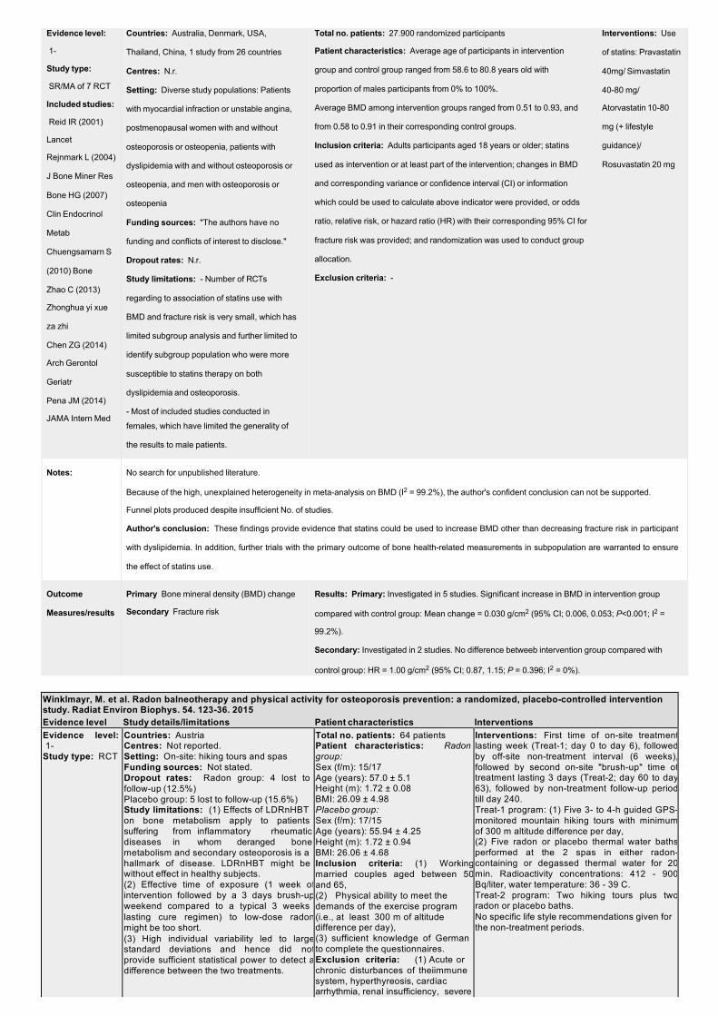
Evidence level:
1- Study type:
SR/MA of 7 RCT
Included studies:
Reid IR (2001)
Lancet
Rejnmark L (2004)
J Bone Miner Res
Bone HG (2007)
Clin Endocrinol
Metab
Chuengsamarn S
(2010) Bone
Zhao C (2013) Zhonghua yi xue
za zhi
Chen ZG (2014) Arch Gerontol
Geriatr
Pena JM (2014) JAMA Intern Med
Countries: Australia, Denmark, USA,
Thailand, China, 1 study from 26 countries
Centres: N.r.
Setting: Diverse study populations: Patients
with myocardial infraction or unstable angina,
postmenopausal women with and without
osteoporosis or osteopenia, patients with
dyslipidemia with and without osteoporosis or
osteopenia, and men with osteoporosis or
osteopenia
Funding sources: "The authors have no
funding and conflicts of interest to disclose."
Dropout rates: N.r.
Study limitations: - Number of RCTs
regarding to association of statins use with
BMD and fracture risk is very small, which has
limited subgroup analysis and further limited to
identify subgroup population who were more
susceptible to statins therapy on both
dyslipidemia and osteoporosis.
- Most of included studies conducted in
females, which have limited the generality of
the results to male patients.
Total no. patients: 27.900 randomized participants Patient characteristics: Average age of participants in intervention
group and control group ranged from 58.6 to 80.8 years old with
proportion of males participants from 0% to 100%.
Average BMD among intervention groups ranged from 0.51 to 0.93, and
from 0.58 to 0.91 in their corresponding control groups.
Inclusion criteria: Adults participants aged 18 years or older; statins
used as intervention or at least part of the intervention; changes in BMD
and corresponding variance or confidence interval (CI) or information
which could be used to calculate above indicator were provided, or odds
ratio, relative risk, or hazard ratio (HR) with their corresponding 95% CI for
fracture risk was provided; and randomization was used to conduct group
allocation.
Exclusion criteria: -
Interventions: Use
of statins: Pravastatin
40mg/ Simvastatin
40-80 mg/ Atorvastatin 10-80
mg (+ lifestyle
guidance)/
Rosuvastatin 20 mg
Notes:
No search for unpublished literature.
Because of the high, unexplained heterogeneity in meta-analysis on BMD (I2 = 99.2%), the author's confident conclusion can not be supported.
Funnel plots produced despite insufficient No. of studies.
Author's conclusion: These findings provide evidence that statins could be used to increase BMD other than decreasing fracture risk in participant
with dyslipidemia. In addition, further trials with the primary outcome of bone health-related measurements in subpopulation are warranted to ensure
the effect of statins use.
Outcome
Measures/results
Primary Bone mineral density (BMD) change
Secondary Fracture risk
Results: Primary: Investigated in 5 studies. Significant increase in BMD in intervention group
compared with control group: Mean change = 0.030 g/cm2 (95% CI; 0.006, 0.053; P<0.001; I2 =
99.2%).
Secondary: Investigated in 2 studies. No difference betweeb intervention group compared with control group: HR = 1.00 g/cm2 (95% CI; 0.87, 1.15; P = 0.396; I2 = 0%).
Winklmayr, M. et al. Radon balneotherapy and physical activity for osteoporosis prevention: a randomized, placebo-controlled intervention study. Radiat Environ Biophys. 54. 123-36. 2015 Evidence level Study details/limitations Patient characteristics Interventions Evidence level: 1-
Study type: RCT
Countries: Austria Centres: Not reported. Setting: On-site: hiking tours and spas Funding sources: Not stated. Dropout rates: Radon group: 4 lost to follow-up (12.5%) Placebo group: 5 lost to follow-up (15.6%) Study limitations: (1) Effects of LDRnHBT on bone metabolism apply to patients suffering from inflammatory rheumatic diseases in whom deranged bone metabolism and secondary osteoporosis is a hallmark of disease. LDRnHBT might be without effect in healthy subjects. (2) Effective time of exposure (1 week of intervention followed by a 3 days brush-up weekend compared to a typical 3 weeks lasting cure regimen) to low-dose radon might be too short. (3) High individual variability led to large standard deviations and hence did not provide sufficient statistical power to detect a difference between the two treatments.
Total no. patients: 64 patients Patient characteristics: Radon group: Sex (f/m): 15/17 Age (years): 57.0 ± 5.1 Height (m): 1.72 ± 0.08 BMI: 26.09 ± 4.98 Placebo group: Sex (f/m): 17/15 Age (years): 55.94 ± 4.25 Height (m): 1.72 ± 0.94 BMI: 26.06 ± 4.68 Inclusion criteria: (1) Working married couples aged between 50 and 65, (2) Physical ability to meet the demands of the exercise program (i.e., at least 300 m of altitude difference per day), (3) sufficient knowledge of German to complete the questionnaires. Exclusion criteria: (1) Acute or chronic disturbances of theiimmune system, hyperthyreosis, cardiac arrhythmia, renal insufficiency, severe
Interventions: First time of on-site treatment lasting week (Treat-1; day 0 to day 6), followed by off-site non-treatment interval (6 weeks), followed by second on-site "brush-up" time of treatment lasting 3 days (Treat-2; day 60 to day 63), followed by non-treatment follow-up period till day 240. Treat-1 program: (1) Five 3- to 4-h guided GPS- monitored mountain hiking tours with minimum of 300 m altitude difference per day, (2) Five radon or placebo thermal water baths performed at the 2 spas in either radon- containing or degassed thermal water for 20 min. Radioactivity concentrations: 412 - 900 Bq/liter, water temperature: 36 - 39 C. Treat-2 program: Two hiking tours plus two radon or placebo baths. No specific life style recommendations given for the non-treatment periods.

cardiovascular diseases, acute infections or fever, iritis or an acute attack of polyarthritis, (2) Participants should not have used hormone replacement therapy or any other therapy affecting the bone metabolism during the last 12 months before enrollment.
Notes: No reason stated for exclusive recruiting of working married couples. Results might not be directly applicable for other patient groups. Test for baseline differences not stated. QUALEFFO-41 not validated for patients without vertebral fractures. Use of Zerssen Beschwerdenliste 1976, not revised version of 2011. No ITT-analyses. Neither funding nor COI stated. Author's conclusion: In conclusion, the combined exercise and thermal water bath intervention regimen applied to an apparent healthy population as included in this study revealed marked and unexpectedly sustained changes in humoral regulators and markers of bone turnover as well as self-reported somatic complaints and pain. These factors certainly indicate a delicate and complex physiological interplay of central, systemic and local regulators of bone metabolism which needs to be unraveled. However, given the changes of the primary outcome parameters OPG, RANKL and the OPG/RANKL ratio, it is feasible to assume that the overall bone metabolism is tuned toward an anabolic condition. Moreover, these effects are accompanied by significant improvements of perceived quality of life and pain. Although low-dose radon hyperthermia balneo treatment does not significantly outmatch conventional thermal water treatment in this study, borderline significant differences of RANKL and the OPG/ RANKL ratio indicate a possible additive effect of radon balneo treatment on the achieved biological effects. Mountain hiking is a popular form of exercise in the alpine region and a low cost non-pharmacologic intervention. Even though men develop osteoporosis later than women, as a matter of fact age-related bone loss begins at the age of fifty in both sexes (Campion and Maricic 2003). The immense and continuously rising costs caused by osteoporosis make a preventive intervention as performed in this study worth considering and useful for both men and women.
Outcome Measures/results
Primary OPG, RANKL and OPG/RANKL ratio Secondary Somatic complaints (Beschwerdenliste, von Zerssen 1976); Quality of life (Qualeffo-41)
Results: OPG: No absolute change of values reported, only model analysis of relative change. Increases of OPG from T1 to T2, T2 to T3, T2 to T4 in placebo and radon group. OPG concentration decreased approximately to the baseline levels after 6 months (T5). RANKL: No absolute change of values reported, only model analysis of relative change. RANKL concentrations declined in entire study population from T1 to T2. OPG/RANKL ratio: Significantly elevated OPG/RANKL ratio from time T1 to T2 and subsequent decrease from T4 to T5 in both groups. Complaints list and quality of life: Results not quantified but stated as "significant decline in self-reported health complaints [...] after Treat-1 (p ≤ 0.000, T2) and Treat-2 (p ≤ 0.000, T4), which remained reduced until T5 (day 240; p<0.0004)". No significant difference between groups (p = 0.1923). Qualeffo-41 showed significant reduction (p = 0.042) of pain (domain Qualeffo-Pain) in the whole study population 6 months after treatments (T5) (data not shown).
Wright, E. et al. Bisphosphonates and evidence for association with esophageal and gastric cancer: a systematic review and meta-analysis. BMJ Open. 5. e007133. 2015
Evidence level Study details/limitations Patient Interventions characteristics
Evidence level: 2+ Study type: Systematic Review of 11 retrospective, case control (6) and cohort (5) studies (and Meta Analyis of n= 5 ) Included studies: Abrahamsen B, Pazianas M, Eiken P, et al. Esophageal and gastric cancer incidence and mortality in alendronate users. J Bone Miner Res 2012;27:679–86. Cardwell CR, Abnet CC, Cantwell MM, et al. Exposure to oralbisphosphonates and risk of esophageal cancer. JAMA 2010;304:657–63. Chen YM, Chen DY, Chen LK, et al. Alendronate and risk of esophageal cancer: a nationwide population-based study in Taiwan. J Am Geriatr Soc 2011;59:2379–81. Chiang CH, Huang CC, Chan WL, et al. Oral alendronate use and risk of cancer in postmenopausal women with osteoporosis: a nationwide study. J Bone Miner Res 2012;27:1951–8. Green J, Czanner G, Reeves G, et al. Oral bisphosphonates and risk of cancer of oesophagus, stomach, and colorectum: case-control analysis within a UK primary care cohort. BMJ 2010;341:c4444. Lee WY, Sun LM, Lin MC, et al. A higher dosage of oral alendronate will increase the subsequent cancer risk of osteoporosis patients in Taiwan: a population-based cohort study. PLoS ONE 2012;7:e53032. Nguyen D, Schwartz J, Richardson P, et al. Oral bisphosphonate prescriptions and the risk of esophageal adenocarcinoma in patients
Countries: USA, the UK, Denmark and Taiwan Centres: Setting: Funding sources: National Institute for Health Research (NIHR) Biomedical Research Centre at Guy’s and St Thomas’ NHS Foundation Trust and King’s College London Dropout rates: - Study limitations: A particular problem was the use of overlapping cohorts in the same database, which reduced the numbers of studies we were able to combine because of the risk of double counting. This necessarily limited the power of the analysis. A further problem that limited the number of studies for meta-analyses including sensitivity analyses, was the differing outcomes and exposures selected.
Total no. patients: cohort studies:
between 30.606 - 55.090 cases of cancer case control: between 116-5364 cases Patient characteristics: women, men aged 18-90, bisphosphonate, alendronate users case-control: esophageal, gastric cancers vs controls Inclusion criteria: (1) the study used
a case–control or cohort design; (2) the study examined bisphosphonates and the risk of esophageal and/or gastric cancer; (3) the risk estimate was reported as an OR, HR or relative risk; (4) the 95% CI for the risk estimate Exclusion criteria: -
Interventions: association between
bisphosphonate prescribing and esophageal and gastric cancer

with Barrett’s esophagus. Dig Dis Sci 2010;55:3404–7. Vestergaard P. Occurrence of gastrointestinal cancer in users of bisphosphonates and other antiresorptive drugs against osteoporosis. Calcif Tissue Int 2011;89:434–41 Vinogradova Y, Coupland C, Hippisley-Cox J. Exposure to bisphosphonates and risk of gastrointestinal cancers: series of nested case-control studies with QResearch and CPRD data. BMJ 2013;346:f114 Wright E, Schofield PT, Seed P, et al. Bisphosphonates and risk of upper gastrointestinal cancer—a case control study using the General Practice Research Database (GPRD). PLoS ONE 2012;7:e47616. Notes: Unclear, whether study selection and data extraction was performed by 2 reviewers.
Author's conclusion: This is the fourth and most detailed meta-analysis on this topic. We have not identified any compelling evidence for a significantly raised risk of esophageal cancer or gastric cancer in male and female patients prescribed bisphosphonates.
Outcome Measures/results Primary - Secondary (1) esophageal cancer and all bisphosphonates; (2) gastric cancer and all bisphosphonates; (3) esophageal cancer and alendronate; (4)gastric cancer and alendronate; (5) cohort studies—UGI cancer and all bisphosphonates; (6) case–control studies—UGI cancer and all bisphosphonates.
Results: Esophageal cancer and all bisphosphonates - no significant association between use of bisphosphonates and esophageal cancer, OR 1.11 (0.97 to 1.27) - sensitivity analyses: different combinations of CPRD: all non-significant Gastric cancer and all bisphosphonates - no significant association between use of bisphosphonates and gastric cancer, OR 0.96 (95% CI 0.82 to 1.12) Esophageal cancer and alendronate - no increased risk, OR 0.98 (0.87 to 1.11), of esophageal cancer in alendronate users - sensitivity analyses: all non significant
Gastric cancer and alendronate - no increased risk of gastric cancer with alendronate, OR 0.99 (95% CI 0.71 to 1.38) Cohort studies—UGI cancer and all bisphosphonates no significant association, OR 1.32 (95% CI 0.81 to 2.13)
Case–control studies—UGI cancer and all bisphosphonates no significant association, OR 1.06 (95% CI 0.93 to 1.22).
Wu, A. M. et al. The relationship between dietary protein consumption and risk of fracture: a subgroup and dose-response meta-analysis of prospective cohort studies. Sci Rep. 5. 9151. 2015
Evidence level Study details/limitations Patient characteristics Interventions Evidence level:
2-
Study type:
SR/MA of 12
prospective cohort
studies
Included studies:
Mussolino ME
(1998) J Bone
Miner Rese
Beasley JM
(2014) Am J Clin
Nutr
Meyer HE (1997)
Am J Epidemiol
Misra D (2011)
Osteoporosis Int
Countries: 9 studies conducted in the United States and Canada, 2 in
Europe, 1 in Asia.
Centres: N.r. Setting: N.r.
Funding sources: "This work is funded by National Natural Science
Foundation of China (81372014, 81371988); and Xinmiao talent plan of
Zhejiang Province (2014R413053). The funders had no role in the design,
execution, and writing of the study."
Dropout rates: N.r. Study limitations: - No meta-analysis on vertebral fractures possible
because of only one study reporting risk of vertebral fracture.
- Meta-analysis on subgroups and dose-response apart from hip fracture not
possible due to insufficient data.
- Significantly heterogeneity in most subgroup analysis.
Total no. patients: 407.104
participants
Patient characteristics: Age
(range): 20 – 89 years.
6 studies recruited women only, 1
study men only, 7 studies with
mixed populations.
Inclusion criteria: 1) Designed as
a prospective cohort study;
2) Exposure of interest is (animal or
vegetable/plant) protein
consumption;
3) Primary outcome of interest is
any fractures/ hip fracture/ vertebral
fracture/ limb fracture;
4) the relative risk (RR) estimates
with 95% confidence intervals (CI)
Interventions: Protein
consumption: Total protein
consumption, animal protein,
vegetable protein.
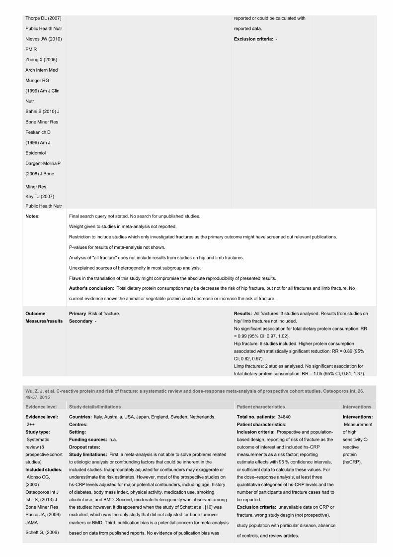
Thorpe DL (2007)
Public Health Nutr
Nieves JW (2010)
PM R
Zhang X (2005)
Arch Intern Med
Munger RG
(1999) Am J Clin
Nutr
Sahni S (2010) J
Bone Miner Res
Feskanich D
(1996) Am J
Epidemiol
Dargent-Molina P
(2008) J Bone
Miner Res Key TJ (2007)
Public Health Nutr
reported or could be calculated with
reported data.
Exclusion criteria: -
Notes:
Final search query not stated. No search for unpublished studies.
Weight given to studies in meta-analysis not reported.
Restriction to include studies which only investigated fractures as the primary outcome might have screened out relevant publications.
P-values for results of meta-analysis not shown.
Analysis of "all fracture" does not include results from studies on hip and limb fractures.
Unexplained sources of heterogeneity in most subgroup analysis.
Flaws in the translation of this study might compromise the absolute reproducibility of presented results. Author's conclusion: Total dietary protein consumption may be decrease the risk of hip fracture, but not for all fractures and limb fracture. No
current evidence shows the animal or vegetable protein could decrease or increase the risk of fracture.
Outcome
Primary Risk of fracture.
Results: All fractures: 3 studies analysed. Results from studies on hip/ limb fractures not included. No significant association for total dietary protein consumption: RR = 0.99 (95% CI; 0.97, 1.02). Hip fracture: 6 studies included. Higher protein consumption associated with statistically significant reduction: RR = 0.89 (95% CI; 0.82, 0.97). Limp fractures: 2 studies analysed. No significant association for total dietary protein consumption: RR = 1.05 (95% CI; 0.81, 1.37).
Measures/results Secondary -
Wu, Z. J. et al. C-reactive protein and risk of fracture: a systematic review and dose-response meta-analysis of prospective cohort studies. Osteoporos Int. 26. 49-57. 2015
Evidence level Study details/limitations Patient characteristics Interventions Evidence level: 2++ Study type: Systematic review (8 prospective cohort studies). Included studies: Alonso CG, (2000) Osteoporos Int J Ishii S, (2013) J Bone Miner Res Pasco JA, (2006)
JAMA Schett G, (2006)
Countries: Italy, Australia, USA, Japan, England, Sweden, Netherlands. Centres: Setting: Funding sources: n.a. Dropout rates: Study limitations: First, a meta-analysis is not able to solve problems related to etiologic analysis or confounding factors that could be inherent in the included studies. Inappropriately adjusted for confounders may exaggerate or underestimate the risk estimates. However, most of the prospective studies on hs-CRP levels adjusted for major potential confounders, including age, history of diabetes, body mass index, physical activity, medication use, smoking, alcohol use, and BMD. Second, moderate heterogeneity was observed among the studies; however, it disappeared when the study of Schett et al. [16] was excluded, which was the only study that did not adjusted for bone turnover
markers or BMD. Third, publication bias is a potential concern for meta-analysis
based on data from published reports. No evidence of publication bias was
Total no. patients: 34840 Patient characteristics: Inclusion criteria: Prospective and population- based design, reporting of risk of fracture as the outcome of interest and included hs-CRP measurements as a risk factor; reporting estimate effects with 95 % confidence intervals, or sufficient data to calculate these values. For the dose–response analysis, at least three quantitative categories of hs-CRP levels and the number of participants and fracture cases had to be reported. Exclusion criteria: unavailable data on CRP or
fracture, wrong study desgin (not prospective),
study population with particular disease, absence
of controls, and review articles.
Interventions: Measurement of high sensitivity C- reactive protein (hsCRP).
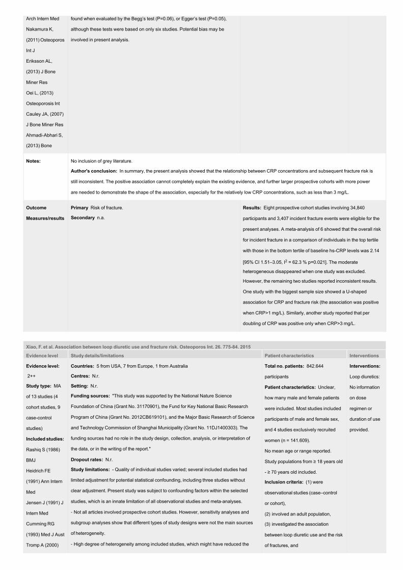
Arch Intern Med
Nakamura K,
(2011) Osteoporos
Int J
Eriksson AL,
(2013) J Bone
Miner Res
Oei L, (2013)
Osteoporosis Int
Cauley JA, (2007)
J Bone Miner Res
Ahmadi-Abhari S,
(2013) Bone
found when evaluated by the Begg’s test (P=0.06), or Egger’s test (P=0.05),
although these tests were based on only six studies. Potential bias may be
involved in present analysis.
Notes:
No inclusion of grey literature.
Author's conclusion: In summary, the present analysis showed that the relationship between CRP concentrations and subsequent fracture risk is
still inconsistent. The positive association cannot completely explain the existing evidence, and further larger prospective cohorts with more power
are needed to demonstrate the shape of the association, especially for the relatively low CRP concentrations, such as less than 3 mg/L.
Outcome
Measures/results
Primary Risk of fracture.
Secondary n.a.
Results: Eight prospective cohort studies involving 34,840
participants and 3,407 incident fracture events were eligible for the
present analyses. A meta-analysis of 6 showed that the overall risk
for incident fracture in a comparison of individuals in the top tertile
with those in the bottom tertile of baseline hs-CRP levels was 2.14
[95% CI 1.51–3.05, I2 = 62.3 % p=0.021]. The moderate
heterogeneous disappeared when one study was excluded. However, the remaining two studies reported inconsistent results.
One study with the biggest sample size showed a U-shaped
association for CRP and fracture risk (the association was positive
when CRP>1 mg/L). Similarly, another study reported that per
doubling of CRP was positive only when CRP>3 mg/L.
Xiao, F. et al. Association between loop diuretic use and fracture risk. Osteoporos Int. 26. 775-84. 2015
Evidence level Study details/limitations Patient characteristics Interventions Evidence level:
2++
Study type: MA
of 13 studies (4
cohort studies, 9
case-control
studies)
Included studies:
Rashiq S (1986)
BMJ
Heidrich FE
(1991) Ann Intern
Med
Jensen J (1991) J
Intern Med
Cumming RG
(1993) Med J Aust
Tromp A (2000)
Countries: 5 from USA, 7 from Europe, 1 from Australia
Centres: N.r.
Setting: N.r.
Funding sources: "This study was supported by the National Nature Science
Foundation of China (Grant No. 31170901), the Fund for Key National Basic Research
Program of China (Grant No. 2012CB619101), and the Major Basic Research of Science
and Technology Commission of Shanghai Municipality (Grant No. 11DJ1400303). The
funding sources had no role in the study design, collection, analysis, or interpretation of
the data, or in the writing of the report."
Dropout rates: N.r. Study limitations: - Quality of individual studies varied; several included studies had
limited adjustment for potential statistical confounding, including three studies without
clear adjustment. Present study was subject to confounding factors within the selected
studies, which is an innate limitation of all observational studies and meta-analyses.
- Not all articles involved prospective cohort studies. However, sensitivity analyses and
subgroup analyses show that different types of study designs were not the main sources
of heterogeneity.
- High degree of heterogeneity among included studies, which might have reduced the
Total no. patients: 842.644
participants
Patient characteristics: Unclear,
how many male and female patients
were included. Most studies included
participants of male and female sex,
and 4 studies exclusively recruited
women (n = 141.609).
No mean age or range reported.
Study populations from ≥ 18 years old
- ≥ 70 years old included. Inclusion criteria: (1) were
observational studies (case–control
or cohort),
(2) involved an adult population, (3) investigated the association
between loop diuretic use and the risk
of fractures, and
Interventions:
Loop diuretics:
No information
on dose
regimen or
duration of use
provided.
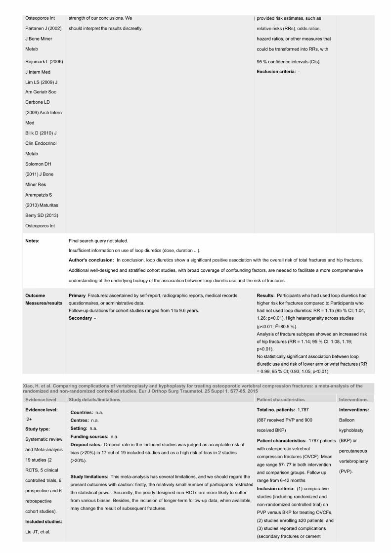
Osteoporos Int
Partanen J (2002)
J Bone Miner
Metab
Rejnmark L (2006)
J Intern Med
Lim LS (2009) J Am Geriatr Soc
Carbone LD
(2009) Arch Intern
Med
Bilik D (2010) J
Clin Endocrinol
Metab
Solomon DH
(2011) J Bone
Miner Res
Arampatzis S
(2013) Maturitas
Berry SD (2013)
Osteoporos Int
strength of our conclusions. We
should interpret the results discreetly.
(4) provided risk estimates, such as
relative risks (RRs), odds ratios,
hazard ratios, or other measures that
could be transformed into RRs, with
95 % confidence intervals (CIs). Exclusion criteria: -
Notes:
Final search query not stated.
Insufficient information on use of loop diuretics (dose, duration ...).
Author's conclusion: In conclusion, loop diuretics show a significant positive association with the overall risk of total fractures and hip fractures.
Additional well-designed and stratified cohort studies, with broad coverage of confounding factors, are needed to facilitate a more comprehensive
understanding of the underlying biology of the association between loop diuretic use and the risk of fractures.
Outcome Measures/results
Primary Fractures: ascertained by self-report, radiographic reports, medical records, questionnaires, or administrative data. Follow-up durations for cohort studies ranged from 1 to 9.6 years. Secondary -
Results: Participants who had used loop diuretics had higher risk for fractures compared to Participants who had not used loop diuretics: RR = 1.15 (95 % CI; 1.04, 1.26; p<0.01). High heterogeneity across studies
(p<0.01; I2=80.5 %). Analysis of fracture subtypes showed an increased risk of hip fractures (RR = 1.14; 95 % CI, 1.08, 1.19; p<0.01). No statistically significant association between loop diuretic use and risk of lower arm or wrist fractures (RR = 0.99; 95 % CI; 0.93, 1.05; p<0.01).
Xiao, H. et al. Comparing complications of vertebroplasty and kyphoplasty for treating osteoporotic vertebral compression fractures: a meta-analysis of the randomized and non-randomized controlled studies. Eur J Orthop Surg Traumatol. 25 Suppl 1. S77-85. 2015
Evidence level Study details/limitations Patient characteristics Interventions Evidence level:
2+
Study type:
Systematic review
and Meta-analysis
19 studies (2
RCTS, 5 clinical
controlled trials, 6
prospective and 6
retrospective
cohort studies).
Included studies:
Liu JT, et al.
Countries: n.a.
Centres: n.a.
Setting: n.a.
Funding sources: n.a.
Dropout rates: Dropout rate in the included studies was judged as acceptable risk of
bias (>20%) in 17 out of 19 included studies and as a high risk of bias in 2 studies
(>20%).
Study limitations: This meta-analysis has several limitations, and we should regard the
present outcomes with caution: firstly, the relatively small number of participants restricted
the statistical power. Secondly, the poorly designed non-RCTs are more likely to suffer
from various biases. Besides, the inclusion of longer-term follow-up data, when available,
may change the result of subsequent fractures.
Total no. patients: 1,787
(887 received PVP and 900
received BKP)
Patient characteristics: 1787 patients
with osteoporotic vetrebral
compression fractures (OVCF). Mean
age range 57- 77 in both intervention
and comparison groups. Follow up
range from 6-42 months
Inclusion criteria: (1) comparative
studies (including randomized and
non-randomized controlled trial) on
PVP versus BKP for treating OVCFs,
(2) studies enrolling ≥20 patients, and
(3) studies reported complications
(secondary fractures or cement
Interventions:
Balloon
kyphoblasty
(BKP) or
percutaneous
vertebroplasty
(PVP).
(2010)
Osteoporos Int
Vogl TJ, et al.
(2013) Spine
(Phila Pa 1976)
Kumar K, et al.
(2010)
Neurosurgery
Li X, et al. (2012)
J Spinal Disord
Tech
Omidi-Kashani F,
et al. (2013) J
Osteoporos
Rollinghoff M, et
al. (2009) Minim
Invasive
Neurosurg
Schofer MD, et al.
(2009) Arch
Orthop Trauma
Surg
Lovi A, et al.
(2009) Eur Spine
J
Movrin I, et al.
(2010) Arch
Orthop Trauma
Surg
Pflugmacher R, et
al. (2005) Rofo
Santiago FR, et
al. (2010) Eur J
Radiol
Josef G, et al.
(2005) J Spinal
Disord Tech
Frankel BM, et al.
(2007) Spine J
Hiwatashi A, et al.
(2009) Am J
Neuroradiol
Kim K-H, et al.
(2012) J Spinal
Disord Tech
Yan D, et al.
(2010) Arch
Orthop Trauma
Surg
Jian-lin Z, et al.
(2008) Chinese J
leakage) as outcome parameters.
Exclusion criteria: (1) diagnosis of
OVCFs was not clinically established
(patients with traumatic, or cancer-
related vertebral fractures), (2) patient
had infection, neurological deficits, or
spinal stenosis, (3) single-arm studies,
(4) no relevant data could be extracted
and (5) systematic review and meta-
analysis.

Traumatol
Bozkurt M, et al.
(2014) Asian
Spine J
Bae H, et al.
(2010) Spine
Notes:
Differences in the follow-up periods of the included studies range from 6-42 months (follow-up times were not pre-specified). The loss to follow up was
not reported in 11 of the 19 studies, but it was considered as low risk of bias (not unclear risk of bias) in 17 out of 19 studies. No listing of excluded
studies or inclusion of grey literature. No investigation of publication bias. Confllict of interest for the primary studies are not reported. Author's conclusion: Based on this meta-analysis, we have the following conclusions: the two procedures suffer from equal risk of subsequent
spinal fractures; PVP has a significant higher cement leakage rate compared to BKP, mainly caused by a higher paravertebral leakage, hence,
patients with extremely poor pulmonary function or unstable haemodynamic are better candidates for BKP. To confirm the present assessments,
high-quality RCT should be conducted.
Outcome Measures/results
Primary Incidence of adjacent vertebral fractures; Incidence of non-adjacent vertebral fractures. Cement leakage into disk spaces, or into paravertebral areas or total cement leakage rate. Secondary -
Results: 19 studies encompassing 1,787 patients in total,
of whom 887 received PVP and 900 received BKP, met
the inclusion criteria. Fractures: For subsequent fractures,
our meta-analysis detected no significant difference
between the two procedures, both for adjacent fractures (p
= 0.29) and non-adjacent fractures (p = 0.37). Cement
leakage: For cement extravasations, there was no
significant difference between the two interventions if
considering disc spaces extravasations only (p = 0.24),
while considering total extravasations and paravertebral
extravasations, the cement leakage rate in the PVP group
was significantly higher than the BKP group (total: p<0.01;
paravertebral: p<0.01).
Yan, A. et al. Does tea consumption correlate to risk of fracture? A meta-analysis. Int J Clin Exp Med. 8. 8347-57. 2015
Evidence level Study details/limitations Patient characteristics Interventions Evidence level: 2-
Study type: MA of 9
studies (3 cohort
studies, 6 case-
control studies)
Included studies:
Kreiger N (1992) Int
J Epidemiol
Johnell O (1995) J
Bone Miner Res
Tavani A (1995) Prev
Med
Chen Z (2003) Am J Epidemiol
Jha RM (2010) BMC
Musculoskelet Disord
Kanis J (1999)
Osteoporos Int
Zeng FF (2013) J Clin Endocrinol
Metab
Tomata Y (2012) Am
J Clin Nutr
Countries: 2 China, 2 Southern Europe, 1 Canada,
1 Italy, 1 India, 1 USA, 1 Japan
Centres: N.r.
Setting: N.r.
Funding sources: "This study was granted by the
project of publication of series books (No.
Z131110001913-154)." Dropout rates: N.r.
Study limitations: - Most of included studies with
case-control design, and therefore with inherent
recall and selection bias
- Tea mainly grouped into green tea and black tea.
Functional contents and effects not identical for their
two tea subtypes and thus the potential bias should
be noted.
Total no. patients: 147.950
Patient characteristics: Mostly
female patients. Largest study with
91.465 patients with female patients
only.
Patients of varying ages included
(range: 19 - 100 years).
Inclusion criteria: (1) The
association between tea consumption
and risk of fractures was evaluated;
(2) a cohort or case-control study
design;
(3) the value of relative risk (RR),
odds ratio (OR) with 95% confidence
intervals (CI) or enough data to
calculate these.
Exclusion criteria: (1) Fractures
caused by other reasons, such as
injury, trauma, and etc.
(2) Patients who consumed the tea
only for a few days or months, or with
intervals.
Interventions: Varying categories of tea
consumption: No drinking/drinking, Former
drinking/current drinking or never, 1-2 cup/day, 3
cup/day, ≥ 4 cup/day.
Notes:
Final search query not stated. Unclear, whether 2 person selected studies. No search for unpublished/ grey literature.
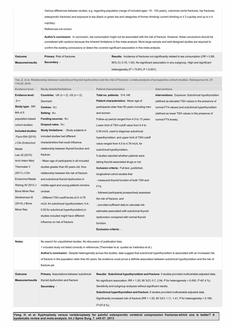
Various differences between studies, e.g. regarding population (range of included ages: 19 - 100 years), outcomes (wrist fractures, hip fractures,
osteoporotic fractures) and exposure to tea (black or green tea and categories of former drinking/ current drinking or ≤ 3 cup/day and up to ≥ 4
cup/day).
References not correct.
Author's conclusion: In conclusion, tea consumption might not be associated with the risk of fracture. However, these conclusions should be
considered with cautions because the inherent limitations in this meta-analysis. More large-sample and well-designed studies are required to
confirm the existing conclusions or detect the covered significant association in this meta-analysis.
Outcome
Measures/results
Primary Risk of fractures
Secondary -
Results: Incidence of fractures not significantly related to tea consumption (OR = 0.89;
95% CI; 0.78, 1.04). No significant association in any subgroup. High and significant
heterogeneity (I2 = 73.6%; P < 0.001).
Yan, Z. et al. Relationship between subclinical thyroid dysfunction and the risk of fracture: a meta-analysis of prospective cohort studies. Osteoporos Int. 27. 115-25. 2016
Evidence level Study details/limitations Patient characteristics Interventions Evidence level:
2++
Study type: SR/
MA of 5
population-based
cohort studies)
Included studies:
Flynn RW (2010)
J Clin Endocrinol
Metab
Lee JS (2010) Arch Intern Med
Thenmalar V
(2011) J Clin
Endocrinol Metab
Waring AV 2013 J
Bone Miner Res
Abrahemson B
(2014) J Bone
Miner Res
Countries: UK (n = 2), US (n = 2),
Denmark
Centres: N.r. Setting: N.r.
Funding sources: N.r.
Dropout rates: N.r.
Study limitations: - Study subjects in
included studies had different
characteristics that could influence
relationship between thyroid function and
fracture.
- Mean age of participants in all included
studies greater than 60 years old, thus
relationship between the risk of fracture
and subclinical thyroid dysfunction in
middle-aged and young patients remains
unclear.
- Different TSH cutoff levels (4.0–4.78
mU/L for subclinical hypothyroidism; 0.4–
0.55 for subclinical hyperthyroidism) in
studies included might have different
influence on risk of fracture.
Total no. patients: 314.146
Patient characteristics: Mean age of
participants older than 60 years including men
and women.
Follow-up period ranged from 4.5 to 13 years.
Lower limit of TSH cutoff value from 0.4 to
0.55 mU/L used to diagnose subclinical
hyperthyroidism, and upper limit of TSH cutoff
value ranged from 4.0 to 4.78 mU/L for
subclinical hypothyroidism.
5 studies reported whether patients were
taking thyroid-associated drugs or not.
Inclusion criteria: Full-text, published,
longitudinal cohort studies that
- measured thyroid function of both TSH and
FT4,
- followed participants prospectively assessed
the risk of fracture, and
- provided sufficient data to calculate risk
estimates associated with subclinical thyroid
dysfunction compared with normal thyroid
function.
Exclusion criteria: -
Interventions: Exposure: Subclinical hypothyroidism
(defined as elevated TSH values in the presence of
normal FT4 values) and subclinical hyperthyroidism
(defined as lower TSH values in the presence of
normal FT4 levels).
Notes:
No search for unpublished studies. No discussion of publication bias.
1 included study not listed correctly in references (Thenmalar et al. quoted as Vadiveloo et al.)
Author's conclusion: Despite heterogeneity across the studies, data suggest that subclinical hyperthyroidism is associated with an increased risk
of fracture in the population older than 60 years. No evidence could prove a definite association between subclinical hypothyroidism and the risk of
fracture yet.
Outcome
Measures/results
Primary Associations between subclinical
thyroid dysfunction and fracture
Secondary -
Results: Subclinical hypothyroidism and fracture: 3 studies provided multivariable-adjusted data.
No significant association: RR = 1.20, 95 %CI; 0.7, 2.04; P for heterogeneity = 0.000, I2=87.4 %).
Sensitivity and subgroup analyses without significant results.
Subclinical hyperthyroidism and fracture: 5 studies provided multivariable-adjusted data.
Significantly increased risk of fracture (RR = 1.25, 95 %CI; 1.11, 1.41; P for heterogeneity = 0.169,
I2=37.9 %).
Yang, H. et al. Kyphoplasty versus vertebroplasty for painful osteoporotic vertebral compression fractures-which one is better? A systematic review and meta-analysis. Int J Spine Surg. 7. e45-57. 2013
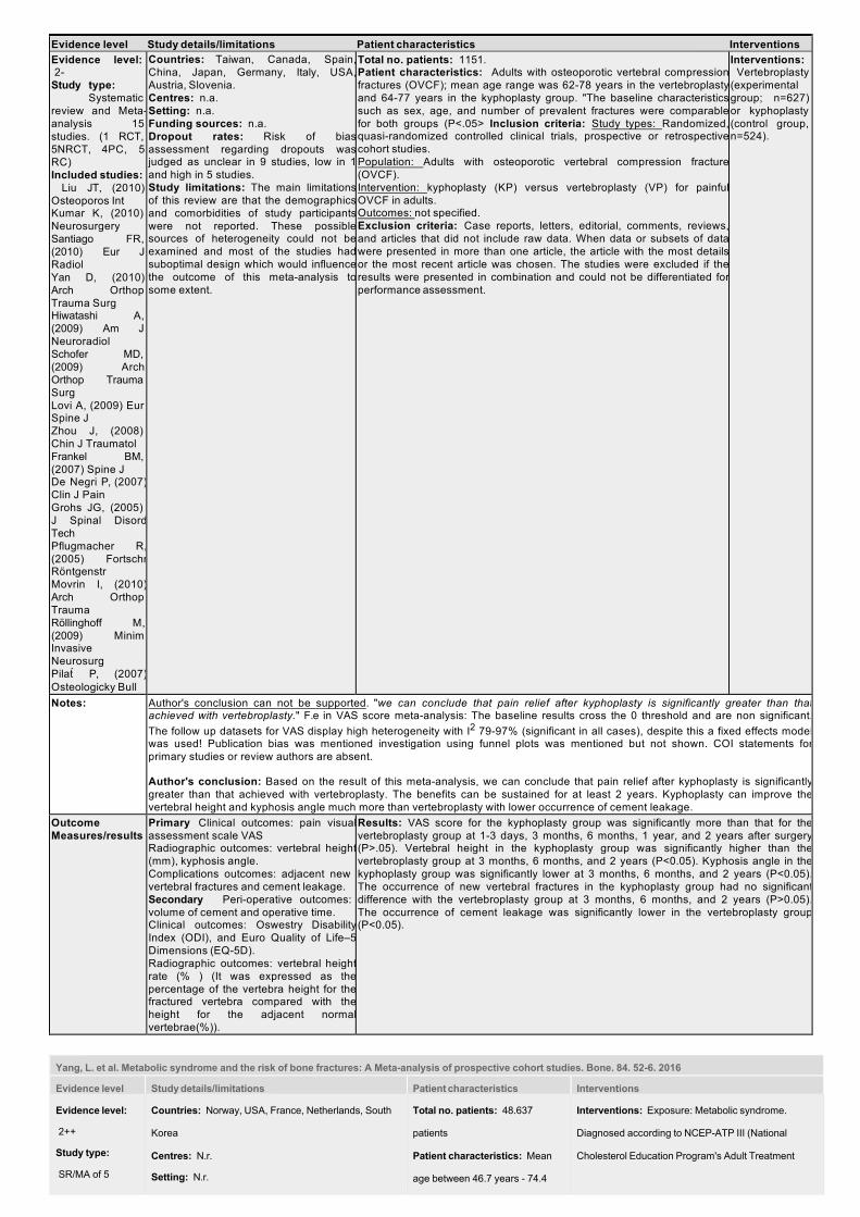
Evidence level Study details/limitations Patient characteristics Interventions Evidence level: 2-
Study type: Systematic
review and Meta- analysis 15 studies. (1 RCT, 5NRCT, 4PC, 5 RC) Included studies:
Liu JT, (2010) Osteoporos Int Kumar K, (2010) Neurosurgery Santiago FR, (2010) Eur J Radiol Yan D, (2010) Arch Orthop Trauma Surg Hiwatashi A, (2009) Am J Neuroradiol Schofer MD, (2009) Arch Orthop Trauma Surg Lovi A, (2009) Eur Spine J Zhou J, (2008) Chin J Traumatol Frankel BM, (2007) Spine J De Negri P, (2007) Clin J Pain Grohs JG, (2005) J Spinal Disord Tech Pflugmacher R, (2005) Fortschr Röntgenstr Movrin I, (2010) Arch Orthop Trauma Röllinghoff M, (2009) Minim Invasive Neurosurg Pilat́ P, (2007) Osteologicky Bull
Countries: Taiwan, Canada, Spain, China, Japan, Germany, Italy, USA, Austria, Slovenia. Centres: n.a. Setting: n.a. Funding sources: n.a. Dropout rates: Risk of bias assessment regarding dropouts was judged as unclear in 9 studies, low in 1 and high in 5 studies. Study limitations: The main limitations of this review are that the demographics and comorbidities of study participants were not reported. These possible sources of heterogeneity could not be examined and most of the studies had suboptimal design which would influence the outcome of this meta-analysis to some extent.
Total no. patients: 1151. Patient characteristics: Adults with osteoporotic vertebral compression fractures (OVCF); mean age range was 62-78 years in the vertebroplasty and 64-77 years in the kyphoplasty group. "The baseline characteristics such as sex, age, and number of prevalent fractures were comparable for both groups (P<.05> Inclusion criteria: Study types: Randomized, quasi-randomized controlled clinical trials, prospective or retrospective cohort studies. Population: Adults with osteoporotic vertebral compression fracture (OVCF). Intervention: kyphoplasty (KP) versus vertebroplasty (VP) for painful OVCF in adults. Outcomes: not specified. Exclusion criteria: Case reports, letters, editorial, comments, reviews, and articles that did not include raw data. When data or subsets of data were presented in more than one article, the article with the most details or the most recent article was chosen. The studies were excluded if the results were presented in combination and could not be differentiated for performance assessment.
Interventions: Vertebroplasty
(experimental group; n=627) or kyphoplasty (control group, n=524).
Notes: Author's conclusion can not be supported. "we can conclude that pain relief after kyphoplasty is significantly greater than that achieved with vertebroplasty." F.e in VAS score meta-analysis: The baseline results cross the 0 threshold and are non significant. The follow up datasets for VAS display high heterogeneity with I2 79-97% (significant in all cases), despite this a fixed effects model was used! Publication bias was mentioned investigation using funnel plots was mentioned but not shown. COI statements for primary studies or review authors are absent. Author's conclusion: Based on the result of this meta-analysis, we can conclude that pain relief after kyphoplasty is significantly greater than that achieved with vertebroplasty. The benefits can be sustained for at least 2 years. Kyphoplasty can improve the vertebral height and kyphosis angle much more than vertebroplasty with lower occurrence of cement leakage.
Outcome Measures/results
Primary Clinical outcomes: pain visual assessment scale VAS Radiographic outcomes: vertebral height (mm), kyphosis angle. Complications outcomes: adjacent new vertebral fractures and cement leakage. Secondary Peri-operative outcomes: volume of cement and operative time. Clinical outcomes: Oswestry Disability Index (ODI), and Euro Quality of Life–5 Dimensions (EQ-5D). Radiographic outcomes: vertebral height rate (% ) (It was expressed as the percentage of the vertebra height for the fractured vertebra compared with the height for the adjacent normal vertebrae(%)).
Results: VAS score for the kyphoplasty group was significantly more than that for the vertebroplasty group at 1-3 days, 3 months, 6 months, 1 year, and 2 years after surgery (P>.05). Vertebral height in the kyphoplasty group was significantly higher than the vertebroplasty group at 3 months, 6 months, and 2 years (P<0.05). Kyphosis angle in the kyphoplasty group was significantly lower at 3 months, 6 months, and 2 years (P<0.05). The occurrence of new vertebral fractures in the kyphoplasty group had no significant difference with the vertebroplasty group at 3 months, 6 months, and 2 years (P>0.05). The occurrence of cement leakage was significantly lower in the vertebroplasty group (P<0.05).
Yang, L. et al. Metabolic syndrome and the risk of bone fractures: A Meta-analysis of prospective cohort studies. Bone. 84. 52-6. 2016
Evidence level Study details/limitations Patient characteristics Interventions Evidence level:
2++
Study type:
SR/MA of 5
Countries: Norway, USA, France, Netherlands, South
Korea
Centres: N.r. Setting: N.r.
Total no. patients: 48.637
patients
Patient characteristics: Mean
age between 46.7 years - 74.4
Interventions: Exposure: Metabolic syndrome.
Diagnosed according to NCEP-ATP III (National
Cholesterol Education Program's Adult Treatment
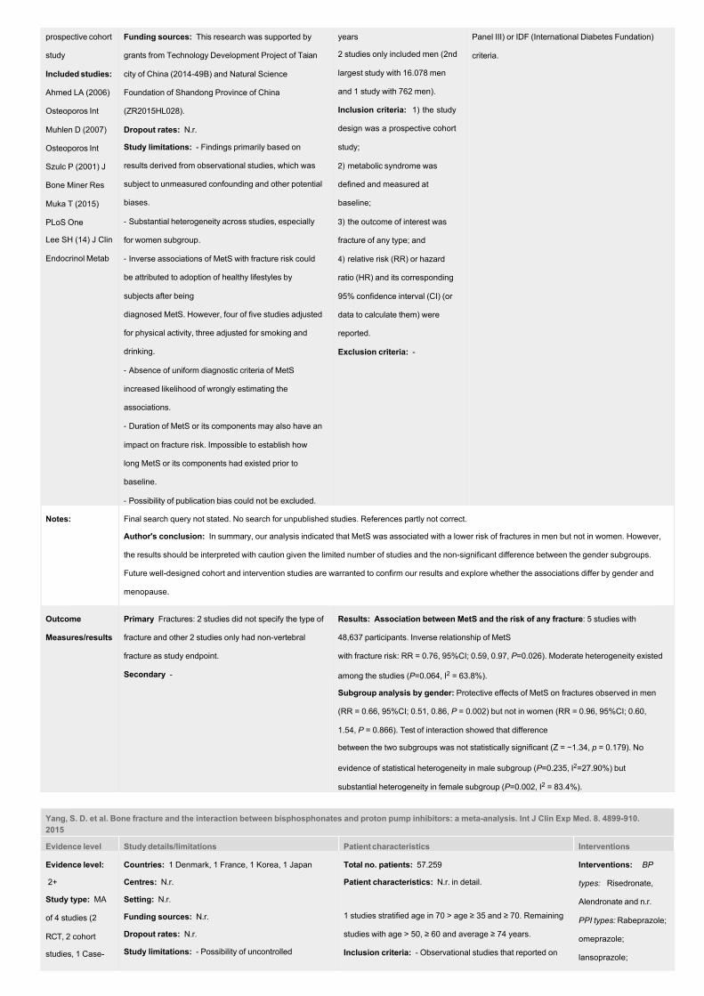
prospective cohort
study
Included studies:
Ahmed LA (2006)
Osteoporos Int
Muhlen D (2007)
Osteoporos Int
Szulc P (2001) J
Bone Miner Res
Muka T (2015)
PLoS One Lee SH (14) J Clin
Endocrinol Metab
Funding sources: This research was supported by
grants from Technology Development Project of Taian
city of China (2014-49B) and Natural Science
Foundation of Shandong Province of China
(ZR2015HL028).
Dropout rates: N.r. Study limitations: - Findings primarily based on
results derived from observational studies, which was
subject to unmeasured confounding and other potential
biases.
- Substantial heterogeneity across studies, especially
for women subgroup.
- Inverse associations of MetS with fracture risk could
be attributed to adoption of healthy lifestyles by
subjects after being
diagnosed MetS. However, four of five studies adjusted
for physical activity, three adjusted for smoking and
drinking.
- Absence of uniform diagnostic criteria of MetS
increased likelihood of wrongly estimating the
associations.
- Duration of MetS or its components may also have an
impact on fracture risk. Impossible to establish how
long MetS or its components had existed prior to
baseline.
- Possibility of publication bias could not be excluded.
years 2 studies only included men (2nd
largest study with 16.078 men
and 1 study with 762 men).
Inclusion criteria: 1) the study
design was a prospective cohort
study;
2) metabolic syndrome was
defined and measured at
baseline;
3) the outcome of interest was
fracture of any type; and
4) relative risk (RR) or hazard
ratio (HR) and its corresponding
95% confidence interval (CI) (or
data to calculate them) were
reported.
Exclusion criteria: -
Panel III) or IDF (International Diabetes Fundation)
criteria.
Notes:
Final search query not stated. No search for unpublished studies. References partly not correct.
Author's conclusion: In summary, our analysis indicated that MetS was associated with a lower risk of fractures in men but not in women. However,
the results should be interpreted with caution given the limited number of studies and the non-significant difference between the gender subgroups.
Future well-designed cohort and intervention studies are warranted to confirm our results and explore whether the associations differ by gender and
menopause.
Outcome
Measures/results
Primary Fractures: 2 studies did not specify the type of
fracture and other 2 studies only had non-vertebral
fracture as study endpoint.
Secondary -
Results: Association between MetS and the risk of any fracture: 5 studies with
48,637 participants. Inverse relationship of MetS
with fracture risk: RR = 0.76, 95%CI; 0.59, 0.97, P=0.026). Moderate heterogeneity existed among the studies (P=0.064, I2 = 63.8%).
Subgroup analysis by gender: Protective effects of MetS on fractures observed in men
(RR = 0.66, 95%CI; 0.51, 0.86, P = 0.002) but not in women (RR = 0.96, 95%CI; 0.60,
1.54, P = 0.866). Test of interaction showed that difference between the two subgroups was not statistically significant (Z = −1.34, p = 0.179). No
evidence of statistical heterogeneity in male subgroup (P=0.235, I2=27.90%) but
substantial heterogeneity in female subgroup (P=0.002, I2 = 83.4%).
Yang, S. D. et al. Bone fracture and the interaction between bisphosphonates and proton pump inhibitors: a meta-analysis. Int J Clin Exp Med. 8. 4899-910. 2015
Evidence level Study details/limitations Patient characteristics Interventions Evidence level:
2+
Study type: MA
of 4 studies (2
RCT, 2 cohort studies, 1 Case-
Countries: 1 Denmark, 1 France, 1 Korea, 1 Japan
Centres: N.r.
Setting: N.r.
Funding sources: N.r.
Dropout rates: N.r.
Study limitations: - Possibility of uncontrolled
Total no. patients: 57.259
Patient characteristics: N.r. in detail.
1 studies stratified age in 70 > age ≥ 35 and ≥ 70. Remaining
studies with age > 50, ≥ 60 and average ≥ 74 years.
Inclusion criteria: - Observational studies that reported on
Interventions: BP
types: Risedronate,
Alendronate and n.r.
PPI types: Rabeprazole;
omeprazole;
lansoprazole;
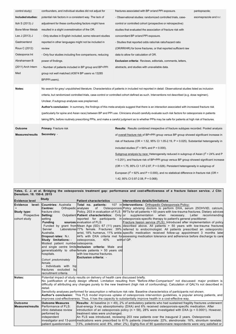
control study)
Included studies:
Itoh S (2013) J
Bone Miner Metab
Lee J (2013) J
Gastroenterol
Roux C (2012)
Osteoporos Int
Abrahamsen B
(2011) Arch Intern
Med
confounders, and individual studies did not adjust for
potential risk factors in a consistent way. The lack of
adjustment for these confounding factors might have
resulted in a slight overestimation of the OR.
- Only studies in English included, some relevant studies
reported in other languages might not be included in
review
- Only four studies including five comparisons, reducing
power of findings.
- Number of patients included in BP group and BP+PPI
group not well matched (43974 BP users vs 13285
BP/PPI users).
fractures associated with BP or/and PPI exposure.
- Observational studies: randomized controlled trials, case-
control or controlled cohort (prospective or retrospective)
studies that evaluated the association of fracture risk with
concomitant BP or/and PPI exposure.
- Studies that reported odds ratio/risk ratio/hazard ratio
(OR/RR/HR) for bone fractures, or that reported sufficient raw
data to allow for calculation of OR.
Exclusion criteria: Reviews, editorials, comments, letters,
abstracts, and studies with unavailable data.
pantoprazole;
esomeprazole and n.r.
Notes:
No search for grey/ unpublished literature. Characteristics of patients in included not reported in detail. Observational studies listed as inclusion
criteria, but randomized controlled trials, case-control or controlled cohort defined as such. Interventions not described (e.g. dose regimen).
Unclear, if subgroup analyses was preplanned. Author's conclusion: In summary, the findings of this meta-analysis suggest that there is an interaction associated with increased fracture risk
(particularly for spine and Asian race) between BP and PPI use. Clinicians should carefully evaluate such risk factors for osteoporosis in patients
taking BPs, before routinely prescribing PPIs, and make a careful judgment as to whether PPIs may be safe for patients at high risk of fractures.
Outcome
Measures/results
Primary Fracture risk
Secondary -
Results: Results combined irrespective of fracture subtypes recorded. Pooled analysis
of overall fracture risk of BP+PPI group versus BP group showed significant increase in
risk of fractures (OR = 1.52, 95% CI 1.05-2.19, P = 0.025). Substantial heterogeneity in
included studies (I2 = 94% and P = 0.000).
Subgroup analysis by race: Heterogeneity reduced in subgroup of Asian (I2 = 24% and P
= 0.251), and fracture risk of BP+PPI group versus BP group showed significant increase
(OR = 1.75, 95% CI 1.07-2.87, P = 0.026). Persistent heterogeneity in subgroup of
European (I2 = 92% and P = 0.000), and no statistical difference in fracture risk (OR =
1.42, 95% CI 0.97-2.08, P = 0.068).
Yates, C. J. et al. Bridging the osteoporosis treatment gap: performance and cost-effectiveness of a fracture liaison service. J Clin Densitom. 18. 150-6. 2015
Evidence level Study Patient characteristics Interventions details/limitations Evidence level: +
Study type: Prospective
cohort study
Countries: Australia Centres: Orthopedic fracture clinic Setting: Outpatient setting Funding sources: Funded by grant from
Servier Laboratories Australia. Dropout rates: N.r. Study limitations: - Modest patient number and single centre limits generalizability to other hospitals.
- Cohort predominately female. - Individuals with hip fractures excluded by recruitment criteria.
Total no. patients: 107 in analyses of Osteoporosis Policy, 203 in evaluation of FLS Patient characteristics: Only reported for participants in evaluation of FLS: Mean Age (SD): 67 (11) years. 77% female. Fractures: 39% wrist, 18% humerus, 11% ankle. 44% with DXA criteria met for osteoporosis, 40% with osteopenia. Inclusion criteria: Male and female patients > 50 years old with low-trauma fractures. Exclusion criteria: -
Interventions: Orthopedic Osteoporosis Policy: Policy advising doctors to perform DXA, serum 25OHVitD, calcium, RFTs for all patients > 50 years with low-trauma fractures. Dietary advices or supplementation when necessary. Letter recommending osteoporosis-specific therapy to patient's general practitioner. Fracture liasion service (FLS): Introduced after implementation of policy described above. All patients > 50 years with low-trauma fractures referred to endocrinologist. All patients prescribed an osteoporotic- specific medication received follow-up appointment 3 months later assessing medication tolerance and adherence before discharge to care of GP.
Notes: Potential impact of study results on delivery of health care discussed briefly. No justification of study design offered. Limitation resulting from "Before-After-Comparison" not discussed: major problem is difficulty of attributing any changes purely to the new treatment (high risk of confounding). Calculation of QALYs not described in detail. Sensitivity analyses performed for assumption o refracture risk rate. Baseline characteristics of participants not shown. Author's conclusion: This FLS model improves uptake of osteoporosis intervention guidelines, is popular among patients, and improves cost-effectiveness. Thus, it has the capacity to substantially improve health in a cost-effective way.
Outcome Measures/results
Outcome Measures Performance of FLS: clinic database review performed by independent investigator and 13-point patient questionnaire.
Results: At baseline (n = 49), 2% of ambulatory patients who had sustained fragility fractures underwent dual-energy X-ray absorptiometry (DXA) and 6% received osteoporosis-specific medication. After introduction of an osteoporosis policy (n = 58), 28% were investigated with DXA (p < 0.0001). However, treatment rates were unchanged. An FLS was introduced, reviewing 203 new patients over the inaugural 2 years. Osteoporosis medications were prescribed to 61% patients (risedronate: 22%, alendronate: 16%, strontium ranelate: 13%, zoledronic acid: 8%, other: 2%). Eighty-five of 90 questionnaire respondents were very satisfied or
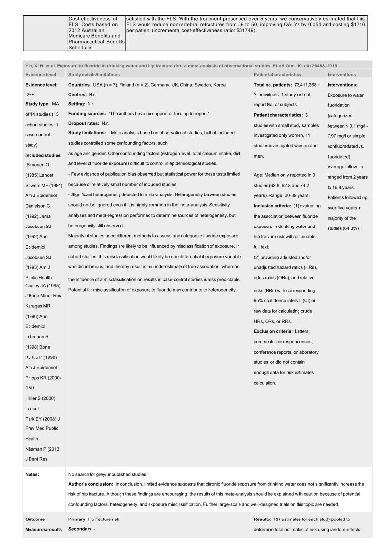
Cost-effectiveness of FLS: Costs based on 2012 Australian Medicare Benefits and Pharmaceutical Benefits Schedules.
satisfied with the FLS. With the treatment prescribed over 5 years, we conservatively estimated that this FLS would reduce nonvertebral refractures from 59 to 50, improving QALYs by 0.054 and costing $1716 per patient (incremental cost-effectiveness ratio: $31749).
Yin, X. H. et al. Exposure to fluoride in drinking water and hip fracture risk: a meta-analysis of observational studies. PLoS One. 10. e0126488. 2015
Evidence level Study details/limitations Patient characteristics Interventions Evidence level:
2++
Study type: MA
of 14 studies (13
cohort studies, 1
case-control
study)
Included studies:
Simonen O
(1985) Lancet
Sowers MF (1991)
Am J Epidemiol
Danielson C
(1992) Jama
Jacobsen SJ
(1992) Ann
Epidemiol
Jacobsen SJ
(1993) Am J
Public Health
Cauley JA (1995)
J Bone Miner Res
Karagas MR
(1996) Ann
Epidemiol
Lehmann R
(1998) Bone
Kurttio P (1999)
Am J Epidemiol
Phipps KR (2000)
BMJ
Hillier S (2000)
Lancet
Park EY (2008) J Prev Med Public
Health.
Näsman P (2013)
J Dent Res
Countries: USA (n = 7), Finland (n = 2), Germany, UK, China, Sweden, Korea
Centres: N.r.
Setting: N.r.
Funding sources: "The authors have no support or funding to report."
Dropout rates: N.r.
Study limitations: - Meta-analysis based on observational studies, half of included
studies controlled some confounding factors, such
as age and gender. Other confounding factors (estrogen level, total calcium intake, diet,
and level of fluoride exposure) difficult to control in epidemiological studies.
- Few evidence of publication bias observed but statistical power for these tests limited
because of relatively small number of included studies.
- Significant heterogeneity detected in meta-analysis. Heterogeneity between studies
should not be ignored even if it is highly common in the meta-analysis. Sensitivity
analyses and meta-regression performed to determine sources of heterogeneity, but
heterogeneity still observed.
- Majority of studies used different methods to assess and categorize fluoride exposure
among studies. Findings are likely to be influenced by misclassification of exposure. In
cohort studies, this misclassification would likely be non-differential if exposure variable
was dichotomous, and thereby result in an underestimate of true association, whereas
the influence of a misclassification on results in case-control studies is less predictable.
Potential for misclassification of exposure to fluoride may contribute to heterogeneity.
Total no. patients: 73,411,368 +
? individuals. 1 study did not
report No. of subjects.
Patient characteristics: 3
studies with small study samples
investigated only women, 11
studies investigated women and
men.
Age: Median only reported in 3
studies (62.8, 62.8 and 74.2
years). Range: 20-89 years.
Inclusion criteria: (1) evaluating
the association between fluoride
exposure in drinking water and
hip fracture risk with obtainable
full text;
(2) providing adjusted and/or
unadjusted hazard ratios (HRs),
odds ratios (ORs), and relative
risks (RRs) with corresponding
95% confidence interval (CI) or
raw data for calculating crude
HRs, ORs, or RRs.
Exclusion criteria: Letters,
comments, correspondences,
conference reports, or laboratory
studies; or did not contain
enough data for risk estimates
calculation.
Interventions:
Exposure to water
fluoridation
(categorized
between ≤ 0.1 mg/l -
7.97 mg/l or simple
nonfluoradated vs.
fluoridated).
Average follow-up
ranged from 2 years
to 16.8 years.
Patients followed up
over five years in
majority of the
studies (64.3%).
Notes:
No search for grey/unpublished studies.
Author's conclusion: In conclusion, limited evidence suggests that chronic fluoride exposure from drinking water does not significantly increase the
risk of hip fracture. Although these findings are encouraging, the results of this meta-analysis should be explained with caution because of potential
confounding factors, heterogeneity, and exposure misclassification. Further large-scale and well-designed trials on this topic are needed.
Outcome
Measures/results
Primary Hip fracture risk
Secondary -
Results: RR estimates for each study pooled to
determine total estimates of risk using random-effects
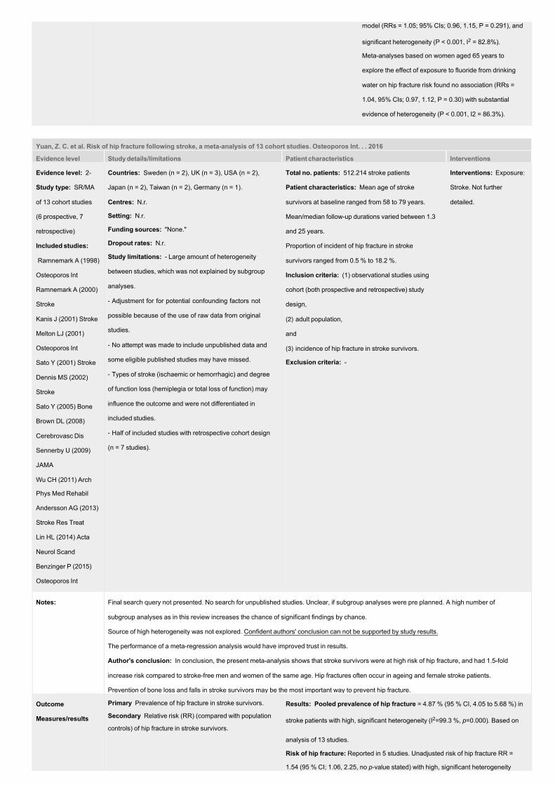
model (RRs = 1.05; 95% CIs; 0.96, 1.15, P = 0.291), and
significant heterogeneity (P < 0.001, I2 = 82.8%).
Meta-analyses based on women aged 65 years to
explore the effect of exposure to fluoride from drinking
water on hip fracture risk found no association (RRs =
1.04, 95% CIs; 0.97, 1.12, P = 0.30) with substantial
evidence of heterogeneity (P < 0.001, I2 = 86.3%).
Yuan, Z. C. et al. Risk of hip fracture following stroke, a meta-analysis of 13 cohort studies. Osteoporos Int. . . 2016
Evidence level Study details/limitations Patient characteristics Interventions Evidence level: 2-
Study type: SR/MA
of 13 cohort studies
(6 prospective, 7
retrospective)
Included studies:
Ramnemark A (1998)
Osteoporos Int
Ramnemark A (2000)
Stroke
Kanis J (2001) Stroke
Melton LJ (2001)
Osteoporos Int
Sato Y (2001) Stroke
Dennis MS (2002)
Stroke
Sato Y (2005) Bone
Brown DL (2008)
Cerebrovasc Dis
Sennerby U (2009)
JAMA
Wu CH (2011) Arch Phys Med Rehabil
Andersson AG (2013)
Stroke Res Treat
Lin HL (2014) Acta
Neurol Scand
Benzinger P (2015)
Osteoporos Int
Countries: Sweden (n = 2), UK (n = 3), USA (n = 2),
Japan (n = 2), Taiwan (n = 2), Germany (n = 1).
Centres: N.r. Setting: N.r.
Funding sources: "None."
Dropout rates: N.r.
Study limitations: - Large amount of heterogeneity
between studies, which was not explained by subgroup
analyses.
- Adjustment for for potential confounding factors not
possible because of the use of raw data from original
studies.
- No attempt was made to include unpublished data and
some eligible published studies may have missed.
- Types of stroke (ischaemic or hemorrhagic) and degree
of function loss (hemiplegia or total loss of function) may
influence the outcome and were not differentiated in
included studies.
- Half of included studies with retrospective cohort design
(n = 7 studies).
Total no. patients: 512.214 stroke patients
Patient characteristics: Mean age of stroke
survivors at baseline ranged from 58 to 79 years.
Mean/median follow-up durations varied between 1.3
and 25 years.
Proportion of incident of hip fracture in stroke
survivors ranged from 0.5 % to 18.2 %.
Inclusion criteria: (1) observational studies using
cohort (both prospective and retrospective) study
design,
(2) adult population,
and
(3) incidence of hip fracture in stroke survivors. Exclusion criteria: -
Interventions: Exposure:
Stroke. Not further
detailed.
Notes:
Final search query not presented. No search for unpublished studies. Unclear, if subgroup analyses were pre planned. A high number of
subgroup analyses as in this review increases the chance of significant findings by chance.
Source of high heterogeneity was not explored. Confident authors' conclusion can not be supported by study results.
The performance of a meta-regression analysis would have improved trust in results.
Author's conclusion: In conclusion, the present meta-analysis shows that stroke survivors were at high risk of hip fracture, and had 1.5-fold
increase risk compared to stroke-free men and women of the same age. Hip fractures often occur in ageing and female stroke patients.
Prevention of bone loss and falls in stroke survivors may be the most important way to prevent hip fracture. Outcome
Measures/results
Primary Prevalence of hip fracture in stroke survivors.
Secondary Relative risk (RR) (compared with population
controls) of hip fracture in stroke survivors.
Results: Pooled prevalence of hip fracture = 4.87 % (95 % CI, 4.05 to 5.68 %) in
stroke patients with high, significant heterogeneity (I2=99.3 %, p=0.000). Based on
analysis of 13 studies. Risk of hip fracture: Reported in 5 studies. Unadjusted risk of hip fracture RR =
1.54 (95 % CI; 1.06, 2.25, no p-value stated) with high, significant heterogeneity
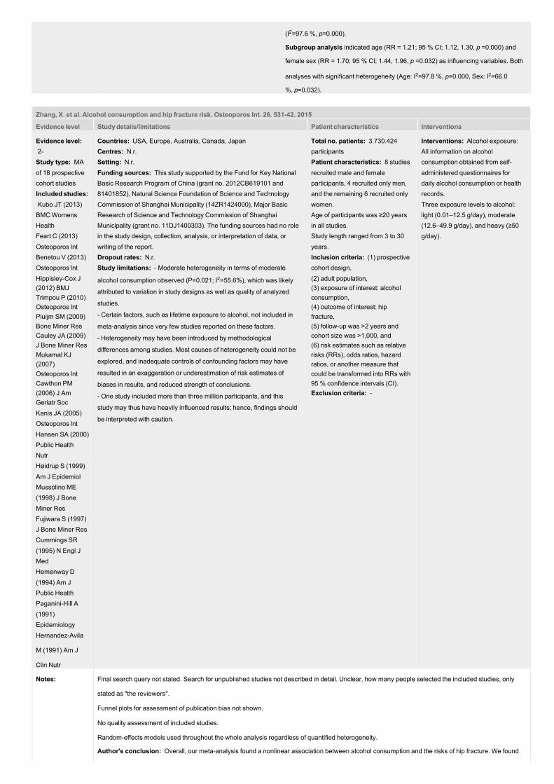
(I2=97.6 %, p=0.000).
Subgroup analysis indicated age (RR = 1.21; 95 % CI; 1.12, 1.30, p =0.000) and female sex (RR = 1.70; 95 % CI; 1.44, 1.96, p =0.032) as influencing variables. Both
analyses with significant heterogeneity (Age: I2=97.8 %, p=0.000, Sex: I2=66.0
%, p=0.032).
Zhang, X. et al. Alcohol consumption and hip fracture risk. Osteoporos Int. 26. 531-42. 2015
Evidence level Study details/limitations Patient characteristics Interventions Evidence level: 2- Study type: MA of 18 prospective cohort studies Included studies: Kubo JT (2013) BMC Womens Health Feart C (2013) Osteoporos Int Benetou V (2013) Osteoporos Int Hippisley-Cox J (2012) BMJ Trimpou P (2010) Osteoporos Int Pluijm SM (2009) Bone Miner Res Cauley JA (2009) J Bone Miner Res Mukamal KJ (2007) Osteoporos Int Cawthon PM (2006) J Am Geriatr Soc Kanis JA (2005) Osteoporos Int Hansen SA (2000) Public Health Nutr Høidrup S (1999) Am J Epidemiol Mussolino ME (1998) J Bone Miner Res Fujiwara S (1997) J Bone Miner Res Cummings SR (1995) N Engl J Med Hemenway D (1994) Am J Public Health Paganini-Hill A (1991) Epidemiology Hernandez-Avila
M (1991) Am J
Clin Nutr
Countries: USA, Europe, Australia, Canada, Japan Centres: N.r. Setting: N.r. Funding sources: This study supported by the Fund for Key National Basic Research Program of China (grant no. 2012CB619101 and 81401852), Natural Science Foundation of Science and Technology Commission of Shanghai Municipality (14ZR1424000), Major Basic Research of Science and Technology Commission of Shanghai Municipality (grant no. 11DJ1400303). The funding sources had no role in the study design, collection, analysis, or interpretation of data, or writing of the report. Dropout rates: N.r. Study limitations: - Moderate heterogeneity in terms of moderate
alcohol consumption observed (P=0.021; I2=55.6%), which was likely
attributed to variation in study designs as well as quality of analyzed
studies.
- Certain factors, such as lifetime exposure to alcohol, not included in
meta-analysis since very few studies reported on these factors.
- Heterogeneity may have been introduced by methodological
differences among studies. Most causes of heterogeneity could not be
explored, and inadequate controls of confounding factors may have
resulted in an exaggeration or underestimation of risk estimates of
biases in results, and reduced strength of conclusions.
- One study included more than three million participants, and this
study may thus have heavily influenced results; hence, findings should
be interpreted with caution.
Total no. patients: 3.730.424 participants Patient characteristics: 8 studies recruited male and female participants, 4 recruited only men, and the remaining 6 recruited only women. Age of participants was ≥20 years in all studies. Study length ranged from 3 to 30 years. Inclusion criteria: (1) prospective cohort design, (2) adult population, (3) exposure of interest: alcohol consumption, (4) outcome of interest: hip fracture, (5) follow-up was >2 years and cohort size was >1,000, and (6) risk estimates such as relative risks (RRs), odds ratios, hazard ratios, or another measure that could be transformed into RRs with 95 % confidence intervals (CI). Exclusion criteria: -
Interventions: Alcohol exposure: All information on alcohol consumption obtained from self- administered questionnaires for daily alcohol consumption or health records. Three exposure levels to alcohol: light (0.01–12.5 g/day), moderate (12.6–49.9 g/day), and heavy (≥50 g/day).
Notes:
Final search query not stated. Search for unpublished studies not described in detail. Unclear, how many people selected the included studies, only
stated as "the reviewers".
Funnel plots for assessment of publication bias not shown.
No quality assessment of included studies.
Random-effects models used throughout the whole analysis regardless of quantified heterogeneity. Author's conclusion: Overall, our meta-analysis found a nonlinear association between alcohol consumption and the risks of hip fracture. We found
that compared with no alcohol consumption, light alcohol consumption was significantly inversely associated with hip fracture risk, whereas heavy
alcohol consumption was associated with an elevated hip fracture risk.
Outcome
Measures/results
Primary Hip fractures: Fractures identified using self-reports of
fracture experience, radiologic diagnoses, or medical records.
Secondary -
Results: No significant difference regarding hip fractures between any
alcohol drinkers vs. non-drinkers: Pooled RR = 1.03 (95 % CI; 0.91,
1.15), with high heterogeneity observed between studies (P<0.001,
I2=72.6 %). Both Egger’s and Begg’s test showed no publication bias
(P=0.089 and 0.085, respectively).
Dose–response analysis: Light alcohol consumption vs. no alcohol: RR = 0.88 (95 % CI; 0.83, 0.92). No significant heterogeneity P=0.256, I2 = 20.4 %.
Moderate alcohol consumption vs. no alcohol: RR = 1.00 (95 % CI; 0.85, 1.14). No significant heterogeneity P=0.796, I2 = 0 %. Heavy alcohol consumption vs. no alcohol: RR = 1.71 (95 % CI; 1.41, 2.01).No significant heterogeneity P=0.021, I2 = 55.6 %.
Zhang, Z. L. et al. Alendronate sodium/vitamin D3 combination tablet versus calcitriol for osteoporosis in Chinese postmenopausal
women: a 6-month, randomized, open-label, active-comparator-controlled study with a 6-month extension. Osteoporos Int. 26. 2365-74. 2015
Evidence level Study details/limitations Patient Interventions characteristics
Evidence level: 1++
Study type: RCT
Countries: China Centres: 13 centers Setting: - Funding sources: MSD China Holding Co., Ltd., China Dropout rates: 4,5% in ALN/D5600 group, 3,7% in Calcitriol group Study limitations: Open-label design which may have introduced bias. In addition, although BMD was measured by either Hologic (Hologic Corporation) or GE Lunar (GE Medical Systems) DXA machines and individual sites followed quality control procedures recommended by the manufacturers, there was no independent central BMD quality control/quality assessment program. However, because the study was not large enough to compare fracture incidence, central adjudication of events such as fractures was not performed by either an internal or an external committee. Moreover, the study was not designed to distinguish the relative contributions of ALN and vitamin D3 in reducing bone loss and increasing BMD.
Total no. patients: n=219
Patient characteristics: Postmenopausal(at least 1 year) women >55 years Inclusion criteria:
- BMD T- score≤-2.5 in at least one anatomic site (lumbar spine, total hip, or femoral neck) or - prior non- pathological fragility fracture (of the spine, wrist,humerus, or clavicle) and - BMD T-score≤-1.5 in at least one of the same anatomic sites; 25(OH)D levels ≥8 ng/mL (20 nmol/L) - no prior hip fracture - no abnormal finding of clinical significance (no pronounced medical history other than osteoporosis) - no treatment with specific drugs for specified time periods - no contraindications to alendronate, calcitriol, or vitamin D Exclusion criteria: -
Interventions: 12 months ALN/D5600 (Fosamax® Plus D, alendronate 70 mg/vitamin D3 5600 IU) once weekly as a combination tablet versus calcitriol (Rocaltrol®,calcitriol 0.25 μg) 0.25 μg daily. Calcium supplements (500mg/daily)
Notes: sample-size calculation not reported Author's conclusion: In summary, ALN/D5600 showed significantly greater increases in BMD at the lumbar spine and significantly greater decreases in bone turnover markers compared with calcitriol in Chinese postmenopausal women with osteoporosis. The study did not evaluate whether the greater increase in BMD resulted in fewer fractures. ALN/D5600 was also generally well tolerated in Chinese patients with a safety profile consistent with previous studies across various regions and races.
Outcome Measures/results
Primary Percent change from baseline in lumbar spine BMD at month 6. Secondary At month 6 - percent change from baseline in the biochemical markers of bone turnover procollagen type 1 N-terminal propeptide (P1NP) serum C-telopeptides of type 1 collagen (s-CTx) - percent change from baseline in total hip, femoral neck, trochanter BMD - proportion of patients with serum 25(OH)D <20 ng/mL; - proportion of patients with serum 25(OH)D <15 ng/mL;
Results: Lumbar spine BMD Month 6 - percent change (least squares LS) from baseline: ALN/D5600 3.5 % (95 % CI 2.7, 4.4; standard error (SE), 0.42) vs Calcitriol 1.6 % (95 % CI 0.8, 2.4; SE 0.41) between treatment difference of 2.0 % (95

- change from baseline in mean serum 25(OH)D level At month 12 - percent change from baseline in lumbar spine, total hip, femoral neck, trochanter BMD - percent change from baseline in the biochemical markers of bone turnover P1NP and s-CTx - proportion of patients with serum 25(OH)D <20 ng/mL - proportion of patients with serum 25(OH)D <15 ng/mL - change from baseline in mean serum 25(OH)D level
The safety and tolerability of ALN/D5600 once weekly
% CI 0.8, 3.1; p<0.001 adjusted for 25(OH)D level stratum as a baseline covariate) Month 12 - percent changes 5.2 % (95 % CI 4.3, 6.0) vs 2.3 % (95 % CI 1.4, 3.1) between treatment difference of 2.9 % (95 % CI 1.8, 4.1; p<0.001 adjusted for 25(OH)D level
Total hip, femoral neck, trochanter BMD Month 6 - percent change (LS) from baseline: ALN/D5600 1.6 % (95 % CI 0.9, 2.4), 2.8 % (95 % CI 1.2, 4.3), 2.2 % (95 % CI 0.9 3.5) vs Calcitriol 0.5 % (95 % CI −0.2, 1.2), 0.8 % (95 % CI −0.7, 2.2), 0.4 % (95 % CI −0.8, 1.7) Month 12 - percent changes ALN/D5600 2.7 % (95 % CI 1.7, 3.6), 3.3 % (95 % CI 1.9,4.7), 2.4 % (95 % CI 0.9, 3.8), vs calcitriol 0.1 % (95 % CI −0.8, 1.0), 0.4 % (95 % CI −0.9, 1.8), 0.1 % (95 % CI −1.3, 1.5)
Between-treatment differences were statistically significant for the total hip and trochanter at month 6, and for the total hip, femoral neck, and trochanter at month 12.
Biomarkers Month 6 - percent changes from baseline (LS mean) of P1NP and s-CTx for ALN/D5600 −59.1 % (95 % CI −62.0, −56.0) and −79.2 %(95%CI −81.4, −76.8)vs Calcitriol −16.8%(95% CI −22.5, −10.5) and −27.2 % (95 % CI −34.6, −19.0) Month 12 - percent changes ALN/D5600 −68.1 % (95 % CI −70.8, −65.1) and −76.2 % (95 % CI −78.6, −73.5)vs calcitriol −17.0 % (95 % CI −23.8, −9.6) and −24.2% (95 % CI −31.6, −16.0) Between-treatment differences were statistically significant for P1NP and s-CTx at months 6 and 12.
Serum 25(OH)D Month 6-12 - ALN/D5600 group mean levels increased from 19.7 ng/mL at baseline to 29.4 and 30.2 ng/mL at months 6 and 12 - calcitriol group, mean levels decreased from baseline (19.4 ng/mL) to month 6 (16.1 ng/mL) but were similar to baseline levels at month 12 (20.3 ng/mL) - Month 6: 25(OH)D thresholds (<15 and <20 ng/mL) significantly higher in the calcitriol group (48.6 and 72.9 %) than in the ALN/D5600 group (1.0 and 5.9 %) (both, p<0.001) - Month 12: 25(OH)D thresholds (<15 and <20 ng/mL)decreased in the calcitriol group (25.5 and 47.1 %), remained very low in the ALN/D5600 group (1.0 and 4.1 %) (both, p<0.001)
Safety drug-related AEs in the ALN/D5600 group (14.0 %) vs calcitriol group (7.4%). None of the SAEs were considered to be
related to the study drug.
Zhao, R. et al. Efficiency of jumping exercise in improving bone mineral density among premenopausal women: a meta-analysis. Database of Abstracts of Reviews of Effects. . 1393-1402. 2014
Evidence level Study Patient characteristics Interventions details/limitations Evidence level: 1+
Study type: SR of 6 RCT Included studies: Bassey EJ (1994) Osteoporos Int Bassey EJ (1998) J Bone Miner Res Kato T (2006) J Appl Physiol Niu K (2010) J Bone Miner Metab Vainionpää A (2005) Osteoporos Int Winters-Stone KM (2006) Bone
Countries: 2 UK, 2 Japan, 1 Finland, 1 US Centres: Not reported. Setting: Not reported. Funding sources:
This article was based on work funded by the
Zhejiang Provincial Natural Science Foundation of China under grant numbers Y2110954 and LY14H070001. Dropout rates: Not quantified.
Study limitations: - Inclusion of only
6 studies, - Highly selective samples of premenopausal women of varying ages, - Future studies should include both traditional resistance exercises and high impact intervention studies to examine the optimal exercise for improving premenopausal bone health.
Total no. patients: 377 women Patient characteristics: All studies conducted with healthy premenopausal women between 20.5 ± 0.6 years and 41.3 ± 3.8 years (mean ± SD). Sample sizes varied from 27 to 120 participants. Premenopausal status determined by menstrual history and self- completed lifestyle questionnaires. 201 women performed jumping exercise intervention from 6 to 12 months in duration, 176 women assigned to non- exercise control groups. Inclusion criteria: Study design: Controlled and randomized controlled trials Population: Healthy premenopausal women (age range 18–50 years) without a history of medicine administration or disease experience affecting bone metabolism Exercise experience: Subjects without regular exercise (no more than 2.5 h per week) prior to study enrollment Intervention: High-impact and short-duration jumping exercise intervention at least lasting for 6 months Comparison: Exercise intervention compared with non-exercise control or sham exercises Outcome measurement: Data available of changes in BMD at femoral neck, trochanter and lumbar spine determined by DXA at pro- and post- intervention respectively Exclusion criteria: -
Interventions: Jumping exercise interventions. All studies prescribed high- impact exercise (jumping) with one exercise plus lower or lower/upper body resistance exercise and one exercise combining stamping, running, and walking. Study duration ranged from 6 to 12 months, with a training frequency of two to three times per week in most interventions. Two exercise protocols were conducted six or seven times per week, but the intensity of training was relatively low, and the total training volume was similar to other studies. Three control groups were asked to maintain their usual diet and lifestyle. Two control groups performed mild arm exercises or stretching and balance exercises. Specific information was not available on one of the control groups.
Notes: No search for grey literature possibly introducing publication bias. Misleading interpretation of funnel plots by authors ("asymmetry[...] indicating positive treatment effect"). Instead, funnel plots indicate publication bias, resulting in an overestimation of treatment effect and its direction. However, funnel plots futile due to insufficient number of studies. Primary Outcomes (Absolute changes in BMD) not reported. Author's conclusion: Based on meta-analysis of existing studies, the sensitivity of skeletal response to jumping exercise in premenopausal women is significant and site-specific, with significant benefit from high-impact exercise noted, especially at the hip.
Outcome Measures/results
Primary Absolute changes in BMD and its standard deviation (SD) at femoral neck, trochanter, and lumbar spine, as assessed by dual- energy X-ray absorptiometry (DXA). Secondary
Weighted mean difference (WMD) between exercise and control groups, pooled SDs of mean difference between exercise and control groups.
Results: Jumping exercise significantly increased BMD in the femoral neck (WMD [fixed effect] = 0.017 g/cm2, 95 %CI; 0.014, 0.20, p<0.001) and trochanter (WMD [fixed effect] = 0.021, 95 % CI; 0.018, 0.024, p<0.001). No significant effect for lumbar spine (WMD [fixed effect] = -0.001, 95 % CI; -0.003, 0.001, p = 0.181).
Zhao, R. et al. The effects of differing resistance training modes on the preservation of bone mineral density in postmenopausal women: a meta-analysis. Osteoporos Int. 26. 1605-18. 2015 Evidence level Study details/limitations Patient characteristics Interventions Evidence level: 2+
Study type: MA of 24 studies (19
Countries: USA, Brazil, Australia, Canada, Germany, UK, Belgium Centres: Not reported. Setting: Not reported.
Total no. patients: 1.769 postmenopausal women Patient characteristics: Healthy postmenopausal women.
Interventions: All of the included trials were based on high-intensity resistance training with study durations ranging from 6 months to 12 years, with a training frequency of 2–3 times per week in most
RCT and 5 CT) Included studies:
Bemben DA (2000) Med Sci Sports Exerc Bemben DA (2010) Bone Bocalini DS (2009) J Aging Health Bolton KL (2012) J Sci Med Sport Chilibeck PD (2002) Can J Physiol Pharmacol Chilibeck PD (2013) J Bone Miner Res Chiun A (2009) Osteoporos Int Engelke K (2006) Osteoporos Int Going S (2003) Osteoporos Int Jessup JV (2003) Biol Res Nurs Kemmler W (2003) Arch Phys Med Rehabil Kemmler W (2004) Arch Intern Med Kemmler W (2012) Osteoporos Int Kerr D (2001) J Bone Miner Res Maddalozzo GF (2007) Bone Marques EA (2011) Exp Gerontol Milliken LA (2003) Calcif Tissue Int Nelson ME (1994) JAMA Pruitt LA (1992) J Bone Miner Res Pruitt LA (1995) J Bone Miner Res Rhodes EC (2000) Br J Sports Med Smidt GL (1992) Spine Verschueren SM (2004) J Bone Miner Res von Stengel S (2011) Osteoporos Int
Funding sources: This material was based upon the work funded by Zhejiang Provincial Natural Science Foundation of China under Grant No. LY14H070001. Dropout rates: High dropout rates (range: 0% - 52.3% in intervention groups, 0% - 41.2% in control groups) 1 study with no statement regarding drop-outs Study limitations: - Measurements of BMD made with DXA or DPA: → may not be optimal for examining bone strength since these methods do not incorporate other aspects of bone quality such as microarchitecture - Highly selected samples of postmenopausal women of varying ages in included studies - Relatively low quality of the included studies - 4 studies included that reported findings of the same program “EFOPS” → might potentially impact the results of pooling intervention effects
Age between 50.5±6.3 and 69.6± 4.2 years Inclusion criteria: Study design: Controlled or randomized controlled trials; Populations of interest: Healthy postmenopausal women without surgical experience, pharmacological treatment, or disease history affecting bone metabolism; Exercise experience: Subjects without regular exercise (less than 2.5 h per week) prior to study enrollment; Intervention: Resistance training interventions at least lasting for 6 months; Comparison: Exercise interventions compared with nonexercise control or sham exercise; Outcome measurement: The change in BMD at femoral neck and lumbar spine determined by DXA or DPA on the pre- and postexercise interventions. Exclusion criteria: Participants receiving hormone replacement therapy (HRT) or antiresorptive treatment (AT)
interventions. - 14 studies with resistance-alone protocols with a 10- to 20-min warm-up and a 5- to 10-min cooldown, - 10 clinical trials with combined resistance exercise protocols.
Notes: Process of study selection not described. No search for grey literature. No investigation of publication bias because "the subjective nature of visual interpretation of funnel plots seems to have limited use in the examination of publication bias in the current meta-analysis". Adverse events not investigated. Author's conclusion: The general conclusions of the present meta-analysis were that progressive high-intensity resistance training tended to be effective for improving or preserving hip and spine BMD in postmenopausal women. However, skeletal adaptation was dependent on training modes, as the combined protocols that integrated resistance training with high-impact or weight bearing exercises showed significant beneficial effects on postmenopausal bone loss, while resistance-alone protocols did not. Although the number of training-related injuries reported in the included studies was very low, caution is advised when resistance training is performed at home or without supervision because the resistance exercise conducted by the participants in the clinical trials was usually performed with special training equipment and under supervision. The poor quality of several trials reminds us that well-designed studies with large sample sizes are still needed. Further studies are also needed to characterize both material and structural changes to determine exercise-induced gains in bone strength. Nevertheless, combined resistance training protocols furnish a feasible nonpharmacological strategy for preventing postmenopausal bone loss.
Outcome Measures/results
Primary areal BMD (BMD g/cm2) assessed by dual X-ray absorptiometry (DXA) or dual photon absorptiometry (DPA) Secondary -
Results: Overall analysis of resistance training impact: Resistance training interventions significantly increased femoral neck BMD: SMD = 0.303 (95% CI; 0.127, 0.479, p = 0.01; I2 = 46.5%) and lumbar spine BMD: SMD = 0.311 (95% CI; 0.115, 0.507, p = 0.002; I2 = 62.3%). Combined resistance versus resistance-alone protocols: Combined resistance training protocols significantly affected both the hip BMD: SMD = 0.411 (95% CI; 0.176, 0.645, p = 0.001; I2 = 48.3 %) (analyses is for femoral neck) and spine BMD: SMD = 0.431 (95% CI; 0.159, 0.702, p = 0.002; I2 = 71.3 %). Resistance-alone protocols had no significant effects on the preservation of femoral neck BMD: SMD = 0.212 (95% CI; −0.043, 0.468, p = 0.104; I2 = 45.6%) or lumbar spine BMD: SMD = 0.180 (95% CI; −0.09, 0.456, p = 0.203; I2 = 48.3%). Differences between combined resistance and resistance-alone groups not significant: p = 0.149 for femoral neck BMD and p = 0.253 for spine BMD.

Ulrike
Textfeld
Erstveröffentlichung: 12/2017 Überarbeitung von: Nächste Überprüfung geplant: 12/2022
Ulrike
Textfeld
Die AWMF erfasst und publiziert die Leitlinien der Fachgesellschaften mit größtmöglicher Sorgfalt - dennoch kann die AWMF für die Richtigkeit des Inhalts keine Verantwortung übernehmen. Insbesondere bei Dosierungsangaben sind stets die Angaben der Hersteller zu beachten!
Ulrike
Textfeld
Autorisiert für elektronische Publikation: AWMF online