ANESTHESIA FOR TRAUMA PATIENTS - Doctor 2017
143
ANESTHESIA FOR TRAUMA PATIENTS Professor Subhi AlGhanem Department of anesthesia &Intensive Care School of medicine The University of Jordan
Transcript of ANESTHESIA FOR TRAUMA PATIENTS - Doctor 2017
ANESTHESIA FOR TRAUMA
PATIENTS
Professor Subhi AlGhanem
Department of anesthesia &Intensive Care
School of medicine
The University of Jordan
Injury is responsible for 9% of the total annual
mortality (more than 5 million people) in the
world
Traffic accidents alone killed 1.24 million people
in 2010
the vast majority reside in low- and middle-
income countries
Trauma especially afflicts young people , as of
2013 it was the leading cause of death for those
aged between 1 and 46 years, and the third most
common cause of death after cardiovascular
diseases and cancer.
Approximately 75% of hospital deaths from high-
energy trauma such as motor vehicle accidents, falls,
and gunshot or stab wounds occur within 48 hours
after admission, most commonly from central
nervous system (CNS), thoracic, abdominal,
retroperitoneal, or vascular injuries.
Nearly one-third of these patients die within the first
4 hours after admission
CNS injury and hemorrhage are the most common
causes of early trauma mortality
5% to 10% occur between the third and seventh day
of admission, usually from CNS injuries
the rest in subsequent weeks, most commonly as a
result of multi organ failure. (PE and infectious
complications)
injuries caused by low-energy impacts, mainly
from falls, usually in the elderly, also produce
significant mortality from head injury and
complications of skeletal injuries.
Of these deaths, 20% occur within 48 hours, 32%
after 3 to 7 days, and 48% after 7 days
Pre-existing conditions such as congestive heart
failure, cirrhosis, warfarin intake, and/or β-
blocker usage increase the mortality rate in
trauma patients
INITIAL EVALUATION AND RESUSCITATION
After information has been obtained from paramedics
about the mechanism of injury, possible injuries, vital
signs at the field and during transport, pre hospital
treatment, and, if available, pre-existing medical
disease(s)
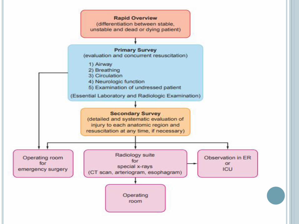
the general approach to evaluation of the acute
trauma victim has three sequential components:
rapid overview
primary survey
secondary survey
Resuscitation is initiated, if needed, at any time
during this continuum
RAPID OVERVIEW
takes only a few seconds and is used to
determine whether the patient is stable,
unstable, dying, or dead

PRIMARY SURVEY
rapid evaluation of functions that are crucial to survival. The ABCs are assessed. Then a brief neurologic examination is performed, and the patient is examined for any external injuries that might have been overlooked.
A rapid limited transthoracic echocardiogram with parasternal long and short axis, apical, and subxiphoid views may give useful information about myocardial contractility, intravascular volume, and the presence of pericardial effusion at this point
Essential laporatory (ABGs or electrolytes ) and radiologic examination are included.
SECONDARY SURVEY
The secondary survey begins only when the ABCs are
stabilized.
involves a more elaborate systematic examination of the
entire body to identify additional injuries. radiography
(focused assessment with sonography [FAST], [CT],
angiography, interventional radiologic procedures, [MRI])
and other diagnostic procedures.
With installation of multi detector CT scans (MDCT) >>
total body imaging is accomplished rapidly, helping
substantially to direct subsequent surgical, interventional
radiologic, or conservative management
TERTIARY SURVEY
Occurs within the first 24 hours after admission (which may include a period of anesthesia)
can potentially diagnose the majority of clinically significant injuries missed during initial evaluation by the care team’s repeating the primary and secondary examinations and reviewing the results of radiologic and laboratory testing
AIRWAY EVALUATION AND INTERVENTION
Airway evaluation involves
the diagnosis of any trauma to the airway or
surrounding tissues
recognition and anticipation of the respiratory
consequences of these injuries
prediction of the potential for exacerbation of these or
other injuries by any contemplated airway
management maneuvers by (mask ventilation,
tracheal intubation, or placement of a surgical
airway).
(ASA) difficult airway algorithm can be applied
with certain modifications to various trauma
airway management scenarios ((cancellation of
airway management when difficulty arises may
not be an option))
Airway management is tailored to the type of
injury, the nature and degree of airway
compromise, and the patient’s hemodynamic and
oxygenation status.
Continuous communication with members of the
trauma team and obtaining information may
help reduce the extent of the difficulty
AIRWAY OBSTRUCTION
most frequent cause of asphyxia after trauma
may result from posteriorly displaced or lacerated pharyngeal soft tissues (upper airway edema); cervical or mediastinal hematoma; bleeding, secretions, or foreign bodies within the airway; and/or displaced bone or cartilage fragments.
Signs of upper and lower airway obstruction include dyspnea, cyanosis, hoarseness, stridor, dysphonia, subcutaneous emphysema, and hemoptysis.
Cervical deformity, edema, crepitation, tracheal tug and/or deviation, or jugular venous distention may be present before these symptoms appear and may help indicate that specialized techniques are required to secure the airway
AIRWAY MANAGEMENT
chin lift, jaw thrust.
clearance of the oropharynx
placement of an oropharyngeal or nasopharyngeal
airway
>> in inadequately breathing patients, ventilation
with a self-inflating bag.
Immobilization of the cervical spine and
administration of oxygen should be applied
simultaneously.
Blind passage of a nasopharyngeal airway or a
nasogastric or nasotracheal tube should be avoided if
a basilar skull fracture is suspected because the
airway may enter the anterior cranial fossa.
A supraglottic airway may permit ventilation with a self-
inflating bag, although these devices do not provide
protection against aspiration of gastric contents ( used as
temporary for a brief period to re-establish the airway
patency or to facilitate intubation aided by a flexible
fiberoptic bronchoscope (FOB).
If they do not provide adequate ventilation, the trachea
must be secured immediately using direct laryngoscopy,
video laryngoscopy, or cricothyroidotomy, depending on
the results of airway assessment.
Maxillofacial, neck, and chest injuries, as well as
cervicofacial burns, are some of the difficult trauma-
related reasons for tracheal intubation.
Airway assessment should include a rapid examination of the anterior neck for feasibility of access to the cricothyroid membrane.
Tracheostomy is not desirable during initial management (longer to perform than a cricothyroidotomy and requires neck extension, which may cause or exacerbate cord trauma) >>> Conversion to a tracheostomy delayed 2 to 3 days later.
Possible contraindications to cricothyroidotomyinclude age younger than 12 years (Permanent laryngeal damage) and suspected laryngeal trauma (uncorrectable airway obstruction).

FULL STOMACH
The urgency of securing the airway often does
not permit adequate time for pharmacologic
measures to reduce gastric volume and acidity.
Thus we should use a safe technique for securing
the airway when necessary:
rapid-sequence induction with cricoid pressure for
those patients without serious airway problems
awake intubation with sedation and topical
anesthesia, if possible, for those with anticipated
serious airway difficulties.
Possible immediate cricothyroidotomy ?? >>>
In agitated and uncooperative patients, topical anesthesia of the airway may be impossible, whereas administration of sedative agents may result in apnea or airway obstruction, with an increased risk of aspiration of gastric contents and inadequate conditions for tracheal intubation.
>>>
locating the cricothyroid membrane
denitrogenating the lungs
a rapid-sequence induction may be used to allow securing of the airway with direct or video laryngoscopyor, if necessary, immediate cricothyroidotomy.
Personnel and material necessary to perform translaryngeal ventilation or cricothyroidotomy must be in place before induction of general anesthesia.

CERVICAL SPINE INJURY
most common causes include high-speed motor
vehicle accidents, falls, diving accidents, and
gunshot wounds.
Head injuries, especially those with a low
Glasgow coma score (GCS) and focal neurologic
deficits, are likely to be associated with C-spine
injuries. Approximately 2% to 10% of head
trauma victims have C-spine injuries, whereas
25% to 50% of patients with C-spine injuries have
an associated head injury

Accurate and timely evaluation is important because
blunt trauma–induced C-spine injury patients develop
new or worsening neurologic deficits after admission,
partly attributable to delayed diagnosis and improper C-
spine protection and/or manipulation.
>>> emergency airway management may have to be
performed without ruling out C-spine injury while the
patients are in a rigid collar and neck-stabilizing
devices.
Clearance of the neck at the earliest possible time after
airway management should be performed to minimize
the complications associated with the collar, such as
pressure ulceration, ICP elevation in head-injured
patients, compromised central venous access
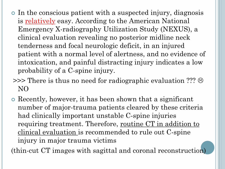
In the conscious patient with a suspected injury, diagnosis
is relatively easy. According to the American National
Emergency X-radiography Utilization Study (NEXUS), a
clinical evaluation revealing no posterior midline neck
tenderness and focal neurologic deficit, in an injured
patient with a normal level of alertness, and no evidence of
intoxication, and painful distracting injury indicates a low
probability of a C-spine injury.
>>> There is thus no need for radiographic evaluation ???
NO
Recently, however, it has been shown that a significant
number of major-trauma patients cleared by these criteria
had clinically important unstable C-spine injuries
requiring treatment. Therefore, routine CT in addition to
clinical evaluation is recommended to rule out C-spine
injury in major trauma victims
(thin-cut CT images with sagittal and coronal reconstruction)
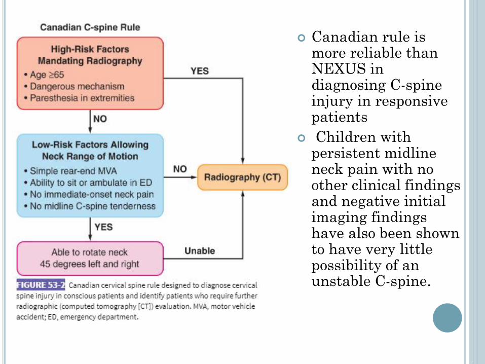
Canadian rule is more reliable than NEXUS in diagnosing C-spine injury in responsive patients
Children with persistent midline neck pain with no other clinical findings and negative initial imaging findings have also been shown to have very little possibility of an unstable C-spine.
CT is not sensitive in diagnosing soft tissue and
ligamentous injury, ruling out ligamentous C-
spine injury.
MRI is a reliable tool; a normal examination can
conclusively exclude C-spine injury. It is thus the
gold standard for ruling C-spine injury in or out.
However, it is so sensitive that it can detect
subtle injuries that are clinically insignificant.
MRI cannot be performed in multiple-trauma
patients who have metallic skeletal fixators. It is
expensive and requires patient transport
AIRWAY MANAGEMENT FOR C-SPINE
INJURY
hard cervical collar alone, which is routinely placed, does
not provide absolute protection, especially against
rotational movements of the neck.
manual inline stabilization (MILS) of the neck is the
standard of care for these patients in the acute stage.
MILS is best accomplished by having two operators in
addition to the physician who is actually managing the
airway. The first operator stabilizes and aligns the head in
neutral position without applying cephalad traction. The
second operator stabilizes both shoulders by holding them
against the table or stretcher.
The anterior portion of the hard collar, which limits
mouth opening, may be removed after immobilization.
Based on the available data, it is, however, reasonable to allow some relaxation of the MILS to improve the glotticview when visualization of the larynx is restricted ( not applying pressure to the tongue by the laryngoscope blade >> worsen the unstable fracture)
Or using other measures such as videolaryngoscopy , use of a gum elastic bougie, carfull cricoid pressure ,translaryngeal (retrograde) intubation, and cricothyroidotomy can be used to secure the airway in the acute phase of cervical spine immobilization.
Flexible fiberoptic laryngoscopy, cause almost no neck movement, but blood or secretions in the airway, a long preparation time, and difficulty in their use in comatose, uncooperative, or anesthetized patients reduce their utility during initial management.
use of FOB in the awake sedated patient with appropriate topical anesthesia is preferred (patient cooperation)
Nasotracheal intubation carries the risks of epistaxis, failure of intubation, and possibility of entry of the endotracheal tube into the cranial vault or the orbit if there is damage to the cranial base or the maxillofacial complex.
Absence of the usual signs of cranial base fracture (battle sign, raccoon eyes, or bleeding from the ear or the nose) cannot be relied on to exclude the possibility of its occurrence because with rapid prehospital transport, these signs may not be immediately apparent
It is possible that airway management–related cervical cord injury in C-spine–injured patients can occur, but, if it does, it is rare
MANAGEMENT OF BREATHING
ABNORMALITIES
tension pneumothorax, flail chest, and open
pneumothorax are immediate threats to the patient’s
life and therefore require rapid diagnosis and
treatment.
Hemothorax, closed pneumothorax, pulmonary
contusion, diaphragmatic rupture with herniation of
abdominal contents into the thorax, and atelectasis
from a mucous plug, aspiration, or chest wall
splinting can also interfere with breathing and
pulmonary gas exchange and deteriorate into life-
threatening complications
TENSION PNEUMOTHORAX
cyanosis, tachypnea, hypotension, neck vein
distention (may be absent in hypovolemic),
tracheal deviation (difficult to appreciate), and
diminished breath sounds on the affected side are
the classic signs.
Inability to position most trauma patients
upright and the likelihood of inadequate imaging
decrease the diagnostic value of chest
radiographs
definitive diagnosis is made by CT scanning
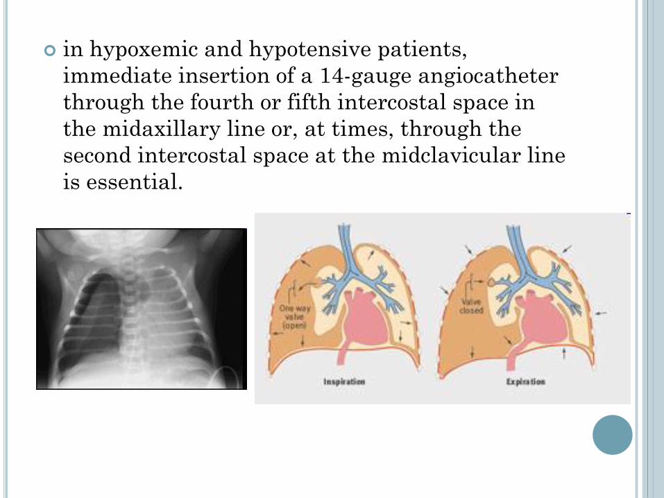
in hypoxemic and hypotensive patients,
immediate insertion of a 14-gauge angiocatheter
through the fourth or fifth intercostal space in
the midaxillary line or, at times, through the
second intercostal space at the midclavicular line
is essential.
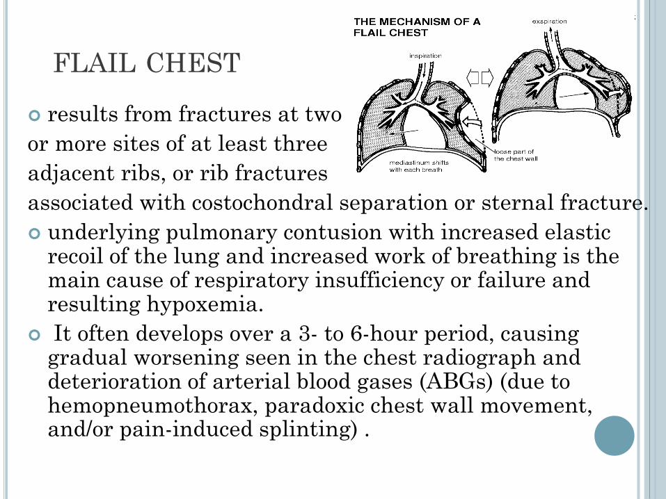
FLAIL CHEST
results from fractures at two
or more sites of at least three
adjacent ribs, or rib fractures
associated with costochondral separation or sternal fracture.
underlying pulmonary contusion with increased elastic recoil of the lung and increased work of breathing is the main cause of respiratory insufficiency or failure and resulting hypoxemia.
It often develops over a 3- to 6-hour period, causing gradual worsening seen in the chest radiograph and deterioration of arterial blood gases (ABGs) (due to hemopneumothorax, paradoxic chest wall movement, and/or pain-induced splinting) .
The fraction of lung volume contused, as determined by chest radiography or CT, may be predictive of the subsequent development of acute respiratory distress syndrome (ARDS). The likelihood increases abruptly once the contusion volume exceeds 20% of total lung volume.
Vital capacity (VC) may be another predictive parameter. Patients with a VC greater than 50% of a nomogram-based normal VC had little likelihood of pulmonary complications, whereas the probability increased 2.5 times in those with VC below 30%
There is evidence that liberal use of tracheal intubation and mechanical ventilation in the presence of a flail chest or pulmonary contusion increases the rate of pulmonary complications and mortality and prolongs the hospital stay.
MANAGMEMT Effective pain relief by itself can improve respiratory function and
often avoids the need for mechanical ventilation.
continuous epidural analgesia with local anesthetics and opioids, preferably directed to thoracic segments.
if epidural access is not possible, thoracic paravertebral block with local anesthetics provide better pain relief and ventilatory function than parenteral opioids, reducing morbidity and mortality in elderly patients with chest wall trauma.
supplemental oxygen
continuous positive airway pressure of 10 to 15 cm water (H2O) by face mask
airway humidification
chest physiotherapy
incentive spirometry
Bronchodilators
airway suctioning (using FOB, if necessary)
nutritional support
Overzealous infusion of fluids and transfusion of blood products may result in deterioration of oxygenation by worsening the underlying pulmonary injury.
INDICATIONS FOR TRACHEAL INTUBATION
AND MECHANICAL VENTILATION
severe pulmonary contusion
respiratory insufficiency or failure despite adequate analgesia
clinical evidence of severe shock
associated severe head injury or injury requiring surgery
airway obstruction
significant pre-existing chronic pulmonary disease.
Ventilation with low tidal volumes (6 to 8 mL/kg) and moderate positive end-expiratory pressure (PEEP), producing low inspiratory alveolar or plateau pressures, appears to be the best pattern to prevent hemodynamic deterioration and decrease the likelihood of ARDS. Avoid hyperventilation unless the clinical evidence suggests imminent cerebral herniation.

In intubated, spontaneously breathing patients, airway
pressure release ventilation, in which spontaneous
breathing is superimposed on mechanical ventilation by an
intermittent brief decrease of continuous positive airway
pressure, provides improved ventilation/perfusion
matching and systemic blood pressure, lower sedation
requirements, greater oxygen (O2) delivery, shorter periods
of intubation, and a decreased incidence of ventilator-
associated pneumonia.
Severe unilateral pulmonary contusion unresponsive to
these measures may be treated by differential lung
ventilation via a double-lumen endobronchial tube.
In bilateral severe contusions with life-threatening
hypoxemia, high-frequency jet ventilation may enhance
oxygenation and cardiac function.
SYSTEMIC AIR EMBOLISM
occurs mainly after penetrating lung trauma or, less frequently, after blunt thoracic trauma that produces lacerations of both distal air passages and pulmonary veins.
Positive-pressure ventilation after tracheal intubation may then result in entrainment of air into the systemic circulation.
Hemoptysis, circulatory instability, and CNS dysfunction immediately after starting artificial ventilation, as well as detection of air in blood from the radial artery, establishes the diagnosis.
Air bubbles may also be seen in the coronary arteries during thoracotomy.
Surgical management involves immediate
thoracotomy and clamping of the hilum of the
lacerated lung.
Respiratory maneuvers that minimize or prevent air
entry into the systemic circulation include isolating
and collapsing the lacerated lung by means of a
double-lumen tube or ventilating with the lowest
possible tidal volumes via a single-lumen tube.
Transesophageal echocardiography (TEE) of the left
side of the heart may permit visualization of air
bubbles and their disappearance with therapeutic
maneuvers

MANAGEMENT OF SHOCK
CAUSES OF TRAUMATIC HYPOTENSION AND
SHOCK
Hemorrhage is the most common cause and after
head injury, the second most common cause of
mortality after trauma
abnormal pump function
myocardial contusion
pericardial tamponade
pre-existing cardiac disease
coronary artery or cardiac valve injury
pneumothorax or hemothorax
spinal cord injury.
Anaphylaxis occurs rarely in the acute stage,
sepsis
In bleeding patients the primary goal is the urgent
control of the source, surgical control or temporarily
controlled with nonsurgical measures, such as:
finger compression of open neck injuries
tourniquet control of external bleeding from extremities.
Tourniquets should be removed as soon as urgent
surgical control is achieved to avoid pressure-
induced nerve damage, skin necrosis, or limb
ischemia.
severity of hemorrhagic shock in the initial phase is
based on:
the mechanism
anatomic pattern of injury
prehospital and ED hemodynamic data
the response to fluid resuscitation.
Free falls from heights over 6 meters, high-energy deceleration
impact, and high-velocity gunshot wounds are very likely to
produce major damage and bleeding. Noncompressible
thoracoabdominal and pelvic injuries also are likely to be
associated with major bleeding.
Immediate evaluation of these anatomic sites
clinically and with radiographs of the chest and
pelvis, FAST, multislice CT, or, rarely, diagnostic
peritoneal lavage is necessary.
CLINICAL ASSESSMENT
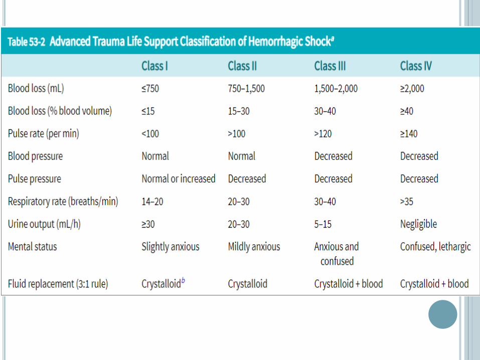
Although traditional vital signs are relatively unreliable for recognizing life-threatening shock, heart rate, systemic blood pressure, pulse pressure, respiratory rate, urine output, and mental status are still used as early clinical indicators of the severity of hemorrhagic shock.
For example, tachycardia, which is traditionally used as an index of hypovolemia, may be absent in up to 30% of hypotensive trauma patients because of activated Bezold–Jarisch reflex, increased vagal tone, chronic cocaine use, or other reasons (but inability of the patient to elevate the heart rate in the face of hypoperfusion is considered a predictor of increased mortality)
Increasing catecholamine output, tissue injury and associated pain may result in maintenance of tachycardia and normal systemic blood pressure in the presence or absence of hypovolemia without necessarily increasing the cardiac index or tissue oxygen delivery. In fact, in this situation an increase in intestinal vascular resistance and a decrease in splanchnic blood flow may occur and, if prolonged, may allow entry of intestinal microorganisms into the circulation and increase the likelihood of subsequent sepsis and organ failure .
Thus, equating a normal heart rate and systemic blood pressure with normovolemia during initial resuscitation may lead to loss of valuable time for treating underlying occult hypovolemia or hypoperfusion
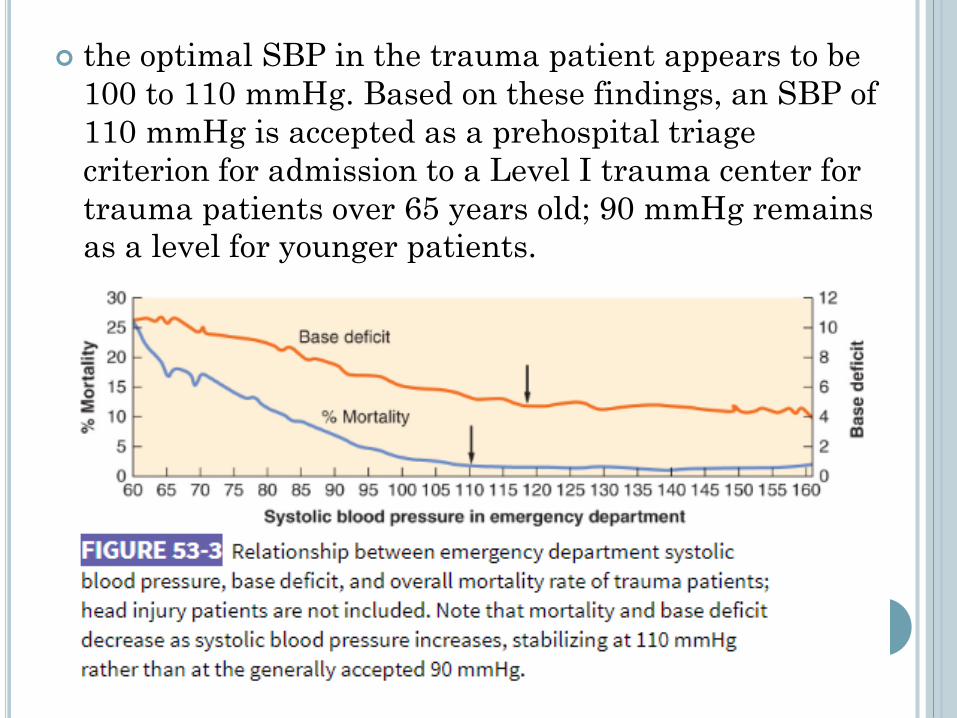
the optimal SBP in the trauma patient appears to be
100 to 110 mmHg. Based on these findings, an SBP of
110 mmHg is accepted as a prehospital triage
criterion for admission to a Level I trauma center for
trauma patients over 65 years old; 90 mmHg remains
as a level for younger patients.

PREDICTIVE MEASURES
low Hct on admission should elicit the suspicion of
significant bleeding. However, decision making based
on a single Hct value may lead to erroneous
management decisions. On the other hand, serial Hct
measurements and consideration of the type and
amount of fluid received may be useful in deciding
the timing and amount of transfusion.
Among many scoring systems the most practical is
the Assessment of Blood Consumption (ABC) score,
which asks four yes/no questions: penetrating
mechanism of injury, SBP of 90 mmHg or less, heart
rate of 120/min or greater, and a positive FAST
finding.
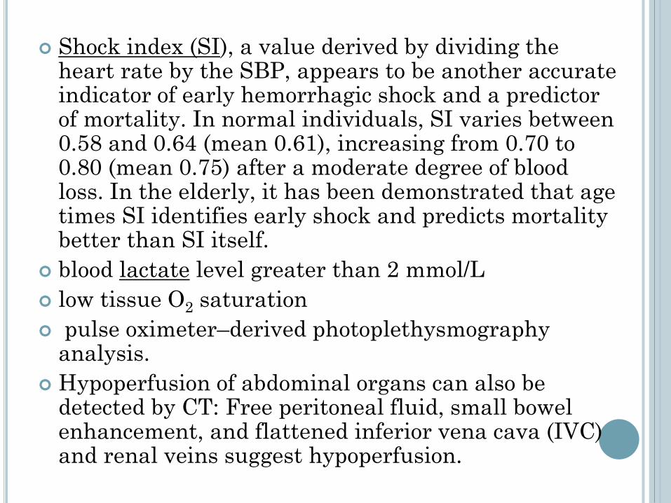
Shock index (SI), a value derived by dividing the heart rate by the SBP, appears to be another accurate indicator of early hemorrhagic shock and a predictor of mortality. In normal individuals, SI varies between 0.58 and 0.64 (mean 0.61), increasing from 0.70 to 0.80 (mean 0.75) after a moderate degree of blood loss. In the elderly, it has been demonstrated that age times SI identifies early shock and predicts mortality better than SI itself.
blood lactate level greater than 2 mmol/L
low tissue O2 saturation
pulse oximeter–derived photoplethysmographyanalysis.
Hypoperfusion of abdominal organs can also be detected by CT: Free peritoneal fluid, small bowel enhancement, and flattened inferior vena cava (IVC) and renal veins suggest hypoperfusion.

One of the reasons scoring systems have been introduced for assessing the severity of hemorrhage is to determine the need for initiating the massive transfusion protocol (MTP)
In the ABC score each question are given 1 point, and a minimum score of 1 or 2 suggests activation of the MTP.
It has been realized that relying only on these scoring systems without using the clinical gestalt is likely to lead to under- or over use of MTP. Thus these scoring systems, preferably the revised massive transfusion score, should be relied upon only in conjunction with clinical judgment.
Although the scoring systems are generally used for initial assessment in the ED, they also can be helpful in judging whether the MTP should be continued or stopped later during the process of management.
The method of resuscitation of the hemorrhaging
patient has changed over the past several years since
the Iraq and Afghanistan wars.
The concept of damage control resuscitation has
replaced the classic crystalloid resuscitation, which
served to replenish depleted interstitial fluid and also
to estimate the severity of intravascular volume
depletion during the initial period.
The response to initial fluid resuscitation with
lactated Ringer’s (LR) or normal saline solution of
about 2 L, or 20 mL/kg in children, over a period of 15
to 30 minutes allowed estimation of the severity of
hemorrhage. Transient or no blood pressure response
to this maneuver suggested major hemorrhage and
dictated administration of blood products.

DAMAGE CONTROL RESUSCITATION
Here the severity of hemorrhage is estimated using the
combination of clinical, laboratory, ultrasonographic,
and radiologic diagnostic measures described earlier.
After a major hemorrhage is identified, several
components of the process are initiated.
permissive hypotension
rapid control of any bleeding source
minimal crystalloid infusion
early administration of plasma and other blood products in
a balanced ratio (preferably 1:1:1) of packed red blood cells
(PRBCs), plasma, and platelets by activation of the MTP
tranexamic acid
The purpose of damage control resuscitation is to prevent the pulmonary edema, ARDS, coagulopathy, multiple organ failure (MOF), and abdominal compartment syndrome attributed to administration of large volumes of resuscitative crystalloids.
In addition, administration of large volumes of LR solution and normal saline are associated with elevated blood lactate levels and increasing base deficit, respectively.
Neil et al.85 found that a cumulative crystalloid to PRBC ratio greater than 1.5:1 (liters to units) during the first 24 hours after admission was an independent cause of ARDS and abdominal compartment syndrome. Using ratios of less than 0.75:1 and more than 0.75:1 in two groups of trauma patients, another study found no statistical differences in oxygenation, ARDS and mortality between the low- and high-ratio groups, although fewer people in the low-ratio group died.
Thus, it is obvious that the crystalloid volume should be kept low during initial resuscitation.
Overinfusing fluids before control of the hemorrhage
may lead to further bleeding by increasing arterial and
venous pressures, displacing a hemostatic plug, diluting
clotting factors and platelets, reducing body
temperature, and decreasing blood viscosity.
Bickell et al. showed that delaying fluid resuscitation
until surgical control of bleeding improved survival to
hospital discharge and decreased the length of hospital
stay. Feasibility of the time-sensitive permissive hypotension described by
Bickell et al. has been studied in a prospective randomized study
comparing low-volume versus standard-volume (2L) crystalloid
administration to hypotensive trauma patients during the prehospital
phase. mortality was lower in patients who received low-volume
crystalloids despite maintenance of hypotension.
Permissive hypotension is also contraindicated in
traumatic brain and spinal cord injuries and in elderly
patients with chronic systemic hypertension in which
adequate perfusion is crucial
Early use of vasopressors to maintain hemodynamic
stability also may be associated with deleterious
effects. However, judicious use of these drugs along
with carefully titrated fluids may offer some
advantages
BASE DEFICIT
The base deficit reflects the severity of shock, the oxygen
debt, changes in O2 delivery, the adequacy of fluid
resuscitation, and the likelihood of MOF and survival with
reasonable accuracy in previously healthy adult and
pediatric trauma patients.
Base deficit is considered a better prognostic marker than
the arterial pH.
base deficit between −2 and −5 mmol/L suggests mild shock
between −6 and −9 mmol/L indicates moderate shock
more than 10 m/mol is a sign of severe shock.
An admission base deficit below −5 to −8 mmol/L
correlates with increased mortality. Thus, normalization
of the base deficit is one of the end points of resuscitation.
LACTATE LEVEL
Elevation of the blood lactate level is less specific than base deficit as a marker of tissue hypoxia because it can be generated in well-oxygenated tissues and affected by hepatic clearance.Thus, blood lactate and base deficit may not closely correlate with each other. Nevertheless, in most trauma victims an elevated lactate level correlates with other signs of hypoperfusion, rendering it an important marker of dysoxia and an end point of resuscitation.
The normal plasma lactate concentration is 0.5 to 1.5 mmol/L; levels over 5 mmol/L indicate significant lactic acidosis. The half-life of lactate is approximately 15 to 30 minutes in healthy individuals; thus, the level decreases rather rapidly after correction of the cause.
Failure to clear lactate within 24 hours after reversal of circulatory shock is a predictor of increased mortality.

BLOOD TRANSFUSION
The usefulness of hemoglobin (Hgb) or Hct as a
PRBC transfusion threshold remains unclear,
although the recommended target Hgb concentration
in all phases of management is 7 to 9 g/dL
Transfusion of PRBCs has been shown to be an
independent risk factor for mortality, lung injury,
increased infection rate, renal failure, and intensive
care unit (ICU) and hospital length of stay in trauma
patients, especially when the transfused red cells are
older than 14 days; this finding was independent of
the severity of shock.
if the situation dictates immediate transfusion, type
O Rh-positive PRBCs and AB-negative fresh frozen
plasma (FFP) are satisfactory in most situations.
Controversy exists about the use of uncrossmatched
type O PRBCs because of concern about the
development of alloantibodies and allergic reactions.
Most trauma patients are hypercoagulable when
admitted to the ED and do not develop coagulopathy
when administration of hemostatic agents is delayed.
However, in the estimated 10% to 15% of patients
with severe trauma and shock who enter the hospital
in a hypocoagulable state or rapidly develop
hypocoagulation, resuscitative fluids and PRBCs
may further worsen the coagulopathy and facilitate
the vicious cycle.
In Hirshberg et al study, the prothrombin time (PT)
increased to below hemostatic levels after
replacement of 1 blood volume, fibrinogen function
would become inadequate at replacement of 1.25
blood volumes, and platelets would become
inadequate with replacement of 1.75 blood volumes.
This strongly recommend starting liquid plasma
replacement along with fluids and PRBCs as soon as
the patient arrives in the ED and continuing it
throughout surgery
Liquid plasma differs from FFP in that it is never
frozen and can be used up to 28 days after collection,
but at a cost of containing a much lower level of
hemostatic factors

FFP
most centers keep thawed plasma stored in liquid form available to be used until FFP or PF 24 is thawed, which takes about 30 to 45 minutes. PF 24 is plasma frozen within 24 hours of collection, whereas FFP is frozen within 8 hours.
One unit of FFP contains approximately 7% of the coagulation factor activity of a 70-kg man

MASSIVE BLOOD TRANSFUSION
the administration of 50% of blood volume over 24 hours.
For children the term massive hemorrhage is relatively new and is considered if transfusion volume exceeds 40 mL/kg.
The adult definitions and treatments do not apply to the pediatric population because of the differences in size, physiology, nature of injury, and demographics.
The circulating blood volume in the infant is 90 mL/kg and in children over 3 months, it is 70 mL/kg.
Children have a greater hemodynamic reserve than adults, and vital signs deteriorate only if a significant quantity (about 35% to 40%) of blood is lost.
A narrow pulse pressure is the most constant vital sign for early volume loss.
The high metabolic rate increases oxygen extraction, underscoring the importance of an adequate hemoglobin concentration.
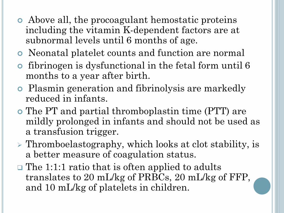
Above all, the procoagulant hemostatic proteins including the vitamin K-dependent factors are at subnormal levels until 6 months of age.
Neonatal platelet counts and function are normal
fibrinogen is dysfunctional in the fetal form until 6 months to a year after birth.
Plasmin generation and fibrinolysis are markedly reduced in infants.
The PT and partial thromboplastin time (PTT) are mildly prolonged in infants and should not be used as a transfusion trigger.
Thromboelastography, which looks at clot stability, is a better measure of coagulation status.
The 1:1:1 ratio that is often applied to adults translates to 20 mL/kg of PRBCs, 20 mL/kg of FFP, and 10 mL/kg of platelets in children.
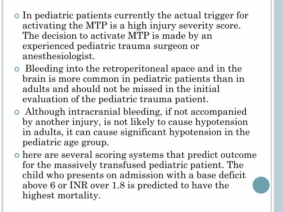
In pediatric patients currently the actual trigger for activating the MTP is a high injury severity score. The decision to activate MTP is made by an experienced pediatric trauma surgeon or anesthesiologist.
Bleeding into the retroperitoneal space and in the brain is more common in pediatric patients than in adults and should not be missed in the initial evaluation of the pediatric trauma patient.
Although intracranial bleeding, if not accompanied by another injury, is not likely to cause hypotension in adults, it can cause significant hypotension in the pediatric age group.
here are several scoring systems that predict outcome for the massively transfused pediatric patient. The child who presents on admission with a base deficit above 6 or INR over 1.8 is predicted to have the highest mortality.
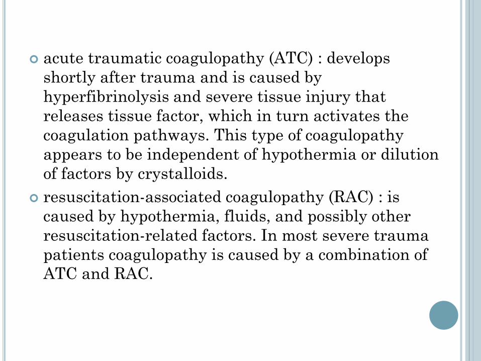
acute traumatic coagulopathy (ATC) : develops
shortly after trauma and is caused by
hyperfibrinolysis and severe tissue injury that
releases tissue factor, which in turn activates the
coagulation pathways. This type of coagulopathy
appears to be independent of hypothermia or dilution
of factors by crystalloids.
resuscitation-associated coagulopathy (RAC) : is
caused by hypothermia, fluids, and possibly other
resuscitation-related factors. In most severe trauma
patients coagulopathy is caused by a combination of
ATC and RAC.

VENOUS ACCESS
large-bore cannulae placed in peripheral veins that drain both above and below the diaphragm is essential for adequate fluid resuscitation.
When vascular collapse and extremity injury impair access to arm or leg vessels, percutaneous cannulation of the internal jugular, subclavian, or femoral veins can be performed.
Ultrasound guidance may facilitate cannulation of the internal jugular vein , infraclavicular access to the axillary vein, the cephalic or basilic veins at the midarmlevel, or the femoral vein.
If necessary, a cutdown to a saphenous or arm vein can be rapidly performed in older children and adults.
In children less than 5 years of age, intraosseouscannulation has a high success rate and a low incidence of complications. although a pressure infusion device may be necessary to achieve adequate flow
the success rate of external cardiac massage in hypovolemictrauma victims is likely to be low
ED thoracotomy (according to predictors of survival below) not only permits performance of open cardiac massage but also aids resuscitation efforts by allowing drainage of pericardial blood, control of cardiac and great vessel bleeding, and application of a cross-clamp to the aorta. A small Foley catheter introduced into the right atrium or, in desperate situations, a large-bore catheter or introducer inserted in the descending aorta can be used for rapid administration of fluids
The highest survival with or without intact neurologic function occurred after penetrating thoracic trauma presented with signs of life.
Those without signs of life on arrival had a lower rate of survival, but an ED thoracotomy could still be justified.
Patients presenting pulseless after penetrating extrathoracicinjury had more favorable outcome if they had some signs of life than those who did not.
Those patients who had blunt injury with or without signs of life had a very poor survival rate, precluding ED thoracotomy.
EARLY MANAGEMENT OF SPECIFIC
INJURIES
Head Injury
Spine and Spinal Cord Injury
Chest Injury
Abdominal and Pelvic Injuries
Extremity Injuries

HEAD INJURY
40% of deaths from trauma are caused by head
injury.
Traumatic brain injuries are categorized as
either primary or secondary .
Primary brain injuries are usually focal injuries
directly related to trauma, disrupting normal
anatomy or physiology, or both.
Of the possible secondary insults to the injured
brain, decreased oxygen delivery as a result of
hypotension and hypoxia has the greatest
detrimental impact
The most common early complications of head
trauma are intracranial hypertension, brain
herniation, seizures, neurogenic pulmonary edema,
cardiac dysrhythmias, bradycardia, systemic
hypertension, and coagulopathy.
Brain injury by itself does not cause hypotension in
adults except as a preterminal event and in
pediatric patients.
hypotension is the most important cause of death in
the head-injured patient. hesnut demonstrated that a
single episode of SBP less than 90 mmHg is associated with a
50% increase in mortality, and subsequent episodes or lower
pressures increase mortality even further
DIAGNOSIS
every effort should be made to support the blood pressure with fluids and vasopressors (preferably phenylephrine, which does not constrict cerebral vessels) and ensure adequate oxygenation before the unconscious patient is evaluated.
A baseline neurologic examination should be performed after initial resuscitation but before any sedative or muscle relaxant agents are administered, and this should be repeated at frequent intervals because the patient’s condition may change rapidly.
Anesthetic and adjunct drugs may render an adequate neurologic examination impossible; thus, long-acting muscle relaxants, opioids, sedatives, or hypnotics should be given selectively.

Consciousness can be initially assessed within a few
seconds using the AVPU system (alert; responds
to verbal stimuli; responds to pain; unresponsive)
More precise information is provided by the GCS,
which provides a standard means of evaluating the
patient’s neurologic status. its correlates with the
state of consciousness, the severity of the head
injury, and the prognosis. (motor function should be
performed on the extremity that responds best. The
limb affected by neurologic injury is examined, but
the result is not considered in the GCS)
GCS ≤8 = deep coma, severe head trauma, poor outcome
GCS 9–12 = conscious patient with moderate injury
GCS >12 = mild injury

pupillary findings (size and response)
CT scanning is used for the diagnosis of most acute
head injuries. Positive CT findings after acute head
injury include :
midline shift
distortion of the ventricles and cisterns
effacement of the sulci in the uninjured hemisphere
intracranial air
depressed skull fractures.
presence of a hematoma at any location in the cranial
vault (40% when GCS <8)
Subdural hematomas usually have a concave border,
whereas epidural hematomas present with a convex
outline classically termed a lenticular configuration

MANAGEMENT
early management of brain trauma is to prevent or
alleviate the secondary injury process that may follow any
complication that decreases the oxygen supply to the brain,
including systemic hypotension, hypoxemia, anemia,
raised ICP, acidosis, and possibly hyperglycemia (serum
glucose >200 mg/dL).
The most important therapeutic maneuvers in these
patients are aimed at normalizing ICP, CPP, and oxygen
delivery
Primary therapy includes normalization of the
systemic blood pressure (mean blood pressure >80 mmHg)
maintaining the PaO2 over 95
the ICP below 20 to 25 mmHg
the CPP at 50 to 70 mmHg. (>70 no longer advised increased
incidence of ARDS)
The patient is kept at 30-degree head elevation
sedation and paralysis are employed as necessary
cerebrospinal fluid is drained through a ventriculostomycatheter if available.
Rapid and adequate restoration of the intravascular volume with isotonic crystalloid and, if necessary, with colloid solutions (maintaining the CPP between 50 and 70 mmHg minimizing further brain swelling).
LR solution, which is slightly hypotonic (Na+ 130 mEq/L, osmolality ∼255 mOsm/L), may promote swelling in uninjured areas of the brain if it is given in large quantities.
Edema tends to occur in injured brain regions regardless of the type of solution administered because of increased permeability of the blood–brain barrier.
To minimize edema formation, it is wise to monitor serum osmolality and to replace LR solution with isotonic normal saline. If serum osmolality cannot be measured, this change can be made empirically after 3 L of LR solution.
Davis et al. found that patients with GCS no higher than 8, in
whom endotracheal intubation was attempted at the accident
site, had a higher mortality than those who were not intubated
until arrival at the emergency room . This related to physiologic
insults during intubation (elevated ICP, oxygen desaturation,
or inadvertent postintubation hyperventilation and cerebral
hypoperfusion with ischemia) and concluded that there may be
no benefit to prehospital endotracheal intubation.

Normalization of the ICP has been shown to reduce mortality. Effective reduction in ICP can be provided, or at least aided, by administration of mannitol(osmotic diuretic), an important part of the management of severe head injury. It is administered in boluses of 0.25 to 0.5 g/kg and repeated every 4 to 6 hours as needed to control the ICP.
used with great care in the presence of hypotension, sepsis, nephrotoxic drugs, or pre-existing renal disease, because these may also precipitate renal failure.
Can exacerbate edema in injured areas in which it may easily enter the tissues
Because of a synergistic action between mannitol and loop diuretics in improving the ICP, addition of furosemide may be a safer and more effective treatment than increasing the dose of mannitol when intracranial hypertension persists
Relatively small volumes of hypertonic saline in
concentrations between 3% (6 to 8 mL/kg) and 7.5%
(4 mL/kg) followed by infusion of LR may be
beneficial in multiple-trauma patients with head
injury
It draws fluid from the intracellular space
deacreasing edema
Also like mannitol, increase edema in the injured
region of the brain
Serum concentrations of sodium (Na+) and Cl− and
the patient’s acid–base status should be followed
Resuscitation with albumin 5% or 25% provides a sustained improvement in vital signs, but the increase in colloid osmotic pressure produced by these solutions may be associated with an increased risk of mortality.
hyperventilation to a PaCO2 of 25 to 30 mmHg was a mainstay of the therapy of head injury.
But studies showed that a significant increase in the region of critical hypoperfusion may result from hyperventilation. This hypoperfusion seems to be caused largely by increased cerebral vascular resistance, which may be enhanced by hyperventilation.
However, some degree of hyperventilation may be necessary for short periods of time in patients who have severe injuries and elevated ICP that does not respond to normal ventilation and diuretics although this should not be used during the first 24 hours following injury.
It should be noted that hyperventilation in the severely brain-injured patient may also be associated with acute lung injury.
If the ICP remains elevated despite all of these
measures, pentobarbital (3 to 10 mg/kg given over 0.5
to 2.5 hours, followed by a maintenance infusion of 0.5
to 3.0 mg/kg/hr, aimed at a serum concentration
between 2.5 and 4.0 mg/dL) may be required.
High-dose barbiturates are of no value in the routine
therapy of head injury and should be used only for
refractory ICP elevation.
Of course, immediate surgical decompression,
especially of epidural hematomas, is an important
factor in reducing morbidity and mortality
In brain-injured patients , the brain metabolism is
altered by the injury and is heavily dependent on
glucose.
Hypoglycemia (<40 g/dL) may cause metabolic crisis
whereas hyperglycemia (>200 g/dL) can cause
detrimental effects through excitotoxicity, oxidative
stress, and inflammatory cytokine release. However,
tight insulin control therapy (80 to 110 mg/dL) has
been associated with episodes of hypoglycemia.
As a result, the current recommendations are to
maintain glucose levels of 110 to 180 mg/dL
Nearly 75% of severely brain-injured patients who
die expire within the first 3 days following the
initial trauma.
Many of the survivors will later succumb to
nonneurologic organ dysfunction involving
pulmonary failure and cardiac impairment, which
may be related to sympathetic hyperactivity.
β-Blocker therapy has been proposed as a
treatment that may be beneficial in these patients.
The optimal agent, the dose level, the timing, and
the duration of treatment, however, remain to be
determined.
CONCLUSION
If the patient is hemodynamically stable, a CT scan is performed. The strictest attention should be paid to ensure adequate oxygenation, ventilation, blood pressure, and ICP control during the procedure.
If the patient is hemodynamically unstable or requires emergency surgery for associated injuries and has a history suggesting a head injury, even though a significant intracranial hematoma is unlikely on clinical grounds, intraoperative ICP monitoring is indicated to permit rapid detection of ICP elevation.
Both intracranial hematomas and hemorrhage in other regions have a high surgical priority.
Because there is no time for a CT scan of the head in patients with both profuse hemorrhage and brain herniation, the patient is brought directly to the OR for simultaneous control of the bleeding site and evacuation of the intracranial hematoma. The site of the craniotomy can be determined by a ventriculogram or an ultrasound examination with a pencil-tip probe; both tests may be performed under local anesthesia through a frontal burr hole.
ANESTHETIC MANAGEMENT
INTRAOPERATIVE
It’s a continuation of the pre-existing intensive care. maintain the blood pressure, oxygenation, and CPP.
there have been no studies comparing intravenous to inhalation techniques. preserving the vital signs is more important than the specific means.
arterial line will permit beat-to-beat monitoring of the blood pressure, along with following blood gas levels and blood glucose.
ICP monitor will generally be placed by the neurosurgeons.
Vital signs, PaO2, and PaCO2 should be maintained at the same levels as they are before the patient reaches the OR.
Preoperative fluid management (plus blood if needed) is also continued during surgery

IMPROVE THE OUTLOOK FOR BRAIN-
INJURED PATIENTS
The earlier definitive treatment is initiated, the better the outcome is likely to be. Rudehill et al. have demonstrated improvement in outcomes in a large series of patients when care was initiated by anesthesiologists at the accident scene. On the other hand, Haltmeier et al. found no difference between the outcomes for traumatic brain injury patients treated onsite by anesthesiologists in Bern or paramedics in US.
Meanwhile, the wide variety of types and severities of injury and of responses to treatment—both among different patients and in the same patient at different times—imply that therapeutic interventions must be individualized. These aims may be met, at least partly, by carefully structured intensive care.Therapeutic goals should be set explicitly and reviewed, and altered if necessary, at every change of shift.
SPINE AND SPINAL CORD INJURY
The objective in the evaluation of spinal trauma is to diagnose instability of the spine and the extent of neurologic involvement.
until a definitive diagnosis is established there is a risk of converting a neurologically intact patient into a paraplegic or quadriplegic.
During transport to the hospital, the patient should be immobilized with a hard collar, a spine board, and tape.
After admission, patients should not be left on a rigid spine board for longer than 1 hour, especially when they are paralyzed, because of the risk of decubitus ulcers
Diagnosis done as mentiond by the history of the
truama , clinical symptoms and signs , then a
multislice helical CT imaging is sufficient to detect
unstable cervical spine injuries
spinal pain is not always localized to the level of
injury
in the comatose patient, flaccid areflexia, loss of
rectal sphincter tone, paradoxic respiration, and
bradycardia in a hypovolemic patient suggest the
diagnosis
In cervical spine trauma, an ability to flex but not to
extend the elbow and response to painful stimuli
above but not below the clavicle also indicate
neurologic injury
Depending on the degree of deficit, spinal cord injuries are categorized as complete or incomplete.
Intact sensory perception over the sacral distribution and voluntary contraction of the anus (sacral sparing) are present in incomplete, but not in complete, injuries.
There is practically no possibility of significant neurologic recovery in complete injury, whereas functional restoration may occur in up to 50% of patients after incomplete injuries.
In some patients the development of spinal shock, which is manifested by absolute flaccidity and loss of reflexes, precludes distinguishing between complete and incomplete injuries during the initial phase of treatment.
Spinal shock is a misnomer for neurogenic shock defined as hypotension and bradycardia caused by the loss of vasomotor tone and sympathetic innervation of the heart as a result of functional depression of the descending sympathetic pathways of the spinal cord. It is usually present after high thoracic and cervical spine injuries and improves within 3 to 5 days.

Therefore, even in the absence of sacral sparing, the
possibility of neurologic recovery dictates that all possible
efforts be made at this time to prevent further damage
and to preserve cord function.
A similar principle applies to the evaluation of the level of
injury. After the first few days, spinal cord edema
subsides, and the final injury level is commonly a few
segments lower than on initial presentation.
Thus, early therapeutic efforts should not be abandoned
even in the patient with a high-level injury, which carries
a grim functional prognosis.
The spinal cord, is also vulnerable to a secondary injury
process that may be a product of hypotension, hypoxia,
and probably other physiologic complications
MANAGEMENT
Maintenance of immobilization (If a cervical spine fracture is suspected, immobilization or MILS of the neck. If the patient has a thoracic or lumbar injury, a careful logrolling maneuver should be used).
Intubation (respiratory distress or fatigue, or a rising respiratory rate or PaCO2, are major indications for ventilatory assistance)
Severe bradycardia or dysrhythmias may result from unopposed vagal activity during tracheal intubation or suctioning: The patient must be preoxygenated, and atropine (0.4 to 0.6 mg) should be given before any instrumentation. If bradycardia develops during airway management, treatment includes additional atropine, glycopyrrolate, isoproterenol, or, if necessary, cardiac pacing.

RESPIRATORY COMPLICATIONS
associated brain, neck, chest, or abdominal injury; alcohol intoxication; or the effects of self-administered or iatrogenic drugs
Injuries at C5 or lower are usually associated with normal tidal volumes because the function of the diaphragm is intact, whereas patients with injuries at C4 or above may require permanent ventilatoryassistance.
Nevertheless, accessory respiratory muscle paresis may cause a significant loss of expiratory reserve even when the injury involves the lower spinal segments
Pulmonary edema (severe catecholamine surge follows acute trauma to the spinal cord / severe hypertension / pulmonary capillary damage / left ventricular dysfunction)
Paradoxic respiration in the quadriplegic patient results from partial chest wall collapse during inspiration. It may produce limitation of the tidal volume and an increased risk of hypoventilation , this situation is aggravated when the patient is in an upright position (weight of the thoracic contents is not opposed by the normal tone of the abdominal muscles).
Thus, in contrast to other diseases that produce respiratory insufficiency, the supine position improves respiration in persons with quadriplegia
aspiration of gastric contents
Atelectasis\
Pneumonia
bronchoconstriction
HEMODYNAMIC MANAGEMENT
Insertion of a central venous or pulmonary artery catheter (PAC) if necessary, as early as possible after injury. In as many as 25% of patients with cervical spinal cord injuries, left ventricular dysfunction may contribute to the hypotension.
maintenance of mean arterial pressure above 85 mmHg.
Decreased preload can be treated with fluid infusion using cardiac function curves as a guide. volume may be safely replaced to a central venous or (PCWP) of 18 mmHg.(avoids, or at least limits, the severity of the pulmonary edema).
Hypotension, despite adequate fluid infusion, acidosis, or low mixed venous PO2, requires treatment with inotropes such as dopamine.
initiation of low–molecular-weight or low-dose unfractionatedheparin therapy, combined with a rotating bed, compression stockings, or electrical stimulation, within 72 hours of the injury. This therapy should be held on the day of any surgical procedure (pt at risk of DVT)

CHEST INJURY
Chest Wall Injury
Rib, scapula, and sternal fractures (interfering
with adequate respiration)
The management principles for these injuries are
similar to those previously described for flail
chest, although the need for mechanical
ventilation is less likely in single rib fractures
treated with systemic analgesics than in a flail
chest.
Effective pain relief, preferably with continuous
thoracic epidural anesthesia or paravertebral or
intercostal block, is central to management.
Pleural Injury
Occult pneumothorax is easy to miss in major trauma.
The presence of subcutaneous emphysema, pulmonary
contusion, and rib fractures should raise suspicion of
coexisting pneumothorax.
Tension pneumothorax involving over 50% of a
hemithorax presents with dyspnea, tachycardia, cyanosis,
agitation, diaphoresis, neck vein distention, tracheal
deviation, and displacement of the maximal cardiac
impulse to the contralateral side
upright plain chest radiograph provides the best
opportunity for detection of pneumothorax (impossible or
contraindicated)
Transthoracic ultrasound by positioning the ultrasound
probe longitudinally over the intercostal space may be
used for the emergency diagnosis of pneumothorax
Ultrasound examination may also be helpful in
detecting residual pleural air after placement of the
thoracostomy tube and diagnosis of pulmonary
embolism (PE), pneumonia, and hemothorax.
Chest CT is the definitive test for diagnosis of
pneumothorax.
a small closed pneumothorax can be safely managed
by observation alone , even in those patients who
require positive-pressure ventilation
most recent Advanced Trauma Life Support
recommendation strongly believe that a traumatic
pneumothorax, no matter how small, should be
treated with thoracostomy drainage before tracheal
intubation and positive-pressure ventilation.
Bleeding intercostal vessels are responsible for most
hemothoraces.
Severe airway deviation with respiratory distress and
shock may be produced by a hemothorax
Treatment consists of drainage , Initial drainage of
1,000 mL of blood or collection of over 200 mL/hr for
several hours is an indication for thoracotomy.
Retained clotted blood after tube thoracostomy may
be treated conservatively with intrapleural
fibrinolytic agents
Hemodynamically stable patients with persistent
bleeding of less than 150 mL/hr are often managed
with video-assisted thoracoscopic surgery (VATS) to
control bleeding (using a double-lumen tube or a
bronchial blocker)
Penetrating Cardiac Injury
Pericardial tamponade, cardiac chamber perforation,
and fistula formation between the cardiac chambers
and the great vessels are the consequences of a
penetrating cardiac trauma.
Any penetrating wound of the chest, especially one
within the “cardiac window” (midclavicular lines
laterally, clavicles superiorly, and costal margins
inferiorly), can cause this injury.
These injuries are often fatal at the scene, especially
if they are gunshot rather than stab wounds and
involve the right rather than the thicker-walled left
ventricle
transported directly to the OR, and immediate
sternotomy or left thoracotomy as soon as possible
Emergency cardiopulmonary bypass

TTE can be used for screening stable patients, but it
may be inconclusive in obese patients and in those
with pneumothorax (TEE provides an accurate
diagnosis in these patients, but it is impractical
during the initial evaluation phase of trauma)
ubxiphoid pericardial window created in the OR,
often under general anesthesia, may not drain all the
blood in the pericardium, but even partial drainage
can improve hemodynamics temporarily in this
setting
Pericardial Tamponade
Both penetrating and blunt trauma can cause hemopericardium.
The classic findings of pericardial tamponade—tachycardia, hypotension, distant heart sounds, distended neck veins, pulsus paradoxus, or pulsusalternans—are difficult to appreciate or may be absent in a hypovolemic trauma patient.
A chest radiograph may reveal a globular heart, although this sign is often not appreciated.
TTE with placement of the probe in the subxiphoidregion, which is part of FAST, or intraoperative TEE can demonstrate blood in the pericardial sac and the presence of ventricular diastolic collapse, which indicates at least a 20% reduction in cardiac output.
FAST ( FOCUSED ASSESSMENT WITH
SONOGRAPHY FOR TRAUMA)
FAST requires one-third of the time, is less
expensive to perform than CT, and is without the
hazard of radiation.
Screening with abdominal ultrasonography is
performed by placing a 3.0- to 5.0-MHz probe on
four distinct areas of the abdomen: subxiphoid, to
detect pericardial blood; right upper quadrant,
for blood in the hepatorenal pouch; left upper
quadrant, to detect perisplenic blood; and just
above the pubic symphysis, for blood in the
rectovesical pouch.

FRACTURES OF THE PELVIS
Pelvic fractures occur in widely varied anatomic forms and
physiologic severity. Major hemorrhage, which is one of the major
causes of mortality, occurs in about 25% of patients;
exsanguination occurs in 1% of injuries.
In most of these fractures bleeding results from venous disruption
by fragments of bone. Retroperitoneal pelvic bleeding is self-
limited in most patients with venous injuries because of the
tamponading effect, except in those with open fractures.
Approximately 18% to 20% of patients have arterial bleeding that
does not stop.
Early detection and intervention to control bleeding are
important. Pelvic ring disruption, arterial extravasation (CT
blush), and elevated bladder pressure secondary to compression
by hematoma volumes greater than 500 mL are important signs
that can be detected on CT examination, making it akey
diagnostic measure.

EXTREMITY INJURIES
Surgical repair of extremity fractures, whether open or closed,
should be performed as soon as possible. Delayed fracture repair
is associated with an increased risk of DVT, pneumonia, sepsis,
and the pulmonary and cerebral complications of fat embolism. In
open fractures, an additional important concern is infection.
Wounds left unrepaired for more than 6 hours are likely to
become septic.
Associated vascular trauma must be recognized early. Most
vascular injuries exhibit at least some part of the classic
syndrome of pain, pulselessness,pallor, paresthesias, and paresis.
COMPARTMENT SYNDROME
Characterized by severe pain in the affected extremity,
Should be recognized early so that emergency fasciotomy can be effective in preventing irreversible muscle and nerve damage.
In unconscious patients, swelling and tenseness of the extremity indicate the presence of this complication.
The definitive diagnosis is made by measuring compartment pressures using a transducer attached to a fluid-filled extension tube and a needle inserted into the various compartments of the extremity.
pressure exceeding 30 cm of H2O is an indication for immediate surgery.
Caution must be exercised when using epidural or nerve block analgesia for perioperative pain relief in the presence of extremity fractures. Absence of pain can delay the diagnosis of compartment syndrome.

OPERATIVE MANAGEMENT
MONITORING
Hemodynamic Monitoring
Direct intra-arterial pressure monitoring, which permits beat-to-
beat data acquisition and sampling for measurement of blood gases,
should be in place before surgery.
The right radial artery is preferred in cases of chest trauma in which
cross-clamping of the descending aorta might result in occlusion of the
left subclavian artery.
Several devices, such as the PiCCO, LiDCO and CO monitor are
able to display systolic blood pressure variation, PPV, and SVV, which
appear to predict responsiveness to fluid administration with greater
accuracy than static markers of preload such as CVP, pulmonary
artery occlusion pressure.
Threshold values to discriminate responders from nonresponders to
fluid infusion have been determined (PPV or SVV >12% for
responders)

MONITORING
Hemodynamic Monitoring
Delaying emergent surgery to place a central venous line is rarely indicated unless a large-bore catheter is needed for volume resuscitation.
However, if the patient is elderly, if there is a likelihood of myocardial damage, or if there is multiple organ damage with a requirement for anticipated prolonged surgery, massive fluid replacement, and administration of vasoactive drugs, early placement of a CVP or PAC may be indicated before the development of coagulopathy renders it hazardous.
The TEE provides valuable diagnostic information in BCI, cardiac septal or valvular damage, coronary artery injury, pericardial tamponade, and aortic rupture. It also permits assessment of cardiac function, including right and left ventricular volume, EF, wall motion abnormalities, pulmonary hypertension, and cardiac output, and detects acute ischemia more accurately than either ECG or
pulmonary artery pressure monitoring.
MONITORING
Urine Output
Urine output is routinely monitored as an indicator of
organ perfusion, hemolysis, skeletal muscle destruction,
and urinary tract integrity after trauma.
Dark, cola-colored urine in the trauma patient suggests
either hemoglobinuria resulting from incompatible blood
transfusion or myoglobinuria caused by massive skeletal
muscle destruction after blunt or electrical trauma.
Red-colored urine usually is caused by hematuria, which,
in the traumatized patient, suggests urinary tract injury.

MONITORING
Oxygenation
Trauma patients frequently develop hypoxemia (O2
saturation <90%), hypothermia, hypotension,
and/or decreased peripheral perfusion. Of the
available O2 saturation (SpO2) devices, finger or
earlobe pulse oximeters are more affected by
decreased perfusion than forehead probes, probably
because the latter senses the pulsation of the
supraorbital artery, a branch of thecarotid artery,
which is presumably less affected by shock or
hypothermia.
MONITORING
Organ Perfusion and Oxygen Utilization
Base deficit and blood lactate level, are considered
acceptable markers of organ hypoperfusion in the
apparently resuscitated patient and may be used
intraoperatively to set the optimal end points of
resuscitation.
Another parameter that may provide information
about the global perfusion of the body is the arterial to
end-tidal CO2 difference. Values greater than 10
mmHg after resuscitation predict mortality.

MONITORING
Organ Perfusion and Oxygen Utilization
Oxygen delivery (DO2), O2 consumption (VO2), O2
extraction ratio and the DO2 index (DO2I) which is a
particularly useful end point because it integrates three
important variables: Hgb concentration, arterial oxygen
saturation, and cardiac output. The minimum acceptable
value for this marker is 500 mL/min/m2.
Central venous, instead of pulmonary artery, monitoring
with CVP above 10 mmHg, mean systemic blood pressure
of 65 mmHg, and Hgb over 10 g/dL as threshold values also
suggests adequate organ perfusion.
MONITORING
Coagulation:
Conventional blood coagulation monitoring includes a baseline and subsequent serial measurements of INR, activated partial thromboplastin time (aPTT), platelet count, blood fibrinogen level, and fibrin degradation products (FDPs).
Thromboelastography and rotation transmission electron microscopy are point-of-care devices that provide a relatively rapid, comprehensive, and quantitative graphic evaluation of clotting function.
The TEG determines the time necessary for initial fibrin formation, the rapidity of fibrin deposition, the clot consistency, the rate of clot formation, and the times required for clot retraction and lysis
ANESTHETIC AND ADJUNCT
DRUGS
Airway compromise, Hypovolemia, Head or open eye
injuries, cardiac injury

AIRWAY COMPROMISE
Anesthetics and muscle relaxants should be avoided before the airway is secured.
If time permits, lateral neck radiographs, CT scanning, and endoscopy can be used to define the problems better.
Topical anesthesia with mild sedation can be used to secure the airway with a conventional blade, videolaryngoscope, or FOB.
If a rapid-sequence induction is contemplated,ketamine and etomidate may confer advantages over propofol.
Succinylcholine, with its short onset time and duration, is still the muscle relaxant of choice for rapid-sequence induction, rocuronium (1.2 to 1.5 mg/kg) has almost the same onset time and does not have the undesirable side effects associated with succinylcholine (e.g., increased intragastric, intraocular, and intracranial pressures and potassium release in patients with burns and neurologic diseases).
Surgical standby for cricothyroidotomy should be considered if failure of these techniques is anticipated. Bradycardia , dysrhythmias, and cardiac arrest may occur after succinylcholine in the presence of hypoxia and hypercarbia.
HYPOVOLEMIA
First, anesthetic agents not only have direct
cardiovascular depressant effects but also inhibit
compensatory hemodynamic mechanisms such as
central catecholamine output and baroreflex
(neuroregulatory) mechanisms, which maintain
systemic pressure in hypovolemia.
Second, hemorrhage and hypovolemia alter the
pharmacokinetics and pharmacodynamics of
almost all anesthetic agents and often lead to a
higher than normal blood concentration of
intravenous agents and increased sensitivity of
the brain and heart.
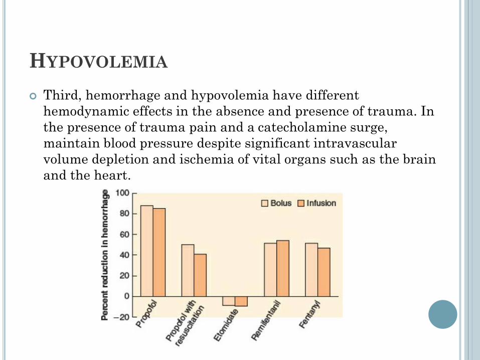
HYPOVOLEMIA
Third, hemorrhage and hypovolemia have different
hemodynamic effects in the absence and presence of trauma. In
the presence of trauma pain and a catecholamine surge,
maintain blood pressure despite significant intravascular
volume depletion and ischemia of vital organs such as the brain
and the heart.
HYPOVOLEMIA
Two important principles in the use of anesthetic agents are
accurate estimation of the degree of hypovolemia and reduction
of doses accordingly.
The presence of hypotension suggest uncompensated
hypovolemia, in which case anesthetics almost invariably
produce further deterioration of systemic blood pressure and
sometimes cardiac standstill. Intravascular volume, to the
extent possible, must be restored before their use.
Ketamine and etomidate are the preferred induction agents ,
though at low doses other intravenous anesthetics are also
unlikely to produce hypotension. Therefore, the use of any of
these drugs in reduced doses is probably more important than
the particular agent chosen.
Maintenance of anesthesia in the hypovolemic trauma patient
raises concerns similar to those pertaining to induction.
Experimental data have shown that depending on its severity,
hemorrhagic shock decreases minimum alveolar concentration
(MAC) by approximately 25%.
HEAD AND OPEN EYE INJURIES
Anesthetic agents selected for management of brain injury should
produce the least increase in ICP, the least decrease in mean
arterial pressure, and the greatest reduction in cerebral metabolic
rate (CMRO2).
Utmost attention should be paid during anesthesia to avoidance of
hypotension (mean arterial pressure <60 to 70 mmHg or SBP <90
to 100 mmHg).
All intravenous anesthetics including ketamine cause comparable
degrees of cerebrovascular constriction and ICP reduction.
Administration of succinylcholine should follow pretreatment doses
of nondepolarizing agents to prevent fasciculation-induced
elevation of ICP and IOP.
Avoiding succinylcholine usually does not alleviate the problem
because laryngoscopy and tracheal intubation produce a greater
and longer-lasting increase in these pressures. Rocuronium, 1.2 to
1.5 mg/kg, has an onset time comparable with that of
succinylcholine.

HEAD AND OPEN EYE INJURIES
All inhalation anesthetics may increase CBF and
cerebral blood volume, and thus the ICP. Cerebral
autoregulation, CO2 responsiveness, and CMRO2 are
reduced.
In hyperventilated patients with cerebral tumors or
mild edema, isoflurane does not raise the IC if it is
administered at an inspired concentration below 1
MAC.
CARDIAC INJURY
If there is pericardial tamponade, preload and
myocardial contractility must be maintained. Any
decrease in these parameters may exacerbate an
already existing right ventricle (RV) inflow obstruction.
ketamine supports the cardiac index better than other
intravenous agents thus it remains the agent of choice.
It should be given in small doses after adequate fluid
infusion.
Similar principles apply to the use of maintenance
agents, which should be given in the smallest possible
doses until the heart is decompressed.

MANAGEMENT OF INTRAOPERATIVE
COMPLICATIONS
PERSISTENT HYPOTENSION
Persistent hypotension following trauma is usually the
result of one of four mechanisms: bleeding, tension
pneumothorax, neurogenic shock, or cardiac injury.
Although many other causes, such as citrate intoxication
(hypocalcemia), hypothermia, coronary artery disease,
allergic reactions, or incompatible transfusion.
Hypotension is most likely due to bleeding, management
includes early diagnosis and control of the bleeding site
plus effective fluid resuscitation with a rapid-infusion
system, which should be connected to a 14-gauge or larger
cannula, preferably inserted into veins both above and
below the diaphragm.
PERSISTENT HYPOTENSION
Of the isotonic crystalloid solutions, LR is preferred over
normal saline. Resuscitation with normal saline during
uncontrolled hemorrhage is associated with greater urine
output and thus greater fluid requirement compared with
LR, hyperchloremic acidosis, and dilutional coagulopathy.
Acidosis does not occur with LR, but tissue edema may
result from its slight hypotonicity (∼255 mOsm/L), and
neutralization of the citrate anticoagulant in PRBCs may
occur because of its Ca2+ content.

HYPOTHERMIA
Shock, alcohol intoxication, exposure to cold, fluid resuscitation,
and abnormalities in thermoregulatory mechanisms render the
major trauma patient hypothermic during the initial phase of
injury. A core body temperature below 35°C is often associated with
acidosis, hypotension, and coagulopathy, which in turn may lead to
an increased risk of severe bleeding, need for transfusion, and
mortality.
Severe hypothermia, which in the trauma patient is defined as
core temperature below 32°C,273 was associated with a 100%
mortality rate in one study,274 although survival of a few patients
with admission temperatures even lower than 32°C has been
reported.

HYPOTHERMIA
Other effects of hypothermia are cardiac depression, myocardial ischemia, arrhythmias, peripheral vasoconstriction, impaired tissue oxygen delivery, elevated oxygen consumption during rewarming, blunted response to catecholamines, increased blood viscosity, metabolic acidosis, abnormalities of K+ and Ca2+ homeostasis, reduced drug clearance, and increased risk of infection.
Rewarming after hypothermia, especially at a rapid rate, may release accumulated metabolic products into the central circulation, causing further myocardial depression, hypotension, and increased acidosis.
Prevention of hypothermia and restoration of normal body temperature appear to decrease mortality rate, blood loss, fluid requirement, organ failure, and length of ICU stay.
HYPOTHERMIA
Warming methods :
Convective warming with forced dry air at 43°C
Circulating-water warmers may produce faster
rewarming even though they cover a relatively
smaller body surface area than forced air
warmers.
Airway warming can reduce the heat loss caused
by the latent heat of vaporization, but this
technique also transfers very little heat.

COAGULATION ABNORMALITIES
TAC ( Trauma associated coagulopathy) develops shortly after trauma from tissue injury and hypoperfusion; this activates thrombomodulin from the endothelial cells and the thrombomodulin–thrombin complex, which in turn activates protein C inhibiting factors V and VIII.
RAC (Resuscitation associated coagulopathy ) develops later and results from dilution of coagulation factors and platelets; tissue hypoperfusion; disturbance of fibrinogen/fibrin polymerization and platelet activity caused by decreased serum ionized Ca++ from infusion of colloids or binding to citrate in PRBCs, FFP, and platelet units281; hypoxia, hypothermia and acidosis; and disseminated intravascular coagulation (DIC).
COAGULATION ABNORMALITIES
DIC results from acute release of thromboplastin from
injured brain, fat, amniotic fluid, or other sources or
subacutely from endothelial inflammation or failure
interfering with clearance of activated coagulation
factors, causing microthrombi and consumption
coagulopathy.
Fibrinolysis may develop by depleted plasminogen
activator inhibitor-1 (PA-1), which accelerates the
formation of plasmin. Normally thrombin facilitates
the conversion of fibrinogen to fibrin. In severe
trauma, thrombin binds to thrombomodulin, which
slows or reduces the activation of thrombin-activated
fibrinolysis inhibitor, leading to hyperfibrinolysis.
DIAGNOSIS OF COAGULOPATHY
INR, TEG, and thromboelastometry.
The presence of elevated circulating FDP,
especially when above 40 mg/mL, is suggestive of
DIC, but the result of this study will reach the
clinician long after the completion of initial
resuscitation.
A fibrinogen level below 100 mg/dL is also
suggestive of DIC, but reduction to this value
often takes a long time, decreasing the diagnostic
value of the test, although serial measurements
may be useful.
TREATMENT :
Red Blood Cell Transfusion and Hemostasis:
Red blood cell administration should continue at least to a Hct of 30.
The optimal ratio of PRBCs:FFP:platelet during transfusion of the massively bleeding patient appears to be between 1:1:1 and 2:1:1.
Fresh Frozen Plasma:
FFP contains most of the components of the coagulation cascade and fibrinolytic and complement systems, proteins that enhance oncotic pressure and modulate immunity, and fats, carbohydrates and minerals in similar concentrations to those in blood.
Fresh Frozen Plasma:
Newer guidelines recommend administration of
thawed AB Rh- FFP immediately after the arrival
of severely traumatized, bleeding, coagulopathic
patients.
The recommended dose is 10 to 15 mL/kg, but
additional doses may be needed.
The risks associated with FFP include circulatory
overload, ABO incompatibility, transmission of
infectious diseases, mild allergic reactions, and
transfusion-related acute lung injury, in which
platelet concentrates are also implicated.
Platelets:
Platelet transfusion is indicated when the platelet count
falls below 50 ×109/L.
It is possible that in patients with DIC or hyperfibrinolysis
and in those with head injury and massive bleeding,
higher levels (75 or 100 × 109/L) may be beneficial.
High platelet to PRBC ratios (1:1 or 1:2) appear to
decrease mortality in trauma patients.
From each unit of whole blood, platelet concentrates of 7.5
× 1010/L can be prepared, which increase platelet count by
5 to 10 × 109/L; 4 to 8 U are usually sufficient.
Cryoprecipitate:
Cryoprecipitate is produced by slowly thawing
FFP at 4°C. This process causes the
“cryoproteins” to precipitate out.
Cryoproteins include factor VIII, fibrinogen, von
Willebrand factor, fibronectin, and factor XIII.
Fibrinogen levels are commonly low upon arrival
to the trauma emergency room and are
effectively replaced with cryoprecipitate during
the early phase of trauma management.
Antifibrinolytic Agents:
Tranexamic acid and Aminocaproic acid, competitive inhibitors of plasmin and plasminogen, are effective in reducing bleeding in cardiac and elective surgery.
Antifibrinolytics, especially tranexamic acid, should be considered in patients who demonstrate fibrinolysis during serial thromboelastographic or thromboelastometric monitoring.
Currently most trauma centers are using tranexamic acid routinely during the initial resuscitation with continuation into the intraoperative phase.
Tranexamic acid given within 3 hours of injury (1 g in a 10-minute bolus and then 1 g infused over the next 8 hours) decreased mortality from hemorrhage.

ACID–BASE DISTURBANCES
Metabolic acidosis is caused by shock in most trauma patients. Other rare causes of acidosis in this population are alcoholic lactic acidosis, alcoholic ketoacidosis, diabetic ketoacidosis, and CO or CN− poisoning after inhalation injuries.
The differential diagnosis between hypovolemic, diabetic, and alcoholic acidosis, all of which have anion gaps, requires measurement of blood lactate, urinary ketone bodies, and blood sugar and assessment of intravascular volume.
Alcoholic ketoacidosis is treated with intravenous dextrose, whereas diabetic ketoacidosis is managed with insulin.
No specific treatment except intravenous normal saline exists for alcoholic lactic acidosis.
DAMAGE CONTROL RESUSCITATION
Damage control principle has reduced not only the intraoperative but also the overall mortality from trauma surgery.
The damage control principle, originally proposed for abdominal trauma, is now applied to injuries at other anatomic sites, including chest, pelvis, extremities, and soft tissues
First phase :
ED attention to recognize the pattern of injury, control of bleeding if possible, and rapid transport to the OR,
The decision to initiate damage control resuscitation by limiting crystalloid infusion, allowing permissive hypotension, activating rewarming, and initiating blood component replacement with early administration of FFP and platelets at high ratios with PRBCs
The second phase occurs in the OR where, in addition to efforts to maintain the patient’s intravascular volume, temperature, acid–base status, and coagulation at near-normal levels with damage control resuscitation, the surgeons rapidly control bleeding and leave the abdominal cavity open without fascial closure but temporarily covered by a vacuum pack dressing, which allows an enlarged space for edematous organs and controlled egress of fluid.
The third phase takes place in the ICU where intravascular volume, hypothermia, acidosis, and coagulation abnormalities are corrected.
The fourth phase, the stabilized patient is returned to the OR for definitive surgery and abdominal closure. The fourth phase involves multiple returns to the OR at 24- to 48-hour intervals for organ repair, abdominal washout, and debridement before the final closure of the abdomen.
DAMAGE CONTROL RESUSCITATION
EARLY POSTOPERATIVE
CONSIDERATIONS
Major trauma patients should be admitted
directly to the ICU.
Ventilatory Support:
If mechanical ventilation is needed considerVT
no more than 6 mL/kg; an appropriate level of
PEEP, allowing titration of FiO2 to lowest
possible level, plateau airway pressures below 35
cm H2O, and avoidance of auto-PEEP with
consideration of adjustment to hypo- or
hypercapnia; and prevention of ventilator-
induced pneumonia should be undertaken.
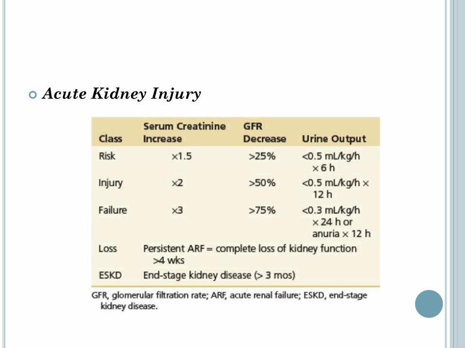
Acute Kidney Injury
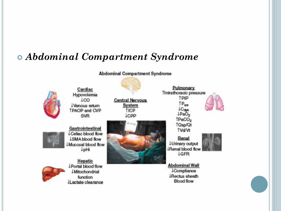
Abdominal Compartment Syndrome
Thromboembolism:
The overall incidence of VTE involving DVT and PE is 3.2% in blunt trauma patients. However, DVT occurs in 30% of major lower extremity injuries, 30% of spine injuries, 46% of major head injuries, 33% of major thoracic injuries, and 15% of serious injuries of the face or abdomen.
Diagnosis can be made by duplex ultrasonography, but this method has low sensitivity in the absence of symptoms. Venography, which is the gold standard, can be performed in equivocal cases.
Hypoxemia, especially when sudden and associated with dyspnea and hemodynamic abnormalities, even very early after injury, is highly suggestive of PE. The definitive diagnosis is established by spiral CT and pulmonary angiography.

THANK YOU