Neuro Anesthesia for Trauma Patients - c.ymcdn.comc.ymcdn.com/sites/€¦ · The Brain Trauma...
74
Neuro Anesthesia for Traumatic Brain Injury A Review of The Basics Michele Kolowitz CRNA, MHS October 2014
Transcript of Neuro Anesthesia for Trauma Patients - c.ymcdn.comc.ymcdn.com/sites/€¦ · The Brain Trauma...

Neuro Anesthesia
for
Traumatic Brain
Injury
A Review of The Basics
Michele Kolowitz CRNA, MHS
October 2014

Introduction CDC
• 1.7 Million sustained TBI • 52,000 Deaths • 275,000 Hospitalizations • 80% treated & released from ER (~1.3 million) • Those ≥ 75 highest rates of TBI-related hospitalization & death
CDC 2002-2006
The BTF 2007
The Brain Trauma Foundation
• TBI affects 2% of the population annually
• Major cause of death & severe disability among young people
• Most important complication—intracranial hematoma

Objectives
• Review Cerebral Anatomy, Physiology, & Circulation
• Explore The 2007 Brain Trauma Foundation Guidelines for
Management of Severe TBI
• Discuss Sodium & Water Balance after TBI
• Central Neurogenic Diabetes Insipidus
• Syndrome of Inappropriate Secretion of Antidiuretic Hormone
• Cerebral Salt-Wasting Syndrome

Cerebral Anatomy
Cranial Vault Brain 80%
Blood 12%
CSF 8%
Brain 1300 grams (3lbs)
~20% Cardiac Output
High metabolic rate
Absence of O2 stores

Cerebral Metabolism—CMRO2
Oxygen Consumption 3-3.8mL/100g/min
Average adult ~50ml/min
60% generate ATP neuronal electrical activity
*ABSENCE of significant O2 reserves
when O2 tension <30 mm/Hg
3-8 min before ATP depletedirreversible cellular injury

Cerebral Metabolism & Glucose
Glucose 5mg/100g/min
Average Adult ~65-70mg/min
• 90% aerobic metabolism
• CMRO2 parallels glucose consumption
• Can metabolize some lactate
*Acute Sustained HYPOglycemia
is equally as devastating as hypoxia

Aerobic vs. Anaerobic Metabolism

Cerebral Blood Flow
Normal CBF 40-50ml/100g/min
Average adult ~750 ml/min
• Global BF & metabolic rate remain fairly stable
• Regional BF & metabolic rate can change dramatically
As metabolic rate goes up, BF goes up—Coupling
Increase [K+ & H+] in ECF arteriole dilation & BF
Barash 2006

Manipulating CO2
CO2 causes vasodilation & Blood Flow
CO2 from 40 to 80 mm/Hg—DOUBLE BF
CO2 from 40 to 20 mm/Hg—HALVES BF
*Changes are transcient lasting ~6-8hours. BF
returns to normal even if we attempt to maintain
the CO2 levels. HCO3- level of brain ECF returns
the pH to normal
Barash 2006

Low CBF Rates & Consequences
• CBF <20-25ml/100g/min
▫ Cerebral impairment
▫ Slowing EEG
• CBF 15-20ml/100g/min
▫ Flat isoelectric EEG
• CBF 10ml/100g/min
▫ Irreversible brain damage
Morgan Mikhail & Murray 2006

Agent CMR CBF CSF
Production
CSF
Absorption
CBV ICP
Isoflurane ±
Desflurane
Sevoflurane ? ?
Barbiturates ±
Etomidate ±
Propofol ? ?
Benzos ±
Ketamine ± ±
Opioids ± ± ± ± ±
Lidocaine ? ?

Anatomy of Cerebral Circulation
• Originates from 2 arterial circulations—from 2 distinct systemic arteries
• Anterior Circulation
▫ Carotid arteries
• Posterior Circulation
▫ Vertebral arteries

Circle of Willis
Graphic: Vascularultrasound.net
Internal Carotids • Anterior Cerebral
• Middle Cerebral
*Supply medial & lateral
surfaces of cerebral
hemispheres
Vertebral Arteries • Posterior Cerebral
*Supply wide area within
Brain & spinal cord;
Occipital & Temporal lobes
Nagelhout & Plaus 2010
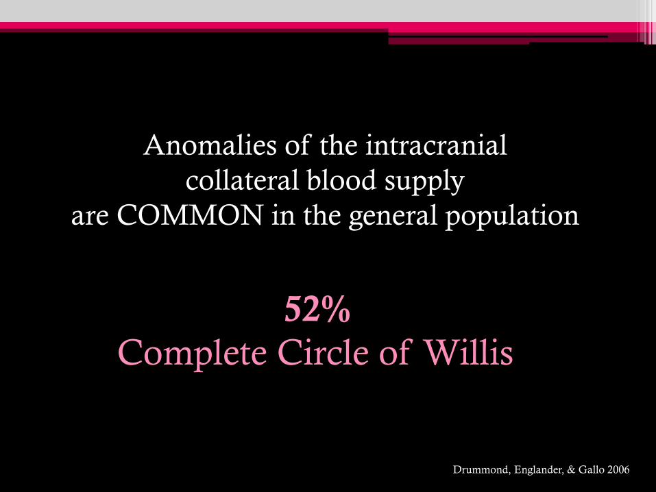
Anomalies of the intracranial
collateral blood supply
are COMMON in the general population
52%
Complete Circle of Willis
Drummond, Englander, & Gallo 2006

Cerebral Ischemia as an Apparent Complication of Anterior
Cervical Discectomy in a Patient with an Incomplete Circle of
Willis. The University of California, San Diego; VA Medical Center, San Diego; Department of Neurology,
Department of Neurosurgery, Sacred Heart Medical Center, Eugene, Oregon
Drummond, Englander & Gallo 2006
58yo Male
Ischemic injury ipsilateral to retraction
Carotid compression
Moderate arterial BP reduction Preop MAP 99 mm/Hg; Intraop MAP ~56 mm/Hg
Extubated…Reintubated
Immediate postop—all tests normal CT, MRI, MRA, etc
POD #13 Repeated Brain MRI
MRA Circle of Willis
Case Report

Drummond, Englander, & Gallo 2006
MRA Circle of Willis Brain MRI

Sinuses Drain into the Internal Jugular Vein
Graphic: rci.rutgers.edu

Cerebral Spinal Fluid
Production Composition
• Formed Choroid Plexus
• 21ml/hr or 500ml/day
• Quick turnover rate
• ~150ml present at a
given time
• Reabsorbed Arachnoid
Villi
• Na+ 141 mEq/l
• K+ 2.9 mEq/l
• Ca2+ 2.5 mEq/l
• Mg2+ 2.4 mEq/l
• Cl- 124 mEq/l
• HCO3- 21 mEq/l
• PRO 28 mg/100mL
• Glu 61 mg/100mL
pH 7.31 Barash 2006

Lateral
Ventricles
Foramen Of Monro
3rd Ventricle *Aqueduct of Sylvius
4th Ventricle
Lushka & Magendie
Subarachnoid Space Spinal
Cord
Brain
Arachnoid Villi
Pattern of CSF Flow

Intracranial Pressure
• Normal <10 mm/Hg
• ICP >15 mm/Hg Intracranial HTN
• Most centers treat when ICP >20-25 mm/Hg
*BTF Guidelines support the initiation of
treatment when ICP is ≥ 20 mm/Hg
Li, Timofeev, Czosnyka, & Hutchinson 2010
ICP
Marked INCREASES in ICP can DECREASE Cerebral
Perfusion Pressure & Cerebral Blood Flow producing regional
& general ischemia.

P1—Percussion wave
Atrial pulsation
P2—Tidal wave
Intracranial compliance
P3—Dicrotic wave
aortic valve closure
Flow of 3 upstrokes in 1 wave
Graphic: eneurosurgery.com
*If P2 is higher than P1 indicates Intracranial HTN
ICP Waveform

Cerebral Perfusion Pressure
CPP= MAP-ICP
Or CVP (which ever is greater)
Normal CPP is 80-100 mm/Hg
• CPP <50mm/Hg—EEG slowing
• CPP 25-40mm/Hg—Flat EEG
• CPP <25mm/Hg—Irreversible brain damage
*BTF Guidelines target CPP of 50-70 mm/Hg
No more than 70 mm/Hg
Morgan Mikhail & Murray 2006

Autoregulation at Work
• CPP—cerebral vasoconstriction prevents sudden CBF & CBV
• CPP—cerebral vasodilation maintain CBF & perfusion
*Upper limits of CPP 150-160mm/Hg CPP >150 Risk: Disruption of BBB Cerebral edema Hemorrhage
TBI capillaries become leaky & more permeable to water
vessels dilate, flow increases and edema worsens
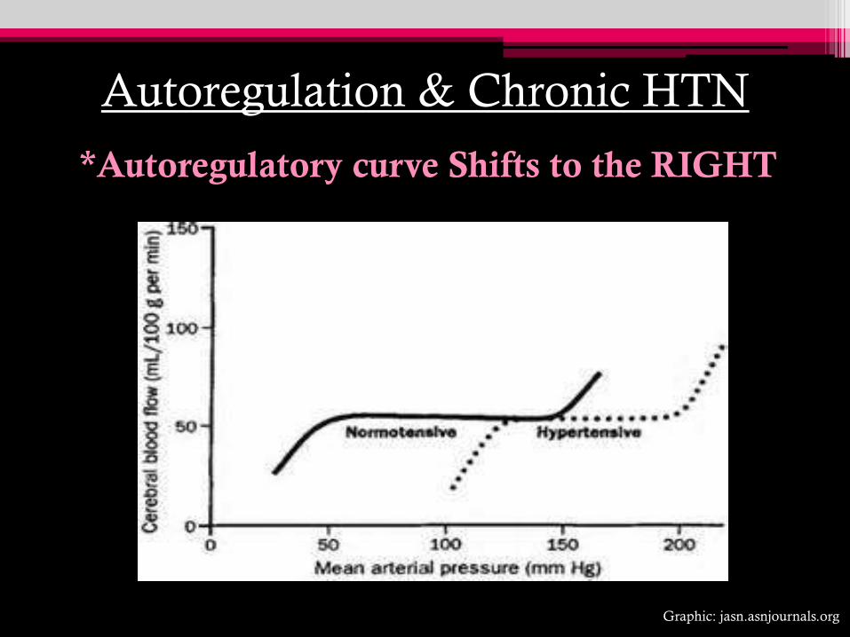
Autoregulation & Chronic HTN
*Autoregulatory curve Shifts to the RIGHT
Graphic: jasn.asnjournals.org

Impaired Autoregulation
• Cerebral edema—Trauma
• Cerebral ischemia—Hypoxia
• Hypercarbia
• Volatile agents—Vasodilating drugs
• Subarachnoid Hemorrhage—vasospasm

Symptoms of Increasing ICP Mild to Moderate TBI
Vague & Nonspecific
Confusion
Headache
Drowsiness
*Fundamental Clinical Variable*
GLASGOW COMA SCALE
Value in MOTOR component=potential increasing ICP
Determines how the neuro status will be monitored

BEHAVIOR RESPONSE SCORE
Eye Opening
Response
Spontaneous
To speech
To pain
No Response
4
3
2
1
Best Verbal
Response
Oriented x3
Confused
Inappropriate words
Incomprehensible sounds
No response
5
4
3
2
1
Best Motor
Response
Obeys Commands
Moves to localized pain
Flexion/withdrawal from pain
Abnormal flexion Decorticate
Abnormal extension Decerebrate
No response
6
5
4
3
2
1
Total Score Best Response
Comatose Unresponsive
15
≤8 3

The Surgical Approach to the Management of Increased
Intracranial Pressure After Traumatic Brain Injury; Academic
Neurosurgery unit, University of Cambridge/Addenbrookes Hospital, United Kingdom.
ICP within 1st 24hours of injury as well as any
Secondary ICP (3-10 days posttrauma)
POOR prognosis
Li, Timofeev, Czosnyka, & Hutchinson 2010
Secondary Causes of increased ICP
Cerebral Edema Posttraumatic seizure
Mass lesion Intrathoracic pressure
Cerebral vasodilation Hyperthermia
Systemic HTN
Venous sinus thrombosus

Surgical Approach Cont’
Optimal approach—Anticipate the onset ICP
• Neurosurgery involved early
Assessment, treatment, & planning
• Treated in Neurosurgical Centers
Even if injury does NOT require neurosurgical
intervention
• High Risk patients Neurosurgical unit w/option for
Neuro ICU care
Li, Timofeev, Czosnyka, & Hutchinson 2010
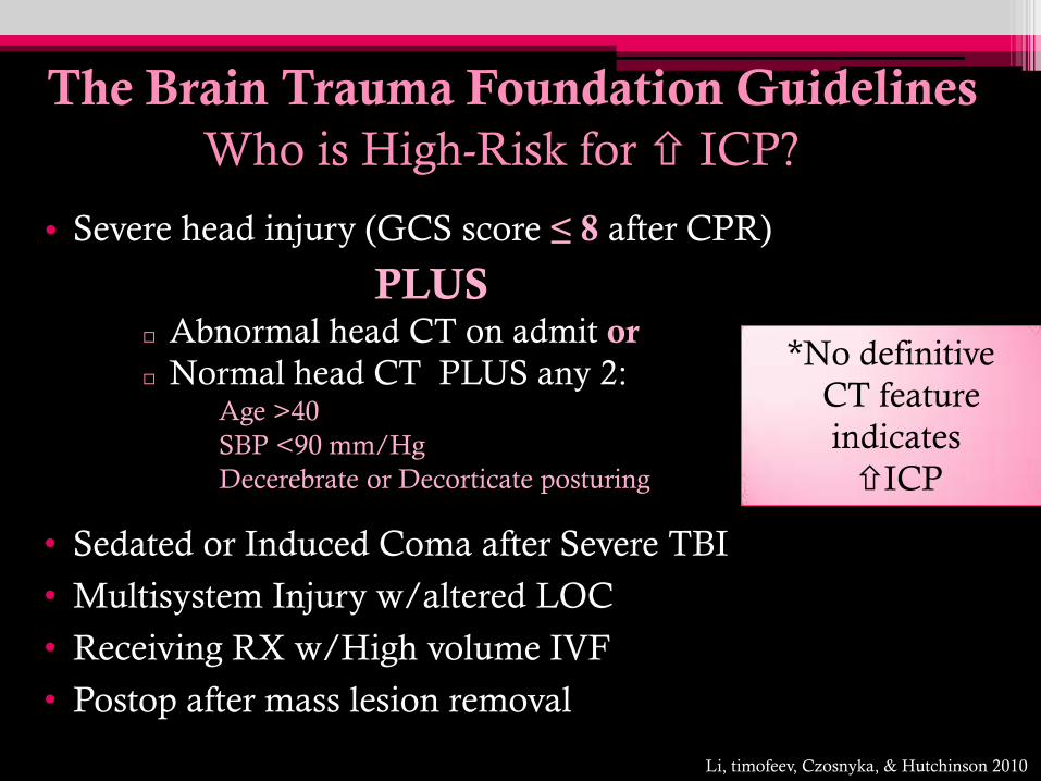
• Severe head injury (GCS score ≤ 8 after CPR)
PLUS Abnormal head CT on admit or
Normal head CT PLUS any 2: Age >40
SBP <90 mm/Hg
Decerebrate or Decorticate posturing
• Sedated or Induced Coma after Severe TBI
• Multisystem Injury w/altered LOC
• Receiving RX w/High volume IVF
• Postop after mass lesion removal
Li, timofeev, Czosnyka, & Hutchinson 2010
The Brain Trauma Foundation Guidelines
Who is High-Risk for ICP?
*No definitive
CT feature
indicates
ICP

Mainstay of ICP Management University of Cambridge/Addenbrookes Hospital, United Kingdom
• Head elevation 10-15 degrees
• Adequate oxygenation Sat ≥ 97%
• Fluid resuscitation CVP 6-10
• Sedation Propofol, versed, fentanyl
• Muscle relaxation Atracurium 0.5mg/kg/hr
• Mild hyperventilation
• Cooling ≤ 37°C
Medical Management with Protocols
Li, Timofeev, Czosnyka, & Hutchinson 2010
Surgery is 2nd tier treatment
in patient’s whose ICP is refractory to maximal medical management

Pharmacology Barbiturate
• Thiopental
▫ Hypnosis
▫ Depression of CMR
▫ CBF r/t CVR
▫ Anticonvulsant property
▫ Facilitate absorption of CSF
“Robin Hood”, reverse steal phenomenon
induces vasoconstriction in normal brain tissue, blood
flow is redistributed to ischemic areas of the brain
Morgan, Mikhail, Murray 2006
250mg boluses
up to 3-5grams
IV infusion 4-8 mg/kg/hr

• Arterial line
• CVP line
• ICP monitor
• Rt SjVO2 catheter ≥ 55%
Establish transcranial doppler & multimodality
monitoring within first 6hrs of Neuro ICU stay.
Mainstay of ICP Management University of Cambridge/Addenbrookes Hospital, United Kingdom
All Patients with or at risk for developing increased ICP
Li, Timofeev, Czosnyka, & Hutchinson 2010

Herniation
Elevated ICP can induce brain herniation
• Across meninges
• Down spinal canal
• Through opening in
the skull
• Rapid neurological
deterioration and death
Graphic: emsworld.com
Cushing’s Triad
SBP
Bradycardia
Abnormal breathing pattern

The Brain Trauma Foundation Guidelines
• Hypoxemia and Hypotension
• Hyperosmolar Therapy
• Indications for ICP Monitoring
• ICP Monitoring Technology
• ICP & CPP Thresholds
• Brain Oxygen Monitoring and Thresholds
• Analgesics, Anesthetics, and Sedatives

The Brain Trauma Foundation Guidelines
• Defining level of Hypotension is unclear
• SBP <90 mm/Hg—avoid or rapidly correct
Given the influence that CPP has on outcome:
SBP >90mm/Hg would be more desirable
esp during pre-hospital & resuscitative phases
• Hypoxia—apnea cyanosis in the field or
PaO2 <60 mm/Hg
Hypoxemia and Hypotension

The Brain Trauma Foundation Guidelines
Agents currently in clinical use for TBI
• Mannitol
▫ Single administration—short term effects
▫ Prolonged therapy for ICP
*Lack of evidence for repeated regular administration over several days
• Hypertonic Saline
▫ Current evidence is NOT strong enough to recommend use,
concentration, & administration of HS in traumatic
intracranial HTN
Hyperosmolar Therapy

Mannitol 0.25gm/kg to 1gm/kg
Avoid SBP <90mm/Hg
• Immediate plasma expanding effect
▫ HCT and Blood viscosity
▫ CBF and O2 delivery
• Osmotic effect—delayed 15-30 minutes
▫ Establishment of gradients between cells & plasma
▫ Effects can persist for ~90 min to 6+ hours
Exerts beneficial effects by 2 mechanisms:
BTF 2007

Hypertonic Saline
• Principle effect on ICP is osmotic movement of water
across an intact BBB
• Dehydrates endothelial cells and erythrocytes
▫ Increasing diameter of vessels
▫ Plasma volume expansion
▫ Increased CBF
• Reduces leukocyte adhesion in traumatized brain
BTF 2007
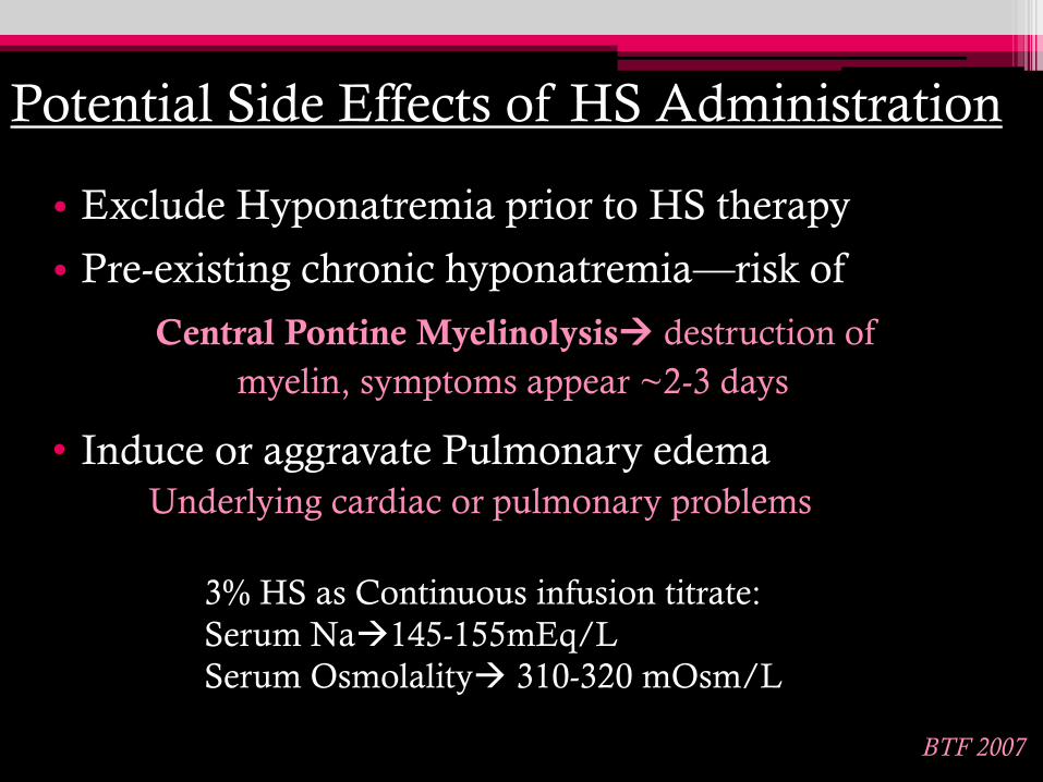
Potential Side Effects of HS Administration
• Exclude Hyponatremia prior to HS therapy
• Pre-existing chronic hyponatremia—risk of
Central Pontine Myelinolysis destruction of
myelin, symptoms appear ~2-3 days
• Induce or aggravate Pulmonary edema
Underlying cardiac or pulmonary problems
3% HS as Continuous infusion titrate:
Serum Na145-155mEq/L
Serum Osmolality 310-320 mOsm/L
BTF 2007

The Brain Trauma Foundation Guidelines
The only way to reliably determine CPP and cerebral
hypoperfusion is via continuous ICP and BP monitoring
• Predict outcome & worsening pathology
• Calculate & manage CPP
• Therapeutic CSF drainage w/Ventricular ICP monitoring
• Restrict potential deleterious ICP reduction therapies
Indications for Intracranial Pressure Monitoring
Hypotension and ICP –leading cause of death in severe TBI

• Ventricular Catheter
▫ Accurate
▫ Low cost
▫ Reliable
▫ Recalibrated in situ
• Parenchymal ICP Monitors
▫ Cannot be recalibrated
• Subarachnoid, Subdural, and Epidural
▫ Less accurate
The Brain Trauma Foundation Guidelines
Intracranial Pressure Monitoring Technology

EVD Placement
Placed via bur hole
Frontal horn of Lateral Vent
Right sided
Patient lying at 30 degrees
External auditory meatus—
Horizontal plane as Foramen of Monro
*Landmark for zeroing transducer of ICP monitor*
Adjust drain height
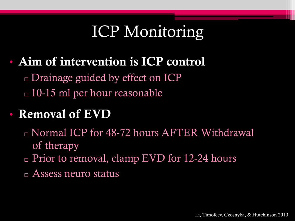
ICP Monitoring
• Aim of intervention is ICP control
Drainage guided by effect on ICP
10-15 ml per hour reasonable
• Removal of EVD
Normal ICP for 48-72 hours AFTER Withdrawal
of therapy
Prior to removal, clamp EVD for 12-24 hours
Assess neuro status
Li, Timofeev, Czosnyka, & Hutchinson 2010

Complications of ICP Monitoring
• Infection
• Hemorrhage
• Malfunction
• Obstruction
• Malposition
Complications causing patient morbidity are rare
BTF 2007

20-25 mm/Hg is the UPPER threshold above which
treatment to lower ICP should be initiated
• Initiate treatment when ICP >20mm/Hg
• Guide patient management/treatment
ICP values
Clinical Findings
Brain CT findings
Brain herniation can occur at ICP <20-25 mm/Hg
The Brain Trauma Foundation Guidelines
ICP Thresholds

CPP= MAP - ICP
• CPP Range 50-70 mm/Hg
▫ Intact pressure autoregulation tolerate higher CPP
• Avoid CPP <50 mm/Hg
• Avoid aggressive attempts to keep CPP >70 mm/Hg
with fluids and pressors
▫ ARDS
The Brain Trauma Foundation Guidelines
Cerebral Perfusion Thresholds

Treatment Thresholds
• SjVO2 Jugular Venous Saturation <50%
Global measurement of O2
• PbrO2 Brain Tissue Oxygen Tension <15 mm/Hg
Local measurement of O2
The Brain Trauma Foundation Guidelines
Brain Oxygen Monitoring and Thresholds
Mortality rates are HIGHER in those with episodes of desaturation

SjVO2 Monitoring
• Indirectly assesses brain’s ability to extract & metabolize
O2
• Normally CMRO2 coupled to CBF
*Extraction ratio of arterial & venous blood remains constant
• ~50% TBI patients exhibit evidence of
Defective cerebral autoregulation
Uncoupling
White & Baker 2002
As long as the hemoglobin & arterial saturation remain constant
the SjVO2 is an indicator of cerebral O2 demand

SjVO2 Catheter Placement

PbrO2 Monitoring Treatment value > 15 mm/Hg
• Low PbrO2—both Depth & Duration correlated
w/mortality
• 50% risk of death associated with
▫ Values <15 mm/Hg
▫ Lasting 4hrs or longer
*BTF Guidelines—PbrO2 values <10-15 mm/Hg
with a duration >30 minutes are associated with
higher mortality
BTF 2007

Multimodal Monitoring
ICP
Arterial BP
Transcranial Doppler
Evoked potentials
SjVO2
Should be used TOGETHER to assess the impact of
the various interventions on cerebral metabolism
BTF 2007

The Brain Trauma Foundation Guidelines
Anesthetics, Analgesics, and Sedatives
• Common management strategy for ICP control
• No evidence to support their efficacy
• Have not been shown to positively affect outcome
Research Panels are looking at OUTCOME

• Barbiturate ▫ Administration of high-dose to control ICP refractory to maximal
treatment
Loading dose: 10mg/kg over 30 minutes
5mg/kg Q1hr x3doses
Maintenance: 1mg/kg/hr
▫ Prophylactic administration of barbiturates to produce burst suppression
EEG is NOT recommended
• Propofol for control of ICP
▫ Not for improvement in mortality
▫ High-dose can produce significant morbidity
*Do NOT EXCEED 5mg/kg/hr
The Brain Trauma Foundation Guidelines Anesthetics, Analgesics, and Sedatives

Propofol Infusion Syndrome PRIS Common Clinical Features
• Hyperkalemia
• Hepatomegaly
• Metabolic acidosis
• Myocardial failure
Extreme caution must be taken when using doses >5mg/kg/hr OR
ANY dose exceeding 48hr in critically ill adults
• Hyperlipidemia
• Rhabdomyolysis
• Renal failure
• Death
BTF 2007

Why the Increased Risk of PRIS in Acute
Neurological Injury?
• Catecholamines Cardiac output
• Propofol concentration clearance & first pass effect
• effect of propofol—reversal of anesthesia
• Propofol can depresses cardiac function β-receptor
antagonism
• catecholamine requirements
Negative inotropic effect creates a VISCIOUS CYCLE
Sabsovich, Rehman, Yunen, & Coritsidis 2007
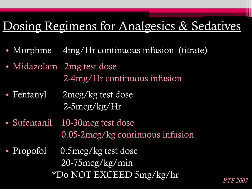
Dosing Regimens for Analgesics & Sedatives
• Morphine 4mg/Hr continuous infusion (titrate)
• Midazolam 2mg test dose
2-4mg/Hr continuous infusion
• Fentanyl 2mcg/kg test dose
2-5mcg/kg/Hr
• Sufentanil 10-30mcg test dose
0.05-2mcg/kg continuous infusion
• Propofol 0.5mcg/kg test dose
20-75mcg/kg/min
*Do NOT EXCEED 5mg/kg/hr BTF 2007

Sodium & Water Balance After TBI
Secondary Injuries r/t Force of the impact
• Cerebral edema
• Injury to hypothalamus
• Injury to pituitary gland
What Causes Electrolyte & Fluid imbalance in TBI?

Hypothalamic-pituitary Dysfunction
• Central Neurogenic Diabetes Insipidus—CNDI
▫ Associated w/HYPERnatremia
• Syndrome of Inappropriate Secretion of
Antidiuretic Hormone—SIADH
and
• Cerebral Salt-Wasting Syndrome—CSWS
*Associated w/HYPOnatremia
3 common electrolyte imbalances
John & Day 2012

ADH
• Osmoregulation—maintain water balance
▫ Serum Na+ osmolality 280-295 mOsm/kg
▫ Serum Os <280 ADH not secreted
▫ Serum Os >295 ADH is secreted
• Baroregulation—changes in BV & BP
▫ Located in chest, left atrium, aortic arch, carotid sinuses
▫ Transmitted via vagus & glossopharyngeal nerves
▫ BV & BP = ADH secretion
John & Day 2012
HYPOtension & HYPOvolemia are common in TBI
ADH secretion is INCREASED

Central Neurogenic DI
• Damage to posterior pituitary—ADH is stored & secreted
• Associated with:
Neurosurgery & Tumors
Increased ICP
Brain death
CNS infections—encephalitis & meningitis
• CNDI occurs in 16% of TBI patients
• Occurs ~5-10 days after trauma
John & Day 2012
Decreased ADH secretion & Hypernatremia

CNDI Signs & Symptoms
• Polyuria
*Urine specific gravity <1.005
*Urine Os <200 mOsm/kg
* Na+ >145 mEq/L
* Serum Os >295 mOsm/kg
* How CNDI is
diagnosed in TBI
patient
John & Day 2012
250 ml/hr
• Polydipsia
• Hypovolemia
• Hypernatremia
• Lose 3% to 5% of body weight

Treatment Options Central Neurogenic DI
• Fluid replacement with 0.45% NSS
• Desmopressin
Intranasally—5 to 2 mcg/day divided doses
Parenterally—5 to 40 mcg/day divided doses
• Vasopressin
Intravenous—0.5 to 2 U Q3hrs
If U/O is >300mL/hr for 2 consecutive hrs
Infusion– 0.2 to 0.9 U/min

Pharmacology DDAVP 4mcg/ml
• Neurogenic DI—lack of ADH
▫ DDAVP is synthetic replacement—of natural hormone
arginine vasopressin
▫ Decreased vasopressor action
• 1 mcg DDVAP = 4 IU
• Injection is 10x the ADH effect vs. intranasal
• Mixed by pharmacy & sent “on call” r/t stability
• Administered over 10-15 minutes
Neurogenic DI
NOT
Nephrogenic DI
Mayoclinic.com

SIADH
• Renal reabsorption & water retention
• Concentrated urine
• Hyponatremia common after TBI
▫ Affects ~33% of head injury patients
• Causes of SIADH in TBI
▫ Traumatic Subarachnoid hemorrhage
▫ Increased ICP
▫ Damage to hypothalamic-pituitary region
• Body weight increase of 5% to 10%
John & Day 2012
Increased ADH secretion & Dilutional Hyponatremia

SIADH Signs & Symptoms
• Decreased U/O—400 to 500 ml/24 hrs
• Serum Na+ level— <135 mEq/L
▫ Seizures Na+ <120 mEq/L
• Serum Os— <275 mOsm/L
• Na+ in urine— >25mEq/L
• Urine osmolality > Serum osmolality
Headache, NV, fatigue, lethargy, confusion, & muscle twitching
John & Day 2012

Treatment Options SIADH
• Fluid restriction— <1000 mL/24hr
• Slow Na+ replacement with 3% NSS
▫ Too rapid—Central Pontine Myelinolysis
▫ Recommend Na+ by 10 to 20 mEq/d
• Diuretics
• Demeclocycline hydrochloride—suppresses ADH activity
• Lithium carbonate—inhibits renal response to ADH
John & Day 2012

Cerebral Salt-Wasting Syndrome
• Lose Na+ and ECF—plasma volume decreases
• Decrease in body weight
• Pathophysiology unclear—primary mechanism renal loss Na+
• Occurs most often in:
▫ Stroke
▫ Intracerebral hemorrhage
▫ Neuro surgery
John & Day 2012
Elevated ADH Levels & True Hyponatremia
*Can develop in TBI patients with ICP

CSWS Signs & Symptoms
• Headache
• Increased thirst
• Dehydration
• Weight loss
• Tachycardia
• Hypotension
• Lethargy & LOC
• Seizures & coma
*Primary distinction
between CSWS & SIADH
volume status
CSWS Volume Depletion
SIADH Volume Expansion
John & Day 2012

Treatment Options Cerebral Salt-Wasting Syndrome
• Replace fluids with physiologic NSS
• IV replacement with 3% NSS
• If taking PO—oral salt tablet supplements
• Fluid restriction is contraindicated
▫ Cerebral vasospasm
▫ Cerebral ischemia & infarction
John & Day 2012
Summary
• Neurological damage does not occur solely with the
primary injury. We need to recognize & manage the
secondary injuries.
• Maintain adequate cerebral blood flow, cerebral perfusion &
oxygenation while the brain recovers.
• Management of ICP begins with anticipating it’s
development & having treatment protocols in place.
• Be aware of the electrolyte imbalances that can occur with
TBI and correctly manage the cause of hyponatremia.

Thank You

References
• U.S. DEPARTMENT OF HEALTH AND HUMAN SERVICES Centers for Disease
Control and Prevention National Center for Injury Prevention and Control. Traumatic
Brain Injury in the Unites States, Emergency Department Visits, Hospitalizations, and
Deaths 2002-2006. Accessed April 21,2013.
• The Brain Trauma Foundation. Guidelines for the Management of Severe Traumatic
Brain Injury 3rd ed. The American Association of Neurological Surgeons & The
Congress of Neurological Surgeons. 2007. Braintrauma.org
• Li LM, et al. The Surgical Approach to the Management of Increased Intracranial
Pressure After Traumatic Brain Injury. Anesthesia-Analgesia.org. 2010;111(3):736-748.
• White H, Venkatesh B. Cerebral Perfusion Pressure in Neurotrauma: A Review.
Anesthesia & Analgesia. 2008;107(3):979-987.
• Drummond JC, Englander RN, Gallo CJ. Cerebral Ischemia as an Apparent
Complication of Anterior Cervical Discectomy in a Patient with an Incomplete Circle
of Willis. Anesthesia & Analgesia. 2006;102:896-899.

• White H, Baker A. Continuous Jugular Venous Oximetry in the Neuro-Intensive Care
Unit, a Brief Review. Canadian Journal of Anesthesia. 2002;49(6):623-629
• Barash PG, Cullen BF, Stoelting RK. Clinical Aesthesia 5th ed. 2006
• Morgan GE, Mikhail MS, Murray MJ. Clinical Anesthesiology 4th ed. 2006
• Nagelhout JJ, Plaus KL. Nurse Anesthesia 4th ed. 2010
• John CA, Day MW. Central Neurogenic Diabetes Insipidus, Syndrome of Inappropriate
Secretion of Antidiuretic Hormone, and Cerebral salt-wasting Syndrome in Traumatic
Brain Injury. Critical Care Nurse. 2012;32(2):e1-e8. ccn.aacnjournals.org
• Sabsovich I, Rehman Z, Yunen J, Coritsidis G. Propofol Infusion Syndrome: A Case of
Increasing Morbidity With Traumatic Brain Injury. American Journal of Critical Care.
2007;16:82-85