Anemia 101, Too Low, No Go! - Georgia Association of...
66
Anemia 101, Too Low, No Go! Allan Platt PA-C, MMSc, DFAAPA Assistant Professor Emory University PA Program Atlanta GA [email protected] www.EmoryPA.org
-
Upload
truongngoc -
Category
Documents
-
view
214 -
download
0
Transcript of Anemia 101, Too Low, No Go! - Georgia Association of...

Anemia 101, Too Low, No Go!
Allan Platt PA-C, MMSc, DFAAPAAssistant ProfessorEmory University PA ProgramAtlanta [email protected]

Disclosure
• I have nothing to disclose except
– I do work for food
– I promote giving Blood

Blood Components
Plasma 54%
White cells and
platelets 1%
Red Cells 45%

White Blood Cells• Fight infections
• Are increased in infections
• Move inside and outside of blood vessels
• Are made in the bone marrow

White Blood CellsWBC - White Blood Cells 4.5 - 11.0 K/uL
Low = Leukopenia High = Leukocytosis
WBC Differential
Neutrophils - Segs 54 -62%
Neutrophils - Bands 3 -5 %
Lymphocytes - Lymphs 25 - 33%
Monocytes - Monos 3 - 7%
Eosinophils - Eos 1 - 3%
Basophils - Basos 0 - 0.75%
Atypical Lymphs 0

Platelets
• Plug holes in the body to stop bleeding
• Can help cause blood to clot
• Made in the bone marrow
Fibrin
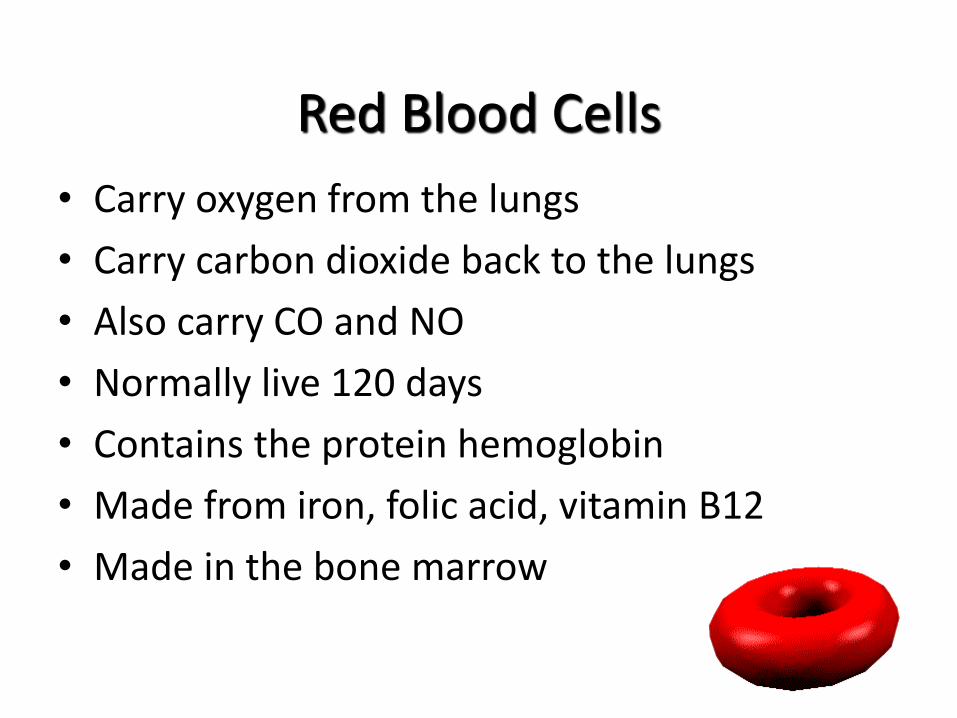
Red Blood Cells
• Carry oxygen from the lungs
• Carry carbon dioxide back to the lungs
• Also carry CO and NO
• Normally live 120 days
• Contains the protein hemoglobin
• Made from iron, folic acid, vitamin B12
• Made in the bone marrow
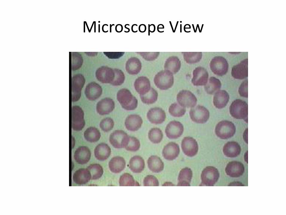
Microscope View

Red Blood Cells - Hemoglobin
Oxygen
Normal
Hemoglobin A
has 2 alpha and
2 beta globin
chains with 4
iron binding
sites

Hemoglobin FA – Biochemistry – Normal Newborn
alpha alpha gamma gamma beta
alpha alpha gamma gamma delta beta
Chromosome 16 Chromosome 11
Mom
Dad
alpha alpha
alpha alpha
alpha alpha
beta beta
gamma gamma
delta delta
delta
50 – 20 % Hemoglobin A
Hb F (newborn): 50% to 80%
(6 months): 8%
(over 6 months): 1% to 2%
2% Hemoglobin A2
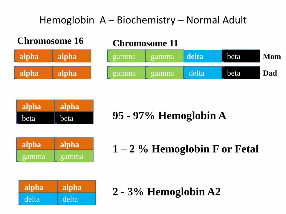
Hemoglobin A – Biochemistry – Normal Adult
alpha alpha gamma gamma beta
alpha alpha gamma gamma delta beta
Chromosome 16 Chromosome 11
Mom
Dad
alpha alpha
alpha alpha
alpha alpha
beta beta
gamma gamma
delta delta
delta
95 - 97% Hemoglobin A
1 – 2 % Hemoglobin F or Fetal
2 - 3% Hemoglobin A2

Red Blood Cells - Retics • Reticulocytes, or Retics are young red cells just released
from the bone marrow. The Retic count is the best indicator about how the marrow factory is doing.

Red Blood Cells
Red cells live
120 days in the
circulation
Food with iron
and vitamins
is digested
Red cells are
made in the
bone marrow
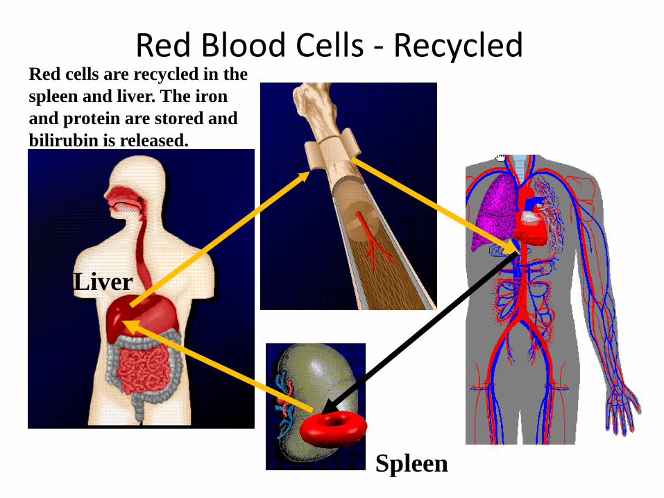
Red Blood Cells - Recycled Red cells are recycled in the
spleen and liver. The iron
and protein are stored and
bilirubin is released.
Spleen
Liver

Hemoglobin RecycledBilirubin must be Directed
through the Liver to be
Conjugated
Liver
Indirect
Bilirubin
Hemoglobin
Direct
Bilirubin
LDH - Lactate
Dehydrogenase
Iron – Fe bound
to Transferrin Kidney -
Hemoglobinuria

Red Blood Cells - The Kidney
Erythropoietin is made by
the kidney as a signal to
the bone marrow to make
more red cells

Hepcidin
Decreased levels
increase iron
absorption and
release from cells –
Erythropoetin, low
iron
Hormone
made in the
liver
Increased levels
blocks absorption of
Iron and cell release
- inflammation IL6

The History
• Weakness
• Tiredness - Fatigue
• Dyspnea
• Dizzy – non vertigo
• Palpitations
• New angina

The History -2
• - History of melena, abdominal pain, Aspirin or non-steroidal anti-inflammatory agents (NSAIDs) use, past peptic ulcer disease , then consider GI bleeding, platelet dysfunction.
• - In females the menstrual history quantifying the amount of bloodloss ,or possible pregnancy should be obtained.
• - History of pica or abnormal craving for ice, clay, starch...; dysphagia then consider iron deficiency.
• - Poor diet, then consider iron or folate deficiency, and general • malnutrition• - History of gastric surgery, distal paresthesias, gait problems -
consider B12 deficiency•
The History - 3
• - History of alcohol abuse - consider folate deficiency or liver disease. If moonshine use or lead paint/pipe exposure, consider lead toxicity.
• - Family history of blood cell or bleeding disorder: consider Sickle Cell disease, G6PD,Thalassemia, Hemophilia, von Willebrand
• - History of jaundice, transfusion, new medication, infection -consider hemolytic process
• - History of weight loss, Cancer, HIV, rheumatoid arthritis, thyroid disease, renal disease -then consider secondary cause
• - History of fever and chills, cough, dyspnea, then consider Infection.
• Medications – hemolysis, bone marrow toxicity, block nutrients (metformin – B12, Dilantin –Folate, PPIs) block EPO –(ACE inhibitors)

Physical Exam

Sclera

Spoon Nails – Fe Def.

Glossitis and Chelosis – Fe and B12

Physical Exam• GENERAL INSPECTION- clubbing in TB or lung cancer• Skin- Hypothyroid, SLE, Bruises, lesions, petechiae or
purpura.• Weight - Loss in Cancer, HIV, Chronic disease, gain in
hypothyroid
• VITAL SIGNS- Pulse: Tachycardia from increased cardiac output
• Respirations: Tachypnea from decreased oxygen transport• BP: Orthostatic if volume depleted• Temp: Fever in infections and drug or transfusion reactions, • HEENT- Eye: Jaundice if hemolysis, pallor in palpebral
conjunctiva• Mouth: Glossitis and angular stomatitis in iron or B12
deficiency

Physical Exam - 2• NECK- Thyroid enlargement or nodules, lymph nodes• HEART- Increased output/murmur- JVD, LVH, S3, S4, consider
high output failure• LUNG- consider infection, lesion• ABDOMINAL- Liver/spleen size, masses, tenderness, surgical
scars• RECTAL- Stool guaiac, prostate exam in men• PELVIC/BREAST- Uterine abnormality, Pap smear, Breast
nodule• LYMPHNODES- consider lymphoma,
leukemia,infection,connective tissue Disease• NEUROLOGIC- Decreased vibratory and position sense in B12
deficiency

LAB- INITIAL SCREENING TESTS
• Urinalysis- Hematuria/proteinuria in renal disease hemoglobinuria in hemolysis.
• CBC, red cell morphology and white blood cell differential, Reticulocyte count
• Chemistry profile (LDH, Bilirubin- Direct and Indirect, BUN, Creatinine, GPT),
• Hemoglobin Electrophoresis if hereditary hemoglobinopathy is suspected
• IF BLEEDING - Platelet Count, PT, aPTT, PFA

CBC- Red Cell Measures PARAMETER NORMAL ADULT COMMENTS
HB - Hemoglobin Male= 15.5 +/- 2 mg/dl Low = Anemia
Female = 13.5 +/- 2 High = polycythemia
HCT - Hematocrit Male= 46.0 +/- 6% "
Female= 41.0 +/- 6% "
RBC - Red Blood Male = 4.3 - 5.9 Million/uL High in Thalassemia
Cell Count Female = 4.0 - 5.2 "

Red Cell Indices MCH, MCHC
MCH - Mean Corpuscular 27 -32 pg Low = Hypochromic
Hemoglobin High = Hyperchromic
MCHC - Mean Corpuscular 30 - 36 gm/dl Low = R/O Fe def.
Hemoglobin Concentration High = Spherocytosis

Red Cell Indices MCV - RDWMCV - Mean Corpuscular Volume 80 - 94 fl
Low = Microcytosis High = Macrocytosis
RDW - Red Cell Distribution Width 11.5 - 14.5
Variation in RBC size

RBC MorphologyRed Cell Morphology SIGNIFICANCE
Burr Cells Uremia, Low K, artifact, Ca stomach, PUDSpur Cell Post-splenectomy, Alcoholic liver diseaseStomatocyte Hereditary, Alcoholic liver disease,Spherocyte Hereditary, Immune hemolytic anemia,
water dilution, post-transfusionShistocyte - helmet TTP, DIC, vasculitis, glomerulonephritis,
heart valve, burnsEliptocyte - Ovalocyte Hereditary, Thalassemia, Fe Def.,
Myelophthistic, megaloblastic anemiasSickle Cells Sickle cell diseaseTarget Cells Thalassemias, hemoglobinopathiesMicrocytes Thalassemia, Iron Def., Lead Toxic,Macrocytes B12 of Folate Def.Parasites Malaria, Babesiosis, Bartonellosis
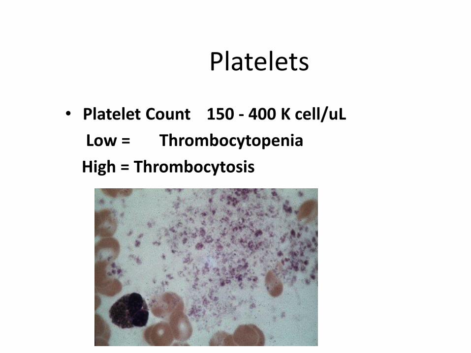
Platelets
• Platelet Count 150 - 400 K cell/uL
Low = Thrombocytopenia
High = Thrombocytosis

Retics or Reticulocyte count
• Retic - Reticulocyte Count 0.5 -1.5 %Low in anemia = low marrow output
High = RBC loss

Correcting the Retic• absolute reticulocyte count (measured)
• reticulocyte (%) = absolute number of reticulocytes ÷ number of RBC × 100
• reticulocyte index = % reticulocytes × actual hematocrit ÷normal hematocrit
• corrected reticulocyte index (corrects for appropriate bone marrow release of reticulocytes) = reticulocyte index ÷maturation factor
• maturation factor = 3.25 – (actual hematocrit ÷ 20) – if hematocrit 45, maturation factor = 1
– if hematocrit 35, maturation factor = 1.5
– if hematocrit 25, maturation factor = 2
– if hematocrit 15, maturation factor = 2.5
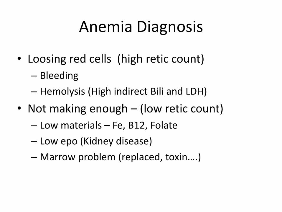
Anemia Diagnosis
• Loosing red cells (high retic count)
– Bleeding
– Hemolysis (High indirect Bili and LDH)
• Not making enough – (low retic count)
– Low materials – Fe, B12, Folate
– Low epo (Kidney disease)
– Marrow problem (replaced, toxin….)

Diagnostic Pathway
Reticulocyte Production Index
<2 Decreased Production>2 Increased Loss
Red Cell Indicies MCVHemolysis Bleeding
>94 80-94<80MacroNormo Micro Extrinsic Intrinsic
Coombs CoombsPositive Negative
Drug Warm ColdAntibody Antibody
Membrane Hb Enzyme

Anemic- Lab –CBC, Retic, RBC
morphology, Metabolic Profile,
UA
Retic Production index < 2 – Marrow
Production Problem
Check MCV
MCV <80 –Microcytic
Order Iron studies, HbELP,
Lead Level
MCV 80 – 100 Normocytic
Order West SedRate, TSH, Renal
Hepatic, Preg Test
MCV > 100 Macrocytic
Order B12, RBC and serum Folate
Retic Production Index >2 RBC Loss
Bleeding or Hemolysis
Increased Indirect Bilirubin and LDH =
Hemolysis
Order Coombs, HinzeBody stain, HbELP
Bleeding
Anemia – low Hb/Hct Lab work-up
BPH = Bleeding/Production/Hemolysis

Microcytic• MICROCYTIC = "TICS"
• T-Thalassemias
• I-Iron Deficiency
• C-Chronic Inflammation
• S-Sideroblastic - lead, drug, or hereditary

Microcytic Tests
TESTS TO ORDER:• Serum Iron
• TIBC = Transferrin binding sites
• % Saturation = Transferrin saturation with Iron
• Ferritin = Storage Iron
• HBELP = Hemoglobin Electrophoresis
• Lead level if exposed

Microcytic workupTICS – Thalassemia, Iron Deficiency, Chronic
inflammation, Sideroblastic (Lead)
Iron studies (Ferritin) Low
Yes = Iron Deficiency
Work up for Chronic Blood loss
– GI, MensesDiet
No, West Sed rate CRP elevated-
Inflammatory Block
No, Lead Level elevated –
Chelation therapy
No, Abnormal HbELP?
Yes = Thalassemia –Refer to
hematologist if severe
Refer to Hematologist for Bone Marrow Bx
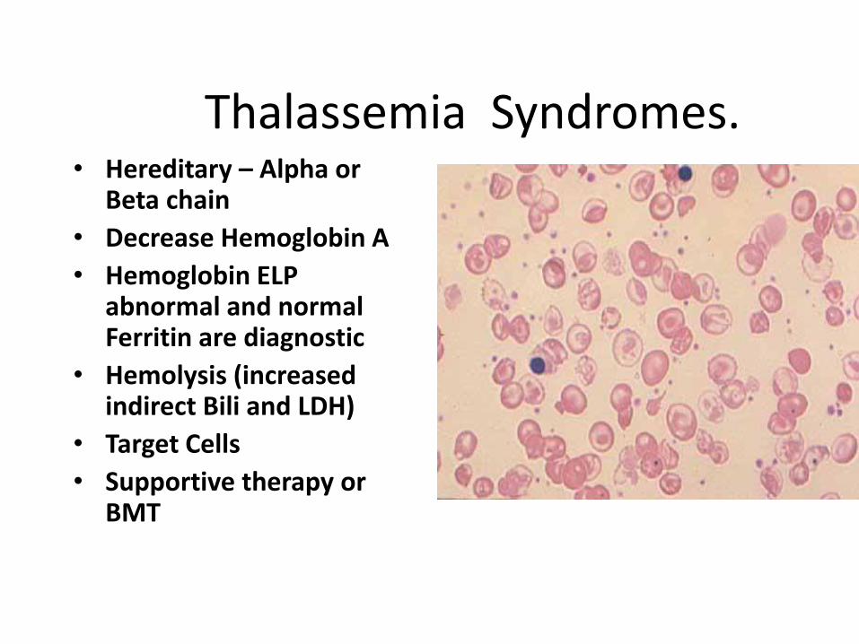
Thalassemia Syndromes.• Hereditary – Alpha or
Beta chain
• Decrease Hemoglobin A
• Hemoglobin ELP abnormal and normal Ferritin are diagnostic
• Hemolysis (increased indirect Bili and LDH)
• Target Cells
• Supportive therapy or BMT

Iron deficiency
• Low Serum iron, Low Ferritin, High TIBC
• Find out why –GI bleed, menses, diet, H pylori, celiac disease
• Treat FeSO4 300mg tid
• Add vitamin C/meat to increase absorbtion
• Follow up Retic increase 1 week, Ferritin 1 month

Chronic Inflammation
• Block of normal iron stores transport to bone marrow factory
• Normal Ferritin, serum iron and TIBC are low with a low saturation
• 30% Microcytic, 70% Normocytic
• High Sed rate or c-reactive protein
• Treat inflammation – RA, SLE, HIV….

Sideroblastic
• Ring sideroblasts in bone marrow
• Serum iron is increased and TIBC normal resulting in a high saturation. Serum ferritin is increased
• RBC Basophillic stippling
• Lead toxicity is suspect

Normocytic Anemia
• NORMOCYTIC = "NORMAL SIZE"
• N-Normal Pregnancy
• O-Over hydration, Drowning
• R-Renal Disease
• M-Myelophthistic – Marrow replaced
• A-Acute Blood Loss
• L-Liver Disease
• SI-Systemic Infection/Inflammation
• Z-Zero Production- Aplastic anemia
• E-Endocrine: Hypothyroid, hypoadrenal, decreased androgen

Normocytic Tests
• Blood Urea Nitrogen (BUN), Creatinine, SGOT, Alkaline Phosphatase, Bilirubin, Erythrocyte Sedimentation Rate (ESR), Urinalysis, and Thyroid profile
• Renal Function tests
• Pregnancy Test
• Bone Marrow Biopsy

Normocytic workup“NORMAL SIZE”
Check BUN/Creat/ Liver, UA, West Sed
Rate, Preg Test
BUN/Creat elevated or abnormal UA
Work up for Renal Disease and Low
EPO
TSH elevated = Hypothyroid
AST/ALT/AlkP –Liver disease
West Sed rate elevated-
Inflammatory Block
Pregnancy test +
Prenatal carePancytopenia
No - Repeat CBC, Retic in 2 week
Refer to Hematologist for Bone Marrow Bx

Normocytic - Renal Failure
• Anemia caused by decrease erythropoetin production causing decreased bone marrow production
• Can monitor erythropoetin levels
• Treat with epoetin alfa injections weekly or darbepoetin alpha every other week or monthly
• Check for Iron deficiency (altered metabolism) – May need to suppliment

Macrocytic Anemia• MACROCYTIC = "BIG FAT RED CELLS“Or my “BF”
• B-B12 Malabsorbtion• I-Inherited• G-Gastrointestinal disease or surgery•• F-Folic Acid Deficiency• A-Alcoholism• T-Thiamine responsive•• R-Reticulocytes miscounted as large RBCs• E- Endocrine - hypothyroid• D-Dietary
• C-Chemotherapeutic Drugs• E-Erythro Leukemia• L- Liver Disease• L- Lesch-Nyhan Syndrome• S-Splenectomy

Macrocytic Tests
• The peripheral blood changes include: • -Anemia with decreased reticulocyte count, -Increased MCV• -Neutropenia with hypersegmented • Neutrophils• -Thrombocytopenia with large platelets.
• LABS to order:• B12, Serum Folate, RBC Folate• if all normal, consider Metylmalonic Acid and Homocyteine
levels, TSH, and a Bone Marrow Bx.

Macrocytic Work-up
Serum B12
RBC/Serum Folate
B12 nromal/Folatenormal - Order
Metylmalonic Acid and Homocyteine levels
Metylnalonic Acid Elevated in early B12
Deficiency
Consider Liver disease, hypothyroid, Drugs,
Toxins – Refer for BM Bx
B12 low /Folate low = B12 Deficiency or both
Replace with oral, nasal or IM B12 and
Folate

B12 Cobalamin Deficiency
Physical signs include edema, pallor, jaundice, smooth tongue, dementia, decreased vibratory and position sensation,
Hypersegmented polysElevated LDH, Indirect BiliLow serum B12 levelMetformin, Gastric bypass, H2 or PPI as
cause?Methylmalonic acid (B12) and
homocysteine levels elevatedPernicious anemia - anti- intrinsic factor
antibodies Schilling's test• Rx - cobalamin 1000 mg I.M., oral, or
Nasal Spray

Folate Deficiency
• Causes - liver disease, diet vitamin B12 deficiency (needed as co-factor), and drugs such as methotrexate, ethanol, and dilantin.
• Lab – low serum and RBC Folate
• Rx – Folate 1mg po qD

Hemolysis (HIT)• Hereditary (HEM)
– Hemoglobin (sickle cell, thalassemia)
– Enzyme (G6PD deficiency)
– Membrane (Spherocytosis, Eliptocytosis)
• Immune attack – Coombs positive (transfusion, IgM – cold antibody-infections, IgG warm antibody – Drug induced, PNH)
• Trauma– Microangiopathic ( TTP, ITP, HUS, DIC, HIT, HELLP- Eclampsia, Malaria, Splenomegaly)

Hemolytic Anemia• HEMOLYTIC = "HEMATOLOGIST"
• H-Hemoglobinopathy: sickle cell disease• - Hemoglobinuria: Paroxysmal Nocturnal Hemoglobinuria• E-Enzyme Deficiency• M-Medication - drug induced: aldomet, INH• A-Antibodies - Immune attack• T-Trauma to the red cells: D.I.C , artificial heart valves• O-Ovalocytosis• L-Liver disease• O-Osmotic fragility in Hereditary spherocytosis• and in Hereditary Eliptocytosis• G-G6PD Glucose-6-Phosphate Dehydrogenase Deficiency• I-Infection: malaria, babesiosis• S-Splenic destruction in hypersplenism• T-Transfusion• - Thalassemias

Hemolytic Signs
• 1. Elevated reticulocyte count, with stable or falling hemoglobin.•
• 2. Elevated indirect bilirubin -
• 3. Eevated serum lactate dehydrogenase (LDH)-
• 4. Decreased Haptoglobin levels - Haptoglobin binds hemoglobin released in the plasma from red cell breakdown.
• 5. Hemoglobinemia and hemoglobinuria
• 6. Erythroid hyperplasia in bone marrow in chronic hereditary causes
• 7 Abnormal Hemoglobin Electrophoresis

Hemolytic Tests• 1. The direct antiglobulin (Coombs') test Direct Coombs
test looks for antibody on the red cells. The Indirect Coombs looks for antibody in the serum.
• 2. Hemoglobin electrophoresis
• 3. Heinz body stain
• 4. Osmotic fragility
• 5. Blood smear

HemolysisRetic Production Index > 2, high LDH High indirect Bilirubin
Coombs or DAT
No – Heinz body +
Yes = G6PD Deficiency
No – HbELP abnormal+ Hemoglobinopathy –SS, SC, SD S beta Thal
Thalassemia
Yes
Warm Antibody Cold Antibody
No – RBC Morphology+
Shistocytes and Low Platelets in DIC, HIT,
TTP, HELLP

Genetic Hemoglobin Issues
• Thalassemia – Normal DNA sequence , Reduction in globin production
• Alpha - Not enough alpha globin production -Southeast Asian, Indian, southern Chinese, Middle Eastern and African ancestry
• Beta – Not enough Beta globin production Greek, Italian, Middle Eastern, Southeast Asian, southern Chinese and African descent
Hemoglobinopathy
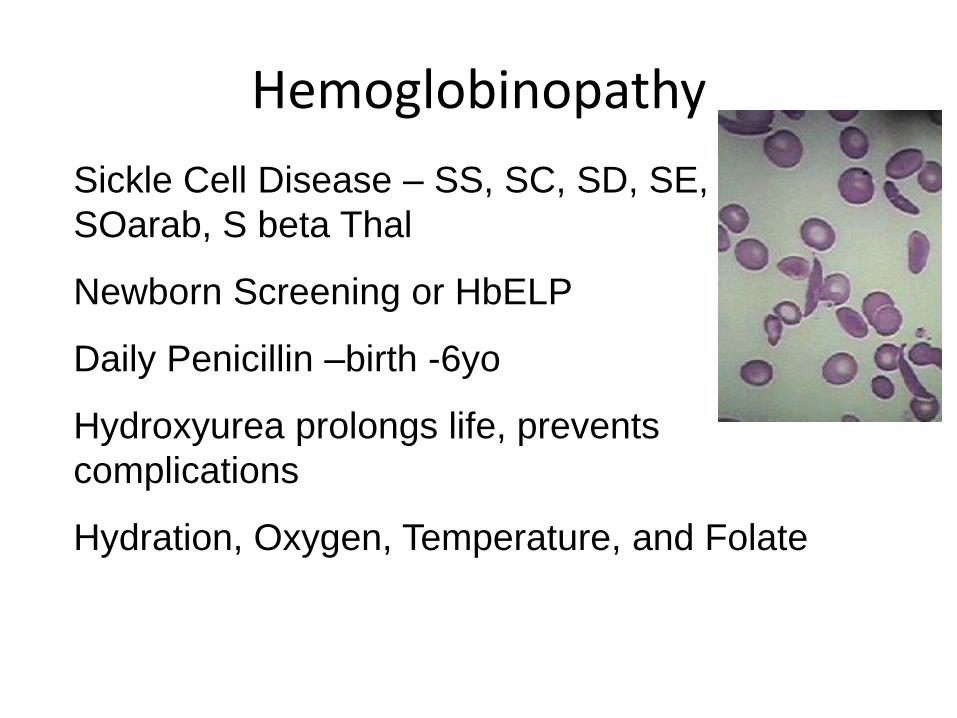
Sickle Cell Disease – SS, SC, SD, SE,
SOarab, S beta Thal
Newborn Screening or HbELP
Daily Penicillin –birth -6yo
Hydroxyurea prolongs life, prevents
complications
Hydration, Oxygen, Temperature, and Folate
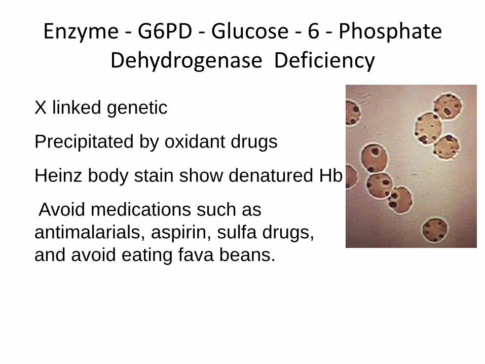
Enzyme - G6PD - Glucose - 6 - Phosphate Dehydrogenase Deficiency
X linked genetic
Precipitated by oxidant drugs
Heinz body stain show denatured Hb
Avoid medications such as
antimalarials, aspirin, sulfa drugs,
and avoid eating fava beans.

Membrane problems Spherocytosis and Ovalocytosis

Immune Attack• Coombs Test: IgG and Compliment +/-
• Transfusion reaction: immediate or delayed
• IgM – (IgG Neg Comp +) cold antibody-infections like mycoplasma, EBV, HIV
• IgG warm antibody – Drug induced –Antibiotics, Ibuprofen, Autoimmune diseases
• PNH Paroxysmal Nocturnal Hemoglobinuria –Red cells attacked by complement Lack of CD55 or CD59 on RBC surface

Trauma To Red Cells
• Microangiopathic- Coagulation gone wild, fibrin shredding red cells- TTP, ITP, HUS, DIC, HELLP-Eclampsia
• Splenomegaly
• Infections within the red cell
– Malaria – Mosquito parasite- Tropics
– Babesiosis – Tick parasite- New England area
– Bartonellosis – Bacteria - Cat Scratch
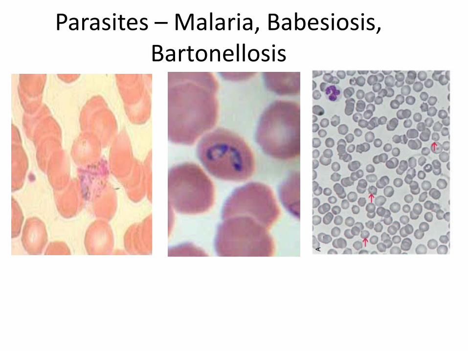
Parasites – Malaria, Babesiosis, Bartonellosis

Take Home Points
• The cause of Iron deficiency confirmed by a low serum ferritin should be pursued.
• Neuropathy, dementia, and anemia should prompt a search for vitamin B12 deficiency.
• A low corrected reticulocyte count is your best indicator of marrow decreased production vs red cell loss
• The MCV is the best guide to diagnose decreased marrow production anemias (Micro, Normo and Macrocytic)
• Microcytic anemia should prompt a TICS work up (Thalassemia, Iron deficiency, Chronic Inflammation and Sideroblastic/lead)