repository.edgehill.ac.uk and... · Web view28.Short KR, Bigelow ML, Kahl J, Singh R,...
35
Article type: Advanced Review Article title: Aging and Computational Systems Biology Authors: Full name and affiliation; email address if corresponding author; any conflicts of interest First author Kathleen M Mooney Faculty of Health and Social care Edge Hill University L39 4QP UK Second author Amy Morgan Faculty of Science and Engineering University of Chester CH2 4 NU UK Third author Mark T Mc Auley Faculty of Science and Engineering University of Chester CH2 4 NU UK Correspondence to [email protected] 1 2 3 4 5 6 7 8 9 10 11 12 13 14
Transcript of repository.edgehill.ac.uk and... · Web view28.Short KR, Bigelow ML, Kahl J, Singh R,...
Article type: Advanced Review
Article title: Aging and Computational Systems Biology
Authors:
Full name and affiliation; email address if corresponding author; any conflicts of interest
First authorKathleen M MooneyFaculty of Health and Social careEdge Hill UniversityL39 4QPUKSecond authorAmy MorganFaculty of Science and EngineeringUniversity of ChesterCH2 4 NUUKThird authorMark T Mc AuleyFaculty of Science and EngineeringUniversity of ChesterCH2 4 NUUKCorrespondence to [email protected]
1
2
3
4
5
6
7
8
9
10
11
12
13
14
15
16

Abstract
Aging research is undergoing a paradigm shift, which has led to new and innovative methods of exploring this complex phenomenon. The systems biology approach, endeavours to understand biological systems in a holistic manner, by taking account of intrinsic interactions, whilst also attempting to account for the impact of external inputs, such as diet. A key technique employed in systems biology, is computational modeling, which involves mathematically describing and simulating the dynamics of biological systems. Although a large number of computational models have been developed in recent years, these models have focused on various discrete components of the aging process, and to date no model has succeeded in completely representing the full scope of aging. Combining existing models or developing new models may help to address this need and in so doing could help achieve an improved understanding of the intrinsic mechanisms which underpin aging.
INTRODUCTION- AGING AND THE NEED FOR COMPUTATIONAL SYSTEMS BIOLOGY
The world’s population is aging. Globally, the number of older people (aged 60 years or over) is expected to more than double, from 841 million people in 2013 to more than 2 billion in 2050 1. Those aged 80 years and over, the fastest growing group of older people, make up approximately 14% of the global population, and it is projected by 2050 there will be more than three times the present number of this age group. To help put this demographic shift into perspective, it is worth noting, that the number of older people in the world’s population will exceed the number of younger people by 20471. An aging population poses many challenges for all sectors of society. Particularly as advancing age is associated with an increased risk of developing many disease states, such as cancer2, cardiovascular disease (CVD)3, Alzheimer’s disease (AD)4 and Parkinson’s disease5. Thus, there is a growing imperative to better understand the aging process and health-span. However, to date, there is no overall consensus as to what constitutes healthy-span6 or what the key mechanisms are that underpin human aging. This is partly due to the inherent complexity of aging, which effects every component of a living system, from the disruption of DNA integrity to the dysregulation of whole-body homeostatic mechanisms (Figure 1)7. Thus, aging is especially challenging to investigate. Consequently there are many approaches to study the complexities of this phenomenon, from studying single genes in isolation, to using simple organisms such as yeast, or employing epidemiological studies. Over the last decade and half, aging research has become increasingly affected by the systems biology paradigm, which eschews reductionism and treats the organism as a whole8, 9. By placing aging research firmly within a systems biology framework a means of dealing with its intrinsic complexity is provided. A key element of this approach is the juxtapositioning of computational modelling with experimental investigations10-12. These models both compliment and inform the experimental work by facilitating hypothesis testing, generating new insights, deepening biological understanding, making predictions, tracing chains of causation, integrating knowledge, and inspiring new experimental approaches13-15. Computational models developed to date to understand the aging process, have in the main represented several discrete mechanisms that are associated with aging. Examples include models of mitochondrial dysregulation16, telomere attrition17 and the disruption of protein turnover18. Despite this, there are relatively few examples whereby aging has been represented using a computational model in a
17
1819202122232425262728
29
30
31323334353637383940414243444546474849505152535455565758
holistic fashion. In this paper we will 1) use oxidative stress as a framework to discuss the interconnectivity of aging 2) briefly outline the two main theoretical approaches used to assemble computational models in systems biology 3) discuss recent models that have been used to represent various aspects of aging 4) suggest how these models could be further developed in the future to lead to a more holistic representation of aging.
THE QUEST FOR A COMMON THREAD
Many theories have been proposed to explain the aging process. From an evolutionary standpoint aging is generally regarded as a non-adaptive process which is a by-product of evolution (for a review of the main evolutionary theories see Gavrilov and Gavrilova (2002)19). If we assume that aging is a by-product of evolution, the question remains, how does this process unfold? Moreover, is there a common thread that regulates aging in all organisms? It is generally accepted that aging is not underpinned by one biological mechanism, rather it is the result of the interaction between an array of processes that act over a diverse range of spatial and temporal scales. As a result of this consensus, it has been recognized that in order to gain a more complete understanding of the mechanics of aging, integration of multiple biological pathways need to be considered. However, despite this complexity, the free radical theory of aging is arguably the closest gerontology has come to a framework, which connects together the disparate aspects of the aging process. The free radical theory of aging proposes that damage to biological macromolecules by reactive oxygen species (ROS) accounts for aging20. Due to the role of the mitochondrial electron transport chain (ETC) in cellular respiration, mitochondria are central to this theory and are regarded as the main producers of ROS21. Together with other cellular organelles and macromolecules, mitochondria are vulnerable to the destructive capabilities of ROS. During aging mitochondrial DNA (mtDNA) accumulate deletions across a variety of somatic cell types22, 23. These deletions contribute to the overall decline in mitochondrial dysfunction24. Specifically, age-related mitochondrial changes include fusion and fission dysregulation25, impaired proteostasis26, diminished mitophagy27 and diminished ATP production28. This damage to mitochondria affects their integrity, exacerbating ROS emissions and driving the aging process. This assertion is backed up by experimental evidence, which has shown
that mitochondrial emission rates of O2-. and H2O2 increase continuously with age at species-specific rates 29. In this paper we will use ROS as a conduit to emphasise the interconnected nature of the aging process and we will stress that no single factor is responsible for the aging process but rather a multitude of overlapping mechanisms. Moreover, it is imperative at this point, to emphasise that low levels of ROS have also been suggested to improve host resistance to oxidative damage in a process termed mitohormesis30. Thus, although it is generally regarded that ROS cause cellular damage, their role within the aging process maybe much broader.
5960616263
64
65
666768697071727374757677787980818283848586
87
888990919293
94
95
96
97

Telomeres Attrition, Cellular Senescence and Oxidative Stress
The free radical theory of aging converges with a multitude of other cellular processes, which have been implicated with aging, including the maintenance of telomere integrity31. Telomeres are repetitive TTAGGG sequences at the ends of chromosomes. Telomeres operate like a protective cap while telomerase, the enzyme responsible for maintaining telomere length, is largely absent from human somatic cells32. Consequently, each time a somatic cell divides, some of the telomere is lost. Hence, in humans, telomeres are shorter in older individuals. This was initially confirmed experimentally by the seminal work of Harley et al. (1990), who showed that both the quantity and length of telomeric DNA in human fibroblasts decrease during aging in vitro33. Moreover, the relationship between telomeres and cellular senescence was further cemented when telomerase-negative normal human cells were transfected with the telomerase catalytic subunit34. As a result, these cells had elongated telomeres, divided vigorously and displayed reduced senescence, when compared to telomerase-negative control clones, which exhibited telomere shortening and senescence34. More recently, investigations using telomerase knock-out rodents and human studies with telomere maintenance disorders have shown that a reduction in telomere length is associated with functional decline in a wide variety of tissues35. This brings us to oxidative stress and telomere shorting; experimental studies have determined that telomerase is not the sole factor governing the rate of loss of telomeric DNA. It has been shown that mild oxidative stress, as demonstrated by the culturing of human fibroblasts under 40% oxygen partial pressure, resulted in an increase telomere shortening from 90 base pairs(bp) per population doubling under normoxia, to more than 500 bp per population doubling under hyperoxia36. Thus, further embedding the free radical theory and oxidative stress as the epicentre of the aging process.
Caloric Restriction and Oxidative Stress
Oxidative stress is one possible mechanism which might explain the effect of caloric restriction (CR) on longevity. However, it is important to again stress at this point that oxidative damage is likely to be one key mechanism among many deleterious processes that underlie aging 37. For instance, it is suggested that the beneficial effects of CR are mediated via a reduction in the production of ROS 36. CR is a dietary regime that involves reducing nutrient intake without inducing malnutrition (usually a 20–40% reduction in calorie intake)38. CR has been demonstrated to extend lifespan in a diverse range of organisms39-41; although its effect on humans is yet to be fully established. What has been established is that CR positively effects mitochondrial function in a number of ways. Most notably, CR has been shown to reduce the emission of ROS. For example, CR dampens the release of ROS from complex I of mitochondria in cardiac tissue of rats42. Furthermore, it has also been found that CR lessens the accumulation of oxidative damage. This damage characterises aging, in many tissue types across a diverse array of species43.
98
99
100101102103104105106107108109110111112113114115116117118119120
121
122
123124125126127128129130131132133134
135
136
137
Sirtuins and Caloric Restriction
Metabolically, the effects of CR on the mitochondria could be modulated by several important biochemical pathways which have been implicated with increased longevity. For instance, in yeast mother cells the NAD+ dependent class III of histone deacetylase enzymes (sirtuins) have been suggested to mediate the life-extending effects of CR44. In particular sirtuin 2 (Sir2) is implicated in the response to CR in yeast models45. Homologues of Sir2 have been shown to mediate some of the effects of CR in other organisms. For instance, it has been reported that an increase in Drosophila Sir2 extends life span, whereas a decrease in Sir2 blocks the life-span-extending effect of CR46, while similar findings have been reported in Caenorhabditis elegans47. Mammals possess 7 homologues of the Sir2 protein, which have been implicated in the regulation of a number of processes, from cell growth and apoptosis, to mitochondrial metabolism48. SIRT1, is the homologue of Sir2, a gene whose activity has also been shown to be modulated by CR49. For instance, it has been shown that expression of mammalian Sir2 (SIRT1) is induced in CR rats as well as in human cells that are treated with serum from these animals50. In certain cells this response could be induced by nitric oxide synthase (eNOS), which can activate the SIRT1 promoter51. This view is tentatively supported by recent findings from Shinmura et al. (2015), who showed that eNOS knock-out mice exhibited elevated blood pressure and left ventricular hypertrophy compared with wild-type mice, although they underwent CR52. Other sirtuins have also been implicated as mediators of the effects of CR52. For instance, mice lacking the mitochondrial deacetylase SIRT3 have been shown to suffer from increased levels of oxidative damage53. Specifically, this study showed that SIRT3 reduced cellular ROS levels by deacetylating superoxide dismutase 2 (SOD2), a major mitochondrial antioxidant enzyme. This alteration promoted its antioxidative activity, thus emphasising the close coupling of many of the factors that have been implicated in aging and longevity.
mTOR the Missing Metabolic Link?
Another key pathway implicated in longevity is the pathway defined by the mammalian target of rapamycin (mTOR)54. mTOR is a serine/threonine protein kinase of the phosphatidylinositol-3-OH kinase (PI(3)K)-related family. mTOR comprises of two separate complexes, mTORC1 and mTORC2, which coordinate a variety of nutrient and hormonal cellular signals, which control a variety of cellular processes including cell growth, cell size, and metabolism55. The connection between mTOR and longevity was first identified over two decades ago, when it was found that knocking out Sch9, the homolog of the mTORC1 substrate S6K, augmented chronological lifespan 56. Subsequently, a number of key studies using a variety of organisms have revealed that the mTOR is highly conserved57. For example, mutations in daf-15 a homolog of Raptor, a constituent of mTORC1, can extend the lifespan of C. elegans. The mutants adapted their metabolism to accumulate lipids, while there was also an increase in adult life span58. Moreover, it has been suggested that the effects of CR are coordinated by mTOR. For instance, CR has been shown to activate eukaryotic translation initiation factor 4E-binding protein 1 in Drosophila59. Activation of this translation protein provoked an increase in the translation of several molecules involved in the mitochondrial electron transport chain and an increase in lifespan. This lifespan increase could be due to a concomitant drop in oxidative stress. This assertion is supported by experimental evidence, which has shown that the
138
139
140141142143144145146147148149150151152153154155156157158159160161
162
163
164165166167168169170171172173174175176177178179
inhibition of mTORC1 lowers mitochondrial membrane potential, O2 consumption and ATP levels60. In addition, mTOR has been shown to interact with other aspects mitochondrial function including biogenesis, apoptosis and mitochondrial hormesis61.
Mitochondrial Function and Epigenetic Processes
Given the key role mitochondrial metabolism plays in ROS generation, and its putative connection with CR, it is worth considering how both mitochondrial function and the emission of ROS interact with other important biochemical and genetic processes. The Krebs cycle occurs in the mitochondrial matrix and intermediates of this fundamental metabolic pathway are required for epigenetic processes. Epigenetic processes are those factors that influence gene expression without changing the actual nucleotide sequence of the DNA molecule62. One of the best characterised epigenetic processes is DNA methylation, a process key to the regulation of gene expression63. Methylated DNA have a covalently bonded methyl group at the carbon-5 position of a deoxycytidine. This is followed by a deoxyguanidine, to form tissue specific methyl patterns64. Advancing age has been associated with the disruption of these DNA methylation patterns which are key to the fidelity of gene expression65. Specifically, during aging, human DNA undergoes genome wide hypomethylation across a variety of different tissues66. Moreover, advancing age also results in regional increases in DNA methylation at the promoter regions of a multitude genes67. This alteration, which is referred to as site-specific hypermethylation has significant implications for health68. For example, cancers regularly display global hypomethylation and concomitant gene specific hypermethylation69, while it has also been observed that autoimmune diseases70 and CVD71 also manifest this phenomenon.
We derive methyl groups from the B vitamin folate in our diet72, however deficiencies in the intake of this vitamin or other B vitamins can disrupt the methylation process. However, it has been recently acknowledged that intrinsic aging is also a contributing factor to age-related aberrant DNA methylation73. It has been found that with age changes occur to the activity of the enzymes that dynamically regulate DNA methylation patterns74. Of these enzymes, DNA methyltransferase 1 (Dnmt1) is primarily responsible for maintaining genomic DNA methylation75. DNA methylation events are counterbalanced by active and passive demethylation76. Passive demethylation occurs during replication, while active methylation involves ten eleven translocation (TET) dioxygenases, which oxidize the methyl groups of cytosine and appear central to demethylation 77. Intriguingly, the activity of the TET demethylation enzymes is dependent on fluctuations in α-ketoglutarate an important intermediate in the Krebs cycle78. Moreover, several enzymes involved in the Krebs cycle including, isocitrate dehydrogenase, fumarate hydratase and succinate dehydrogenase (SDH) are also known to modulate TET enzymes78. Adding further intrigue to the connection between methylation and metabolism, recent experimental evidence has shown that Dnmt1 activity is elevated in response to caloric (CR) in human fibroblast cell lines79. Importantly, it has also been suggested that the response of Dnmt1 to CR is mediated by SIRT1, which has been shown to modulate the activity of this key methylation enzyme80. Finally, there is also experimental evidence that age related changes to the DNA methylation landscape are at least in part impacted by increases in oxidative stress81. For example, it has been shown that DNA lesions, caused by oxidative stress, can disrupt the ability of DNA to function as a substrate for the DNMT1 82. Taken together,
180181182
183
184
185186187188189190191192193194195196197198199200
201
202203204205206207208209210211212213214215216217218219220221
these findings suggest that both ROS emissions by the mitochondria and mitochondrial metabolism could be key players that mediate how DNA methylation changes unfold with age.
Reasons for Adopting Mechanistic Computational Modelling for Aging Research
From our discussion of the aging process, it is apparent that it is an inherently complex process. Traditionally, aging has been investigated like many other aspects of biology in a reductionist manner. However, investigating aging cannot be viewed as just one single aspect of biology. Thus, it is important to acknowledge and appreciate the biological uniqueness of aging and that aging needs to be studied in a holistic manner. Fortunately, there is an increasing appreciation in recent years that biological systems need to be studied within integrated frameworks, and that viewing complex biological systems through a reductionist lens in no longer an adequate experimental paradigm 83. The aim of systems biology is to provide an integrated understanding of biological processes from the molecular through to the physiological84. Computational modelling is an ideal means of facilitating this paradigm shift and they are now increasingly used alongside more conventional biological approaches. The contributions such models can make to the understanding of aging are clear. 1) Computational models can represent the intrinsic complexity associated with aging. 2) Modelling can improve our understanding of the biology underpinning aging and help to generate new insights. 3) It can highlight gaps in current knowledge. 4) A model can help to develop clear, testable predictions about aging that are not always possible to do using conventional means. 5) A model may lead to counterintuitive explanations and unusual predictions about aging that would otherwise be unapparent if the system was not studied in an integrated manner. 6) Models can provide a quick way to analyse a biological system under a wide range of conditions, for example by examining the effects of an array of dietary components. 7) There are many conflicting ideas about aging and models can be used to test a particular hypothesis which may lead to counterintuitive explanations.
APPROACHES TO MODELING AGING
In order to appreciate what computational modeling is, and how it is used in systems biology, it is firstly necessary to give an overview of what it is. Computational modeling is an abstract process which uses mathematics to dynamically represent the components of a biological system and their interactions within a mathematical framework. A key aspect of this techniques is that it allows the simulation of a system’s dynamic behaviour. At the heart of computational modeling is mathematics, and there are a number of theoretical frameworks that can be used to construct a computational systems model85. The approach that is adopted is largely dependent on the nature of the system that is to be modelled86. Recently, Petri nets have been used to model a variety of process in biology87. These are a directed bipartite graph, with two types of nodes, called places and transitions, which are represented diagrammatically by circles and rectangles, respectively. Places and transitions are connected via arrows/arcs. Each circle or place contains a number of tokens, which is a kin to a discrete number of biochemical molecules, while the stoichiometry is indicated by the weight above the arrow/arc. Tokens can be both consumed and produced within the Petri net. A Petri net functions by input-output firing at the transitions within the network. The ‘firing’ of transitions is a
222223
224
225
226227228229230231232233234235236237238239240241242243244245246
247
248
249250251252253254255256257258259260261262

kin to a biochemical reaction taking place. Biological systems can also be represented with a Bayesian network (BN)88. BNs are a type of probabilistic network graph, where each node within the graph represents a variable. Nodes can be discrete or continuous and are connected to a probability density function, which is dependent on the values of the inputs to the nodes. Agent-based models have been increasingly used in aging research also89. This is a rule-based approach which is used to investigate biological systems using clusters of independent agents whose behaviour is underpinned by simple rules. These agents are capable of interacting with one another through space and time. However, by far the most commonly adopted theoretical approach to modelling in systems biology is a deterministic framework. However, more recent developments have witnessed the adoption of stochastic modelling. In the next sections we will introduce these two important approaches and will highlight some examples that have been used in recent aging research.
Deterministic models versus Stochastic Models
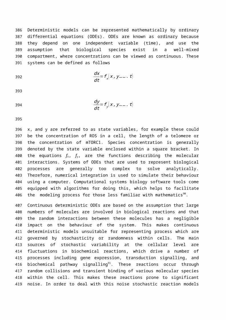
Deterministic models can be represented mathematically by ordinary differential equations (ODEs). ODEs are known as ordinary because they depend on one independent variable (time), and use the assumption that biological species exist in a well-mixed compartment, where concentrations can be viewed as continuous. These systems can be defined as follows
dxdt
=f x ( x , y……. t )
dydt
=f y ( x , y……. t )
x, and y are referred to as state variables, for example these could be the concentration of ROS in a cell, the length of a telomere or the concentration of mTORC1. Species concentration is generally denoted by the state variable enclosed within a square bracket. In the equations fx, fy, are the functions describing the molecular interactions. Systems of ODEs that are used to represent biological processes are generally too complex to solve analytically. Therefore, numerical integration is used to simulate their behaviour using a computer. Computational systems biology software tools come equipped with algorithms for doing this, which helps to facilitate the modeling process for those less familiar with mathematics90.
Continuous deterministic ODEs are based on the assumption that large numbers of molecules are involved in biological reactions and that the random interactions between these molecules has a negligible impact on the behaviour of the system. This makes continuous deterministic models unsuitable for representing process which are governed by stochasticity or randomness within cells. The main sources of stochastic variability at the cellular level are fluctuations in biochemical reactions, which drive a number of processes including gene expression, transduction signalling, and biochemical pathway signalling91. These reactions occur through random collisions and transient binding of various molecular species within the cell. This makes these reactions prone to significant
263264265266267268269270271272273
274
275
276277278279
280
281
282
283
284285286287288289290291
292293294295296297298299
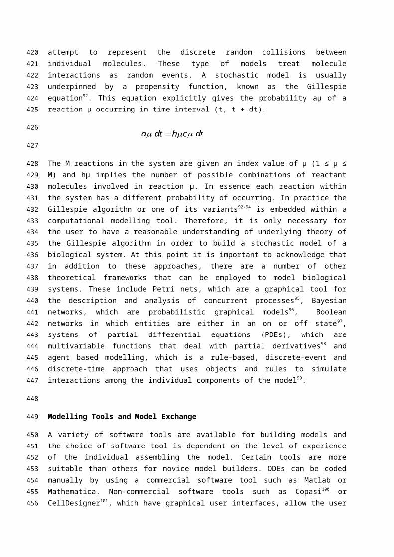
a dt h c dt
noise. In order to deal with this noise stochastic reaction models attempt to represent the discrete random collisions between individual molecules. These type of models treat molecule interactions as random events. A stochastic model is usually underpinned by a propensity function, known as the Gillespie equation92. This equation explicitly gives the probability aμ of a reaction μ occurring in time interval (t, t + dt).
The M reactions in the system are given an index value of μ (1 ≤ μ ≤ M) and hμ implies the number of possible combinations of reactant molecules involved in reaction μ. In essence each reaction within the system has a different probability of occurring. In practice the Gillespie algorithm or one of its variants92-94 is embedded within a computational modelling tool. Therefore, it is only necessary for the user to have a reasonable understanding of underlying theory of the Gillespie algorithm in order to build a stochastic model of a biological system. At this point it is important to acknowledge that in addition to these approaches, there are a number of other theoretical frameworks that can be employed to model biological systems. These include Petri nets, which are a graphical tool for the description and analysis of concurrent processes95, Bayesian networks, which are probabilistic graphical models96, Boolean networks in which entities are either in an on or off state 97, systems of partial differential equations (PDEs), which are multivariable functions that deal with partial derivatives98 and agent based modelling, which is a rule-based, discrete-event and discrete-time approach that uses objects and rules to simulate interactions among the individual components of the model99.
Modelling Tools and Model Exchange
A variety of software tools are available for building models and the choice of software tool is dependent on the level of experience of the individual assembling the model. Certain tools are more suitable than others for novice model builders. ODEs can be coded manually by using a commercial software tool such as Matlab or Mathematica. Non-commercial software tools such as Copasi100 or CellDesigner101, which have graphical user interfaces, allow the user to build the model by creating a succession of kinetic reactions/a process diagram, which in the in the case of a deterministic model is then converted to a series of coupled ODEs. As discussed in the previous section the software tool then uses an algorithm to solve the ODEs and produce a deterministic output. Once a computational model has been assembled, it is important that it can be both easily accessed and updated by the community as a whole. To facilitate model portability a number of exchange frameworks have been developed102. These frameworks allow models to be shared and reused by researchers even if they do not use the same modeling software tool. At present, the leading exchange format is the systems biology markup language (SBML)103. This framework is supported by a broad range of modelling software tools (http://sbml.org/SBML_Software_Guide/SBML_Software_Summary). Models that have been encoded in this format can be archived in the BioModels database, a repository designed specifically for archiving models of biological systems104.
300301302303304
305
306
307308309310311312313314315316317318319320
321
322
323324325326327328329330331332333334335336337338
339
Computational Models of Mitochondrial Dynamics
As outlined, oxidative stress and the emission of ROS by mitochondria is one of the fundamental cellular processes that impacts aging. Therefore, it is unsurprising that various aspects of mitochondrial dynamics have been modelled over the years (for a comprehensive review see Kowald and Klipp (2014)16). An early network model of mitochondrial dynamics that examined this was developed by Kowald and Kirkwood (1994). This model showed that during increased free radical production and/or inadequate protection from these free radicals, damage can occur to an otherwise stable translation system105. Another area of keen focus is mitochondrial fission and fusion. Briefly, fission and fusion events can be viewed as mitochondrial caretakers whose responsibility it is to control cellular ATP concentration, and to mitigate against the accumulation of damage to mitochondrial DNA (mtDNA). One of the earliest models that focused on these processes was the model developed by Kowald et al. (2005). In this model stochastic simulation of mitochondrial replication, mutation and degradation showed a low mosaic pattern of oxidative phosphorylation (OXPHOS) impaired cells in old organisms106. More recently, Tam and colleagues (2013) used computational modelling to investigate the effects of mitochondrial fusion and fission dynamics on mutant mtDNA accumulation107. In this stochastic model, simulations indicated that the slowing down of mitochondrial fusion-fission results in higher variability in the mtDNA mutation burden among cells over time, and mtDNA mutations have a higher propensity to clonally expand due to an increase in stochasticity. The model was able to suggest that the protective ability of retrograde signalling (biochemical communication between mitochondria and nucleus) depends on the efficiency of fusion-fission process107. Another model which focuses on fusion-fission cycles is the model by Figge and colleagues (2012). This probalistic model demonstrated that cycles of fusion and fission and mitophagy are needed to maintain a high average quality of mitochondria, even under conditions in which random molecular damage is present108. Recent mitochondrial models have also focused on specific regions within the mitochondrial ETC. For instance, a model of superoxide production at complexes I and III of the ETC, was able to generate an improved mechanistic understanding of how ROS are generated by complex III. This model also described ROS production by antimycin A inhibited complex III. In order to validate the model, output from its simulations was matched to experimental data from rodents109. On a similar theme Markevich and Hoek (2015) used a computational model of mitochondrial bioenergetics to monitor superoxide production under different substrate conditions. Their model suggested that the semiquinone of Complex I should be included as an additional source of ROS110.
340
341
342
343344345346347348349350351352353354355356357358359360361362363364365366367368369370371372373
374
375
376
377
378

Telomere Models
A number of models have explored telomere dynamics. Most recently, Bartholomäus and colleagues (2014) used a computational model to investigate telomere length under a variety of perturbations111. The model was used to explore telomeres during different conformational states, specifically t-loops, G-quadruplex structures and those being elongated by telomerase. This deterministic model was used to examine how different levels of telomerase impacted telomere length. Moreover, the authors used the model to explore the impact of adding different levels of a G4-stabilising drug on the distribution of telomere lengths. Several older models can be found in the literature. Others of note include the model by Rodriguez-Brenes and Peskin (2010) who modelled telomere state on the basis of the biophysics of t-loop formation112. The model was able to predict the steady-state length distribution for telomerase positive cells, the time evolution of telomere length, and the life span of a cell line on the basis of the levels of telomeric repeat-binding factor 2; a protein that protects telomeres from end-to-end fusion of chromosomes. The model was also able to predict the life span of a cell line based on telomerase levels. Stochastic models of telomere dynamics include the model by Portugal et al. (2008) which made the assumption that cell division is a stochastic phenomenon whose probability decreases linearly with telomere shortening113. Proctor and Kirkwood (2003) also used a model informed by probability to model cellular senescence as a result of telomere state17. From an oxidative stress perspective Trusina (2014) recently used a computational model to investigate the effect of genotoxic stress on telomere attrition 114. Virtual populations of cells were compared and it was found that when ROS was distributed unequally among cells, telomere shortening increased longevity, while also reducing the DNA mutation rate.
Computational Modelling of Metabolic Signalling
In the first section of this paper we described the increasing attention there has been on certain metabolic pathways and how they may have a significant role to play in longevity. Most notably we identified those metabolic pathways that are defined by mTOR and by sirtuins. Several attempts have been made to computationally model various aspects of these pathways115. For example, Kriete et al. (2010) developed a computational model that included the mTOR pathway together with other pathways associated with intrinsic aging116. This rule based model is of note as it encapsulated many important aspects of aging, including mitochondrial biosynthesis, metabolic fluxes, mTOR as an energy sensor and NF-κB, to detect oxidative stress. Another model which successfully included oxidative stress is that developed by Smith and Shanley (2013)117. By building a model of insulin signalling in rodent adipocytes that included transcriptional feedback through the Forkhead box type O (FOXO) transcription factor, it was demonstrated that oxidative stress can have a significant effect on insulin signalling and aging. The model produced a range of findings including the combination of insulin and oxidative stress produced a lower degree of activation of insulin signalling than insulin alone. Antioxidant defences were upregulated in the presence of fasting and weak oxidative stress, whereas, stronger oxidative stress caused short term activation of insulin signalling. The model also demonstrated that if prolonged high insulin may negate the protective effects of moderate oxidative
379
380
381
382383384385386387388389390391392393394395396397398399400401
402
403
404405406407408409410411412413414415416417418419
stress. The complex nature of this model is evident, but, combining it with other factors that can influence insulin signalling such as the mTOR pathway could add to our understanding of insulin signalling.
Computational Models of DNA Methylation Dynamics and Aging
In spite of increasing age related experimental data there is a paucity of computational models that have focused specifically on intrinsic aging and DNA methylation dynamics. However, methylation dynamics have been represented computationally within a number of disease states. For instance, Mc Govern et al. (2012) developed a dynamic multi-compartmental model of DNA methylation, which was used as a predictive tool for hematological malignancies118. The model centred on the activity of DNMTs. PDEs were used to represent methylation reactions and the model was able to predict the relative abundances of unmethylated, hemimethylated, fully methylated, and hydroxymethylated CpG dyads in the DNA of cells with fully functional Dnmt and Tet proteins. It would be worthwhile adapting this model to include oxidative stress, folate biochemistry and the effects of aging on the activity of the methylation enzymes. This model is also deterministic in nature. However, it has been recognised that DNA methylation dynamics are susceptible to inherent stochasticity119. Consequently a number of theoretical frameworks have been proposed for modeling the noise associated with DNA methylation dynamics. For example, reduced mathematical representations of methylation dynamics have been proposed by Riggs and Xiong (2004) 120 and more recently by Jeltsch and Jurkowska (2014), in which DNA methylation at each genomic site is determined by the activity of Dnmts, demethylation enzymes, and the DNA replication rate 121. An awareness of the stochastic nature of these mechanisms has important implications for the aging process, as experimental evidence indicates that the persistent nature of the human methylome results give rise to this noise122. Accordingly, it is imperative that computational models which seek to represent the dynamics of DNA methylation need to account for this inherent variability. One such recent model that has dealt with the intrinsic stochasticity associated with DNA methylation is the model developed by Przybilla et al. (2014), which simulated age-related changes of DNA methylation in stem cells. The findings of this model, which compared age-related changes of regulatory states in quiescent stem cells, with those observed in proliferating cells, suggest that epigenetic aging strongly affects stem cell heterogeneity and that homing at stem cell niches retarded epigenetic aging123.
Cholesterol Metabolism and Aging
The aging process results in the gradual decline of a biological system. This decline is associated with a broad range of pathological states. An example of this decline is the dysregulation of cholesterol metabolism which is inextricably linked to CVD. Therefore, a keen area of focus is how intrinsic aging impacts whole-body cholesterol metabolism124-127. Recently we developed a whole-body model that attempted to capture whole-body cholesterol metabolism. The model was used to examine how age related mechanistic changes to the intestinal absorption of cholesterol resulted in a rise in low-density lipoprotein cholesterol (LDL-C), as increased levels are a risk factor for CVD. The model also revealed that an age related decrease in the hepatic clearance of LDL-C resulted in significant rise in LDL-C by 65 years of age. This model is coded in SBML and is archived in the BioModels database (http://www.ebi.ac.uk/biomodels-main/BIOMD0000000434). In theory this model should be
420421422
423
424425426427428429430431432433434435436437438439440441442443444445446447448449
450
451
452453454455456457458459460461
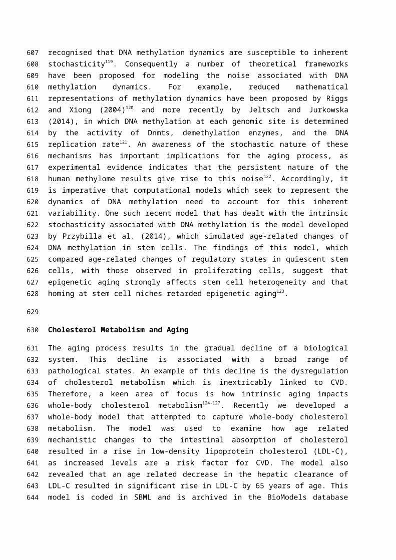
straightforward to update and expand to include other important aspects of aging. As we have eluded to, the free radical theory of aging is a useful means of gluing together disparate aspects of the aging process. It is therefore possible to extend this model by framing it around the insidious rise in ROS that occurs with age in endothelial, vascular smooth muscle, and adventitial cells. This rise in ROS is suggested to be the key driver in a signalling cascade that results in atherosclerosis. Atherosclerosis occurs when LDL molecules migrate into the artery wall at a site which is undermined by endothelial damage. The LDLs are then oxidised upon coming into contact with ROS. The oxidatively modified lipoproteins (oxLDL) are more atherogenic than the native LDL and lead to the recruitment of the macrophages to the site of the lesion. Monocytes pass into the intima before differentiating into macrophages. These molecules engulf oxidized LDL to form cholesterol-laden foam cells. This ultimately results in the formation of an atherosclerotic plaque which eventually ruptures and causes an artery to block128 (Figure 2). This can lead to a stroke or myocardial infarction129. Computational modeling offers a way of dealing with the different molecular, cellular and hemodynamic events associated with this process.
Brain Aging and Pathology
Recently, we also created a computational model which incorporated key brain regions that characterise AD and combined these with the homeostatic regulation of the stress hormone cortisol130. The aim of this model was to examine how increased levels of cortisol impinge on the integrity of the hippocampal region of the brain, which is the core pathological substrate for AD. The model was able to replicate the in vivo aging of the hippocampus. Moreover, both acute and chronic elevations in cortisol increased aging-associated hippocampal atrophy and concomitant loss in the activity of the hippocampus. This computational systems model could be updated to include a number of other processes. For instance, cortisol is synthesised from cholesterol and also acts is also involved in provoking the breakdown of lipids, and a wide variety of other metabolites 131. Therefore, the model could be integrated with the cholesterol model discussed previously. Moreover, this model could be used as a framework for investigating vascular dementia (VAD). VAD is underpinned by a dysregulation in the supply of O2 following a stroke or small vessel deterioration, and oxidative stress is central to the processes that underpin the progression of VAD132. Oxygen deprivation results in mitochondrial dysregulation and the release of ROS133. This increase in oxidative stress damages blood vessels and neurons, resulting in a process which has been termed neurovascular uncoupling134, 135. Moreover, this burst of ROS can disrupt mitochondrial function and further induce hypoxia and oxidative stress136.
A recent ODE model explored a number of the cellular processes associated with Parkinsons Disease (PD). Among the many cellular features of this model, the feedback interactions between damaged α-synuclein and ROS137 were explored. Simulation results showed, hat the Parkinsonian condition, with elevated oxidative stress and misfolded α-synuclein accumulation, can be induced in the model by intrinsic aging, together with exposure to toxins and genetic defects. Computational approaches could also be used to investigate other key aspects of brain aging. For instance, many individuals with Parkinson’s disease report problems with their respiratory, cardiovascular, and gastrointestinal systems138. There is also ample evidence of increased neuroinflammatioin Parkinsons individuals, due to oxidative stress, with reports of increased levels of cytokines, macrophages and microglia
462463464465466467468469470471472473474475
476
477
478479480481482483484485486487488489490491492493494
495496497498499500501502503

activation in brain tissues139,140. A computational model could thus consider abnormalities in central autonomic nuclei, as to our knowledge, there have been no studies to determine whether abnormalities in central autonomic nuclei contributes to autonomic dysfunction or whether peripheral autonomic nuclei also show perturbed development and increased inflammation in PD. Autonomic dysfunction could be reflective of systemic autonomic pathology in PD, and that in fact PD is, in part, an autonomic disorder. It is therefore logical that integrated approaches are required to disentangle the pathological onset of this disease. A worthwhile approach that could address these questions would be to construct a computational systems model of these key processes. In Figure 3, we have used Systems Biology Graphical Notation (SBGN) to represent these processes, which could be modelled computationally.
Other recent Models that have focused on Integrating Aspects of Aging
To date, no model has been able to represent aging in its entirety. However, there have been a number of recent examples, whereby various components associated with aging have been integrated together within a mathematical framework, in an attempt to complete a more global view of how aging impacts a particular biological system. For example, Xue and colleagues (2007) demonstrated that aging is associated with the alteration of a few key brain network modules instead of many, and that the aging process preferentially affects regulatory nodes involved in network stability141. Multi-level aging based models have also been used to gain an insight into intracellular protein aggregate damage, during aging in Escherichia coli142. Moreover, multi-scale models have also had a mammalian focus, for example to examine collagen turnover and the adaptive nature of arterial tissue, in response to mechanical and chemical stimuli143. Furthermore, this type of modelling has also been utilised to examine disease pathophysiology, such as the muscle fibre arrangement and damage susceptibility in Duchenne muscular dystrophy144.
FUTURE OPPORTUNITIES AND CHALLENGES
As outlined, the intrinsic biological mechanisms which characterise the aging process are complex and their activities transcend scale and time. In addition, they involve the interplay of a broad range of molecular, biochemical and physiological processes. In the main, computational models have focused on these process at a cellular level. However, these models are not an adequate representation of whole body human aging. In the final section, we will explore the challenges and opportunities for the future integration of mechanistic models associated with the aging process.
Embedding Existing Models into a Multi-Scale Holistic Framework of Aging
A long term goal of aging research is to have whole-body mechanistic models of the aging process. It is important to note that there are currently no models of this nature in existence. However, in order to fully computationally represent aging from cell to tissue level, there are a number of outstanding
504505506507508509510511512513
514
515
516517518519520521522523524525526527
528
529
530
531532533534535536
537
538
539540541
challenges that remain. Rather than reinventing the wheel it is worth considering extending existing models. In this final section we will outline some of the challenges that exist in combining models and will propose a number of potential solutions. It is important to recognise that a number of these biological systems need to be further characterised before they can be successfully represented by a computational model. A solution to this problem could be to firstly work on aspects of the aging process that are reasonably well characterised, so that future models are founded upon well characterised biological mechanisms. Moreover, it is important that model building is coupled closely with wet-laboratory experimentation. Systems biology experiments that are designed with existing in silico models firmly embedded within their methodology would significantly improve both the model and extend our understanding of the underlying biology. Another significant issue relates to representing biological systems at different levels of scale. It is common place to represent biological systems using models which consist of a system of ODEs that can be analysed, whose dynamics can be solved using a computer. This deterministic approach neglects those reactions that occur at a much smaller scale and involve fluctuations in low molecular populations. Implementing models which combine both the deterministic and stochastic features of biological systems is challenging. However, recently there have been some examples of computational models that have succeeded in accounting for both these effects. For example, Singhania (2011)145 used a hybrid approach that combined differential equations and discrete Boolean networks to represent mammalian cell cycle regulation. This is particularly important from the perspective of the aging process as in order to truly represent it requires the integration of a variety of processes which traverse different biological and temporal scales. Assembling holistic models which represent the aging process is also hindered by the need to determine realistic values for the many parameters that are the essence of large complex models of biological systems. Due to the nature of the experiments it can be difficult to estimate these parameters from existing experimental data. It is important to recognise however that this is a persistent problem within systems biology generally. Thus, as previously eluded to it is necessary to align computational modelling within any future experimental methodology. In addition a broad range of statistical techniques have been applied to this area recently. For instance, Aitken et al. (2015) embedded an algorithm based on Bayesian inference within the computational systems biology software tool Dizzy146, 147. There are several other approaches in which statistical techniques can be used to estimate unknown parameters in systems biology148. Continuing developments in this area will no doubt increase in the utility of computational systems models, and this will be of benefit to those models which represent aging.
Conclusion
In this paper we have presented a broad overview of some of the processes associated with the biology of aging. We have also introduced a number of approaches that are currently used to computationally model biological systems and have described in detail a number of models that have been developed to represent a wide variety of discrete components of the aging process. Some of these models include the key role of ROS in the aging process, while others do not. From our perspective, it is hoped that by converging around ROS in coming years we will witness a more
542543544545546547548549550551552553554555556557558559560561562563564565566567568569570571572573
574
575
576
577578579580581582
comprehensive view of aging that encapsulates the various different mechanisms and their interactions, whose dysregulation result in age associated disease.
References
1. WHO. World Population Ageing 2013. Department of Economic and Social Affairs Population Division 2013.
2. Yancik R, Ries LA. Aging and cancer in America. Demographic and epidemiologic perspectives. Hematol Oncol Clin North Am 2000, 14:17-23.
3. Jousilahti P, Vartiainen E, Tuomilehto J, Puska P. Sex, age, cardiovascular risk factors, and coronary heart disease: a prospective follow-up study of 14 786 middle-aged men and women in Finland. Circulation 1999, 99:1165-1172.
4. Lindsay J, Laurin D, Verreault R, Hebert R, Helliwell B, Hill GB, McDowell I. Risk factors for Alzheimer's disease: a prospective analysis from the Canadian Study of Health and Aging. Am J Epidemiol 2002, 156:445-453.
5. Reeve A, Simcox E, Turnbull D. Ageing and Parkinson's disease: why is advancing age the biggest risk factor? Ageing Res Rev 2014, 14:19-30.
6. Kelly GA, Lazarus J. Perceptions of Successful Aging: Intergenerational Voices Value Well-Being. Int J Aging Hum Dev 2015.
7. Chauhan A, Liebal UW, Vera J, Baltrusch S, Junghanss C, Tiedge M, Fuellen G, Wolkenhauer O, Kohling R. Systems biology approaches in aging research. Interdiscip Top Gerontol 2015, 40:155-176.
8. Auffray C, Imbeaud S, Roux-Rouquie M, Hood L. From functional genomics to systems biology: concepts and practices. C R Biol 2003, 326:879-892.
9. Hou L, Huang J, Green CD, Boyd-Kirkup J, Zhang W, Yu X, Gong W, Zhou B, Han JD. Systems biology in aging: linking the old and the young. Curr Genomics 2012, 13:558-565.
10. Choi H, Mc Auley MT, Lawrence DA. Prenatal exposures and exposomics of asthma. . AIMS Environmental Science 2015, 2:87-109.
11. Kriete A, Lechner M, Clearfield D, Bohmann D. Computational systems biology of aging. Wiley Interdiscip Rev Syst Biol Med 2011, 3:414-428.
12. Kilner J, Corfe BM, McAuley MT, Wilkinson SJ. A deterministic oscillatory model of microtubule growth and shrinkage for differential actions of short chain fatty acids. Mol Biosyst 2015.
13. Brodland GW. How computational models can help unlock biological systems. Semin Cell Dev Biol 2015.
14. Mc Auley MT, Choi H, Mooney K, Paul E, Miller VM. Systems Biology and Synthetic Biology: A New Epoch for Toxicology Research. Advances in Toxicology 2015, 2015:14.
15. Mc Auley MT, Mooney KM. Computational systems biology for aging research. Interdiscip Top Gerontol 2015, 40:35-48.
16. Kowald A, Klipp E. Mathematical models of mitochondrial aging and dynamics. Prog Mol Biol Transl Sci 2014, 127:63-92.
17. Proctor CJ, Kirkwood TB. Modelling cellular senescence as a result of telomere state. Aging Cell 2003, 2:151-157.
583584
585
586
587
588
589
590591592593594595596597598599600601602603604605606607608609610611612613614615616617618619620621622623624625626627
18. Proctor CJ, Soti C, Boys RJ, Gillespie CS, Shanley DP, Wilkinson DJ, Kirkwood TB. Modelling the actions of chaperones and their role in ageing. Mech Ageing Dev 2005, 126:119-131.
19. Gavrilov LA, Gavrilova NS. Evolutionary theories of aging and longevity. ScientificWorldJournal 2002, 2:339-356.
20. Sanz A, Stefanatos RK. The mitochondrial free radical theory of aging: a critical view. Curr Aging Sci 2008, 1:10-21.
21. Harman D. The biologic clock: the mitochondria? J Am Geriatr Soc 1972, 20:145-147.22. Bender A, Krishnan KJ, Morris CM, Taylor GA, Reeve AK, Perry RH, Jaros E, Hersheson JS,
Betts J, Klopstock T, et al. High levels of mitochondrial DNA deletions in substantia nigra neurons in aging and Parkinson disease. Nat Genet 2006, 38:515-517.
23. Yu-Wai-Man P, Lai-Cheong J, Borthwick GM, He L, Taylor GA, Greaves LC, Taylor RW, Griffiths PG, Turnbull DM. Somatic mitochondrial DNA deletions accumulate to high levels in aging human extraocular muscles. Invest Ophthalmol Vis Sci 2010, 51:3347-3353.
24. Trifunovic A, Larsson NG. Mitochondrial dysfunction as a cause of ageing. J Intern Med 2008, 263:167-178.
25. Youle RJ, van der Bliek AM. Mitochondrial fission, fusion, and stress. Science 2012, 337:1062-1065.
26. Jensen MB, Jasper H. Mitochondrial proteostasis in the control of aging and longevity. Cell Metab 2014, 20:214-225.
27. Kim I, Rodriguez-Enriquez S, Lemasters JJ. Selective degradation of mitochondria by mitophagy. Arch Biochem Biophys 2007, 462:245-253.
28. Short KR, Bigelow ML, Kahl J, Singh R, Coenen-Schimke J, Raghavakaimal S, Nair KS. Decline in skeletal muscle mitochondrial function with aging in humans. Proc Natl Acad Sci U S A 2005, 102:5618-5623.
29. Barja G. Aging in vertebrates, and the effect of caloric restriction: a mitochondrial free radical production-DNA damage mechanism? Biol Rev Camb Philos Soc 2004, 79:235-251.
30. Ristow M, Schmeisser K. Mitohormesis: Promoting Health and Lifespan by Increased Levels of Reactive Oxygen Species (ROS). Dose-Response 2014, 12:288-341.
31. Aubert G, Lansdorp PM. Telomeres and aging. Physiol Rev 2008, 88:557-579.32. Collins K, Mitchell JR. Telomerase in the human organism. Oncogene 2002, 21:564-579.33. Harley CB, Futcher AB, Greider CW. Telomeres shorten during ageing of human fibroblasts.
Nature 1990, 345:458-460.34. Bodnar AG, Ouellette M, Frolkis M, Holt SE, Chiu CP, Morin GB, Harley CB, Shay JW,
Lichtsteiner S, Wright WE. Extension of life-span by introduction of telomerase into normal human cells. Science 1998, 279:349-352.
35. Sahin E, Depinho RA. Linking functional decline of telomeres, mitochondria and stem cells during ageing. Nature 2010, 464:520-528.
36. Weindruch R, Walford R. The Retardation of Aging and Disease by Dietary Restriction. Charles C Thomas, Springfield, Illinois 1988.
37. Masoro EJ. Overview of caloric restriction and ageing. Mech Ageing Dev 2005, 126:913-922.38. Sinclair DA. Toward a unified theory of caloric restriction and longevity regulation. Mech
Ageing Dev 2005, 126:987-1002.39. Anderson RM, Shanmuganayagam D, Weindruch R. Caloric restriction and aging: studies in
mice and monkeys. Toxicol Pathol 2009, 37:47-51.40. Colman RJ, Beasley TM, Kemnitz JW, Johnson SC, Weindruch R, Anderson RM. Caloric
restriction reduces age-related and all-cause mortality in rhesus monkeys. Nat Commun 2014, 5:3557.
41. Lin SJ, Ford E, Haigis M, Liszt G, Guarente L. Calorie restriction extends yeast life span by lowering the level of NADH. Genes Dev 2004, 18:12-16.
628629630631632633634635636637638639640641642643644645646647648649650651652653654655656657658659660661662663664665666667668669670671672673674675676
42. Gredilla R, Sanz A, Lopez-Torres M, Barja G. Caloric restriction decreases mitochondrial free radical generation at complex I and lowers oxidative damage to mitochondrial DNA in the rat heart. Faseb j 2001, 15:1589-1591.
43. Agarwal S, Sohal RS. Relationship between susceptibility to protein oxidation, aging, and maximum life span potential of different species. Exp Gerontol 1996, 31:365-372.
44. Guarente L. Calorie restriction and sirtuins revisited. Genes & Development 2013, 27:2072-2085.
45. Lin SJ, Defossez PA, Guarente L. Requirement of NAD and SIR2 for life-span extension by calorie restriction in Saccharomyces cerevisiae. Science 2000, 289:2126-2128.
46. Rogina B, Helfand SL. Sir2 mediates longevity in the fly through a pathway related to calorie restriction. Proc Natl Acad Sci U S A 2004, 101:15998-16003.
47. Tissenbaum HA, Guarente L. Increased dosage of a sir-2 gene extends lifespan in Caenorhabditis elegans. Nature 2001, 410:227-230.
48. Haigis MC, Guarente LP. Mammalian sirtuins--emerging roles in physiology, aging, and calorie restriction. Genes Dev 2006, 20:2913-2921.
49. Canto C, Auwerx J. Caloric restriction, SIRT1 and longevity. Trends Endocrinol Metab 2009, 20:325-331.
50. Cohen HY, Miller C, Bitterman KJ, Wall NR, Hekking B, Kessler B, Howitz KT, Gorospe M, de Cabo R, Sinclair DA. Calorie restriction promotes mammalian cell survival by inducing the SIRT1 deacetylase. Science 2004, 305:390-392.
51. Xia N, Strand S, Schlufter F, Siuda D, Reifenberg G, Kleinert H, Forstermann U, Li H. Role of SIRT1 and FOXO factors in eNOS transcriptional activation by resveratrol. Nitric Oxide 2013, 32:29-35.
52. Shinmura K, Tamaki K, Ito K, Yan X, Yamamoto T, Katsumata Y, Matsuhashi T, Sano M, Fukuda K, Suematsu M, et al. Indispensable role of endothelial nitric oxide synthase in caloric restriction-induced cardioprotection against ischemia-reperfusion injury. Am J Physiol Heart Circ Physiol 2015, 308:H894-903.
53. Qiu X, Brown K, Hirschey MD, Verdin E, Chen D. Calorie restriction reduces oxidative stress by SIRT3-mediated SOD2 activation. Cell Metab 2010, 12:662-667.
54. Mazucanti CH, Cabral-Costa JV, Vasconcelos AR, Andreotti DZ, Scavone C, Kawamoto EM. Longevity Pathways (mTOR, SIRT, Insulin/IGF-1) as Key Modulatory Targets on Aging and Neurodegeneration. Curr Top Med Chem 2015, 15:2116-2138.
55. Laplante M, Sabatini DM. mTOR signaling at a glance. Journal of Cell Science 2009, 122:3589-3594.
56. Heitman J, Movva NR, Hall MN. Targets for cell cycle arrest by the immunosuppressant rapamycin in yeast. Science 1991, 253:905-909.
57. Takahashi T, Hara K, Inoue H, Kawa Y, Tokunaga C, Hidayat S, Yoshino K, Kuroda Y, Yonezawa K. Carboxyl-terminal region conserved among phosphoinositide-kinase-related kinases is indispensable for mTOR function in vivo and in vitro. Genes Cells 2000, 5:765-775.
58. Jia K, Chen D, Riddle DL. The TOR pathway interacts with the insulin signaling pathway to regulate C. elegans larval development, metabolism and life span. Development 2004, 131:3897-3906.
59. Zid BM, Rogers AN, Katewa SD, Vargas MA, Kolipinski MC, Lu TA, Benzer S, Kapahi P. 4E-BP extends lifespan upon dietary restriction by enhancing mitochondrial activity in Drosophila . Cell 2009, 139:149-160.
60. Schieke SM, Phillips D, McCoy JP, Jr., Aponte AM, Shen RF, Balaban RS, Finkel T. The mammalian target of rapamycin (mTOR) pathway regulates mitochondrial oxygen consumption and oxidative capacity. J Biol Chem 2006, 281:27643-27652.
61. Wei Y, Zhang Y-J, Cai Y, Xu M-H. The role of mitochondria in mTOR-regulated longevity. Biological Reviews 2015, 90:167-181.
677678679680681682683684685686687688689690691692693694695696697698699700701702703704705706707708709710711712713714715716717718719720721722723724725726
62. Brunet A, Berger SL. Epigenetics of aging and aging-related disease. J Gerontol A Biol Sci Med Sci 2014, 69 Suppl 1:S17-20.
63. Jaenisch R, Bird A. Epigenetic regulation of gene expression: how the genome integrates intrinsic and environmental signals. Nat Genet 2003, 33 Suppl:245-254.
64. Guo H, Zhu P, Yan L, Li R, Hu B, Lian Y, Yan J, Ren X, Lin S, Li J, et al. The DNA methylation landscape of human early embryos. Nature 2014, 511:606-610.
65. Jung M, Pfeifer GP. Aging and DNA methylation. BMC Biol 2015, 13:7.66. Liu L, Wylie RC, Andrews LG, Tollefsbol TO. Aging, cancer and nutrition: the DNA methylation
connection. Mech Ageing Dev 2003, 124:989-998.67. Esteller M, Fraga MF, Guo M, Garcia-Foncillas J, Hedenfalk I, Godwin AK, Trojan J, Vaurs-
Barriere C, Bignon YJ, Ramus S, et al. DNA methylation patterns in hereditary human cancers mimic sporadic tumorigenesis. Hum Mol Genet 2001, 10:3001-3007.
68. Esteller M, Corn PG, Baylin SB, Herman JG. A gene hypermethylation profile of human cancer. Cancer Res 2001, 61:3225-3229.
69. Kulis M, Esteller M. DNA methylation and cancer. Adv Genet 2010, 70:27-56.70. Quintero-Ronderos P, Montoya-Ortiz G. Epigenetics and Autoimmune Diseases.
Autoimmune Diseases 2012, 2012:16.71. Glier MB, Green TJ, Devlin AM. Methyl nutrients, DNA methylation, and cardiovascular
disease. Mol Nutr Food Res 2014, 58:172-182.72. Cuskelly GJ, Mooney KM, Young IS. Folate and vitamin B12: friendly or enemy nutrients for
the elderly. Proc Nutr Soc 2007, 66:548-558.73. Zampieri M, Ciccarone F, Calabrese R, Franceschi C, Burkle A, Caiafa P. Reconfiguration of
DNA methylation in aging. Mech Ageing Dev 2015.74. Zhang Z, Deng C, Lu Q, Richardson B. Age-dependent DNA methylation changes in the ITGAL
(CD11a) promoter. Mech Ageing Dev 2002, 123:1257-1268.75. Robertson KD, Uzvolgyi E, Liang G, Talmadge C, Sumegi J, Gonzales FA, Jones PA. The human
DNA methyltransferases (DNMTs) 1, 3a and 3b: coordinate mRNA expression in normal tissues and overexpression in tumors. Nucleic Acids Res 1999, 27:2291-2298.
76. Jurkowska RZ, Jurkowski TP, Jeltsch A. Structure and function of mammalian DNA methyltransferases. Chembiochem 2011, 12:206-222.
77. Scourzic L, Mouly E, Bernard OA. TET proteins and the control of cytosine demethylation in cancer. Genome Med 2015, 7:9.
78. Salminen A, Kauppinen A, Hiltunen M, Kaarniranta K. Krebs cycle intermediates regulate DNA and histone methylation: epigenetic impact on the aging process. Ageing Res Rev 2014, 16:45-65.
79. Li Y, Liu Y, Strickland FM, Richardson B. Age-dependent decreases in DNA methyltransferase levels and low transmethylation micronutrient levels synergize to promote overexpression of genes implicated in autoimmunity and acute coronary syndromes. Exp Gerontol 2010, 45:312-322.
80. Peng L, Yuan Z, Ling H, Fukasawa K, Robertson K, Olashaw N, Koomen J, Chen J, Lane WS, Seto E. SIRT1 deacetylates the DNA methyltransferase 1 (DNMT1) protein and alters its activities. Mol Cell Biol 2011, 31:4720-4734.
81. Cencioni C, Spallotta F, Martelli F, Valente S, Mai A, Zeiher A, Gaetano C. Oxidative Stress and Epigenetic Regulation in Ageing and Age-Related Diseases. International Journal of Molecular Sciences 2013, 14:17643.
82. Campos AC, Molognoni F, Melo FH, Galdieri LC, Carneiro CR, D'Almeida V, Correa M, Jasiulionis MG. Oxidative stress modulates DNA methylation during melanocyte anchorage blockade associated with malignant transformation. Neoplasia 2007, 9:1111-1121.
83. Loscalzo J, Barabasi A-L. Systems biology and the future of medicine. Wiley Interdisciplinary Reviews: Systems Biology and Medicine 2011, 3:619-627.
727728729730731732733734735736737738739740741742743744745746747748749750751752753754755756757758759760761762763764765766767768769770771772773774775776

84. Weston AD, Hood L. Systems biology, proteomics, and the future of health care: toward predictive, preventative, and personalized medicine. J Proteome Res 2004, 3:179-196.
85. Vallabhajosyula RR, Raval A. Computational modeling in systems biology. Methods Mol Biol 2010, 662:97-120.
86. Mc Auley MT, Proctor CJ, Corfe BM, Cuskelly CJ, Mooney KM. Nutrition Research and the Impact of Computational Systems Biology. Journal of Computer Science and Systems Biology 2013, 6:271-285.
87. Machado D, Costa RS, Rocha M, Ferreira EC, Tidor B, Rocha I. Modeling formalisms in Systems Biology. AMB Express 2011, 1:45.
88. Seixas FL, Zadrozny B, Laks J, Conci A, Muchaluat Saade DC. A Bayesian network decision model for supporting the diagnosis of dementia, Alzheimers disease and mild cognitive impairment. Comput Biol Med 2014, 51:140-158.
89. Figueredo GP, Siebers PO, Aickelin U, Whitbrook A, Garibaldi JM. Juxtaposition of system dynamics and agent-based simulation for a case study in immunosenescence. PLoS One 2015, 10:e0118359.
90. Ghosh S, Matsuoka Y, Asai Y, Hsin K-Y, Kitano H. Software for systems biology: from tools to integrated platforms. Nat Rev Genet 2011, 12:821-832.
91. Springer M, Paulsson J. Biological physics: harmonies from noise. Nature 2006, 439:27-28.92. Gillespie D. A general method for numerically simulating stochastic time evolution of
coupled chemical-reactions. Journal of Computational Physics 1976, 22::403–434.93. Gibson MA, Bruck J. Efficient exact stochastic simulation of chemical systems with many
species and many channels. . J. Phys. Chem 2000, A 104:1876–1889.94. Gillespie DT, Hellander A, Petzold LR. Perspective: Stochastic algorithms for chemical
kinetics. J Chem Phys 2013, 138:170901.95. Goss PJ, Peccoud J. Quantitative modeling of stochastic systems in molecular biology by
using stochastic Petri nets. Proc Natl Acad Sci U S A 1998, 95:6750-6755.96. Needham CJ, Bradford JR, Bulpitt AJ, Westhead DR. A primer on learning in Bayesian
networks for computational biology. PLoS Comput Biol 2007, 3:e129.97. Wang RS, Saadatpour A, Albert R. Boolean modeling in systems biology: an overview of
methodology and applications. Phys Biol 2012, 9:055001.98. Farlow SJ. Partial Differential Equations for Scientists and Engineers: Dover Books on
Mathematics; 2003.99. An G, Mi Q, Dutta-Moscato J, Vodovotz Y. Agent-based models in translational systems
biology. Wiley Interdiscip Rev Syst Biol Med 2009, 1:159-171.100. Hoops S, Sahle S, Gauges R, Lee C, Pahle J, Simus N, Singhal M, Xu L, Mendes P, Kummer U.
COPASI—a COmplex PAthway SImulator. Bioinformatics 2006, 22:3067-3074.101. Matsuoka Y, Funahashi A, Ghosh S, Kitano H. Modeling and simulation using CellDesigner.
Methods Mol Biol 2014, 1164:121-145.102. Sauro HM, Bergmann FT. Standards and ontologies in computational systems biology. Essays
Biochem 2008, 45:211-222.103. Hucka M, Finney A, Sauro HM, Bolouri H, Doyle JC, Kitano H, Arkin AP, Bornstein BJ, Bray D,
Cornish-Bowden A, et al. The systems biology markup language (SBML): a medium for representation and exchange of biochemical network models. Bioinformatics 2003, 19:524-531.
104. Le Novère N, Bornstein B, Broicher A, Courtot M, Donizelli M, Dharuri H, Li L, Sauro H, Schilstra M, Shapiro B, et al. BioModels Database: a free, centralized database of curated, published, quantitative kinetic models of biochemical and cellular systems. Nucleic Acids Research 2006, 34:D689-D691.
105. Kowald A, Kirkwood TB. Towards a network theory of ageing: a model combining the free radical theory and the protein error theory. J Theor Biol 1994, 168:75-94.
777778779780781782783784785786787788789790791792793794795796797798799800801802803804805806807808809810811812813814815816817818819820821822823824825826

106. Kowald A, Jendrach M, Pohl S, Bereiter-Hahn J, Hammerstein P. On the relevance of mitochondrial fusions for the accumulation of mitochondrial deletion mutants: a modelling study. Aging Cell 2005, 4:273-283.
107. Tam ZY, Gruber J, Halliwell B, Gunawan R. Mathematical modeling of the role of mitochondrial fusion and fission in mitochondrial DNA maintenance. PLoS One 2013, 8:e76230.
108. Figge MT, Reichert AS, Meyer-Hermann M, Osiewacz HD. Deceleration of fusion-fission cycles improves mitochondrial quality control during aging. PLoS Comput Biol 2012, 8:e1002576.
109. Guillaud F, Drose S, Kowald A, Brandt U, Klipp E. Superoxide production by cytochrome bc1 complex: a mathematical model. Biochim Biophys Acta 2014, 1837:1643-1652.
110. Markevich NI, Hoek JB. Computational modeling analysis of mitochondrial superoxide production under varying substrate conditions and upon inhibition of different segments of the electron transport chain. Biochim Biophys Acta 2015, 1847:656-679.
111. Hirt BV, Wattis JA, Preston SP. Modelling the regulation of telomere length: the effects of telomerase and G-quadruplex stabilising drugs. J Math Biol 2014, 68:1521-1552.
112. Rodriguez-Brenes IA, Peskin CS. Quantitative theory of telomere length regulation and cellular senescence. Proc Natl Acad Sci U S A 2010, 107:5387-5392.
113. Portugal RD, Land MG, Svaiter BF. A computational model for telomere-dependent cell-replicative aging. Biosystems 2008, 91:262-267.
114. Trusina A. Stress induced telomere shortening: longer life with less mutations? BMC Syst Biol 2014, 8:27.
115. Auley MT, Mooney KM, Angell PJ, Wilkinson SJ. Mathematical modelling of metabolic regulation in aging. Metabolites 2015, 5:232-251.
116. Kriete A, Bosl WJ, Booker G. Rule-based cell systems model of aging using feedback loop motifs mediated by stress responses. PLoS Comput Biol 2010, 6:e1000820.
117. Smith GR, Shanley DP. Computational modelling of the regulation of Insulin signalling by oxidative stress. BMC Syst Biol 2013, 7:41.
118. McGovern AP, Powell BE, Chevassut TJ. A dynamic multi-compartmental model of DNA methylation with demonstrable predictive value in hematological malignancies. J Theor Biol 2012, 310:14-20.
119. Landan G, Cohen NM, Mukamel Z, Bar A, Molchadsky A, Brosh R, Horn-Saban S, Zalcenstein DA, Goldfinger N, Zundelevich A, et al. Epigenetic polymorphism and the stochastic formation of differentially methylated regions in normal and cancerous tissues. Nat Genet 2012, 44:1207-1214.
120. Riggs AD, Xiong Z. Methylation and epigenetic fidelity. Proc Natl Acad Sci U S A 2004, 101:4-5.
121. Jeltsch A, Jurkowska RZ. New concepts in DNA methylation. Trends Biochem Sci 2014, 39:310-318.
122. Shipony Z, Mukamel Z, Cohen NM, Landan G, Chomsky E, Zeliger SR, Fried YC, Ainbinder E, Friedman N, Tanay A. Dynamic and static maintenance of epigenetic memory in pluripotent and somatic cells. Nature 2014, 513:115-119.
123. Przybilla J, Rohlf T, Loeffler M, Galle J. Understanding epigenetic changes in aging stem cells--a computational model approach. Aging Cell 2014, 13:320-328.
124. Mc Auley MM, Wilkinson DJ, Jones JJ, Kirkwood TT. A whole-body mathematical model of cholesterol metabolism and its age-associated dysregulation. BMC Syst Biol 2012, 6:130.
125. Mc Auley M, Jones J, Wilkinson D, Kirkwood T. Modelling Lipid Metabolism to Improve Healthy Ageing. BMC Bioinformatics 2005, 6:P21.
126. Mc Auley MT, Mooney KM. Computationally Modeling Lipid Metabolism and Aging: A Mini-review. Computational and Structural Biotechnology Journal 2015, 13:38-46.
827828829830831832833834835836837838839840841842843844845846847848849850851852853854855856857858859860861862863864865866867868869870871872873874875876
127. Mooney KM, Mc Auley MT. Cardiovascular disease and healthy ageing. Journal of Integrative Cardiology 2015, 1:76-78.
128. Bentzon JF, Otsuka F, Virmani R, Falk E. Mechanisms of Plaque Formation and Rupture. Circulation Research 2014, 114:1852-1866.
129. Badimon L, Vilahur G. Thrombosis formation on atherosclerotic lesions and plaque rupture. Journal of Internal Medicine 2014, 276:618-632.
130. McAuley MT, Kenny RA, Kirkwood TB, Wilkinson DJ, Jones JJ, Miller VM. A mathematical model of aging-related and cortisol induced hippocampal dysfunction. BMC Neurosci 2009, 10:26.
131. Mc Auley MT, Mooney KM. Lipid metabolism and hormonal interactions: impact on cardiovascular disease and healthy aging. Expert Review of Endocrinology & Metabolism 2014, 9:357-367.
132. Iadecola C. The pathobiology of vascular dementia. Neuron 2013, 80:844-866.133. Popa-Wagner A, Mitran S, Sivanesan S, Chang E, Buga AM. ROS and brain diseases: the good,
the bad, and the ugly. Oxid Med Cell Longev 2013, 2013:963520.134. Zlokovic BV. Neurovascular pathways to neurodegeneration in Alzheimer's disease and other
disorders. Nat Rev Neurosci 2011, 12:723-738.135. Rancillac A, Geoffroy H, Rossier J. Impaired neurovascular coupling in the APPxPS1 mouse
model of Alzheimer's disease. Curr Alzheimer Res 2012, 9:1221-1230.136. Stanimirovic DB, Friedman A. Pathophysiology of the neurovascular unit: disease cause or
consequence? J Cereb Blood Flow Metab 2012, 32:1207-1221.137. Cloutier M, Wellstead P. Dynamic modelling of protein and oxidative metabolisms simulates
the pathogenesis of Parkinson's disease. IET Syst Biol 2012, 6:65-72.138. Pfeiffer RF. Autonomic dysfunction in Parkinson's disease. Expert Rev Neurother 2012,
12:697-706.139. Perry VH. Innate Inflammation in Parkinson’s Disease. Cold Spring Harbor Perspectives in
Medicine 2012, 2.140. Hwang O. Role of oxidative stress in Parkinson's disease. Exp Neurobiol 2013, 22:11-17.141. Xue H, Xian B, Dong D, Xia K, Zhu S, Zhang Z, Hou L, Zhang Q, Zhang Y, Han J-DJ. A modular
network model of aging. Molecular Systems Biology 2007, 3:n/a-n/a.142. Koleva KZ, Hellweger FL. From protein damage to cell aging to population fitness in E. coli:
Insights from a multi-level agent-based model. Ecological Modelling 2015, 301:62-71.143. Sáez P, Peña E, Tarbell JM, Martínez MA. Computational model of collagen turnover in
carotid arteries during hypertension. International Journal for Numerical Methods in Biomedical Engineering 2015, 31:n/a-n/a.
144. Virgilio KM, Martin KS, Peirce SM, Blemker SS. Multiscale models of skeletal muscle reveal the complex effects of muscular dystrophy on tissue mechanics and damage susceptibility. Interface Focus 2015, 5.
145. Singhania R, Sramkoski RM, Jacobberger JW, Tyson JJ. A hybrid model of mammalian cell cycle regulation. PLoS Comput Biol 2011, 7:e1001077.
146. Aitken S, Kilpatrick AM, Akman OE. Dizzy-Beats: a Bayesian evidence analysis tool for systems biology. Bioinformatics 2015, 31:1863-1865.
147. Ramsey S, Orrell D, Bolouri H. Dizzy: stochastic simulation of large-scale genetic regulatory networks. J Bioinform Comput Biol 2005, 3:415-436.
148. Ashyraliyev M, Fomekong-Nanfack Y, Kaandorp JA, Blom JG. Systems biology: parameter estimation for biochemical models. Febs j 2009, 276:886-902.
Figure Legends
877878879880881882883884885886887888889890891892893894895896897898899900901902903904905906907908909910911912913914915916917918919920921922
923
924
925
FIGURE 1. An integrated overview of aging and some of its key players. This figure emphasises the extent of interplay between the different components that underpin intrinsic aging, and how age-related changes to these components affect health-span and longevity. The integrated nature of this diagram highlights the complexities of ageing and why computational models are needed to help study its dynamics. IGF-1, insulin-like growth factor-1; ROS, reactive oxygen species; PARP, poly ADP ribose polymerase; mTOR, mammalian target of rapamycin.
FIGURE 2. Integrating a computational model of cholesterol metabolism with a variety of other factors involved in the onset of CVD. Our extended model is framed around the insidious rise in ROS that occurs with age. This rise in ROS is a key driver which underpins a pathological cascade that ultimately results in CVD.
FIGURE 3. An SBGN representation of the autonomic nervous system. The aim of this proposed model would be to simulate physiological responses associated with the autonomic nervous system such as heart rate, rate of movements in the gastrointestinal tract, or synthesis of B cells by the spleen. These processes are regulated in part by neurotransmitters and cytokines. Dysregulation of these processes together with oxidative stress have been strongly implicated in the pathology which underpins Parkinson’s disease. NE, Norepinephrine; 5HT, serotonin; Ach, acetylcholine.
Further Reading
Systems BiologyEdda Klipp, Wolfram Liebermeister, Christoph Wierling, Axel Kowald, Hans Lehrach, Ralf HerwigISBN: 978-3-527-31874-22009, Wiley-Blackwell
Aging and Health - A Systems Biology Perspective. Interdiscipl Top Gerontol. Basel, Karger, 2015
Systems Biology of Parkinson’s DiseasePeter Wellstead & Mathieu CloutierISBN: 14939012652012, Springer
Systems Biology in Practice: Concepts, Implementation and ApplicationEdda Klipp, Ralf Herwig, Axel Kowald, Christoph Wierling & Hans LehrachISBN: 35273107892005, Wiley VCH
A First Course in Systems BiologyEberhard VoitISBN: 08153446782012, Garland Science
926927928929930931
932
933934935936
937
938939940941942943
944
945
946947948949950951952953954955956957958959960961962963964965966967968
Related Articles
Article ID [or Subtopic] Article titleWSBM.1209 Modeling cellular compartmentation in one-carbon metabolismwsbm.126 Computational systems biology of agingwsbm.52 Computational modeling of mammalian signaling networks
969
970
971
972
973
974