Anatomy of spine and autonomic ns
45
Joey Erll Hulleza RN October 20, 2014 ANATOMY OF THE SPINE
-
Upload
joey-hulleza -
Category
Healthcare
-
view
279 -
download
0
Transcript of Anatomy of spine and autonomic ns

Joey Erll Hulleza RN
October 20, 2014
ANATOMY OF THE SPINE

OverviewThe spine is made of 33 individual bones stacked one on the top of the other.
Ligaments and muscles connect bones together and keep them aligned
The spinal column provides the main support for your body allowing you to stand Upright, bend, and twist
Protected inside the bones, the spinal cord connects your body to the brain Allowing movement of your arms and legs
Strong muscles and bones, flexible tendons and ligaments and sensitive nervesContribute to a healthy spine

Spinal Anatomy

Spinal curves The neck (cervical) and low back (lumbar) regions have a slight Concave curve.
The thoracic and sacral regions have a gentle convex curve
The curves work like a coiled spring to absorb shock and Maintain balance, and allow range of motion throughoutThe spinal column.

An abnormal curve of the lumbar spine is LORDOSIS, also called sway back.
An abnormal curve of the Thoracic spine is KYPHOSIS, also called hunchback
An abnormal curve from side-to-side is called SCOLIOSIS
Spinal curves

Vertebrae

• Cervical region (7)1
• Thoracic region (12)2
• Lumbar region (5)3
• Sacral region (5)4
• Coccyx region5
Vertebrae: numbered and divided into regions
Cervical (neck)
The main function of cervical spine is to support the Weight of the head (about 10lbs)
C1- is the ring- shaped ATLAS that connects directly to theSkull; this joint allows for the nodding or “yes” motion of The head
C2 is the peg shaped AXIS, this jointallows for the side-to-side or “no” motion of the head.

Thoracic (Mid Back)
The main function of the thoracic spine is to:hold the rib cage and protect the heart and lungs.

Lumbar (low back)
The main function of the lumbar spine is to bear the weight of the body

Sacrum
The main function of the sacrum is to connect the spine to the hip bones (iliac)
Together with the iliac bones, they form a ring called Pelvic Girdle

Coccyx
The four fused bones of the coccyx or tail bone provide Attachment for ligaments and muscles of the pelvic floor.
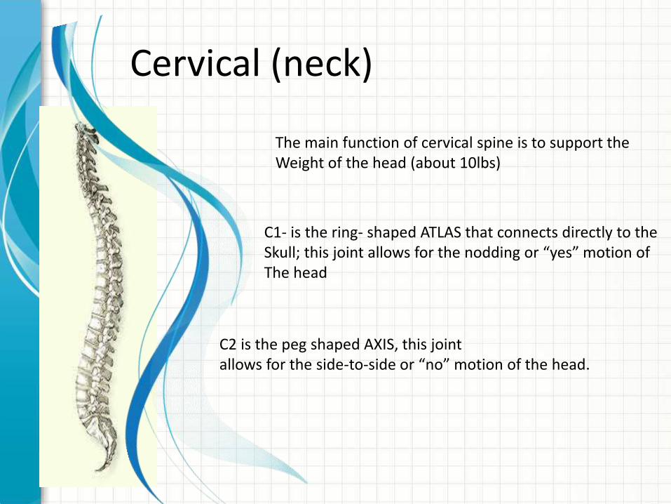
Vertebral Unique regional features:
A drum- shaped body designed to bear weight andWithstand compression
An arch-shaped bone the creates a hollow tube for the spinal cordand nerves.
Star- shaped processes designed as outriggers for the Muscle attachment.

Intervertebral Discs
Annulus – the outer ring, criss-crossing fibrous bands much Like tire thread; inside the disc is a gel-filled center called the Nucleus, much like a tire tube; these bands attach betweenthe bodies of each vertebra
With age, our disc incraesingly lose the ability to reabsorb fluid andBecome brittle and flatter. • We became shorter as we grow older• Osteoporosis• Osteoarthritis• Injury and strain can cause isc to herniate, a condition is which
the nucleus is pushed out through the annulus to compressThe nerve roots—causing back pain.
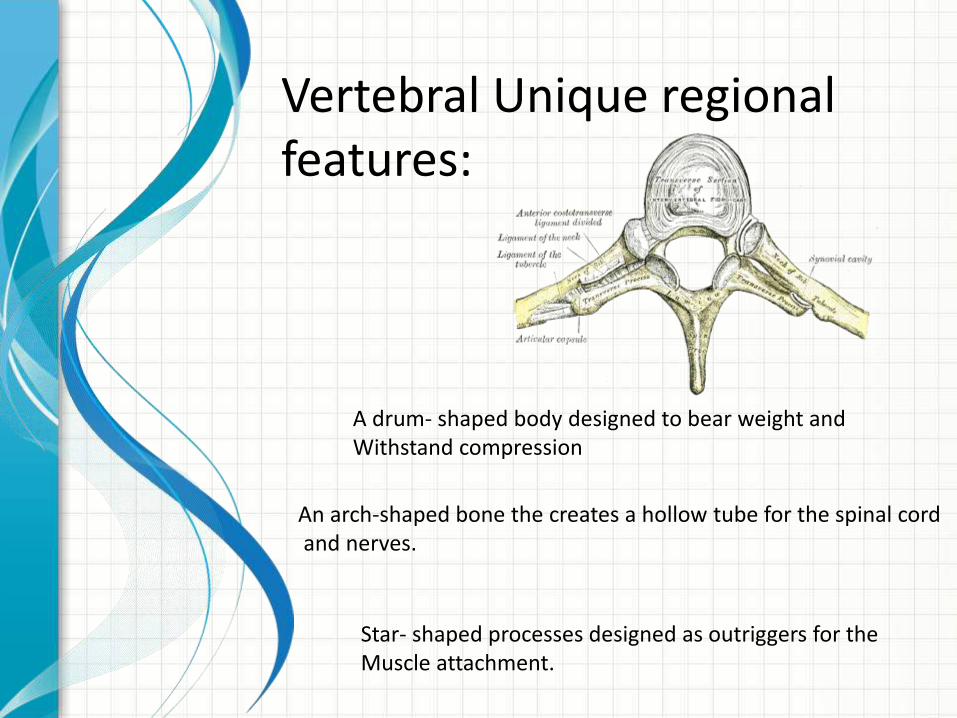
Vertebral Arch and Spinal Canal
Vertebral arch is made of 2 supporting pedicles and 2 laminae
The hollow spinal canal contains the spinal cord, fat, ligaments, andBlood vessels.
Under each pedicle a pair of spinal nerves exits the spinal cordAnd pass through the intervertebral foramen to the branch of The body.
Seven processes arise fro the vertebral arch:spinous process; 2 transverse process;2 superior facetsand 2 inferior facets.


Facet joints
The facet joints allows back motion.
Each vertebra has 4 facet joints2 superior facets2 inferior facets
Ligaments
The three major ligaments:
Ligamentum flavumAnterior longitudinal ligamentPosterior longetudinal ligament

Spinal Cord
• 18 inches long and is thickness of your thumb• Runs within the spinal canal from brain stem to L1• At the end of the cord, the cord fibers separate into
CAUDA EQUINA and continue down trough the spinal canal to tailbone before branching off to your legs
• Serves as information highway, relaying messages between the brain and the body.
• Brain to body via SC-allows movement/ reaction• Limbs/body to brain/limbs via SC- allows interpretation• Nerve cells that make up spinal cord are called UPPER
MOTOR NEURONS, the nerves that braches off are calledLOWER MOTOR NEURONS

Spinal Nerves
• 31 pairs of spinal nerves• Act as telephone line- carrying messages back and
forth• 2 roots
• Ventral (front) – carries motor impulses• Dorsal (back) – carries sensory impulses
Smaller branch is called POSETRIOR PRIMARY RAMUSSupply the skin and muscles of the back of the body.
Larger branch is called ANTERIOR PRIMARY RAMUSSupply the skin and muscles of the front of the bodyAnd form most of the major nerves.

Spinal nerves
8 cervival nerve from C1-C812 Thoracic nerve from T1-T125 lumbar nerve from L1-L55 Sacral nerve from S1-S51 coccygeal nerve


Coverings and Spaces
The SC is covered with the same three membranes as the brain called MENINGES
PIA- inner membraneARACHNOID- mid membraneDURA- outer membrane
Subarachmoid space- surrounds the CSF;Accessed during lumbar puncture, Myelogram to inject dye
Epidural space (between dura and thebone.; for epidural anestesia


AUTONOMIC
NERVOUS SYSTEMJoey Erll Hulleza RNOctober 20, 2014

Nervous System
Central Nervous System
• brain & spinal cord
Peripheral Nervous System
• Cranial and peripheral nerves
1. Somatic nervous system
(voluntary movement)
1. Autonomic

Central nervous system
Peripheral Nervous system
Somatic Nervous system
Autonomic nervous system
Parasympathetic nervous system
Sympathetic nervous system
Alpha receptors Beta receptors

Autonomic Nervous System
Responsible for control of involuntary or visceral bodily functions visceral functions:
• Cardiovascular• Respiratory• Digestive• Urinary• Reproduction functions• Key role in the bodies response to stress
Central nervous system
Peripheral Nervous system
Somatic Nervous system
Autonomic nervous system
Parasympathetic nervous system
Sympathetic nervous system
Alpha receptors Beta receptors

Autonomic Nervous System
• Sympathetic nervous System
– Allow body to function under stress
– Fight or flight
Parasympathetic Nervous system
– Controls vegetative functions
• Feed or breed or restand repose
• Constant opposition to sympathetic system
Central nervous system
Peripheral Nervous system
Somatic Nervous system
Autonomic nervous system
Parasympathetic nervous system
Sympathetic nervous system
Alpha receptors Beta receptors


Major Components
• Neuron– Sensory- afferent
– Motor neurons- efferent
• Neurotransmitter– Chemical substance which travels across a
synaptic junction to act on a target cell
• Effect Organs– Specific tissues stimulated by the autonomic
nervous system

Neurotransmitters
• Norepinephrine
– Post ganglionic neurotransmitter for the sympathetic nervous system
• Acetylcholine
– Pre- ganglionic neurotransmitter for both system
– Post-ganglionic neurotransmitter for the parasympathetic nervous system

Synapse
• Junction of pre and post synaptic neurons
– Neurotransmitter produced in presynaptic membrane
– Moves across synaptic space
– Binds with receptor on post synaptic membrane
– Causes impulse potential

Synapse
Pre ganglionic neuron
post ganglionic neuron
Norepi

SYMPATHETIC PARASYMPATHETIC
• Thoracolumbar• Conduction takes place along
very short neurons, effecting on gland or muscle
• Vagus Nerve• Main regulator of automatic
functions• Neuron pathways are much
longer, making this a slower system
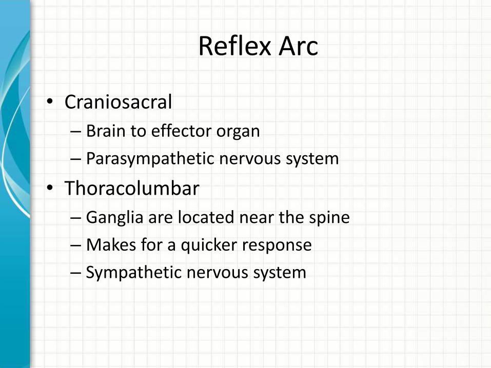
Reflex Arc
• Craniosacral
– Brain to effector organ
– Parasympathetic nervous system
• Thoracolumbar
– Ganglia are located near the spine
– Makes for a quicker response
– Sympathetic nervous system

Sympathetic Nervous System
• Stimulates sweat glands• Constrict peripheral vessels• Increase blood to skeletal muscles• Increase chronotropic and inotropic
effect• Bronchodilation• Stimulation of NRG production• Reduce blood flow to abdomen• Decrease digestive activity• Relax smooth muscle in wall
bladder• Release glucose stores from liver

Adrenal Medulla
• This area of the nervous system is also stimulated by the release of norepinephrine
– This releases
• Noradrenaline and adrenaline
– These are releases into the body to also react with the receptors and prolong the effect of sympathetic stimulation

Sympathetic receptors
• As norepineprhine is released, it travels across the synaptic cleft and interact with adreneric receptors
– Epinephrine will also stimulate these receptors
• Two types of sympathetic receptors
– Adrenergic
– dopaminergic

Adrenergic receptors
• Alpha 1– Peripheral vasoconstriction
– Positive inotropic effect
– Negative chronotropic effect
• Alpha 2– Peripheral vasodilation
• Limits release of norepinephrine
– Stimulates by excessive amounts of norepineprhine in the synaptic cleft
Adrenergic Receptors
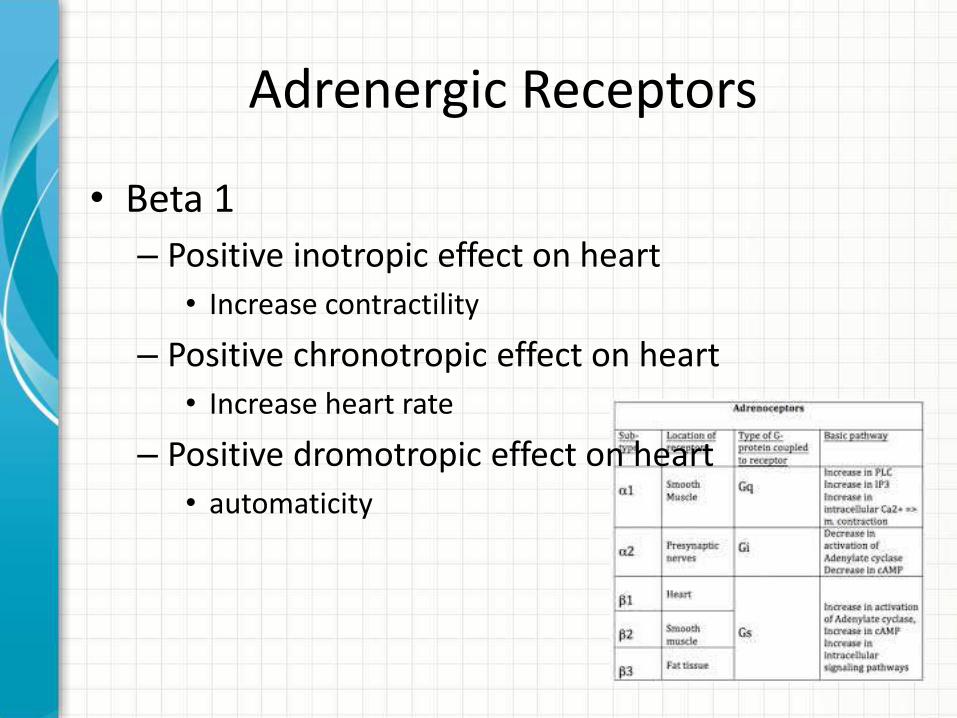
• Beta 1
– Positive inotropic effect on heart
• Increase contractility
– Positive chronotropic effect on heart
• Increase heart rate
– Positive dromotropic effect on heart
• automaticity
Adrenergic receptors
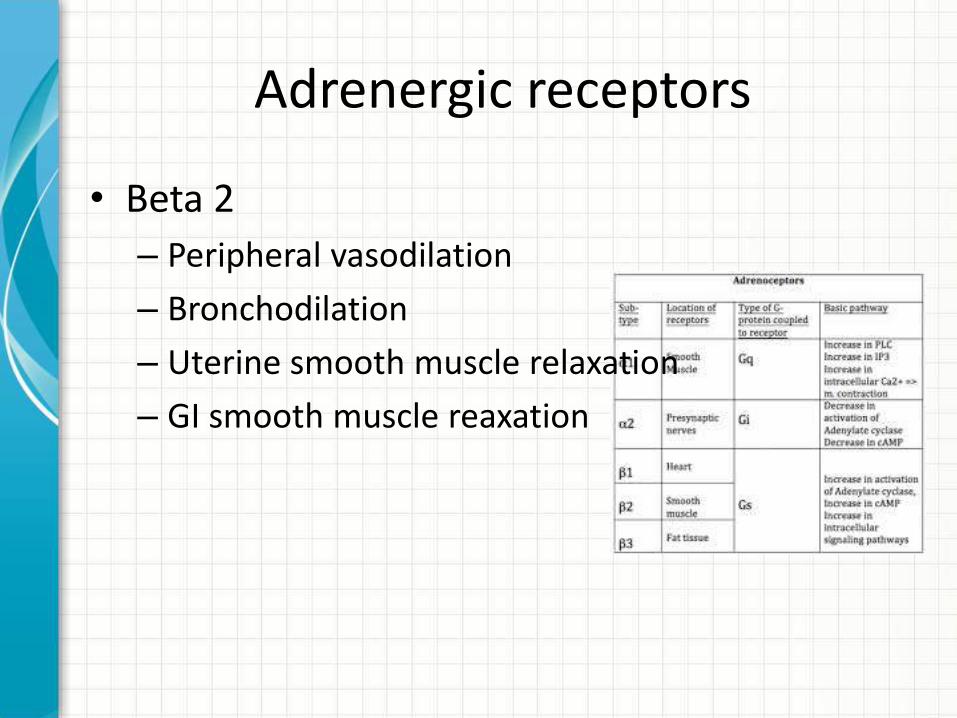
• Beta 2
– Peripheral vasodilation
– Bronchodilation
– Uterine smooth muscle relaxation
– GI smooth muscle reaxation

• Sympathomimetics– meds that stimulates the sympathetic nervous
system
• Sympatholytics– Inhibits the sympathetic system
• Alpha or betagonist– Stimulate alpha sites
• Alpha or beta antagonist– Block effects of beta stimulation

Parasympathetic Nervous System
• Pulillary constriction
• Secretion by digestive glands
• Increase smooth muscle activity along GI tract
• Brochoconstriction
• Reduced HR and negative Inotropic effects

• Parasympathomimetics
– Stimulates parasympathetic nervous system
– Physostigmine
• Parasympatholytics
– Block action of parasympathetic system
– Atropine sulfate

THANK YOU


![#APMP2016 - cdn.ymaws.com · Vivekananda ) #APMP2016 Spine - Autonomic Nervous System ... Research has linked health risks with both sitting and standing for prolonged periods.[1]](https://static.fdocuments.in/doc/165x107/5e764e6e2d5f7669f52d12f8/apmp2016-cdnymawscom-vivekananda-apmp2016-spine-autonomic-nervous-system.jpg)


