Anatomical study of different fruit types in Argentine · Anatomical study of different fruit types...
12
Anales del Jardín Botánico de Madrid ISSN: 0211-1322 [email protected] Consejo Superior de Investigaciones Científicas España Chiarini, Franco; Barboza, Gloria Anatomical study of different fruit types in Argentine species of Solanum subgen. Leptostemonum (Solanaceae) Anales del Jardín Botánico de Madrid, vol. 64, núm. 2, julio-diciembre, 2007, pp. 165-175 Consejo Superior de Investigaciones Científicas Madrid, España Available in: http://www.redalyc.org/articulo.oa?id=55664206 How to cite Complete issue More information about this article Journal's homepage in redalyc.org Scientific Information System Network of Scientific Journals from Latin America, the Caribbean, Spain and Portugal Non-profit academic project, developed under the open access initiative
Transcript of Anatomical study of different fruit types in Argentine · Anatomical study of different fruit types...
Anales del Jardín Botánico de Madrid
ISSN: 0211-1322
Consejo Superior de Investigaciones
Científicas
España
Chiarini, Franco; Barboza, Gloria
Anatomical study of different fruit types in Argentine species of Solanum subgen. Leptostemonum
(Solanaceae)
Anales del Jardín Botánico de Madrid, vol. 64, núm. 2, julio-diciembre, 2007, pp. 165-175
Consejo Superior de Investigaciones Científicas
Madrid, España
Available in: http://www.redalyc.org/articulo.oa?id=55664206
How to cite
Complete issue
More information about this article
Journal's homepage in redalyc.org
Scientific Information System
Network of Scientific Journals from Latin America, the Caribbean, Spain and Portugal
Non-profit academic project, developed under the open access initiative
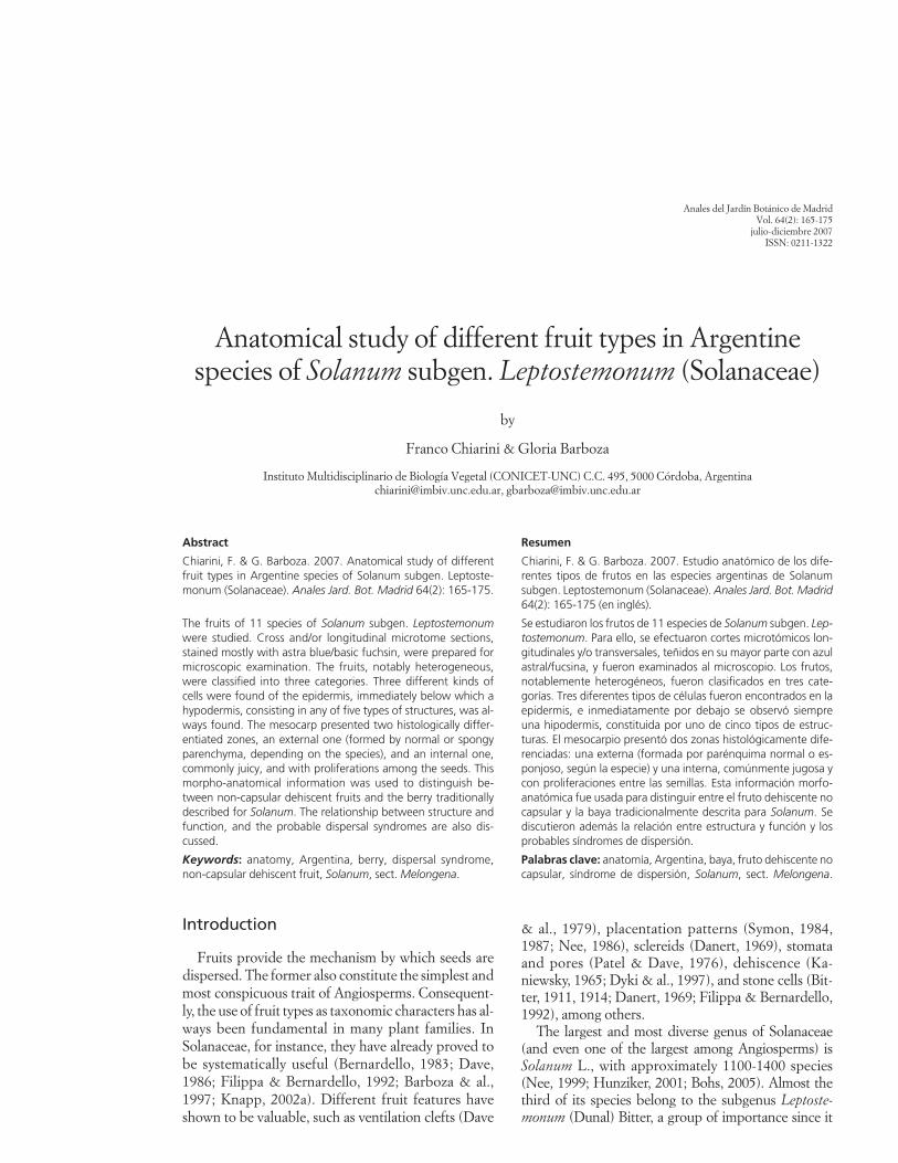
Anatomical study of different fruit types in Argentinespecies of Solanum subgen. Leptostemonum (Solanaceae)
by
Franco Chiarini & Gloria Barboza
Instituto Multidisciplinario de Biología Vegetal (CONICET-UNC) C.C. 495, 5000 Córdoba, [email protected], [email protected]
Abstract
Chiarini, F. & G. Barboza. 2007. Anatomical study of differentfruit types in Argentine species of Solanum subgen. Leptoste-monum (Solanaceae). Anales Jard. Bot. Madrid 64(2): 165-175.
The fruits of 11 species of Solanum subgen. Leptostemonumwere studied. Cross and/or longitudinal microtome sections,stained mostly with astra blue/basic fuchsin, were prepared formicroscopic examination. The fruits, notably heterogeneous,were classified into three categories. Three different kinds ofcells were found of the epidermis, immediately below which ahypodermis, consisting in any of five types of structures, was al-ways found. The mesocarp presented two histologically differ-entiated zones, an external one (formed by normal or spongyparenchyma, depending on the species), and an internal one,commonly juicy, and with proliferations among the seeds. Thismorpho-anatomical information was used to distinguish be-tween non-capsular dehiscent fruits and the berry traditionallydescribed for Solanum. The relationship between structure andfunction, and the probable dispersal syndromes are also dis-cussed.
Keywords: anatomy, Argentina, berry, dispersal syndrome,non-capsular dehiscent fruit, Solanum, sect. Melongena.
Resumen
Chiarini, F. & G. Barboza. 2007. Estudio anatómico de los dife-rentes tipos de frutos en las especies argentinas de Solanumsubgen. Leptostemonum (Solanaceae). Anales Jard. Bot. Madrid64(2): 165-175 (en inglés).
Se estudiaron los frutos de 11 especies de Solanum subgen. Lep-tostemonum. Para ello, se efectuaron cortes microtómicos lon-gitudinales y/o transversales, teñidos en su mayor parte con azulastral/fucsina, y fueron examinados al microscopio. Los frutos,notablemente heterogéneos, fueron clasificados en tres cate-gorías. Tres diferentes tipos de células fueron encontrados en laepidermis, e inmediatamente por debajo se observó siempreuna hipodermis, constituida por uno de cinco tipos de estruc-turas. El mesocarpio presentó dos zonas histológicamente dife-renciadas: una externa (formada por parénquima normal o es-ponjoso, según la especie) y una interna, comúnmente jugosa ycon proliferaciones entre las semillas. Esta información morfo-anatómica fue usada para distinguir entre el fruto dehiscente nocapsular y la baya tradicionalmente descrita para Solanum. Sediscutieron además la relación entre estructura y función y losprobables síndromes de dispersión.
Palabras clave: anatomía, Argentina, baya, fruto dehiscente nocapsular, síndrome de dispersión, Solanum, sect. Melongena.
Introduction
Fruits provide the mechanism by which seeds aredispersed. The former also constitute the simplest andmost conspicuous trait of Angiosperms. Consequent-ly, the use of fruit types as taxonomic characters has al-ways been fundamental in many plant families. InSolanaceae, for instance, they have already proved tobe systematically useful (Bernardello, 1983; Dave,1986; Filippa & Bernardello, 1992; Barboza & al.,1997; Knapp, 2002a). Different fruit features haveshown to be valuable, such as ventilation clefts (Dave
& al., 1979), placentation patterns (Symon, 1984,1987; Nee, 1986), sclereids (Danert, 1969), stomataand pores (Patel & Dave, 1976), dehiscence (Ka-niewsky, 1965; Dyki & al., 1997), and stone cells (Bit-ter, 1911, 1914; Danert, 1969; Filippa & Bernardello,1992), among others.
The largest and most diverse genus of Solanaceae(and even one of the largest among Angiosperms) isSolanum L., with approximately 1100-1400 species(Nee, 1999; Hunziker, 2001; Bohs, 2005). Almost thethird of its species belong to the subgenus Leptoste-monum (Dunal) Bitter, a group of importance since it
Anales del Jardín Botánico de MadridVol. 64(2): 165-175
julio-diciembre 2007ISSN: 0211-1322
includes food plants (e.g. eggplant, S. melongena L.;naranjilla or lulo, S. quitoense Lam.; gilo, S. aethiopicumL.; cocona or cubiu, S. sessiliflorum Dunal) and weeds(e.g. tropical soda apple, S. viarum Dunal; silverleafnightshade, S. elaeagnifolium Cav.; sticky nightshade orwild tomato, S. sisymbriifolium Lam.; horsenettle, S.carolinense L.; buffalo bur, bull thistle or Texas thistle,S. rostratum Dunal). Among the sections in which Lesp-tostemonum has been divided, sect. Melongena (Mill.)Dunal, with 34 species in the New World (Nee, 1999),deserves special attention, because it includes the egg-plant, and some of its representatives have been thesubject of several studies due to their andromonoecy(Wakhloo, 1975a, b; Dulberger & al., 1981; Solomon,1986, 1987; Elle, 1999; Connolly & Anderson, 2003).Nevertheless, the boundaries of sect. Melongena areunclear, according to recent molecular studies (Bohs,2005; Levin & al., 2006; Weese & Bohs, 2007). In addi-tion, the fruits of several species assigned by Nee (1999)to this section are different from the traditionally de-fined berry (Matesevach, 2002; Dottori & Cosa, 2003).In this sense, Knapp (2002a) has discussed the appar-ent uniformity of fruit type in Solanum, suggesting a re-markable range of subtle variation. This author pro-posed that within the vast subgen. Leptostemonum,fruits can be either fleshy berries, hard berries (all with-out stone cells) or variously modified non-capsular de-hiscent fruits. Such classification of fruit types is basedmostly on macroscopic features (colour, size, dehis-cence), since studies which focus on the anatomical/histological point of view are scarce. For the entire sub-genus, only some scattered species have been studied(Miller, 1969; Dave & al., 1979; Dottori, 1998; Dottori& Cosa, 1999, 2003). Thus, a contribution that consid-ers these aspects of Solanum subgen. Leptostemonumshould be welcome.
Taking these facts into account, the present workattempts to assess the anatomical differences amongfruit types in Argentine species of Solanum subgen.Leptostemonum (formerly assigned to sect. Melonge-na) as a contribution towards clarifying the systematicof this group and to understanding the relationshipbetween structure and function.
Materials and methods
Eleven wild species of Solanum subgen. Leptoste-monum were analyzed. All but one species grow in Ar-gentina; some of them are exclusive of this territory,while others are South American or naturalized inother parts of the world. Solanum marginatum is na-tive of Africa, but is usually cultivated in many placesand grows naturalized in disturbed areas of the Andes
F. Chiarini & G. Barboza
from Colombia to Chile. According to Nee’s (1999)Solanum classification system (the latest to give formaltaxonomic rank to the different species groups) all theconsidered species belong to sect. Melongena. How-ever, the clade system proposed in Levin & al. (2006)is taken into account for the discussion. The followingare the voucher data of the studied material:
S. aridum Morong. ARGENTINA. Córdoba: Capital, 1-XII-1998,Chiarini 16 (CORD). Salta: Capital, 19-I-2002, Barboza 331 (CORD).
S. comptum C.V. Morton. ARGENTINA. Corrientes: Capital,near the airport, 13-V-2004, Barboza & al. 999 (CORD). Perichón,29º24’34’’S 58º45’09’’W, 13-V-2004, Barboza & al. 1001 (CORD).San Cosme, 27º18’42’’S 58º29’22’’W, 13-V-2004, Barboza & al.1005 (CORD).
S. elaeagnifolium Cav. ARGENTINA. Córdoba: Sobremonte,San Francisco del Chañar, 9-XII-2001, Chiarini 565 (CORD).
S. euacanthum Phil. ARGENTINA. Córdoba: Sobremonte, 29º46’06’’S 64º34’03’’W, 28-II-2002, Chiarini & al. 560, 563 (CORD).Neuquén: Collón Curá, 20-II-2005, Barboza & al. 1181 (CORD).
S. hieronymi Kuntze. ARGENTINA. La Rioja: Chilecito,Puesto Las Trancas, 19-II-2003, Barboza & al. 569 (CORD). Cór-doba: San Javier, Yacanto, 9-I-1996, Cosa 266 (CORD). Rio II, Co-lazo, 23-VI-1983, Hunziker & al. 3674 (CORD). San Luis: Cha-cabuco, Concarán, 17-II-1989, Hunziker & al. 25332 (CORD).
S. homalospermum Chiarini. ARGENTINA. Córdoba: Sobre-monte, 29°46’34’’S 63°59’59’’W, 29-XI-2001, Chiarini 505 (CORD).
S. juvenale Thell. ARGENTINA. Córdoba: Capital, 8-XII-2001, Chiarini 504 (CORD). La Pampa: Toay, 36º38’51’’S64º22’42’’W, 19-II-2005, Barboza & al. 1173 (CORD).
S. marginatum L. f. SPAIN, Canary Islands, 15-VI-2005, Ober-ti s.n. (CORD 1040). CHILE. V Región: Laguna Verde,33º06’32”S 71º39’09”W, 8-II-2007, Chiapella & al. 1654 (CORD).
S. mortonii Hunz. ARGENTINA. Catamarca: Capayán,28º41’55’S 66º02’53’’W, 23-II-2003, Barboza & al. 633 (CORD);ibídem, 28º42’23’S 66º01’29’’W, 23-II-2003, Barboza & al. 639(CORD); ibídem, 28º34’56’S 65º56’07’’W, 23-II-2003, Barboza &al. 644 (CORD).
S. multispinum N.E. Brown. ARGENTINA. Formosa: Pilco-mayo, Laguna Blanca, 25º07’50’’S 58º15’57’’W, 14-XII-2002, Bar-boza & al. 511 (CORD). Route 86, 25º05’49’’S 51º18’59’’W, 14-XII-2002, Barboza & al. 520 (CORD).
S. sisymbriifolium Lam. ARGENTINA. Córdoba: Capital, XII-1998, Chiarini 27 (CORD). Salta: Rosario de Lerma, Corralito, 29-XII-1987, Novara 7363 (CORD).
For microscopic examination, whole or cut up ripefruits were preserved in a formaldehyde-acetic acid-ethanol mixture, then dehydrated in a 50 to 100 %ethanol series, and embedded in Paramat® resin.Cross and/or longitudinal and/or tangential micro-tome sections 10-12 mm thick were stained mostly witha 1% astra blue solution in a 1% water/basic fuchsinsolution in 50º ethanol. Astra blue stains cell wall poly-saccharides such as cellulose and pectins, while basicfuchsin shows affinity for lignified, suberizied or cu-tinized walls, i.e., structures embedded in phenolicsubstances (Kraus & al., 1998). Basic fuchsin also stainschloroplasts and nucleic acids. In some cases, addition-al sections were stained with a 0.05% cresyl blue solu-tion in water (Pérez & Tomassi, 2002).
166
Anales del Jardín Botánico de Madrid 64(2): 165-175, julio-diciembre 2007. ISSN: 0211-1322
167Fruit anatomy in Solanum subgen. Leptostemonum
Anales del Jardín Botánico de Madrid 64(2): 165-175, julio-diciembre 2007. ISSN: 0211-1322
The specimens were visualized using a Zeiss Axio-phot microscope and the images were captured with adigital camera assembled to the microscope.
Results
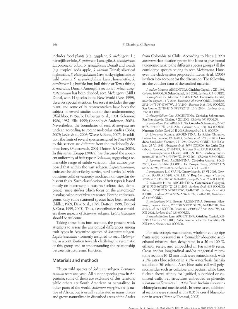
According to Knapp (2002a), who developed aclassification that subsumes that of Spjut (1994), thefruits of Solanum sect. Melongena studied here areberries in a conventional sense, or non-capsular dehis-cent fruits. In Spjut’s system, and according to our ob-servations, fruits might be classified as follows:
Berry = a simple fruit with an indehiscent pericarp,containing many seeds embedded in a solid fleshymass, supported by an epicarp that is less than 2 mmthick. E.g.: S. sisymbriifolium (Figs. 1 D, 2 E, F).
Foraminicidal capsule (Non-capsular dehiscentfruit according to Knapp, 2002) = Dry or slightly juicyfruit with a thin pericarp, which cracks in an irregularfashion, thus leaving the seeds exposed at the senes-cent stage. E.g.: S. homalospermum, S. euacanthum(Fig. 1 A-C).
Carcerulus = Fruit resembling the true berries, butwith an aerial space between the seeds and the pericapwhen the fruit is completely ripe, as in peppers (Cap-sicum). E.g.: S. multispinum.
Details of the anatomy of each species are summa-rized in Table 1.
Mature fruits are of a single colour. They can be red(e.g. S. sisymbriifolium, Fig. 1 D), yellow (S. multi-spinum, S. juvenale), black or brownish (S. mortonii)or greyish (S. homalospermum, S. euacanthum, Fig. 1A-C). The fruiting calyx may be accrescent or not. Inthe first case, different degrees of accrescence are ob-served. Calices may enclose the berry almost com-pletely (S. sisymbriifolium, Fig. 1D, S. comptum) oronly half or a third part of the fruit (S. juvenale, S. homalospermum). Moreover, at maturity, the calyxlobes can split open (S. sisymbriifolium, Fig. 1 D) orcontinue to enclose the berry (S. comptum).
The pericarp comprises three clearly distinguishablezones: the exocarp, the mesocarp, and the endocarp.
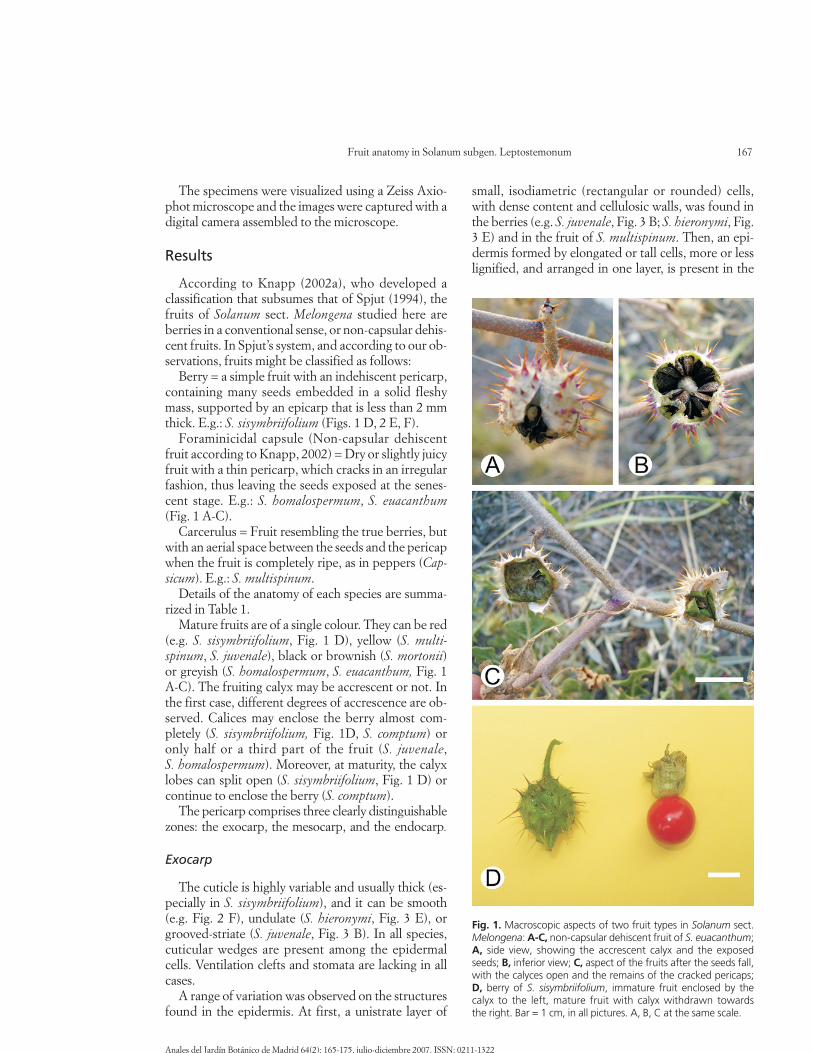
Exocarp
The cuticle is highly variable and usually thick (es-pecially in S. sisymbriifolium), and it can be smooth(e.g. Fig. 2 F), undulate (S. hieronymi, Fig. 3 E), orgrooved-striate (S. juvenale, Fig. 3 B). In all species,cuticular wedges are present among the epidermalcells. Ventilation clefts and stomata are lacking in allcases.
A range of variation was observed on the structuresfound in the epidermis. At first, a unistrate layer of
small, isodiametric (rectangular or rounded) cells,with dense content and cellulosic walls, was found inthe berries (e.g. S. juvenale, Fig. 3 B; S. hieronymi, Fig.3 E) and in the fruit of S. multispinum. Then, an epi-dermis formed by elongated or tall cells, more or lesslignified, and arranged in one layer, is present in the
Fig. 1. Macroscopic aspects of two fruit types in Solanum sect.Melongena: A-C, non-capsular dehiscent fruit of S. euacanthum;A, side view, showing the accrescent calyx and the exposedseeds; B, inferior view; C, aspect of the fruits after the seeds fall,with the calyces open and the remains of the cracked pericaps; D, berry of S. sisymbriifolium, immature fruit enclosed by the calyx to the left, mature fruit with calyx withdrawn towards the right. Bar = 1 cm, in all pictures. A, B, C at the same scale.
berries of S. elaeagnifolium (Fig. 2 A) and in the non-capsular dehiscent fruits of S. euacanthum, (Fig. 3 C).Similar cells were found in S. marginatum, butarranged in more than one layer (Fig. 3 F). Finally, inthe non-capsular dehiscent fruits of S. homalosper-mum and S. mortonii, the epidermis is composed bysclereids, one layer of brachysclereids in the firstspecies (Fig. 2 D) and two layers of macrosclereids inthe second (Fig. 2 C).
F. Chiarini & G. Barboza
Immediately below the epidermis, a hypodermis isdifferentiated, consisting of any of the following kindsof structures: 1) one to several layers of collenchyma(S. comptum, S. multispinum), 2) one to three layers ofradially compressed parenchymatous cells, withdense content, followed by collenchyma (e.g. S. juve-nale, Fig. 3 B; S. hieronymi Fig. 3 E), 3) a well definedlayer of fibres with evident pits in both the apical andthe basal ends and with a single calcium oxalate crys-
168
Anales del Jardín Botánico de Madrid 64(2): 165-175, julio-diciembre 2007. ISSN: 0211-1322
Fig. 2. Photomicrographs of fruit anatomy in Solanum sect. Melongena species: A, exocarp of S. elaeagnifolium (Chiarini 565, CORD);B, S. elaeagnifolium, detail of the sclereids (Chiarini 565, CORD); C, exocarp of S. mortonii (Barboza & al. 639, CORD); D, exocarp ofS. homalospermum (Chiarini 505, CORD); E, pericarp of S. sisymbriifolium (Chiarini 27, CORD); F, S. sisymbriifolium, detail of the epi-dermis (Chiarini 27, CORD). Abbreviations: bs = brachysclereids; co = collenchyma; fc = fibres containing a crystal; ms = macrosclereids;sc = sclerified collenchyma; si = sclereids islets. Scale bars: A = 80 µm; B = 15 µm; C, D, F = 50 µm; E = 0.5 mm.
tal occupying the whole lumen of each fibre, followedby 1 or 2 true collenchymatous layers (S. homalosper-mum, Fig. 2 D, S. euacanthum, Fig. 3 C, S. mortoniiFig. 2 C) 4) Several layers of very thickened and ligni-fied cell walls, taller than wide, which could be con-sidered a sclerified collenchyma (E.g. S. marginatum,Fig. 3 F). 5) A layer of elongated cells, some of themcontaining a crystal, followed by sclerified collenchy-ma (S. elaeagnifolium, Fig. 2 A)
The epidermis and the hypodermis constitute the
Fruit anatomy in Solanum subgen. Leptostemonum
exocarp in the form of a unit, which generally has layersthat gradually decrease their degree of lignificationfrom the outside to the inside of the fruit. Usually, whenthe fruit is immature, the cell layers located below theepidermis (or below the crystalliferous layer or layer offibres, when present) have chloroplasts and chromo-plasts. When the fruit matures, the chloroplasts disap-pear and the cells become compressed. A collenchymais always present, in which the number of layers and thedegree of lignification vary according to the species.
169
Anales del Jardín Botánico de Madrid 64(2): 165-175, julio-diciembre 2007. ISSN: 0211-1322
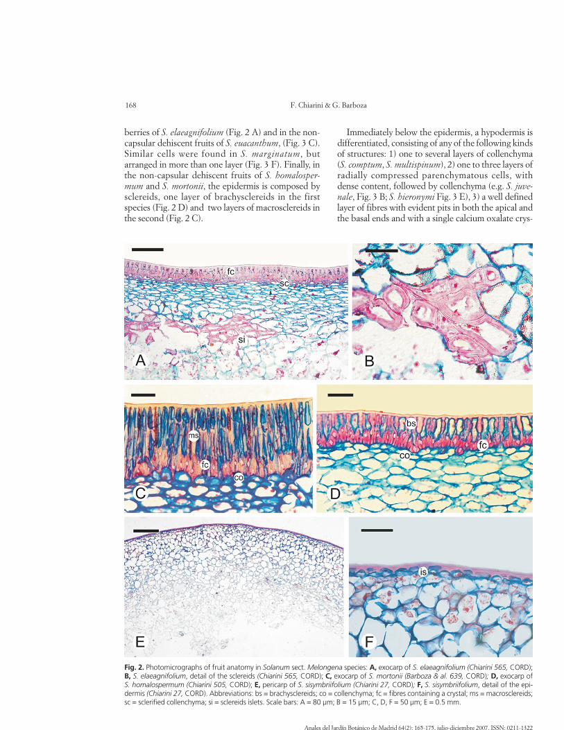
Fig. 3. Photomicrographs of fruit anatomy in Solanum sect. Melongena species: A, spongy mesocarp of S. multispinum (Barboza & al.511, CORD); B, pericarp of S. juvenale (Chiarini 504, CORD), dotted line showing the division of the two zones of the mesocarp, in thesquare at the bottom left, a detail of the epidermis; C, exocarp of S. euacanthum (Chiarini 563, CORD); D, pericarp of S. comptum (Bar-boza & al. 999, CORD); E, exocarp of S. hieronymi (Barboza & al. 569, CORD); F, exocarp of S. marginatum (Oberti s.n., CORD 1040).Abbreviations: fc = fibres containing a crystal; g = cells filled with grana; is = isodiametric cells; p = proliferations among the seeds; sc = sclerified collenchyma; sm = spongy mesocarp; tl = tall lignified cells. Scale bars: A = 100 µm; B, D = 1 mm; C, E, F = 50 µm.
F. Chiarini & G. Barboza170
Anales del Jardín Botánico de Madrid 64(2): 165-175, julio-diciembre 2007. ISSN: 0211-1322
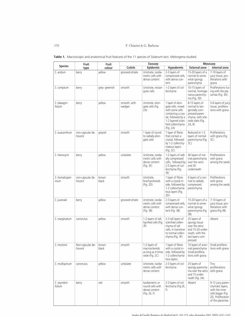
Table 1. Macroscopic and anatomical fruit features of the 11 species of Solanum sect. Melongena studied.
S. aridum
S. comptum
S. elaeagni-folium
S. euacanthum
S. hieronymi
S. homalosper-mum
S. juvenale
S. marginatum
S. mortonii
S. multispinum
S. sisymbrii-folium
berry
berry
berry
non-capsular de-hiscent
berry
non-capsular de-hiscent
berry
carcerulus
Non-capsular de-hiscent
carcerulus
berry
yellow
grey- greenish
yellow
greyish
yellow
brownblack
yellow
yellow
brownblack
yellow
red
Cuticlegrooved-striate
smooth
smooth, withwedges
smooth
undulate
smooth
grooved-striate
smooth
smooth
undulate
smooth
EpidermisUnistrate, isodia-metric cells withdense content
Unistrate, rectan-gular cells
Unistrate, elon-gate cells (Fig.2A)
1 layer of roundto radially elon-gate cells
Unistrate, isodia-metric cells withdense content(Fig. 3E)
Unistrate,brachysclereids(Fig. 2D)
Unistrate, isodia-metric cells withdense content(Fig. 3B)
1-2 layers of tall,lignified cells (Fig.3F)
1-2 layers ofmacrosclereidsas long as 4 timeswide (Fig. 2C)
Unistrate, isodia-metric cells withdense content
Isodiametric orround cells withdense content(Fig. 2E, F)
Hypodermis2-3 layers ofcompressed cells,with dense con-tent1-2 layers of col-lenchyma
1 layer of elon-gate cells, mixedwith some cellscontaining a crys-tal, followed by a1-2 layered scleri-fied collenchyma(Fig. 2A)1 layer of fibresthat contain acrystal, followedby 1-2 collenchy-matous layers(Fig. 3C)1-2 layers of radi-ally compressedcells, followed by2-5 layers of col-lenchyma (Fig.3E)1 layer of fibreswith a crystal in-side, followed by1-2 collenchyma-tous layers (Fig.2D).2-3 layers ofcompressed cells,with dense con-tent (Fig. 3B).
2-3 tall layers ofsclerified collen-chyma of tallcells, in transitionto normal collen-chyma (Fig. 3F)
1 layer of fibreswith a crystal in-side, followed by1-2 collenchyma-tous layers.2-3 layers of col-lenchyma
2-3 layers of col-lenchyma (Fig 2E,F)
External zone15-20 layers of anormal to some-what spongyparenchyma10-15 layers ofnormal, homoge-neous parenchy-ma (Fig. 3D)8-15 layers ofnormal to tan-gentially com-pressed paren-chyma, with scle-reids islets (Fig.2A, B)
Reduced to 1-2layers of normalparenchyma (Fig.3C)
40 layers of nor-mal parenchymaover the veinsand 30underneath
6 layers of a nor-mal to radiallycompressedparenchyma.
15-20 layers of anormal to some-what spongyparenchyma (Fig3B)25 layers ofspongy tissueover the veinsand 15-20 under-neath, with thelast layers com-pressed10 layers of anor-mal parenchyma.Small prolifera-tions with grana
25 layers ofspongy parenchy-ma over the veinsand 15 under-neath (fig. 3A)Absent
Internal zone7-10 layers ofjuicy tissue; pro-liferations withgranaProliferations fus-ing with the pla-centas (Fig. 3D)
5-6 layers of juicytissue, prolifera-tions with grana
Proliferationswith grana (Fig3C)
Proliferationswith granaamong the seeds
Proliferationswith granaamong the seeds
7-10 layers ofjuicy tissue; pro-liferations withgrana (Fig 3B)
Absent
Small prolifera-tions with grana
Tinyproliferationswith grana
9-12 juicy paren-chymatic layers,with the innercells bigger (Fig2E). Proliferationof the placentas
Species Fruittype
Fruitcolour
Exocarp Mesocarp
Mesocarp
The number of layers of this structure gives thethickness to the pericarp. The higher the number ofmesocarp layers, the thicker the pericarp. Fruits witha thick pericarp have usually more than 10 layers. Themesocarp consists of two zones histologically differ-entiated: an external one (immediately below the hy-podermis), which we identified with astra blue, andan internal one, identified with basic fuchsin.
In the majority of the species, the external zoneconsists of regular, vacuolated, medium-sized cellswith small intercellular spaces. Instead, in S. margina-tum and especially in S. multispinum, the externalzone consists of big, very vacuolated or almost empty,loosely connected cells, with large intercellularspaces, forming a spongy parechymatic tissue, resem-bling the albedo of the hesperidium (i.e. the whitepith of the inner peel of citrus fruits). These cells in-crease their size towards the endocarp, whose cellwalls get lose and undulated. At maturity, the meso-carp is not in direct contact with the seeds.
The internal zone is commonly juicy, and developsproliferations among the seeds. The cells are large,with dense content filled with grana, which disorga-nize and release their content to the locules and pro-duce a mucilage-like substance that surrounds theseeds in the ripe fruit. It is worth mentioning that inseveral species this mucilaginous content turns blackon contact with the air, perhaps due to its phenolic orsaponinic nature.
The thickness of each zone varies notably accord-ing to the species. For instance, in S. juvenale (Fig. 3B), the difference between the two zones is remark-able and well-defined, each one having many layers ofcells. In other species, the two zones are not so clear-ly-defined and have fewer layers.
Occasionally, scattered or grouped sclereids arepresent in the mesocarp of S. hieronymi and S. elaeag-nifolium (Fig. 2 B). Stone cells or sclerosomes, widelypresent in many sections of Solanum and in relatedgenera, are absent in subgenus Leptostemonum, or atleast in the species analyzed here.
Endocarp
Finally, no specific particularities were observed inthe endocarp. This layer, which is very difficult to ob-serve due to its delicate structure, is uniseriate and, asin many Angiosperms (Roth, 1977), lacks stomata inall cases.
Fruit anatomy in Solanum subgen. Leptostemonum
Discussion
Anatomical features
All the analyzed structures presented variations,but only some of these variants can be related to afunction and can be useful in delimiting fruit types.
The cuticle, for instance, is a variable feature inSolanum (Dottori & Cosa, 1999, 2003) and does notseem to be associated to a determined fruit type.
Instead, the epidermis has shown an important di-versification. A factor influencing epidermal structurewould be the calyx accrescence, since fruits that arealmost completely enclosed by it show a simple, thinepidermis, which is the case of S. sisymbriifolium(Chaparro, 1989; this paper). Apparently, in thesefruits, the protective or mechanical function of a col-lenchymatous exocarp is strengthened by the enclos-ing calyx. Another modification related to externalfactors is the presence of fibres and sclereids in crack-ing fruits. In this case, there is not a protective calyxand the pericarp is destined to tear, releasing theseeds. All these derived types of epidermal cells (fi-bres and sclereids) may have originated from thesmall, isodiametric cells with cellulosic walls.
The presence of a hypodermis, mainly constitutedby collenchyma, is constant in all the analyzed species;however, there is a specific variation in the number oflayers and the degree of lignification. The collenchy-matous hypodermis is common in fruits with a thickouter skin, which is the case of many berries and dru-pes, such as some species of Ribes, Berberis and Paris(Roth, 1977) and even in berries of some members ofSolanaceae (Valencia, 1985; Filippa & Bernardello,1992). The function of hypodermal cells would be toprovide mechanical support or, in some cases, to par-ticipate in the dehiscence mechanism (Klemt, 1907;Dyki & al., 1997). The presence of a collenchyma infruits of Solanaceae and, more precisely, in Solanum,has already been noticed (Klemt, 1907; Roth, 1977;Dottori & Cosa, 1999) and the species studied herealso fit this pattern.
Some hypodermal cells, whose walls are impregnat-ed with lignin, resemble the outline of a true col-lenchyma. Layers with such features are called here“sclerified collenchyma”. The sclerified collenchyma,may be the structure that make the fruits harder andmore resistant to deformation, and perhaps are a de-fence against phytophage insects. Indeed, fruit fea-tures are usually interpreted in relation to vertebratedispersion and consumption, while the more impor-tant insect and microbial attack is neglected (Tewks-bury, 2002). Among the species with sclerified col-lenchyma analyzed here, we observed fruits with no
171
Anales del Jardín Botánico de Madrid 64(2): 165-175, julio-diciembre 2007. ISSN: 0211-1322
sign of insect attack (e.g. S. marginatum). By contrast,in the case of fruits whose collenchyma consists of nomore than three layers, berries show evident harmcaused by phytophage insects (e.g. S. juvenale).
Regarding the mesocarp, the presence of a spongyparenchyma is very obvious in S. multispinum. Thistissue, characterised by large intercellular spaces andcells that change their shape from rounded to ellipti-cal, to elongate, and even to stellate, was accuratelydescribed in S. mammosum (Miller, 1969) with thename of aerenchyma. Something similar occurs in thealbedo of the orange, where parenchyma cells developarms in different directions (Roth, 1977). The spongytissue does not exclusively belong to sect. Melongena,but is also present in several species of sect. Acan-thophora (Miller, 1969; Nee, 1991; Cipollini & al.,2002; Levin & al., 2005).
Regarding the pulp of the fleshy fruited species, thepattern observed coincides with that which is alreadyknown, in which both the placenta and, especially thepericarp, contribute to form the pulp (Garcin, 1888;Murray, 1945). It is the same in the case of Physalis pe-ruviana (Valencia, 1985) and other Solaneae (Filippa &Bernardello, 1992). Instead, in Solanum lycopersicum(sub nom. Lycopersicon esculentum) only the placentasare responsible for the formation of the pulp (Murray,1945; Roth, 1977). The first pattern is the most com-mon and the second one is peculiar to S. lycopersicum.The disorganisation of the inner mesocarp and the en-docarp noticed in some species, such as P. peruviana,occurs also in the fleshy or juicy fruits examined here.
In S. lycopersicum, fruit softening is associated withcell disassembly and modifications to the pectin frac-tion of the cell walls, catalysed by polygalacturonaseand pectate lyases (Marín-Rodríguez & al., 2002; Sey-mour & al., 2002). Pectate lyase sequences have beenreported for several species from different families(Medina-Escobar & al., 1997; Marín-Rodríguez & al.,2002). Perhaps different levels of expression of suchgenes are responsible for the formation of the stiffzone and the juicy zone in the mesocarp of the specieshere studied.
The fibre layer formed by cells that contain a singleprismatic crystal is a type of structure with mineraldepositions. It was observed in the hypodermis ofS. euacanthum (Dottori & Cosa, 2003), and also inS. homalospermum, S. mortonii and in the species ofsect. Torva (Chiarini, in prep.). The presence of min-eral deposits of calcium oxalate may have evolved as aprimary mechanism for controlling the excess of calci-um in a great many plants. These deposits would pro-vide multiple benefits to different plant organs, forexample, an internal calcium reservoir, or a defence
F. Chiarini & G. Barboza
against herbivores, etc. (Sakai & al., 1972; Thurston,1976; Franceschi & Horner, 1980; Webb, 1999). Nev-ertheless, the function of crystals in fruits remains un-explained.
Fruit types and dispersion
Usually, fruits are classified into different dispersalsyndromes according to their morphological charac-ters. Van der Pijl’s (1982) criterion is usually followed,but direct observation of the dispersion is seldom pos-sible, so the fruits or seeds are assigned to a dispersalsyndrome on the basis of speculations, which leads topuzzling discussions, as Levin & al. (2005) pointedout. In this sense, the morpho-anatomical data weprovide may clarify some points.
Solanum euacanthum, S. homalospermum and S.mortonii develop non-capsular dehiscent fruits (sensuKnapp, 2002a). They differ from the traditionally de-fined berry because they are dry or slightly juicy, theycan be easily cracked and have fibres in the hypoder-mis. In S. mortonii and S. homalospermum the fibresare combined with sclereids in the epidermis. Surpris-ingly, this type of pericarp is reminiscent of the Nican-dra physalodes pericarp, according to Kaniewsky(1965). This author suggested that the fruit of N. phy-salodes is not a berry, since it is hard and dry, like thefruit of S. mortonii and S. homalospermum. In thesenon-capsular dehiscent fruits, changes in temperatureand humidity can trigger the rupture of the pericarp,the fibres and the sclereids probably being responsi-ble for such a mechanism. Thus, it is obvious that be-yond the external appearance, there are many traitsrelated to the dispersal syndrome. In addition, thecolour of the fruits of S. homalospermum, S. mortoniiand S. euacanthum is dull and their appearance is un-attractive to predators or dispersers.
The berries of S. aridum, S. juvenale, S. hieronymiand S. comptum are indehiscent, small to mediumsized, yellow when ripe, and a little enclosed by the ca-lyx. The mesocarp has an external and more consis-tent parenchymatic zone, and an internal one, formedby cells that dissolve, thus releasing its mucilaginouscontent. Both the mesocarp and the placentas developprojections that surround the seeds. Something simi-lar has been observed in other Solanum, such as S. ni-grum, S. pseudocapsicum, S. lycopersicon, and inPhysalis (Garcin, 1888; Murray, 1945). When this sortof fruit matures, it becomes fleshy and pulpy and iseaten either by birds or by terrestrial vertebrates (Ed-monds & Chweya, 1997; Knapp 2002b). In addition,the fruits of S. juvenale and S. aridum would be attrac-tive to consumers, since they have a pleasant odour(Parodi, 1930; our observations).
172
Anales del Jardín Botánico de Madrid 64(2): 165-175, julio-diciembre 2007. ISSN: 0211-1322
The fruit of S. sisymbriifolium is a particular case.This species, has fruits enclosed by the calyx up to ma-turity, but the calyx then splits entirely open andshows a red, juicy, indehiscent berry. It is the softestand the juiciest of those studied here. Apparently, theformation of collenchyma in the hypodermis is sup-pressed, since the calyx develops a protective coverover the fruit. The layers that disorganize are not somuch like those of other species (E.g. S. juvenale). Theplacentas contribute much more than those of otherspecies to the formation of the pulp of the ripe fruit(Chaparro, 1989). This fruit would be also a berry, ac-cording to previous classifications, but it is clearly dif-ferent from the berries of other species of the section.This soft, juicy and showy fruit is probably consumedby vertebrates (Von Reis Altschul, 1975). Indeed,brightly coloured fruits would be more attractive tobirds (Van der Pijl, 1982; Edmonds & Chweya, 1997).
Beyond the spongy mesocarp, no special featureswere detected in the fruit of S. multispinum. The factthat many species of Solanum subgen. Leptostemo-num have potentially poisonous fruits (Cipollini &Levey, 1997), in addition to the spongy structure,makes dispersion by vertebrates hardly plausible inthis case. The function of this spongy tissue has not yetbeen explained until now, but some authors (Nee,1979, 1991; Bryson & Byrd, 1994; Levin & al., 2005)have suggested that it might be an adaptation to float-ing, as the fruits are dispersed by drain water after arainstorm.
Systematic implications of fruit anatomy
Regarding the fruit, sect. Melongena (as traditional-ly circumscribed) seems to be a very heterogeneousgroup, since no single feature is shared by all thespecies studied here. On the one hand, hypodermal fi-bres of S. mortonii, S. euacanthum and S. homalosper-mum are very alike and are not seen in species of othersections (Chiarini, in prep.). On the other hand, thesection also includes fleshy-fruited species, such asS. juvenale, and species which have spongy fruits, likeS. multispinum and S. marginatum. In agreement withthe differences found in fruit type, representatives ofsection Melongena (sensu Nee, 1999) appear scatteredin the molecular studies of Bohs (2005), Levin & al.(2006) and Weese & Bohs (2007) and would not forma natural group. Although these phylogenetic analysesdo not include all the species examined here, they pro-vide an interesting subject for discussion. For instance,S. aridum (sub. nom. S. conditum) and S. carolinenseare placed in the same clade (the “Carolinense clade”),and both species develop yellow, odorous, mammal-syndrome fruits. Nevertheless, S. hieronymi and
Fruit anatomy in Solanum subgen. Leptostemonum
S. comptum, with a similar fruit type, appear separatedfrom each other, S. hieronymi being closer to S. elaeag-nifolium. Interestingly, S. sisymbriifolium, with juicyberries, seems to be more related to S. rostratum (aspecies of the “Androceras clade”), which has dehis-cent fruits (Whalen, 1979). Solanum multispinum andS. marginatum, both with spongy fruits, are distantlyrelated: the former species is isolated among the NewWorld clades, while the latter species is placed withinthe African clade.
At the same time, Cipollini & al. (2002), in a studythat remarks phytochemical aspects, state that there isno significant correlation among the fruit types theydistinguished and the phylogenetic lineages in Sola-num. For these authors, fruit typology may be due tophysiological constraints, holding an independentevolution of the different dispersal syndromes. Re-garding morpho-anatomy, our data lead to similarconclusions.
It is very important to compare morpho-anatomicalinformation to a phylogenetic background, as a meansto arrive at safer conclusions. For instance, some au-thors suggested that stone cells or accretions of scle-renchyma in the mesocarp of some Solanum and Ly-cianthes species may be remnants of a stony endocarpor rudiments of a drupaceous fruit (Bitter, 1911, 1914;Danert, 1969). To test which kind of fruit is ancestraland which is derived within the family Solanaceae,Knapp (2002a) mapped fruit characters onto a mole-cular tree framework provided by previous works. Asa result, the berry appears as a synapomorphy of thelarge and derived “Solanoideae clade”. Thus, sclerei-dal islets, as we found in S. elaeagnifolium and S. hi-eronymi, would be secondarily derived characters thatwould have been either lost or gained several timesalong the different clades’ evolution.
If the capsule is considered ancestral and berry de-rived (Knapp, 2002a), the non-capsular, dehiscent, or“capsule-like” fruits probably represent a secondaryderivation to a capsule from a berry-fruited ancestor.It is noticeable that all these dry and variously dehis-cent fruits derived from fleshy fruits are found inspecies occurring in arid habitats (Matesevach, 2002),which suggests that environmental factors have beenimportant in the evolution of the fruits of Solanum.There is a parallelism with species from other parts ofthe world (Symon, 1979, 1984; Whalen, 1979, 1984;Lester & Symon, 1989; Knapp, 2002a) which havesimilar fruits. In some species of Solanum section An-droceras, the fruit becomes a cup-like structure con-taining loose seeds, and their release is mediated bywind or rain shaking (Whalen, 1979; Symon, 1987).Among the species studied here, a similar means of
173
Anales del Jardín Botánico de Madrid 64(2): 165-175, julio-diciembre 2007. ISSN: 0211-1322

Connolly, B.A. & Anderson, G.J. 2003. Functional significance ofthe androecium in staminate and hermaphroditic flowers ofSolanum carolinense (Solanaceae). Plant Systematics and Evolu-tion 240: 235-243.
Danert, S. 1969. Über die Entwicklung der Steinzellkonkretionenin der Gattung Solanum. Die Kulturplanze 17: 299-311.
Dave, Y.S., Patel, N.D. & Rao, K.S. 1979. The study of origin ofpericarp layers in Solanum melongena. Phyton (Austria) 19: 233-241.
Dave, Y.S. 1986. Taxonomic significance and use of the pericarpstructure of Capsicums (Family: Solanaceae). Journal of PlantAnatomy and Morphology 3: 85-90.
Dottori, N. 1998. Anatomía y ontogenia del fruto y semilla deSolanum juvenale Thell. (Solanaceae). Kurtziana 26: 13-22.
Dottori, N. & Cosa M.T. 1999. Anatomía y ontogenia de fruto ysemilla en Solanum hieronymi (Solanaceae). Kurtziana 27: 293-302.
Dottori, N. & Cosa M.T. 2003. Desarrollo del fruto y semilla enSolanum euacanthum (Solanaceae). Kurtziana 30: 17-25.
Dulberger, R., Levy, A. & Palevitch, D. 1981. Andromonoecy inSolanum marginatum. Botanical Gazette 142: 259-266.
Dyki, B., Jankiewicz, L.S. & Staniaszek, M. 1997. Anatomy and sur-face micromorphology of Tomatillo fruit (Physalis ixocarpaBrot.). Acta Societatis Botanicorum Poloniae 66: 21-27.
Edmonds, J.M. & Chweya. J.A. 1997. Black nightshades. Solanumnigrum L. and related species: 1-113. IPGRI. Gatersleben, Ger-many.
Filippa, E.M. & Bernardello L.M. 1992. Estructura y desarrollo delfruto y semilla en especies de Athenaea, Aureliana y Capsicum(Solaneae, Solanaceae). Darwiniana 31: 137-150.
Franceschi, V.R. & Horner H.T. 1980. Calcium oxalate crystals inplants. Botanical Review 46: 361-428.
Garcin, M.A.G. 1888. Sur le fruit des Solanées. Journal de Botani-que 2: 108-115.
Hunziker, A.T. 2001. Genera Solanacearum. The genera of Sola-naceae illustrated, arranged according to a new system: 1-500.A.R.G. Gantner Verlag K.-G. Ruggell.
Kaniewsky, K. 1965. Fruit histogenesis in Nicandra physaloi-des (L.) Gaertn. Bulletin de l’Academie Polonaise des Sciences,Serie des Sciences Biologiques, Warsaw 13: 553-556.
Klemt, F. 1907. Über den bau und die Entwicklung einigerSolanaceenfrüchte. Inaugural Diss., 1-35. Berlin.
Knapp, S. 2002a. Tobacco to tomatoes, a phylogenetic perspectiveon fruit diversity in the Solanaceae. Journal of ExperimentalBotany 53: 2001-2022.
Knapp, S. 2002b. Solanum section Geminata (Solanaceae). FloraNeotropica Monograph 84.
Kraus J.E., de Sousa, H.C., Rezende, M.H., Castro, N.M., Vec-chi, C. & Luque, R. 1998. Astra blue and basic fuchsin doublestaining of plant material. Biotechnic and Histochemistry 73:236-243.
Lester, R.N. & Symon, D.E. 1989. A Mexican Solanum with splash-cup or censer fruits. Solanaceae Newsletter 3: 72-73.
Levin, R.A., Myers, N.R. & Bohs, L. 2006. Phylogenetics relation-ships among the “Spiny Solanums” (Solanum subgenus Lep-tostemonum, Solanaceae). American Journal of Botany 93: 157-169.
Levin, R.A., Watson, K. & Bohs, L. 2005. A four-gene study of evo-lutionary relationships in Solanum section Acanthophora.American Journal of Botany 92: 603-612.
Marín-Rodríguez, M.C., Orchard, J. & Seymour, G.B. 2002. Pec-tate lyases, cell wall degradation and fruit softening. Journal ofExperimental Botany 53: 2115-2119.
Matesevach, A.M. 2002. Solanum, Subgen. Leptostemonum. FloraFanerogámica Argentina. Fascículo 79: 1-35. CONICET. Cór-doba.
F. Chiarini & G. Barboza
dispersal is evident in S. euacanthum. Nevertheless,the morphological analysis revealed important histo-logical differences, such as the absence of sclereids orfibres in the epidermis of sect. Androceras species(Chiarini, unpublished data).
In short, we can certainly hold the existence of a bi-ological relationship between histology and the dis-persal syndrome. As Knapp (2002a) pointed out, fruitfeatures are not uniform in Solanum, and we provideanatomical information to support the recognition ofat least three different fruit types. Finally, we proposethat a significant morphological variation is not asso-ciated with significant DNA sequences changes. Fruittraits seem to respond quickly to selection constraintson the dispersal syndromes. Our study shows thatsome species, closely related as regards molecularphylogenies (Bohs, 2005; Levin & al., 2006; Weese &Bohs, 2007), differ notably regarding fruit traits.
Acknowledgements
The authors thank Consejo Nacional de Investigaciones Cientí-ficas y Técnicas (CONICET), SECyT (UNC), Agencia CórdobaCiencia S.E. (Argentina), and Coordenação de Aperfeiçoamento dePessoal de Nível Superior (CAPES, Brazil) for financial support.
References
Barboza, G., Carrizo García, C. & Hunziker, A.T. 1997. Estudiossobre Solanaceae. XLIV. Exodeconus: Anatomía del fruto e im-plicancias sobre su posición sistemática. Kurtziana 25: 123-139.
Bernardello, L.M. 1983. Estudios en Lycium (Solanaceae). III. Es-tructura y desarrollo de fruto y semilla en Lycium y Grabowskia.Boletín de la Sociedad Argentina de Botánica 22: 147-176.
Bitter, G. 1911. Steinzellkonkretionen in Fruchtfleisch beerentra-gender Solanaceen un deren systematische Bedeutung. Botanis-che Jahrbücher für Systematik 45: 483-507.
Bitter, G. 1914. Weitere Untersuchungen über das Vorkommenvon Steinzellkonkretionen in Fruchtfleisch beerentragenderSolanaceen. Abhandlungen Naturwissenschaften Vereine Bre-men 23: 114-163.
Bohs, L. 2005. Major clades in Solanum based on ndhF sequencedata. In: Hollowell V., Keating, R., Lewis, W. & Croat, T. (eds.),A festschrift for William D’Arcy. Monographs in SystematicBotany from the Missouri Botanical Garden 104: 27-50. Mis-souri Botanical Garden Press, St. Louis, Missouri.
Bryson, C.T. & Byrd, J.D. 1994. Solanum viarum (Solanaceae), newto Mississippi. Sida 16: 382-385.
Chaparro de V., M.L. 1989. Fruit anatomy of Solanum sisymbrii-folium Lam. Solanaceae Newsletter 3: 15.
Cipollini, M.L. & Levey, D.J. 1997. Why are some fruits toxic? Gly-coalkaloids in Solanum and fruit choice by vertebrates. Ecology78: 782-798.
Cipollini, M.L., Bohs, L., Mink, K., Paulk, E. & Böhning-Gaese, K.2002. Secondary metabolites of ripe fleshy fruits. Ecology andphylogeny in genus Solanum. In: Levey, D.J., Silva, W.R. &Galetti, M. (eds.), Seed dispersal and frugivory. Ecology, evolu-tion and conservation: 111-128. CAB International Publishing.Wallingford, Oxfordshire.
174
Anales del Jardín Botánico de Madrid 64(2): 165-175, julio-diciembre 2007. ISSN: 0211-1322
Fruit anatomy in Solanum subgen. Leptostemonum
Medina-Escobar, N., Cárdenas, J., Moyano, E., Caballero, J.L. &Muñoz-Blanco, J. 1997. Cloning molecular characterisation andexpression pattern of a strawberry ripening-specfic cDNA withsequence homology to pectate lyase from higher plants. PlantMolecular Biology 34: 867-877.
Miller, R.H. 1969. A morphological study of Solanum mammosumand its mammiform fruit. Botanical Gazette 130: 230-237.
Murray, M.A. 1945. Carpellary and placental structure in the Sola-naceae. Botanical Gazette 107: 243-260.
Nee, M. 1979. Patterns in biogeography in Solanum, section Acan-thophora. In: Hawkes, J.G., Lester, R.N. & Skelding, A.J. (eds.),The biology and taxonomy of the Solanaceae, Linnean SocietySymposium Series 7: 569-580. The Linnean Society of London.London.
Nee, M. 1986. Placentation patterns in the Solanaceae. In: D’Arcy,W.G. (ed.), Solanaceae, biology and systematics: 169-175. Co-lumbia University Press. New York.
Nee, M. 1991. Synopsis of Solanum Section Acanthophora, Agroup of interest for glycoalkaloids. In: Hawkes, J.G., Lester, R.,Nee, M. & Estrada, N. (eds.), Solanaceae III, Taxonomy, Chem-istry, Evolution: 257-266. Royal Botanic Gardens. Kew.
Nee, M. 1999. Synopsis of Solanum in the New World. In: Nee, M.,Symon, D.E., Lester, R.N. & Jessop, J.P. (eds.), Solanaceae IV.Advances in Biology and Utilization: 285-333. Royal BotanicGardens. Kew.
Parodi, L.R. 1930. Ensayo fitogeográfico sobre el partido de Perga-mino. Revista de la Facultad de Agronomía y Veterinaria 7: 65-271.
Patel, N.D. & Dave, Y.S. 1976. Stomata in the pericarp of Daturainnoxia Mill., D. metel L. and ventilating pores of Physalis mi-nima L. Flora 165: 61-64.
Pérez, A. & Tomassi, V. H. 2002. Tinción con azul brillante de cre-silo en secciones vegetales con parafina. Boletín de la SociedadArgentina de Botánica 37: 211-215.
Roth, I. 1977. Fruit of Angiosperms. In: Linsbauer, K. (ed.), Ency-clopaedia of plant anatomy 10: 1-675. Gebrüder Borntraeger.Berlin.
Sakai, W.S., Hanson, M., & Jones, R.C. 1972. Raphides with barbsand grooves in Xanthosoma sagittifolium (Araceae). Science178: 314-315.
Seymour, G.B., Manning, K., Eriksson, E.M., Popovich, A.H. &King, G.J. 2002. Genetic identification and genomic organiza-tion of factors affecting fruit texture. Journal of ExperimentalBotany 53: 2065-2071.
Solomon, B.P. 1986. Sexual allocation and andromonoecy: re-source investment in male and hermaphrodite flowers ofSolanum carolinense (Solanaceae). American Journal of Botany73: 1215-1221.
175
Anales del Jardín Botánico de Madrid 64(2): 165-175, julio-diciembre 2007. ISSN: 0211-1322
Solomon, B.P. 1987. The role of male flowers in Solanum caroli-nense, pollen donors or pollinator attractors? EvolutionaryTrends in Plants 1: 89-93.
Spjut, R.W. 1994. A systematic treatment of fruit types. Memoirs ofthe New York Botanical Garden 70: 1-82.
Symon, D.E. 1979. Fruit diversity and dispersal in Solanum in Aus-tralia. Journal of the Adelaide Botanic Gardens 1: 321-331.
Symon, D.E. 1984. A new form of Solanum fruit. Journal of the Ade-laide Botanic Gardens 7: 123-126.
Symon, D.E. 1987. Placentation patterns and seed numbers inSolanum (Solanaceae) fruits. Journal of the Adelaide BotanicGardens 10: 179-199.
Tewksbury, J. 2002. Fruits, frugivores and the evolutionary armsrace. New Phytologist 156: 137-139.
Thurston, E.L. 1976. Morphology, fine structure and ontogeny ofthe stinging emergence of Tragia ramosa and T. saxicola (Eu-phorbiaceae). American Journal of Botany 63: 710-718.
Valencia, M.L.C de. 1985. Anatomía del fruto de la uchuva, Physa-lis peruviana L. Acta Biologica Colombiana 1: 63-89.
Van der Pijl, L. 1982. Principles of Dispersal in Higher Plants 3rd edi-tion: 1-215. Springer-Verlag. Berlin; Heidelberg. New York.
Von Reis Altschul, S. 1975. Drugs and Foods from little-knownplants. Notes in Harvard University Herbaria: 1-336. HarvardUniversity Press. Cambridge, Massachusetts and London.
Wakhloo, J.L. 1975a. Studies on growth, flowering, and produc-tion of female sterile flowers as affected by different levels of fo-liar potassium in Solanum sisymbriifolium Lam. II. Interactionbetween oliar potassium and applied gibberelic acid and 6-fur-furylaminopurine. Journal of Experimental Botany 26: 433-440.
Wakhloo, J.L. 1975b. Studies on growth, flowering, and produc-tion of female sterile flowers as affected by different levels of fo-liar potassium in Solanum sisymbriifolium Lam. III. Interactionbetween foliar potassium and applied daminozide, chlormequatchloride, and chlorflurecol-methyl. Journal of ExperimentalBotany 26: 441-450.
Webb, M.A. 1999. Cell-Mediated crystallization of calcium oxalatein plants. The Plant Cell 11: 751-761.
Weese, T.L. & Bohs, L. 2007. A Three-Gene phylogeny of thegenus Solanum (Solanaceae). Systematic Botany 32: 445-463.
Whalen, M.D. 1979. Taxonomy of Solanum section Androceras.Gentes Herbarum 11: 359- 426.
Whalen, M.D. 1984. Conspectus of Species Groups in SolanumSubgenus Leptostemonum. Gentes Herbarum 12: 179-282.
Associate Editor: C. AedoReceived: 11-VI-2007
Accepted: 22-VIII-2007