Anassessment techniques diagnosis haemoglobin C samples · SYNOPSIS Agar gel, cellulose acetate,...
5
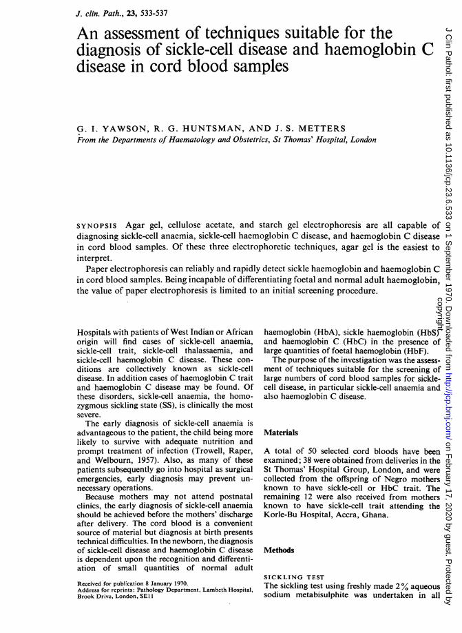
J. clin. Path., 23, 533-537 An assessment of techniques suitable for the diagnosis of sickle-cell disease and haemoglobin C disease in cord blood samples G. I. YAWSON, R. G. HUNTSMAN, AND J. S. METTERS From the Departments of Haematology and Obstetrics, St Thomas' Hospital, London SYNOPSIS Agar gel, cellulose acetate, and starch gel electrophoresis are all capable of diagnosing sickle-cell anaemia, sickle-cell haemoglobin C disease, and haemoglobin C disease in cord blood samples. Of these three electrophoretic techniques, agar gel is the easiest to interpret. Paper electrophoresis can reliably and rapidly detect sickle haemoglobin and haemoglobin C in cord blood samples. Being incapable of differentiating foetal and normal adult haemoglobin, the value of paper electrophoresis is limited to an initial screening procedure. Hospitals with patients of West Indian or African origin will find cases of sickle-cell anaemia, sickle-cell trait, sickle-cell thalassaemia, and sickle-cell haemoglobin C disease. These con- ditions are collectively known as sickle-cell disease. In addition cases of haemoglobin C trait and haemoglobin C disease may be found. Of these disorders, sickle-cell anaemia, the homo- zygmous sickling state (SS), is clinically the most severe. The early diagnosis of sickle-cell anaemia is advantageous to the patient, the child being more likely to survive with adequate nutrition and prompt treatment of infection (Trowell, Raper, and Welbourn, 1957). Also, as many of these patients subsequently go into hospital as surgical emergencies, early diagnosis may prevent un- necessary operations. Because mothers may not attend postnatal clinics, the early diagnosis of sickle-cell anaemia should be achieved before the mothers' discharge after delivery. The cord blood is a convenient source of material but diagnosis at birth presents technical difficulties. In the newborn, the diagnosis of sickle-cell disease and haemoglobin C disease is dependent upon the recognition and differenti- ation of small quantities of normal adult Received for publication 8 January 1970. Address for reprints: Pathology Department, Lambeth Hospital, Brook Drive, London, SEI I haemoglobin (HbA), sickle haemoglobin (HbS) and haemoglobin C (HbC) in the presence of large quantities of foetal haemoglobin (HbF). The purpose of the investigation was the assess- ment of techniques suitable for the screening of large numbers of cord blood samples for sickle- cell disease, in particular sickle-cell anaemia and also haemoglobin C disease. Materials A total of 50 selected cord bloods have been examined; 38 were obtained from deliveries in the St Thomas' Hospital Group, London, and were collected from the offspring of Negro mothers known to have sickle-cell or HbC trait. The remaining 12 were also received from mothers known to have sickle-cell trait attending the Korle-Bu Hospital, Accra, Ghana. Methods SICKLING TEST The sickling test using freshly made 2 % aqueous sodium metabisulphite was undertaken in all copyright. on February 17, 2020 by guest. Protected by http://jcp.bmj.com/ J Clin Pathol: first published as 10.1136/jcp.23.6.533 on 1 September 1970. Downloaded from
Transcript of Anassessment techniques diagnosis haemoglobin C samples · SYNOPSIS Agar gel, cellulose acetate,...
J. clin. Path., 23, 533-537
An assessment of techniques suitable for thediagnosis of sickle-cell disease and haemoglobin Cdisease in cord blood samples
G. I. YAWSON, R. G. HUNTSMAN, AND J. S. METTERSFrom the Departments of Haematology and Obstetrics, St Thomas' Hospital, London
SYNOPSIS Agar gel, cellulose acetate, and starch gel electrophoresis are all capable ofdiagnosing sickle-cell anaemia, sickle-cell haemoglobin C disease, and haemoglobin C diseasein cord blood samples. Of these three electrophoretic techniques, agar gel is the easiest tointerpret.
Paper electrophoresis can reliably and rapidly detect sickle haemoglobin and haemoglobin Cin cord blood samples. Being incapable of differentiating foetal and normal adult haemoglobin,the value of paper electrophoresis is limited to an initial screening procedure.
Hospitals with patients of West Indian or Africanorigin will find cases of sickle-cell anaemia,sickle-cell trait, sickle-cell thalassaemia, andsickle-cell haemoglobin C disease. These con-ditions are collectively known as sickle-celldisease. In addition cases of haemoglobin C traitand haemoglobin C disease may be found. Ofthese disorders, sickle-cell anaemia, the homo-zygmous sickling state (SS), is clinically the mostsevere.The early diagnosis of sickle-cell anaemia is
advantageous to the patient, the child being morelikely to survive with adequate nutrition andprompt treatment of infection (Trowell, Raper,and Welbourn, 1957). Also, as many of thesepatients subsequently go into hospital as surgicalemergencies, early diagnosis may prevent un-necessary operations.
Because mothers may not attend postnatalclinics, the early diagnosis of sickle-cell anaemiashould be achieved before the mothers' dischargeafter delivery. The cord blood is a convenientsource of material but diagnosis at birth presentstechnical difficulties. In the newborn, the diagnosisof sickle-cell disease and haemoglobin C diseaseis dependent upon the recognition and differenti-ation of small quantities of normal adult
Received for publication 8 January 1970.Address for reprints: Pathology Department, Lambeth Hospital,Brook Drive, London, SEI I
haemoglobin (HbA), sickle haemoglobin (HbS)and haemoglobin C (HbC) in the presence oflarge quantities of foetal haemoglobin (HbF).The purpose of the investigation was the assess-
ment of techniques suitable for the screening oflarge numbers of cord blood samples for sickle-cell disease, in particular sickle-cell anaemia andalso haemoglobin C disease.
Materials
A total of 50 selected cord bloods have beenexamined; 38 were obtained from deliveries in theSt Thomas' Hospital Group, London, and werecollected from the offspring of Negro mothersknown to have sickle-cell or HbC trait. Theremaining 12 were also received from mothersknown to have sickle-cell trait attending theKorle-Bu Hospital, Accra, Ghana.
Methods
SICKLING TESTThe sickling test using freshly made 2 % aqueoussodium metabisulphite was undertaken in all
copyright. on F
ebruary 17, 2020 by guest. Protected by
http://jcp.bmj.com
/J C
lin Pathol: first published as 10.1136/jcp.23.6.533 on 1 S
eptember 1970. D
ownloaded from

G. I. Yawson, R. G. Huntsman, and J. S. Metters
samples (Daland and Castle, 1948). The unsealedslides were incubated for 30 minutes at 37°C.
ELECTROPHORETIC TECHNIQUESEach sample was converted into haemolysatefollowing the method described in the ACPBroadsheet no. 33 (Lehmann and Ager, 1965).The following electrophoretic techniques wereemployed.
Paper electrophoresisA vertical tank and Tris bufferpH 8-9 (Cradock-Watson, Fenton, and Lehmann, 1959) were used.
Cellulose acetate electrophoresisThe method of Kohn (1969) was used with a
discontinuous barbitone-Tris buffer (Graham andGrunbaum, 1963). Electrophoresis was conductedfor one hour at 350 V and subsequently thecellulose acetate was fixed in a solution of 5%sulphosalicylic acid in 3% trichloracetic acid forabout one hour before staining with benzidine(Smith, 1968).
Vertical starch gel electrophoresisThe gel was made from Electrostarch (Electro-starch Co., Madison, Wisconsin, USA) andpoured into a Boyer tray (Buchler Instruments,New Jersey, USA). The haemolysate was con-verted to 2 g% cyanmethaemoglobin solutionbefore electrophoresis by dilution of the haemo-lysate in a potassium cyanide-potassium ferri-cyanide solution. The EDTA, boric acid, NaOHbuffer used for the tray and the tanks were as
described by Jonxis and Huismann (1968) andthe postelectrophoretic orthotolidine staining wasas described by Huehns (1968).
Agar gel electrophoresisThe buffer solutions described by Robinson,Robson, Harrison, and Zuelzer (1957) and themethod described by Marder and Conley (1959),both modified, were employed.The stock buffer is made up as follows:
Trisodium citrate .. .. ..147 gCitric acid .. .. .. 4-3 gDissolve and make up to 1 litre with distilledwater.20% (w/v) citric acid in distilled water.Both solutions tend to grow moulds and should
be refrigerated.One hundred ml of stock buffer was diluted
to 1 litre with distilled water. One hundred mlof this diluted buffer (pH 6 6) was used in making1% agar (Difco Bactoagar) solution, which wasthen allowed to cool to about 60°C and pouredinto a glass plate, bordered to give a gel area of15 x 22 cm. The remaining 900 ml was adjustedto pH 6-2 by the addition of 2-2 ml of 21 %citric acid and used in the buffer compartmentsof the tank.Approximately 1 cm-long slits were cut at
intervals across the gel about 5 cm from theanodic end. (A second row of slits could bemade 15 cm from the anodic end.) Pieces of10 mm x 3 mm Whatman no. 42 paper were
soaked in 2 g% haemoglobin solutions. Theexcess haemoglobin was removed by gentlyblotting, and the paper introduced into the agar
slits: 50m amps (approximately 100 v)was appliedfor half an hour at 4°C and the paper stripswere gently removed. The slits were 'closed up' byreadjusting the gel and electrophoresis was con-
tinued for a total of six hours, The gel was fixedand stained with benzidine (Smith, 1968).
Results
SICKLING TESTProviding the samples were received within 24hours, it was found possible to predict correctlythe presence or absence of sickle haemoglobinin all the cord blood samples. Two several-day-old samples of cord blood from babies carrying
Cc,,_C
Fig. 1 Paper electrophoresis of cord bloods.From left to right: 1 C disease; 2 C trait; 3 SC
disease; 4 sickle-cell anaemia; 5 S trait; 6 normalcord blood control.Note that haemoglobins S and C do not clearly
separate, but HbC tends to be more cathodal inposition.
Fig. 2 Cellulose acetate electrophoresis of cordbloods with one adult sickle-cell trait control.From left to right: I normal cord blood; 2 S trait;
3 S-thalassaemia; 4 sickle-cell anaemia; 5 SC disease;6 adult S trait control.
The apparent presence ofHbA in the SC sample(no. S) is an artefact resulting from the age of thisspecimen (see text).
534copyright.
on February 17, 2020 by guest. P
rotected byhttp://jcp.bm
j.com/
J Clin P
athol: first published as 10.1136/jcp.23.6.533 on 1 Septem
ber 1970. Dow
nloaded from

An assessment of techniques suitable for the diagnosis of sickle-cell disease and haemoglobin C disease
Fig. 3 Vertical starch gel electrophoresis of cordbloods with one adult sickle-cell trait control.From left to right: 1 normal cord blood; 2 S trait;
3 sickle-cell anaemia; 4 adult S trait control.The trace of haemoglobin anodol to HbA in the
S trait sample (no. 2) represents y4 (Bart's).
qmw
ow -~ 40 0, de
Fig. 4 Agar gel electrophoresis of cord bloodsamples.From left to right: I SC disease; 2 C disease;
3 C trait; 4 sickle-cell anaemia; 5 S trait; 6 normalcord blood control.
Ib4
t..._- LrsYs
......t.s-
r...'.:2X.|.:. ..:a.::; . .:E .:' .. .,9'*ffg:,
_ ,4...e. .t_ _ _S_ c:'.e.w:'6.
UX .o g
8^&. a-- , ;'='' b-bi;.>>:PPW iilSi |- g.l'sx-s-i[it ; <t' | -( i :i'b '.:._
Fig. 5 Agar gel electrophoresis of cord bloodsamples.From left to right: 1 sickle-cell anaemia;
2 S-thalassaemia; 3 S trait; 4 normal cord bloodcontrol.
The trace ofhaemoglobin anodal to HbF in theS trait and normal cord blood control samples(nos. 3 and 4) represents y4 (Bart's).
the sickle trait were falsely reported as negative.The samples from Ghana were, because of age,found unsatisfactory for the sickling test.
ELECTROPHORESIS
PaperFigure 1 demonstrates the electrophoretic patternfrom cord blood of the newborn children withvarying forms of sickle-cell disease and also HbCtrait and HbC disease.
Cellulose acetateFigure 2 shows the electrophoretic pattern ofcordblood from a normal baby and babies with sickle-cell trait, a sickle-cell thalassaemia, sickle-cellanaemia, and sickle-cell haemoglobin C diseasewith an adult sickle-cell trait control.
Vertical starch gelFigure 3 shows the electrophoretic pattern fromcord blood of a normal baby and babies withsickle-cell trait and sickle-cell anaemia and anadult sickle-cell trait control.
Agar gelFigures 4 and 5 show the electrophoretic patternfrom cord blood of newborn children with vary-ing forms of sickle-cell disease and also HbC traitand HbC disease.The results of 50 samples examined showed
17 normal cases, 24 cases of sickle-cell traits,three of sickle-cell anaemias, one of sickle-cellthalassaemia, and one of sickle-cell HbC disease.In addition there were three samples with HbCtrait and one with HbC disease.
It was possible to confirm the diagnosis in 28of the 38 London cord blood samples by a laterhaematological examination. Two of the threecases of sickle-cell anaemia, the case of sickle-cellthalassaemia, and that of HbC disease were allconfirmed. In 22 cases it was possible also toexamine the fathers, and in these cases the diag-nosis of the baby was compatible with the parentalhaemoglobin pattern.No follow-up or family study was possible on
the 12 samples received from Ghana, whichincluded the case of sickle-cell HbC disease andone case of sickle-cell anaemia.
Discussion
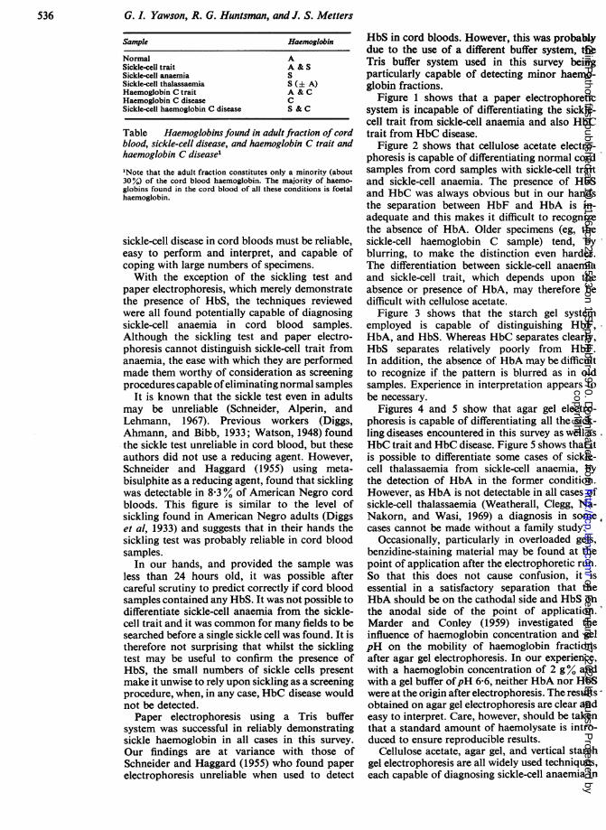
About 70% of the total cord blood haemoglobinis HbF. The differentiation between a normalbaby and a baby with the haemoglobin disordersinvestigated in this paper depends on the recog-nition of the constituent haemoglobins makingup the residual small adult fraction. Theseconstituents are shown in the Table.A recommended technique for the diagnosis of
535
4 _p
q
copyright. on F
ebruary 17, 2020 by guest. Protected by
http://jcp.bmj.com
/J C
lin Pathol: first published as 10.1136/jcp.23.6.533 on 1 S
eptember 1970. D
ownloaded from
G. L Yawson, R. G. Huntsman, and J. S. Metters
Sample Haemoglobin
Normal ASickle-cell trait A & SSickle-cell anaemia SSickle-cell thalassaemia S (+ A)Haemoglobin C trait A & CHaemoglobin C disease CSickle-cell haemoglobin C disease S & C
Table Haemoglobins found in adult fraction of cordblood, sickle-cell disease, and haemoglobin C trait andhaemoglobin C disease'
"Note that the adult fraction constitutes only a minority (about30%) of the cord blood haemoglobin. The majority of haemo-globins found in the cord blood of all these conditions is foetalhaemoglobin.
sickle-cell disease in cord bloods must be reliable,easy to perform and interpret, and capable ofcoping with large numbers of specimens.With the exception of the sickling test and
paper electrophoresis, which merely demonstratethe presence of HbS, the techniques reviewedwere all found potentially capable of diagnosingsickle-cell anaemia in cord blood samples.Although the sickling test and paper electro-phoresis cannot distinguish sickle-cell trait fromanaemia, the ease with which they are performedmade them worthy of consideration as screeningprocedures capable ofeliminating normal samples
It is known that the sickle test even in adultsmay be unreliable (Schneider, Alperin, andLehmann, 1967). Previous workers (Diggs,Ahmann, and Bibb, 1933; Watson, 1948) foundthe sickle test unreliable in cord blood, but theseauthors did not use a reducing agent. However,Schneider and Haggard (1955) using meta-bisulphite as a reducing agent, found that sicklingwas detectable in 8-3% of American Negro cordbloods. This figure is similar to the level ofsickling found in American Negro adults (Diggset al, 1933) and suggests that in their hands thesickling test was probably reliable in cord bloodsamples.
In our hands, and provided the sample wasless than 24 hours old, it was possible aftercareful scrutiny to predict correctly if cord bloodsamples contained any HbS. It was not possible todifferentiate sickle-cell anaemia from the sickle-cell trait and it was common for many fields to besearched before a single sickle cell was found. It istherefore not surprising that whilst the sicklingtest may be useful to confirm the presence ofHbS, the small numbers of sickle cells presentmake it unwise to rely upon sickling as a screeningprocedure, when, in any case, HbC disease wouldnot be detected.
Paper electrophoresis using a Tris buffersystem was successful in reliably demonstratingsickle haemoglobin in all cases in this survey.Our findings are at variance with those ofSchneider and Haggard (1955) who found paperelectrophoresis unreliable when used to detect
HbS in cord bloods. However, this was probablydue to the use of a different buffer system, theTris buffer system used in this survey beingparticularly capable of detecting minor haemo-globin fractions.
Figure 1 shows that a paper electrophoreticsystem is incapable of differentiating the sickle-cell trait from sickle-cell anaemia and also HbCtrait from HbC disease.
Figure 2 shows that cellulose acetate electro-phoresis is capable of differentiating normal cordsamples from cord samples with sickle-cell traitand sickle-cell anaemia. The presence of HbSand HbC was always obvious but in our handsthe separation between HbF and HbA is in-adequate and this makes it difficult to recognizethe absence of HbA. Older specimens (eg, thesickle-cell haemoglobin C sample) tend, byblurring, to make the distinction even harder.The differentiation between sickle-cell anaemiaand sickle-cell trait, which depends upon theabsence or presence of HbA, may therefore bedifficult with cellulose acetate.
Figure 3 shows that the starch gel systememployed is capable of distinguishing HbF,HbA, and HbS. Whereas HbC separates clearly,HbS separates relatively poorly from HbF.In addition, the absence of HbA may be difficultto recognize if the pattern is blurred as in oldsamples. Experience in interpretation appears tobe necessary.
Figures 4 and 5 show that agar gel electro-phoresis is capable of differentiating all the sick-ling diseases encountered in this survey as well as
HbC trait and HbC disease. Figure 5 shows that itis possible to differentiate some cases of sickle-cell thalassaemia from sickle-cell anaemia, bythe detection of HbA in the former condition.However, as HbA is not detectable in all cases ofsickle-cell thalassaemia (Weatherall, Clegg, Na-Nakorn, and Wasi, 1969) a diagnosis in some 4cases cannot be made without a family study.
Occasionally, particularly in overloaded gels,benzidine-staining material may be found at thepoint of application after the electrophoretic run.
So that this does not cause confusion, it isessential in a satisfactory separation that theHbA should be on the cathodal side and HbS on
the anodal side of the point of application.Marder and Conley (1959) investigated theinfluence of haemoglobin concentration and gelpH on the mobility of haemoglobin fractionsafter agar gel electrophoresis. In our experience,with a haemoglobin concentration of 2 g% andwith a gel buffer ofpH 6-6, neither HbA nor HbSwere at the origin after electrophoresis. The results,obtained on agar gel electrophoresis are clear andeasy to interpret. Care, however, should be takenthat a standard amount of haemolysate is intro-duced to ensure reproducible results.
Cellulose acetate, agar gel, and vertical starchgel electrophoresis are all widely used techniques,each capable of diagnosing sickle-cell anaemia in
536copyright.
on February 17, 2020 by guest. P
rotected byhttp://jcp.bm
j.com/
J Clin P
athol: first published as 10.1136/jcp.23.6.533 on 1 Septem
ber 1970. Dow
nloaded from
An assessment of techniques suitable for the diagnosis of sickle-cell disease and haemoglobin C disease
cord samples. If sickle-cell anaemia is to berecognized in cord blood samples, the mostsuitable of these three electrophoretic techniquesis likely to be the one with which the laboratoryhas had previous experience.
In many cases a laboratory will not routinelybe using any of these three electrophoretictechniques. The starch gel technique will befound to be adequate but it is tedious to performif large numbers of specimens are involved andinterpretation is sometimes difficult. Celluloseacetate, with its advantage of rapidity, would alsobe an acceptable technique, and would be capableof coping with large numbers of specimens, butagain interpretation may not always be easy.Agar gel electrophoresis was suggested for the
diagnosis of sickle-cell anaemia by Fessas (1965)and has been used by Centa and Sciarratta (1967)to follow up a sickle-cell trait baby from birth to6 months old. On a larger scale Van Baelen,Vandepitte, and Eeckels (1968) used agar gel forthe diagnosis of sickle-cell anaemia in Congoleseneonates. In our hands, the ease of interpretationof the agar gel makes this the most suitableelectrophoretic technique for the diagnosis ofsickle-cell anaemia in cord blood. Because agargel electrophoresis is a reasonably time-consumingprocedure, the possible use of a previous screen-ing technique to eliminate normal samples wasinvestigated.
If one only examines the cord bloods of babieswith sickle-positive mothers, one would expect,in a community with a 20% sickling rate, thatover 50% of the babies would have the sickle-celltrait and approximately 5% sickle-cell anaemia.If cord bloods of all babies are examined, about20% of babies would have the sickle-cell traitand approximately 1% sickle-cell anaemia.Particularly in the latter case, in order to eliminatethe high percentage of normal cord bloods, itappears desirable to adopt an initial rapidscreening procedure to detect the presence ofHbS.Two of the techniques considered, paper and
cellulose acetate electrophoresis, are potentialscreening procedures because they are capable ofrapidly and reliably detecting HbS in cordblood samples. Of these two techniques, paperelectrophoresis is the cheaper and requires lessoperational skill.
If large numbers of cord blood samples are tobe examined for sickle-cell and HbC diseases, themost suitable technique appears to be an initial
screening with paper electrophoresis. All sampleswhere HbS and HbC are detected are subse-quently submitted to agar gel electrophoresis.
We are grateful to Dr F. S. Boi-Doku of TheHealth Laboratory Services, Ghana, who kindlysent us the samples from the Korle-Bu Hospital,Ghana.
References
Centa, A., and Sciarratta, V. (1967). Su di un caso di falcemianeonatale seguito per tutto il primo semestre di vita.Minerva pediat., 19, 1919-1924.
Cradock-Watson, J. E., Fenton, J. C. B., and Lehmann, H. (1959).TRIS buffer for the demonstration of Haemoglobin A, bypaper electrophoresis. J. clin. Path., 12, 372-373.
Daland, G. A., and Castle, W. B. (1948). A simple and rapidmethod for demonstrating sickling of the red blood cells:J. Lab. clin. Med., 33,1082-1088.
Diggs, L. W., Ahman, C. F., and Bibb, J. (1933). The incidenceand significance of the sickle-cell trait. Ann. intern. Med.,7, 769-778.
Fessas, P. (1965). Discussion in AbnormalHaemoglobins in Africa,edited by J. H. P. Jonxis, p. 457. Blackwell, Oxford.
Graham, J. L., and Grunbaum, B. W. (1963). A rapid method formicroelectrophoresis and quantitation of haemoglobinson cellulose acetate. Amer. J. clin. Path., 39, 567-578.
Huehns, E. (1968). In Chromatographic and ElectrophoreticTechniques, 2nd ed., edited by Ivor Smith, vol 2, pp. 291-294. Heinemann, London.
Jonxis, J. H. P., and Huisman, T. H. J. (1968). A LaboratoryManual on Abnormal Haemoglobins, 2nd ed., pp. 31-33.Blackwell, Oxford and Edinburgh.
Kohn, J. (1969). Separation of haemoglobins on cellulose acetate.J. clin. Path., 22, 109-111.
Lehmann, H., and Ager, J. A. M. (1965). Laboratory detectionof abnormal haemoglobins. ACP Broadsheet No. 33.
Marder, V. J., and Conley, C. L. (1959). Electrophoresis ofhemoglobin on agar gels. Bull. Johns Hopk. Hosp., 105,77-88.
Robinson, A. R., Robson, M., Harrison, A. P., and Zuelzer,W. W. (1957). A new technique for differentiation ofhemoglobin. J. Lab. clin. Med., 50, 745-752.
Schneider, R. G., and Haggard, M. E. (1955). Sickling: Aquantitatively delayed genetic character. Proc. Soc. exp.Biol. (N. Y.), 89, 196-199.
Schneider, R. G., Alperin, J. B., and Lehmann, H. (1967).Sickling tests. J. Amer. med. Ass., 202, 419-421.
Smith, I. (1968). In Chromatographic and electrophoreticTechniques, 2nd ed., edited by I. Smith, vol. 2, p. 233.Heinemann, London.
Trowell, H. C., Raper, A. B., and Welbourn, H. F. (1957). Thenatural history of homozygous sickle-cell anaemia inCentral Africa. Quart. J. Med., 26, 401-422.
Van Baelen, H., Vandepitte, J., and Eeckels, R. (1968). In Proc.inter. Colloq. trop. Anaemias. p. 81, Prins Leopold Institutevoor Tropische Geneeskunde, Antwerp.
Watson, J. (1948). The significance of the paucity of sicklecells in newborn negro children. Amer. J. med. Sci., 215,419-423.
Weatherall, D. J., Clegg, J. B., Na-Nakorn, S., and Wasi,P. (1969). The pattern of disordered haemoglobinsynthesis in homozygous and heterozygous R-thalassaemia.Brit. J. Haemat., 16, 251-267.
537copyright.
on February 17, 2020 by guest. P
rotected byhttp://jcp.bm
j.com/
J Clin P
athol: first published as 10.1136/jcp.23.6.533 on 1 Septem
ber 1970. Dow
nloaded from