ANALYSIS OF X CHROMOSOME INACTIVATION IN PRIMARY AND SECONDARY SJOGREN ... · ABSTRACT Lack of...
106
ANALYSIS OF X CHROMOSOME INACTIVATION IN PRIMARY AND SECONDARY SJOGREN SYNDROME A THESIS SUBMITTED TO THE DEPARTMENT OF MOLECULAR BIOLOGY AND GENETICS AND THE INSTITUTE OF ENGINEERING AND SCIENCE OF BILKENT UNIVERSITY IN PARTIAL FULFILLMENT OF THE REQUIREMENTS FOR THE DEGREE OF MASTER OF SCIENCE BY MELDA KANTAR AUGUST, 2008
-
Upload
nguyencong -
Category
Documents
-
view
216 -
download
0
Transcript of ANALYSIS OF X CHROMOSOME INACTIVATION IN PRIMARY AND SECONDARY SJOGREN ... · ABSTRACT Lack of...

ANALYSIS OF X CHROMOSOME INACTIVATION IN PRIMARY
AND SECONDARY SJOGREN SYNDROME
A THESIS SUBMITTED TO THE DEPARTMENT OF MOLECULAR BIOLOGY
AND GENETICS AND THE INSTITUTE OF ENGINEERING AND SCIENCE OF
BILKENT UNIVERSITY IN PARTIAL FULFILLMENT OF THE
REQUIREMENTS FOR THE DEGREE OF MASTER OF SCIENCE
BY MELDA KANTAR
AUGUST, 2008

I certify that I have read this thesis and that in my opinion it is fully adequate, in scope and in quality, as a thesis for the degree of Master of Science. ___________________
Prof. Tayfun Özçelik
I certify that I have read this thesis and that in my opinion it is fully adequate, in scope and in quality, as a thesis for the degree of Master of Science.
__________________ Assist. Prof. Ali O. Güre
I certify that I have read this thesis and that in my opinion it is fully adequate, in scope and in quality, as a thesis for the degree of Master of Science.
__________________ Assist. Prof. Hüseyin Boyacı Approved for the Institute of Engineering and Science
_____________________________
Director of Institute of Engineering and Science Prof. Dr. Mehmet Baray
ii
ABSTRACT
Lack of evidence for the role of skewed X chromosome inactivation in the female
predisposition to Sjogren Syndrome
Melda Kantar
M.S. in Molecular Biology and Genetics Supervisor: Prof. Dr. Tayfun Özçelik
August 2008, Pages 91
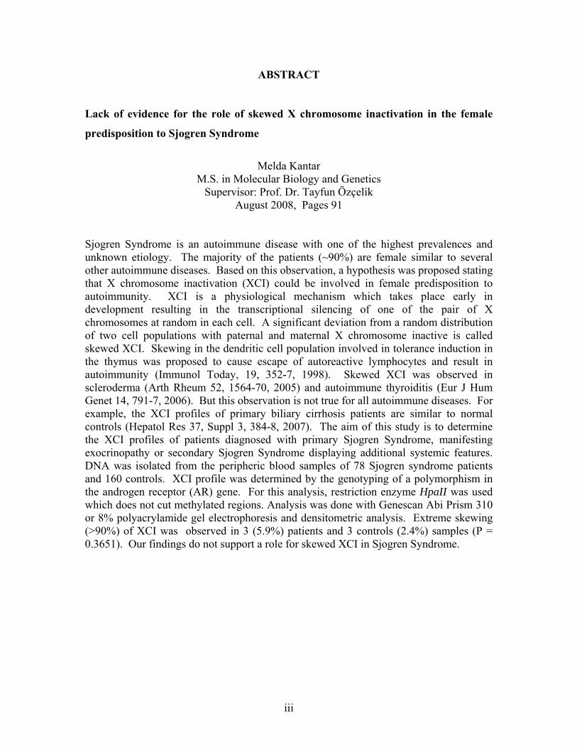
Sjogren Syndrome is an autoimmune disease with one of the highest prevalences and unknown etiology. The majority of the patients (~90%) are female similar to several other autoimmune diseases. Based on this observation, a hypothesis was proposed stating that X chromosome inactivation (XCI) could be involved in female predisposition to autoimmunity. XCI is a physiological mechanism which takes place early in development resulting in the transcriptional silencing of one of the pair of X chromosomes at random in each cell. A significant deviation from a random distribution of two cell populations with paternal and maternal X chromosome inactive is called skewed XCI. Skewing in the dendritic cell population involved in tolerance induction in the thymus was proposed to cause escape of autoreactive lymphocytes and result in autoimmunity (Immunol Today, 19, 352-7, 1998). Skewed XCI was observed in scleroderma (Arth Rheum 52, 1564-70, 2005) and autoimmune thyroiditis (Eur J Hum Genet 14, 791-7, 2006). But this observation is not true for all autoimmune diseases. For example, the XCI profiles of primary biliary cirrhosis patients are similar to normal controls (Hepatol Res 37, Suppl 3, 384-8, 2007). The aim of this study is to determine the XCI profiles of patients diagnosed with primary Sjogren Syndrome, manifesting exocrinopathy or secondary Sjogren Syndrome displaying additional systemic features. DNA was isolated from the peripheric blood samples of 78 Sjogren syndrome patients and 160 controls. XCI profile was determined by the genotyping of a polymorphism in the androgen receptor (AR) gene. For this analysis, restriction enzyme HpaII was used which does not cut methylated regions. Analysis was done with Genescan Abi Prism 310 or 8% polyacrylamide gel electrophoresis and densitometric analysis. Extreme skewing (>90%) of XCI was observed in 3 (5.9%) patients and 3 controls (2.4%) samples (P = 0.3651). Our findings do not support a role for skewed XCI in Sjogren Syndrome.
iii

ÖZET Sjogren sendromu ile X inaktivasyonu bozukluğu arasında bir ilişki bulunmuyor
Melda Kantar Moleküler Biyoloji ve Genetik Yüksek Lisans
Tez Yöneticisi: Prof. Dr. Tayfun Özçelik Ağustos 2007, Sayfa 91
Sjogren sendromu etiyolojisi bilinmeyen ve prevalansı en yüksek olan otoimmün hastalıklardan biridir. Pek çok otoimmün hastalıkta olduğu gibi hastaların büyük bir çoğunluğu (~%90) kadındır. Bu gözlemden yola çıkarak, bozuk X inaktivasyonunun otoimmun hastalıkları tetikleyebileceği hipotezi ileri sürülmüştür (Immunol Today, 19, 352-7, 1998). X inaktivasyonu kadınlarda erken gelişme döneminde hücrelerdeki X kromozomu çiftinden rastgele birinin susturulmasıyla sonuçlanan normal bir fizyolojik olaydır. Hücrelerin önemli bir kısmında aynı X kromozomunun susturulması bozuk X inaktivasyonu olarak adlandırılır. Timusda, tolerans oluşumunda rol alan dendritik hücrelerdeki X inaktivasyonu bozukluğunun otoreaktif lenfositlerin kaçmasına ve otoimmüniteye neden olabileceği hipotezi ileri sürülmüştür. Skleroderma (Arth Rheum 52, 1564-70, 2005) ve otoimmün tiroiditis (Eur J Hum Genet 14, 791-7, 2006) hastalıklarında bozuk X inaktivasyonu gözlenmiştir. Fakat bu gözlem tüm otoimmün hastalıklar için geçerli değildir. Örneğin primer biliyer siroz hastalarının X inaktivasyonu profilleri normal kontroller gibidir (Hepatol Res 37, Suppl 3, 384-8, 2007). Bu çalışmanın amacı sadece salgı bezi patolojisi olan primer Sjogren Sendromu ve aynı zamanda başka sistemik patolojiler içeren sekonder Sjogren Sendromu tanısı alan hastalarda X-inaktivasyonu profillerinin belirlenmesidir. 78 hasta ve 160 kontrolün periferik kanından DNA izole edilmiştir. X inaktivasyon profili androjen reseptör geninde bulunan bir polimorfizmin genotiplemesi ile belirlenmiştir. Bu inceleme için metilli bölgeleri tanımayan bir restriksiyon enzimi olan HpaII ile kesim yapılmış ve hedef bölge çoğaltılmıştır. İnceleme Genescan Abi Prism 310 ya da %8 poliakrilamid jel elektroforezi ve densitometrik ölçümle yapılmıştır. İleri derecede (>%90) X inaktivasyonu bozukluğu 3 (%5.9) hasta ve 3 kontrol (%2.4) örneğinde gözlenmiştir (P = 0.3651). Bulgularımız X inaktivasyonu bozukluğu ile Sjogren Sendromu arasında bir ilişkinin varlığını desteklememiştir.
iv

TO MY FAMILY, FOR THEIR LOVE AND SUPPORT
v

ACKNOWLEDGEMENTS
It is my pleasure to express my deepest gratitude to my advisor Prof. Dr. Tayfun
Özçelik for his scientific guidance and support all through my thesis. I especially want to
thank him for introducing me to research..
I would like to adress my special thanks to Zeynep Özbalkan for obtaining the
blood samples and coordinating the clinical studies.
I would like to thank Scientific and Technological Research Council of Turkey
for the funding.
I would like to express my thanks to Elif Uz and Çiğdem Aydın Mustafa for
guiding me in every step of my research and always being there for my questions and for
their endless support in every obstacle I was faced to. A special thank to Elif Uz for her
help in determining the XCI status of Sjogren Syndrome patients. I would like to thank
all my group members Emre Onat, Şafak Çağlayan, Süleyman Gülsuner for their
incredible help and support. I would like to thank Mustafa Gökhan Ertosun for helping
me with the format of my thesis.
I would like to thank all MBG members for making Bilkent Molecular Biology
Department an incredible place to live and work.
I would also like to thank my friends Hülya Budunoğlu, Hande Erkut, Cansu
Aksu, Banu Demir, Emre Onat, Fuat Yağcı, Elif Yaman, Hande Kocak, Pelin
Telkoparan, Esen Oktay, Gizem Tinçer, Ceren Sucularlı and all the others I have not
mentioned for listening to my every problem during my academic and personal life. I
want to thank my boyfriend, Onur Karakuş for sharing my life.
Lastly, I want to express my sincere love and thanks to my family for being there
whenever I needed help and support. I want to thank my mother, Nujan Kocagöz and my
father, Elvend Kantar for helping me through every struggle and especially my dear
sister, Selda Kantar without whom life would be unbearable.
vi

TABLE OF CONTENTS
ABSTRACT III
ÖZET IV
DEDICATION PAGE V
ACKNOWLEDGEMENTS VI
TABLE OF CONTENTS VII
LIST OF TABLES X
LIST OF FIGURES XI
ABBREVIATIONS XII
1. INTRODUCTION 1
1.1.Sjogren Syndrome 1
1.1.1. History 1
1.1.2. Symptoms 3
1.1.3. Criteria for diagnosis 3
1.1.4. Treatment 5
1.1.5. Epidemiology 6
1.1.6. Classification as primary and secondary 6
Sjogren Syndrome
1.1.7. Pathophysiology and Etiology 7
1.1.7.1 Genetic predisposition 12
1.1.7.2 Associated genes 13
1.1.7.3 Associated antibodies 14
1.1.7.4 Viruses 16
1.1.8 .Research to relate microchimerism 17
and Sjogren Syndrome
1.2. X Chromosome Inactivation 18
1.2.1 X dosage compensation 18
1.2.2. Discovery of mammalian dosage compensation 18
vii

1.2.3. Features of Inactive X 20
1.2.4. Clonal Nature of XCI 20
1.2.5. Timing of XCI 21
1.2.6. Master control region of XCI and its components 22
1.2.7. Steps of XCI 24
1.2.8. Genes escaping XCI 26
1.2.9. Primary and secondary causes of skewed XCI 27
1.3.Self tolerance and Autoimmunity 28
1.3.1 Immune system 28
1.3.1.1 Clonal theory of Jerne and Burnett 28
1.3.1.2 Self tolerance 28
1.3.2 Autoimmunity 29
1.3.2.1 Genes associated with autoimmunity 30
1.3.2.2 X-linked genes implicated in immunity 32
1.3.2.3 Molecular Mimicry 32
1.3.2.4 Female predominance in autoimmunity 33
1.3.2.4.1 Hormones 35
1.3.2.4.2 Microchimerism 36
1.3.2.4.3 X chromosome Monosomy 37
1.4 Skewed X inactivation and autoimmunity 38
1.4.1 Kast and Stewart hypothesis 38
1.5. Aim and Strategy 40
2. MATERIALS AND METHODS 41
2.1 Samples 41
2.1.1 Control Samples 41
2.1.2 Sjogren Syndrome patient Samples 41
2.2 Materials 42
2.2.1 Primers 42
2.2.2 Enzymes 42
viii

2.2.3 Oligonucleotide 44
2.2.4 Chemicals Reagents, Kits 45
2.2.4 Standard solutions and buffers 46
2.3 Methods 47
2.3.1 Sample collection 47
2.3.2 DNA Isolation from venous blood 47
2.3.4 AR genoyping assay 48
2.3.4.1. Restriction Enzyme Digestion 50
2.3.4.2 Polymerase chain reaction (PCR) 51
2.3.4.3 Visualization 51
2.3.4.3.1 Agarose gel electrophoresis 51
2.3.4.3.2 Polyacrylamide gel 52
electrophoreis (PAGE)
2.3.4.4 Densitometric and Statistical Analysis 52
3. RESULTS 53
4. DISCUSSION 56
5. FUTURE PERSPECTIVES 61
6. REFERENCES 62
7. APPENDICIES 80
ix

LIST OF TABLES
Table 1.1 Timeline of important advances in delineating Sjogren Syndrome 2 Table 1.2 Current criteria for Sjogren Syndrome diagnosis 4 Table 1.3 Prevalence of sicca features and associated Sjogren Syndrome 7
in systemic autoimmune diseases Table 1.4 Polymorphisms of interleukins and other genes associated 14
with Sjogren Syndrome Table 1.5 Autoantibodies described in Sjogren Syndrome patients 15
and their corresponding prevelances Table 1.6 Ratio of female:male occurence of autoimmune disorders 34 Table 2.1 List of chemicals, reagents, kits used in this study 45 Table 2.2 Characteristics of Sjogren patients Table 3.1 Proportion of Sjogren Syndrome patients and controls with 54
skewed XCI
x
LIST OF FIGURES
Figure 1.1 Minor salivary gland biopsy photomicrographs 8 Figure 1.2 Steps of the Sjogren Syndrome pathogenesis according 10
to the epithelithis model Figure 1.3 Models in relation to XCI timing in the embryo 22 Figure 1.4 Elements of Xic 24 Figure 1.5 Different mechanisms of self tolerance 29 Figure 1.6 Incidence of autoimmune disorders by sex 35 Figure 1.7 Consequences of skewed XCI in thymic cells 39 Figure 2.1 AR genotyping amplicon 43 Figure 2.2 Sizes of the fragments of pUC Mix Marker 8 and 44
MassRuler DNA Ladder Mix and appearance on gel electrophoresis. Figure 2.3 Establishing random and skewed XCI with AR genotyping assay 49 Figure 3.1 XCI inactivation status in Sjogren Syndrome patients 54
xi

ABBREVIATIONS
ANA Antinuclear antibodies
Abs Antibodies
AID Autoimmune disease
AIDS Acquired immunodeficiency disease
AITD Autoimmune tyroid disease
AIRE Autoimmune regulator
ALPS Autoimmune lymphoproliferative syndrome
AQP Aquaporin
APC Antigen presenting cell
AR Androgen receptor
BAFF B cell survival factor
bp Base pair
BCR B cell receptor
Csk c-src tyrosine kinase
CTLA-4 Cytotoxic T lymphocyte antigen 4
DEMS Dry eyes and mouth symptoms
ddH2O Deionized water
dNTP Deoxynucleotide triphosphate
DNA Deoxyribonucleic acid
EBV Epstein barr virus
EDTA Ethylenediaminetetraacetic acid
EtBr Ethidium bromide
EtOH Ethanol
G6PD Glucose 6-phosphate dehydrogenase
GVHD Graft versus host disease
HBV Hepatitis B virus
HLA Human leukocyte antigen
HIV Human immunodeficiency virus
HSV-1 Herpes simplex virus Type I
xii

HTLV Human T lymphotrophic virus
HIGM1 Hyper-IgM syndrome type I
Ig Immunoglobulin
IgVλ Immunoglobulin variable λ
IFN-γ Interferon γ
IL Interleukin
IPEX Immune dysregulation, polyendocrinopahty, enteropathy
L1 LINE1
Mb Megabase
MCTD Muscle connective tissue disease
MgCl2 Magnesium chloride
MHC Major histocompatibility complex
MBL Mannose binding lectin
mM Millimolar
ml Milliliter
µl Microliter
MS Multiple sclerosis
kb Kilobase
L1 Line1 elements
NF-қB Nuclear factor kappa B
PAGE Polyacrylamide gel electrophoresis
PI3K Phosphatidylinositol-3-kinase
PBC Primary biliary cirrhosis
PCR Polymerase chain reaction
PcG Polycomb-group proteins
PI3K Phosphoinositide 3 phosphatide
PLP Proteolipid protein
PKC Phosphokinase C
RA Rheumatoid arthritis
RE Restriction enzyme
RF Rheumatoid factor
xiii
SCID Severe combined immunodeficiency syndrome
SCLE Subacute cutaneous lupus erthematosus
SDS Sodium dodecyl sulphate
SLE Systemic lupus erythramatosus
SNP Single nucleotide polymophism
SH3 Src homology 3
SSc Systemic sclerosis
T1D Type 1 diabetis
TAE Tric-acetic acid-EDTA
TCR T cell receptor
TEMED N, N, N, N-tetramethyl-1-2, diaminoethane
Th1 T helper 1
Th2 T helper 2
TLR Toll like receptor
TNF Tumour necrosis factor
Tris Tris aminomethan
WAS Wiskott-Aldrich syndrome
Xa Active X
XCI X chromosome inactivation
Xce X chromosome controlling element
Xi Inactive X
Xic X-inactivation center
Xist X-inactive specific element
Xite X-inactivation intergenic transcription element
XEDA-ID X-linked recessive anhydortic ectodermal dysplasia with
immunodeficiency
XLA X-linked agammaglobulinemia
XLP X-linked lymphoproliferative syndrome
xiv

CHAPTER I: INTRODUCTION
1.1. Sjogren Syndrome
Sjogren Syndrome is a chronic and systemic autoimmune disease
characterized by progressive lymphocytic infiltration of polyglandular tissue and
subsequent tissue destruction. This results in the functional impairment of
salivary and lacrimal glands, and causes keratoconjunctivitis sicca, which refers to
dryness of the eyes and xerostomia meaning dryness of the mouth. The ease and
safety of a minor salivary gland biopsy enables study of the molecular biology of
this autoimmune exocrinopathy. With microscopic examination, the lymphocytic
replacement of the epithelium and lymphoepithelial lesion can be observed. It is
an inflammatory rheumatic disease with main features in the eyes and mouth
(Ramos-Casals, 2005; Kassan, 2004; Schwartz, 2007; Fox, 2000).
1.1.1 History
Sjogren Syndrome is a relatively newly identified disease. Its histology
was described and a link with arthritis was reported in the late 19th and early 20th
centuries as single case reports. In 1933, Henrick Sjogren reported the association
of xeropthalmia meaning dry eyes and xerostomia with polyarthritis in 19 cases.
In the early years, research was mainly on the opthalmology of Sjogren
Syndrome. The clinical spectrum of Sjogren Syndrome in 1956 was defined in 62
subjects. The rheumatologic aspect of Sjogren Syndrome was heavily
emphasized when the distinction between primary and secondary Sjogren
Syndrome was first described in 1965. In 1970s, autoantibodies characteristic of
Sjogren Syndrome were described. In 1990s, the diagnostic tests became a
routine procedure and research on disease-modifying drugs started (Venables,
2004). Table 1.1 shows the timeline of advances in delineating Sjogren
Syndrome.
1
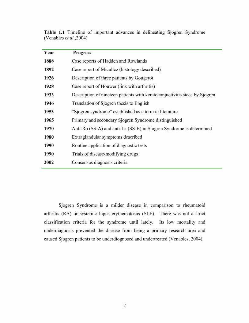
Table 1.1 Timeline of important advances in delineating Sjogren Syndrome (Venables et al.,2004)
Year Progress
1888 Case reports of Hadden and Rowlands
1892 Case report of Miculicz (histology described)
1926 Description of three patients by Gougerot
1928 Case report of Houwer (link with arthritis)
1933 Description of nineteen patients with keratoconjuctivitis sicca by Sjogren
1946 Translation of Sjogren thesis to English
1953 “Sjogren syndrome” established as a term in literature
1965 Primary and secondary Sjogren Syndrome distinguished
1970 Anti-Ro (SS-A) and anti-La (SS-B) in Sjogren Syndrome is determined
1980 Extraglandular symptoms described
1990 Routine application of diagnostic tests
1990 Trials of disease-modifying drugs
2002 Consensus diagnosis criteria
Sjogren Syndrome is a milder disease in comparison to rheumatoid
arthritis (RA) or systemic lupus erythematosus (SLE). There was not a strict
classification criteria for the syndrome until lately. Its low mortality and
underdiagnosis prevented the disease from being a primary research area and
caused Sjogren patients to be underdiognosed and undertreated (Venables, 2004).
2

1.1.2 Symptoms
The symptoms in relation to the eyes are more serious than the symptoms
of the mouth. Underlying collagen can be revealed as a result of erosions in the
conjunctiva leading to the appearance of filamentary keratitis. More dental care is
a sign of xerostomia. Parotid swelling occurs in half of the patients. Other
exocrine glands can be involved effecting skin, vagina, gastrointestinal tract,
colon and rectum. 70% of the patients complain of fatigue.
Systemic features include renal involvement, neurological involvement,
vascular involvement, cutenous involvement, pulmonary involvement,
muscuskeletal involvement, gastroenterologic involvement, congenital heart block
and lymphoma (Venables, 2004; Kassan, 2008; Ramos-Casals, 2005).
1.1.3 Criteria for diagnosis
Until recently, a consensus international diagnosis criteria for Sjogren
Syndrome was unavailable. Because of its heterogeneous nature, Sjogren
Syndrome is hard to diagnose. It has been hard to achieve a consensus because
there are diseases that mimic Sjogren Syndrome such as age-related atrophy,
chronic anxiety, chronic fatigue syndrome, fibromyalgia and dry eyes and mouth
symptoms (DEMS). Previously, there were `Copenhagen`, `Californian` and
`European` criteria. The revised American-European consensus criteria was
established in 2002 (Vitali, 2002). The application of this criteria was essential
both for clinical diagnosis and research.
The current criteria is as designated in Table 1.2. Four of the listed
observations including either the fourth or sixth one is needed to diagnose a
patient as Sjogren Syndrome. The observations include objective signs and
symptoms of dryness. Evaluation of symptoms is based on a short questionaire.
Ocular signs are evaluated based on Schirmer`s test or Rose Bengal staining. Oral
signs are evaluated with scintiagraphy, sialography or a test for salivary flow rate.
One observation that indicates the inflammatory nature of the disease is the
3

characteristic appearance of a minor salivary gland biopsy with focal
inflammation. Focal lymphocytic infiltrates are expected in the minor salivary
gland biopsy resulting in a focus score of more than 1 (A cluster of 50 or more
lymphocytes is called a focus). Another observation that indicates inflammation
is the presence of characteristic autoantibodies, rheumatoid factor or anti-nuclear
antibodies in the serum. To prevent confusion with dry eyes and mouth
symptoms (DEMS), these observations for determining the inflammatory nature
of the disease are necessary (Venables, 2004).
Table 1.2 Current Criteria for Sjogren Syndrome diagnosis (Venables et al, 2004)
Ocular symptoms
1. Have you had daily, persistance, troublesome dry eyes for more than 3 months 2. Do you have a recurrent sensation of sand and gravel in the eyes
I
3. Do you use a tear substitutive more than 3 times a day
Oral symptoms
1. Have you had a daily feeling of dry mouth for more than 3 months 2. Have you had recurrently or persistantly swollen salivary glands as an adult
II
3. Do you frequently drink liquids to aid in swallowing dry foods
III Positive Schirmer`s I test or Rose Bengal score
IV Abnormal lower lip biopsy (focus score >= 1)
V Positive result for unstimulated whole salivary flow (<= 1.5ml in 15 min)
VI Antibodies to Ro (SS-A) or La (SS-B), or both
In diagnosis, it is important to show that the salivary dysfunction roots
from an inflammation. Salivary dysfunction can also be caused by drugs,
infection, head and neck radiation treatment, acquired immune deficiency
syndrome (AIDS), preexisting lymphoma, sarcoidosis, graft versus host disease
(GVHD), use of anticholinergic drugs and the autonomic nervous system. These
are exclusions to diagnosis (Venables, 2004).
4

1.1.4 Treatment
Until recently, the treatment was only topical and symptomatic to improve
moisture; decrease inflammation and prevent damage. These include performing
general measures and using tear and saliva substitutes and mucolytic agents.
Recently, research on disease-modifying drugs have been intensified with the
recognition that Sjogren Syndrome is a disease of considerable morbidity.
Currently a more aggressive approach to therapy is applied with topical and
systemic treatment. The disease-modifying drugs include secretagogues and
immunomodulatory drugs.
If Sjogren Syndrome is a consequence of the destruction of glands,
secretagogues can not be a rational form of treatment. However there is currently
increasing evidence that hypofunction of the glands is caused by inflammation
and dryness is caused by this hypofunction. Sialogogues, pilocarpine and
cevimeline hydrocholorine are both cholinergic agents with muscarinic agonist
activity and are of proven benefit for Sjogren Syndrome (Vivino, 2001 ; Fife,
2002).
It seems logical to use immunomodulatory drugs because most of the
morbidity is caused by widespread disruption of the immune system. There is
supportive evidence for the use of hydroxycholoroquine sulphate. These can be
effective to cure most disabling features such as fatique (Fox, 1996).
There is limited research on the use of biological agents for the treatment
of Sjogren Syndrome. There are trials of a recombinant interferon-alpha, anti-
tumour necrosis factor antibody Infliximab and anti-B cell antibodies such as anti-
CD20 antibody Rituximab (Cummins, 2003; Serge, 2002; Bradley, 2003).
Anti-inflammatory agents and cytotoxic drugs are used to treat systemic
complications (Venables, 2004).
5

1.1.5 Epidemiology
The prevalance of Sjogren Syndrome have been controversial based on the
lack of a consensus diagnostic criteria (Thomas, 1998). An estimated 2 to 4
million people in the United States have this syndrome. The heterogeneity of its
clinical manifestations is a barrier against its diagnosis in some cases.
Approximately 1 million people have an established diagnosis of Sjogren
Syndrome. No racial predisposition to the syndrome is known. Females are
affected more than males in a 9:1 ratio. It is a disease of middle-aged or elderly
women although it can occur at any age. It has a low pediatric prevelance.
Approximately 60% of the Sjogren patients have secondary syndromes.
Secondary Sjogren Syndrome results in death, especially in cases with
myelopathy (Schwartz, 2007; Kassan, 2004).
1.1.6 Classification as primary and secondary Sjogren Syndrome
The presence of autoimmune inflammatory exocrinopathy and sicca
symptoms alone is designated as primary Sjogren Syndrome. The occurence of
keratoconjuctivitis sicca and xerostomia in association with another connective
tissues diseases is designated secondary Sjogren Syndrome. These associated
autoimmune diseases could be rheumatoid arthritis, systemic lupus
erythematosus, scleroderma (SSc), muscle connective tissue disease (MCTD),
subacute cutaneous lupus erythematosus (SCLE), polymyositis, sarcoidosis,
systemic vasculitides and antiphospholipid syndrome (Ramos-Casals, 2007). The
prevelance of Sjogren Syndrome in other autoimmune diseases is shown in
Table 1.3.
6

Table 1.3 Prevalence of sicca features and associtated Sjogren Syndrome in systemic autoimmune diseases (Ramos-Casals et al, 2007)
Diseases Sicca features (%) Sjogren Syndrome (%)
Systemic lupus erythematosus 18-34 9-19
Rheumatoid arthritis 30-50 4-31
Systemic sclerosis 67-68 14-20
Sarcoidosis 9 3
Cryoglobulinemic vasculitis 42 -
Sjogren Syndrome patients have a 44 times increased risk of developing B cell
lymphoma (Kassan, 1978). Lymphoma is the cause of death in one of five
Sjogren patients (Ioannidis, 2002; Bolstad, 2002). The prevalence of malignant
lymphoma in Sjogren Syndrome patients is 4.3%, being mostly low-grade
marginal zone B-cell lymphoma. Whether this lymphoma is primary or
secondary to Sjogren Syndrome is unknown. (Yamamoto, 2002).
1.1.7. Pathophysiology and Etiology
The etiology of Sjogren Syndrome is unknown. It is a complex disease, in
other words, both genetic predisposition and environmental factors have a role in
the pathogenesis. The aetiopathogenesis is probably sequential leading to
selective dysfunction of target organs caused by migration of lymphocytes
(Ramos-Casals, 2005). In a minor salivary biopsy, the infiltrated lymphocytes
can be observed as in Figure 1.1.
Sjogren Syndrome is generally accepted a T-cell mediated disease. The
infiltrating cells in the glands are mostly T cells. T helper cells (CD4+) are more
commonly seen than cytotoxic T cells (CD8+) in a ratio of 5:3. In labial salivary
glands of Sjogren Syndrome patients, 2% of the infiltrating mononuclear cells
were reported to be dendritic cells (Xanthou, 1999). Additionally, ductal and
acinar epithelial cells of Sjogren Syndrome patients express B7.1 and B7.2
7

co-stimulatory molecules (Monoussakis, 1999). The majority of the T cells in the
glands express CD45RO, which is a feature of activated or memory cells.
T helper (Th1) cytokines such as interleukin 2 (IL-2), interleukin 10 (IL-10) are
produced in much higher amounts in comparison to normal controls. There is
also an increase in T helper 2 (Th2) cytokines such as interleukin 4 (IL-4) and
interleukin 5 (IL-5). There is additionally an increase in cytokines interleukin 1
(IL-1), interleukin 6 (IL-6), interleukin 10 (IL-10), tumor necrosis factor (TNF)
and interferon gamma (IFN-γ) expressed by the tissue (Fox, 1994, Ohyama,
1996). A few T cells proliferate in the area. It is possible that memory T cells are
semiactivated and the other T cells are probably suppressed in response to the
strong immune response (Yamamoto, 2003).
Figure 1.1 Minor salivary gland biopsy photomicrographs A) 50X, Sjogren Syndrome B) 50X, normal C) D) 200X, Sjogren Syndrome E) F) 500X, Sjogren Syndrome (Fox et al., 2000)
8

It seems likely that Sjogren Syndrome results from glandular destruction
so infiltrating cytotoxic T cells are candidates for the pathogenesis. Both
apoptosis with perforin/granzyme B and Fas/Fas ligand pathways were implicated
with the etiology. It was shown that cytotoxic T cells expressing integrin localize
around acinar epithelial cells that express E-cadherin (Fujihare, 1999). However
the findings in relation to glandular apoptosis in Sjogren Syndrome patients in
comparison to controls is controversial (Ohlson, 2001;Yammato 2003).
Several features of the disease can result from B cell stimulation and
hyperglobulinemia. 20% of the infiltrating population consists of B cells.
Antigen-driven, germinal center-type B cell response takes place within the
salivary glands of Sjogren syndrome patients. Immunoglobulin G (IgG) isotype is
extraordinarily seen more than immunoglobulin A (IgA) in the glands and in the
serum. Immunoglobulin variable λ (IgVλ) light chain usage in primary Sjogren
Syndrome was researched and it was found that there were differences in V-J
recombination in Sjogren Syndrome patients from controls. It was concluded that
there are defects in B-cell selection and maturation; immunoglobulin receptor
editing and mutational targetting. There is B cell clonal expansion with antigen
stimulation (Scott, 1998;Yammato, 2003).
9

Figure 1.2 Steps of the Sjogren Syndrome pathogenesis according to epithelithis model (Ramos-Casals et al, 2005)
The current aetiopathogenic hypothesis of Sjogren Syndrome is
autoimmune epithelitis. According to this theory, intrinsic influences such as
genetic make-up and/or extrinsic influences such as environmental factors form
the background for the pathogenesis. The initiation occurs as a result of an
altered immune system incapable of discriminating between ‘foreign’, ‘self’
molecules and /or altered self antigens of the gland epithelium. This results in
autoimmunity. The establishment process includes an abnormal immune response
with abnormal T cell dysfunction and B cell hyperreactivity. This results in
lymphocytic destruction forming histopathological lesions of acinar and ductal
epithelial cells. It may also cause change in cytokine and chemokine secretion
leading to perpetuation of the response. Then mechanisms of tissue damage such
as apoptosis are activated leading to chronic inflammation with fibroses and loss
of secretory function. Other mechanisms involved can be enhanced proteolytic
mechanisms or altered epithelial repair. Apoptosis is thought to be related to
formation of further abnormal autoantigens (Ramos-Casals, 2005). An overview
of this model is given in Figure 1.2.
10

There are several potential mechanisms that are related to the etiology of
Sjogren Syndrome: (1) Unsuccessful deletion of autoimmune T cells in the
thymus. (2) Expression of increased levels of cell adhesive molecules in high
endothelial venules leads to homing of autoimmune lymphocytes to glands. (3)
Increase in expression of human leukocyte antigens (HLAs) in aiding increased
presentation of antigens to lymphocytes. (4) In the gland, lymphocyte activation
by their interaction with HLA-DR, cell adhesion molecules, and co-stimulatory
factors. (5) Circulating autoantibodies against the ribonucleoproteins Ro and La
(6) Inflamatory response is perpetuated by the secretion of pro-inflammatory
cytokines by lymphocytes and epithelial cells. (7) Anti-apoptotic markers such
as Bcl-2 and Bcl-x in the lymphocytes and apoptotic markers such as Fas and Bax
in the epithelium are upregulated (Fox, 2000).
The role of apoptosis in autoimmune diseases including Sjogren
Syndrome has been a major research area. Apoptosis has been proposed to have
two aspects in the Sjogren Syndrome pathogenesis: (1) Increase in apoptosis in
the ductal epithelial cells of the salivary glands was suggested to cause salivary
decrease. (2) Accumulation of lymphocytes and displacement of functional
acinar cells result from lymphocytes escaping apoptosis because of the defects of
in the pathway.
Another major hypothesis on the etiology of Sjogren Syndrome is
autonomic dysfunction. It was seen that half of the acinar cells remain
histologically intact when patient biopsies were observed. So hypofunction rather
than destruction of glandular epithelial cells is proposed as a theory for the
secretory problem in Sjogren Syndrome. In one model, the gland can not receive
enough neural signals from medulla. This can result from decreased neural
innervation. Decreased neural axon-specific protein 9.5 and synaptophysin was
observed with immunohistology (Konttinen, 1992). Release of acetylcholine is
needed for glandular secretion. Cytokines can be toxic to the nerves and prevent
the release of acetylcholine. It was shown in vitro and in transgenic mice that
interleukin 1 and tumor necrosis factor α are toxic to nerves (Main, 1993).
Another model suggests that acinar cells respond in a limited manner to neural
11
input, in other words there are problems in post signal transduction. Decrease of
protein kinase C (PKC) isoforms involved in secretory response were reported for
epithelial and myoepithelial cells (Törnwall 1997; Campbell, 1995). Antibodies
against muscarinic M3 receptors were also found in Sjogren Syndrome patients
(Borda, 1996; Venables, 2004; Fox, 2000).
Interactions of ductal and acinar cells with matrix may be important in the
etiology of the disease. These interactions mediate response to cytokines, growth
signals and hormones; they aid in homeostasis, regeneration and function of the
cells. It was reported that the receptors on epithelial cells and some cytokines are
expressed at higher amounts in these patients (Fox, 1994, Ohyama, 1996). It was
also reported that cell-matrix interactions are important for secretory functions of
to glandular cells in response muscarinic M3 agonists (Laurie, 1996).
The higher prevalence of Sjogren Syndrome in perimenopausal women
suggest that hormonal factors can have a role in the disease etiology. The
research on this field is controversial (Ramos-Casals, 2005).
1.1.7.1 Genetic predisposition
Based on animal models, familial aggregation and candidate gene
association studies, a genetic predisposition to Sjogren Syndrome was suggested.
Several families with more than two cases of Sjogren Syndrome have been
identified and presence of multiple autoimmune diseases in families and in an
individual have frequently been reported (Lichtenfeld, 1976; Koivukangas, 1973;
Mason, 1971; Boiling, 1983, Sabio, 1983; Tanaka, 2001). Approximately 35% of
Sjogren Syndrome patients have relatives with other autoimmune diseases.
Limited number of case reports is present on twin concordance of primary
Sjogren Syndrome. Reported twins have very similar phenotypes including
clinical presentation, serological data, specificity of immune response to Ro/SSA
and biopsy focus score (Scofield, 1997; Besena, 1991; Kogo, 1980; Bolstad,
2000; Bolstad, 2002).
12
1.1.7.2. Associated genes
Many genes were implicated in Sjogren Syndrome etiology and it is a
polygenic disease. The best documented genetic risk factors of autoimmune
diseases are polymorphic major histocompatibility complex (MHC) genes. For
Sjogren Syndrome, MHC class II genes are of importance, especially HLA-DR
and HLA-DQ alleles. HLA class II haplotypes, the DRB*03-DQB1*02 were
frequently reported to be linked with the disease (Kang, 1993). These distinct
polymorphisms of HLA alleles were associated with differences in autoantibody
repertoire, especially anti-Ro (SS-A) and anti-La (SS-B) antibodies (Gottenberg,
2003; Davies, 2001). Link with MHC class I genes was also reported. The higher
frequency haplotype found was HLA-A24 (Loiseau, 2001).
Lately, research has focused on finding association between Sjogren
Syndrome and polymorphic genes that encode molecules involved in the immune
system, especially cytokines. Cyokines mediate and regulate immune and
inflammatory responses and they were suggested to have a role in autoimmune
diseases including Sjogren Syndrome pathogenesis. Polymorphism of cytokines
researched are interleukin 10, interleukin 4, interleukin 6, tumor necrosis factor α
and tumor growth factor β (Ramos-Casals,2005; Hulkkonen, 2001; Gottenberg,
2004; Pertovaara, 2004; Youn, 2000).
Additional candidate gene studies were also undertaken. Sjogren
Syndrome was associated with the polymorphisms of mannose binding lectin
(MBL), Fas/FasL and receptor mononuclear cell attracting chemokine receptor,
CCR5. (Tsutsumi, 2001, Wang, 2001, Bolstad, 2000, Petrek, 2002; Ramos-
Casals,2005). Table 1.4 lists the non-MHC genes reported to be associated with
Sjogren Syndrome.
13

Table 1.4 Polymorphisms of interleukins and other genes associated with Sjogren Syndrome
Polymorphic gene Altered polymorphic
distibution
Citation
Cytokine genes
IL10 increase in G at position -
1082 , C at position -819 ,
C at -592
Hulkkonen, 2001; Font,
2002 ; Gottenberg, 2003
IL4R altered IL4R allele Youn, 2000
TNFα increase in A at position -
308
Gottenberg, 2003;
Pertovaara, 2004
Other genes
MBL increase in wild type
codon 54
Tsutnami, 2001; Wang,
2001
CCR5 decrease in delta 32
genotype
Petrek, 2002
Fas/FasL altered Fas alleles Bolstad, 2002
There are recently investigated candidate antigens for a role in Sjogren
Syndrome etiology. The most important of these is aquaporins, plasma
membrane transporters of water. There is controversial results on the aberrant
cytoplasmic localization of aquaporin 5 (AQP-5) channel proteins in this disease
(Tsubota, 2001; Steinfeld, 2002; Beroukas, 2001). There is one report on the
reduced expression of aquaporin 3 (AQP-3) in acinar cells in primary Sjogren
Syndrome patients (Waterman, 2003 ; Ramos-Casals, 2005).
14

1.1.7.3 Associated antibodies
Table 1.5 Autoantibodies described in Sjogren Syndrome patients and their corresponding prevalences (Nakamura et al, 2006)
Autoantibodies Molecular weight of the
corresponding Antigen
Prevalence in Sjogren
patients
Anti-SSA/Ro Ab SSA/Ro (52-kd / 60-kd) 25-36%
Anti-SSB/La Ab SSB/La (48-kd) 13-48%
anti-centromere Ab centromere protein B (80-
kd)
centromere protein C
(140-kd)
centromere protein H (33-
kd)
2-19.9%
anti-alpha-fodrin Ab alpha-fodrin (120-kd) 73-93%
anti-type-3 muscarinic
acethylcholine receptor
Ab
type-3 muscarinic
acethylcholine receptor
(75-kd)
90%
Sjogren Syndrome is an autoimmune disease, and a wide variety of auto-
antibodes have been described in these patients (Table 1.5).
In Sjogren Syndrome, especially anti-Ro/SS-A and to a lesser extent anti-
La/SS-B and anti-centromere antibodies are seen. These are organ non-specific
antibodies. Antigens towards these antibodies are 52 kd SSA/Ro, 60 kd SS-A/Ro
and 48 kd SS-B/La. Anti-Ro/SS-A and to a lesser extent anti-La/SS-B are
frequently also found in relatives of the patients. Anti-Ro/SS-A antibodies are
found in 65% ; anti-La/SS-B is found in 50% of the patients. Anti-Lo/SS-A is
restricted to Sjogren Syndrome but anti-Ro/SS-B can be found in systemic lupus
erythematosus, rheumatoid arthritis or polymyositis. The presence of these
autoantibodies is related to density of lymphocytic infiltrates, the severity of
15
extraglandular symptoms, parotid gland enlargement, early disease onset and
longer disease duration (Yamamato, 2002).
Rheumatoid factor is found both in primary and secondary Sjogren
Syndrome, but the presence of rheumatoid factor is a general feature of
autoimmune diseases.
Alpha-fodrin is an apoptotically cleaved form of fodrin, which is a
membrane skeletal protein found in many tissues. Antibodies against alpha-
fodrin is found in Sjogren Syndrome sera. It is not known whether it is a primary
or secondary event to the disease.
Based on one theory, these antibodies are produced in the salivary glands
(Tengner, 1998). The abnormal localization of anti-Lo and anti-Ro was reported
several times in disease and stress states. Surface expression of an antigen
reactive to these antibodies was shown after irradiation of keratocytes. This raises
the possibility that altered localization of antigens after apoptosis can be the
immunogen in autoimmune responses (Lefeber,1984). Another theory on the
production of these antibodies is molecular mimicry. Sequence similarities of SS-
A/Ro and SS-B/La determinants with retroviral antigens have been reported
(Kohsaka,1990;Yamamato, 2002).
Anti-M3 antibody can particularly have a role in the pathogenesis because
it can cause exocrine dysfunction by blocking neurotransmission (Nakkamura,
2006; Venables, 2004).
1.1.7.4 Viruses
Viral infection is one of the most important proposed environmental
factors. Viruses of the herpes family are implicated in Sjogren Syndrome
pathogenesis. Cytomegalovirus was shown to induce sialadenitis with Sjogren
Syndrome antibodies in a mouse model (Fleck, 1998). Epstein barr virus (EBV)
commonly infects the salivary glands and induces strong T cell responses.
16

Hepatitis C Virus (HCV) infected people present sialadenitis and lymphocytes in
the gland without other phenotypes of Sjogren Syndrome (Haddah, 1992).
Retroviruses, human immunodeficiency virus (HIV) and human T lymphotrophic
virus (HTLV-1) were also impilicated in this syndrome because they are known to
infect cells of the immune system and cause immune dysregulation and present
lymphcytosis. HIV infected people were shown to exhibit Sjogren like
syndromes more frequently (Kordossis, 1998). Sjogren syndrome patients were
shown to have HTLV-1 antibodies (Terada, 1994). Novel retroviral genome was
found in the salivary glands of Sjogren Syndrome patients (Griffiths, 1997).
These were treated in an antiretroviral manner and improvement in sicca
symptoms was observed (Steinfeld, 1999;Yamamato, 2002).
1.1.8. Research to relate microchimerism and Sjogren Syndrome
Microchimerism is the presence of a small population of genetically
distinct cells in an individual. Fetal microchimerism is the presence of fetal cells
in the maternal blood due to their transfer during pregnancy. There is intense
research on a possible contribution of fetal microchimerism in Sjogren Syndrome
pathogenesis, but the results are controversial. A preliminary research concluded
that circulating fetal cells are uncommon among the peripheral blood
mononuclear cells of Sjogren Syndrome patients (Toda, 2000). Carlucci et al. or
Aractingi et al. could not find significant correlation between the disease and
salivary gland microchimerism (Carlucci, 2001; Aractingi, 2002) Further
research identified fetal cells in salivary glands of Sjogren Syndrome patients. In
the same article, no difference between patients and controls in relation to
microchimerism in their peripheral blood was reported (Endo, 2001). In another
research fetal cells were found in the salivary glands and bronchoalveolar lavage
fluid of Sjogren Syndrome patients (Kuroki, 2002).
17

1.2. X Chromosome Inactivation
1.2.1 X Dosage compensation
Sex dosage compensation is a mechanism to equalize X chromosome
expression in females and males. The term was fırst used by Hermann Muller, a
Drosophilia geneticist (Muller, 1947). X dosage compensation is achieved
through different mechanisms in D. melanogaster, C. elegans, birds and
mammals. In fruit flies, it is provided by doubling of male X chromosome
expression. In roundworms, expression from each of the X chromosome in
females is halved. In mammals, dosage compensation takes place by
chromosome silencing. X chromosome inherited from either parent is silenced at
random, and normal women are thus a mosaic of 2 cell populations (Willard,
2006; Migeon, 2007).
1.2.2 Discovery of X chromosome inactivation
Murray Barr and Evart Bertram observed that in cat nerve cells, there was
a dotlike body that stained like chromatin, found near the nucleus, which they
named “nucleolar satellite”. They noticed the female specific presence of this
“sex chromatin body” or “Barr body” and extended their research showing that
this body was present in a variety of species including humans (Barr, 1949). This
female specific feature was used in research of abnormal sexual development. By
1954, it was shown that Turner patients had no Barr body, but Klinefelter patients
had one. In 1956, it was possible to examine chromosomes directly and it was
obvious that the number of Barr bodies in an individual was one less than the
number of X chromosomes. At the time, researchers hypothesized that Barr body
was formed by the crossing of two X chromosomes. Susumo Ohno proposed that
Barr body was derived from a single X chromosome showing that there was a
single condensed chromosome in liver cells from female rats and mice. This
chromosome was female specific like the Barr body (Ohno, 1959). Liane Russell
and Ernest Beutler also proposed that only one X was active in female cells.
Russell came to this conclusion based on her observations of mice with aberrant
18

sex chromosomes (Russell, 1961). Beutler`s proposal was based on his
observations that heterozygotes for glucose 6 phophate dehyrdrogenease (G6PD)
deficiency had mixtures of normal and G6PD deficient blood cells (Beutler,
1961).
Mary Lyon who studied the genetic effects of radiation noticed
unexpected findings in relation to a X-linked coat color gene in mice (Lyon,
1953). The mutation of this gene resulted in death in males and white spotting on
the coat of female heterozygotes. This was unusual when compared with other
coat color genes. After other relevant findings accumulated, Lyon suggested that
only one active X is necessary in each female cell. She proposed that one X is
inactivated early in embryogenesis and take the appereance of a condensed
chromosome like the one observed by Ohno. She suggested that the females are
mosaics since either the paternal or maternal X is inactivated in each cell. She
reasoned the large color patches on the coat of female mice correspond to clones
of cells with one parental X inactivated early in development. She later extended
her hypothesis to other mammals including humans (Lyon, 1962). She also
predicted that this mottling effect should be present for all other X-linked coat
color genes. She also hypothesized that this effect should be present in XXY
males since they are also mosaics (Lyon, 1962). She noticed heterozygous
females with the ocular albinism gene mutation partially manifested the disease.
She also hypothesized that there were regions on X chromosome which were not
subject to X chromosome inactivation (XCI) meaning pseudoautosomal regions
and symptoms of Turner Syndrome result from loss of normal two fold dosage of
these genes. She also reasoned the unexpected viability of individuals with
multiple X chromosomes with XCI. Because it is known that more than two
copies of a chromosome is not viable (Lyon, 1962). Lyon performed
heterozygous mouse breeding experiments to show that only one X-linked coat
color gene was inactivated in each cell. The most obvious proof that single X was
active in each somatic cell and this inactivation was stably inherited came from
single cloning experiments of skin cells heterozgous for G6PD. Clones of single
19

cell dilutions derived from these cells expressed either G6PD A or G6PD B
(Davidson, 1963 ; Migeon, 2007).
1.2.3. Features of Inactive X
One feature of the inactive X is that it is condensed during interphase and
named as Barr body in other words sex chromatin mass, although it looks the
same as its active homologue during metaphase. The first indicator of an inactive
X is that it`s late replicating before cell division, which can be determined by
labeling the chromosomes using a tagged version of thymidine (Morishima,
1962). Another feature of inactive X is that it is heterochromatic so
trancriptionally inactive except its pseudoautosomal regions. It has
underacetylated H3 and H4 histones, which is demonstrated with acetylated
histone specific antibodies. Inactive X chromosome has a characteristic letter ”C”
shape with a bend placing the ends of the two arms closer to each other (Walker,
1991). This shape is mostly distinguished in the interphase. The genes on the
inactive X have methylated CpG islands in their promoter regions. Latest
research has shown that active X has twofold methylation at intergenic CpGs
(Hellman, 2007). One of the most important features of inactive X is that it
transcribes X-inactive-specific transcript (XIST).
1.2.4. Clonal Nature of XCI
The inactivation pattern is clonally inherited and irreversible in somatic
cells. XCI occurs and is reversible during a limited time durindg embryogenesis
and then becomes irreversible as shown in stem cell models (Wutz and Jaenisch,
2000). Adding X chromosomes with hybridization/selection experiments or
removing Xist with silencing RNA after the critical developmental stage has no
effect. The reactivation of silent X only occurs in oocytes during their maturation
from ovarian germ cells. Reversal of XCI can be induced to occur also in
placental cells (Migeon,1986; Migeon, 2005).
20

1.2.5. Timing of XCI
The exact timing of XCI is not known. There are three proposed models
for XCI. These models are as shown in Figure 1.3.
The classical model supports that the X chromosomes are transmitted from
gametes to embryo fully active and XCI occurs during blastocyst stage coupled
with differentiation. This model doesn`t explain how embryo can tolerate twofold
imbalance of X-linked gene expression. Paternal X in the sperm is inactived by a
process of sex chromosome body in other words XY body inactivation. So
another hypothesis called the preinactivation hypothesis defends that inactive X of
the sperm is transmitted to the embryo and and more global silencing occurs in
the trophoblast while reactivation and random inactivation occur in the epiblast
(Huhyn, 2001). It is supported by the finding that in the early cleavage steps of
embryogenesis, there is some expression from both Xs and the expression from
the paternal X is increasing (Huhyn, 2003; Mak., 2004). The de novo inactivation
hypothesis states zygote inherits active X chromosomes and inactivation starts at
four-eight cell stage. In the epiblast, reactivation occurs following random XCI.
This is based on the observation that two-cell stage embryos have some features
of active X (Huhyn, 2004; Migeon 2007).
Imprinted XCI is known to take place in marsupial cells (Cooper, 1971;
Namekawa 2007) and placental cells of some eutherians (Takagi, 1975). So it
seems likely that XCI evolved as an imprinted phenomenon, which was later
modified in eutherians (Graves, 1996).
21

Figure 1.3 Models in relation to the XCI timing in the embryo a) pre-inactivation b) de novo inactivation c) classical model (Huhyn, 2004)
1.2.6. Master control region of XCI and its components
Russell was the first to hypothesize that there is a region on X
chromosome from which XCI initiates and spreads bidirectionally (Russell,
1963). In both mice (Russell, 1965) and humans (Therman, 1974, 1979), the
master control region of XCI was identified observing X-autosome translocations.
The X-inactivation center (XIC) was first mapped to band Xq13 in humans and D
region of the mouse X chromosome. The region was further defined by
transfecting parts of the putative region into mouse embryonic stem cells to see if
they could function (Lee, 1999; Heard, 1999: Lee, 1996; Migeon, 1999). XIC is
poor in protein-coding genes and it includes many repeats (Ross, 2005). Several
genes involved in XCI were identified in XIC (Figure 1.4).
The most crucial gene within is Xist in mouse and XIST in humans. It is
larger than 17 kb and it codes for a regulatory RNA transcript. It was identified in
many eutherians including mice and humans (Brown, 1992; Brockdorff, 1992).
22

However it was not identified in monotremes or marsupials (Duret, 2006). The
sequence homology among Xist from different species is less than 50% (Chureau,
2002). However genomic organization, promoter region, transcription start sites,
tandem repeat and most intron-exon boundries are conserved among species.
When male mouse embryonic stem cells were transfected with a YAC transgene
containing XIST, it induced random inactivation in male transgenic mice derived
from these cells. (Migeon, 1999) This noncoding RNA is expressed from both
male and female Xs partially before XCI. The significance of this low expression
is not known (Ray, 1997). It continues to be fully expressed after XCI, so it is
hypothesized that it can have a function in maintanance of XCI. Its different
isoforms exist, which may have a role in regulation (Ma, 2005). Its deletions do
not reactivate the inactivated X since there are other mechanisms of maintanance
(Brown, 1994). Induced Xist deletions in mouse, spontaneous human XIST
mutations such as seen in the cases of people with ring chromosomes and XIC
transgenes have shown that role of Xist is XCI.
Another gene within XIC is Tsix which is antisense to Xist. Mouse Tsix is
a 40-kb RNA originating 15 kb downstream of Xist and transcribed across the Xist
locus (Lee, 1999). Deleting a 65-kb region downstream of Xist results in
constitutive Xist expression and X inactivation. Before the onset of X
inactivation, Tsix is expressed from both X chromosomes. At the onset of X
inactivation, Tsix expression becomes monoallelic, is associated with the future
active X and persists until Xist is turned off. Tsix is not found on the inactive X
once cells enter the X inactivation pathway. Human TSIX produces a >30-kb
transcript that is expressed only in cells of fetal origin. Differences in the
structure of human and murine genes indicate that human TSIX was truncated
during evolution. Human TSIX can not repress XIST and is coexpressed with it
through embryonic development (Migeon, 2001). It was implicated that Tsix and
X-inactivation intergenic transcription element (Xite) mutations effect choice and
counting (Lee,2005; Migeon, 2002).
A cis element called Xite was found and shown to downregulate Tsix at
the onset of XCI. This was suggested as a candidate for X chromosome
23

controlling element (Xce) (Ogowa, 2003). Before the discovery of Xist, the best
candidate for XCI was Xce because it effects the randomness of XCI (Johnston,
1981; Simmler, 1993).
Figure 1.4 Elements of Xic The Xic center that is determined by deletion and trangene studies. Green parts demonstrate genes that give rise to non-coding transcripts. Grey parts show protein-coding genes with unknown function in relation to XCI. Black parts indicate parts involved in counting and choice determined by targeted deletions. Mapped Xce implicated in choice is also shown (Heard et al, 2004).
1.2.7. Steps of XCI
Steps of XCI include counting, choice, inactivation, spreading and
maintenance. In all mammalian cells, all X chromosomes are inactivated but one
remains active. The mechanism of XCI seems to depend on determining an active
X and repressing all the other X chromosomes by a default pathway. Silencing is
chromosome wide. Cis inactivation is mediated by a noncoding RNA molecule
transcribed from Xist in XIC. The molecule spreads along the chromosome from
which it is expressed and transcriptionally silences it by changing the underlying
chromosome.
Counting is the essential first step in general dosage compensation
mechanims. A transient interaction is seen between X chromosomes at their Xic
at the onset of inactivation in mouse embryonic fibroblasts (Xu, 2006; Bacher,
2006). Whether this transient interaction is a chromosomal cross-talk in relation to
XCI and relate to counting is not known (Carrel, 2006). Recently, an X
chromosome pairing region was found >100kb upstream of Xist (Augui, 2007).
24

Choice is the second step. It is hypothesized that inactivation is the
default pathway and XIC on the future active X should be repressed somehow.
Xist has the features of a housekeeping gene and silencing X chromosomes
individually is a more complex program than silencing Xist-expressing
chromosomes since there is only one active X in each diploid cell. Based on this
view, XIC should receive repressory signals for the Xist gene on the future active
X. This regulation can be done through DNA elements in the vicinity of Xist
acting as enhancers or binding sites for trans-acting factors. The idea of a
blocking factor that protects the active X from inactivation was proposed (Lyon
1972). The support for the existence of an autosomal Xist repressor comes from
observations that triploid males with 69,XXY chromosomes can have two active
Xs in contrast to Klinefelter males (Migeon, 2008).
Another important step in XCI is the spreading of inactivation by
modifying chromatin bidirectionally. Spreading and silencing should be
distinguished and achieved by different domains of Xist (Wutz, 2002). The
specific binding of Xist depends on conserved tandem repeat sequences within its
first exon (Beletskii, 2001). These sequences can form stem loop structures
which are recognized by binding factors. Silencing occurs only at a critical
window of development. This was suggested to depend on the different
processing of Xist RNA during development (Ma, 2005).
The observation that inactivation spreading is better in X chromosomes
than autosomes have raised the possibility that there are ``way stations`` that
enhance spreading (Riggs, 1985; Lyon, 1998). Line 1 (L1) elements were
suggested by Mary Lyon as candidates. Studies of X-autosome translocations
supported the hypothesis showing the extend of silencing into the autosomes
varies. The maximum spreading to autosome observed is 45 Mb from the
translocation break point. The spreading can be either continuous or
discontinuous so the ability of autosomal parts to maintain the inactive state can
be different (Sharp, 2002).
Silencing of X requires its heterochromatinization. After Xist
accumulation, transient enrichment of Polycomb-group (PcG) protein complex
25

around the future inactive X occurs. Than all histone modifications specific to
active chromatin are lost. Hypoacetylation occurs, especially globally for H4.
During spreading of Xist, histones H3 and H4 undergo lysine modificatons
specific to inactive X. Lysine of H2B is ubiquitinated before H3 methylation
takes place. The signatures specific to active X are acetylation of H3K9 and di-tri
methylation of H3K4. The signatures specific to inactive X are methylation of
H3K9 and di-methylation of H3K4. Variants of histone H2 accumulates and is
also ubiquitinated. A PcG protein accumulates around future inactive X and have
a methylase activity for H3K9 and H3K27 methylation. BRCA1 was shown to
localize around inactive X. It has a ubiquitin ligase acitivity, so it is possible that
it has a role in H2B ubiquitination (Heard, 2004).
The last event in the silencing is CpG island methylation, which is thought
to play a role in the maintanance of XCI. Methylated CpGs bind many methyl
binding proteins which bind transcriptional silencing complexes such as methyl
CpG binding protein 2 (MeCP2) (Lyon, 2007; Plath, 2002).
1.2.8. Genes escaping XCI
All the genes on the inactive X chromosome are not subject to full
inactivation. 15% of the genes escape inactivation based on expression patterns
of 95% of X assayable genes in fibroblast systems (Carrel, 2005). These escapees
are variable among species. With the exception of pseudoautosomal region, most
genes expressed from the inactive X have less than 15% activity. It has been
hypothesized that these sequences are skipped by the spreading of inactivation or
failure of maintanance (Flippova, 2005 ; Lingenfelter, 1998).
26
1.2.9.Primary and secondary causes of skewed XCI
The choice of the active X is not dependent on parental origin of the
chromosome or its gene content. Exceptions include marsupials and nonhuman
placental tissues in which exclusively paternal XCI takes place which is called
imprinted XCI. In normal females, approximately half of the cells inactivate their
paternal X while the others choose it as active. When the ratio of the two cell
populations deviates significantly from a 1:1 distribution, it is called skewed XCI.
The causes of skewed XCI is classified depending on whether skewing occurs
primary or secondary to the inactivation event. Any alteration in the germline
XIC can hypothethetically cause bias in the initial choice of which X chromosome
will be inactivated in the germline and these causes of skewing are called primary
causes (Puck&Willard, 1998). Secondary causes include lethal X-linked
mutations, X-autosome translocations, aging, twinning, or monoclonal expansion
of cells. In the presence of a deleterious mutation on one of the X chromosomes,
the cells in which this X remains active dies. In X-autosome translocations, the
cells in which the translocated X is inactivated dies. Skewing was observed to be
increased in twins raising several explanations. One of them is twining reduces
the number of cells contributing to the embryo increasing the chance of skewing.
Skewing can occur as a result of monoclonal expansion of cells with selection for
cells that carry the mutant X chromosome (Belmont, 1996; Brown, 2000).
27

1.3.Self tolerance and Autoimmunity
1.3.1 Immune system
1.3.1.1 Clonal theory of Jerne and Burnett
According to Burnet and Jerne`s theory of clonal selection theory of
antibody formation, in a certain point of development, clones of lymphocytes are
produced in central lymphoid organs as a result of heritable changes such as a
somatic mutation. Lymphocytes have receptors similar to antibodies in structure
so antigens stimulate lymphocytes; the corresponding clones of lymphocytes are
stabilized in secondary lymphocytic tissue and increase in number. There is a
developmental process in the primary lymphoid tissue when any clones of
lymphocytes which carry reactive sites corresponding to body determinants are
eliminated (Jerne, 1955 ; Burnet, 1957). This theory is important not only for the
clonal selection it proposes, but also for the discovery of immunological
tolerance.
1.3.1.2 Self tolerance
When antigen binding to T cell receptors (TCR) and B cell receptor (BCR)
occur, they turn on cell growth and survival pathways leading to clonal
lymphocyte proliferation and immunity. These pathways include nuclear factor
kappa B (NF-қB) activation; Myc activation and phosphoinositide 3 phosphate
(PI3K) activation of AKT and Ras activation of ERK. The growth induced by
antigens is blocked by several mechanisms conferring self-tolerance (Goodnow,
2008). These mechanisms are designated in Figure 1.5.
Mechanisms of self-tolerance can be listed as 1) Autoreactive cells can be
deleted with apoptosis. 2) Autoreactive cells can gain a different receptor by
receptor editing. 3) The cell can be intrinsically made functionally unresponsive,
28

which is termed anergy. 4) The cell is extrinsically shut off by limiting the supply
of co-stimuli, cytokines etc. The cell can also be suppressed by regulatory T
cells. Tolerance mechanisms that take place in generative lymphoid organs when
thymocytes are still immature are called central tolerance. The tolerance
mechanisms in peripheral lymphoid organs are called peripheral tolerance
(Goodnow, 2005).
Figure 1.5 Different mechanisms of self tolerance a) T cell deletion through BIM induced or FAS activated apoptosis b) Receptor editing by V(D)J recombination or by BCR hypermutation c) Receptor downregulation, role of inhibitory receptors on T cells; role of phosphatases in increasing the threshold of B cell-activation and ubiquitin ligases to tag TCR-CD28 or cytokine receptors to interfere with signalling d) B cell require B cell survival factor (BAFF); self-reactive receptors on T cells require survival factor, IL-7. Costimulation with Toll like receptors (TLRs) for B cells; costimulation through CD28 for T cells; costimulation of B cells with CD40L on T cells are needed for activation (Goodnow et al, 2005).
1.3.2 Autoimmunity
Autoimmune diseases are a diverse group of diseases including more than
80 conditions with an estimated prevalence of 5% to 8% in the United States,
effection 14-22 million people. The common feature of autoimmune diseases
(AIDs) is that the patients have a defect which leads to the attack of the host`s
immune system to self-tissues and organs. There are two subgroups of
29

autoimmune diseases, which are systemic and organ specific. Autoimmunity is
thought to be a complex disease resulting from several genetic sequence variants
and environmental triggers including infections and stochastic events. Familial
clustering and twin concordance studies have shown that AIDs have a genetic
basis. Cooccurence of AIDs in individuals and the occurence of different AIDs in
families support the view that AIDs have common predisposing genetic elements
and pathogenic pathways (Eaton, 2007; Fox, 2007).
1.3.2.1 Genes associated with autoimmunity
Studies with sib-pairs, isolated populations and comparing patients and
controls have shown that there are genetic risk factors for autoimmune diseases
(Feltkamp, 1999). In simple diseases, the disease state is determined by a single
gene. Common diseases result from a combination of susceptibility alleles.
Genome-wide associations, linkage studies and candidate gene based approaches
were performed and several MHC and non-MHC genes were found to contribute
to predisposition to autoimmunity (Maier, 2008; Welcome Trust Case Control
Consortium & Autralo-Angolo-American Spondylitis Consortium, 2007).
Mutations in a single gene can cause autoimmunity, but most autoimmune
diseases are associated with several sequence variants. Below, there is brief
information on some simple genetic traits associated with human autoimmunity
(Rioux&Abbas 2005).
Autoimmune regulator (AIRE) was identified as a gene mutated in
systemic autoimmune disease, autoimmune polyendocrine syndrome (APS-1).
Patients with this syndrome show autoimmunity against endocrine organs, skin
and other tissues. AIRE is a transcription factor and normally it is expressed at
high levels in several central and peripheral organs and blood cells. Studies with
knock out mouse showed that AIRE gene plays a role in the thymic expression of
organ specific antigens. This supports a role for AIRE in central tolerance in the
thymus.
30

Cytotoxic T lymphocyte antigen 4 (CTLA-4) is an inhibitory receptor on
T cells, which binds to B7-1 and B7-2 on APCs to prevent their binding with
CD28 on T cells. This prevents the costimulation necessary for activation of T
cells and promotes anergy. CTLA4 knockout mouse develops features of
systemic autoimmunity including enlargement of lymphoid organs and organ
lympocytic infiltrates. CTLA4 polymorphism was associated with Graves
disease, type 1 diabetes (T1D), autoimmune hypothyroidism and other
endocrinopathies.
Forkhead box P 3 (FOXP3) encodes a transcription factor of the forkhead
family and its expression commits naive T cells to become regulatory T cells that
aid in tolerance induction. Mouse studies have shown FOXP3 mutations cause
lack of regulatory T cells and lead to systemic autoimmune disease. FOXP3 is
also associated with immune dysregulation, polyendocrinopathy, enteropathy and
X-linked inheritance (IPEX) in humans. FOXP3 is located within Xp11.23.
FOXP3 polymorphisms are associated with autoimmune tyroid disease
susceptibility (Ban, 2007).
Protein tyrosine phosphatase, non-receptor type 22 (PTPN22) encodes a
lymphoid tyrosine phosphatase which aids in T cell signalling and activation. A
polymorphism in PTPN22 was identified in type 1 diabetis and also associated
with rheumatoid arthritis and systemic lupus erythematosus.
31

1.3.2.1.1 X-linked genes associated with immune system
Observation of several immune related pathologies including
immunodeficiences and immunoproliferative disease associated with the X
chromosome is critical in understanding the role of X chromosome genes in the
immune system. These diseases include XLP syndrome (X-linked
lymphoproliferative syndrome), X-linked recessive combined immunodeficiency
syndrome (SCID), Hyper-IgM syndrome type I (HIGM 1), Wiskott-Aldrich
syndrome (WAS) which affect T cells and X-linked agammaglobulinemia (XLA)
and X-linked recessive anhydortic ectodermal dysplasia with immunodeficiency
(XEDA-ID) which effect B cells (Murchinick, 2006).
As above mentioned, FOXP3, implicated in immune dysregulation
syndrome characterized by dysfunction of regulatory T cells, is X-linked. A
number of non-MHC loci found to be associated with AID susceptibility also
maps to X chromosome (Becker, 1998). Further, there are reports on X
chromosome abnormalities implicated in autoimmune diseases (Chagnon, 2006).
1.3.2.2 Molecular Mimicry
It`s observed that there is an association of infectious agents with
autoimmune diseases. One of the mechanisms proposed to explain the role of
infectious agents in autoimmune etiology is molecular mimicry.
Molecular mimicry is the cross-reactivity of immune reagents with host
self antigens and microbial determinants because of shared structures. It was first
observed during monoclonal antibody generation that the phosphoprotein of
measles virus and a protein of Herpes Simplex virus type 1 (HSV-1) cross-reacted
with an human intermediate filament protein (Fujinami,1983). 5% of 800
monoclonal antibodies made against 15 viruses cross-reacted with host self
determinants (Bahmanyar, 1987; Oldstone, 1987). By observing symptoms after
injecting Hepatitis B virus (HBV) proteins that share sequence similarity with
myelin basic protein which is known to cause allergic encephalomyelitis, in vivo
32

evidence of molecular mimicry was obtained. In this way molecular mimicry was
proposed to have a role in autoimmune pathogenesis (Fujinami, 1985). Several
diseases were implicated to have a molecular mimicry in their etiology (Oldstone,
1998). Molecular mimicry is demonstrated in animal models but has not been
clearly shown in humans (Fairweather, 2004).
There are other mechanisms explaining the role of viruses in the loss of
self-tolerance. One of them is epitope spreading, meaning autoimmune response
is targeted to a secondary self-antigen after tissue-damage. Another one is the
activation of bystander T cells in an antigen-non-specific manner. This enhances
the effects of epitope spreading and molecular mimicry. As well as viruses, drugs
are also implicated in causing autoimmunity with the drug metabolism forming a
neo-self (Christen, 2004; Bach, 2005). Another proposed theory is that
microorganisms expose self-antigens to immune system by damaging tissues
(Fairweather, 2004).
1.3.2.3. Female predominance in autoimmunity Most of the rheumatic diseases with autoimmune features exhibit higher
female incidence. In Table 1.6, AIDs and corresponding female:male ratios are
listed. In Figure 1.6, the incidences of selected of AIDs in females and males is
designated. The reasons why AID occurs predominantly in women remains to be
eluciaded.
33
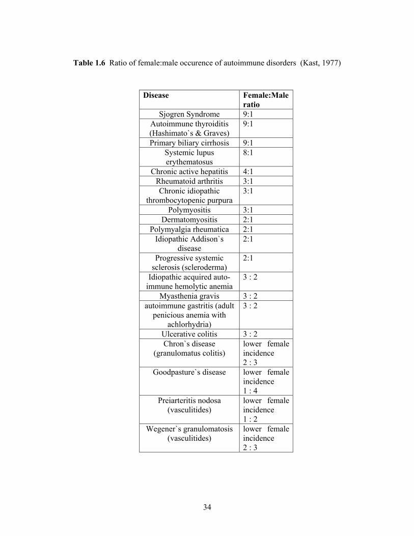
Table 1.6 Ratio of female:male occurence of autoimmune disorders (Kast, 1977)
Disease Female:Male
ratio Sjogren Syndrome 9:1
Autoimmune thyroiditis (Hashimato`s & Graves)
9:1
Primary biliary cirrhosis 9:1 Systemic lupus erythematosus
8:1
Chronic active hepatitis 4:1 Rheumatoid arthritis 3:1 Chronic idiopathic
thrombocytopenic purpura 3:1
Polymyositis 3:1 Dermatomyositis 2:1
Polymyalgia rheumatica 2:1 Idiopathic Addison`s
disease 2:1
Progressive systemic sclerosis (scleroderma)
2:1
Idiopathic acquired auto-immune hemolytic anemia
3 : 2
Myasthenia gravis 3 : 2 autoimmune gastritis (adult
penicious anemia with achlorhydria)
3 : 2
Ulcerative colitis 3 : 2 Chron`s disease
(granulomatus colitis) lower female incidence 2 : 3
Goodpasture`s disease lower female incidence 1 : 4
Preiarteritis nodosa (vasculitides)
lower female incidence 1 : 2
Wegener`s granulomatosis (vasculitides)
lower female incidence 2 : 3
34
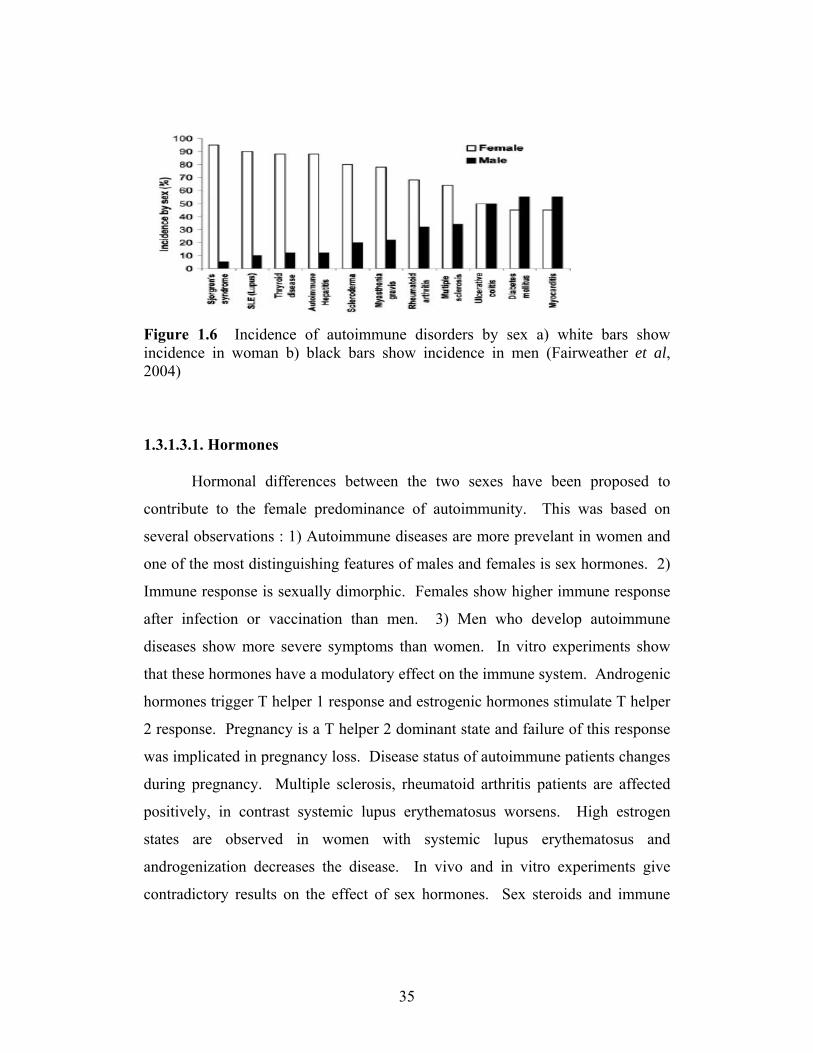
Figure 1.6 Incidence of autoimmune disorders by sex a) white bars show incidence in woman b) black bars show incidence in men (Fairweather et al, 2004) 1.3.1.3.1. Hormones
Hormonal differences between the two sexes have been proposed to
contribute to the female predominance of autoimmunity. This was based on
several observations : 1) Autoimmune diseases are more prevelant in women and
one of the most distinguishing features of males and females is sex hormones. 2)
Immune response is sexually dimorphic. Females show higher immune response
after infection or vaccination than men. 3) Men who develop autoimmune
diseases show more severe symptoms than women. In vitro experiments show
that these hormones have a modulatory effect on the immune system. Androgenic
hormones trigger T helper 1 response and estrogenic hormones stimulate T helper
2 response. Pregnancy is a T helper 2 dominant state and failure of this response
was implicated in pregnancy loss. Disease status of autoimmune patients changes
during pregnancy. Multiple sclerosis, rheumatoid arthritis patients are affected
positively, in contrast systemic lupus erythematosus worsens. High estrogen
states are observed in women with systemic lupus erythematosus and
androgenization decreases the disease. In vivo and in vitro experiments give
contradictory results on the effect of sex hormones. Sex steroids and immune
35

system seem to have a dialogue. There are hormone receptors on immune cells
and cytokine receptors on hormone producing cells.
Some cytokines can stimulate the hypothalamus-pitiutary-adrenal axis to
secrete homones. Some researchers belive the disease severity is observed as a
consequence of varying exposure to hormones (Lockshin, 2002; Whitacre, 2001;
Fairweather, 2004;Gleicher 2006).
1.3.1.3.2. Microchimerism
Microchimerism refers to presence of a small number of cells or DNA of
an individual present in another individual and its possible causes are blood
transfusions; passage of cells or DNA from the twin during embryogenesis or cell
passage during pregnancy. Fetal microchimerism refers to fetal cell passage to
mother during pregnancy or elective termination. Microchimerism was suggested
to be critical in female predominance of autoimmunity: 1) It is an important
immunological challenge being a period of semi-allogeneic graft transplantation
similar to graft vs. host disease. 2) The incidence of autoimmune diseases are
high in females and certain autoimmune diseases show increased incidence after
pregnancy. The presence of fetal DNA in the blood, mononuclear cells and skin
of scleroderma patients were investigated and the HLA relationship of the mother
and the fetus was determined (Arlett, 1998; Nelson, 1998). There are variable
reports, mostly favoring the presence of microchimerism in sclerosis. It was also
shown that sclerosis patients without male pregnancies have microchimerism in
their circulation (Lambert, 2007). Microchimerism in skin was studied for patients
with polymorphic eruption and positive results were obtained. It was studied in
liver biopsies of primary biliary cirrhosis (PBC) and thyroid biopsies of
autoimmune thyroiditis patients and the results were variable. There are
accumulated data on microchimerism in both patients with autoimmunity, people
with non-immune conditions and controls. There is also data of microchimerism
in both blood and other tissues. Maternal microchimerism refers to the presence
of maternal cell in an individual. This kind of microchimerism was detected in
myopathies (Arlett, 2000). It`s not known whether microchimerism is primary or
36

secondary to autoimmunity. It was hypothesized that microchimeric cells can
become activated and proliferate in response to an environmental trigger and
cause autoimmunity. When a mouse model was treated with a known
scleroderma causing agent, vinyl chloride, increased levels of microchimeric cells
were observed in blood. (Christner, 2000). Fetal cells were detected in the bone
marrow and ribs of women with male pregnancies, but not in the controls
(O`Donoghue, 2004). Another alternative hypothesis for the role of
microchimeric cells is that they are beneficial aiding in tissue repair of the
damaged organ. Microchimeric cells can aid in pathogenesis, repair, or they can
be normal remnants of pregnancy (Gleicher, 2006).
1.3.1.3.3. X chromosome Monosomy
X chromosome monosomy is a form of aneuploidy with the presence of
only one X chromosome in an individual`s karyotype and partial monosomy
occurs when only a portion of cells have one X chromosome. Recent findings
show that X chromosome abnormalities can have a role in autoimmunity. This is
supported by the following observations. 1) Many genes involved in immune
response are on the X chromosome. 2) Patients with X chromosome
abnormalities such as Turner`s syndrome show autoimmune features. 3)
Mutations in some X-linked genes cause immunodeficiency diseases and display
autoimmune features (Invernizzi, 2008).
It was shown that patients with primary biliary cirrhosis, scleroderma and
autoimmune tyroid disease have enhanced X monosomy rate in their peripheral
blood mononuclear cells, especially in lymphocytes (Invernizzi, 2004; Invernizzi,
2005). Monosomy rates were not found to be related to disease severity, but to
age. This high level of X monosomy in blood cells is not observed in systmic
lupus erythematosus (Invernizzi, 2007). It was also shown that X chromosome
loss was preferental in primary biliary cirrhosis (Invernizzi, 2007).
37

1.4 Skewed X inactivation and autoimmunity
1.4.1 Kast and Stewart hypothesis
Kast proposed an explanation for the high female incidence of
autoimmune diseases based on the forbidden clone elimination stated in Burnet-
Jerne`s theory. According to this theory, lymphocytes are negatively selected in
the thymus, MHCs being the major survival determinants (Burnet 1959, Jerne,
1971). Some activity as survival determinants was also proposed for any antigen
at a generative site (Kast, 1975). Kast proposed a mechanism other than classical
patterns of inheritance, disturbance in XCI, may have a role in female
predisposition to autoimmunity (Kast, 1977). Stewart developed this hypothesis
in systemic lupus erythematosus by stating that differences between sexes in the
self-antigen presentation profiles to the immune system due to XCI mosaicism
can account for the female preponderance (Stewart, 1998). According to this
theory, skewed XCI in the thymus can lead to the escape of autoreactive
lymphocytes specific for self antigens expressed from one parental X and this can
lead to autoimmunity (Kast, 1977; Stewart, 1998). This results because naive T
cells can not be selected for alleles expressed from one X chromosome because
there is XCI skewing in the dendritic cells that are localized on the path the naive
lymphocyte follows during negative selection (Stewart, 1998). This hypothesis is
illustrated in figure 1.7. High frequency of skewed XCI was reported in the blood
of autoimmune patients: scleroderma, autoimmune tyroid disease, pre-eclampsia
patients (Ozbalkan, 2005; Ozcelik, 2006, Yin, 2007; Uz, 2006). No significant
correlation was found between skewed XCI and the occurence of primary biliary
cirrhosis, systemic lupus erythematosus, type 1 diabetis and multiple sclerosis
(Miozzo, 2007; Huang, 1997; Chitnis, 2000).
38
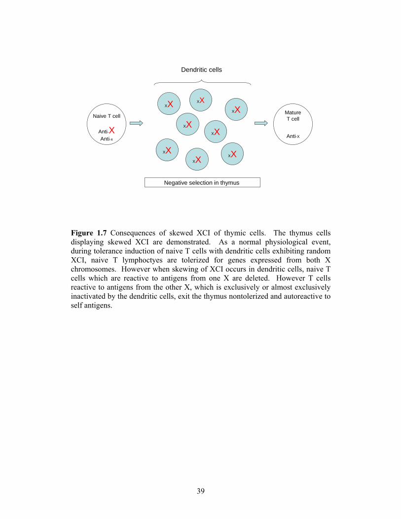
Naive T cell
Anti-
X
XAnti-x
XXX
XX
XX
XX
X
XX
X
X
Dendritic cells
X
MatureT cell
Anti-X
Negative selection in thymus
Figure 1.7 Consequences of skewed XCI of thymic cells. The thymus cells displaying skewed XCI are demonstrated. As a normal physiological event, during tolerance induction of naive T cells with dendritic cells exhibiting random XCI, naive T lymphoctyes are tolerized for genes expressed from both X chromosomes. However when skewing of XCI occurs in dendritic cells, naive T cells which are reactive to antigens from one X are deleted. However T cells reactive to antigens from the other X, which is exclusively or almost exclusively inactivated by the dendritic cells, exit the thymus nontolerized and autoreactive to self antigens.
39

1.5. Aim and Strategy
It was proposed that skewing of XCI in the hematopoietic cells may cause
failure in representation of antigens of one X by the antigen presenting cells
(APC) in the thymus and result in autoimmunity. This was hypothesized based on
the higher female incidence of most of the autoimmune diseases and XCI being a
major sex difference (Kast,1977; Stewart,1998). A positive correlation between
skewed XCI and a number of autoimmune diseases is demonstrated (Ozbalkan,
2005; Ozcelik, 2006; Yin, 2007; Uz, 2006). Sjogren Syndrome is an autoimmune
disease with unkown etiology and it shows one of the highest female
prependorances. To test whether disturbances in XCI may play a role in the
pathogenesis of Sjogren Syndrome, we genotyped a polymorphic repeat in the
human androgen receptor (AR) gene to determine XCI profiles of Sjogren patients
and controls. (Allen, 1992).
40

CHAPTER II: MATERIALS AND METHODS
2. MATERIALS AND METHODS
2.1 Samples
2.1.1 Control Samples
Caucasian females comprised of 160 Turkish healthy unrelated volunteers
is the control group in this study. The mean age for Turkish controls at analysis
was 46±10.0 (mean±SD). There was no clinical evidence or family history of
autoimmune or inflammatory joint disease. The XCI profiles of this group was
used to compare with those of Sjogren Syndrome patients (Ozbalkan, 2005;
Ozcelik, 2006).
2.1.2 Sjogren Syndrome Patient Samples
Caucasian women (n=78) diagnosed with Sjogren Syndrome were referred
to Bilkent University, Facutly of Science, Molecular Biology and Genetics
Department (Ankara, Turkey) by Zeynep Özbalkan at the Ankara Numune
Education and Research Hospital, Rheumatology Clinic (Ankara, Turkey). Their
venous blood samples were transferred in EDTA collection tubes. Informed
consent forms were obtained from all subjects. These samples were genotyped
and their XCI patterns were determined.
41

2.2 Materials
2.2.1 Primers
The sequences of the primers used in polymerase chain reaction (PCR) are :
primer 1, 5'- GTC CAA GAC CTA CCG AGG AG-3'; and primer 2, 5'-CCA
GGA CCA GGT AGC CTG TG -3'. The primers were synthesized by IONTEK
(Istanbul, Turkey). The region amplified by the primers is as designated in figure
2.1.
2.2.2 Enzymes
Thermus aquaticus (Taq) DNA polymerase was used in the PCR reactions.
Restriction digestion enzymes, RsaI and metylation sensitive HpaII were used in
the digestion reactions. All enzymes were purchased from MBI Fermentas Inc.
(Amh, NY, USA). The recognition sites of restriction digestion enzymes are as
designated in figure 2.1.
42

CGAGATCCCGGGGAGCCAGCTTGCTGGGAGAGCGGGACGGTCCGGAGCAAGCCCAGAGGCAGAGGAGGCGACAGAGGGAAAAAGGGCCGAGCTAGCCGCTCCAGTGCTGTACAGGAGCCGAAGGGACGCACCACGCCAGCCCCAGCCCGGCTCCAGCGACAGCCAACGCCTCTTGCAGCGCGGCGGCTTCGAAGCCGCCGCCCG ... AACTCTTCTGAGCAAGAGAAGGGGAGGCGGGGTAAGGGAAGTAGGTGGAAGATTCAGCCAAGCTCAAGGATGGAAGTGCAGTTAGGGCTGGGAAGGGTCTACCCTCGGCCGCCGTCCAAGACCTACCGAGGAGCTTTCCAG forward primer (RS6) AATCTGTTCCAGAGCGTGCGCGAAGTGATCCAGAACCCGGGCCCCAG HpaII restriction site GCACCCAGAGGCCGCGAGCGCAGCACCTCCCGGCGCCAGTTTGCTGC HpaII restriction site TGCTGCAGCAGCAGCAGCAGCAGCAGCAGCAGCAGCAGCAGCAGC CAG repeat AGCAGCAGCAGCAGCAGCAGCAGCAGCAAGAGACTAGCCCCAGGCAGCAGCAGCAGCAGCAGGGTGAGGATGGTTCTCCCCAAGCCCATCGTAGAGGCCCCACAGGCTACCTGGTCCTGGATGAGGAACAGCAACCTTC reverse primer (RS7) ACAGCCGCAGTCGGCCCTGGAGTGCCACCCCGAGAGAGGTTGCGTCCCAGAGCCTGGAGCCGCCGTGGCCGCCAGCAAGGGGCTGCCGCAGC ... GCTGTAGCCCCCTACGGCTACACTCGGCCCCCTCAGGGGCTGGCGGGCCAGGAAAGCGACTTCACCGCACCTGATGTGTGGTACCCTGGCGGCATGGTGAGCAGAGTGCCCTATCCCAGTCCCACTTGTGTCAAAAGCGAAATGGGCCCCTGGATGGATAGCTACTCCGGACCTTACGGGGACATGCG
Figure 2.1 AR genotyping amplicon (287 bp). Sequence of Homo sapiens AR
exon 1 on the X chromosome between 66680599-66860844 bp. is shown.
Reference assembly is NC_000023.9 in NCBI and ENSE00001326500 in
Ensembl. HpaII digestion sites and CAG repeat regions are designated (Allen,
1992).
43

2.2.3 Oligonucleotide
The length of DNA fragments were estimated by comparing to known molecular
weight standards that had been run on the same gel. pUC Mix Marker 8
(#SM0303) (0.1µg/µl) and Massruler DNA Ladder (#SM0403) Mix were used as
markers and purchased from MBI Fermentas (Amh, NY, USA) The sizes of the
fragments are given in figure 2.2.
Figure 2.2 Sizes of the fragments of pUC Mix Marker 8 and MassRuler DNA Ladder Mix and appearance on gel electrophoresis.
44

2.2.4.Chemicals, Reagents, Kits
The chemicals and reagents used in this project and the suppliers are listed in
table 2.1.
Table 2.1 List of chemicals and reagents used in this study
Experiment Reagent or Chemical Company Nucleospin® DNA
Isolation Kit (Buffers B3, BE, BW, B5)
Macherey-Nagel Inc., PA,USA
Proteinase K Appligene-Oncor, USA
DNA Isolation
100% Ethanol (EtOH) Merck, Frankfurt, Germany
Agarose Gel Preparation Agarose Basica LE, EU Bisacrylamide Sigma, St. Louis, MO
Acrylamide Sigma, St. Louis, MO APS Carlo Erba, Milano, Italy
Polyacrylamide Gel Preparation
TEMED Carlo Erba, Milano, Italy 6X MassRuler™ DNA
Loading Dye MBI Fermentas, Amherst,
NY, USA Ethidium Bromide
(EtBr) Sigma, St. Louis, MO,
USA
EDTA (pH 8.0) Carlo Erba, Milano, Italy Tris Merck, USA
Gel Electrophoresis
100% glacial acetic acid
Sigma, USA
Restriction Digestion Tango Buffer (10X) MBI Fermentas, Amherst, NY, USA
dNTP (10 mM) MBI Fermentas, Amherst, NY, USA
MgCl2 (25 mM) MBI Fermentas, Amherst, NY, USA
PCR
Buffer (10X) MBI Fermentas, Amherst, NY, USA
45

2.2.5 Standard solutions and buffers 50X TAE (Tris-acetic acid-EDTA): 242gr Tris, 57.10 ml acetic acid, 37.2 gr
EDTA. The volume was adjusted to 100 ml by adding ddH2O and pH is adjusted
to 8.0
1X TAE (Tris-acetic acid-EDTA): 40mM Tris-acetate, 2 nM EDTA, pH 8.0.
Acrylamide:Biacrylamide Stock Solution (30%): 29.5 gr acrylamide, 0.44 gr
bisacrylamide. The volume was adjusted to 100 ml by adding ddH2O.
APS solution (10%): Prepared freshly with 0.1gr ammonium persulfate in 1mL
ddH2O.
Ethidium bromide: 10mg/ml in water (stock solution) 30 ng/ml (working solution)
6X MassRuler™ DNA Loading Dye : 10mM Tris-HCl (pH 7.6) 0.03%
bromophenol blue, 0.03% xylene cyanol FF, 60% glycerol 60mM EDTA.
pUC Mix 8 Marker : 1 volume of pUC Mix 8 Marker, 1 volume of 6X
MassRuler™ DNA Loading Dye and 4 volumes of ddH2O.
46

2.3 Methods
2.3.1 Sample collection
Venous blood samples obtained from Sjogren Syndrome patients and
controls were transported in EDTA tubes. Their entry to the lab was recorded.
They were seperated as 1ml aliquots to eppendorf tubes (1.5 ml). The bloods (1-3
eppendorfs) were stored at -80oC. 200 μl blood was seperated for DNA isolation
and stored at -20oC.
2.3.2 DNA Isolation from venous blood
DNA from venous blood was isolated by using Nucleospin™ Blood kit
(Macherey-Nagel Inc., PA, USA) (#740 951.250) according to the protocol of the
manufacturer. The starting blood sample was 200 μl and the resulting material
was 100 μl elution buffer containing DNA. The efficiency of this kit is
typically 40-60ng/µl genomic DNA with an A260/280 value of 1.6-1.9.
Blood samples preequilibrated to room temperature were lysed with
proteinase K and lysis buffer vortexing and heating at 70 oC. Adjusting of DNA
binding was performed with ethanol (100%). DNA was bound to silica
membrane which was then washed with buffers. With centrifugation residual
ethanol was removed. Using preheated 70 oC elution buffer, DNA was eluted.
The concentrations and purity values of DNA were measured with
spectrophotometric reading using NanoDrop™ ND-1000 UV-Vis
Spectrophotometer.
In the process, 2 µl of the sample was used for measurement. 2 µl elution
buffer was used as blank. The integrity of the DNA samples were controlled with
1% agarose gel electrophoresis.
47

2.3.3 AR genotyping
To determine the XCI status of individuals, androgen receptor (AR)
genotyping was performed. (Figure 2.3) AR locus spans a region larger than 90kb
and it has been mapped to Xcen-Xq13. Its cDNA has an open reading frame of
2,751 nucleotides and its protein has 917 amino acids. In its first exon there is an
inframe CAG repeat that codes for 11-31 glycine residues.
A 280 bp region (nucleotides 229-508) of the AR is amplified. (Figure 2.3)
This region includes a highly polymorphic trinucleotide repeat (20 alleles, 90%
heterozygosity) and HpaII and HhaI sites less than 100 bp away from this short
tandem repeat (STR). The proximity of STR to enzyme target sites gives an
opportunity to distinguish between maternal and paternal alleles and to determine
their XCI status.
HpaII and HhaI are methylation sensitive enzymes which have target sites
with CpG. Cytosine residues in CpG islands, which are clustered at the 5` of
genes are methylated in the inactive X. AR genotyping relates methylation of
HpaII and HhaI sites in the AR locus with XCI. (All enzyme sites are consistently
methylated on the inactive X, but one or more enzyme sites are unmethylated on
the active X.) In AR genotyping, DNA is first digested with HpaII and/or HhaI
and than a region including the enzymes site and the repeat is amplified. The
enzyme target sites on the active X chromosome, which have unmethylated
cytosines will be cleaved and no amplicon will be observed for the active X.
Since they are methylated, the sites on the inactive X will not be cleaved, so an
amplicon will be observed for the inactive X. The X inactivation patterns can be
determined in females who are informative at their CAG repeat (Allen, 1992).
48
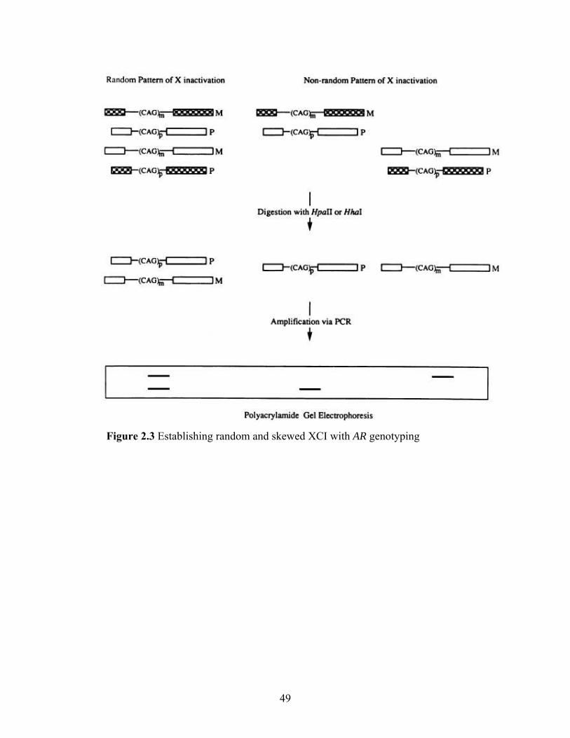
Figure 2.3 Establishing random and skewed XCI with AR genotyping
49
2.3.3.1 Restriction Enzyme Digestion
For each sample, two restriction enzyme digestion mixtures were
prepared, each of which have a total reaction volume of 20 μl. The amount of
DNA was adjusted to 150-250 ng in the digestion mixture. 2 µl of Y tango buffer
with BSA (#BY5) (33mM Tris-acetat (pH 7.9), 10mM magnesium acetate, 66
mM ptoasium acetate, 0.1 mg/mL BSA) (10X) was added to adjust the final
buffer in the mix to 1X. 0.2 µl RsaI (#ER1122) (10U/µl) (5`-GT↓AC-3`) was
included in both mixes to adjust its concentration to 2U in the final mix. RsaI has
target sites outside the PCR product and it makes the DNA more accessible. 0.8
µl HpaII (#ER0512) (10U/µl) (5`-C↓CGG-3`), which is methylation sensitive,
was included in the second mix to adjust its concentration to 8U in the final mix.
The reactions were incubated at 37oC in the incubator overnight. Heat
inactivation of the reactions was performed at 70oC for 10-15 minutes in the
heater. Restriction digestion products were stored at 4oC for further use. First
digestion is used for AR genotyping, the second digestion was used to assess XCI
pattern. Negative controls were performed without DNA and the positive controls
were performed with male DNA.
50

2.3.3.2 Polymerase chain reaction (PCR)
The digest amount was adjusted to 50ng-100ng in the PCR mix. The final
concentrations in the PCR mix were 10mM for MgCl2 (25 mM), 0.12 for dNTP
(10mM), 10pmol for the primers (10pmol/µl), 1U for the Taq polymerase
(#EP0402) (5U/ µl), 1X for the buffer (10X) (750mM Tris-HCl (pH 8.8 at
25oC),200 mM (NH4)2SO4, 0.1% (v/v) Tween 20). In order to amplify the desired
region, 94oC 5 min initial denaturation step was performed. PCR reactions were
performed in Techne™ Techgene thermal cycler. This was followed by the
amplification step including 30 cycles of 94oC 30 sec denaturation, 58oC 30 sec
annealing, 72oC 30sec extension. Final extension was performed at 72oC for 5
min. Amplicons were controlled with 1.5% gel electrophoresis.
2.3.3.3 Visualization
2.3.3.3.1 Agarose gel electrophoresis
1% agarose gel electrophoresis was performed to check whether total
human genomic DNA was isolated. 2 μl of MassRuler DNA Ladder mix
(#SM0403), 2 μl DNA with 1 μl 6X MassRuler™ DNA Loading Dye (#R0611)
(103 ng/µl) was loaded.
To visualize the PCR products for the determination of the success, 1.5%
agarose gel electrophoresis was performed. 4 µl of pUC Mix Marker (#SM0301)
(0.5 µg/µl) and 5 μl of sample with 6X MassRuler™ DNA Loading Dye (5:1
ratio) were loaded.
The gels were prepared by dissolving required amount of agaose in 1X
TAE. Required amount of EtBr (10mg/mL) was added. The gel was run in 1X
TAE for the required amount and time. UV transilluminator was used during
visualization and photography.
51

2.3.3.3.2 Polyacrylamide gel electrophoresis (PAGE)
The maternal and paternal alleles were resolved using 8% PAGE
(polyacrylamide gel electrophoresis). 8% PAGE gel solution for pouring two gels
(60mL) was prepared by adding the reagents in the following order: in order to
prepare 16 ml acryamide: bisacrylamide solution (30%), 6 ml 10X TAE, 500 µl
10% APS, 40 µl TEMED and 37,5 ml ddH2O were included.
The solution solidifies for 1 hour and the gels are attached to the vertical
setting. PCR product with the loading dye is loaded. pUC Mix Marker is used as
a marker. The gels were run at 20 Watt per gel in 1X TAE buffer for 2 hours.
They were stained in buffer containing EtBr for 5 minutes, washed in ddH2O for
another 5 minutes.
2.3.3.4 Densitometric and Statistical Analysis
Multi-Analyst software version 1.1 (Bio-Rad, Hercules, CA) was used for
the densitometric analysis. Densitometric analyses included normalization of the
ratios based on the undigested samples. Normalization was performed by
dividing the allele ratio of the digested sample by the allele ratio of the undigested
sample. This calculation is performed as a correction for the preferential
amplification of the shorter allele. The results of the Sjogren Syndrome and
control group were compared by χ2 test with Yates` correction and Fisher`s exact
test. In addition, odds ratio value was calculated in the 95% confidence interval.
Power analysis was performed to estimate the sample size that is needed to make
accurate statistical judgements. The minimum effect size was determined based
on the results of XCI status studies of our group for other autoimmune diseases
(Ozbalkan, 2005; Ozcelik, 2006 ; Uz, 2006). The significance level was set as
α=0.05 and the power was set as β=0.1.
52

CHAPTERIII: RESULTS
Androgen receptor assay was performed for XCI status determination of
Sjogren Syndrome patients and healthy controls. Methylated inactive X
chromosome is resistant to digestion by methylation specific HpaII enzyme, while
unmethylated X chromosome is prone to digestion. There are highly polymorphic
triplet repeats adjacent to HpaII methylation sites in the androgen receptor gene,
which provide a method to distinguish the two alleles and their methylation status.
Figure 3.1 demonstrates the gels of uninformative, random and skewed
XCI patterns. Uncut (U) refers to DNA amplified prior to HpaII digestion,
genotyping for the polymorphic repeat. Cut (C) refers to DNA amplified after
HpaII digestion, giving information on the XCI status. The individuals, who are
not polymorphic in the CAG repeat within the amplified region of AR gene were
uninformative and not included in the densitometric and statistical analysis. For
informative samples, densitometric analysis was performed as indicated in the
methods section. 04-474 and 04-476 are examples of informative samples, 04-
473 demonstrating random and 04-476 demonstrating skewed XCI.
Skewed XCI refers to a concentration difference of more than 80%
between the two alleles and extremely skewed XCI refers to a concentration
difference of more than 90%. (Naumova et al., 1996).
53

M 04-473 04-474 04-476 HpaII - + - + _ +
Figure 3.1 XCI inactivation status in Sjogren Syndrome patients. Polymerase chain reaction products from the androgen receptor methylation assay demonstrate random X chromosome inactivaton pattern in sample 04-473 (allele ratio 64% : 36%) and skewed pattern in sample 04-476 (100% : 0%). Sample 04-474 is not informative for the androgen receptor polymorphism. For each sample, DNA was either undigested (-) or digested (+) with the methylation-sensitive restriction enzyme HpaII. Marker (M; pUC mix 8[331-bp and 242-bp])
XCI patterns of 78 Sjogren Syndrome patients were studied and 51 of 78 patients
were informative (65.4%). The control group comprised of females with no
autoimmune condition. XCI status of 160 controls were studied and 124 were
informative (77.5%) Extreme skewing (>90:10) was observed 3 out of 51
informative patients (5.9%) and in 3 of 124 (2.4%) informative controls. Skewing
(>80:20) was observed in 10 of (8.1%) 124 informative controls and in 8 of 51
informative patients (15.6%). Statistical verification of the significance of
correlation was assesed with the odds ratio and P value. The probability value was
calculated with Fisher`s exact test. The odds ratio and P-value for extreme
skewing is P=0.3592 and OR=2.5208 with 95% CI: 0.4915-12.9289. The odds
ratio and P-value for skewing is P=0.17 OR=2.1209 with 95% CI: 0.7852-5.7289.
These results favor that there is not a statistically significant correlation between
skewed XCI and Sjogren Syndrome.
Power analysis was performed to estimate the sample size that is needed to
enable accurate statistical judgements. The estimated sample size needed to test a
difference of 0.18 of extreme skewing between patient and control samples with a
power of 0.9 at a significance level of 0.05 is 47 Sjogren Syndrome and 113
54

control samples. The estimated sample size needed to test a difference of 0.27 of
skewing between patient and control samples with a power of 0.9 at a
significiance level of 0.05 is 36 Sjogren Syndrome and 87 control samples. The
analysis shows that the sample size used for this experiment is adequate to make
accurate statistical judgements.
55

Table 3.1 Proportion of Sjogren Syndrome patients and controls with skewed
XCI
Degree of skewing (%) No. (%) observed with skewing
Sjogren Syndrome Controls patients (n=51) (n=124) 90+ 3 (5.9) 3 (2.4) 80-89% 5 (9.9) 7 (5.6) 70-79% 14 (27.5) 22 (17.8) 60-69% 9 (17.7) 29 (23.4) 50-59% 20 (39.2) 63 (50.8) _________________________________________________ For comparison by χ2, P= 0.2 (>80% skewing); P= 0.4 (>90% skewing)
56

CHAPTER IV: DISCUSSION
Female predominance is a characteristic of most autoimmune diseases.
(Whitacre, 2001). In recent decades, candidate mechanisms that could be
important in autoimmune disease pathogenesis have been studied. Currently there
are explanations for the female predominance in autoimmune diseases. These
include genetic traits associated with autoimmunity (Rioux and Abbas, 2005) and
pregnancy related microchimerism (Arlett, 1998; Nelson, 1999). Another sex
specific feature of females is XCI and it is known that skewed XCI can influence
manifestations of X-linked disorders in females (Lyon, 2002). The hypothesis
that skewed XCI in the thymus can cause defects in negative selection and lead to
autoimmunity was proposed. (Kast, 1977; Stewart, 1998)
Thymocytes are negatively selected in the thymic parencyma and the
selecting elements appear to be derived from the bone marrow and are dendritic
cells. If the thymic dendritic cell population exhibits skewed XCI, T cells specific
for certain X antigens will not be deleted. This hypothesis assumes that the initial
breakdown in tolerance involves an X-linked autoantigen expressed in peripheral
site of the body. In a recent report, lack of expression of one splice variant of
proteolipid protein (PLP) protein in the thymus was shown to be related to
susceptibility to autoimmunity (Klein, 2000). This illustrates a role of defective
thymic negative selection in autoimmunity. Since proteolipid protein is X-linked,
it also accounts for the hypothesis stated above.
Loss of mosaicism based on X chromosome skewing was shown to
correlate with autoimmunity in several AIDs (Ozbalkan, 2005; Ozcelik, 2006; Uz,
2006; Yin, 2007). These reports supported that factors associated with loss of
mosaicism based on X chromosome skewing could account for a significant
proportion of these autoimmune diseases.
Currently, the relationship between skewed XCI and AIDs is not known.
Skewing can be either secondary to breakdown of self-tolerance or it can be a
causative factor in the breakdown of self-tolerance. We currently believe the
second view is more promising since skewing ratios of patients are not mild.
57

XCI skewing can result either from primary or secondary reasons.
Whether skewing of XCI patterns are transmitted to offspring in humans was
researched on the hypothesis that an X-linked locus can alter initial random
inactivation. No supporting evidence was obtained (Minks, 2008; Bolduc, 2008).
The secondary reasons of XCI include X-linked lethal mutations, X-autosome
translocations, aging, twining or monoclonal expansion (Brown, 1999). We
believe that deleterious X-linked mutations can account for skewing leading to
autoimmunity. This view is supported by the observation that recurrent
spontaneous abortions are increased in AIDs and skewed XCI was associated with
recurrent spontaneous abortions (Bagislar, 2006; Shelton, 1994). If the cause of
skewing is indeed a X-linked deleterious mutations as hypothesized, this should
be inherited from the mother unless they occur de novo in gamategenesis. An X-
linked lethal mutation causes skewing in the mother and it is lethal for a male
infant. This kind of skewing was reported in X-linked immunodeficiency where
males have a survival disadvantage and heterozygous females have XCI skewing
(Puck&Willard, 1998). We believe deleterious X-linked mutations and their
differential expression patterns could lead to skewing, conferring a disadvantage
on blood cells (Uz, 2007 ; Ozcelik, 2008).
Although extremely skewed XCI is rare in normal population, it does not
always lead to the development of disease. To provide an estimate of the
frequency of individuals in the general population, XCI status of 1,005 unaffected
individuals was determined. The percentage of population with skewed XCI was
found to be 8.8% and with extremely skewed XCI was found to be 1.8% (Amos-
Landgraf, 2006). There are also other proposed factors for autoimmune etiology
other than correlation with skewing. A subsequent event such as environmental
exposure to viral, chemical and other agents may trigger autoimmunity. The co-
inheritance of genetic susceptibility factors for example functional variants of
molecules involved in immune tolerance may exacerbate the effects of skewed
XCI leading to autoimmunity.
58

Sjogren Syndrome is an autoimmune disorder with one of the highest
female prependorance and unknown etiology. It has been associated with genetic
factors such as HLA-DR/HLA-DQ, cytokine and other gene polymorphisms
(Hulkkonen, 2001; Gottenberg, 2004; Pertovaara, 2004; Youn, 2000; Tsutsumi,
2001, Wang, 2001, Bolstad, 2000, Petrek, 2002). Environmental factors,
especially viral infections have been reported as causative factors (Fleck, 1998;
Haddack, 1992; Kordossis, 1998;Tereda, 1994;Giffiths, 1997). There are also
controversial reports on the role of microchimerism in Sjogren Syndrome (Toda,
2000; Kuroki, 2002; Endo, 2001). In this research, a potential correlation
between skewed XCI and Sjogren Syndrome was investigated. Patients showed
skewing patterns comparable to that of controls in their blood cells. Our results
did not support a correlation between Sjogren Syndrome and skewed XCI based
on the P value and odds ratio calculated. Our sample size was adequate to make a
true statistical verification based on hypothesis testing.
Although the role of X mosaicism in breakdown of self tolerance caught
attention, X inactivation patterns comparable to those of control women were
observed in the blood from females with primary biliary cirrhosis, multiple
sclerosis, juvenile diabetes and systemic lupus erythematosus (Miozzo, 2007;
Knudsen, 2007; Chitnis, 2000) These results display that other reasons than XCI
may have role in the pathogenesis of these diseases. For example, molecular
mimicry is known to have a role in the initiation of the primary biliary cirrhosis
(Kaplan, 2005). Based on our results, we can conclude other factors can trigger
Sjogren Syndrome, other than skewed XCI based mosaicism.
The tissues of an organism are accepted to reflect the same XCI pattern
since inactivation process takes place very early during development. However
there can be exceptions to this. It has been showed that although there is a
significant association of the X inactivation ratios between each tissue in younger
individuals, there are significant variations in elder individuals (Sharp, 2000;
Busque, 1996). It was observed that there can be variation in XCI patterns
among tissues (Rosemary, 1994). These results emphasize the role of secondary
selection events leading to skewing. We can suspect that the patterns of skewing
59

in blood cells does not demonstrate the skewing pattern of dendritic cells in the
thymus. However this is unlikely because it was observed tissues of mesodermal
origin do not display differences in X inactivation patterns (Chitnis, 2000).
Another consideration is that tissue microenvironments may exhibit skewed XCI
because of a small number of founder cells even though the tissue as a whole is
not skewed. If this is the case, this can not be reported by working with blood
cells. Our group studied buccal mucosa, hair and effected tissues of SSc and
AITDs. We can further confirm our findings by studying other tissues such as
thymic tissue sections. (Chitnis, 2000).
In a recent study, the X chromosome skewing status of multiple sclerosis
patients were assessed and compared in two subgroups of multiple sclerosis.
They concluded there was a significantly higher degree of skewing in progressive
multiple sclerosis than relapse remitting subgroup. (Knudsen, 2007) Our data can
further be used to extrapolate the differences in the degree of X chromosome
skewing in primary and secondary Sjogren Syndrome patients.
X inactivation assay we used in this research is AR genotyping. There are
various assays to determine X inactivation pattern based on a marker. Assays
based on variant proteins or common exonic polymorphisms to assess expression
are direct assays. AR genotyping is an indirect assay which utilizes a DNA repeat
polymorphism and is based on the presumption that CpG island which is the
target of HpaII is always methylated in the inactive X. These kinds of DNA
assays are highly informative because the polymorphisms in the nontranscribed
regions of the genome can be used. They are also more rapid and suitable for
research in which establishing large informative number is more important than
determining slight changes in X inactivation status. AR locus has a
heterozygosity of >90% and a high degree of accuracy with a variance on
repeated assays is around 4%. (Chen, 2007; Ozcelik, 2007; Migeon, 2007)
60

CHAPTER V: FUTURE PERSPECTIVES
In this research, we tested the hypothesis that disturbances in XCI may
have a role in the etiology of Sjogren Syndrome. However our results does not
support a correlation between skewed XCI and Sjogren Syndrome. The results of
our group for two other autoimmune diseases, scleroderma and autoimmune
thyroid disease favor a role of XCI in the loss of tolerance. The view of our group
is that X-linked mutations can have a role in skewed XCI based on secondary
selection events. A research strategy of our group was to conduct a high-density
microarray analysis including 1618 nonsynonymous, 1091 synonymous and 2802
intronic SNPs on X chromosome and 166 SNPs on autosomal chromosomes that
are known to be associated with autoimmune diseases. Exonic SNPs
corresponded to 783 genes and intronic ones corresponded to 160 genes.
Experimental part included 465 AIDs and 2 Sjogren Syndrome patient samples
with extremely skewed XCI was included in this study. This study was designed
to raise putative genes involved in AIDs by determining allelic frequencies and
copy number variations.
61

CHAPTER VI: REFERENCES
Allen RC, Zoghbi HY, Moseley AB, Rosenlatt HM, Belmont JW. (1992) Methylation of HpaII and HhaI Sites Near the Polymorphic CAG Repeat in the Human Androgen-Receptor Gene Correlates with X Chromosome Inactivation. Am. J. Genet. 51:1229-1239. Amos-Landgraf JM, Cottle A, Plenge RM, Friez M, Schwartz CE, Longshore J, Willard HF. (2006) X Chromosome–Inactivation Patterns of 1,005 Phenotypically Unaffected Females Am J Hum Genet. 79:493-9. Aractingi S, Sibilia J, Meignin V et al. (2002) Presence of microchimersim in labial salivary glands in systemic sclerosis but not in Sjogren`s syndrome. Arthritis Rheum. 46:1039-43. Artlett CM, Smith JB, Jimenez SA. (1998) Identification of fetal DNA and cells in skin lesions from women with systemic sclerosis. N Engl J Med. 338:1186-91. Artlett CM, Ramos R, Jiminez SA, Patterson K, Miller FW, Rider LG. (2000) Chimeric cells of maternal origin in juvenile idiopathic inflammatory myopathies. Childhood Myositis Heterogeneity Collaborative Group. Lancet. 356:2155-6 Augui S, Filion GJ, Huart S, Nora E, Guggiari M, Maresca M, Stewart AF, Heard E. (2007) Sensing X chromosome pairs before X inactivation via a novel X-pairing region of the Xic. Science. 318:1632-6. Azuma T, Takei M, Yoshikawa T. (2002) Identification of candidate genes for Sjogren’s syndrome using MRL/lpr mouse model of Sjogren’s syndrome and cDNA microarray analysis. Immunol Lett 81:171–6. Bach JF (2005) Infections and autoimmune diseases. J Autoimmun. 25:74-80. Bacher CP, Guggiari M, Brors B, Augui S, Clerc P, Avner P, Eils R, Heard E. (2006) Transient colocalization of X-inactivation centres accompanies the initiation of X inactivation. Nat Cell Biol. 8:293-9. Bagislar S, Ustuner I, Cengiz B, Soylemez F, Akyerli CB, Ceylaner S, Ceylaner G, Acar A, Ozcelik T. (2006) Extremely skewed X-chromosome inactivation patterns in women with recurrent spontaneous abortion. Aust N Z J Obstet Gynaecol. 46:384-7. Bahmanyar S, Srinivasappa J, Casali P, Fujinami RS, Oldstone MB, Notkins AL.
(1987) Antigen mimicry between measles virus and human T lymphocytes. J.
62
Infect. Dis. 156:526–527 (no abstract) Ban Y, Tozaki T, Tobe T, Ban Y, Jacobson EM, Concepcion ES, Tomer Y. (2007) The regulatory T cell gene FOXP3 and genetic susceptibility to thyroid autoimmunity: an association analysis in Caucasian and Japanese cohorts. J Autoimmun. 28:201-7. Barr ML, Bertram EG. (1949) A morphological distinction between neurones of the male and female, and the behaviour of the nucleolar satellite during accelerated nucleoprotein synthesis. Nature. 163:676. (no abstract) Beasley E, Thomson G, Amos CI, Begovich AB. (2005) PTPN22 genetic variation: evidence for multiple variants associated with rheumatoid arthritis. Am J Hum Gen. 77:567-81. Becker KG, Simon RM, Bailey-Wilson JE, Freidlin B, Biddison WE, McFarland HF, Trent JM. (1998) Clustering of non-major histocompatibility complex susceptibility candidate loci in human autoimmune diseases. Proc Natl Acad Sci USA. 95:9979-84. Beletskii A, Hong YK, Pehrson J, Egholm M, Strauss WM. (2001) PNA interference mapping demonstrates functional domains in the noncoding RNA Xist.Proc Natl Acad Sci U S A. 98:9215-20. Belmont JW. (1996) Genetic Control of X Inactivation and Processes Leading to X-Inactivation Skewing. Am J Hum Genet 58:1101-1108. Besana C, Salmaggi C, Pellegrino C, Pierro L, Vergani S, Faravelli A, Rugarli C. (1991) Chronic bilateral dacryo-adenitis in identical twins: a possible incomplete form of Sjogren syndrome. Eur J Pediatr. 150:652-655. Beroukas D, Hiscock J, Jonsson R, Waterman SA, Gordon TP. (2001) Subcellular distribution of aquaporin 5 in salivary glands in primary Sjogren’s syndrome. Lancet. 358:1875–6. Beutler E, Yeh M, Fairbanks VF. (1961) The normal human female as a mosaic of X-chromosome activity: studies using the gene for C-6-PD-deficiency as a marker. Proc Natl Acad Sci U S A. 48:9-16. (no abstract) Bolduc V, Chagnon P, Provost S, Dubé MP, Belisle C, Gingras M, Mollica L, Busque L. (2008) No evidence that skewing of X chromosome inactivation patterns is transmitted to offspring in humans. J Clin Invest. 118:333-41. Boling EP, Wen J, Reveille JD, Bias WB, Chused TM, Arnett FC: (1983) Primary Sjogren’s syndrome and autoimmune hemolytic anemia in sisters. A family study. Am J Med. 74:1066-1071.
63

Bolstad AI, Haga HJ, Wassmuth R, Jonsson R. (2000) Monozygotic twins with primary Sjogren’s syndrome. J Rheumatol. 27: 2264-2266. Bolstad AI, Jonsson R. (2002) Genetic aspects of Sjogren’s syndrome. Arthritis Res. 4:353-359. Bolstad AI, Wargelius A, Nakken B, Haga HJ, Jonsson R. (2000) Fas and Fas ligand gene polymorphisms in primary Sjogren’s syndrome. J Rheumatol. 27:2397–405. (abstract) Borda E, Camusso JJ, Perez Leiros C, Bacman S, Hubscher O, Arana R, Sterin-Borda L. (1996) Circulating antibodies against neonatal cardiac muscarinic acetylcholine receptor in patients with Sjogren's syndrome. Mol Cell Biochem. 163-164:335-41. Brockdorff N, Ashworth A, Kay GF, McCabe VM, Norris DP, Cooper PJ, Swift S, Rastan S. (1992) The product of the mouse Xist gene is a 15 kb inactive X-specific transcript containing no conserved ORF and located in the nucleus. Cell. 71(3):515-26. Brown CJ, Willard HF. (1994) The human X-inactivation centre is not required for maintenance of X-chromosome inactivation. Nature. 368(6467):154-6. Brown CJ, Hendrich BD, Rupert JL, Lafrenière RG, Xing Y, Lawrence J, Willard HF.(1992) The human XIST gene: analysis of a 17 kb inactive X-specific RNA that contains conserved repeats and is highly localized within the nucleus. Cell. 71(3):527-42. Brown CJ. (1999). Skewed X-chromosome inactivation: cause or consequence? J Natl Cancer Inst. 91:304–305. Brown CJ, Robinson WP. (2000) The causes and consequences of random and non-random X chromosome inactivation in humans. Clin Genet. 2000. 58:353-63. Burnet FM (1957) A Modification of Jerne`s Theory of Antibody Production using the Concept of Clonal Selection. CA Cancer J Clin. 26:119-21. Busque L, Mio R, Mattioli J, Brais E, Blais N, Lalonde Y, Maragh M, Gilliland DG. (1996) Nonrandom X-inactivation patterns in normal females: lyonization ratios vary with age. Blood. 88:59-65. Campbell IL. (1995) Neuropathogenic actions of cytokines assessed in transgenic mice. Int J Dev Neurosci. 13:275–84.
64

Carlton VE, Hu X, Chokkalingam AP, Schrodi SJ, Brandon R, Alexander HC, Chang M, Catanese JJ, Leong DU, Ardlie KG, Kastner DL, Seldin MF, Criswell LA, Gregersen PK. (2005) PTPN22 genetic variation: evidence for multiple variants associated with rheumatoid arthritis. Am J Hum Genet. 77:567-81.
Carlucci F, Priori R, Alessandri C, Stoppacciaro A, Valesini G. (2001) Y chromosome microchimerism in Sjögren`s syndrome. Ann Rheum Dis. 41:4900-5. Carrel L. (2006) Molecular biology. "X"-rated chromosomal rendezvous. Science. 311:1107-9. Carrel L, Willard HF. (2005) X-inactivation profile reveals extensive variability in X-linked gene expression in females. Nature. 434:400-4. Casals RM, Zeron P, Font J. (2007) The Overlap of Sjogren Syndrome with other Systemic Autoimmune Diseases. Arthritis Rheum. 36:246-255.
Chagnon P, Schneider R, Hébert J, Fortin PR, Provost S, Belisle C, Gingras M, Bolduc V, Perreault C, Silverman E, Busque L. (2006) Identification and characterization of an Xp22.33;Yp11.2 translocation causing a triplication of several genes of the pseudoautosomal region 1 in an XX male patient with severe systemic lupus erythematosus. Arthritis Rheum. 54:1270-8.
Chen GL., Josef TP. (2007) X Linked Clonality Testing: Interpretation and Limitations. Blood. 110:2769.
Chitnis S, Monteiro J, Glass D, Apatoff B, Salmon J, Concannon P, Gregersen PK. (2000) The role of X-chromosome inactivation in female predisposition to autoimmunity. Arthritis Res. 2:399-406. Christen U, von Herrath MG. (2004) Initiation of autoimmunity. Curr. Opin. Immunol. 16:759-67. Christner PJ, Artlett CM, Conway RF, Jiménez SA. (2000) Increased numbers of microchimeric cells of fetal origin are associated with dermal fibrosis in mice following injection of vinyl chloride. Arthritis Rheum. 43:2598-605. Chureau C, Prissette M, Bourdet A, Barbe V, Cattolico L, Jones L, Eggen A, Avner P, Duret L. (2002) Comparative sequence analysis of the X-inactivation center region in mouse, human, and bovine. Genome Res. 12:894-908. Cooper DW, VandeBerg JL, Sharman GB, Pole WE (1971). Phosphoglycerate kinase polymorphism in kangaroos provides further evidence for paternal X inactivation. Nature New Biol. 230:155-157. (no abstract)
65

Davidson RG, Nitowsky HM, Childs B. (1962) Demonstration of two populations of cells in the human female heterozygous for glucose-6-phosphate dehydrogenase variants. Proc Natl Acad Sci U S A. 50:481-5. Davies ML, Taylor EJ, Gordon C et al. (2002) Candidate T cell epitopes of the human La/SSB autoantigen. Arthritis Rheum. 46:209–14. Duret L, Chureau C, Samain S, Weissenbach J, Avner P. (2006) The Xist RNA gene evolved in eutherians by pseudogenization of a protein-coding gene. Science. 312:1653-5. Eaton WW, Rose NR, Kalaydjian A, Pedersen MG, Mortensen PB. (2007) Epidemiology of autoimmune diseases in Denmark. J Autoimmun. 29:1-9. Endo Y, Negishi I, Ishikawa O. (2001) Possible contribution of microchimerism to the pathogenesis of Sjogren Syndrome. Rheumatology. 41: 490-495. Fairweather D, Rose NR. (2004) Women and autoimmune diseases. Emerg. Infect Dis. 10:2005-11. Feltkamp (1999) Genetic risk factors for autoimmune diseases. Immunol Today. 20:10-2. Ferguson-Smith AC. (2004) X inactivation: pre- or post-fertilisation turn-off? Curr Biol. 20;14:R323-5. Fife RS, Chase WF, Dore RK, et al. (2002) Cevimeline for the treatment of xerostomia in patients with Sjogren syndrome: a randomized trial. Archives of Internal Medicine 162: 1293–1300. Filippova GN, Cheng MK, Moore JM, Truong JP, Hu YJ, Nguyen DK, Tsuchiya KD, Disteche CM. (2005) Boundaries between chromosomal domains of X inactivation and escape bind CTCF and lack CpG methylation during early development. Dev Cell. 8:31-42. Fleck M, Kern ER, Zhou T, Lang B, Mountz JD. (1998) Murine cytomegalovirus induces a Sjogren like diseases in C57B1/6-Ipr/Ipr mice. Arthritis Rheum. 41:2175-84. Fox RI, Dixon R, Guarrasi V & Krubel S. (1996) Treatment of primary Sjogren’s syndrome with hydroxychloroquine; a retrospective, open-label study. Lupus. 5: 31–36. Fox RI.(2005) Sjogren's syndrome.Lancet. 366:321-31.
66

Fox RI, Michelson P, Casiano CA, Hayashi J, Stern M. (2000) Sjogren's syndrome. Clin Dermatol. 18:589-600. Fox PC. (2007) Autoimmune diseases and Sjogren syndrome: an autoimmune exocrinopathy. Ann N Y Acad Sci. 1098:15-21. Fox R, Kang H, Pisa E. (1994) Cytokine transcription in salivary gland biopsies of Sjogren’s syndrome. J Immunol. 151:132– 42. Fox PC. (2007) Autoimmune diseases and Sjogren's syndrome: an autoimmune exocrinopathy. Ann N Y Acad Sci. 1098:15-21. Fujihara T, Fujita H, Tsubota K, Saito K, Tsuzaka K, Abe T, Takeuchi T. (1999) Preferential localization of CD8q alpha E beta 7q T cells around acinar epithelial cells with apoptosis in patients with in Sjogren’s syndrome. J Immunol 163:2226 –35. Fujinami RS, Oldstone MB, Wroblewska Z, Frankel ME, Koprowski H. (1983) Molecular mimicry in virus infection: crossreaction of measles virus phosphoprotein or of herpes simplex virus protein with human intermediate filaments. Proc. Natl. Acad. Sci. USA 80: 2346–2350. Fujinami RS, Oldstone MB. (1985) Amino acid homology between the encephalitogenic site of myelin basic protein and virus: mechanism for autoimmunity. Science 230: 1043–1045. Gleicher N, Barad DH. (2006) Gender as risk factor for autoimmune diseases. J Autoimmun. 28:1-6. Goodnow CC, Sprent J, Fazekas de St Groth B, Vinuesa CG. (2005) Cellular and genetic mechanisms of self tolerance and autoimmunity. Nature. 435:590-7. Goodnow CC. (2008) Multistep Pathogenesis of Autoimmune Disease. Cell. 130:25-35 Gottenberg JE, Busson M, Loiseau P. (2003) In primary Sjogren’s syndrome, HLA class II is associated exclusively with autoantibody production and spreading of the autoimmune response. Arthritis Rheum. 48:2240–5. Gottenberg JE, Busson M, Loiseau P. (2004) Association of transforming growth factor beta1 and tumor necrosis factor alpha polymorphisms with anti-SSB/La antibody secretion in patients with primary Sjogren’s syndrome. Arthritis Rheum. 50:570–80. Graves JA. (1996) Mammals that break the rules: genetics of marsupials and monotremes. Annu Rev Genet. 30:233-60.
67
Griffiths DJ, Venabels PJ, Weiss RA, Boyd MT. (1997) A novel exogenous retrovirus sequence in humans. Virology. 71:2866-72. Haddad J, Deny P, Munz-Gotheil C, Ambrosini JC, Trinchet JC, Pateron D, et al. (1992) Lymphocytic sialadenitis of Sjogren associated with chronic hepatitis C virus liver disease. Lancet. 339:321-3. Heard E, Mongelard F, Arnaud D, Chureau C, Vourc'h C, Avner P. (1999) Human XIST yeast artificial chromosome transgenes show partial X inactivation center function in mouse embryonic stem cells. Proc Natl Acad Sci U S A. 96:6841-6. Heard E. (2004) Recent advances in X-chromosome inactivation. Curr Opin Cell Biol. 16:247-55. Hellman A, Chess A. (2007) Gene body-specific methylation on the active X chromosome. Science. 315:1141-3. Hernández-Molina G, Svyryd Y, Sánchez-Guerrero J, Mutchinick OM. (2006) The role of X chromosome in immunity and autoimmunity. Autoimmun Rev. 6:218-22. Hulkkonen J, Pertovaara M, Antonen J, Lahdenpohja N, Pasternack A, Hurme M. (2001) Genetic association between interleukin-10 promoter region polymorphisms and primary Sjogren’s syndrome. Arthritis Rheum. 44:176–9. Huynh KD, Lee JT. (2003) Inheritance of a pre-inactivated paternal X chromosome in early mouse embryos. Nature. 426:857-62.
Huynh KD, Lee JT. (2001) Imprinted X inactivation in eutherians: a model of gametic execution and zygotic relaxation. Curr Opin Cell Biol. 13:690-7.
Huynh KD, Lee JT. (2004) A continuity of X-chromosome silence from gamete to zygote. Cold Spring Harb Symp Quant Biol. 69:103-12.
Invernizzi P, Pasini S, Selmi C, Miozzo M, Podda M (2008) Skewing of X chromosome inactivation in autoimmunity. Autoimmunity. 41(4):272-7. Invernizzi P, Miozzo M, Battezzati PM, Bianchi I, Grati FR, Simoni G, Selmi C, Watnik M, Gershwin ME, Podda M. (2004) Frequency of monosomy X in women with primary biliary cirrhosis. Lancet. 363:533-5. Invernizzi P, Miozzo M, Selmi C, Persani L, Battezzati PM, Zuin M, Lucchi S, Meroni PL, Marasini B, Zeni S, Watnik M, Grati FR, Simoni G, Gershwin ME,
68

Pod. (2005) X chromosome monosomy: a common mechanism for autoimmune diseases. J Immunol. 175:575-8. Invernizzi P, Miozzo M, Oertelt-Prigione S, Meroni PL, Persani L, Selmi C, Battezzati PM, Zuin M, Lucchi S, Marasini B, Zeni S, Watnik M, Tabano S, Maitz S, Pasini S, Gershwin ME, Podda M. (2007) X monosomy in female systemic lupus erythematosus. Ann N Y Acad Sci. 1110:84-91. Miozzo M, Selmi C, Gentilin B, Grati FR, Sirchia S, Oertelt S, Zuin M, Gershwin ME, Podda M, Invernizzi P. (2007) Preferential X chromosome loss but random inactivation characterize primary biliary cirrhosis. Hepatology. 46(2):456-62. Selmi C, Invernizzi P, Miozzo M, Podda M, Gershwin ME. (2004) Primary biliary cirrhosis: does X mark the spot? Autoimmun Rev. 3:493-9. Ioannidis JP, Vassiliou VA & Moutsopoulos HM. (2002) Long-term risk of mortality and lymphoproliferativedisease and predictive. Arthritis Rheum. 46:741-7. Jakymiw A, Ikeda K, Fritzler MJ, Reeves WH, Satoh M, Chan EK. (2006) Autoimmue targeting of key components of RNA interference Arthritis Res Ther. 8:R87. Jerne NK. (1955) The Natural-Selection Theory of Antibody Formation. Proc Natl Acad Sci U S A. 41:849-57. Johnston PG, Cattanach BM.(1981) Controlling elements in the mouse. Evidence of non-random X-inactivation. Genet Res. 37:151-60. Kang HI, Fei HM, Saito I, Sawada S, Chen SL, Yi D, Chan E, Peebles C, Bugawan TL, Erlich HA, et al. (1993) Comparison of HLA class II genes in Caucasoid, Chinese, and Japanese patients with primary Sjogren’s syndrome. J Immunol. 150:3615- 3623. Kaplan MM, Gershwin ME.(2005) Primary biliary cirrhosis. N Engl J Med. 353:1261-73. Kassan SS, Moutsopoulos HM. (2004) Clinical manifestations and early diagnosis of Sjogren syndrome. Arch Intern Med. 164:1275–84.
Kassan SS, Thomas TL, Moutsopoulos HM, Hoover R, Kimberly RP, Budman DR, Costa J, Decker JL, Chused TM. (1978) Increased risk of lymphoma in sicca syndrome. Ann Intern Med. 89:888-92
Kast RE. (1977) Predominance of Autoimmune and Rheumatic Diseases in Females. J Rheumatol. 4:288-92.
69

Kast RE (1975) Lymphocytes and cells in malignant transformation: similarities and possible relationship between the two cell types. Oncology. 32:175-89.
Klein L, Klugmann M, Nave KA, Tuohy VK, Kyewski B. (2000) Shaping of the autoreactive T-cell repertoire by a splice variant of self protein expressed in thymic epithelial cells. Nat Med. 6:56-61. Knudsen GP, Harbo HF, Smestad C, Celius EG, Akesson E, Oturai A, Ryder LP, Spurkland A, Ørstavik KH. (2007) X chromosome inactivation in females with multiple sclerosis. Eur J Neurol. 14:1392-6. Konttinen YT, Sorsa T, Hukkanen M, Segerberg M, Kuhlefelt-Sundström M, Malmström M, Polak JM. (1992) Topology of innervation of labial salivary glands by protein gene product 9.5 and synaptophysin immunoreactive nerves in patients with Sjogren's syndrome. J Rheumatol. 19:30-7. Koga T, Ebata H, Tanigawa K, Yamagata Y. (1980) Identical twins of Sjogren’s syndrome with renal tubular acidosis. Nippon Naika Gakkai Zasshi. 69: 1458-1462. Kohsaka H, Yamamoto K, Fujii H, Miura H, Miyasaka N, Nishioka K, et al. (1990) Fine epitope mapping of the human SS-B/La protein: identification of a distinct autoepitope homologous to a viral gag polyprotein. J Clin. Invest. 85:1566-74. Koivukangas T, Simila S, Heikkinen E, Rasanen O, Wasz-Hockert O. (1973) Sjogren’s syndrome and achalasia of the cardia in two siblings. Pediatrics. 51:943-945. (no abstract) Kaplan MM, Gershwin ME. ( 2005) Primary biliary cirrhosis. N Engl J Med. 353:1261-73. Kordossis T, Paikos S, Aroni K, et al. (1998) Prevalence of Sjogren-like syndrome in a cohort of HIV-1 positive patients: descriptive pathology and immunopathology. British Journal of Rheumatology. 37: 691-695. Kuroki M, Okayama A, Nakamura S, Sasaki T, Murai K, Shiba R, Shinohara M, Tsubouchi H. (2002) Detection of maternal-fetal microchimerism in the inflammatory lesions o patients with Sjogren syndrome. Ann Rheum Dis. 61;1041-1046. Lambert NC, Pang JM, Yan Z, Erickson TD, Stevens AM, Furst DE, Nelson JL. (2007) Male microchimerism in women with systemic sclerosis and healthy women who have never given birth to a son. Ann Rheum Dis. 64:845-8.
70

Laurie G, Glass D, Ogle R. (1996) BM180: A novel basement membrane protein with a role in stimulus secretion coupling by lacrimal acinar cells. Am J Physiol. 39:C1743–50. Lee JT, Strauss WM, Dausman JA, Jaenisch R. (1996) A 450 kb transgene displays properties of the mammalian X-inactivation center. Cell. 86:83-94. Lee JT, Lu N, Han Y. (1999) Genetic analysis of the mouse X inactivation center defines an 80-kb multifunction domain. Proc Natl Acad Sci U S A. 96:3836-41. Lee JT, Lu N. (1999) Targeted mutagenesis of Tsix leads to nonrandom X inactivation. Cell. 99:47-57. Lee JT, Davidow LS, Warshawsky D. (1999) Tsix, a gene antisense to Xist at the X-inactivation centre. Nat Genet. 21:400-4. Lee JT. (2002) Homozygous Tsix mutant mice reveal a sex-ratio distortion and revert to random X-inactivation. Nat Genet. 32:195-200. Lee JT. (2005) Regulation of X-chromosome counting by Tsix and Xite sequences. Science. 309:768-71. Lefeber WP, Norris DA, Ryan SR, Huff JC, Lee LA, Kubo M, et al. (1984) Ultraviolet light induces binding of antibodies to selected nuclear anitigens on cultured human kerationocytes J Clin Invest. 74:1545-51. Lichtenfeld JL, Kirschner RH, Wiernik PH: (1976) Familial Sjogren’s syndrome with associated primary salivary gland lymphoma. Am J Med. 60:286-292. Lingenfelter PA, Adler DA, Poslinski D, Thomas S, Elliott RW, Chapman VM, Disteche CM. (1998) Escape from X inactivation of Smcx is preceded by silencing during mouse development. Nat Genet. 18:212-3. (no abstract) Lockshin MD (2002) Sex ratio and rheumatic disease. Autoimmun Rev. 1:162-7. Loiseau P, Lepage V, Djelal F, Busson M, Tamouza R, Raffoux C,Menkes CJ, Meyer O, Charron D, Goldberg D. (2001) HLA class I and class II are both associated with the genetic predisposition to primary Sjogren syndrome. Hum Immunol 62:725-731. Lyon MF. (1961) Gene action in the X-chromosome of the mouse (Mus musculus L.) Nature. 190:372-3. (no abstract) Lyon MF. (1962) Sex chromatin and gene action in the mammalian X-chromosome. Am J Hum Genet. 14:135-48. (no abstract)
71

Lyon MF. (1972) X-chromosome inactivation and developmental patterns in mammals. Biol Rev Camb Philos Soc. 47:1-35. (no abstract) Lyon MF. (1998) X-chromosome inactivation spreads itself: effects in autosomes. Am J Hum Genet. 63:17-9.
Lyon MF. (2002) X-chromosome inactivation and human genetic disease. Acta Paediatr Suppl. 91:107-12.
Maier L, Hafler DA. (2008) The developing mosaic of autoimmune diseases risk. Nature Genetics. 40:131-2. Main C, Blennerhassett P, Collins SM. (1993) Human recombinant interleukin-1-beta suppresses acetylcholine release from rat myenteric plexus. Gastroenterology. 104:1648–54. Mak W, Nesterova TB, de Napoles M, Appanah R, Yamanaka S, Otte AP, Brockdorff N. (2004) Reactivation of the paternal X chromosome in early mouse embryos. Science. 303:633-4. Manoussakis MN, Dimitriou ID, Kapsogeorgou EK, Xanthou G, Palikos S, Polihronis M, (1999) Expression of B7 costimulatory molecules by salivary gland epithelial cells in patients with Sjogren’s syndrome. Arthritis Rheum 42:229 –39. Mason AM, Golding PL. (1971) Multiple immunological abnormalities in a family. J Clin Pathol. 24:732-735. Ma M, Strauss WM. (2005) Analysis of the Xist RNA isoforms suggests two distinctly different forms of regulation. Mamm Genome. 16:391-404.
Migeon BR, Pappas K, Stetten G, Trunca C, Jacobs PA. (2008) X inactivation in triploidy and trisomy: the search for autosomal transfactors that choose the active X. Eur J Hum Genet. 16:153-62. Migeon BR, Kazi E, Haisley-Royster C, Hu J, Reeves R, Call L, Lawler A, Moore CS, Morrison H, Jeppesen P. (1999) Human X inactivation center induces random X chromosome inactivation in male transgenic mice. Genomics. 59:113-21. Migeon BR, Chowdhury AK, Dunston JA, McIntosh I. (2001) Identification of TSIX, encoding an RNA antisense to human XIST, reveals differences from its murine counterpart: implications for X inactivation. Am J Hum Genet. 69:951-60. Migeon BR, Lee CH, Chowdhury AK, Carpenter H. (2002) Species differences in TSIX/Tsix reveal the roles of these genes in X-chromosome inactivation. Am J Hum Genet. 71:286-93.
72

Migeon BR, Schmidt M, Axelman J, Cullen CR. (1986) Complete reactivation of X chromosomes from human chorionic villi with a switch to early DNA replication. Proc Natl Acad Sci U S A. 83:2182-6. Migeon BR, Axelman J, Jeppesen P. (2005) Differential X reactivation in human placental cells: implications for reversal of X inactivation. Am J Hum Genet. 77:355-64. Migeon BR. (2007) Females Are Mosaics, X Inactivation and Sex Differences in Disease. Oxford University Press. Minks J, Robinson WP, Brown CJ. (2008) A skewed view of X chromosome inactivation. J Clin Invest. 118:20-3. Miozzo M, Selmi C, Gentilin B, Grati FR, Sirchia S, Oertelt S, Zuin M, Gershwin ME, Podda M, Invernizzi P. (2007) Preferential X Chromosome Loss But Random Inactivation Characterize Primary Biliary Cirrhosis. Hepatology; 46:456-462. Miyashita Y, Ono M, Ueki H, Kurasawa K. (2000) Y chromosome microchimerism in rheumatic autoimmune disease. Ann Rheum Dis. 59:655-6.
Manoussakis MN, Dimitriou ID, Kapsogeorgou EK, Xanthou G, Paikos S, Polihronis M, Moutsopoulos HM. (1999) Expression of B7 costimulatory molecules by salivary gland epithelial cells in patients with Sjogren's syndrome. Arthritis Rheum. 42:229-39.
Morishima A, Grumbach MM, Taylor JH. (1962) Asynchronous duplication of human chromosomes and the origin of sex chromatin. Proc Natl Acad Sci U S A. 48:756-63.
Muller HJ. (1947) Evidence of the precision of genetic adaptation. Harvey Lectures. 43:165-229. (no abstract) Nagafuchi S, Katsuta H, Koyanagi-Katsuta R, Yamasaki S, Inoue Y, Shimoda K, Ikeda Y, Shindo M, Yoshida E, Matsuo T, Ohno Y, Kogawa K, Anzai K, Kurisaki H, Kudoh J, Harada M, Shimizu N. (2006) Autoimmune regulator (AIRE) gene is expressed in human activated CD4+ T-cells and regulated by mitogen-activated protein kinase pathway. Microbiol Immunol. 50:979-87. Nakamura H, Kawakami A, Eguchi K. (2006) Mechanisms of autoantibody production and the relationship between autoantibodies and the clinical manifestations in Sjogren's syndrome. Transl Res. 148:281-8.
73

Nelson JL, Furst DE, Maloney S, Gooley T, Evans PC, Smith A, Bean MA, Ober C, Bianchi DW. (1998) Microchimerism and HLA-compatible relationships of pregnancy in scleroderma. Lancet. 351:559-62.
Namekawa SH, VandeBerg JL, McCarrey JR, Lee JT. (2007) Sex chromosome silencing in the marsupial male germ line. Proc Natl Acad Sci U S A. 104:9730-5.
O'Donoghue K, Chan J, de la Fuente J, Kennea N, Sandison A, Anderson JR, Roberts IA, Fisk NM. (2004) Microchimerism in female bone marrow and bone decades after fetal mesenchymal stem-cell trafficking in pregnancy. Lancet. 364:179-82. Ogawa Y, Lee JT. (2003) Xite, X-inactivation intergenic transcription elements that regulate the probability of choice. Mol Cell. Mar;11:731-43. Ohlsson M, Skarstein K, Bolstad AI, Johannessen AC, Jonsson R. (2001) Fas-induced apoptosis is a rare event in Sjogren’s syndrome. Lab Invest 81:95 –105.
Ohno S, Kaplan WD, Kinosata R. (1959) Formation of the sex chromatin by a single X-chromosome in liver cells of Rattus norvegicus. Exp Cell Res. 18:415-8. (no abstract) Ohyama Y, Nakamura S, Matsuzaki G. (1996) Cytokine messenger RNA expression in the labial salivary glands of patients with Sjogren’s syndrome. Arthritis Rheum. 39:1376–84. Okamoto I, Arnaud D, Le Baccon P, Otte AP, Disteche CM, Avner P, Heard E. (2005) Evidence for de novo imprinted X-chromosome inactivation independent of meiotic inactivation in mice. Nature. 438:369-73.
Oktenli C, Yesilova Z, Kocar IH, Musabak U, Ozata M, Inal A, Gül D, Sanisoglu Y. (2002) Study of autoimmunity in Klinefelter's syndrome and idiopathic hypogonadotropic hypogonadism. J Clin Immunol. 22:137-43.
Oldstone MB (1998). Molecular mimicry and immune-mediated diseases. Journal of theFederation of American Societies for Experimental Biology. 12: 1255-1265. Oldstone MB (1987) Molecular mimicry and autoimmune disease. Cell. 50: 819–820. (no abstract) Ozbalkan Z, Bagişlar S, Kiraz S, Akyerli CB, Ozer HT, Yavuz S, Birlik AM, Calgüneri M, Ozçelik T. (2005) Skewed X chromosome inactivation in blood cells of women with scleroderma. Arthritis Rheum. May:52:1564-70. Ozcelik T. (2008) X chromosome inactivation and female predisposition to autoimmunity. Clin Rev Allergy Immunol. 34:348-51
74

Ozcelik T, Uz E, Akyerli CB, Bagislar S, Mustafa CA, Gursoy A, Akarsu N, Toruner G, Kamel N, Gullu S. (2006) Evidence from autoimmune thyroiditis of skewed X-chromosome inactivation in female predisposition to autoimmunity. Eur J Hum Genet. 14:791-7.
Ozcelik T (2007) X-linked clonality testing and autoimmune diseases. Blood. 110:2769.
Patel YI. (2000) Apoptosis-new clues to the pathogenesis of Sjogren's syndrome? Rheumatology. 39:119-21. Pertovaara M, Hulkkonen J, Antonen J. (2004) Polymorphism of the tumour necrosis factor-alpha gene at position –308 and renal manifestations of primary Sjogren’s syndrome. Rheumatology. 43:106–7. Petrek M, Cermakova Z, Hutyrova B et al. (2002) CC chemokine receptor 5 and interleukin-1 receptor antagonist gene polymorphisms in patients with primary Sjogren’s syndrome. Clin Exp Rheumatol. 20:701–3. (abstract) Plath K, Mlynarczyk-Evans S, Nusinow DA, Panning B. (2002) Xist RNA and the mechanism of X chromosome inactivation. Annu Rev Genet. 36:233-78. Price EJ & Venables PJ. (2002) Dry eyes and mouth syndrome—a subgroup of patients presenting with sicca symptoms. Rheumatology. 41: 416–422. Puck J, Willard H (1998). X-inactivation in females with X-linked disease. N Engl J Med.338:325–328. Ramos-Casals M, Brito-Zerón P, Font J. (2007) The overlap of Sjogren's syndrome with other systemic autoimmune diseases. Semin Arthritis Rheum. 36:246-55. Ramos-Casals M, Tzioufas AG, Font J. (2005) Primary Sjogren’s syndrome: new clinical and therapeutic concepts. Ann Rheum Dis. 64:347–54. Ramos-Casals M, Font J. (2005) Primary Sjogren’s syndrome: current and emergent aetiopathogenic concepts. Rheumatology; 44:1354-1367. Ray PF, Winston RM, Handyside AH. (1997) XIST expression from the maternal X chromosome in human male preimplantation embryos at the blastocyst stage. Hum Mol Genet. 6:1323-7. Riggs AD, Singer-Sam J, Keith DH. (1985) Methylation of the PGK promoter region and an enhancer way-station model for X-chromosome inactivation. Prog Clin Biol Res. 198:211-22.
75

Rioux JD, Abbas AK. (2005) Paths to understanding the genetic basis of autoimmune disease. Nature. 435:584-9. Rosemary EG.,Wheadon H. Boulos P., Linch D. (1994) Tissue Specificity of X-Chromosome Inactivation Patterns. Blood. 83:2899-905.
Russell LB. (1961) Genetics of mammalian sex chromosomes. Science. 133:1795-803.
Russell LB. (1963) Mammalian X-chromosome action : inactivation limited in spread and in region of origin. Science. 140:976-978. (not in pubmed) Russell LB., Montgomery CS. (1965) The use of X-autosome translcoations in locating the X-chromosome inactivation center. Genetics. 52:470-471. (not in pubmed) Ross MT. (2005) The DNA sequence of the human X chromosome. Nature. 434:279-80. Rosemary E et al. (1994) Tissue specificity of X-Chromosome Inactivation Patterns. Blood. 83:2899-2905. Sabio JM, Milla E, Jimenez-Alonso J: (2001) A multicase family with primary Sjogren’s syndrome. J Rheumatol. 28:1932-1934. Scofield RH, Kurien BT, Reichlin M: (1997) Immunologically restricted and inhibitory anti-Ro/SSA in monozygotic twins. Lupus. 6:395-398. Scott DI, Hiepe F, Hummel M, Steinhauser G, Berek C. (1998) Antigen-driven clonal proliferation of B cells within the target tissue of an autoimmune diseases. The salivary glands of patients with Sjogren’s syndrome. J Clin Inves.t 102:938 –46. Selmi C, Invernizzi P, Miozzo M, Podda M, Gershwin ME. (2004) Primary biliary cirrhosis: does X mark the spot? Autoimmun Rev. 3:493-9. Sharp AJ, Spotswood HT, Robinson DO, Turner BM, Jacobs PA. (2002) Molecular and cytogenetic analysis of the spreading of X inactivation in X;autosome translocations. Hum Mol Genet. 11:3145-56. Ship JA, Fox PC, Michalek JE, et al. (1999) Treatment of primary Sjogren’s syndrome with low-dose naturalhuman interferon-alpha administered by the oral mucosal route: a phase II clinical trial. IFN Protocol Study Group. Journal of Interferon and Cytokine Research. 19: 943–951.
76

Simmler MC, Cattanach BM, Rasberry C, Rougeulle C, Avner P. (1993) Mapping the murine Xce locus with (CA)n repeats. Mamm Genome. 4:523-30. Somer BG, Tsai DE, Downs L, et al. (2003) American College of Rheumatology ad hoc Committee on Immunologic Testing Guidelines. Improvement in Sjogren’s syndrome following therapy with rituximab for marginal zone lymphoma. Arthritis and Rheumatism. 49: 394–398. Spurkland A, Sollid LM. (2005) Mapping genes and pathways in autoimmune disease. Trends Immunol.. 27:336-42. Steinfeld SD, Appelboom T, Delporte C. (2002) Treatment with infliximab restores normal aquaporin 5 distribution in minor salivary glands of patients with Sjö gren’s syndrome. Arthritis Rheum. 46:2249–51. Steinfeld SD, Demols P & Appelboom T. (2002) Infliximab in primary Sjogren’s syndrome: one-year followup. Arthritis and Rheumatism. 46: 3301–3303. Steinfeld SD, Delporte C. (2002) Distribution of salivary aquaporin-5 in Sjogren syndrome. Lancet. 359:1777-8. (No abstract) Stewart JJ. (1998) The female X-inactivation mosaic in systemic lupus erythematosus. Immunol Today. 19:352-7. Takagi N, Sasaki M (1975). Preferential inactivation of the paternally derived X chromosome in the extrambryonic membranes of the mouse. Nature. 256: 640-642. (no abstract) Tanaka A, Igarashi M, Kakinuma M, Oh-i T, Koga M, Okuda T. (2001) The occurrence of various collagen diseases in one family: a sister with ISSc, PBC, APS, and SS and a brother with systemic lupus erythematosus. J Dermatol 28:547-553. Tegner P, Halse AK, Haga HJ, Jonsson R, Wahren-Herlenius M. (1998) Detection of anti-Ro/SSA and anti-La/SSB autoantibody-producing cells in salivary glands from patients with Sjogren Syndrome. Arhritis Rheum 41:2238-48. Tereda K, Katamine S, Eguchi K, Moriuchi R, Kita M, Shimada H, et al. (1994) Prevalence of serum and salivary antibodies to HTLV-1 in Sjogren syndrome. Lancet. 344:1116-9. Therman E, Sarto GE, Patau K. (1974) Center for Barr body condensation on the proximal part of the human Xq: a hypothesis. Chromosoma. Jan 29;44(4):361-6. (no abstract) Therman E, Sarto GE, Palmer CG, Kallio H, Denniston C. (1979) Position of the human X inactivation center on Xq. Hum Genet. 50(1):59-64.
77
Thomas E, Hay EM, Hajeer A & Silman AJ. (1998) Sjogren syndrome: a community-based study of prevalence and impact. British Journal of Rheumatology 37: 1069–1076. Toda I, Kuwana M, Tsubota K, Kawakami Y. (2001) Lack of evidence for an increased microchimerism in the circulation of patients with Sjogren. Ann Rheum Dis. 60: 248-253. Törnwall J, Konttinen YT, Tuominen RK, Törnwall M. (1997) Protein kinase C expression in salivary gland acinar epithelial cells in Sjogren's syndrome. Lancet. 349:1814-5 (no abstract) Tsutsumi A, Sasaki K, Wakamiya N. (2001) Mannose-binding lectin gene: polymorphisms in Japanese patients with systemic lupus erythematosus, rheumatoid arthritis and Sjogren’s syndrome. Genes Immun. 2:99–104. Tsubota K, Hirai S, King LS, Agre P, Ishida N. (2001) Defective cellular trafficking of lacrimal gland aquaporin-5 in Sjogren’s syndrome. Lancet. 357:688–9. Ueda H, Howson JM, Esposito L, Heward J, Snook H, Chamberlain G, Rainbow DB et al. (2003) Association of the T-cell regulatory gene CTLA4 with susceptibility to autoimmune disease. Nature. 423:506-11. Uz E, Dolen I, Al AR, Ozcelik T. (2006) Extremely skewed X-chromosome inactivation is increased in pre-eclampsia. Hum Genet. 121:101-5. Uz E, Loubiere LS, Gadi VK, Ozbalkan Z, Stewart J, Nelson JL, Ozcelik T. (2008) Skewed x-chromosome inactivation in scleroderma. Clin Rev Allergy Immunol. 34:352-5. Venables Patrick J. W. (2004) Sjogren Syndrome. Best practice & Research Clinical Rheumatology. 18:313-329. Vitali C, Bombardieri S, Montsopoulos HM. (2002) Classification criteria for Sjogren syndrome: a revised version of the European criteria proposed by the American–European consensus group. Annals of the Rheumatic Diseases 61: 554–558. Vivino FB. Salagenw. (2001) The treatment of Sjogren’s syndrome with pilocarpine tablets. Scandinavian Journal of Rheumatology. 115: 1–13.
Walker CL, Cargile CB, Floy KM, Delannoy M, Migeon BR. (1991) The Barr body is a looped X chromosome formed by telomere association. Proc Natl Acad Sci U S A. 88:6191-5.
78

Wang ZY, Morinobu A, Kanagawa S, Kumagai S. (2000) Polymorphisms of the mannose binding lectin gene in patients with Sjogren’s syndrome. Ann Rheum Dis. 60:483–6. Waterman SA, Beroukas D, Hiscock J, Jonsson R, Gordon TP. (2003) Aquaporins in primary Sjogren’s syndrome: comment on the articles by Steinfeld et al. Arthritis Rheum. 48:1167–8. Wellcome Trust Case Control Consortium; Australo-Anglo-American Spondylitis Consortium (TASC), Burton PR et al. (2007) Association scan of 14,500 nonsynonymous SNPs in four diseases identifies autoimmunity variants. Nat Genet. 39:1329-37. Whitacre CC (2001) Sex differences in autoimmune disease. Nat Immunol. 2:777-80.
Wutz A, Jaenisch R. (2000) A shift from reversible to irreversible X inactivation is triggered during ES cell differentiation. Mol Cell. 5:695-705.
Wutz A, Rasmussen TP, Jaenisch R. (2002) Chromosomal silencing and localization are mediated by different domains of Xist RNA. Nat Genet. 30:167-74. Xanthou G, tapions NI, Polihronis M, Nezis IP, Margaritis LH, Moutsopoulos HM. (1999) CD4 cytotoxic and dendritic cells in the immunopathologic lesion of Sjogren’s syndrome. Clin Exp Immunol. 118:154 –63. Xu N, Tsai CL, Lee JT. (2006) Transient homologous chromosome pairing marks the onset of X inactivation. Science. 311:1149-52. Yammato K. (2003) Pathogenesis of Sjogren Syndrome. Autoimmun Rev. 2:13-8. Yin X, Latif R, Tomer Y, Davies TF. (2007) Thyroid epigenetics: X chromosome inactivation in patients with autoimmune thyroid disease. Ann N Y Acad Sci. 1110:193-200. Youn J, Hwang SH, Cho CS. (2000) Association of the interleukin-4 receptor alpha variant Q576R with Th1/Th2 imbalance in connective tissue diseases. Immunogenetics. 51:743–6. (no abstract)
79

CHAPTER VII. APPENDICIES
Appendix A The list of Sjogren Syndrome patients and their XCI pattern
Number Sample XCI Pattern
90+ 5.9% 1 1 04-476 100 2 2 07-1057 95 3 3 07-1054 90 80-89 9.9% 4 1 05-543 86 5 2 07-1059 85 6 3 04-472 84 7 4 04-479 80 8 5 07-1041 80 70-79 27.5% 9 1 05-279 76 10 2 05-545, 05-571 78,7 11 3 07-1062 75 12 4 07-1064 75 13 5 07-1066 75 14 6 07-1068 75 15 7 05-008 74 16 8 04-488 73 17 9 05-567, 07-1046 73, 75 18 10 05-005 71 19 11 05-007 71 20 12 04-491 70 21 13 07-1043 70 22 14 07-1063 70 60-69 17.7% 23 1 04-477 68 24 2 04-480 66 25 3 07-1051 65 26 4 04-473 64 27 5 04-487 63 28 6 05-544, 05-565 60, 59 29 7 05-282 60 30 8 07-1048 60 31 9 07-1061 60 50-59 39.2% 32 1 05-276 59 33 2 04-492 58 34 3 04-485 57 35 4 05-563 57
80

36 5 05-564 57 37 6 04-489 55 38 7 05-540 55 39 8 05-561 55 40 9 07-1045 55 41 10 07-1056 55 42 11 05-546, 05-566 56, 53 43 12 05-547 54 44 13 05-570 54 45 14 07-1050 53 46 15 05-281 53 47 16 05-001 52 48 17 05-004 51 49 18 06-219 50 50 19 07-1060 50 51 20 07-1055 NI 34.6% 52 1 04-482, 05-569 NI,NI 53 2 04-490, 05-568 NI, NI 54 3 04-474 NI 55 4 04-475 NI 56 5 04-478 NI 57 6 04-481 NI 58 7 04-483 NI 59 8 04-484 NI 60 9 04-486 NI 61 10 04-493 NI 62 11 05-002 NI
63 12 05-003, 05-277,05-539, 05-572 NI
64 13 05-278 NI 65 14 05-280 NI 66 15 05-541, 05-573 NI 67 16 05-542 NI 68 17 05-562 NI 69 18 05-574 NI 70 19 07-1040 NI 71 20 07-1042 NI 72 21 07-1044 NI 73 22 07-1047 NI 74 23 07-1049 NI 75 24 07-1065 NI 76 25 07-1053 NI 77 26 07-1058 NI 78 27 07-1069 NI
81

Appendix B The list of healthy controls and their XCI pattern
Number Sample XCI Pattern
90+ 2.4% 1 1 03-733 92 2 2 03-715 90 3 3 03-745 90 80-89 5.6% 4 1 03-698 85 5F 2 03-772 85 6 3 03-771 84 7 4 7462 83 8 5 03-777 82 9 6 03-723 80 10 7 5713 80 70-79 15.8% 11 1 03-856 77 12 2 03-861 77 13 3 NK-036 77 14 4 03-826 76 15 5 03-727 75 16 6 03-840 75 17 7 03-858 74 18 8 03-711 73 19 9 03-726 73 20 10 03-738 73 21 11 03-847 73 22 12 NK-001 73 23 13 03-846 72 24 14 GK-11 72 25 15 GK-19 71 26 16 03-708 70 27 17 03-841 70 28 18 03-850 70 29 19 03-712 70 30 20 03-839 70 31 21 03-852 70 32 22 03-862 70 60-69 31.6% 33 1 7977 68 34 2 03-833 67 35 3 GK-01 67 36 4 NK-053 67 37 5 2670 66 38 6 7688 66 39 7 GK-22 66 40 8 NK-018 66
82

41 9 03-701 65 42 10 GK-02 65 43 11 03-836 64 44 12 1828 64 45 13 1883 63 46 14 2718 63 47 15 NK-003 63 48 16 NK-052 63 49 17 1717 62 50 18 03-363 62 51 19 03-366 62 52 20 03-709 62 53 21 03-746 62 54 22 03-778 62 55 23 03-864 62 56 24 03-731 61 57 25 03-813 61 58 26 03-703 60 59 27 03-720 60 60 28 03-728 60 61 29 03-760 60 50-59 52.6% 62 1 03-821 59 63 2 GK-23 59 64 3 GK-?? 59 65 4 03-713 59 66 5 4396 59 67 6 GK-05 59 68 7 NK-015 59 69 8 03-364 58 70 9 03-706 58 71 10 03-717 58 72 11 03-729 58 73 12 03-747 58 74 13 03-768 58 75 14 03-855 58 76 15 7127 58 77 16 03-775 57 78 17 03-822 57 79 18 GK-16 57 80 19 NK-012 57 81 20 1944 56 82 21 03-365 56 83 22 03-705 56 84 23 03-718 56 85 24 03-737 56 86 25 03-740 56 87 26 03-761 56 88 27 03-773 56 89 28 03-780 56
83

90 29 03-816 56 91 30 03-860 56 92 31 4610 56 93 32 6096 56 94 33 GK-08 56 95 34 NK-039 56 96 35 03-700 55 97 36 03-708 55 98 37 03-735 55 99 38 03-769 55 100 39 03-830 55 101 40 2290 54 102 41 03-722 54 103 42 03-744 54 104 43 03-817 54 105 44 NK-040 54 106 45 7264 53 107 46 03-730 53 108 47 03-734 53 109 48 03-762 53 110 49 03-814 53 111 50 03-832 53 112 51 8268 53 113 52 GK-24 53 114 53 03-704 52 115 54 03-714 52 116 55 03-725 52 117 56 03-732 52 118 57 03-741 52 119 58 03-776 52 120 59 03-823 52 121 60 03-767 51 122 61 03-820 51 123 62 03-702 50 124 63 03- 50 NI 22.5% 125 1 03-699 NI 126 2 03-707 NI 127 3 03-710 NI 128 4 03-719 NI 129 5 03-721 NI 130 6 03-724 NI 131 7 03-743 NI 132 8 03-763 NI 133 9 03-764 NI 134 10 03-765 NI 135 11 03-766 NI 136 12 03-770 NI 137 13 03-774 NI 138 14 03-779 NI
84

139 15 03-781 NI 140 16 03-782 NI 141 17 03-783 NI 142 18 03-815 NI 143 19 03-818 NI 144 20 03-819 NI 145 21 03-824 NI 146 22 03-825 NI 147 23 03-827 NI 148 24 03-828 NI 149 25 03-835 NI 150 26 03-837 NI 151 27 03-838 NI 152 28 03-842 NI 153 29 03-843 NI 154 30 03-844 NI 155 31 03-845 NI 156 32 03-848 NI 157 33 03-849 NI 158 34 03-851 NI 159 35 03-853 NI 160 36 03-854 NI
85

Appendix C The PAGE figures of XCI patterns of Sjogren Syndrome patients
M U C U C U C U C U C U C 07-1040 07-1041 07-1042 07-1043 07-1044 07-1045
NI 80:20 NI 70:30 NI 55:45
M U C U C U C U C U C U C 07-1046 07-1047 07-1048 07-1049 07-1050 07-1051 75:25 NI 60:40 NI 53:47 65:35
M U C U C U C U C U C 07-1060 07-1062 07-1063 07-1064 07-1065
50:50 75:25 70:30 75:25 NI
86

M U C U C U C U C U C U C
NI 90:10 50:50 55:45 95:5 NI
M U C U C U C U C U C 07-1059 07-1061 07-1066 07-1068 07-1069
85:15 60:40 75:25 75:25 NI
M U C U C U C U C U C
04-472 04-473 04-474 04-475 NI 84:16 64:36 NI 100:0 NI
87

M U C U C U C U C U C
04-472 04-473 04-474 04-476 04-478 84:16 64:36 NI 100:0 NI
M U C U C U C U C U C
04- 479 04-483 04-487 04-488 04-489 80:20 NI
U C U C U C U C U C U C
04-472 04-475 04-477 04-480 04-481 04-484
84:16 NI 68:32 66:34 NI NI
M
88

U C U C U C U C U C U C
04-485 04-486 04-490 04-491 04-492 04-493
57:43 NI NI 70:30 58:42 NI
04-472 04-479 04-482 04-484
84:16 80:20 NI NI
M U C U C U C
04-487 04-488 04-489
3:37 73:27 55:45
M
M U C U C U C U C
6
89

M U C U C U C U C U C
05-539 05-540 05-541 05-542 05-543 NI 55:45 NI NI 86:14
M U C U C U C U C
05-544 05-545 05-546 05-547
60:40 78:22 56:44 54:46
M U C U C U C U C U C U C
05-4 05-5 05-276 05-281 05-561 05-562 51:49 71:29 59:41 53:47 55:45 NI
90
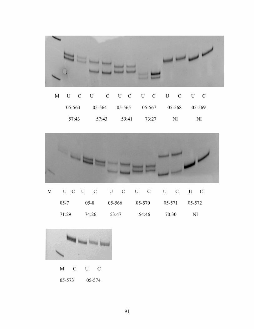
M U C U C U C U C U C U C
05-563 05-564 05-565 05-567 05-568 05-569
57:43 57:43 59:41 73:27 NI NI
M U C U C U C U C U C U C
05-7 05-8 05-566 05-570 05-571 05-572
71:29 74:26 53:47 54:46 70:30 NI
M C U C 05-573 05-574
91

92