An Introduction to Single Cell ICP-MS Analysis · PAPER An Introduction to Single Cell ICP-MS...
5
WHITE PAPER An Introduction to Single Cell ICP-MS Analysis Introduction The introduction and development of Single Particle ICP-MS (SP-ICP-MS) has opened a new area of research which allows the rapid detection and analysis of metal-based particles in a variety of matrices and applications. 1-6 The key feature of SP-ICP-MS is that it allows discrete pulses of positively charged ions to be detected and measured in a time-resolved manner using microsecond (µs) data acquisition rates. This advance in data acquisition capabilities is opening up a number of new application areas. In this white paper, we will introduce the concept of Single Cell ICP-MS (SC-ICP-MS), where individual cells are rapidly analyzed for metal content. Before delving into SC-ICP-MS, let’s briefly review the theory of SP-ICP-MS. SP-ICP-MS: A Brief Review Comprehensive descriptions of SP-ICP-MS are available, 7,8 so only a brief review will be given here. When a nanoparticle enters the plasma, it is completely ionized, producing a burst of ions which can be detected with ICP-MS. While conventional ICP-MS looks at a continuous signal, the output from SP-ICP-MS looks at discrete signals: one nanoparticle yields one ion burst, with the intensity of the resulting signal being related to the size of a particle (nm) and the number of pulses being related to the particle concentration (part/mL). The key to SP-ICP-MS is rapid, continuous measurement, which minimizes the chance of more than one particle being detected at the same time and ensures that particles are all counted. These tasks can be accomplished with ICP-MS instrumentation which contains fast electronics and eliminates the quadrupole Authors: Lauren Amable 1 Chady Stephan 2 Stan Smith 2 Ruth Merrifield 3 1 National Institute on Minority Health and Health Disparities, National Institute of Health, Bethesda, MD USA 2 PerkinElmer Inc., Shelton, CT USA 3 Center for Environmental NanoScience and Risk (CENR), Arnold School of Public Health, University of South Carolina, SC USA
Transcript of An Introduction to Single Cell ICP-MS Analysis · PAPER An Introduction to Single Cell ICP-MS...
WHITEPAPER An Introduction to Single Cell ICP-MS Analysis
Introduction
The introduction and development of Single Particle ICP-MS (SP-ICP-MS) has opened a new area of research which allows the rapid detection and analysis of metal-based particles in a variety of matrices and applications.1-6 The key feature of SP-ICP-MS is that it allows discrete pulses of positively charged ions to be detected and measured in a time-resolved manner using microsecond (µs) data acquisition rates. This advance in data acquisition capabilities is opening up a number of new application areas. In this white paper, we will introduce the concept of Single Cell ICP-MS (SC-ICP-MS), where individual cells are rapidly analyzed for metal content. Before delving into SC-ICP-MS, let’s briefly review the theory of SP-ICP-MS.
SP-ICP-MS: A Brief Review
Comprehensive descriptions of SP-ICP-MS are available,7,8 so only a brief review will be given here. When a nanoparticle enters the plasma, it is completely ionized, producing a burst of ions which can be detected with ICP-MS. While conventional ICP-MS looks at a continuous signal, the output from SP-ICP-MS looks at discrete signals: one nanoparticle yields one ion burst, with the intensity of the resulting signal being related to the size of a particle (nm) and the number of pulses being related to the particle concentration (part/mL).
The key to SP-ICP-MS is rapid, continuous measurement, which minimizes the chance of more than one particle being detected at the same time and ensures that particles are all counted. These tasks can be accomplished with ICP-MS instrumentation which contains fast electronics and eliminates the quadrupole
Authors:
Lauren Amable1 Chady Stephan2 Stan Smith2 Ruth Merrifield3
1 National Institute on Minority Health and Health Disparities, National Institute of Health, Bethesda, MD USA
2 PerkinElmer Inc., Shelton, CT USA
3 Center for Environmental NanoScience and Risk (CENR), Arnold School of Public Health, University of South Carolina, SC USA

2
settling time between measurements. These characteristics are important for accurate analyses: non-continuous analysis will miss particles, while slow sampling times/ long sampling windows will detect ions from multiple particles in the same measurement window. Both of these lead to inaccurate particle size and counting results.
Another important factor is the particle concentration in the sample. As particle concentration increases, the chances that multiple particles are ionized at the same time increases (i.e. coincidence). The resulting ion burst will be of multiple particles and not representative of the particle size as the particle concentration will be biased low and the particle size will be biased high. In samples with high particle concentrations, dilution is the best way to minimize the chances of multiple particles arriving at the plasma at the same time.
Applying SP-ICP-MS to SC-ICP-MS
A cell suspension is nebulized through a dedicated SC-ICP-MS introduction system in the same manner as SP-ICP-MS: as each cell enters the plasma, it is ionized, and the resulting ion burst from the intrinsic metal is detected by the ICP-MS, applying the same concept of data acquisition previously mentioned. In SC-ICP-MS analysis, each cell is considered as an individual entity, or particle, if you will, which generates its own ion burst. As in SP-ICP-MS, there are certain conditions that must be considered with regard to cell and dissolved micronutrient concentrations in order to obtain optimal results. For cell concentrations, it is preferable if the concentration is around 100,000 cell/mL to minimize coincidence. In some cases, such as when the ionic concentration of interest is high in the media, the contaminant is a nanoparticle (NP), or the media is complex, it is required that the cells are isolated from the culture media and re-suspended in either fresh media or an isotonic solution that does not contain the metal of interest. This will help avoid high dissolved backgrounds, interferences, or excess particles that could mask the signal generated by individual cells. Let's now consider examples of SC-ICP-MS.
Research into Cellular Uptake of Platinum-Containing Cancer Chemotherapy Compounds
Platinum (Pt)-containing compounds (i.e cisplatin (SP-4-2)-diamminedichloroplatinum(II)) are among the most commonly used cancer chemotherapy compounds. The efficacy of these compounds is related to their cellular uptake, meaning that increased uptake translates to more effective cancer treatment.9 While many cancers are responsive to Pt treatment, some will exhibit resistance, resulting in decreased Pt cellular uptake. Therefore, the Pt content of individual cancer cells could serve as a predictor to the effectiveness of the treatment.
The conventional method of assessing cellular cisplatin uptake consists of isolating a cell population, digesting all the cells, and measuring the Pt content. The limitation of this approach is that it is time consuming and leads to the assumption that all cells take up cisplatin on an equal basis.
Information on an individual cell basis will provide an accurate account of Pt content, Pt distribution, and the number of cells containing Pt within the cell population, a key factor in understanding cell resistance to cisplatin treatment. All of this information is available with SC-ICP-MS as the obtained signal is related to individual cells rather than to a concentration averaged over a large number of cells.
To determine the effectiveness of SC-ICP-MS for cisplatin, an ovarian cancer cell line (CP70) was exposed to cisplatin and monitored over time. Figure 1 shows the raw SC-ICP-MS signal of the cisplatin-exposed cells where the 195Pt isotope was monitored. Because background levels of Pt are non-existent and there are no common interferences on 195Pt, each spike represents Pt detected in an individual cell. Using an analysis time of one minute and a dwell time of 50 µs, a total of 1.2 million data points were collected in Figure 1. The variation in peak size reflects the information that we are seeking in this technique: an insight into the uptake mechanism as various cells will have different amounts of intrinsic Pt, all dependent on the molecular mechanism that is occurring and their ability to uptake and store Pt. Figure 2 shows Pt content distribution in CP70 after being exposed to cisplatin for four hours.
Furthermore, we explored the ability of SC-ICP-MS to track the uptake of Pt by CP70 ovarian cancer cells over the course of eight hours. Figure 3 shows that the Pt content within cells increases over time, signifying increased cisplatin uptake, which is represented with the shift to the right in the distribution.
Figure 1. Raw SC-ICP-MS signal of ovarian cancer cells exposed to cisplatin for four hours.
Figure 2. Platinum content distribution in CP70 exposed to cisplatin for four hours.

3
1 hour 2 hours
4 hours 8 hours
Figure 3. Cisplatin uptake by ovarian cancer cells over eight hours. The blue line is included as a reference.
The average Pt content vs. exposure time was repeated on two non-consecutive days and shows the great precision and reproducibility of SC-ICP-MS (Figure 4). These studies can be further expanded to explore how different cell lines uptake cisplatin under similar experimental conditions.
Figure 4. Cisplatin uptake of CP 70 ovarian cancer cells over eight hours on two non-consecu-tive days. Higher intensities correspond to more cisplatin in the cells.
Time (hours)
Inte
nsity
(cou
nts)
CP70 Study 1
CP70 Study 3Implications of Nanomaterials: Studying Cellular Uptake
Nanoparticles are being used in a wide variety of applications, from improving the quality of various consumer products to enhancing cancer research. Like every chemical, there is a potential risk associated with the release of NPs into the environment. As a traditional remedy against infections, silver’s alleged curative powers are now marketed as NP additives in a host of consumer antimicrobial products ranging from socks that fight odors to stuffed animals for children that fend off germs. At the same time, research studies conclusively show the toxicity of nanosilver on cells. The latest is a report from the Max Planck Institute in Germany, which concludes that silver NPs are highly toxic once they cross the cell membrane.10
In relation to both environmental and human health, there are three questions that need to be answered:
1. How many NPs are in a particular system? (Exposure)
2. Do these NPs enter organisms and at what concentration? (Dose)
3. What effect do they have? (Response)
The answers to these questions allow the development of more targeted drugs and the ability to fully assess the risk of NPs in the environment. SC-ICP-MS plays a major role in answering the first two questions about how unicellular organisms interact with NPs by providing a means to simply and quickly monitor cellular NP uptake. Fundamental studies involve adding NPs to a culture media containing cells, then monitoring the uptake of NPs into the cells using SC-ICP-MS.
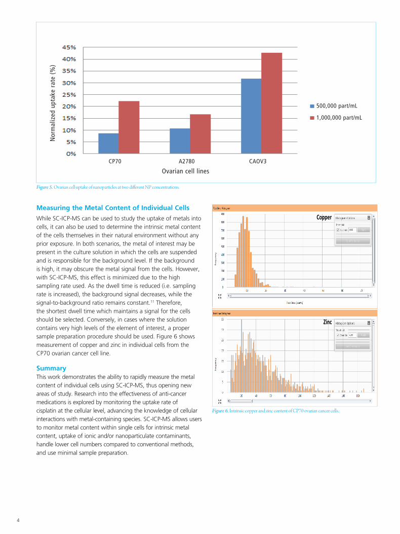
An example of such a study is shown in Figure 5, where three different lines of ovarian cells were exposed to two different gold NP concentrations. The cells were washed to remove excess NPs and then analyzed after 21 hours to determine the cellular content. The data in Figure 5 shows that different cell lines have different NP uptake rates, which is dependent, to a certain extent, on the NP concentrations studied.
4
Measuring the Metal Content of Individual Cells
While SC-ICP-MS can be used to study the uptake of metals into cells, it can also be used to determine the intrinsic metal content of the cells themselves in their natural environment without any prior exposure. In both scenarios, the metal of interest may be present in the culture solution in which the cells are suspended and is responsible for the background level. If the background is high, it may obscure the metal signal from the cells. However, with SC-ICP-MS, this effect is minimized due to the high sampling rate used. As the dwell time is reduced (i.e. sampling rate is increased), the background signal decreases, while the signal-to-background ratio remains constant.11 Therefore, the shortest dwell time which maintains a signal for the cells should be selected. Conversely, in cases where the solution contains very high levels of the element of interest, a proper sample preparation procedure should be used. Figure 6 shows measurement of copper and zinc in individual cells from the CP70 ovarian cancer cell line. Summary This work demonstrates the ability to rapidly measure the metal content of individual cells using SC-ICP-MS, thus opening new areas of study. Research into the effectiveness of anti-cancer medications is explored by monitoring the uptake rate of cisplatin at the cellular level, advancing the knowledge of cellular interactions with metal-containing species. SC-ICP-MS allows users to monitor metal content within single cells for intrinsic metal content, uptake of ionic and/or nanoparticulate contaminants, handle lower cell numbers compared to conventional methods, and use minimal sample preparation.
Figure 6. Intrinsic copper and zinc content of CP70 ovarian cancer cells..
Copper
Zinc
Figure 5. Ovarian cell uptake of nanoparticles at two different NP concentrations.
CP70 A2780 CAOV3
Nor
mal
ized
upt
ake
rate
(%
)
500,000 part/mL
1,000,000 part/mL
Ovarian cell lines
For a complete listing of our global offices, visit www.perkinelmer.com/ContactUs
Copyright ©2017, PerkinElmer, Inc. All rights reserved. PerkinElmer® is a registered trademark of PerkinElmer, Inc. All other trademarks are the property of their respective owners. 012774A_01 PKI
PerkinElmer, Inc. 940 Winter Street Waltham, MA 02451 USA P: (800) 762-4000 or (+1) 203-925-4602www.perkinelmer.com
References
1. Mitrano, D., Ranville, J.F, Stephan, C. “Quantitative Evaluation of Nanoparticle Dissolution Kinetics using Single Particle ICP-MS: A Case Study with Silver Nanoparticles”, PerkinElmer, 2014.
2. Davidowski, L., Stephan, C. “Characterization of Silver Nanoparticles in Dietary Supplements by Single Particle ICP-Mass Spectrometry”, PerkinElmer, 2014.
3. Neubauer, K., Stephan, C. “Determination of Gold and Silver Nanoparticles in Blood using Single Particle ICP-MS”, PerkinElmer, 2014.
4. Dan, Y., Shi, H., Liang, X. “Measurement of Titanium Dioxide Nanoparticles in Sunscreen using Single Particle ICP-MS”, PerkinElmer, 2015.
5. Donovan, A.R., Shi, H. Adams, C., Stephan, C. “Rapid Analysis of Silver, Gold, and Titanium Dioxide Nanoparticles in Drinking Water by Single Particle ICP-MS”, PerkinElmer, 2015.
6. Stephan, C. “Analysis of Iron Nanoparticles in Organic Solvents Used in the Semiconductor Industry Using Single Particle ICP-MS in Reaction Mode”, PerkinElmer, 2015.
7. Stephan, C., Neubauer, K. “Single Particle Inductively Coupled Plasma Mass Spectrometry: Understanding How and Why”, PerkinElmer, 2014.
8. Hineman, A., Stephan, C. J. Anal. At. Spectrom. 2014, 29, 1252.
9. Amable L. "Cisplatin resistance and opportunities for precision medicine," Pharmacol Res, 2016, 106, 27-36.
10. Kennedy, D., Orts-Gil, G., Lai, CH., Müller, L., Haase, A., Luch, A., Seeberger, P. “Carbohydrate functionalization of silver nanoparticles modulates cytotoxicity and cellular uptake”, Journal of Nanobiotechnology, 2014 12:59 DOI: 10.1186/s12951-014-0059-z.
11. Neubauer K., Stephan, C., Kobayashi, K. “Analysis of SiO2 Nanoparticles in Standard Mode with Single Particle ICP-MS”, PerkinElmer, 2015.
The PerkinElmer instruments included in this paper are for research use only, not for use in diagnostic procedures.