an analysis of acid-base disturbances in the haemolymph following ...
12
exp. Biol. (i979). 79. 47-58 47 ih sfigura Printed in Great Britain AN ANALYSIS OF ACID-BASE DISTURBANCES IN THE HAEMOLYMPH FOLLOWING STRENUOUS ACTIVITY IN THE DUNGENESS CRAB, CANCER MAGISTER BY D. G. MCDONALD,* B. R. M C M A H O N AND C. M. WOOD* Department of Biology, University of Calgary, Calgary, Alberta, Canada, T2N 1N4. (Received zjune 1978) 8UMMARY Enforced activity causes a marked depression of haemofymph pH in Cancer magister. Both lactate concentration and Pco, of the haemolymph are elevated immediately following exercise but resting P ^ is restored within 30 min whereas resting lactate levels are not restored for at least 8 h. The haemolymph acid-base disturbance is caused largely by elevated haemolymph lactate levels but a Davenport analysis based on measurements of pH and total CO 2 reveals a marked discrepancy between the amount of metabolic acid buffered by the haemolymph and the lactate anion concen- tration. This appears due to a more rapid release of lactate from the tissues than H + ions produced with lactate. INTRODUCTION Strenuous activity causes a marked depression in haemolymph pH in decapod crustaceans (Johansen, Lenfant & Mecklenberg, 1970; Mangum & Wieland, 1975; Phillips et al. 1977). Although the nature of this acidosis has not been examined in crustaceans it may, by analogy with vertebrate studies, have both respiratory and metabolic acid (lactic acid) components. Two recent studies on marine fish offer differing views of the relative contributions of these components to the depression of blood pH following exercise. In the elasmobranch Scyliorhinus stellaris (Piiper, Meyer & Drees, 1972) most of the acid-base disturbance was attributable to an increase in blood lactic acid, a situation similar to that in mammals. Unlike mammals, a large deficit developed in the recovery period between calculated blood levels of metabolic acid and measured blood lactate concentrations. Piiper et al. (1972) thus suggested that a significant portion of the H + ions produced with lactate were not immediately excreted into the blood but were initially retained and buffered intra- cellularly. In contrast in the starry flounder Platichthys stellatus the acidosis following exhaustive exercise was due largely to elevated PQQ, levels (Wood, McMahon & McDonald, 1977). In this animal blood lactic acid contributed significantly to the Present address: Department of Biology, McMaater University, Hamilton, Ontario, Canada S4K1.
Transcript of an analysis of acid-base disturbances in the haemolymph following ...
exp. Biol. (i979). 79. 47-58 47ih sfigura
Printed in Great Britain
AN ANALYSIS OF ACID-BASE DISTURBANCES IN THEHAEMOLYMPH FOLLOWING STRENUOUS ACTIVITY IN
THE DUNGENESS CRAB, CANCER MAGISTER
BY D. G. M C D O N A L D , * B. R. M C M A H O N AND C. M. WOOD*
Department of Biology, University of Calgary,Calgary, Alberta, Canada, T2N 1N4.
(Received zjune 1978)
8UMMARY
Enforced activity causes a marked depression of haemofymph pH inCancer magister. Both lactate concentration and Pco, of the haemolymphare elevated immediately following exercise but resting P ^ is restoredwithin 30 min whereas resting lactate levels are not restored for at least 8 h.The haemolymph acid-base disturbance is caused largely by elevatedhaemolymph lactate levels but a Davenport analysis based on measurementsof pH and total CO2 reveals a marked discrepancy between the amount ofmetabolic acid buffered by the haemolymph and the lactate anion concen-tration. This appears due to a more rapid release of lactate from the tissuesthan H + ions produced with lactate.
INTRODUCTION
Strenuous activity causes a marked depression in haemolymph pH in decapodcrustaceans (Johansen, Lenfant & Mecklenberg, 1970; Mangum & Wieland, 1975;Phillips et al. 1977). Although the nature of this acidosis has not been examined incrustaceans it may, by analogy with vertebrate studies, have both respiratory andmetabolic acid (lactic acid) components. Two recent studies on marine fish offerdiffering views of the relative contributions of these components to the depressionof blood pH following exercise. In the elasmobranch Scyliorhinus stellaris (Piiper,Meyer & Drees, 1972) most of the acid-base disturbance was attributable to anincrease in blood lactic acid, a situation similar to that in mammals. Unlike mammals,a large deficit developed in the recovery period between calculated blood levels ofmetabolic acid and measured blood lactate concentrations. Piiper et al. (1972) thussuggested that a significant portion of the H + ions produced with lactate were notimmediately excreted into the blood but were initially retained and buffered intra-cellularly.
In contrast in the starry flounder Platichthys stellatus the acidosis followingexhaustive exercise was due largely to elevated PQQ, levels (Wood, McMahon &McDonald, 1977). In this animal blood lactic acid contributed significantly to the
Present address: Department of Biology, McMaater University, Hamilton, Ontario, CanadaS4K1.
48 D. G. MCDONALD, B. R. MCMAHON AND C. M. WOOD
acidosis only late in the recovery period. Furthermore it was apparent that all H 'ions produced with lactate contributed to the observed acidosis. Wood et al., infact, postulated that an additional metabolic acid (possibly ammonium) may alsohave contributed to the pH drop during the recovery period.
The purpose of the present study was to investigate acid-base disturbances follow-ing exercise in a marine crustacean by monitoring haemolymph pH, total COa andlactate and to quantify, using analytical procedures similar to those in the abovestudies, the relative contributions of lactic acid and P ^ to the observed acidosis.C. magister was chosen for study because of its large size and because of the relativeease with which haemolymph samples can be repetitively withdrawn.
MATERIALS AND METHODS
Adult Dungeness crabs (C magister Dana; 490 to 1030 g) in intermolt stage C4
(Drach & Tchernigovtzeff, 1967) were obtained from the Vancouver Public Aquariumand held for at least 2 weeks before use in large sandy bottomed tanks in the marineaquarium facility at the University of Calgary. The acclimation conditions werethose employed in subsequent experiments; recirculating, filtered sea water, salinity= 27 ± 1 %o, temperature = 8 ± 1 °C.
I. In vivo experimentsFor all in vivo experiments the crabs (N = 7) were restrained in air while post-
branchial (i.e. arterial) sites were prepared for haemolymph sampling. Small holeswere drilled in the dorsal carapace so as to expose the epidermis overlying the anterio-lateral corners of the pericardial sinus. These two holes were sealed with a rubbermembrane (dental latex) glued to the carapace with cyanoacrylate glue. In threecrabs pre-branchial (i.e. venous) sites were similarly prepared by gluing dentallatex to the arthrodial membranes at the bases of either the third or fourth walkinglegs. The animals were then transferred to individual chambers (18x30x10011deep) and allowed to recover for at least 72 h before haemolymph sampling wasbegun.
Haemolymph was withdrawn from the prepared sites with a 1 ml glass syringe and23 gauge needle. The dead volume of the syringe was filled with Millipore (0-22 /im)filtered seawater and the syringes were chilled on ice prior to sampling. Post-branchialhaemolymph samples (0-4-0-5 ml) could be routinely withdrawn without apparentdisturbance to the animal. Sampling was alternated between the two post-branchialsites to minimize the development of clots within the pericardial cavity. In preliminaryexperiments (McDonald, 1977) it was determined that haemolymph samples drawnfrom the limb sinuses accessible through the arthrodial membranes represented mixedprebranchial haemolymph. These samples were somewhat more difficult to obtainand usually involved moving the animal to insert the sampling needle. Consequentlypre-branchial samples were taken immediately following post-branchial samples andsmaller volumes (0-1-0-2 ml) were drawn.
One to two haemolymph samples were withdrawn before the crab was provokedinto activity. Animals were exercised for 20 min, initially by periodic proddi^and then subsequently more severely manipulated by hand to ensure near continuous
Acid-base disturbances after activity in the crab 49
fnotion. By the end of the exercise period the crabs were normally refractory tostimulation. Post-branchial haemolymph samples were drawn immediately post-exercise (time o) and then at 30 min, 1 h, 2 h, 4 h, 8 h, 24 h, 48 h and 72 h. Pre-branchial samples were drawn at time o, +30 min, +1 h and +4 h.
II. In vitro experiments
In vitro determinations of CO2 combining curves and haemolymph buffer capa-cities (A [HC07] + [COJTJ/A pH) were performed by a method similar to that ofTruchot (19766) on haemolymph drawn from each crab at the termination of thein vivo experiment. Four to six ml of haemolymph were withdrawn from the peri-cardial sinus, shaken to promote clotting, centrifuged at 5000 g for 10 min to removethe clot and then transferred to a round-bottomed tonometer mounted in a temperaturecontrolled (8 °C) water bath. The tonometer was spun to distribute haemolymphon its walls and was gassed with humidified mixtures of CO2 in air supplied by aWosthoff gas mixing pump. Haemolymph was equilibrated (1 h) to 4-5 differentlevels of CO2 in air (Pco, 0-65 to 6-5 torr). To minimize equilibration time bovinecarbonic anhydrase obtained from Sigma, St Louis (final concentration 0-25 g I"1)was added to the haemolymph (Truchot, 19766).
III. Analytical procedures
All in vivo post-branchial samples were analysed for total CO2, pH and lactateand in two experiments haemolymph PCOj was also determined. Pre-branchial andin vitro haemolymph samples were analysed for pH and total COa. Haemocyaninoxygen carrying capacity, C g** O2 was determined from measurements of oxygencontent of haemolymph air equilibrated m vitro.
Haemolymph pH was measured on 40-60/il samples injected into a RadiometerpH electrode thermostatted to the experimental temperature. The pH value wasdisplayed on a Radiometer PHM71 acid-base meter. The pH measurement systemwas calibrated with Radiometer precision buffers (Si500 and Si510) having values of6-928 and 7-475 at 8 °C. The linearity of this instrument at higher pH was checkedperiodically with a Sigma precision buffer (Trizma 7-4); pH 7-88 at 8 °C. TotalCO2 content of haemolymph was determined on 70-80 fi\ samples by the method ofCameron (1971). Each sample was bracketed with sodium bicarbonate standards toincrease the accuracy of these determinations. Haemolymph lactate concentrationswere determined on 0-25 ml of haemolymph. This volume was immediately deprotein-ated in 0-5 ml of ice-cold perchloric acid and then centrifuged at 5000 g for 10 min.The supernatant was analysed enzymatically (lactic dehydrogenase) for L-lactatewith Sigma reagents (see Sigma bulletin no: 826-UV). Haemolymph PCOj wasmeasured on 0-2 ml samples with a thermostatted Radiometer P ^ electrode fittedwith a thin (25 fi\) silicone membrane and connected to a second PHM71. At 8 °C8-10 min was required for the electrode response. This, plus the low Pco, levels incrab haemolymph (1-4 torr) may mean that the measurements of Pco, Tniiy be inerror by as much as ± 20 %. C J 5 " O 2 was calculated from measurements of haemolymphoxygen content on air-equilibrated samples according to the formula:
Cg«O 2 = Co , -adissO2.PO l
50 D. G. MCDONALD, B. R. MCMAHON AND C. M. WOOD
where Co> is the haemolymph oxygen content measured on 80 fi\ aliquots injectedinto a Lex-Oa-Con oxygen content analyser, a diss 0 2 is the physical solubility ofoxygen in haemolymph in vols % torr"1 (from Truchot, 1971) and POl is the oxygenpartial pressure of air-equilibrated haemolymph.
IV. Theoretical approach
C02 dissociation in the haemolymph is described by the Henderson-Hasselbalchequation:
where pk[ andpk'% are the negative logarithms of the apparent first and second dissoci-ation constants of HjCC^ and HCO^ respectively and aCO2 is the solubility co-efficient of C02 in haemolymph. Values for aC02, pk'i and pk'2 at the experimentaltemperature and salinity were obtained from alignment nomograms constructed forthe haemolymph of the shore crab Cardnus maenas (Truchot, 1976 a).
The close correspondence found between measured values of Vco and valuescalculated using the Henderson-Hasselbalch equation confirms the applicability ofthese constants to acid-base calculations on C. magister haemolymph. The meandifference between measured and calculated values was 0-24 + 0-18 (s.D.) torr (N =20). Paired 't' test analysis of measured and calculated values showed that they werenot significantly different. Only calculated values for PCOt are presented in the textas the errors inherent in P ^ measurements (described above) are likely greater thanerrors in measurements of either total C02 or pH on which Pco, calculations arebased.
In acid-base studies on vertebrates the dissociation of HCO^ to H+ and COST 19
usually ignored as negligible. At the high pH and ionic acitivity of crustacean haemo-lymph this dissociation is not negligible but at pH 7-9, [HCOJ] exceeds [CO^] by aratio of approximately 30:1. Thus, bicarbonate concentrations calculated from theHenderson-Hasselbalch equation are referred to as [HCOsJ + fCOjJ1].
Increases in P ^ and in the concentration of acid metabolites (e.g. lactic acid)in the haemolymph will increase the quantity of H+ ions buffered by the haemolymphbicarbonate and non-bicarbonate (mainly protein) buffer systems. By the use of aDavenport diagram (pH vs [HC07 + COJ]; Davenport, 1974) constructed specificallyfor the haemolymph of the crab the quantities of H+ ions added by respiratory acids(AH+c) and by metabolic acids (AH+m) were separately estimated from in vivomeasurements of haemolymph pH and bicarbonate concentrations and in vitrodeterminations of the haemolymph buffering power (A[HC07] + COJJ/A pH).This is the standard method originally designed for estimating metabolic and res-piratory acid levels in human blood and is described in detail in Woodbury (1974)and Wood et al. (1977). This method is based on physico-chemical principles whichshould be common to any binary buffer system (i.e. protein plus bicarbonate buffers)and thus should be equally valid when applied to the crab system since accuratevalues for pk[ and aC02 of crustacean haemolymph are available (Truchot, 1976a).
Acid-base disturbances after activity in the crab
6 P A
I4
3.2
3crVS4lin
8+, 2
82 4PCOj(tOIT)
r B
i
7-4 7-6 7-8pH
8-0
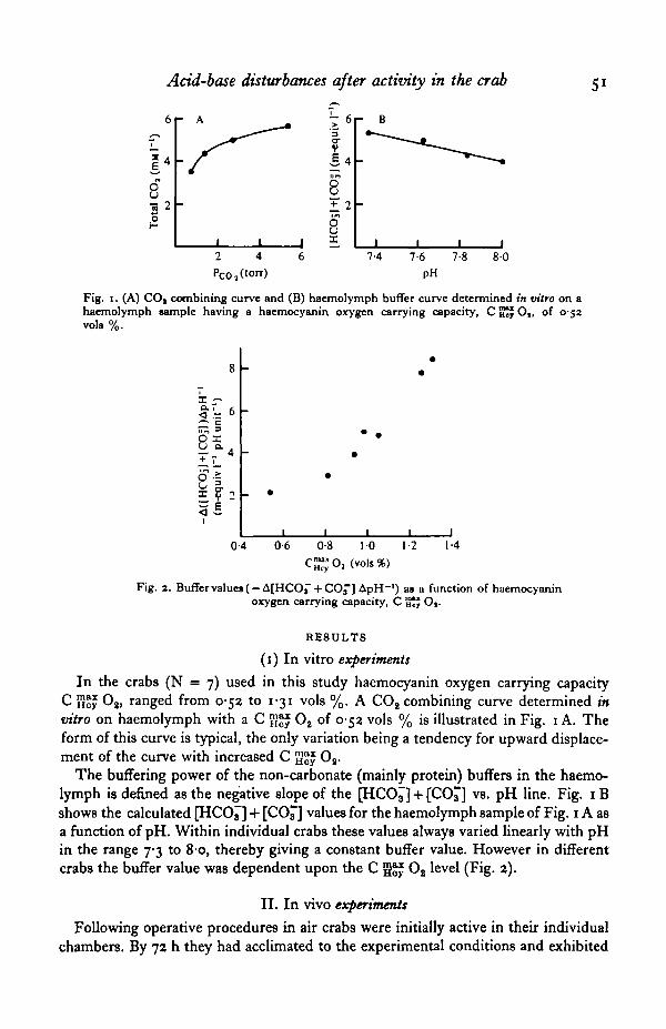
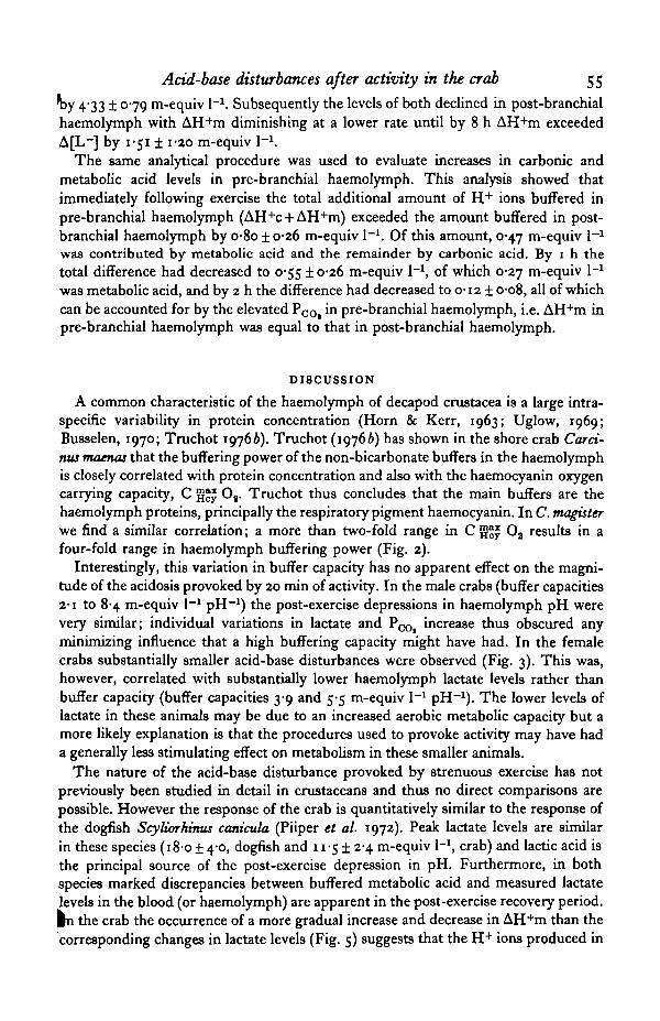
Fig. 1. (A) CO, combining curve and (B) haemolymph buffer curve determined in vitro on ahaemolymph sample having a haemocyanin oxygen carrying capacity, C SJJ O,, of 0-52vols %.
X -~
II. •
0-4 0-6 0-8 10 1-2
™XO2 (vols%)
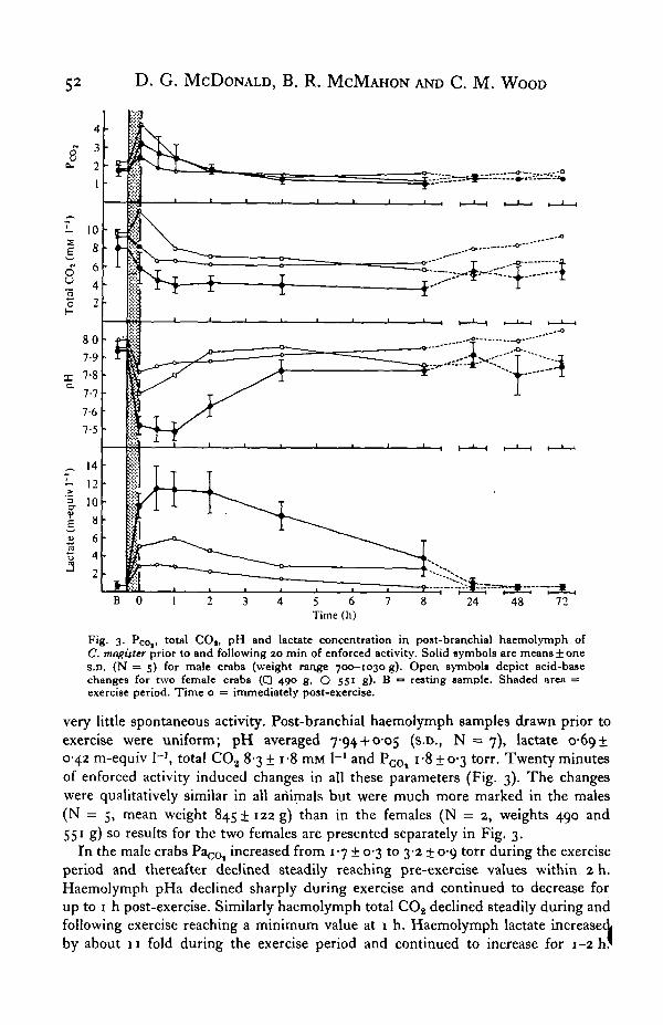
Fig. 2. Buffervalues( —A[HCOi" + CO^] ApH"1) as a function of haemocyaninoxygen carrying capacity, C 5«r Of-
RESULTS
(i) In vitro experiments
In the crabs (N = 7) used in this study haemocyanin oxygen carrying capacityC H " Os, ranged from 052 to 131 vols %. A CO2 combining curve determined invitro on haemolymph with a C g ^ O2 of 0-52 vols % is illustrated in Fig. 1 A. Theform of this curve is typical, the only variation being a tendency for upward displace-ment of the curve with increased C gj}£ O2.
The buffering power of the non-carbonate (mainly protein) buffers in the haemo-lymph is defined as the negative slope of the [HCO^] + [CC£] vs. pH line. Fig. 1 Bshows the calculated [HCO^] + [CO^] values for the haemolymph sample of Fig. 1A asa function of pH. Within individual crabs these values always varied linearly with pHin the range 7-3 to 80, thereby giving a constant buffer value. However in differentcrabs the buffer value was dependent upon the C g ^ O2 level (Fig. 2).
II. In vivo experiments
Following operative procedures in air crabs were initially active in their individualchambers. By 72 h they had acclimated to the experimental conditions and exhibited
52 D. G. MCDONALD, B. R. MCMAHON AND C. M. WOOD
5 6 7Time (h)
48 72
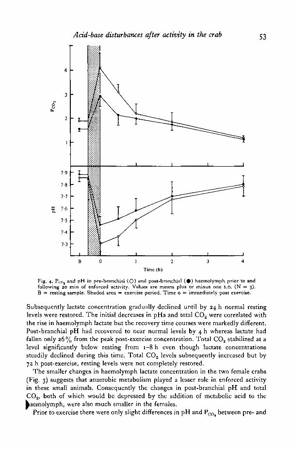
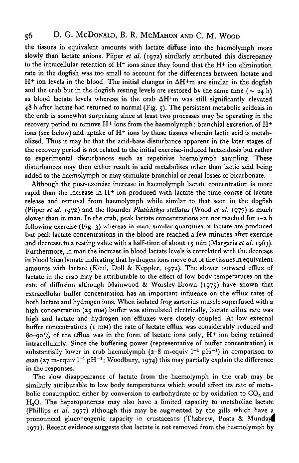
Fig. 3. Pco,. total CO|, pH and lactate concentration in post-branchial haemolymph ofC. magister prior to and following 20 min of enforced activity. Solid symbols are means ± oneS.D. (N = 5) for male crabs (weight range 700—1030 g). Open symbols depict acid-basechanges for two female crabs ( • 400 g, O 55' g). B = resting sample. Shaded area =exercise period. Time o = immediately post-exercise.
very little spontaneous activity. Post-branchial haemolymph samples drawn prior toexercise were uniform; pH averaged 794 + O-O5 (s.D., N = 7), lactate 0-6910-42 m-equiv I-3, total CO2 8-3 ± i-8 mM I"1 and P c 0 | i-8 ± 0-3 torr. Twenty minutesof enforced activity induced changes in all these parameters (Fig. 3). The changeswere qualitatively similar in all animals but were much more marked in the males(N = 5, mean weight 845! 122 g) than in the females (N = 2, weights 490 and551 g) so results for the two females are presented separately in Fig. 3.
In the male crabs PaCOt increased from 1-7 ± 0-3 to 3-2 ± 0-9 torr during the exerciseperiod and thereafter declined steadily reaching pre-exercise values within 2 h.Haemolymph pHa declined sharply during exercise and continued to decrease forup to i h post-exercise. Similarly haemolymph total COg declined steadily during andfollowing exercise reaching a minimum value at 1 h. Haemolymph lactate increasedby about 11 fold during the exercise period and continued to increase for 1-2 h '
Acid-base disturbances after activity in the crab 53
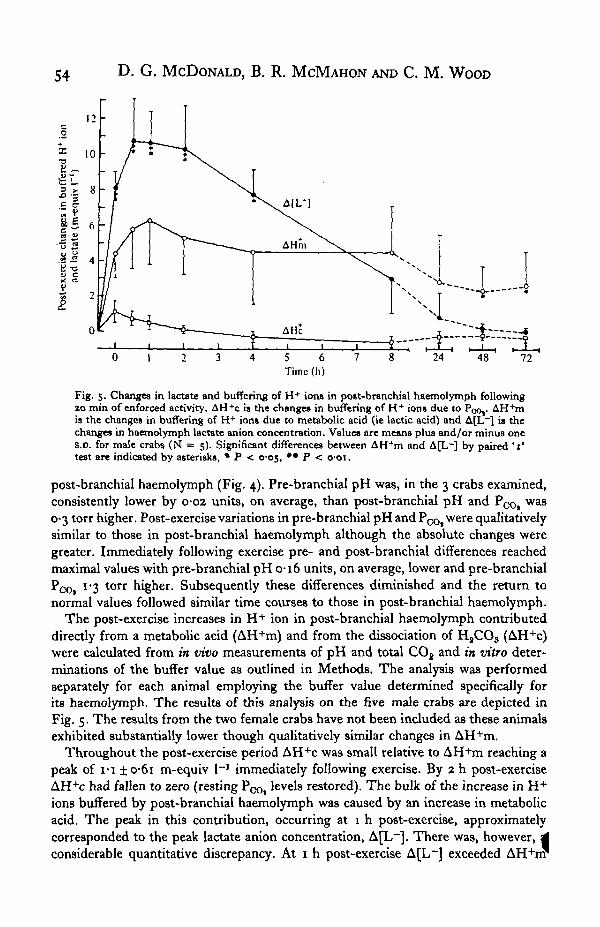
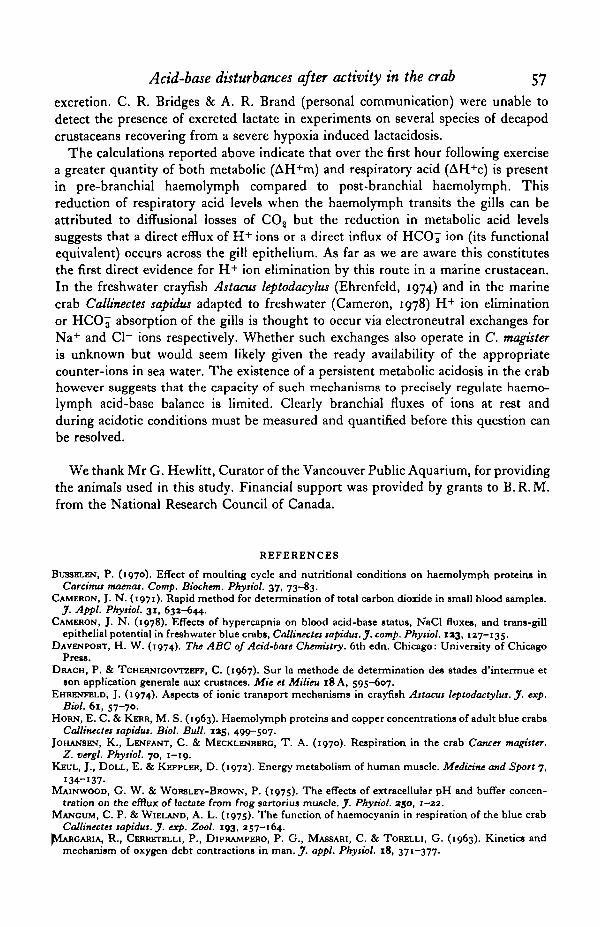
Fig. 4. Pco, and pH in pre-branchial (O) and post-branchial ( # ) haemolymph prior to andfollowing 20 min of enforced activity. Values are means plus or minus one s.D. (N = 3).B = resting sample. Shaded area = exercise period. Time o = immediately post exercise.
Subsequently lactate concentration gradually declined until by 24 h normal restinglevels were restored. The initial decreases in pHa and total C02 were correlated withthe rise in haemolymph lactate but the recovery time courses were markedly different.Post-branchial pH had recovered to near normal levels by 4 h whereas lactate hadfallen only 26 % from the peak post-exercise concentration. Total CO2 stabilized at alevel significantly below resting from 1-8 h even though lactate concentrationssteadily declined during this time. Total CO2 levels subsequently increased but by72 h post-exercise, resting levels were not completely restored.
The smaller changes in haemolymph lactate concentration in the two female crabs(Fig. 3) suggests that anaerobic metabolism played a lesser role in enforced activityin these small animals. Consequently the changes in post-branchial pH and totalCO2, both of which would be depressed by the addition of metabolic acid to the^aemolymph, were also much smaller in the females.
Prior to exercise there were only slight differences in pH and P ^ between pre- and
54 D. G. MCDONALD, B. R. MCMAHON AND C. M. WOOD
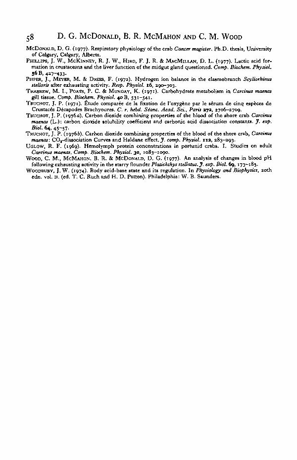
Fig. 5. Changes in lactate and buffering of H+ ions in po»t-branchial haemolymph followingao min of enforced activity. AH+c is the changes in buffering of H+ ions due to Poo,. AH+mis the changes in buffering of H+ ions due to metabolic acid (ie lactic acid) and A[L~] is thechanges in haemolymph lactate anion concentration. Value* are means plus and/or minus ones.D. for male crabs (N = 5). Significant differences between AH+m and A[L~] by paired ' t'test are indicated by asterisks, • P < cos, ** P <
post-branchial haemolymph (Fig. 4). Pre-branchial pH was, in the 3 crabs examined,consistently lower by 0-02 units, on average, than post-branchial pH and PQQ, was0-3 torr higher. Post-exercise variations in pre-branchial pH and PQO, were qualitativelysimilar to those in post-branchial haemolymph although the absolute changes weregreater. Immediately following exercise pre- and post-branchial differences reachedmaximal values with pre-branchial pH 0-16 units, on average, lower and pre-branchialP<x>, l"i t o r r higher. Subsequently these differences diminished and the return tonormal values followed similar time courses to those in post-branchial haemolymph.
The post-exercise increases in H + ion in post-branchial haemolymph contributeddirectly from a metabolic acid (AH+m) and from the dissociation of H8CO3 (AH+c)were calculated from m vivo measurements of pH and total CO2 and in vitro deter-minations of the buffer value as outlined in Methods. The analysis was performedseparately for each animal employing the buffer value determined specifically forits haemolymph. The results of this analysis on the five male crabs are depicted inFig. 5. The results from the two female crabs have not been included as these animalsexhibited substantially lower though qualitatively similar changes in AH+m.
Throughout the post-exercise period AH+c was small relative to AH+m reaching apeak of I-I ±o*6i m-equiv I"1 immediately following exercise. By 2 h post-exerciseAH+c had fallen to zero (resting PCOt levels restored). The bulk of the increase in H+ions buffered by post-branchial haemolymph was caused by an increase in metabolicacid. The peak in this contribution, occurring at 1 h post-exercise, approximatelycorresponded to the peak lactate anion concentration, A[L~]. There was, however, 4considerable quantitative discrepancy. At 1 h post-exercise A[L~] exceeded AH+rrr
Acid-base disturbances after activity in the crab 55by 4-33 ± 079 m-equiv I"1. Subsequently the levels of both declined in post-branchialhaemolymph with AH+m diminishing at a lower rate until by 8 h AH+m exceededA[L~] by 1-51 ± 1-20 m-equiv 1-1.
The same analytical procedure was used to evaluate increases in carbonic andmetabolic acid levels in pre-branchial haemolymph. This analysis showed thatimmediately following exercise the total additional amount of H + ions buffered inpre-branchial haemolymph (AH+c + AH+m) exceeded the amount buffered in post-branchial haemolymph by o-8o ± 0*26 m-equiv I"1. Of this amount, 0-47 m-equiv 1-1
was contributed by metabolic acid and the remainder by carbonic acid. By 1 h thetotal difference had decreased to 0-55 ±0-26 m-equiv I"1, of which 0-27 m-equiv 1-1
was metabolic acid, and by 2 h the difference had decreased to 0-12 ± 0-08, all of whichcan be accounted for by the elevated POOi in pre-branchial haemolymph, i.e. AH+m inpre-branchial haemolymph was equal to that in post-branchial haemolymph.
DISCUSSION
A common characteristic of the haemolymph of decapod Crustacea is a large intra-specific variability in protein concentration (Horn & Kerr, 1963; Uglow, 1969;Busselen, 1970; Truchot 19766). Truchot (19766) has shown in the shore crab Carci-nus mamas that the buffering power of the non-bicarbonate buffers in the haemolymphis closely correlated with protein concentration and also with the haemocyanin oxygencarrying capacity, C gg£ 08. Truchot thus concludes that the main buffers are thehaemolymph proteins, principally the respiratory pigment haemocyanin. In C. magisterwe find a similar correlation; a more than two-fold range in C g^y Oa results in afour-fold range in haemolymph buffering power (Fig. 2).
Interestingly, this variation in buffer capacity has no apparent effect on the magni-tude of the acidosis provoked by 20 min of activity. In the male crabs (buffer capacities2-1 to 8-4 m-equiv I"1 pH"1) the post-exercise depressions in haemolymph pH werevery similar; individual variations in lactate and PCOi increase thus obscured anyminimizing influence that a high buffering capacity might have had. In the femalecrabs substantially smaller acid-base disturbances were observed (Fig. 3). This was,however, correlated with substantially lower haemolymph lactate levels rather thanbuffer capacity (buffer capacities 3-9 and 5-5 m-equiv I"1 pH"1). The lower levels oflactate in these animals may be due to an increased aerobic metabolic capacity but amore likely explanation is that the procedures used to provoke activity may have hada generally less stimulating effect on metabolism in these smaller animals.
The nature of the acid-base disturbance provoked by strenuous exercise has notpreviously been studied in detail in crustaceans and thus no direct comparisons arepossible. However the response of the crab is quantitatively similar to the response ofthe dogfish Scyliorkmus canicula (Piiper et al. 1972). Peak lactate levels are similarin these species (18-0 ± 4-0, dogfish and 11-51 2-4 m-equiv I"1, crab) and lactic acid isthe principal source of the post-exercise depression in pH. Furthermore, in bothspecies marked discrepancies between buffered metabolic acid and measured lactatelevels in the blood (or haemolymph) are apparent in the post-exercise recovery period.In the crab the occurrence of a more gradual increase and decrease in AH+m than thecorresponding changes in lactate levels (Fig. 5) suggests that the H + ions produced in
56 D. G. MCDONALD, B. R. M C M A H O N AND C. M. WOOD
the tissues in equivalent amounts with lactate diffuse into the haemolymph moreslowly than lactate anions. Piiper et al. (1972) similarly attributed this discrepancyto the intracellular retention of H+ ions since they found that the H+ ion eliminationrate in the dogfish was too small to account for the differences between lactate andH + ion levels in the blood. The initial changes in AH+m are similar in the dogfishand the crab but in the dogfish resting levels are restored by the same time (~ 24 h)as blood lactate levels whereas in the crab AH+m was still significantly elevated48 h after lactate had returned to normal (Fig. 5). The persistent metabolic acidosis inthe crab is somewhat surprising since at least two processes may be operating in therecovery period to remove H + ions from the haemolymph: branchial excretion of H +
ions (see below) and uptake of H + ions by those tissues wherein lactic acid is metab-olized. Thus it may be that the acid-base disturbance apparent in the later stages ofthe recovery period is not related to the initial exercise-induced lactacidosis but ratherto experimental disturbances such as repetitive haemolymph sampling. Thesedisturbances may then either result in acid metabolites other than lactic acid beingadded to the haemolymph or may stimulate branchial or renal losses of bicarbonate.
Although the post-exercise increase in haemolymph lactate concentration is morerapid than the increase in H + ion produced with lactate the time course of lactaterelease and removal from haemolymph while similar to that seen in the dogfish(Piiper et al. 1972) and the flounder Platichthys stellatus (Wood et al. 1977) is muchslower than in man. In the crab, peak lactate concentrations are not reached for 1-2 hfollowing exercise (Fig. 3) whereas in man, similar quantities of lactate are producedbut peak lactate concentrations in the blood are reached a few minutes after exerciseand decrease to a resting value with a half-time of about 15 min (Margaria et al. 1963).Furthermore, in man the increase in blood lactate levels is correlated with the decreasein blood bicarbonate indicating that hydrogen ions move out of the tissues in equivalentamounts with lactate (Keul, Doll & Keppler, 1972). The slower outward efflux oflactate in the crab may be attributable to the effect of low body temperatures on therate of diffusion although Mainwood & Worsley-Brown (1975) have shown thatextracellular buffer concentration has an important influence on the efflux rates ofboth lactate and hydrogen ions. When isolated frog sartorius muscle superfused with ahigh concentration (25 mM) buffer was stimulated electrically, lactate efflux rate washigh and lactate and hydrogen ion effluxes were closely coupled. At low externalbuffer concentrations (1 mM) the rate of lactate efflux was considerably reduced and80-90% of the efflux was in the form of lactate ions only, H+ ion being retainedintracellularly. Since the buffering power (representative of buffer concentration) issubstantially lower in crab haemolymph (2-8 m-equiv 1-1 pH"1) in comparison toman (27 m-equiv I"1 pH- 1; Woodbury, 1974) this may partially explain the differencein the responses.
The slow disappearance of lactate from the haemolymph in the crab may besimilarly attributable to low body temperatures which would affect its rate of meta-bolic consumption either by conversion to carbohydrate or by oxidation to CO2 andH2O. The hepatopancreas may also have a limited capacity to metabolize lactate(Phillips et al. 1977) although this may be augmented by the gills which have apronounced gluconeogenic capacity in crustaceans (Thabrew, Poats & Mundaj^1971). Recent evidence suggests that lactate is not removed from the haemolymph by
Acid-base disturbances after activity in the crab 57
excretion. C. R. Bridges & A. R. Brand (personal communication) were unable todetect the presence of excreted lactate in experiments on several species of decapodcrustaceans recovering from a severe hypoxia induced lactacidosis.
The calculations reported above indicate that over the first hour following exercisea greater quantity of both metabolic (AH+m) and respiratory acid (AH+c) is presentin pre-branchial haemolymph compared to post-branchial haemolymph. Thisreduction of respiratory acid levels when the haemolymph transits the gills can beattributed to diffusional losses of CO2 but the reduction in metabolic acid levelssuggests that a direct efflux of H+ ions or a direct influx of HCO^ ion (its functionalequivalent) occurs across the gill epithelium. As far as we are aware this constitutesthe first direct evidence for H + ion elimination by this route in a marine crustacean.In the freshwater crayfish Astacus leptodacylus (Ehrenfeld, 1974) and in the marinecrab Callinectes sapidus adapted to freshwater (Cameron, 1978) H+ ion eliminationor HCO3 absorption of the gills is thought to occur via electroneutral exchanges forNa+ and Cl~ ions respectively. Whether such exchanges also operate in C. magisteris unknown but would seem likely given the ready availability of the appropriatecounter-ions in sea water. The existence of a persistent metabolic acidosis in the crabhowever suggests that the capacity of such mechanisms to precisely regulate haemo-lymph acid-base balance is limited. Clearly branchial fluxes of ions at rest andduring acidotic conditions must be measured and quantified before this question canbe resolved.
We thank Mr G. Hewlitt, Curator of the Vancouver Public Aquarium, for providingthe animals used in this study. Financial support was provided by grants to B.R. M.from the National Research Council of Canada.
REFERENCES
BUSSELEN, P. (1970). Effect of moulting cycle and nutritional conditions on haemolymph protein* inCarcinus maenas. Comp. Biochem. Pkysiol. 37, 73-83.
CAMERON, J. N. (1971). Rapid method for determination of total carbon dioxide in small blood samples.J. Appl. Phytiol. 31, 632-644.
CAMERON, J. N. (1978). Effects of hypercapnia on blood acid-base status, NaCl fluxes, and trans-gillepithelial potential in freshwater blue crabs, Callinectes sapidus. J. comp. Pkysiol. 133, 127-135.
DAVENPORT, H. W. (1974). The ABC of Acid-base Chemistry. 6th edn. Chicago: University of ChicagoPress.
DRACH, P. & TCHERNICOVTZEFF, C. (1967). Sur la methode de determination des stades d'intermue etson application generale aux crustaces. Mie et Milieu 18A, 595-607.
EHRENFELD, J. (1974). Aspects of ionic transport mechanisms in crayfish Astacus leptodactylus. J. exp.Biol. 61, 57-70.
HORN, E. C. & KERR, M. S. (1963). Haemolymph proteins and copper concentrations of adult blue crabsCallinectes sapidus. Biol. Bull. 125, 499-507.
JOHANSEN, K., LENFANT, C. & MECKLENBBRO, T. A. (1970). Respiration in the crab Cancer magister.Z. vergl. Physiol. 70, 1—19.
KBUL, J., DOLL, E. & KEPPLER, D. (1972). Energy metabolism of human muscle. Medicine and Sport 7,134-137-
MAINWOOD, G. W. & WORSLEY-BROWN, P. (1975). The effects of extracellular pH and buffer concen-tration on the efflux of lactate from frog gartorius muscle. J. Physiol. 250, 1-22.
MANGUM, C. P. & WIELAND, A. L. (1975). The function of haemocyanin in respiration of the blue crabCallinectes sapidus. J. exp. Zool. 193, 257—164.
^IARGARIA, R., CERRETELLI, P., DIPRAMPERO, P. G., MASSARI, C. & TORELLI, G. (1963). Kinetics andmechanism of oxygen debt contractions in man. J. appl. Physiol. 18, 371-377.
58 D. G. MCDONALD, B. R. MCMAHON AND C. M. WOOD
MCDONALD, D. G. (1977). Respiratory physiology of the crab Cancer magister. Ph.D. thesis, Universityof Calgary, Calgary, Alberta.
PHILLIPS, J. W., MCKINNEY, R. J. W., HIRD, F. J. R. & MACMILLAN, D. L. (1977). Lactic acid for-mation in crustaceans and the liver function of the midgut gland questioned. Comp. Biochem. Phyriol,56 B, 427-433.
PIIPER, J., MEYER, M. & DREES, F. (1972). Hydrogen ion balance in the elasmobranch Scyliorhinusttellarit after exhausting activity. Resp. Pkyriol. 16, 290—303.
THABREW, M. I., POATB, P. C. & MUNDAY, K. (1971). Carbohydrate metabolism in Carcima maenasgill tissue. Comp. Biochem. Pkytiol. 40 B, 531-541.
TRUCHOT, J. P. (1971). fitude compared de la fixation de l'oxygene par le sirum de cinq especes deCrustacea Decapodes Brachyoures. C. r. hebd. Sianc. Acad. Set., Paris vpL, 2706-2709.
TRUCHOT, J. P. (1976a). Carbon dioxide combining properties of the blood of the shore crab Carcimamaenas (L.): carbon dioxide solubility coefficient and carbonic acid dissociation constants. J. exp.Biol. 64, 45-57-
TRUCHOT, J. P. (19766). Carbon dioxide combining properties of the blood of the shore crab, Carcimamaenas: COt-dissociation Curves and Haldane effect. J. comp. Physiol. 11a, 283-203.
UGLOW, R. F. (1969). Hemolymph protein concentrations in portunid crabs. I. Studies on adultCarcinus maenas. Comp. Biochem. Physiol. 30, 1083-1000.
WOOD, C. M., MCMAHON, B. R. & MCDONALD, D. G. (1977). An analysis of changes in blood pHfollowing exhausting activity in the starry flounder Platichthys steUatta. J. exp. Biol. 69, 173-185.
WOODBURY, J. W. (1974). Body acid-base state and its regulation. In Physiology and Biophysics, 20thedn. vol. 11. (ed. T. C. Ruch and H. D. Patton). Philadelphia: W. B. Saunders.