Develop Your Self For Best Outcomes In Cisco Exam With 700-150 Dumps PDF

AN 700 EXAM 2 KEY TERMINOLOGY
TERM STRUCTURE LOCATION FUNCTIONLumen Empty Adjacent to epithelial layer Hollow; contains blood flow in living
organisimsMucous membrane (mucosa) Layers: epithelium lamina propria
smooth muscle muscularis mucosaeLining on inside of tube
GutSecretes mucus to protect absorptive cells
Lamina propria Layer of loose CT that contains blood vessels Mucous membrane – below epithelium and above muscularis mucosae
Part of mucous membrane
Muscularis mucosae Smooth muscle layer Below lamina propria and above submucosa Part of mucous membraneSubmucosa Supportive CT layer ranging from loose to
dense (dense = more EC fibers, fewer cells); contains larger blood vessels than those in
lamina propria
Below muscularis mucosae; above inner muscularis externae
Part of mucous membrane
Muscularis externa Smooth muscle layer; contains inner (closer to submucosa; circular but longitudinal in transverse cut) and outer (longitudinal but
circular in transverse cut) ME layers
Below submucosa; inner layer above outer ME; outer ME above mesothelium
Part of mucous membrane
Serous membrane (serosa) Consists of upper underlying layer of CT (contains blood vessels, thin or thick) and
lower mesothelium (circular flat thin cells on free surface)
Between muscularis externae (top) and mesothelium (bottom)
(pleural – lung; pericardium – heart; peritoneum – abdomen cavity)
Lines suspended cavities
Adventitia CT layer that blends in with body wall Beyond muscularis externaDuodenum, kidneys, ureters, adrenal glands
For organs that are not suspended in peritoneal cavity (so are retroperitoneal)
Cell membrane Semipermeable barrier; contains proteins, lipid bilayer and carbs; trilaminar appearance
in EM
Between cytoplasm and environment Permeability barrier (lipids), dynamic functions (prot), cell recognition and specific
binding (carbs)Unit membrane EM trilaminar appearance: outer leaflet
(dark, e- dense), hydrophobic area (light, e-
lucent), inner leaflet (e- dense); all are 2.5 nm wide
Cell membrane
Glycocalyx Carbs covalently attached to plasma membrane’s external surface; glycoproteins,
glycolipids, proteoglycans; cell coat
External surface of plasma membrane Protects cell from chemical and physical injury; role in intercellular recognition and adhesion; absorbs water ands cell surface
slipperyIntegral membrane proteins Include transmembrane proteins; lodged
within lipid bilayer; can be dislodged with detergents
Lipid bilayer; folded into where hydrophobic portions of protein contact fatty acid tails; some protrude form one memb surface; ransmemb prot penetrate entire memb
External signal receptors, enzymes, transport prot, structural links connecting plasma
memb to cytoskeleton or ECM
Peripheral membrane proteins Bound by noncovalent interactions with other memb prot
Associated with internal or external surface of plasma memb; anchored to plasma memb
via lipid side chain insertion into lipid bilayer
External signal receptors, enzymes, transport prot, structural links connecting plasma
memb to cytoskeleton or ECMOrganelles Nucleus – nuclear envelopes, pores, hetero- Cell – see notecards See notecards

TERMS STRUCTURE LOCATION FUNCTION
and eu-chromatin, nucleolus, nucleoplasm, nuclear lamina, nuclear skeleton
MitochondriaSmooth ERRough ER
Golgi apparatusLysosome
PeroxisomeRibosome
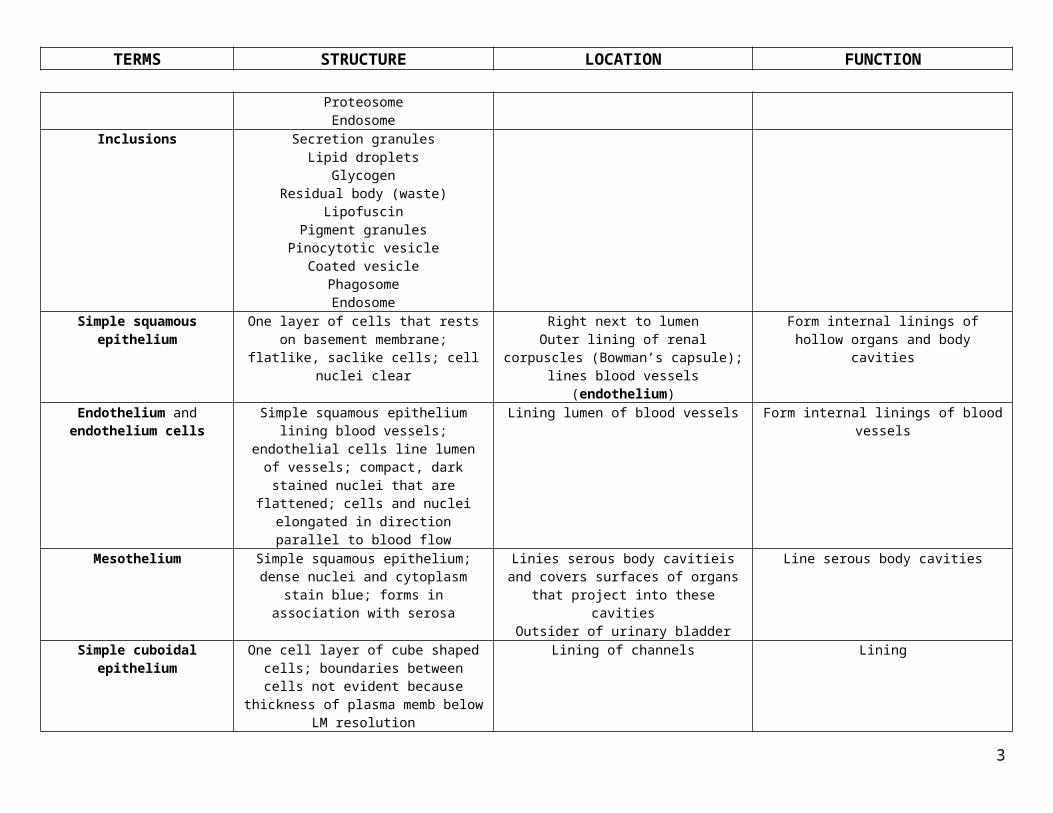
PolyribosomeProteosomeEndosome
Inclusions Secretion granulesLipid droplets
GlycogenResidual body (waste)
LipofuscinPigment granules
Pinocytotic vesicleCoated vesicle
PhagosomeEndosome
Simple squamous epithelium One layer of cells that rests on basement membrane; flatlike, saclike cells; cell nuclei
clear
Right next to lumenOuter lining of renal corpuscles (Bowman’s capsule); lines blood vessels (endothelium)
Form internal linings of hollow organs and body cavities
Endothelium and endothelium cells
Simple squamous epithelium lining blood vessels; endothelial cells line lumen of
vessels; compact, dark stained nuclei that are flattened; cells and nuclei elongated in
direction parallel to blood flow
Lining lumen of blood vessels Form internal linings of blood vessels
Mesothelium Simple squamous epithelium; dense nuclei and cytoplasm stain blue; forms in
association with serosa
Linies serous body cavitieis and covers surfaces of organs that project into these
cavitiesOutsider of urinary bladder
Line serous body cavities
Simple cuboidal epithelium One cell layer of cube shaped cells; boundaries between cells not evident because
thickness of plasma memb below LM resolution
Lining of channels Lining
Simple columnar epithelium One cell layer of column cells; apical an basal surfaces different; if cut tangentionally, cells’
outlines are polygons with circular nuclei
Lines small intestine lumen Lining
Transitional epithelium Stratified epithelium; appearance varies with Line urinary bladder Lining that allows for stretching
2
TERMS STRUCTURE LOCATION FUNCTION
stretching; basal cells are small and irregular in shape; in middle layer, cells are larger and
pear-shaped; surface cells are swollen (umbrella cells); fewer layers of cells when
sretchedStratified cuboidal epithelium Tends to only have 2 layers of cells cube
shapedLining next to lumen Lining
Stratified columnar epithelium Multiple layers of columnar cells Lining next to lumen LiningStratified squamous,
keratinized epitheliumSurperficial cells keratinized (flat cells, no
nucleus, cytoplasm replaced by keratin); cells below are irregular in shape and larger; cells
of single basal layer are cuboidal or low columnar; pyknotic layer is where cells are dying (below keratinized layer – thick pink
line)
Lining where excessive abrasion will beencountered (i.e. epidermis)
Protects against excessive abrasion
Stratified squamous non-keratinized epithelium
Multiple cell surface layers; appears to be thick sheet if cut obliquely; uniform thickness
Where free surface in body needs special protection against abrasive forces
Esophagus, parts of mouth, epiglottis, vagina
Protect against abrasive forces
Pseudostratified ciliated epithelium
One layer of cells but looks like multiple layers; all cells contact basement membrane
Lining near lumen Lining
Pseudostratified epithelium with stereocilia
Has cilia and single mucus-secreting cells Tracheal lining Lining to collect debris with mucus
Endocrine gland Secrete product into underlying CT and/or general circulation where product reaches all regions of body; ductless; vascularized and
release product on blood vessels
Pituitary gland, thyroid gland, pineal gland, parathyroid gland
Release product
Exocrine gland Release product onto surface of epithelium directly or via duct; simple (unbranched) or
compound (branched) glands
Lactating mammary glands, sebaceous glands, submandibular glands, salivary glands
Protection of epithelium, digestion, lubrication
Goblet cell Dark blue staining; mucus-secreting; part of epithelium; goblet-shape in LM; unicellular
glands
Lamina propria; part of epithelium lining small and large intestine
Secretes muocus
Mucous acinus (pl. acini) Secretory units of gland that secrete mucous (high [glycoprotein], low [protein] – high
viscosity)
Gastrointestinal system Produce mucous
Serous acinus (pl. acini) Secretory uits that secrete serous (low [glycoprotein], high [protein] – low viscosity)
Gastrointestinal system Produce serous
Serous demilune Cluster of serous cells tha surround mucous acini
In mixed gland with high proportion of serous acini
Produce both mucous and serous
Actin filaments Microfilaments; flexible, double-stranded helical polymers of actin (globular g-actin monomers polymerize into filamentous f-
Muscle cells, meshwork beneath apical plasma membrane, cores of microvilli
In nonmuscle cells – beneath plasma membrane as component of cell cortex (actin
mesh = terminal web)
3

TERMS STRUCTURE LOCATION FUNCTION
actin); 5-7 nm diameter; interact with actin-binding proteins (myosin, alpha-actinin, spectrin, fimbrin, filamen, gelsolin, talin)
Microtubules Long, rigid, hollow polymeric cylinders with diameter of 25 nm; made of tubulin
heterodimer subunits (alpha-tubulin + beta tubulin molecule); polarized with rapidly
growing plus and minus ends that depolymerize if not stabilized; minus end embedded in centromere (centrioles in its center); MTOC (microtubule organizing center) near nucleus of cell; interact with MAPS such as motor proteins (kinesin,
dynein)
Cilia, flagella; cytoskeleton Maintain cell shape and provide rigidity; control intracellular movement of organelles
and vesicles; core structures of cilia and flagella
Intermediate filaments (keratins, vimentin, desmin,
GFAP, neurofilaments, nuclear lamins)
Ropelike polymer fibers more stable tha others; 10-12 nm in diameter; associated with
neurofilaments, keratins, GFAP, desmin, vimentin, nuclear lamins (associated with
interior of nuclear envelope)
Cytoplasm, sometimes attached to cytosolic side of plasmalemma
Maintain cell shape, prvide structural support; tensile strength; protect cell from stresses and
strains
Junctional complex Three intercellular junctions = zonula occludens, zonula adherens, desmosomes;
position of JC indicates that junctions correspond to terminal bar (LM)
Intercellular junction location; between cells Intercellular communication
Terminal bar Zonula occludens, zonula adherens, desmosomes correspond to terminal bar in
LM
Junctional complex between cells See individual junction functions
Terminal web Meshwork of actin beneath apical plasma membrane of epithelial cells (aka cell cortex)
Beneath plasma membrane Cytoskeletal structure
Tight junction (zonula occludens)
Occluding junction formed by fusion of transmembrane junctional proteins in
adjacent cell membranes; forms pentalaminar structure; visualized in TEM or freeze-
fracture; form belt around cell; tight or leaky depending on complexity of interlacing
strands; principle molecules = claudins and occludins; no intercellular space nor
associated cytoskeletal element
Terminal bar; somatic capillaries as fascia occludens; urinary bladder; kidney nephron
Occluding junction; restricts movement of membrane proteins
Desmosome (macula adherens) Two dense, disk-shaped attachment plaques made of desmoplakins on cytoplasmic
surfaces of opposing cell membranes; loops of tonofilaments (cytokeratin intermed filament) insert into plaques and provide
Terminal bar; intercalalted disc; skin (keratinocytes)
Adhering juction: spot weld
4

TERMS STRUCTURE LOCATION FUNCTION
resistance to forces; 30 nm intercellular space; contains desmoglein (filamentous) and
cadherins that require Ca2+; principal cytoskeletal elements = intermediate
filaments (tonofilaments)Zonula adherens (intermediate
junction)Belt-like with intercellular space of 15-20 nm
space occupied by cadherins that bond together across gap between adjacent cells
and link to actin filaments via alpha-actinin; fascia adherens (cardiac muscle) is sheet-
like vs. belt-like; principal cytoskeletal element = cadherins
Terminal bar; intercalalted disc as fascia adherens
Adhering junction: belt (zonula) or ribbon (fascia)
Basement membrane LM term - visualized in LM with PAS or silver stain; sheet-like upon which all
epithelia rest; sieve-like barrier between epithelium and underlying CT
Under epithelia Tissue organization and cell adhesion; successful repair of damaged epithelium and
muscle and peripheral nerve regeneration
Basal lamina – lamina densa, lamina lucida (or lamina rara)
EM term – contains type IV collagen, laminin and heparan sulfate to which bases of epithelial cells are attached by binding sites (transmembrane integrins) to prot such as
lamininLamina lucida – variable thickness
Lamina densa – 20 to 110 nm in thickness
Under epithelia Tissue organization and cell adhesion; successful repair of damaged epithelium and
muscle and peripheral nerve regeneration
Lamina reticularis Reticular laminaReinforces basal lamina to underlying CT;
EC component of basement membrane composed of type III collagen (reticular
fibers) and type VII fibrils that loop into type I collagen of CT; CT cells produce reticular
lamina
Skin, areas where friction is high Protects against friction
Brush border or striated border
Microvillus borders on cell surfacesBrush border = uneven, 1-2 micromtrsStriated border = even, 0.5-1 micromtrs
Brush – proximal convoluted tubules of kidney
Striated – intestinal epithelium; alimentary tract and gall bladder
Absorption; increases surface area
Microvilli Non-motile surface projections about 1 micrometer in length; closely packed non-branching cytoplasmic processes; 2000 per cell; within MV are clusters of 20-30 actin filmanets cross-linked to each other and to memb via fimbrin and villin; basal ends of
actin itermingle withactin of terminal web in
Cell surfaces; see brush and striated border Absorption
5

TERMS STRUCTURE LOCATION FUNCTION
apical region of absorptive cellsStereocilia Longest microvilli; uneven length, branching
and often clumped together; <2000/cell; bundles of actin anchored to terminal web and extending to apex; 3-50 micromtrs in
length
Ductus epididymis, ductus deferens; inner ear Absorption; signal transduction in inner ear
Cilia Hair-like cytoplasmic extensions, anchored by basal bodies; 5-15 micromtrs length;
250/cell; axoneme: 2 central MTs surrounded by 9 doublet tubules that extend fro tip of cilium into basla body in apex of
cell
Respiratory system, oviduct, ependymal lining of brain ventricles
Movement of material in surface-parallel direction; luminal transport of mucus or
oocyte
Axoneme 2 central MTs surrounded by 9 doublet tubules that extend fro tip of cilium into basla
body in apex of cell; structure of cilia
Cilia Movement
Basal striations Basal striations – LM termBasal enfoldings – EM term
Invaginations of cell membrane on basal surface of epithelial cells usually those forming duct walls; contain ion pumps
(ATPase) and lots of mitochondria present
Striated ducts of salivary glands; proximal tubules in kidney
Foldings contain mitochondria that provide energy for active transport of sodium ions out
of cell and potassium in
Striated duct Folded membrane to increases ruface area; contains ion pups; granules; mucus and
serous travel along duct surface; path through ducts = saller intercalated ducts striated
duct; basal striations
Salivary glands, proximal tubules in kidney Foldings contain mitochondria that provide energy for active transport of sodium ions out
of cell and potassium in
Gap Junction (nexus) Disk-shaped communicating junction with 2 nm intercellular space bridged by connexons that are each composed of 6 connexins that
form hydrophilic pore to allow ion/small molecules passage; open and close with
stimulus; principal cytoskeletal element = none
Nearly all tissues (except skeletal muscle); intercalated disc; embryonic tissues
Communicating junction
Hemidesmosome Half-desmosomes with 30 nm intercellular space; principal cytoskeletal element =
keratins; principal molecules = integrins
Contact zone between certain epithelia and basal lamina (e.g. skin)
Adhering junction; add strength to attachment of epithelial cells to underlying CT via basal
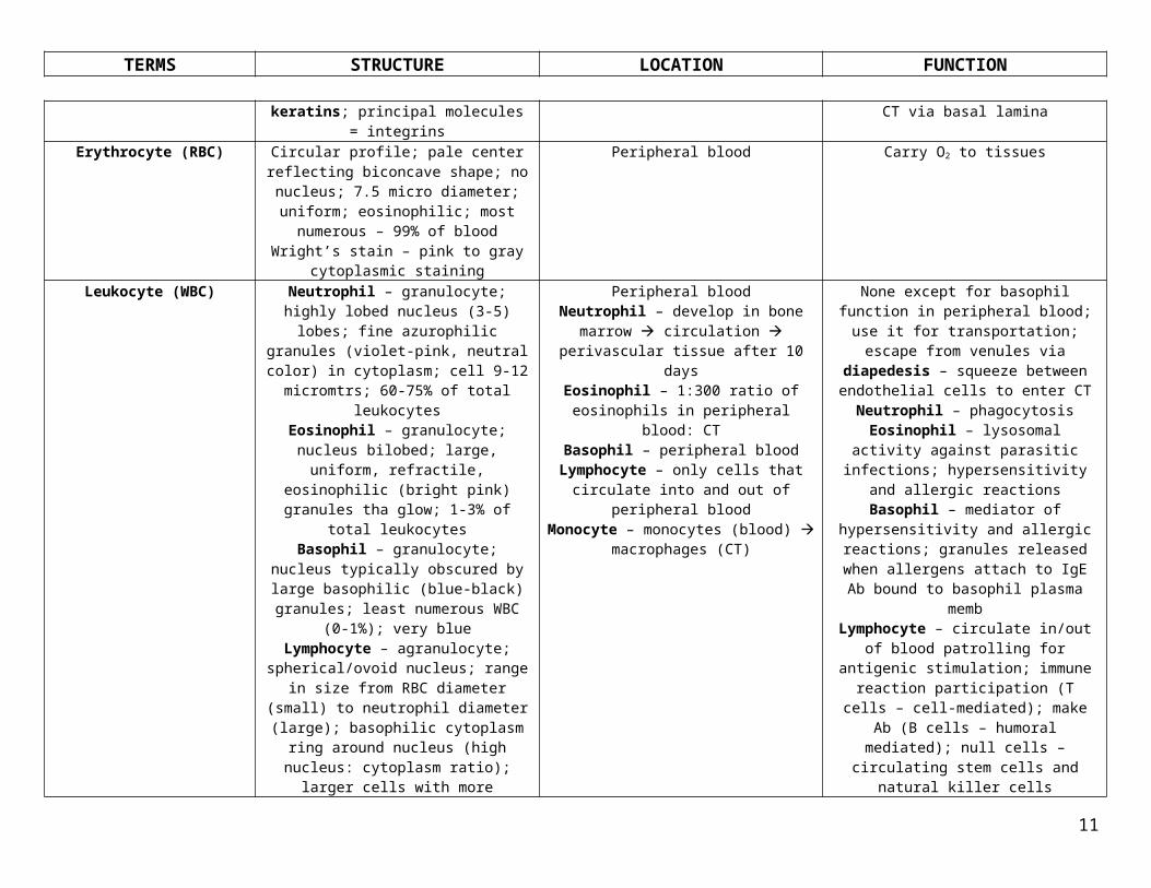
laminaErythrocyte (RBC) Circular profile; pale center reflecting
biconcave shape; no nucleus; 7.5 micro diameter; uniform; eosinophilic; most
numerous – 99% of bloodWright’s stain – pink to gray cytoplasmic
staining
Peripheral blood Carry O2 to tissues
6
TERMS STRUCTURE LOCATION FUNCTION
Leukocyte (WBC) Neutrophil – granulocyte; highly lobed nucleus (3-5) lobes; fine azurophilic granules (violet-pink, neutral color) in cytoplasm; cell 9-12 micromtrs; 60-75% of total leukocytesEosinophil – granulocyte; nucleus bilobed;
large, uniform, refractile, eosinophilic (bright pink) granules tha glow; 1-3% of total
leukocytesBasophil – granulocyte; nucleus typically obscured by large basophilic (blue-black)
granules; least numerous WBC (0-1%); very blue
Lymphocyte – agranulocyte; spherical/ovoid nucleus; range in size from RBC diameter
(small) to neutrophil diameter (large); basophilic cytoplasm ring around nucleus
(high nucleus: cytoplasm ratio); larger cells with more cytoplasm have more azuophilic
graules; 20-30% of leukocytesMonocyte – eccentrically located and
indented nucleus; largest leukocyte; 12-24 micrometers in diameter; finely clumped pale basophilic cytoplasm; may have azurophilic granules; comprise 3-8% of total leukocytes
Peripheral bloodNeutrophil – develop in bone marrow circulation perivascular tissue after 10
daysEosinophil – 1:300 ratio of eosinophils in
peripheral blood: CTBasophil – peripheral blood
Lymphocyte – only cells that circulate into and out of peripheral blood
Monocyte – monocytes (blood) macrophages (CT)
None except for basophil function in peripheral blood; use it for transportation;
escape from venules via diapedesis – squeeze between endothelial cells to enter CT
Neutrophil – phagocytosisEosinophil – lysosomal activity against parasitic infections; hypersensitivity and
allergic reactionsBasophil – mediator of hypersensitivity and allergic reactions; granules released when
allergens attach to IgE Ab bound to basophil plasma memb
Lymphocyte – circulate in/out of blood patrolling for antigenic stimulation; immune
reaction participation (T cells – cell-mediated); make Ab (B cells – humoral
mediated); null cells – circulating stem cells and natural killer cells
Monocyte – no function in peripheral blood; become macrophages in CT; mononuclear
phagocyte system
Granulocyte Leukocytes with cell-specific granulesNeutrophil, eosinophil, basophil
See neutrophil, eosinophil, basophil See neutrophil, eosinophil, basophil
Agranulocyte Leukocytes with no granulesLymphocyte, monocyte
See lymphocyte, monocyte See lymphocyte, monocyte
Polymorphonuclear neutrophil (PMN, polys)
Granulocyte; highly lobed nucleus (3-5) lobes; fine azurophilic granules (violet-pink,
neutral color) in cytoplasm; cell 9-12 micromtrs; 60-75% of total leukocytes
Develop in bone marrow circulation perivascular tissue after 10 days
Phagocytosis
Polymorphonuclear eosinophil (PME)
Granulocyte; nucleus bilobed; large, uniform, refractile, eosinophilic (bright pink) granules
tha glow; 1-3% of total leukocytes
1:300 ratio of eosinophils in peripheral blood: CT
Lysosomal activity against parasitic infections; hypersensitivity and allergic
reactions
Lymphocyte Agranulocyte; spherical/ovoid nucleus; range in size from RBC diameter (small) to
neutrophil diameter (large); basophilic cytoplasm ring around nucleus (high nucleus:
cytoplasm ratio); larger cells with more cytoplasm have more azuophilic granules;
Only cells that circulate into and out of peripheral blood
Circulate in/out of blood patrolling for antigenic stimulation; immune reaction
participation (T cells – cell-mediated); make Ab (B cells – humoral mediated); null cells – circulating stem cells and natural killer cells
7

TERMS STRUCTURE LOCATION FUNCTION
20-30% of leukocytes
Monocyte Eccentrically located and indented nucleus; largest leukocyte; 12-24 micrometers in diameter; finely clumped pale basophilic
cytoplasm; may have azurophilic granules; comprise 3-8% of total leukocytes
1:300 ratio of eosinophils in peripheral blood: CT
No function in peripheral blood; become macrophages in CT; mononuclear phagocyte
system
Platelet ThromobocytesCytoplasmic fragments of megakaryocytes in bone marrow; smallest circulating cellular
elements in blood; 2-4 micromtrs; stain granular blue and contain few azurophilic
granules; 8 day lifespan; biconvex with outer clear hyalomere (actin, myosin, 2 tubular systems) and inner granulomere (alpha,
delta, lambda granules) with basic and acidic granules containing clotting factors;
circumferential ring of 10-15 MTs (marginal bundle); glycocalyx
Peripheral blood Clotting factors and factors for tissue growth 1. Adhesion and primary aggregation
form platelug plug2. Secondary Aggregation
3. Blood coagulation, forming a thumbus
4. Clot retraction5. Clot removal
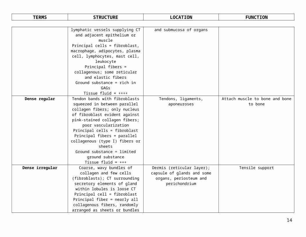
Loose (areolar) Cells interspersed singly or in small groups among fine collagen fibers of ECM; lots of small blood vessels and lymphatic vessels supplying CT and adjacent epithelium or
musclePrincipal cells = fibroblast, macrophage,
adipocytes, plasma cell, lymphocytes, mast cell, leukocyte
Principal fibers = collagenous; some reticular and elastic fibers
Ground substance = rich in GAGsTissue fluid = ++++
Under luminal epithelium and above cartilageSubcutaneous tissue, mesenteries, lamina
propria and submucosa of organs
Support, anchor, and padding of tissues lipid storage
Dense regular Tendon bands with fibroblasts squeezed in between parallel collagen fibers; only nucleus
of fibroblast evident against pink-stained collagen fibers; poor vascularization
Principal cells = fibroblastPrincipal fibers = parallel collagenous (type I)
fibers or sheetsGround substance = limited ground substance
Tissue fluid = +++
Tendons, ligaments, aponeuroses Attach muscle to bone and bone to bone
Dense irregular Coarse, wavy bundles of collagen and few cells (fibroblasts); CT surrounding secretory
Dermis (reticular layer); capsule of glands and some organs, periosteum and
Tensile support
8

TERMS STRUCTURE LOCATION FUNCTION
elements of gland within lobules is loose CTPrincipal cell = fibroblast
Principal fiber = nearly all collagenous fibers, randomly arranged as sheets or bundles
Ground substance = limitedTissue fluid = +++
perichondrium
Reticular tissue Fibroblasts part of scaffolding (stroma) of organ and are called reticular cells; loosely
arranged fiber sponge filled with parenchymal cells ; cells very densely packed
and can’t be steen with fiber stainPrincipal cells = reticular cell (fibroblast)
Principal fibers = network of reticular fibersGround substance = considerable GS
Tissue fluid = ++++
Storma of glands, lymphoid organs and bone marrow
Flexible, supporting framework
White adipose Fat cells cluster in lobules bound by fibrous septa and individually held in place by
delicate reticular fibers; unilocular, large cells; nucleus is flattened semilunar structure
at side of cell; inside is unstained since standard tissue prep removes lipids
Principal cells = unilocular adipocytePrincipal fibers = network of reticular fibers
Ground substance = scanty GSTissue fluid = ++
Subcutaneous hypodermis, peri-renal issue fat deposits
Energy storage, padding, insulation against temperature
Brown adipose Multilocular; contains many mitochondria with transmemb protein in inner mitoch
membrane (thermogenin = UCP1 – releases energy as heat); persist in adult but small
droplets coalesce into single droplet so morph like unilocular cells
Principal cells = multilocular adipocytePrincipal fibers = network of reticular fibers
Ground substance = scanty GSTissue fluid = ++
Interscapular and ingual regions, esp in infant Thermogenic
Adipocyte (fat cell) – unilocular (white) and multilocular (brown)
Precurso for both = mesenchymal cellWhite/unilocular – large cells; nucleus is flattened semilunar structure at side of cell;
inside is unstained since standard tissue prep removes lipids
Brown/multilocular – contains many mitochondria with transmemb protein in
White – Subcutaneous hypodermis, peri-renal issue fat deposits
Brown – Interscapular and ingual regions, esp in infant
White – energy reservoirBrown - thermoregulation
9

TERMS STRUCTURE LOCATION FUNCTION
inner mitoch membrane (thermogenin = UCP1 – releases energy as heat); persist in adult but small droplets coalesce into single
droplet so morph like unilocular cellsNeutrophil Precursor = hematopoietic stem cell in bone
marrowMost abusndant granulocyte; aka
microphage in CT; lysosomal and Ab granules (azurophilic, specific, tertiary)
Peripheral blood; in tissues during acute phase of infection
Defense; phagocytosis of foreign substances, bacteria
Fibroblast; fibrocyte Precursor = mesenchymal cellFusiform shape with thin protoplasmic
processes that contact other cells; mitotic division; fibrocyte = fibroblasts in
established CT
In CT Produces fibers and amorphous ground substance (GAGs, proteoglycans); structural
function
Lymphocyte Precursor = lymphocyte precursor and immunocompetent lymphocytes
B-cell – humoral immunityT-cell – cell-mediated immunity
Numerous in CT only inpathological states; normal accumulations of diffuse lymphoid
tissue (MALT); patrolling tissue
Immunosurveillance; generates immunocompetent cells for B and T cells
Mesenchymal cell Mesenchyme tissueMesenchyme-derived cells = osteoblasts,
adipocytes, chondroblast, fibroblasts, endothelial cels
Principal fibers = few primitive collagenous fibrils
Ground substance = amorphousTissue fluid = +++++
Embryo and early fetus, surrounding developing organs
Undifferentiated cels present in adult CT for repair or growth
Precursor of nearly all CT
Macrophage Aka histiocytePrecursor = monocyte, from bone marrow
precursorsCirculate in blood as monocytes and after 40 hours move to CT and become macrophages;
capable of cell division; either attaché dto matrix fibers (fixed macrophages) or motile (wandering macrophages); serve as APC (antigen-presenting cells); Mononuclear Phagocyte System = monocytes that take
residence in specific organs and have specific names
Areolar CTBone marrow, spleen, thymus, lymph node
Defense; clean-up; antigen presentation; phagocytosis of foreign substances, cell
debris, bacteria; secretes cytokines
Mast cell Precursor = unknown progenitor cellLarge cell with oval nucleus and
metachromatic granules; long lived; related to basophils because of staining but are larger
Along arterial side of small blood vessels Inflammation; releases pharmacologically active substances in inflammation
10
TERMS STRUCTURE LOCATION FUNCTION
than them; rapid degranulation via compound exocytosis; release leukotrienes
(agents of inflammation)Eosinophil Precursor = hematopoietic stem cell in bone
marrowTerminally differentiated granulocyte; in blood and CT; contains lysosomes and
specific granules high in peroxidase content
Peripheral blood and CT Phagocytosis of antigen-antibody complex; kill and phagocytose parasites; defense
Plasma cell Precursor = B-lymphocyteEnd-stage cell with short life span; no
secretion granules b/c Ab release is constitutive
Lymph node, spleen, areolar CT, NOT in blood stream
Produces and secretes Ab
Proerythroblast (pronormoblast)
RNP (methylene blue) (cytoplasm) + eosin staining (Hb)
12-20 micromtrs diameter; intensely basophilic cytoplasm (much brighter blue); round nucleus with fine chromatin and 3-5 pale gray nucleoli (pale spots in Wright’s
stain); few or none cytoplasmic granules; 4%
Bone marrow Proerythroblast basophilic erythroblast polychromatophilic erythrobast
orthochromatophilic erythroblast reticulocyte erythrocyte
Basophilic erythroblast (b. normoblast)
RNP (methylene blue) (cytoplasm) + eosin staining (Hb)
Daughter cell of proerythroblast; nucleoli not visible; nuclear chromatin in discrete clumps;
cytoplasm intensely basophilic; fine chromatin clumps; 10-15 micromtrs; nucleus looks cracked; nucleolus looks like plae spot; uniform cytoplasm without granules; 1-4%
Bone marrow Proerythroblast basophilic erythroblast polychromatophilic erythrobast
orthochromatophilic erythroblast reticulocyte erythrocyte
Polychormatophilic erythroblast (p. normoblast)
RNP (methylene blue) (cytoplasm) + eosin staining (Hb)
Daughter of basophilic erythroblast; nucleoli not visible; cytoplasm gray-blue, chromatin clumps very coarse; 8-15 micromtrs; smaller than preceding cells; cytoplasm color blothy
and variable; 12-22%
Bone marrow Proerythroblast basophilic erythroblast polychromatophilic erythrobast
orthochromatophilic erythroblast reticulocyte erythrocyte
Orthochromatophilic erythroblast (o. normoblast)
RNP (methylene blue) (cytoplasm) + eosin staining (Hb)
Daughter of polychromatophilic erythroblast; nucleoli not visible; cytoplasm orange-gray
(color of reticulocytes), small dense nucleus, 8-12 micromtrs; not able to divide;
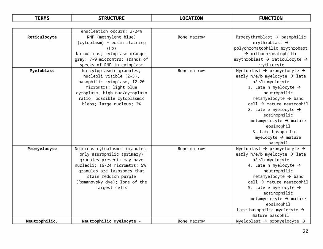
enucleation occurs; 2-24%
Bone marrow Proerythroblast basophilic erythroblast polychromatophilic erythrobast
orthochromatophilic erythroblast reticulocyte erythrocyte
Reticulocyte RNP (methylene blue) (cytoplasm) + eosin Bone marrow Proerythroblast basophilic erythroblast
11

TERMS STRUCTURE LOCATION FUNCTION
staining (Hb)No nucleus; cytoplasm orange-gray; 7-9 micromtrs; srands of specks of RNP in
cytoplasm
polychromatophilic erythrobast orthochromatophilic erythroblast
reticulocyte erythrocyte
Myeloblast No cytoplasmic granules; nucleoli visible (2-5), basophilic cytoplasm, 12-20 micromtrs; light blue cytoplasm, high nuc/cytoplasm ratio, possible cytoplasmic blebs; large
nucleus; 2%
Bone marrow Myeloblast promyelocyte early n/e/b myelocyte late n/e/b myelocyte1. Late n myelocyte neutrophilic
metamyelocyte band cell mature neutrophil
2. Late e myelocyte eosinophilic metamyelocyte mature eosinophil
3. Late basophilic myelocyte mature basophil
Promyelocyte Numerous cytoplasmic granules; only azurophilic (primary) granules present; may
have nucleoli; 16-24 micromtrs; 5%; granules are lysosomes that stain reddish purple
(Romanovsky dye); lone of the largest cells
Bone marrow Myeloblast promyelocyte early n/e/b myelocyte late n/e/b myelocyte4. Late n myelocyte neutrophilic
metamyelocyte band cell mature neutrophil
5. Late e myelocyte eosinophilic metamyelocyte mature eosinophil
Late basophilic myelocyte mature basophil
Neutrophilic, eosinophilic, or basophilic myelocyte
Neutrophilic myelocyte – numerous granules; azurophilic (primary) and specific (secondary) graules; nucleus oal or slightly indented; 8-18 micromtrs; 12%; clear pale
area in Golgi regionEosinophilic myelocyte – pink-stained
granules in Golgi region; specific granules eosinophilic (large, red-orange); nucleus oval
or slightly indented; 1.5%Basophilic myelocyte – specific granules
basophilic (deeply blue-black); nucleus oval or slightly indented; 0.3%
Bone marrow Myeloblast promyelocyte early n/e/b myelocyte late n/e/b myelocyte6. Late n myelocyte neutrophilic
metamyelocyte band cell mature neutrophil
7. Late e myelocyte eosinophilic metamyelocyte mature eosinophil
Late basophilic myelocyte mature basophil
Neutrophilic, eosinophilic, or basophilic metamyelocyte
Neutrophilic metamyelocyte – secondary granules; nucleus deeply indented (C-shape);
10-14 micromtrs; 22%Eosinophilic metamyelocyte – secondary
granules red-orange and large; nucleus deeply indented; 2%
No basophilic metamyelocyteBasophil – specific secondary blue-black
Bone marrow Myeloblast promyelocyte early n/e/b myelocyte late n/e/b myelocyte8. Late n myelocyte neutrophilic
metamyelocyte band cell mature neutrophil
9. Late e myelocyte eosinophilic metamyelocyte mature eosinophil
Late basophilic myelocyte mature
12

TERMS STRUCTURE LOCATION FUNCTION
graules; nucleus deeply indented or segmented
basophil
Neutrophilic band stage (stab) Specific granules (secondary); nucleus U-shape with some constriction, 10-12
micromtrs; horse-shoe shaped nucleus
Bone marrow Myeloblast promyelocyte early n/e/b myelocyte late n/e/b myelocyte10. Late n myelocyte neutrophilic
metamyelocyte band cell mature neutrophil
11. Late e myelocyte eosinophilic metamyelocyte mature eosinophil
Late basophilic myelocyte mature basophil
Megakaryocyte Large size; 3.5 times neutrophil diameter; abundant pink cytoplas and convoluted
nucleus that is polyploidy; 50-100 micromtrs in diameter; multilobed nucleus with 64 times
normal haploid number of chromosomes
Bone marrow; adjacent to abluminal side of blood sinusoids so that it can release platelets
into blood
Produce platelets
Bone marrow; red marrow and yellow marrow
Red marrow – marrow with mostly parenchymal cells; dominant at birth; at
proximal quarters of long bones by adulthoodYellow marrow – marrow with mostly white fat cells; arrives at age 4; dominant in adults; begins in middle of shafts of long bones at
age 10-14 and extends gradually
Bone Conversion from red to yellow is reversible if body needs greater blood-forming capacity
Chondrocyte Mature cells of cartilage dispersed throughout solid cartrige matrix in lacuna; occur in
clusters (isogenous group)
Lacuna of cartilage Make cartilage
Lacuna Spherical ovoid hole in matrix in which chondrocyte resides; surrounded by gel-like,
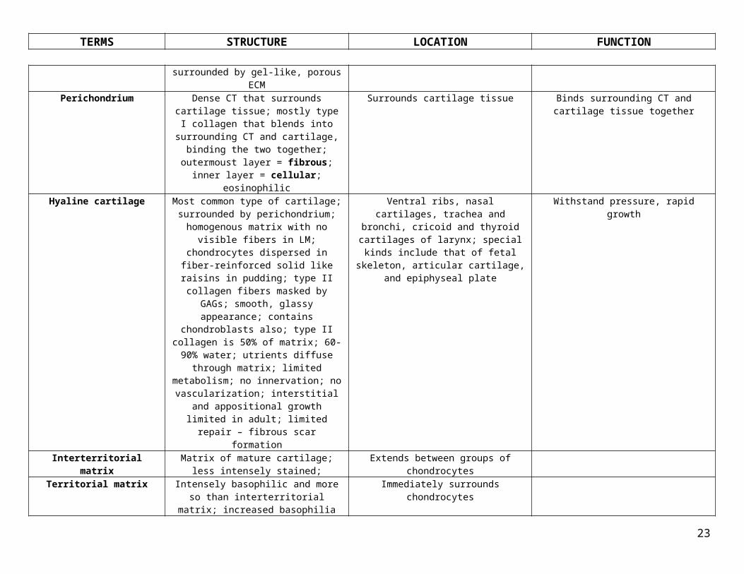
porous ECM
ECM of cartilage House chondrocytes
Perichondrium Dense CT that surrounds cartilage tissue; mostly type I collagen that blends into
surrounding CT and cartilage, binding the two together; outermoust layer = fibrous;
inner layer = cellular; eosinophilic
Surrounds cartilage tissue Binds surrounding CT and cartilage tissue together
Hyaline cartilage Most common type of cartilage; surrounded by perichondrium; homogenous matrix with
no visible fibers in LM; chondrocytes dispersed in fiber-reinforced solid like raisins in pudding; type II collagen fibers masked by GAGs; smooth, glassy appearance; contains
chondroblasts also; type II collagen is 50% of
Ventral ribs, nasal cartilages, trachea and bronchi, cricoid and thyroid cartilages of larynx; special kinds include that of fetal
skeleton, articular cartilage, and epiphyseal plate
Withstand pressure, rapid growth
13
TERMS STRUCTURE LOCATION FUNCTION
matrix; 60-90% water; utrients diffuse through matrix; limited metabolism; no
innervation; no vascularization; interstitial and appositional growth limited in adult; limited repair – fibrous scar formation
Interterritorial matrix Matrix of mature cartilage; less intensely stained;
Extends between groups of chondrocytes
Territorial matrix Intensely basophilic and more so than interterritorial matrix; increased basophilia
due to concentrations of GAGs and few collagen fibers since type II fibers not visible
in matrix
Immediately surrounds chondrocytes
Isogenous group Daughter cells of chondroblasts; clusters of chondrocytes
In lacuna Secrete small amount of matrix
Articular cartilage Layer of hyaline cartilage that covers the ends of bones that face joint cavities;
superficial layer is eosinophilic because of type II collagen fibrils within matrix in the outer region of cartilage; flat chondrocytes; deeper chondrocytes are in ertical columns b/c of radially penetrating collagen fibril
bundles
Ends of bones that face joint cavities; free surface of bone
Withstand pressure, rapid growth
Tidemark Basophilic line that marks interface between regular non-mineralized cartilage and deeper region of mineralized cartilage matrix next to
bone
Articular cartilage Mineralized region anchors articular cartilage to bone
Elastic cartilage Yellowish when fresh; elastic fibers in matrix; elastic stain (Verhoeff’s) needed to
visualize fibers; consists of cells, type II collagen fibrils, ground substance; elastic fiber meshwork; perichondrium present
External ear, Eustaachian tube, epiglottis, small cartilages in larynx; occurs where
support with additional flexibility is required
Provides elastic support
Fibrous cartilage FibrocartilageIntermediate in form between hyaline
cartilage and dense CT; prominent type I collagen fibers in matrix; NO perichondrium;
very flat fibroblasts; sparse, ovoid chondrocytes arranged singly, in groups, or in
rows
Insertion of tendon into bone; at insertion of certain ligaments and tendons into bone and
in articular disks (menisci)
Provides tough support and tensile strength
Osteocyte Completely surrounded by bone; derived from osteoblasts; exist in lacunae that are
interconnected with each other and Haversian canal via canaliculi (channels); contact each
Bone Produce osteoid; resorption (osteocytic osteolysis); mechanical sensor cells of bone –
sense deformation; death followed by bone breakdwon
14

TERMS STRUCTURE LOCATION FUNCTION
other via gap junctions at ends of processesOsteoblast Aligned on bone surfaces, one-cell thick in
sheets resembling simple cuboidal/columnar epithelium; interconnected with gap
junctions; basophilic cytoplasm; no mitotic division; membrane receptors for hormones
and cytokines
Bone surfaces; on irregular surfaces within the wall and bordering the marrow cavity
Bone formation; growth via apposition (NO interstitial growth b/c calcified bone can’t expand); synthesize and lay down osteoid (mineralizes to become bone; stains pale);
when Ca2+ levels drop, parathormone release suppresses bone deposition and removes
osteoid in vicinity of osteoclastsOsteoclast Large multinucleated cells specialized for
bone resorption (breakdown); polarized with 3 zones:
Ruffled border – face of osteoclast adjacent to bone
Clear zone – sourrounds ruffled border encircling osteoclast; devoid of organelles;
rich in actin; integrins in plasmalemmaBasal zone – anti-resorptive surface – plasmalemma on side away from ruffled
border; contains Cl-/HCO3- anion exchange
channels
Found in Howship’s lacunae or resorption tunnels within bone; outer surface of
developing diaphysis beneath periosteum
Ruffled border – principal activity (bone resorption
Clear zone – allows osteoclasts to seal to bone and create tiny subosteoclastic space between them and bone; proton pumps in
ruffled border pump H+ tino space, acidify it, dissolving inorganic matrix component; enzymes released onto bone surface and
degrade matrixBasal zone – channels maintain proper
intracellular ionic balance
Howship’s lacuna Subosteoclastic compartments containing osteoclasts
Bone
Osteoprogenitor cell Partially undifferentiated stem cells; mitotic division; transform into osteoblasts under low
O2 tension
In cellular periosteum layer, within bone marrow next to endosteum, and lining
Haversian canals
Differentiation
Bone lining cell Simple squamous cells; quiescent osteoblasts; connected through gap junctions to other
BLCs and osteocytes; ion-barrier separating fluids; incapable of dividing; willl become osteoblasts as required since not actively
synthesizing matrix
On all bone surfaces Mineral homeostasis; become osteoblasts
Cancellous bone (spongy, rabecular bone)
MACROSCOPIC term; 3-D lattice of interconnected rods, plates, arches with many
open spaces
Bone interior; inside epiphysis or head of long bones
Thin layer arranged so that compressive forces on bone surface dispersed amongst
infrastructure; keeps bone strong when with thick compact bone layer
Compact bone (cortical bone) MACROSCOPIC term; Lacks large spaces and trabecula; solid except for minute canals
coursing through it; 50% organic matter (collage, GAGs, bound H2O) and 50%
mineral (calcium hydroxyapatite)
Bone exterior
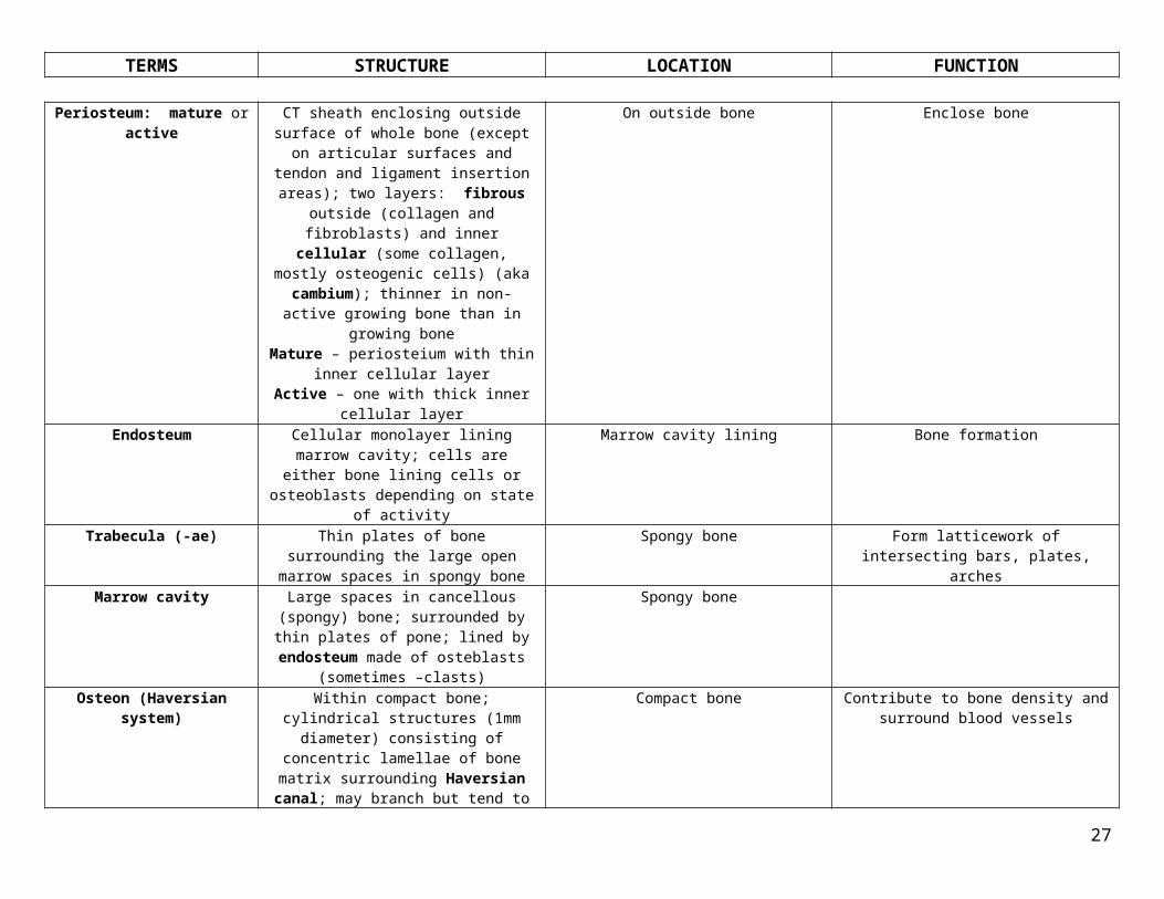
Periosteum: mature or active CT sheath enclosing outside surface of whole bone (except on articular surfaces and tendon
On outside bone Enclose bone
15

TERMS STRUCTURE LOCATION FUNCTION
and ligament insertion areas); two layers: fibrous outside (collagen and fibroblasts) and
inner cellular (some collagen, mostly osteogenic cells) (aka cambium); thinner in
non-active growing bone than in growing bone
Mature – periosteium with thin inner cellular layer
Active – one with thick inner cellular layerEndosteum Cellular monolayer lining marrow cavity;
cells are either bone lining cells or osteoblasts depending on state of activity
Marrow cavity lining Bone formation
Trabecula (-ae) Thin plates of bone surrounding the large open marrow spaces in spongy bone
Spongy bone Form latticework of intersecting bars, plates, arches
Marrow cavity Large spaces in cancellous (spongy) bone; surrounded by thin plates of pone; lined by
endosteum made of osteblasts (sometimes –clasts)
Spongy bone
Osteon (Haversian system) Within compact bone; cylindrical structures (1mm diameter) consisting of concentric
lamellae of bone matrix surrounding Haversian canal; may branch but tend to run
longitudinal in long bones
Compact bone Contribute to bone density and surround blood vessels
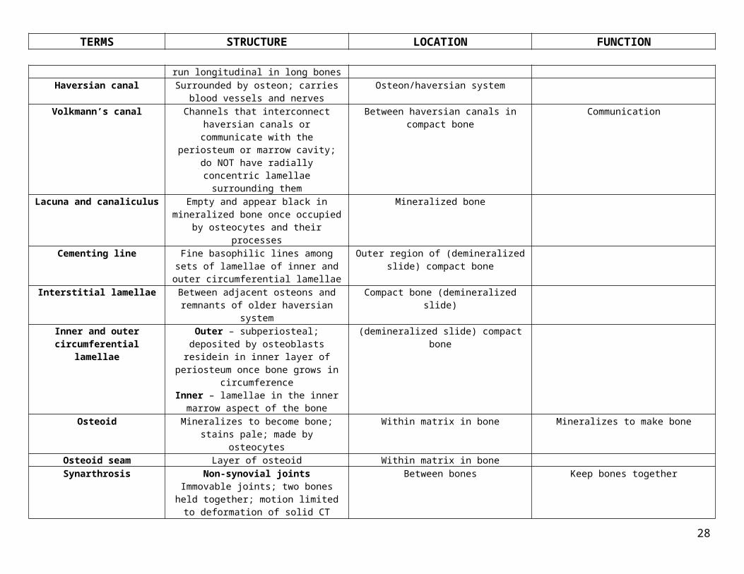
Haversian canal Surrounded by osteon; carries blood vessels and nerves
Osteon/haversian system
Volkmann’s canal Channels that interconnect haversian canals or communicate with the periosteum or marrow cavity; do NOT have radially concentric lamellae surrounding them
Between haversian canals in compact bone Communication
Lacuna and canaliculus Empty and appear black in mineralized bone once occupied by osteocytes and their
processes
Mineralized bone
Cementing line Fine basophilic lines among sets of lamellae of inner and outer circumferential lamellae
Outer region of (demineralized slide) compact bone
Interstitial lamellae Between adjacent osteons and remnants of older haversian system
Compact bone (demineralized slide)
Inner and outer circumferential lamellae
Outer – subperiosteal; deposited by osteoblasts residein in inner layer of
periosteum once bone grows in circumference
Inner – lamellae in the inner marrow aspect of the bone
(demineralized slide) compact bone
16

TERMS STRUCTURE LOCATION FUNCTION
Osteoid Mineralizes to become bone; stains pale; made by osteocytes
Within matrix in bone Mineralizes to make bone
Osteoid seam Layer of osteoid Within matrix in boneSynarthrosis Non-synovial joints
Immovable joints; two bones held together; motion limited to deformation of solid CT
between 2 bones
Between bones Keep bones together
Syndesmosis Fibrous joints2 bones held together by CT
Sutures (union of skull flat bones), gomphosis (attachment of teeth to alveoli via
periodontal ligament, joint between distal tibia and fibula) – peg in pocket
Hold bones together
Synchondrosis Cartilaginous jointsBones held together by cartilage
Hyaline cartilage holding growth plates; symphysis (articulating bones capped with cartilage – symphysis pubis and annulus
fibrosus)
Hold bones together
Synostosis Bones held together by bone Between epiphysis and diaphysis after growth plate fused
Hold bones together
Diarthrosis Synovial jointsJoint cavity; caps of hyaline cartilage
persisting on articulating bone surfaces; two-layered capsule surrounding articulating
bones outer fibrous layer (dense fibrous CT) and inner synovial membrane (lines fibrous capsule and covers everything but
articular cartilages); synovial fluid to nourish joint capsule structures (viscoelastic)
Articulating ends of bones or cartilages Motion occurs as articulating ends of bones (or cartilages) move relative to one another; motion limited by geometry of articulating ends, by fibrous capsule surrounding joint,
and by ligaments, tendons, muscles crossing joint
Synovial membrane Lines fibrous capsule and covers everything but articular cartilage; has cellular intima
and deeper fibrovascular subintimal lamina
Lines non-articular surfaces of joints, bursae, tendon sheaths in joint cavities
Surrounds articular cartilage
Cellular intima Consists of synoviocytes in amorphous fiber-free intercellular matrix; synoviocytes form
interlacing, discontinuous layer 1-4 cell layers thick; no intercellular junctions; no
basement membrane; cellular intima is NOT an epithelium
Synovial membrane Type A synoviocytes – remove debris from joint cavity
Type B – synthesize synovial fluid component
A and B synoviocytes Type A – resemble macrophages; precursors = bone-marrow derived monocytes
Type B – resemble fibroblasts, synthesize type III collagen and hyaluronic acid
Cellular intima of synovial membrane Type A – remove debris from joint cavity; antigen-presenting cells that stimulate
immune responses to foreigners in joint cavity; control chondrocyte function and
blood flow in synovial membrane by releasing vasodilating and vasoconstricting
agents
17

TERMS STRUCTURE LOCATION FUNCTION
Type B – synthesize synovial fluid componenSubintimal lamina CT containing collagen, areolar, or adipose
tissue; deepr fibrovascular layerSynovial membrane Surround articular cartilage
Joint (articular) cavity Contains joint; feature of diarthroses (synovial joints)
Synovial joints
Joint capsule Feature of diarthroses (synovial joints) Synovial jointsSynovial fluid Clear, pale yellow, viscous; slightly alkaline;
contains filtrate of blood plasma and hyaluronate; low volume; varies at each joint
Synovial joints (diarthroses) Lubricates and nourishes structures within joint capsule
Primary center of ossification Where ossification – endochondral bone formation – in long bones initiates
Shaft of diaphysis (long bone) Initiate bone formation
Secondary center of ossification
Where ossification in end of long bone begins Within each epiphysis (end of a long bone that is initially separated by the main bone by cartilage which is then replaced by bone via
ossification); ossification in ends (long bones) or projections (vertebrae)
Postnatal event of ossification; initiate bone formation
Primary spongiosum Immature bone with irregular eosinophilic ring of bone surrounded by periosteum;
neither trabecular or compact bone; actively growing bone
Endochondral bone formation – in long bones; in immature bones
Part of endochondral formation
Epiphyseal plate (growth plate)
Plate of cartilage remaining between primary and secondary centers that allows long bone
to grow in lengthZone of resting cartilage
Zone of proliferationZone of maturation and hypertrophy
Zone of calcifying cartilage and degenerating chondrocytes
Zone of resorption and ossification
Between primary and secondary ossification centers
Allows for elongation of long bones to procee from this point via interstitial growth
(cartilage proliferation) within the growth plate and endochondral bone deposited on diaphyseal side of plate; ossifies after final growth attained, thus united epiphysis and
diaphysis
Matrix vesicles
Bone collar (mid-diaphyseal ring)
Intramembranous ossification; periosteal bone collar; osteoblasts create this bony matrix lining on the calcified cartilage;
increases in thickness and length
Outside of bone; periosteum location Bony matrix that allows for vascular CT from its periosteum to invade calcified cartilage; osteoblasts lay down bone on remnants of
calcified cartilageThe zones of the epiphyseal
plateZone of resting cartilage – hyaline cartilage
with slight slow growthZone of proliferation – active division of
chondrocytes with arrangement into columns oriented along long axis of cartilage modelZone of maturation and hypertrophy –
Epiphyseal plate Elongation of long bones
18

TERMS STRUCTURE LOCATION FUNCTION
cells hypertrophy and begin changes that lead to calcification of matrix
Zone of calcifying cartilage and degenerating chondrocytes – vascular
primary marrow will invade spaces left after matrix removed between successive cells; chondrocytes undergo apoptosis and are
removedZone of resorption and ossification – osteoblasts deposit bone on remaining delicate columns of calcified cartilage
Resorption tunnel or cavity Cavity created by osteoclasts; hole drilled into bone during remodeling
Old woven bone that is being replaced by new lamellar bone
Bone remodeling
Cutting cone Formed by osteoclasts during bone remodeling
Old woven bone that is being replaced by new lamellar bone
Bone remodeling
Callus Formed by initial repair of a fracture; developed in immature bone; formed by woven bone to compensate for weakness
Fracture repaired bone Fracture repair; formed by woven bone to strengthen point of injury
Woven pone (primary, immature bone)
MICROSCOPIC TERM; more cellular, more ground substance and less collagen; grows more rapidly/resorbs more rapidly but less
completely
First bone to appear during new bone formation or in fracture repair; immature
bone
Lamellar bone (secondary, mature bone)
MICROSCOPIC TERM; less cellular, less ground substance and more collagen; grows more slowly and resorbs slowly; mineralizes
slowly but more completely
Stronger, more superior bone that replaces woven bone
Spinal cord Shrinkage spaces if fixation by immersion; part of CNS; contains somatic motor neurons in grey matter; enclosed by protective bone
Spine Cell bodies in grey matter (anterior or ventral horn); myelinated axons in white matter
(stains blue); sensory inputs, motor outputsWhite matter Contains myelinated axons; stains blue; part
of spinal cordSpine Contain axons
Grey matter H-shaped core that contains cell bodies (nuclei = cell bodies); in spinal cord; also
known as anterior horn and contains anterior horn cells, somatic and lower motor neurons; is the neuropil – intermingled dendrites and
axons of neuroglial cells
Spine Contain cell bodies (soma)
Neuroglial (glial) cell Supporting cells found ONLY in CNS; cytoplasm not visible; small nuclei in
surrounding nueurpil
Gray matter of spinal cord Oligodendrocyte – forms myelin of CNSMicroglial cell – macrophage of mononuclear phagocyte system
Astrocyte – supportive functionsNeuropil Feltwork of intermingled dendrites and axons Gray matter of spinal cord Contain neuroglial cell processes
19
TERMS STRUCTURE LOCATION FUNCTION
from neurons and processes of neuroglial cells; surrounds anterior horn cells
Dorsal (posterior) root Spinal ganglionAxons entering CNS at level of spinal cordNerve fiber bundle that protrudes from both ends of spinal cord in the back; two dorsal
roots; large cell bodies belonging to primary sensory neurons (unipolar with Nissl
substance in perikaryon; large euchromatic nucleus and prominent nucleolus); satellite
cells around each neuron
Outside of white matter; in the back Sensory receptors primary sensory neuron DRG
Conveys sensation to CNS
Ventral (anterior) root Axons exiting CNS at spinal cordConsists of motor fibers outside and in front of the white matter; large nerve bundle made of myelinated axons originating from lower
motor neurons in anterior horn of spinal cord; axon has grayish core with thick, pink, frothy
myelin (frothy because of lipid extraction during fixation); also contain axons of
autonomic (visceral) motor system
Outside of white matter; in the front Innervate striated muscle via neuromuscular junctions; conveys motor/impulses to muscle
Spinal nerve Nerves that arise in pairs from spinal cord; each attached to cord by ventral or dorsal
roots; 32 pairsCranial nerves – axons directly
exiting/entering brain = cranial nerves
Within the spinal cord Contain nerves
Anterior horn cell (lower motor neuron)
(Aka somatic motor neurons)smaller neuronal cell bodies entirely confined
to CNS; multipolar cells (several dendrites coming out of cell body); round euchormatic nuclei with prominent nucleolus; violet Nissl
bodies in cytoplasm surrounding nucleus (clumps of RER); axon leaves spinal cord in
anterior root
Anterior horn of spinal cord Axon leaving spinal cord innervates skeletal muscle
Nissl substance (Nissl body) Violet colored substances in cytoplasm surrounding nucleus and extending into
dendrite tips (clumps of RER); basophilic in LM; decreasing amount as you get closer to
dendrite bases
Cell body of neuron; in cytoplasm surrounding nucleus
RER function; protein synthesis
Axon, myelinated and unmyelinated
Myelinated – fatty insulating sheath; thick, pink, frothy form fixation; frothy from lipid
extraction; supporting cells wrap plasma membranes around axon in spiral fashion;
On axons in CNS and PNS – myelinatedNerve fibers in bundles between the ganglia –
unmyelinated
Faster transduction of action potential
20

TERMS STRUCTURE LOCATION FUNCTION
myelin constructed in internodal length segments; internal (inner) and external
(outer) mesaxon (seen in EM); major dense lines (form inner cytoplasmic surfaces)
separated by intraperiod lines (formed by apposed external surfaces)
Unmyelinated – CNS axons not associated with mylin; axons in PNS enveloped by
Schwann cells but do NOT have characteristic spiral wrapping; severe
unmyelinated axons are enclosed by one Schwann cell
Oligodendrocyte Neuroglial cell (supporting cells of CNS only); small nuclei, no visible cytoplasm
Neuropil of gray matter surrounding anterior horn cells
Produces myelin sheaths for CNS
Schwann cell Neuroglial cell (supporting cells of CNS only); small nuclei, no visible cytoplasm
Neuropil of gray matter surrounding anterior horn cells
Produces myelin for PNS
Mesaxon, internal and external Internal – innermost layer of paired membrane spiral of myelin sheath; forms
major dense lines in EM prepExternal – outer layer of paired membrane spiral; forms intraperiod lines that separate
dense lines
Myelin sheath around axons Aid in myelin formation
Node of Ranvier Interruptions in tubular myelin sheath where adjacent Schwann cells meet; 4 bubbles of
unmyelinated region; eosinophilic, concentric pattern
Axon; between myelin sheaths Salutatory conduction; faster transduction of action potential
Internode (internodal length) Part of nerve fiber between two nodes of Ranvier; how myelin is constructed
Between two nodes of Ranvier One Schwann per internodal lengthOne oligodendrocyte per many internodes
Schmidt-Lanterman cleft Bubbles/disruptions in myelin wrappings (2 bubbles = cleft vs. 4 bubbles that = node); columns of cytoplasm extend from bottom
cell to top axon and cytoplasmic strip – way to maintain connection between axon and cell; 2 bubbles b/c of way sectioning was
done
Discruptions in myelin wrappings Maintain connection between axon and cell
Peripheral nerve Nerve outside of CNS; nerves entering/leaving brain (cell bodies in CNS); ensheathed; organized by CT; is an organ
with structural arrangement of nerve fibers, associated CT, and cellular envelopes;
myelinated transvere nerves are bluish-grey central spots within myelin sheaths; myelin is
Everything outside of brain and spinal cord Ganglia = cell groups (NOT gray matter/nuclei in PNS)
21

TERMS STRUCTURE LOCATION FUNCTION
lightly stained pink collars with an occasional associated Schwann cell nucleus; consists of endoneurium, perineurium, epineurium
(CT investments)Endoneurium Type III collagen fibers between myelin
sheaths; NOT visible in EM; reticular fibers surrounding individual nerve fibers
Interior of peripheral nerve CT
Perineurium Collagen fibers with squamous cells; fiber-reinforced cellular layer
Outside of endoneurium in peripheral nerve CT
Epineurium Dense CT sheath Surrounding peripheral nerve CTGanglion (one of two classes) Collection of neuronal cell bodies located
outside CNS; (nucleus = collection of neuronal cell bodies inside CNS);
Dorsal (spinal) root ganglia– in PNS, contains cell bodies carrying sensory signals
to CNSAutonomic ganglia – contain cell bodies relaying signals to visceral motor system
In PNS Dorsal – sensory functionAutonomic – motor function
Satellite Schwann cell Surround autonomic ganglion neurons In PNS Surround autonomic ganglion neurons
Dorsal root (spinal) ganglion Unipolar neuron; very large cell body; cell bodies carry sensory signals to CNS;
surrounded by neurpil; axon target = skeletal; input = synaptic input from neurons, mostly
CNSInput = afferent signals
Dorsal root ganglion (spinal ganglion) Somatic motor function
Sensory ganglion cell Ganglia on roots of cranial nervesDorsal root ganglion neuron – unipolar,
very large cell body, surrounded by satellite (Schwan) cells, Schwann cel (PNS) and olidgodendrocyte (CNS) for myelination
Dorsal root ganglion Axon target = neurons in CNS grey matterPrimary sensory functionInput = sensory receptors
Autonomic ganglion Contains cell bodies that relay signals to visceral motor system; multipolar neuron
(many dendrites); medium sized cell body; surrounded by satellite Schwann cells; no
myelination or thinly myelinated via Schwann cell; Output = efferent signals
(voluntary or involuntary)Cell bodies relay signals to visceral motor
system
Parasympathetic ganglion; Auerbach’s and Meissner’s plexes
2nd neuron in visceral motor chain; input = preganglionic neurons (visceral motor
neurons)axon target = smooth muscle or glands
Parasympathetic ganglion Part of autonomic system; long initial axon; short axon to target
Close to, or within target structure Rest/digest (increase visceral/gland and decreases heart); innervate effector organ
22
TERMS STRUCTURE LOCATION FUNCTION
Visceral motor ganglion cell Autonomic ganglia cells that includes motor neurons of Auerbach and Meissner; small,
interconnected terminal ganglia
Walls of digestive tract Motor funcion
Myenteric plexus of Auerbach Visceral motor neurons Muscularis externa between two layers of smooth muscle (this is the Auerbach plexus)
Autonomic function
Submucosal plexus of Meissner Visceral motor neurons In submucosal CT (this is the Meissner plexus)
Autonomic function
Axon terminal Swellings at axon ends (boutons); terminal separated from target by synaptic cleft; terminals packed with synaptic vesicles
containing neurotransmitters
At end of axons Where neural signal relayed to another excitable cell
Neuromuscular junction (myoneural junction)
Synaptic junction (synaptic cleft and plasma membrane of pre/post synaptic elements) between axon and muscle; terminals have
smooth ER, mitochondria and occupy depression indenting skeletal muscle fiber
(motor end plate)
Between axon terminal and skeletal muscle Cause muscle movement with neurotransmitters
Motor end plate Depression indenting a skeletal muscle fiber; occupied by nerve terminal; sarcolemma in
junctional folds that are separated from plasmam membrane of axon by synaptic
cleft; post-synaptic element; sarcolemma = plasma membrane
On skeletal fiber near nerve terminal Allows for Ach action and muscle innervation
Synaptic vesicle Contains neurotransmitters; line up in active zone of axon terminal before release and
fusion with presynaptic membrane
Axon terminal Contain neurotransmitters for release
Active zone Specialized region that contains synaptic vesicles
Axon terminal House synaptic vesicles
Synaptic cleft 60-100 nm wide space that separates axon terminal from motor end plate of muscle cell;
where neurotransmitter will be transmitted across
Between axon terminal and receiving cell Where Ach is released and where diffusion occurs to postsynaptic membrane
Junctional cleft
Soma Cell body that contains neuron nucleus, nucleolus, Nissl bodies, RER stacks, Golgi,
microtubules, neurofilaments
Neuron
Perikaryon Cell body that contains neuron nucleus, nucleolus, Nissl bodies, RER stacks, Golgi,
microtubules, neurofilaments
Neuron
Axon hillock Thick region where axon meets cell body Axon; right near cell body Where axon potential is generatedDendrite Multiple processes extended off of cell body; Off of cell bodies of neurons Receive signals from other neurons and
23

TERMS STRUCTURE LOCATION FUNCTION
taper off as you get further from cell body; amount of Nissl substance decreases and
cytoplasm becomes dominated with microtubules and mitochondria
convey signals to cell body
Dendritic spine Focal evaginations of dendrite Neurons Mainreceptive structures for axon terminalsAxon One per neuron; myelinated or unmyelinated;
long with constant diameter; doesn’t taper; axoplasm (its cytoplasm) doesn’t contain
ribosomes or RER; axoplasm contains mitochondria, microtubules, and
neurofilaments
Off of cell body of neuron Carries output signal (action potential)
Synapse Three portions:1. Pre-synaptic element – usually
axon terminal2. Post-synaptic element – usually
cell body, dendrite, or dendritic spine; can be an axon
3. Synaptic cleft – space between pre and post-synaptic elements; 12-18
nm wide in CNS
Between pre-synaptic element and post-synaptic element with cleft in between
When pre and post-synaptic elements contact each other and neurotransmitters are released
to relay signals
Sarcoplasm Cytoplasm of muscle cells; in skeletal muscle contains aligned myofibrils (banding pattern in LM); consists of non-myofibril and
myofibrillar structures; include organelles
Muscle cell
Sarcolemma Cell membrane of muscle fiber; external basal lamina
Surrounding muscle fiber Depolarizes and sends signal to sarcoplasmic reticulum to cause release of Ca2+ sequestered
in terminal cisternsSarcoplasmic reticulum Smooth ER of muscle cell
Cardiac – less developed than in skeletalMuscle cell Sequesters calcium after receiving
depolarizing signal from sarcolemma; rich in calsequestrin (binds calcium)
Terminal cisterns Junctional reticulumLook like small dots
Triad (skeletal muscle); dyad (cardiac); come from the sarcoplasmic reticulum and in close
proximity of t-tubuleSkeletal – between A-I interface
Cardiac – Z line
Muscle contraction – convey signal from depolarizing sarcolemma to sarcoplasmic reticulum ; signal causes release of Ca2+
sequestered in terminal cisterns which leads to muscle contraction/shortening of
sarcomereT-tubule Skeletal muscle – narrow tubular
invagination of sarcolemma; penetrates into sarcoplasm; perpendicular to myofibrls;
aligned with A-I band junctionsCardiac – larger; lined by external (basal)
lamina
Next to terminal cisterns in triads and dyadsSkeletal – aligned with A-I band
Muscle contraction – convey signal from depolarizing sarcolemma to sarcoplasmic reticulum ; signal causes release of Ca2+
sequestered in terminal cisterns which leads to muscle contraction/shortening of
sarcomere
24

TERMS STRUCTURE LOCATION FUNCTION
Triad Skeletal – 2 terminal cisterns that surround t-tubule (3 membraneous units)
Between A-I interface Contributes to muscle contraction
Dyad Cardiac – 1 terminal cistern next to t-tubule Z line Contributes to muscle contractionMyofibril Principal cytoplasmic component of muscle
fiber; unbranched rod-like; 1-3 micromtrs diameter; composed of repeating segments of sarcomeres; composed of actin and myosin
(2 myofilaments)
Cytoplasm of muscle cell (smallest to largest): filament fibril fiber (muscle cell/myocyte) fascicle
gross muscle
Myofilament Smallest component of muscle cell; actin and myosin components; myosin – thicker, polymerized molecules in antiparallel orientation; held in place by M-link
crosslinks; actin – thinner; component of thin filaments consisting of pair of F-actin
(helical polymers) and g-actin (globular)
What myofibril consists of in muscle cell Contributes to calcium dependency on actin-myosin interaction
Sarcomere Functional unit of myofibril; 2-3 micromtrs long; limited by Z line at each end; provides myofibirl with patern of repeating dark (A
band) and light (I band)
Within myofibril of muscle cell Shortens with contraction
A, I and H band A band – dark band; titin keeps it centralized in sarcomere; myosin; bisected by H band
In sarcomere of muscle cell
Z and M line Z line – basophilic, bisect I bands; contains alpha-actinin (anchors one end of thin
filaments (actin) of adjacent sarcomeres and maintains spatial distribution
M line – bisects the H band; myosin held together here; creatine kinase here (consumes ATP and makes ADP)Desmin – found at Z and M lines
In sarcomere Z line – anchors actinM line – anchors myosin
Skeletal muscle tissue Skeletal – striated; ordered filaments; large unbranched, multinucleated fibers
Limbs, body wall, diaphragm Strong, quick discontinuous voluntary contraction
Skeletal muscle fiber Skeletal muscle cells; roughly cylindrical; elongate and multinucleate; vary in length,
diameter
Skeletal muscle unit Makes up muscle
Red, white, and intermediate types of muscle
Differentiate with cytochemistry (LM) or PAS stain
Red = Type I = slow twitch, oxidative – dark red; smaller in size; mitochondria in
periphery, between myofibrils and abundant/large/close cristae; abundant
myoglobin; less glycogen; rich in oxidative enzymes; poor in myosin ATPase; rich
Red – postural muscles (ex. Soleus)White – fast-acting muscles (gastrocnemius)Intermediate – intermediate between red ad
white
Red – slower, weaker contraction; not easily fatigued
White – faster, stronger contraction; easily fatigued
Intermediate – intermediate between red and white; fatigue resistant
25

TERMS STRUCTURE LOCATION FUNCTION
capillary density; stains white with PAS stain (because glycogen poor)
White = Type IIB = fast twitch, glycolytic – less well vascularized; larger nerve fibers; larger neurons, larger fiber size; smaller,
sparse mitochondria with fewer cristae; less myoglobin; abundant glycogen; poor in
oxidative enzymes; rich in myosin ATPase and phosphatase; stains magenta (glycogen
rich)Intermediate = Type IIA, fast twitch,
oxidative-glycolytc – intermediate between red and white fibers; stains pink
Muscle fascicle Bundles of individual fibers that together form whole skeletal muscle; gross term; ensheathed by CT tha carries vessels and
nerves to individual fibers
Within skeletal muscle Contain muscle bundles
Epimysium CT that surrounds whole muscle; anchors it to fascia and tendons; fascia of muscle
Surrounding skeletal muscle CT outer sheath
Perimysium CT that surrounds fascicle and is continuous with endo and epimysium; distinct septum-
like bands carry nerves and blood vessels into muscle body
Between fascicles CT
Endomysium Delicate reticular fibers that surround each individual muscle fiber
Around each muscle fiber CT
Cardiac muscle tissue Striated; medium-sized branched fibers with centrally located nucleus
Heart myocardium and proximal portions of aorta and vena cava
Innervated by autonomic nerve fibers; aerobic; strong, quick continuous involuntary
contractionCardiac myocyte Cardiac muscle cell; 1-2 central nuclei; cells
may branch at ends to form connections with other cells; intercalated discs join them
together; same A, I, H bands and Z, M lines; whorled pattern to allow myocardium to
wring out blood from changers; dark lines = junctional complexes; fiber size = 100 micromtrs long and 10-15 micromtrs
diameter; more delicate striations; lots of mitochondria; fewer myofibrils;
sarcoplasmic cone; intercalated disc
Cardiac muscle Cardiac muscle spontaneous, rhythmic contraction
Cardiac conducting cell Myocardial conduction tissueInterconnected; lots of gap junctions;
decreased content of myofilaments; increased
Sinoatrial and atrioventricular nodes and internodal nodes (smaller than those of
interventricualr bundles and branches called
Myocytes modified for conducting electrical signals; facilitate timing in heart cycle
26
TERMS STRUCTURE LOCATION FUNCTION
content of glycogen Purkinje fibers)Purkinje fibers Branches of interventricular bundles in
myocardial conduction tissue; larger than most myocytes of myocardium
Myocardial conduction tissue (cardiac conducting cells)
Form a network in ventricular walls tha conduct electric impulses responsible for
ventricular contractionMyocardial endocrine cell Resemble working atrial myocytes except
they contain membrane-bound secretory granules that contain the precursor of a
peptide hormone (atrial natriuretic peptide)
Atrium of heart Atrial natriuretic peptide released in response to stretch in wall of atrium due to increased
blood volume; regulates bp and blood valume by acting on peripheral vasculature and kidneys to cause sodium and water loss
Intercalated disc Distinct irregular band oriented perpendicular to cell’s long axis in cardiac muscle; dark transverse lines between muscle fibers that join contiguous cardiac muscle cells to one
another; junctional complexThree types of junctional specializations:
Fascia adherens – anchoring sites for sarcomere nearest the ends of cells; like ZA
Macula adherens = desmosome – intercalated disk binds cells to prevent
separation during contractionGap junction – plane parallel to myocyte
axis; provide electrical communication between cells and pass stimulus for
contraction from cell to cell
Cardiac muscle between muscle fibers Junctional complex function
Sarcoplasmic cone Poles of nucleus; contain mitochondria and glycogen; seen in EM; in cardiac muscle
Poles of nucleus in cardiac muscle Energy for cardiac muscle
Smooth muscle tissue Lacks cross-banded pattern Blood vessels, gut (esophagus, stomach), trachea, muscularis externae
Weak, slow involuntary contraction; Under ANS control
Smooth myocyte Smooth muscle cell; elongate, fusiform shape, single central nucleus; no banding; no striations; no sarcoplasmic cone; form sheets
of staggered packed cells
Circulatory system walls, respiratory, gastrointestinal
Contract 10 times slower than skeletal but maintain contraction for long time; fibers
shorten by 75% (vs. 25% for skeletal); synthesize and secrete ECM; no high
differentiation; mitotic division, synthesize/secrete ECM, plasticity,
contractility power but prolonged; needs less energy than striated muscles
Caveolae Look like pinocytic vesicles in size Smooth muscle Regulate Ca2+ availability; unknown function but may bind calcium ions
Dense bodies, dense plaques Dense bodies – correspond to Z line of striated muscle; what thin actin filaments insert into; electron dense structures (not membrane bound) dispersed throughout
Dense bodies – in sarcoplasmDense plaques – attached to sarcolemma
Dense bodies – hold actin filaments in sarcoplasm
Dense plaques – hold actin filaments in sarcoplasm
27
TERMS STRUCTURE LOCATION FUNCTION
sarcoplasm; intermediate filaments insert hereDense plaques – electron dense structures
still attached to sarcolemma; correspond to Z line of striated muscle; intermediate filaments
insert here
28
![Athens presentation.ppt [Read-Only] › 2011 › 11 › athens... · 2011-11-15 · •Translation and Terminology • 22 Language Units for 23 languages = ca. 700 translators, 300](https://static.fdocuments.in/doc/165x107/5f24e0d8ee7301637c14566a/athens-read-only-a-2011-a-11-a-athens-2011-11-15-atranslation-and.jpg)