
Alternative Methods at the Medical University of...
16
Alternative Methods at the Medical University of Graz Medical University of Graz
Transcript of Alternative Methods at the Medical University of...
Alternative Methods at the Medical University of Graz
Medical University of Graz
EU regulations, the Austrian legislator and innovative re-search approaches require new dimensions for the imple-mentation of the three Rs (3Rs). The 3Rs „Replacement“, „Reduction“ and „Refinement“ are guiding principles for more ethical use of animals in testing.
A first initiative in this direction was the establishment of the Core Facility Alternative Biomodels and Preclinical Imaging belonging to the Division of Biomedical Research. The focus of this Core Facility is to set up alternative methods and give support by developing new alternative methods for all re-searchers at the Medical University of Graz.
The 3Rs at the Medical University of Graz
Birgit Reininger-Gutmann, PhDHead of Division of Biomedical Research
Roseggergasse 48, 8036 GrazPhone: +43 316 385-12524

The folder provides an overview of alternatives to animal testing, already established and offered at the Medical University of Graz: > Cell Culture > Organotypic Culture > Preclinical Imaging > Barrier Models > Cell Based Toxicity Assays > Flexcell FX5K TM Tension System > Chick Chorioallantoic Membrane Assay > Ex-Vivo Placenta Transfer Model > Prometheus Network
The CellBank Graz offers a wide ran-ge of services and expert technical support related to cell and tissue cul-ture to all researchers of the Medical University of Graz.
Our facility is ISO 9001:2008 certi-fied, thus ensuring state-of-the-art operating procedures with respect to the culturing and cryo-preserva-tion of cell lines and primary cells as well as the routine monitoring for contaminations.
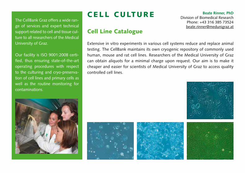
CELL CULTURE
Cell Line Catalogue
Extensive in vitro experiments in various cell systems reduce and replace animal testing. The CellBank maintains its own cryogenic repository of commonly used human, mouse and rat cell lines. Researchers of the Medical University of Graz can obtain aliquots for a minimal charge upon request. Our aim is to make it cheaper and easier for scientists of Medical University of Graz to access quality controlled cell lines.
Beate Rinner, PhDDivision of Biomedical Research
Phone: +43 316 385 [email protected]
CELL CULTURE
Primary Cell Culture
Over the last three decades, protocols for primary cell cultures have been refined and have become an essential tool in bio-medical research. Primary cells are taken directly from human or animal tissues, closely mimic the in vivo state and there-fore generate more physiologically relevant data. We offer two isolation systems:
Manual dissociationTissue can be dissociated into single-cell suspension by me-chanical dissociation and/or enzymatic digestion. A wide range of various enzymatic possibilities are avai lable, while the choice of appropriate enzymes (collagenase, trypsin, pronase, dispase) needs to be adjusted for a particu-lar type of cell isolation.
gentleMACS™ semi-automated dissociationThe gentleMACS™ Dissociator is a benchtop instrument for the semi-automated dissociation of tissues into single-cell sus-pensions or thorough homogenates. The instrument offers optimized gentleMACS programs for a variety of specific ap-plications. Special protocols have been developed for various tissues. The standardized tissue dissociation or homogeniza-tion procedures enable reliable and reproducible results.
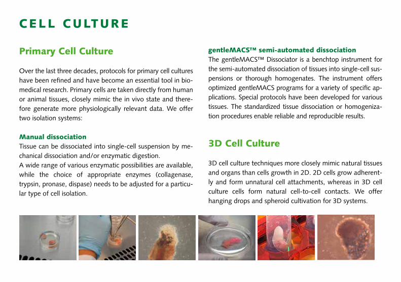
3D Cell Culture
3D cell culture techniques more closely mimic natural tissues and organs than cells growth in 2D. 2D cells grow adherent-ly and form unnatural cell attachments, whereas in 3D cell culture cells form natural cell-to-cell contacts. We offer hanging drops and spheroid cultivation for 3D systems.
Silke Patz, PhDDepartment of NeurosurgeryPhone: +43 316 385 72920
ORGANOTYP IC CULTURESORGANOTYP IC CULTURES
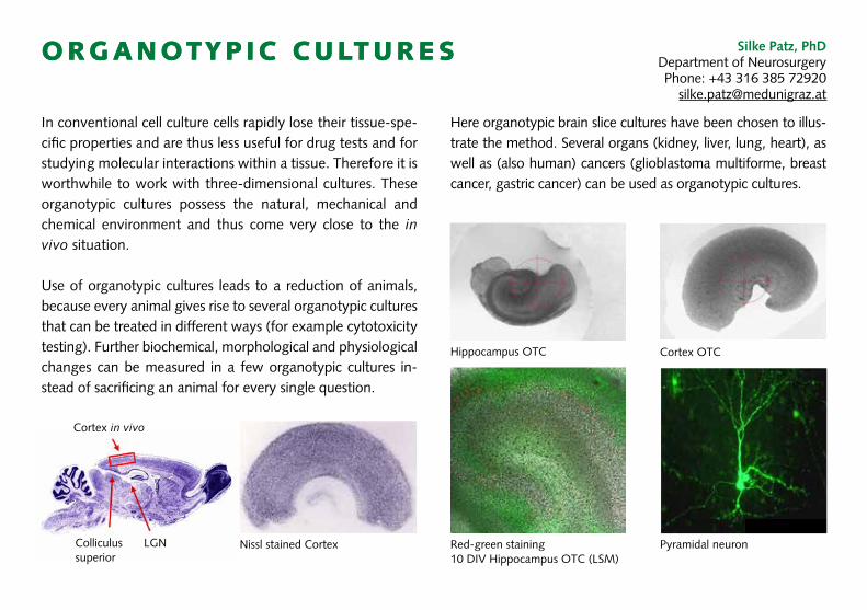
In conventional cell culture cells rapidly lose their tissue-spe-cific properties and are thus less useful for drug tests and for studying molecular interactions within a tissue. Therefore it is worthwhile to work with three-dimensional cultures. These organotypic cultures possess the natural, mechanical and chemical environment and thus come very close to the in vivo situation.
Use of organotypic cultures leads to a reduction of animals, because every animal gives rise to several organotypic cultures that can be treated in different ways (for example cytotoxicity testing). Further biochemical, morphological and physiological changes can be measured in a few organotypic cultures in-stead of sacrificing an animal for every single question.
Hippocampus OTC
Nissl stained Cortex
Cortex in vivo
Colliculus superior
LGN
Cortex OTC
Pyramidal neuronRed-green staining 10 DIV Hippocampus OTC (LSM)
Here organotypic brain slice cultures have been chosen to illus-trate the method. Several organs (kidney, liver, lung, heart), as well as (also human) cancers (glioblastoma multiforme, breast cancer, gastric cancer) can be used as organotypic cultures.
Alexander Hofmeister, MScDivision of Biomedical Research
Phone: +43 316 385 [email protected]
PRECL IN ICAL IMAGING
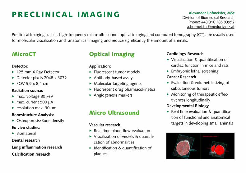
MicroCT
Detector: > 125 mm X Ray Detector> Detector pixels 2048 x 3072> FOV 5,5 x 8,4 cm
Radiation source:> max. voltage 80 keV> max. current 500 µA> resolution max. 30 µm
Bonestructure Analysis:> Osteoporosis/Bone density
Ex-vivo studies:> Biomaterial
Dental research
Lung inflammation research
Calcification research
Preclinical Imaging such as high-frequency micro-ultrasound, optical imaging and computed tomography (CT), are usually used for molecular visualization and anatomical imaging and reduce significantly the amount of animals.
Optical Imaging
Application:> Fluorescent tumor models> Antibody-based assays > Molecular targeting agents > Fluorescent drug pharmacokinetics > Angiogenesis markers
Micro Ultrasound
Vascular research> Real time blood flow evaluation> Visualization of vessels & quantifi-
cation of abnormalities> Identification & quantification of
plaques
Cardiology Research> Visualization & quantification of
cardiac function in mice and rats> Embryonic lethal screeningCancer Research> Evaluation & volumetric sizing of
subcutaneous tumors> Monitoring of therapeutic effec-
tiveness longitudinallyDevelopmental Biology> Real time evaluation & quantifica-
tion of functional and anatomical targets in developing small animals
BARRIER MODELS
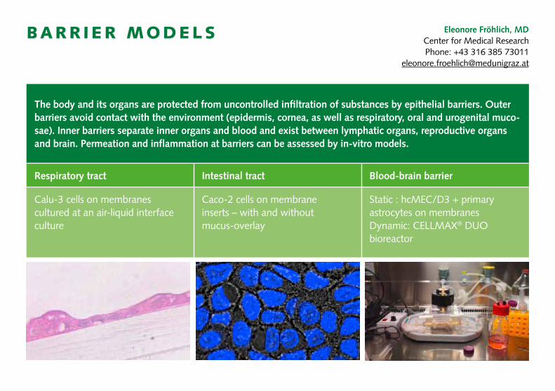
The body and its organs are protected from uncontrolled infiltration of substances by epithelial barriers. Outer barriers avoid contact with the environment (epidermis, cornea, as well as respiratory, oral and urogenital muco-sae). Inner barriers separate inner organs and blood and exist between lymphatic organs, reproductive organs and brain. Permeation and inflammation at barriers can be assessed by in-vitro models.
Respiratory tract Intestinal tract Blood-brain barrier
Calu-3 cells on membranes cultured at an air-liquid interface culture
Caco-2 cells on membrane inserts – with and without mucus-overlay
Static : hcMEC/D3 + primary astrocytes on membranesDynamic: CELLMAX® DUO bioreactor
Eleonore Fröhlich, MDCenter for Medical ResearchPhone: +43 316 385 73011
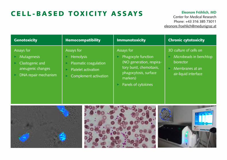
CELL-BASED TOX IC ITY ASSAYS
Genotoxicity Hemocompatibility Immunotoxicity Chronic cytotoxicity
Assays for
> Mutagenesis
> Clastogenic and aneugenic changes
> DNA repair mechanism
Assays for
> Hemolysis
> Plasmatic coagulation
> Platelet activation
> Complement activation
Assays for
> Phagocyte function (NO generation, respira-tory burst, chemotaxis, phagocytosis, surface markers)
> Panels of cytokines
3D culture of cells on
> Microbeads in benchtop biorector
> Membranes at an air-liquid interface
Eleonore Fröhlich, MDCenter for Medical ResearchPhone: +43 316 385 73011
Birgit Lohberger, PhDDepartment of Orthopedic Surgery
Phone: +43 316 385 [email protected]
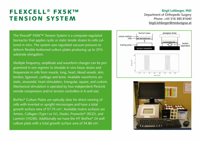
FLEXCELL ® FX5K™ TENS ION SYSTEM
The Flexcell® FX5K™ Tension System is a computer-regulated bioreactor that applies cyclic or static tensile strains to cells cul-tured in vitro. The system uses regulated vacuum pressure to deform flexible-bottomed culture plates pro ducing up to 25% substrate elongation.
Multiple frequency, amplitude and waveform changes can be pro-grammed in one regimen to simulate in vivo tissue strains and frequencies in cells from muscle, lung, heart, blood vessels, skin, tendon, ligament, cartilage and bone. Available waveforms are static, sinusoidal, heart stimulation, triangular, square, and custom. Mechanical stimulation is operated by four independent FlexLink remote compression and/or tension controllers in 6 well size.
BioFlex® Culture Plates are optically clear for direct viewing of cells with inverted or upright microscopes and have a total growth surface area of 57.75 cm2. Available matrix surfaces are Amino, Collagen (Type I or IV), Elastin, Pronectin® (RGD), and Laminin (YIGSR). Additionally we have the HT BioFlex® 24 well culture plate with a total growth surface area of 34.86 cm2.

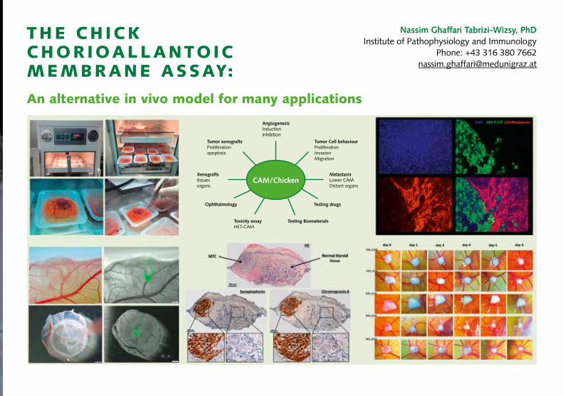
THE CH ICK CHORIOALLANTOIC MEMBRANE ASSAY:
An alternative in vivo model for many applications
Nassim Ghaffari Tabrizi-Wizsy, PhDInstitute of Pathophysiology and Immunology
Phone: +43 316 380 [email protected]
CAM/Chicken
AngiogenesisInductioninhibition
Tumor Cell behaviourProliferationInvasionMigration
Tumor xenograftsProliferationapoptosis
Xenograftstissuesorgans
Toxicity assayHET-CAM
Ophthalmology
Testing Biomaterials
Testing drugs
MetastasisLower CAMDistant organs
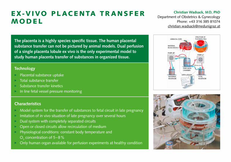
Christian Wadsack, M D, PhDDepartment of Obstetrics & Gynecology
Phone: +43 316 385 [email protected]
EX-V IVO PL ACENTA TR ANSFER MODEL
The placenta is a highly species specific tissue. The human placental substance transfer can not be pictured by animal models. Dual perfusion of a single placenta lobule ex vivo is the only experimental model to study human placenta transfer of substances in organized tissue.
Technology
> Placental substance uptake> Total substance transfer> Substance transfer kinetics> In line fetal vessel pressure monitoring
Characteristics
> Model system for the transfer of substances to fetal circuit in late pregnancy> Imitation of in vivo situation of late pregnancy over several hours> Dual system with completely separated circuits> Open or closed circuits allow recirculation of medium> Physiological conditions: constant body temperature and
O2 concentration of 5 – 8 %> Only human organ available for perfusion experiments at healthy condition
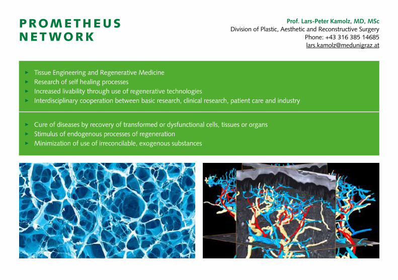
Prof. Lars-Peter Kamolz, MD, MScDivision of Plastic, Aesthetic and Reconstructive Surgery
Phone: +43 316 385 [email protected]
PROMETHEUS NET WORK
> Tissue Engineering and Regenerative Medicine> Research of self healing processes> Increased livability through use of regenerative technologies > Interdisciplinary cooperation between basic research, clinical research, patient care and industry
> Cure of diseases by recovery of transformed or dysfunctional cells, tissues or organs > Stimulus of endogenous processes of regeneration> Minimization of use of irreconcilable, exogenous substances

Contact
Eleonore Fröhlich, MDCenter for Medical ResearchPhone: +43 316 385 73011
Christian Wadsack, MD, PhDDepartment of Obstetrics & Gynecology
Phone: +43 316 385 [email protected]
Silke Patz, PhDDepartment of NeurosurgeryPhone: +43 316 385 [email protected]
Prof. Lars-Peter Kamolz, MD, MScDivision of Plastic Surgery
Phone: +43 316 385 [email protected]
Birgit Lohberger, PhDDepartment of Orthopedic Surgery
Phone: +43 316 385 [email protected]
Nassim Ghaffari Tabrizi-Wizsy, PhDInst. of Pathophysiology & Immunology
Phone: +43 316 380 [email protected]
Alexander Hofmeister, MScDivision of Biomedical Research
Phone: +43 316 385 [email protected]
Beate Rinner, PhDDivision of Biomedical Research
Phone: +43 316 385 [email protected]


![CellMax Antenna - 201105 [Compatibility Mode]](https://static.fdocuments.in/doc/165x107/5477b38ab4af9f76108b4919/cellmax-antenna-201105-compatibility-mode.jpg)


