Alteration of the Kidney Structure of White Rat after ...
9
Available on line at: ISSN: 2354-5844 (Print) http://ijwem.ulm.ac.id/index.php/ijwem ISSN: 2477-5223 (Online) -----Accredited by Directorate General of Higher Education Indonesia, No. 21/E/KPT/2018, Valid until 9 July 2023----- Alteration of the Kidney Structure of White Rat after Water Administration from Martapura River IDA YULIANA 1 , LENA ROSIDA 2 , HUSNUL KHATIMAH 3 , RAYATUL AMINAH 4 , ALWIYAH 4 , AND EKA AMELIA 4 1-2 Department of Biomedics, Histology Division, Faculty of Medicine, Lambung Mangkurat University 3 Departement of Biomedics, Biology Division, Faculty of Medicine, Lambung Mangkurat University 4 Medical Education Study Programs, Faculty of Medicine, Lambung Mangkurat University ABSTRACT Martapura River is indicated to have been contaminated by heavy metal can adversely affect the kidneys. This study aims to analyze the impact of water consumption of the Martapura River on the microscopic image of white rat kidneys. Microscopic structure of the kidneys studied were the glomerulus, proximal renal tubule, and renal medullary tubule. Research design used a posttest only with control group, with 32 white rats as the subject divided into 2 research groups, namely control group (distilled water) and treatment group (water from Martapura River) ad libitum for 30 days. Analysis of the data was independent t-test at 95% confidence level. The results showed that the number of glomerulus in the control group was less than the treatment group (p = 0.017); the glomerular diameter in the treatment group was smaller than the control group (p = 0.007); the number of proximal renal tubules in the treatment group was less than the control group (p = 0.025); lumen diameter of proximal renal tubules in the treatment group was not significantly different than the control group (p = 0.025); the number of renal medullary tubules in the treatment group was not significantly different than the control group (p = 0.347); and the lumen diameter of the renal medulla tubules in the treatment did not differ significantly compared to the control group (p = 0.015). Therefore, consuming water from Martapura River which contains heavy metals causing damage to the glomerulus, proximal renal tubule, and tubule of the renal medulla. Keywords: Martapura River water, glomerulus, proximal renal tubule, renal medullary tubul INTRODUCTION Martapura or Kayutangi River is a tributary of the Barito River which empties into the city of Banjarmasin and upstream is the city of Martapura, the capital of Banjar Regency, South Kalimantan (Ulmi EL & Amal N, 2017). 59.4% of the people along the Martapura River use river water as a source of water for household needs (Mirwan A & Indrawati R, 2012). Data from the Regional Environmental Agency (BLHD) of South Kalimantan and other studies explain that heavy metals content in the Martapura River exceeds the predetermined quality standard (Mirwan A, 2012; Amalia et al., 2012; Fokus Batulicin, 2011). Heavy metals are dangerous pollutants because they are not easily destroyed in the body and the environment, so they tend to accumulate in organs (Harteman, 2011). According to Robbins and Jannette, heavy metals that enter through the digestive tract will be distributed to tissues and organs, one of which is the kidney which has a major function in the urinary system (Janardani et al., 2018). These changes are related to the metabolism of heavy metal content in the body. Heavy metals in the digestive system then accumulate in the blood. Heavy metals pass through the blood to the kidneys. This will cause a cell adaptive response due to inflammation by heavy metals. Heavy metal ions that accumulate in the kidneys cause the antioxidant enzymes to be inactive, increasing the production of Reactive Oxygen Species (ROS). This increase causes an increase in the formation of free radicals, which triggers oxidative stress in cells (Danusantoso, 2012; Kumar et al., 2010). Cells are hypoxic due to Correspondence: Ida Yuliana, Department of Biomedics, Histology Division, Faculty of Medicine, Lambung Mangkurat University, South Kalimantan Indonesia. E-mail: [email protected]. 24
Transcript of Alteration of the Kidney Structure of White Rat after ...

Available on line at: ISSN: 2354-5844 (Print) http://ijwem.ulm.ac.id/index.php/ijwem ISSN: 2477-5223 (Online)
Journal of Wetlands Environmental Management
Vol 9, No 2 (2021) 24 - 32
http://dx.doi.org/10.20527/jwem.v9i2.269
-----Accredited by Directorate General of Higher Education Indonesia, No. 21/E/KPT/2018, Valid until 9 July 2023-----
Alteration of the Kidney Structure of White Rat after Water
Administration from Martapura River
IDA YULIANA
1, LENA ROSIDA
2, HUSNUL KHATIMAH
3,
RAYATUL AMINAH4, ALWIYAH
4, AND
EKA AMELIA4
1-2
Department of Biomedics, Histology Division, Faculty of Medicine, Lambung Mangkurat
University 3Departement of Biomedics, Biology Division, Faculty of Medicine, Lambung Mangkurat University
4 Medical Education Study Programs, Faculty of Medicine, Lambung Mangkurat University
ABSTRACT
Martapura River is indicated to have been contaminated by heavy metal can adversely affect the kidneys.
This study aims to analyze the impact of water consumption of the Martapura River on the microscopic
image of white rat kidneys. Microscopic structure of the kidneys studied were the glomerulus, proximal
renal tubule, and renal medullary tubule. Research design used a posttest only with control group, with 32
white rats as the subject divided into 2 research groups, namely control group (distilled water) and
treatment group (water from Martapura River) ad libitum for 30 days. Analysis of the data was
independent t-test at 95% confidence level. The results showed that the number of glomerulus in the
control group was less than the treatment group (p = 0.017); the glomerular diameter in the treatment
group was smaller than the control group (p = 0.007); the number of proximal renal tubules in the
treatment group was less than the control group (p = 0.025); lumen diameter of proximal renal tubules in
the treatment group was not significantly different than the control group (p = 0.025); the number of renal
medullary tubules in the treatment group was not significantly different than the control group (p =
0.347); and the lumen diameter of the renal medulla tubules in the treatment did not differ significantly
compared to the control group (p = 0.015). Therefore, consuming water from Martapura River which
contains heavy metals causing damage to the glomerulus, proximal renal tubule, and tubule of the renal
medulla.
Keywords: Martapura River water, glomerulus, proximal renal tubule, renal medullary tubul
INTRODUCTION
Martapura or Kayutangi River is a
tributary of the Barito River which empties
into the city of Banjarmasin and upstream is
the city of Martapura, the capital of Banjar
Regency, South Kalimantan (Ulmi EL & Amal
N, 2017). 59.4% of the people along the
Martapura River use river water as a source of
water for household needs (Mirwan A &
Indrawati R, 2012). Data from the Regional
Environmental Agency (BLHD) of South
Kalimantan and other studies explain that
heavy metals content in the Martapura River
exceeds the predetermined quality standard
(Mirwan A, 2012; Amalia et al., 2012; Fokus
Batulicin, 2011). Heavy metals are dangerous
pollutants because they are not easily
destroyed in the body and the environment, so
they tend to accumulate in organs (Harteman,
2011). According to Robbins and Jannette,
heavy metals that enter through the digestive
tract will be distributed to tissues and organs,
one of which is the kidney which has a major
function in the urinary system (Janardani et
al., 2018). These changes are related to the
metabolism of heavy metal content in the
body. Heavy metals in the digestive system
then accumulate in the blood. Heavy metals
pass through the blood to the kidneys. This
will cause a cell adaptive response due to
inflammation by heavy metals. Heavy metal
ions that accumulate in the kidneys cause the
antioxidant enzymes to be inactive, increasing
the production of Reactive Oxygen Species
(ROS). This increase causes an increase in the
formation of free radicals, which triggers
oxidative stress in cells (Danusantoso, 2012;
Kumar et al., 2010). Cells are hypoxic due to Correspondence: Ida Yuliana, Department of Biomedics, Histology Division, Faculty of Medicine, Lambung Mangkurat
University, South Kalimantan Indonesia.
E-mail: [email protected].
24

Available on line at: ISSN: 2354-5844 (Print) http://ijwem.ulm.ac.id/index.php/ijwem ISSN: 2477-5223 (Online)
Journal of Wetlands Environmental Management
Vol 9, No 2 (2021) 24 - 32
http://dx.doi.org/10.20527/jwem.v9i2.269
-----Accredited by Directorate General of Higher Education Indonesia, No. 21/E/KPT/2018, Valid until 9 July 2023-----
ROS. Hypoxia causes cell damage with
reduced aerobic oxidative respiration (Kumar
et al., 2010). The kidney, as one of the vital
organs in the body, is most often subject to
damage by harmful chemicals (Suhita et al.,
2013). This is because the kidneys receive
blood flow of 25% of the volume of blood
flowing to the heart (Fatimah U, 2013). The
kidneys histologically consist of the cortex,
medulla, and renal pelvis. Within the renal
cortex are the main structures that produce
urine known as nephrons. The nephron is
composed of the renal corpusculum (combined
glomerulus and Bowman's capsule), and the
renal tubule (proximal convoluted tubule,
Henle's ansa, distal convoluted tubule).
Meanwhile, in the renal medulla, there is a
duct system in the form of a tubule which
functions as a channel that distributes urine to
the urinary tract to the next (Kumar et al.,
2012; Junquiera et al., 2007). Research on the
impact of water pollution by heavy metals has
been widely reported. Research by Wahyuni et
al. stated that water contaminated with heavy
metals causes necrosis of snakehead fish
glomeruli (Wahyuni et al., 2017). Khoiriyah et
al's research on tilapia kidneys also found
severe damage such as necrosis and cell
swelling in the proximal tubular epithelium
(Khoiriyah et al., 2016). In addition, the
research of Wagiman et al also stated that
heavy metals can cause abnormalities in the
ansa Henle tubule of fish in the form of cell
swelling and necrosis (Wagiman et al., 2014).
Based on these data, this study was conducted
to further determine the impact of water
consumption of the Martapura River on the
microscopic image of the kidneys in Sprague
Dawley white rats. This can provide input for
the government to make policies related to
water pollution in the Martapura river and
provide information and education to the
public about the correct use of Martapura river
water to improve their health status.
MATERIALS AND METHODS
The research design used was true
experimental laboratory research with a
posttest only design with the control group
(Dahlan SM, 2012). The research subjects
were white (Rattus novergicus) Sprague
Dawley strain, male, 2-6 months old, and
weighing 250-300 g. The number of samples
of the study was 32 rats (determined by
Federer's formula). The research group was
divided into two groups, namely the control
group which was given ad libitum distilled
water and the treatment which was given water
from the Martapura River ad libitum (Yuliana
et al., 2018).
Rat obtained from BVET Banjarbaru;
Martapura River water; neutral buffer formalin
10% I, II, and III; ethanol 70%, 80%, 96%;
xylol I, II and III; liquid paraffin I and II;
distilled water; hematoxylin harris; eosin 1%;
and gummy. Bottles, Perkin Elmer 5100 PC
atomic absorption spectrophotometer, beaker
glass, filter paper, water bath, hotplate, mouse
cage, Ohaus analytical balance, gloves, masks,
minor surgical instruments, syringes, raster
image software, optical camera, binocular light
microscope, scalpel, base mold, tissue cassette,
slide glass, cover glass and microtome.
Identification of water characteristics
Martapura River water was taken from
the waters behind the Darussalam Martapura
Islamic Boarding School using a grab
sampling technique in August 2019. An
examination of the water characteristics of
the Martapura River was carried out by the
Banjarbaru Center for Environmental Health
and Disease Control Engineering
(BBTKLPP) using the atomic absorption
spectrophotometric method (Torowati et al.,
2008).
Research procedure
Starting from the treatment process on
experimental animals provided that the
animals used are healthy, male, 2-6 months
old, and weighing 250-300 g. Then the
acclimatization process was carried out for
one week. The experimental animals were
randomly grouped into a control group and a
treatment group. The implementation of this
research was carried out for 30 days
(Yuliana et al., 2018). After the 31st day,
the rats were sacrificed, and the kidneys
were taken and made histological
preparations stained with HE.
Observation of Renal Histological
Preparations
In the initial stage, the histological
preparations were examined with a binocular
25

Available on line at: ISSN: 2354-5844 (Print) http://ijwem.ulm.ac.id/index.php/ijwem ISSN: 2477-5223 (Online)
Journal of Wetlands Environmental Management
Vol 9, No 2 (2021) 24 - 32
http://dx.doi.org/10.20527/jwem.v9i2.269
-----Accredited by Directorate General of Higher Education Indonesia, No. 21/E/KPT/2018, Valid until 9 July 2023-----
light microscope, then the best histological
preparations were taken of the white rat kidney
(Rattus norvegicus) with the criteria of
glomerular shape, proximal tubule, and intact
medullary tubule. Micro photos were made
using an optilab camera in jpeg format with a
magnification of 100x and 400x, then
observed using raster image computer
software. The results obtained were numerical
data.
Data Analysis
The data obtained were tested for
normality with the Shapiro-Wilk test. Then
proceed with the Levene test for
homogeneity. The analytical test used was
the independent t-test (95% confidence
level).
RESULTS
Based on the results of the study, to find
out whether there were differences in the
microscopic picture of the kidneys of rats in
treatment group and control group can be
seen in Table 1 and Figure 1-6.
Table 1. Microscopic features of white rat’s kidney control and treatment groups
Alteration in the kidney’s microscopic
features
Control Treatment P value
Number of glomerulus ren 4.19 ± 0.77 4.14 ± 0.74 0.017*
Glomerular diameter 89.51 ± 6.71 81.29 ± 9.27 0.0007*
Number of proximal tubules 122.35 ± 21.12 94.12 ± 18.74 0.000*
The lumen diameter of the proximal tubule 26.78 ± 3.19 25.38 ± 1.91 0.142
Number of tubules in the medulla 157.89 ± 3.04 148.41 ± 2.54 0.347*
Number of tubules in the medulla 54.36 ± 7.93 47.97 ± 5.94 0.015*
* t-test independent significance
Figure 1. Description of the number of a glomerulus control group (A) and treatment (B). The black
arrow is a countable glomerulus; the red circle is the glomerulus that cannot be counted
because the glomerulus is fragmented, the boundary between the parietal stratum and the
stratum visceral is not visible, and the glomerular structure is not intact; the glomerulus
atrophy due to adhesions ; HE staining. Magnification 100x.
A B
A B
26
Available on line at: ISSN: 2354-5844 (Print) http://ijwem.ulm.ac.id/index.php/ijwem ISSN: 2477-5223 (Online)
Journal of Wetlands Environmental Management
Vol 9, No 2 (2021) 24 - 32
http://dx.doi.org/10.20527/jwem.v9i2.269
-----Accredited by Directorate General of Higher Education Indonesia, No. 21/E/KPT/2018, Valid until 9 July 2023-----
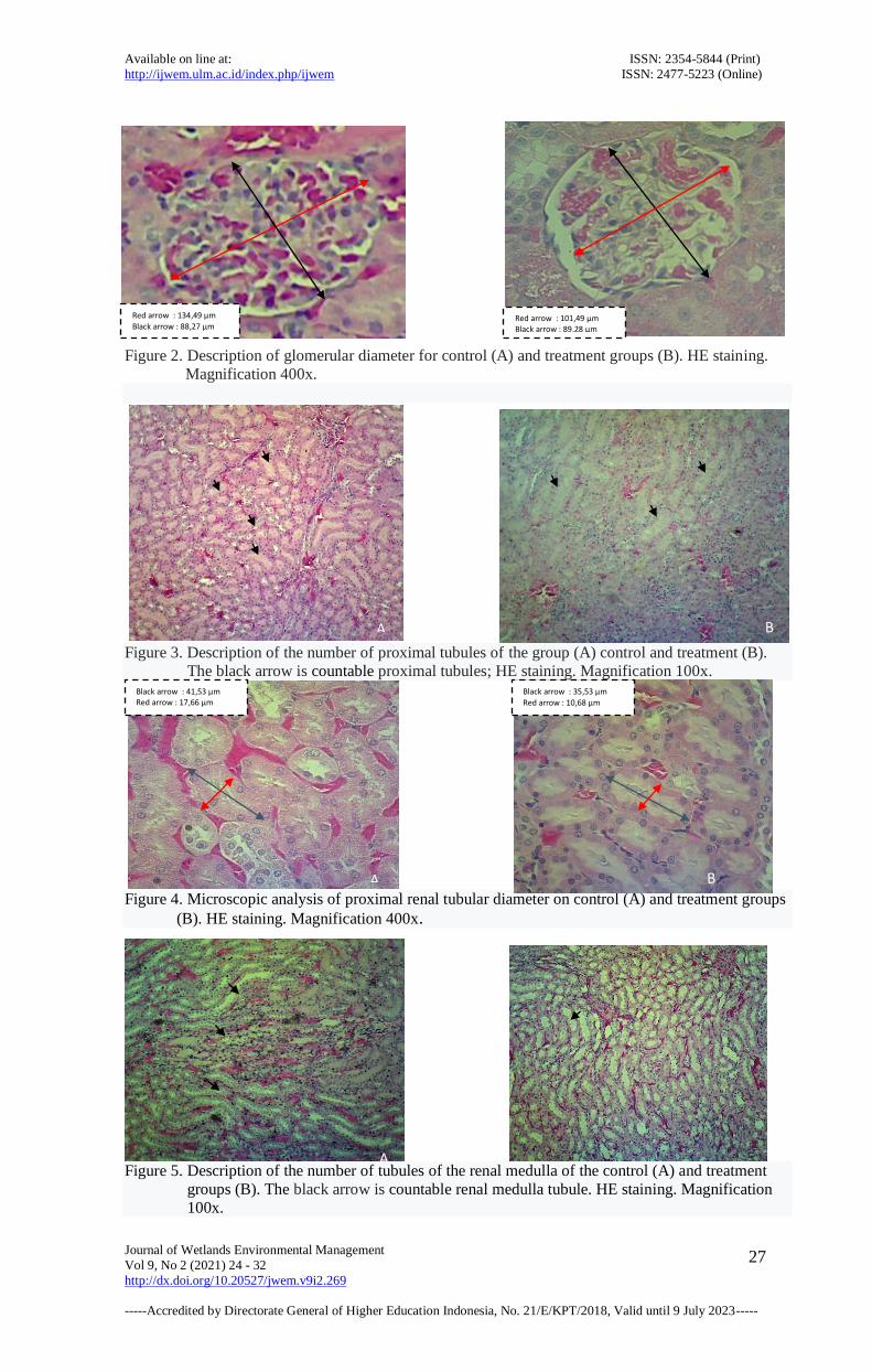
Figure 2. Description of glomerular diameter for control (A) and treatment groups (B). HE staining.
Magnification 400x.
Figure 3. Description of the number of proximal tubules of the group (A) control and treatment (B).
The black arrow is countable proximal tubules; HE staining. Magnification 100x.
Figure 4. Microscopic analysis of proximal renal tubular diameter on control (A) and treatment groups
(B). HE staining. Magnification 400x.
Figure 5. Description of the number of tubules of the renal medulla of the control (A) and treatment
groups (B). The black arrow is countable renal medulla tubule. HE staining. Magnification
100x.
Black arrow : 41,53 μm Red arrow : 17,66 μm
Black arrow : 35,53 μm
Red arrow : 10,68 μm
Red arrow : 101,49 μm Black arrow : 89,28 μm
Red arrow : 134,49 μm
Black arrow : 88,27 μm
A
A
B
B
A B
27

Available on line at: ISSN: 2354-5844 (Print) http://ijwem.ulm.ac.id/index.php/ijwem ISSN: 2477-5223 (Online)
Journal of Wetlands Environmental Management
Vol 9, No 2 (2021) 24 - 32
http://dx.doi.org/10.20527/jwem.v9i2.269
-----Accredited by Directorate General of Higher Education Indonesia, No. 21/E/KPT/2018, Valid until 9 July 2023-----
Figure 6. Microscopy analysis of lumen diameter of renal medullary tubules control (A) and treatment
groups (B). HE staining. Magnification 400x.
DISCUSSION
Microscopic features of the glomerulus
Table 1 showed a significant decreased in
the mean number of renal glomerulus in white
rats, namely 89.51 ± 6.71 in the control group
and 81.29 ± 9.27 in the treatment group; with a
value of P = 0.017. These results indicated a
significant reduction in the number of
glomeruli in the treatment group compared to
the control group. This data was also
corroborated by the results of observations on
renal histological preparations in Figure 1
which shows the difference in the number of
glomeruli between the study groups. The
decreased in the number of glomeruli in the
treatment group was indicated by a picture of
the boundary between glomerular podocyte
cells and stratum parietal epithelial cells,
Bowman's capsule was not clear so that the
spatial space appeared widened and some even
disappeared. This was probably the result of
the inflammatory response of cells around the
glomerulus due to toxic substances. In
addition, the appearance of Bowman's capsule
in the treatment group appeared to be atrophic
and partly fragmented. The provision of water
from the Martapura River containing heavy
metals for a long time was thought to have
caused this situation. These results are in line
with research conducted by Ratnaningsih
which showed that high Cd levels caused a
change in the image of the white rat kidney in
the form of the boundary between glomerular
podocytes and stratum parietal epithelial cells,
Bowman's capsule was not clear and even
disappeared.
Cd causes inflammation of the glomerular
membrane gap, causing an adaptive response
in the form of leakage of red blood cells from
the glomerular capillaries and infiltration of
inflammatory cells in the glomerulus
(Ratnaningsih et al., 2016). The inflammatory
response also causes adhesions between the
glomerulus and Bowman's capsule.
Glomerulus that experiences atrophy due to
this adhesion cannot perform optimal
glomerular function (Fahrimal et al., 2016).
Glomerulus with unclear boundaries,
structures that experience fragmentation and
atrophy due to adhesion cannot be calculated
on the variable number of glomeruli in this
study so that there is a reduction in the number
of glomeruli in the treatment.
In Table 2, it is also known that there was
a decrease in the mean glomerular diameter of
the renal white rats, namely 89.51 ± 6.71 in the
control group and 81.29 ± 9.27 in the
treatment group; with a P-value = 0.007. These
results indicate a shortening of the glomerular
diameter in the treatment group. This data is
collaborated by the results of observations on
renal histological preparations in Figure 2
which shows the difference in the size of the
glomerular diameter between the study groups.
The theory that underlies a shortening of the
glomerular diameter is due to atrophy of the
glomerular capillary endothelial cells in
response to cell inflammation due to toxic
substances. Puspitasari's research states that
the shortening of the glomerular diameter was
thought to occur due to atrophy of the
glomerular capillary endothelial cells
(Puspitasari, 2015). It was suspected that the
shortening of the glomerular diameter was due
to chronic consumption of the Martapura river
water which contains heavy metals. This
result was in line with the research of
Widyaningrum et al who observed that
Black arrow : 12,66 μm
Red arrow : 30,39 μm Red arrow : 63,36 μm Black arrow : 28,17 μm
28

Available on line at: ISSN: 2354-5844 (Print) http://ijwem.ulm.ac.id/index.php/ijwem ISSN: 2477-5223 (Online)
Journal of Wetlands Environmental Management
Vol 9, No 2 (2021) 24 - 32
http://dx.doi.org/10.20527/jwem.v9i2.269
-----Accredited by Directorate General of Higher Education Indonesia, No. 21/E/KPT/2018, Valid until 9 July 2023-----
exposure to the toxic substance HgCl2 for six
weeks caused glomerular cell atrophy so that
the glomerular diameter shortened.
Microscopic Features of the Proximal
Renal Tubule
The results of research observations on the
proximal renal tubule showed a significant
decrease (p-value = 0.000) with the mean
number of proximal renal tubules of white rats,
namely 122.35 ± 21.12 in the control group
and 94.12 ± 18.74 in the treatment group
(Table 2). Figure 3 shows a decrease in the
number of proximal tubules in the treatment
group. The decrease in the number of
proximal tubules was thought to be due to
toxic substances that cause cell swelling and
congestion in the proximal tubules. This
results in stretching the space between the
tubules. This result was in line with Sari's
research (2010) which stated that the cause of
the decrease in the number of proximal renal
tubules can be due to Pb exposure, which was
also identified in the water content of the
Martapura River used in this study.
The proximal renal tubule is the part most
frequently damaged due to exposure to
nephrotoxic substances. Active transport
systems for ions, organic acids, low molecular
weight proteins, peptides, and heavy metals
mostly occur in the proximal tubule, causing
accumulation and proximal tubular toxicity
which ultimately results in proximal tubular
damage. The loose epithelium of the proximal
tubule facilitates the entry of various
components into the proximal tubular cell.
This was thought to be something that
contributes to damage to the proximal renal
tubule.
The results of research observations on the
lumen diameter of the proximal renal tubule
showed a tendency to narrow the lumen
diameter, namely 26.78 ± 3.19 in the control
group and 25.38 ± 1.91 in the treatment group,
although not significantly different from the
control group (p = 0.142) (Table 2 and Figure
4).This was in line with Khoiriyah et al's
research which states that there is a cause of
narrowing of the proximal tubular lumen is Cd
(Khoiriyah et al., 2016). Sari's research also
found an image of the proximal tubule with
swelling of the tubular epithelial cells which
caused a narrowing of the lumen due to Pb
administration (Sari DH, 2010). Cell swelling
is an important disorder associated with
reversible injury due to failure of the energy-
dependent / energy-dependent ion pump on the
plasma membrane which results in the cell
being unable to maintain ion and fluid
homeostasis, causing Na buildup in the cell
and K excretion from the cell. The final result
is the addition of iso-osmotic water (Khoiriyah
et al., 2016).
Microscopic Features of the Renal
Medullary Tubules
The observations of the results of the next
study were changes in the number of renal
medullary tubules. The results showed that
there was a tendency to decrease the mean
number of tubules in the renal medulla of
white rats, namely 157.89 ± 30.44 in the
control group and 148.41 ± 25.42 in the
treatment group. However, the decrease in the
number of medullary tubules was not
statistically significant (P - value = 0.347)
(Table 2). Microscopic changes in the form of
a decrease in the number of tubules in the
medulla in the treatment group can be seen in
Figure 5. The decrease in the number of
tubules of the renal medulla occurred due to
the swelling of the medullary tubular cells
because of reversible injury. This was in line
with the research of Wagiman et al., which
states that heavy metals can cause
abnormalities in Henle fish such as cell
swelling and necrosis (Wagiman et al., 2014).
As a result of cell swelling, the number of
medullary tubules seen in one field of view
was less in the treatment group. In addition,
the number of medullary tubules decreases as
well as possible due to congestion in the
medullary tubules, a condition in which there
is excessive accumulation of blood in the
blood vessels.
The results of research observations on the
lumen diameter of the medullary tubule found
that there was a significant
narrowing/shortening (P-value = 0.015) in the
lumen diameter with an average size of the
medullary tubular lumen diameter of 54.36 ±
7.93 in the control group and 47.97 ± 5.94.
treatment group (table 2). Figure 6 shows the
damage that occurred in the medullary tubule
which was characterized by narrowing of the
medullary tubule lumen, in the treatment
group it was seen that the tubular lumen was
narrower than the control group. The results of
29

Available on line at: ISSN: 2354-5844 (Print) http://ijwem.ulm.ac.id/index.php/ijwem ISSN: 2477-5223 (Online)
Journal of Wetlands Environmental Management
Vol 9, No 2 (2021) 24 - 32
http://dx.doi.org/10.20527/jwem.v9i2.269
-----Accredited by Directorate General of Higher Education Indonesia, No. 21/E/KPT/2018, Valid until 9 July 2023-----
these observations are in line with the research
of Wagiman et al., who stated that heavy
metals can cause abnormalities in fish Henle
ansa such as swelling of cells and necrosis.
The tubular lumen narrows due to epithelial
swelling and is filled with detached cells and
other debris (Wagiman et al., 2014). Edema is
indicated by the condition of the renal tubular
lumen which is narrowing due to the size of
the tubular epithelial cells that are enlarging so
that the inter-tubules will be stretched. Cell
swelling occurs because the electrolyte charge
outside and inside the cell is unbalanced
(Anderson PS, 1995).
Based on the results of observations on
the structure of the kidneys (glomerulus,
proximal renal tubule, and tubule of the renal
medulla), it was found that there were
differences in the microscopic picture between
the control group and the treatment group.
This difference indicates that there was an
indication of damage to the kidney structure in
the experimental group of mice that were
given the Martapura River water as their daily
drinking water for a long period. This
condition was strongly related to the presence
of heavy metal content in the Martapura river
water even though the levels do not exceed the
water quality standard (table 1) (Permenkes,
2017). However, because it was consumed for
a long time, there was an accumulation of
heavy metals in the body. This situation was
related to the metabolism of heavy metal
content in the body. This was in line with
Safitri's research which states that the long
duration of exposure to consuming heavy
metal Cd even though at low concentrations
continuously will still cause a disturbing effect
on the kidneys (Safitri FZ, 2015).
Heavy metals in the digestive system then
accumulate in the blood. Heavy metals pass
through the blood to the kidneys. This will
cause a cell adaptive response due to
inflammation by heavy metals. Heavy metal
ions that accumulate in the kidneys cause the
antioxidant enzymes to be inactive, increasing
in the production of ROS. This increase
causes an increase in the formation of free
radicals, which triggers oxidative stress in
cells. Cells are hypoxic due to ROS. Hypoxia
causes cell damage with reduced aerobic
oxidative respiration (Kumar et al., 2016).
Free radicals cause a pathological response in
the form of reversible or irreversible cell
damage. The disorder associated with
reversible injury to cells is cell swelling with a
microscopic appearance of vacuole
degeneration. Continuous and prolonged
exposure to toxic substances will result in cells
leading to irreversible injury and cell death.
The transition from reversible lesions to
irreversible lesions through a process of
increasing cell swelling, swelling and damage
to lysosomes, damage to cell membranes and
changes in cell nucleus chromatin which will
end in necrosis (Irianti et al., 2017). This is
confirmed by Ressang's research in Fahrimal
et al which explains that severe glomerular
damage makes the peritubular vascular system
disrupted and has the potential to drain toxic
substances into the tubule, while severe
tubular damage due to increased
intraglomerular pressure will cause glomerular
atrophy (Fahrimal et al., 2016).
CONCLUSION
Based on the research results, it can be
concluded that administration of Martapura
River water in the long term tends to cause
damage to the kidney structure, namely
damage to the glomerulus, proximal renal
tubule and tubule of the renal medulla. This
was thought to be due to the heavy metal
content in the Martapura River water which
accumulates in the kidneys and caused an
increase in free radicals which triggered
oxidative stress on kidney cells which
damaging to the kidney structure.
ACKNOWLEDGMENTS
Financial support for this study was
provided by a grant from Lambung
Mangkurat University.
REFERENCE
A Candra, HF Trianto. 2015. A histological
representation of the rat's kidney cortex
(Rattus Norvegicus) after monosodium
glutamate exposure per oral. Cerebellum
Journal 1(3)206-7
30

Available on line at: ISSN: 2354-5844 (Print) http://ijwem.ulm.ac.id/index.php/ijwem ISSN: 2477-5223 (Online)
Journal of Wetlands Environmental Management
Vol 9, No 2 (2021) 24 - 32
http://dx.doi.org/10.20527/jwem.v9i2.269
-----Accredited by Directorate General of Higher Education Indonesia, No. 21/E/KPT/2018, Valid until 9 July 2023-----
Banjarbaru Veterinary Hall. 2019. Production
operational standard of histological
preparate and dyeing techniques.
Banjarbaru: BVET
Batulicin Fokus. 2011 [Cited Apr 18 2019].
South Kalimantan’s river quality is
decreasing [Internet]. Available from:
http://www.fokusbatulicin.com/2011/04/k
ualitas-air-sungai-di-kalimantan.html
H Danusantoso. 2003. Free radicals’ role
towards several lung disease. Medical
journal of Trisakti 22(1):31-6
Harteman E. 2011. The impact of heavy metal
content on the emergence of
polymorphisms of badukang (Arius
maculatus Fis & Bian) and sembilang
(Plotosus canius Eb and Bia)
polymorphisms in the estuaries of the
Kahayan and Katingan rivers [thesis].
Bogor: Bogor Institute of Agriculture
Irianti TT, Kuswandi, Nuranto S, Budiyatni
A. 2017. Heavy metals and health.
Yogyakarta: CV Grafika Indah
Janardani NMK, Berata IK, Kardena IM.
2018. Histopathological study and level
of heavy metals (Pb) in the kidneys of
Bali cattle in the Denpasar suwung
Landfill. Indonesia Medicus Veterinus
7(1):47-8.
Junqueira LC, J Carneiro, RO Kelley. 2007.
Basic Histology. 5th ed. EGC: Jakarta
Khoiriyah I, Hayati A, Sugiharto. 2016. The
effect of cadmium to Kidney cell
structure of tilapia (Oreochromis
niloticus ) at different salinity [Thesis].
Surabaya: Airlangga University
Kumar V, Cotran RS, Fausto N. 2010.
Robbins and cotran pathologic basis of
disease. 7th ed. Jakarta: EGC
Kumar V, Cotran RS, Robbins SL. 2012.
Pathology textbooks. 7th ed. Jakarta:
EGC
Mirwan A, Indrawati R. 2012. Preliminary
study of the utilization of acid from coal
mines as a coagulant for water from the
Martapura River. In: Proceedings of the
National Seminar of the Faculty of
Engineering, UNLAM; 2011 Jul 30;
Banjarbaru. Banjarbaru: University of
Lambung Mangkurat Press
Mirwan A. 2012. Reuse of solid waste
sludge (LPL) of PDAM Intan Banjar as
a coagulant for purification from the
Martapura river, South Kalimantan.
Bumi Lestari Journal 12 (1): 77.
P.S. Anderson. 1995. Pathophysiologists of
clinical concepts of disease processes.
Jakarta: EGC
Puspitasari D. 2015. Subchronic toxicity
test of katuk leaf water extract
(Sauropus andrygynus) on histology
and kidney weight of female rats
(Rattus norvegicus) [thesis]. Malang:
Maulana Malik Ibrahim State Islamic
University
Ratnaningsih A. 2004. The Effect of
cadmium on pathological disorders in
the rat kidney. Journal of Mathematics,
Science and Technology (1): 53-63.
Regulation of the Minister of Health of the
Republic of Indonesia. 2017.
Environmental health quality standards
and water health requirements for
hygiene sanitation, swimming pools,
solutions per aqua and public baths.
Jakarta: MENKES RI
Safitri FZ. 2015. The level of environmental
health effects of the heavy metal
content of cadmium (Cd) in green
mussel (Perna virdis) which was
consumed by the Kaliadem Muara
Angke community in North Jakarta in
2015 [thesis]. Jakarta: Syarif
Hidayatullah State Islamic University
Sari DH. 2010. The effect of lead (Pb) in
highway air on the microscopic image
of the kidneys and levels of lead (Pb) in
the blood of male balb / c mice [thesis].
Semarang: Diponegoro University
SM Dahlan. 2012. Statistics for galenical
and health. 3rd edition. Jakarta:
Salemba Medical
Suhita NLPR, Sudira IW, Winaya IBO. 2013.
Histopathology of rats' kidneys as a result
of oral administration of gotu kola
(Centella asiatica) extract. Udayana
Veterinary Bulletin 5 (1): 63-9.
Torowati, Asminar, Rahmiati. 2008. The
analysis of the elements of Pb, Ni and Cu
in uranium solution as a result of
stripping of uranium effluent in the
nuclear fuel field. BATAN (2):2.
U Fatimah. 2013. The histological structures
of hepar and the white rat’s ren (Rattus
norvegicus) feminina grafid after the
rhodamin B orally [thesis]. Surakarta:
Sebelas Maret University
Ulmi EI, Amal N. 2017. The study of the
Martapura River ecohydraulics [thesis].
31
31

Available on line at: ISSN: 2354-5844 (Print) http://ijwem.ulm.ac.id/index.php/ijwem ISSN: 2477-5223 (Online)
Journal of Wetlands Environmental Management
Vol 9, No 2 (2021) 24 - 32
http://dx.doi.org/10.20527/jwem.v9i2.269
-----Accredited by Directorate General of Higher Education Indonesia, No. 21/E/KPT/2018, Valid until 9 July 2023-----
Banjarbaru: Lambung Mangkurat
University
Wagiman, Yusfiati, Roza E. 2014. The
structure of the kidney of the cayenne
fish (Ompokhypopthalmus Bleeker ) in
the waters of the Siak River Pekanbaru
City. JOM FMIPA 1(2):63-69
Wahyuni S, Windarti, Putra RM. 2017.
Comparative study of the gill and kidney
tissue structure of snakehead fish
(Channa striata Bloch ) from the Sibam
River and Kulim River Riau Province
[thesis]. Pekanbaru: University of Riau
Widyanti IH. 2015. Case study:
Histopathological image of kidney
organs in bats in Gorontalo [thesis]. 1
(3): 206-7.
WR Amalia, B Halang, Naparin A. 2016.
Cadmium (Cd) content in water, flesh,
also microanatomy of kelpfish gills
(Osteochillus melanopleurus) in
Martapura creek.
Y Fahrimal, Rahmiwati, D Aliza. 2016. A
histological representation of the white
rat’s kidney cortex (Rattus Norvegicus)
infected with Trypanosoma evansi and
extract of fluorescent leaves. Medical
Veterinaria Journal 10(2):166-70.
Yuliana I, Rosida L, Khatimah H,
Skripsiana NS, Dayana P. 2018. The
effect of addition of Martapura river
water on changes in macroscopic and
microscopic morphological changes in
rat testicles (Rattus norvegicus)
[research report]. Banjarmasin:
Lambung Mangkurat University
Yuniarti, Rousdy DW, Rahmawati. 2015.
Anti-inflammatory test of flower
infusion of lotus flower (Nelumbo
nucifera Gaertn) on the microanatomy
structure of the kidney of mice (Mus
musculus) under stress. Protobiont 4
(1): 242-7.
32