Anemoside B4 Protects Rat Kidney from Adenine-Induced ...
12
Research Article Anemoside B4 Protects Rat Kidney from Adenine-Induced Injury by Attenuating Inflammation and Fibrosis and Enhancing Podocin and Nephrin Expression Qin Gong, 1,2 Lu-Ling He, 2 Mu-Lan Wang, 2 Hui Ouyang, 1,2 Hong-Wei Gao, 3 Yu-Lin Feng, 1,2 Shi-Lin Yang, 1,2 Li-Jun Du , 2,4 Jun Li , 1,2 and Ying-Ying Luo 1,2 Jiangxi University of Traditional Chinese Medicine, Nanchang , China State Key Laboratory of Innovative Drugs and Efficient Energy-Saving Pharmaceutical Equipment, Nanchang , China College of Pharmacy, Guangxi University of Chinese Medicine, Nanning , China School of Life Sciences, Tsinghua University, Beijing , China Correspondence should be addressed to Jun Li; [email protected] and Ying-Ying Luo; [email protected] Received 11 November 2018; Revised 24 December 2018; Accepted 27 March 2019; Published 2 May 2019 Academic Editor: Mayank akur Copyright © 2019 Qin Gong et al. is is an open access article distributed under the Creative Commons Attribution License, which permits unrestricted use, distribution, and reproduction in any medium, provided the original work is properly cited. Anemoside B4 (B4) isolated from Radix Pulsatilla has anti-inflammatory activities in the colon and antitumor effects. However, its role in the prevention and treatment of kidney injury has not been reported. Here, we reported the effects of B4 on chronic kidney injury (CKI) and studied its related mechanism based on an adenine-induced kidney injury model in rats. e results showed that serum BUN (blood urea nitrogen), Crea (creatinine), and urinary proteins increased significantly aſter oral administration of adenine. Meanwhile, the adenine contents in both renal tissue and urine increased markedly compared with those of normal rats. Moreover, IL-1, IL-6, TNF, and NFB expression was upregulated in the kidney. Simultaneously, the expression of NLRP3 (the nucleotide-binding and oligomerization domain–like receptor, leucine-rich repeat and pyrin domain–containing 3) in the inflammasome, which consists of Caspase 1, ASC (apoptosis-associated speck-like protein containing a caspase recruitment domain), and IL-18, was significantly upregulated. B4 could significantly decrease BUN and Crea; reduce urinary proteins in rats; suppress the expression of IL-6, IL-1, NFB, NLRP3, Caspase 1, ASC, and IL-18; and increase urinary adenine contents and promote its excretion. In addition, B4 also upregulated the expression of podocin and nephrin, two major podocyte proteins, and reduced the fiber collagen in the renal interstitial, suggesting that B4 could protect the glomerular matrix from adenine injury in addition to its anti-inflammatory effects. e results of this study show new perspective of B4 as a potential drug against adenine-induced renal injury. 1. Introduction Pulsatilla chinensis (Bge.) Reg (Pulsatilla) is commonly used in traditional Chinese medicine and has antimicrobial and anti-inflammatory functions [1–4]. Saponins are major ingre- dients in Pulsatilla [5–8]; for example, anemoside B4 (B4), which is isolated from the radix of Pulsatilla, has a content of 4% [9, 10]. Referring to the pharmacological studies on B4 in the latest five years mainly includes the detection of phar- macokinetics, tissue distribution, excretion by LC-MS /MS method, and biotransformation and metabolic profile of anemoside B4 in rat small and large intestine microflora [11–13]. Relevant studies have shown that B4 has antiviral and immunoregulatory effects in vitro and in viv; B4 could reduce the incidence and severity of porcine circovirus 2 PCV2-induced immunopathological damage, improving the phenomenon of body temperature elevation, weight loss, anaemia, and internal organ swelling, and affect the immunoglobulin levels and protein absorption [14]. B4 might effectively regulate immune responses via upregulated IL- 2 expression levels aſter endothelial cells were challenged with PRRSV [15]. Moreover, B4 has both anti-inflammatory and anticancer effects [16–21]. It has certain cytotoxicity on the human myelogenous leukemia K562 cell line and its metabolites could exhibit a reduction in cell viability of Hindawi Evidence-Based Complementary and Alternative Medicine Volume 2019, Article ID 8031039, 11 pages https://doi.org/10.1155/2019/8031039
Transcript of Anemoside B4 Protects Rat Kidney from Adenine-Induced ...
Research ArticleAnemoside B4 Protects Rat Kidney from Adenine-InducedInjury by Attenuating Inflammation and Fibrosis and EnhancingPodocin and Nephrin Expression
Qin Gong,1,2 Lu-Ling He,2 Mu-LanWang,2 Hui Ouyang,1,2 Hong-Wei Gao,3 Yu-Lin Feng,1,2
Shi-Lin Yang,1,2 Li-Jun Du ,2,4 Jun Li ,1,2 and Ying-Ying Luo 1,2
1 Jiangxi University of Traditional Chinese Medicine, Nanchang 330006, China2State Key Laboratory of Innovative Drugs and Efficient Energy-Saving Pharmaceutical Equipment, Nanchang 330006, China3College of Pharmacy, Guangxi University of Chinese Medicine, Nanning 530000, China4School of Life Sciences, Tsinghua University, Beijing 100084, China
Correspondence should be addressed to Jun Li; [email protected] and Ying-Ying Luo; [email protected]
Received 11 November 2018; Revised 24 December 2018; Accepted 27 March 2019; Published 2 May 2019
Academic Editor: MayankThakur
Copyright © 2019 QinGong et al.This is an open access article distributed under theCreative CommonsAttribution License, whichpermits unrestricted use, distribution, and reproduction in any medium, provided the original work is properly cited.
Anemoside B4 (B4) isolated from Radix Pulsatilla has anti-inflammatory activities in the colon and antitumor effects. However, itsrole in the prevention and treatment of kidney injury has not been reported. Here, we reported the effects of B4 on chronic kidneyinjury (CKI) and studied its related mechanism based on an adenine-induced kidney injury model in rats. The results showedthat serum BUN (blood urea nitrogen), Crea (creatinine), and urinary proteins increased significantly after oral administrationof adenine. Meanwhile, the adenine contents in both renal tissue and urine increased markedly compared with those of normalrats. Moreover, IL-1𝛽, IL-6, TNF𝛼, and NF𝜅B expression was upregulated in the kidney. Simultaneously, the expression of NLRP3(the nucleotide-binding and oligomerization domain–like receptor, leucine-rich repeat and pyrin domain–containing 3) in theinflammasome, which consists of Caspase 1, ASC (apoptosis-associated speck-like protein containing a caspase recruitmentdomain), and IL-18, was significantly upregulated. B4 could significantly decrease BUN and Crea; reduce urinary proteins in rats;suppress the expression of IL-6, IL-1𝛽,NF𝜅B,NLRP3,Caspase 1, ASC, and IL-18; and increase urinary adenine contents and promoteits excretion. In addition, B4 also upregulated the expression of podocin and nephrin, two major podocyte proteins, and reducedthe fiber collagen in the renal interstitial, suggesting that B4 could protect the glomerular matrix from adenine injury in additionto its anti-inflammatory effects. The results of this study show new perspective of B4 as a potential drug against adenine-inducedrenal injury.
1. Introduction
Pulsatilla chinensis (Bge.) Reg (Pulsatilla) is commonly usedin traditional Chinese medicine and has antimicrobial andanti-inflammatory functions [1–4]. Saponins aremajor ingre-dients in Pulsatilla [5–8]; for example, anemoside B4 (B4),which is isolated from the radix of Pulsatilla, has a contentof 4% [9, 10]. Referring to the pharmacological studies on B4in the latest five years mainly includes the detection of phar-macokinetics, tissue distribution, excretion by LC-MS /MSmethod, and biotransformation and metabolic profile ofanemoside B4 in rat small and large intestine microflora[11–13]. Relevant studies have shown that B4 has antiviral
and immunoregulatory effects in vitro and in viv; B4 couldreduce the incidence and severity of porcine circovirus2 PCV2-induced immunopathological damage, improvingthe phenomenon of body temperature elevation, weightloss, anaemia, and internal organ swelling, and affect theimmunoglobulin levels and protein absorption [14]. B4mighteffectively regulate immune responses via upregulated IL-2 expression levels after endothelial cells were challengedwith PRRSV [15]. Moreover, B4 has both anti-inflammatoryand anticancer effects [16–21]. It has certain cytotoxicityon the human myelogenous leukemia K562 cell line andits metabolites could exhibit a reduction in cell viability of
HindawiEvidence-Based Complementary and Alternative MedicineVolume 2019, Article ID 8031039, 11 pageshttps://doi.org/10.1155/2019/8031039

2 Evidence-Based Complementary and Alternative Medicine
SMMC-7721. In a preliminary study, we found that B4 couldprotect the kidney from injury induced by a variety factorsin mice. Thus, we speculated on its protective effects againstrenal damage.
Most kidney injury is caused by multiple causes andprogressive deterioration, resulting in high levels of nephrondestruction, irreversible damage to the renal parenchyma,and the subsequent deterioration of renal function. Thereare clinical manifestations of metabolite retention, includingwater, electrolyte, acid-base balance, and systemic involve-ment [22]. During this pathological process, there are alsovarious associated inflammatory factors, such as TNF𝛼 andIL-6 [23–25]. Excessive adenine cannot be excreted fromthe kidney in a timely manner, which could lead to crys-tal formation and kidney microinflammation, resulting insevere kidney injury [26]. Therefore, adenine is a commonagent for inducing chronic renal injury [27], as it interfereswith the body's normal adenine metabolism, leading tometabolic disorders. Xanthine oxidase in corroboration with2,8-dihydroxyadenine, an adenine metabolite that is verydifficult to dissolve inwater and is often deposited in the renaltubules, causes renal tubular obstruction, which leads to asignificant increase in serum uric acid, creatinine (Crea) andurea nitrogen (BUN) [28].
To explore the pharmacological actions of B4, we eval-uated the effects of B4 on rat renal injury induced byoverloaded adenine in this work. We observed changes inthe renal pathology and elucidated themechanism associatedwith the actions of B4.
2. Materials and Methods
2.1. Animals. Male Wistar rats weighing 160g-180g werepurchased from Hunan SJA Laboratory Animal Co., Ltd.,Changsha, China. This experiment was completed at theLaboratory of Barrier Environment of the Jiangxi Bencao-Tiangong Technology Co., Ltd. (Nanchang, China). The ani-mals were housed in temperature- and humidity-controlledrooms under a 12 h light/dark cycle and provided withunrestricted amounts of rodent chow and drinkable water.All procedures described were reviewed and approved bythe Institutional Animal Care & Use Committee of JiangxiUniversity of Traditional Chinese Medicine (TCM) and theAnimal Welfare & Ethics Committee of Jiangxi Universityof TCM (approval ID: 17-JunLi-B4). The experimental pro-cedure strictly followed the guidelines of the ExperimentalAnimal Welfare and Ethics of China.
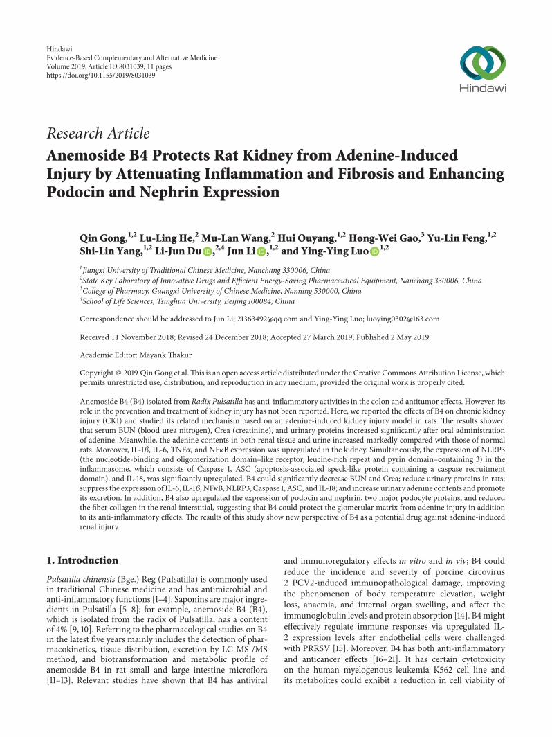
2.2. Chemical and Materials. Anemoside B4 (B4), whosechemical structure is shown in Figure 1(a) (Batch No.20161107, purity of 98% using HPLC determination, thechromatogram of anemoside B4 is shown in Figure 1(b))was presented by Professor Yu-Lin Feng from the Phy-tochemical Department of our university. Prednisolone(pred.) (11-beta,17-alpha; 11,17,21-trihydroxypregna-1,4-diene-3,20-dione; (11beta)-11,17-dihydroxy- 3,20- dioxopregna-1,4-dien-21-yl acetate) (Batch No. 161165) was purchased fromXianju Pharmaceutical Ltd. (Zhejiang, China). Adenine(Sigma-Aldrich, Lot#WXBB0585V) was purchased from
Sigma (Shanghai, China). Creatinine (Crea), urea nitrogen(BUN), and total protein (TP) kits were purchased fromHeguan Chemical Company (Beijing, China). The Brad-ford Protein Assay Kit (P0006) was purchased from Bey-otime Biotechnology Company (Beijing, China). The qPCRdetection kit (Lot# L20509) was purchased from Quan-shijin BioChem Tech Ltd. (Beijing, China). Hematoxylin(MHS16-500ML, LOT#SLBK4909V) and Eosin Y (HT110116-500ML, LOT#J6425V) were purchased from Sigma (Shang-hai, China). Masson’s trichrome kits (Lot:0606A18) werepurchased from Leagene Biotechnology Co., Ltd. (Beijing,China). A Bio-Rad electrophoresis unit and Bio-Rad Chemi-DocXRS+ Gel Imaging System (Beijing, China), LEICARM2235 paraffin slicer, and LEICA DM2500 Optical Micro-scope (Beijing, China) were used in this study.
2.3. Dosages and Groups. All rats were randomly separatedinto the following six groups (each group consisting of 10rats, n=10): the normal control group, model control group,positive control groups, B4 large dose group (2.5 mg/kg),B4 medium dose group (1.25 mg/kg), and B4 small dosegroup (0.625 mg/kg). All the B4 dosages were determinedby the preexperiment, and B4 was given by intravenousadministration. Prednisolone, a type of glucocorticoid, wasused as positive control (5mg/kg) by oral administration.
2.4. Experimental Process. The chronic kidney disease modelwas referenced in the Ali and Diwan reports [29, 30]. Therat model was created by oral administration of adenine(mixture with 0.5% CMC-Na solution, 220 mg/kg) for 3weeks. At day 14 after adenine administration, three dosagesof B4 were given by intravenous injection continuously for 4weeks (one time per day) (the schematic protocol is shownin Figure 1(c)). The normal control was given vehicle with0.5% CMC-Na solution, the model control was given normalsaline, and the positive control was given prednisolone. After4 weeks of administration, the blood and urine samples werecollected, and the rats were killed with anesthesia. The ratkidneys were isolated; the right kidney was fixed with 10%formalin and left kidney was stored at -80∘C for proteinexpression analysis.
During the experiment, the general manifestations of therats, such as their spirit, motor action, and rat fire lightand color, were observed and recorded. Additionally, theirbody weights were detected and recorded each week. Raturine was collected each week using metabolite cages. Thequantity of urine collected in 24 h was determined, and theurine protein content was tested. Serum was isolated fromthe rat blood for the biochemical tests. The serum BUN,Crea, and TP were tested using a 7100 automatic biochemicalanalyzer (Hitachi, Japan). The kidney tissue treated with 10%formalin was analyzed by H.E. preparation or by stainingthe tissue with picrosirius red and Masson’s trichrome andmaking observations with a microscope. Grayscale scanningof the images was carried out by using Adobe PhotoshopCS3(Adobe, California, USA).
2.5. Protein Expression. Protein expression was analyzedusing Western blotting as previously described [31, 32]. For
Evidence-Based Complementary and Alternative Medicine 3
OH
OHOH
OH
OH
OH
OHH
H
HH
HO
HO
HO
HO
HOHO
HO
HO
O
O
O
O
OO
OO
O
O
O
Week2
Week3 Week6
Week0
Adenine
Anemoside B4(4 weeks)
Week 3##
B4 (mg/kg)
Control PrednModel 2.5 1.25 0.625
0 1 2 3 4
Week
ControlModelPredn
2.51.250.625
*
Anemoside B4
PubChem CID:
11636713
0.0 5.0 10.0 15.0 20.0 25.0 30.0 35.0 min
0
25
50
75
100 mAU
(a)
(c)
(d)
(e)(b)
201nm,4nm
150
200
250
300
350
400
450
Body
wei
ght (
g)
100
300
500Bo
dy w
eigh
t (g) ∗
∗
Chemical structure of Anemoside B4
Figure 1: The body weight of rats injured by adenine after the intravenous administration of B4. (a) Chemical structure of B4. (b) Thechromatogram of anemoside B4. (c) Time schematic of the experiment. (d) Body weight during 4 weeks. (e) Body weight at week 3.The datawere shown as mean ± SEM from 10 rats in each group. ##, compared with the control, P < 0.01. ∗, compared with model groups, P < 0.05.Predn: prednisolone, 5mg/kg.
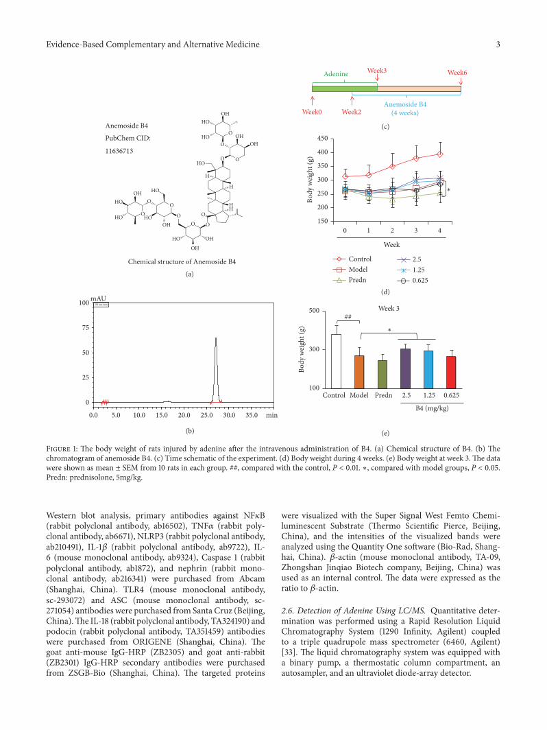
Western blot analysis, primary antibodies against NF𝜅B(rabbit polyclonal antibody, ab16502), TNF𝛼 (rabbit poly-clonal antibody, ab6671), NLRP3 (rabbit polyclonal antibody,ab210491), IL-1𝛽 (rabbit polyclonal antibody, ab9722), IL-6 (mouse monoclonal antibody, ab9324), Caspase 1 (rabbitpolyclonal antibody, ab1872), and nephrin (rabbit mono-clonal antibody, ab216341) were purchased from Abcam(Shanghai, China). TLR4 (mouse monoclonal antibody,sc-293072) and ASC (mouse monoclonal antibody, sc-271054) antibodies were purchased from Santa Cruz (Beijing,China).The IL-18 (rabbit polyclonal antibody, TA324190) andpodocin (rabbit polyclonal antibody, TA351459) antibodieswere purchased from ORIGENE (Shanghai, China). Thegoat anti-mouse IgG-HRP (ZB2305) and goat anti-rabbit(ZB2301) IgG-HRP secondary antibodies were purchasedfrom ZSGB-Bio (Shanghai, China). The targeted proteins
were visualized with the Super Signal West Femto Chemi-luminescent Substrate (Thermo Scientific Pierce, Beijing,China), and the intensities of the visualized bands wereanalyzed using the Quantity One software (Bio-Rad, Shang-hai, China). 𝛽-actin (mouse monoclonal antibody, TA-09,Zhongshan Jinqiao Biotech company, Beijing, China) wasused as an internal control. The data were expressed as theratio to 𝛽-actin.
2.6. Detection of Adenine Using LC/MS. Quantitative deter-mination was performed using a Rapid Resolution LiquidChromatography System (1290 Infinity, Agilent) coupledto a triple quadrupole mass spectrometer (6460, Agilent)[33]. The liquid chromatography system was equipped witha binary pump, a thermostatic column compartment, anautosampler, and an ultraviolet diode-array detector.

4 Evidence-Based Complementary and Alternative Medicine
Chromatographic separation was achieved on a WelchUltimate Luna 3𝜇m HILIC 200A (2.0×150mm). The mobilephase was a mixture of 0.1% formic acid in water (A) andacetonitrile (B). The gradient elution was programmed asfollows: 95% B,0.01-2 min; 95-80% B,2-3 min; 80-70% B,3-4.5min; 60-95 % B,5.5- 6 min; and 95% B,6.0-7.0 min. The totalrun timewas 7min.The column temperature wasmaintainedat 30∘C and the injection volume was 2𝜇L. The flow ratewas set at 0.25 mL/min. The electrospray ionization (ESI)was performed in positive mode. The mass spectrometerparameters were optimized as follows: drying gas flow rate,9 L/min; drying gas temperature, 325∘C; nebulizer pressure,45 psi; and capillary voltage, 3000 V. Nitrogen was used in allcases. The precursor product ion pairs used in MRM modewere m/z 136.1-119.5 for epinephrine. The fragmentor was setat 100 V and the collision energy (CE) was set at 23 eV. Thedwell time of each ion pair was 100 ms. Instrument control,data acquisition, and evaluation were performed with theMass Hunter workstation software (Version B.04.00).
2.7. Data Analysis. The data were expressed as the mean± SEM and statistically analyzed using one-way ANOVAanalysis, and the t-t test was performed for two groups. Thetesting was performed using the SPSS 19.0 software (IBM,Chicago, USA). P values less than 0.05 were consideredstatistically significant. The statistical graphs were producedusing theGraphPad Prism 5.0 software (GraphPadCompany,San Diego, California, USA) or the Microsoft Office Excel2010 software (Microsoft, Maryland, USA).
3. Results
3.1. Effect of B4 on the General Conditions and BodyWeights ofthe Rats with Adenine-Induced Renal Injuries. After adenineadministration, rats in each group showed signs of apathy,coarse and messy fur, weight loss, and decreased activity.After B4 intervention, these phenomena were improved. Therats’ body weights are displayed in Figures 1(d) and 1(e).Theirweights were obviously reduced, implying that the adeninemodel could influence body weight. B4 at the large andmediumdoses could reverse the decrease inweight.The smallB4 dose had no effect on weight. Prednisolone could notinhibit the decrease in body weight.
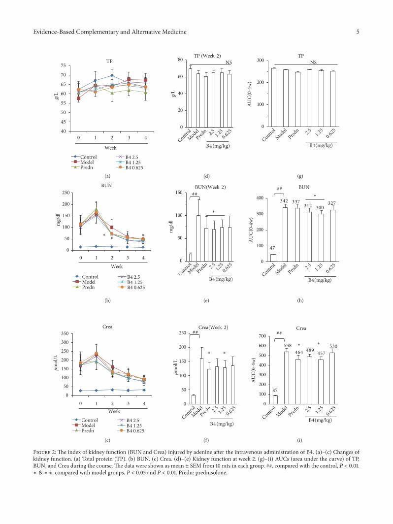
3.2. Effect of B4 on Serum Biochemical Indexes in Rats withAdenine-Induced Renal Damage. Figure 2 presents the rats’kidney functions after the intravenous administration of B4.First, adenine could injure the rat kidney, causing a distinctincrease in BUN and Crea. B4 at the large and mediumdoses could suppress the increase in BUNandCrea comparedwith the model groups after 2 weeks of B4 administration.Based on Figures 2(a), 2(b), and 2(c), we found that BUNand Crea could decrease with time, reflecting kidney self-recovery. The AUCs (area under the curve) of BUN and Creawere calculated to evaluate the effect of B4 on BUN andCrea, respectively. The AUCs of BUN and Crea in the ratmodel increased compared with those of the control groups,indicating that adenine actually injured the rat kidneys. B4 at
the large andmedium doses could also decrease the BUN andCrea AUCs, which is in agreement with the BUN and Crealevels at week 2 (Figures 2(h) and 2(i)). The small B4 dosehad no effects. Prednisolone could decrease BUN and Creasimilar to B4. However, during all the experiments, the bloodtotal protein (TP) displayed negative changes compared withthe control groups (Figures 2(a), 2(d), and 2(g)).
3.3. Effects of B4 on 24 h Urinary Protein Excretion in Ratswithin Adenine-Induced Renal Injury. Urinary protein is akey index reflecting kidney injury. After adenine overload,the urinary protein levels in the model group were higherthan those in the control group, and the total urinary proteinat 24 h after 4 weeks was also higher than that of the controlgroup (Figure 3). B4 at the large and small doses could reducethe 24h total protein levels, implying that B4 protects thekidney from adenine injury (Figure 3(d)). After 4 weeks of B4treatment, there was more urine from the model group at 24h, and there were no differences in the protein concentrationsinmodel groups; however, themodel groupswere higher thanthe normal groups (Figures 3(e) and 3(f)). Finally, the totalprotein in the B4-treated rat was lower than in the model rat,showing that B4 affects kidney damage by decreasing urinaryprotein levels.
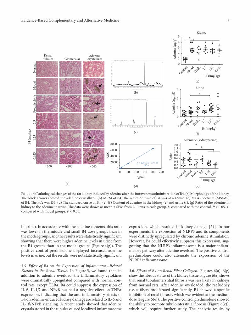
3.4. Effects of B4 on the Renal Histopathology of Rats withAdenine-Induced Renal Injury. With respect to morphology,the kidney structure of the normal group rats was clear andshowed normal glomeruli (renal body), renal tubules, andinterstitium. In contrast, the number of nephrons in themodel control group decreased, and granulomaswere presentin the renal interstitium (mainly consisting of macrophage-like lesions). Visible brown adenine crystals were observedin the lumen and interstitium of the renal tubules, and therenal tubules expanded in a reticular pattern and partiallydisappeared, indicating that adenine could damage the kid-ney renal tubules (Figure 4). The renal tubule dilation wasalleviated in the B4 high- and low-dose groups.
In the model groups, adenine crystallized, implying thatthe kidney could not handle the adenine overload, andresulted in injury to the kidney tubules. B4 could protectthe kidney from the damage caused by the adenine byreducing the number of adenine crystals (Figure 4(a)). Todetermine the exact adenine contents in the kidney, LC/MSwas employed for adenine detection (Figures 4(b)–4(d)). Astandard curve was constructed, and the following correla-tion equation was determined to calculate the adenine con-centration: y=326.13x+237.46 (R2=1). After adenine overload,the concentration of adenine in the rat kidney was distinctlyhigher than in the normal control rat. B4 at the small doseand prednisolone could decrease the adenine levels, but theresults were not statistically significant (Figure 4(e)). Inter-estingly, the adenine contents in rat urine from the groupstreated with the middle and small B4 doses were higherthan those from the model groups, implying that B4 couldpromote the secretion of adenine in the urine (Figure 4(f)).To confirm this result, we examined the ratio of adenine inthe kidney to adenine in urine (adenine in kidney/adenine
Evidence-Based Complementary and Alternative Medicine 5
40
45
50
55
60
65
70
75
0 1 2 3 4
g/L
TP
ControlModelPredn
B4 2.5B4 1.25B4 0.625
ControlModelPredn
B4 2.5B4 1.25B4 0.625
ControlModelPredn
B4 2.5B4 1.25B4 0.625
0
50
100
150
200
250
0 1 2 3 4
mg/
dl
BUN
050
100150200250300350
0 1 2 3 4
m
ol/L
Crea
Week
0
20
40
60
80
g/L
TP (Week 2)
0
50
100
150
mg/
dl
BUN(Week 2)
0
50
100
150
200
250
m
ol/L
Crea(Week 2)
0
100
200
300
AU
C(0
-4w)
TP
47
342 337312 300
327
BUN
87
538464 489 457
530
Crea
NSNS
##
####
##
Week
Week
(a) (d) (g)
(e) (h)
(f) (i)
(b)
(c)
B4 (mg/kg)
B4 (mg/kg)
B4 (mg/kg) B4 (mg/kg)
B4 (mg/kg)
B4 (mg/kg)
Control
ModelPred
n 2.5
1.25
0.625
Control
ModelPred
n 2.5
1.25
0.625
Control
ModelPred
n 2.5
1.25
0.625
ControlModel
Predn 2.
51.25
0.625
ControlModel
Predn 2.
51.250.625
ControlModel
Predn 2.
51.250.625
∗ ∗
∗
∗
∗ ∗
∗
0
100
200
300
400
AU
C(0
-4w)
0
100
200
300
400
500
600
700
AU
C(0
-4w)
∗
Figure 2: The index of kidney function (BUN and Crea) injured by adenine after the intravenous administration of B4. (a)–(c) Changes ofkidney function. (a) Total protein (TP). (b) BUN. (c) Crea. (d)–(e) Kidney function at week 2. (g)–(i) AUCs (area under the curve) of TP,BUN, and Crea during the course. The data were shown as mean ± SEM from 10 rats in each group. ##, compared with the control, P < 0.01.∗ & ∗ ∗, compared with model groups, P < 0.05 and P < 0.01. Predn: prednisolone.

6 Evidence-Based Complementary and Alternative Medicine
Urine(Week4)
Protein(Week4)
Concentration(Week4)
#
#
0
50
100
150
200
0 1 2 3 4
mg/
24h
Protein
ControlModelPredn
B4 2.5B4 1.25B4 0.625
ControlModelPredn
B4 2.5B4 1.25B4 0.625
ControlModelPredn
B4 2.5B4 1.25B4 0.625
0
10
20
30
40
50
0 1 2 3 4
ml/2
4h
Urine
(a) (d)
(b) (e)
(c) (f)
0 1 2 3 4
Concentration
Week
Week
Week
B4 (mg/kg)
B4 (mg/kg)
B4 (mg/kg)
0
1
2
3
4
5
6
mg/
ml/2
4h
0
2
4
6
mg/
ml/2
4h
0
10
20
30
40
50
ml/2
4h
0
50
100
150
mg/
24h
Control
ModelPred
n 2.5
1.25
0.625
Control
ModelPred
n 2.5
1.25
0.625
Control
ModelPred
n 2.5
1.25
0.625
∗ ∗
∗
∗
∗
∗ ∗
Figure 3: Urine and total proteins in urine of rats after the intravenous administration of B4. (a) Total protein of urine in 24h. (b) Quantityof urine during 24h. (c) Concentration of urinary protein in 24h. (d)–(f) Urinary protein of rats after administration of B4 at week 4. At theend of experiment (week 4), the urinary protein per 24h of the model groups was higher than that of normal rats (control). B4 in large andsmall dosage decreased the protein content. The data were shown as mean ± SEM from 10 rats in each group. #, compared with the control,P < 0.05. ∗, compared with model groups, P < 0.05. Predn: prednisolone.
Evidence-Based Complementary and Alternative Medicine 7
y = 328.13x + 237.46R² = 1
Nor
mal
Mod
elPr
edni
solo
ne2.
51.
250.
625
B4 (m
g/kg
)
×200 ×400 ×640
Renaltubules
Adenine crystallizesGlomerular
Kidney
(a)
(b)
(e)
(f)
(g)
(c)
(d)
Control
ModelPred
n 2.5 1.25
0.625
012345
B4(mg/kg)
#
Aden
ine (
g/
g)
Urine
Control
ModelPred
n 2.5 1.25
0.625
B4(mg/kg)
Adeninue (kidney/urine)
Control
ModelPred
n 2.5 1.25
0.625
B4(mg/kg)
2x10
0
0.1
0.2
0.3
0.4
0.5
0.6
0.7
0.8
0.9
1
+ESI Product Ion (0.12 min) Frag=135.0V [email protected] (136.1 ->∗∗) xianpiaoling-pos-20180201-0004.d
119.0
136.0
92.0
67.0
94.0
Counts (%) vs. Mass-to-Charge (m/z)50 55 60 65 70 75 80 85 90 95 100 105 110 115 120 125 130 135 140 145 150
2x10
0.50.6
0.70.8
0.91
1.11.2
1.31.41.5
1.61.7
1.81.9
2
+ESI TIC MRM Frag=110.0V [email protected] (∗∗ -> ∗∗) z-Y-1.d ∗ 4.431 1
Counts vs. Acquisition Time (min)
0.2 0.4 0.6 0.8 1 1.2 1.4 1.6 1.8 2 2.2 2.4 2.6 2.8 3 3.2 3.4 3.6 3.8 4 4.2 4.4 4.6 4.8 5 5.2 5.4 5.6 5.8 6 6.2 6.4 6.6 6.8
0
20000
40000
60000
80000
AUC
50 100 150 200 2500ng/ml
0.00.20.40.60.81.01.52.0
Ratio
0
1
2
3
Aden
ine (
g/
ml) ∗
∗
Figure 4: Pathological changes of the rat kidney induced by adenine after the intravenous administration of B4. (a)Morphology of the kidney.The black arrows showed the adenine crystallizes. (b) MRM of B4. The retention time of B4 was at 4.43min. (c) Mass spectrum (MS/MS)of B4. The m/z was 136. (d) The standard curve of B4. (e)-(f) Content of adenine in the kidney (e) and urine (f). (g) Ratio of the adenine inkidney to the adenine in urine. The data were shown as mean ± SEM from 7-10 rats in each group. #, compared with the control, P < 0.05. ∗,compared with model groups, P < 0.05.
in urine). In accordance with the adenine contents, this ratiowas lower in the middle and small B4 dose groups than inthemodel groups, and the results were statistically significant,showing that there were higher adenine levels in urine fromthe B4 groups than in the model groups (Figure 4(g)). Thepositive control prednisolone displayed increased adeninelevels in urine, but the results were not statistically significant.
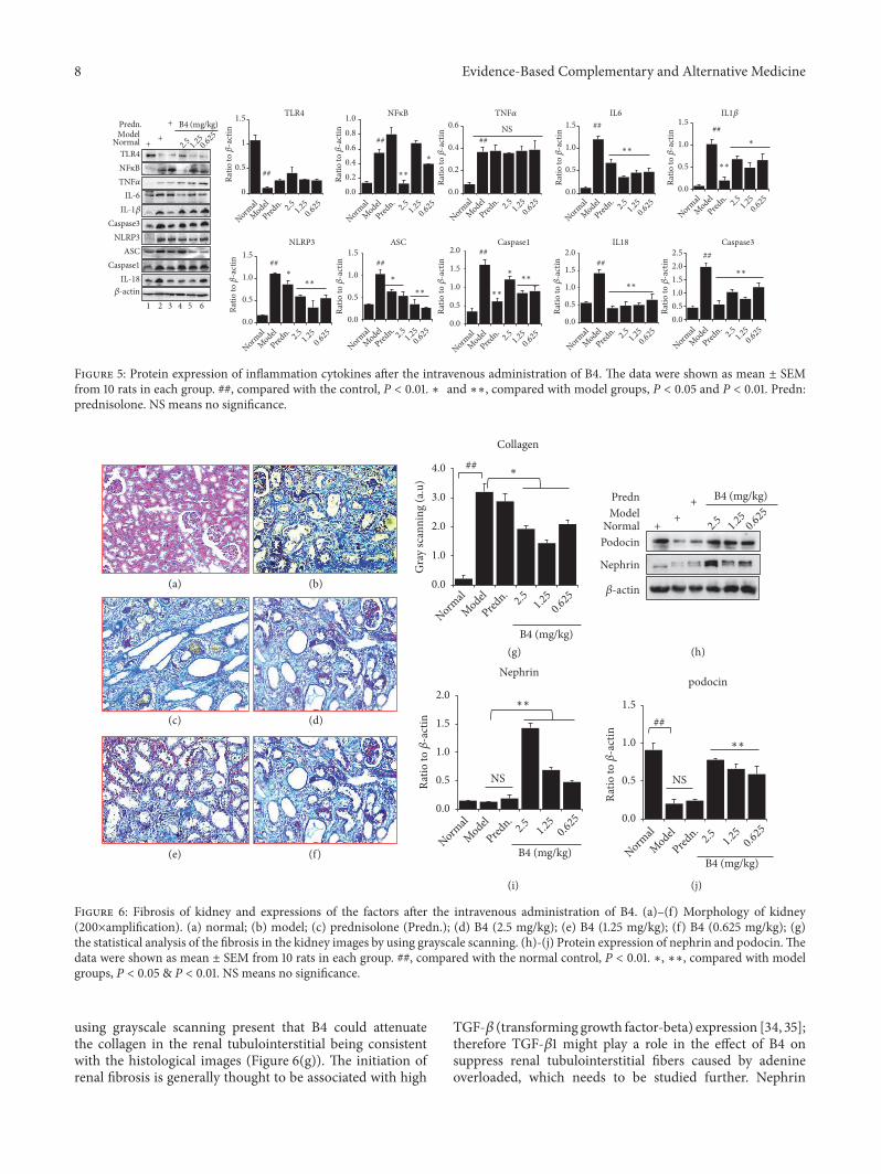
3.5. Effect of B4 on the Expression of Inflammatory-RelatedFactors in the Renal Tissue. In Figure 5, we found that, inaddition to adenine overload, the inflammatory cytokineswere dramatically upregulated compared with normal con-trol rats, except TLR4. B4 could suppress the expression ofIL-6, IL-1𝛽, and NF𝜅B but had a negative effect on TNF𝛼expression, indicating that the anti-inflammatory effects ofB4 on adenine-induced kidney damage are related to IL-6 andIL-1𝛽/NF𝜅B signaling. A recent study showed that adeninecrystals stored in the tubules caused localized inflammasome
expression, which resulted in kidney damage [24]. In ourexperiments, the expression of NLRP3 and its componentswere distinctly upregulated by chronic adenine stimulation.However, B4 could effectively suppress this expression, sug-gesting that the NLRP3 inflammasome is a major inflam-matory pathway after adenine overload. The positive controlprednisolone could also attenuate the expression of theNLRP3 inflammasome.
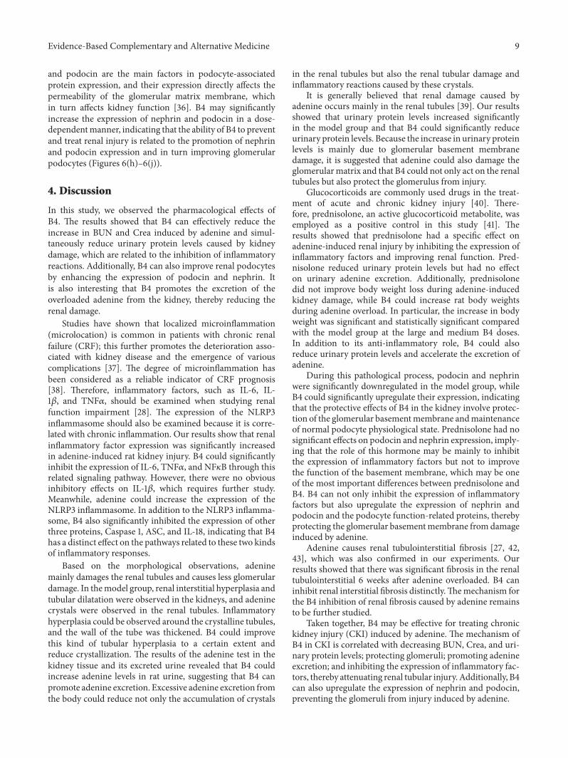
3.6. Effects of B4 on Renal Fiber Collagen. Figures 6(a)–6(g)show the fibrous status of the kidney tissue. Figure 6(a) showsthat renal tubulointerstitial fibrosis was less likely in kidneysfrom normal rats. After adenine overloaded, the rat kidneytissue fibers proliferated significantly. B4 showed a specificinhibition of renal fibrosis, which was evident at the mediumdose (Figure 6(e)).The positive control prednisolone showedthe ability to promote tubulointerstitial fibrosis (Figure 6(c)),which will require further study. The analytic results by
8 Evidence-Based Complementary and Alternative Medicine
0.0
0.5
1.0
1.5IL6
0
0.5
1
1.5 TLR4
0.0
0.2
0.4
0.6TNF
0.00.20.40.60.81.0 NFκB
0.0
0.5
1.0
1.5IL1
0.0
0.5
1.0
1.5
Ratio
to
-act
in
Ratio
to
-act
in
Ratio
to
-act
in
Ratio
to
-act
in
Ratio
to
-act
in
NLRP3
0.0
0.5
1.0
1.5ASC
0.0
0.5
1.0
1.5
2.0Caspase1
0.0
0.5
1.0
1.5
2.0IL18
0.00.51.01.52.02.5
Caspase3
IL-18
NLRP3ASC
Caspase1
IL-1
TLR4
TNFIL-6
Caspase3
NFκB
NormalModelPredn. +
++
B4 (mg/kg)
1 2 3 4 5 6
##
## ##NS ## ##
##∗
∗∗
∗∗∗∗ ∗∗
∗∗∗∗
∗∗
∗∗
∗∗
∗∗
∗
∗
####
####
NormalModel
Predn.
2.51.25
0.625
NormalModel
Predn.
2.51.250.625
NormalModel
Predn.
2.51.250.625
NormalModel
Predn.
2.51.250.625
NormalModel
Predn.
2.51.250.625
NormalModel
Predn.
2.51.250.625
NormalModel
Predn.
2.51.250.625
NormalModel
Predn.
2.51.250.625
NormalModel
Predn.
2.51.250.625
NormalModel
Predn.
2.51.250.625
2.51.250.625
Ratio
to
-act
in
Ratio
to
-act
in
Ratio
to
-act
in
Ratio
to
-act
in
Ratio
to
-act
in
-actin
Figure 5: Protein expression of inflammation cytokines after the intravenous administration of B4. The data were shown as mean ± SEMfrom 10 rats in each group. ##, compared with the control, P < 0.01. ∗ and ∗∗, compared with model groups, P < 0.05 and P < 0.01. Predn:prednisolone. NS means no significance.
B4 (mg/kg)B4 (mg/kg)
B4 (mg/kg)
(a) (b)
(g) (h)
(i) (j)
(c) (d)
(e) (f)
Nephrin
PodocinNormal
ModelPredn +
++
B4 (mg/kg)
Collagen
##
Nephrin
NS
0.0
0.5
1.0
1.5
podocin
##
NS
0.0
1.0
2.0
3.0
4.0
Gra
y sc
anni
ng (a
.u)
0.0
0.5
1.0
1.5
2.0
Ratio
to
-act
in
Ratio
to
-act
in
Normal
Model
Predn.
2.5
1.25
0.625
Normal
Model
Predn.
2.5
1.25
0.625
2.5
1.25
0.625
Normal
Model
Predn.
2.5
1.25
0.625
∗∗
∗∗
∗
-actin
Figure 6: Fibrosis of kidney and expressions of the factors after the intravenous administration of B4. (a)–(f) Morphology of kidney(200×amplification). (a) normal; (b) model; (c) prednisolone (Predn.); (d) B4 (2.5 mg/kg); (e) B4 (1.25 mg/kg); (f) B4 (0.625 mg/kg); (g)the statistical analysis of the fibrosis in the kidney images by using grayscale scanning. (h)-(j) Protein expression of nephrin and podocin.Thedata were shown as mean ± SEM from 10 rats in each group. ##, compared with the normal control, P < 0.01. ∗, ∗∗, compared with modelgroups, P < 0.05 & P < 0.01. NS means no significance.
using grayscale scanning present that B4 could attenuatethe collagen in the renal tubulointerstitial being consistentwith the histological images (Figure 6(g)). The initiation ofrenal fibrosis is generally thought to be associated with high
TGF-𝛽 (transforming growth factor-beta) expression [34, 35];therefore TGF-𝛽1 might play a role in the effect of B4 onsuppress renal tubulointerstitial fibers caused by adenineoverloaded, which needs to be studied further. Nephrin
Evidence-Based Complementary and Alternative Medicine 9
and podocin are the main factors in podocyte-associatedprotein expression, and their expression directly affects thepermeability of the glomerular matrix membrane, whichin turn affects kidney function [36]. B4 may significantlyincrease the expression of nephrin and podocin in a dose-dependentmanner, indicating that the ability of B4 to preventand treat renal injury is related to the promotion of nephrinand podocin expression and in turn improving glomerularpodocytes (Figures 6(h)–6(j)).
4. Discussion
In this study, we observed the pharmacological effects ofB4. The results showed that B4 can effectively reduce theincrease in BUN and Crea induced by adenine and simul-taneously reduce urinary protein levels caused by kidneydamage, which are related to the inhibition of inflammatoryreactions. Additionally, B4 can also improve renal podocytesby enhancing the expression of podocin and nephrin. Itis also interesting that B4 promotes the excretion of theoverloaded adenine from the kidney, thereby reducing therenal damage.
Studies have shown that localized microinflammation(microlocation) is common in patients with chronic renalfailure (CRF); this further promotes the deterioration asso-ciated with kidney disease and the emergence of variouscomplications [37]. The degree of microinflammation hasbeen considered as a reliable indicator of CRF prognosis[38]. Therefore, inflammatory factors, such as IL-6, IL-1𝛽, and TNF𝛼, should be examined when studying renalfunction impairment [28]. The expression of the NLRP3inflammasome should also be examined because it is corre-lated with chronic inflammation. Our results show that renalinflammatory factor expression was significantly increasedin adenine-induced rat kidney injury. B4 could significantlyinhibit the expression of IL-6, TNF𝛼, and NF𝜅B through thisrelated signaling pathway. However, there were no obviousinhibitory effects on IL-1𝛽, which requires further study.Meanwhile, adenine could increase the expression of theNLRP3 inflammasome. In addition to the NLRP3 inflamma-some, B4 also significantly inhibited the expression of otherthree proteins, Caspase 1, ASC, and IL-18, indicating that B4has a distinct effect on the pathways related to these two kindsof inflammatory responses.
Based on the morphological observations, adeninemainly damages the renal tubules and causes less glomerulardamage. In themodel group, renal interstitial hyperplasia andtubular dilatation were observed in the kidneys, and adeninecrystals were observed in the renal tubules. Inflammatoryhyperplasia could be observed around the crystalline tubules,and the wall of the tube was thickened. B4 could improvethis kind of tubular hyperplasia to a certain extent andreduce crystallization. The results of the adenine test in thekidney tissue and its excreted urine revealed that B4 couldincrease adenine levels in rat urine, suggesting that B4 canpromote adenine excretion. Excessive adenine excretion fromthe body could reduce not only the accumulation of crystals
in the renal tubules but also the renal tubular damage andinflammatory reactions caused by these crystals.
It is generally believed that renal damage caused byadenine occurs mainly in the renal tubules [39]. Our resultsshowed that urinary protein levels increased significantlyin the model group and that B4 could significantly reduceurinary protein levels. Because the increase in urinary proteinlevels is mainly due to glomerular basement membranedamage, it is suggested that adenine could also damage theglomerularmatrix and that B4 could not only act on the renaltubules but also protect the glomerulus from injury.
Glucocorticoids are commonly used drugs in the treat-ment of acute and chronic kidney injury [40]. There-fore, prednisolone, an active glucocorticoid metabolite, wasemployed as a positive control in this study [41]. Theresults showed that prednisolone had a specific effect onadenine-induced renal injury by inhibiting the expression ofinflammatory factors and improving renal function. Pred-nisolone reduced urinary protein levels but had no effecton urinary adenine excretion. Additionally, prednisolonedid not improve body weight loss during adenine-inducedkidney damage, while B4 could increase rat body weightsduring adenine overload. In particular, the increase in bodyweight was significant and statistically significant comparedwith the model group at the large and medium B4 doses.In addition to its anti-inflammatory role, B4 could alsoreduce urinary protein levels and accelerate the excretion ofadenine.
During this pathological process, podocin and nephrinwere significantly downregulated in the model group, whileB4 could significantly upregulate their expression, indicatingthat the protective effects of B4 in the kidney involve protec-tion of the glomerular basementmembrane andmaintenanceof normal podocyte physiological state. Prednisolone had nosignificant effects on podocin and nephrin expression, imply-ing that the role of this hormone may be mainly to inhibitthe expression of inflammatory factors but not to improvethe function of the basement membrane, which may be oneof the most important differences between prednisolone andB4. B4 can not only inhibit the expression of inflammatoryfactors but also upregulate the expression of nephrin andpodocin and the podocyte function-related proteins, therebyprotecting the glomerular basementmembrane from damageinduced by adenine.
Adenine causes renal tubulointerstitial fibrosis [27, 42,43], which was also confirmed in our experiments. Ourresults showed that there was significant fibrosis in the renaltubulointerstitial 6 weeks after adenine overloaded. B4 caninhibit renal interstitial fibrosis distinctly.Themechanism forthe B4 inhibition of renal fibrosis caused by adenine remainsto be further studied.
Taken together, B4 may be effective for treating chronickidney injury (CKI) induced by adenine. The mechanism ofB4 in CKI is correlated with decreasing BUN, Crea, and uri-nary protein levels; protecting glomeruli; promoting adenineexcretion; and inhibiting the expression of inflammatory fac-tors, thereby attenuating renal tubular injury. Additionally, B4can also upregulate the expression of nephrin and podocin,preventing the glomeruli from injury induced by adenine.

10 Evidence-Based Complementary and Alternative Medicine
Data Availability
The data used to support the findings of this study areincluded within the article.
Conflicts of Interest
The authors declare that they have no conflicts of interest.
Authors’ Contributions
Qin Gong and Lu-Ling He equally contributed to this work.
Acknowledgments
We thank all the colleagues in our laboratory for the assis-tance of the article. This work was supported by NationalInnovative Drugs 13th Five-Year Major Special Project ofChina (2018ZX09301030-002).
References
[1] W.-H. Wang, J. Yang, H.-S. Peng, and J.-P. Qian, “Studyon morphological characteristics and microscopic structureof medicinal organs of pulsatilla chinensis (Bunge) regel,”Microscopy Research and Technique, vol. 80, no. 8, pp. 950–958,2017.
[2] Y. Shi, M. Zhao, H. Yao et al., “Rapidly discriminate commercialmedicinal Pulsatilla chinensis (Bge.) regel from its adulterantsusing ITS2 barcoding and specific PCR-RFLP assay,” ScientificReports, vol. 7, Article ID e40000, 2017.
[3] L.-D. Li, W.-C. Li, C.-W. Liu et al., “Giardia intestinalis: effectsof pulsatilla chinensis extracts on trophozoites,” ParasitologyResearch, vol. 111, no. 5, pp. 1929–1935, 2012.
[4] F. Zhou, O. Lv, Y. Zheng et al., “Inhibitory effect of Pulsatillachinensis polysaccharides on glioma,” International Journal ofBiological Macromolecules, vol. 50, no. 5, pp. 1322–1326, 2012.
[5] Y.Mimaki, A. Yokosuka,M. Kuroda,M.Hamanaka, C. Sakuma,and Y. Sashida, “New bisdesmosidic triterpene saponins fromthe roots of Pulsatilla chinensis,” Journal of Natural Products,vol. 64, no. 9, pp. 1226–1229, 2001.
[6] L. I. Glebko, N. P. Krasovskaj, L. I. Strigina, K. P. Ulanova, V. A.Denisenko, and P. S. Dmitrenok, “Triterpene glycosides fromPulsatilla chinensis,” Russian Chemical Bulletin, vol. 51, no. 10,pp. 1945–1950, 2002.
[7] M.-M. Jin, W.-D. Zhang, Y.-M. Xu et al., “Simultaneous deter-mination of 12 active components in the roots of Pulsatillachinensis using tissue-smashing extraction with liquid chro-matography and mass spectrometry,” Journal of SeparationScience, vol. 40, no. 6, pp. 1283–1292, 2017.
[8] K. Xu, Z. Shu, Q.-M. Xu et al., “Cytotoxic activity of Pulsatillachinensis saponins and their structure-activity relationship,”Journal of Asian Natural Products Research, vol. 15, no. 6, pp.680–686, 2013.
[9] Y. Ling, Z. Lin, W. Zha, T. Lian, and S. You, “Rapid detectionand characterisation of triterpene saponins from the rootof pulsatilla chinensis (Bunge) Regel by HPLC-ESI-QTOF-MS/MS,” Phytochemical Analysis, pp. 174–183, 2016.
[10] L. Wang, Q. Xu, S. Su et al., “Simultaneous purification ofpulchinenoside B4 and B5 from pulsatilla chinensis using
macroporous resin and preparative high performance liquidchromatography,” Industrial & Engineering Chemistry Research,vol. 51, no. 45, pp. 14859–14866, 2012.
[11] L. Yang, X. Meng, X. Yu, and H. Kuang, “Simultaneousdetermination of anemoside B4, phellodendrine, berberine,palmatine, obakunone, esculin, esculetin in rat plasma byUPLC–ESI–MS/MS and its application to a comparative phar-macokinetic study in normal and ulcerative colitis rats,” Journalof Pharmaceutical and Biomedical Analysis, vol. 134, pp. 43–52,2017.
[12] X. Guo, Y. Xie, S. Lian et al., “A sensitive HPLC–MS/MSmethod for the simultaneous determination of anemoside B4,anemoside A3 and 23-hydroxybetulinic acid: application to thepharmacokinetics and liver distribution of Pulsatilla chinensissaponins,” Biomedical Chromatography, vol. 32, no. 3, Article IDe4124, 2018.
[13] M. He, H. Ouyang, M. He et al., “Application of a liquidchromatography–tandem mass spectrometry method to theapplication of a liquid chromatography–tandem mass spec-trometry method to the pharmacokinetics, tissue distributionand excretion in the study of anemoside B4, a novel antiviralagent candidate, in rats,” Biomedical Chromatography, vol. 31,no. 7, Article ID e3914, 2017.
[14] H. Yang, X. Chen, C. Jiang, K. He, and Y. Hu, “Antiviral andimmunoregulatory role against PCV2 in vivo of Chinese herbalmedicinal ingredients,” Journal of Veterinary Research (Poland),vol. 61, no. 4, pp. 405–410, 2017.
[15] Y. Hu, A. Mao, Y. Tan, Y. Zhao, and K. He, “Role of 5 saponinsin secretion of cytokines by PRRSV-induced endothelial cells,”Drug Research, vol. 66, no. 7, pp. 357–362, 2016.
[16] M. Liu, X. Zhao, L. Xiao et al., “Cytotoxicity of the compoundsisolated from Pulsatilla chinensis saponins and apoptosisinduced by 23-hydroxybetulinic acid,” Pharmaceutical Biology,vol. 53, no. 1, pp. 1–9, 2015.
[17] Y.-Y. Luo, L.-Y. Chen, H. Jian et al., “Regulation of saponinsfrom Pulsatilla chinensis on energy metabolism of Bel-7402xenograft in nude mice,” Chinese Traditional and Herbal Drugs,vol. 45, no. 7, pp. 973–977, 2014.
[18] T. Liu, L. Ye, X. Guan et al., “Immunopontentiating andantitumor activities of a polysaccharide from Pulsatilla chinen-sis (Bunge) Regel,” International Journal of Biological Macro-molecules, vol. 54, no. 1, pp. 225–229, 2013.
[19] Q.-M.Xu, Z. Shu,W.-J.He et al., “Antitumor activity of Pulsatillachinensis (Bunge) Regel saponins in human liver tumor 7402cells in vitro and in vivo,” Phytomedicine, vol. 19, no. 3-4, pp.293–300, 2012.
[20] Y. Mimaki, M. Kuroda, T. Asano, and Y. Sashida, “Triterpenesaponins and lignans from the roots of Pulsatilla chinensis andtheir cytotoxic activity against HL-60 cells,” Journal of NaturalProducts, vol. 62, no. 9, pp. 1279–1283, 1999.
[21] J. Y. Wan, Y. Z. Zhang, J. B. Yuan et al., “Biotransformationand metabolic profile of Anemoside B4 with rat small andlarge intestine microflora by ultra-high performance liquidchromatography-quadrupole time of flight tandem mass spec-trometry,” Biomedical Chromatography, 2016.
[22] M. Al Za’abi, A. Shalaby, P. Manoj, and B. H. Ali, “The invivo effects of adenine-induced chronic kidney disease on somerenal and hepatic function andCYP450metabolizing enzymes,”Physiological Research, vol. 66, no. 2, pp. 263–271, 2017.
[23] H. L. Runolfsdottir, R. Palsson, I. M. Agustsdottir, O. S.Indridason, and V. O. Edvardsson, “Kidney disease in adenine
Evidence-Based Complementary and Alternative Medicine 11
phosphoribosyltransferase deficiency,”American Journal of Kid-ney Diseases, vol. 67, no. 3, pp. 431–438, 2016.
[24] O. Akchurin, A. Sureshbabu, S. B. Doty et al., “Lack of hep-cidin ameliorates anemia and improves growth in an adenine-induced mouse model of chronic kidney disease,” AmericanJournal of Physiology-Renal Physiology, vol. 311, no. 5, pp. F877–F889, 2016.
[25] M. Correa-Costa, T. T. Braga, R. J. F. Felizardo et al.,“Macrophage trafficking as key mediator of adenine-inducedkidney injury,”Mediators of Inflammation, vol. 2014, Article IDe291024, 12 pages, 2014.
[26] S. L. Chong and Y. H. Ng, “Obstructive uropathy and severeacute kidney injury from renal calculi due to adenine phospho-ribosyltransferase deficiency,” World Journal of Pediatrics, vol.12, no. 2, pp. 243–245, 2016.
[27] V. Diwan, L. Brown, andG. C. Gobe, “Adenine-induced chronickidney disease in rats,” Nephrology, vol. 23, no. 1, pp. 5–11, 2018.
[28] B. S. Franklin, M. S. Mangan, and E. Latz, “Crystal formation ininflammation,” Annual Review of Immunology, vol. 34, pp. 173–202, 2016.
[29] B. H. Ali, L. Cahlikova, L. Opletal et al., “Effect of aqueousextract and anthocyanins of calyces ofHibiscus sabdariffa (Mal-vaceae) in rats with adenine-induced chronic kidney disease,”Journal of Pharmacy and Pharmacology, vol. 69, no. 9, pp. 1219–1229, 2017.
[30] V. Diwan, G. Gobe, and L. Brown, “Glibenclamide improveskidney and heart structure and function in the adenine-dietmodel of chronic kidney disease,” Pharmacological Research,vol. 79, pp. 104–110, 2014.
[31] X.Wang, X. Yu, X. Yan et al., “TRPM8 in the negative regulationof TNF𝛼 expression during cold stress,” Scientific Reports, vol. 7,no. 1, Article ID e45155, 2017.
[32] Z. Yuan, X. Lu, F. Lei et al., “TATA boxes in gene transcriptionand poly(A) tails in mRNA stability: new perspective on theeffects of berberine,” Scientific Reports, vol. 5, no. 1, Article IDe18326, 2016.
[33] J. Shi, H.-F. Liu, J. M. Wong, R. N. Huang, E. Jones, andT. J. Carlson, “Development of a robust and sensitive LC-MS/MS method for the determination of adenine in plasma ofdifferent species and its application to in vivo studies,” Journalof Pharmaceutical and Biomedical Analysis, vol. 56, no. 4, pp.778–784, 2011.
[34] Y.-Y. Zhao, X.-L. Cheng, F. Wei et al., “Intrarenal metabolomicinvestigation of chronic kidney disease and its TGF-𝛽1 mech-anism in induced-adenine rats using UPLC Q-TOF/HSMS/MSE,” Journal of Proteome Research, vol. 12, no. 2, pp. 692–703,2013.
[35] P. Wang, M. L. Luo, E. Song et al., “Long noncoding RNAlnc-TSI inhibits renal fibrogenesis by negatively regulating theTGF-/Smad3 pathway,” Science Translational Medicine, vol. 10,p. e2039, 2018.
[36] A. Tofighi, S. Ahmadi, S. M. Seyyedi, A. Shirpoor, F. Kherad-mand, and F. H. Gharalari, “Nandrolone administration with orwithout strenuous exercise promotes overexpression of nephrinand podocin genes and induces structural and functionalalterations in the kidneys of rats,” Toxicology Letters, vol. 282,pp. 147–153, 2018.
[37] S. Cottone, M. C. Lorito, R. Riccobene et al., “Oxidativestress, inflammation and cardiovascular disease in chronic renalfailure,” Journal of Nephrology, vol. 21, no. 2, pp. 175–179, 2008.
[38] E. Avci, E. Cakir, S. C. Cevher, H. Yaman,M. Agilli, and C. Bilgi,“Determination of oxidative stress and cellular inflammation in
patients with diabetic nephropathy and non-diabetic nephropa-thy being administered hemodialysis treatment due to chronicrenal failure,” Renal Failure, vol. 36, no. 5, pp. 767–773, 2014.
[39] D. Claramunt, H. Gil-Pena, R. Fuente et al., “Chronic kid-ney disease induced by adenine: a suitable model of growthretardation in uremia,” American Journal of Physiology-RenalPhysiology, vol. 309, no. 1, pp. F57–F62, 2015.
[40] T. Hamano, N. Fujii, Y. Nagasawa et al., “Serum NTX isa practical marker for assessing antiresorptive therapy forglucocorticoid treated patients with chronic kidney disease,”Bone, vol. 39, no. 5, pp. 1067–1072, 2006.
[41] R. Rebolledo, B. Liu, M. Z. Akhtar et al., “Prednisolone hasa positive effect on the kidney but not on the liver of braindead rats: a potencial role in complement activation,” Journalof Translational Medicine, vol. 12, no. 1, article e111, 2014.
[42] V. Diwan, L. Brown, and G. C. Gobe, “The flavonoid rutinimproves kidney and heart structure and function in anadenine-induced rat model of chronic kidney disease,” Journalof Functional Foods, vol. 33, pp. 85–93, 2017.
[43] R.Thakur, A. Sharma,M.C. Lingaraju et al., “Ameliorative effectof ursolic acid on renal fibrosis in adenine-induced chronickidney disease in rats,” Biomedicine & Pharmacotherapy, vol.101, pp. 972–980, 2018.

Stem Cells International
Hindawiwww.hindawi.com Volume 2018
Hindawiwww.hindawi.com Volume 2018
MEDIATORSINFLAMMATION
of
EndocrinologyInternational Journal of
Hindawiwww.hindawi.com Volume 2018
Hindawiwww.hindawi.com Volume 2018
Disease Markers
Hindawiwww.hindawi.com Volume 2018
BioMed Research International
OncologyJournal of
Hindawiwww.hindawi.com Volume 2013
Hindawiwww.hindawi.com Volume 2018
Oxidative Medicine and Cellular Longevity
Hindawiwww.hindawi.com Volume 2018
PPAR Research
Hindawi Publishing Corporation http://www.hindawi.com Volume 2013Hindawiwww.hindawi.com
The Scientific World Journal
Volume 2018
Immunology ResearchHindawiwww.hindawi.com Volume 2018
Journal of
ObesityJournal of
Hindawiwww.hindawi.com Volume 2018
Hindawiwww.hindawi.com Volume 2018
Computational and Mathematical Methods in Medicine
Hindawiwww.hindawi.com Volume 2018
Behavioural Neurology
OphthalmologyJournal of
Hindawiwww.hindawi.com Volume 2018
Diabetes ResearchJournal of
Hindawiwww.hindawi.com Volume 2018
Hindawiwww.hindawi.com Volume 2018
Research and TreatmentAIDS
Hindawiwww.hindawi.com Volume 2018
Gastroenterology Research and Practice
Hindawiwww.hindawi.com Volume 2018
Parkinson’s Disease
Evidence-Based Complementary andAlternative Medicine
Volume 2018Hindawiwww.hindawi.com
Submit your manuscripts atwww.hindawi.com