Alkaline Phosphatase and Hypophosphatasia · J. L. Milla´n, M. P. Whyte: Alkaline Phosphatase and...
19
REVIEW Alkaline Phosphatase and Hypophosphatasia Jose ´ Luis Milla ´n 1 • Michael P. Whyte 2,3 Received: 16 September 2015 / Accepted: 28 October 2015 / Published online: 21 November 2015 Ó The Author(s) 2015. This article is published with open access at Springerlink.com Abstract Hypophosphatasia (HPP) results from ALPL mutations leading to deficient activity of the tissue-non- specific alkaline phosphatase isozyme (TNAP) and thereby extracellular accumulation of inorganic pyrophosphate (PP i ), a natural substrate of TNAP and potent inhibitor of mineralization. Thus, HPP features rickets or osteomalacia and hypomineralization of teeth. Enzyme replacement using mineral-targeted TNAP from birth prevented severe HPP in TNAP-knockout mice and was then shown to res- cue and substantially treat infants and young children with life-threatening HPP. Clinical trials are revealing aspects of HPP pathophysiology not yet fully understood, such as craniosynostosis and muscle weakness when HPP is severe. New treatment approaches are under development to improve patient care. Keywords Rickets Osteomalacia Enzyme replacement Calcification Seizures Introduction In 1923, Robert Robison, Ph.D. discovered a phosphatase abundant in the skeleton possibly to generate inorganic phosphate (P i ) required to form bone mineral [1]. In 1932, he postulated that an additional but unknown factor regulates skeletal mineralization [2]. This factor would prove to be inorganic pyrophosphate (PP i ), a potent inhibitor of miner- alization and natural substrate for Robison’s enzyme [3]. The phosphatase, now called tissue-non-specific alkaline phos- phatase (TNAP, or TNSALP), is encoded by the ALPL gene [4] expressed richly in bone, liver, and kidney, but also in the central nervous system, fibroblasts, and endothelial and other cell types. Hypomorphic ALPL mutation(s) cause hypophosphatasia (HPP) [4], a rare form of rickets or osteomalacia [5] featuring low serum alkaline phosphatase (ALP) activity and with an incidence for its most severe forms of 1:100,000 [6] and 1:300,000 [7] in the general population [6] of North America and Europe, respectively, but 1 per 2500 births in Canadian Mennonites [8]. The effi- cacy of enzyme replacement therapy (EzRT) using a min- eral-targeted form of recombinant TNAP has been demonstrated for severe HPP [9]. Our review summarizes knowledge concerning TNAP, including revelations about its physiological function coming from investigation of HPP patients and mouse models and successes using EzRT. The Enzyme In humans, three genes besides ALPL (ALPI, ALPP, and ALPPL2) encode alkaline phosphatase (ALP) isozymes, but with restricted tissue expression; i.e., ‘‘intestinal,’’ ‘‘pla- cental,’’ and ‘‘germ cell’’ ALP and are not compromised in HPP [10]. TNAP structure modeling is based on its sequence & Jose ´ Luis Milla ´n [email protected] 1 Sanford Children’s Health Research Center, Sanford Burnham Prebys Medical Discovery Institute, La Jolla, CA 92037, USA 2 Center for Metabolic Bone Disease and Molecular Research, Shriners Hospital for Children, St. Louis, MO 63110, USA 3 Division of Bone and Mineral Diseases, Washington University School of Medicine at Barnes-Jewish Hospital, St. Louis, MO 63110, USA 123 Calcif Tissue Int (2016) 98:398–416 DOI 10.1007/s00223-015-0079-1
Transcript of Alkaline Phosphatase and Hypophosphatasia · J. L. Milla´n, M. P. Whyte: Alkaline Phosphatase and...

REVIEW
Alkaline Phosphatase and Hypophosphatasia
Jose Luis Millan1• Michael P. Whyte2,3
Received: 16 September 2015 / Accepted: 28 October 2015 / Published online: 21 November 2015
� The Author(s) 2015. This article is published with open access at Springerlink.com
Abstract Hypophosphatasia (HPP) results from ALPL
mutations leading to deficient activity of the tissue-non-
specific alkaline phosphatase isozyme (TNAP) and thereby
extracellular accumulation of inorganic pyrophosphate
(PPi), a natural substrate of TNAP and potent inhibitor of
mineralization. Thus, HPP features rickets or osteomalacia
and hypomineralization of teeth. Enzyme replacement
using mineral-targeted TNAP from birth prevented severe
HPP in TNAP-knockout mice and was then shown to res-
cue and substantially treat infants and young children with
life-threatening HPP. Clinical trials are revealing aspects of
HPP pathophysiology not yet fully understood, such as
craniosynostosis and muscle weakness when HPP is severe.
New treatment approaches are under development to
improve patient care.
Keywords Rickets � Osteomalacia � Enzyme
replacement � Calcification � Seizures
Introduction
In 1923, Robert Robison, Ph.D. discovered a phosphatase
abundant in the skeleton possibly to generate inorganic
phosphate (Pi) required to form bonemineral [1]. In 1932, he
postulated that an additional but unknown factor regulates
skeletal mineralization [2]. This factor would prove to be
inorganic pyrophosphate (PPi), a potent inhibitor of miner-
alization and natural substrate for Robison’s enzyme [3]. The
phosphatase, now called tissue-non-specific alkaline phos-
phatase (TNAP, or TNSALP), is encoded by the ALPL gene
[4] expressed richly in bone, liver, and kidney, but also in the
central nervous system, fibroblasts, and endothelial and other
cell types. Hypomorphic ALPL mutation(s) cause
hypophosphatasia (HPP) [4], a rare form of rickets or
osteomalacia [5] featuring low serum alkaline phosphatase
(ALP) activity and with an incidence for its most severe
forms of 1:100,000 [6] and 1:300,000 [7] in the general
population [6] of North America and Europe, respectively,
but 1 per 2500 births in Canadian Mennonites [8]. The effi-
cacy of enzyme replacement therapy (EzRT) using a min-
eral-targeted form of recombinant TNAP has been
demonstrated for severe HPP [9]. Our review summarizes
knowledge concerning TNAP, including revelations about
its physiological function coming from investigation of HPP
patients and mouse models and successes using EzRT.
The Enzyme
In humans, three genes besides ALPL (ALPI, ALPP, and
ALPPL2) encode alkaline phosphatase (ALP) isozymes, but
with restricted tissue expression; i.e., ‘‘intestinal,’’ ‘‘pla-
cental,’’ and ‘‘germ cell’’ ALP and are not compromised in
HPP [10]. TNAP structure modeling is based on its sequence
& Jose Luis Millan
1 Sanford Children’s Health Research Center, Sanford
Burnham Prebys Medical Discovery Institute, La Jolla,
CA 92037, USA
2 Center for Metabolic Bone Disease and Molecular Research,
Shriners Hospital for Children, St. Louis, MO 63110, USA
3 Division of Bone and Mineral Diseases, Washington
University School of Medicine at Barnes-Jewish Hospital,
St. Louis, MO 63110, USA
123
Calcif Tissue Int (2016) 98:398–416
DOI 10.1007/s00223-015-0079-1

homology to the placental isozyme (PLAP) for which crys-
tallographic coordinates have been determined [11]. TNAP
functions physiologically as a homodimer (Fig. 1) [12]. The
twomonomers become related by a twofold crystallographic
axis, with the monomer–monomer interface displaying a
strong hydrophobic character with fewer than 30 % of its
amino acid residues (hereafter ‘‘residues’’) involved in
hydrogen-bonding interactions [13]. This feature renders the
monomer–monomer interface crucial for stability and
enzymatic function, and TNAP (and mammalian ALPs in
general) is therefore an obligatory homodimer. A flexible
surface loop, ‘‘the crown domain,’’ contains residues
important for stabilizing the binding of uncompetitive TNAP
inhibitors, such as E429 and Y367 [14–16]. Furthermore,
this domain contains a low-affinity collagen-binding motif
[16]. The N-terminal a-helix (residues 9–25) of each
monomeric subunit reaches the active site of the contralateral
subunit (Fig. 1) [17]. Both the crown domain and the
N-terminal arm help stabilize the dimeric structure and
determine allosteric properties [18]. Thus, structural and
functional properties explain how some hypomorphic ALPL
alleles compromise the kinetic properties of the entire dimer
(a dominant-negative effect) leading to TNAP insufficiency
and generation-to-generation inheritance of HPP [19]. Three
metal-binding sites surrounding the catalytic Ser residue are
essential for TNAP enzymatic activity; i.e., M1 and M2
(occupied byZn2?) andM3 (occupied byMg2?) [11, 14]. An
additional metal-binding site, M4, apparently occupied by
Ca2? and absent in bacterial ALPs, was revealed by solving
the 3D structure of PLAP [11, 20]. However, this M4
structural metal site does not influence TNAP catalytic
activity [21]. During TNAP-mediated catalysis, Zn2? ions
occupy theM1 andM2 sites, Mg2? theM3 site, and Ca2? the
M4 site. With skeletal mineralization, the increasing Ca2?
gradient in the extracellular matrix first activates TNAP by
replacing the Mg2? with Ca2? at M3, but at high Ca2?
concentrations, it gradually deactivates TNAP as Ca2?
competes out the Zn2? from theM1 andM2metal sites [21].
This explains why TNAP loses activity at the completion of
the mineralization process [22].
Thus, ALPL mutations that alter residues at the mono-
mer–monomer interface, the crown domain, the N-terminal
arm, and the divalent cation-binding sites can all cause
HPP [20]. Additionally, post-translational modifications of
TNAP are important. TNAP (indeed all mammalian ALPs)
is bound to the surface of plasma membranes via a gly-
cosylphosphatidylinositol (GPI) anchor that enables
movement of the enzyme by enhancing membrane fluidity
[23]. This GPI anchor can be cleaved by the enzymatic
action of phospholipases found in plasma membranes,
perhaps explaining how TNAP is released into the circu-
lation and other biological fluids [24]. Also, TNAP con-
tains five putative N-linked glycosylation sites, N123,
N213, N254, N286, and N413 [25], with the sugar chains
required for catalytic activity [26]. The types of sugars in
TNAP explain the different biophysical and kinetic prop-
erties of its ‘‘isoforms’’ produced by bone, liver, kidney,
and vascular cells [27].
In vitro, TNAP has broad substrate specificity and can
hydrolyze or transphosphorylate a considerable variety of
compounds [10]. However, only a few [inorganic
pyrophosphate (PPi), pyridoxal 50-phosphate (PLP), and
likely phosphoethanolamine (PEA)] are natural substrates
for TNAP based on studies of HPP patients and fibroblasts
and Alpl knockout (KO) mice [28, 29]. Nevertheless, it is
unclear what metabolic pathway leads to PEA accumula-
tion [27, 29]. Recent studies also suggest that ATP [30–32],
di-phosphoryl lipopolysaccharide (LPS) [33], and phos-
phorylated osteopontin (p-OPN) [34] are natural substrates
of TNAP.
The Disease
HPP features extraordinarily broad-ranging severity
(Fig. 2), but all patients carry one or two loss-of-function
mutations in their ALPL alleles [4, 35, 36]. Traditionally,
HPP is classified in the clinic firstly by whether there are
dental manifestations alone (i.e., without skeletal disease or
other complications), and then according to the patient’s
age when any additional complications initially mani-
fested. Thus, in order of decreasing severity, clinicians
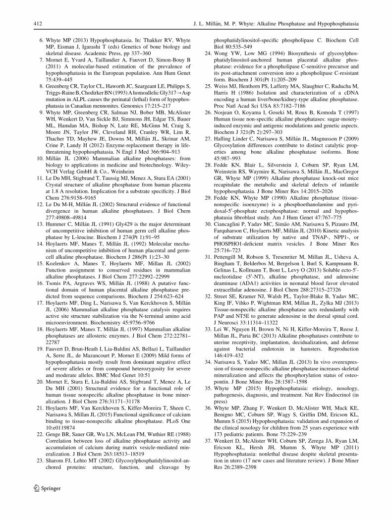
Fig. 1 Ribbon representation of the 3D structure of alkaline phos-
phatase (1EW2) [10]. The active site phosphate (PO43-) bound to
Ser92 during catalysis is shown in green. The three active site metal
ions, two Zn2? (Zn1 and Zn2) and one Mg2? (Mg), are shown in
white as well as the structural Ca2? ion (Ca). Also indicated are the
flexible exposed sequence known as the ‘‘crown domain’’; the
N-terminal helix of one subunit that reaches close to the active site of
the contralateral subunit; and the location where the glycosylphos-
phatidylinositol (GPI) anchor is attached to the C-terminus of the
mature enzyme (Color figure online)
J. L. Millan, M. P. Whyte: Alkaline Phosphatase and Hypophosphatasia 399
123
recognize perinatal HPP, infantile HPP, childhood HPP,
adult HPP, and odonto-HPP [35]. Perinatal HPP is
detectable in utero using fetal sonography, and can cause
stillbirth or be fatal soon after [37]. Infantile HPP presents
before 6 months-of-age, often with rickets, failure-to-
thrive, hypotonia, and muscle weakness, and can be com-
plicated by hypercalcemia, nephrocalcinosis, vitamin B6-
dependent seizures, and craniosynostosis [5, 38]. Vitamin
B6-dependent seizures and skeletal deterioration predict
death from respiratory complications [36]. If there is sur-
vival beyond infancy, deciduous teeth are lost prematurely
(i.e., before the patient’s 5th birthday) because insuffi-
ciently mineralized cementum can not anchor their roots to
the periodontal ligament [39]. Childhood HPP also has
broad-ranging severity, but almost always includes pre-
mature loss of primary teeth. Rickets sometimes causes
short stature, and skeletal deformities can include bowed
legs and bony enlargement near joints due to widened
metaphyses. Affected children often manifest some degree
of motor impairment and fatigue easily. In 2015, ‘‘mild’’
versus ‘‘severe’’ childhood HPP were distinguished [36]. In
occasional cases, childhood HPP presents in the guise of
chronic recurrent multifocal osteomyelitis, possibly due to
marrow edema secondary to pyrophosphate crystal depo-
sition [40]. Adult HPP usually presents during middle age
and features osteomalacia, although some patients report a
history of rickets and/or early loss of ‘‘baby’’ teeth [41].
Recurrent, poorly healing, metatarsal stress fractures and
then pseudofractures are typical skeletal complications.
Some of these patients suffer calcium pyrophosphate
dihydrate (CPPD) crystal deposition (chondrocalcinosis),
PPi arthropathy including attacks of pseudogout, or some-
times calcific periarthritis [42]. Odonto-HPP is diagnosed
when the only clinical abnormality is dental disease, with
radiological and biopsy studies revealing no evidence of
rickets or osteomalacia. Figure 2 shows representative
radiographic images of the skeleton in these principal
forms of HPP.
HPP is a remarkable type of rickets or osteomalacia
because circulating levels of calcium, Pi, and vitamin D
metabolites are not low. Instead, there is a block of mineral
entry into the skeleton [35].
At this writing, 300 ALPL mutations have been identi-
fied in HPP, 70 % of which are missense (http://www.
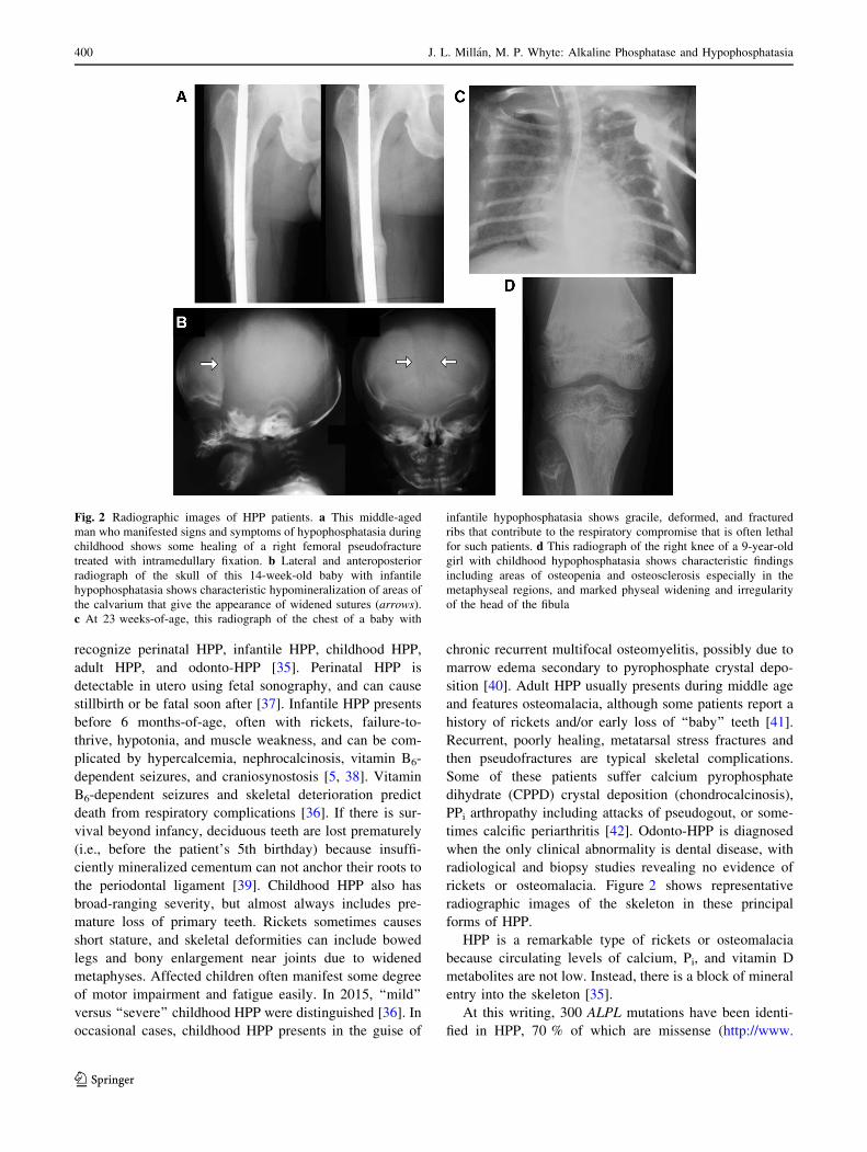
Fig. 2 Radiographic images of HPP patients. a This middle-aged
man who manifested signs and symptoms of hypophosphatasia during
childhood shows some healing of a right femoral pseudofracture
treated with intramedullary fixation. b Lateral and anteroposterior
radiograph of the skull of this 14-week-old baby with infantile
hypophosphatasia shows characteristic hypomineralization of areas of
the calvarium that give the appearance of widened sutures (arrows).
c At 23 weeks-of-age, this radiograph of the chest of a baby with
infantile hypophosphatasia shows gracile, deformed, and fractured
ribs that contribute to the respiratory compromise that is often lethal
for such patients. d This radiograph of the right knee of a 9-year-old
girl with childhood hypophosphatasia shows characteristic findings
including areas of osteopenia and osteosclerosis especially in the
metaphyseal regions, and marked physeal widening and irregularity
of the head of the fibula
400 J. L. Millan, M. P. Whyte: Alkaline Phosphatase and Hypophosphatasia
123

sesep.uvsq.fr/03_hypo_mutations.php). ALPL mutations
can cause autosomal recessive (AR) or autosomal-domi-
nant (AD) HPP. Perinatal and infantile HPP generally
reflect compound heterozygosity for ALPL missense
mutations [36], but sometimes there is homozygosity [9].
Childhood HPP reflects either AR or AD inheritance [35,
36]. Adult and odonto-HPP are usually caused by AD
inheritance of ALPL alleles with dominant-negative effects
[9, 19, 36].
Mouse Models of HPP
In the 1990’s, two Alpl global KO mouse models for HPP
(Alpltm1Sor and Alpltm1Jlm), that differ in the design of the
targeting construct, were developed independently by the
research groups of Soriano [43] and of Millan [44]. In
Alpltm1Sor KO mice, the genomic Alpl sequence spanning
from the middle of exon 2 to the middle of exon 6 was
replaced with a LacZ-Neo cassette to enable expression of
b-galactosidase under control of the endogenous Alpl
promoter. These mice had elevated plasma PLP levels and
died from seizures caused by diminished hydrolysis of PLP
to pyridoxal (PL) and thus deficient PL availability for cells
of the central nervous system [45]. Surviving animals
manifested dental dysplasia [43]. In contrast, the Alpltm1Jlm
KO model was generated by inserting a Neo cassette into
exon 6. Homozygous animals exhibited vitamin B6-de-
pendent seizures and impaired bone mineralization [44].
Their lifespan averaged 8.8 ± 2.3 days on the 129J back-
ground, and 10.6 ± 3.4 days on the 129J, C57Bl/6J hybrid
background. Plasma of Alpl KO mice contains little ALP
activity, which comes from the gut [46], whereas
heterozygous Alpl?/- mice have approximately 50 % the
plasma ALP activity of wild-type (WT) mice and are
healthy and fertile [44].
In 1999, these two KO mouse models, Alpltm1Sor and
Alpltm1Jlm, were further characterized and compared [28].
Radiographic bone abnormalities appeared at *10 days-
of-age, and osteopenia and fractures worsened thereafter.
Both had elevated urinary PEA and PPi and plasma PLP
levels. That same year, Beertsen et al. [47] reported a
2–3 day delay in incisors eruption and onset of mineral-
ization of the mantle dentin within the developing molars
of the Alpltm1Sor KO mice. In contrast, the dentin and
enamel developed normally, except for some localized
hypoplasia. The most conspicuous finding, however,
involved the acellular cementum along the molar roots,
which deposited as thin and irregularly shaped patches
rather than a continuous layer around the base of the
periodontal ligament fibers [47]. The Alpltm1Jlm KO mice
had impaired dentin mineralization in the incisor and molar
roots, ranging from a mild delay to severely disturbed
dentinogenesis. Subsequently noted were lack of acellular
cementum [48, 49] and disrupted organization of the rods
and inter-rod structures in the enamel [50]. Also, premature
fusion of the coronal suture (craniosynostosis) appeared in
these Alpltm1Jlm KO mice [51]. Figure 3 shows represen-
tative radiographic images of their skeletal and dental
phenotype. Alpltm1Jlm KO mice also have an apoptotic
thymus, thin descending nerve roots, leukopenia, and gas
accumulation in their small intestine [44, 52]. Most studies
concerning murine HPP and the preclinical work for EzRT
for HPP used Alpltm1Jlm mice. Accordingly, later, we sim-
ply refer to them as the KO mice.
Now, other Alplmutant mouse lines have been generated,
although their skeletal and dental phenotypes remain to be
determined. In 2009, the AlplALPLD1model was produced by
ENU mutagenesis and carries an A to G mutation at
nucleotide (nt) position 326 in exon 5 [53]. This causes an
Asp to Gly change at residue 109. In 2012, Sabrautzki et al.
reported new mouse models for various metabolic bone
diseases produced by the Munich ENUMutagenesis Project
[54], including phenotypes associated with lowered plasma
ALP activity due to mutations in Alpl. AlplBAP023 carries a
missense T to G mutaton in exon 7 at nt 755 (Leu to Pro at
residue 251). AlplBAP026 has a splice-site mutation in intron
9. AlplBAP027 carries a T to A mutation in exon 10 at nt 1194
(Ile to Asn at residue 395).AlplBAP032 has anA toGmutation
in exon 11 at nt 1217 (Asp to Gly at residue 406). The
homologous mutation had been found in an HPP patient.
AlplSAP007 has an A to G point mutation in exon 12 at nt 1357
(Thr to Ala at residue 453).
Currently, mouse models of adult HPP are receiving
attention. In 2007, the Gena 328 mouse, AlplHpp, from ENU
mutagenesis exhibited autosomal semi-dominant adult HPP
[55]. A point mutation at the splice site for exon 8 produced a
truncated, inactive TNAPhaving 276 residues rather than the
525 WT residues. However, some correct splicing was
detected, suggesting the mutation is hypomorphic. These
mice have low plasma ALP activity and late-onset skeletal
abnormalities, but a normal life span and no epilepsy.
The first murine model of odonto-HPP harbors a mis-
sense mutation (c.346G[A) that changes codon 116 from
Ala to Thr as identified in a kindred with AD odonto-HPP
[56]. These Alpl?/A116T mice have *50 % WT plasma
ALP activity and no differences versus WT mice in litter
size, survival, or body weight. Their postcranial skeleton is
normal radiographically, including no differences in femur
length, cortical or trabecular structure or mineral density,
or mechanical properties. However, their alveolar bone has
radiolucencies and resorptive lesions and osteoid accumu-
lation on the crest. Non-significant changes in acellular
cementum seem not to affect periodontal attachment,
although circulating ALP activity correlates significantly
with incisor cementum thickness.
J. L. Millan, M. P. Whyte: Alkaline Phosphatase and Hypophosphatasia 401
123

Now, two murine models of adult HPP reflect condi-
tional ablation of Alpl in chondrocytes and osteoblasts
together (Prx1-Cre; Alplflox/flox), or in osteoblasts alone
(Col1a1-Cre; Alplflox/flox) (manuscript in preparation).
Their phenotype compared to KO and Alpl?/A116T mice is
summarized in Table 1.
Pathophysiology of HPP
In the skeleton, TNAP is confined to the surface of
osteoblasts and chondrocytes, including their shed matrix
vesicles (MVs) [57, 58] where the enzyme is particularly
enriched [59]. In humans and mice with HPP (vide infra),
electron microscopy revealed that TNAP-deficient MVs
contain hydroxyapatite (HA) crystals, but extravesicular
growth of these crystals is blocked (Fig. 4) by the
extracellular accumulation of PPi [3, 60–62]. Elevated
urinary and plasma levels of PPi were discovered in the
1960s [63–65]. Indeed, breeding Alpl KO mice to mice
deficient in either the extracellular production of PPi(Enpp1-/-) or transport extracellularly (ank/ank) of PPinormalized extracellular levels of PPi. This prevented
skeletal disease in the double KO mice, and affirmed PPiaccumulation as the cause of the rickets/osteomalacia in
HPP [66, 67]. The effect of the double ablation was partial
at some skeletal sites, likely reflecting variations of Enpp1
expression [62, 66].
In the KO mice, ATP-dependent 45Ca precipitation was
reduced within calvarial osteoblast-derived MVs and
attributed to increases within the MV of PPi from impaired
hydrolysis [68]. However, TNAP is not only a PPiase but,
on MVs, is also a potent ATPase [31, 69]. Indeed, Yadav
et al. showed that combined ablation of TNAP and
Fig. 3 Phenotype of the Alpl-/- mouse model of infantile HPP. a X-
ray images of a hind paw of a post-natal day 22 (P22) WT and Alpl-/-
knockout (KO) mouse. b X-ray of the femur, tibia, and fibula of a P22
WT and KO mouse. Images from a and b were taken from [126] and
reproduced with permission from BONE. c Radiographs of the ribs of
a P16 WT and KO mouse. Images taken from [124] and reproduced
with permission from the Journal of Bone and Mineral Research.
d Micro-CT isosurface images of a P15 WT and KO mouse skull.
Multiple cranial vault and facial bones are so severely hypominer-
alized in P15 that they do not appear on isosurface images calibrated
to a bone threshold. The KO skull appears decreased in anterior–
posterior length but increased in height when compared to the WT
skull, and is more dome-shaped in overall appearance. Images taken
from [100] and reproduced with permission from Bone. e Micro-CT
demonstrates poorly mineralized molar roots and incisor in 10-day-
old KO mice compared with WT, in addition to generalized reduction
of bone mineralization in the mandible. Images taken from [47] and
reproduced with permission from the Journal of Bone and Mineral
Research. f Immunohistochemical localization of osteopontin (red,
arrows and inset), as a marker for acellular cementum, shows a
distinct line of acellular cementum in the WT sample, but an absence
of a discrete immunostained layer in a 16-day-old KO mouse. PDL
periodontal ligament, En-S enamel space after decalcification.
Magnification bars equal 100 lm. Taken from [46] and reproduced
with permission from the Journal of Dental Research. g Scanning
electron microscopy (SEM) analysis of incisors (top) and molars
(bottom) of WT and KO mice at 20 days-of-age. The SEM images
show well-decussated enamel rods and inter-rod in the molar crowns
and crown analogs of incisors of WT mice. Note that there is a lack of
rod–inter-rod organization in the KO mice. Images taken from [48]
and reproduced with permission from the Journal of Bone and
Mineral Research (Color figure online)
402 J. L. Millan, M. P. Whyte: Alkaline Phosphatase and Hypophosphatasia
123
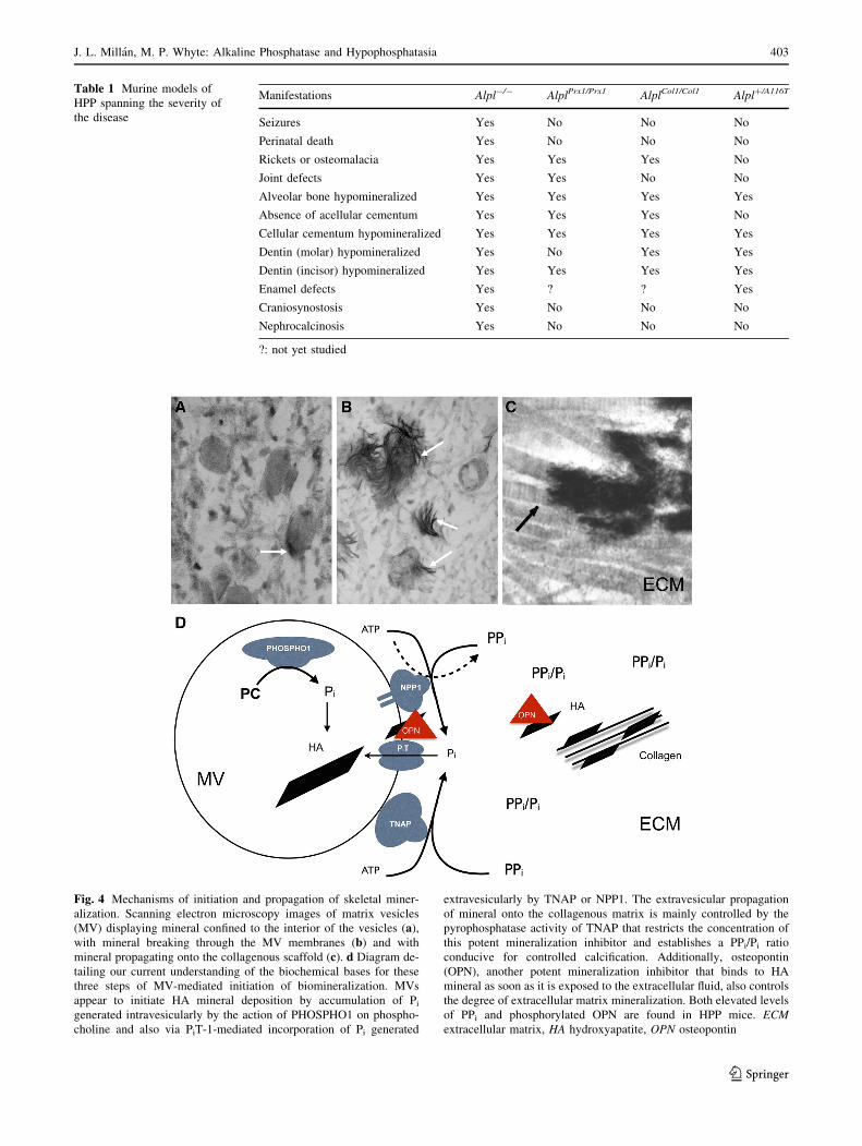
Table 1 Murine models of
HPP spanning the severity of
the disease
Manifestations Alpl-/- AlplPrx1/Prx1 AlplCol1/Col1 Alpl?/A116T
Seizures Yes No No No
Perinatal death Yes No No No
Rickets or osteomalacia Yes Yes Yes No
Joint defects Yes Yes No No
Alveolar bone hypomineralized Yes Yes Yes Yes
Absence of acellular cementum Yes Yes Yes No
Cellular cementum hypomineralized Yes Yes Yes Yes
Dentin (molar) hypomineralized Yes No Yes Yes
Dentin (incisor) hypomineralized Yes Yes Yes Yes
Enamel defects Yes ? ? Yes
Craniosynostosis Yes No No No
Nephrocalcinosis Yes No No No
?: not yet studied
Fig. 4 Mechanisms of initiation and propagation of skeletal miner-
alization. Scanning electron microscopy images of matrix vesicles
(MV) displaying mineral confined to the interior of the vesicles (a),with mineral breaking through the MV membranes (b) and with
mineral propagating onto the collagenous scaffold (c). d Diagram de-
tailing our current understanding of the biochemical bases for these
three steps of MV-mediated initiation of biomineralization. MVs
appear to initiate HA mineral deposition by accumulation of Pigenerated intravesicularly by the action of PHOSPHO1 on phospho-
choline and also via PiT-1-mediated incorporation of Pi generated
extravesicularly by TNAP or NPP1. The extravesicular propagation
of mineral onto the collagenous matrix is mainly controlled by the
pyrophosphatase activity of TNAP that restricts the concentration of
this potent mineralization inhibitor and establishes a PPi/Pi ratio
conducive for controlled calcification. Additionally, osteopontin
(OPN), another potent mineralization inhibitor that binds to HA
mineral as soon as it is exposed to the extracellular fluid, also controls
the degree of extracellular matrix mineralization. Both elevated levels
of PPi and phosphorylated OPN are found in HPP mice. ECM
extracellular matrix, HA hydroxyapatite, OPN osteopontin
J. L. Millan, M. P. Whyte: Alkaline Phosphatase and Hypophosphatasia 403
123

PHOSPHO1 is additive and leads to the lack of HA crystal
formation within MVs, the absence of skeletal mineral-
ization, and embryonic lethality. PHOSPHO1 generates Piinside MVs thus contributing to the initiation of HA min-
eral formation in the intravesicular space [70]. TNAP is a
PPiase crucial for allowing extravesicular growth of HA
crystals, while also generating Pi from the hydrolysis of PPiand acting as an extracellular ATP phosphohydrolase that
generates Pi in the perivesicular space. Thus, current
understanding of TNAP function supports Robison’s
hypothesis concerning skeletal mineralization [35, 70, 71].
NPP1, the enzyme that when on the cell surface produces
PPi, is also a potent ATPase in the MV microenvironment,
and may act as a phosphatase in the absence of TNAP [30,
69–71]. Thus, NPP1 could be a modifier of the HPP phe-
notype, at least in mice. In fact, PHOSPHO1-deficient mice
have increased plasma PPi levels caused by reciprocal
reduction of TNAP activity and increased NPP1 expres-
sion, as well as accumulation in the circulation of phos-
phorylated OPN, an inhibitor of mineralization that perhaps
exacerbates the rickets/osteomalacia and fracturing of HPP.
Because PHOSPHO1 modifies the HPP phenotype in KO
mice, we have searched for Phospho1 gene mutations in
patients with HPP-like disease without ALPL mutations,
however, thus far with negative results. Accordingly, the
hypotheses proposed by Robison for skeletal mineraliza-
tion [1, 2]: (i) TNAP as a Pi-generating enzyme, and (ii) an
unknown factor controlling the supersaturated milieu for
mineralization (now recognized to be PPi), are validated by
current data stemming largely from investigations of HPP
[35, 72–75]. Furthermore, we currently appreciate that
there are interactions between PHOSPHO1, NPP1, and
TNAP during MV-mediated calcification: the first step is a
convergence of two independent biochemical pathways:
intravesicular Pi generation by the enzymatic action of
PHOSPHO1, and the influx via Pi transporters of Pi gen-
erated in the perivesicular space by the enzymatic actions
of TNAP and NPP1 (Fig. 4).
Another natural substrate of TNAP discovered by
studying HPP patients is PLP, the major circulating form of
vitamin B6. Vitamin B6, mediated by its various vitamers,
is a cofactor for at least 110 enzymes. The three vitameric
forms, pyridoxal (PL), pyridoxamine (PM), and pyridoxine
(PN), can all be phosphorylated by PL kinase to their 50-derivatives, PLP, PMP, and PNP, respectively [76, 77].
PLP is a coenzyme for the catabolizing of various amino
acids, and for decarboxylations necessary for neurotrans-
mitter generation including dopamine, serotonin, his-
tamine, taurine, and gamma-aminobutyric acid (GABA).
PLP and PMP are interconvertible through aminotrans-
ferases or PMP/PNP oxidase. Removal of Pi from PLP to
form PL is one important function of TNAP [78]. Only the
non-phosphorylated vitamers can enter cells, and once
inside the cells, these non-phosphorylated vitamers are
converted back to PLP to be used as a coenzyme for var-
ious enzymatic pathways. TNAP deficiency increases
plasma PLP levels in all HPP patients, but leads to low
levels of PL in the circulation in severely affected infants
who consequently have insufficient incorporation of PL
into the CNS and therefore vitamin B6-dependent seizures
[45, 78–80]. Resembling the 100 % lethality when vitamin
B6-dependent seizures occur in infantile HPP [38], KO
mice too have 100 % mortality after weaning, always
heralded by seizures. TNAP is highly expressed in the
developing murine neural tube [81] and certain areas of the
mature brain [82]. The absence of TNAP in KO mice is
associated with hypomyelination and thinning of spinal
nerves, the absence of myelinated axons, and an increased
proportion of immature cortical synapses [44, 52, 83].
Matching clinical experience treating vitamin B6-depen-
dent seizures in HPP, injection or ingestion of PL, a
hydrophobic form of vitamin B6 that traverses biological
membranes temporarily suppresses the epilepsy of KO
mice [43, 44, 52].
PEA is elevated in the blood and urine of patients with
HPP [28, 29, 45], but its endogenous origin is uncertain.
PEA is a component of the cell-surface glycosylphos-
phatidylinositol link for proteins, including TNAP. How-
ever, an additional or alternative source of PEA may reflect
diminished hepatic O-phosphorylethanolamine phospho-
lyase (PEA-P-lyase) activity—the enzyme reported to
hydrolyze PEA using PLP as a cofactor [84–86]. Because
TNAP hydrolyzes extracellular PLP to PL to allow incor-
poration of PL into cells for the formation of PLP needed
as a coenzyme, insufficient PLP inside hepatic cells could
increase PEA accumulation. In this regard, the authors
began their nearly career-long collaborations in 1979 when
they studied a large multigenerational kindred with adult
HPP who showed elevated PEA and phosphoserine levels
in the urine which correlated inversely with serum total and
liver TNAP activity, but not serum bone TNAP activity
[87].
TNAP KO mice have high plasma osteopontin (OPN)
levels encoded by Spp1, and elevated expression of its
RNA in cultured osteoblasts [88–90]. Although the func-
tion of OPN is incompletely understood, it anchors osteo-
clasts to HA by its poly-aspartate sequences, while also
binding CD44 and avb3 integrin via its RGD sequence, thus
mediating cell signaling and/or migration [91]. OPN is a
highly phosphorylated glycoprotein, with 36 serine/thre-
onine phosphorylation sites [92]. This phosphorylation is
important because OPN’s inhibitory effect on mineral
deposition diminishes if 84 % of this covalently bound
phosphate is removed [93]. Phosphorylated, but not
dephosphorylated, OPN inhibits mineralization in vascular
smooth muscle cells [94]. Certain phosphorylated OPN
404 J. L. Millan, M. P. Whyte: Alkaline Phosphatase and Hypophosphatasia
123
peptides also inhibit HA formation in vitro [95], and cause
dose-dependent inhibition of mineralization by cultured
cells [96]. Extracellular PPi levels control OPN expression
by cultured osteoblasts [91, 96], and high plasma levels of
phosphorylated OPN accompany the increased extracellu-
lar PPi levels in Alpl KO mice [90]. In turn, [Alpl-/-;
Spp1-/-] double KO mice have partial improvement of the
hypomineralization compared to Alpl-/- KO mice [90].
Hence, phosphorylated OPN accumulation may contribute
to the impaired bone mineralization of Alpl KO mice.
Absent TNAP function leads to accumulation of phos-
phorylated OPN [34]. This suggests that OPN is another
substrate of TNAP, and OPN phosphorylation is an addi-
tional biochemical pathway in the pathophysiology of
murine HPP (Fig. 4). We are currently reviewing the OPN
status of HPP patients.
Interestingly, PHOSPHO1-deficient mice also have
increased plasma levels of PPi and phosphorylated OPN.
Here, concurrent ablation of the OPN gene corrects the
skeletal disease of Phospho1 deficiency [97]. Thus, Alpl and
Phospho1 deficiency engender similar skeletal phenotypes
and comparable changes in PPi and OPN expression levels.
However, there is a clear dissociation in the hierarchical roles
of these potent inhibitors ofmineralization, with elevated PPiand phosphorylated OPN levels causing the respective
skeletal phenotypes in Alpl-/- and Phospho1-/- mice.
Other biological roles for TNAP are now being explored
inspired by studies concerning intestinal ALP in protection
of the gut mucosa and regulation of the gut microbiota via
detoxification of bacterial endotoxins, especially di-phos-
phoryl-lipopolysaccharide (LPS) and pro-inflammatory
ATP [98–100]. Lei et al. [33] showed in hamsters that LPS
detoxification by TNAP conditions uterine receptivity for
implantation and decidualization, while protecting the uterus
and pregnancy against bacterial infection. High circulating
TNAP activity in neonates enhances their high plasma levels
of anti-inflammatory adenosine, produced by the dephos-
phorylation of pro-inflammatory ATP [31]. Furthermore,
TNAP is one of three enzymes involved in purine metabo-
lism, contributing anti-nociceptive adenosine for murine
somatosensory dorsal root ganglia neurons and the dorsal
spinal cord [32]. Altered purinergic signaling in Alpl KO
mice could result from an increased ATP/adenosine ratio
caused by TNAPdeficiency. Such changesmay contribute to
the seizures, hyperalgia, and allodynia seen inAlplKOmice,
and perhaps patients with severe HPP (Fig. 5).
What We Don’t Yet Understand About HPP
Premature fusion of cranial sutures (craniosynostosis) in
HPP and other forms of rickets may seem counter-intuitive
and remain unexplained. Patients with severe HPP
receiving asfotase alfa still often require craniotomies
[101] to relieve intracranial pressure. However, asfotase
alfa [102] and ChimAP [103] (a soluble chimeric form of
human ALP), both can prevent craniosynostosis, at least in
mice (vide infra), if treatment starts at birth. So, prevention
of craniosynostosis may depend on the time of initiation of
the treatment, although there are data to indicate that in
humans craniosynostosis may begin during embryonic
life [104]. Thus, clarifying the onset and mechanism of
craniosynostosis may help design interventions at a
developmental stage when enzyme replacement may not be
feasible in patients.
While the role of TNAP in the skeleton seems well
understood, little is known about its function in the kidney,
despite reference to this ALP isozyme as liver/bone/kidney
type ALP. Expression of TNAP in the kidney may seem
counter-intuitive because TNAP hydrolyses PPi, a potent
mineralization inhibitor that could help prevent kidney
stones. In fact, reduced urinary PPi predisposes to
nephrolithiasis [105]. PPi:creatinine ratios were reduced in
107 patients with recurrent calcium kidney stones [106]. PPilevels in urine are[10 lM compared to plasma concentra-
tions of approximately 1-6 lM that mainly arise from liver
metabolism [107]. Intravenous 32PPi is rapidly hydrolyzed in
plasma, with PPi also being filtered at the glomerulus and
subject to further hydrolysis within the kidney; \5 % of
intravenous 32PPi appears in urine. These data indicate that
PPi in the kidney likely originates locally. Thus, nephrocal-
cinosis despite high urinary concentrations of PPi in severely
affected babies with HPP could seem puzzling, but their
severe hypercalcemia and hypercalciuria is likely the key
factor. Interestingly, children and adults with HPP are often
hyperphosphatemic with increased TmP/GFR, suggesting
kidney TNAP plays a role in Pi excretion [36].
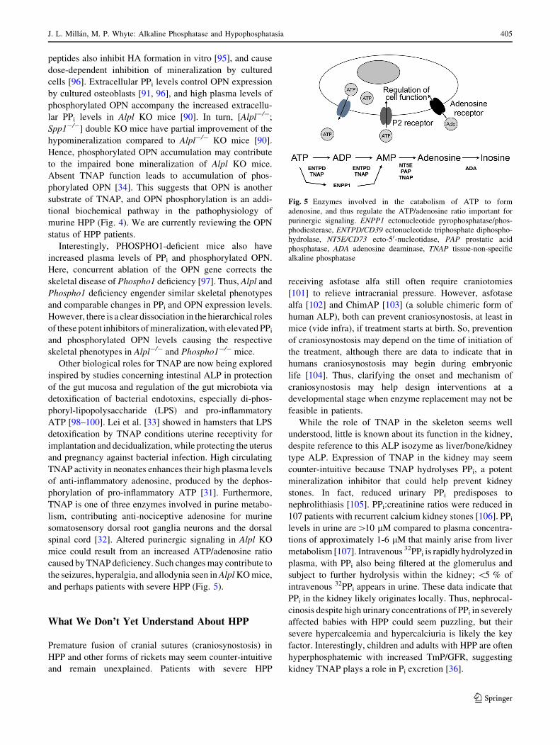
Fig. 5 Enzymes involved in the catabolism of ATP to form
adenosine, and thus regulate the ATP/adenosine ratio important for
purinergic signaling. ENPP1 ectonucleotide pyrophosphatase/phos-
phodiesterase, ENTPD/CD39 ectonucleotide triphosphate diphospho-
hydrolase, NT5E/CD73 ecto-50-nucleotidase, PAP prostatic acid
phosphatase, ADA adenosine deaminase, TNAP tissue-non-specific
alkaline phosphatase
J. L. Millan, M. P. Whyte: Alkaline Phosphatase and Hypophosphatasia 405
123

Supportive Management of HPP
HPP management has been, until very recently, supportive
[9]. Bone-targeted enzyme replacement therapy (asfotase
alfa, now StrensiqTM) was approved for HPP in Japan
(July, 2015), and for pediatric-onset HPP in Canada (Au-
gust, 2015), Europe (August, 2015), and the USA (October,
2015). Supportive treatment includes hydration and
restriction of dietary calcium and perhaps administration of
calciuretics for infants with hypercalcemia and hypercal-
ciuria [35, 36]. Management of the respiratory complica-
tions of perinatal and infantile HPP may be complex as
multiple factors compromise their pulmonary function
[108]. Vitamin B6-dependent seizures can respond tem-
porarily to pyridoxine administration. Craniosynostosis
may need surgical intervention. Fractures can mend, but
often slowly and requiring prolonged casting or stabiliza-
tion with intramedullary hardware. Femoral pseudofrac-
tures can also respond to intramedullary rodding [109].
Dental manifestations can be helped by a knowledgeable
dentist.
Targeting TNAP to Bone Mineral
Bolstered by insight derived from discovery of elevated
plasma PLP levels in HPP [45] that predicted TNAP is cell-
surface anchored, intravenous administration of ALPs to
HPP patients was attempted to decrease extracellular PPilevels and thereby improve skeletal mineralization. How-
ever, experience with such EzRT, including plasma from
patients with Paget bone disease containing high bone
TNAP activity, proved unsuccessful [110]. A 6-month-old
girl with worsening infantile HPP repeatedly given intra-
venous infusions of TNAP-rich plasma obtained by
plasmapheresis from two men with Paget bone disease
showed circulating ALP activity with a half-life of *2
days that persisted during a 5-week period of six ALP
infusions. Sequential radiographic studies were interpreted
as showing arrest of worsening rickets with slight rem-
ineralization of metaphyses, although urinary levels of
PEA and PPi seemed unaltered [110]. However, three
subsequent patients deteriorating from infantile HPP [111]
and given similar infusions of Paget plasma showed no
radiographic or other benefit. Discouraging findings also
occurred with attempted EzRT using purified human liver
TNAP [112] and purified PLAP [113]. The cumulative
findings indicated necessity for increasing TNAP activity
in situ in the skeleton to reverse the pathophysiology of
HPP. This hypothesis was supported in 2003 and 2007 by
rescue with radiographic improvement demonstrated by
two unrelated girls with life-threatening infantile HPP
following attempts to transplant healthy mesenchyme-
derived marrow cells, hoping TNAP-replete donor osteo-
blasts or chondrocytes might engraft in their skeletons
[114, 115]. Subsequently, a woman with adult HPP car-
rying the most common American ALPL mutation seemed
to benefit from teriparatide injections given to increase
TNAP biosynthesis by her osteoblasts [116]. Then, ana-
bolic treatment with parathyroid hormone 1-34 or 1-84
benefitted some adults with HPP [117–119], but not others
[120, 121]. Parathyroid hormone therapy is not sanctioned
for children, because osteosarcoma appeared in growing
rats given teriparatide [122]. Evaluation of anti-sclerostin
antibody (BPS804) therapy, which has been reported to
increase bone-specific ALP when administered to healthy
post-menopausal women [123] (www.clinicaltrials.gov
identifier NCT01406977), was not carried forward. Ana-
bolic approaches may favor patients with heterozygous
ALPL mutations, because the WT ALPL allele is intact for
upregulation; however, it will probably not be useful for
severe AR HPP with at best ‘‘more of a bad enzyme.’’
In 2005, Enobia Pharma, Montreal, Canada bio-engi-
neered and then expressed in Chinese hamster ovary
(CHO) cells a first-in-class mineral-seeking recombinant
TNAP to treat HPP. This biologic reflected in tandem the
coding sequences for (i) soluble human TNAP, by
excluding the hydrophobic C-terminal GPI anchoring motif
(i.e., soluble TNAP: sALP), (ii) the Fc region of human
IgG gamma-1 (Fc) to facilitate purification and prolong the
circulating half-life, and (iii) ten acidic aspartate residues
(D10) for selective drug delivery to bone [124–126]. The
resulting 726-amino acid fusion protein was initially des-
ignated sALP-FcD10 to indicate its different domains
(Fig. 6), renamed ENB-0040 when produced under Current
Good Manufacturing Practice (cGMP), and asfotase alfa
after clinical trials began. Alexion Pharmaceuticals, Che-
shire, CT acquired Enobia Pharma in 2012. Asfotase alfa is
now also called StrensiqTM.
Preclinical Evaluation of Bone-Targeted EzRTfor HPP
Preclinical testing of sALP-FcD10 primarily involved
Alpltm1Jlm (KO) mice. Pharmacokinetics (PK) and tissue
distribution evaluations included newborn and adult mice
and comparisons of different routes of administration
[127]. Histochemical staining confirmed localization and
catalytic activity of sALP-FcD10 in bone tissue (Fig. 6).
The first study involved newborn KO mice given sub-
cutaneous (SC) injections for 15 days, then investigated
with micro-computed tomography (lCT) of the calvarium
and proximal tibial growth plate [127]. At 2 mg/kg daily,
general appearance, body weight, and tail length improved
with normal growth. At 8.2 mg/kg daily, body weight was
406 J. L. Millan, M. P. Whyte: Alkaline Phosphatase and Hypophosphatasia
123

greater than control mice, and plasma PPi concentrations
were normal. sALP-FcD10 minimized hypomineralization
in the feet and reduced the number of mice with severely
dysmorphic rib cages. The hind limbs appeared healthy in
all treated animals [127]. A 52-day study of daily SC
injections of 8.2 mg/kg also showed normal plasma PL and
calcium concentrations and prevention of skeletal defects
and epilepsy with good survival [127]. Hypomineralization
of dentin and alveolar bone was also prevented and acel-
lular cementum now formed properly [48, 49]. Immuno-
histochemical staining for OPN revealed unremarkable,
rather than absent, acellular cementum on root surfaces
[48]. The KO mouse dentin defect results from inability of
MV-initiated mineralization foci to expand into a miner-
alization front, and varies from delayed mineralization to
arrest of mantle dentin mineralization together with lack of
circumpulpal dentin and odontoblast differentiation defects
[49]. The OPN accumulation likely contributes to the
impaired mineralization of mantle dentin. Early treatment
with sALP-FcD10 enabled dentinogenesis and molar
mineralization.
Then, the relationship was defined between sALP-Fc-D10
doses and therapeutic responses after 43 days [128] (Fig. 7).
Endpoint assessments included survival, body weight, tibial
and femoral length, and bone mineralization in the feet, rib
cage, and lower limbs assessed radiographically. Radio-
graphs, lCT, and histomorphometry evaluated skeletal
mineralization. Daily dose correlated clearly with the per-
centage of normal feet, rib cages, and lower limbs. An
effective dosewas established for 80 %of themice (ED80) of
*3.2, *2.8, and *2.9 mg/kg/day for feet, rib cage, and
lower limbs, respectively. The ED80, along with serum ALP
activity and sALP-FcD10 PK data were used to estimate the
minimum effective dose for these KO mice.
Further studies then validated the mineral targeting,
including to sites notorious for poor vascularity, e.g., the
enamel organ during tooth development. Untreated KO
mice showed with scanning electron microscopy disorga-
nization of the rod and inter-rod structures of enamel.
Histology revealed enamel hypomineralization both in
molars and incisors, loss of polarization of ameloblasts
important for enamel matrix formation, and even absent
enamel. What was now called ENB-0040 reached the
enamel organ during its secretory and maturation phases
[50]. lCT showed, with treatment to age 23 days, dose-
dependent improvements of absolute and relative enamel
volumes. Notably, odonto-HPP typically manifests the
mildest reductions of serum ALP levels [35], usually
Fig. 6 Structure and binding of asfotase alfa to bone mineral.
a Three-dimensional modeling of ENB-0040. The model shows rigid
ALP and Fc modules connected by a highly flexible linker. The
terminal poly-Asp region is exposed on the opposite site of the ALP
module. The whole structure is dimeric that conforms to the preferred
oligomeric state of the ALP as well as the Fc region of the antibody.
The three active site metal ions (two Zn2? and one Mg2?) are marked
with blue spheres. b Transmission electron micrograph showing the
binding of sALP-FcD10 to synthetic hydroxyapatite crystals as
revealed by immunogold labeling (inset is control incubation without
sALP-FcD10 showing an absence of gold-particle labeling). Magni-
fication bar equals 100 nm. c RosettaSurface-simulated model of D10
binding to a calcium-rich plane of the [100] crystallographic face of
hydroxyapatite. d Histochemical staining for ALP activity in long
bones of an ENB-0040–treated Alpl KO mouse compared with an
age-matched untreated Alpl KO mouse. Images shown in b and c weretaken from [46] (Color figure online)
J. L. Millan, M. P. Whyte: Alkaline Phosphatase and Hypophosphatasia 407
123

explained by dominant inheritance of a single mutated
ALPL allele [19], and reveals that tooth formation, espe-
cially cementogenesis, is the developmental process most
sensitive to TNAP function, involving changes in the local
Pi/PPi ratio [129, 130]. ENB-0040 treatment preserved
acellular cementum formation in the KO mice, supporting
this premise [48] and that the structural integrity of enamel
by the enamel organ is directly regulated by extracellular
PPi concentrations [50]. TNAP is highly expressed in
mature odontoblasts, and the molar and incisor roots of KO
mice feature dentin hypomineralization ranging from mild
to severe. Lack of mantle dentin mineralization accompa-
nies disordered and dysmorphic odontoblasts. However, in
KO mice, the formation of, initiation of mineralization
within, and rupture of MVs in dentin matrix are not com-
promised. ENB-0040 corrected the defective dentin min-
eralization in the molar roots [49]. Liu et al. [51] showed
that these mice by age 3 weeks have craniofacial shape
abnormalities suggestive of limited anterior–posterior head
growth with craniosynostosis (i.e., bony coronal suture
fusion). lCT isosurface images showed ENB-0040 8.2 mg/
kg/day from birth prevented at age 15 days craniofacial
bone mineralization defects and premature suture fusion. In
untreated KO mice, multiple cranial vault and facial bones
lacked adequate mineralization on lCT that was not evi-
dent in the treated mice. Digital caliper linear measure-
ments demonstrated that treatment improved nose length,
nasal bone length, and frontal bone length [102].
Clinical Trials Using Mineral-Targeting TNAPin Pediatric HPP
In June 2008, an Investigational New Drug (IND) appli-
cation to test ENB-0040 for HPP patients was filed by
Enobia Pharma. Initial dosing was based partly on (i) dose–
response data in KO mice [128], (ii) efficacy observed in
treated mice that achieved an equivalent to serum ALP
activity in the range of 2400–6000 U/L; (iii) the No-Ob-
served-Adverse-Effect Level (NOAEL) in more sensitive
species (rats and monkeys) established in 1-month IV
toxicology studies, and 1-month IV/SC bridging and tol-
erability studies in rats; and (iv) a safety factor of 10
applied to the NOAEL.
Fig. 7 Preclinical efficacy of asfotase alfa. a Percentage survival of
Alpl-/- mice receiving either vehicle (white circle) or escalating
doses of asfotase alfa, i.e., Tx-0.5 (black circle), Tx-2.0 (white down-
pointing triangle), or Tx-8.2 (black square). b lCT images of tibiae
of the 22-day-old Alpl-/- mice treated with vehicle, Tx-0.5, Tx-2.0,
and untreated WT mice. The images clearly show improved tissue
mineral density and callus formation at the site of fractures in the
treated mice. Transaxial views at the bottom. c Percentage of Alpl-/-
mice considered normal as a function of asfotase alfa dose for feet
(black square), rib cage (white down-pointing triangle), and lower
limbs (white circle). d Asfotase alfa treatment maintains complete
mineralization of all molar dentin as well as the surrounding alveolar
bone such that no mineralization differences are seen between the
molar teeth and bone of the treated Alpl-/- mice (8.2 mg/kg/day)
compared with WT mice. Images taken from [128] with permission
from BONE
408 J. L. Millan, M. P. Whyte: Alkaline Phosphatase and Hypophosphatasia
123

The first results in HPP patients were from an open-
label, multicenter center, dose-escalating phase 1 study of
the safety, tolerability, and pharmacology of ENB-0040 in
six adults with HPP (www.clinicaltrials.gov identifier
NCT00739505). They received one dose iv of 3 mg/kg,
and then 1 or 2 mg/kg sc once weekly for 3 weeks.
The first full report concerning HPP treatment was from
an open-label, multicenter, international study published in
2012 [9]. It evaluated the safety, tolerability, bioavailabil-
ity, PK, pharmacodynamics, and efficacy of asfotase alfa
given for nearly 1 year to infants and young children
B3 years-of-age with life-threatening perinatal and infan-
tile HPP. Before treatment, one 3-year-old girl had lost
nearly all radiographically apparent skeletal mineral. The
Supplementary Appendix to that paper provides a detailed
description of each patient [9]. Efficacy assessments
included changes in radiographic scales to evaluate the
characteristic skeletal findings, gross and fine motor func-
tion, cognitive development, and pulmonary complications
and their management. Asfotase alfa was administered as a
single iv infusion of 2 mg/kg, followed by sc injections of
1 mg/kg thrice weekly for 24 weeks, with an extension
study thereafter. The sc dose could be increased up to
3 mg/kg for worsening failure-to-thrive, deteriorating pul-
monary function, or no radiographic evidence of skeletal
improvement [9]. Of eleven recruited patients, parents
withdrew one because she had a moderate adverse reaction
during the iv infusion. One infant who completed the first
6 months of treatment died soon after from pneumonia and
sepsis judged unrelated to the study drug. Of the nine
patients treated for 1 year, four represented perinatal HPP
and five represented infantile HPP. Typically, serum cal-
cium levels were elevated (most had nephrocalcinosis), and
dietary calcium had been restricted in all but one patient.
During treatment, substantial radiographic improvement in
skeletal abnormalities was documented at week 24 in all
but one patient, with continued healing through week 48.
The girl with the most extreme skeletal disease eventually
improved with calcification apparent after 9 months of
therapy and the delay probably reflecting her profound
deficit of skeletal mineral. The radiographic improvement
is illustrated for the oldest patient (3 years old) (Fig. 8),
and for the youngest patient (\1 month old) (Fig. 9).
Positive mineral balance throughout the skeleton was
obvious radiographically, sometimes after several weeks or
months, and involved diffusely membranous as well as
endochondral bone. Symptomatic hypocalcemia from
‘‘hungry-bones’’ did not occur, but serum calcium
decreased in some patients, consistent with improved
uptake of calcium into mineralized bone. Increases in
serum parathyroid hormone levels called for liberalization
of dietary calcium intakes [9]. Deciduous teeth erupted in
all the patients during therapy, with only one patient
subsequently having HPP-related tooth loss [9]. Manage-
ment of perinatal HPP in particular is critically dependent
on early diagnosis [131]. An important concern is that
some clinical laboratories do not flag as abnormal low
serum ALP and this can delay the diagnosis and increase
the risk of severe respiratory morbidity. Institution of EzRT
should also not be delayed in order to correct any vitamin
D deficiency—this can be accomplished contemporane-
ously if necessary.
Subsequently, asfotase alfa was given for 5 years to 12
children who were 5–12 years of age at study entry and
substantially impaired by HPP [132]. Two radiographic
scales to quantitate HPP skeletal disease documented rapid
and significant improvement at 6-months of treatment,
including comparisons to serial radiographs from similarly
affected historical controls. Further improvements included
patient growth, strength, motor function, and agility that
achieved normal values for age- and gender-matched peers
and were sustained at 5 years of therapy. Pain and dis-
ability resolved for most patients [132]. Mild-to-moderate
injection site reactions were common, and sometimes
associated with lipohypertrophy. Low titers of anti-asfotase
alfa antibodies were noted in all. No evidence emerged for
clinically significant ectopic mineralization or resistance to
the treatment [132].
Future Treatments for HPP
Kiffer-Moreira et al. recently evaluated ChimAP in KO
mice. ChimAP is a chimeric ALP engineered by substi-
tuting the flexible crown domain of human IAP by that of
human PLAP [133]. A clinical study is currently underway
to assess ChimAP for treating acute kidney injury (www.
clinicaltrials.gov identifier NCT02182440). Daily SC
injections of ChimAP to KO mice at doses of 1, 8, or
16 mg/kg from birth to age 53 days were associated with
normal lifespan and body weight and prevention of vitamin
B6-dependent seizures at 16 mg/kg/day [103]. Radio-
graphs, lCT, and histological analyses documented sus-
tained mineralization of cortical and trabecular bone and
secondary ossification centers in long bones. Craniosyn-
ostosis was prevented, and no evidence emerged of ectopic
calcification by radiography and histology of the aorta,
stomach, kidneys, or lungs [103]. AM Pharma (Bunnik,
The Netherlands) has recently received FDA and EMA
orphan drug designation for investigation of ChimAP
(RecAP) for HPP. Daily sc injections of asfotase alfa are
required to prevent HPP in mice, whereas nearly daily or
thrice weekly sc injections are used to treat HPP patients.
Viral vector delivery of mineral-targeting TNAP by this
type of gene therapy might be an alternative to repetitive
injections.
J. L. Millan, M. P. Whyte: Alkaline Phosphatase and Hypophosphatasia 409
123

In 2011, Yamamoto et al. [134] demonstrated that a
single sc iv injection of a lentiviral vector expressing
mineral-targeting TNAP at birth permanently increased
plasma TNAP levels in KO mice that survived more than
10 months and had normal physical activity, healthy
appearance, no epileptic seizures, and radiographs showing
significantly improved or preserved skeletal mineralization
using this gene therapy. Also in 2011, Matsumoto et al.
[135] demonstrated similar treatment effects, but using the
adeno-associated virus serotype 8 (AAV8) vector that is
more promising than lentiviral vectors for clinical trials.
They expressed mineral-targeted TNAP and soluble, non-
targeted TNAP tagged with the FLAG epitope. A single IV
injection of 5 9 1010 vector genomes of either TNAP into
KO mice at day 1 of life prolonged survival and prevented
the skeletal abnormalities, suggesting that sustained ALP
activity in the circulation at some threshold level may
prevent the manifestations of HPP.
In 2012, Sugano et al. [136] demonstrated the feasibility
of fetal gene therapy for HPP by giving KO mouse fetuses,
at 15 days gestation, a single transuterine intraperitoneal
injection of AAV serotype 9 (AAV9) expressing mineral-
targeted TNAP. After birth, these mice showed normal
weight gain and seizure-free survival for at least 8 weeks.
ALP activities in plasma and bone were consistently high,
and sustained mineralization was demonstrated on radio-
graphs of the chest and forepaw.
Hence, viral vector delivery of mineral-targeted TNAP
has potential to treat HPP with significantly reduced fre-
quency of injections and cost of treatment.
Concluding Remarks
There has been substantial progress during the past decade
in understanding TNAP and therefore in treating HPP
patients. Clinical trials of EzRT using asfotase alfa (now
approved as StrensiqTM in Japan, Canada, Europe, and the
USA generally for pediatric-onset HPP) have shown
skeletal, respiratory, and functional improvement as well as
prevention of seizures in the most severe perinatal and
infantile forms of the disease and attainment of good health
in survivors of infantile HPP and with severe childhood
HPP. Ongoing clinical trials are revealing aspects of HPP
that we do not yet fully understand, such as its treat-
able muscle weakness, or that seemingly cannot be pre-
vented such as craniosynostosis. Now that life-threatening
and debilitating pediatric HPP is treatable using asfotase
Fig. 8 Efficacy of asfotase alfa treatment in a 36-month-old girl (at
therapy baseline) with life-threatening HPP. a She has a short, bowed
femur detected in utero by ultrasound. b At 12 days-of-age, her chest
radiograph showed thin, osteopenic ribs with lytic areas and fractures.
c, d Images of the skull before and after 24 weeks of ENB-0040
treatment. Note the severe pan-suture closure, including a marked
increase in ‘‘digital’’ markings (‘‘beaten-copper’’ appearance). e The
ribs at baseline were osteopenic and had fracture deformities with thin
cortices. f By week 24 of treatment, the ribs were wider and better
mineralized with sharper cortical margins and less deformity.
g Improvement of the rickets with therapy is apparent. Images taken
for the online supplementary data in [9] and reproduced with
permission from The New England Journal of Medicine
410 J. L. Millan, M. P. Whyte: Alkaline Phosphatase and Hypophosphatasia
123

alfa, and the risks and benefits of this EzRT in managing
HPP in adults require study. These patients may present
unique challenges because vascular calcification can be a
comorbidity of aging, diabetes mellitus, or chronic kidney
disease. Binding of mineral-targeted TNAP to such sites of
ectopic calcification could theoretically lead to cardiovas-
cular complications [137]. It is not known if non-targeted
ALPs will be useful alternatives. Safe viral vectors for
delivery of mineral-targeted or soluble ALPs may stream-
line HPP treatment in the future.
Funding This study was funded by grant DE12889 from NIDCR,
NIH (JLM), Shriners Hospitals for Children (MPW), The Clark and
Mildred Cox Inherited Metabolic Bone Disease Fund (MPW), The
hypophosphatasia Research Fund (MPW) and the Barnes-Jewish
Hospital Foundation (MPW).
Compliance with Ethical Standards
Conflict of Interest JLM and MPW have received grant support,
honoraria, and travel reimbursements from Alexion Pharmaceuticals,
Cheshire, CT. JLM has received grant support and honoraria from
AM-Pharma, Bunnik, The Netherlands.
Human and Animal Rights and Informed Consent All proce-
dures performed in studies involving human participants were in
accordance with the ethical standards of the institutional and/or
national research committee and with the 1964 Helsinki declaration
and its later amendments or comparable ethical standards. Informed
consent was obtained from all individual participants included in the
study. All applicable international, national, and/or institutional
guidelines for the care and use of animals were followed.
Open Access This article is distributed under the terms of the
Creative Commons Attribution 4.0 International License (http://crea
tivecommons.org/licenses/by/4.0/), which permits unrestricted use,
distribution, and reproduction in any medium, provided you give
appropriate credit to the original author(s) and the source, provide a
link to the Creative Commons license, and indicate if changes were
made.
References
1. Robison R (1923) The possible significance of hexosephos-
phoric esters in ossification. Biochem J 17:286
2. Robison R (1932) The significance of phosphoric esters in
metabolism. New York University Press, New York
3. Meyer JL (1984) Can biological calcification occur in the
presence of pyrophosphate? Arch Biochem Biophys 231:1–8
4. Weiss MJ, Cole DE, Ray K, Whyte MP, Lafferty MA, Mulivor
RA, Harris H (1988) A missense mutation in the human liver/
bone/kidney alkaline phosphatase gene causing a lethal form of
hypophosphatasia. Proc Natl Acad Sci USA 85:7666–7669
5. Rathbun JC (1948) Hypophosphatasia; a new developmental
anomaly. Am J Dis Child 75:822–831
Fig. 9 Efficacy of asfotase alfa in a 20-day-old (at therapy baseline)
patient with life-threatening HPP. a This boy had shortened and
bowed extremities and ‘‘fractures’’ detected by prenatal sonography at
17–18 weeks gestation. b Before asfotase alfa treatment, little or
decreased mineral was present in the frontal, parietal, or occipital
bones, skull base, facial bones, and sphenoid. c Before asfotase alfa,
the femora were short, sclerotic, bowed, irregular, and lacked defined
medullary cavities, cortices, and mineralized metaphyses and epi-
physes. The fibulae were not calcified. After therapy, striking
mineralization was evident. At week 24 of therapy, all areas showed
striking remineralization. d The improvement in the left hand and
wrist was remarkable. Images taken for the online supplementary data
in [9] and reproduced with permission from The New England Journal
of Medicine
J. L. Millan, M. P. Whyte: Alkaline Phosphatase and Hypophosphatasia 411
123
6. Whyte MP (2013) Hypophosphatasia. In: Thakker RV, Whyte
MP, Eisman J, Igarashi T (eds) Genetics of bone biology and
skeletal disease. Academic Press, pp 337–360
7. Mornet E, Yvard A, Taillandier A, Fauvert D, Simon-Bouy B
(2011) A molecular-based estimation of the prevalence of
hypophosphatasia in the European population. Ann Hum Genet
75:439–445
8. Greenberg CR, Taylor CL, Haworth JC, Seargeant LE, Philipps S,
Triggs-RaineB,ChodirkerBN(1993)AhomoallelicGly317?Asp
mutation in ALPL causes the perinatal (lethal) form of hypophos-
phatasia in Canadian mennonites. Genomics 17:215–217
9. Whyte MP, Greenberg CR, Salman NJ, Bober MB, McAlister
WH, Wenkert D, Van Sickle BJ, Simmons JH, Edgar TS, Bauer
ML, Hamdan MA, Bishop N, Lutz RE, McGinn M, Craig S,
Moore JN, Taylor JW, Cleveland RH, Cranley WR, Lim R,
Thacher TD, Mayhew JE, Downs M, Millan JL, Skrinar AM,
Crine P, Landy H (2012) Enzyme-replacement therapy in life-
threatening hypophosphatasia. N Engl J Med 366:904–913
10. Millan JL (2006) Mammalian alkaline phosphatases: from
biology to applications in medicine and biotechnology. Wiley-
VCH Verlag GmbH & Co., Weinheim
11. Le Du MH, Stigbrand T, Taussig MJ, Menez A, Stura EA (2001)
Crystal structure of alkaline phosphatase from human placenta
at 1.8 A resolution. Implication for a substrate specificity. J Biol
Chem 276:9158–9165
12. Le Du M-H, Millan JL (2002) Structural evidence of functional
divergence in human alkaline phosphatases. J Biol Chem
277:49808–49814
13. Hummer C, Millan JL (1991) Gly429 is the major determinant
of uncompetitive inhibition of human germ cell alkaline phos-
phatase by L-leucine. Biochem J 274(Pt 1):91–95
14. Hoylaerts MF, Manes T, Millan JL (1992) Molecular mecha-
nism of uncompetitive inhibition of human placental and germ-
cell alkaline phosphatase. Biochem J 286(Pt 1):23–30
15. Kozlenkov A, Manes T, Hoylaerts MF, Millan JL (2002)
Function assignment to conserved residues in mammalian
alkaline phosphatases. J Biol Chem 277:22992–22999
16. Tsonis PA, Argraves WS, Millan JL (1988) A putative func-
tional domain of human placental alkaline phosphatase pre-
dicted from sequence comparisons. Biochem J 254:623–624
17. Hoylaerts MF, Ding L, Narisawa S, Van Kerckhoven S, Millan
JL (2006) Mammalian alkaline phosphatase catalysis requires
active site structure stabilization via the N-terminal amino acid
microenvironment. Biochemistry 45:9756–9766
18. Hoylaerts MF, Manes T, Millan JL (1997) Mammalian alkaline
phosphatases are allosteric enzymes. J Biol Chem 272:22781–
22787
19. Fauvert D, Brun-Heath I, Lia-Baldini AS, Bellazi L, Taillandier
A, Serre JL, de Mazancourt P, Mornet E (2009) Mild forms of
hypophosphatasia mostly result from dominant negative effect
of severe alleles or from compound heterozygosity for severe
and moderate alleles. BMC Med Genet 10:51
20. Mornet E, Stura E, Lia-Baldini AS, Stigbrand T, Menez A, Le
Du MH (2001) Structural evidence for a functional role of
human tissue nonspecific alkaline phosphatase in bone miner-
alization. J Biol Chem 276:31171–31178
21. Hoylaerts MF, Van Kerckhoven S, Kiffer-Moreira T, Sheen C,
Narisawa S, Millan JL (2015) Functional significance of calcium
binding to tissue-nonspecific alkaline phosphatase. PLoS One
10:e0119874
22. Genge BR, Sauer GR, Wu LN, McLean FM, Wuthier RE (1988)
Correlation between loss of alkaline phosphatase activity and
accumulation of calcium during matrix vesicle-mediated min-
eralization. J Biol Chem 263:18513–18519
23. Sharom FJ, Lehto MT (2002) Glycosylphosphatidylinositol-an-
chored proteins: structure, function, and cleavage by
phosphatidylinositol-specific phospholipase C. Biochem Cell
Biol 80:535–549
24. Wong YW, Low MG (1994) Biosynthesis of glycosylphos-
phatidylinositol-anchored human placental alkaline phos-
phatase: evidence for a phospholipase C-sensitive precursor and
its post-attachment conversion into a phospholipase C-resistant
form. Biochem J 301(Pt 1):205–209
25. Weiss MJ, Henthorn PS, Lafferty MA, Slaughter C, Raducha M,
Harris H (1986) Isolation and characterization of a cDNA
encoding a human liver/bone/kidney-type alkaline phosphatase.
Proc Natl Acad Sci USA 83:7182–7186
26. Nosjean O, Koyama I, Goseki M, Roux B, Komoda T (1997)
Human tissue non-specific alkaline phosphatases: sugar-moiety-
induced enzymic and antigenic modulations and genetic aspects.
Biochem J 321(Pt 2):297–303
27. Halling Linder C, Narisawa S, Millan JL, Magnusson P (2009)
Glycosylation differences contribute to distinct catalytic prop-
erties among bone alkaline phosphatase isoforms. Bone
45:987–993
28. Fedde KN, Blair L, Silverstein J, Coburn SP, Ryan LM,
Weinstein RS, Waymire K, Narisawa S, Millan JL, MacGregor
GR, Whyte MP (1999) Alkaline phosphatase knock-out mice
recapitulate the metabolic and skeletal defects of infantile
hypophosphatasia. J Bone Miner Res 14:2015–2026
29. Fedde KN, Whyte MP (1990) Alkaline phosphatase (tissue-
nonspecific isoenzyme) is a phosphoethanolamine and pyri-
doxal-50-phosphate ectophosphatase: normal and hypophos-
phatasia fibroblast study. Am J Hum Genet 47:767–775
30. Ciancaglini P, Yadav MC, Simao AM, Narisawa S, Pizauro JM,
Farquharson C, Hoylaerts MF, Millan JL (2010) Kinetic analysis
of substrate utilization by native and TNAP-, NPP1-, or
PHOSPHO1-deficient matrix vesicles. J Bone Miner Res
25:716–723
31. Pettengill M, Robson S, Tresenriter M, Millan JL, Usheva A,
Bingham T, Belderbos M, Bergelson I, Burl S, Kampmann B,
Gelinas L, Kollmann T, Bont L, Levy O (2013) Soluble ecto-50-nucleotidase (50-NT), alkaline phosphatase, and adenosine
deaminase (ADA1) activities in neonatal blood favor elevated
extracellular adenosine. J Biol Chem 288:27315–27326
32. Street SE, Kramer NJ, Walsh PL, Taylor-Blake B, Yadav MC,
King IF, Vihko P, Wightman RM, Millan JL, Zylka MJ (2013)
Tissue-nonspecific alkaline phosphatase acts redundantly with
PAP and NT5E to generate adenosine in the dorsal spinal cord.
J Neurosci 33:11314–11322
33. Lei W, Nguyen H, Brown N, Ni H, Kiffer-Moreira T, Reese J,
Millan JL, Paria BC (2013) Alkaline phosphatases contribute to
uterine receptivity, implantation, decidualization, and defense
against bacterial endotoxin in hamsters. Reproduction
146:419–432
34. Narisawa S, Yadav MC, Millan JL (2013) In vivo overexpres-
sion of tissue-nonspecific alkaline phosphatase increases skeletal
mineralization and affects the phosphorylation status of osteo-
pontin. J Bone Miner Res 28:1587–1598
35. Whyte MP (2015) Hypophosphatasia: etiology, nosology,
pathogenesis, diagnosis, and treatment. Nat Rev Endocrinol (in
press)
36. Whyte MP, Zhang F, Wenkert D, McAlister WH, Mack KE,
Benigno MC, Coburn SP, Wagy S, Griffin DM, Ericson KL,
Mumm S (2015) Hypophosphatasia: validation and expansion of
the clinical nosology for children from 25 years experience with
173 pediatric patients. Bone 75:229–239
37. Wenkert D, McAlister WH, Coburn SP, Zerega JA, Ryan LM,
Ericson KL, Hersh JH, Mumm S, Whyte MP (2011)
Hypophosphatasia: nonlethal disease despite skeletal presenta-
tion in utero (17 new cases and literature review). J Bone Miner
Res 26:2389–2398
412 J. L. Millan, M. P. Whyte: Alkaline Phosphatase and Hypophosphatasia
123

38. Baumgartner-Sigl S, Haberlandt E, Mumm S, Scholl-Burgi S,
Sergi C, Ryan L, Ericson KL, Whyte MP, Hogler W (2007)
Pyridoxine-responsive seizures as the first symptom of infantile
hypophosphatasia caused by two novel missense mutations
(c.677T[C, p. M226T; c.1112C[T, p.T371I) of the tissue-
nonspecific alkaline phosphatase gene. Bone 40:1655–1661
39. van den Bos T, Handoko G, Niehof A, Ryan LM, Coburn SP,
Whyte MP, Beertsen W (2005) Cementum and dentin in
hypophosphatasia. J Dent Res 84:1021–1025
40. Whyte MP, Wenkert D, McAlister WH, Mughal Z, Freemont
AJ, Whitehouse R, Baildam E, Mumm S (2009) Chronic
recurrent multifocal osteomyelitis mimicked in childhood
hypophosphatasia. J Bone Miner Res 24:1493–1505
41. Whyte MP, Teitelbaum SL, Murphy WA, Bergfeld MA, Avioli
LV (1979) Adult hypophosphatasia. Clinical, laboratory, and
genetic investigation of a large kindred with review of the lit-
erature. Medicine (Baltimore) 58:329–347
42. Whyte MP, Murphy WA, Fallon MD (1982) Adult hypophos-
phatasia with chondrocalcinosis and arthropathy. Variable pen-
etrance of hypophosphatasemia in a large Oklahoma kindred.
Am J Med 72:631–641
43. Waymire KG, Mahuren JD, Jaje JM, Guilarte TR, Coburn SP,
MacGregor GR (1995) Mice lacking tissue non-specific alkaline
phosphatase die from seizures due to defective metabolism of
vitamin B-6. Nat Genet 11:45–51
44. Narisawa S, Frohlander N, Millan JL (1997) Inactivation of two
mouse alkaline phosphatase genes and establishment of a model
of infantile hypophosphatasia. Dev Dyn 208:432–446
45. Whyte MP, Mahuren JD, Vrabel LA, Coburn SP (1985) Mark-
edly increased circulating pyridoxal-50-phosphate levels in
hypophosphatasia. Alkaline phosphatase acts in vitamin B6
metabolism. J Clin Invest 76:752–756
46. Halling Linder C, Englund UH, Narisawa S, Millan JL, Mag-
nusson P (2013) Isozyme profile and tissue-origin of alkaline
phosphatases in mouse serum. Bone 53:399–408
47. Beertsen W, VandenBos T, Everts V (1999) Root development
in mice lacking functional tissue non-specific alkaline phos-
phatase gene: inhibition of acellular cementum formation.
J Dent Res 78:1221–1229
48. McKee MD, Nakano Y, Masica DL, Gray JJ, Lemire I, Heft R,
Whyte MP, Crine P, Millan JL (2011) Enzyme replacement
therapy prevents dental defects in a model of hypophosphatasia.
J Dent Res 90:470–476
49. Foster BL, Nagatomo KJ, Tso HW, Tran AB, Nociti FH Jr,
Narisawa S, Yadav MC, McKee MD, Millan JI, Somerman MJ
(2013) Tooth root dentin mineralization defects in a mouse
model of hypophosphatasia. J Bone Miner Res 28:271–282
50. Yadav MC, de Oliveira RC, Foster BL, Fong H, Cory E, Nari-
sawa S, Sah RL, Somerman M, Whyte MP, Millan JL (2012)
Enzyme replacement prevents enamel defects in hypophos-
phatasia mice. J Bone Miner Res 27:1722–1734
51. Liu J, Nam HK, Campbell C, Gasque KC, Millan JL, Hatch NE
(2014) Tissue-nonspecific alkaline phosphatase deficiency cau-
ses abnormal craniofacial bone development in the Alpl(-/-)
mouse model of infantile hypophosphatasia. Bone 67:81–94
52. Narisawa S, Wennberg C, Millan JL (2001) Abnormal vitamin
B6 metabolism in alkaline phosphatase knock-out mice causes
multiple abnormalities, but not the impaired bone mineraliza-
tion. J. Pathol. 193:125–133
53. Aigner B, Rathkolb B, Klaften M, Sedlmeier R, Klempt M,
Wagner S, Michel D, Mayer U, Klopstock T, de Angelis MH,
Wolf E (2009) Generation of N-ethyl-N-nitrosourea-induced
mouse mutants with deviations in plasma enzyme activities as
novel organ-specific disease models. Exp Physiol 94:412–421
54. Sabrautzki S, Rubio-Aliaga I, Hans W, Fuchs H, Rathkolb B,
Calzada-Wack J, Cohrs CM, Klaften M, Seedorf H, Eck S,
Benet-Pages A, Favor J, Esposito I, Strom TM, Wolf E, Lorenz-
Depiereux B, Hrabe de Angelis M (2012) New mouse models
for metabolic bone diseases generated by genome-wide ENU
mutagenesis. Mamm Genome 23:416–430
55. Hough TA, Polewski M, Johnson K, Cheeseman M, Nolan PM,
Vizor L, Rastan S, Boyde A, Pritzker K, Hunter AJ, Fisher
EMC, Terkeltaub R, Brown SDM (2007) Novel mouse model of
autosomal semidominant adult hypophosphatasia has a splice
site mutation in the tissue nonspecific alkaline phosphatase gene
Akp2. J Bone Miner Res 22:1397–1407
56. Foster BL, Sheen CR, Hatch NE, Liu J, Cory E, Narisawa S,
Kiffer-Moreira T, Sah RL, Whyte MP, Somerman MJ, Millan JL
(2015) Periodontal defects in the A116T knock-in murine model
of odontohypophosphatasia. J Dent Res 94:706–714
57. Ali SY, Sajdera SW, Anderson HC (1970) Isolation and char-
acterization of calcifying matrix vesicles from epiphyseal car-
tilage. Proc Natl Acad Sci USA 67:1513–1520
58. Bernard GW (1978) Ultrastructural localization of alkaline
phosphatase in initial intramembranous osteogenesis. Clin
Orthop Relat Res 135:218–225
59. Morris DC, Masuhara K, Takaoka K, Ono K, Anderson HC
(1992) Immunolocalization of alkaline phosphatase in osteo-
blasts and matrix vesicles of human fetal bone. Bone Miner
19:287–298
60. Anderson HC, Harmey D, Camacho NP, Garimella R, Sipe JB,
Tague S, Bi X, Johnson K, Terkeltaub R, Millan JL (2005)
Sustained osteomalacia of long bones despite major improve-
ment in other hypophosphatasia-related mineral deficits in tissue
nonspecific alkaline phosphatase/nucleotide pyrophosphatase
phosphodiesterase 1 double-deficient mice. Am J Pathol
166:1711–1720
61. Anderson HC, Hsu HH,Morris DC, Fedde KN,WhyteMP (1997)
Matrix vesicles in osteomalacic hypophosphatasia bone contain
apatite-like mineral crystals. Am J Pathol 151:1555–1561
62. Anderson HC, Sipe JB, Hessle L, Dhanyamraju R, Atti E,
Camacho NP, Millan JL (2004) Impaired calcification around
matrix vesicles of growth plate and bone in alkaline phos-
phatase-deficient mice. Am J Pathol 164:841–847
63. Russell RG (1965) Excretion of inorganic pyrophosphate in
hypophosphatasia. Lancet 2:461–464
64. Russell RG, Bisaz S, Donath A, Morgan DB, Fleisch H (1971)
Inorganic pyrophosphate in plasma in normal persons and in
patients with hypophosphatasia, osteogenesis imperfecta, and
other disorders of bone. J Clin Invest 50:961–969
65. Fleisch H, Russell RG, Straumann F (1966) Effect of
pyrophosphate on hydroxyapatite and its implications in calcium
homeostasis. Nature 212:901–903
66. Harmey D, Hessle L, Narisawa S, Johnson KA, Terkeltaub R,
Millan JL (2004) Concerted regulation of inorganic pyrophos-
phate and osteopontin by Akp2, Enpp1, and Ank: an integrated
model of the pathogenesis of mineralization disorders. Am J
Pathol 164:1199–1209
67. Hessle L, Johnson KA, Anderson HC, Narisawa S, Sali A,
Goding JW, Terkeltaub R, Millan JL (2002) Tissue-nonspecific
alkaline phosphatase and plasma cell membrane glycoprotein-1
are central antagonistic regulators of bone mineralization. Proc
Natl Acad Sci USA 99:9445–9449
68. Johnson KA, Hessle L, Vaingankar S, Wennberg C, Mauro S,
Narisawa S, Goding JW, Sano K, Millan JL, Terkeltaub R
(2000) Osteoblast tissue-nonspecific alkaline phosphatase
antagonizes and regulates PC-1. Am J Physiol Regul Integr
Comp Physiol 279:R1365–R1377
69. Simao AM, Yadav MC, Narisawa S, Bolean M, Pizauro JM,
Hoylaerts MF, Ciancaglini P, Millan JL (2010) Proteoliposomesharboring alkaline phosphatase and nucleotide pyrophosphatase
as matrix vesicle biomimetics. J Biol Chem 285:7598–7609
J. L. Millan, M. P. Whyte: Alkaline Phosphatase and Hypophosphatasia 413
123
70. Yadav MC, Simao AM, Narisawa S, Huesa C, McKee MD,
Farquharson C, Millan JL (2011) Loss of skeletal mineralization
by the simultaneous ablation of PHOSPHO1 and alkaline
phosphatase function: a unified model of the mechanisms of
initiation of skeletal calcification. J Bone Miner Res 26:286–297
71. Millan JL (2013) The role of phosphatases in the initiation of
skeletal mineralization. Calcif Tissue Int 93:299–306
72. Moss DW, Eaton RH, Smith JK, Whitby LG (1967) Association
of inorganic-pyrophosphatase activity with human alkaline-
phosphatase preparations. Biochem J 102:53–57
73. Majeska RJ, Wuthier RE (1975) Studies on matrix vesicles
isolated from chick epiphyseal cartilage. Association of
pyrophosphatase and ATPase activities with alkaline phos-
phatase. Biochim Biophys Acta 391:51–60
74. Fallon MD, Whyte MP, Teitelbaum SL (1980) Stereospecific
inhibition of alkaline phosphatase by L-tetramisole prevents
in vitro cartilage calcification. Lab Invest 43:489–494
75. Rezende AA, Pizauro JM, Ciancaglini P, Leone FA (1994)
Phosphodiesterase activity is a novel property of alkaline
phosphatase from osseous plate. Biochem J 301(Pt 2):517–522
76. Coburn SP, Slominski A, Mahuren JD, Wortsman J, Hessle L,
Millan JL (2003) Cutaneous metabolism of vitamin B-6. J Invest
Dermatol 120:292–300
77. Jansonius JN (1998) Structure, evolution and action of vitamin
B6-dependent enzymes. Curr Opin Struct Biol 8:759–769
78. Whyte MP, Landt M, Ryan LM, Mulivor RA, Henthorn PS,
Fedde KN, Mahuren JD, Coburn SP (1995) Alkaline phos-
phatase: placental and tissue-nonspecific isoenzymes hydrolyze
phosphoethanolamine, inorganic pyrophosphate, and pyridoxal
50-phosphate. Substrate accumulation in carriers of hypophos-
phatasia corrects during pregnancy. J Clin Invest 95:1440–1445
79. Chodirker BN, Coburn SP, Seargeant LE, Whyte MP, Green-
berg CR (1990) Increased plasma pyridoxal-50-phosphate levels
before and after pyridoxine loading in carriers of perinatal/in-
fantile hypophosphatasia. J Inherit Metab Dis 13:891–896
80. Whyte MP, Mahuren JD, Fedde KN, Cole FS, McCabe ER,
Coburn SP (1988) Perinatal hypophosphatasia: tissue levels of
vitamin B6 are unremarkable despite markedly increased cir-
culating concentrations of pyridoxal-50-phosphate. Evidence for
an ectoenzyme role for tissue-nonspecific alkaline phosphatase.
J Clin Invest 81:1234–1239
81. Narisawa S, Hasegawa H, Watanabe K, Millan JL (1994) Stage-
specific expression of alkaline phosphatase during neural
development in the mouse. Dev Dyn 201:227–235
82. Fonta C, Negyessy L (2015) Subcellular biochemistry. In: Fonta
C, Negyessy L (eds) Neuronal tissue-nonspecific alkaline
phosphatase (TNAP). Springer, Dordrecht
83. Hanics J, Barna J, Xiao J, Millan JL, Fonta C, Negyessy L
(2012) Ablation of TNAP function compromises myelination
and synaptogenesis in the mouse brain. Cell Tissue Res
349:459–471
84. Fleshood HL, Pitot HC (1969) O-phosphorylethanolamine
ammonia lyase, a new pyridoxal phosphate-dependent enzyme.
Biochem Biophys Res Commun 36:110–118
85. Fleshood HL, Pitot HC (1970) The metabolism of O-phospho-
rylethanolamine in animal tissues. I. O-phosphorylethanolamine
phospho-lyase: partial purification and characterization. J Biol
Chem 245:4414–4420
86. Fleshood HL, Pitot HC (1970) The metabolism of O-phospho-
rylethanolamine in animal tissues. II. Metabolic regulation of
O-phosphorylethanolamine phospho-lyase in vivo. Arch Bio-
chem Biophys 141:423–429
87. Millan JL, Whyte MP, Avioli LV, Fishman WH (1980)
Hypophosphatasia (adult form): quantitation of serum alkaline
phosphatase isoenzyme activity in a large kindred. Clin Chem
26:840–845
88. Wennberg C, Hessle L, Lundberg P, Mauro S, Narisawa S,
Lerner UH, Millan JL (2000) Functional characterization of
osteoblasts and osteoclasts from alkaline phosphatase knockout
mice. J Bone Miner Res 15:1879–1888
89. Johnson K, Goding J, Van Etten D, Sali A, Hu SI, Farley D,
Krug H, Hessle L, Millan JL, Terkeltaub R (2003) Linked
deficiencies in extracellular PP(i) and osteopontin mediate
pathologic calcification associated with defective PC-1 and
ANK expression. J Bone Miner Res 18:994–1004
90. Harmey D, Johnson KA, Zelken J, Camacho NP, Hoylaerts MF,
Noda M, Terkeltaub R, Millan JL (2006) Elevated skeletal
osteopontin levels contribute to the hypophosphatasia phenotype
in Akp2(-/-) mice. J Bone Miner Res 21:1377–1386
91. Goldberg HA, Warner KJ, Li MC, Hunter GK (2001) Binding of
bone sialoprotein, osteopontin and synthetic polypeptides to
hydroxyapatite. Connect Tissue Res 42:25–37
92. Christensen B, Nielsen MS, Haselmann KF, Petersen TE, Sor-
ensen ES (2005) Post-translationally modified residues of native
human osteopontin are located in clusters: identification of 36
phosphorylation and five O-glycosylation sites and their bio-
logical implications. Biochem J 390:285–292
93. Hunter GK, Kyle CL, Goldberg HA (1994) Modulation of
crystal formation by bone phosphoproteins: structural specificity
of the osteopontin-mediated inhibition of hydroxyapatite for-
mation. Biochem J 300(Pt 3):723–728
94. Jono S, Peinado C, Giachelli CM (2000) Phosphorylation of
osteopontin is required for inhibition of vascular smooth muscle
cell calcification. J Biol Chem 275:20197–20203
95. Pampena DA, Robertson KA, Litvinova O, Lajoie G, Goldberg
HA, Hunter GK (2004) Inhibition of hydroxyapatite formation
by osteopontin phosphopeptides. Biochem J 378:1083–1087
96. Addison WN, Masica DL, Gray JJ, McKee MD (2010) Phos-
phorylation-dependent inhibition of mineralization by osteo-
pontin ASARM peptides is regulated by PHEX cleavage. J Bone
Miner Res 25:695–705
97. Yadav MC, Huesa C, Narisawa S, Hoylaerts MF, Moreau A,
Farquharson C, Millan JL (2014) Ablation of osteopontin
improves the skeletal phenotype of phospho1(-/-) mice.
J Bone Miner Res 29:2369–2381
98. Goldberg RF, Austen WG Jr, Zhang X, Munene G, Mostafa G,
Biswas S, McCormack M, Eberlin KR, Nguyen JT, Tatlidede
HS, Warren HS, Narisawa S, Millan JL, Hodin RA (2008)
Intestinal alkaline phosphatase is a gut mucosal defense factor
maintained by enteral nutrition. Proc Natl Acad Sci USA
105:3551–3556
99. Malo MS, Alam SN, Mostafa G, Zeller SJ, Johnson PV,
Mohammad N, Chen KT, Moss AK, Ramasamy S, Faruqui A,
Hodin S, Malo PS, Ebrahimi F, Biswas B, Narisawa S, Millan
JL, Warren HS, Kaplan JB, Kitts CL, Hohmann EL, Hodin RA
(2010) Intestinal alkaline phosphatase preserves the normal
homeostasis of gut microbiota. Gut 59:1476–1484
100. Malo MS, Moaven O, Muhammad N, Biswas B, Alam SN,
Economopoulos KP, Gul SS, Hamarneh SR, Malo NS, Teshager
A, Mohamed MM, Tao Q, Narisawa S, Millan JL, Hohmann EL,
Warren HS, Robson SC, Hodin RA (2014) Intestinal alkaline
phosphatase promotes gut bacterial growth by reducing the
concentration of luminal nucleotide triphosphates. Am J Physiol
Gastrointest Liver Physiol 306:G826–G838
101. Panchal J, Uttchin V (2003) Management of craniosynostosis.
Plast Reconstr Surg 111:2032–2048 quiz 2049102. Liu J, Campbell C, Nam HK, Caron A, Yadav MC, Millan JL,
Hatch NE (2015) Enzyme replacement for craniofacial skeletal
defects and craniosynostosis in murine hypophosphatasia. Bone
78:203–211
103. Gasque KC, Foster BL, Kuss P, Yadav MC, Liu J, Kiffer-
Moreira T, van Elsas A, Hatch N, Somerman MJ, Millan JL
414 J. L. Millan, M. P. Whyte: Alkaline Phosphatase and Hypophosphatasia
123

(2015) Improvement of the skeletal and dental hypophosphatasia
phenotype in Alpl(-/-) mice by administration of soluble (non-
targeted) chimeric alkaline phosphatase. Bone 72:137–147
104. Weber B, Schwabegger AH, Oberaigner W, Rumer-Moser A,
Steiner H (2010) Incidence of perinatal complications in children
with premature craniosynostosis. J Perinat Med 38:319–325
105. Baumann JM, Bisaz S, Felix R, Fleisch H, Ganz U, Russell RG
(1977) The role of inhibitors and other factors in the patho-
genesis of recurrent calcium-containing renal stones. Clin Sci
Mol Med 53:141–148
106. Laminski NA, Meyers AM, Sonnekus MI, Smyth AE (1990)
Prevalence of hypocitraturia and hypopyrophosphaturia in
recurrent calcium stone formers: as isolated defects or associ-
ated with other metabolic abnormalities. Nephron 56:379–386
107. Russell RG, Smith R, Preston C, Walton RJ, Woods CG, Hen-
derson RG, Norman AW (1975) The effect of 1,25-dihydroxy-
cholecalciferol on renal tubular reabsorption of phosphate,
intestinal absorption of calcium and bone histology in
hypophosphataemic renal tubular rickets. Clin Sci Mol Med
48:177–186
108. Rodriguez E, Bober MB, Davey L, Zamora A, Li Puma AB,
Chidekel A, Shaffer TH (2012) Respiratory mechanics in an
infant with perinatal lethal hypophosphatasia treated with
human recombinant enzyme replacement therapy. Pediatr Pul-
monol 47:917–922
109. Coe JD, Murphy WA, Whyte MP (1986) Management of
femoral fractures and pseudofractures in adult hypophosphata-
sia. J Bone Joint Surg Am 68:981–990
110. Whyte MP, Valdes R Jr, Ryan LM, McAlister WH (1982)
Infantile hypophosphatasia: enzyme replacement therapy by
intravenous infusion of alkaline phosphatase-rich plasma from
patients with Paget’s bone disease. J Pediatr 101:379–386
111. Whyte MP, McAlister WH, Patton LS, Magill HL, Fallon MD,
Lorentz WB Jr, Herrod HG (1984) Enzyme replacement therapy
for infantile hypophosphatasia attempted by intravenous infu-
sions of alkaline phosphatase-rich Paget plasma: results in three
additional patients. J Pediatr 105:926–933
112. Weninger M, Stinson RA, Plenk H Jr, Bock P, Pollak A (1989)
Biochemical and morphological effects of human hepatic alka-
line phosphatase in a neonate with hypophosphatasia. Acta
Paediatr Scand Suppl 360:154–160
113. Whyte MP, Habib D, Coburn SP, Tecklenburg F, Ryan L, Fedde
KN, Stinson RA (1992) Failure of hyperphosphatasemia by
intravenous infusion of purified placental alkaline phosphatase
(ALP) to correct severe hypophosphatasia: evidence against a
role for circulating ALP in skeletal mineralization (abstract).
J Bone Miner Res 7:S155
114. Whyte MP, Kurtzberg J, McAlister WH, Mumm S, Podgornik
MN, Coburn SP, Ryan LM, Miller CR, Gottesman GS, Smith
AK, Douville J, Waters-Pick B, Armstrong RD, Martin PL
(2003) Marrow cell transplantation for infantile hypophos-
phatasia. J Bone Miner Res 18:624–636
115. Cahill RA, Wenkert D, Perlman SA, Steele A, Coburn SP,
McAlister WH, Mumm S, Whyte MP (2007) Infantile
hypophosphatasia: transplantation therapy trial using bone
fragments and cultured osteoblasts. J Clin Endocrinol Metab
92:2923–2930
116. Whyte MP, Mumm S, Deal C (2007) Adult hypophosphatasia
treated with teriparatide. J Clin Endocrinol Metab 92:1203–1208
117. Camacho PM, Painter S, Kadanoff R (2008) Treatment of adult
hypophosphatasia with teriparatide. Endocr Pract 14:204–208
118. Doshi KB, Hamrahian AH, Licata AA (2009) Teriparatide
treatment in adult hypophosphatasia in a patient exposed to
bisphosphonate: a case report. Clin Cases Miner Bone Metab
6:266–269
119. Schalin-Jantti C, Mornet E, Lamminen A, Valimaki MJ (2010)
Parathyroid hormone treatment improves pain and fracture
healing in adult hypophosphatasia. J Clin Endocrinol Metab
95:5174–5179
120. Gagnon C, Sims NA, Mumm S, McAuley SA, Jung C, Poulton
IJ, Ng KW, Ebeling PR (2010) Lack of sustained response to
teriparatide in a patient with adult hypophosphatasia. J Clin
Endocrinol Metab 95:1007–1012
121. Laroche M (2012) Failure of teriparatide in treatment of bone
complications of adult hypophosphatasia. Calcif Tissue Int
90:250
122. Vahle JL, Long GG, Sandusky G, Westmore M, Ma YL, Sato M
(2004) Bone neoplasms in F344 rats given teriparatide
[rhPTH(1-34)] are dependent on duration of treatment and dose.
Toxicol Pathol 32:426–438
123. McColm J, Hu L, Womack T, Tang CC, Chiang AY (2014)
Single- and multiple-dose randomized studies of blosozumab, a
monoclonal antibody against sclerostin, in healthy post-
menopausal women. J Bone Miner Res 29:935–943
124. Kasugai S, Fujisawa R, Waki Y, Miyamoto K, Ohya K (2000)
Selective drug delivery system to bone: small peptide (Asp)6
conjugation. J Bone Miner Res 15:936–943
125. Yokogawa K, Miya K, Sekido T, Higashi Y, Nomura M, Fuji-
sawa R, Morito K, Masamune Y, Waki Y, Kasugai S, Miyamoto
K (2001) Selective delivery of estradiol to bone by aspartic acid
oligopeptide and its effects on ovariectomized mice.
Endocrinology 142:1228–1233
126. Nishioka T, Tomatsu S, Gutierrez MA, Miyamoto K, Tranda-
firescu GG, Lopez PL, Grubb JH, Kanai R, Kobayashi H,
Yamaguchi S, Gottesman GS, Cahill R, Noguchi A, Sly WS
(2006) Enhancement of drug delivery to bone: characterization
of human tissue-nonspecific alkaline phosphatase tagged with an
acidic oligopeptide. Mol Genet Metab 88:244–255
127. Millan JL, Narisawa S, Lemire I, Loisel TP, Boileau G,
Leonard P, Gramatikova S, Terkeltaub R, Camacho NP,
McKee MD, Crine P, Whyte MP (2008) Enzyme replacement
therapy for murine hypophosphatasia. J Bone Miner Res
23:777–787
128. Yadav MC, Lemire I, Leonard P, Boileau G, Blond L, Beliveau
M, Cory E, Sah RL, Whyte MP, Crine P, Millan JL (2011) Dose
response of bone-targeted enzyme replacement for murine
hypophosphatasia. Bone 49:250–256
129. Nociti FH Jr, Berry JE, Foster BL, Gurley KA, Kingsley DM,
Takata T, Miyauchi M, Somerman MJ (2002) Cementum: a
phosphate-sensitive tissue. J Dent Res 81:817–821
130. Foster BL, Nagatomo KJ, Nociti FH Jr, Fong H, Dunn D, Tran
AB, Wang W, Narisawa S, Millan JL, Somerman MJ (2012)
Central role of pyrophosphate in acellular cementum formation.
PLoS ONE 7:e38393
131. Whyte MP, Rockman-Greenberg C, Ozono K, Riese R, Moseley
S, Melian A, Thompson D, Bishop N, Hofmann C, study ENB-
002–08, ENB-003–08, ENB-010–10ENB-011–10 Investigators
(2015) Asfotase alfa treatment improves survival for perinatal
and infantile hypophosphatasia. J Clin Endocrinol Metab.
doi:10.1210/jc.2015-3462
132. Whyte MP, Madson KL, Phillips KL, Reeves A, McAlister WH,
Yakimoski A, Mack KE, Hamilton K, Kagan K, Fugita K,
Thompson D, Moseley S, Odrljin T, Greenberg CR Asfotase alfa
therapy for children with hypophosphatasia (submitted)
133. Kiffer-Moreira T, Sheen CR, Gasque K, Bolean M, CiancagliniP, Van Elsas A, Hoylaerts MF, Millan JL (2014) Catalytic
signature of a heat-stable, chimeric human alkaline phosphatase
with therapeutic potential. PLoS One 9(2):e89374
134. Yamamoto S, Orimo H, Matsumoto T, Iijima O, Narisawa S,
Maeda T, Millan JL, Shimada T (2011) Prolonged survival and
J. L. Millan, M. P. Whyte: Alkaline Phosphatase and Hypophosphatasia 415
123

phenotypic correction of Akp2(-/-) hypophosphatasia mice by
lentiviral gene therapy. J Bone Miner Res 26:135–142
135. Matsumoto T, Miyake K, Yamamoto S, Orimo H, Miyake N,
Odagaki Y, Adachi K, Iijima O, Narisawa S, Millan JL, Fuku-
naga Y, Shimada T (2011) Rescue of severe infantile
hypophosphatasia mice by AAV-mediated sustained expression
of soluble alkaline phosphatase. Hum Gene Ther 22:1355–1364
136. Sugano H, Matsumoto T, Miyake K, Watanabe A, Iijima O,
Migita M, Narisawa S, Millan JL, Fukunaga Y, Shimada T
(2012) Successful gene therapy in utero for lethal murine
hypophosphatasia. Hum Gene Ther 23:399–406
137. Sheen CR, Kuss P, Narisawa S, Yadav MC, Nigro J, Wang W,
Chlea TN, Sergienko E, Kapoor K, Jackson MR, Hoylaerts MF,
Pinkerton AB, O’Neill WC, Millan JL (2015) Pathophysiolog-
ical role of vascular smooth muscle alkaline phosphatase in
medial artery calcification. J Bone Miner Res 30:824–836.
doi:10.1002/jbmr.2420
416 J. L. Millan, M. P. Whyte: Alkaline Phosphatase and Hypophosphatasia
123