HYPOPHOSPHATASIA - PATHOPHYSIOLOGY AND … · tismo y anomalías dentarias (incluyendo den-tina,...
19
Actualizaciones en Osteología, VOL. 8 - Nº 3 - 2012 164 Actual. Osteol 8(3): 164-182, 2012. Internet: http://www.osteologia.org.ar ACTUALIZACIONES / Reviews HYPOPHOSPHATASIA - PATHOPHYSIOLOGY AND TREATMENT José Luis Millán 1 , Horacio Plotkin 2 1) Sanford Children’s Health Research center, Sanford-Burnham Medical Research Institute, La Jolla, CA 92037. 2) Alexion Pharmaceuticals, Cambridge, MA 02142. Summary Hypophosphatasia (HPP) is the “inborn-error- of-metabolism” caused by loss-of-function mutation(s) in the gene that encodes the tissue-nonspecific isozyme of alkaline phos- phatase (TNAP). The disease can be classi- fied according to patient age when the first signs and symptoms manifest; i.e., perinatal, infantile, childhood, adult HPP. Odonto HPP presents with only dental problems. Babies with the perinatal/infantile forms of HPP of- ten die with severe rickets and sometimes hypercalcemia and vitamin B 6 -responsive seizures. The primary biochemical defect in HPP is a deficiency of TNAP catalytic activ- ity that leads to elevated circulating levels of inorganic pyrophosphate (PP i ), a potent cal- cification inhibitor. To-date, the management of HPP has been essentially symptomatic or orthopedic. However, enzyme replacement therapy with mineral targeting TNAP (sALP- * Dirección postal: José Luis Millán, Ph. D. Sanford-Burnham Medical Research Institute 10901 North Torrey Pines Road. La Jolla, CA 92037. Correo electrónico: [email protected] FcD10, a.k.a. or asfotase alfa) has shown remarkably successful results in a mouse model of HPP (Alpl -/- mice). Administration of mineral-targeting TNAP from birth increased survival and prevented the seizures, rickets, as well as all the tooth abnormalities, includ- ing dentin, acelular cementum, and enamel defects, in this model of severe HPP. Clini- cal trials using mineral-targeted TNAP in children 3 years of age or younger with life- threatening HPP was associated with heal- ing of the skeletal manifestations of HPP as well as improved respiratory and motor func- tion. Improvement is still being observed in the patients receiving continued asfotase alfa therapy, with more than 3 years of treatment in some children. Asfotase alfa appears to be a potential enzyme replacement therapy in patients with life-threatening HPP. Keywords: Hypophosphatasia, alkaline phosphatase, asfotase alfa.
Transcript of HYPOPHOSPHATASIA - PATHOPHYSIOLOGY AND … · tismo y anomalías dentarias (incluyendo den-tina,...
Actualizaciones en Osteología, VOL. 8 - Nº 3 - 2012164
Actual. Osteol 8(3): 164-182, 2012. Internet: http://www.osteologia.org.ar
ACTUALIZACIONES / Reviews
HYPOPHOSPHATASIA - PATHOPHYSIOLOGY AND TREATMENTJosé Luis Millán1, Horacio Plotkin2
1) Sanford Children’s Health Research center, Sanford-Burnham Medical Research Institute, La Jolla, CA 92037. 2) Alexion Pharmaceuticals, Cambridge, MA 02142.
SummaryHypophosphatasia (HPP) is the “inborn-error-of-metabolism” caused by loss-of-function mutation(s) in the gene that encodes the tissue-nonspecific isozyme of alkaline phos-phatase (TNAP). The disease can be classi-fied according to patient age when the first signs and symptoms manifest; i.e., perinatal, infantile, childhood, adult HPP. Odonto HPP presents with only dental problems. Babies with the perinatal/infantile forms of HPP of-ten die with severe rickets and sometimes hypercalcemia and vitamin B6-responsive seizures. The primary biochemical defect in HPP is a deficiency of TNAP catalytic activ-ity that leads to elevated circulating levels of inorganic pyrophosphate (PPi), a potent cal-cification inhibitor. To-date, the management of HPP has been essentially symptomatic or orthopedic. However, enzyme replacement therapy with mineral targeting TNAP (sALP-
* Dirección postal: José Luis Millán, Ph. D. Sanford-Burnham Medical Research Institute 10901 North Torrey Pines Road. La Jolla, CA 92037. Correo electrónico: [email protected]
FcD10, a.k.a. or asfotase alfa) has shown remarkably successful results in a mouse model of HPP (Alpl-/- mice). Administration of mineral-targeting TNAP from birth increased survival and prevented the seizures, rickets, as well as all the tooth abnormalities, includ-ing dentin, acelular cementum, and enamel defects, in this model of severe HPP. Clini-cal trials using mineral-targeted TNAP in children 3 years of age or younger with life-threatening HPP was associated with heal-ing of the skeletal manifestations of HPP as well as improved respiratory and motor func-tion. Improvement is still being observed in the patients receiving continued asfotase alfa therapy, with more than 3 years of treatment in some children. Asfotase alfa appears to be a potential enzyme replacement therapy in patients with life-threatening HPP.Keywords: Hypophosphatasia, alkaline phosphatase, asfotase alfa.
Actualizaciones en Osteología, VOL. 8 - Nº 3 - 2012 165
Millán and Plotkin: Hypophosphatasia
Resumen
HIPOFOSFATASIA – FISIOPATOLOGÍA Y TRATAMIENTO
La hipofosfatasia (HPP) es un error innato del metabolismo causado por mutaciones con pérdida de función en los genes que codifi-can la fosfatasa alcalina tejido no específica (TNAP). Esta enfermedad puede clasificarse de acuerdo a la edad en la que los primeros signos son aparentes (perinatal, infantil, de la niñez y adulta). Un tipo particular es la odon-tofosfatasia, en la que solo el cemento dental está afectado, sin manifestaciones óseas. Pa-cientes con las formas perinatal e infantil fre-cuentemente fallecen con raquitismo severo, hipercalcemia, y, en ocasiones, convulsiones que responden al tratamiento con vitamina B6. El defecto bioquímico primario en HPP es una deficiencia de actividad catalítica de TNAP, que conlleva a una elevación de los niveles séricos de pirofosfato inorgánico (PPi), que es un potente inhibidor de la calcificación. Hasta ahora, el manejo de la HPP ha sido sintomáti-co y/u ortopédico. Sin embargo, el uso de una terapia de reemplazo enzimático con TNAP modificada para ser más ávida por el tejido óseo (sALP-FcD10, también llamada ENB-0040 o asfotasa alfa) ha mostrado resultados remarcables en un modelo murino de HPP (ra-tón Alpl-/-). La administración de TNAP dirigida específicamente al mineral óseo aumentó la supervivencia y previno convulsiones, raqui-tismo y anomalías dentarias (incluyendo den-tina, cemento acelular y defectos del esmalte) en este modelo de HPP severa. Estudios de investigación clínica utilizando TNAP dirigida específicamente al mineral óseo en niños me-nores de 3 años con HPP potencialmente letal demostraron que su uso se asocia con reso-lución de las manifestaciones óseas de HPP, así como con mejoría en la función motora y respiratoria. Los pacientes continúan respon-diendo aún después de más de tres años de tratamiento con asfotasa alfa. El tratamiento
de HPP con asfotasa alfa es un tratamiento prometedor para pacientes con HPP poten-cialmente letal.Palabras claves: hipofosfatasia, fosfatasa al-calina, asfotasa alfa.
The diseaseHypophosphatasia (HPP) is a rare, heritable form of rickets or osteomalacia with an in-cidence for the severe forms ranging from 1:100,000 in the general population1 to as great as 1 per 2,500 births in Canadian Men-nonites.2 This “inborn error of metabolism” is caused by loss-of-function mutation(s) in the chromosome 1 gene (ALPL) that encodes the tissue-nonspecific isozyme of alkaline phos-phatase (TNAP; a.k.a. liver/bone/kidney type AP). The other three isozymes, i.e., placen-tal AP (PLAP), placental-like or germ cell AP (GCAP), and intestinal AP (IAP), all syntenic in chromosome 2, are encoded by ALPP, ALPP2, and ALPI, respectively, and have a much more restricted, i.e. tissue-specific, ex-pression pattern.3
The clinical severity of HPP varies greatly. The disease can be classified according to pa-tient age when the first signs and symptoms manifest; i.e., perinatal, infantile, childhood, and adult HPP. Additional clinical forms in-clude odonto-HPP where there is only dental manifestations and prenatal benign-HPP.4 The severity of HPP ranges from absence of bone mineralization and stillbirth to only dental prob-lems with no bone phenotype. Perinatal (lethal) HPP is seen in utero and can cause stillbirth.5 Some of these neonates may survive several days but suffer increased respiratory compro-mise due to the hypoplastic and rachitic de-formity of the chest. Common features are a failure to gain weight, irritability, high-pitched cry, fever of unknown origin, periodic apnea, myelophthisic anemia, intracranial hemor-rhage, and vitamin B6-responsive seizures. In-fantile HPP presents before 6 months of age. Postnatal development often appears normal
Actualizaciones en Osteología, VOL. 8 - Nº 3 - 2012166
Millán and Plotkin: Hypophosphatasia
until the onset of poor feeding, inadequate weight gain, and rickets. The marked radio-logical features are characteristic and some-times progressive, and resemble those found in the perinatal form although somewhat less severe. Serial radiological studies may reveal persistence of impaired skeletal mineralization as well as gradual demineralization of osseous tissue. Childhood HPP has also highly variable clinical expression. Premature loss of decidu-ous teeth results from aplasia, hypoplasia or dysplasia of dental cementum that connects the tooth root with the periodontal ligament. Rickets causes short stature and the skeletal deformities may include bowed legs, enlarge-ment of the wrists, knees, and ankles as a re-sult of widened metaphyses. Morbidity can be significant, including compromise of ambula-tion and activities of daily living. Adult HPP usually presents during middle age, although frequently there is a history of rickets and/or early loss of teeth followed by good health dur-ing adolescence and young adult life.6 Then, recurrent metatarsal stress fractures are com-mon and calcium pyrophosphate dihydrate deposition can cause attacks of arthritis and pyrophosphate arthropathy.7 Again, morbidity can be significant, with numerous fractures, chronic pain, and loss of ambulation. Odonto HPP is diagnosed when the only clinical ab-normality accompanying biochemical and ge-netic alterations is dental disease and radio-logical studies and even bone biopsies reveal no signs of rickets or osteomalacia. Prenatal benign HPP is suspected when patients pres-ent with limb deformities in-utero or at birth, but without chest deformities or respiratory compromise.8 The subjects then show spon-taneous improvement in their deformities.
A multitude of TNAP mutations leads to sub-optimal activity of TNAP that in turn leads to HPP. The first identified HPP mutation was a homozygous missense mutation in exon 6 of the ALPL gene, A162T.9 Subsequently, com-pound heterozygosity and new mutations,
(i.e., R54C, R54P, E174K, Q190P, Y246H, D277A, D361V, and Y419H) were reported.10, 11 Other mutations were then described such as G317D2; E281K, A160T, F310L and G439R12 and a frame shift-mutation at position 328 and another at position 503.13 Mornet et al. (1998) reported 16 new missense mutations in Euro-pean patients (i.e., S-1F, A23V, R58S, G103R, G112R, N153D, R167W, R206W, W253X, E274K, S428P, R433C, G456S, G474R and splice mutations in intron 6 and 9).14 Interest-ingly, in a historical vignette, the mutations present in the original 3-week-old infant boy with HPP reported by Rathbun in 1948 were identified.15 That patient was a compound het-erozygote for the A97T and D277A mutations. Dr. Etienne Mornet has compiled a download-able database of all known TNAP mutations and polymorphisms which lists ~ 260 distinct mutations (http://www.sesep.uvsq.fr/Data-base.html).
Pathophysiology of HPPThe primary biochemical defect in HPP is a deficiency of TNAP catalytic activity which leads to elevated circulating levels of inorganic pyrophosphate (PPi), pyridoxal-5’-phosphate (PLP), and phosphoethanolamine (PEA) which are all molecules that are presumed to be sub-strates of TNAP. PPi and PEA also appear in the urine at increased levels and can be used as diagnostic markers.
In skeletal tissues, TNAP is confined to the cell surface of osteoblasts and chondrocytes, including the membranes of their shed ma-trix vesicles (MVs)16, 17 where the enzyme is particularly enriched.18 Electron microscopy revealed that TNAP-deficient MVs, in both humans and mice, contain hydroxyapatite crystals, but that extravesicular crystal prop-agation is retarded.19-21 These abnormalities are caused by the extracellular accumulation of PPi, a potent inhibitor of calcification22, in the bone matrix due to absent TNAP pyro-phosphatase activity. This suggests that PPi

Actualizaciones en Osteología, VOL. 8 - Nº 3 - 2012 167
Millán and Plotkin: Hypophosphatasia
is a natural substrate of TNAP.23-25 Breeding Alpl-/- mice to mice deficient in the production (Enpp1-/-) or transport (ank/ank) of PPi partially corrects the skeletal defect in Alpl-/- mice, con-firming that increased PPi levels are the culprit causing rickets/osteomalacia in HPP.23, 24
We observed previously that ATP-dependent45
Ca precipitation was reduced in calvarial osteoblast-derived MVs isolated from Alpl-/- mice26, an observation that we had attributed to the increases in MV PPi content resulting from reduced PPi hydrolysis in Alpl-/- MVs. However, Ciancaglini et al. reported that TNAP is not only an efficient PPiase but that in the MVs compartment it is also a potent ATPase and ADPase.27 Thus, our data support the two functions that had been proposed for TNAP during skeletal mineralization: i) a Pi-generat-ing enzyme, and ii) an enzyme that destroys a potent inhibitor of mineralization.5, 28-31 Hence, TNAP seems to play a major role as a pyro-phosphatase promoting extravesicular growth of bone mineral while it generates Pi from ATP and ADP in the perivesicular space to help ini-tiate intravesicular mineralization.32
Another substrate of TNAP is pyridoxal-5’-phosphate (PLP), the predominant circulat-ing vitameric form of Vitamin B6. Vitamin B6
serves as a cofactor for at least 110 enzymes, and can be found in three free forms (or vita-mers), i.e., pyridoxal (PL), pyridoxamine (PM), and pyridoxine (PN), all of which can be phos-phorylated to the corresponding 5’-phosphat-ed derivatives, PLP, PMP, and PNP.33, 34 PLP serves as a coenzyme in reactions that involve the catabolism of various amino acids and de-carboxylation reactions necessary for genera-tion of the neurotransmitters dopamine, sero-tonin, histamine, γ-aminobutyric acid (GABA), and taurine. The phosphorylation of PL, PM, and PN is catalyzed by PL kinase. PLP and PMP are interconvertible through aminotrans-ferases or PMP/pyridoxine 5’-phosphate oxi-dase. Removal of the phosphate group is a
function of TNAP.35 Since only dephosphory-lated vitamers can be transported into the cells, decreased TNAP activity in HPP results in marked increases in plasma PLP levels.35-38 Injection or ingestion of PL or PM, both hydro-phobic forms of vitamin B6 that can easily tra-verse biological membranes, temporarily sup-presses the epileptic seizures in Alpl-/- mice confirming the role of TNAP in the metabolism of PLP in vivo.39-41 However, Alpl-/- mice still have a 100% mortality rate at weaning, and their demise is often heralded by epileptic sei-zures.
The origin of the increased urinary excretion of PEA in HPP remains unclear. One possibility is that PEA is a natural substrate of TNAP35, but the putative metabolic pathway involved has not been elucidated. An alternative expla-nation relates to abnormalities in the function of O-phosphorylethanolamine phospholyase (PEA-P-lyase), an enzyme reported to require PLP as cofactor.42, 43 Since TNAP is crucial for the hydrolysis of PLP, possibly insufficient PLP inside cells leads to suboptimal activity of PEA-P-lyase which in turn increases excretion of PEA in HPP. Of interest in this regard, mem-bers of a large kindred with adult HPP showed PEA and phosphoserine levels in their urine, which correlated inversely with both total and liver TNAP activity in their serum but not with the activity of bone TNAP.44
Alpl-/- mice also display marked changes in osteopontin (OPN, encoded by Spp1) levels, with elevated expression at both its RNA and protein levels.45-47 While the biological role of OPN is incompletely understood, one known function is to anchor osteoclasts to the hy-droxyapatite surface through its poly-aspartic acid sequences, while it also binds to CD44 and αvβ3 integrin via its RGD sequence and mediates cell signaling and/or migration.48 OPN is a highly phosphorylated glycoprotein, with 36 serine/threonine phosphorylation sites in the human protein.49 This phosphorylation is

Actualizaciones en Osteología, VOL. 8 - Nº 3 - 2012168
Millán and Plotkin: Hypophosphatasia
functionally important as the inhibitory effect of OPN on mineral deposition was diminished if 84% of the covalently bound phosphates were removed from OPN.50 Phosphorylated OPN inhibits mineralization in vascular smooth muscle cells, while dephosphorylated OPN does not.51 Certain phosphorylated OPN pep-tides are also capable of inhibiting hydroxy-apatite formation in vitro52 and cause dose-dependent inhibition of mineralization in cul-tured cells.53 OPN expression is controlled by PPi levels 45, 53 and high plasma OPN levels ac-company the increased extracellular PPi levels in Alpl-/- mice. In turn, [Alpl-/-; Spp1-/-] double knockout mice, have partial improvement of the hypomineralization of Alpl-/- mice.45 This indicates that OPN accumulation contributes to the impaired bone mineralization of Alpl-/- mice.45 Recently, the Millán laboratory used genetic means to alter the phosphorylation status of OPN in vivo, and showed that ab-sence of TNAP function leads to accumula-tion of phosphorylated OPN (Narisawa et al., unpublished results). This defines OPN as another natural substrate of TNAP, and points to alterations in OPN phosphorylation as an additional biochemical pathways involved in the pathophysiology of HPP (at least in mice). Future studies will need to look at OPN status in patients with HPP.
To summarize our current views of the patho-physiology of HPP as revealed by the Alpl-/- mouse model which recapitulates the infantile form of HPP: lack of TNAP activity leads to re-duced perivesicular production of Pi, extrave-sicular accumulation of PPi and accumulation of phosphorylated OPN, all factors that cause soft bones (rickets/osteomalacia). Inadequate extracellular levels of PL, due to insufficient dephosphorylation of PLP, causes the epilep-tic seizures that are 100% penetrant in this null TNAP model.
Clinical management of HPP to-dateTo-date, the management of HPP has been
essentially symptomatic or orthopedic.4 For example, infants with hypercalcemia from HPP may require restriction of dietary calcium or administration of calciuretics, or pediatric pa-tients with craniosynostosis may need surgical intervention. Likewise, fractures are treated by prolonged casting or stabilization with ortho-pedic hardware while dental hygiene should be carefully monitored in individuals with den-tal manifestations. Until recently, attempts at more definitive HPP treatment have met with limited success. Use of a bisphosphonate (a synthetic analog of pyrophosphate) in one in-fant reportedly had no discernible effect on the skeleton, and the infant experienced pro-gressive disease with death at 14 months of age.54 In 2012, aminobisphosphonate treat-ment for “osteoporosis” may have unmasked HPP in an adult.55 This is not surprising, be-cause bisphosphonates are synthetic analogs of pyrophosphate. Marrow cell transplantation has been attempted for two severely affect-ed infants and was followed by some clinical and radiographic improvement, but significant morbidity remains.56, 57
Anabolic treatment with parathyroid hormone (1-34 or 1-84) apparently benefitted some adults with HPP 58-61; however, others have reported no benefit.62, 63 This agent is not ap-proved for children because its use was as-sociated with osteosarcoma in rats.64 Patents have been filed relevant to an anti-sclerostin agent developed primarily to treat osteoporo-sis as a means of promoting osteoblastic ac-tivity and thereby stimulating TNAP activity in HPP patients (Patent # WO 2008/092894 A1, published Aug 7, 2008: Modulators of scleros-tin binding partners for treating bone related disorders), but no data concerning this treat-ment for HPP has been published. The Millán laboratory is also developing small molecule activators of TNAP activity that could poten-tially be used to upregulate residual TNAP activity seen in some HPP patients.65 How-ever, all of these anabolic treatments, includ-
Actualizaciones en Osteología, VOL. 8 - Nº 3 - 2012 169
Millán and Plotkin: Hypophosphatasia
ing parathyroid hormone, anti-sclerostin and TNAP activators, presume a sufficiently mild ALPL mutation(s) such that the enzyme retains sufficient catalytic activity for upregulation. Such a circumstance may apply to a subset of HPP patients, presumably those with milder, adult forms of the disease but will therefore not likely be effective in more severe forms of the disease.
It would seem intuitive that providing active TNAP to HPP patients should lead to a de-crease in extracellular PPi levels that would result in improved skeletal mineralization. However, the early trials of enzyme replace-ment therapy (EzRT) using intravenous in-fusions of plasma, for example from Paget bone disease patients that contains high TNAP activity, were disappointing.66, 67 Whyte and colleagues tested EzRT for a 6-month-old girl with infantile HPP66 by administering repeated intravenous infusions of TNAP-rich plasma obtained by plasmapheresis from two men with Paget bone disease. Her cir-culating AP activity had a half-life of ~ two days, similar to that reported in adults, which did not change during a five-week period of six AP infusions. Sequential radiographic studies revealed an arrest of worsening rick-ets with slight remineralization of metaphy-ses, although urinary levels of PEA and PPi were unaltered. This approach was used for three additional patients with infantile HPP67 who received similar weekly intravenous in-fusions. Despite partial or complete correc-tion of the deficiency of circulating AP activ-ity, they observed no radiographic evidence for arrest of progressive osteopenia or im-provement in rachitic defects in any of the patients and concluded that the failure of infants with HPP to show significant healing of rickets on correction of circulating TNAP activity supported the hypothesis that TNAP functions in situ during skeletal mineraliza-tion. In a similar study, Weninger and col-leagues attempted EzRT for a severely af-
fected premature boy with HPP by infusions of purified human liver TNAP. Sequential ra-diographic studies showed no improvement of bone mineralization.68
From these studies, it would appear that TNAP activity must be increased not in the circulation, but in the skeleton itself to pre-vent or reverse the pathophysiology of HPP. This hypothesis is supported by the im-provement demonstrated by two unrelated girls with infantile HPP following attempts to transplant healthy mesenchyme-derived cells where perhaps small numbers of TNAP-containing osteoblasts and chondrocytes were introduced throughout the skeleton.57, 58 Below, we describe the recent therapeutic breakthrough achieved by targeting TNAP to mineralizing tissues.
Targeting TNAP to bone mineralIn 2005, Enobia Pharma, Montreal, Canada bio-engineered and expressed mineral-tar-geting recombinant TNAP in CHO cells by modifying the coding sequence of human TNAP in several ways69: firstly, the GPI an-chor sequence of the hydrophobic C-termi-nal domain of human TNAP was removed to generate a soluble, secreted enzyme (sALP); secondly, the human TNAP ectodomain se-quence was extended with the coding se-quence for the Fc region of human IgG gam-ma1 (Fc); and finally the C-terminus of the Fc region was extended with ten aspartate resi-dues (D10) because this acidic oligopeptide moiety had previously been demonstrated to be an effective carrier for selective drug de-livery to bone.70-72 The resulting fusion protein comprised of 726-amino acids was initially designated sALP-FcD10 to indicate the dif-ferent domains that had been fused together. Subsequently, it was renamed ENB-0040 when it was produced under Current Good Manufacturing Practice (cGMP), and finally renamed asfotase alfa, soon after this protein reached clinical trials in HPP patients. This

Actualizaciones en Osteología, VOL. 8 - Nº 3 - 2012170
Millán and Plotkin: Hypophosphatasia
is also the name adopted by Alexion Phar-maceuticals, Cheshire, CT who acquired Enobia Pharma in 2012. Here, we retain all three designations to precisely recount which studies where done with what batch of this recombinant mineral-targeted TNAP. While the Fc region was incorporated into ENB-0040 primarily to facilitate purification, Fc is also known to considerably increase the cir-culating half-life of short-lived proteins, thus
allowing lower doses and less frequent injec-tions (Fig. 1). The pharmacokinetics (PK) and tissue distribution properties of sALP-FcD10 were determined in studies of adult and new-born mice comparing different administration routes.69 Figure 1 shows the histochemical staining for ALP activity in untreated and treated Alpl-/- mouse bone sections, which proved the localization and activity of inject-ed sALP-FcD10 in bone tissue.
Figure 1. Left panel: three-dimensional modeling of ENB-0040. The model shows rigid AP and Fc modules connected by a highly flexible linker. The terminal poly-Asp region is exposed on the opposite site of the AP module. The whole structure is dimeric that conforms to the preferred oligomeric state of the AP as well as the Fc region of the antibody. The three active site metal ions (two Zn2+ and one Mg2+) are marked with blue spheres. Upper right panel: The affinity of the purified ENB-0040 for hydroxyapatite mineral was compared to that of soluble TNAP purified from bovine kidney. ENB-0040 bound 32-fold more efficiently to reconstituted hydroxyapatite (HA) than did unmodified bovine kidney TNAP (Data taken from Millán et al.69) Lower right panel: Histochemical staining for ALP activity in long bones of an ENB-0040–treated Alpl-/- mouse compared with an age-matched untreated Alpl-/- mouse.

Actualizaciones en Osteología, VOL. 8 - Nº 3 - 2012 171
Millán and Plotkin: Hypophosphatasia
Preclinical validation of EzRT for HPP using recombinant mineral-targeting TNAPAlpl-/- mice, which were created via homolo-gous recombination by insertion of the Neo cassette into exon 6 of the mouse gene en-coding TNAP (Alpl, a.k.a. Akp2),39 show no detectable TNAP mRNA or protein. Pheno-typically, Alpl-/- knockout mice closely mimic infantile HPP.73 Like HPP patients, Alpl-/- mice exhibit global deficiency in TNAP activity, en-dogenous accumulation of the ALP substrates PPi and PLP, and postnatally acquire impaired mineralization of skeletal matrix leading to rickets or osteomalacia. Alpl-/- mice manifest stunted growth, and develop radiographically and histologically apparent rickets together with epileptic seizures and apnea, and die be-tween postnatal days 10-12.39, 41, 73
The first disease efficacy study utilized sub-cutaneous sALP-FcD10 injections of newborn Alpl-/- mice daily for 15 days at 1 mg/kg per dose.69 µCT analysis of animals treated at this dosage showed no attenuation of skele-tal disease in the calvarium, and the proximal tibial growth plate (physis) showed excessive widening of the hypertrophic zone, consistent with early rickets in both sALP-FcD10- and vehicle-injected Alpl-/- animals. Increasing the daily dose to 2 mg/kg improved the gen-eral appearance, body weight, and tail length of treated Alpl-/- mice, which also showed a normal growth rate. Next, Alpl-/- mice un-derwent 15 days of daily SC injections at 8.2 mg/kg sALP-FcD10. Treated animals had greater body weight than vehicle-treated mice, and had plasma PPi concentrations that were in the normal range. In this short-term experiment, sALP-FcD10 treatment minimized hypomineralization in the feet and reduced the number of mice that exhibited severely dysmorphic rib cages. Similarly, the hind limbs appeared healthy in all treated animals.69 Long-term (52 day) survival and complete prevention of skeletal defects and epilepsy were obtained with daily subcutane-
ous injections of 8.2 mg/kg sALP-FcD10, an outcome accompanied by normal plasma pyridoxal concentrations and unremarkable calcium concentrations.
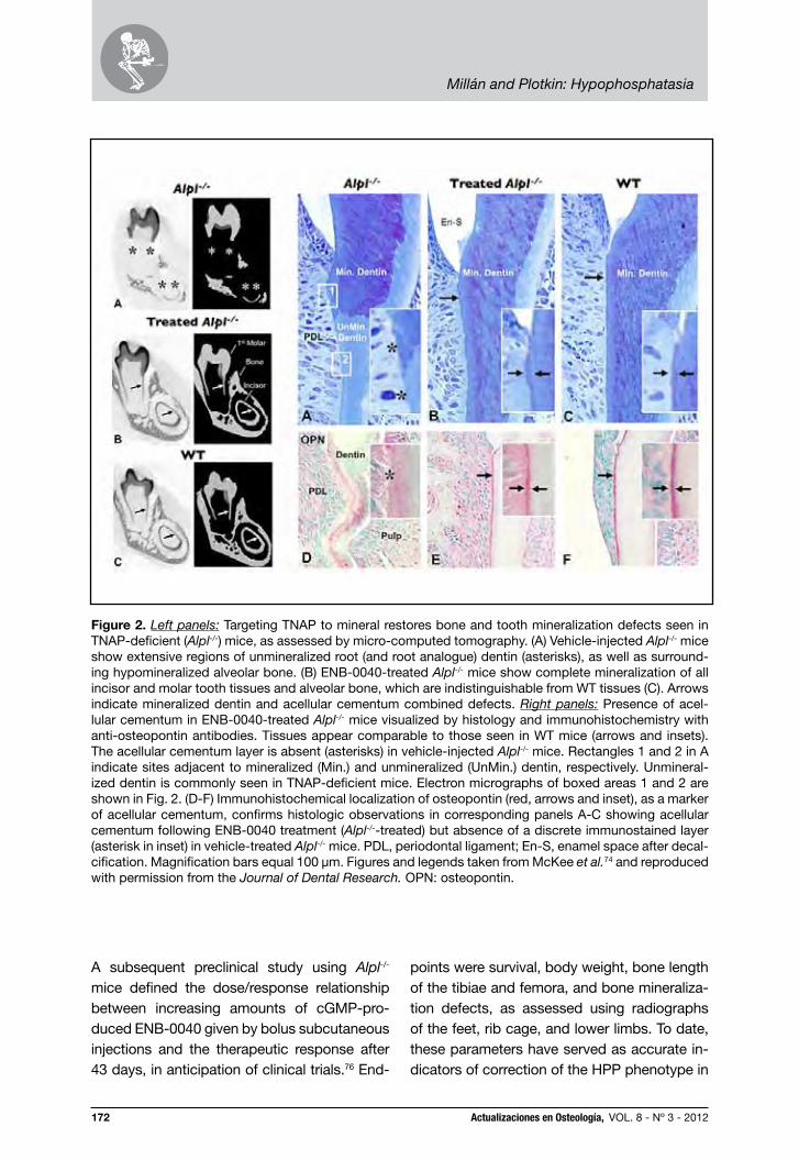
In these mice, sALP-FcD10 treatment also prevented hypomineralization of alveolar bone and dentin69 and acellular cementum formed normally, which is typically missing or reduced in Alpl-/- mice.74 Micro-computed tomography revealed a consistent reduc-tion of mineralization in both alveolar bone and root dentin (and root analogue dentin in the incisor) in the control (vehicle-alone-injected) Alpl-/- mice (Fig. 2). This feature was not present in the sALP-FcD10-treated Alpl-/- mice, which showed complete min-eralization of alveolar bone and dentin. Im-munohistochemical staining for OPN (Fig. 2), revealed absence of acellular cementum along the root surface in untreated Alpl-/- mice, while sALP-FcD10-treated Alpl-/- mice had an unremarkable acellular cementum layer comparable with that seen in wild-type mice.74 More recently, we showed that the dentin defect is root-targeted and var-ies from delayed mineralization in the mild-est case, to arrest of root mantle dentin mineralization, lack of circumpulpal dentin, and odontoblast differentiation defects, in the most severe manifestations.75 The mild-ly delayed mineralization was corrected given time, but when dentinogenesis was significantly perturbed at the mantle den-tin phase, defects persevered throughout molar tooth development. Our data further reveal that the Alpl-/- dentin defect resulted from inability of MV-initiated mineralization foci to expand into a mineralization front, and that increased OPN accumulation likely contributed to the aborted mineralization of Alpl-/- mantle dentin. Importantly, adminis-tration of bioengineered TNAP showed that early intervention allows for rescue of den-tinogenesis and restoration of molar miner-alization.75
Actualizaciones en Osteología, VOL. 8 - Nº 3 - 2012172
Millán and Plotkin: Hypophosphatasia
A subsequent preclinical study using Alpl-/- mice defined the dose/response relationship between increasing amounts of cGMP-pro-duced ENB-0040 given by bolus subcutaneous injections and the therapeutic response after 43 days, in anticipation of clinical trials.76 End-
points were survival, body weight, bone length of the tibiae and femora, and bone mineraliza-tion defects, as assessed using radiographs of the feet, rib cage, and lower limbs. To date, these parameters have served as accurate in-dicators of correction of the HPP phenotype in
Figure 2. Left panels: Targeting TNAP to mineral restores bone and tooth mineralization defects seen in TNAP-deficient (Alpl-/-) mice, as assessed by micro-computed tomography. (A) Vehicle-injected Alpl-/- mice show extensive regions of unmineralized root (and root analogue) dentin (asterisks), as well as surround-ing hypomineralized alveolar bone. (B) ENB-0040-treated Alpl-/- mice show complete mineralization of all incisor and molar tooth tissues and alveolar bone, which are indistinguishable from WT tissues (C). Arrows indicate mineralized dentin and acellular cementum combined defects. Right panels: Presence of acel-lular cementum in ENB-0040-treated Alpl-/- mice visualized by histology and immunohistochemistry with anti-osteopontin antibodies. Tissues appear comparable to those seen in WT mice (arrows and insets). The acellular cementum layer is absent (asterisks) in vehicle-injected Alpl-/- mice. Rectangles 1 and 2 in A indicate sites adjacent to mineralized (Min.) and unmineralized (UnMin.) dentin, respectively. Unmineral-ized dentin is commonly seen in TNAP-deficient mice. Electron micrographs of boxed areas 1 and 2 are shown in Fig. 2. (D-F) Immunohistochemical localization of osteopontin (red, arrows and inset), as a marker of acellular cementum, confirms histologic observations in corresponding panels A-C showing acellular cementum following ENB-0040 treatment (Alpl-/--treated) but absence of a discrete immunostained layer (asterisk in inset) in vehicle-treated Alpl-/- mice. PDL, periodontal ligament; En-S, enamel space after decal-cification. Magnification bars equal 100 μm. Figures and legends taken from McKee et al.74 and reproduced with permission from the Journal of Dental Research. OPN: osteopontin.
Actualizaciones en Osteología, VOL. 8 - Nº 3 - 2012 173
Millán and Plotkin: Hypophosphatasia
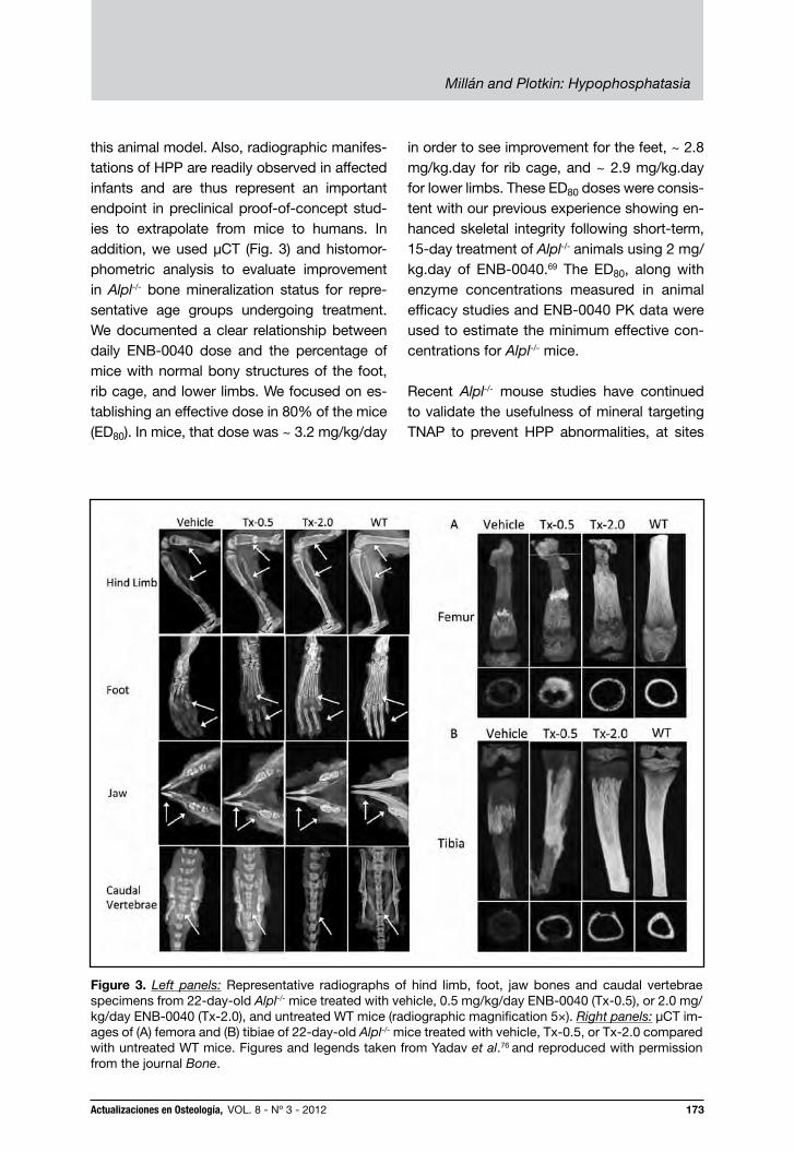
this animal model. Also, radiographic manifes-tations of HPP are readily observed in affected infants and are thus represent an important endpoint in preclinical proof-of-concept stud-ies to extrapolate from mice to humans. In addition, we used µCT (Fig. 3) and histomor-phometric analysis to evaluate improvement in Alpl-/- bone mineralization status for repre-sentative age groups undergoing treatment. We documented a clear relationship between daily ENB-0040 dose and the percentage of mice with normal bony structures of the foot, rib cage, and lower limbs. We focused on es-tablishing an effective dose in 80% of the mice (ED80). In mice, that dose was ~ 3.2 mg/kg/day
in order to see improvement for the feet, ~ 2.8 mg/kg.day for rib cage, and ~ 2.9 mg/kg.day for lower limbs. These ED80 doses were consis-tent with our previous experience showing en-hanced skeletal integrity following short-term, 15-day treatment of Alpl-/- animals using 2 mg/kg.day of ENB-0040.69 The ED80, along with enzyme concentrations measured in animal efficacy studies and ENB-0040 PK data were used to estimate the minimum effective con-centrations for Alpl-/- mice.
Recent Alpl-/- mouse studies have continued to validate the usefulness of mineral targeting TNAP to prevent HPP abnormalities, at sites
Figure 3. Left panels: Representative radiographs of hind limb, foot, jaw bones and caudal vertebrae specimens from 22-day-old Alpl-/- mice treated with vehicle, 0.5 mg/kg/day ENB-0040 (Tx-0.5), or 2.0 mg/kg/day ENB-0040 (Tx-2.0), and untreated WT mice (radiographic magnification 5×). Right panels: μCT im-ages of (A) femora and (B) tibiae of 22-day-old Alpl-/- mice treated with vehicle, Tx-0.5, or Tx-2.0 compared with untreated WT mice. Figures and legends taken from Yadav et al.76 and reproduced with permission from the journal Bone.

Actualizaciones en Osteología, VOL. 8 - Nº 3 - 2012174
Millán and Plotkin: Hypophosphatasia
notorious for their lack of vascularity. Such is the case of the enamel organ during tooth development. We have shown here that ENB-0040 reaches the enamel organ during the se-cretory and maturation phases in the treated Alpl-/- mice.77 Scanning electron microscopy of Alpl-/- mice demonstrated a lack of organization of rod and inter-rod structure of enamel (Fig. 4). Micro-computed tomography showed that at 23 postnatal days, ENB-0040 has dose-depen-dent normalizing effects on both absolute and relative enamel volumes. Histological analysis of Alpl-/- mice showed reduced enamel mineral-ization both in molars and incisors, loss of po-larization of ameloblasts that are important for enamel matrix formation, and even complete
lack of enamel formation in the absence of TNAP. The fact that patients with odonto-HPP typically have the mildest reductions of serum TNAP levels4 suggests that tooth formation is the most sensitive developmental process re-quiring TNAP function.69, 74 Previous studies of cementum had demonstrated the sensitivity of this tissue to changes in the local Pi/PPi ratio78 and the ability of mineral-targeted TNAP to preserve acellular cementum in Alpl-/- mice had further validated that premise.74 Here, too, the ability of ENB-0040 to preserve the structural integrity of enamel supports the notion that the enamel organ is under direct regulation by the extracellular Pi/PPi ratio and that TNAP plays a crucial role in this regulation.77
Figure 4. Scanning electron microscopy (SEM) analysis of incisors (top) and molars (bottom) of WT and Alpl-/- mice at 20 dpn. The SEM images showed well- decussated enamel rods and inter-rod in the molar crowns and crown analogs of incisors of WT mice. Note that there is a lack of rod–inter-rod organization in the Alpl-/- mice. The images were taken in the erupted part of the tooth. Figure taken from Yadav et al.77 and reproduced with permission from the Journal of Bone and Mineral Research.

Actualizaciones en Osteología, VOL. 8 - Nº 3 - 2012 175
Millán and Plotkin: Hypophosphatasia
Preclinical validation of enzyme replace-ment for HPP using viral vector delivery of mineral-targeting TNAPWe have recently shown that a single injection of a lentiviral vector harboring TNAP-D10 (that is, soluble TNAP linked directly to D10) resulted in sustained AP expression and phenotypic correction of HPP in Alpl-/- neonatal mice79 including prevention of epileptic seizures and skeletal defects and preservation of a normal life-span. In another recent study, we demonstrated that a single intravenous injection of neonatal Alpl-/- mice with an adeno-associated viral vector expressing TNAP-D10 was as effective as the lentiviral vector treatment.80 In both of these viral approaches, the Fc region was not included in the construct because neither purification of the enzyme nor an increase in the circu-lating plasma half-life was required. Indeed, these two viral delivery systems utilize either the liver (lentiviral vector) or skeletal muscle (adeno-associated viral vector) as factories of bone-targeted TNAP for the life of the animal. These delivery systems show promise as an alternative means of delivery of bone-targeted TNAP that would also decrease treatment frequency and po-tentially cost.
Furthermore, Matsumoto et al. demonstrat-ed that TNAP lacking the D10 mineral-target-ing domain could be an effective treatment for HPP mice, indicating that sustained and substantial TNAP production may be the most critical component of the treatment, more so than mineral-targeting.80 More re-cently, Sugano and collaborators demon-strated the usefulness of viral vectors for delivery of mineral-targeting TNAP in ute-ro, thus fetal therapy of HPP.81 These very encouraging proof-of-principle preclinical studies could pave the way to gene therapy clinical trials using viral vectors as means of delivering mineral-targeting TNAP to pa-
tients. The potential rewards include the greatly reduced number of injection that a patient might need to receive in their lifetime and potentially reduced cost of the therapy. However, viral vector safety still remains a concern while EzRT is demonstrating a good track record of safety and efficacy.
Clinical trials using mineral-targeting TNAP for life-threatening HPPAn Investigational New Drug (IND) applica-tion to use asfotase alfa to treat HPP was filed in June of 2008. Initial dosing was then based on: 1) dose-response data of ENB-0040 in Alpl-/- mice,76 2) efficacy observed in treated Alpl-/- mice that achieved an equiv-alent to serum ALP activity in the range of 2,400-6,000 U/L; 3) the No-Observed-Ad-verse-Effect Level (NOAEL) in more sensitive species (rats and monkeys) established in one-month IV toxicology studies, and one-month IV/SC bridging and tolerability stud-ies in rats; 4) a safety factor of 10 applied to the NOAEL; and 5) results from a multi-center, open-label, dose-escalating phase 1 study of the safety, tolerability, and pharma-cology of ENB-0040 in six adults with HPP who received one dose of 3 mg/kg IV and then either 1 or 2 mg/kg ENB-0040 SC once weekly for three weeks. To date, three clini-cal studies have been completed, and three extension studies are ongoing, as well as an additional study in infants with HPP, cur-rently recruiting patients (www.clinicaltrials.gov). The first full report of a clinical trial us-ing asfotase alfa was on an open-label study to evaluate the safety, tolerability, bioavail-ability, pharmacokinetics, pharmacodynam-ics, and efficacy of treatment with asfotase alfa in severely affected infants and young children with HPP.82 Efficacy assessments included radiographic skeletal changes, and changes in gross and fine motor func-tion and cognitive development. The study included children 3 years old or less at en-rollment, with signs of HPP occurring before

Actualizaciones en Osteología, VOL. 8 - Nº 3 - 2012176
Millán and Plotkin: Hypophosphatasia
the age of 6 months, hypophosphatasemia, elevated plasma PLP levels, radiological signs of HPP, and failure to thrive. Other signs at baseline included rachitic chest deformity, pyridoxine-responsive seizures, history of a non-traumatic or poorly healing fractures, hypercalcemia, craniosynostosis, nephrocalcinosis, and respiratory compro-mise from HPP. The patients received asfo-tase alfa (at a concentration of 40 mg per milliliter) as a single intravenous infusion at a dose of 2 mg/kg, followed by subcutaneous injections three times per week at a dose of 1 mg/kg for 24 weeks, with an extension thereafter. The subcutaneous dose could be increased up to 3 mg/kg if there was wors-ening failure to thrive, deteriorating pulmo-nary function, or no radiographic evidence of skeletal improvement.82 Eleven patients were recruited.82 One patient
had a moderate reaction to the IV injection and the parents decided to discontinue the treat-ment. Another patient completed the first 6 months of treatment, but soon after succumbed to pneumonia and sepsis, judged unrelated to the study treatment. Of the nine patients who were treated for at least 1 year, four had peri-natal HPP and five had infantile HPP. One had lost nearly all radiographically apparent skeletal mineral during prolonged ventilation and immo-bilization. Serum calcium levels were generally high at diagnosis (unlike calcium levels in other forms of rickets), and initially required dietary calcium restriction in all patients (most of whom had nephrocalcinosis). Skeletal remineralization occurred during treatment, including the patient mentioned above with extremely severe disease. Images of radiographic improvement are shown here for the oldest patient (3 years old) (Fig. 5) and for the youngest patient (<1 month old)
Figure 5. This 36-month-old girl had a short, bowed femur detected in utero by ultrasound (A). At 12 days-of-age, her chest radiograph showed thin, osteopenic ribs with lytic areas and fractures (B). Be-fore treatment, severe pansuture closure was present, including a marked increase in “digital” markings (“beaten-copper” appearance) (D). The ribs at baseline were osteopenic and had fracture deformities with thin cortices (F). By week 24 of treatment, the ribs were wider and better mineralized with sharper cortical margins and less deformity (G). Improvement of the rickets with therapy is apparent (M, N, O, P). Images taken for the online supplementary data to Whyte et al.82 and reproduced with permission from The New England Journal of Medicine.

Actualizaciones en Osteología, VOL. 8 - Nº 3 - 2012 177
Millán and Plotkin: Hypophosphatasia
Figure 6. This 20-day-old (at therapy baseline) boy had shortened and bowed extremities and “fractures” detected by prenatal sonography at 17 – 18 weeks gestation (A, B). Before ENB-0040 treatment, little or decreased mineral was present in the frontal, parietal, or occipital bones, skull base, facial bones, and sphenoid (E). At week 24 of therapy, all areas showed striking remineralization (F). The improvement in the left hand and wrist was remarkable (I, J). Before ENB-0040, the femora were short, sclerotic, bowed, irregular, and lacked defined medullary cavities, cortices, and mineralized metaphyses and epiphyses. The fibulae were not calcified (K). After therapy, striking mineralization was evident (L). Images taken for the online supplementary data to Whyte et al.82 and reproduced with permission from The New England Journal of Medicine.
(Fig. 6) at the start of the treatment. Treated infants showed no signs of symptomatic hy-pocalcemia from “hungry-bones” syndrome. Initially the treatment was often accompa-nied by increases in serum parathyroid hor-mone levels that called for liberalization of dietary calcium but not additional vitamin D.82 A positive mineral balance throughout the skeleton was obvious on radiography after several weeks or months of treatment, and effects were evident in both membra-nous and endochondral bone. Substantial radiographic improvement in skeletal ab-normalities was noted at week 24 in all but one patient, with continued healing through week 48. In the one patient who had no ra-
diographically visible mineral at baseline, calcification was observed after 9 months of therapy. This delay probably reflected the profound deficit of skeletal mineral in the patient, although sufficient hydroxyapatite was apparently present for targeting with asfotase alfa. After the start of therapy, de-ciduous teeth erupted in all the patients, with only one patient having HPP-related loss of a tooth.82
In summary, of the 11 patients recruited, 10 completed 6 months of therapy; 9 complet-ed 1 year. Healing of rickets was evident at 6 months in 9 patients. All patients showed improvement in developmental milestones
Actualizaciones en Osteología, VOL. 8 - Nº 3 - 2012178
Millán and Plotkin: Hypophosphatasia
and stabilization or improvement of pulmo-nary function. Elevated plasma levels of the TNAP substrates PPi and PLP diminished. There was no evidence of symptomatic hy-pocalcemia, ectopic calcification, or defi-nite drug-related serious adverse events. Low titers of anti–asfotase alfa antibodies developed in four patients, with no evident clinical, biochemical, or autoimmune ab-
normalities at 48 weeks of treatment. Thus, asfotase alfa therapy was associated with improved findings on skeletal radiographs and improved pulmonary and physical func-tion in infants and young children with life-threatening HPP.
(Recibido: septiembre de 2012.Aceptado: octubre 2012)
References
1. Fraser D. Hypophosphatasia. Am J Med 1957;
22:730-46.
2. Greenberg CR, Taylor CL, Haworth JC,
Seargeant LE, Philipps S, Triggs-Raine B,
Chodirker BN. A homoallelic Gly317-->Asp
mutation in ALPL causes the perinatal (lethal)
form of hypophosphatasia in Canadian men-
nonites. Genomics 1993; 17:215-7.
3. Millán JL. Mammalian alkaline phosphatases.
From biology to applications in medicine and
biotechnology. Weinheim, Germany: Wiley-
VCH Verlag GmbH & Co; 2006.
4. Whyte M. Hypophosphatasia. In: Glorieux F,
Jueppner H, Pettifor J, editors. Pediatric Bone.
San Diego, CA: Elsevier (Academic Press);
2012. p. 771-94.
5. Fallon MD, Teitelbaum SL, Weinstein RS, Gold-
fischer S, Brown DM, Whyte MP. Hypophos-
phatasia: clinicopathologic comparison of the
infantile, childhood, and adult forms. Medicine
(Baltimore) 1984; 63:12-24.
6. Whyte MP, Teitelbaum SL, Murphy WA, Berg-
feld MA, Avioli LV. Adult hypophosphatasia.
Clinical, laboratory, and genetic investigation
of a large kindred with review of the literature.
Medicine (Baltimore) 1979; 58:329-47.
7. Whyte MP, Murphy WA, M. Fallon MD. Adult
hypophosphatasia with chondrocalcinosis and
arthropathy. Variable penetrance of hypophos-
phatasemia in a large Oklahoma kindred. Am J
Med 1982; 72:631-41.
8. Wenkert D, Mcalister WH, Coburn SP, Zerega
JA, Ryan LM, Ericson KL, Hersh JH, Mumm S,
Whyte MP. Hypophosphatasia: nonlethal dis-
ease despite skeletal presentation in utero (17
new cases and literature review). J Bone Miner
Res 2011; 26:2389-98.
9. Weiss MJ, Cole DE, Ray K, Whyte MP, Lafferty
MA, Mulivor RA, Harris H. A missense mutation
in the human liver/bone/kidney alkaline phos-
phatase gene causing a lethal form of hypo-
phosphatasia. Proc Natl Acad Sci USA 1988;
85:7666-9.
10. Henthorn PS, Raducha M, Fedde KN, Lafferty
MA, Whyte MP. Different missense mutations
at the tissue-nonspecific alkaline phosphatase
gene locus in autosomal recessively inherited
forms of mild and severe hypophosphatasia.
Proc Natl Acad Sci USA 1992; 89:9924-8.
11. Henthorn PS, Whyte MP. Missense mutations
of the tissue-nonspecific alkaline phosphatase
gene in hypophosphatasia. Clin Chem 1992;
38:2501-5.
12. Ozono K, Yamagata M, Michigami T, et al.
Identification of novel missense mutations
(Phe310Leu and Gly439Arg) in a neonatal case
of hypophosphatasia. J Clin Endocrinol Metab
1996; 81:4458-61.

Actualizaciones en Osteología, VOL. 8 - Nº 3 - 2012 179
Millán and Plotkin: Hypophosphatasia
13. Orimo H, Hayashi Z, Watanabe A, Hirayama T,
Hirayama T, Shimada T. Novel missense and
frameshift mutations in the tissue-nonspecific
alkaline phosphatase gene in a Japanese pa-
tient with hypophosphatasia. Hum Mol Genet
1994; 3:1683-4.
14. Mornet E, Taillandier A, Peyramaure S, et al. Iden-
tification of fifteen novel mutations in the tissue-
nonspecific alkaline phosphatase (TNSALP) gene
in European patients with severe hypophospha-
tasia. Eur J Hum Genet 1998; 6:308-14.
15. Mumm S, Jones J, Finnegan P, Whyte MP. Hy-
pophosphatasia: molecular diagnosis of Rath-
bun’s original case. J Bone Miner Res 2001;
16:1724-7.
16. Ali SY, Sajdera SW, Anderson HC. Isolation and
characterization of calcifying matrix vesicles
from epiphyseal cartilage. Proc Natl Acad Sci
USA 1970; 67:1513-20.
17. Bernard GW. Ultrastructural localization of al-
kaline phosphatase in initial intramembranous
osteogenesis. Clin Orthop Relat Res 1978;
218-25.
18. Morris DC, Masuhara K, Takaoka K, Ono K,
Anderson HC. Immunolocalization of alkaline
phosphatase in osteoblasts and matrix ves-
icles of human fetal bone. Bone Miner 1992;
19:287-98.
19. Anderson HC, Harmey D, Camacho NP, et al.
Sustained osteomalacia of long bones despite
major improvement in other hypophosphata-
sia-related mineral deficits in tissue nonspecif-
ic alkaline phosphatase/nucleotide pyrophos-
phatase phosphodiesterase 1 double-deficient
mice. Am J Pathol 2005; 166:1711-20.
20. Anderson HC, Hsu HH, Morris DC, Fedde KN,
Whyte MP. Matrix vesicles in osteomalacic hy-
pophosphatasia bone contain apatite-like min-
eral crystals. Am J Pathol 1997; 151:1555-61.
21. Anderson HC, Sipe JB, Hessle L, et al. Impaired
calcification around matrix vesicles of growth
plate and bone in alkaline phosphatase-defi-
cient mice. Am J Pathol 2004; 164:841-7.
22. Meyer JL. Can biological calcification occur
in the presence of pyrophosphate? Arch Bio-
chem Biophys 1984; 231:1-8.
23. Harmey D, Hessle L, Narisawa S, Johnson KA,
Terkeltaub R, Millán JL. Concerted regulation
of inorganic pyrophosphate and osteopontin
by akp2, enpp1, and ank: an integrated model
of the pathogenesis of mineralization disor-
ders. Am J Pathol 2004; 164:1199-209.
24. Hessle L, Johnson KA, Anderson HC, et al.
Tissue-nonspecific alkaline phosphatase and
plasma cell membrane glycoprotein-1 are cen-
tral antagonistic regulators of bone mineraliza-
tion. Proc Natl Acad Sci USA 2002; 99: 9445-9.
25. Murshed M, Harmey D, Millán JL, Mckee MD,
Karsenty G. Unique coexpression in osteo-
blasts of broadly expressed genes accounts
for the spatial restriction of ECM mineralization
to bone. Genes Dev 2005; 19:1093-104.
26. Johnson KA, Hessle L, Vaingankar S, et al.
Osteoblast tissue-nonspecific alkaline phos-
phatase antagonizes and regulates PC-1. Am
J Physiol Regul Integr Comp Physiol 2000;
279:R1365-77.
27. Ciancaglini P, Yadav MC, Simao AM, et al.
Kinetic analysis of substrate utilization by na-
tive and TNAP-, NPP1-, or PHOSPHO1-defi-
cient matrix vesicles. J Bone Miner Res 2010;
25:716-23.
28. Majeska RJ, Wuthier RE. Studies on matrix
vesicles isolated from chick epiphyseal car-
tilage. Association of pyrophosphatase and
ATPase activities with alkaline phosphatase.
Biochim Biophys Acta 1975; 391:51-60.
29. Moss DW, Eaton RH, Smith JK, Whitby LG. As-
sociation of inorganic-pyrophosphatase activ-
ity with human alkaline-phosphatase prepara-
tions. Biochem J 1967; 102:53-7.
30. Rezende AA, Pizauro JM, Ciancaglini P, Le-
one FA. Phosphodiesterase activity is a novel
property of alkaline phosphatase from osseous
plate. Biochem J 1994; 301 (Pt 2):517-22.
31. Robison R. The Possible Significance of
Hexosephosphoric Esters in Ossification. Bio-
chem J 1923; 17:286-93.
32. Yadav MC, Simao AM, Narisawa S, et al. Loss
of skeletal mineralization by the simultaneous
ablation of PHOSPHO1 and alkaline phospha-
tase function: a unified model of the mecha-

Actualizaciones en Osteología, VOL. 8 - Nº 3 - 2012180
Millán and Plotkin: Hypophosphatasia
nisms of initiation of skeletal calcification. J
Bone Miner Res 2011; 26:286-97.
33. Coburn SP. Modeling vitamin B6 metabolism.
Adv Food Nutr Res 1996; 40:107-32.
34. Jansonius JN. Structure, evolution and action
of vitamin B6-dependent enzymes. Curr Opin
Struct Biol 1998; 8:759-69.
35. Whyte MP, Landt M, Ryan LM, et al. Alkaline
phosphatase: placental and tissue-nonspe-
cific isoenzymes hydrolyze phosphoethanol-
amine, inorganic pyrophosphate, and pyridoxal
5’-phosphate. Substrate accumulation in carri-
ers of hypophosphatasia corrects during preg-
nancy. J Clin Invest 1995; 95:1440-5.
36. Chodirker BN, Coburn SP, Seargeant LE,
Whyte MP, Greenberg CR. Increased plasma
pyridoxal-5’-phosphate levels before and af-
ter pyridoxine loading in carriers of perinatal/
infantile hypophosphatasia. J Inherit Metab Dis
1990; 13:891-6.
37. Whyte MP, Mahuren JD, Fedde KN, Cole FS, Mc-
cabe ER, Coburn SP. Perinatal hypophosphata-
sia: tissue levels of vitamin B6 are unremarkable
despite markedly increased circulating concen-
trations of pyridoxal-5’-phosphate. Evidence for
an ectoenzyme role for tissue-nonspecific alka-
line phosphatase. J Clin Invest 1988; 81:1234-9.
38. Whyte MP, Mahuren JD, Vrabel LA, Coburn SP.
Markedly increased circulating pyridoxal-5’-
phosphate levels in hypophosphatasia. Alka-
line phosphatase acts in vitamin B6 metabo-
lism. J Clin Invest 1985; 76:752-6.
39. Narisawa S, Frohlander N, Millán JL. Inactiva-
tion of two mouse alkaline phosphatase genes
and establishment of a model of infantile hypo-
phosphatasia. Dev Dyn 1997; 208:432-46.
40. Narisawa S, Wennberg C, Millán JL. Abnormal
vitamin B6 metabolism in alkaline phosphatase
knock-out mice causes multiple abnormalities,
but not the impaired bone mineralization. J
Pathol 2001; 193:125-33.
41. Waymire KG, Mahuren JD, Jaje JM, Guilarte
TR, Coburn SP, Macgregor GR. Mice lacking
tissue non-specific alkaline phosphatase die
from seizures due to defective metabolism of
vitamin B-6. Nat Genet 1995; 11:45-51.
42. Fleshood HL, Pitot HC. O-phosphorylethanol-
amine ammonia lyase, a new pyridoxal phos-
phate-dependent enzyme. Biochem Biophys
Res Commun 1969; 36:110-8.
43. Fleshood HL, Pitot HC. The metabolism of O-
phosphorylethanolamine in animal tissues. II.
Metabolic regulation of O-phosphorylethanol-
amine phospho-lyase in vivo. Arch Biochem
Biophys 1970; 141:423-9.
44. Millán JL, Whyte MP, Avioli LV, Fishman WH.
Hypophosphatasia (adult form): quantitation of
serum alkaline phosphatase isoenzyme activity
in a large kindred. Clin Chem 1980; 26:840-5.
45. Harmey D, Johnson KA, Zelken J, et al. Ele-
vated skeletal osteopontin levels contribute to
the hypophosphatasia phenotype in Akp2(-/-)
mice. J Bone Miner Res 2006; 21:1377-86.
46. Johnson K, Goding J, Van Etten D, et al. Linked
deficiencies in extracellular PP(i) and osteo-
pontin mediate pathologic calcification associ-
ated with defective PC-1 and ANK expression.
J Bone Miner Res 2003; 18: 994-1004.
47. Wennberg C, Hessle L, Lundberg P, et al. Func-
tional characterization of osteoblasts and os-
teoclasts from alkaline phosphatase knockout
mice. J Bone Miner Res 2000; 15:1879-88.
48. Goldberg HA, Warner KJ, Li MC, Hunter GK.
Binding of bone sialoprotein, osteopontin and
synthetic polypeptides to hydroxyapatite. Con-
nect Tissue Res 2001; 42:25-37.
49. Christensen B, Nielsen MS, Haselmann KF,
Petersen TE, Sorensen ES. Post-translationally
modified residues of native human osteopon-
tin are located in clusters: identification of 36
phosphorylation and five O-glycosylation sites
and their biological implications. Biochem J
2005; 390:285-92.
50. Hunter GK, Kyle CL, Goldberg HA. Modulation
of crystal formation by bone phosphoproteins:
structural specificity of the osteopontin-me-
diated inhibition of hydroxyapatite formation.
Biochem J 1994; 300 (Pt 3):723-8.
51. Jono S, Peinado C, Giachelli CM. Phosphory-
lation of osteopontin is required for inhibition
of vascular smooth muscle cell calcification. J
Biol Chem 2000; 275:20197-203.
Actualizaciones en Osteología, VOL. 8 - Nº 3 - 2012 181
Millán and Plotkin: Hypophosphatasia
52. Pampena DA, Robertson KA, Litvinova O, La-
joie G, Goldberg HA, Hunter GK. Inhibition of
hydroxyapatite formation by osteopontin phos-
phopeptides. Biochem J 2004; 378:1083-7.
53. Addison WN, Masica DL, Gray JJ, Mckee MD.
Phosphorylation-dependent inhibition of min-
eralization by osteopontin ASARM peptides is
regulated by PHEX cleavage. J Bone Miner Res
2010; 25:695-705.
54. Deeb AA, Bruce SN, Morris AA, Cheetham
TD. Infantile hypophosphatasia: disappoint-
ing results of treatment. Acta Paediatr 2000;
89:730-3.
55. Sutton RA, Mumm S, Coburn SP, Ericson KL,
Whyte MP. “Atypical femoral fractures” during
bisphosphonate exposure in adult hypophos-
phatasia. J Bone Miner Res 2012; 27:987-94.
56. Cahill RA, Wenkert D, Perlman SA, et al. Infan-
tile hypophosphatasia: transplantation therapy
trial using bone fragments and cultured osteo-
blasts. J Clin Endocrinol Metab 2007; 92:2923-
30.
57. Whyte MP, Kurtzberg J, Mcalister WH, et al.
Marrow cell transplantation for infantile hypo-
phosphatasia. J Bone Miner Res 2003; 18:624-
36.
58. Camacho PM, Painter S, Kadanoff R. Treat-
ment of adult hypophosphatasia with teripara-
tide. Endocr Pract 2008; 14:204-8.
59. Doshi KB, Hamrahian AH, Licata AA. Teripa-
ratide treatment in adult hypophosphatasia in
a patient exposed to bisphosphonate: a case
report. Clin Cases Miner Bone Metab 2009;
6:266-9.
60. Schalin-Jantti C, Mornet E, Lamminen A, Vali-
maki MJ. Parathyroid hormone treatment im-
proves pain and fracture healing in adult hypo-
phosphatasia. J Clin Endocrinol Metab 2010;
95: 5174-9.
61. Whyte MP, Mumm S, Deal C. Adult hypophos-
phatasia treated with teriparatide. J Clin Endo-
crinol Metab 2007; 92:1203-8.
62. Gagnon C, Sims NA, Mumm S, et al. Lack of
sustained response to teriparatide in a patient
with adult hypophosphatasia. J Clin Endocrinol
Metab 2010; 95:1007-12.
63. Laroche M. Failure of teriparatide in treatment
of bone complications of adult hypophospha-
tasia. Calcif Tissue Int 2012; 90:250.
64. Vahle JL, Long GG, Sandusky G, Westmore M,
Ma YL, Sato M. Bone neoplasms in F344 rats
given teriparatide [rhPTH(1-34)] are dependent
on duration of treatment and dose. Toxicol
Pathol 2004; 32:426-38.
65. Chung TD, Sergienko E, Millán JL. Assay format
as a critical success factor for identification of
novel inhibitor chemotypes of tissue-nonspe-
cific alkaline phosphatase from high-through-
put screening. Molecules 2010; 15:3010-37.
66. Whyte MP, Valdes Jr. R, Ryan LM, McAlister
WH. Infantile hypophosphatasia: enzyme re-
placement therapy by intravenous infusion of
alkaline phosphatase-rich plasma from pa-
tients with Paget’s bone disease. J Pediatr
1982; 101:379-386.
67. Whyte MP, Mcalister WH, Patton LS, et al.
Enzyme replacement therapy for infantile hy-
pophosphatasia attempted by intravenous
infusions of alkaline phosphatase-rich Paget
plasma: results in three additional patients. J
Pediatr 1984; 105:926-33.
68. Weninger M, Stinson RA, Plenk H, Jr., Bock P,
Pollak A. Biochemical and morphological ef-
fects of human hepatic alkaline phosphatase in
a neonate with hypophosphatasia. Acta Paedi-
atr Scand 1989; 360(Suppl):154-60.
69. Millán JL, Narisawa S, Lemire I, et al. Enzyme
replacement therapy for murine hypophospha-
tasia. J Bone Miner Res 2008; 23:777-87.
70. Kasugai S, Fujisawa R, Waki Y, Miyamoto K,
Ohya K. Selective drug delivery system to
bone: small peptide (Asp)6 conjugation. J Bone
Miner Res 2000; 15:936-43.
71. Nishioka T, Tomatsu S, Gutierrez MA, et al. En-
hancement of drug delivery to bone: charac-
terization of human tissue-nonspecific alkaline
phosphatase tagged with an acidic oligopep-
tide. Mol Genet Metab 2006; 88:244-55.
72. Yokogawa K, Miya K, Sekido T, et al. Selective
delivery of estradiol to bone by aspartic acid
oligopeptide and its effects on ovariectomized
mice. Endocrinology 2001; 142:1228-33.
Actualizaciones en Osteología, VOL. 8 - Nº 3 - 2012182
Millán and Plotkin: Hypophosphatasia
73. Fedde KN, Blair L, Silverstein J, et al. Alka-
line phosphatase knock-out mice recapitulate
the metabolic and skeletal defects of infantile
hypophosphatasia. J Bone Miner Res 1999;
14:2015-26.
74. Mckee MD, Nakano Y, Masica DL, et al. En-
zyme replacement therapy prevents dental de-
fects in a model of hypophosphatasia. J Dent
Res 2011; 90:470-6.
75. Foster BL, Nagatomo KJ, Tso HW, et al. Tooth
root dentin mineralization defects in a mouse
model of hypophosphatasia. J Bone Miner Res
2012; In press.
76. Yadav MC, Lemire I, Leonard P, et al. Dose re-
sponse of bone-targeted enzyme replacement
for murine hypophosphatasia. Bone 2011;
49:250-6.
77. Yadav MC, De Oliveira RC, Foster BL, et al.
Enzyme replacement prevents enamel defects
in hypophosphatasia mice. J Bone Miner Res
2012; 27:1722-34.
78. Nociti FH, Jr., Berry JE, Foster BL, Get al. Ce-
mentum: a phosphate-sensitive tissue. J Dent
Res 2002; 81:817-21.
79. Yamamoto S, Orimo H, Matsumoto T, et al.
Prolonged survival and phenotypic correction
of Akp2(-/-) hypophosphatasia mice by len-
tiviral gene therapy. J Bone Miner Res 2011;
26:135-42.
80. Matsumoto T, Miyake K, Yamamoto S, et al.
Rescue of severe infantile hypophosphatasia
mice by AAV-mediated sustained expression of
soluble alkaline phosphatase. Hum Gene Ther
2011; 22:1355-64.
81. Sugano H, Matsumoto T, Miyake K, et al. Suc-
cessful gene therapy in utero for lethal murine
hypophosphatasia. Hum Gene Ther 2012;
23:399-406.
82. Whyte MP, Greenberg CR, Salman NJ, et al.
Enzyme-replacement therapy in life-threaten-
ing hypophosphatasia. N Engl J Med 2012;
366:904-13.