Aging Induced Ag Nanoparticle Rearrangement under Ambient Atmosphere and Consequences for...
7
Aging Induced Ag Nanoparticle Rearrangement under Ambient Atmosphere and Consequences for Nanoparticle-Enhanced DNA Biosensing Hsin-I Peng, † Todd D. Krauss, ‡,§ and Benjamin L. Miller* ,†,| Department of Biomedical Engineering, Department of Chemistry, The Institute of Optics, and Department of Dermatology, University of Rochester, Rochester, New York 14627 Localized surface plasmons of metallic nanoparticles can strongly amplify the magnitude of the surrounding electric field. This in turn enhances fluorescence from nearby fluorophores. However, little is known regarding how time-dependent changes in nanoparticle structure due to exposure to the ambient environment affect their behavior in plasmonic devices. Here, we report the interesting finding that the aging of a nanostructured Ag substrate in ambient atmosphere markedly improves the fluorescence signal of a plasmonic-based DNA detection system. The effect can be observed with an exposure time as short as two days, and a nearly 17-fold signal enhancement can be achieved with 30 days of aging. Analysis of substrate surface topography by atomic force microscopy (AFM) reveals a substantial change in nanoparticle morphology as the substrates age despite being covalently attached to a solid dry substrate. Nanoparticle morphological changes also manifest in extinction spectra. This process can be further accelerated by light. Together, our findings ad- dress the important question of Ag nanoparticle stability over time and its potential ramifications for plasmon- enabled sensors. They also imply that nanoparticle aging may be used strategically to tune nanoparticle size and geometry and plasmon spectrum, which may be beneficial for studies on plasmonics as well as sensor optimization. The ability of metal nanomaterials to strongly modulate the local electric field has led to their introduction into many photonic systems, including photovoltaics, light emitting diodes (LED), electronics, and sensors. 1-5 Metal nanomaterials have found a particularly important area of application in biosensor develop- ment. 6,7 Unlike bulk metals, metal nanoparticles display a strong UV-vis extinction band, resulting from the collective oscillation of free electrons in resonance with the incident light, an effect known as localized surface plasmon resonance (LSPR). 8,9 The large electric field localized at the metal surface enables metal nanoparticles to dramatically enhance both the fluorescence (metal-enhanced fluorescence, or MEF) and Raman signals from neighboring fluorophores and Raman labels, making them ideal candidates for incorporation into biosensors. 10-15 In addition, the plasmon extinction band is tunable based on nanoparticle size and shape, and has a high sensitivity to the local dielectric constant. Thus metal nanoparticles can also be incorporated into LSPR sensors in which molecular recognition is observed through changes in extinction spectra. 16,17 As the number of sensing applications employing metal nanoparticles continues to increase, it is crucially important to understand how the environmental stability of metal nanomaterials (or lack thereof) affects detection precision and system reliability. Among different metals, nanoparticulate Ag has been the most prominently studied element in the field of surface enhanced spectroscopies owing to the very large natural plasmonic enhance- ment for Ag, coupled with its ease of handling and low cost. 12,18 However, there is a perception that surface oxidation or other aging processes of metals can rapidly degrade a system’s performance, given the fact that Ag rapidly oxidizes in ambient air. 19,20 Special care for Ag nanoparticle-based materials, such as storage under vacuum, is often taken as a precaution to prevent oxidation. 21 Nonoxidative structural rearrangements of Ag nano- * Corresponding author phone: (585) 275-9805; e-mail: Benjamin_Miller@ urmc.rochester.edu. † Department of Biomedical Engineering. ‡ Department of Chemistry. § The Institute of Optics. | Department of Dermatology. (1) Moulin, E.; Sukmanowski, J.; Schult, M.; Gordijn, A.; Royer, F. X.; Stiebig, H. Thin Solid Films 2008, 516, 6813–6817. (2) Coe, S.; Woo, W.-K.; Bawendi, M. G.; Bulovic, V. Nature 2002, 420, 800– 803. (3) Ko, S. H.; Park, I.; Pan, H.; Grigoropoulos, C. P.; Pisano, A. P.; Luscombe, C. K.; Fre `chet, J. M. Nano Lett. 2007, 7, 1869–1877. (4) Prasad, P. N. Nanophotonics, Chapter 13; Wiley-Interscience: Hoboken, NJ, 1998. (5) Skrabalak, S. E.; Chen, J.; Au, L.; Lu, X.; Li, X.; Xia, Y. Adv. Mater. 2007, 19, 3177–3184. (6) Anker, J. N.; Hall, W. P.; Lyanders, O.; Shan, N. C.; Zhao, J.; Van Duyne, R. P. Nat. Mater. 2008, 7, 442–453. (7) Taton, T. A.; Mirkin, C. A.; Letsinger, R. L. Science 2000, 289, 1757–1760. (8) Willets, K. A.; Van Duyne, R. P. Annu. Rev. Phys. Chem. 2007, 58, 267– 297. (9) Kreibig, U.; Vollmer, M. Optical Properties of Metal Clusters, Chapter 2; Toennies, J. P., Gonser, U., Osgood, R. M., Jr., Panish, M. B., Sakaki, H., Eds.; Springer-Verlag: Berlin, 1995; Vol. 25. (10) Bharadwaj, P.; Anger, P.; Novotny, L. Nanotechnology 2007, 18, 044017. (11) Lakowicz, J. R. Anal. Biochem. 2005, 337, 171–194. (12) Moskovits, M. Rev. Mod. Phys. 1985, 57, 783–826. (13) Nie, S.; Emory, S. R. Science 1997, 275, 1102–1106. (14) Yonzon, C. R.; Haynes, C. L.; Zhang, X.; Walsh, J. T., Jr.; Van Duyne, R. P. Anal. Chem. 2004, 76, 78–85. (15) Aslan, K.; Zhang, Y.; Geddes, C. D. Anal. Chem. 2009, 81, 4713–4719. (16) Haes, A. J.; Van Duyne, R. P. J. Am. Chem. Soc. 2002, 124, 10596–10604. (17) Jain, P. K.; Huang, X.; El-Sayed, I. H.; El-Sayed, M. A. Acc. Chem. Res. 2008, 41, 1578–1586. (18) Chan, G. H.; Zhao, J.; Hicks, E. M.; Schatz, G. C.; Van Duyne, R. P. Nano Lett. 2007, 7, 1947–1952. Anal. Chem. 2010, 82, 8664–8670 10.1021/ac101919h 2010 American Chemical Society 8664 Analytical Chemistry, Vol. 82, No. 20, October 15, 2010 Published on Web 09/21/2010
-
Upload
benjamin-l -
Category
Documents
-
view
212 -
download
0
Transcript of Aging Induced Ag Nanoparticle Rearrangement under Ambient Atmosphere and Consequences for...
Aging Induced Ag Nanoparticle Rearrangementunder Ambient Atmosphere and Consequences forNanoparticle-Enhanced DNA Biosensing
Hsin-I Peng,† Todd D. Krauss,‡,§ and Benjamin L. Miller*,†,|
Department of Biomedical Engineering, Department of Chemistry, The Institute of Optics, and Department ofDermatology, University of Rochester, Rochester, New York 14627
Localized surface plasmons of metallic nanoparticles canstrongly amplify the magnitude of the surrounding electricfield. This in turn enhances fluorescence from nearbyfluorophores. However, little is known regarding howtime-dependent changes in nanoparticle structure due toexposure to the ambient environment affect their behaviorin plasmonic devices. Here, we report the interestingfinding that the aging of a nanostructured Ag substrate inambient atmosphere markedly improves the fluorescencesignal of a plasmonic-based DNA detection system. Theeffect can be observed with an exposure time as short astwo days, and a nearly 17-fold signal enhancement canbe achieved with 30 days of aging. Analysis of substratesurface topography by atomic force microscopy (AFM)reveals a substantial change in nanoparticle morphologyas the substrates age despite being covalently attached toa solid dry substrate. Nanoparticle morphological changesalso manifest in extinction spectra. This process can befurther accelerated by light. Together, our findings ad-dress the important question of Ag nanoparticle stabilityover time and its potential ramifications for plasmon-enabled sensors. They also imply that nanoparticle agingmay be used strategically to tune nanoparticle size andgeometry and plasmon spectrum, which may be beneficialfor studies on plasmonics as well as sensor optimization.
The ability of metal nanomaterials to strongly modulate thelocal electric field has led to their introduction into many photonicsystems, including photovoltaics, light emitting diodes (LED),electronics, and sensors.1-5 Metal nanomaterials have found aparticularly important area of application in biosensor develop-
ment.6,7 Unlike bulk metals, metal nanoparticles display a strongUV-vis extinction band, resulting from the collective oscillationof free electrons in resonance with the incident light, an effectknown as localized surface plasmon resonance (LSPR).8,9 Thelarge electric field localized at the metal surface enables metalnanoparticles to dramatically enhance both the fluorescence(metal-enhanced fluorescence, or MEF) and Raman signals fromneighboring fluorophores and Raman labels, making them idealcandidates for incorporation into biosensors.10-15 In addition, theplasmon extinction band is tunable based on nanoparticle size andshape, and has a high sensitivity to the local dielectric constant.Thus metal nanoparticles can also be incorporated into LSPRsensors in which molecular recognition is observed throughchanges in extinction spectra.16,17
As the number of sensing applications employing metalnanoparticles continues to increase, it is crucially important tounderstand how the environmental stability of metal nanomaterials(or lack thereof) affects detection precision and system reliability.Among different metals, nanoparticulate Ag has been the mostprominently studied element in the field of surface enhancedspectroscopies owing to the very large natural plasmonic enhance-ment for Ag, coupled with its ease of handling and low cost.12,18
However, there is a perception that surface oxidation or otheraging processes of metals can rapidly degrade a system’sperformance, given the fact that Ag rapidly oxidizes in ambientair.19,20 Special care for Ag nanoparticle-based materials, such asstorage under vacuum, is often taken as a precaution to preventoxidation.21 Nonoxidative structural rearrangements of Ag nano-
* Corresponding author phone: (585) 275-9805; e-mail: [email protected].
† Department of Biomedical Engineering.‡ Department of Chemistry.§ The Institute of Optics.| Department of Dermatology.
(1) Moulin, E.; Sukmanowski, J.; Schult, M.; Gordijn, A.; Royer, F. X.; Stiebig,H. Thin Solid Films 2008, 516, 6813–6817.
(2) Coe, S.; Woo, W.-K.; Bawendi, M. G.; Bulovic, V. Nature 2002, 420, 800–803.
(3) Ko, S. H.; Park, I.; Pan, H.; Grigoropoulos, C. P.; Pisano, A. P.; Luscombe,C. K.; Frechet, J. M. Nano Lett. 2007, 7, 1869–1877.
(4) Prasad, P. N. Nanophotonics, Chapter 13; Wiley-Interscience: Hoboken, NJ,1998.
(5) Skrabalak, S. E.; Chen, J.; Au, L.; Lu, X.; Li, X.; Xia, Y. Adv. Mater. 2007,19, 3177–3184.
(6) Anker, J. N.; Hall, W. P.; Lyanders, O.; Shan, N. C.; Zhao, J.; Van Duyne,R. P. Nat. Mater. 2008, 7, 442–453.
(7) Taton, T. A.; Mirkin, C. A.; Letsinger, R. L. Science 2000, 289, 1757–1760.(8) Willets, K. A.; Van Duyne, R. P. Annu. Rev. Phys. Chem. 2007, 58, 267–
297.(9) Kreibig, U.; Vollmer, M. Optical Properties of Metal Clusters, Chapter 2;
Toennies, J. P., Gonser, U., Osgood, R. M., Jr., Panish, M. B., Sakaki, H.,Eds.; Springer-Verlag: Berlin, 1995; Vol. 25.
(10) Bharadwaj, P.; Anger, P.; Novotny, L. Nanotechnology 2007, 18, 044017.(11) Lakowicz, J. R. Anal. Biochem. 2005, 337, 171–194.(12) Moskovits, M. Rev. Mod. Phys. 1985, 57, 783–826.(13) Nie, S.; Emory, S. R. Science 1997, 275, 1102–1106.(14) Yonzon, C. R.; Haynes, C. L.; Zhang, X.; Walsh, J. T., Jr.; Van Duyne, R. P.
Anal. Chem. 2004, 76, 78–85.(15) Aslan, K.; Zhang, Y.; Geddes, C. D. Anal. Chem. 2009, 81, 4713–4719.(16) Haes, A. J.; Van Duyne, R. P. J. Am. Chem. Soc. 2002, 124, 10596–10604.(17) Jain, P. K.; Huang, X.; El-Sayed, I. H.; El-Sayed, M. A. Acc. Chem. Res. 2008,
41, 1578–1586.(18) Chan, G. H.; Zhao, J.; Hicks, E. M.; Schatz, G. C.; Van Duyne, R. P. Nano
Lett. 2007, 7, 1947–1952.
Anal. Chem. 2010, 82, 8664–8670
10.1021/ac101919h 2010 American Chemical Society8664 Analytical Chemistry, Vol. 82, No. 20, October 15, 2010Published on Web 09/21/2010
particles on surfaces or in solution are also known. For example,Ag clusters were observed to aggregate spontaneously on metalsurfaces via diffusion of adatoms at temperatures at or below roomtemperature in ultrahigh vacuum.22 Ostwald ripening (a processwhereby smaller particles shrink, providing material which isdrawn to larger particles, causing them to grow further) of Agnanoparticles on metal surfaces driven by chemical potentialdifferences between different sized nanoparticles has also beenreported.23-25 Structural rearrangement of Ag nanoparticles ontin oxide and graphite surfaces in solution through electrochemicalOstwald ripening has been demonstrated,26 and photoirradiationwith auxiliary electric fields has been shown to induce Agnanoparticle rearrangement on glass substrates in solution.27
Other groups have described structural conversion of dispersedAg nanoparticles from spheres to prisms in solutions utilizingvisible light in resonance with the surface plasmon wavelength.28-32
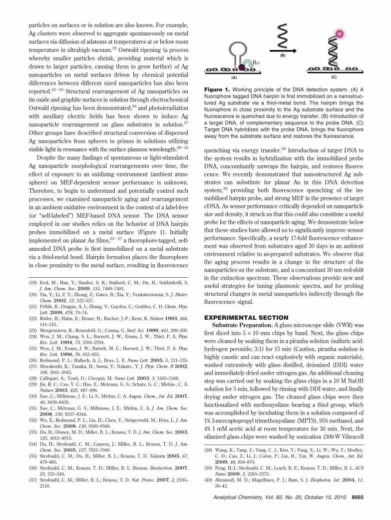
Despite the many findings of spontaneous or light-stimulatedAg nanoparticle morphological rearrangements over time, theeffect of exposure to an oxidizing environment (ambient atmo-sphere) on MEF-dependent sensor performance is unknown.Therefore, to begin to understand and potentially control suchprocesses, we examined nanoparticle aging and rearrangementin an ambient oxidative environment in the context of a label-free(or “self-labeled”) MEF-based DNA sensor. The DNA sensoremployed in our studies relies on the behavior of DNA hairpinprobes immobilized on a metal surface (Figure 1). Initiallyimplemented on planar Au films,33-37 a fluorophore-tagged, self-annealed DNA probe is first immobilized on a metal substratevia a thiol-metal bond. Hairpin formation places the fluorophorein close proximity to the metal surface, resulting in fluorescence
quenching via energy transfer.38 Introduction of target DNA tothe system results in hybridization with the immobilized probeDNA, concomitantly unwraps the hairpin, and restores fluores-cence. We recently demonstrated that nanostructured Ag sub-strates can substitute for planar Au in this DNA detectionsystem,39 providing both fluorescence quenching of the im-mobilized hairpin probe, and strong MEF in the presence of targetcDNA. As sensor performance critically depended on nanoparticlesize and density, it struck us that this could also constitute a usefulprobe for the effects of nanoparticle aging. We demonstrate belowthat these studies have allowed us to significantly improve sensorperformance. Specifically, a nearly 17-fold fluorescence enhance-ment was observed from substrates aged 30 days in an ambientenvironment relative to as-prepared substrates. We observe thatthe aging process results in a change in the structure of thenanoparticles on the substrate, and a concomitant 30 nm red-shiftin the extinction spectrum. These observations provide new anduseful strategies for tuning plasmonic spectra, and for probingstructural changes in metal nanoparticles indirectly through thefluorescence signal.
EXPERIMENTAL SECTIONSubstrate Preparation. A glass microscope slide (VWR) was
first diced into 5 × 10 mm chips by hand. Next, the glass chipswere cleaned by soaking them in a piranha solution (sulfuric acid:hydrogen peroxide; 3:1) for 15 min (Caution, piranha solution ishighly caustic and can react explosively with organic materials),washed extensively with glass distilled, deionized (DDI) waterand immediately dried under nitrogen gas. An additional cleaningstep was carried out by soaking the glass chips in a 10 M NaOHsolution for 5 min, followed by rinsing with DDI water, and finallydrying under nitrogen gas. The cleaned glass chips were thenfunctionalized with methoxysilane bearing a thiol group, whichwas accomplished by incubating them in a solution composed of1% 3-mercaptopropyl trimethoxysilane (MPTS), 95% methanol, and4% 1 mM acetic acid at room temperature for 30 min. Next, thesilanized glass chips were washed by sonication (300-W Vibracell
(19) Erol, M.; Han, Y.; Stanley, S. K.; Stafford, C. M.; Du, H.; Sukhishvili, S.J. Am. Chem. Soc. 2009, 131, 7480–7481.
(20) Yin, Y.; Li, Z. Y.; Zhong, Z.; Gates, B.; Xia, Y.; Venkateswaran, S. J. Mater.Chem. 2002, 12, 522–527.
(21) Pribik, R.; Dragan, A. I.; Zhang, Y.; Gaydos, C.; Geddes, C. D. Chem. Phys.Lett. 2009, 478, 70–74.
(22) Roder, H.; Hahn, E.; Brune, H.; Bucher, J.-P.; Kern, K. Nature 1993, 366,141–143.
(23) Morgenstern, K.; Rosenfeld, G.; Comsa, G. Surf. Sci. 1999, 441, 289–300.(24) Wen, J. M.; Chang, S. L.; Burnett, J. W.; Evans, J. W.; Thiel, P. A. Phys.
Rev. Lett. 1994, 73, 2591–2594.(25) Wen, J. M.; Evans, J. W.; Bartelt, M. C.; Burnett, J. W.; Thiel, P. A. Phys.
Rev. Lett. 1996, 76, 652–655.(26) Redmond, P. L.; Hallock, A. J.; Brus, L. E. Nano Lett. 2005, 5, 131–135.(27) Murakoshi, K.; Tanaka, H.; Sawai, Y.; Nakato., Y. J. Phys. Chem. B 2002,
106, 3041–3045.(28) Callegari, A.; Tonti, D.; Chergui, M. Nano Lett. 2003, 3, 1565–1568.(29) Jin, R. C.; Cao, Y. C.; Hao, E.; Metraux, G. S.; Schatz, G. C.; Mirkin., C. A.
Nature 2003, 425, 487–490.(30) Xue, C.; Millstone, J. E.; Li, S.; Mirkin, C. A. Angew. Chem., Int. Ed. 2007,
46, 8436–8439.(31) Xue, C.; Metraux, G. S.; Millstone, J. E.; Mirkin, C. A. J. Am. Chem. Soc.
2008, 130, 8337–8344.(32) Wu, X.; Redmond, P. L.; Liu, H.; Chen, Y.; Steigerwald, M.; Brus, L. J. Am.
Chem. Soc. 2008, 130, 9500–9506.(33) Du, H.; Disney, M. D.; Miller, B. L.; Krauss, T. D. J. Am. Chem. Soc. 2003,
125, 4012–4013.(34) Du, H.; Strohsahl, C. M.; Camera, J.; Miller, B. L.; Krauss, T. D. J. Am.
Chem. Soc. 2005, 127, 7932–7940.(35) Strohsahl, C. M.; Du, H.; Miller, B. L.; Krauss, T. D. Talanta 2005, 67,
479–485.(36) Strohsahl, C. M.; Krauss, T. D.; Miller, B. L. Biosens. Bioelectron. 2007,
23, 233–240.(37) Strohsahl, C. M.; Miller, B. L.; Krauss, T. D. Nat. Protoc. 2007, 2, 2105–
2110.
(38) Wang, K.; Tang, Z.; Yang, C. J.; Kim, Y.; Fang, X.; Li, W.; Wu, Y.; Medley,C. D.; Cao, Z.; Li, J.; Colon, P.; Lin, H.; Tan, W. Angew. Chem., Int. Ed.2009, 48, 856–870.
(39) Peng, H.-I.; Strohsahl, C. M.; Leach, K. E.; Krauss, T. D.; Miller, B. L. ACSNano 2009, 3, 2265–2273.
(40) Abramoff, M. D.; Magelhaes, P. J.; Ram, S. J. Biophoton. Int. 2004, 11,36–42.
Figure 1. Working principle of the DNA detection system. (A) Afluorophore tagged DNA hairpin is first immobilized on a nanostruc-tured Ag substrate via a thiol-metal bond. The hairpin brings thefluorophore in close proximity to the Ag substrate surface and thefluorescence is quenched due to energy transfer. (B) Introduction ofa target DNA, of complementary sequence to the probe DNA. (C)Target DNA hybridizes with the probe DNA, brings the fluorophoreaway from the substrate surface and restores the fluorescence.
8665Analytical Chemistry, Vol. 82, No. 20, October 15, 2010
probe sonicator, Sonic & Materials Inc.) in a 95% ethanol: 5% watersolution for 2 min, and dried under nitrogen gas. Coating of Agnanoparticles on the silanized glass chips was achieved byincubating them in a solution of 10 mM AgNO3 in dimethylfor-mamide (DMF) at room temperature in the dark for 1 h.41 Theresulting Ag nanoparticle-coated glass chips were then washedby sonication in a 95% ethanol: 5% water solution for 4 min,and dried under nitrogen gas. The Ag nanoparticle coatedsubstrates were then stored under ambient atmosphere (aver-age temperature: 24 °C, average humidity: 36.8%), in the darkfor different periods of time (0, 2, 5, 9, 12, 16, 19, 30 days)prior to DNA immobilization. For the nonoxidative environmentstudy, the substrates were stored in a glovebox (VacuumAtmosphere Company) filled with N2 gas (average temperature:24 °C, average humidity: 0%) and allowed to age for 30 days.
Self-Assembly. Self-assembly of DNA hairpin probes onnanostructured Ag substrates was accomplished by incubatingeach Ag substrate in a solution consisting of 300 nM probe DNAand 60 nM mercaptopropanol in buffered saline (500 mM NaCl,20 mM cacodylic acid, and 0.5 mM ethylenediaminetetraaceticacid (EDTA), pH 7.0) for 2 h at room temperature in the dark.Probe DNA (Midland) has a sequence of 5′-TCG TTA GTG TTAGGA AAA AAT CAA ACA CTC GCG A-3′ and is conjugated witha trityl-thiol group and a tetramethylrhodamine (TAMRA, Abmax:543 nm, Emmax: 571 nm) fluorophore at the 5′ and 3′ ends,respectively. Next, nonspecifically absorbed DNA probes wereremoved by washing the substrates in boiling DDI water for30 s. Substrates were then air-dried in the dark at roomtemperature for 45 min. Hairpin formation was promoted byimmersing dried substrates in buffered saline in the dark atroom temperature for another 45 min. Prehybridization fluo-rescence intensity was acquired after removal from the salinesolution. Subsequently, the probe immobilized Ag substrateswere incubated in a 2.5 µM label-free DNA target solutionovernight followed by posthybridization fluorescence intensitymeasurement. Target DNA (IDT) has a sequence complemen-tary to the probe DNA (5′-TCG CGA GTG TTT GAT TTT TTCCTA ACA CTA ACG A-3′).
Imaging Acquisition and Data Analysis. Fluorescenceimages were acquired by using an epifuorescence microscope(Olympus BX-60) equipped with a thermoelectrically (TE) cooledcharge coupled device (CCD). Sample substrates were excitedwith incident light from a Hg lamp (100 W), which was filteredwith an excitation bandpass filter (531 ± 20 nm), reflected by adichroic mirror, and guided through a 10× objective lens. Theemitted light was collected by CCD after the light was reflectedfrom the sample, guided through the objective lens, the dichroicmirror, and a bandpass filter (593 ± 20 nm). Fluorescence imageswere then analyzed using ImageJ software.40 To analyze particlesize distributions, AFM images were first converted to 8-bit imagesfollowed by threshold adjustment. Each image was adjusted withfixed threshold for comparison purpose. Processed images werethen analyzed with “Analyze Particles” function from ImageJ andthe information made available was then binned in 500 nm2
intervals using histogram commend in Matlab.Substrate Characterization. Extinction spectra of nanostruc-
trued Ag substrates were measured using a Perkin-Elmer Lambda
950 UV/vis/NIR spectrophotometer over wavelengths rangingfrom 330 to 750 nm. AFM images of the Ag substrates weremeasured in air using a Digital Instruments Nanoscope IIIaoperated in tapping mode using a Si tip (300 kHz, 40 N/m).Substrate roughness measurements were made offline usingDigital Instrument (DI) software.
TAMRA Characterization. The absorption spectrum of TAM-RA was acquired using a Shimadzu UV-1601PC spectrophotometerover wavelengths ranging from 450 to 600 nm. The emissionspectrum of TAMRA was collected with a modular Acton Researchfluorimeter equipped with a PMT (photomultiplier tubes) detectorover wavelengths ranging from 530 to 750 nm at an excitationwavelength of 500 nm.
RESULTS AND DISCUSSIONTo study nanostructured Ag substrate aging under ambient
atmosphere and its potential effect on biomolecule detection,substrates were prepared at day 0 and examined for target DNA-dependent fluorescence at 0, 2, 5, 9, 12, 16, 19, and 30 days aftersubstrate preparation. Ag nanoparticles were formed by AgNO3
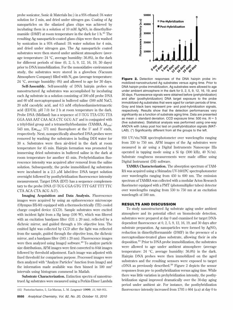
reduction in dimethylformamide (DMF) in the presence of amercaptosilane-treated glass substrate, allowing their in situdeposition.41 Prior to DNA probe immobilization, the substrateswere allowed to age under ambient atmosphere (averagetemperature: 24 °C, average humidity: 36.8%) in the dark.Hairpin DNA probes were then immobilized on the agedsubstrates and the resulting sensors were exposed to targetcDNA as previously described.39 Figure 2 depicts the sensorresponses from pre- to posthybridization versus aging time. Whilethere was little variation in prehybridization intensity, the posthy-bridization signal improved dramatically over the 30-day agingperiod under ambient air. For instance, the posthybridizationfluorescence intensity increased from 1793 ± 664 (a.u) at day 0 to(41) Pastoriza-Santos, I.; Liz-Marzan, L. M. Langmuir 1999, 15, 948–951.
Figure 2. Detection responses of the DNA hairpin probe im-mobilized-nanostructured Ag substrates versus aging time. Prior toDNA hairpin probe immobilization, Ag substrates were allowed to ageunder ambient atmosphere in the dark for 0, 2, 5, 9, 12, 16, 19, and30 days. Fluorescence signals were obtained before (prehybridization)and after (posthybridization) DNA target exposure to the probeimmobilized-Ag substrates that were aged for certain periods of time.Gray and black bars represent pre- and post-hybridization signals,respectively. Results show that the detection performances varysignificantly as a function of substrate aging time. Data are presentedas mean ( standard deviation. CCD exposure time: 500 ms. N ) 5(five substrates). Statistical analysis was performed using one-wayANOVA with tukey post hoc test on posthybridization signals (MAT-LAB). (*) Significantly different from all the groups to the left.
8666 Analytical Chemistry, Vol. 82, No. 20, October 15, 2010

35626 ± 6518 (a.u) at day 30, corresponding to a nearly 17-foldsignal enhancement. Given the known ability of Ag to readilyoxidize,19,20 this observation was contrary to the expectation thatprolonged substrate incubation under ambient atmosphere woulddegrade detection performance.
In order to develop a structural understanding of the nano-particle aging process and its effect on detection performance,we first turned our attention to the extinction spectra of the agedsubstrates. As shown in Figure 3, the spectrum at day 0(as-prepared substrate) displays a sharp peak at 398 ± 5 nm (N )5). This peak red-shifts to 426 ± 2 nm (N ) 3) over a 30-day period.Peak widths also increase as a function of aging time: the fullwidth at half-maximum (fwhm) of the spectrum increased from∼96 nm (day 0) to ∼127 nm (day 30). Electrodynamic theoryindicates that both phenomena can result from modulation of thedielectric constant of the embedding medium, changes in clustersize, or changes in cluster morphology.8,9,42,43
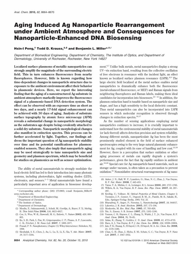
We next investigated substrate surface topographies as afunction of substrate aging by atomic force microscopy (AFM) toelucidate the contributing factors for spectral evolution over time.Inspection (Figure 4) and particle analysis (Supporting Information(SI) S1) of the images both reveal a decrease in the number ofsmaller particles, an increase in the number of larger particlesand an overall decrease in particle number. As shown in Figure4a, particles are largely spherical at day 0, with an apparentdiameter of ∼16 nm and a root-mean-square (rms) roughness of2.2 nm. Particles undergo a readily observable increase in sizebetween day 0 and day 12, while largely retaining their sphericalshape (Figure 4b and c). By day 30 a more pronounced
transformation has obviously taken place (Figure 4d): in additionto further increases in particle size, many are rodlike. Theserodlike structures may be the result of incomplete coalescence(prespheroidization) between two or more particles.44,45 Theprogression of particle evolution, specifically the growth of largerparticles at the expense of smaller particles, is consistent withthe Ostwald ripening process described above. This led us topostulate that the mechanism of particle rearrangement observedin our system to be a result of Ag oxidation under ambient airand subsequent deposition onto the nearby Ag nanoparticleswhere greater stability is gained. Ag+ ions that are formed afteroxidation travel to neighboring nanoparticles potentially througha thin moisture layer that is normally formed on the nano-structured substrate by water condensation under ambientatmosphere.46,47
AFM images of 60 and 480 day-old substrates illustrate thepresence of both rodlike structures and larger particles on thesubstrate surface (SI S2). Furthermore, examination of the 480day-old Ag substrates demonstrates that the substrate remainsresponsive, albeit less sensitive as compared to the 30 day-oldAg substrate (SI S3). We note that no special care was taken toprotect thes substrates from ambient dust, chemical vapors, etc.during storage. Therefore, numerous factors in addition to thesechanges in particle size may have contributed to the reduction indetection sensitivity observed from the 480-day old substrate, anyof which may have contributed to substrate degeneration. Inparticular, prolonged aging may have resulted in extensiveoxidation, thereby impeding the Ag-thiol bond formation. Thisfinding indicates the importance of storing the nanostructuredmetal substrates in a nonoxidative (or a more appropriate)environment for long-term use.
The influence of the dielectric constant of the embeddingmedium on extinction spectra of metal nanoparticles has beendemonstrated extensively,20,48-50 and could also contribute to thechanges in device performance. In particular, exposure of Agnanoparticles to oxygen or ambient air results in spectral red-shifts and broadening, stemming from a depressing surface chargein turn caused by the change in the dielectric medium.9 Theresultant extinction spectrum change can be well characterizedby electrodynamical calculations modeling core-shell nanostruc-tures, in which the shell has the dielectric constant of theembedding medium (oxide) and the core remains pure Ag.43
Unfortunately, a direct quantification of oxide content on our Agsubstrates by analytical techniques such as energy dispersive X-rayspectroscopy (EDS) and X-ray photoelectron spectroscopy (XPS)is problematic due to the low conductivity of the Ag substrates.51
Since the Ag substrates were aged in ambient air, oxide formationon the Ag substrate was almost inevitable.52 While the contribution
(42) Kreibig, U.; Genzel, L. Surf. Sci. 1985, 156, 678–700.(43) Kelly, K. L.; Coronado, E.; Zhao, L. L.; Schatz, G. C. J. Phys. Chem. B 2003,
107, 668–677.
(44) Nichols, F.; Mullins, W. J. Appl. Phys. 1965, 36, 1826–1835.(45) Zhang, L. H.; Sui, M. L.; Zhang, L.; Hu, K. Y.; Li, D. X. Mater. Sci. Eng., A
2004, 379, 1–6.(46) Roark, S. E.; Semin, D. J.; Rowlen, K. L. Anal. Chem. 1996, 68, 473–480.(47) Thundat, T.; Zheng, X.-Y.; Chen, G. Y.; Warmack, R. J. Surf. Sci. Lett. 1993,
294, L939–L943.(48) Henglein, A. Chem. Mater. 1998, 10, 444–450.(49) Kreibig, U.; Gartz, M.; Hilger, A. Ber. Bunsenges. Phys. Chem. 1997, 101,
1593–1604.(50) Xu, G.; Tazawa, M.; Jin, P.; Nakao, S.; Yoshimura, K. Appl. Phys. Lett. 2003,
82, 3811–3813.(51) Tricoli, A.; Pratsinis, S. E. Nat. Nanotechnol. 2010, 5, 54–60.(52) Cai, W.; Zhong, H.; Zhang, L. J. Appl. Phys. 1998, 83, 1705–1710.
Figure 3. Extinction spectra of aged nanostructured Ag substratesobtained at different days (0, 2, 30, and 45 days) after substratepreparation. The figure is also embedded with absorption andemission spectra of the DNA probe fluorophore tetramethylrhodamine(TAMRA). Extinction spectrum of the Ag substrate displays a sharppeak position at 398 ( 5 nm (N ) 5) nm at day 0, which is found toshift to 426 ( 2 nm (N ) 3) nm over a 30 day period. These shiftsare accompanied by spectrum broadenings, which can be results ofchanges in particle size, shape, or dielectric environment. Shifts ofthe spectra brought them closer to resonance with the absorptionand emission energies of TAMRA.
8667Analytical Chemistry, Vol. 82, No. 20, October 15, 2010

of this oxide formation could not be evaluated explicitly, the directexperimental examination of changes in nanoparticle size andshape can account for our observations and thus any dielectriceffects are probably small.
As mentioned earlier, the presence of visible and UV light hasbeen implemented to assist metal particle reformation on bothconductive and dielectric substrates. Particles of a particulargeometry can be tailored from initial spherical or pseudosphericalseeds based on the wavelength of the light source.27-32 Toexamine the effect of light on substrate aging, the freshlyfabricated Ag substrates were left on the lab bench and exposedto ambient light (fluorescent tubes located on the laboratoryceiling, 4100k fluorescent lamp, F32T8/FL841, 32 W) for 24 hper day for a period of 30 days. The intensity of the incident lighton the substrates was measured to be ∼0.02 mw cm-2 with theuse of a light meter (Gossen Scout 3, Japan). The light-exposedAg substrates show a diminishing extinction amplitude butnearly identical peak position (∼428 nm) as compared to thesubstrate aged in the dark for 30 days (Figure 5). Thediminishing amplitude seen with the presence of ambient light(as compared to the substrates that were aged in the dark) ispossibly a result of accelerated Ag dissolution caused by theambient light. The AFM image of the substrate (Figure 6B)illustrates a decrease in particle number (∼60% decrement),
increase in particle size, and a decrease in particle height. rmsroughness of the substrate decreased from 2.2 nm at day 0 to 1.5nm at day 30. Particle morphology stayed primarily spherical, witha few elongated structures (<0.5%).
The effect of ambient light on particle coalescence is currentlyunclear. It is possible that ambient light facilitates Ag+ depositiononto the larger particles via the plasmon excitation mechanism(the emission spectrum of the fluorescence tube used in thisstudy displays two peaks, positioned at ∼405 and 436 nm,respectively), resulting in an increase in the rate of largeparticle growth.54 Clearly, the dried and air- exposed substrateswe describe in this study are very different from the experi-mental conditions previously employed to examine light-accelerated Ag nanoparticle remodeling,27 but it is possible thatsufficient moisture remains on the substrate surface to permitsome of the same processes to occur. To test this hypothesis,we replaced the oxidative environment (air and humidity) withN2 gas. We placed the freshly prepared Ag substrates in aglovebox (Vacuum Atmosphere Company) filled with N2 thatallows for a rigorous control of humidity (humidity: 0%). Thesubstrates were also exposed under a fluorescent tube. Theresultant extinction spectrum shows no change in extinctionamplitude (Figure 5), indicating the significant role of moistureon damping of the extinction amplitude and structural rearrange-ment. It also shows that light alone in a nonoxidative environmentis insufficient to induce a significant spectral change over time.Although the N2 aged group (30 days) appears to show a peakposition slightly to the right of the 0-day group, the difference
(53) Lu, J.; Bravo-Suarez, J. J.; Takahashi, A.; Haruta, M.; Oyama, S. T. J. Catal.2005, 232, 85–95.
(54) Millstone, J. E.; Hurst, S. J.; Metraux, G. S.; Cutler, J. I.; Mirkin, C. A. Small2009, 5, 646–664.
Figure 4. AFM images illustrate structural revolution of the nanostructured Ag substrate at (A) 0, (B) 2, (C) 12, and (D) 30 days after substratepreparation and storage in the dark and ambient atmosphere. Shapes of the particles evolved from mainly spherical at 0 day to a combinationof spherical and rodlike structures at 30 days. Scale bar: 200 nm.
Figure 5. Extinction spectra of an untreated nanostructured Agsubstrate (purple), and aged nanostructured Ag substrates treatedunder different environmental conditions (N2+ambient light (humidity:0%, green), air + ambient light (pink) and air-ambient light (humidity:36.8%), blue) for 30 days. The uptick displayed in the spectrum (air+ ambient light) below 350 nm is due to the interband electronictransition in Ag.53
Figure 6. AFM images of the (A) as-prepared nanostructured Agsubstrate and (B) the nanostructured Ag substrate aged 30 daysunder ambient air and ambient light. Prolonged aging of the substrateunder ambient light and ambient air results in a decrease in particlenumber, an increase in particle size and a lower rms roughness. Scalebar: 200 nm.
8668 Analytical Chemistry, Vol. 82, No. 20, October 15, 2010

is within the chip-to-chip standard deviation (398 ± 4.6 nm, N) 5). DNA detection performed on substrates treated with N2
(humidity: 0%) in the dark for 30 days showed no improvementof fluorescence signal as compared to as-prepared substrates(data not shown).
Besides photonic energy derived from auxiliary sources suchas fluorescent tubes, certain chemical additives also acquire theability to drive particle rearrangement. For instance, exposure toexcess chalcogen elements such as oxygen and sulfur candramatically increase the propensity of the Ag nanoparticles tocoalesce.55,56 In contrast, additives such as n-alkanethiols canprotect the metal surfaces from corrosion.57 To investigate thequestion of whether attached DNA hairpin probes could alter therate of particle aging, we examined the detection responses oftwo 12-day aged substrates that were prefunctionalized with DNAhairpin probes at day 0, and compared their performances to theresponses of the substrates that received no againg treatments(SI S4). In contrast to the unfunctionlized substrates, no significantimprovement in fluorescence signals was observed from the agedsubstrates as compared to the as-prepared substrates. Theseresults are consistent with the DNA hairpin probes acting asprotective molecules, preventing structural rearrangement of theunderlying Ag nanoparticles.
In experiments discussed thus far, we have addressed theeffect of substrate aging on nanoparticle coarsening under theinfluences of ambient air and ambient light. How does this aging-induced structural change improve the fluorescence detectionsignal as demonstrated in Figure 2? The improving systemperformance over time can be attributed to both plasmonic and“lightning rod” effects. Studies have shown that the level offluorescence enhancement is strongly influenced by the spectraloverlap between the plasmonic wavelength and the absorption/emission bands of the fluorophores.10,58-61 Substrate aging underambient atmosphere induced spectrum red-shifts as a result ofnanoparticle coarsening (Figures 3 and 4), which brought theplasmonic wavelength closer to the absorption/emission band ofthe TAMRA molecule (Abmax: 543 nm, Emmax: 571 nm) andresulted in a greater fluorescence enhancement over time.Another possible contributing factor for the dramatic enhance-ment of fluorescence signals observed over time is the muchhigher local electric fields enhanced by the surface irregulari-ties, sharp corners, and small aggregates that are formed onthe substrates.62,63 Both elliptical nanowires and arbitrary particleshapes with sharp edges have demonstrated much higher levelsof localized electric fields surrounding the structures, which canenable much more pronounced enhancing phenomena.
In previous work, we observed that increasing Ag exposureto the mercaptosilane-treated glass substrates also shifted theextinction spectra closer to the excitation/emission band ofTAMRA. However, a decrease in detection performance with anequal amount of DNA probe hairpin and spacing agent wasobserved.39 How does one reconcile those results with theobservations reported herein? The reduction in detection perfor-mance seen in the previous study stemmed mainly from anincrease in prehybridization signal with increasing Ag exposuretime, a result of steric crowding or continuum backgroundemission of the rough Ag surface. As demonstrated in this study,substrate aging induces particle reformation, but this occurs in amanner such that enough void space remains between particleson the substrate to alleviate steric crowding and continuumbackground emission from the Ag surfaces. A rough estimationof the particle surface area change from day 0 to 30 days of agingshowed only limited changes over time (an approximate 4.5%increase in surface area). Therefore, it is unlikely that stericcrowding effects of DNA probes play more than a minimal rolein the observed changes in the detection performance over time.In addition, our previous findings suggest that the steric crowdingphenomenon primarily manifests in an increase in the prehybrid-ization signals, as hairpin probes are unable to achieve a config-uration suitable for full quenching of the fluorophore. Thus, onewould expect to observe a noticible increase in prehybridizationsignals over time if steric crowding is taking place. This was notobserved.
This report summarizes preliminary studies on the effect ofAg nanomaterial stability on the performance of a fluorescencebased DNA biosensor. Our findings demonstrate the dependenceof fluorescence signals on substrate aging time. A lengtheningincubation (30 days) of nanostructured Ag substrates underambient atmosphere induced particle coalescence, despite covalentattachment of particles to a glass substrate. Ag nanoparticlecoalescence red-shifted the extinction spectrum and generated anearly 17-fold increase in fluorescence response. We hypothesizethat the improvement in the fluorescence response observed afteraging is due to both plasmonic and lightning rod effects.
We attribute the mechanism of particle rearrangement underambient conditions to oxidative dissolution of Ag, and subsequentdeposition of Ag+ onto nearby Ag nanoparticles as a reductivereaction. Based on electrode potential calculations, smaller Agnanoparticles are more easily oxidized as compared to largerparticles.26 Ag+ ions formed on the smaller particles underambient conditions can potentially travel to the neighboringlarger particles with the assistance of the moisture layer onthe substrate. This process is driven by the electric fieldgradient since larger nanoparticles have a partial negativecharge at electrical equilibrium. To reestablish electricalequilibrium after ion deposition, the larger nanoparticle thenaccepts an electron from touching smaller nanoparticles.Inclusion of ambient light in conjunction with ambient air inthe system further facilitated particle coalescence. Replacementof the oxidative environment (ambient air) with N2 gas in thepresence of ambient light diminished the structural changesignificantly, indicating ambient air is necessary for structuralrearrangement, and light alone is insufficient to initiate particlereformation.
(55) Layson, A. R.; Evans, J. W.; Thiel, P. A. Phys. Rev. B 2002, 65, 193409-4.(56) Thiel, P. A.; Shen, M.; Liu, D.-L.; Evans, J. W. J. Phys. Chem. C 2009, 113,
5047–5067.(57) Jennings, G. K.; Yong, T.-H.; Munro, J. C.; Laibinis, P. E. J. Am. Chem. Soc.
2003, 125, 2950–2957.(58) Stranik, O.; Nooney, R.; McDonah, C.; MacCraith, B. D. Plasmonics 2007,
2, 15–22.(59) Tam, F.; Goodrich, G. P.; Johnson, B. R.; Hala, N. J. Nano Lett. 2007, 7,
496–501.(60) Chen, Y.; Munechika, K.; Plante, I. J.-L.; Munro, A. M.; Strabalak, S. E.;
Xia, Y.; Ginger, D. S. Appl. Phys. Lett. 2008, 93, 053106-3.(61) Cade, N. I.; Ritman-Meer, T.; Kwakwa, K. A.; Richards, D. Nanotechnology
2009, 20, 285201.(62) Gersten, J.; Nitzan, A. J. Chem. Phys. 1980, 73, 3023–3037.(63) Kottmann, J. P.; Martin, O. J. F.; Smith, D. R.; Schultz, S. Chem. Phys. Lett.
2001, 341, 1–6.
8669Analytical Chemistry, Vol. 82, No. 20, October 15, 2010

CONCLUSIONSA detailed prediction of metal nanostructures after aging
treatments is extremely challenging, as many factors includingparticle coverage, temperature, impurity, additives, auxiliaryenergy source, and interactions with the substrate can influenceinterfacial energy and mass transfer greatly. Further work willbe required to fully understand the impact of each of these factorson particle rearrangement. It is clear, however, that from theapplications perspective (at least in the context of MEF-basedbiosensors), Ag nanoparticle substrate aging in ambient atmo-sphere does not necessarily degrade a system’s performance, asone might expect. On the contrary, it improved the detection/fluorescence signal significantly. In conclusion, this work il-lustrates the importance of understanding a studied nanomaterial’sstability over time. We anticipate the DNA biosensing systemdescribed herein will be of further analytical utility for probingstructural changes in nanostructured metal substrates throughthe fluorescence signal.
ACKNOWLEDGMENTWe acknowledge the New York State Foundation for Science,
Technology, and Innovation (NYSTAR), the National Institutesof Health (2 R24 A1054953-06), and the Department of EnergyOffice of Basic Energy Sciences, Division of Chemical Sciences,Geosciences, and Biosciences (DE-FG02-06ER15821) for financialsupport.
SUPPORTING INFORMATION AVAILABLEAnalysis of particle size and number as a function of time; AFM
imaging and DNA detection performance of substrates followingprolonged aging (3 pp.). This material is available free of chargevia the Internet at http://pubs.acs.org.
Received for review July 20, 2010. Accepted September 8,2010.
AC101919H
8670 Analytical Chemistry, Vol. 82, No. 20, October 15, 2010