Advanced Trauma PRE-Workshop Assignment CASE STUDY #1 ...
115
1 Advanced Trauma PRE-Workshop Assignment CASE STUDY #1 - Amos and Chow REFERENCE REASON FOR CHOOSING (explain) ABSTRACT Analysis of Nail Bed Injuries - Cause, Outcome, Treatment Gu,JH; Choi,HS; Yoon,JH (2021). Annals of Plastic Surgery Vol 87, Number 2, August 2021. ▪ Recent 2021 ▪ Relevant Discussion on treatment of nail bed injuries ▪ Clinical reasoning Treatment determined should be in relation to the extent of the injury and structures involved. Treatment outcomes are generally good if care is taken in suturing and replacement nail to nail fold. ▪ Evidence Treatment generally resulted in good to very good outcomes with poor outcomes in 6.6% of the group. Poor outcomes related to greater complexity of injury. Poor outcomes were due to nail splitting, roughness and adherence including dirtiness, catching and bending. ▪ Innovative Consideration of conservative (non surgical treatment, healing by secondary intention) that appears to offer similar results to surgical treatment for certain cohorts. Background: Although fingertip and nail bed injuries have a high incidence, appropriate management of nail bed injuries remains controversial. This study is the completion of data derived from nail bed injuries with follow-up of a minimum of 6 months to suggest an appropriate treatment. Methods: In the retrospective study, we analyzed data from 549 nail bed injuries for 6 years and age, type of injury, fractures, treatment methods, and outcomes were reviewed. Results were determined and these were divided to identical to the opposite group, abnormalities based on Zook criteria. Statistical analysis was done according to injury category (type, site, nail substitute, and fracture) and overall final grade. Results: Over 50% (293 cases) had excellent results. Rates of very good, good, fair, and poor results were 22.6%, 11.3%, 6.2%, and 6.6%, respectively. Poorer results were obtained for fold injuries, crush, and avulsive injuries. The presence of a fracture was associated with poor results. Conclusions: The cause of poor results is thought to be multifactorial. Although, overall outcomes were good, nail splitting, nail roughness, and nail adherence can cause dirtiness, catching, bending, and various cosmetic problems. Thus, careful suture and replacement of nail to nail fold are important to reach good results. Key Words: nail, fingertip, injury, nail deformity (Ann Plast Surg 2021;87: 156–160)
Transcript of Advanced Trauma PRE-Workshop Assignment CASE STUDY #1 ...

1
Advanced Trauma PRE-Workshop Assignment CASE STUDY #1 - Amos and Chow
REFERENCE REASON FOR CHOOSING (explain) ABSTRACT
Analysis of Nail Bed Injuries -
Cause, Outcome, Treatment
Gu,JH; Choi,HS; Yoon,JH (2021).
Annals of Plastic Surgery Vol 87,
Number 2, August 2021.
▪ Recent 2021
▪ Relevant
Discussion on treatment of nail bed injuries
▪ Clinical reasoning
Treatment determined should be in relation
to the extent of the injury and structures
involved. Treatment outcomes are generally
good if care is taken in suturing and
replacement nail to nail fold.
▪ Evidence
Treatment generally resulted in good to very
good outcomes with poor outcomes in 6.6%
of the group. Poor outcomes related to
greater complexity of injury. Poor outcomes
were due to nail splitting, roughness and
adherence including dirtiness, catching and
bending.
▪ Innovative
Consideration of conservative (non surgical
treatment, healing by secondary intention)
that appears to offer similar results to
surgical treatment for certain cohorts.
Background: Although fingertip and nail bed injuries have a high
incidence, appropriate management of nail bed injuries remains
controversial. This study is the completion of data derived from nail bed
injuries with follow-up of a minimum of 6 months to suggest an
appropriate treatment.
Methods: In the retrospective study, we analyzed data from 549 nail
bed injuries for 6 years and age, type of injury, fractures, treatment
methods, and outcomes were reviewed. Results were determined and
these were divided to identical to the opposite group, abnormalities
based on Zook criteria. Statistical analysis was done according to injury
category (type, site, nail substitute, and fracture) and overall final grade.
Results: Over 50% (293 cases) had excellent results. Rates of very good,
good, fair, and poor results were 22.6%, 11.3%, 6.2%, and 6.6%,
respectively. Poorer results were obtained for fold injuries, crush, and
avulsive injuries. The presence of a fracture was associated with poor
results.
Conclusions: The cause of poor results is thought to be multifactorial.
Although, overall outcomes were good, nail splitting, nail roughness,
and nail adherence can cause dirtiness, catching, bending, and various
cosmetic problems. Thus, careful suture and replacement of nail to nail
fold are important to reach good results.
Key Words: nail, fingertip, injury, nail deformity
(Ann Plast Surg 2021;87: 156–160)

2
Approach to Fingertip Injuries
Martin-Playa.P; Foo.A, (2019).
Clinical Plastic Surgery vol 46, p
275-283
▪ Recent 2019
▪ Relevant
Bio-psycho-social approach to finger tip
injury management and treatment choice.
▪ Clinical reasoning
Emphasis on considering the psychosocial
aspects and how they impact outcomes
including clinician bias
▪ Evidence
“The characteristics of he injury determine
the range of treatment, whereas
psychological factors aid clinicians in
selecting the most appropriate option.” -
these two provide the most likelihood of
successful outcomes for the surgeon and
client alike.
▪ Innovative
Emphasis is given in this paper as to the
patient factors and how things such as
patient expectations, societal and cultural
norms appear to affect greatly the outcome
of any intervention provided. As such the
treatment plan should take into
consideration these factors.
The fingertip is mankind's tactile interface with the physical world, from
reading braille, to using touchscreens, to wielding power tools. Its
special tissue architecture demands astute evaluation and meticulous
surgical or nonsurgical care after injury to return patients to their
preinjury level of function. Attentive deliberation of physiologic,
vocational, and psychosocial factors could improve the odds of
achieving satisfactory results. In this article, we explore these aspects of
fingertip injury to provoke readers to examine their practices and
philosophies.
Keywords: Closed injuries; Fingertip amputation; Fingertip injuries; Nail
bed injuries; Psychosocial factors.
Seah, B; Sebastian, S. (2020).
Retrograde flow digital artery
flaps. The Hand Clinic. VOLUME
▪ Recent - 2020
▪ Relevant
This article highlighted the technically
challenging aspects of retrograde flow
● Retrograde flow digital artery flaps are a versatile single-stage
option for the coverage of fingertip and dorsal digital defects.

3
36, ISSUE 1, P47-56. digital artery described potential
complications of flexion contracture
formation and venous congestion, and
provided technical solutions to addressing
and preventing such issues from surfacing
▪ Clinical reasoning
Overview of indication for retrograde flow
digital artery flap, surgical anatomy,
operative technique, its advantages,
disadvantages and outcome. This article
assists therapists in understanding
structures involved in surgery and it’s
related complications to consider when
tailoring hand therapy treatment.
▪ Evidence
Review article
A summary of the functional outcomes and
complications found in several studies
associated with the standard retrograde
digital artery flap was discussed.
The motion of the involved digit was
not limited, but sensation was affected. All
flaps had the ability to detect light touch,
sharp from dull stimuli, and hot from cold
stimuli without sensory reeducation.
▪ Innovative
The main postoperative complication was
venous congestion. Venous congestion can
be prevented by suturing the flap loosely
● Techniques vary predominantly in the tissue incorporated
(adipofascial or cutaneous) and the inclusion of the digital nerve or
its branches in the pedicle with subsequent neurorrhaphy.
● Complications include venous congestion, flexion contracture, and
cold intolerance

4
and addressed by removing a few sutures
especially over the bridge segment and
doing a delayed inset after 4 to 5 days
Neustein, T; Payne, S; Seiler, J.
(2020). Treatment of Fingertip
injuries. JBJS Reviews8(4):e0182
▪ Recent - 2019
▪ Relevant
A review of current fingertip soft tissue
coverage procedures and different types of
flap dissection and the advantages and
disadvantages of it’s use in finger tip injury
including homodigital island flap with key
anatomic and technical remarks on surgical
procedures and relevant functional
outcome and complications.
▪ Clinical reasoning
Provides understanding of surgical
procedures steps, anatomical structures
affected by procedures and effects of such
procedure have on postoperative outcome
such as venous congestion, flexion
contractures and cold intolerance
▪ Evidence
Review article - provides evidence that this
procedure has a higher success rate and
sensation similar to those reported for
replantation.
▪ Innovative
The goal of care when treating fingertip injuries is to minimize the risk
of infection while maximizing function, tactile sensation, digit length,
pulp padding, and appearance. This outcome can be achieved with
careful soft-tissue coverage and, if possible, nail-bed preservation.
When replantation for a fingertip amputation is not possible for
anatomic or logistical reasons, local or regional flap reconstruction can
be a useful alternative to gain early soft-tissue coverage and allow more
functional rehabilitation. Reviewing current fingertip soft-tissue
coverage procedures and demonstrating key anatomic and technical
points with cadaveric dissections provides a foundation for the
incorporation of these techniques into practice.
5
As an alternative technique to retrograde
homodigital flap, the nerve can be cut at the
time the artery is ligated. The proximal
stump can be buried in the tissue to prevent
formation of a neuroma, and the distal end
can be sutured to the remaining
contralateral digital nerve to the pulp
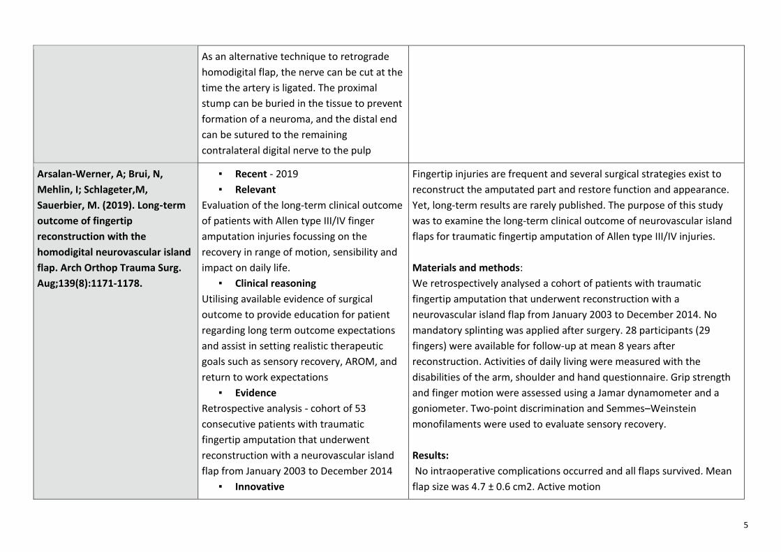
Arsalan-Werner, A; Brui, N,
Mehlin, I; Schlageter,M,
Sauerbier, M. (2019). Long‑term
outcome of fingertip
reconstruction with the
homodigital neurovascular island
flap. Arch Orthop Trauma Surg.
Aug;139(8):1171-1178.
▪ Recent - 2019
▪ Relevant
Evaluation of the long-term clinical outcome
of patients with Allen type III/IV finger
amputation injuries focussing on the
recovery in range of motion, sensibility and
impact on daily life.
▪ Clinical reasoning
Utilising available evidence of surgical
outcome to provide education for patient
regarding long term outcome expectations
and assist in setting realistic therapeutic
goals such as sensory recovery, AROM, and
return to work expectations
▪ Evidence
Retrospective analysis - cohort of 53
consecutive patients with traumatic
fingertip amputation that underwent
reconstruction with a neurovascular island
flap from January 2003 to December 2014
▪ Innovative
Fingertip injuries are frequent and several surgical strategies exist to
reconstruct the amputated part and restore function and appearance.
Yet, long-term results are rarely published. The purpose of this study
was to examine the long-term clinical outcome of neurovascular island
flaps for traumatic fingertip amputation of Allen type III/IV injuries.
Materials and methods:
We retrospectively analysed a cohort of patients with traumatic
fingertip amputation that underwent reconstruction with a
neurovascular island flap from January 2003 to December 2014. No
mandatory splinting was applied after surgery. 28 participants (29
fingers) were available for follow-up at mean 8 years after
reconstruction. Activities of daily living were measured with the
disabilities of the arm, shoulder and hand questionnaire. Grip strength
and finger motion were assessed using a Jamar dynamometer and a
goniometer. Two-point discrimination and Semmes–Weinstein
monofilaments were used to evaluate sensory recovery.
Results:
No intraoperative complications occurred and all flaps survived. Mean
flap size was 4.7 ± 0.6 cm2. Active motion

6
Study found risk for disabling flexion
contracture seems to be small even without
mandatory splinting – an interesting
approach for hand therapists to consider.
of the fingers was over 95% of the contralateral side at follow-up. Three
patients showed mild extension lag of the proximal
interphalangeal joint. The grip strength of the affected hand and of each
of the affected fingers was over 70% of the contralateral side. In
comparison to the contralateral side we did not detect any significant
difference for the Semmes–Weinstein monofilament test, but two-point
discrimination (5.1 ± 1.7 mm) was significantly impaired. According to
the Lim classification 1 of 14 nails with hook nail deformity showed
grade 3 breaking of the nail. The DASH score was 16.0. All patients
returned to their original occupation and patient satisfaction with the
procedure was high.
Conclusions
The risk for disabling flexion contracture seems to be small even
without mandatory splinting. Neurovascular island flaps for fingertip
amputation of Allen type III/IV injuries are a reliable tool in fingertip
reconstruction in the long term.
Xu, J; Cao, J; Graham, D; Lawson,
R. (2021). Clinical Outcomes and
Complications of Primary
Fingertip Reconstruction Using a
Reverse Homodigital Island Flap:
A Systematic Review. Hand :
Official Journal Of The American
Association For Hand Surgery.
Apr 9
▪ Recent - 2021
▪ Relevant
Systematic review of evidence on the
functional outcomes and complications of
reverse homodigital island flap as a surgical
technique for reconstructing traumatic
fingertip injuries
▪ Clinical reasoning
It is important to consider the outcome and
common complications associated with this
surgical treatment and how these can affect
Background: Reverse homodigital island flaps (RHIFs) are increasingly
used to reconstruct traumatic fingertip injuries, but there is limited
evidence on the efficacy of this technique. We performed a systematic
review of the literature to establish the safety and functional outcomes
of RHIF for traumatic fingertip injuries.
Methods: Electronic searches were performed
using 3 databases (PubMed, Ovid Medline, Cochrane CENTRAL) from
their date of inception to April 2020. Relevant studies were required to
report on complications and functional outcomes for patients
7
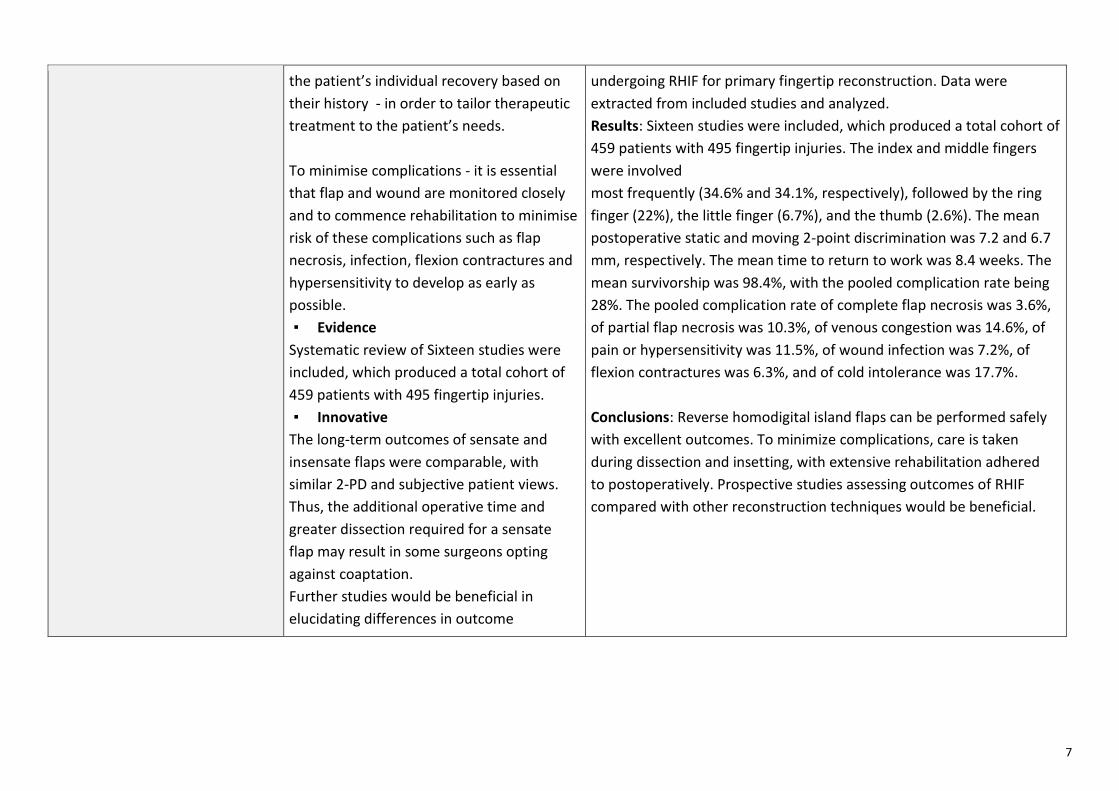
the patient’s individual recovery based on
their history - in order to tailor therapeutic
treatment to the patient’s needs.
To minimise complications - it is essential
that flap and wound are monitored closely
and to commence rehabilitation to minimise
risk of these complications such as flap
necrosis, infection, flexion contractures and
hypersensitivity to develop as early as
possible.
▪ Evidence
Systematic review of Sixteen studies were
included, which produced a total cohort of
459 patients with 495 fingertip injuries.
▪ Innovative
The long-term outcomes of sensate and
insensate flaps were comparable, with
similar 2-PD and subjective patient views.
Thus, the additional operative time and
greater dissection required for a sensate
flap may result in some surgeons opting
against coaptation.
Further studies would be beneficial in
elucidating differences in outcome
undergoing RHIF for primary fingertip reconstruction. Data were
extracted from included studies and analyzed.
Results: Sixteen studies were included, which produced a total cohort of
459 patients with 495 fingertip injuries. The index and middle fingers
were involved
most frequently (34.6% and 34.1%, respectively), followed by the ring
finger (22%), the little finger (6.7%), and the thumb (2.6%). The mean
postoperative static and moving 2-point discrimination was 7.2 and 6.7
mm, respectively. The mean time to return to work was 8.4 weeks. The
mean survivorship was 98.4%, with the pooled complication rate being
28%. The pooled complication rate of complete flap necrosis was 3.6%,
of partial flap necrosis was 10.3%, of venous congestion was 14.6%, of
pain or hypersensitivity was 11.5%, of wound infection was 7.2%, of
flexion contractures was 6.3%, and of cold intolerance was 17.7%.
Conclusions: Reverse homodigital island flaps can be performed safely
with excellent outcomes. To minimize complications, care is taken
during dissection and insetting, with extensive rehabilitation adhered
to postoperatively. Prospective studies assessing outcomes of RHIF
compared with other reconstruction techniques would be beneficial.

1
Advanced Trauma PRE-Workshop Assignment Case Study 2 Bray and Sevier
REFERENCE REASON FOR CHOOSING (explain) ABSTRACT
Miles, M. R., Krul, K. P, et. al. (2021). ‘Minimally Invasive Intramedullary Screw Versus Plate Fixation for Proximal Phalanx Fractures: A Biomechanical Study’, Journal of Hand Surgery America, vol. 46, pp. 518.e1 – 518.e8.
Recent: 2021
Relevant/Clinical Reasoning: As Mr W has had a proximal phalanx ORIF with plate and screw, this article is relevant as it looks at fractures fixed with an intramedullary headless compression screw (IMHCS) vs a plate-and-screw fixation. The theoretical benefits are that an IMHCS has a potential for lower post-operative stiffness, need for tenolysis and hardware removal due to the absence of hardware outside of the bone.
Evidence: 24 simulated P1 fractures of the
index, middle, ring and little fingersfrom 3 matched pairs of fresh-frozenhuman cadaveric hands
The IMHCS provided biomechanicalstability equivalent to plate-and-screws for short oblique P1 fracturesat the 2,000-cycle mark in thiscadaveric model.
Purpose: To compare the maximum interfragmentary displacement of short oblique proximal phalanx (P1) fractures fixed with an intramedullary headless compression screw (IMHCS) versus a plate-and-screws construct in a cadaveric model that generates finger motion via the flexor and extensor tendons of the fingers. Methods: We created a 30 oblique cut in 24 P1s of the index, middle, ring, and little fingers for 3 matched pairs of cadaveric hands. Twelve fractures were stabilized with an IMHCS using an antegrade, dorsal articular margin technique at the P1 base. The 12 matched-pair P1 fractures were stabilized with a radially placed 2.0-mm plate with 2 bicortical nonlocking screws on each side of the fracture. Hands were mounted to a frame allowing a computer-controlled, motor- driven, linear actuator powered movement of fingers via the flexor and extensor tendons. All fingers underwent 2,000 full-flexion and extension cycles. Maximum interfragmentary displacement was continuously measured using a differential variable reluctance transducer. Results: The observed mean displacement differences between IMHCS and plate-and-screws fixation was not statistically significant throughout all time points during the 2,000 cycles. A 2 one-sided test procedure for paired samples confirmed statistical equivalence in fracture displacement between fixation methods at the final 2,000-cycle time point. Conclusions: The IMHCS provided biomechanical stability equivalent to plate-and-screws for short oblique P1 fractures at the 2,000-cycle mark in this cadaveric model.

2
REFERENCE REASON FOR CHOOSING (explain) ABSTRACT
Kootstra, T. J. M., Keizer, J., et. al. (2020). ‘Patient-Reported Outcomes and Complications After Surgical Fixation of 143 Proximal Phalanx Fractures’, Journal of Hand Surgery America, vol. 45, pp. 327-334.
Recent: 2020
Relevant/Clinical Reasoning: As Mr. W has had an ORIF with plate and screw, it is interesting to note that this article found higher complication/unplanned reoperation was more prevalent post ORIF with plate and screw. The group with k-wire fixation was immobilized for 4 weeks until the wire was removed, comparted to the lag screw and plate fixation group who were mobilized immediately, yet had better outcomes in the areas of cosmesis and function.
Evidence: Patient reported outcomes –
subjective QDASH PRWHE Type of study/level of evidence:
Prognostic IV
Purpose: Multiple methods exist to surgically fix unstable phalangeal fractures. Whereas these methods have different rates of complications or reoperation, it is not known whether these differences lead to changes in patient reported outcome. We compared patient-reported out- comes measures and complications of Kirschner wire (K-wire), lag-screw and plate fixation of proximal phalanx fractures (excluding the thumb). Methods: From 2010 to 2015, 159 patients with 159 proximal phalanx fractures were identified in 2 level 2 trauma centres and fixed with K-wires (44% of patients), lag-screws (26%), or plates (30%). Disabilities of the Arm, Shoulder, and Hand (DASH), and Patient-Rated Wrist/Hand Evaluation (PRWHE) and complications were assessed. In addition, subjective outcomes were assessed. Follow-up was achieved for 143 fractures (90%) and average time to follow-up was 3.4 years. Results: Mean DASH and PRWHE scores were 5.0 and 8.2, respectively. No differences in functional outcomes were found between fixation methods, although unplanned reoperation was more common in the plate fixation group (9 patients; 21%) than in the K-wire and lag-screw fixation groups (3 patients and 1 patient; 4.8 and 2.7%, respectively). We also found that K- wire fixation was associated with better aesthetic outcome than open reduction internal fixation. Conclusions: Overall patient-reported outcomes measure scores were similar across fixation methods, and unplanned reoperation was more prevalent after plate fixation. In addition, we found that regardless of fracture pattern, percutaneous fixation with K-wires was often sufficient and associated with better aesthetic outcome than open reduction and internal fixation.

3
REFERENCE REASON FOR CHOOSING (explain) ABSTRACT
El-Saeed, M., Sallam, A., Radwan, M, & Metwally, A. (2019). Kirschner Wires versus titanium plates and screws in management of unstable phalangeal fractures: A randomised, controlled clinical trial. Journal of Hand Surgery America, Vol 44, 1-9.
Recent: 2019 Relevant/ Clinical Reasoning: This article explores the difference in outcomes between Kirschner wire fixation, and plate and screw fixation. We wanted to see whether potential hypertrophic scarring could have been avoided with a different surgical technique. While this may have been the case, this article also finds that patients have better range of motion outcomes with plate and screw fixation. The rate of clinical and radiological union was also greater in the plate and screw group, which could appeal to Mr W so that he can return to rugby. Evidence:
• Randomised control trial • 40 participants • Minimum 6 months follow up • Outcomes:
- Total Active Motion (TAM) - Fracture union - Grip and pinch strength - Pain (VAS) - QDASH score
Purpose :To compare clinical, radiological and functional outcomes of percutaneous K-wires and lateral titanium plates and screws in the management of unstable extra-articular proximal and middle phalangeal fractures. Methods: In a randomized controlled clinical trial, 40 patients with unstable transverse, long oblique or spiral diaphyseal fractures of the proximal and middle phalanges were divided into 2 groups: the K-wire group (20 patients), which included 12 proximal and 8 middle phalangeal fractures fixed by percutaneous K-wires; and the plate group (20 patients), which included 13 proximal and 7 middle phalangeal fractures treated with open reduction and internal fixation with a lateral titanium plate and screws. The patients were observed for at least 6 months (mean [range], 6.9 [6e8] months). Results were evaluated by total active motion (TAM), grip strength, fracture union, pain assessed by visual analog scale and the Quick-Disabilities of the Arm, Shoulder, and Hand questionnaire, and complications. Results: Clinical and radiological union was achieved in all patients except one in the K-wire group. Mean TAM was significantly better in the plate group than in the K-wire group. Both groups were similar in terms of postoperative loss of grip strength compared with the opposite healthy hand, and as assessed by visual analog scale and the Quick-Disabilities of the Arm, Shoulder, and Hand questionnaire. Fewer complications occurred in the plate group (2 of 20 patients) compared with the K-wire group (5 of 20 patients). Conclusions: Fixation of unstable proximal and middle phalangeal fractures using a titanium plate and screws through a mid-lateral approach is a reliable and safe method for most fracture types and is associated with higher TAM and fewer complications.

4
REFERENCE REASON FOR CHOOSING (explain) ABSTRACT
Logters, T., Lee, H., Gehrmann, S., Windolf, J., Kaufmann. (2018). Proximal Phalanx Fracture Management. Hand, Vol 13(4), 376-383.
Recent: 2018
Relevant/ Clinical Reasoning: This article reviews the current (at the time) literature of treatment options of proximal phalanx fractures, specifically reviewing the occurrence of particular complications. It provides an insight into the benefits and risks of Mr W’s fixation, which has important indications for post-operative management. Mr W had a plate and screw fixation, which is known to provide stability to allow early range of motion, but can also lead to stiffness caused by adhesions between the plate and extensor tendons
Evidence - Literature review- Plate and screw fixation is
accepted as the most appropriatefixation for P1 shaft fractures
- Stiffness relating to adhesions iscommon following plate andscrew fixation
- Subsequent tenolysis is commonfollowing plate and screwfixation.
The goal of proximal phalangeal fracture management is to allow for fracture healing to occur in acceptable alignment while maintaining gliding motion of the extensor and flexor tendons. Methods: We reviewed the most current literature on various treatment methods of proximal phalanx fractures, focusing on the indications and outcomes of nonoperative as well as operative interventions. Results: Stable fractures can be successfully treated nonoperatively, whereas unstable injuries benefit from surgery. Regardless of the surgical intervention employed, the overriding goal is to restore anatomy and impart enough stability to allow for early motion. The surgical dissection contributes to soft tissue scarring and should be minimized. Conclusions: Clinical success is achieved when acceptable fracture alignment and stability occur in the setting of unobstructed tendon gliding and early active range of motion.

5
REFERENCE REASON FOR CHOOSING (explain) ABSTRACT
Nabai L, Pourghadiri A, Ghahary A. (2020. Hypertrophic Scarring:Current Knowledge ofPredisposing Factors, Cellular andMolecular Mechanisms. Journalof Burn Care and Research; Vol41(1):48-56.
Recent: 2020
Relevant/ CLlinical : Mr W is prone to hypertrophic scarring.
Clinical Reasoning: Understanding the predisposing factors will help guide treatment to minimise scarring. This article highlighted that shorter healing timeframes is one factors which helps to reduce the chance of hypertrophic scar formation.
Evidence: - Literature review- Expert opinion
Hypertrophic scarring (HSc) is an age-old problem that still affects millions of people physically, psychologically, and economically. Despite advances in surgical techniques and wound care, prevention and treatment of HSc remains a challenge. Elucidation of factors involved in the development of this common fibroproliferative disorder is crucial for further progress in preventive and/or therapeutic measures. Our knowledge about pathophysiology of HSc at the cellular and molecular level has grown considerably in recent decades. In this article, current knowledge of predisposing factors and the cellular and molecular mechanisms of HSc has been reviewed.
1
Advanced Trauma PRE-Workshop Assignment Case Study 3 Barnett and Johnson
REFERENCE REASON FOR CHOOSING (explain) ABSTRACT
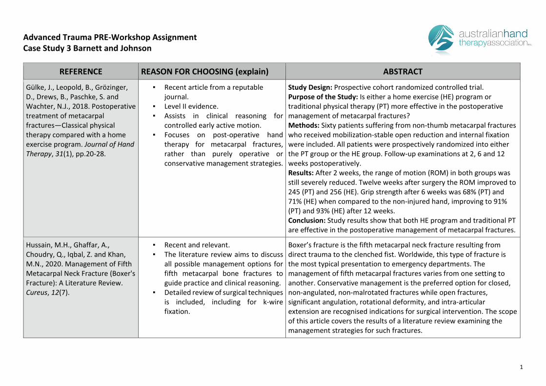
Gülke, J., Leopold, B., Grözinger, D., Drews, B., Paschke, S. and Wachter, N.J., 2018. Postoperative treatment of metacarpal fractures—Classical physical therapy compared with a home exercise program. Journal of Hand Therapy, 31(1), pp.20-28.
▪ Recent article from a reputablejournal.
▪ Level II evidence.▪ Assists in clinical reasoning for
controlled early active motion.▪ Focuses on post-operative hand
therapy for metacarpal fractures,rather than purely operative orconservative management strategies.
Study Design: Prospective cohort randomized controlled trial. Purpose of the Study: Is either a home exercise (HE) program or traditional physical therapy (PT) more effective in the postoperative management of metacarpal fractures? Methods: Sixty patients suffering from non-thumb metacarpal fractures who received mobilization-stable open reduction and internal fixation were included. All patients were prospectively randomized into either the PT group or the HE group. Follow-up examinations at 2, 6 and 12 weeks postoperatively. Results: After 2 weeks, the range of motion (ROM) in both groups was still severely reduced. Twelve weeks after surgery the ROM improved to 245 (PT) and 256 (HE). Grip strength after 6 weeks was 68% (PT) and 71% (HE) when compared to the non-injured hand, improving to 91% (PT) and 93% (HE) after 12 weeks. Conclusion: Study results show that both HE program and traditional PT are effective in the postoperative management of metacarpal fractures.
Hussain, M.H., Ghaffar, A., Choudry, Q., Iqbal, Z. and Khan, M.N., 2020. Management of FifthMetacarpal Neck Fracture (Boxer'sFracture): A Literature Review.Cureus, 12(7).
▪ Recent and relevant.▪ The literature review aims to discuss
all possible management options forfifth metacarpal bone fractures toguide practice and clinical reasoning.
▪ Detailed review of surgical techniques is included, including for k-wirefixation.
Boxer’s fracture is the fifth metacarpal neck fracture resulting from direct trauma to the clenched fist. Worldwide, this type of fracture is the most typical presentation to emergency departments. The management of fifth metacarpal fractures varies from one setting to another. Conservative management is the preferred option for closed, non-angulated, non-malrotated fractures while open fractures, significant angulation, rotational deformity, and intra-articular extension are recognised indications for surgical intervention. The scope of this article covers the results of a literature review examining the management strategies for such fractures.
2
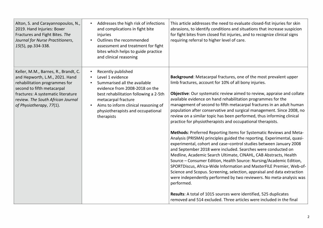
Alton, S. and Carayannopoulos, N., 2019. Hand Injuries: Boxer Fractures and Fight Bites. The Journal for Nurse Practitioners, 15(5), pp.334-338.
▪ Addresses the high risk of infectionsand complications in fight biteinjuries
▪ Outlines the recommendedassessment and treatment for fightbites which helps to guide practiceand clinical reasoning
This article addresses the need to evaluate closed-fist injuries for skin abrasions, to identify conditions and situations that increase suspicion for fight bites from closed fist injuries, and to recognize clinical signs requiring referral to higher level of care.
Keller, M.M., Barnes, R., Brandt, C. and Hepworth, L.M., 2021. Hand rehabilitation programmes for second to fifth metacarpal fractures: A systematic literature review. The South African Journal of Physiotherapy, 77(1).
▪ Recently published▪ Level 1 evidence▪ Summarised all the available
evidence from 2008-2018 on thebest rehabilitation following a 2-5thmetacarpal fracture
▪ Aims to inform clinical reasoning ofphysiotherapists and occupationaltherapists
Background: Metacarpal fractures, one of the most prevalent upper limb fractures, account for 10% of all bony injuries.
Objective: Our systematic review aimed to review, appraise and collate available evidence on hand rehabilitation programmes for the management of second to fifth metacarpal fractures in an adult human population after conservative and surgical management. Since 2008, no review on a similar topic has been performed, thus informing clinical practice for physiotherapists and occupational therapists.
Methods: Preferred Reporting Items for Systematic Reviews and Meta-Analysis (PRISMA) principles guided the reporting. Experimental, quasi-experimental, cohort and case–control studies between January 2008 and September 2018 were included. Searches were conducted on Medline, Academic Search Ultimate, CINAHL, CAB Abstracts, Health Source – Consumer Edition, Health Source: Nursing/Academic Edition, SPORTDiscus, Africa-Wide Information and MasterFILE Premier, Web-of-Science and Scopus. Screening, selection, appraisal and data extraction were independently performed by two reviewers. No meta-analysis was performed.
Results: A total of 1015 sources were identified, 525 duplicates removed and 514 excluded. Three articles were included in the final

3
data extraction: one randomised controlled trial (RCT) and two observational studies.
Conclusion: Limited evidence is available that a well-designed, well-implemented home-based exercise programme results in statistically significant improved hand function (p ˂ 0.0001) and digital total active motion (TAM) (p = 0.013) compared with traditional physiotherapy (PT) post-surgically.
Clinical implications: Our study contributes to the knowledge base of hand rehabilitation after an individual sustained a second to fifth metacarpal fracture. The authors identified a gap where future studies should further investigate the effect of hand rehabilitation after conservative and surgical management.
Cepni, S.K., Aykut, S., Bekmezci, T. and Kilic, A., 2016. A minimally invasive fixation technique for selected patients with fifth metacarpal neck fracture. Injury, 47(6), pp.1270-1275.
▪ New research, which suggests that k-wires are a reliable method oftreatment to minimise the functionalloss and allow for early return todaily activities in those whosustained a fifth metacarpal neckfracture.
Objective: The objective of this study was to compare the short-term results of treatment of fifth metacarpal neck fractures using a minimally invasive surgical fixation technique and the gold standard splinting method in a selected patient group of office workers with high expectations. Patients and methods: Twenty-four male patients (mean age: 28 years, range: 18–46 years) satisfying the inclusion criteria were enrolled in the study in two groups: surgical treatment and splinting (U-shaped ulnar gutter) groups. Hygienic interactions during daily activities and the use of keyboard and pens were allowed in the posttreatment period. The Short Form-Disabilities of the Arm, Shoulder and Hand Score (DASH) questionnaire was used to assess patient satisfaction and functionality of the extremity on the 30th and 45th days. Joint ranges of motion were measured on the 45th day. Functional and radiological evaluation data were analyzed statistically. Results: In the conservative treatment group, initial palmar angulation was measured to be 42.6°, whereas a mean of 13.5° was noted and metacarpal shortening of 5.6 mm decreased to 2 mm after treatment,

4
respectively. In terms of total joint range of motion (ROM), flexion of the treated side was at 91.25% and extension at 92.5% when measured versus the healthy-side values at the final follow-up. The mean time for return to work in this group was 33.6 days. The mean Quick-DASH score on the 30th-day follow-up was 69.5, whereas it was 39.3 at the 45th-day follow-up. The radiological findings showed a correction of the mean palmar angulation from 43° to 8° at follow-up in the surgically treated group. The initial metacarpal shortening of 9.3 mm improved to 0.5 mm at final examination. In terms of total joint ROM, flexion of the treated side was at 94% and extension at 95.5% when measured versus the healthy-side values on the 45th-day follow-ups. The mean time for return to work was 3.9 days. The mean Quick-DASH score on the 30th-day follow-up was 2.96, whereas it was noted as 0.69 at the 45th-day follow-up. Conclusions: We recommend antegrade intramedullary K-wire fixation as a reliable method, which minimizes the functional loss and allows for early return to daily activities in office workers who sustained a fracture of the fifth metacarpal neck.
Chen, K.J., Wang, J.P., Yin, C.Y., Huang, H.K., Chang, M.C. and Huang, Y.C., 2020. Fixation of fifth metacarpal neck fractures: a comparison of medial locking plates with intramedullary K-wires. Journal of Hand Surgery (European Volume), 45(6), pp.567-573.
▪ Findings support that k-wires are a safe and successful method of reducing 5th metacarpal neck fractures, with a lower rate of complications
▪ Recent article from a reputable source
Surgical treatment for metacarpal neck fractures may be indicated for malrotation, palmar angulation exceeding 30° or metacarpal shortening exceeding 3 mm, although these thresholds have not been firmly established. In a retrospective study, we compared the clinical and radiographic results of 54 patients with displaced fifth metacarpal neck fractures who were treated with either medial locking plates (14 patients) or retrograde intramedullary K-wires (40 patients). At a mean follow-up of 26 months (range 12 to 62), metacarpal shortening and angulation were 2 mm greater and 4° greater, respectively, in the K-wire group. The plate group had an earlier return to work and greater aesthetic satisfaction, but operative time and complication incidence were higher. Range of motion, time to union, grip strength and Quick Disability of the Arm, Shoulder and Hand scores were similar. We conclude that medial plating offers no clear advantage over K-wire fixation in treating metacarpal neck fractures.

5
1
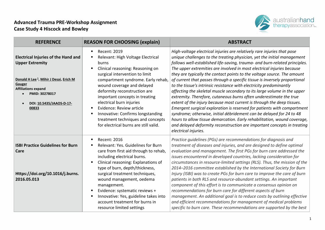
Advanced Trauma PRE-Workshop Assignment Case Study 4 Hiscock and Bowley
REFERENCE REASON FOR CHOOSING (explain) ABSTRACT
Electrical Injuries of the Hand and Upper Extremity
Donald H Lee 1, Mihir J Desai, Erich M Gauger Affiliations expand
• PMID: 30278017
• DOI: 10.5435/JAAOS-D-17-00833
Recent: 2019 Relevant: High Voltage Electrical
burns Clinical reasoning: Reasoning on
surgical intervention to limitcompartment syndrome. Early rehab,wound coverage and delayeddeformity reconstruction areimportant concepts in treatingelectrical burn injuries
Evidence: Review article Innovative: Confirms longstanding
treatment techniques and conceptsfor electrical burns are still valid.
High-voltage electrical injuries are relatively rare injuries that pose unique challenges to the treating physician, yet the initial management follows well-established life-saving, trauma- and burn-related principles. The upper extremities are involved in most electrical injuries because they are typically the contact points to the voltage source. The amount of current that passes through a specific tissue is inversely proportional to the tissue's intrinsic resistance with electricity predominantly affecting the skeletal muscle secondary to its large volume in the upper extremity. Therefore, cutaneous burns often underestimate the true extent of the injury because most current is through the deep tissues. Emergent surgical exploration is reserved for patients with compartment syndrome; otherwise, initial débridement can be delayed for 24 to 48 hours to allow tissue demarcation. Early rehabilitation, wound coverage, and delayed deformity reconstruction are important concepts in treating electrical injuries.
ISBI Practice Guidelines for Burn Care
Https://doi.org/10.1016/j.burns.2016.05.013
Recent: 2016 Relevant: Yes. Guidelines for Burn
care from first aid through to rehab,including electrical burns.
Clinical reasoning: Explanations oftype of burn, depth/thickness,surgical treatment techniques,wound management, oedemamanagement.
Evidence: systematic reviews + Innovative: Yes, guideline takes into
account treatment for burns inresource limited settings
Practice guidelines (PGs) are recommendations for diagnosis and treatment of diseases and injuries, and are designed to define optimal evaluation and management. The first PGs for burn care addressed the issues encountered in developed countries, lacking consideration for circumstances in resource-limited settings (RLS). Thus, the mission of the 2014–2016 committee established by the International Society for Burn Injury (ISBI) was to create PGs for burn care to improve the care of burn patients in both RLS and resource-abundant settings. An important component of this effort is to communicate a consensus opinion on recommendations for burn care for different aspects of burn management. An additional goal is to reduce costs by outlining effective and efficient recommendations for management of medical problems specific to burn care. These recommendations are supported by the best
2
REFERENCE REASON FOR CHOOSING (explain) ABSTRACT
research evidence, as well as by expert opinion. Although our vision was the creation of clinical guidelines that could be applicable in RLS, the ISBI PGs for Burn Care have been written to address the needs of burn specialists everywhere in the world.
Lower amputation rate after fasciotomy by straight midline incision technique for a 22,900-V electrical injury to the upper extremities
Young-Soo Janga,1, Byung Hoon Leeb,*, Hyun-Soo Parkc,1
Recent:2017 Relevant: Yes. High voltage burn
injury to upper limb. Clinical reasoning: Currently burns
units in Australia use mostly volar-ulnar incision.
Evidence: Retrospective analysis Innovative: Comparsion to current
used techniques and outcomes appear to show good results.
Purpose: The purpose of this study is to compare the major amputation rate following two different fasciotomy techniques, conventional versus straight midline, in patients with high-voltage arc burn injury by electric currents of 22,900 V to the upper extremities. Methods: A retrospective analysis of 230 patients (270 burned upper limbs) who underwent fasciotomy after high-voltage electrical injuries between 1996 and 2007 was performed. The patients were divided into two groups according to the fasciotomy method used. From 1996 to 2002, 158 patients (184 limbs) underwent conventional fasciotomy by Green’s volar-ulnar incision (conventional fasciotomy group). From 2003 to 2007, 72 patients (86 limbs) underwent fasciotomy using a straight midline curved incision (midline fasciotomy group). The patients were also divided into two groups based on whether the fasciotomy procedure was performed early or late. Patients who underwent fasciotomies <8h after injury were classified as early, while those who underwent it >8 h after injury were classified as late. Major amputation rates were compared between two fasciotomy methods and analyzed following fasciotomy timing. Results: The midline fasciotomy group had a significantly lower major amputation rate (33.7%) than the conventional fasciotomy group (59.2%) (p < 0.001). A subsequently decreased major amputation rate of 27.8% was observed in the early fasciotomy subgroup of the midline fasciotomy group (p = 0.025). Conclusion: Early fasciotomy remarkably reduced the major amputation rate after high-voltage arc injury; in the setting of minimized vascular exposure after fasciotomy, a midline straight incision could ensure that various types of reconstructive

3
REFERENCE REASON FOR CHOOSING (explain) ABSTRACT
microsurgical procedures and primary skin closures can be used to save limbs.
High-voltage electrical injury complicated by compartment syndrome and acute kidney injury with successful limb salvage: A case report and review of the literature Christopher Wei Guang Hoa,∗, Shi-Hui Yangb, Chu Hui Wongc, Si Jack Chonga
Recent: 2017 Relevant: Provide insight into
surgical techniques and other diagnosis that can occur post electrical burn. Other complications that can occur due to compartment syndrome.
Clinical reasoning: Surgical techniques to inform treatment
Evidence: Case report Innovative: Confirms pedicled flaps
remain current and important.
INTRODUCTION: Although an uncommon form of admission to a burns centre, the deep, penetrating nature of noxious currents mean that electrical burns have the most catastrophic consequences of all burn injuries. Understanding the physics of electricity is crucial to explaining the mechanisms of tissue damage and organ failure in electrical injuries which necessitate special management above and beyond that of regular thermal burns. PRESENTATION OF CASE: We present a young man who suffered significant occupation-related electrical burns that was complicated by compartment syndrome, rhabdomyolysis and acute kidney injury. He required multiple surgeries (including fasciotomy as well as soft tissue reconstruction), critical care and lengthy rehabilitation. DISCUSSION: Rhabdomyolysis is common sequela of electrical burns and may result in severe and permanent metabolic and renal impairment. High cut-off dialysis membranes have shown great promise in myoglobin removal but further studies are required to determine whether this improves clinical out- comes. Debridement and decompression are the cornerstones of initial surgical intervention and are crucial to minimising infectious complications and preserving vital structures. Free tissue transfer has become increasingly popular, but the ideal timing of microsurgery is still uncertain. Nonetheless, pedicled flaps remain widely used and still have an important role in reconstruction of electrical burns. CONCLUSION: Patients with electrical injuries have several unique acute manifestations that differ from other burns. Prognosticating outcomes is difficult, as the full scale of damage is seldom immediately evi- dent. Multiple organ systems are often affected, which makes the

4
REFERENCE REASON FOR CHOOSING (explain) ABSTRACT
treatment of such patients exceptionally challenging, multi-disciplinary and resource-intensive.
Fasciotomy closure techniques: A meta-analysis Julio J Jauregui1, Samantha J Yarmis2, Justin Tsai2, Kemjika O Onuoha2, Emmanuel Illical2, and Carl B Paulino2
Recent:2017 Relevant: Yes Clinical reasoning: Surgical
techniques to inform treatment wound care, oedema management Splinting etc.
Evidence: Meta analysis Innovative: Yes new techniques due
to higher incidence of infection.
We evaluated the risks and success rates of the three major techniques for compartment syndrome fasciotomy closure by reviewing all literature published to date. Following the Preferred Reporting Items for Systematic Reviews and Meta- Analyses guidelines, we systematically evaluated the Medline (PubMed) database until July 2015, utilizing the Boolean search sting ‘‘compartment syndrome OR fasciotomy closure.’’ Two authors independently assessed all studies published in the literature to ensure validity of extracted data. The data was compiled into an electronic spreadsheet, and the wound closure rate with each technique was assessed utilizing a proportion random model effect. Success was defined as all wounds that could be closed without skin grafting, amputation, or death. The highest success rate was observed for dynamic dermatotraction and gradual suture approximation, whereas vacuum-assisted closure had the lowest complications.
Wound healing and dermal regeneration in severe burn patients treated with NovoSorb® Biodegradable Temporising Matrix: A prospective clinical study
Cheng Hean Lo a,b, *, Jason N. Brown c , Eric J.G. Dantzer d , Peter K.M. Maitz e , John G. Vandervord f , Marcus J.D.
Recent: 2021 Relevant: Dermal substitute for deep
burns Clinical reasoning: New treatment
for deep burns. Evidence: A multicentre study Innovative: Very new innovation in
primary dermal repair for deep burns showing positive results.
Introduction: For extensive burns, autologous donor skin may be insufficient for early debridement and grafting in a single stage. A novel, synthetic polyurethane dermaltemplate (NovoSorb1 Biodegradable Temporising Matrix, BTM) was developed to address this need. The aim of this study was to evaluate use of BTM for primary dermal repair after deep burn injury. Methods: A multicentre, prospective, clinical study was conducted from September 2015 to May 2018. The primary endpoint was % split skin graft take over applied BTM at 710 days after grafting. Secondary endpoints included % BTM take, incidence of infection and adverse events, and scar quality to 12 months after BTM application. Results: Thirty patients were treated with BTM and delayed split skin
5
REFERENCE REASON FOR CHOOSING (explain) ABSTRACT
Wagstaff g , Timothy M. Barker h , Heather Cleland a,b
grafting. The % graft take had a mean of 81.9% and % BTM take had a mean of 88.6%, demonstrating effective integration of BTM. When managed appropriately, it was possible for BTM to integrate successfully despite findings suggestive of infection. Scar quality improved over time. Discussion: These results provide additional clinical evidence on the safety and performance of BTM as an effective dermal substitute in the treatment of patients with deep burn injuries
Early physiotherapy experience with a biodegradable polyurethane dermal substitute: Therapy guidelines for use
Brad Schmitt a,b,*, Kathryn Heath b, Rochelle Kurmis b, Tanja Klotz b, Marcus J.D. Wagstaff c , John Greenwood b
Recent: 2020 Relevant: Yes. Clinical Reasoning: Physio guidelines
for AROM during BTM integrationand post delamination and SSG.
Evidence: retrospective care noteaudit.
Innovative: Very new innovation inwound closure for deep burn injuriesand the practice guidelines forAROM.
Objective: The purpose of this study was to investigate and develop range of motion (ROM) and mobilisation guidelines in adult patients where a newly developed synthetic dermal substitute was applied in our adult burn centre. Method: A retrospective case note audit was conducted on the first 20 acute burn injured patients who had a synthetic dermal substitute applied. Data collected included days to commencement of ROM, days to clearance for mobilisation, and joint ROM achieved after dermal substitute application (prior to delamination) and after split skin grafting (SSG) for the elbow, knee and shoulder joints. Scar assessments were completed at 12 months after injury using two scar assessment scales.
Results: Clearance to mobilise occurred at mean 10.4 and 4.9 days after dermal substitute and after skin graft application to lower limbs respectively. ROM commenced at a mean of 9.9 (upper limbs) and 12.7 (lower limbs) days after dermal substitute application. Following skin grafting, ROM commenced at a mean of 6.6 and 6.5 days for upper limbs and lower limbs respectively. Prior to dermal substitute delamination mean flexion at the knee (86.3 ), elbow (114.0 ) and shoulder (143.4 ) was achieved. Mean ROM continued to improve after grafting with knee (133.2 ), elbow (126.1 ) and shoulder (151.0 ) flexion approaching normal ROM in most cases. Mean extension of the elbow (-4.6 ) was maintained close to normal levels after skin grafting. There
6
REFERENCE REASON FOR CHOOSING (explain) ABSTRACT
were no recorded instances of knee extension contracture. Patient and Observer Scar Assessment Scale and Matching Assessment of Photographs of Scars scores indicated good cosmetic outcomes with relatively low levels of itch and minimal pain reported at 12 months after injury.
Conclusion: A steep learning curve was encountered in providing therapy treatment for patients managed with this relatively new synthetic dermal substitute. Trends indicated that as experience with this new dermal substitute grew, patients progressed toward active therapy earlier. A guideline for therapy treatment has been developed but will continue to be evaluated and adjusted when required.

Advanced Trauma PRE-Workshop AssignmentCase Study 5 MacAskill and Gray
Mr F is a 17-year-old deckhand on a trawler. His hand sustained a severe guillotine injury to all 4 digits at the level of the proximal phalanx when it was caught
under a crate. He was airlifted to hospital and underwent replantation of Index, Middle and Ring and amputation of Little finger.
REFERENCE REASON FOR CHOOSING (explain) ABSTRACT
Cho, H. E., Zhong, L., Kotsis, S. V.,
& Chung, K. C. (2018). Finger
Replantation Optimization Study
(FRONT): Update on National
Trends. The Journal of hand
surgery, 43(10), 903–912.e1.
https://doi.org/10.1016/j.jhsa.201
8.07.021
Level of Evidence: III
This study is relevant to establishing
background for our patient, and how
likely they are to have a successful
outcome before they commence hand
therapy. The study was retrospective,
and therefore innovative only in
quantifying variables affecting finger
replantation. It used a very large cohort,
and was published by a very reputable
source. It is relevant to the case study
with regards to anticipating where this
patient may find themselves
prognostically, and to help direct
Purpose
Traumatic digit amputations have an adverse impact on patients’ daily living.
Despite experts advocating for digit replantation, studies have shown a
continued decrease in rate of replantation. We performed a national-level
investigation to examine the recent trend of practice for digital replantation.
Methods
We used the National Inpatient Sample database under the Healthcare Cost
and Utilization Project to select adult patients with traumatic digit
amputation from 2001 to 2014. We calculated the rate of attempted and
rate of successful digit replantation per year, subcategorizing for digit type
(thumb or finger) and for hospital type (rural, urban nonteaching, or urban
teaching). We also analyzed the pattern of distribution of case volume to
each hospital type per year. We used 2 multivariable logistic
regression models to investigate patient demographic and hospital
1
therapist understanding of why the
patient was medically treated the way
they were.
characteristics associated with the odds of replantation attempt and
success.
Results
Among the 14,872 adult patients with a single digit amputation from 2001
to 2014, only 1,670 (11.2%) underwent replantation. The rate of
replantation attempt trended down over the years for both thumb and
finger injuries at all hospital types, despite increasing proportions of cases
being sent to urban teaching hospitals where they were more than twice as
likely to undergo replantation. The rate of successful replantation stayed
stable for the thumb at 82.9% and increased for fingers from 76.1% to 82.4%
over the years. Patients were more likely to undergo replantation if they had
private insurance or a higher level of income. Neither hospital case volume
nor hospital type was predictive of successful replantation.
Conclusions
Although more single-digit amputations were treated by urban teaching
hospitals with higher likelihood to replant, the downward trend in rate of
attempt regardless of hospital type demonstrates that concentration of case
volume is not the solution to reverse the declining trend.
Clinical relevance
2

Financial aspects of digit replantation need to be considered from both
the patients’ and the surgeons’ perspectives to improve delivery of care for
digit replantation.
Ma, Z., Guo, F., Qi, J., Xiang, W., &
Zhang, J. (2016). Effects of
non-surgical factors on digital
replantation survival rate: a
meta-analysis. Journal of Hand
Surgery (European Volume), 41(2),
157–163.
https://doi.org/10.1177/1753193
415594572
Level of Evidence: I
This Meta-Analysis paper is very recent,
and drills down into patient specific
factors that affect outcome, knowing
what modifiable and non-modifiable
factors should be included in more
specifics of the assignment, to justify the
patient having a good outcome. Again it
is good evidence, from a reputable
journal.
This study aimed to evaluate the risk factors affecting survival rate of digital
replantation by a meta-analysis. A computer retrieval of MEDLINE, OVID,
EMBASE, and CNKI databases was conducted to identify citations for digital
replantation with digit or finger or thumb or digital or fingertip and
replantation as keywords. RevMan 5.2 software was used to calculate the
pooled odds ratios. In total, there were 4678 amputated digits in 2641
patients. Gender and ischemia time had no significant influence on the
survival rate of amputation replantation (P > 0.05). Age, injured hand, injury
type, zone, and the method of preservation the amputated digit significantly
influence the survival rate of digital replantation (P < 0.05). Children, right
hand, crush, or avulsion and little finger are the risk factors that adversely
affect the outcome.
Yoon, Alfred P. M.D.; Kaur,
Surinder Ph.D.; Chou, Ching-Han
M.S.; Chung, Kevin C. M.D.,
M.S.; For the FRANCHISE
Group Reliability and Validity of
Level of Evidence: IV
This cohort study was completed very
recently, and is of interest to hand
therapists, with regards to choosing
Background:
This study investigates the psychometric properties of patient-reported
outcome instruments for assessing outcomes in postsurgical traumatic digit
amputation patients. The authors hypothesize that the Michigan Hand
3
Upper Extremity Patient-Reported
Outcome Measures in Assessing
Traumatic Finger Amputation
Management, Plastic and
Reconstructive Surgery: January
2020 - Volume 145 - Issue 1 - p
94e-105e doi:
10.1097/PRS.0000000000006326
appropriate outcome measures to assess
recovery progress and treatment
outcomes in a reliable and valid way.
This will be of importance to justify
therapy, and determine how well a
patient is doing with regards to
achieving therapy goals. It may also
indicate when a patient could be
classified as being stable and stationary,
for discharge to self manage.
Outcomes Questionnaire (MHQ) and Disabilities of the Arm, Shoulder and
Hand (DASH) questionnaire are the most valid and reliable instruments.
Methods:
The authors studied traumatic digit amputation patients as part of the
Finger Replantation and Amputation Challenges in Assessing Impairment,
Satisfaction, and Effectiveness (FRANCHISE) study initiated by The Plastic
Surgery Foundation. The MHQ, DASH questionnaire, Patient-Reported
Outcomes Measurement Information System (PROMIS), and 36-Item
Short-Form Health Survey were used to assess patients at least 1 year
postoperatively. Internal consistency was measured by Cronbach’s alpha and
criterion validity with Pearson correlation coefficient (r). Construct validity
was tested with four predefined hypotheses. Discriminant validity was
analyzed by receiver operating characteristic curves.
Results:
One hundred sixty-eight replantation and 74 revision amputation patients
met the inclusion criteria. All instruments demonstrated fair to good internal
consistency in both cohorts (0.7 < α < 0.9). The MHQ and DASH
questionnaire scores correlated strongly (r > 0.60) in both cohorts. The
36-Item Short-Form Health Survey had moderate to weak correlation with
the remaining instruments, and its mental component had poor discriminant
validity (area under the curve, 0.64 to 0.67). The MHQ, DASH questionnaire,
and PROMIS demonstrated good construct validity confirming 75 to 100
4
percent of predefined hypotheses, whereas the 36-Item Short-Form Health
Survey confirmed only 25 percent.
Conclusions:
The authors recommend using the Michigan Hand Outcomes Questionnaire
or the Disabilities of the Arm, Shoulder and Hand questionnaire when
assessing patient-reported outcomes in digit amputation patients based on
good internal consistency and validity. The Patient-Reported Outcomes
Measurement Information System has fair validity and reliability but should
be an adjunct instrument. The 36-Item Short-Form Health Survey should not
be used as a primary assessment tool, but as an adjunct to assess overall
quality of life.
Sebastin, S. J., & Chung, K. C.
(2011). A systematic review of the
outcomes of replantation of distal
digital amputation. Plastic and
reconstructive surgery, 128(3),
723–737.
https://doi.org/10.1097/PRS.0b01
3e318221dc83
Level of Evidence: I
This systematic-review is not as recent,
but still only 10 years old, and drills
down into patient specific factors that
affect outcome, knowing what
modifiable and non-modifiable factors
should be included in more specifics of
the assignment, to justify the patient
having a good outcome when further
Background
The aim of this study is to conduct a systematic review of the English
literature on replantation of distal digital amputations to provide the best
evidence of survival rates and functional outcomes.
Methods
A MEDLINE search using “digit, finger, thumb, and replantation” as keywords
and limited to humans and the English language identified 1297 studies.
Studies were included in the review if they: (1) present primary data; (2)
5
developing the case for the later parts of
the assignment. Again it is good
evidence, from a reputable journal.
report 5 or more single or multiple distal replantations; (3) present survival
rates. Additional data extracted from the studies meeting the inclusion
criteria included demographic information, nature and level of amputation,
venous outflow technique, nerve repair, recovery of sensibility, range of
motion, return to work, and complications.
Results
30 studies representing 2,273 distal replantations met the inclusion criteria.
The mean survival rate was 86%. There was no difference in survival
between zone I and zone II replantations (Tamai classification). There was a
significant difference in survival between replantation of clean-cut versus
the more crushed amputations (crush-cut and crush-avulsion). The repair of
a vein improved survival in both zone I and zone II replantation. The mean
2-PD was 7mm (n=220) and 98% returned to work (n=98). Complications
included pulp atrophy in 14% of patients (n=639) and nail deformity in 23%
(n=653).
Conclusion
The common perception that distal replantation is associated with little
functional gain is not based on scientific evidence. This systematic review
showed a high success rate and good functional outcomes following distal
digital replantation.
6

Prsic, A., & Friedrich, J. B. (2019).
Postoperative Management and
Rehabilitation of the Replanted or
Revascularized Digit. Hand clinics,
35(2), 221–229.
https://doi.org/10.1016/j.hcl.2019
.01.003
Level of Evidence: V
Detailed description of differing post-op
protocols following digital replantation.
Recent article with very relevant
information that reflects the goals and
intended protocols for hand therapy
intervention. Clinical reasoning
expressed with regards to how
intra-operative results can affect
post-operative care. Unfortunately, this
article is not written in reflection of any
specific research, more anecdotal
evidence. As such it’s evidence-base is
low.
Postoperative care of amputated digits begins before replantation. Detailed
informed consent should be obtained and completion amputation discussed
if revascularization is not ultimately successful. Complications and failure of
the replanted digit should also be addressed. Postoperative pharmacologic
treatment should consist of aspirin, at minimum. Complications, such as
venous congestion or occlusion, and arterial thrombosis, should be dealt
with expediently. Digital motion rehabilitation should start after 5 to 7 days
of digital viability and splinting of the affected digit. Early protective motion
protocol is implemented to maintain digital motion with emphasis on
tendon glide and joint motion.
7
Gürbüz, K., & Yontar, Y. (2021). A
four-year community hospital
experience regarding procedures
for the replantation and
revascularization of fingers. Joint
diseases and related surgery,
32(2), 383–390.
https://doi.org/10.52312/jdrs.202
1.32
Level of Evidence: III
Retrospective study discussing
patient-specific factors for choosing
replantation or amputation. Very recent
information which should help in guiding
decision making for the hand-therapist
post-operatively. Investigates short and
long-term outcomes of replantation,
including ROM, function and sensation
(all relevant to the hand-therapist input).
Objectives: This study aims to evaluate the clinical results and experiences
in a community hospital regarding procedures for the replantation and
revascularization of fingers.
Patients and methods: Between June 2015 and December 2019, a total of
58 patients (51 males, 7 females; mean age: 33.4±6.3 years; range, 23 to 46
years) who were followed after total and/or subtotal amputation and
replantation were retrospectively analyzed. The patients were evaluated at
nine months in terms of cold intolerance, static two-point discrimination,
and functional results using the range of motion (ROM) and Quick
Disabilities of the Arm, Shoulder and Hand (QuickDASH) questionnaire.
8
Results: The majority of the patients presented with work-related injuries
(70%), most commonly by the mechanism of guillotine (64%), and to the
dominant hand (76%) and the third finger (36%) most frequently. The overall
success rate of digit salvage was 72.9% (n=51). Of 19 digits with unsuccessful
surgical outcomes, seven were from total and 12 were from subtotal
amputations. In the long-term, cold intolerance was observed in 14 patients
(24.1%) according to the cold intolerance severity scale. The mean static
two-point discrimination value was 6.0±0.7 mm and the mean QuickDASH
score was 22.3±5.0. The mean ROM measured at nine months after surgery
in the metacarpophalangeal and interphalangeal joints of the third and
fourth digits was significantly lower than that in the others (p<0.05).
Conclusion: The predictors of survival of a replanted digit indicated in this
study can be used as a guide and decision-making aid for any attempts for
replantation.
9

Ono, Shimpei, MD, PhD, & Chung,
Kevin C., MD, MS. (2019).
Efficiency in Digital and Hand
Replantation. Clinics in Plastic
Surgery, 46(3), 359–370.
https://doi.org/10.1016/j.cps.201
9.03.002
Level of Evidence: IV
Recent Literature Review looking at
specific surgical techniques including:
- bone stabilisation
- tendon repair
- blood vessel/nerve repair
- skin closure
- post-operative care
Clinically relevant for the surgeon
performing the surgery, but also the
hand therapist in determining how
aggressive treatment can be. Provides
detailed description of outcomes in
relation to the specific surgical
procedures. Unfortunately, does not
research a specific population, and is
more discussing previous protocols and
retrospective outcomes.
The literature on surgical techniques and recent evidence in microsurgical
digital and hand replantation is reviewed. Replantation should not be done
routinely without considering postoperative functional outcomes. Achieving
best outcomes is related to the success of microvascular anastomosis and to
adequacy of bone fixation, tendon and nerve repair, and soft-tissue
coverage. Replantation surgery has become a routine procedure. However,
little is known about the decision-making process for digital and hand
amputation. A study comparing the outcomes of digital and hand
amputations treated with replantation or revision amputation is needed.
Outcome assessment includes not only function but also patient-reported
outcomes.
➔ Multiple factors should be considered before conducting
replantation: patient’s age, occupation, hand dominance, severity
and level of injury, warm ischemia time, general condition,
motivation, economic factors.
➔ Strong indications to replantation include thumb, multiple digits,
transmetacarpal and proximal, and any pediatric amputations
whatever the level.
➔ For successful replantation, the usefulness of a 2-team approach,
bone shortening, tension-free anastomosis, and vein graft is
10
emphasized. Early recognition of postoperative vascular compromise
is also important.
➔ Recent studies have shown high survival rates after fingertip
replantation by providing excellent functional and aesthetic
outcomes. Artery-only fingertip replantation requires several
methods to restore venous outflow: removal of the nail bed, use of
medical leeches, and heparin-soaked gauze dressing.
11
Chen, C., Scott, F., Ipaktchi, K. R., &
Lauder, A. (2021). Postoperative
Digit and Hand Replantation
Protocols: A Review of the
Literature. Journal of the American
Academy of Orthopaedic
Surgeons, 29(15), e732–e742.
https://doi.org/10.5435/JAAOS-D-
20-01176
Literature Review (Level V)
Very detailed description of differing
post-op protocols following digital
replantation. Recent article with very
relevant information that reflects the
goals and intended protocols for hand
therapy intervention. Clinical reasoning
expressed with regards to how
intra-operative results can affect
post-operative care. Very detailed
description of ROM protocols and when
to progress hand therapy treatment.
Unfortunately, as a literature review, this
article is not reflecting primary evidence.
Successful replantation and revascularization of the hand and digit
require a skilled team with urgent access to an operating room with
microsurgical capabilities. Although careful indications and surgical
techniques contribute to success, postoperative management also
plays a vital role in the survival of a replanted digit. Previous research
has assessed surgical efficiency and techniques to conduct these
procedures, but few studies evaluate postoperative protocols to care for
patients undergoing these procedures. Because of the lack of high-level
evidence specific to replantation,many common post-operative practices
related to monitoring, anticoagulation, and diet have been inferred from
elective microsurgical procedures, despite notable differences in
operating conditions. The highest level of evidence pertaining to digital
replantation was found with the use of peripheral nerve blockade,
leeching/bleeding, and nicotine use. This review provides an in-depth
evaluation of the literature and insight into the rationale and level of
evidence that supports each postoperative intervention. It highlights
institutional variability and a paucity of high-level evidence pertaining to
this topic while identifying the areas of future research
12

Accompanying Video ScriptMiles (Part I)
Mr F is a 17-year-old deckhand on a trawler. His hand sustained a severe guillotine injury to all 4 digits at the level of the proximal phalanx when it was caught
under a crate. He was airlifted to hospital and underwent replantation of Index, Middle and Ring and amputation of Little finger.
Traumatic digit amputations have an adverse impact on a patient’s daily living. Despite evidence advocating for digit replantation, studies have shown acontinued decrease in rate of replantation generally.
Among the 14,872 adult American patients with a single digit amputation from 2001 to 2014, only 11.2% underwent replantation. The rate of replantationattempt trended down over the years for both thumb and finger injuries at all hospital types, despite increasing proportions of cases being sent to urbanteaching hospitals where they were more than twice as likely to undergo replantation.
Data from these American patients showed the rate of successful replantation stayed stable for the thumb at around 83%, but increased for fingers from76.1% to 82.4% over those years. Similarly, pooling data from Europe, a systematic review in 2011 showed a high success rate and good functional outcomesfollowing distal digital replantation. In around 2300 patients, distal replantation had a mean survival rate of 86%. Patients were more likely to undergoreplantation if they had private insurance or a higher level of income.
A meta-analysis of replantation, found that gender and ischemia time had no significant influence on the survival rate of amputation replantation. Youngerage, injury type, the repair of a vein, and the method of preservation of the amputated digit significantly increased the survival rate of digital replantation.Children, right hand, crush, avulsion and little finger injuries were risk factors that adversely affected outcomes. Complications following viable replantationsincluded pulp atrophy in 14% of patients and nail deformity in 23% of patients. It seems to be a common perception that finger replantation is associatedwith little functional gain, but the literature would indicate that this is not based on scientific evidence. General outcomes showed that 98% of these patientsreturned to work in all patient groups.
Little research has been done when looking at reliable and valid outcome measures in assessing traumatic finger amputation management outcomes.Recently a group of authors in 2020 looked into using multiple measures in over 200 patients, 1 year post operatively. They found the Michigan HandOutcomes Questionnaire or the Disabilities of the Arm, Shoulder and Hand questionnaire had good internal consistency and validity. Using Patient-ReportedOutcomes Measures had fair validity and reliability, but should be an adjunct instrument. Any other measures they assessed were not recommended.
13

Taylah (Part II)
Limited research exists pertaining to the specific rehabilitation protocol of the replanted or amputated digit, more so the surgical outcomes of success orfailure of affected tissue. Postoperative digit replantation protocols have been found to arise from level III and IV retrospective reviews, case studies, andanecdotal expert opinion.
During the postoperative period, success has been defined as the full viability of the digit based on its colour, temperature, capillary refill time, and pulseoximetry reading. An additional measure of success is lack of a secondary operation due to circulatory complications, such as stump revision or debridementof necrotic tissue.
In the early days of digital replantation, varying philosophies on the timing of digital range of motion rehabilitation were published. Some have proposed aslate as 3 to 4 weeks postoperatively to as early as 1 day. Treatment has been detailed to consist of early active ROM, continuous passive ROM, splinting andsensory stimulation.
Like most post-operative referrals, the specific role of the hand therapist’s intervention is determined in reflection of the surgeon’s intraoperative injuryassessment. However, the goal of any digital replantation is function for daily use with adequate stability, range of motion, and sensation.
Stability and sensibility are achieved with good surgical technique and healing; however, range of motion can only be achieved with aggressive rehabilitation.Close communication between the therapist and the surgeon is also essential, as knowing the boundaries of rehab during early stages of healing.
When the viability of the replanted digit has been established, typically 5 to 7 days postoperatively, the postoperative splint is converted to a thermoplasticone. In situations that require longer periods of immobilization, the patient can be placed in a cast. The hand should be placed in a position of safestimmobilisation (e.g. wrist in neutral or slight flexion, MCPJs in 30-40 degrees flexion and IP joints in extension). A choice of volar or dorsal orthosis should bedetermined early depending on tissue integrity.
An evidence-based model of early protected motion (EPM) in digital revascularization and replantation was developed by Silverman and colleagues. Theprotocol is based on a graded progressive model of hand therapy in 2 stages: EPM I and EPM II.
EPM I is based on the following treatment goals: to protect all repaired structures, maintain and improve MCP collateral ligament length, prevent jointstiffness, control oedema, monitor wound care, educate the patient, and aid in psychological adjustment to disease.
EPM II is based on a differing set of treatment goals: to protect all repaired structures, maintain intrinsic muscle function, prevent PIP joint stiffness, minimizetendon adhesions, provide differential gliding of tissues, and improve tendon tensile strength.
14

1
Advanced Trauma PRE-Workshop Assignment. Case Study 6 Grange & Criticos
REFERENCE REASON FOR CHOOSING (explain) ABSTRACT
Soong M, Chase S & Kasparyan NG. Metacarpal fractures in the athlete. Hand and wrist sports medicine, musculoskeletal medicine (2017). 10:23-27. DOI 10.1007/s12178-017-9380-0
Recent: 2017 Relevant: article is specifically looks
at metacarpal fracture managementin relation to returning to sport
Clinical reasoning: Discusses k-wirefixation versus plate/screw fixation ofthe fracture
Evidence: Statistics on generalmetacarpals and gender not specificto the basal thumb region.
Innovative: brief discussion of bonestimulation although it does say thereis limited evidence in relation tometacarpal fractures. Also refers tohand therapy input follow andpossible orthosis/protectiveequipment that could be used toassist in return to play
Purpose of review: To describe current evaluation and treatment of metacarpal fractures in athletes
Recent findings: Biomechanical and clinical studies involving lower-profile, locking, shorter length, and double-row or separate-dual plate configurations, as well as intramedullary screw fixation, have demonstrated the potential benefits of internal fixation with promising results.
Summary: Treatment should be customized to the specific athlete and injury, and is often successful without surgery, or with percutaneous pin fixation. Internal fixation of metacarpal fractures has improved with new hardware and new techniques, and may expedite return to play, although further clinical studies are needed.
Rust, P & Brown, M. Fractures of the thumb metacarpal base. (2020). International journal of the care of the injured. Volume 51, ISSUE 11, P2421-2428. DOI: 10.1016/j.injury.2020.07.053
Recent: - 2020
Relevant: - Specifically discusses fractures of the 1st
metacarpal base. Review common fracturepatterns for differential diagnosis
A variety of fractures can occur at the thumb metacarpal base, which classically result from axial loading through a partially flexed thumb. Thumb base fractures account for 4% of all hand fractures and may occur in association with fractures of the trapezium, especially in men. Displaced intra-articular fractures include the eponymous patterns described by Edward Bennett and Silvio Rolando, in addition to more comminuted variants. These intra-articular fractures are unstable, with important implications for their management. Bennett fractures account for 30% of all thumb metacarpal fractures and occur four times more frequently than

2
REFERENCE REASON FOR CHOOSING (explain) ABSTRACT
- Detailed anatomical discussion followed bysurgical management options for Rolandofractures- Long term follow up and hand therapybriefly discussedClinical reasoning:- Reviews initial management pathway
step by step and reasoning behind this.Xray/CT (images required for accuratediagnosis) and surgical options (k-wirefixation, lag screws/plate, externalfixation)
Evidence: - Literature review completed by the
authors in regards to surgicalmanagement options and they also havesome limited evidence regarding longterm follow up (5.8 years)
- The authors outline there isn’t highquality evidence favouring one surgicalapproach over another
- Briefly mentions post operative followup for lag screw and t plate fixation withhand therapy at 7 days post op. Activemobilisation and splinting can occur atthis time
Innovative: - Good discussion regarding why each
surgical method should be chosen andrefers to long term outcomes
the Rolando fracture. Extra-articular fractures are common, typically occurring at the metaphyseal-diaphyseal junction, and are often termed epi-basal fractures.

3
REFERENCE REASON FOR CHOOSING (explain) ABSTRACT
Halim A, Arnold P. Weiss. Return to Play After Hand and Wrist Fractures. Division of Hand Surgery, Department of Orthopaedics. Clin Sports Med 35 (2016) 597–608 http://dx.doi.org/10.1016/j.csm.2016.05.005
Recent: - 2016
Relevant: - Generalised discussion regarding
metacarpal fractures andconsiderations for management
Clinical reasoning: - Good discussion at the end about
how to clinically reason whensomeone should return to sport andthat the discussion is one of the mostimportant parts of post op follow up.Managing the expectations andensuring the athlete is making a wellinformed decision when they decideto back
Evidence: - Minimal specific Rolando based
management evidence
Innovative: - Doesn’t address specific hand
therapy options or specific surgicaltechniques
Wrist and hand injuries are common among athletes, and can lead to considerable disability. Dislocations and soft tissue injuries are common and require prompt recognition and treatment. Accurate diagnosis and early immobilization are often key to getting players back to their sport early. Some injuries require surgery; operative intervention allows the player to return to their sport more quickly or with less long-term disability. This article discusses the spectrum of injury from distal radius fractures to mallet fingers, and offers some general guidelines for the surgeon in how to counsel and treat athletes with these problems.

4
REFERENCE REASON FOR CHOOSING (explain) ABSTRACT
Lucian Lior Marcovici, M.D, Andrea Atzei, M.D, Roberto Cozzolino, M.D and Riccardo Luchetti, M.D. Arthroscopic Assisted Treatment of Thumb Metacarpal Base Articular Fractures. (2021). Arthroscopy Techniques, Vol 10, No 7 (July), 2021: pp e1783-e1792.DOI; 10.1016/j.eats.2021.03.022
Recent: 2021 Relevant: Describes arthroscopic
assisted treatment (AAT) for surgicalintervention of thumb metacarpalbase articular fractures. Details thetechnique used and its implications,benefits for optimising outcome andfracture fixation methods.
Clinical reasoning: Procedure allowsvisualisation of the joint and fracturefragments and simplifies soft tissueto achieve proper reduction andfixation.
Evidence: Authors comment onarthroscopy for basal thumb jointshave been presented since 1995 andsince small joint arthroscopy hasevolved and smaller optics havebeen made available with higherdefinition systems and smallerinstruments. Therefore, handsurgeons can better approach thesefracture types.
Innovative less invasive method ofsurgery which limits soft tissuedamage and allows earlyrehabilitation and therefore return toplay.
Fracture of the base of the thumb metacarpal (M1) is a common finding in hand trauma. Closed reduction and K-wire fixation and open reduction internal fixation are traditional treatments of choice. The arthroscopic assisted technique has been introduced to improve intra-articular fragment reduction and to preserve fragment vascularization and capsular and ligamentous integrity along with joint stability. Indications for arthroscopic assistance are all types of intra-articular fractures or pending malunions involving the base of M1 and/or the trapezium. The aim of this article is to describe the surgical technique used in managing articular fractures of the base of M1, with arthroscopic assistance. Our experience with this technique confirms the advantages of a minimally invasive method that provides articular reduction under direct vision, with limited soft-tissue damage, and allows early rehabilitation (from day 1 after surgery). This technique is extremely valuable for high-demand patients such as manual workers or athletes. The relative disadvantage of the technique is its technical difficulty, which requires experience with small-joint arthroscopy.

5
REFERENCE REASON FOR CHOOSING (explain) ABSTRACT
Wahl E, Marc J. Elizabet P, Richard M. Management of Metacarpal and Phalangeal Fractures in the Athlete. (2020) Clin Sports Med 39 (2020) 401–422 DOI 10.1016/j.csm.2019.12.002
Recent: 2019 Relevant: Addresses Rolando
fractures, athletes returning to sport,surgical management options andalso post-operative follow up.Addresses base M1 fractures, intra-articular and mal-union fracturestherefore Rolando’s and Bennett’sfractures represent an appropriateindication for AAT.
Clinical reasoning: good clinicalreasoning around specific factors tolooks for when providing athletesadvice about returning sport. Advisesthat Rolando fractures generallyrequire a longer period away fromsport (6-8 weeks) due to theircomplexity
Evidence: Literature reviewcompleted by the authors in regardsto surgical management options
Innovative: Understanding thesatisfaction for an athlete to return tosport without delay post surgery andchoice of hardware fixation has beenstrategic for fracture alignment aswell as allowing early motion andtherefore return to play (RTP).Discussion to RTP with guard whenrequired from 2-10 weeks post-surgery.
Metacarpal and phalangeal fractures are common injuries in athletes and occur frequently in contact and ball-handling sports. They usually result after direct hits from other players or athletic equipment. The fractures often are minimally displaced and require a short period of immobilization followed by early range of motion for expeditious return to play. Unstable or intra-articular fractures may require operative fixation. Open reduction and internal fixation afford the most stability while allowing for early rehabilitation. Athletes represent a unique population, and treatment of these fractures requires consideration of specific sport, timing of injury, and level of play

6
REFERENCE REASON FOR CHOOSING (explain) ABSTRACT
Tiffany R. Kadow, MD, John R. Fowler. Thumb Injuries in Athletes. 2017. Hand Clin 33 (2017) 161–173 DOI 10.1016/j.hcl.2016.08.008
Recent - 2016
Relevant - Specific section addressing the
surgical treatment and a stagedbreakdown for when k-wire orplate/screw would be considered
Clinical reasoning - Addresses surgical management of
common sporting injuries in athletes(thumb specific) well and outlinesthe anatomicalconsiderations/deforming forces thatimpact on fracture management
- Limited long-term studies but it doeslook at one study with 6 year followup. Arthritic changes were noted inthe majority of patients (11 out of14) but they were unable tocorrelate this with how well thefracture was reduced
- Large fragments were recommendedto have lag screw/plate fixationwhilst more comminuted fractureswere better managed withdistraction pinning
Evidence - Uses the available evidence however
like other literature it does state
Thumb injuries are common in athletes and present a challenging opportunity for upper extremity physicians. Common injuries include metacarpal base fractures (Bennett and Rolando types), ulnar and radial collateral ligament injuries, dislocation of the carpometacarpal and metacarpophalangeal joints, and phalanx fractures. This review, although not exhaustive, highlights some of the most common thumb injuries in athletes. The treating physician must balance pressure from athletes, parents, coaches, and executives to expedite return to play with the long-term well-being of the athlete. Operative treatment may expedite return to play; however, one must carefully weigh the added risks involved with surgical intervention.
7
REFERENCE REASON FOR CHOOSING (explain) ABSTRACT
there is limited high quality evidence for their management or long-term outcomes
- The article doesn’t delve intodetailed return to sportrecommendations. It highlights theneed to balance the pressure fromathletes (and significant others) withtheir long-term wellbeing.
- Doesn’t address handtherapy/general managementconsiderations post operatively
Innovative - The article isn’t particularly
innovative. It addresses themanagement options availableconcisely, more so from a surgicalperspective and when you considerthese
Advanced Trauma PRE-Workshop Assignment
Case study 7: Milgate and Dornsbusch
Mrs N was riding to work on her motorbike when a vehicle hit her, and she was airborne before landing on both arms. X-ray showed an multi-fragment articularfracture of right distal radius and fracture of ulnar styloid. On the left X-ray showed radial styloid fracture with 3mm step. Both fractures to the radius were fixedwith volar locking plates. At three months post- surgery Mrs N is having problems loading her right wrist.
REFERENCE REASON FOR CHOOSING (explain) ABSTRACT
https://www.sciencedirect.com/sc
ience/article/pii/S0894113020300
132
Outcomes of surgically treated
distal radius fractures associated
with triangular fibrocartilage
complex injury
Journal of Hand Therapy
Volume 33, Issue 3,
2020,
Pages 339-345,
(Charlotte)
This article was published in 2020 in The
Journal of Hand Therapy and is within the
required 2016-2021 date requirement.
This article is relevant to the case study as it
highlights a link between TFCC injury and
distal radius fractures. The article noted in
one cadaveric study, where a hyperextension
force was applied to cadaveric wrists until a
distal radial fracture occurred, an injury to
the TFCC occurred in 63% of the specimens.
Furthermore, the article reported that healed
radial fractures were often complicated by
chronic debilitating wrist pain and one of the
Study Design
Prospective cohort.
Introduction
Clinical studies that evaluate the correlation between associated lesions
of the triangular fibrocartilage complex (TFCC) and outcome of distal
radius fractures expressed with the patient-rated disability are missing.
The purpose of this study was to evaluate the outcomes of distal radius
fractures associated with or without an injury of the TFCC.
Methods
Patients undergoing operative treatment for distal radius fracture were
prospectively enrolled (n = 70). TFCC was examined by wrist
arthroscopy and injuries classified according to Palmer. Comparative
analyses were performed on 45 patients with TFCC injury (injured
group) and 25 patients with intact TFCC (intact group). The outcome
1
Reference: Katerina
Kasapinova, Viktor Kamiloski,
Outcomes of surgically treated
distal radius fractures associated
with triangular fibrocartilage
complex injury,
Journal of Hand Therapy,
Volume 33, Issue 3,
2020,
Pages 339-345,
ISSN 0894-1130,
https://doi.org/10.1016/j.jht.2017
.09.012.
important causes being TFCC tears with or
without distal radio-ulnar joint (DRUJ)
instability. Ulnar avulsion of the TFCC can
result in ulnar-sided wrist pain, decreased
grip strength, reduced range of forearm
rotation, and clinical signs of DRUJ
instability. Unrepaired peripheral tears of the
TFCC interfere with its important functions
related to wrist stability and load bearing.
This journal article is relevant to Mrs N’s
case study as she has fractured her right
distal radius and ulnar styloid, these
structures correlate with TFCC injuries
(according to this research) and may be
contributing to her difficulty loading the
right wrist. The article also explores how an
ulnar avulsion of the TFCC can result in
ulnar sided wrist pain, decreased grip
strength, reduced range of forearm rotation,
and clinical signs of DRUJ instability. These
factors may also be contributing to her
measures included the Patient-Rated Wrist Evaluation (PRWE) and the
Disabilities of the Arm, Shoulder and Hand (DASH) questionnaires, 3
and 12 months after injury.
Results
TFCC was injured in 45 patients (64%). In patients with intact TFCC,
mean total PRWE score was 27 (3 months) and 16 (12 months),
compared to patients with TFCC injury with 40 (at 3 months) and 24 (at
12 months). Mean DASH scores were 26 and 13 at 3 and 12 months for
the intact group and 39 and 27 for the injured group. PRWE and DASH
results showed significant difference at 3 and 12 months when compared
with Mann–Whitney test.
Discussion
PRWE and DASH scores evaluation showed that patients with associated
TFCC injury had greater pain and disability at 3 and 12 months after
injury.
Conclusions
Disability outcomes were worse in patients with distal radius fracture,
where TFCC was injured. TFCC injuries are an important cofactor
affecting the outcome of distal radius fractures.
2
limited function as she has fractured the
ulnar styloid as well.
Therefore, it is important as therapists to
assess and treat not only the bony injury but
also the surrounding soft tissue structures
that may hinder functional outcomes.
https://journals.lww.com/md-jou
rnal/Fulltext/2020/09180/Arthros
copic_treatment_of_chronic_wri
st_pain_after.46.aspx
Arthroscopic treatment of
chronic wrist pain after distal
radius fractures
Medicine: September 18, 2020 -
Volume 99 - Issue 38 - p e22196
(Charlotte)
Reference: Lee, Young-Keun
MD, PhD∗; Kwon, Tae-Young
This observational study in Medicine was
published in September 2020 and is within
the required 2016-2021 date requirement.
The article explores causes of pain after
Distal Radius Fractures (DRF). Synovitis
was found in all cases as well as soft tissue
injury including the following:
- TFCC
- Intercarpal and radiocarpal ligament
ruptures
- ulnar impaction syndrome
- Cartilage lesion
After surgical intervention for patients'
particular source of pain, their post op values
We report the arthroscopic and clinical findings of patients with chronic
wrist pain following distal radius fracture (DRF) who underwent
diagnostic arthroscopy and arthroscopically-assisted tailored treatment.
We retrospectively analyzed the records of 15 patients with chronic wrist
pain following DRF, who underwent diagnostic arthroscopy and
arthroscopically-assisted tailored treatment from 2010 to 2017. The
average patient age was 44 years (range, 20–68 years), average time from
injury to treatment 21 ± 23.46 months (range, 3–96 months) and average
follow up period 20.13 ± 8.71 months (range, 12–39 months). The
functional outcome was evaluated by comparing the preoperative and
final follow up values of the range of motion, grip strength, pinch
strength, visual analogue scale for pain and quick disabilities of the arm,
shoulder and hand score.
Based on the arthroscopic findings, synovitis was found in all cases and
the pathologic intra-articular lesions were classified into 4 patterns.
3
MD; Lee, Ha-Song MD
Arthroscopic treatment of
chronic wrist pain after distal
radius fractures, Medicine:
September 18, 2020 - Volume 99
- Issue 38 - p e22196 doi:
10.1097/MD.0000000000022196
were increased in ROM, grip strength and
pinch strength.
This study indicates that there can be
underlying soft tissue structures that are
damaged during DRF. These structures need
to be considered during rehabilitation and
may be contributing sources of limited
function and pain of the wrist. Furthermore,
if they are a persistent injury that cannot be
healed with conservative management,
surgical intervention should be considered.
The article also explored how complications
of DRF can be as high as 31%. Among these
complications, residual chronic pain is
common. Consequently, wrist and forearm
motion are impaired by stiffness, thereby
leading to dysfunction of the entire upper
limb.
Previous publications were limited to the
discussion of operative treatment for certain
Triangular fibrocartilage complex rupture was seen in 14 cases,
intercarpal and radiocarpal ligament ruptures in 9 cases, ulnar impaction
syndrome in 5 cases, and cartilage lesion in 9 cases. In terms of surgical
treatment, 15 patients underwent arthroscopic synovectomy, 7 foveal or
capsular repair of TFCC, 7 intercarpal Kirschner wires fixation or
intercarpal thermal shrinkage, 1 intercarpal ligament reconstruction, 2
Sauve-Kapandji procedure, and 2 unlar shortening osteotomy.
Postoperatively, the average range of motion, grip strength, and pinch
strength increased significantly. From preoperative to final follow up
values, the average visual analogue scale and quick disabilities of the arm
score decreased from 5.93 ± 1.58 (range, 3–8) to 1.33 ± 1.29 (range, 0–3)
(P = .001) and from 49.38 ± 19.09 to 12.63 ± 7.63 (P = .001),
respectively.
Diagnostic arthroscopy and arthroscopically-assisted tailored treatment
of chronic wrist pain following DRF can provide an accurate diagnosis,
significant pain relief, and functional improvement.
4

specific problems such as the correction of
radius malunion or the treatment of TFCC
injury or ulnar impaction. However, the
authors conclude, the chronic pain arises
from many complex causes. Thus, it is
essential to treat the complex pathoanatomy
in order to overcome the chronic wrist pain
following DRF.
Therefore, the likelihood of Mrs N sustaining
a soft tissue injury as a result of her DRF is
very high and further surgical intervention
such as arthroscopic investigation and repair
could be considered to improve her pain and
functional outcomes.
Scapholunate, lunotriquetral
and TFCC ligament injuries
associated with intraarticular
distal radius fractures:
Arthroscopic assessment and
correlation with fracture types
This article was published in 2019 in the
Hand Surgery and Rehabilitation Journal.
The publication date is within the 2016-2021
date requirement.
The article by Roulet et al is relevant to the
topic as it explores the prevalence of
Scapholunate, lunotriquetral and TFCC
The aim of this study was to evaluate the prevalence of arthroscopic
scapholunate (SL) and/or lunotriquetral (LQ) laxity and triangular
fibrocartilaginous complex (TFCC) injuries in patients who have an
intraarticular fracture of the distal radius and to correlate these lesions
with fracture type. Fifty-seven intra articular radius fractures, whether or
not they were associated with an ulnar styloid fracture, were evaluated
and treated by arthroscopy. Scapholunate and lunotriquetral ligament
injuries were classified according to the EWAS classification. TFCC
5
S. Roulet et al. / Hand Surgery
and Rehabilitation 39 (2020)
102–106
https://www.clinique-main-nante
s.org/wp-content/uploads/2021/0
6/roulet2019.pdf
(Charlotte)
Reference: S. Roulet, L.
Ardouin, P. Bellemère, M. Leroy,
Scapholunate, lunotriquetral
and TFCC ligament injuries
associated with intraarticular
distal radius fractures:
Arthroscopic assessment and
correlation with fracture types,
Hand Surgery and
Rehabilitation,
Volume 39, Issue 2,
2020,
Pages 102-106,
ligament injuries associated with
intraarticular distal radius fractures. It also
discusses the correlation of these occurring
depending on the fracture type. As Mrs N has
sustained a right distal radius fracture and is
having difficulty loading her wrist it is
imperative we explore soft tissue
involvement as the bone should be stably
healed by 3 months post op. The article also
assessed individuals of both genders aged 18
to 65 and we can assume Mrs N falls within
the study population.
The article highlighted that at least one soft
tissue injury occurs in 39 intra articular
fractures of the distal radius (68.4%).
Furthermore, Arthroscopic SL laxity was
present in 8 of 11 cases (72.7%) of radial
styloid fractures. There was no statistically
significant relationship between the different
types of radius fractures and soft tissue
injuries. Except, ulnar styloid fracture was
predictive of TFCC injury.
lesions were assessed according to Palmer’s classification. Each injury
was documented through preoperative X-rays and a CT scan. Fracture
type and soft tissue injury were not significantly associated one to
another. Arthroscopic examination revealed at least one soft tissue injury
in 39 intra articular fractures of the distal radius (68.4%). Twenty-five
percent of arthroscopic SL laxities (including severe EWAS 3 injuries)
were not detected on standard radiographs. Arthroscopic SL laxity was
present in 8 of 11 cases (72.7%) of radial styloid fracture and in 15 of 25
cases (60%) of fractures with at least one radial styloid component. There
was no association between LQ integrity and fracture type. Ulnar styloid
fractures (base or tip) and TFCC lesions were significantly correlated (P
< 0.0001). The prevalence of soft tissue lesions secondary to intra
articular fractures of the distal radius was 68.4%. However, there was no
statistically significant relationship between the different types of radius
fractures and soft tissue injuries. On the other hand, ulnar styloid fracture
was predictive of TFCC injury.
6

ISSN 2468-1229,
https://doi.org/10.1016/j.hansur.
2019.11.009.
In the study TFCC injury was associated in
16 of 57 cases (28%) with intraarticular
fracture of the radius and in 14 of 27 cases
(52%) with ulnar styloid fracture. In this
study, as in others, there was a statistically
significant correlation between ulnar styloid
fractures and TFCC injury
The prevalence of soft tissue lesions
responsible for dissociative instability or
TFCC injury secondary to intra articular
fracture of the distal radius was 68.4%, and
72.7% of pure radial styloid fractures were
associated with arthroscopic SL laxity.
This study highlights that Mrs N is likely
experiencing a TFCC injury related to her
DRF which would impact loading of the
wrist. I hypothesize this is occurring as the
study noted 52% of ulnar styloid fractures
are coupled with TFCC injury. Furthermore,
even though Mrs N is only having difficulties
7

loading the right wrist which sustained a
DRF and ulnar styloid fracture it should be
highlighted that the study noted 72.7% of
pure radial styloid fractures were associated
with arthroscopic SL laxity. Therefore, we
should also assess Mrs N’s left wrist to
ensure there are no soft tissue
injuries/complications to be factored into
rehabilitation and treatment.
Functional and radiological
outcome of distal radius
fractures stabilized by
volar-locking plate with a
minimum follow-up of 1 year.
Quadlbauer, S., Pezzei, C.,
Jurkowitsch, J. et al.
Archives of Orthopaedic and
Trauma Surgery (2020)
140:843–852
This study was published in 2020, thus
meeting the stipulated date range for
inclusion.
This study examined the long term outcomes
associated with volar locking plates for distal
radius fracture. Mrs N’s bilateral radius
fractures were treated with Volar locking
plates. Results showed that the use of volar
locking plates to treat distal radius fracture is
safe, associated with low rates of
complication and results in good clinical
outcomes at least 1 year post surgery. The
Introduction
Distal radius fractures (DRF) are the most common fractures of the upper
extremities and incidence is expected to continue rising as life
expectancy increases. Palmar locking plate stabilizing has since become
the standard treatment for dorsally displaced DRF. Main aim of this study
was to investigate correlation between radiological and clinical outcome
in patients stabilized by palmar locking plate with a minimum follow-up
of one year.
Methods
A total of 524 patients with DRF, stabilized using palmar angular stable
locking plate fixation were included in the study. Of these, 117 patients
8

https://doi.org/10.1007/s00402-02
0-03411-9
JOE
study also demonstrated that age, gender and
type of postoperative immobilisation had no
significant impact on clinical outcome,
making the findings transferrable to Mrs N
who’s demographics and postoperative care
is not stipulated.
The study did show a connection between a
residual ulnar variance and grip strength,
along with patient self assessment scores, but
the degree of ulnar variance (>2mm or
<2mm) didn't have a significant impact on
these scores. This is also relevant as Mrs N
had radial styloid fracture with 3mm step on
initial X Ray.
had to be excluded and another 177 were not accessible. The study group
thus compromised 230 patients who returned for the follow-up
investigation and were followed-up clinically and radiologically with a
mean follow-up interval of 20 months. Outcome was evaluated using
pain, range of motion (ROM) and grip strength parameters. In addition,
self-assessment by patients was registered on the QuickDASH, PRWE
and Mayo Score. The immediate postoperative and final checkup
radiographs were scrutinized for alignment and intra-articular step-off.
Results
Bivariant correlation analysis showed a significant correlation between
ulnar variance and QuickDASH (r=0.18, p=0.01), grip strength (r=−
0.18, p=0.04) and Mayo Score (r=− 0.23, p=0.001). No significant
differences could be found between an unacceptable (>2 mm) and
acceptable (<2 mm) ulnar variance in respect of pain, ROM, grip strength
and patient-reported outcome measurements. Age, gender, additional
fracture to the ulnar styloid, or type of postoperative immobilization
showed no significant or clinically important impact on the final
patient-reported outcome. No significant differences in incidence of
complications, ROM or loss of reduction could be found in any patients
over or under 65 years of age.
Conclusions
9

Stabilization of DRF by palmar angular stable locking plate is a safe
form of treatment and results in a good clinical and radiological outcome
with low complication rate. Ulnar variance showed a significant
correlation to grip strength, QuickDASH and Mayo Score, but an
unacceptable ulnar variance (>2 mm) was not associated with a worse
clinical important outcome. Age (<65/>65 years), gender and type of
immobilization had no impact on the complication rate or in the final
functional or radiological outcome.
The Presence and the Location
of an Ulnar Styloid Fracture
Associated With Distal Radius
Fracture Predict the Presence of
Triangular Fibrocartilage
Complex 1B Injury
Tomori, Y., Nanno, M., & Takai,
S.
The Journal of Arthroscopic and
Related Surgery. Vol 36 (10),
2020, pp 2674-2680.
This study was published in 2020 and
therefore meets the recency of publication
requirement.
Mrs N suffered both distal radius and ulnar
styloid fractures on the right side, hence this
study was selected to assist with determining
the likelihood of a concurrent TFCC injury to
the affected wrist.
The study demonstrated that the presence of
an ulnar styloid fracture indicated a likely
TFCC injury, furthermore, the location of
Purpose: To investigate the correlation between ulnar styloid fracture
(USF) associated with distal radius fracture (DRF) and triangular
fibrocartilage complex (TFCC) injuries and to elucidate whether the
presence or location of an USF in a patient with DRF predicts the
presence of traumatic TFCC injuries.
Methods: From 2005 to 2018, an arthroscopic evaluation was performed
to detect TFCC injuries associated with DRF. The presence and location
of USFs were evaluated using computed tomography. TFCC injuries
were classified in accordance with Palmer's classification. All wrists
were divided into group A (DRF without USF) and group B (DRF with
USF). The incidence of TFCC injuries in the 2 groups was compared.
group B was then divided into 2 subgroups in accordance with the USF
10

JOE the fracture was indicative of the type of
TFCC injury. However, the research found
that TFCC injury still occurs in a significant
number of distal radius fractures, without the
presence of an ulnar styloid fracture.
location: the tip or middle fracture subgroup and the base fracture
subgroup. Data were analyzed with significance set at P < .05.
Results: One hundred thirty-eight patients were enrolled in this study.
Group A included 42 wrists in 42 patients, whereas group B included 96
wrists in 96 patients. There were significant differences between the 2
groups regarding the incidence of traumatic TFCC injuries (P = .036) and
TFCC 1B injury (P = .002), although there were no differences between
the 2 groups regarding age, sex, injured side, direction of displacement,
and type of DRF. Within group B, the tip and middle fracture subgroup
included 37 wrists in 37 patients, whereas the base fracture group
included 59 wrists in 59 patients; a significant difference was observed
between the two subgroups regarding the incidences of TFCC 1B injuries
(P = .044).
Conclusions: The presence of USF associated with DRF predicted the
presence of frequently occurring traumatic TFCC injury and TFCC 1B
injury. Moreover, the location of USFs was a predictive factor for TFCC
1B injury in adults with DRF. On the other hand, traumatic TFCC injury
had occurred in adults with DRF, regardless of the presence of USF.
11

Polytrauma and High-energy
Injury Mechanisms are
Associated with Worse
Patient-reported Outcomes
After Distal Radius Fractures
Van Der Vliet, Q.M.J., et al. 2019
Clinical Orthopadics and Related
Research 477(10): 2267–2275.
JOE
This study was published in 2019, thus meets
the recency of publication.
This study was selected as it examines the
relationship between high-energy injuries,
such as that which Mrs N sustained, and the
impact on long term wrist function and
Quality of Life. This relationship may
influence the long term clinician and patient
expectations and goals.
This study specifically compared the long
term self reported outcomes of those over 16
years of age who sustained traumatic distal
radius fracture post high-energy or
poly-trauma incidents, with the outcomes
after simple low energy distal radius
fractures.
Results confirmed that high-energy trauma
injuries lead to slightly worse Quality of Life
scores and somewhat impaired wrist
Background
Patient-reported outcomes (PROs) are increasingly relevant when
evaluating the treatment of orthopaedic injuries. Little is known about
how PROs may vary in the setting of polytrauma or secondary to
high-energy injury mechanisms, even for common injuries such as distal
radius fractures.
Questions/purposes
1. Are polytrauma and high-energy injury mechanisms associated with
poorer long-term PROs (EuroQol Five Dimension Three Levels
[EQ-5D-3L] and QuickDASH scores) after distal radius fractures?
2. What are the median EQ-5D-3L, EQ-VAS [EuroQol VAS], and
QuickDASH scores for distal radius fractures in patients with
polytrauma, high-energy monotrauma and low-energy monotrauma
Methods
This was a retrospective study with followup by questionnaire. Patients
treated both surgically and conservatively for distal radius fractures at a
single Level 1 trauma center between 2008 and 2015 were approached to
12

functioning when compared with patients
who sustained low-energy distal radius
fractures as their only injury.
complete questionnaires on health-related quality of life (HRQoL) (the
EQ-5D-3L and the EQ-VAS) and wrist function (the QuickDASH).
Patients were grouped according to those with polytrauma (Injury
Severity Score [ISS] ≥ 16), high-energy trauma (ISS < 16), and
low-energy trauma based on the ISS score and injury mechanism.
Initially, 409 patients were identified, of whom 345 met the inclusion
criteria for followup. Two hundred sixty-five patients responded
(response rate, 77% for all patients; 75% for polytrauma patients; 76%
for high-energy monotrauma; 78% for low-energy monotrauma (p =
0.799 for difference between the groups). There were no major
differences in baseline characteristics between respondents and
nonrespondents. The association between polytrauma and high-energy
injury mechanisms and PROs was assessed using forward stepwise
regression modeling after performing simple bivariate linear regression
analyses to identify associations between individual factors and PROs.
Median outcome scores were calculated and presented.
Results
Polytrauma (intraarticular: β -0.11; 95% confidence interval [CI], -0.21
to -0.02]; p = 0.015) was associated with lower HRQoL and poorer wrist
function (extraarticular: β 11.9; 95% CI, 0.4–23.4; p = 0.043;
intraarticular: β 8.2; 95% CI, 2.1–14.3; p = 0.009). High-energy was
associated with worse QuickDASH scores as well (extraarticular: β 9.5;
13

95% CI, 0.8–18.3; p = 0.033; intraarticular: β 11.8; 95% CI, 5.7–17.8; p
< 0.001). For polytrauma, high-energy trauma, and low-energy trauma,
the respective median EQ-5D-3L outcome scores were 0.84 (range, -0.33
to 1.00), 0.85 (range, 0.17–1.00), and 1.00 (range, 0.174–1.00). The VAS
scores were 79 (range, 30–100), 80 (range, 50–100), and 80 (range,
40–100), and the QuickDASH scores were 7 (range, 0– 82), 11 (range,
0–73), and 5 (range, 0–66), respectively.
Conclusions
High-energy injury mechanisms and worse HRQoL scores were
independently associated with slightly inferior wrist function after wrist
fractures. Along with relatively well-known demographic and injury
characteristics (gender and articular involvement), factors related to
injury context (polytrauma, high-energy trauma) may account for
differences in patient-reported wrist function after distal radius fractures.
This information may be used to counsel patients who suffer a wrist
fracture from polytrauma or high-energy trauma and to put their
outcomes in context. Future research should prospectively explore
whether our findings can be used to help providers to set better
expectations on expected recovery.
14
Advanced Trauma PRE-Workshop AssignmentCase Study 8: Hwang and Chu
REFERENCE REASON FOR CHOOSING (explain) ABSTRACT
Koshy, K., Prakash, R., Luckiewicz,A., Alamouti, R., & Nikkhah, D.(2018). An extensive volarforearm laceration - The spaghettiwrist: A systematic review. JPRASOpen, 18, 1–17.https://doi.org/10.1016/j.jpra.2018.06.003
▪ Published in 2018▪ Provides summary of the available
literature regarding themanagement, rehabilitation methodsand reported outcome measure ofthe spaghetti wrist injury
▪ Assist with clinical reasoning byallowing readers to compare rehabmethods and outcomes
▪ Level of evidence: IIa
Purpose: ‘Spaghetti wrist’ is an extensive laceration that involvesmultiple structures in the volar wrist, including tendons, nerves andarteries. This injury is frequently encountered in trauma units, butdespite its complex nature, management is often handled by juniorsurgeons. Furthermore, the guidance on how to approach these injuriesis limited, with a relatively poor evidence base on management andoutcomes.Methods: In this article, we perform a systematic review of the lit-erature on the management and outcomes of the spaghetti wrist injury.Patient demographics, definitions of spaghetti wrist, mecha- nism ofinjury, operative and rehabilitative techniques and surgical outcomes arediscussed.Results: Results from this study show significant physical, func- tional,psychological and financial impacts of spaghetti wrist in- juries.Operative technique appears relatively consistent; even thoughreporting of injuries and outcomes was heterogeneous, no currentclassification system is in common usage.Conclusions: An increased focus on the standardisation of assess- ment,management and rehabilitation and on overcoming the ob- stacles tocare will serve as a guidance to the operative and post- operativemanagement of the spaghetti wrist injury. The use of a single definitionand classification system has been proposed to standardise outcomemeasures and improve inter-observer reliability.
Yazdanshenas, H., Naeeni, A. F.,Ashouri, A., Washington, E. R.,3rd, Shamie, A. N., & Azari, K.
▪ Published in 2016▪ Brief summary of post-op
rehabilitation and functionaloutcome measures
▪ Helps guide outcome measure
Background The outcomes of treating severe wrist injuries are not wellunderstood and despite their complexity and prevalence, particularlyamong young adults, spaghetti wrist is rarely investigated. The aim ofthis study is to evaluate the postsurgery, functional outcome ofspaghetti wrist injuries.
1

(2016). Treatment andPostsurgery Functional Outcomeof Spaghetti Wrist. Journal ofhand and microsurgery, 8(3),127–133.https://doi.org/10.1055/s-0036-1586487
▪ Level of Evidence: III Material and Methods In this prospective cross-sectional study, 153patients with spaghetti wrist injuries were followed up for approximately20 months and were assessed regarding returning to work andpostsurgical functional outcomes that included tendon functionality,opposition, intrinsic function, deformity, sensation, and grip strength.Results The mean age was 28.3 ± 5 years. The most common cause ofinjury was glass window panes and bottles. Moreover, the mostcommonly involved structures were the tendons of flexor digitorumsuperficialis 3, 4, and 5. During the follow-up, the tendon functionality in120 (78%), opposition in 115 (75.1%), and intrinsic function in 62(40.5%) were "excellent." Hand sensation was "fair" in 75 patients(49.1%), "good" in 46 patients (30%), and "excellent" in 28 patients(18.3%). The average return time to activities of daily living was 10months.Conclusion In this study, worse outcomes were seen in older patientsand those with higher number of damaged structures (especiallynerves).
Yildirim A, Nas K. Evaluation ofpostoperative early mobilizationin patients with repaired flexortendons of the wrist, thespaghetti wrist. J BackMusculoskelet Rehabil.2010;23(4):193-200. doi:10.3233/BMR-2010-0266. PMID:21079298.
▪ Published in 2010▪ Detailed rehab program and
outcome measures▪ compared results from injury due to
“fights” and “accidents”▪ Allows us to expand and comment on
rehab program▪ Level of evidence: III
Purpose: The hands and wrists are very important for performing theactivities of daily life independently. The spaghetti wrist may involvemajor nerves and arteries, as well as the wrist and finger flexors. Thepurpose of this study was to evaluate the rehabilitation results ofpostoperative early mobilization in patients with repaired flexor tendonsof the wrist.Methods: Thirty-three patients with repaired flexor tendon injuries wereincluded. Patients were divided into two groups due to ethylogy. Group1 included 23 patients who incurred tendon injuries during fights withtheir family members or friends or due to broken glass after fighting.Two patients had cut their hands with a razor. The other patient had cuthis hands during discussion. The Group 2 included 10 patients. Theinjuries in this group were due to work and home accidents.Results: The functional result was excellent in 46% of fingers, good in22%, fair in 17%, and poor in 15% in the Group 1 patients. The results
2

were excellent in 55% of fingers, good in 17%, fair in 18%, and poor in10% in the Group 2 patients.Conclusion: Early primary repair and effective rehabilitation are of greatimportance during the postoperative period for successful results in thetreatment of extensive volar wrist lacerations.
Meals, C. G., & Chang, J. (2018)Ten tips to simplify the spaghettiwrist. Plastic and ReconstructiveSurgery, Global Open, 6(12),31971-e1971.https://doi.org/10.1097/GOX.0000000000001971
▪ Published in 2018▪ Explains surgical procedure of
spaghetti wrist from surgeon’sperspective
▪ Helps therapists to understandsurgical procedure, hence, to developtreatment plan accordingly
▪ Level of evidence: V (expert opinion)
Nil formal abstract
Hand surgeons refer to deep lacerations of the volar distal forearm as“spaghetti wrists.” Given that multiple tendons, vessels, and nervesoften require repair, this injury may be intimidating. We reviewmanagement of spaghetti wrists and summarize with 10 simplifying tips.
Saini, N., Kundnani, V., Patni, P., &Gupta, S. (2010). Outcome ofearly active mobilization afterflexor tendons repair in zones II-Vin hand. Indian journal oforthopaedics, 44(3), 314–321.https://doi.org/10.4103/0019-5413.65155
▪ Published in 2010▪ Very detailed rehab program for
flexor tendon repair (ZII-V)▪ Rehabilitation program adopted was
a modification of Kleinert’s regimen,and Silfverskiold regimen
▪ Also discussed about nerves-relevant to case
▪ Level of evidence: III
Background: The functional outcome of a flexor tendon injury afterrepair depends on multiple factors. The postoperative management oftendon injuries has paved a sea through many mobilization protocols.The improved understanding of splinting techniques has promoted theunderstanding and implication of these mobilization protocols. Weconducted a study to observe and record the results of early activemobilization of repaired flexor tendons in zones II-V.Materials and methods: 25 cases with 75 digits involving 129 flexortendons including 8 flexor pollicis longus (FPL) tendons in zones II-V ofthumb were subjected to the early active mobilization protocol.Eighteen (72%) patients were below 30 years of age. Twenty-four cases(96%) sustained injury by sharp instrument either accidentally or byassault. Ring and little finger were involved in 50% instances. In all digits,either a primary repair (n=26) or a delayed primary repair (n=49) wasdone. The repair was done with the modified Kessler core suturetechnique with locking epitendinous sutures with a knot inside therepair site, using polypropylene 3-0/4-0 sutures. An end-to-end repair ofthe cut nerves was done under loupe magnification using a 6-0/8-0
3
polyamide suture. The rehabilitation program adopted was amodification of Kleinert's regimen, and Silfverskiold regimen. The finalassessment was done at 14 weeks post repair using the Louisville systemof Lister et al.Results: Eighteen of excellent results were attributed to ring and littlefingers where there was a flexion lag of < 1 cm and an extension lag of <15 degrees . FPL showed 75% (n=6) excellent flexion. 63% (n=47) digitsshowed excellent results whereas good results were seen in 19% (n=14)digits. Nine percent (n=7) digits showed fair and the same numbershowed poor results. The cases where the median (n=4) or ulnar nerve(n=6) or both (n=3) were involved led to some deformity (clawing/apethumb) at 6 months postoperatively. The cases with digital or commondigital nerve involvement (n=7 with 17 digits) showed five excellent, twogood, four fair, and six poor results. Complications included tendonruptures in 2 (3%) cases (one thumb and one ring finger) andcontracture in 2 (3%) cases whereas superficial infection and flapnecrosis was seen in 1 case each.Conclusion: The early active mobilization of cut flexor tendons in zonesII-V using the modified mobilization protocol has given good results,with minimal complications.
Rosén B, Vikström P, Turner S,McGrouther DA, Selles RW,Schreuders TA, Björkman A.Enhanced early sensoryoutcome after nerve repair as aresult of immediatepost-operative re-learning: arandomized controlled trial. JHand Surg Eur Vol. 2015Jul;40(6):598-606. doi:10.1177/1753193414553163.
▪ Published in 2015▪ RCT of sensory re-education
following nerve repair▪ Level of evidence: IIa
We assessed the use of guided plasticity training to improve theoutcome in the first 6 months after nerve repair. In a multicentrerandomized controlled trial, 37 adults with median or ulnar nerve repairat the distal forearm were randomized to intervention, starting the firstweek after surgery with sensory and motor re-learning using mirrorvisual feedback and observation of touch, or to a control group withre-learning starting when reinnervation could be detected. The primaryoutcome at 3 and 6 months post-operatively was discriminative touch(shape texture identification test, part of the Rosen score). At 6 months,discriminative touch was significantly better in the early interventiongroup. Improvement of discriminative touch between 3 and 6 monthswas also significantly greater in that group. There were no significant
4

Epub 2014 Oct 7. PMID:25294735.
differences in motor function, pain or in the total score. We concludethat early re-learning using guided plasticity may have a potential toimprove the outcomes after nerve repair. LEVEL OF EVIDENCE II.
El-Lamie, K. K., & Younes, T. B.(2010). Spaghetti wrist: Goodprognosis with adequate surgicaltechnique and early physiotherapy.Egyptian Journal of PlasticReconstruction Surgery, 34(2),161-166.
▪ Published in 2010▪ One of the early case control studies
(level of evidence III)▪ Author attempted to minimise
variables (operated by same surgeon,rehabilitated by samephysiotherapist, same post opprotocol) to review outcomes ofdeep cut wrists
▪ Group fascicular repair displaysbetter outcome of sensory recoverycompared to epineural repair
▪ Ulnar nerve injury results in moreproblems with intrinsic musclefunction and sensory recoverycompared to median nerve
Few studies on spaghetti wrist have been published. Themajority of those works are retrospective reviews of cases ofdifferent surgeons or centres and hence conflicting data.Moreover, a consensus has not been reached for what shouldbe considered a spaghetti wrist injury. The authors present astudy consisting of 11 patients (10 males and 1 female) allperformed within a 2 years period with average follow-up of18 months. All cases were operated by the senior author andrehabilitated by the same physiotherapist. The average age ofpatients was 34 and none of the cases was a suicidal attempt.The injuries were defined as occurring between the distalwrist crease and the flexor musculotendinous junctions involvingat least 5 tendons including one mixed nerve andsometimes an artery. The outcome of tendons repair and fingerrange of motion was excellent in all cases and this can beattributed to early mobilization by using active dynamicsplints. Nerve repair was done in group-fascicular patternwith median nerve showing a faster and better recovery thanthe ulnar nerve. Sensory return was better than previouslyreported in other published works with all 9 patients withmedian nerve showing a 5-7mm two-point discrimination.The muscles innervated by the median nerve showed goodfunction and adequate bulk. As for muscles innervated byulnar nerve, despite an excellent opposition, fingers’ abductionand adduction was slow and incomplete. Immediate surgicalinterference in cases of spaghetti wrist injuries with adequateand early physiotherapy can yield favourable results.
5

1
Advanced Trauma PRE-Workshop Assignment Case Study 9 Fitzpatrick & Pistrin
REFERENCE REASON FOR CHOOSING (explain) ABSTRACT
Aibinder, W. R., Izadpanah, A., & Elhassan, B. T. (2018). Ulnar shortening versus distal radius corrective osteotomy in the management of ulnar impaction after distal radius malunion. Hand, 13(2), 194-201.
Recent- Published in 2018 Relevant-
- Direct comparison of the two procedures proposed for Mrs J(distal radius osteotomy vs ulnar shortening osteotomy).
- Looking at options for surgical management of ulnarimpaction as a result of distal radius malunion. Mrs J’s DR #healed in shortened position.
Clinical reasoning- - Good comparison of both procedures being considered
for Mr’s J to help guide clinical reasoning in relation tothis case study.
- Brief discussion re: post operative managementincluding use of munster cast for 6 weeks, thenremovable wrist splint, mobilization, strengthening).
- Use of above elbow splint for 6 weeks. Mr’s J lives alongso need to consider practicality of this.
Evidence- - Averaged 14.8 months follow up (however wide range from
6-50 months) making inferences around recovery timeframes challenging.
- No statistically significant change in ROM from pre to postulnar shortening. Statistically significant improvement inpain (VAS) and grip strength.
- Statistically significant improvement in ROM, strength andVAS from pre to post distal radial osteotomy.
- No statistically significant difference in final outcomebetween procedures- both viable options.
- USO appeared to have less potential complications andshorter procedure time (may be favorable given Mrs J’s ageand lives alone).
Background: Distal radius malunions lead to functional deficits. This study compares isolated ulnar shortening osteotomy (USO) to distal radius osteotomy (DRO) for the treatment of ulnar impaction syndrome following distal radius malunion. Methods: We retrospectively reviewed 11 patients with extra-articular distal radius malunions treated for ulnar impaction with isolated USO. This group was compared to a 1:1 age- and sex-matched cohort treated with isolated DRO for the same indication. Pain visual analog scale (VAS), wrist motion, grip strength, radiographic parameters, and perioperative complications were analyzed. Mean follow-up was 14.8 months. Results: VAS scores improved. Wrist range of motion improved in both cohorts with the exception of radial deviation, pronation, and supination in the USO cohort, which decreased from a mean of 17°-16°, 67°-57°, and 54°-52°, respectively. There was no significant difference between groups in regard to change in pain or range of motion, with the exception of pronation and ulnar deviation. The mean tourniquet time was shorter in the USO group. The final ulnar variance was 1.8 mm negative in the USO group and 1.1 mm positive in the DRO group. There was 1 reoperation following USO for painful nonunion, while there were 2 reoperations following DRO for persistent ulnar impaction. Conclusions: An improvement in range of motion, grip strength, and VAS with restoration of the radioulnar length relationship was observed in both cohorts. USO is a simpler procedure with a shorter tourniquet time that can be an attractive alternative to DRO for ulnar impaction syndrome after distal radius malunions.

2
REFERENCE REASON FOR CHOOSING (explain) ABSTRACT
- Small sample size and retrospective study are key limitationsInnovative- Direct comparison of the two procedures recommended for Mrs J- only article we could find with a direct comparison.
Urita, A., Iwasaki, N., Kondo, M., Nishio, Y., Kamishima, T., & Minami, A. (2013). Effect of low-intensity pulsed ultrasound on bone healing at osteotomy sites after forearm bone shortening. The Journal of hand surgery, 38(3), 498-503.
Recent- Published 2013, however the only article we could find directly relating to the use of LIPUS for healing post forearm osteotomy. Relevant-
- Only article we could find looking at LIPUS at forearmosteotomy site. (including after ulnar shorteningosteotomy using dynamic compression plate which isone of the recommended procedures for Mrs J).
- 25 out of 27 patient in this study underwent and ulnarshortening osteotomy for ulnar impaction syndrome.We are anticipating Mrs J has ulnar impaction given herdistal radius fracture had healed in a shortened position- making this study relevant to her.
Clinical reasoning- - Useful application given the article outlined clear
parameters for the provision of LIPUS (20 mins daily for12 weeks, commencing 1 week post op).
- Regular follow up for assessment of union.- Sample included mainly women with an age range of up
to 70 years- Mrs J fits this demographic.- Brief discussion of therapy after procedure aside from
use of LIPUS… 2 weeks of short arm splint. Then activeand assisted passive wrist and forearm motion.
Evidence- - This study showed 27% faster cortical union, and 18%
endosteal union post forearm osteotomy with theapplication of LIPUS compared to without.
- Although small sample size is promising that LIPUS mayhelp to accelerate bone healing post osteotomy.
Purpose To test the hypothesis that low-intensity pulsed ultrasound (LIPUS) may accelerate healing at osteotomy sites after forearm bone shortening osteotomies. Methods In this prospective study, we enrolled 27 patients who underwent ulnar shortening osteotomy for ulnar impaction syndrome or radial shortening osteotomy for Kienböck disease. We randomized limbs to be treated with LIPUS (14 osteotomies, LIPUS group) or without LIPUS (13 osteotomies, control group). At 1 week postoperatively, patients in the LIPUS group received once-daily 20-minute LIPUS treatments that continued until at least 12 weeks postoperatively. At 2, 4, 6, 8, 12, 16, and 24 weeks postoperatively, we assessed union of the osteotomy site to determine the time to union using 4 projections of x-rays. Results In this study, all osteotomies achieved complete union. The mean times to complete cortical union were 57 days in the LIPUS group and 76 days in the control group. Regarding endosteal union, the mean times were 121 days in the LIPUS group and 148 days in the control group. The LIPUS group had significantly reduced times for both types of union. Conclusions Application of LIPUS shortened the time to cortical union by 27%, and to endosteal union by 18%. Our results indicate that LIPUS accelerated bone healing after we performed forearm bone shortening osteotomies. This may provide earlier return to activity and work for patients undergoing forearm osteotomies.

3
REFERENCE REASON FOR CHOOSING (explain) ABSTRACT
Innovative- no previous studies found on the application of LIPUS post forearm osteotomy. There is previous research regarding application of LIPUS post non-union but not in the case of speeding boney healing immediately post op. Not typically applied intervention (LIPUS) post osteotomy.
Mulders, M. A., d’Ailly, P. N., Cleffken, B. I., & Schep, N. W. L. (2017).Corrective osteotomy is an effectivemethod of treating distal radiusmalunions with good long-termfunctional results. Injury, 48(3), 731-737.
Recent- published in 2017 Relevant-
- Discusses outcomes post corrective radial osteotomywhich is one of the recommended procedures for Mrs Jto manage her malunited distal radius fracture
- Mrs J meets inclusion criteria for this study including,had malunion of distal radius fracture, age range, female(made up 71% of study).
- Focus on functional outcomesClinical reasoning-
- Use of outcome measures including DASH and PRWE toreview long term functional results. Average follow upwas 27 months post op.
- No significant difference in functional outcome betweenuse of bone graft vs not with radial osteotomy
- Almost all osteotomy's healed within 4 months- need toconsider this as Mrs J is wanting to go interstate in 3months' time.
- Effective treatment for distal radius malunion- goodfunctional outcomes noted. However, need to considerhigh complication rate
- Limitation: no outcome measures taken pre-surgery totruly compare final outcome
Evidence- - Improvement in VAS pre to post op- 38% of patients had complications after this procedure- Grip strength recovered to 85% of uninjured side
Innovative- This article takes into account radiographic improvement in position of distal radius as well as functional outcomes post procedure.
Introduction Malunion occurs in approximately 23% of non-operatively treated and 11% of operatively treated distal radius fractures. The decision whether to correct a malunion is primarily based on functional impairment and wrist pain. The purpose of this study was to assess the long-term functional outcomes of corrective osteotomies for symptomatic malunited distal radius fractures. Methods All consecutive corrective osteotomies of the distal radius performed in one centre between January 2009 and January 2016 were included. The primary outcome was the functional outcome assessed with the Disability of the Arm, Shoulder and Hand (DASH) and the Patient-Rated Wrist Evaluation (PRWE) score. Secondary outcomes were range of motion, grip strength, pain as indicated on the Visual Analogue Scale (VAS) before and after corrective osteotomy, radiological parameters, time to union and complications. Additionally, we aimed to determine if there were any difference in graft versus no graft usage. Results A total of 48 patients were included. The median age was 54.5 years (IQR 39–66) and 71% was female. The median time to follow-up was 27 months. The median DASH and PRWE score were respectively 10.0 (IQR 5.8–23.3) and 18.5. (6.5–37.0). Except for pronation and supination, range of motion and grip strength of the injured wrist were significantly less compared to the uninjured side. Palmar and dorsal flexion and radial and ulnar deviation of the injured wrist were significantly less compared with the uninjured side. VAS pain scores decreased

4
REFERENCE REASON FOR CHOOSING (explain) ABSTRACT
significantly from 6.5 preoperative to 1.0 postoperative. The median time to union was 23 weeks (IQR 12–29.5). Eighteen patients (38%) had a complication for which additional treatment was required. Except for a significant difference in radial inclination and length after the corrective osteotomy in favour of graft usage, there were no significant differences between graft an no graft usage. Conclusions Corrective osteotomy is an effective method of treating symptomatic distal radius malunions with good long-term functional results, measured with the DASH and PRWE score, and improvement in radiographic parameters and pain scores. Additionally, no differences in functional outcomes were found between graft and no graft usage.
Schmidle, G., Kastenberger, T., & Arora, R. (2018). Time-dependent recovery of outcome parameters in ulnar shortening for positive ulnar variance: a prospective case series. HAND, 13(2), 215-222.
Recent- Published 2018 Relevant-
- Evaluation of ulnar shortening osteotomy with use of locking plate (one of the suggested procedures for Mrs J).
- Evaluated improvement using patient-rated outcome measures as well as objective measures to assess functional improvements, this will help inform post op care. Return of independence and getting back to hobbies is the main goal for Mrs J.
- 5 of the study participants had positive ulnar variance due to malunited distal radius fracture- as is the case for Mrs J.
Clinical reasoning- - Outlines post-operative treatment which may help to
guide therapy. Ie: period of immobilization, commencement of active mobilization. Wrist rotation limited to 30’ until radiological signs of bone healing. Weight bearing and passive motion not initiated until after bony union.
Background: This study evaluates the results of ulnar shortening using the ulna osteotomy locking plate system (UOL; I.T.S. GmbH, Graz, Austria) with special regard to the time-dependent recovery of subjective and objective outcome parameters and surgeons’ experiences. Methods: Ulnar shortening using the UOL was performed on 11 patients (3 men, 8 women) with an average age of 47 ± 19.6 years. Range of motion (ROM) and grip strength were compared with the contralateral hand. Patient-rated outcomes were measured using a visual analogue scale (VAS) for pain and the Disability of the Arm, Shoulder and Hand (DASH) and the Patient Rated Wrist Evaluation (PRWE) survey for subjective outcomes. Ulnar variance and bony union were assessed using conventional wrist radiographs. The surgeons evaluated intraoperative handling through a standardized feedback form. Results: ROM improved and grip strength increased significantly between preoperative values and final follow-up. Flexion and supination improved significantly between weeks 8 and 12 and grip strength from week 8 onward. Patient-rated outcomes changed significantly with a final DASH score of 14.2 ± 12.4 and a PRWE score of 24.3 ± 17.0.

5
REFERENCE REASON FOR CHOOSING (explain) ABSTRACT
- Measures of ROM and strength taken at 8 weeks, 12 weeks and 6 months post op. Help to give a guideline on when to expect an improvement in ROM and strength.
Evidence- - Use of DASH and PRWE to gather subjective info re:
functional outcome - Increase in strength was noted from 8 weeks onward - ROM improved most between 8-12 week mark - Subjective improvement in function noted from 8 weeks
onward- continued to show further improvement over time
- Small sample size- only 11 participants Innovative- Looks at multiple considerations that may help in decision to perform ulnar shortening osteotomy including patient subjective experience as well as surgeon subjective experience of procedure. This differs to all other studies.
Pain levels improved significantly with no pain at rest and a mean VAS of 0.8 ± 1.2 during activity. The average amount of shortening was 4.0 ± 1.9 mm with a final ulnar variance of 0.2 ± 1.8 mm. All osteotomies healed with 2 cases of delayed union. Conclusions: In ulnar shortening with the UOL, wrist function recovered after an initial decrease from week 8 onward. Subjective outcome parameters showed early recovery and improved continuously over time.
Athanasios, T., Koehler, S., Sebald, J., & Sauerbier, M. (2020). Ulnar shortening osteotomy as a treatment of symptomatic ulnar impaction syndrome after malunited distal radius fractures. Archives of orthopaedic and trauma surgery, 140(5), 681-695.
Recent- Published in 2020 Relevant-
- For 12/32 patients the cause of ulnar impaction was a malunited distal radius fracture- as is the case for Mrs J.
- Inclusion criteria for the study was positive ulnar variance of at least 2mm- which we can assume to be the case for Mrs J given her distal radius fracture had healed in a shortened position.
- Dynamic compression plate was used as was considered in case study
Clinical reasoning- - Some discussion re: post operative management which
may help with clinical reasoning regarding therapy post op. Ie: 6 weeks in forearm-based splint. Full elbow ROM allowed. PRO/SUP restricted. After bony union identified on radiograph then ROM and load can be increased gradually.
A malunited distal radius fracture can lead to symptomatic ulnar impaction syndrome, which is a common cause for ulnar-sided wrist pain. If conservative treatment fails and symptoms persist after an arthroscopic ulnocarpal debridement, ulnar shortening osteotomy (USO) is the treatment of choice. Since the first USO described by Milch in 1941 after a malunited Colles fracture, many techniques have been described varying in surgical approach, type of osteotomy and osteosynthesis material used. Many studies demonstrated good to very good functional results after USO, reporting, however, a delayed union or non-union rate up to 18%. A modern, low profile, locking plate showed in our short-term study very good functional results and no implant-associated complications, in particular no non-union.
6
REFERENCE REASON FOR CHOOSING (explain) ABSTRACT
- Also presented discussion regarding conservativemanagement which may be implemented first tomanage ulnar sided wrist pain prior to considering USO.
Evidence- - Statistically significant improvement in outcome
measures including wrist AROM, strength and DASH- Small sample size of 12- Pre and post op measures taken but no clearly outlined
follow up time (varied from 2-25 months). Althoughimprovement in symptoms was reported post op, basedon these varied follow up timeframes it would bedifficult to determine when.
Innovation- - Use of a newer low profile dynamic compression plate
which is secured on the ulnar aspect and has roundededged. May reduce risk of complications which includenon-union or hardware irritation.
Andreasson, I., Kjellby-Wendt, G., Olsén, M. F., Aurell, Y., Ullman, M., & Karlsson, J. (2020). Functional outcome after corrective osteotomy for malunion of the distal radius: a randomised, controlled, double-blind trial. International Orthopaedics, 44(7), 1353-1365.
Recent- published in 2020 Relevant-
- Discusses functional outcomes of radial osteotomy (withor without bone graft) as corrective procedure postmalunited distal radius fracture.
- Relevant as this is one of the procedures that wasconsidered for Mrs J as she is having ongoing functionallimitations post malunited DR fracture.
- Mrs J meets inclusion criteria as study looks at womenwithin her age range with the same injury.
Clinical reasoning- - Clear outline of post op regime which could be
replicated including casting/splinting, exercises etc.- Use of multiple functional outcome measures (including
PRWE, Q-DASH, COPM and RAND-36) and follow up atvarious times frames post op including 3, 6 and 12months. May help to guide Mrs J regarding expectedoutcome.
Purpose The purpose of this randomised, controlled, double-blind trial was to evaluate functional outcome during the first year after corrective osteotomy for malunited distal radius fractures, with or without filling the osteotomy void. Method Patients were randomised to receive a HydroSet bone substitute or no graft. Cortical contact was maintained and stabilisation of the osteotomy was carried out with a DiPhos R- or RM Plate. To evaluate subjective functional outcome, the Patient-Rated Wrist Evaluation (PRWE), the Quick Disabilities of the Arm, Shoulder and Hand Questionnaire (Q-DASH), the Canadian Occupational Performance Measure (COPM) and the RAND-36 were used. Moreover, range of motion and grip strength were measured by blinded evaluators. Evaluations were made pre-operatively and three, six and 12 months post-operatively.

7
REFERENCE REASON FOR CHOOSING (explain) ABSTRACT
Evidence- - High quality study- Showed improvement in patient rated function, ROM
and grip strength post radial osteotomy regardless ofwhether bone graft was used at 12 month follow up.
- Study found there was likely to still be some ongoinglimitation in wrist extension and some pain present postthis procedure. This information may be helpful toensure therapists outline clear expectations to thepatient, however noting that overall functional had stillsignificantly increased.
Innovative- Use of multiple functional outcome measures to assess improvement.
Results There were no significant differences between the groups at any time point post-operatively with respect to any of the PROMs that were used or range of motion or grip strength (p > 0.05). In both groups, there was a significant improvement at the 12-month follow-up compared with pre-operatively for the PRWE, the Q-DASH and the COPM satisfaction scores. The RAND-36 revealed no significant differences except for two domains, in which there was an improvement in the treatment group (p < 0.05). For grip strength and for range of motion in all movement directions, except dorsal extension, there was a significant improvement in both groups (p < 0.05). Conclusion There is no significant difference in functional outcome during the first year after corrective open-wedge distal radius osteotomy, where cortical contact is maintained, regardless of whether or not bone substitute to fill the void is used.
1
Advanced Trauma PRE-Workshop Assignment: Case Study 10 Cawte & Hedges
REFERENCE (APA 6) REASON FOR CHOOSING (explain) ABSTRACT
Bergner, A., Farrar, J., & Coronado, R. (2020). Dart thrower’s motion and the injured scapholunate interosseous ligament: A scoping review of studies examining motion, orthoses, and rehabilitation. Journal of Hand Therapy, 33, 45-59. DOI:10.1016/j.jht.2018.09.005
Recent: 2020; Scoping review.
Relevant: Purpose of the study is to summarise the literature on the effects of DTM on the injured and surgically repaired SLIL and the extent to which various DTM orthotics designs promote SLIL recovery.
Clinical reasoning: The scoping review provides evidence for when to introduce DTM as a treatment modality post surgical repair of the SLIL and the benefits to client outcomes.
Evidence-based information:
The scoping review found 3 studies that commented on the incorporation of DTM in early rehabilitation post-operatively. These studies incorporate DTM at 1-2 weeks postoperatively, with progression to unrestricted ROM at 4-8 weeks. These studies indicated positive improvements in flexion and extension ROM, grip strength, pain and patient reported functional outcome measures.
The scoping review found no studies to comment on the safe arc of DTM for repaired or surgically reconstructed SLIL. The paper found that for intact SLIL , DTM may minimise the stress to the ligament. With SLIL tear patients demonstrating positive outcomes when DTM is part of the rehabilitation program targeting proprioceptive neuromuscular reeducation. End range DTM may stress and gap the SLIL with biomechanical studies supporting midrange DTM for best outcomes.
The scoping review reported that there has been no known study investigating the direct influence of the DTM after direct surgical repair, tendon grafting.
The paper hypothesised that if the SLIL is correctly repaired, approximation of the scaphoid and lunate should be restored and rotation deformity corrected therefore the repaired SLIL should behave similar to an intact SLIL.
The paper comments on limitations including the inclusion of all level evidence including case series, reports and expert opinions thus including bias. Noting that cadaver studies may not fully replicate the complex interrelationships of proprioceptive neuromuscular control.
The paper highlighted future recommendations for research for the influence of the DTM after direct repair, tendon grafting/reconstruction.
Study design: Scoping review.
Introduction: Dart throwers motion (DTM) of the wrist primarily arises from the midcarpal joint, and minimises stress to the scapholunate ligament (SLIL). After SLIL injury or surgery, early controlled DTM may reduce the effects of prolonged immobilisation, while protecting SLIL integrity.
Purpose of the study: To summarise the literature on the effects of DTM on the injured and surgically repaired SLIL and the extent to which various DTM orthotic designs promote SLIL recovery.
Methods: A systematic literature search was conducted within 6 databases for articles published between 2003- march 2018. Eligible studies examined the DTM in the context of SLIL injury or repair. Relevant data were extracted by 2 independent reviewers.
Results: Of 425 identified articles, 15 were eligible for inclusion. Five biomechanical studies examined the influence of DTM on the injured SLIL, whereas 5 articles described DTM orthotic designs. Also included were five articles that reported outcomes when DTM was used in the rehabilitation protocol.
Discussion: The included studies suggested limiting end ranges of DTM in the injured/repaired SLIL, while blocking orthogonal plan movements. Custom orthotic designs may accomplish this goal. DTM has been used in comprehensive therapy programs with small case studies reporting short-term and intermediate clinical outcomes.
Conclusion: Caution should be exercised when using STM on the torn SLIL as gap increases, particularly at the end-range of motion. Orthosis designs have potential to limit this motion to midrange, while allowing early movement. Further high-level research is needed to understand the influence of DTM on injured and post-surgical populations.

2
REFERENCE (APA 6) REASON FOR CHOOSING (explain) ABSTRACT
Wolff, A., & Wolfe, S. (2016). Rehabilitation for scapholunate injury: Application of scientific and clinical evidence to practice. Journal of Hand Therapy, 29, 146-153. DOI: 10.1016/j.jht.2016.03.010
Recent: 2016; Journal article
Relevant: Purpose of this article is to review the recent and current evidence on the kinematics of wrist motion during functional activity, stability and proprioception. The article highlights relevant findings with suggestions and descriptions for specific rehabilitation applications including SLIL surgical repair.
Clinical reasoning: Provides evidence based understanding of the wrist biomechanics and role of the SLIL in carpal function. The article highlights the importance of proprioception and its implication to wrist stability and provides evidence based examples of proprioceptive rehabilitation exercises for SLIL post surgery.
Evidence-Based Information:
The article reports SLIL is the primary stabilizer of the SL joint and is richly innervated and provides critical proprioceptive input for dynamic wrist stability.
Highlights findings that only a small range in the radial/extension plane is recommended in the early post-SL repair period.
Highlights studies that suggest a detailed treatment approach for re-establishment of neuromuscular control after wrist injury or surgery therapy. Once the ligament is sufficiently healed and strengthening can begin. The article reports joint stability exercises including tossing and catching a ball while wrist in static position, bouncing a ball on a racket and then progressing to unconscious muscular control such as powerball exercises. The report notes that these recommendations have been proposed based on the understanding of basic science mechanisms and the efficently of these approaches on improving joint stability after SL injury has not been validated clinically.
Limitations: it is important to note limitations. This paper is published within the last 5 years, however is a journal article that comments on research papers between 2010-2016.
Future direction recommended to test this scientific evidence in a clinical setting with large cohorts to compare treatment approaches.
In this article, the development of a rehabilitation approach is described using scapholunate injury as a model. We demonstrate how scientific and clinical evidence is applied to a treatment paradigm and modified based on emerging evidence. Role of scapholunate interosseous ligament within the pathomechanics of the carpus, along with the progression of pathology and specific rehabilitation algorithms tailored to the stage of injury. We review the recent and current evidence on the kinematics of wrist motion during functional activity, role of the muscles in providing dynamic stability of the carpus and basic science of proprioception. Key relevant findings in each of these interrelated areas are highlighted to demonstrate how together they form the basis for current writ rehabilitation. Finally we make recommendations for future research to further test the efficacy of these approaches in improving functional outcomes.
Konopka, G., & Chim, H. (2018). Optimal management of scapholunate ligament injuries. Orthopedic Research and Reviews, 10,41-54. DOI: 10.2147/ORR.S129620
Recent: 2018, Research review.
Relevant: Review of current literature on SL injuries, treatment options and post operative care.
Clinical reasoning: Provides review of literature on different post surgery treatment/therapy options that can be used in practice to ensure best evidence-based care.
Evidence-based information:
Imaging: key characteristics to look for. PA view diastasis of > 3mm between scaphoid and lunate can indicate pathology (Terry Thomas sign), but is not specific to the injury. Another is the ring sign, when
Scapholunate ligament (SLL) injuries are a common cause of wrist pain and instability. Treatment of SLL injuries requires intricate understanding of wrist anatomy and biomechanics. Mindful physical exam and appropriate diagnostic studies can orient the surgeon to the defined stage of injury. Review of the literature on each treatment by stage can prepare the upper extremity surgeon to provide the best evidence-based care. The optimal management of SLL injuries should result in a stable, painless wrist.

3
REFERENCE (APA 6) REASON FOR CHOOSING (explain) ABSTRACT
the scaphoid flexes abnormally. Both the Terry Thomas sign and ring sign can be seen in wrists without trauma and so comparison of the contralateral wrist and history should be considered.
Stage 2: complete repairable SLL injury. Usually repairable up to 1 month after the injury when the ligament has a healing potential and is sufficiently strong enough to hold a repair. The recommended treatment for stage 2 SLL injury is an open repair and internal fixation with k-wires. The k-wires can be removed at 8-10 weeks. The wrist should be immobilized for this time and protected for an additional 4 weeks from weight bearing, in a removable splint. ROM exercises for the fingers should begin as soon as possible. ROM and strengthening of the wrist can begin at 12 weeks post surgery.
Esplugas, M., Garcia-Elias, M., Lluch, A., & Pérez, M. L. (2016). Role of muscles in the stabilization of ligament-deficient wrists. Journal of Hand Therapy, 29(2), 166-174. DOI: https://doi.org/10.1016/j.jht.2016.03.009
Recent: 2016, Journal article.
Relevant: Published in the Journal of Hand Therapy, this article reviews the results of a series of cadaver investigations aimed at clarifying the role of muscles in the stabilization of ligament-deficient wrists. The authors completed a series of cedarver investigations to examine the changes in carpal alignment with axial or muscle isometric loading before and after sectioning specific carpal ligaments, including the scapholunate.
Clinical reasoning: The information relating to which muscles and wrist positions stabilise and destabilise the scapholunate ligament/joint can be applied to practice, and this article provides the evidence based information to do so. Following a scapholunate ligament repair, the clinician should be aware of how the dynamically stabilising muscles of the forearm act on the carpal joints, particularly so they can target the muscles and wrist positions which support the newly repaired joint, and which to avoid. The article also examines the differences between muscle and axial loading on the wrist, which is important to note for how weight bearing will affect the carpals on a healing scapholunate ligament.
Evidence-based information:
The normal wrist:
• Axial loading: through the third metacarpal generates compressive and shear forces at themidcarpal (MC) joint, causing scaphoid flexion and pronation, and triquetrum extension. If thescapholunate (SL) and lunotriquetral (LTq) ligaments are intact, the opposing movements of thescaphoid and triquetrum with axial loading counteract each other, resulting in a stableequilibrium within the proximal row.
• Muscle loading: loading of APL or ECRL induces MC supination. Loading of ECU induces MCpronation. FCR is peculiar as it causes pronation at the distal row, however supination at thescaphoid, and is the only MC pronator to supinate the scaphoid. Loading all 5 wrist motortendons (APL, ECRL, FCR, FCU and ECU) generate supination at the distal carpal row.
• Isometric muscle loading and axial muscle loading are not equivalent forms of evaluatingcarpal kinetics - each have their own distinct effects.
• Muscle loading and forearm rotation: Due to ECU’s obliquity being maximised in supination, inall forearm positions except maximal forearm pronation, the MC supinator muscles (APL,ECRL, and FCU) could hardly counteract the MC pronation effect of the MC pronator muscles(ECU and FCR). Important to consider forearm rotation when planning muscle strengtheningprograms to stabilise the wrist.
This article reviews the results of a series of cadaver investigations aimed at clarifying the role of muscles in the stabilization of ligament-deficient wrists. According to these studies, isometric contraction of some forearm muscles induces midcarpal (MC) supination (ie, the abductor pollicis longus, extensor carpi radialis longus, and flexor carpi ulnaris), whereas other muscles induce MC pronation (ie, the extensor carpi ulnaris). Because MC supination implies tightening of the volar scaphoid-distal row ligaments, the MC supination muscles are likely to prevent scaphoid collapse of wrists with scapholunate ligament insufficiency. MC pronator muscles, by contrast, would be beneficial in stabilizing wrists with ulnar-sided ligament deficiencies owing to their ability to tighten the triquetrum-distal row ligaments. Should these laboratory findings be validated by additional clinical research, proprioceptive reeducation of selected muscles could become an important tool for the treatment of dynamic carpal instabilities.
4
REFERENCE (APA 6) REASON FOR CHOOSING (explain) ABSTRACT
• Muscle and ligaments coordination: Helical antisupination ligaments (HASLs) = ligaments limiting intercarpal supination, arranged in a helix around the capitate. Helical antipronation ligaments (HAPLs) = ligaments limiting intercarpal pronation, arranged in a helix around the capitate. With adequate proprioception, the HAPLs in coordination with the MC supinator muscles (APL, ECRL, and FCU) prevent excessive mobility of the distal row into pronation. Whereas the HASLs in coordination with the MC pronators prevent excessive mobility of the distal carpal row into supination.
• If wrist proprioception is not adequate, the stabilising efficacy of muscles may diminish drastically.
• Failure of any portion of HASLs may be compensated by enhancing the MC pronators, and failure of any HAPLs may be compensated with MC supinator muscles.
The SL deficient wrist:
• If not properly stabilised by adjacent muscles, a minor injury to the SL may evolve into painful dysfunction
• Axial loading. If the SL is torn and axial load is applied, the scaphoid behaves as a distal carpal row bone, whereas the lunate and triquetrum don’t change their kinematic patterns. If overloaded, the scaphoid may over rotate beyond the dorsal border of the scaphoid fossa and subluxes over the dorsal rim of the radius. The lunate may extend and supinate, but won’t sublux except in rare cases. The more this occurs, the wider the SL gap becomes.
• Muscle loading: when ECU is loaded in presence of complete SL ligament dissociation, both distal carpal row and scaphoid undergo abnormal rotation into flexion, pronation, and slight radial deviation. This results in the formation of an SL gap and scaphoid dorsoradial subluxation. ECU is an unfriendly muscle to the SL ligament. When the MC supinator muscles (ECRL, ECRB, APL, and FCU) are loaded, the STT and scaphocapitate ligaments pull the scaphoid into extension and supination, and the proximal pole of the scaphoid drops into the normal reduced position. If the MC supinator muscles are activated, the SL gap closes. FCR pushes the scaphoid into supination also, generating SL gap closure. The MC supinator muscles and FCR are SL friendly muscles.
• Forearm rotation: the negative effects of ECU are in a SL deficient wrist are maximised when the forearm is in supination
Clinical implication: • Therapeutic goal is to avoid activities that pronate the distal row, and neutralise the
flexion/pronation effect of a loaded scaphoid. • An orthosis may be made to maintain the scaphoid in a reduced position relative to the
radius and lunate. This could be modeled to place the wrist in a slight extension, ulna deviation, and the distal carpal row in supination, to neutralise the negative effects of a sudden ECU muscle contraction.
• Pts with SL deficiencies should focus on strengthening and proprioception reeducation of the MC supinator muscles (FCR, APL, ECRL)/SL friendly muscles, while avoiding/neutralising ECU.
• Exercises should never be performed in forearm supination. • The SL friendly muscles can only be activated, without tightening the ECU, in the dorso-
radial section of the dart throwing range of motion.

5
REFERENCE (APA 6) REASON FOR CHOOSING (explain) ABSTRACT
Karagiannopoulos, C., & Michlovitz, S. (2016). Rehabilitation strategies for wrist sensorimotor control impairment: From theory to practice. Journal Of Hand Therapy, 29(2), 145-165. DOI:https://doi.org/10.1016/j.jht.2015.12.003
Recent: 2016, Journal article
Relevant: Published in the Journal of Hand Therapy, this article reviews the neuroanatomy of the complex sensorimotor control system of the wrist, and how this is impacted after trauma to the wrist, including scapholunate ligament damage, distal radius fracture, as well as lunotriquetral ligament and DRUJ damage. THe article then goes onto discuss a proposed rehabilitation program for the impaired wrist sensorimotor system following wrist trauma.
Clinical reasoning: This article gives guidelines on what to consider regarding exercises, and when, when establishing a sensorimotor rehabilitation program following wrist trauma, including for a damaged scapholunate. The article does however state that the proposed rehabilitation program is based on basic science knowledge and promising sensorimotor control rehabilitation paradigms that are derived from clinical research at other joints, such as the shoulder, knee, and ankle. This must be taken into consideration when applying the proposed rehab program clinically as a sensorimotor program, and use clinical judgement regarding the client in question.
Evidence-based information:
Sensorimotor and neuroanatomy:
• The beginning of the articles explains the detailed neuroanatomy of the sensorimotor system. Itexplains the physiological processes involved, including the sensory feedback input, centralprocessing and motor feedforward input. It explains in detail these three processes, includingthe receptors and pathways involved.
• Trauma may lead to soft tissue injury, which could disrupt the generation and transmission ofadequate proprioceptive input from wrist mechanoreceptors, leading to significant joint SMimpairment.
• The SM system is organised into two distinct senses: conscious and unconscious. Unconsciousbeing the body’s involuntary function to maintain dynamic joint stability and equilibrium duringhuman kinesis. Unconscious sense is derived from muscle and ligament receptors. Theconscious sense embodies the body’s willful recognition of joint motion (kinesthesia) and jointposition. Conscious sense is derived from muscle and cutaneous receptors, as well asvolitional muscle contraction.
• When discussing exteroceptive and articular proprioceptive mechanoreceptors (i.e. MeissnerCorpuscles, <merkel discs, Ruffini endings, Pacinian corpuscle, Gogi-like ending receptors),the article states that most of these receptors exist within the midportion of the dorsal (ie,radiocarpal, intercarpal) and volar wrist ligaments. Thus, the dorsal radiocarpal and midcarpalas well as ulnocarpal and radioulnar joint regions present higher afferent innervation densitiesand play a vital role on wrist proprioceptive function.
Scapholunate:
• The article relates the importance of sensorimotor rehab to wrist trauma overall. The articlehighlights scapholunate ligament injuries, distal radius fractures, and DRUJ and LTq ligamentinjuries as the wrist trauma focus for rehab.
• The dorsal scapholunate interosseous ligament can rapidly (ie, 20 ms) elicit an alpha motorneuron monosynaptic spinal reflex, which controls reciprocal active wrist ROM. (e.g.
This clinical review discusses the organization, neuroanatomy, assessment, clinical relevance, and rehabilitation of sensorimotor (SM) control impairment after wrist trauma. The wrist SM control system encompasses complex SM pathways that control normal wrist active range of motion and mediate wrist joint neuromuscular stability for maintaining joint function. Among various known assessment methods of wrist SM control impairment, the active wrist joint position sense test is determined to be a clinically meaningful and responsive measure for wrist SM control impairment after wrist fracture. Wrist trauma may involve significant soft tissue injury (ie, skin, ligament, muscle), which could disrupt the generation and transmission of adequate proprioceptive input from wrist mechanoreceptors, thus leading to significant joint SM impairment. Various clinical examples of wrist trauma (eg, distal radius fracture, scapholunate joint injury) along with known prognostic factors (eg, pain) that may influence wrist SM control impairment recovery are discussed to illustrate this point. This article proposes promising rehabilitation strategies toward restoring wrist joint conscious and unconscious SM control impairments, integrating current research evidence with clinical practice. These strategies require more rigorous evaluation in clinical trials.

6
REFERENCE (APA 6) REASON FOR CHOOSING (explain) ABSTRACT
preparatory active wrist extension can rapidly elicit an antagonistic wrist flexor group response for proper acceleratory wrist flexion motion during a ball throwing activity)
• When wrist joint stability is needed, the same ligament can induce coactivation of wrist flexor and extensor muscle groups via a much slower (ie, 50-150 ms) polysynaptic spinal reflex mechanism (e.g. weight bearing at the wrist can elicit a coactivation of both wrist flexors and extensors for proper static joint stability.
• SLIL contains a high density of mechanoreceptors and its injury could disrupt both of the gamma and alpha motor ligament-mediated proprioceptive reflex mechanisms, disturbing proper wrist dynamic stability.
• FCU and ECRL provide SL interval stabilisation whereas ECU creased widening. • DTM engages the mid carpal joint with minimal SL widening, and activates the SL protector
muscles, and should be incorporated in exercise programs that intend to optimize wrist SM control for clients with carpal instability
Clinical application/proposed sensorimotor rehabilitation program:
• Measurement: various sensory and motor deficits have been correlated with significant functional impairment after wrist trauma. Wrist joint position sense has been determined as the most clinically meaningful indicator related to sensorimotor functional loss. Active JPS can be measured with a goniometer where the pt is placed in a measured wrist position, taken out of it, then the client actively moves to the same position, which is measured again. The difference between the two angles is the JPS score, where the closer to 0 the better.
• The proposed SM program is split into early and late phases. • Early: aim to address physical impairments including pain, oedema, and decreased ROM. It
also includes treatment of wrist and hand sensibility loss, and conscious proprioception. Pain management intends to prevent or reverse possible central neuroplastic changes which is linked to SM impairment. Pain management can be achieved via activity modification, visual feedback (i.e. mirror box) and desensitisation (tactile and vibration cutaneous modalities - which also constitutes the basis for an early sensory re-education process). Closed-chain wrist active ROM exercises are suggested to be completed in the early phase, which can enhance kinaesthetic perception, reduce pain and enhance functional joint motion. Manually assisted techniques are recommended where passive placement and active reproduction of the pre learned reference wrist positions with vision blocked may enhance JPS. Similarly wrist motion initiation at various angles during passive ROM may enhance the conscious kinaesthesia sense. Functional wrist active ROM methods are required for reestablishing conscious proprioception awareness or self-perception of the involved limb (e.g. grasping and manipulating objects, rolling a ball the the table, wiping down a table with a towel.
• Late: focuses on the conscious and unconscious neuromuscular training techniques to enhance individual muscle strengthening and joint stability via reactive muscle activation patterns - this is appropriate once adequate tissue healing is obtained. Isometric and isotonic strengthening can be used to facilitate conscious neuromuscular control for agonist and antagonist muscle groups. Isometric used first to safely promote static joint stabilisation at different angles. Isotonic exercises aim to improve dynamic joint control via reciprocal and recurrent muscle activation patterns throughout the joint ROM (e.g. free weight, elastic band resistance). Focusing on exercise duration aims to improve muscle endurance, which plays an important role on SM joint control. To focus on the unconscious SM sense, techniques including perturbation or reactive exercises that aim to restore autonomic anticipatory joint

7
REFERENCE (APA 6) REASON FOR CHOOSING (explain) ABSTRACT
control and stability (e.g. exercise ball with perturbation - both open and closed chain, gyroscope, exercise ball push ups on wall or floor).

1
Advanced Trauma PRE-Workshop Assignment Case Study 11: Smith & Hawes
REFERENCE REASON FOR CHOOSING (explain) ABSTRACT
Bohn, K., Ipaktchi, K., Livermore, M., Cao, J., & Banegas, R. (2014). Current treatment concepts for “Terrible triad” injuries of the elbow. Orthopedics, 37(12), 831-837. doi:10.3928/01477447-20141124-06
Slightly older article (2014) but had a good explanation of the different surgical approaches for terrible triad injury. Relevant and gave list of complications.
Elbow fracture-dislocations destabilize the elbow, preventing functional rehabilitation. If left untreated, they commonly result in functional compromise and poor out- comes. The “terrible triad” injury is classically described as a combination of a coronoid process and radial head fractures, as well as a posterolateral elbow dislocation. Surgical treatment to restore stable elbow range of motion has evolved in the past few decades based on increased understanding of elbow biomechanics and the anatomy of these injuries. This article highlights current concepts in the treatment of these complicated injuries. Orthopedics. 2014; 37(12):831-837.
Giannicola, G., Calella, P., Piccioli, A., Scacchi, M., & Gumina, S. (2015). Terrible triad of the elbow: Is it still a troublesome injury? Injury, 46, S68-S76. doi:10.1016/s0020-1383(15)30058-9
Recent and relevant article (2015) that includes a study on the outcomes of 26 patients who underwent surgery for Terrible Triad Injury, including details on the post op treatment. Outcomes were favourable so we have included this as evidence. It also provides details on the complications that their patients experienced so we know to look out for these things with Mr C.
Background: Terrible triad injury (TTI), one of the main patterns of complex elbow instability, is difficult to treat and yields conflicting surgical results. We analyzed prospectively a series of patient affected by TTI and treated according to the current diagnostic and surgical protocols to investigate whether their application allow to obtain more predictable outcomes.
Material and methods: We analyzed 26 patients with a mean age of 52 years. Preoperative X-rays and CT were performed; all patients were operated by the same elbow surgeon and underwent the same surgi- cal and rehabilitation treatment. Final functional outcome was assessed by the Mayo Elbow Performance Score (MEPS), Quick-Disability of the Arm Shoulder and Hand-score (Q-DASH) and the modified- American Shoulder and Elbow Surgeons score (m-Ases). A radiographic evaluation was also performed. Results: Mean follow-up was 31 months. At final evaluation, mean flexion, extension, supination and pronation were 137°, 10°, 77° and 79°, respectively; mean MEPS, m-ASES and Q-DASH

2
REFERENCE REASON FOR CHOOSING (explain) ABSTRACT
scores were respectively 96, 91 and 8 points. Complications observed after first surgery were: elbow stiffness in 5 cases, mild posterolateral instability in 3 cases, chronic subluxation in 1 case. Radiographic evaluation showed secondary arthritis in 9 cases, symptomatic HO in 3 cases and late hardware displacement in 2 cases. Six out of 26 patient underwent reoperation with final satisfactory results.
Conclusion: The current diagnostic and therapeutic protocols allow obtaining satisfactory clinical out- comes in majority of cases but a high number of major and minor unpredictable complications persist yet. In this series, low compliance, obesity, and extensive soft elbow tissue damage caused by high- energy trauma represented negative prognostic factors unrelated to surgery. On the other hand, the strict application of current algorithms by an expert elbow surgeon appears to improve clinical results by reducing the influence of other avoidable negative prognostic factors well known in current litera- ture, such as the incomplete recognition of injuries, delayed treatment, inadequate treatment of bony and ligamentous injuries, prolonged immobilization and, last but not least, the surgeon’s inexperience.
Hackl, M., Leschinger, T., Uschok, S., Müller, L. P., & Wegmann, K. (2017). Rehabilitation of elbow fractures and dislocations. Obere Extremität, 12(4), 201-207. doi:10.1007/s11678-017-0425-1
Recent article (2017) that goes through the general rehabilitation guidelines for all elbow fractures and dislocations. Gives us a good basic guideline to then apply our clinical reasoning to. This included the overhead approach. Which is very practical tool to apply to this patient group.
Introduction: Elbow fractures and dislocations cause injury to important stabilizing structures and require specific treatment in order to regain functionality of the elbow joint. Regardless of whether surgical or non- surgical treatment is performed, correct rehabilitation following diagnosis of the injury pattern is crucial to achieve the best possible outcome in each individual case.
Distal humerus fractures. After osteosynthesis, as well as after total elbow arthroplasty of distal humerus fractures, the postoperative protocol depends on whether a triceps- on or triceps-off approach was used. While triceps-on approaches allow active extension immediately

3
REFERENCE REASON FOR CHOOSING (explain) ABSTRACT
postoperatively, triceps-off approaches require a more restrictive protocol.
Radial head fractures. Radial head fractures are frequently accompanied by additional osteoligamentous injuries. Rehabilitation following radial head fractures usually involves active-assisted mobilization over the full range of motion; however, concomitant injuries need to be considered when formulating a treatment plan.
Proximal ulna fractures. Rehabilitation of surgically treated olecranon fractures commonly includes active flexion and gravity- assisted extension. Coronoid fractures are often the result of posterior elbow dislocation, which needs to be kept in mind during rehabilitation of these injuries. Complex proximal ulna fractures (e. g. Monteggia- like injuries) are frequently associated with persisting disability.
Ligamentous elbow dislocation. Simple elbow dislocations are usually treated non- surgically. Early functional treatment should be preferred over prolonged immobilization as it decreases the risk of post-traumatic elbow stiffness without increasing the possibility of persistent instability.
He, X., Fen, Q., Yang, J., Lei, Y., Heng, L., & Zhang, K. (2021). Risk factors of elbow stiffness after open reduction and internal fixation of the terrible triad of the elbow joint. Orthopaedic Surgery, 13(2), 530-536. doi:10.1111/os.12879
Very recent article (2021). Innovative in that it studied and provided details on risk factors for elbow stiffness after terrible triad injury. It is relevant as we have used the findings to guide our clinical reasoning when writing a rehab protocol for Mr C, in an effort to minimize elbow stiffness for him.
Objective: To analyze the risk factors of elbow stiffness following open reduction and internal fixation of the terrible triad of the elbow joint.
Methods: A retrospective study was conducted of 100 patients with the terrible triad of the elbow joint, who had been treated at our hospital from January 2015 to December 2018. All patients were treated with a loop plate to repair the ulnar coronoid process. According to the severity of the injury, the radial head was either fixed or replaced, and the lateral collateral ligament was repaired with an anchor. According to the range of motion of the elbow during the last follow-up, the patients

4
REFERENCE REASON FOR CHOOSING (explain) ABSTRACT
were divided into two groups. The stiffness group (displayed extension–flexion or pronation– supination <100_) consisted of 30 patients. The second group, named the non-stiffness group (exhibited extension– flexion and pronation–supination ≥100_), consisted of 70 patients. Related risk factors included age, gender, smoking, diabetes, whether the fracture is on the dominant side, mechanism of injury, fracture classification, time from injury to surgery, configuration of internal fixation of the radial head, postoperative immobilization time, and use of anti-heterotopic ossification drugs (oral indomethacin). Both t-test and chi squared test were used to analyze any significant differences. Only the variables with a P < 0.05 in the tests were retested into a logistic multiple regression in order to screen risk factors of elbow stiffness.
Results: All patients were followed up for 12–48 months (average, 25.7 months), and all patients exhibited bone healing. Multivariate regression analysis showed that high-energy injury (OR = 3.068, 95% CI 1.134–8.295, P = 0.027), time from injury to surgery > 1 week (OR = 2.714, 95% CI 1.029–7.159, P = 0.044), and postoperative immobilization time (OR = 3.237, 95% CI 1.176–8.908, P = 0.023) were independent risk factors of elbow stiffness after surgery for the terrible triad of the elbow.
Conclusion: High-energy injury, the time from injury to surgery > 1 week, and postoperative joint immobilization time > 2 weeks are the independent risk factors of elbow stiffness after surgery of the terrible triad of the elbow, which should be treated carefully in clinical treatment.

5
REFERENCE REASON FOR CHOOSING (explain) ABSTRACT
Jones, A. D., & Jordan, R. W. (2017). Complex elbow dislocations and the “Terrible triad” injury. The Open Orthopaedics Journal, 11(1), 1394-1404. doi:10.2174/1874325001711011394
Recent and relevant article (2017). Gives good background on the mechanism of injury and talks about classification system.
Background: The elbow is the second most commonly dislocated joint in adults and up to 20% of dislocations are associated with a fracture. These injuries can be categorised into groups according to their mechanism and the structures injured.
Methods: This review includes a literature search of the current evidence and personal experiences of the authors in managing these injuries.
Results: All injuries are initially managed with closed reduction of the ulno-humeral joint and splinting before clinical examination and radiological evaluation. Dislocations with radial head fractures should be treated by restoring stability, with treatment choice depending on the type and size of radial head fracture. Terrible triad injuries necessitate operative treatment in almost all cases. Traditionally the LCL, MCL, coronoid and radial head were reconstructed, but there is recent evidence to support repairing of the coronoid and MCL only if the elbow is unstable after reconstruction of lateral structures. Surgical treatment of terrible triad injuries carries a high risk of complications with an average reoperation rate of 22%. Varus posteromedial rotational instability fracture- dislocations have only recently been described as having the potential to cause severe long-term problems. Cadaveric studies have reinforced the need to obtain post-reduction CT scans as the size of the coronoid fragment influences the long-term stability of the elbow. Anterior dislocation with olecranon fracture has the same treatment aims as other complex dislocations with the added need to restore the extensor mechanism.
Conclusion: Complex elbow dislocations are injuries with significant risk of long-term disability. There are several case-series in the literature but few studies with sufficient patient numbers to provide evidence over
6
REFERENCE REASON FOR CHOOSING (explain) ABSTRACT
level IV.
Sim, G., Fleming, J., & Glasgow, C. (2021). Mobilizing orthoses in the management of post-traumatic elbow contractures: A survey of Australian hand therapy practice. Journal of Hand Therapy, 34(1), 90-99. doi:10.1016/j.jht.2019.12.014
From the other articles, we know that elbow stiffness is a major complication in terrible triad injuries. This very recent article is relevant as it provided a brief overview on studies that present evidence on elbow mobilizing orthoses. It also provided expert opinions on elbow stiffness and rehab protocols.
Study design: Mixed-methods survey.
Introduction: Elbow stiffness and contractures often develop after trauma. There is a lack of evidence on mobilizing orthoses and the factors guiding orthotic prescription.
Purpose of study: To investigate hand therapists' orthotic preferences for varying extension and flexion deficits, and describe the factors affecting orthotic choice for post-traumatic elbow contractures.
Methods: 103 members responded to the electronic survey via the Australian Hand Therapy Association mailing list. Five post-surgical scenarios were used to gather information regarding orthotic preferences, reasons and orthotic protocol: (1) week 8 with 55° extension deficit; (2) week 12 with 30° extension deficit; (3) week 12 with 55° extension deficit; (4) week 8 with flexion limited to 100°; (5) week 12 with limited flexion.
Results: Most responders (89.9%) used mobilizing orthoses, predominantly for extension (88.5%). Orthotic preferences for scenarios 1 to 5 were (1) serial static (78.3%); (2) custom-made three-point static progressive (38.8%); (3) custom-made turnbuckle static progressive (33.8%); (4) "no orthosis" (27.9%); and (5) custom-made hinged (27.1%) and nonhinged (27.1%) dynamic. Choices were based on "effectiveness," "ease for patients to apply and wear," and "ease of fabrication/previous experience/comfortable with design." The

7
REFERENCE REASON FOR CHOOSING (explain) ABSTRACT
recommended daily dosage for extension was 6 to 12 hour.
Discussion: This is the first known study that reflects on the use of mobilizing orthoses in post-traumatic elbows in Australia.
Conclusions: Mobilizing orthoses are used routinely for post-traumatic elbows in Australia. Extension deficits are managed with serial static and static progressive orthoses at weeks 8 and 12, respectively. Research is needed to assess whether orthotic intervention before 12 weeks is beneficial in reducing contractures.
Keywords: Mobilizing orthoses; Post-traumatic elbow contractures; Survey.
Xiao, K., Zhang, J., Li, T., Dong, Y., & Weng, X. (2015). Anatomy, definition, and treatment of the “Terrible triad of the elbow” and contemplation of the rationality of this designation. Orthopaedic Surgery, 7(1), 13-18. doi:10.1111/os.12149
Slightly older article (2015) but a great overview of the anatomy of the elbow, particularly related to the terrible triad injury, which shouldn’t have changed that much in the last few years. Talks about treatment principles which we used to guide our clinical reasoning when developing a rehab protocol.
In the realm of orthopaedics, the terrible triad of the elbow is infamous, not simply because the prognosis is poor for most patients, but also, maybe to a greater extent, because the unique name of this malady attracts considerable attention and interest in both doctors and patients. The adjective terrible is bestowed on an elbow triad that comprises three coexisting complicated traumas; namely, radial head and ulnar coronoid process fractures and posterior dislocation of the elbow joint. In this review, the classification, treatment principles and prognosis for different forms of management of the radial head and ulnar coronoid process fractures and the ligaments lesions are introduced sequentially and various surgical procedures and their efficacy are discussed. This triad has long given orthopedic surgeons headaches. Nonetheless, in recent years a series of anatomical mechanical studies on the elbow joint have been published and there have been several breakthroughs in surgical techniques for managing this elbow triad. This review examines some memorable millstones and

8
REFERENCE REASON FOR CHOOSING (explain) ABSTRACT
unveils trends in the current clinical norm for this triad. The accomplishments achieved recently have reportedly resulted in enhanced prognoses in the last two or three years compared with previous years. It is therefore high time to revise our thoughts about the justice and accuracy of defining this triad of the elbow as terrible. Lastly, we may safely conclude that the terrible triad of the elbow is much less terrible than previously, provided the commonly approved clinical approaches are undertaken.
Zhang, C., Zhong, B., & Luo, C. (2014). Treatment strategy of terrible triad of the elbow: Experience in Shanghai 6th people's hospital. Injury, 45(6), 942-948.doi:10.1016/j.injury.2013.12.012
Slightly older article (2014) but one of the few that had a detailed section on post-op treatment where the patients also had good outcomes. The surgical approach they chose allowed for early active motion. We picked this to use as evidence as to why we chose our rehab protocol.
Background: Terrible triad of the elbow can be a challenging injury to treat, with a history of well-known complications. The purpose of this study is to report the outcomes of a modification of the standard surgical protocol for the repair of terrible triad of the elbow injuries.Methods: We retrospectively reviewed terrible triad of the elbow injuries treated at our hospital using a modified surgical technique. Our surgical procedure includes fixation or replacement of the radial head and repair of the ruptured lateral collateral ligament (LCL) through a lateral approach. Simultaneous fixation of the coronoid process and repair of the common flexor muscle and medial collateral ligament (MCL) injury were performed through an anteromedial incision. Mayo Elbow Performance Score (MEPS) was determined for each patient at the final clinic visit. The Broberg and Morrey classification was used for evaluating traumatic arthritis.
Results: There were 21 patients (21 elbows) included in the analysis, and the mean follow-up period was 32 months (range, 24–48 months). At the last follow-up the mean flexion–extension arc of the elbow was 1268 and the mean forearm rotation was 1398. The mean MEPS was 95 points (range, 85–100 points), with 19 excellent results and two good results. Concentric stability was restored in all cases. Two patients had

9
REFERENCE REASON FOR CHOOSING (explain) ABSTRACT
heterotopic ossification, one patient had radial head nonunion, one patient had a superficial infection, and one patient had ulnar nerve neuropathy.
Conclusion: Our surgical strategy for terrible triad of the elbow has the advantage of providing both bony and soft-tissue stability simultaneously, thereby allowing active early motion as well as functional recovery of the elbow.

Claire Robinson & Rachel Harrison
Advanced Trauma PRE-Workshop Assignment
Case study 12 Harrison and Robinson
Mr T decided to increase his weights at the gym and the volar elbow pain that had been troubling him for weeks gave way to ruptured distal bicep. Surgical repair used two incisions and an endobutton. Mr T uses anabolic steroids and can’t stay away from the gym for more than two days at a time.”
REFERENCE REASON FOR CHOOSING (explain)
ABSTRACT
Wentzell, Matt. (2018). Post-operative rehabilitation of a distal biceps brachii tendon reattachment in a weightlifter: a case report. Journal of Canadian Chiropractic Association, 62 (3), 193-201. https://doi.org/10.1016/j.jse.2015.10.008
Recent
• This case report published in2018, Journal impact factor0.50, article cited 4 times
Relevant
• This case report discussed aweightlifter, who fits thepotential criteria of Mr T(male, weightlifter, traumaticoccurrence). The weightlifteralso required a 2 incisiontechnique due to the
Objective: To describe the successful rehabilitation of a distal biceps brachii tendon reattachment following an acute traumatic tendon rupture.
Clinical features: A 30-year-old weightlifter presented five days post-op after a left distal biceps tendon repair. A three month one pound weight-restriction was recommended by the attending surgeon. Active and passive elbow and wrist range of motion were markedly reduced with profuse post-operative swelling and bruising noted upon initial inspection
Claire Robinson & Rachel Harrison
retraction of the tendon, as did the case study.
Clinical reasoning
• In-depth timeline ofrehabilitation progressionover 15 weeks, that istailored to the patient, as itincludes a progression tobench press at week 8.Discusses lifestyle choices(nutrition, alcohol and druguse) and importance ofcreating a healthy post-operative environment andhow education is extremelyimportant as our case studyis anabolic steroid user andregular gym attendee.
Evidence
• No case study as is a casereport.
Innovative
• Use of blood flow training tomaximise recovery. It wasused in conjunction with softtissue release, laser therapy
Intervention and Outcome: An accelerated treatment program was prescribed that included soft tissue therapy, scar mobilization, laser therapy, kinesiology tape and rehabilitative exercise. A novel training method known as blood flow restriction (BFR) training was utilized throughout the rehabilitative process to maximize recovery and retain muscle mass and strength. The weightlifter returned to near pre-injury activity level after 3.5 months
Claire Robinson & Rachel Harrison
and kinesiotaping, it is difficult to ascertain whether BFR was completely successful, however was an interesting concept to read/learn about and the potential benefits of increased and expedited strength gains.
Logan, C. A., Shahien, A., Haber, D., Foster, Z., Farrington, A. Provencher, M. T. (2019). Rehabilitation Following Distal Biceps Repair. The International Journal of Sports Physical Therapy, 14(2).308-317. http://dx.doi.org/10.26603/ijspt20190308
Recent
• This article was published in 2019, cited 4 times
• Journal impact factor 2.55
Relevant
• Discusses the structural anatomy of the bicep, variety options for surgeons in regards to choice of repair.
• Discusses the rehabilitation program in great depth
Clinical reasoning
Background and Purpose: Distal biceps rupture is less common than injury to the proximal biceps; however, injury distally has profound functional implications on activities which rely on power during elbow flexion and forearm supination. The majority of distal biceps ruptures can be treated with surgical repair of the distal biceps utilizing either a single or two-incision technique; both of which achieve comparable improved outcomes and reported minimal pain and disability at two years. Safe and effective rehabilitation following distal biceps repair is accomplished through a phased progression, with avoidance of premature stress to the healing soft tissue repair. The purpose of this clinical commentary is to provide a

Claire Robinson & Rachel Harrison
• Relates to our case study inregards to return to exerciseand provides a detailedexercise/rehabilitationprogram from immediatelypost-operatively (week 0)through to week 16 andonwards, inclusive ofextensive strengthening andreturn to ADLs.
Evidence
• No comparison group asarticle is a clinicalcommentary to provide aconcise review of distalbiceps repair.
concise review of distal biceps tendon injury, including relevant anatomy, etiology, diagnosis, and operative intervention as well as post-operative factors influencing the pursuit of a criterion based, progressive rehabilitation program after distal biceps tendon repair. This commentary seeks to provide an update on current treatment strategies used in distal biceps rehabilitation with accompanying scientific rationale
Legg, A. J., Stevens, R., Oakes, N. O., Shahane, S. A. (2016). A Comparison of Nonoperative vs. Endobutton Repair of Distal Bicep Ruptures. Journal of Shoulder and Elbow Surgery, 25. 341-348. http://dx.doi.org/10.1016/j.jse.2015.10.008
Recent
• Published in 2016
• Journal impact factor 2.730and article cited 5 times
Relevant
• Endobutton fixation utilisedon an all male cohortconsisting of 65 participants
Background: The aim of this study was to compare the outcome of patients who have undergone distal biceps tendon repair by a single-incision Endobutton fixation technique with the results of another cohort of patients who elected not to undergo surgery for distal biceps tendon rupture.
Claire Robinson & Rachel Harrison
that were all operated on by the same surgeon. Compared conservative management to surgical fixation, where the endobutton fixation appears to have superior strength in biomechanical testing in comparison to alternate options for surgical fixation. The study participants were of the same age bracket, gender and underwent the same surgery as our case study.
Clinical reasoning
• Discussing the outcomes comparing 2 the groups via the use of QuickDASH, oxford elbow score and Mayo Elbow Performance, patients who were operated on using the endobutton fixation had significantly better results. Article demonstrated to show use of an endobutton allows for early mobilisation and restoration of full range of motion and nearly normal
Methods: A retrospective cohort study was performed of patients diagnosed with distal biceps ruptures, repaired with an Endobutton (Smith & Nephew, Andover, MA, USA) technique or treated nonoperatively by the senior surgeon (S.A.S.). With a minimum follow-up of 6 months, a routine elbow examination, radiographs, and functional questionnaires were performed. Isometric supination, flexion, and grip strength was measured using a BTE machine (Baltimore Therapeutic Equipment, Hanover, MD, USA). There were 47 patients available for follow-up with 50 distal biceps ruptures; 40 ruptures have undergone repair, and 10 have been managed nonoperatively. Three patients had sustained bilateral ruptures.
Results: There was a significant difference in flexion and supination isometric strength between the operative and nonoperative cohorts compared with the uninjured contralateral side (92.94% vs.70.65%, P = .01512; 87.91% vs 59.11%, P = .00414,

Claire Robinson & Rachel Harrison
flexion and supination strength. Which is inline with our case study who underwent an endobutton surgical fixation
Evidence
• Retrospective study, however with a low number of participants.
Innovative
• Prior to this study, there have been no studies published that have compared endobutton repair with non-operative treatment outcomes.
• There was reasoning to believe that a genetic component in certain individuals can precede a distal bicep tendon rupture.
respectively). The difference in grip strengths between the 2 cohorts compared with the uninjured side was not significant (100.00% vs. 79.16%; P = .16002). The operated cohort had significantly better QuickDASH score, Oxford Elbow Score, and Mayo Elbow Performance Score (6.29 vs. 14.10, P = .02123; 44.71 vs. 38.70, P = .00429; 93.13 vs. 84.50, P = .01423).
Conclusion: Repair of distal biceps ruptures using an Endobutton fixation results in nearly normal return of strength and function, which is significantly better than in those managed nonoperatively.

Claire Robinson & Rachel Harrison
Matzon, J. L., Graham, J. G., Penna. S, Ciccotti M. G, Abboud, J. A., Lutsky, K. F., Beredjiklian, P. K. (2019). A Prospective Evaluation of Early Postoperative Complications After Distal Biceps Tendon Repairs. The Journal of Hand Surgery, 44(5). 382-386. https://doi.org/10.1016/j.jhsa.2018.10.009
Recent
• Published in 2019 • Journal impact factor 2.09
and article cited 3 times Relevant
• Reports and compares the incidence of postoperative complications following acute distal bicep tendon repairs across 3 different surgical techniques including dual incision with cortical button fixation as per our case study
• Included most commonly observed complications following DBTR such as nerve injury (LABN/RN/PIN), Heterotrophic ossification, wound infection and re-rupture.
Clinical reasoning
• Mr T underwent a 2 incision surgical technique with endobutton – this article confirms a lesser rate of complication (mostly in form
Purpose: The reported incidence of postoperative complications after distal biceps tendon repairs (DBTRs) has been determined largely by retrospective studies. We hypothesized that a large prospective cohort study of DBTRs would demonstrate increased complication rates relative to existing literature values. Secondarily, we hypothesized that most complications would be transient and self-limiting, regardless of the surgical technique employed for the repair. Methods: Consecutive patients undergoing acute, primary DBTR from July 2016 to December 2017 were enrolled. The repair technique, postoperative protocol, and follow-up intervals were determined by the individual surgeons' protocols. Demographic information, surgical data, and complications were tabulated prospectively. Exclusion criteria included chronic DBTRs, secondary DBTRs requiring allograft, DBTRs of partial tears, and postoperative follow-up of less than 12 weeks. We included 212

Claire Robinson & Rachel Harrison
of neuropraxia’s) compared with a 1-incision technique
• Type of complication andlikelihood of occurring isuseful in pre-operativeeducation and postoperativeassessment.
Evidence • Higher incidence of
complications in the 1-incision vs. 2 -incision groupwas found to be statisticallysignificant
• Level of evidence –therapeutic 2
Innovative • Progressive innovation in
surgical incision approachand fixation methods mayinfluence early post-operative complication risk
repairs performed by 37 orthopedic surgeons in 3 different subspecialties. Results: Sixty-five patients (30.7%) had 73 complications. Fifty patients (44.6%) in the 1-incision group experienced complications compared with 15 (15.0%) in the 2-incision group. Sixty patients (28.3%) developed a minor complication. Fifty-seven patients (26.9%) had sensory neurapraxias, 47 after a 1-incision procedure and 10 after a 2-incision procedure, a statistically significant difference. Of the patients with neurapraxias, 94.7% were resolved or improving at the time of the latest follow-up. Five patients (2.4%) developed a major complication, defined as a return to the operating room in the postoperative period due to deep infection or re-rupture. Conclusions: The complication rate after DBTR appears to be higher than 2 other retrospective studies and is predominantly in the form of transient neurapraxias. This study confirms that there is a higher complication rate in 1-
Claire Robinson & Rachel Harrison
incision techniques as compared with 2-incision techniques.
Khwaja, M. K., Oliver, E, Wilson, H, Dhaliwal, K, Choudhry, B, Neen D. (2021). Outcomes of Distal Biceps Tendon Repair Using a Dual Incision, Cortical Button Technique: a Single Surgeon Study. Journal of Shoulder and Elbow surgery, 5(4). 816-820. https://doi.org/10.1016/j.jseint.2021.03.001
Recent • Published in 2021• Journal impact factor 2.730,
no citing articles
Relevant • Standardised surgical
approach (dual incisioncortical button technique)was performed by a singlesurgeon – exact sameapproach as Mr T
• Evaluated HTO andsynostosis – most significantand widely documentedcomplication from a dualincision approach
• Evaluated Patientsatisfaction via DASH,Oxford elbow score (OES)
Background: The purpose of this study was to evaluate patient-reported outcomes, function, complication rates, and radiographs in a series of patients with distal biceps tendon repair using the dual incision cortical button technique by a single surgeon. By having a single surgeon perform the surgery, the technique is standardized to all patients. Twenty-two patients consented to participate in the study. The average time from surgery to review was 2.2 years. Patient satisfaction was assessed using the DASH, Oxford, and Mayo Elbow Performance Scores. Methods: Range of movement was assessed and compared to
Claire Robinson & Rachel Harrison
and Mayo elbow performance score (MEPS)
• Functional outcomes via strength, ROM compared to contralateral side
• Included post-operative rehabilitation protocol specific to technique – 0-2/52 in poly sling, 2-6/52 hinged brace locked at 60deg ext, full flex, 6-12/52 hinged brace extended incrementally to full ext and strength regime implemented
Clinical reasoning
• As per the dual incision, endobutton repair, Mr T is less likely to experience re-rupture, PIN neuropraxia, AROM restriction and score well on patient related outcomes measure. Loss of supination ROM may prompt a radiograph to assess for heterotropic ossification however may not require further surgical intervention if functionally not limiting
the unaffected limb using a goniometer. Isometric flexion and supination strength was tested using a standardized dynamometer both mea- surements taken by a single physiotherapist. Radiographs were discussed at the time of the review by 2 orthopedic surgeons to check for heterotopic ossification. Results: The mean DASH score was 6.3 postsurgery at the time of follow-up. There was no significant difference in active range of movement between the repaired and nonrepaired arm in flexion, extension, supination, or pronation. Four radiographs showed evidence of heterotopic ossification (HTO) none showed synostosis. For patients with HTO, there was evidence that supination was inhibited compared to those patients who did not have HTO. Conclusion: Our study found that at an average of 2 years of follow-up these patients had good outcomes clinically with no major complications. HTO was present in only 4 patients, and there was a significant difference in supination

Claire Robinson & Rachel Harrison
Evidence • Level 4 evidence (case
study) treatment study
Innovative • Evaluates patient
satisfaction and functionaloutcomes (with /withoutcomplications) which reflectsa patient centred approach
compared to those who did not have HTO. These patients had an average DASH of 14 compared to a score of 4.5 in those who did not have an HTO. The study showed that the dual incision cortical button repair remains a procedure with excellent patient outcomes at the risk of HTO.
Kanayama, G, DeLuca, J, Meehan, W. P 3rd, Hudson, J. I., Isaacs, S, Baggish, A, Weiner, R, Micheli, L,Pope, H. G Jr (2015). Ruptured Tendons in Anabolic-Androgenic Steroid Users: A Cross-Sectional CohortStudy. The American Journal of Sports Medicine,43(11). 2638-44. https://doi.org/10.1177/0363546515602010
Recent • Published in 2015• Journal impact factor
5.673, article cited 23times
Relevant • Examines the association
between AAS use andtendon rupture inweightlifting men agedbetween 35-55years ofage
• Included upper bodytendon ruptured –including DBTR
• Virtually all cases weretreated surgically andrequired rehabilitation
Clinical reasoning
Background: Accumulating case reports have described tendon rupture in men who use anabolic-androgenic steroids (AAS). However, no controlled study has assessed the history of tendon rupture in a large cohort of AAS users and comparison nonusers. Hypothesis: Men reporting long-term AAS abuse would report an elevated lifetime incidence of tendon rupture compared with non-AAS-using bodybuilders. Study design: Cohort study; Level of evidence, 3. Methods: Medical histories were obtained from 142 experienced male bodybuilders aged 35 to 55 years recruited in the course of 2 studies. Of these men, 88 reported at least 2 years of cumulative

Claire Robinson & Rachel Harrison
• The cause of associated AAS use is unknown with conflicting literature.
• This study only demonstrates an increased risk of upper body (including DBTR) in AAS user vs. non uses in weightlifters
Evidence
• level 3 evidence Innovative
• No studies to date have assessed the history of tendon ruptures among AA uses vs. non in weight lifters
• Only small case series and various case reports describing tendon rupture in men using AAS
lifetime AAS use, and 54 reported no history of AAS use. In men reporting a history of tendon rupture, the circumstances of the injury, prodromal symptoms, concomitant drug or alcohol use, and details of current and lifetime AAS use (if applicable) were recorded. Surgical records were obtained for most participants. Results: Nineteen (22%) of the AAS users, but only 3 (6%) of the nonusers, reported at least 1 lifetime tendon rupture. The hazard ratio for a first ruptured tendon in AAS users versus nonusers was 9.0 (95% CI, 2.5-32.3; P < .001). Several men reported 2 or more independent lifetime tendon ruptures. Interestingly, upper-body tendon ruptures occurred exclusively in the AAS group (15 [17%] AAS users vs 0 nonusers; risk difference, 0.17 [95% CI, 0.09-0.25]; P < .001 [hazard ratio not estimable]), whereas there was no significant difference between users and nonusers in risk for lower-body ruptures (6 [7%] AAS users, 3 [6%] nonusers; hazard ratio, 3.1 [95% CI, 0.7-13.8]; P =

Claire Robinson & Rachel Harrison
.13). Of 31 individual tendon ruptures assessed, only 6 (19%) occurred while weightlifting, with the majority occurring during other sports activities. Eight (26%) ruptures followed prodromal symptoms of nonspecific pain in the region. Virtually all ruptures were treated surgically, with complete or near-complete ultimate restoration of function. Conclusion: AAS abusers, compared with otherwise similar bodybuilders, showed a markedly increased risk of tendon ruptures, particularly upper-body tendon rupture.

1
Advanced Trauma PRE-Workshop Assignment Case Study 13 Michieletto and Couch
REFERENCE REASON FOR CHOOSING (explain) ABSTRACT
Bumbasirevic M, Palibrk T, Lesic A, Atkinson HDE. Radial nerve palsy. EFORT Open Rev 2016;1:286-294. DOI: 10.1302/2058-5241.1.000028
Published in 2016 in the last 5 years. From a reputable journal EFORT open reviews publishes
peer reviewed high quality instructional review articles. Discusses the anatomy and etiology of radial nerve injuries. Discusses nerve injury classifications. Discusses clinical diagnosis findings/assessments and
diagnostic tools in relation to radial nerve palsy. Discusses non operative and surgical management for the
condition. All the points discussed are highly relevant for therapists to
have a thorough understanding of the anatomy, injury,assessment and management to enable best practicetreatment of the condition.
As a result of its proximity to the humeral shaft, as well as its long and tortuous course, the radial nerve is the most frequently injured major nerve in the upper limb, with its close proximity to the bone making it vulnerable when fractures occur. Injury is most frequently sustained during humeral fracture and gunshot injuries, but iatrogenic injuries are not unusual following surgical treatment of various other pathologies. Treatment is usually non-operative, but surgery is sometimes necessary, using a variety of often imaginative procedures. Because radial nerve injuries are the least debilitating of the upper limb nerve injuries, results are usually satisfactory. Conservative treatment certainly has a role, and one of the most important aspects of this treatment is to maintain a full passive range of motion in all the affected joints. Surgical treatment is indicated in cases when nerve transection is obvious, as in open injuries or when there is no clinical improvement after a period of conservative treatment. Different techniques are used including direct suture or nerve grafting, vascularised nerve grafts, direct nerve transfer, tendon transfer, functional muscle transfer or the promising, newer treatment of biological therapy.

2
REFERENCE REASON FOR CHOOSING (explain) ABSTRACT
Ricci FPFM, McKee P, Zampar AC, Grillo Semedo AC, Pereira Santiago PR, Fonseca MCR. Enhancing function after radial nerve injury with a high-profile orthosis and a bio-occupational orthotic framework. J Hand Ther. 2020 Jan-Mar;33(1):134-139. doi:10.1016/j.jht.2018.09.003.Epub 2019 Jan 21. PMID:30679088.
Published 2018 in the Journal of hand therapy (reputablesource).
Summarises pre-existing splint types. Case report involving young boy 27-year-old with a radial
nerve palsy following an open humeral shaft fracture. Looks at barriers to splinting such as skills of the therapist,
finances available (limited funding), limited material. Discusses the importance of taking a client centred approach
to splinting in this injury and to be creative with limitedresources.
Managed the condition by fabricating static wrist splint anddynamic wrist splint.
Reported grip strength measures and functional abilities indifferent splints.
Reported patients’ preference and wear time for each splint. Uses the Bio-Occupational Orthotic Framework by McKee
and Rivard which summarises the goals of splinting for thecondition.
Makes a comparison between a previous case study involvinga woman in her 80’s and her differing preferences for thesplints. Links this to the need for a client centred approach.
Study Design: Case report. Introduction: Radial nerve injury can cause severe functional impairment due to paralysis of wrist and digit extensors. Various orthotic designs have been described, including static, dynamic, and tenodesis. All provide wrist stabilization or extension assistance. Some, but not all, also provide extension assistance to the wrist, thumb, and fingers. Purpose and Methods: This article tells the story of Max, a 27-year-old male university student, who Sustained a radialnerve injury after a left humeral shaft fracture. He wastreated at a Brazilian tertiary hospital, where the choice ofthermoplastics and dynamic components resulted in limitedoptions for orthotic fabrication. Max was provided withcustom-molded static wrist orthosis and a bulky, older style,high-profile dynamic forearm-based wrist-finger-thumbassistive-extension orthosis. Results and Discussion: Gripstrength and functional status improved, and Max wascompletely satisfied because with the dynamic orthosis, hecould play the guitar again, which was his favorite activity.Conclusion: Max’s story illustrates that a convenientfunctionally oriented orthotic intervention can beperformed even in resource-limited environments byfollowing the client-centered bio-occupational orthoticframework proposed by McKee and Rivard. This frameworkaddresses the client’s biological needs (addressing paralyzedmuscles and maintaining length of soft tissues) andoccupational/functional needs

3
REFERENCE REASON FOR CHOOSING (explain) ABSTRACT
Walsh MT. Interventions in the disturbances in the motor and sensory environment. J Hand Ther. 2012 Apr-Jun;25(2):202-18; quiz 219. Doi: 10.1016/j.jht.2011.12.004. PMID: 22507214.
• Published in the Journal of hand therapy which is a high -quality journal.
• Although this is published in 2012, we opted to include this because we were unable to locate any literature pertaining to radial nerve recovery specifically or any more recent literature relating to therapeutic rehabilitation for peripheral nerve recovery in a generalized fashion.
• Provides an overview of nerve injuries and the process of regeneration and highlights the importance of this understanding as it guides clinical reasoning and treatment.
• Discusses the impact of exercise and electrical stimulation on nerve healing.
• Discusses current evidence regarding nerve rehabilitation and basic principles and goals that are associated with all nerve injuries.
• Discusses sensory re-education and neuropathic pain.
Treatment of peripheral nervous system (PNS) pathology presents intervention challenges to every therapist. Many of the current and future interventions will be directed at restoring the normal anatomy, function, and biomechanical properties of the PNS, restoring normal neural physiology and ultimately patient function and quality of life. Present interventions use mechanical (movement) or electrical procedures to affect various properties of the peripheral nerve. The purpose of this article was to apply basic science to clinical practice. The pathology and accompanying structural and biomechanical changes in the PNS will be presented in three specific areas commonly encountered in the clinic: nerve injury and laceration; compression neuropathies; and neuropathic pain and neural tension dysfunction. The intent is to address possible interventions exploring the clinical reasoning process that combines basic science and evidence-based best practice. The current lack of literature to support any one intervention requires a strong foundation and understanding of the PNSs’ structure and function to refine current and develop new intervention strategies. Current evidence will be presented and linked with future considerations for intervention and research. During this interlude of development and refinement, best practice will rely on sound clinical reasoning skills that incorporate basic science to achieve a successful outcome when treating these challenging patients
McKee P, Nguyen C. Customized dynamic splinting: orthoses that promote optimal function and recovery after radial
• Published in the Journal of hand therapy which is a high- quality journal.
• This was included as there is very limited literature in the last 5 years
Radial nerve injury is a relatively common occurrence and recovery depends on the level of injury and extent of connective tissue damage. Orthoses (splints) are often provided to compensate for lost motor power. This article chronicles the recovery, over 27 months, of a 76-year-old

4
REFERENCE REASON FOR CHOOSING (explain) ABSTRACT
nerve injury: a case report. J Hand Ther. 2007 Jan-Mar;20(1):73-87; quiz 88. doi: 10.1197/j.jht.2006.11.013. PMID: 17254911.
• This article and the earlier article on splinting are themost recently published that we could access thatinclude a case report design. Therefore, it wasincluded despite being published in 2007.
• One article that was relevant was published in 2020but the AHTA did not subscribe to this contentmaking us unable to access this.
• The splint developed was innovative in that it wasdifferent to previously developed splints.
• Discusses the etiology of radial nerve injury,classification of peripheral nerve injuries,classification of radial nerve injuries, prognosis forrecovery and the functional and biologicalconsequences of this injury which are all relevant fordiagnosis, clinical reasoning and treatment.
• Discusses principles of orthotic intervention for thecondition and different types of splints.
• Listed the advantages and disadvantages of differenttypes of pre - existing splints.
• Discusses previous literature investigating thesatisfaction of patients with dynamic splints and usesthis as a basis to summarise the ideal components toaim to achieve for a splint.
• Lists the orthotic requirements as they relate to thecase study, demonstrating an application of the Bio-Occupational Orthotic Framework in practice.
• Discusses fabrication of different splints to aim tomeet the client’s functional goals. Discussesmodification of orthotic design with progressivemuscle reinnervation.
woman who sustained a high radial nerve injury of her dominant arm during surgery for total shoulder replacement (Delta Reverse). Customized, low-profile dynamic splints, unlike any previously published design, were developed to address her goals for functional independence and the biological needs of the tissues. Dynamic power was provided to the wrist, fingers, and thumb by elastic cords and thin, flexible thermoplastic, without the need of an outrigger, thus avoiding the need for wire bending and cutting. At the outset, the splint was forearm-based and when wrist extension power was recovered, a hand-based splint was designed. Eventually, a circumferential hand-based thumb-stabilizing splint fulfilled most of the remaining orthotic requirements.
5
REFERENCE REASON FOR CHOOSING (explain) ABSTRACT
• Case study involves a female in her 70’s and provides a good comparison to the earlier splinting article included as the baseline characteristics and functional needs are different. Highlights the need to have a client tailored approach when splinting.
• The mechanism is from a shoulder injury adding an element of complexity towards management as the therapists had to consider her shoulder when fabricating a splint.
Barimani B, Gallusser N, Vauclair F. Humeral shaft fractures. EFORT Open Rev 2021;6:24-34. DOI: 10.1302/2058-5241.6.200033
Published in 2021 making it very recent EFORT open reviews publishes peer reviewed high quality
instructional review articles. Discusses relevant history and physical examination and
imaging. Discusses the management of humeral fractures
conservative vs surgical and indications for each which is useful in clinical reasoning.
Discusses different operative approaches and outcomes. Discusses associated radial nerve palsy and management
including when to refer from conservative. Discusses all relevant previous literature pertaining to the
topic citing multiple articles and systematic reviews to support recommendations around clinical decision making.
Humeral shaft fractures are relatively common, representing approximately 1% to 5% of all fractures. Conservative management is the treatment of choice for most humeral shaft fractures and offers functional results and union rates that are not inferior to surgical management. Age and oblique fractures of the proximal third are risk factors for nonunion. Surgical indication threshold should be lower in patients older than 55 years presenting with this type of fracture. Functional outcomes and union rates after plating and intramedullary nailing are comparable, but the likelihood of shoulder complications is higher with intramedullary nailing. There is no advantage to early exploration of the radial nerve even in secondary radial nerve palsy
Lotzien, S., Hoberg, C., Rausch, V., Rosteius, T., Schildhauer, T. A., & Gessmann, J. (2019). Open reduction and internal
Published in 2019 making it recent. Retrospective review. Discusses relevant surgical management available. Discusses surgical approaches – anterior vs posterior.
Background: Fractures of the humeral shaft represent 2–4% of all fractures. Fractures of the humerus have traditionally been approached posteriorly for open reduction and internal fixation. Reports of treating midshaft fractures with an open anterolateral approach and anterior plating are

6
REFERENCE REASON FOR CHOOSING (explain) ABSTRACT
fixation of humeral midshaft fractures: anterior versus posterior plate fixation. BMC musculoskeletal disorders, 20(1), 527. https://doi.org/10.1186/s12891-019-2888-2
Discusses outcomes post-surgical approach relating to complications (radial palsy complications noted).
Highlights the lack of current literature in humeral shaft fracture management.
Funded by a body not involved in the study design, collection of data, analysis or interpretation.
limited. The purpose of this study was to evaluate a series of humeral shaft fractures treated with plate osteosynthesis regarding the effect of the approach and plate location on the healing rate and occurrence of complications. Methods: We conducted a retrospective chart review of patients aged over 18 years with humeral midshaft fractures treated with anterior or posterior plate fixation. Selection of the approach to the humerus was based on the particular pattern of injury and soft tissue involvement. The minimum follow-up duration was set at six months. The outcomes included the rate of union, primary nerve palsy recovery, secondary nerve damage, infection and revision surgery. Results: Between 2006 and 2014, 58 patients (mean age, 59.9; range, 19–97 years) with humeral midshaft fractures were treated with anterior (n = 33) or posterior (n = 25) plate fixation. After a mean follow-up duration of 34 months, 57 of 58 fractures achieved union after index procedure. Twelve fractures were associated with primary radial nerve palsy. Ten of the twelve patients with primary radial palsy recovered completely within six months after the index surgery. In total, one patient developed secondary palsy after anterior plating, and three patients developed secondary palsy after posterior plating. No significant difference in the healing rate (p = 0.4), primary nerve palsy recovery rate (p = 0.6) or prevalence of secondary nerve palsy (p = 0.4) was found between the two clinical groups. No cases of infection after plate fixation were documented. Conclusions: Open reduction and internal fixation using an anterior approach with plate fixation provides a safe alternative to posterior plating in the treatment of humeral shaft fractures. An anterior approach allows supine positioning of the patient and yields union and complication

7
REFERENCE REASON FOR CHOOSING (explain) ABSTRACT
rates comparable to those of a posterior approach with plate fixation for the treatment of humeral shaft fractures.

1
Advanced Trauma PRE-Workshop Assignment Case Study 14Grundy & Hill
Case 14: In a MVA Mr RD sustained a dominant right arm lower plexus injury C8-T1. A nerve graft has been used at the site of injury (the musculocutaneous nerve (the branch to brachialis) c8-T1 plexus level) and then additionally a nerve transfer has been performed (nerve to Brachialis transferred to AIN).
REFERENCE REASON FOR CHOOSING (explain) ABSTRACT
Moore A and Novak C (2014) ‘Advances in nerve transfer surgery’, Journal of Hand Therapy, 27: 96-105.
Relevant review of the current evidence as of 2014 detailing indications, principles and considerations of nerve transfers. Provides an understanding of nerve transfer surgeries and provides guidelines for rehabilitation based on healing timeframes and current evidence of surgical outcomes.
Peripheral nerve injuries are devastating injuries and can result in physical impairments, poor functional outcomes and high levels of disability. Advances in our understanding of peripheral nerve regeneration and nerve topography have lead to the development of nerve transfers to restore function. Over the past two decades, nerve transfers have been performed and modified. With the advancements in surgical management and recognition of importance of cortical plasticity, motor-reeducation and perioperative rehabilitation, nerve transfers are producing improved functional outcomes in patients with nerve in- juries. This manuscript explores the recent literature as it relates to current nerve transfer techniques and advances in post-operative rehabilitation protocols, with a focus on indications, techniques and outcomes.
Hawasli A, Chang J, Reynolds M and Ray W (2015) ‘Transfer of the brachialis to the anterior interosseous nerve as a treatment strategy for cervical spinal cord injury: Technical note’, Global Spine J, 5:110-117.
A recent and relevant technical report on the Brachialis to AIN transfer. Provides an in-depth understanding of the surgical procedure, to aid in clinical reasoning in post-operative care.
Study Design: Technical report. Objective: To provide a technical description of the transfer of the brachialis to the anterior interosseous nerve (AIN) for the treatment of tetraplegia after a cervical spinal cord injury (SCI). Methods: In this technical report, the authors present a case illustration of an ideal surgical candidate for a brachialis-to-AIN transfer: a 21-year-old patient with a complete C7 spinal cord injury and failure of any hand motor recovery. The authors provide detailed description including images and video showing how to perform the brachialis- to-AIN transfer. Results: The brachialis nerve and AIN fascicles can be successfully isolated using visual inspection and motor mapping. Then, careful dissection and microsurgical coaptation can be used for a successful anterior interosseous reinnervation. Conclusion: The nerve transfer techniques for reinnervation have been described

2
REFERENCE REASON FOR CHOOSING (explain) ABSTRACT
predominantly for the treatment of brachial plexus injuries. The majority of the nerve transfer techniques have focused on the upper brachial plexus or distal nerves of the lower brachial plexus. More recently, nerve transfers have reemerged as a potential reinnervation strategy for select patients with cervical SCI. The brachialis-to-AIN transfer technique offers a potential means for restoration of intrinsic hand function in patients with SCI.
Ray W, Yarbrough C, Yee A and MacKinnon S (2012) ‘Clinical outcomes following brachialis to anterior interosseous nerve transfers’, J Neurosurg, 117: 604-609.
A case series, albeit limited in sample size, detailing the outcomes of brachialis to anterior nerve transfer surgery. It is one of the few studies to my knowledge detailing outcomes for this specific nerve transfer. The article assists in clinical reasoning by providing information on expected outcomes to help guide treatment and set patient expectations.
The surgical management of lower brachial plexus injuries remains a challenging problem. Although nerve transfers have improved clinical outcomes following brachial plexus injuries, the majority of work has focused on upper trunk injuries. Complete lower plexus injuries often lack suitable donors for either nerve or tendon transfers. The authors describe their experience with isolated lower trunk injuries utilizing the nerve to the brachialis to rein- nervate the anterior interosseous nerve.
Souza S, Bernardino S, Cezar Junior A, Martins H, Souza I, Souza R and Azevedo-Filho H (2020) ‘Nerve transfers for functional hand recovery in traumatic lower brachial plexopathy’, Surg Neurol Int., 11: 358.
A recent prospective study of 33 surgical procedures, including 3 brachialis to AIN transfers. The article assists in clinical reasoning by providing information on expected outcomes to help guide treatment and set patient expectations.
Background: Distal nerve transfers are an innovative modality for the treatment of C8-T1 brachial plexus lesions. e purpose of this case series is to report the authors’ results with hand restoration function by nerve transfer in patients with lower brachial plexus injury. Methods: Three consecutive nerve transfers were performed in a series of 11 patients to restore hand function after injury to the lower brachial plexus: brachialis motor branch to anterior interosseous nerve (AIN) and supinator branch to the posterior interosseous nerve (PIN) in a first surgical procedure, and AIN to pronator quadratus branch of ulnar nerve between 4 and 6 months later. Results: In all, 11 male patients underwent 33 surgical procedures. Time between brachial plexus injury and surgery was a mean of 11 months (range 4–13 months). Postoperative follow-up ranged from 12 to 24 months. We observed recovery of M3 or better finger flexion strength (AIN) and wrist extension (PIN) in 8 of the 11 surgically treated upper

3
REFERENCE REASON FOR CHOOSING (explain) ABSTRACT
limbs. ese patients recovered full thumb and finger extension between 6 and 12 months of surgery, without significant loss of donor function. Conclusion: Nerve transfers represent a way of restoring volitional control of upper extremity function in patients with C8-T1 brachial plexus injury.
Hill J, Turner L, Jones R, Jimulia D, Miller C and Power D (2019) ‘The stages of rehabilitation following motor nerve transfer surgery’, J Musculoskelet Surg Res, 0;0:0.
A recent evidence-based rehabilitation protocol established for nerve transfer surgeries. The article outlines a six-stage rehabilitation program with a clinically reasoned approach to progression based on patient assessment findings. The protocol would be useful in directing treatment for Mr RD.
Nerve transfer surgery is a reliable technique for restoration of motor function for paralysis resulting from peripheral nerve injury. The donor motor branch or fascicle is selected in proximity to the denervated target, and a tension-free end-to-end nerve coaptation is performed allowing rapid neurotisation and functional restoration. To date, a standardised rehabilitation protocol does not exist. The Birmingham Protocol was developed to enhance communication between surgeons and physiotherapists and to improve patients’ understanding of the recovery process. It is a six-phase continuous rehabilitation programme designed to improve the outcomes following motor nerve transfer surgery. The programme was developed in a regional peripheral nerve injury service and has been evaluated over 10 years in >500 motor nerve transfer procedures. The programme is simple to understand and implement, allowing patient engagement and standardisation of treatment by non-specialist physiotherapists in rehabilitation units remote from the regional centre. The phases are described with expected timelines for progression for motor nerve transfers at different sites. Core outcome measures are defined to facilitate multicentre research. It is hoped that this protocol will serve as a framework that can be applied in other centres both in the UK and the international community.
Sturma A, Hruby L, Farina D and Aszmann, O (2019) ‘Structured motor rehabilitation after
A recent evidence-based rehabilitation protocol including a 5-patient feasibility study. The article outlines a four-stage rehabilitation program with a clinically reasoned approach to progression based on
After severe nerve injuries, selective nerve transfers provide an opportunity to restore motor and sensory function. Functional recovery depends both on the successful re-innervation of the targets in the periphery and on the motor re-learning process entailing cortical plasticity. While there is an increasing number of methods to improve
4
REFERENCE REASON FOR CHOOSING (explain) ABSTRACT
selective nerve transfers’. J. Vis. Exp., 150: e59840.
patient assessment findings. The protocol would be useful in directing treatment for Mr RD.
rehabilitation, their routine implementation in a clinical setting remains a challenge due to their complexity and long duration. Therefore, recommendations for rehabilitation strategies are presented with the aim of guiding medical doctors and therapists through the long-lasting rehabilitation process and providing step-by-step instructions for supporting motor re-learning. Directly after nerve transfer surgery, no motor function is present, and therapy should focus on promoting activity in the sensory-motor cortex areas of the paralyzed body part. After about two to six months (depending on the severity and modality of injury, the distance of nerve regeneration and many other factors), the first motor activity can be detected via electromyography (EMG). Within this phase of rehabilitation, multimodal feedback is used to re-learn the motor function. This is especially critical after nerve transfers, as muscle activation patterns change due to the altered neural connection. Finally, muscle strength should be sufficient to overcome gravity/resistance of antagonistic muscles and joint stiffness, and more functional tasks can be implemented in rehabilitation.
Khan L and Moore A (2016) ‘Donor Activation Focused Rehabilitation Approach Maximizing Outcomes After Nerve Transfers’ Hand Clin, 32: 263-277.
A recent article detailing an evidence-based rehabilitation model with an emphasis on cortical plasticity post nerve transfer surgery. The article also presents various rehabilitation protocols for specific nerve transfers including brachialis to AIN, which would be useful in guiding treatment for Mr RD.
As nerve transfers become the mainstay in treatment of brachial plexus and isolated nerve injuries, the preoperative and postoperative therapy performed to restore motor function requires continued dedication and appreciation. Through the understanding of the general principles of muscle activation and patient education, the therapist has a unique impact on the return of function in patients with nerve injuries. As surgeons continue to develop novel nerve transfers, the perioperative training, education, and implementation of the donor activation focused rehabilitation approach model is critical to ensure successful outcomes.

1
Advanced Trauma PRE-Workshop Assignment Case Study 15 Submitted by – Stains and Baskaran
REFERENCE REASON FOR CHOOSING (explain) ABSTRACT
Silveira, A., Luk, J., Tan, M., Kang, S. H., Sheps, D. M., Bouliane, M.,& Beaupre, L. (2021). Move It orLose It? The Effect of Early ActiveMovement on Clinical OutcomesFollowing Rotator Cuff Repair: ASystematic Review With Meta-Analysis. Journal of Orthopaedic &Sports Physical Therapy, (0), 1-65.
Recent - 2021 STUDY DESIGN: Intervention
systematic review. Directly relates to selected topic Clinical reasoning – Best practice
guidelines for commencing earlypostoperative period following RCR isnot well defined. The patientshoulder is typically immobilized for 6to 8 weeks. Passive movement isadvocated for reduction in stiffness,but studies suggest that PROM mightincrease retear rate.
Evidence Level 1
OBJECTIVE: To assess the effect of early active shoulder movement within the first 6 weeks after surgery was compared to delayed active shoulder movement commenced after 6 weeks after rotator cuff repair on clinical outcomes, rotator cuff integrity, and return to work. RESULTS: Eight studies with a total of 756 participants (early active shoulder movement, n = 379; delayed active shoulder movement, n = 377) were included. There was high-certainty evidence favoring earlyactive movement for forward flexion (6 weeks), abduction (6 weeks),and external rotation (6 weeks and 3 and 6 months) post-surgery. Therewas moderate-certainty evidence of worse Western Ontario RotatorCuff Index score (6 weeks) for the early active movement group. Therewere no group differences for all other outcomes.CONCLUSION: Patients who commenced active shoulder movementearly after rotator cuff repair had greater shoulder range of motion andworse shoulder-specific quality of life after surgery than patients whodelayed active shoulder movement. Rotator cuff integrity and strengthmeasurements were similar between groups in most studies. None ofthe included studies reported on return to work.
Jung, C., Tepohl, L., Tholen, R., Beitzel, K., Buchmann, S., Gottfried, T., ... & Mauch, F. (2018). Rehabilitation following rotator cuff repair. Obere Extremität, 13(1), 45-61.
Recent - 2018 Relevant – systematic review and
developing of 4 phase protocolsbased on the systematic review
Clinical reasoning – Provides andevidence-based practice guideline fora four-phase rehabilitation protocol
Evidence Level 2 Innovative
Background: Tears and lesions of the rotator cuff are a frequent cause of shoulder pain and disability. Surgical repair of the rotator cuff is a valuable procedure to improve shoulder function and decrease pain. However, there is no consensus concerning the rehabilitation protocol following surgery. Objectives: To review and evaluate current rehabilitation contents and protocols after rotator cuff repair by reviewing the existing v scientific literature and providing an overview of the clinical practice of selected German Society of Shoulder and Elbow Surgery e.V. (DVSE) shoulder experts.

2
REFERENCE REASON FOR CHOOSING (explain) ABSTRACT
Materials and methods: A literature search for the years 2004–2014 was conducted in relevant databases and bibliographies including the Guidelines International Results: A total of 17 studies, four reviews and one guideline fulfilled the inclusion criteria. Based on these results and the obtained expert opinions, a four-phase rehabilitation protocol could be developed
Sheps, D. M., Silveira, A., Beaupre, L., Styles-Tripp, F., Balyk, R., Lalani, A., ... & Bouliane, M. (2019). Early active motion versus sling immobilization after arthroscopic rotator cuff repair: A randomized controlled trial. Arthroscopy: The Journal of Arthroscopic & Related Surgery, 35(3), 749-760.
Recent - 2019 Relevant – Rehabilitation post
Rotator Cuff Repair Clinical reasoning – RCT comparing
pain-free AROM during first 6 weeksand sling for 6 weeks no AROMduring first 6 weeks
Evidence Level 2 – High=qualityRandomised controlled trial
Innovative - consideration to allowpain-free AROM in first 6 weeks
Purpose To compare the effect of early mobilization (EM) with standard rehabilitation (SR) over the initial 24 months following arthroscopic rotator cuff (RC) repair. Methods A total of 206 patients with full-thickness RC tears undergoing arthroscopic repair were randomized following preoperative assessment of shoulder range of motion (ROM), pain, strength, and health-related quality of life (HRQOL) to either EM (n = 103; self-weaned from sling and performed pain-free active ROM during the first 6 weeks) or SR (n = 103; wore a sling for 6 weeks with no active ROM). Shoulder ROM, pain, and HRQOL were reassessed at 6 weeks and 3, 6, 12, and 24 months postoperatively by a blinded assessor. At 6, 12, and 24 months, strength was reassessed. At 12 months, ultrasound verified RC integrity. Independent t tests assessed 6-week group differences and 2-way repeated measures analysis of variance assessed changes over time between groups. Results The groups were similar preoperatively (P > .12). The mean age of participants was 55.9 (minimum, 26; maximum, 79) years, and 131 (64%) were men. A total of 171 (83%) patients were followed to 24 months. At 6 weeks postoperatively, EM participants had significantly better forward flexion and abduction (P < .03) than the SR participants; no other group differences were noted. Over 24 months, there were no group differences in ROM after 6 weeks (P > .08), and pain (P > .06),
3
REFERENCE REASON FOR CHOOSING (explain) ABSTRACT
strength (P = .35), or HRQOL (P > .20) at any time. Fifty-two (25%) subjects (30% EM; 33% SR) had a full-thickness tear present at 12-month postoperative ultrasound testing (P > .8). Conclusions EM did not show significant clinical benefits, but there was no compromise of postoperative ROM, pain, strength, or HRQOL. Repair integrity was similar at 12 months postoperatively between groups. Consideration should be given to allow pain-free active ROM within the first 6 weeks following arthroscopic RC repair.
Naimark, M., Robbins, C. B., Gagnier, J. J., Landfair, G., Carpenter, J., Bedi, A., & Miller, B. S. (2018). Impact of smoking onpatient outcomes afterarthroscopic rotator cuff repair.BMJ open sport & exercisemedicine, 4(1), e000416.
Recent - 2018 Relevant – effects of smoking, case
study reports patient is a smoker Clinical reasoning – study utilized to
supply evidence to support effects ofsmoking on patient in the case study
This study investigates the impact ofcigarette smoking on outcomes afterarthroscopic rotator
Evidence level 3 Innovative – provides evidence to
support smokers may be prone tolarger tears initially and a lowerfunctional improvement followingsurgery
Background: Cigarette smoking may adversely affect rotator cuff tear pathogenesis and healing. However, the impact of cigarette smoking on outcomes after arthroscopic rotator cuff repair is relatively unknown. Patients and methods: A cohort of 126 patients who underwent arthroscopic rotator cuff repair with minimum 2 years follow-up were retrospectively identified from our institutional database. Patient demographics, comorbidities, and cuff tear index were collected at initial presentation. Outcome measures including American Shoulder and Elbow Surgeons (ASES) score, Western Ontario Rotator Cuff (WORC) score and Visual Analogue Scale (VAS) for pain were collected at each clinical follow-up. Mixed model regression analysis was used to determine the impact of smoking on outcomes, while controlling for tear size and demographics. Results: In our cohort, 14% were active or recent smokers. At baseline, smokers presented with higher pain, greater comorbidities and worse ASES scores than non-smokers. Smokers also had a non-significant trend towards presenting for surgical repair at a younger age and with larger tear sizes. Both smokers and non-smokers had statistical improvements in outcomes at 2 years following repair. Regression analysis revealed that smokers had a worse improvement in ASES but not WORC or VAS pain scores after surgery.
4
REFERENCE REASON FOR CHOOSING (explain) ABSTRACT
Conclusion: The minimal clinically important difference was achieved for ASES, WORC and VAS pain in both smokers and non-smokers, suggesting both groups substantially benefit from arthroscopic rotator cuff repair. Smokers tend to present with larger tears and worse initial outcome scores, and they have a lower functional improvement in response to surgery.
Matlak, S., Andrews, A., Looney, A., & Tepper, K. B. (2021). Postoperative Rehabilitation of Rotator Cuff Repair: A Systematic Review. Sports Medicine and Arthroscopy Review, 29(2), 119-129.
Recent - 2021 Relevant – A review of Rotator Cuff
Rehabilitation protocols, required forcase study
Clinical reasoning – to provideevidence to support rehabilitationfollowing Rotator Cuff Repair
Evidence – Systematic Review Level 2 Innovative – support the use of
supervised physiotherapy, and TENSfor pain management
Rotator cuff tears are the most common cause of shoulder disability and can cause significant pain and dysfunction. This systematic review summarizes the latest research on rehabilitation following arthroscopic rotator cuff repair. Studies were eligible for inclusion if they pertained to postoperative rehabilitation following arthroscopic rotator cuff repair and were published between 2003 and 2019 with a level of evidence of 1 or 2. Two blinded reviewers screened, graded, and extracted data from articles and recommendations on various aspects of rehabilitation were summarized. A total of 4067 articles were retrieved from the database search and 22 studies were included for data extraction. We noted similar outcomes between early and delayed mobilization following surgery. Reviewed articles support the use of supervised physical therapy, bracing in 15 degrees external rotation, and adjunctive transcutaneous electrical nerve stimulation for pain management. Early isometric loading improved outcomes in 1 study. Evidence is lacking for exercise prescription parameters and postoperative rehabilitation of the subscapularis.
Wylie, J. D., Baran, S., Granger, E. K., & Tashjian, R. Z. (2018). A comprehensive evaluation of factors affecting healing, range of motion, strength, and patient-reported outcomes after arthroscopic rotator cuff repair.
Recent (2018) Relevant – case study includes
arthroscopic repair Clinical reasoning
Rotator cuff repair (RCR) leads toimproved patient outcomes, whichmay or may not coincide with
Purpose: To evaluate the effect of various patient factors and tendon healing on range of motion, strength, and functional outcomes after arthroscopic RCR. Methods: This study reviewed patients who underwent arthroscopic RCR. Postoperative endpoints included physical examination, repeat magnetic resonance imaging (MRI), and patient-reported outcome measures. The Short Form–36 (SF-36) was also completed at

5
REFERENCE REASON FOR CHOOSING (explain) ABSTRACT
Orthopaedic journal of sports medicine, 6(1), 2325967117750104.
biological healing of the tendon. Many patient factors may play a role in subjective and objective patient outcomes of surgery.
Evidence - Study Design: Case-control study; Level of
evidence, 3. Innovative- Factors which may
contribute to patients healing
enrollment. Physical examination included range of motion and strength testing. Preoperative tear characteristics and postoperative healing on MRI were recorded. Associations between these characteristics and rotator cuff healing were determined. Multivariate models investigated factors affecting healing and final outcomes. Results: Patients were less likely to heal if they had tears involving multiple tendons, tears >2.2 cm, tears retracted >2.0 cm, and tears with cumulative Goutallier grade ≥3. Patients who healed were stronger on manual muscle testing in forward elevation and external rotation and on forward elevation isometric testing, Tendon healing was associated with less pain and better patient-reported outcomes. increased pain was associated with lower SF-36 mental component summary (MCS) Conclusion: Larger, more retracted tears with greater fatty infiltration are less likely to heal per MRI. Patients who do not heal are weaker and have worse patient-reported outcome measures. Lower SF-36 MCS score was associated with poorer patient-reported outcomes independent of tendon healing.