Pre-hospital Response to Trauma and Brain Injury · Pre-hospital Response to Trauma and Brain...
39
Pre-hospital Response to Trauma and Brain Injury Hans Notenboom, M.D. Asst. Medical Director Sacred Heart Medical Center
Transcript of Pre-hospital Response to Trauma and Brain Injury · Pre-hospital Response to Trauma and Brain...

Pre-hospital Response
to Trauma and Brain
InjuryHans Notenboom, M.D.
Asst. Medical Director
Sacred Heart Medical Center

Traumatic Brain Injury is Common
235,000 Americans hospitalized for non-fatal
TBI each year.
1.1 million seek hospital care for TBI
50,000 people die each year
Finland – 3.8% hospitalized for TBI by age 35
New Zealand – 31.6% of people seek medical
care for TBI by age 25

Other Areas to Address
Spinal cord injury
Hemorrhage
Subarachnoid / Aneurysm / AVM
Pre-hospital Care
Assessment
Immobilization
Rapid Initiation of Treatment?
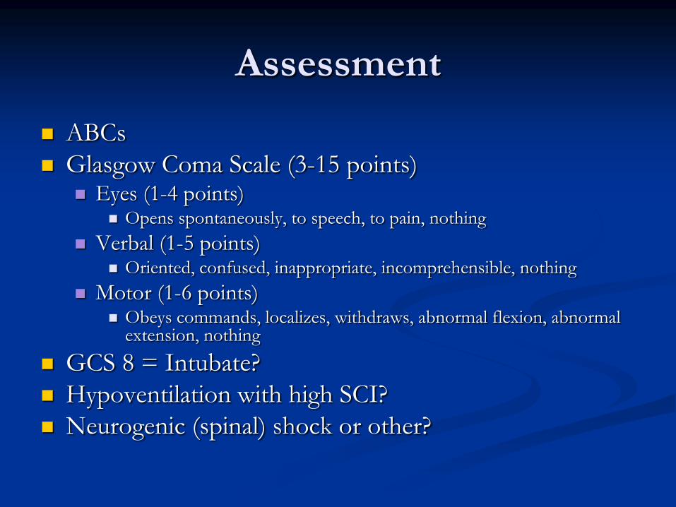
Assessment
ABCs
Glasgow Coma Scale (3-15 points) Eyes (1-4 points)
Opens spontaneously, to speech, to pain, nothing
Verbal (1-5 points) Oriented, confused, inappropriate, incomprehensible, nothing
Motor (1-6 points) Obeys commands, localizes, withdraws, abnormal flexion, abnormal
extension, nothing
GCS 8 = Intubate?
Hypoventilation with high SCI?
Neurogenic (spinal) shock or other?

Immobilization
Cervical Collar
Long Spine Board
In-line traction and log rolling
Do not get fooled by distracting injury

Rapid Initiation of Treatment
Steroids
Moderate Hypothermia
Transport ASAP

To the Emergency Department
Assessment
Diagnosis
Treatment
Consultation

Assessment
Physical exam
ABC‟s
Vitals
GCS
Neurologic Exam
More specific motor (is there a level?)
Is everything working right?
Attention to specific patterns (e.g. central cord, Brown
Sequard, etc.)

Diagnosis
After physical exam, consider imaging
X-ray (Spine)
Is it complete?
CT
MRI

Who needs a CT?
Many factors go into this question
Risk assessment
Reliability (patient)
Support structure
Radiation – It‟s ok to say „no‟.
Obvious cases are obvious.
Low Risk
Asymptomatic
Slight Headache
No LOC
No structural abnormality on exam

Moderate Risk
Altered LOC
Progressive Headache
ETOH / Drugs
Seizure
Amnesia
Vomiting
Secondary or distracting injury
Abuse?

High Risk
Depressed LOC
Obvious exam abnormality
Focal neuro signs
Any penetrating injury

Findings and Treatments
Brain injury
Fracture (skull)
Spinal cord injury
SAH / Aneurysm

Traumatic Brain Injury
Concussion
Epidural Hematoma
Subdural Hematoma
SAH / Axonal Shear

Concussion
Most common
Symptoms may last
Sports injuries are common
“When can I play?”
Multiple opinions.
Asymptomatic x 1wk, no pain with exertion

Epidural Hematoma
True Emergency
Often involves middle meningeal artery
Temporal bone fracture
Rapid accumulation of blood with rapid rise in
ICP
Prompt intervention is key to survival
Burr hole drainage

Epidural Hematoma

Subdural Hematoma
Cortical vessels damaged by shear forces, laceration, or
contusion
Can be complicated by edema secondary to initial injury
Most common over a frontal or parietal area
Consultation
Treatment may vary from observation to surgery
Is atrophy a good thing?
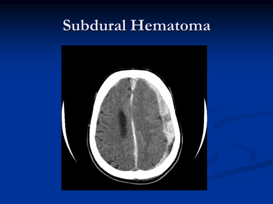
Subdural Hematoma

Traumatic SAH
As much as 60% of people with other serious head
injuries will show some subarachnoid blood
SAH blood increases morbidity and mortality when
seen in moderate and severe brain injuries
Up to 90% of people that present with traumatic SAH
and GCS>12 have good outcomes
Use of supportive care

Diffuse Axonal Shear
Hard to see on a CT scan
MRI more sensitive
Decreased LOC in a relatively benign appearing
CT

General Treatment Guidelines
GCS 8 – intubate
3% of traumatic head injuries have associated spinal cord
injury
Use of in-line cervical stabilization
RSI
Maintaining adequate ICP
Sedation and paralysis (make sure neurosurgeon is involved)
Lidocaine for intubation
Mannitol 1g/kg
Mannitol > hyperventilation (Soustiel)
General Treatment (cont.)
Judicious use of fluids
Use of steroids (later)
Use of moderate hypothermia

Cervical Spine Injuries
Incidence in trauma system entries (Penn and
New Mexico)
Overall incidence of CSI 4.3%
CSI without cord injury 3.0%
Cord injury without fracture 0.7%
Many variations of injury
Thoracic and Lumbar injuries as well

Evaluation of Spinal Injury
Starts pre-hospital with immobilization
Many different criteria
NEXUS
Canadian C-spine Rule
Multiple imaging modalities
Multiple algorithms

National Emergency X-Radiography
Utilization Study (NEXUS)
First mentioned in 1992
Validated again about 10 years later in a study
with over 34,000 patients
Sensitivity to find cervical spine injuries noted to
be 99.6%

NEXUS
Cervical spine radiography is indicated for
patients with trauma unless they meet all of the
following criteria:
No posterior midline cervical spine tenderness
No evidence of intoxication
Normal level of alertness
No focal neurologic deficit
No painful distracting injuries

Spine Injury Diagnostics
Many algorithms include the following
X-ray
How many views?
Flexion / Extension
CT (bones only?)
MRI (cord/ligamentous injury?)
Consultation
Immobilization
What about T and L spines?
Examples (No algorithm is perfect for every patient)

Treatment of Spinal Injury (ED
Perspective)
Immobilization
Surgical Stabilization
Steroids

Steroids
Started in the 90‟s based on results of the
NASCIS II trials
CRASH study 2005 for head injury – no
improvement in mortality
Multiple studies since then show limited
neurologic improvement
Complications such as hyperglycemia and
increased rate of infection

Steroids (cont.)
Still debate on the standard of care
People still mostly giving the steroid over
concern of possibility of improvement and legal
implications.
Not time sensitive for pre-hospital care
30mg/kg IV x1, followed by 5.4 mg/kg IV over
23 hours
Start within 8 hours
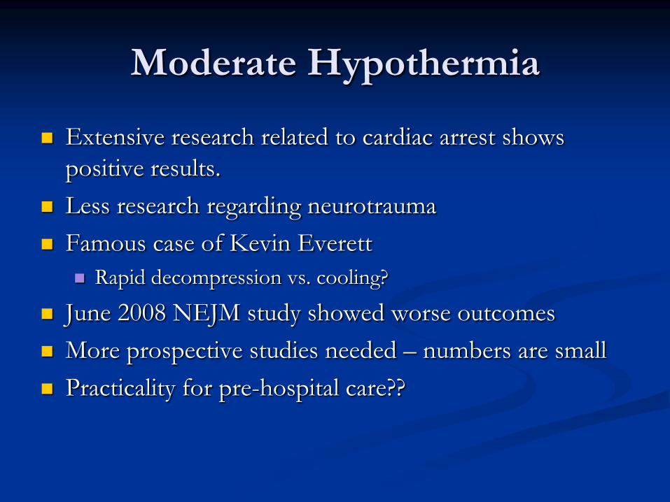
Moderate Hypothermia
Extensive research related to cardiac arrest shows
positive results.
Less research regarding neurotrauma
Famous case of Kevin Everett
Rapid decompression vs. cooling?
June 2008 NEJM study showed worse outcomes
More prospective studies needed – numbers are small
Practicality for pre-hospital care??

Evaluation for SAH
Around 4% of ED visits are for headaches
9 per day in our ED based on this number
1-4% are SAH retrospectively
25-33% of “classic” histories have SAH Small numbers for these studies
Many types, but majority not emergently dangerous
Concerning histories include WHOML, thunderclap HA, syncope.
What needs to be done?

Evaluation for SAH
ED standard is CT/LP
2008 study in Annals of Emergency Medicine states CT alone is inadequate
Sensitivities as low as 93%
Probably closer to 98%
Does the scanner matter?
Another 2008 study in Annals shows that CT + LP gives 100% sensitivity and 67% specificity and is sufficient to rule out SAH

Evaluation for SAH
As time passes, sensitivity for CT decreases.
High 90s to low 90s within 12 hours as blood
dissipates
LP in first 12 hours will show increased red cells
Xanthocromia becomes very important as time
passes, and becomes the most sensitive

Other SAH Questions
Does negative CT/LP need more workup? Probably not. Asymptomatic screening for AVM/aneurysm
is not standard and has associated risk.
Warning headaches? Higher risk for later problems?
Can CT angiography replace LP? Not yet, but probably some day.
What generation is your CT? 4 slice? 64 slice?
Is the neurosurgery literature in agreement with this method????

Conclusions From an ED /
Prehospital Perspective
Neurotrauma patients have life altering / life
threatening injuries
ABC‟s and immobilization are the first step
Rapid diagnosis and consultation are key to
definitive stabilization
Consider time sensitive interventions
Consider the pitfalls of distracting injury and
intoxication


![Transitional Living Programs [clinician brochure] · CLIENT GROUP. TLPs are primarily for people with a brain injury who: –COAST have a moderate to severe brain injury from trauma](https://static.fdocuments.in/doc/165x107/5f610a62a891c260525fe157/transitional-living-programs-clinician-brochure-client-group-tlps-are-primarily.jpg)