Adenomyosis and endometriosis. Re-visiting their association … · 2017-08-25 · Adenomyosis and...
16
GYNECOLOGIC ENDOCRINOLOGY AND REPRODUCTIVE MEDICINE Adenomyosis and endometriosis. Re-visiting their association and further insights into the mechanisms of auto-traumatisation. An MRI study G. Leyendecker • A. Bilgicyildirim • M. Inacker • T. Stalf • P. Huppert • G. Mall • B. Bo ¨ttcher • L. Wildt Received: 22 May 2014 / Accepted: 25 August 2014 / Published online: 21 September 2014 Ó The Author(s) 2014. This article is published with open access at Springerlink.com Abstract Purpose In a series of publications, we had developed the concept that uterine adenomyosis and pelvic endometriosis as well as endometriotic lesions at distant sites of the body share a common pathophysiology with endometriosis constituting a secondary phenomenon. Uterine auto-trau- matization and the initiation of the mechanism of tissue injury and repair (TIAR) were considered the primary events in the disease process. The present MRI study was undertaken (1) to corroborate this concept by re-visiting, in view of discrepant results in the literature, the association of adenomyosis with endometriosis and (2) to extend our views concerning the mechanisms of uterine auto- traumatization. Patients and methods MRI was performed in 143 women attending our center, in whom, on the basis of transvaginal sonography (TVS) and historical data, such as documented endometriosis and dysmenorrhea of various degrees of severity, the presence of uterine adenomyosis was sus- pected. In addition to the measurement of the diameter of junctional zone (JZ) of the anterior and posterior walls in the mid-sagittal plane, the diagnosis of adenomyosis was based on visualization, in that all planes were analyzed with scrutiny. By this method of ‘‘visualization’’ all tran- sient enlargement of the JZ, such as peristaltic waves of the archimyometrium and sporadic neometral contractions that might mimic adenomyotic lesions could be excluded. At the same time, this method allowed to lower the limit of detection in terms of thickness of the JZ for assured diagnosis of adenomyosis. Furthermore, the localizations of the individual lesions, their shapes and patterns were described. Results With the method of ‘visualization’, the diagnosis of uterine adenomyosis could be verified in 127 of the 143 patients studied. The prevalence of endometriosis in ade- nomyosis was 80.6 % and the prevalence of adenomyosis in endometriosis was 91.1 %. As concluded from their localization within the uterine wall, the adenomyotic lesions predominantly developed in the median region of the upper two-thirds of the uterine wall. Cystic cornual angle adenomyosis was a distinct phenomenon that was only observed in patients suffering from extreme primary dysmenorrhea. Aside from this, the majority of the patients complained of primary dysmenorrhea (80 %). On the basis of these findings and the fact that particularly extreme primary dysmenorrhea is associated with high intrauterine pressure, menstrual ‘archimetral compression by neometral contraction’ has to be considered as an important cause of uterine auto-traumatization in addition to uterine peristalsis and hyperperistalsis. Both mechanical functions of the non- pregnant uterus exert their strongest power in the upper G. Leyendecker (&) Á A. Bilgicyildirim Á M. Inacker Á T. Stalf Kinderwunschzentrum (Fertility Center) Darmstadt, Bratustr. 9, 64293 Darmstadt, Germany e-mail: [email protected] P. Huppert Institute of Diagnostic and Interventional Radiology, Academic Teaching Hospital to the Universities of Frankfurt/Main and Heidelberg/Mannheim, Grafenstr. 7, 64283 Darmstadt, Germany G. Mall Institute of Pathology, Klinikum Darmstadt, Academic Teaching Hospital to the Universities of Frankfurt/Main and Heidelberg/ Mannheim, Grafenstr. 7, 64283 Darmstadt, Germany B. Bo ¨ttcher Á L. Wildt Department of Obstetrics and Gynecology, University Clinic of Gynecological Endocrinology and Reproductive Medicine, Medical University Innsbruck, Anichstrasse 35, 6020 Innsbruck, Austria 123 Arch Gynecol Obstet (2015) 291:917–932 DOI 10.1007/s00404-014-3437-8
Transcript of Adenomyosis and endometriosis. Re-visiting their association … · 2017-08-25 · Adenomyosis and...
GYNECOLOGIC ENDOCRINOLOGY AND REPRODUCTIVE MEDICINE
Adenomyosis and endometriosis. Re-visiting their associationand further insights into the mechanisms of auto-traumatisation.An MRI study
G. Leyendecker • A. Bilgicyildirim •
M. Inacker • T. Stalf • P. Huppert •
G. Mall • B. Bottcher • L. Wildt
Received: 22 May 2014 / Accepted: 25 August 2014 / Published online: 21 September 2014
� The Author(s) 2014. This article is published with open access at Springerlink.com
Abstract
Purpose In a series of publications, we had developed the
concept that uterine adenomyosis and pelvic endometriosis
as well as endometriotic lesions at distant sites of the body
share a common pathophysiology with endometriosis
constituting a secondary phenomenon. Uterine auto-trau-
matization and the initiation of the mechanism of tissue
injury and repair (TIAR) were considered the primary
events in the disease process. The present MRI study was
undertaken (1) to corroborate this concept by re-visiting, in
view of discrepant results in the literature, the association
of adenomyosis with endometriosis and (2) to extend our
views concerning the mechanisms of uterine auto-
traumatization.
Patients and methods MRI was performed in 143 women
attending our center, in whom, on the basis of transvaginal
sonography (TVS) and historical data, such as documented
endometriosis and dysmenorrhea of various degrees of
severity, the presence of uterine adenomyosis was sus-
pected. In addition to the measurement of the diameter of
junctional zone (JZ) of the anterior and posterior walls in
the mid-sagittal plane, the diagnosis of adenomyosis was
based on visualization, in that all planes were analyzed
with scrutiny. By this method of ‘‘visualization’’ all tran-
sient enlargement of the JZ, such as peristaltic waves of the
archimyometrium and sporadic neometral contractions that
might mimic adenomyotic lesions could be excluded. At
the same time, this method allowed to lower the limit of
detection in terms of thickness of the JZ for assured
diagnosis of adenomyosis. Furthermore, the localizations
of the individual lesions, their shapes and patterns were
described.
Results With the method of ‘visualization’, the diagnosis
of uterine adenomyosis could be verified in 127 of the 143
patients studied. The prevalence of endometriosis in ade-
nomyosis was 80.6 % and the prevalence of adenomyosis
in endometriosis was 91.1 %. As concluded from their
localization within the uterine wall, the adenomyotic
lesions predominantly developed in the median region of
the upper two-thirds of the uterine wall. Cystic cornual
angle adenomyosis was a distinct phenomenon that was
only observed in patients suffering from extreme primary
dysmenorrhea. Aside from this, the majority of the patients
complained of primary dysmenorrhea (80 %). On the basis
of these findings and the fact that particularly extreme
primary dysmenorrhea is associated with high intrauterine
pressure, menstrual ‘archimetral compression by neometral
contraction’ has to be considered as an important cause of
uterine auto-traumatization in addition to uterine peristalsis
and hyperperistalsis. Both mechanical functions of the non-
pregnant uterus exert their strongest power in the upper
G. Leyendecker (&) � A. Bilgicyildirim � M. Inacker � T. Stalf
Kinderwunschzentrum (Fertility Center) Darmstadt, Bratustr. 9,
64293 Darmstadt, Germany
e-mail: [email protected]
P. Huppert
Institute of Diagnostic and Interventional Radiology, Academic
Teaching Hospital to the Universities of Frankfurt/Main and
Heidelberg/Mannheim, Grafenstr. 7, 64283 Darmstadt, Germany
G. Mall
Institute of Pathology, Klinikum Darmstadt, Academic Teaching
Hospital to the Universities of Frankfurt/Main and Heidelberg/
Mannheim, Grafenstr. 7, 64283 Darmstadt, Germany
B. Bottcher � L. Wildt
Department of Obstetrics and Gynecology, University Clinic of
Gynecological Endocrinology and Reproductive Medicine,
Medical University Innsbruck, Anichstrasse 35, 6020 Innsbruck,
Austria
123
Arch Gynecol Obstet (2015) 291:917–932
DOI 10.1007/s00404-014-3437-8
region of the uterus, which is compatible with the pre-
dominant localization of the adenomyotic lesions.
Conclusions The data confirm our previous results of a
high association of adenomyosis with endometriosis and
vice versa. Our view of the mechanism of uterine auto-
traumatization by mechanical functions of the non-preg-
nant uterus has to be extended, in that ‘archimetral com-
pression by neometral contractions’ could be realized as
the predominant cause of mechanical strain to the non-
pregnant uterus. The data of this study confirm our concept
of the etiology and pathophysiology of adenomyosis and
endometriosis in that the process of chronic proliferation
and inflammation is induced at the level of the archimetra
by chronic uterine auto-traumatization. Furthermore, with
respect to the diagnosis of uterine adenomyosis (and con-
sequently endometriosis) this study shows a high degree of
accordance between the findings in real-time TVS and
MRI.
Keywords Adenomyosis � Endometriosis � Auto-
traumatization � Dysmenorrhea � Pathophysiology � MRI
Introduction
In spite of continuous efforts, the etiology and patho-
physiology of adenomyosis and endometriosis and their
interrelationship are not fully understood. There is
increasing evidence that adenomyosis constitutes an
important factor of infertility. Therefore, the elucidation of
its pathophysiology is of utmost clinical importance. In a
series of reports, we had developed the view that these
lesions are highly associated and are the results of uterine
auto-traumatization [1–6]. The pathophysiological process
was termed ‘‘tissue injury and repair’’ (TIAR) [7–9]. The
present study was conducted to corroborate and extend this
view.
The work of Rokitansky, von Recklinghausen, Freund
and Cullen [10–16] laid the basis of the present under-
standing of adenomyosis as a benign uterine tumor derived
from Mullerian duct tissue [17]. The gross and microscopic
morphology, the expansion beyond the uterine confines, the
occurrence at distant sites in the body and the main clinical
characteristics were described in detail by these authors.
With his basically correct and seminal concept of tubal
dissemination of endometrial tissue into the peritoneal
cavity, Sampson dealt therefore only with a segmental part
of the disease [18]. Furthermore, he did not appreciate
uterine adenomyosis as a significant component of the
pathophysiological process. While it was well established
that uterine adenomyosis resulted from a focal infiltration
and/or broad expansion of Mullerian tissue into the
underlying myometrium with continuity of the endometrial
glandular structures [14, 19], he suggested uterine adeno-
myosis to result from vascular transmission [20]. Although
the view that uterine adenomyosis and endometriosis
constitute a nosological entity was never completely
abandoned [21]; with time and enforced by the introduction
of laparoscopy as a diagnostic procedure, Sampson’s the-
ory led to regard endometriosis mainly as a peritoneal
disease resulting from retrograde menstruation, while
uterine adenomyosis nearly fell into oblivion [8, 22–24].
Until the beginning of this century much scientific effort
was laid on corroborating Sampson’s concept [25]. A re-
consideration of the uterus as a significant determinant in
the disease process was initiated, when it was shown that
the non-pregnant uterus is not a quiescent organ, but rather
actively involved in the early process of reproduction by
mechanical functions that were found to be severely
impaired or dysfunctional in patients with endometriosis
[26–30]. Research efforts using a broad array of methods
resulted in a more precise understanding of the disease
process that led again to the inclusion of uterine adeno-
myosis into the pathophysiology of endometriosis [8].
Continuously improved imaging techniques, such as
transvaginal sonography and MRI, significantly contributed
to this new understanding [31–35]. Of paramount impor-
tance in gaining new insights into the pathophysiology of
endometriosis and adenomyosis was the identification and
detailed description of the endometrial–subendometrial
unit or archimetra [36]. This evolutionarily and ontoge-
netically oldest part of the uterus is physiologically con-
tinuously subjected to mechanical strain during the
reproductive phase of life [7–9].
In studies addressing the prevalence of adenomyosis in
endometriosis and vice versa, discrepant results were
obtained [5, 37]. Since the diagnosis of adenomyosis is
highly dependent upon the diagnostic criteria applied, it
appeared to be necessary to re-evaluate the association of
adenomyosis with endometriosis. Special emphasis was
laid on the signs of early uterine adenomyosis as deter-
mined by MRI.
In our previous studies, we had initially focused on
uterine peristalsis and hyperperistalsis as the main causes
of uterine auto-traumatization within the process of tissue
injury and repair (TIAR) [6–8, 38]. In the meantime,
however, it became evident that, in addition to uterine
peristalsis for directed sperm transport, the contractile
activity of the uterus during menstruation constitutes
another important mechanical function of the non-pregnant
uterus [9]. A considerable number of women experience
these contractions as primary dysmenorrhea of various
strengths, indicating their enormous mechanical power
[39]. Thus, neometral menstrual contractions could con-
tribute to uterine auto-traumatization and, therefore, con-
sidered as an important component of the TIAR process.
918 Arch Gynecol Obstet (2015) 291:917–932
123
In addition, instead of merely studying the association of
adenomyosis with endometriosis, we realized the necessity
to extend and to refine our view of uterine adenomyosis
under the aspect of the preferential localizations of the
lesions within the uterine wall. In relating the predominant
localizations as well as the shapes and patterns of the
lesions to the sites, at which the mechanical functions of
the non-pregnant uterus presumably exerts its strongest
impact on the archimetra, a deeper insight into the mech-
anisms of uterine auto-traumatization was attempted that
would further corroborate our pathophysiological concept
of uterine auto-traumatization.
Patients and methods
In a total of 143 patients aged 23–42 years (mean age
33 years) attending the Fertility Center Darmstadt, MRI
scans were performed on the basis of the following his-
torical data.
Clinical symptoms and findings by transvaginal
sonography (TVS)
Transvaginal sonography
In all 143 patients, transvaginal sonograms were per-
formed and none was considered as normal. The indica-
tion for subsequent MRI on the basis of sonographic
findings were:
enlarged uterus (fibroids excluded);
uterine asymmetry with enlarged uterine walls (fibroids
excluded);
focally destroyed, absent or expanded uterine ‘‘halo’’;
inhomogenous texture of the myometrium;
irregular endometrial–myometrial interface;
cystic structures considered as myometrial endometriotic
cysts.
Endometriosis
In 72 patients aged from 22 to 42 years (mean age
32.3 years), the presence (n = 56, mean age 32.2 years;
range from 22 to 41 years) or absence (n = 16; mean age
32.7 yrs; range from 25 to 42 years) of endometriosis
was documented. The majority of patients presented a
surgical and histological report. Only in one case the
diagnosis of endometriosis was based on visualization
during laparoscopy with high credibility. In two cases,
MRI findings such as infiltration of the urinary bladder
and ovarian endometriomas were indicative of pelvic
endometriosis.
Dysmenorrhea
In 116 patients, the presence or absence of dysmenorrhea
was documented.
Dysmenorrhea was defined as mild with no use, mod-
erate with occasional use and severe with permanent use of
analgetic medication during every menstrual period.
Extreme dysmenorrhea was characterized by absence from
school or work and sometimes collaptic states. Of these
116 patients, 7 had no history of dysmenorrhea at all.
Infertility was not a criterion to perform an MRI scan,
because in some patients that attended our center because
of a history or of symptoms suggestive of endometriosis,
infertility was not proven. Moreover, in most of the
infertile couples low sperm counts of the male partners
were documented.
The patients were recruited mostly by the senior
investigator (G.L.) and also by other members of the
medical team of the fertility center. The senior investigator
did see all the patients at least twice, took their history and
performed all transvaginal ultrasound scans.
Magnetic resonance imaging (MRI)
In all 143 women the uteri were examined by means of
MRI using the same techniques as published before [4, 5].
Slice thickness of t2-weighted images was 3.0 mm with a
gap of 0.3 mm in sagittal orientation and 3 mm with
0.6 mm gap in transversal and coronal orientation. Addi-
tionally, we acquired t2-weighted sagittal-oblique images
(3.0, 0.9 mm gap) and t1-weighted transversal images (5,
1.0 mm gap). To prevent functional alterations of the
image quality regularly, butylscopolamine was adminis-
tered before onset of the scanning. All diameters of the
junctional zone were documented by electronic callipers
and expressed in millimeters and were obtained from the
uteri in a mid- or paramedian sagittal, transverse as well as
in a coronary plane. Maximum diameters of the anterior
and posterior walls, respectively, with respect to partial
volume effect were considered. All quantitative measure-
ments or qualitative (visualized) evidence (presence or
absence of adenomyosis) were obtained separately and
independently by G.L. and a radiologist. In case of dis-
crepant findings, the respective scans were re-analyzed by
G.L. and P.H.
In addition to the measurements of the diameter of the
junctional zone in the posterior and anterior walls of the
uterus, the localizations, shapes and patterns of the lesions
were documented. For this purpose all the planes (sagittal,
coronary and transverse) were analyzed carefully.
An enlargement of the junctional zone of 12 mm or more
was considered as strong evidence of adenomyosis. Below
Arch Gynecol Obstet (2015) 291:917–932 919
123
this thickness, additional findings had to be present to
warrant the diagnosis of adenomyosis such as cystic struc-
tures within the sub-basal myometrium, focal thickening of
the junctional zone that could not be related to functional
alterations such as transient contractions of the neometra or
cyclic peristaltic waves, and irregular and stepwise expan-
sions of the junctional zone [33–35]. This method was
termed diagnosis by visualization and was applied in cases
with a junctional zone diameter below 12 mm. In this study
the term ‘‘visualized adenomyosis’’ is used for the cases that
fulfil either the criteria of visualization or a thickness of the
JZ of C12 mm. Functional alterations that mimicked focal
adenomyosis, such as peristaltic waves and transitory con-
tractions of the neometra, were indentified in that they had
disappeared in scans performed after a lapse of time.
Statistical analysis
Student’s t test was performed whenever appropriate. All
patients gave their written informed consent for use of their
data for scientific evaluation.
Results
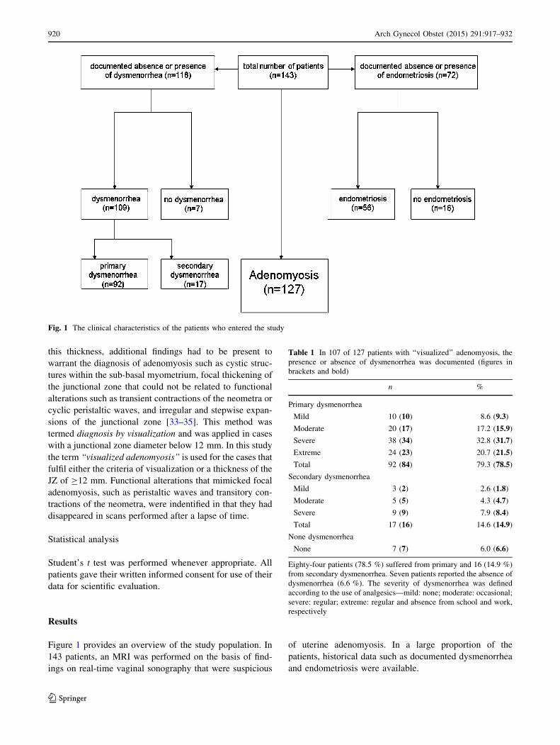
Figure 1 provides an overview of the study population. In
143 patients, an MRI was performed on the basis of find-
ings on real-time vaginal sonography that were suspicious
of uterine adenomyosis. In a large proportion of the
patients, historical data such as documented dysmenorrhea
and endometriosis were available.
Fig. 1 The clinical characteristics of the patients who entered the study
Table 1 In 107 of 127 patients with ‘‘visualized’’ adenomyosis, the
presence or absence of dysmenorrhea was documented (figures in
brackets and bold)
n %
Primary dysmenorrhea
Mild 10 (10) 8.6 (9.3)
Moderate 20 (17) 17.2 (15.9)
Severe 38 (34) 32.8 (31.7)
Extreme 24 (23) 20.7 (21.5)
Total 92 (84) 79.3 (78.5)
Secondary dysmenorrhea
Mild 3 (2) 2.6 (1.8)
Moderate 5 (5) 4.3 (4.7)
Severe 9 (9) 7.9 (8.4)
Total 17 (16) 14.6 (14.9)
None dysmenorrhea
None 7 (7) 6.0 (6.6)
Eighty-four patients (78.5 %) suffered from primary and 16 (14.9 %)
from secondary dysmenorrhea. Seven patients reported the absence of
dysmenorrhea (6.6 %). The severity of dysmenorrhea was defined
according to the use of analgesics—mild: none; moderate: occasional;
severe: regular; extreme: regular and absence from school and work,
respectively
920 Arch Gynecol Obstet (2015) 291:917–932
123
Dysmenorrhea
In 107 out of 127 patients with ‘‘visualized’’ adenomyosis,
absence or presence of dysmenorrhea could be documented.
Primary dysmenorrhea (n = 84; 78.5 %) was predominant
over secondary dysmenorrhea (n = 14; 14.7 %) and within
both groups severe dysmenorrhea prevailed. The extreme
form (n = 23; 21.5 %) was only documented in women with
primary dysmenorrhea. Only seven patients (6 %) were
completely free of menstrual pain (Table 1).
Endometriosis
In 72 of the 143 patients, the absence or presence of endo-
metriosis was documented. In 17 patients no endometriosis
was found by laparoscopy. In 56 patients, endometriosis was
documented (Fig. 1). In this study, unlike in a previous study
[5], the stages of the disease were not specifically docu-
mented. In the meantime, we had come to the conclusion that
the fragments of basal endometrium, following their tran-
stubal dissemination, might have their own fate.
Characterization of adenomyosis
In the study group (n = 143), the diameters of the junc-
tional zone increased with age and showed a trend of being
larger in the posterior than in the anterior wall (Table 2).
In 98 % of the 143 patients, the diameter of the junc-
tional zone was larger than 6 mm. This percentage
declined gradually with the stepwise increase in millime-
ters of the arbitrary detection limit and fell to 62 % when
the diagnosis of adenomyosis was defined to be established
at a thickness of the junctional zone of at least 12 mm. In
30 % of the patients, the diameter of the junctional zone
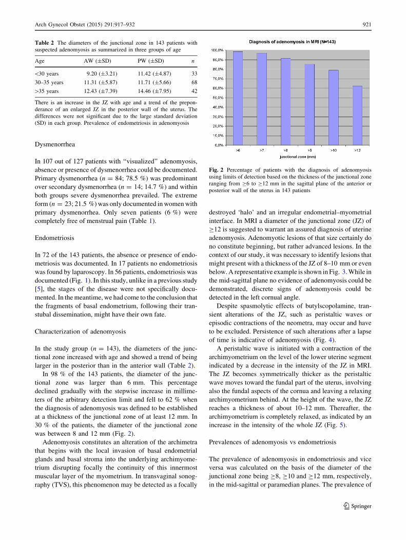
was between 8 and 12 mm (Fig. 2).
Adenomyosis constitutes an alteration of the archimetra
that begins with the local invasion of basal endometrial
glands and basal stroma into the underlying archimyome-
trium disrupting focally the continuity of this innermost
muscular layer of the myometrium. In transvaginal sonog-
raphy (TVS), this phenomenon may be detected as a focally
destroyed ‘halo’ and an irregular endometrial–myometrial
interface. In MRI a diameter of the junctional zone (JZ) of
C12 is suggested to warrant an assured diagnosis of uterine
adenomyosis. Adenomyotic lesions of that size certainly do
no constitute beginning, but rather advanced lesions. In the
context of our study, it was necessary to identify lesions that
might present with a thickness of the JZ of 8–10 mm or even
below. A representative example is shown in Fig. 3. While in
the mid-sagittal plane no evidence of adenomyosis could be
demonstrated, discrete signs of adenomyosis could be
detected in the left cornual angle.
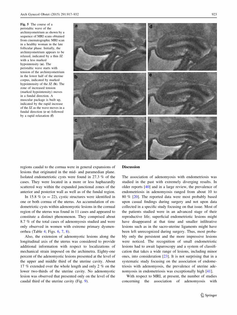
Despite spasmolytic effects of butylscopolamine, tran-
sient alterations of the JZ, such as peristaltic waves or
episodic contractions of the neometra, may occur and have
to be excluded. Persistence of such alterations after a lapse
of time is indicative of adenomyosis (Fig. 4).
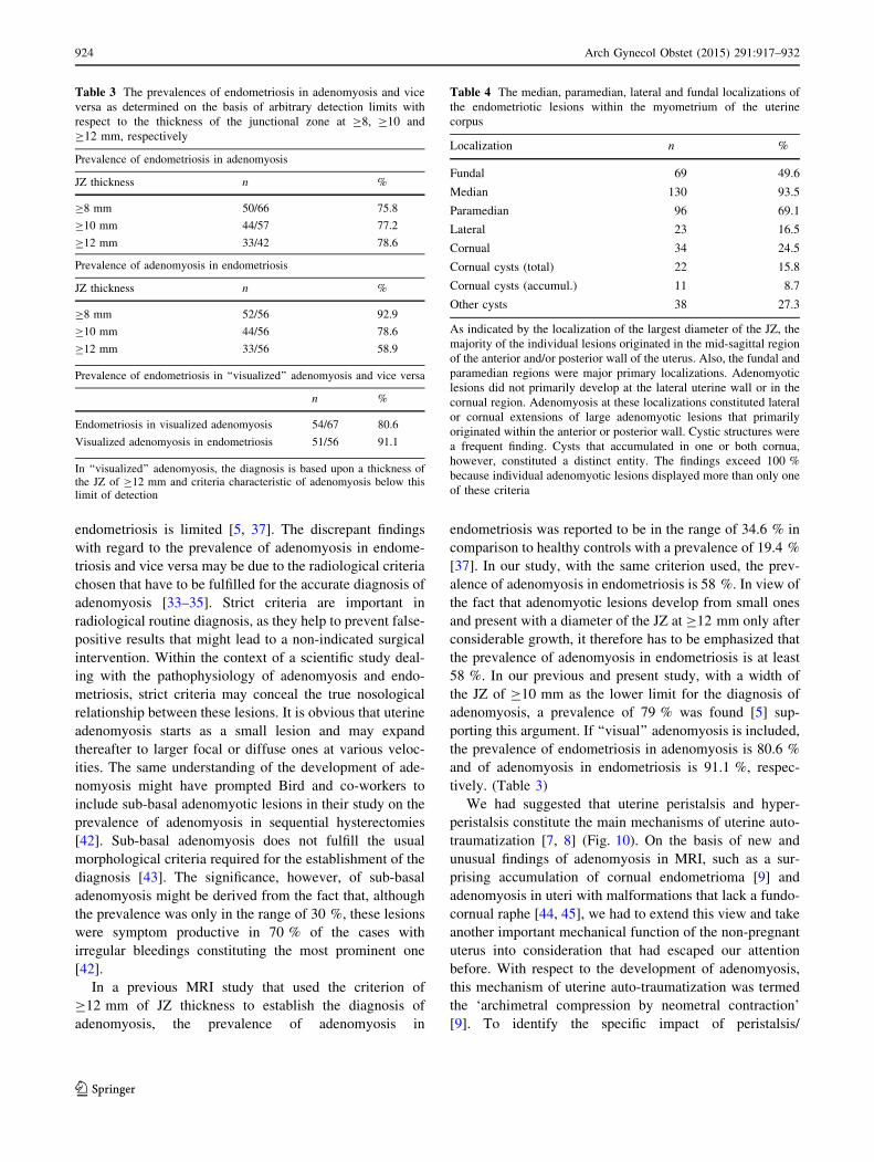
A peristaltic wave is initiated with a contraction of the
archimyometrium on the level of the lower uterine segment
indicated by a decrease in the intensity of the JZ in MRI.
The JZ becomes symmetrically thicker as the peristaltic
wave moves toward the fundal part of the uterus, involving
also the fundal aspects of the cornua and leaving a relaxing
archimyometrium behind. At the height of the wave, the JZ
reaches a thickness of about 10–12 mm. Thereafter, the
archimyometrium is completely relaxed, as indicated by an
increase in the intensity of the whole JZ (Fig. 5).
Prevalences of adenomyosis vs endometriosis
The prevalence of adenomyosis in endometriosis and vice
versa was calculated on the basis of the diameter of the
junctional zone being C8, C10 and C12 mm, respectively,
in the mid-sagittal or paramedian planes. The prevalence of
Fig. 2 Percentage of patients with the diagnosis of adenomyosis
using limits of detection based on the thickness of the junctional zone
ranging from C6 to C12 mm in the sagittal plane of the anterior or
posterior wall of the uterus in 143 patients
Table 2 The diameters of the junctional zone in 143 patients with
suspected adenomyosis as summarized in three groups of age
Age AW (±SD) PW (±SD) n
\30 years 9.20 (±3.21) 11.42 (±4.87) 33
30–35 years 11.31 (±5.87) 11.71 (±5.66) 68
[35 years 12.43 (±7.39) 14.46 (±7.95) 42
There is an increase in the JZ with age and a trend of the prepon-
derance of an enlarged JZ in the posterior wall of the uterus. The
differences were not significant due to the large standard deviation
(SD) in each group. Prevalence of endometriosis in adenomyosis
Arch Gynecol Obstet (2015) 291:917–932 921
123
endometriosi in adenomyosis (n = 72) increased from 75.5
to 77 % and 78 %, respectively, and the prevalence of
adenomyosis in endometriosis (n = 56) declined from
92.5 % to 79 % and 59 %, respectively, with the limits of
diagnosis of adenomyosis being set at C8, C10 and
C12 mm of junctional zone width, respectively (Table 3).
Since criteria suggestive of the diagnosis of adenomy-
osis do exist in addition to the diameters of the junctional
zone of the anterior and/or posterior wall as demonstrated
in sagittal and/or coronary planes, respectively, a meticu-
lous analysis of the MRI scans was performed in all three
planes, the sagittal, coronary and transverse one,
respectively.
Using this method of ‘visualization’ as a diagnostic tool
in addition of the junctional zone diameter 127 (89 %) of
the 143 patients studied fulfilled the diagnostic criteria of
presenting with adenomyosis. In this group of patients the
presence or absence of endometriosis was documented in
67 patients. Fifty-four patients presented with and 13
without endometriosis. Thus, the prevalence of endome-
triosis (n = 54) in visualized adenomyosis (n = 67) was
80.6 %. In the 56 total cases of documented endometriosis,
51 patients presented with adenomyosis determined by the
method of ‘visualization’. Thus, the prevalence of visual-
ized adenomyosis in endometriosis was 91.1 % (Table 3).
Location, shape and patterns of the adenomyotic lesions
To obtain a better understanding of the possible modes of
auto-traumatization, the localizations, shapes and patterns
of the individual adenomyotic lesions were analyzed by
reviewing the sagittal, coronary and transverse planes,
respectively, in each patient. The data are summarized in
Table 4. Nearly all of the lesions were localized in the
longitudinal midline of the uterus with extensions into the
paramedian planes and to the fundal myometrium. Lateral
extensions of the lesions into the cornual and lateral
Fig. 3 MRI scans of a 25 years
old patient with extreme
primary dysmenorrhea as an
example for the establishment
of the diagnosis of adenomyosis
by ‘visualisation’. The diameter
of the JZ in the mid-sagittal
plane would not allow for the
diagnosis of adenomyosis (a). A
closer analysis of an
enlargement of the JZ in the left
cornual angle (b and c) revealed
beginning cystic cornual angle
adenomyosis (d) (arrow)
Fig. 4 Adenomyosis in a 32-year-old woman without dysmenorrhea.
In the sagittal scan the hypointense area could result from an episodic
neometral contraction (top). The coronal scan performed after a short
lapse of time, however, reveals adenomyosis with signs of spread of
the lesion in various directions (bottom)
922 Arch Gynecol Obstet (2015) 291:917–932
123
regions caudal to the cornua were in general expansions of
lesions that originated in the mid- and paramedian plane.
Isolated endometriotic cysts were found in 27.3 % of the
cases. They were located in a more or less haphazardly
scattered way within the expanded junctional zones of the
anterior and posterior wall as well as of the fundal region.
In 15.8 % (n = 22), cystic structures were identified in
one or both cornua of the uterus. An accumulation of en-
dometriotic cysts within adenomyotic lesions in the cornual
region of the uterus was found in 11 cases and appeared to
constitute a distinct phenomenon. They comprised about
8.7 % of the total cases of adenomyosis studied and were
only observed in women with extreme primary dysmen-
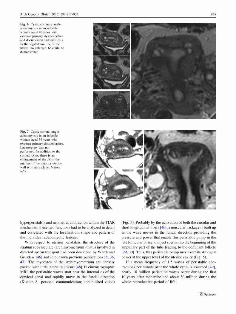
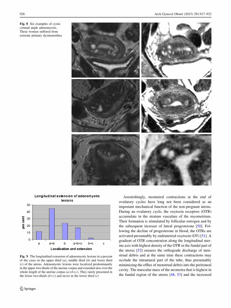
orrhea (Table 4; Figs. 6, 7, 8).
Also, the extension of adenomyotic lesions along the
longitudinal axis of the uterus was considered to provide
additional information with respect to localizations of
mechanical strain imposed on the archimetra. Eighty-one
percent of the adenomyotic lesions presented at the level of
the upper and middle third of the uterine cavity. About
17 % extended over the whole length and only 2 % on the
lower two-thirds of the uterine cavity. No adenomyotic
lesion was observed that presented only on the level of the
caudal third of the uterine cavity (Fig. 9).
Discussion
The association of adenomyosis with endometriosis was
studied in the past with extremely diverging results. In
older reports [40] and in a large review, the prevalence of
endometriosis in adenomyosis ranged from about 10 to
80 % [20]. The reported data were most probably based
upon casual findings during surgery and not upon data
collected in a specific study focusing on that issue. Most of
the patients studied were in an advanced stage of their
reproductive life; superficial endometriotic lesions might
have disappeared at that time and smaller infiltrative
lesions such as in the sacro-uterine ligaments might have
been left unrecognized during surgery. Thus, most proba-
bly only the persistent and the more impressive lesions
were noticed. The recognition of small endometriotic
lesions had to await laparoscopy and a system of classifi-
cation that takes a wide range of lesions, including minor
ones, into consideration [23]. It is not surprising that in a
systematic study focusing on the association of endome-
triosis with adenomyosis, the prevalence of uterine ade-
nomyosis in endometriosis was exceptionally high [41].
With respect to MRI, at present, the number of studies
concerning the association of adenomyosis with
Fig. 5 The course of a
peristaltic wave of the
archimyometrium as shown by a
sequence of MRI scans obtained
from cinematographic MRI scan
in a healthy woman in the late
follicular phase. Initially, the
archimyometrium appears to be
relaxed, indicated by a thin JZ
with a less marked
hypointensity (a). The
peristaltic wave starts with
tension of the archimyometrium
in the lower half of the uterine
corpus, indicated by marked
hypointensity of the JZ (b). The
zone of increased tension
(marked hypointensity) moves
in a fundal direction. A
muscular package is built up,
indicated by the rapid increase
of the JZ as the wave moves in a
fundal direction (c–e) followed
by a rapid relaxation (f)
Arch Gynecol Obstet (2015) 291:917–932 923
123
endometriosis is limited [5, 37]. The discrepant findings
with regard to the prevalence of adenomyosis in endome-
triosis and vice versa may be due to the radiological criteria
chosen that have to be fulfilled for the accurate diagnosis of
adenomyosis [33–35]. Strict criteria are important in
radiological routine diagnosis, as they help to prevent false-
positive results that might lead to a non-indicated surgical
intervention. Within the context of a scientific study deal-
ing with the pathophysiology of adenomyosis and endo-
metriosis, strict criteria may conceal the true nosological
relationship between these lesions. It is obvious that uterine
adenomyosis starts as a small lesion and may expand
thereafter to larger focal or diffuse ones at various veloc-
ities. The same understanding of the development of ade-
nomyosis might have prompted Bird and co-workers to
include sub-basal adenomyotic lesions in their study on the
prevalence of adenomyosis in sequential hysterectomies
[42]. Sub-basal adenomyosis does not fulfill the usual
morphological criteria required for the establishment of the
diagnosis [43]. The significance, however, of sub-basal
adenomyosis might be derived from the fact that, although
the prevalence was only in the range of 30 %, these lesions
were symptom productive in 70 % of the cases with
irregular bleedings constituting the most prominent one
[42].
In a previous MRI study that used the criterion of
C12 mm of JZ thickness to establish the diagnosis of
adenomyosis, the prevalence of adenomyosis in
endometriosis was reported to be in the range of 34.6 % in
comparison to healthy controls with a prevalence of 19.4 %
[37]. In our study, with the same criterion used, the prev-
alence of adenomyosis in endometriosis is 58 %. In view of
the fact that adenomyotic lesions develop from small ones
and present with a diameter of the JZ at C12 mm only after
considerable growth, it therefore has to be emphasized that
the prevalence of adenomyosis in endometriosis is at least
58 %. In our previous and present study, with a width of
the JZ of C10 mm as the lower limit for the diagnosis of
adenomyosis, a prevalence of 79 % was found [5] sup-
porting this argument. If ‘‘visual’’ adenomyosis is included,
the prevalence of endometriosis in adenomyosis is 80.6 %
and of adenomyosis in endometriosis is 91.1 %, respec-
tively. (Table 3)
We had suggested that uterine peristalsis and hyper-
peristalsis constitute the main mechanisms of uterine auto-
traumatization [7, 8] (Fig. 10). On the basis of new and
unusual findings of adenomyosis in MRI, such as a sur-
prising accumulation of cornual endometrioma [9] and
adenomyosis in uteri with malformations that lack a fundo-
cornual raphe [44, 45], we had to extend this view and take
another important mechanical function of the non-pregnant
uterus into consideration that had escaped our attention
before. With respect to the development of adenomyosis,
this mechanism of uterine auto-traumatization was termed
the ‘archimetral compression by neometral contraction’
[9]. To identify the specific impact of peristalsis/
Table 3 The prevalences of endometriosis in adenomyosis and vice
versa as determined on the basis of arbitrary detection limits with
respect to the thickness of the junctional zone at C8, C10 and
C12 mm, respectively
Prevalence of endometriosis in adenomyosis
JZ thickness n %
C8 mm 50/66 75.8
C10 mm 44/57 77.2
C12 mm 33/42 78.6
Prevalence of adenomyosis in endometriosis
JZ thickness n %
C8 mm 52/56 92.9
C10 mm 44/56 78.6
C12 mm 33/56 58.9
Prevalence of endometriosis in ‘‘visualized’’ adenomyosis and vice versa
n %
Endometriosis in visualized adenomyosis 54/67 80.6
Visualized adenomyosis in endometriosis 51/56 91.1
In ‘‘visualized’’ adenomyosis, the diagnosis is based upon a thickness ofthe JZ of C12 mm and criteria characteristic of adenomyosis below thislimit of detection
Table 4 The median, paramedian, lateral and fundal localizations of
the endometriotic lesions within the myometrium of the uterine
corpus
Localization n %
Fundal 69 49.6
Median 130 93.5
Paramedian 96 69.1
Lateral 23 16.5
Cornual 34 24.5
Cornual cysts (total) 22 15.8
Cornual cysts (accumul.) 11 8.7
Other cysts 38 27.3
As indicated by the localization of the largest diameter of the JZ, the
majority of the individual lesions originated in the mid-sagittal region
of the anterior and/or posterior wall of the uterus. Also, the fundal and
paramedian regions were major primary localizations. Adenomyotic
lesions did not primarily develop at the lateral uterine wall or in the
cornual region. Adenomyosis at these localizations constituted lateral
or cornual extensions of large adenomyotic lesions that primarily
originated within the anterior or posterior wall. Cystic structures were
a frequent finding. Cysts that accumulated in one or both cornua,
however, constituted a distinct entity. The findings exceed 100 %
because individual adenomyotic lesions displayed more than only one
of these criteria
924 Arch Gynecol Obstet (2015) 291:917–932
123
hyperperistalsis and neometral contraction within the TIAR
mechanism these two functions had to be analyzed in detail
and correlated with the localization, shape and pattern of
the individual adenomyotic lesions.
With respect to uterine peristalsis, the structure of the
stratum subvasculare (archimyometrium) that is involved in
directed sperm transport had been described by Werth and
Grusdew [46] and in our own previous publications [8, 36,
47]. The myocytes of the archimyometrium are densely
packed with little interstitial tissue [48]. In cinematographic
MRI, the peristaltic waves start near the internal os of the
cervical canal and rapidly move in the fundal direction
(Kissler, S., personal communication; unpublished video)
(Fig. 5). Probably by the activation of both the circular and
short longitudinal fibers [46], a muscular package is built up
as the wave moves in the fundal direction providing the
pressure and power that enable this peristaltic pump in the
late follicular phase to inject sperm into the beginning of the
ampullary part of the tube leading to the dominant follicle
[29, 30]. Thus, this peristaltic pump may exert its strongest
power at the upper level of the uterine cavity (Fig. 5).
If a mean frequency of 1.5 waves of peristaltic con-
tractions per minute over the whole cycle is assumed [49],
nearly 10 million peristaltic waves occur during the first
10 years after menarche and about 30 million during the
whole reproductive period of life.
Fig. 6 Cystic coronary angle
adenomyosis in an infertile
woman aged 40 years with
extreme primary dysmenorrhea
and documented endometriosis.
In the sagittal midline of the
uterus, no enlarged JZ could be
demonstrated
Fig. 7 Cystic cornual angle
adenomyosis in an infertile
woman aged 39 years with
extreme primary dysmenorrhea.
Laparoscopy was not
performed. In addition to the
cornual cysts, there is an
enlargement of the JZ in the
midline of the anterior uterine
wall (coronary plane; bottom
left)
Arch Gynecol Obstet (2015) 291:917–932 925
123
Astonishingly, neometral contractions at the end of
ovulatory cycles have long not been considered as an
important mechanical function of the non-pregnant uterus.
During an ovulatory cycle, the oxytocin receptors (OTR)
accumulate in the stratum vasculare of the myometrium.
Their formation is stimulated by follicular estrogen and by
the subsequent increase of luteal progesterone [50]. Fol-
lowing the decline of progesterone in blood, the OTRs are
activated presumably by endometrial oxytocin (OT) [51]. A
gradient of OTR concentration along the longitudinal uter-
ine axis with highest density of the OTR in the fundal part of
the uterus [52] ensures the orthograde discharge of men-
strual debris and at the same time these contractions may
occlude the intramural part of the tube, thus presumably
minimizing the efflux of menstrual debris into the peritoneal
cavity. The muscular mass of the neometra that is highest in
the fundal region of the uterus [48, 53] and the increased
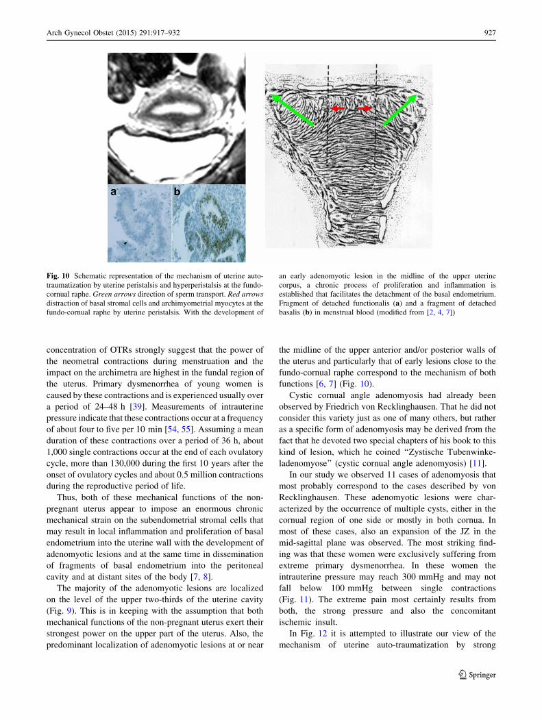
Fig. 8 Six examples of cystic
cornual angle adenomyosis.
These women suffered from
extreme primary dysmenorrhea
Fig. 9 The longitudinal extension of adenomyotic lesions in a percent
of the cases in the upper third (a), middle third (b) and lower third
(c) of the uterus. Adenomyotic lesions were localized predominantly
in the upper two-thirds of the uterine corpus and extended also over the
whole length of the uterine corpus (a?b?c). They rarely presented in
the lower two-thirds (b?c) and never in the lower third (c)
926 Arch Gynecol Obstet (2015) 291:917–932
123
concentration of OTRs strongly suggest that the power of
the neometral contractions during menstruation and the
impact on the archimetra are highest in the fundal region of
the uterus. Primary dysmenorrhea of young women is
caused by these contractions and is experienced usually over
a period of 24–48 h [39]. Measurements of intrauterine
pressure indicate that these contractions occur at a frequency
of about four to five per 10 min [54, 55]. Assuming a mean
duration of these contractions over a period of 36 h, about
1,000 single contractions occur at the end of each ovulatory
cycle, more than 130,000 during the first 10 years after the
onset of ovulatory cycles and about 0.5 million contractions
during the reproductive period of life.
Thus, both of these mechanical functions of the non-
pregnant uterus appear to impose an enormous chronic
mechanical strain on the subendometrial stromal cells that
may result in local inflammation and proliferation of basal
endometrium into the uterine wall with the development of
adenomyotic lesions and at the same time in dissemination
of fragments of basal endometrium into the peritoneal
cavity and at distant sites of the body [7, 8].
The majority of the adenomyotic lesions are localized
on the level of the upper two-thirds of the uterine cavity
(Fig. 9). This is in keeping with the assumption that both
mechanical functions of the non-pregnant uterus exert their
strongest power on the upper part of the uterus. Also, the
predominant localization of adenomyotic lesions at or near
the midline of the upper anterior and/or posterior walls of
the uterus and particularly that of early lesions close to the
fundo-cornual raphe correspond to the mechanism of both
functions [6, 7] (Fig. 10).
Cystic cornual angle adenomyosis had already been
observed by Friedrich von Recklinghausen. That he did not
consider this variety just as one of many others, but rather
as a specific form of adenomyosis may be derived from the
fact that he devoted two special chapters of his book to this
kind of lesion, which he coined ‘‘Zystische Tubenwinke-
ladenomyose’’ (cystic cornual angle adenomyosis) [11].
In our study we observed 11 cases of adenomyosis that
most probably correspond to the cases described by von
Recklinghausen. These adenomyotic lesions were char-
acterized by the occurrence of multiple cysts, either in the
cornual region of one side or mostly in both cornua. In
most of these cases, also an expansion of the JZ in the
mid-sagittal plane was observed. The most striking find-
ing was that these women were exclusively suffering from
extreme primary dysmenorrhea. In these women the
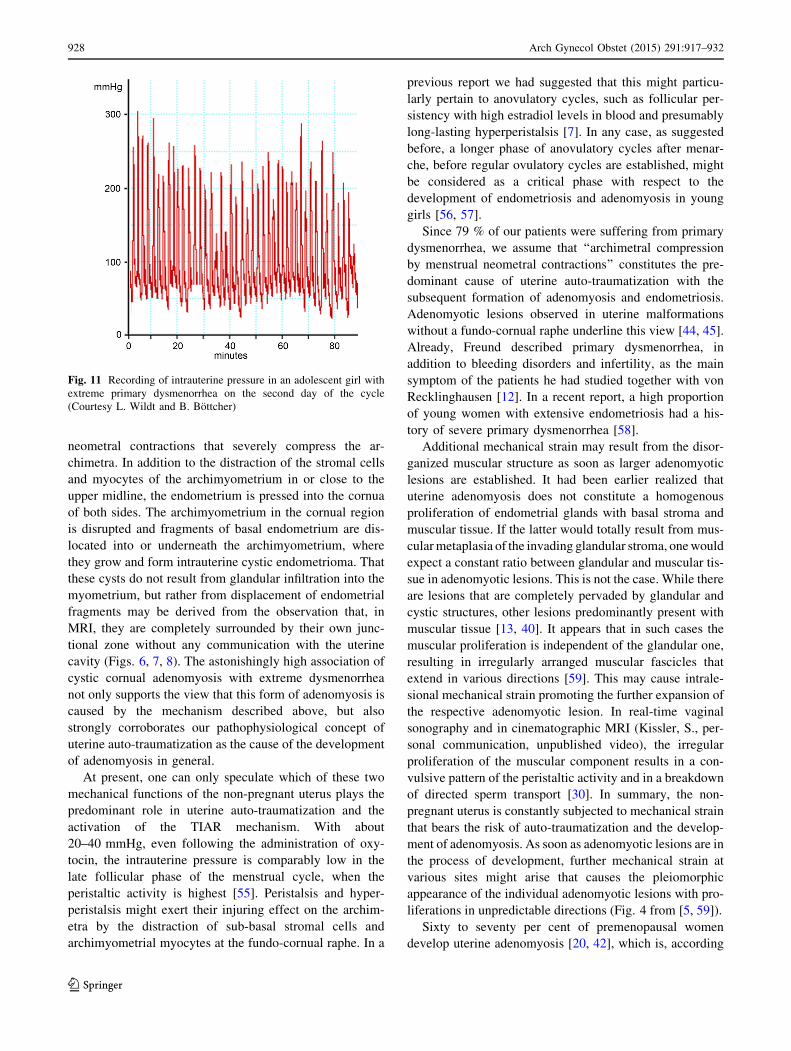
intrauterine pressure may reach 300 mmHg and may not
fall below 100 mmHg between single contractions
(Fig. 11). The extreme pain most certainly results from
both, the strong pressure and also the concomitant
ischemic insult.
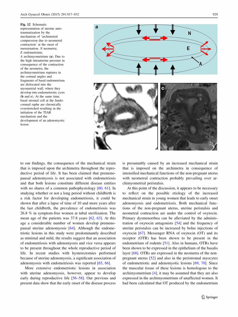
In Fig. 12 it is attempted to illustrate our view of the
mechanism of uterine auto-traumatization by strong
Fig. 10 Schematic representation of the mechanism of uterine auto-
traumatization by uterine peristalsis and hyperperistalsis at the fundo-
cornual raphe. Green arrows direction of sperm transport. Red arrows
distraction of basal stromal cells and archimyometrial myocytes at the
fundo-cornual raphe by uterine peristalsis. With the development of
an early adenomyotic lesion in the midline of the upper uterine
corpus, a chronic process of proliferation and inflammation is
established that facilitates the detachment of the basal endometrium.
Fragment of detached functionalis (a) and a fragment of detached
basalis (b) in menstrual blood (modified from [2, 4, 7])
Arch Gynecol Obstet (2015) 291:917–932 927
123
neometral contractions that severely compress the ar-
chimetra. In addition to the distraction of the stromal cells
and myocytes of the archimyometrium in or close to the
upper midline, the endometrium is pressed into the cornua
of both sides. The archimyometrium in the cornual region
is disrupted and fragments of basal endometrium are dis-
located into or underneath the archimyometrium, where
they grow and form intrauterine cystic endometrioma. That
these cysts do not result from glandular infiltration into the
myometrium, but rather from displacement of endometrial
fragments may be derived from the observation that, in
MRI, they are completely surrounded by their own junc-
tional zone without any communication with the uterine
cavity (Figs. 6, 7, 8). The astonishingly high association of
cystic cornual adenomyosis with extreme dysmenorrhea
not only supports the view that this form of adenomyosis is
caused by the mechanism described above, but also
strongly corroborates our pathophysiological concept of
uterine auto-traumatization as the cause of the development
of adenomyosis in general.
At present, one can only speculate which of these two
mechanical functions of the non-pregnant uterus plays the
predominant role in uterine auto-traumatization and the
activation of the TIAR mechanism. With about
20–40 mmHg, even following the administration of oxy-
tocin, the intrauterine pressure is comparably low in the
late follicular phase of the menstrual cycle, when the
peristaltic activity is highest [55]. Peristalsis and hyper-
peristalsis might exert their injuring effect on the archim-
etra by the distraction of sub-basal stromal cells and
archimyometrial myocytes at the fundo-cornual raphe. In a
previous report we had suggested that this might particu-
larly pertain to anovulatory cycles, such as follicular per-
sistency with high estradiol levels in blood and presumably
long-lasting hyperperistalsis [7]. In any case, as suggested
before, a longer phase of anovulatory cycles after menar-
che, before regular ovulatory cycles are established, might
be considered as a critical phase with respect to the
development of endometriosis and adenomyosis in young
girls [56, 57].
Since 79 % of our patients were suffering from primary
dysmenorrhea, we assume that ‘‘archimetral compression
by menstrual neometral contractions’’ constitutes the pre-
dominant cause of uterine auto-traumatization with the
subsequent formation of adenomyosis and endometriosis.
Adenomyotic lesions observed in uterine malformations
without a fundo-cornual raphe underline this view [44, 45].
Already, Freund described primary dysmenorrhea, in
addition to bleeding disorders and infertility, as the main
symptom of the patients he had studied together with von
Recklinghausen [12]. In a recent report, a high proportion
of young women with extensive endometriosis had a his-
tory of severe primary dysmenorrhea [58].
Additional mechanical strain may result from the disor-
ganized muscular structure as soon as larger adenomyotic
lesions are established. It had been earlier realized that
uterine adenomyosis does not constitute a homogenous
proliferation of endometrial glands with basal stroma and
muscular tissue. If the latter would totally result from mus-
cular metaplasia of the invading glandular stroma, one would
expect a constant ratio between glandular and muscular tis-
sue in adenomyotic lesions. This is not the case. While there
are lesions that are completely pervaded by glandular and
cystic structures, other lesions predominantly present with
muscular tissue [13, 40]. It appears that in such cases the
muscular proliferation is independent of the glandular one,
resulting in irregularly arranged muscular fascicles that
extend in various directions [59]. This may cause intrale-
sional mechanical strain promoting the further expansion of
the respective adenomyotic lesion. In real-time vaginal
sonography and in cinematographic MRI (Kissler, S., per-
sonal communication, unpublished video), the irregular
proliferation of the muscular component results in a con-
vulsive pattern of the peristaltic activity and in a breakdown
of directed sperm transport [30]. In summary, the non-
pregnant uterus is constantly subjected to mechanical strain
that bears the risk of auto-traumatization and the develop-
ment of adenomyosis. As soon as adenomyotic lesions are in
the process of development, further mechanical strain at
various sites might arise that causes the pleiomorphic
appearance of the individual adenomyotic lesions with pro-
liferations in unpredictable directions (Fig. 4 from [5, 59]).
Sixty to seventy per cent of premenopausal women
develop uterine adenomyosis [20, 42], which is, according
Fig. 11 Recording of intrauterine pressure in an adolescent girl with
extreme primary dysmenorrhea on the second day of the cycle
(Courtesy L. Wildt and B. Bottcher)
928 Arch Gynecol Obstet (2015) 291:917–932
123
to our findings, the consequence of the mechanical strain
that is imposed upon the archimetra throughout the repro-
ductive period of life. It has been claimed that premeno-
pausal adenomyosis is not associated with endometriosis
and that both lesions constitute different disease entities
with no shares of a common pathophysiology [60, 61]. In
studying whether or not a long period without childbirth is
a risk factor for developing endometriosis, it could be
shown that after a lapse of time of 10 and more years after
the last childbirth, the prevalence of endometriosis was
26.8 % in symptom-free women at tubal sterilization. The
mean age of the patients was 37.8 years [62, 63]. At this
age a considerable number of women develop premeno-
pausal uterine adenomyosis [64]. Although the endome-
triotic lesions in this study were predominantly described
as minimal and mild, the results suggest that an association
of endometriosis with adenomyosis and vice versa appears
to be present throughout the whole reproductive period of
life. In recent studies with hysterectomies performed
because of uterine adenomyosis, a significant association of
adenomyosis with endometriosis was reported [65, 66].
More extensive endometriotic lesions in association
with uterine adenomyosis, however, appear to develop
early during reproductive life [56–58]. Our previous and
present data show that the early onset of the disease process
is presumably caused by an increased mechanical strain
that is imposed on the archimetra in consequence of
intensified mechanical functions of the non-pregnant uterus
with neometral contraction probably prevailing over ar-
chimyometrial peristalsis.
At this point of the discussion, it appears to be necessary
to reflect on the possible etiology of the increased
mechanical strain in young women that leads to early onset
adenomyosis and endometriosis. Both mechanical func-
tions of the non-pregnant uterus, uterine peristalsis and
neometral contraction are under the control of oxytocin.
Primary dysmenorrhea can be alleviated by the adminis-
tration of oxytocin antagonists [54] and the frequency of
uterine peristalsis can be increased by bolus injections of
oxytocin [67]. Messenger RNA of oxytocin (OT) and its
receptor (OTR) has been shown to be present in the
endometrium of rodents [51]. Also in humans, OTRs have
been shown to be expressed in the epithelium of the basalis
layer [68]. OTRs are expressed in the neometra of the non-
pregnant uterus [52] and also in the peristromal myocytes
of endometriotic and adenomyotic lesions [69, 70]. Since
the muscular tissue of these lesions is homologous to the
archimyometrium [4], it may be assumed that they are also
expressed in the archimyometrium of unaffected women. It
had been calculated that OT produced by the endometrium
Fig. 12 Schematic
representation of uterine auto-
traumatization by the
mechanism of ‘archimetral
compression due to neometral
contraction’ at the onset of
menstruation. N neometra;
E endometrium;
A archimyometrium (a). Due to
the high intrauterine pressure in
consequence of the contraction
of the neometra, the
archimyometrium ruptures in
the cornual angles and
fragments of basal endometrium
are dislocated into the
myometrial wall, where they
develop into endometriotic cysts
(b and c). At the same time,
basal stromal cell at the fundo-
cornual raphe are chronically
overstretched resulting in the
initiation of the TIAR
mechanism and the
development of an adenomyotic
lesion
Arch Gynecol Obstet (2015) 291:917–932 929
123
by far exceeds the secretion of pituitary OT and it was
therefore proposed, based on the data obtained in rodents,
that the OT of endometrial origin and/or the products of an
endometrial autocrine/intracrine OT/OTR interaction such
as PGF2a control the contractile functions of the uterus
[51]. In this respect, it is of interest to note that in women
with endometriosis and adenomyosis, the layer of basal
endometrium distant from adenomyotic lesions was double
as thick as in women without the disease [4]. This could
result in an increased production of OT in these women.
Whether the enlarged basal endometrium and also the
observation of an increased density of OTR in the myo-
metrium of women with adenomyosis and severe primary
dysmenorrhea [71] precede the onset of the disease and
may constitute a genetic predisposition or constitute a
sequel cannot be decided at present. A primarily hyperac-
tivated uterine peristalsis on the basis of a primarily
increased endometrial OT production could also provide a
better understanding for the development of premenarchal
endometriosis [72, 73] rather than a haphazard detachment
and transtubal dissemination of basal endometrial frag-
ments in these girls. In any case, the OT/OTR/PGF2asystem could become a focus of research for the further
elucidation of the molecular biology of early-onset ade-
nomyosis and endometriosis.
The process of continuous proliferation and expansion
of archimetral tissue in consequence of chronic mechanical
strain and injury deserves some further attention. We
assume that this phenomenon is due to the fact that, in the
archimetra, HOX genes, such as HOXA 10, are constantly
operative [74]: (1) the endometrium is cyclically regener-
ating after desquamation of the decidua; (2) there is
cyclical metaplasia of stromal fibroblasts into archimetral
myocytes and back into fibroblasts and (3) the process of
decidualization is initiated at the end of the luteal phase to
prepare the archimetra for implantation and, in case of
failed conception, terminated following the decline of
progesterone in blood and the desquamation of the func-
tionalis layer. It is evident that these morphological and
functional changes are controlled by ovarian steroids. Since
HOXA 10 mRNA is up-regulated by estradiol [74, 75] it
might be further up-regulated by the locally produced
estradiol in consequence of the TIAR mechanism. The
formation of endometriotic and adenomyotic lesions
mimics the development of the respective eutopic struc-
tures during ontology. Already Cullen observed that some
of the cystic and channel-like structures in adenomyosis
constitute uteri ‘‘en miniature’’ [14]. With the formation of
‘‘mini primordial uteri’’ this phenomenon is also observed
in peritoneal endometriosis [4]. Tissue injury and repair in
adenomyosis, however, appears to have become excessive.
With the formation of an adenomyotic lesion multiple sites
of traumatization may emerge and at each of these sites
processes of tissue injury and repair may be initiated anew,
resulting in unexpected proliferations of the lesion in var-
ious directions [59] (Fig. 4).
Conclusions
The present study confirms our concept that adenomyosis
and endometriosis result from uterine auto-traumatization
by physiological mechanical functions, such as uterine
peristalsis for directed sperm transport and neometral
contraction for the orthograde expulsion of menstrual
debris at the end of the cycle. These functions may be
exaggerated in women who acquire the disease early in
their reproductive period of life. These findings not only
provide new insights into the pathophysiology of adeno-
myosis and endometriosis, but also open up additional new
avenues for the prevention and therapy of this disorder.
Furthermore, with respect to the diagnosis of uterine ade-
nomyosis (and consequently endometriosis), this study
demonstrates a high degree of accordance between the
results obtained in real-time TVS and MRI that might be
further enhanced by clinical symptoms and historical data
such as dysmenorrhea and infertility. Thus, the results of
this study should encourage performing transvaginal
sonography with care, to promote and advance early
diagnosis of the disease by the general gynecologist.
Conflict of interest No actual or potential conflict of interest exists
in relation to this article.
Open Access This article is distributed under the terms of the
Creative Commons Attribution License which permits any use, dis-
tribution, and reproduction in any medium, provided the original
author(s) and the source are credited.
References
1. Leyendecker G, Kunz G, Noe M, Herbertz M, Mall G (1998)
Endometriosis: a dysfunction and disease of the archimetra. Hum
Reprod Update 4:752–762
2. Leyendecker G (2000) Endometriosis is an entity with extreme
pleiomorphism. Hum Reprod 15:4–7
3. Kunz G, Beil D, Huppert P, Leyendecker G (2000) Structural
abnormalities of the uterine wall in women with endometriosis
and infertility visualized by vaginal sonography and magnetic
resonance imaging. Hum Reprod 15:76–82
4. Leyendecker G, Herbertz M, Kunz G, Mall G (2002) Endome-
triosis results from the dislocation of basal endometrium. Hum
Reprod 17:2725–2736
5. Kunz G, Beil D, Huppert P, Noe M, Kissler S, Leyendecker G
(2005) Adenomyosis in endometriosis—prevalence and impact
on fertility. Evidence from magnetic resonance imaging. Hum
Reprod 20:2309–2316
6. Leyendecker G, Kunz G, Kissler S, Wildt L (2006) Adenomyosis
and reproduction. Best Pract Res Clin Obstet Gynaecol
20:523–546
930 Arch Gynecol Obstet (2015) 291:917–932
123
7. Leyendecker G, Wildt L, Mall G (2009) The pathophysiology of
endometriosis and adenomyosis: tissue injury and repair. Arch
Gynecol Obstet 280:529–538
8. Leyendecker G, Wildt L (2011) A new concept of endometriosis
and adenomyosis: tissue injury and repair (TIAR). Hum Mol Biol
Clin Invest 5:125–142
9. Leyendecker G, Wildt L (2012) Neue Erkenntnisse zur Patho-
physiologie von Endometriose und Adenomyose: tissue Injury
and Repair (TIAR). Fortschritte in der Endometrioseforschung.
forMed. Exzellenzforschung in der Medizin 3:12–23 ISSN
2191-9003
10. von Rokitansky K (1960) Uber Uterusdrusen-Neubildung.
Z Gesellschaft Arzte 16:577–581
11. von Recklinghausen F (1896) Die Adenomyomata und Cystade-
nomyomata des Uterus und der Tubenwandung: ihre Abkunft von
Resten des Wolff‘schen Korpers. August Hirschwald Verlag, Berlin
12. Freund WA (1896) Klinische Notizen zu den voluminosen Ad-
enomyomen des Uterus. In: Recklinghausen von, F. Die Ade-
nomyomata und Cystadenomyomata des Uterus und der
Tubenwandung: ihre Abkunft von Resten des Wolff‘schen Kor-
pers. August Hirschwald Verlag, Berlin
13. Cullen TS (1896) Adenomyoma uteri diffusum benignum. Johns
Hopkins Hosp Rep 6:133–157
14. Cullen TS (1903) Adeno-Myome des Uterus. (Festschrift
Johannes Orth) Verlag von August Hirschwald, Berlin
15. Cullen TS (1908) Adenomyoma of the uterus. W.B. Saunders
Company, Philadelphia and London
16. Cullen TS (1920) The distribution of adenomyoma containing
uterine mucosa. Arch Surg 1:215–283
17. Batt RE (2011) A history of endometriosis. Springer, New York
18. Sampson JA (1927) Peritoneal endometriosis due to the men-
strual dissemination of endometrial tissue into the peritoneal
cavity. Am J Obstet Gynecol 14:422–469
19. Kossmann R (1897) Die Abstammung der Drusenschlauche in
dem Uterus und in den Tuben. Arch Gynak 54:359–381
20. Emge LA (1962) The elusive adenomyosis of the uterus. It’s
historical past and it’s present state of recognition. Am J Obstet
Gynecol 83:1541–1563
21. Kindermann G (1972) Endometriose. In Kaser O, Friedberg V,
Ober K0, Thomsen K and E. Plotz (Hsg.) Gynakologie und Ge-
burtshilfe Band III. Thieme, Stuttgart
22. Ridley JH (1968) The histogenesis of endometriosis. Obstet
Gynecol Surv 23:1–35
23. Revised American Fertility Society classification of endometri-
osis (1985) Fertil Steril 43:351–352
24. Benagiano G, Brosens I (2006) History of adenomyosis. Best
Pract Res Clin Obstet Gynaecol 20:449–463
25. Kennedy S, Bergqvist A, Chapron C, D’Hooghe T, Dunselman G,
Greb R, Hummelshoj L, Prentice A, Saridogan E, ESHRE Special
Interest Group for Endometriosis and Endometrium Guideline
Development Group (2005) ESHRE guideline for the diagnosis
and treatment of endometriosis. Hum Reprod 20:2698–2704
26. Birnholz J (1984) Ultrasonic visualisation of endometrial move-
ments. Fertil Steril 41:157–158
27. De Vries K, Lyons EA, Ballard G, Levi CS, Lindsay DJ (1990)
Contractions of the inner third of the myometrium. Am J Obstet
Gynecol 162:679–682
28. Lyons EA, Taylor PJ, Zheng XH, Ballard G, Levi CS, Kredentser
JV (1991) Characterisation of subendometrial myometrial con-
tractions throughout the menstrual cycle in normal fertile women.
Fertil Steril 55:771–775
29. Kunz G, Beil D, Deininger H, Wildt L, Leyendecker G (1996)
The dynamics of rapid sperm transport through the female genital
tract. Evidence from vaginal sonography of uterine peristalsis
(VSUP) and hysterosalpingoscintigraphy (HSSG). Hum Reprod
11:627–632
30. Leyendecker G, Kunz G, Wildt L, Beil D, Deininger H (1996)
Uterine hyperperistalsis and dysperistalsis as dysfunctions of the
mechanism of rapid sperm transport in patients with endometri-
osis and infertility. Hum Reprod 11:1542–1551
31. Fleischer AC, Kalemeris GC, Macin JE, Entmann SS, James AE
Jr (1986) Sonographic depiction of normal and abnormal endo-
metrium with histopathologic correlation. J Ultrasound Med
5:445–452
32. Hricak H, Alpers C, Crooks LE, Sheldon PE (1983) Magnetic
resonance imaging of the female pelvis: initial experience. AJR
Am J Roentgenol 141:119–1128
33. Reinhold C, Tafazoli F, Wang L (1998) Imaging features of
adenomyosis. Hum Reprod Update 4:337–349
34. Tamai K, Koyoma T, Umeoka S, Saga T, Fujii S, Togashi K
(2006) Spectrum of MR features in adenomyosis. Best Pract Res
Clin Obstet Gynaecol 20:583–602
35. Levy G, Dehaene A, Laurent N, Lernot M, Collinet P, Lucot JP,
Lions C, Poncelet E (2013) An update on adenomyosis. Diagn
Interv Imaging 94:3–25
36. Noe M, Kunz G, Herbertz M, Mall G, Leyendecker G (1999) The
cyclic pattern of the immunocytochemical expression of estrogen
and progesterone receptors in human myometrial and endometrial
layers: characterisation of the endometrial-subendometrial unit.
Hum Reprod 14:101–110
37. Larsen SB, Lundorf E, Forman A, Dueholm M (2011) Adeno-
myosis and junctional zone changes in patients with endometri-
osis. Eur J Obstet Gynecol Reprod Biol 157:206–211
38. Leyendecker G, Kunz G, Herbertz M, Beil D, Huppert P, Mall G,
Kissler S, Noe M, Wildt L (2004) Uterine peristaltic activity and the
development of endometriosis. Ann NY Acad Sci 1034:338–355
39. Dawood MY (2006) Primary dysmenorrhea. Advances in path-
ogenesis and management. Obstet Gynecol 108:428–440
40. Meyer R (1930) Adenomyosis, Adenofibrosis und Adenomyom.
In: Stoeckel W (ed) Handbuch der Gynakologie. Sechster Band/
Erste Halfte. J.F Bergmann, Munchen, pp 356–669
41. Counseller VS (1938) Endometriosis. A clinical and surgical
review. Am J Obstet Gynecol 36:877–886
42. Bird CC, McElin TW, Manalo-Estrella P (1972) The elusive
adenomyosis of the uterus-revisited. Am J Obstet Gynecol
112:583–593
43. Ferenczy A (1998) Pathophysiology of adenomyosis. Hum Re-
prod Update 4:312–322
44. Su HY, Chen CH, Gao HW, Liu JY (2005) A bicornuate uterus
with a unilateral cornual adenomyosis. Obstet Gynecol
105:1191–1193
45. Hansen T, Vulgaris S, Siggelkow W, Kirkpatrick CJ (2006)
Massive adenomyosis in a patient with uterus septus completus.
Zentralbl Gynakol 128:153–156
46. Werth R, Grusdew W (1898) Untersuchungen uber die Ent-
wicklung und Morphologie der menschlichen Uterusmuskulatur.
Arch Gynakol 55:325–409
47. Leyendecker G, Kunz G, Noe M, Herbertz M, Beil D, Huppert P,
Mall G (1999) Die Archimetra als neues morphologisch-funk-
tionelles Konzept des Uterus sowie als Ort der Primarerkrankung
bei Endometriose. Reproduktionsmedizin 15:356–371
48. Schwalm H, Dubrauszky V (1966) The structure of the muscu-
lature of the human uterus—muscles and connective tissue. Am J
Obstet Gynecol 94:391–404
49. Kunz G, Kissler S, Wildt L, Leyendecker G (2000) Uterine
peristalsis: directed sperm transport and fundal implantation of
the blastocyst. In: Filicori M (ed) Endocrine basis of reproductive
function. Monduzzi, Bologna, pp 402–422
50. Maggi M, Magini A, Fiscella A, Giannini S, Fantoni G, Toffoletti
F, Massi G, Serio M (1992) Sexsteroid modulation of neurohy-
pophysial hormone receptors in human nonpregnant myome-
trium. J Clin Endocrinol Metab 74:385–392
Arch Gynecol Obstet (2015) 291:917–932 931
123
51. Zingg HH, Rosen F, Chu K, Larcher A, Arslan AM, Richard S,
Lefebvre D (1995) Oxytocin and oxytocin receptor gene
expression in the uterus. Recent Prog Horm Res 50:255–273
52. Fuchs AR, Behrens O, Maschek H, Kupsch E, Einspanier A
(1998) Oxytocin and vasopressin receptors in human nonpregnant
endometrium, myometrium and uterine myomas during men-
strual cycle and early pregnancy: characterisation, cellular
localisation and comparison with rhesus monkey. Hum Reprod
Update 4:594–604
53. Wetzstein R (1965) Der Uterusmuskel: Morphologie. Arch
Gynecol 202:1–13
54. Hauksson A, Akerlund M, Melin P (1988) Uterine blood flow and
myometrial activity at menstruation, and the action of vasopressin
and a synthetic antagonist. Br J Obstet Gynaecol 95:898–904
55. Wildt L, Kissler S, Licht P, Becker W (1998) Sperm transport in
the human female genital tract and its modulation by oxytocin as
assessed by hysterosalpingography, hysterotonography, electro-
hysterography and Doppler sonography. Hum Reprod Update
4:655–666
56. Greene R, Stratton P, Cleary SD, Ballweg ML, Sinaii N (2009)
Diagnostic experience among 4,334 women reporting surgically
diagnosed endometriosis. 1. Fertil Steril 91(1):32–39
57. Rosenfield RL (2013) Adolescent anovulation: maturational
mechanisms and implications. J Clin Endocrinol Metab
98:3572–3583
58. Chapron C, Souza C, Borghese B, Lafay-Pillet MC, Santulli P,
Bijaoui G, Goffinet F, de Ziegler D (2011) Oral contraceptives and
endometriosis: the past use of oral contraceptives for treating severe
primary dysmenorrhea is associated with endometriosis, especially
deep infiltrating endometriosis. Hum Reprod 26:2028–2035
59. Goodall JR (1944) A study of endometriosis, endosalpingiosis,
endocervicosis and peritoneo-ovarian sclerosis. A clinical and
pathologic study, 2nd edn. JB Lippincott Company, Philadelphia
60. Parazzini F, Vercellini P, Panazza S, Chatenoud L, Oldani S,
Crosignani PG (1997) Risk factors for adenomyosis. Hum Reprod
12:1275–1279
61. Weiss G, Maseelall P, Schott LL, Brockwell SE, Schocken M,
Johnston JM (2009) Adenomyosis a variant, not a disease? Evidence
from hysterectomized menopausal women in the Study of Women’s
Health Across the Nation (SWAN). Fertil Steril 91:201–206
62. Moen MH, Muus KM (1991) Endometriosis in pregnant and non-
pregnant women at tubal sterilisation. Hum Reprod 6:699–702
63. Moen MH (1991) Is a long period without childbirth a risk factor
for developing endometriosis? Hum Reprod 6:1404–1407
64. Kunz G, Herbertz M, Beil D, Huppert P, Leyendecker G (2007)
Adenomyosis as a disorder of the early and late human repro-
ductive period. Reprod Biomed Online 15:681–685
65. Naphatthalung W, Cheewadhanaraks S (2012) Prevalence of endo-
metriosis among patients with adenomyosis and/or myoma uteri
scheduled for a hysterectomy. J Med Assoc Thail 95:1136–1140
66. Li X, Guo SW (2014) Clinical profiles of 710 premenopausal
women with adenomyosis who underwent hysterctomy. J Obstet
Gynaecol Res 40:485–494
67. Kunz G, Noe M, Herbertz M, Leyendecker G (1998) Uterine
peristalsis during the follicular phase of the menstrual cycle.
Effects of oestrogen, antioestrogen and oxytocin. Hum Reprod
Update 4:667–672
68. Takemura M, Nomura S, Kimura T, Inoue T, Onoue H, Azuma C,
Saji F, Kitamura Y, Tanizawa O (1993) Expression and locali-
sation of oxytocin receptor gene in human uterine endometrium
in relation to the menstrual cycle. Endocrinology 132:1830–1835
69. Mechsner S, Grum B, Gericke C, Loddenkemper C, Dudenhau-
sen JW, Ebert AD (2010) Possible roles of oxytocin receptor and
vasopressin-1a receptor in the pathomechanism of dysperistalsis
and dysmenorrhea in patients with adenomyosis uteri. Fertil Steril
94:2541–2546
70. Barcena de Arellano ML, Gericke J, Reichelt U, Ebert AD,
Chiantera V, Schneider A, Mechsner S (2011) Immunohisto-
chemical characterization of endometriosis- associated smooth
muscle cells in human peritoneal endometriotic lesions. Hum
Reprod 26:2721–2730
71. Guo S-W, Mao X, Ma Q, Liu X (2013) Dysmenorrhea and its
severity are associated with increased contractility and over-
expression of oxytocin receptor (OTR) in women with symp-
tomatic adenomyosis. Fertil Steril 99:231–240
72. Marsh EE, Laufer MR (2005) Endometriosis in premenarcheal girls
who do not have an obstructive anomaly. Fertil Steril 83:758–760
73. Ebert AD, Fuhr N, David M, Schneppel L, Papadopoulos T
(2009) Histological confirmation of endometriosis in a 9-year-old
girl suffering from unexplained cyclic pelvic pain since her
eighth year of life. Gynecol Obstet Invest 67:158–161
74. Taylor HS, Vanden Heuvel GB, Igarashi P (1997) A conserved
HOX axis in the mouse and human female reproductive system:
late establishment and persistent adult expression of the HOXA
cluster genes. Biol Reprod 57:1338–1345
75. Gui Y, Zhang J, Yuan L, Lessey BA (1999) Regulation of
HOXA-10 and its expression in normal and abnormal Endome-
trium. Mol Hum Reprod 5:866–873
932 Arch Gynecol Obstet (2015) 291:917–932
123