Ad Page 3 - nicmag.ca
68
neonatal INTENSIVE CARE Vol. 34 No. 4 Fall 2021
Transcript of Ad Page 3 - nicmag.ca

neonatalINTENSIVE CARE
Vol. 34 No. 4Fall 2021

Ad Page 2
© 2021 Masimo. All rights reserved. PLCO-005107/PLMM-12056A-0621 PLLT-11507A
For over 25 years, Masimo has been a trusted partner in neonatal and newborn care around the world—
delivering innovative Masimo SET® Measure-through Motion and Low Perfusion™ pulse oximetry to
enhance care even in challenging conditions.
Today, we remain committed to protecting your most delicate patients and strive to deliver a variety of
specially tailored solutions that meet their needs and help you improve outcomes.
Caution: Federal (USA) law restricts this device to sale by or on the order of a physician. See instructions for use for full prescribing information, including indications, contraindications, warnings, and precautions.
No Challenge Too Big. No Patient Too Small.
To learn more about how SET® is making a difference for neonates, visit masimo.com/newborn-care
Discover the Masimo SET® Difference
PLMM-12056A Ad, SET Neonatal - No Challenge Too Big, Neonatal Intensive Care - Fall 2021, 8.125 x 10.875, Global.indd 1 6/2/21 8:34 AM
Ad Page 3
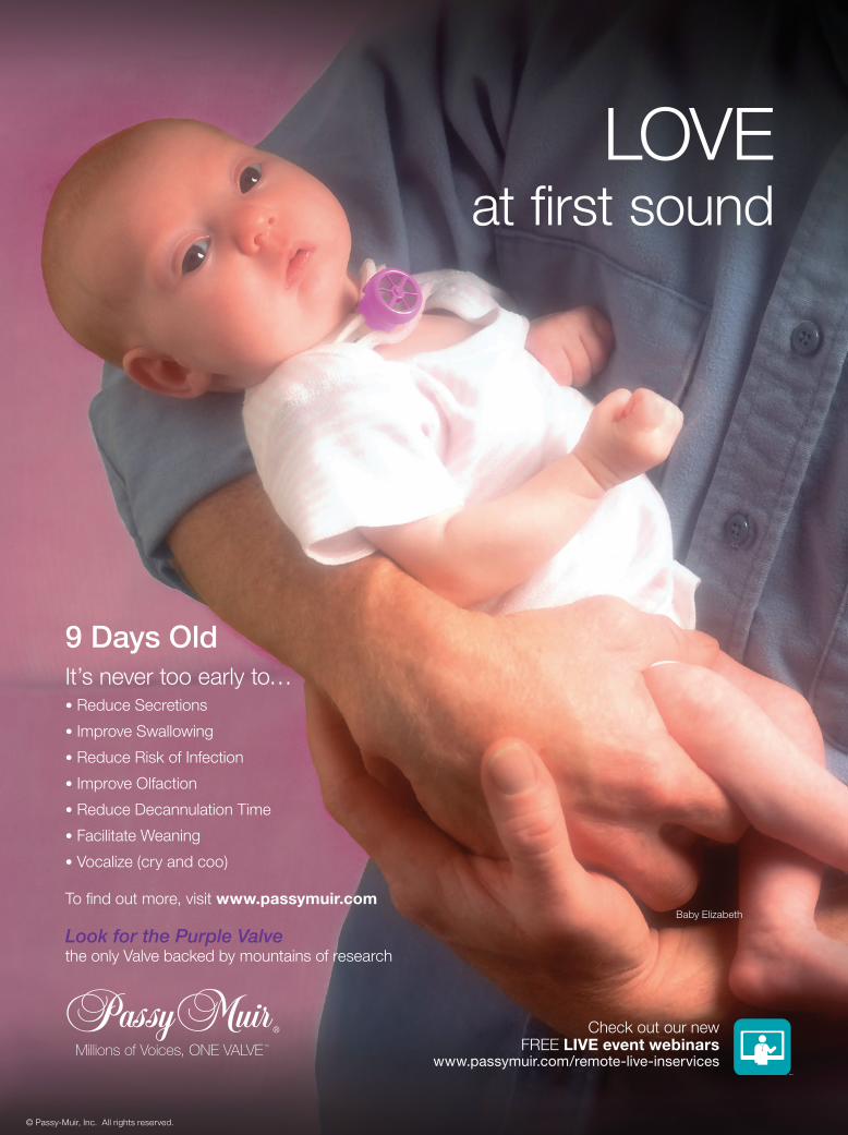
9 Days Old_updated 2_outlines.indd 1 7/1/21 2:42 PM

Arie L. Alkalay, MDClinical Professor of PediatricsDavid Geffen School of MedicinePediatrician, Cedars-SinaiLos Angeles, CA
M. A. Arif, MDProfessor of Pediatrics & Head, NeonatologyNational Institutes of Child HealthKarachi, Pakistan
Muhammad Aslam, MDAssociate Professor of PediatricsUniversity of California, IrvineNeonatologist, UC Irvine Medical CenterOrange, CA
Edward Austin, MDAustin-Hernandez Family Medical CenterCompton, CA
Richard L. Auten, MDAssistant Professor of PediatricsDuke University Medical CenterDurham, NC
Bruce G. Bateman, MDDepartment of Obstetrics & GynecologyUniversity of VirginiaCharlottesville, VA
Sandy Beauman, MSN, RNC-NICCNC ConsultingAlbuquerque, NM
David D. Berry, MDWake Forest University School of MedicineWinston-Salem, NC
Melissa K. Brown, BS, RRT-NPS, RCPFaculty, Respiratory Therapy ProgramGrossmont CollegeEl Cajon, CA
D. Spencer Brudno, MDAssociate Professor of PediatricsMedical Director, Pediatric TherapyMedical College of GeorgiaAugusta, GA
Curtis D. Caldwell, NNPUNM School of Medicine, Dept of PediatricsAlbuquerque, NM
Ed Coombs, MA RRT-NPS, ACCS, FAARCMarketing Director – Intensive CareKey Application Field Manager – Respiratory Care, Draeger MedicalTelford, PA
Jonathan Cronin, MDAssistant Professor of PediatricsHarvard Medical School ChiefNeonatology and Newborn Medicine UnitDepartment of Pediatrics Massachusetts General Hospital for ChildrenBoston, MA
Michael P. Czervinske, RRTNeonatal and Pediatric Critical CareUniversity of Kansas Medical CenterKansas City, KS
Professor Adekunle H. DawoduDirector, International Patient Care and Education, Cincinnati Children’s HospitalCincinnati, OH
Jayant Deodhar, MDAssociate Professor of Clinical PediatricsChildren’s Hospital CenterCincinnati, OH
Leonard Eisenfeld, MDAssociate Professor of PediatricsUniversity of Connecticut School of MedicineDivision of NeonatologyConnecticut Children’s Medical CenterHartford, CT
Sami Elhassani, MDNeonatologistSpartanburg, SC
Ivan Frantz, III, MDChariman of Department of PediatricsChief, Division of Newborn MedicineTufts University School of MedicineBoston, MA
Philippe S. Friedlich, MDAssociate Professor of Clinical PediatricsChildren’s Hospital of Los AngelesLos Angeles, CA
G. Paolo Gancia, MDNeonatologist, Terapia IntensivaNeonatale-Neonatologia, Cuneo, Italy
George A. Gregory, MDProfessor of Pediatrics and AnesthesiaUniversity of CaliforniaSan Francisco, CA
Charles J. Gutierrez, PhD, RRT, FAARCNeurorespiratory Clinical Specialist, J.A. Haley VA Hospital and Assistant Professor, Pulmonary, Critical Care & Sleep Medicine,Morsani College of Medicine, University of South Florida, Tampa, FL
William R. Halliburton, RRT, RCPNeonatal Respiratory Care CoordinatorDepartment of Respiratory CareHillcrest Baptist Medical Center, Waco, TX
Mary Catherine Harris, MDAssociate Professor of PediatricsDivision of NeonatologyUniversity of Pennsylvania School of MedicineThe Children’s Hospital of PhiladelphiaPhiladelphia, PA
David J. Hoffman, MDClinical Associate Professor of PediatricsPenn State College of MedicineStaff NeonatologistThe Reading Hospital and Medical CenterWest Reading, PA
Michael R. Jackson, RRTNewborn Intensive Care Unit Beth Israel Hospital, Boston, MA
Chang-Ryul Kim, MDAssociate Professor of PediatricsCollege of MedicineHanyang University Kuri HospitalSeoul, South Korea
David M. Kissin, BS, RRTPerinatal/Pediatric SpecialistMaine Medical Center, Portiand, ME
Sheldon Korones, MDDirector of Newborn CenterCollege of Medicine, Memphis, TN
Scott E. Leonard, MBA, BA, RRTDirector of Respiratory Therapy, EEG, NeurophysiologyGeorge Washington University HospitalWashington, DC
Raymond Malloy, MHA, RRTDirector of Pulmonary CareThomas Jefferson University HospitalPhiladelphia, PA
Paul J. Mathews, PhD, RRT, FCCM, FCCP, FAARCAssociate Professor of Respiratory CareUniversity of Kansas Medical CenterKansas City, KS
William Meadow, MDProfessor of PediatricsCo-Section Chief, NeonatologyComer Children’s HospitalThe University of Chicago, Chicago, IL
David G. Oelberg, MDCenter for Pediatric ResearchEastern Virginia Medical SchoolChildren’s Hospital of The King’s DaughtersNorfolk, VA
Rahmi Ors, MDDirector, Department of Neonatology and PediatricsProfessor of Pediatrics and NeonatologistMeram Medical FacultyNecmettin Erbakan UniversityKonya, Turkey
T. Michael O’Shea, MD, MPHChief, Neonatology DivisionWake Forest University School of MedicineWinston-Salem, NC
Lisa Pappas, RRT-NPSRespiratory Clinical Coordinator NICUUniversity of Utah HospitalSalt Lake City, UT
G. Battisita Parigi, MDAssociate Professor of Pediatric SurgeryUniversity of Pavia, Italy
Richard Paul, MDChief, Maternal & Fetal MedicineDepartment of Obstetrics & GynecologyUniversity of Southern CaliforniaLos Angeles, CA
Max Perlman, MDProfessor of PediatricsThe Hospital for Sick ChildrenToronto, Ontario, Canada
Boris Petrikovsky, MDDirector, Prenatal Diagnostic Unit ServicesNew York Downtown HospitalNew York, NY
Arun Pramanik, MDProfessor of PediatricsDirector of Neonatal FellowshipLouisiana State UniversityHealth Sciences Center, Shreveport, LA
Benamanahalli K. Rajegowda, MDChief of NeonatologyLincoln Medical and Mental Health CenterProfessor of Clinical PediatricsWeill Medical College of Cornell University, NY
Ruben D Restrepo, MD RRT FAARC FCCPCoordinator of ResearchProfessor - Division of Respiratory CareUT Health San Antonio7703 Floyd Curl Dr, San Antonio, TX
Koravangattu Sankaran, FRCP(C), FAAP, FCCMProfessor of Pediatrics and Director of Neonatology and Neonatal ResearchDepartment of PediatricsRoyal University HospitalUniversity of SaskatchewanSaskatoon, Saskatchewan, Canada
Istvan Seri, MD, PhDProfessor of PediatricsHead, USC Division of Neonatal MedicineUniversity of Southern California, Los Angeles, CA
Tushar A. Shah, MD, MPHDivision of Neonatology Cincinnati Children’s Hospital Medical Center Cincinnati, OH
Dave Swift, RRTOttawa Hospital – Civic SiteCampus Coordinator (Professional Practice) & Special Care Nursery Charge TherapistRespiratory Therapy Team LeadNational Office of the Health Care Emergency Response Team (NOHERT)Subject Matter Expert, Health Canada
Jack TannerNICU Clinical CoordinatorU Mass Memorial HospitalWorcester, MA
Otwell D. Timmons, MDCarolinas Medical CenterCharlotte, NC
Maya Vazirani, MD, FAAPBoard Certified Neonatology and Pediatrics Lancaster, CA
Max Vento, MDAssociate Professor of PediatricsChief, Pediatric ServicesNeonatologia Hospital Virgin del ConsueloValencia, Spain
Dharmapuri Vidyasagar, MDProfessor of Pediatrics Department of PediatricsUniversity of IllinoisChicago, IL
Editorial Advisory Board
Table of ContentsDEPARTMENTS 6 Letter to the Editor
7 News
ARTICLES 16 Using Near-Infrared Spectroscopy
to Guide Your Clinical Decision Making
19 The Risk of Mortality for Patients with Persistently Elevated HeRO Scores
22 A New System of Vitamin Administration During Pregnancy and Postpartum
24 Noninvasive Technique to Track Early Preemies’ Cerebral Oxygen Levels
25 Considerations for the Impact of a Tracheostomy
29 A Nutritional Approach to Feeding a Targeted Bacterial Strain
32 What Clinicians Need to Know About Human Milk Banking
39 The ENFit Enteral-Only System
42 The Benefits of Personalized Lung Protection and Weaning Solutions
46 A New Standard in Developmental Positioning
49 Incidence of Urinary Tract Infection in Neonates with Significant Indirect Hyperbilirubinemia of Unknown Etiology
55 Circulating Tight-Junction Proteins Are Potential Biomarkers For Blood-Brain Barrier Function
Vol. 34 No. 4Fall 2021
4 neonatal INTENSIVE CARE Vol. 34 No. 4 n Fall 2021

Ad Page 5
Request your free samples online at neotech-neonatalic.com
Add a splash of color and character to the incubators in your unit. NeoHug is a cute and versatile NICU accessory, ideal for holding a pacifier, suction tip, tape and more.
Available with NEW smile logo design or original chimp in five bright colors. With suction cup for use in incubator or C-clip for use on IV pole.
Introducing NeoHug® SmileUtility Device Holder
A Hug Makes You Smile!
©2021 Neotech Products LLC. All rights reserved.
TRY IT FOR FREE! neotech-neonatalic.com

Dear Dr Campbell:
I am writing to you regarding your analysis of the study that was originally published by Khoury R et al. in the November 2020 issue of the Journal of Perinatology.1,2 In this study, Khoury R et al. analyzed the performance of two pulse oximeters in a normal cohort of newborn infants during their transition period. Pulse oximetry heart rate stability was recorded “qualitatively” by manually identifying when the oximeter achieved stability as identified by the observer.1
There were multiple concerns with the study, including the fact that this study was conducted only in healthy newborns, those least likely to require extensive resuscitation. As a consequence of the restatement of the AAP guidance regarding resuscitation in the delivery room in 2015, Masimo developed a sensor that was specifically designed to operate in this environment.3 The sensor provided better performance, especially in pulse rate stability, than that which was used in the study. Further, this sensor was optimized to automatically transition the monitor to a 2-second averaging time and maximal sensitivity. Neither of these refinements was incorporated into the study. Studying this pulse oximeter without the benefit of these modes completely invalidates the findings. When comparing oximetry technologies,
it is critically important that the latest technologies from each manufacturer be used.4
Moreover, the study attempted to clarify pulse rate using heart rate as a gold standard for comparison. It is well known that not all ECG electrical activity translates to a heartbeat, and thus a pulse. Pulseless electrical activity (PEA) is a well-recognized phenomenon in the neonatal population. ECG signals can provide a reliable indication of electrical heart activity but cannot predict with absolute certainty the presence of a heartbeat in association with each waveform. Further, an algorithm predicated on the generation of a stable heart rate as opposed to one that genuinely identifies “missed” beats may be closer to the ECG rate but not accurately reflect the presence of a pulse. The bradycardia reported by Masimo may have accurately reflected actual pulsatile activity.5-7
The authors of the study generalized their concern about their findings and the presence of false bradycardia in resuscitation. The need for resuscitation is typically associated with decreased perfusion, unstable heart rate, and abrupt changes in oximetry. PR stability is an inferior metric during these situations. These are areas where Masimo SET technology has been shown to have superior performance in myriad studies. For a pulse oximeter to
Letter to the Editor
ISSN 1062-2454Published five times each year by
Goldstein and Associates, Inc.10940 Wilshire Blvd., Suite 600Los Angeles CA 90024Phone: 310-443-4109Fax: 310-443-4110E-mail: [email protected]: www.nicmag.caPublisher/Editor in ChiefSteve Goldstein
Managing EditorChristopher Hiscox
Senior EditorVincent Terrier
News EditorChris Campbell
Associate EditorJordana Hammeke, Susan Goldstein
Circulation, Coverage, Advertising Rates: Complete details regarding circulation, coverage, advertising rates, space sizes, and similar information are available to prospective advertisers. Closing date is 45 days preceding date of issue.
Change of Address: Notices should be sent promptly to Circulation Department. Provide old mailing label as well as new address; include zip code or postal code. Allow two months for change.
Editorial Contributions may be sent by e-mail and will be handled with reasonable care: however, publishers assume no responsibility for safety of art work, photographs, or manuscripts. Every precaution is taken to ensure accuracy, but the publishers cannot accept responsibility for the correctness or accuracy of informa tion supplied herein or for any opinion expressed. Editorial closing date is the first day of the month preceding month of issue.
©2021 by Goldstein & Associates, Inc. All rights reserved. Reproduction in whole or in part without written permission is strictly prohibited.
Cover: Trees. Credit: Smithsonian American Art Museum, Gift of H. Lyman Sayen to his nation.
6 neonatal INTENSIVE CARE Vol. 34 No. 4 n Fall 2021

prove its mettle, it is inappropriate to suggest that “fair weather” conditions in normal transitioning neonates are in any way similar to those encountered during a full-on resuscitation.8
These concerns were also addressed in a letter to the editor that appeared in the March 2021 Neonatology Today authored by Dr Latorre and corroborated by the response. “Speed of response, notwithstanding, the technology is not just about speed alone. Accuracy, precision, and reproducibility are a sine qua non.”9
Sincerely,
Mitchell Goldstein, MD, MBA, CMLProfessor of PediatricsDivision of NeonatologyLoma Linda University Children’s HospitalEditor in ChiefNeonatology Today
References1. Khoury R, Klinger G, Shir Y, Osovsky M, Bromiker R.
Monitoring oxygen saturation and heart rate during neonatal transition. Comparison between two different pulse oximeters and electrocardiography. J Perinatol. 2020. Epub 2020/12/01. doi: 10.1038/s41372-020-00881-y. PubMed PMID: 33250516.
2. Campbell C. Comparing The Performance Of Two Pulse Oximeters And Electrocardiography During Neonatal Transition. neonatal INTENSIVE CARE Vol. 34 No. 2 n Spring 2021
3. American Academy of Pediatrics, Neonatal Resuscitation Program: https://www.aap.org/en-us/continuing-medical-education/life-support/NRP/Pages/NRP.aspx
4. Barker SJ. “Motion-resistant” pulse oximetry: a comparison of new and old models. Anesth Analg. 2002;95(4):967-72, table of contents. Epub 2002/09/28. doi: 10.1097/00000539-200210000-00033. PubMed PMID: 12351278.
5. Patel S, Cheung PY, Solevag AL, Barrington KJ, Kamlin COF, Davis PG, et al. Pulseless electrical activity: a misdiagnosed entity during asphyxia in newborn infants? Arch Dis Child Fetal Neonatal Ed. 2019;104(2):F215-F7. Epub 2018/06/14. doi: 10.1136/archdischild-2018-314907. PubMed PMID: 29895572.
6. Luong D, Cheung PY, Barrington KJ, Davis PG, Unrau J, Dakshinamurti S, et al. Cardiac arrest with pulseless electrical activity rhythm in newborn infants: a case series. Arch Dis Child Fetal Neonatal Ed. 2019;104(6):F572-F4. Epub 2019/02/24. doi: 10.1136/archdischild-2018-316087. PubMed PMID: 30796058.
7. Sillers L, Handley SC, James JR, Foglia EE. Pulseless Electrical Activity Complicating Neonatal Resuscitation. Neonatology. 2019;115(2):95-8. Epub 2018/10/24. doi: 10.1159/000493357. PubMed PMID: 30352434.
8. Hay WW, Jr., Rodden DJ, Collins SM, Melara DL, Hale KA, Fashaw LM. Reliability of conventional and new pulse oximetry in neonatal patients. J Perinatol. 2002;22(5):360-6. Epub 2002/06/26. doi: 10.1038/sj.jp.7210740. PubMed PMID: 12082469.
9. Latorre HB, Goldstein M. Letter to the Editor. Neonatology Today. Volume 16, Issue 3 Pages 143-145. DOI: https://doi.org/10.51362/neonatology.today/20213163143145
Texas Hospital Delivers 107 Babies in 91 HoursA Texas hospital set a record at the end of June when staff delivered more than 100 babies in 91 hours, according to a local report. The deliveries occurred across two 48-hour stretches at Andrew Women’s Hospital at Baylor Scott & White All Saints Medical Center in Fort Worth, according to WFAA 8, an ABC affiliate in Dallas. On June 24, staff delivered 25 girls and 27 boys in 47 hours. On June 28, staff delivered 55 babies, including a set of twin girls, in 44 hours. “It’s been all fireworks as our Labor and Delivery teams … have been busy delivering an adorable group of Fourth of July babies — 107 of them in just 91 hours to be exact!” the hospital said in a Facebook post. The stretch of deliveries broke the hospital’s record from June 2018, WFAA 8 reported. The hospital averages about 16 deliveries per day and last year delivered 6,000 babies, including 100 sets of twins and two sets of triplets. Six of the baby girls were named Gianna, the news outlet reported. Other popular names included Reign for girls and Atlas and Daniel for boys. The staff expected to see an uptick in births this year as people recover from the pandemic, but the June influx was rare and exceptional, according to NBC 5. “We thought it was going to be higher the last few months because of the pandemic, but didn’t see the baby boom until now, so people must’ve gotten more reassured,” Jay Herd, MD, an obstetrician at the hospital, told the news outlet. The hospital’s labor and delivery teams create photo galleries of babies who are born around the holidays. The Tiniest Texans series features portraits of the newborns, often dressed in holiday-themed clothes.
Fewer Preterm Infants in US Getting Mechanical VentilationThe duration of respiratory support for preterm infants in the US has increased in recent years as the reliance on mechanical ventilation has declined and use of noninvasive ventilation has become more common, a new study suggests. Researchers examined data on preterm infants from a clinical cohort of 259,311 infants in the Pediatrix Clincal Data Warehouse as well as a national cohort of 1.17 million infants in the National Inpatient Sample. All the preterm infants were born before 35 weeks gestation and received care on neonatal intensive care
News Fall 2021
Editor’s Correction: In Vol. 34, No. 3, Summer 2021 edition in the article Not All Donor Milk Is Equal, Table 2 incorrectly listed the temperature of vat pasteurization. The correct temperature is 66°C for 30 minutes.
neonatal INTENSIVE CARE Vol. 34 No. 4 n Fall 2021 7

units between 2008 and 2018. From 2008 to 2018, the proportion of neonates in the clinical cohort receiving mechanical ventilation declined from 29.4% to 18.5% and the proportion receiving noninvasive ventilation climbed from 57.9% to 67.4%. Over this same period, the mean number of days on respiratory support increased from 13.8 to 14.4, driven in large part by more widespread use of continuous positive airway pressure and nasal intermittent positive pressure ventilation, the study team reports in JAMA Pediatrics. “While our study couldn’t provide the exact reason why mechanical ventilation was decreasing, the decline in mechanical ventilation use seemed to begin at about the same time one of the largest clinical trials evaluating continuous positive airway pressure (CPAP) as the primary respiratory support in preterm infants was released,” said Dr. Dupree Hatch, an assistant professor of pediatrics and medical director of the NICU at Vanderbilt University Medical Center in Nashville, Tennessee, who led the current study. “This study, the SUPPORT trial, along with several other studies released about the same time caused many of us in neonatal medicine to reconsider our approach to respiratory support in preterm infants,” Dr Hatch said by email. Over the study period, the mean duration of mechanical ventilation declined from 10.3 days to 9.7 days among infants who received this type of respiratory support. During the same period, the mean duration of noninvasive ventilation increased by 3.2 days among infants receiving this type of support. In the national cohort analysis, researchers also found evidence of reduced use of mechanical ventilation. The proportion of preterm infants receiving this form of respiratory support declined from 22.0% to 18.5% during the study period. One limitation of the analysis is that researchers were unable to link the two data sets, making it possible that the populations in the two cohorts might not be entirely comparable, the authors note. Changes in transfer patterns during the study period also could have influenced the outcomes. Even so, the results underscore the importance of clinicians keeping up to date on the literature around respiratory support and carefully considering how trial outcomes might be relevant in the treatment of individual patients, said Dr Sara DeMauro, associate director of neonatal clinical and epidemiological research at the Children’s Hospital of Philadelphia, who coauthored an editorial accompanying the study. “Mechanical ventilation usage is declining because neonatologists are concerned that exposure to mechanical ventilation is injurious, particularly to the preterm/immature lung,” Dr DeMauro said by email.
Out-of-Pocket Costs for Childbirth More Than $3000Families with private health insurance pay around $3,000 for newborn delivery and hospitalization, while adding neonatal intensive care can push the bill closer to $5,000, based on a retrospective look at almost 400,000 episodes. The findings suggest that privately insured families need prenatal financial counseling, as well as screening for financial hardship after delivery, reported lead author Kao-Ping Chua, MD, PhD, assistant professor and health policy researcher in the department of pediatrics and the Susan B. Meister Child Health Evaluation and Research Center at the University of Michigan, Ann Arbor, and colleagues. “Concern is growing regarding the high and rising financial burden of childbirth for privately insured families,” the investigators wrote in Pediatrics. “Previous studies assessing this burden have focused on out-of-pocket spending for maternal care, including hospitalizations for delivery. However, there are no recent national data on out-of-pocket spending across the childbirth episode, including both deliveries and newborn
hospitalizations.” To address this knowledge gap, Dr Chua and colleagues turned to Optum’s deidentified Clinformatics Data Mart, comprising 12 million privately insured individuals across the United States. The investigators identified 398,410 childbirth episodes occurring between 2016 and 2019. Each episode was defined as one delivery and at least one newborn hospitalization under the same family plan. Out-of-pocket cost included copayment plus coinsurance and deductibles. Primary outcomes included mean total out-of-pocket spending and proportion of episodes exceeding $5,000 or $10,000. Subgroup analyses compared differences in spending between episodes involving neonatal intensive care or cesarean birth. The mean out-of-pocket spending was $2,281 for delivery and $788 for newborn hospitalization, giving a total of $3,068 per childbirth episode. Coinsurance and deductibles accounted for much of that cost, at 55.8% and 42.1%, respectively, whereas copayments accounted for a relatively minor portion (2.2%). Almost all episodes (95%) cost more than zero dollars, while 17.1% cost more than $5,000 and 1.0% cost more than $10,000. Total mean out-of-pocket spending was higher for episodes involving cesarean birth ($3,389) or neonatal intensive care ($4,969), the latter of which cost more than $10,000 in 8.8% of episodes. “Because details on plan benefit design were unavailable, the generalizability of findings to all privately insured Americans is unclear,” the investigators noted. “However, the proportion of childbirth episodes covered by high-deductible health plans in this study is consistent with the prevalence of such plans among Americans with employer-sponsored insurance.” The findings suggest that financial reform is needed, Dr Chua and colleagues concluded. “To avoid imposing undue financial burden on families, private insurers should improve childbirth coverage,” they wrote. “An incremental step would be providing first-dollar coverage of deliveries and newborn hospitalizations before deductibles are met. Ideally, however, insurers would waive most or all cost-sharing for these hospitalizations, consistent with the approach taken by Medicaid programs and many developed countries.”
Probiotic Supplementation Regulates Newborn Immune SystemSupplementing breastfed infants with bifidobacteria promotes development of a well-regulated immune system, theoretically reducing risk of immune-mediated conditions like allergies and asthma, according to investigators. These findings support the importance of early gut colonization with beneficial microbes, an event that may affect the immune system throughout life, reported lead author Bethany M. Henrick, PhD, director of immunology and diagnostics at Evolve Biosystems, Davis, Calif., and adjunct assistant professor at the University of Nebraska, Lincoln, and colleagues. “Dysbiosis of the infant gut microbiome is common in modern societies and a likely contributing factor to the increased incidences of immune-mediated disorders,” the investigators wrote in Cell. “Therefore, there is great interest in identifying microbial factors that can support healthier immune system imprinting and hopefully prevent cases of allergy, autoimmunity, and possibly other conditions involving the immune system.” Prevailing theory suggests that the rising incidence of neonatal intestinal dysbiosis — which is typical in developed countries — may be caused by a variety of factors, including cesarean sections; modern hygiene practices; antibiotics, antiseptics, and other medications; diets high in fat and sugar; and infant formula. According to Dr Henrick and colleagues, a healthy gut microbiome plays the greatest role in immunological development during the first 3 months post-partum; specifically, a lack of bifidobacteria during this
8 neonatal INTENSIVE CARE Vol. 34 No. 4 n Fall 2021

activated immune cells, and reduced levels of regulatory cells indicative of systemic immune dysregulation,” the investigators wrote. The interventional part of the study involved 60 breastfed infants in California. Twenty-nine of the newborns were given 1.8 × 10¹⁰ colony-forming units (CFUs) of B. longum subsp. infantis EVC001 daily from postnatal day 7 to day 28, while the remaining 31 infants were given no supplementation. Fecal samples were collected on day 6 and day 60. At day 60, supplemented infants had high levels of HMO-utilization genes, plus significantly greater alpha diversity (P = .0001; Wilcoxon),
compared with controls. Infants receiving EVC001 also had less inflammatory fecal cytokines, suggesting that microbes expressing HMO-utilization genes cause a shift away from proinflammatory Th2 and Th17 responses, and toward Th1.
Company Signs Agreement with UniversityNeotech Products has entered into a mutually beneficial agreement with Wichita State University. This exciting collaboration will provide Neotech with the resources of an institution well known for advanced research, including clinical work through their partners. Wichita State, a national leader in aerospace research, will benefit from
the experience of a well-established and innovative medical device company as it expands research in the area of medical products. They will work closely with Neotech’s new product development team to explore product ideas that will impact the end user. “Wichita State is committed to using our aviation expertise for expanding research in other industries,” said Rick Muma, President of Wichita State University. “As a clinician in internal medicine and infectious diseases and a public health practitioner, I understand the need to continually innovate through collaboration in the healthcare sector. Partnerships
time has been linked with increased risks of autoimmunity and enteric inflammation, although underlying immune mechanisms remain unclear. Bifidobacteria also exemplify the symbiotic relationship between mothers, babies, and beneficial microbes. The investigators pointed out that breast milk contains human milk oligosaccharides (HMOs), which humans cannot digest, but are an excellent source of energy for bifidobacteria and other beneficial microbes, giving them a “selective nutritional advantage.” Bifidobacteria should therefore be common residents within the infant gut, but this is often not now the case, leading Dr Henrick and colleagues to zero in on the microbe, in hopes of determining the exactly how beneficial bacteria shape immune development. It is only recently that the necessary knowledge and techniques to perform studies like this one have become available, the investigators wrote, noting a better understanding of cell-regulatory relationships, advances in immune profiling at the systems level, and new technology that allows for profiling small-volume samples from infants. The present study involved a series of observational experiments and a small interventional trial. First, the investigators conducted a wide array of blood- and fecal-based longitudinal analyses from 208 infants in Sweden to characterize immune cell expansion and microbiome colonization of the gut, with a focus on bifidobacteria. Their results showed that infants lacking bifidobacteria, and HMO-utilization genes (which are expressed by bifidobacteria and other beneficial microbes), had higher levels of systemic inflammation, including increased T helper 2 (Th2) and Th17 responses. “Infants not colonized by Bifidobacteriaceae or in cases where these microbes fail to expand during the first months of life there is evidence of systemic and intestinal inflammation, increased frequencies of
B&B Products are also available through finer specialty distributors including: Tri-anim, Cardinal Health & Medline.
2018
neonatal INTENSIVE CARE Vol. 34 No. 4 n Fall 2021 9
was part of Columbia University’s ongoing COVID-19 Mother Baby Outcomes (COMBO) initiative to “describe the health and well-being of mother-infant dyads with and without prenatal SARS-CoV-2 infections,” according to the researchers. During the study period, the researchers identified newborns of 327 women who tested positive for COVID-19 at any point during pregnancy and compared them to newborns of 2,125 unexposed women. Demographics were similar between the groups. Overall, the total test positivity was 0.7% for exposed newborns; 1.0% tested positive on an initial test, and 0% were positive on retest. During the newborn hospital stay and a 2-week follow-up, 0% of all newborns showed clinical evidence of infection. No significant differences were noted between exposed and unexposed newborns in clinical outcomes including gestational age, mode of delivery, 5-minute Apgar score, heart rate, respiratory rate, or temperature. Although more infants of COVID-19-exposed mothers compared with unexposed mothers had an emergency department visit within the first 14 days of life (6% vs. 3%, P = .002), none of the infants was diagnosed with COVID-19 during these visits. Cough, fever, congestion, or bilirubin were more frequent reasons for emergency department visits in the exposed infants compared with unexposed infants, but these differences were not significant. The study findings were limited by several factors, including the retrospective design and the limited follow-up period to only the first 2 weeks of life, the researchers noted. In addition, perinatal transmission rates were available only for the 202 newborns who were followed up in the hospital system, they said. However, the results suggest that the risk of mother-to-newborn vertical transmission of COVID-19 remains low, even when mothers are breastfeeding and infants are rooming in, they concluded.
Skip Routine Probiotics for Preemies, AAP SaysThe American Academy of Pediatrics (AAP) now recommends against the routine administration of probiotics to preterm infants, particularly the most vulnerable (those whose birth weight is <1000 g), for the treatment or prevention of necrotizing enterocolitis (NEC) and late-onset sepsis. Although probiotics are increasingly given to preterm infants, the AAP notes that the data on their safety and efficacy are inconsistent. In addition, the supplements are not subject to approval by the US Food and Drug Administration (FDA). Therefore, the academy advises clinicians to use extreme caution in selecting preterm neonates to receive these microorganisms and recommends obtaining informed consent from parents after carefully discussing the risks. It also recommends that centers using probiotics conduct surveillance, inasmuch as probiotics can alter a center’s flora, potentially affecting all patients. Such centers should also carefully document outcomes, adverse events, and safety. The AAP’s clinical report, published online May 24 in Pediatrics, highlights wide differences between commercially available formulations and a lack of regulatory standards in this country. Absent FDA-approved drug labeling, these nutritional supplements cannot be marketed as treatment or prophylaxis, but that has scarcely stopped their use. “Despite lack of availability of a pharmaceutical-grade product, the number of preterm infants receiving probiotics in the United States and Canada is steadily increasing,” write Brenda Poindexter, MD, FAAP, chief of neonatology at Children’s Healthcare of Atlanta, in Atlanta, Georgia, and members of the AAP’s Committee on Fetus and Newborn. Analyses of US collaborative databases indicate that approximately 10% of neonates of extremely low gestational age receive a probiotic preparation in the neonatal intensive care unit (NICU). The use of these preparations varies widely across
with companies like Neotech Products do just that.” With this partnership, Neotech will have access to advanced facilities and technologies, including: Labs for materials and adhesive testing and research; Electron microscopes; CT scanning (for materials); Pull and compression testing; A wide array of 3D printing and scanning; VR visual design space. The partnerships’ first collaboration is a locking mechanism for the NeoBar ET Tube Holder. The idea originated with Dr Mohammed Ansari, a neonatologist with ties to the Wichita area. Wichita State teamed up with Dr Ansari and brought the idea to Neotech to drive the project forward. We’re extremely excited to see where it leads. Overall, the purpose of the partnership between Neotech and Wichita State is to utilize the combined expertise of both institutions to bring medical products to market that will truly make a difference.
Mother-to-Infant COVID-19 Transmission Is UnlikelyMothers with a history of COVID-19 exposure during pregnancy are not likely to transmit the infection to their newborns, based on data from more than 2,000 women. “Uncertainty at the onset of the COVID-19 pandemic led to varying postnatal care recommendations for newborns exposed to SARS-CoV-2 in utero,” said Margaret H. Kyle, of Columbia University, New York, and colleagues. The Columbia University Irving Medical Center, an early epicenter of the pandemic, allowed rooming-in and encouraged direct breastfeeding between infected mothers and their newborns while adopting extensive safety measures, the researchers said. In a study presented at the virtual meeting of the Pediatric Academic Societies, the researchers conducted a retrospective chart review of all newborns born at the medical center from March 22, 2020, through August 7, 2020. The study
10 neonatal INTENSIVE CARE Vol. 34 No. 4 n Fall 2021
institutions. “NEC is a devastating morbidity of prematurity, and it’s multifactorial. Some babies only given mother’s milk still get NEC, and the decision to use these products is a very nuanced one,” Poindexter said. “I suspect some people will disagree with the report, and we tried to give folks some wiggle room.” Evidence from other countries suggests that probiotics can be protective against NEC, she added, “so not to have a reliable product in this country is very frustrating.”
Guidance Reviewed on ‘Kangaroo Care’When a baby is born prematurely, immediate skin-to-skin contact could save their lives. Instead of placing low-weight newborns in an incubator, new research suggests they should be nestled up close to their mother’s chest, or that of a close caregiver’s, and fed exclusively on breast milk. This approach, dubbed kangaroo care, has proved to be one of the best and safest ways to treat preterm infants with low birth weights, resulting in fewer infections, higher rates of breastfeeding, and better weight gain in studies. Despite the growing number of benefits, the practice has not been widely adopted. Currently, the World Health Organization recommends continuous kangaroo care for all preterm infants, but only after they are taken away and declared clinically stable in the neonatal intensive care unit (NICU). A randomized controlled trial in five hospitals now suggests the WHO’s recommendation separates babies and their mothers too soon. Instead, hospitals should implement a mother-infant care unit with beds and chairs so that hospital staff can look after new parents and babies at the same time. The study was conducted among 3,211 low-weight infants in Ghana, India, Malawi, Nigeria, and Tanzania, who were either assigned immediate kangaroo care in a specially arranged “Mother-NICU” or were separated from the parents for
conventional care, with brief moments of touch allowed after the first 24 hours. In the first three days, infants who received immediate skin-to-skin contact were held for roughly 17 hours a day in the Mother-NICU. Meanwhile, those infants placed in incubators or radiant warmers received only 1.5 hours of intermittent daily contact. Compared to conventional neonatal care, those infants who received immediate touch from their parents were 25 percent less likely to die in the first month of life. Continuously held newborns were also less likely to develop hypothermia and bacterial blood poisoning, possibly because these infants had greater exposure to their mother’s protective
microbiome, were more likely to receive early breast milk, and were handled by fewer people. Avoiding separation stress between the mother and the infant might also have contributed to greater health outcomes. Touch between a baby and its mother has been shown to stabilize the newborn’s heart rate, calm its breathing, and decrease its crying. “Keeping the mother and baby together right from birth, with zero separation, will revolutionize the way neonatal intensive care is practiced for babies born early or small,” argues Rajiv Bahl, the Head of Maternal and Newborn Health Research and Development at WHO. “This study illustrates that kangaroo mother care has the potential to save many
more lives if it is started immediately after birth, a finding with relevance for countries of all income levels.” Today, over 96 percent of all infants with a low birth weight are born in developing countries, and these children are particularly vulnerable to infectious disease, developmental delays, and death. Conventional neonatal care is expensive and requires great skill and logistical support, which many countries with lower incomes cannot afford. Kangaroo care, on the other hand, is a safe and effective alternative much easier to implement. The
Read the full white paper at:www.embracemri.com/embrace-the-benefits
or call us at +1 866 609 1554
Discover the
Benefits of an On-Unit NICU MRI System
MRIs offer many benefits for newborns in the Neonatal Intensive Care Unit (NICU), but performing off-unit MRIs is incredibly complex and creates many risks for this vulnerable population. On-unit MRI options for NICU patients greatly reduce these risks and can lead to significant benefits that make them cost-effective and clinically superior to off-unit MRIs.
Discover the benefits of on-unit MRI and learn how the innovative, point-of-care Embrace® Neonatal MRI System is transforming neonatal care from inside for patients, parents and NICU staff.
Become a part of the transformation.
Discover more at embracemri.com
©2021 Aspect Imaging Ltd. All Rights Reserved.
neonatal INTENSIVE CARE Vol. 34 No. 4 n Fall 2021 11

Ad Page 12
Making our world more productive
National Reach, Local ServiceNOXIVENT® (nitric oxide) gas for inhalation, along with the NOxBOXi® delivery system, offered with customizable, consumption-based billing, is backed by Linde’s national network, responsive support and reputation for medical gas distribution.
The NOxBOXi nitric oxide gas delivery system is reliable, accurate and easy to use. System features include:
Æ Real-time, closed-loop monitoring with auto-adjusting alarms
Æ Pre-packaged, configured circuits ready for use with validated ventilators
Æ Disposable circuits, including the NOxFLOW module, for easy clean up
Æ Auto-cylinder changeover with alerts, helping you avoid therapy interruptions
Our Commitment Æ Integrated gas delivery system for inhaled nitric oxide therapy
Æ 24/7/365 service and support
Æ Simplified billing process
Æ Reliable and responsive distribution network
Æ Established reputation for quality and customer satisfaction
A summary of the prescribing information, including indication and other important safety information, is on the adjacent page. For the full prescribing information, visit www.noxiventus.com.
Call 1-844-445-4633 today for a complimentary requirements evaluation.www.noxiventus.com
Comprehensive Solution For Nitric Oxide Inhalation TherapyComplete with 24/7/365 support – peace of mind for critical care providers.

Ad Page 13
Making our world more productive
National Reach, Local ServiceNOXIVENT® (nitric oxide) gas for inhalation, along with the NOxBOXi® delivery system, offered with customizable, consumption-based billing, is backed by Linde’s national network, responsive support and reputation for medical gas distribution.
The NOxBOXi nitric oxide gas delivery system is reliable, accurate and easy to use. System features include:
Æ Real-time, closed-loop monitoring with auto-adjusting alarms
Æ Pre-packaged, configured circuits ready for use with validated ventilators
Æ Disposable circuits, including the NOxFLOW module, for easy clean up
Æ Auto-cylinder changeover with alerts, helping you avoid therapy interruptions
Our Commitment Æ Integrated gas delivery system for inhaled nitric oxide therapy
Æ 24/7/365 service and support
Æ Simplified billing process
Æ Reliable and responsive distribution network
Æ Established reputation for quality and customer satisfaction
A summary of the prescribing information, including indication and other important safety information, is on the adjacent page. For the full prescribing information, visit www.noxiventus.com.
Call 1-844-445-4633 today for a complimentary requirements evaluation.www.noxiventus.com
Comprehensive Solution For Nitric Oxide Inhalation TherapyComplete with 24/7/365 support – peace of mind for critical care providers.
Making our world more productive
Indication
Noxivent® is a vasodilator indicated to improve oxygenation and reduce the need for extracorporeal membrane oxygenation in term and near-term (>34 weeks gestation) neonates with hypoxic respiratory failure associated with clinical or echocardiographic evidence of pulmonary hypertension in conjunction with ventilatory support and other appropriate agents.
Important Safety Information
Contraindications Noxivent is contraindicated in neonates dependent on right-to-left shunting of blood.
Warnings and Precautions Rebound: Abrupt discontinuation of Noxivent may lead to worsening oxygenation and increasing pulmonary artery pressure.
Methemoglobinemia: Methemoglobin levels increase with the dose of Noxivent; it can take 8 hours or more before steady state methemoglobin levels are attained. If methemoglobin levels do not resolve with decrease in dose or discontinuation of Noxivent, additional therapy may be warranted to treat methemoglobinemia.
Airway Injury from Nitrogen Dioxide: Monitor nitrogen dioxide (NO2) levels. Nitrogen dioxide may cause airway inflammation and damage to lung tissue.
Heart Failure: In patients with pre-existing left ventricular dysfunction, Noxivent may increase pulmonary capillary wedge pressure leading to pulmonary edema.
Adverse Reactions
The most common adverse reaction of Noxivent is hypotension.
Drug Interactions
Nitric Oxide donor compounds may increase the risk of developing methemoglobinemia.
Administration
Use only with a calibrated, FDA-cleared NOxBOXi® Nitric Oxide Delivery System (NODS). Refer to the NODS labeling for needed information on training and technical support for users of this drug product with the NODS.
Please see the full Prescribing Information for additional important Noxivent® safety and risk information.
NOXIVENT® Indication and Important Safety Information
Distributed by Praxair Distribution, Inc., a Linde Company 10 Riverview Dr. Danbury, CT 06810 Phone 844.445.4633, www.noxiventus.com
The Linde logo, the Linde wordmark NOXIVENT, NOxFLOW, NOxBOXi and MAKING OUR WORLD MORE PRODUCTIVE are trademarks or registered trademarks of Linde Inc. or its affiliates. The information contained herein is offered for use by technically qualified personnel, at their discretion and risk, without warranty of any kind. © Copyright 2021, Linde Inc.
both e-cigarettes and conventional cigarettes, and those whose mothers smoked conventional cigarettes only. Their estimates were imprecise, but signaled that e-cigarette use may reduce birth weight. The use of e-cigarettes alone appeared to have less of an impact on birth weight than the dual use of conventional cigarettes and e-cigarettes did. Wylie cautioned that outcomes like birth weight are “pretty crude measures of whether an exposure is okay or not in pregnancy. Many of these toxins that we know that are in the aerosols can cause harm, but they may not be reflected in the absolute value of the birth weight.” In addition, clinicians should avoid focusing on the wrong question when caring for patients. “I think the wrong question is: Is vaping safer than smoking?” Wylie said in an interview. “Metals are going into your lungs. Plastics are going into your lungs. It is hard for me to think that we are going to identify that as our champion smoking cessation strategy in pregnancy.”
Children Born Just Weeks Early Face Higher Risk of Developmental ProblemsChildren born preterm (before 37 weeks of pregnancy) remain at high risk of developmental difficulties that can affect their behaviour and ability to learn, finds a study published by The BMJ today. These difficulties were found not only in children born extremely preterm (22-26 weeks) but also in those born very and moderately preterm (between 27 and 34 weeks), say researchers. Survival of preterm babies has increased worldwide. Children born early often have developmental issues, but studies have mainly focused on those born extremely preterm (22-26 weeks’ gestation) and less is known about children born very and moderately preterm (27-34 weeks’ gestation). Given how important it is to identify children most at risk of developmental difficulties, researchers in France set out to describe neurodevelopment among children born before 35 weeks compared with children born at full term. Their findings are based on 3,083 French children aged 5½ born after 24-26, 27-31, and 32-34 weeks gestation who were taking part in the EPIPAGE-2 study (designed to investigate outcomes of preterm children over the past 15 years) and a comparison group of 600 children born at full term. Neurodevelopmental outcomes such as cerebral palsy, sensory impairments (blindness and deafness), and brain function (cognition), as well as behavioural difficulties and movement disorders, were assessed using recognised tests. To further assess the family and social burden of prematurity, measures such as the need for extra support at school, visits to a psychiatrist, speech therapist or physiotherapist, and parental concerns about development, were also recorded. After adjusting for other potentially influential factors, the researchers found that rates of neurodevelopmental disabilities increased as gestational age decreased. For example, among the 3,083 children assessed, rates of severe to moderate neurodevelopmental disabilities were 28%, 19% and 12% and rates of mild disabilities were 39%, 36%, and 34% among children born at 24-26, 27-31 and 32-34 weeks, respectively. Assistance at school was used by 27%, 14% and 7% of children born at 24-26, 27-31, and 32-34 weeks, respectively. And about half of children born at 24-26 weeks received at least one developmental intervention which fell to 26% for those born at 32-34 weeks. Behaviour was the concern most commonly reported by parents. Rates of neurodevelopmental disabilities were also higher in families with low socioeconomic status. This is an observational study, so can’t establish cause, and the researchers point to some limitations that may have affected their results. However, by assessing a wide range of developmental and behavioural issues, they were better able to reflect the complexity of difficulties
findings support a recent meta-analysis that found kangaroo care after clinical stabilization results in 40 percent lower infant mortality. Yet, many premature babies don’t make it to that stage. Studies reveal nearly 50 percent of neonatal deaths in a number of Asian and African nations occur within 24 hours of delivery, and 80 percent occur in the first week of life, which means many lives are being lost before kangaroo care can be initiated. “The idea of giving skin-to-skin contact immediately after delivery to very small, unstable babies has encountered quite strong resistance, but about 75 percent of deaths occur before the infant has been judged sufficiently stable,” explains Nils Bergman from the Karolinska Institutet in Sweden. If low-weight infants receive immediate kangaroo care, the authors of the new study estimate it could save 150,000 underweight newborns each year. WHO is currently in the process of reviewing its guidance on kangaroo care.
Vaping and Pregnancy: Inhaled Toxins Among Reasons for PauseResearchers are trying to understand how e-cigarette use affects pregnancy and birth outcomes. This question may become more relevant as younger vapers, among whom the devices gained considerable popularity, start having children. Limited emerging data from animal experiments and human epidemiologic studies suggest that vaping may have negative effects on fertility and pregnancy. “Even if these impacts are less severe than conventional smoking, we really should be thinking about alternate options that may be safer for our patients than inhalation of this aerosol,” said Blair J Wylie, MD, MPH, a maternal-fetal medicine physician at Beth Israel Deaconess Medical Center in Boston. Wylie reviewed what is known about vaping, including chemicals other than nicotine that have been detected in vape aerosols, and pregnancy at the 2021 virtual meeting of the American College of Obstetricians and Gynecologists. “There’s a lot we don’t know,” she said. “These products were only introduced recently, in 2003. They are marketed aggressively to our youth and have gained tremendous popularity among that population. And it’s only a matter of time, I think, before we see a lot of use in our own patient population.” In a separate study presented at the ACOG meeting, Nicole Izhakoff, a researcher at Florida International University, Miami, and colleagues evaluated the association between e-cigarette use during pregnancy and unfavorable birth outcomes, such as preterm birth, low birth weight, or extended hospital stay for the newborn. The investigators used 2016-2017 survey data from the Pregnancy Risk Assessment Monitoring System. In all, 71,940 women completed the survey, including 859 who reported e-cigarette use during pregnancy. After adjusting for age, race, ethnicity, insurance, maternal education, prenatal care, abuse during pregnancy, and complications during pregnancy, the researchers estimated that the odds of an unfavorable birth outcome were 62% greater among women who used e-cigarettes during pregnancy, compared with those who did not. The researchers lacked information about simultaneous use of alcohol, traditional tobacco, or other drugs, however. “Physicians of all subspecialties, especially those of obstetrics-gynecology and pediatrics, need to increase the implementation of screening for past or current e-cigarette use in at-risk patients,” Ms Izhakoff and coauthors concluded. “Further research regarding the long-term health effects of e-cigarettes is warranted.” Wylie coauthored another study related to this topic that was published online May 24, 2021, in the Journal of Maternal-Fetal & Neonatal Medicine. The researchers examined birth weights of children whose mothers use e-cigarettes alone, those whose mothers used
14 neonatal INTENSIVE CARE Vol. 34 No. 4 n Fall 2021

life epilepsy. Glass and colleagues investigated the impact of early discontinuation of ASM after acute symptomatic neonatal seizures resolved but prior to hospital discharge on functional neurodevelopment and risk for epilepsy at 24 months of age. This prospective, observational, multicenter comparative effectiveness study enrolled 303 infants with acute symptomatic neonatal seizures who were born between July 2015 and March 2018 and enrolled at nine Neonatal Seizure Registry centers with level IV neonatal ICUs and pediatric epilepsy programs. The study included slightly more male infants (56%) than female infants. Glass and colleagues continuously monitored infants using a conventional electroencephalogram. Dosing and treatment were employed under the advisement of local health care professionals. The researchers collected data on ASM type, discontinuation or maintenance and timing and dosage of medication. The researchers also analyzed demographic, clinical and primary seizure causation factors. Parents of the infants reported neurodevelopmental outcomes at 12, 18 and 24 months, corroborated by a medical record review. The primary outcome of the study was functional neurodevelopment at 24 months, which Glass and colleagues measured using the Warner Initial Development Evaluation of Adaptive and Functional Skills questionnaire (WIDEA-FS). Among the 303 infants, 43% of seizures was caused by hypoxic-ischemic encephalopathy, 26% by ischemic stroke, 18% by intracranial hemorrhage and the remaining 13% by another acute brain injury. The local health care professionals prescribed phenobarbital as the first loading ASM in 90% of infants. Upon discharge, a majority (64%) of patients were maintained on ASMs (P < .001). ASM maintenance occurred more often in infants with high seizure
faced by these children and their families. As such, they say their findings indicate that preterm birth “continues to pose a large burden for families, healthcare, and educational systems.” Although rates of severe to moderate neurodevelopmental disabilities decreased with increasing gestational age, they point out that around 35% of the moderately to extremely preterm born children had mild disabilities requiring special care or educational services. And a considerable proportion of parents had concerns about their child’s development, particularly about behaviour, which warrant attention, they add. “Difficulties faced by these groups of children and their families should not be underestimated,” they conclude.
Stopping Seizure Meds Before Hospital Discharge Appears Safe in Most NewbornsResearchers observed no difference in functional neurodevelopment or epilepsy among children aged 24 months regardless of whether antiseizure medication was discontinued or maintained in infants at discharge once seizures ceased. Results of the comparative effectiveness study were published in JAMA Neurology. “These results support discontinuing antiseizure medications (ASMs) for most neonates with acute symptomatic seizures prior to discharge from the hospital, an approach that may represent an evidence-based change in practice for many clinicians,” Hannah C. Glass, MDCM, MAS, a pediatric neurologist, founding codirector of the neurointensive care nursery and director of neonatal critical care services at the University of California, San Francisco Benioff Children’s Hospital, and colleagues wrote. ASMs can be maintained in infants for months or years unnecessarily, according to the researchers, due to concerns over continued seizures and early- Continued on page 18…
How do you measure peace of mind?
When caring for fragile patients in critical condition, there are no obvious care decisions. Etiometry’s monitoring and data aggregation technology helps clinicians in the NICU bolster their
expertise and intuition, by providing a well-informed picture of the patient’s condition at a glance.
Stay ahead of critical inflection points in your patients’ care. Etiometry.com/NICU
neonatal INTENSIVE CARE Vol. 34 No. 4 n Fall 2021 15

Near-Infrared Spectroscopy, or NIRS, is a non-invasive technology that continuously monitors regional tissue oxygenation (rSO2). Originally used for the assessment of oxygen saturation of the brain, use has now been expanded to the evaluation of other organs and tissues.1
In neonates, particularly the premature population, NIRS can be used to manage the delicate shifts and clinical interventions associated with hemodynamic and ventilation stability by monitoring changes in trends over time.2 Each tissue bed will have varying degrees of oxygen extraction, depending on the metabolic needs.3
With prematurity comes immature suboptimal functioning organs, placing these organs at risk for inefficient oxygen exchange and perfusion. Routine vital signs monitoring assesses more global measures of oxygen exchange and may be inadequate for capturing oxygen supply, demand, or content at the organ level. Despite the clinical benefits, NIRS has been underutilized in the NICU population.
Continuous, reliable, and real-time assessment of the cardiovascular function in preterm and term neonates has long been elusive in neonatal medicine despite being available for 20 years. Despite the availability and the use of NIRS as a surrogate to other vital and clinical signs, such as cardiac output (CO) and systemic vascular resistance (SVR), organ flow distribution and tissue oxygen delivery has only recently been explored. In routine clinical care, we rely on the information obtained from blood pressure monitoring but cannot routinely assess changes in cardiac output and/or systemic vascular resistance when hypotension and cardiovascular compromise are diagnosed and treated.4,5 One of the primary roles of cardiovascular circulation is to ensure adequate delivery of oxygen and nutrients to the cell so that their metabolism demand is met.
Maintenance of oxygen demand to oxygen delivery is essential for normal organ function and integrity.6,7 This process is important in the developing brain of very preterm neonates because the vessels of the forebrain do not function as high-
priority vessels at birth. In these neonates, the vasculature of the forebrain responds to stress and decreases in perfusion pressure by vasoconstriction even as BP remains in the perceived normal range. The oxygen demand cannot be satisfied by changes in local blood flow. This is accomplished by secondary mechanisms of oxygen demand-deliver coupling (blow flow and oxygen extraction) such as increasing tissue oxygen extraction and recruitment of more capillaries.8-10
NIRS can be used to monitor some of the mechanisms of oxygen demand-delivery coupling. A multisite NIRS approach, for monitoring other organs and tissues in addition to the brain for hemodynamic assessment, has shown a good correlation with venous oxygen saturation. The two-site model shows that peri-renal rSO2 is more sensitive to circulation changes than cerebral rSO2.11 Multisite NIRS monitoring might help in the detection of low tissue oxygen delivery that may lead to adverse outcomes.12
In the absence of non-invasively and continuously monitored measures of systemic blood flow, clinicians have used non-specific indirect measures of systemic blood flow and tissue perfusion including urine output, capillary refill time, peripheral-core temperature differences, and serum lactate levels. These indirect signs have limitations and can be late indicators of decompensation compared to NIRS.
Using Near-Infrared Spectroscopy to Guide Your Clinical Decision MakingGina Farquharson, MBA, MS, RRT, RRT-NPS, CPPS and Trinh Nguyen, BSN, RN
Gina Farquharson, MS, MBA, RRT, RRT-NPS, CPPS, and Trinh Nguyen, BSN, RN work for Medtronic, Boulder, CO. Gina is a former NICU respiratory therapist and currently a senior field market development specialist. Trinh is a former PICU/NICU nurse with 10+ years of delivering safe and effective use of NIRS education for healthcare professionals. She is currently a Senior Medical Education Specialist.
16 neonatal INTENSIVE CARE Vol. 34 No. 4 n Fall 2021

References1 Moerman A, Wouters P. Near-infrared spectroscopy (NIRS)
monitoring in contemporary anesthesia and critical care. Acta Anaesthesiol Belg. 2010;61(4):185-94.
2 Marin T, Moore J. Understanding Near-Infrared Spectroscopy. Advances in Neonatal Care, 2011; 11(6), 382-388.
3 Mintzer et al. Quiescent variability of cerebral, renal, and splanchnic regional tissue oxygenation in very low birth weight neonates Journal of Perinatal-Neonatal Medicine, 2014; 7, 199-206.
4 Noori S, Seri I. Etiology, pathophysiology and phases of neonatal shock. In: Kleinman C, Seri I (eds).Neonatology Questions and Controversies: Hemodynamics and Cardiology. Saunders/Elsevier: Philadelphia, 2008, pp 3–18.
5 Dempsey EM, Alhazzani F, Barrington KJ. Permissive hypotension in the extremely low birth weight infant with signs of good perfusion. Arch Dis Child Fetal Neonatal Ed2009;94: F241–F244.
6 Wong FY, Barfield CP, Horne RSC, Walker AM. Dopamine therapy promotes cerebral flow- metabolism coupling in preterm infants. Intensive Care Med2009;35:1777–1782.
7 Krimer LS, Muly EC, Williams GV, Goldman-Rakic PS. Dopaminergic regulation of cerebral cortical microcirculation. Nat Neurosci1998;1: 286–289.
8 Noori S, Stavroudis TA, Seri I. Systemic and cerebral hemodynamics during the transitional period after premature birth. Clin Perinatol2009;36: 723–736.
9 Kissack CM, Garr R, Wardle SP, Weindling AM. Postnatal changes in cerebral oxygen extraction in the preterm infant are associated with intraventricular hemorrhage and hemorrhagic parenchymal infarction but not periventricular leukomalacia.PediatrRes2004;56: 111–116.
10 Meek JH, Tyszczuk L, Elwell CE, Wyatt JS. Low cerebral blood flow is a risk factor for severe intraventricular hemorrhage. Arch Dis Child Fetal Neonatal Ed1999;81:F15–F18.
11 Hoffman GM, Ghanayem NS, Tweddell JS. Noninvasive assessment of cardiac output. Semin Thorac Cardiovasc Surg Pediatric Card Surg Annu2005; 12–21.
12 Van der Laan ME, Schat TE, Olthuis AJ, Boezen HM, Bos AF, Kooi EM. The association between multisite near-infrared spectroscopy and routine hemodynamic measurements in relation to short-term outcome in pre terms with clinical sepsis.Neonatology2015;108(4): 297–304.
13 Ferriero DM. Neonatal brain injury. N Engl J Med 2004;351:1985-95.
14 Vohr BR. Neurodevelopmental outcomes of extremely preterm infants. Clin Perinatal 2014;41:241-55.
15 Khwaja O, Volpe JJ. Pathogenesis of cerebral White matter injury of prematurity. Arch Dis Child Fetal Neonatal Ed 2008;93:F153-61.
16 Tortoriello TA, Stayer SA, Mott AR, et al: A noninvasive estimation of mixed venous oxygen saturation using near-infrared spectroscopy by cerebral oximetry in pediatric cardiac surgery patients. Paediatr Anaesth2005;15:495–503.
17 Watkin SL, Spencer SA, Dimmock PW, et al: A comparison of pulse oximetry and near infrared spectroscopy (NIRS) in the detection of hypoxemia occurring with pauses in nasal airflow in neonates. J Clin Monit Comput1999; 15:441–447.
18 Petrova, A, Rajeev, M. Near-infrared spectroscopy in the detection of regional tissue oxygenation during hypoxic events in preterm infants undergoing critical care. Pediatric Crit Care Med 2006;7:449-454.
19 Dix, LML, Weeke, LC, de Cries, LS, et al. Carbon Dioxide
Cerebral injury acquired in the neonatal period can have an impact on the quality of life for both the neonate and their family. Disturbances in cerebral perfusion and oxygenation can contribute to brain injury in neonates born preterm and can contribute to impaired neurodevelopmental outcomes.13-15
A non-invasive approach to the continuous monitoring of oxygen supply and utilization can be helpful for the management of sick neonates admitted to the NICU. Both pulse oximetry and NIRS have accepted methodologies for monitoring arterial and regional tissue oxygenation.16 Watkin et al. reported high sensitivity of NIRS-detected variables of cerebral oxygenation to hypoxemic and ischemic events occurring in preterm infants.17 In one study the results showed most mechanically ventilated preterm neonates with a decreased arterial saturation of 70-80% did not have significantly compromised oxygen utilization in the cerebral tissue but increased oxygen extraction in the peri-renal tissue. This may cause ischemic tissue injury following a further reduction in oxygen delivery.18 To maintain adequate oxygen utilization during the desaturation episodes, the decreased oxygen delivery to the renal tissue is initially compensated by increased oxygen extraction.
Dix et al. showed that an acute increase in End Tidal CO2 (etCO2) is associated with increased cerebral oxygenation and decreased brain activity, and an acute decrease in etCO2 is associated with decreased cerebral oxygenation and slightly increased brain activity.19 The study also noted, using etCO2 and NIRS monitoring may help detect fluctuations in arterial carbon dioxide partial pressure (pCO2) which could be harmful to the neonatal brain.19,20 The etCO2 was used as a surrogate marker for arterial pCO2, or the gold standard in neonatal care to assess ventilation efficiency.19 However, etCO2 monitoring may not be suitable for all patients due to the questionable accuracy in the presence of low tidal volumes.21
Another consideration is how carbon dioxide impacts vasodilation and constriction. Hypercapnia induces vasodilation and hypocapnia induces vasoconstriction of the cerebral arterial blood vessels in newborn infants. The brain of a preterm infant is susceptible to disturbances in flow because of the relatively immature cerebral vascularization and limited autoregulatory capability.22 In the Dix et al. study, fluctuation in pCO2, even within the normal range, appear to affect neonatal cerebral oxygenation and electrical activity. The authors noted that clinicians should be aware of these effects and evaluate the benefits of continuous CO2 monitoring used with NIRS and EEG to identify and limit the effects of CO2 fluctuations.19
Summary: The premature neonate or the sickest of infants are at risk of injury to vital organs and can contribute to neonatal mortality and morbidity. A state of hypoxia and hyperoxia in the cerebral and other tissues can lead to adverse outcomes. The use of NIRS monitoring can assist in hemodynamic and ventilation management by providing a real-time measure of oxygen saturation in the tissues. Having a window into the effect of clinical interventions, objective data, and real-time changes in the physiology of the tissue bed of concern, the clinician can respond quickly and effectively leading to positive outcomes with a first alert to changes in perfusion before changes are seen in vital signs or other data collection.23
neonatal INTENSIVE CARE Vol. 34 No. 4 n Fall 2021 17
Fluctuation are Associated with Changes in Cerebral Oxygenation and Electrical Activity in Infants Born Preterm. J Pediatr 2017;187:66- 72.
20 Hansen NB, Brubakk AM, Bratlid D, Oh W, Stonestreet BS. The effects of variations in PaCO2 on brain blood flow and cardiac output in the newborn piglet. Pediatr Res 1984;18:1132-6.
21 Trevisanto D, Giuliotto S, Cavallin F, Doglioni N, Toniazzo S, Zanardo V. End-tidal carbon dioxide monitoring in very low birth weight infants: correlation and agreement with arterial carbon dioxide. Pediatr Pulmonol 2012;47:367-72.
22 Tsuji M, Saul JP, du Plessis A, Wichenwald E, Sobh J, Crocker R, et al. Cerebral intravascular oxygenation correlates with mean arterial pressure in critically ill premature infants. Pediatrics 2000;106:625-32.
23 Mintzer JP, Moore JE. Regional tissue oxygenation monitoring in the neonatal intensive care unit: evidence for clinical strategies and future directions. Pediatric Research June 2019
burdens, complex clinical courses and abnormal neurological findings at time of discharge. The median treatment length on ASMs in infants with maintained ASMs was 4 months compared with 6 days among those whose medication was discontinued. In the 270 children who returned at 24 months of age for follow-up, the median WIDEA-FS score was 164, with slightly higher scoring (+4 points; 2%) in children whose medication was discontinued prior to a hospital discharge (37%). However, the researchers observed no difference between the two cohorts with regard to functional neurodevelopment or epilepsy at 24 months. “Our findings suggest that staying on antiseizure medication after leaving the hospital doesn’t protect babies from continued seizures or prevent epilepsy and it does not change developmental outcomes,” Glass said in a press release. “Most of the babies in this study went home on antiseizure medications, which suggests we need to re-think standard practice. We’ve never had such robust data from multiple centers to support this type of change for newborns with seizures.”
News…continued from page 15
18 neonatal INTENSIVE CARE Vol. 34 No. 4 n Fall 2021
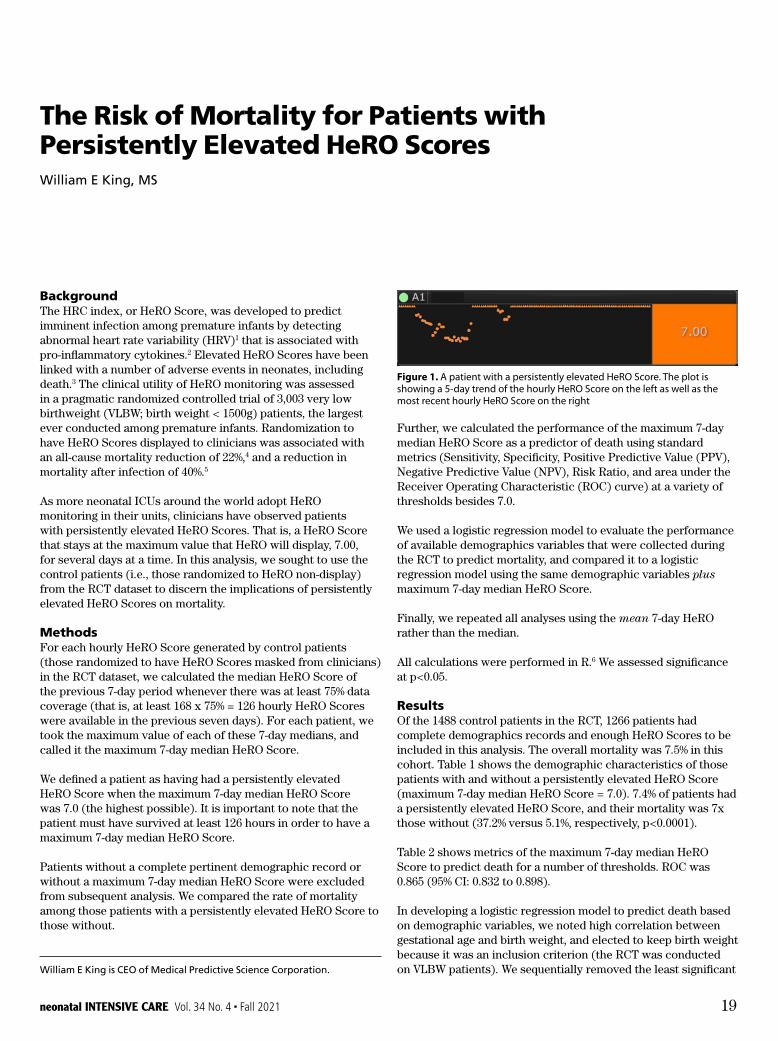
BackgroundThe HRC index, or HeRO Score, was developed to predict imminent infection among premature infants by detecting abnormal heart rate variability (HRV)1 that is associated with pro-inflammatory cytokines.2 Elevated HeRO Scores have been linked with a number of adverse events in neonates, including death.3 The clinical utility of HeRO monitoring was assessed in a pragmatic randomized controlled trial of 3,003 very low birthweight (VLBW; birth weight < 1500g) patients, the largest ever conducted among premature infants. Randomization to have HeRO Scores displayed to clinicians was associated with an all-cause mortality reduction of 22%,4 and a reduction in mortality after infection of 40%.5
As more neonatal ICUs around the world adopt HeRO monitoring in their units, clinicians have observed patients with persistently elevated HeRO Scores. That is, a HeRO Score that stays at the maximum value that HeRO will display, 7.00, for several days at a time. In this analysis, we sought to use the control patients (i.e., those randomized to HeRO non-display) from the RCT dataset to discern the implications of persistently elevated HeRO Scores on mortality.
MethodsFor each hourly HeRO Score generated by control patients (those randomized to have HeRO Scores masked from clinicians) in the RCT dataset, we calculated the median HeRO Score of the previous 7-day period whenever there was at least 75% data coverage (that is, at least 168 x 75% = 126 hourly HeRO Scores were available in the previous seven days). For each patient, we took the maximum value of each of these 7-day medians, and called it the maximum 7-day median HeRO Score.
We defined a patient as having had a persistently elevated HeRO Score when the maximum 7-day median HeRO Score was 7.0 (the highest possible). It is important to note that the patient must have survived at least 126 hours in order to have a maximum 7-day median HeRO Score.
Patients without a complete pertinent demographic record or without a maximum 7-day median HeRO Score were excluded from subsequent analysis. We compared the rate of mortality among those patients with a persistently elevated HeRO Score to those without.
Further, we calculated the performance of the maximum 7-day median HeRO Score as a predictor of death using standard metrics (Sensitivity, Specificity, Positive Predictive Value (PPV), Negative Predictive Value (NPV), Risk Ratio, and area under the Receiver Operating Characteristic (ROC) curve) at a variety of thresholds besides 7.0.
We used a logistic regression model to evaluate the performance of available demographics variables that were collected during the RCT to predict mortality, and compared it to a logistic regression model using the same demographic variables plus maximum 7-day median HeRO Score.
Finally, we repeated all analyses using the mean 7-day HeRO rather than the median.
All calculations were performed in R.6 We assessed significance at p<0.05.
ResultsOf the 1488 control patients in the RCT, 1266 patients had complete demographics records and enough HeRO Scores to be included in this analysis. The overall mortality was 7.5% in this cohort. Table 1 shows the demographic characteristics of those patients with and without a persistently elevated HeRO Score (maximum 7-day median HeRO Score = 7.0). 7.4% of patients had a persistently elevated HeRO Score, and their mortality was 7x those without (37.2% versus 5.1%, respectively, p<0.0001).
Table 2 shows metrics of the maximum 7-day median HeRO Score to predict death for a number of thresholds. ROC was 0.865 (95% CI: 0.832 to 0.898).
In developing a logistic regression model to predict death based on demographic variables, we noted high correlation between gestational age and birth weight, and elected to keep birth weight because it was an inclusion criterion (the RCT was conducted on VLBW patients). We sequentially removed the least significant
The Risk of Mortality for Patients with Persistently Elevated HeRO ScoresWilliam E King, MS
William E King is CEO of Medical Predictive Science Corporation.
Figure 1. A patient with a persistently elevated HeRO Score. The plot is showing a 5-day trend of the hourly HeRO Score on the left as well as the most recent hourly HeRO Score on the right
neonatal INTENSIVE CARE Vol. 34 No. 4 n Fall 2021 19

persistently elevated HeRO Scores. We found that those patients with a maximum 7-day median HeRO Score of 7 or more had a profound risk of death when compared to other patients (37.2% versus 5.1%, respectively).
Furthermore, we found that patients with low maximum 7-day median HeRO Scores had remarkably low rates of death. For the 35% of patients for whom the maximum 7-day median HeRO Score fell below 1.0, survival was 99.8%, and these patients are more than 50x less likely to die than other patients. The mean HeRO Score over the previous 7 days appeared to have even better performance because it was able to capture late spikes in HeRO prior to death better than the median over the previous seven days. Essentially, patients did not die without elevations in their HeRO Scores.
These results remained true when controlling for demographic predictors.
Weaknesses of the current analysis are its retrospective nature and lack of a complete set of demographic predictors. A strength is that it is a large dataset of patients for whom the HeRO Score was generated but not displayed to clinicians, eliminating a potential feedback loop of scores affecting outcomes.
variable one at a time while there were any non-significant predictor variables until all predictor variables were significant. The resulting model included two predictor variables: birth weight and Apgar2, and had an ROC area for predicting death of 0.825 (95% CI: 0.789 to 0.861).
When we re-trained the logistic regression model using the same demographics variables, plus maximum 7-day median HeRO Score, all variables were significant with maximum 7-day median HeRO Score having p < 0.0001. The ROC of this combined demographics plus HeRO model was 0.876 (95% CI: 0.848 to 0.905). Figure 2 shows the predictiveness curve of the combined demographics plus maximum 7-day median HeRO Score model to predict death.
Performance using maximum 7-day mean HeRO Score was slightly improved relative to the median. ROC of the maximum 7-day mean HeRO Score was 0.871 (95% CI: 0.840 to 0.903) versus 0.865 for the median. Remarkably, the risk ratio of death for those above the 50th percentile versus below the 50th percentile of the demographics plus maximum 7-day mean HeRO Score model was 92x, versus 46x for same model using the median.
DiscussionThe randomized controlled trial of HeRO monitoring was a pragmatic design. That is, clinicians were not instructed to perform specific clinical actions at set thresholds of HeRO Score. Instead, they were educated on how the HeRO Score was developed and left to incorporate HeRO into their clinical practice as they saw fit.
An advantage of this pragmatic design is that the profoundly positive results of the RCT should be reproducible in clinical practice outside the rigors of an RCT protocol. Nevertheless, clinicians are left without concrete direction as to what thresholds of HeRO Score are actionable.
Of utmost difficulty for the practicing neonatologist is a patient with persistently elevated HeRO Scores. Although the numeric value is high, there is no discernible trend to guide the clinician that this patient is trending “better” or “worse”.
In the present analysis, we have re-analyzed control patients in the HeRO randomized controlled trial to identify patients with
Table 1. Patient Demographics. Continuous variables were evaluated with a t-test, categorical variables were evaluated with a test of proportions.
No Persistently Elevated HeRO Score
Persistently Elevated HeRO Score p
N (% of total) 1172 (92.6%) 94 (7.4%)Gestational Age (sd) 28.3 (2.7) 25.2 (1.5) < 0.0001Birth Weight (sd) 1018 (282) 724 (184) < 0.0001Male Sex (%) 601 (51.3%) 47 (50%) 0.895Apgar 1 (sd) 5.2 (2.5) 3.2 (2.2) < 0.0001Apgar 2 (sd) 7.2 (1.8) 5.7 (2.0) < 0.0001Died (%) 60 (5.1%) 35 (37.2%) < 0.0001
Table 2. Metrics of various thresholds of maximum 7-day median HeRO Score to predict death.
HeRO Threshold Percentile Sensitivity Specificity PPV NPV Risk Ratio1.0 35.4th 98.9% 38.3% 11.5% 99.8% 51.7x2.0 58.8th 93.7% 63.1% 17.1% 99.2% 21.2x5.0 85.0th 62.1% 88.8% 31.1% 96.7% 9.3x7.0 92.6th 36.8% 95.0% 37.2% 94.9% 7.3x
Figure 2. Predictiveness curve of the combined demographics plus maximum 7-day median HeRO Score model to predict death. The x-axis represents the percentile of model’s predictions, the y-axis represents the rate of death. The smooth blue line is the model’s predicted risk of death. The black bars represent the observed rate of death for each decile, boxed by 95% confidence intervals in green.
20 neonatal INTENSIVE CARE Vol. 34 No. 4 n Fall 2021

6 R Core Team. R: A language and environment for statistical computing. R Foundation for Statistical Computing, Vienna, Austria. URL https://www.R-project.org/, 2020.
ConclusionClinicians should remain vigilant to the subgroup of patients with persistently elevated HeRO Scores due to their profound risk of death, whereas consistently low HeRO Scores provide reassurance of low risk.
References1 Griffin MP, O’Shea TM, Bissonette EA, Harrell FE, Lake DE,
Moorman JR. Abnormal heart rate characteristics preceding neonatal sepsis and sepsis-like illness. Pediatr Res, 53: 920-926, 2003.
2 Fairchild KD, Saucerman JJ, Raynor LL, Sivak JA, Xiao Y, Lake DE, Moorman JR. Endotoxin depresses heart rate variability in mice: cytokine and steroid effects. American Journal of Physiology Regul Integr Comp Physiol. 297:R1019-27, 2009.
3 Sullivan BA, McClure C, Hicks J, Lake DE, Moorman JR, Fairchild KD. Early Heart Rate Characteristics Predict Death and Morbidities in Preterm Infants. J Pediatr 2016.
4 Moorman JR, Carlo WA, Kattwinkel J, Schelonka RL, Porcelli PJ, Navarrete CT, Bancalari E, Aschner JL, Whit Walker M, Perez JA, Palmer C, Stukenborg GJ, Lake DE, Michael O’Shea T. Mortality reduction by heart rate characteristic monitoring in very low birth weight neonates: a randomized trial. J Pediatr. 2011.
5 Fairchild KD, Schelonka RL, Kaufman DA, Carlo WA, Kattwinkel J, Porcelli PJ, Navarrete CT, Bancalari E, Aschner JL, Walker MW, Perez JA, Palmer C, Lake DE, O’Shea TM, Moorman JR. Septicemia mortality reduction in neonates in a heart rate characteristics monitoring trial. Pediatr Res, 2013.
Contact us to learn more:[email protected] 800-394-1625
www.heroscore.com
Clear Evidence of the Best Care: ● Heart rate characteristics monitoring and reduction in mortality or neurodevelopment impairment in extremely low birthweight infants with sepsis. Early Human Development
● Heart-rate-characteristic monitoring decreases NICU length of stay. JPeds
● Predicting Extubation Outcomes - A Model Incorporating Heart Rate Characteristics Index. JPeds
● New Technology is Life Saving Voice for Premature or Critically Ill Infants. Neo Today
● Predictive monitoring for sepsis and necrotizing enterocolitis to prevent shock. J Sem Fetal Neonatal Med.
● HeRO monitoring in the the NICU: sepsis detection and beyond. J Adv Neonatal Care
I use HeRO ... because the evidence matters
neonatal INTENSIVE CARE Vol. 34 No. 4 n Fall 2021 21

Pregnancy requires an increased nutrient intake in order to support the growing fetus and maternal well-being. Maternal micronutrient deficiencies during pregnancy are associated with pregnancy complications, as well as adverse fetal growth and development. The vitamins and microelements required during pregnancy and lactation compared to the non-pregnant state are summarized in Table 1.1
Table 1
Non-Pregnant women
Pregnant Women
Lactating Women
Folic Acid 400 600 500
Vitamin B12 2.4 2.6 2.8
Vitamin D 5 5 5
Calcium 1000 1000-1300 1000-1300
Iron 18 27 9-10
Iodine 150 220 270
Zinc 8 10-11 11-12
Selenium 60 65 75
The current recommendation can be summarized as follows:
Vitamin or Mineral
General Recommendations
Special Considerations
Folic Acid At least 0.4 daily for a minimum of 1 month before conception and the first 12 weeks of pregnancy
A higher daily dose of 5 mg recommended for women with increased risk of NTD
Iodine All pregnant women should take 150 micrograms daily
Mandatory in areas of regional deficiency
Vitamin B12 Routine supplementation not recommended
Consider supplementation in pregnancy and lactation for women who are vegetarian or vegan
Vitamin D Routine screening and supplementation of vitamin D is not currently recommended
Offer vitamin D screening to women with limited exposure to sunlight
Vitamin D deficiency is associated with an increased risk of pregnancy complications, including pre-eclampsia, preterm birth, and low birth weight. Severe maternal vitamin D deficiency has been associated with biochemical evidence of disordered skeletal homeostasis, congenital rickets, and fractures in the newborn.5,6 Vitamin D deficiency is common among high-risk groups, vegetarians, women with limited sun exposure, and ethnic minorities, especially those with darker skin.7-9 In 2010, the food and nutrition board at the Institute of Medicine of the National Academies stated that an adequate intake of vitamin D during pregnancy and lactation is 600 international units daily.10 It is recommended that pregnant women increase their daily iron intake. Iron supplementation reduces the risk of maternal anemia and iron deficiency. There is evidence for an increase in preterm birth and low birth weight in settings where nutritional intake is poor.11 Hypocalcemia has been associated with pre-eclampsia.12 Therefore calcium supplementation is recommended as a preventative measure for pre-eclampsia, particularly in groups of women who avoid dairy products.
To address potential micronutrient deficiencies, many women take pregnancy multivitamins. Despite high-frequency use and widespread marketing and promotion, there is a lack of scientific evidence that routine supplementation with multivitamins is beneficial for pregnancy outcomes.13,14 In many cases, multivitamins may contain excessive amounts of vitamins and minerals. Such supplements may not be without harm. Supplementation with a combination of vitamins C and E from 12-18 weeks gestation was shown to increase the risk of fetal loss or perinatal death.15 Furthermore, vitamin A is known as a teratogen, a high intake during pregnancy is associated with an increased risk of congenital malformations.16,17 Therefore, some degree of caution regarding widespread routine supplementation practices is warranted.1
ACOG recommends vitamins that contain at least 600MG of folic acid, 200mg of DHA, 27 mg of iron, 1.000 mg of calcium, and 600 mg of vitamin D. Most brands (Olly, Nature Made Prenatal, Mama Bird, Smarty Pants, etc.) fulfill these criteria. The recommendations are to use them daily in all the trimesters of pregnancy. However, maternal-fetal demands and possible complications are trimester-dependent. Folic acid, for example, is much needed before conception and in the first trimester to decrease the risk of neural tube defects. Since the mid-1990’s health care organizations around the world have adopted policies designed to increase the dietary intake of folic acid before and during pregnancy to reduce the risk of having a
A New System of Vitamin Administration During Pregnancy and PostpartumBM Petrikovsky, MD, PhD
BM Petrikovsky is a Director at the Prenatal Diagnostic Unit Services, New York Downtown Hospital, New York, NY.
22 neonatal INTENSIVE CARE Vol. 34 No. 4 n Fall 2021

10 Insitute of Medicine of the National Academies (US) Dietary reference intakes for calcium and vitamin D. Washingto,D.C: National Academy Press :2010
11 Scholl TO. Iron status during pregnancy: setting the stage for mother and infant. Am J Clin Nutr 2005;81 121 8s-22s
12 Kazerooni T,Hamze-Nejadi S. Calcuim to cretinine ratio in a spot sample of urine for early predicition of pre-eclampisa. Int J Gynaecol Obstet 2003;80:279-83
13 Rumbold A, Duley L, Crother CA, Haslam RR. Antinxidants for preventing pre-eclampsia Hobokem, NJ: The Cohrane Library:2008
14 Balogun OO, da Silva Lopes K, Ota E, Takemoto Y, Rumbold A, et al. Vitamin supplementation for preventation miscarriage.Hoboken , NJ : The Cochrane Library; 2016.
15 Rumbold A, Ota E, Nagata C, et al.. Vitamin C supplementaion in preganacy. Hoboken,NJ” The Cochrane library:2015
16 Xu H, Perez-Cuevas R, Xiong X, et al. An international trial of antioxidants in the oreventation of preclampsia(INTAPP). Am J Obstet Gynecol 2010;202: study.
17 Mastroiacovo PA, Mazzone T, Addis A, et al. High vitamin A intake in early pregnancy and major malformation: a mulitcenter prospective controlled study. Teratology 1999;59;7-11
18 Al- Gailani S, Making birth defects prevehtable. Pre-conceptional vitamin supplements and politics of risk reduction. Hhpts://ww.ncbi.2021.nih.gov/pmc/articles/pmc4275593
baby with a defect of the brain and/or spinal cord. Folic acid is the synthetic form of folate, a B-group vitamin naturally present in many foods, particularly leafy green vegetables.18 The MRC vitamin study group reported a 72% protective effect for folic acid in at-risk pregnancies.12 There is no proven need for folic acid supplementation after the closure of the neural tube. Calcium on the other hand is much needed in the second trimester with the rapid growth of the fetal skull and skeleton. Iron supplements are mostly needed in the second and third trimesters in anticipation of post-delivery blood loss. Many of the vitamins and supplements have side effects, e.g. constipation and nausea in case of iron supplementations.
An unsuccessful attempt to introduce a similar concept was undertaken by Perelel. (CA) They created trimester-specific packages e.g. the use of probiotics and Omega DHA for brain development, which at this time has no scientific basis. On the other hand, appropriate dosages of vitamin B3 and D in the first trimester are not included. Vitamin B3 is known to prevent miscarriages.
The Fetal Research Fund created a new type of prenatal vitamins based on the nutritional needs of individual patients and the trimester of pregnancy. Regarding vitamin D, Fetal Research Fund uses recommendation of the committee on Nutrition of the American Academy of Pediatrics which recommends that a daily intake higher than that recommended by the Food and Nutrition board may be needed to maintain maternal vitamin D sufficiency. Most experts agree that supplemental vitamin D is safe in dosages up to 4,000 international units per day during pregnancy or lactation.20
References 1 Wilson RL, Gumow JA, McAnich D, et al. Vitamin and mineral
supplementation in pregnancy: evidence to practice Pharm Practice and Reserch 2018,186-192
2 Baker AM, Haeri S, Camargo CA Jr, et al. A nested case-control study of midgestation vitamin D deficiency and risk of severe preeclampsia. J Clin Endocrinol Metab 201:95:5105-9
3 Marjamaki L, Ninisto S, Kenward MG, et al. Maternal intake of vitamin D durning pregnancy and risk of advance beta cell autoimmunity and type 1 diabetes in offspring. Diabetologia 2010;53:1599-607
4 Leffelaar ER, Vrijkotte TG, van Eijsden M. Maternal early pregnancy vitamin D status in relation to fetal and neonatal growth: results of the multi-ethnic Amsterdam born children and their development cohort. Br J Nutr 2010;104:180-17
5 Pawley N , Bishop NH. Prenatal and infant predictors of bone health: the influence of vitamin D. Am J Clin Nutr 2004:80:1748S-51S
6 Gale CR, Robinson SM, Harvey NC,et al. Maternal Vitamin D status during pregnancy and child outcomes. Princess Anne Hospital Study Group.Eur J Clin Nutr 2008;62;68-77
7 Hollis BW, Wagner CL, Assessment of dietary vitamin D requirements during pregnancy and lactation.Am J Clin Nutr 2004;79:717-26
8 Lee JM,Smith JR, Phillip BL, et al Vitam D deficient in a healthy group of mothers and newborn infants. Clin Pediatr 2007;46;42-4
9 Bodnar LM, Simhan HN, Powers High prevalence of vitam D insufficiency in black and white pregnant women residing in the in the northern United States and their neonates. J Nutr 2007;137;447-52
neonatal INTENSIVE CARE Vol. 34 No. 4 n Fall 2021 23

Measuring preterm infants’ cerebral oxygen levels in their first 96 hours of life via near-infrared spectroscopy (NIRS) can help identify patients at risk for death or severe neuroradiographic brain injury in early life, a recent study has demonstrated. Among 103 subjects, who were born at a mean gestational age of 26 weeks and weighed less than 1250 g at birth, cerebral oxygen saturation below 50% was significantly linked with the adverse outcomes measured in the study. The findings were published in December 2020 in the Journal of Pediatrics.
NIRS is a noninvasive method for measuring tissue oxygen saturation at the bedside. Preliminary research suggested that measuring cerebral oxygen levels with a NIRS sensor might provide clinically useful information about preemies’ risk of death or brain injury, but the idea has had only limited testing using sensor equipment designed for neonates.
The multicenter study, led by Stanford Children’s Health neonatologists Valerie Chock, MD, associate professor of pediatrics, and Krisa Van Meurs, MD, professor of pediatrics, enrolled infants during their first 24 hours of life and continuously measured their cerebral oxygen saturation via a NIRS sensor placed on each patient’s forehead, as well as their mean arterial blood pressure via an indwelling arterial line, until 96 hours of age. Twenty-one infants (20% of the study sample) were included in the primary outcome group because they died before hospital discharge (11 patients), or developed grade 3 or 4 intraventricular hemorrhage or periventricular leukomalacia in the first 10 days after birth (14 patients). Four patients who died also had severe intraventricular hemorrhage. During their first four days of life, infants with adverse outcomes spent significantly more time than other study subjects with cerebral oxygen saturation below 65%, 55% and 50%, the study found.
More research, including randomized trials, will be needed to determine what thresholds of oxygen saturation should trigger clinical actions, the researchers said. Future research could also ask whether cerebral oxygen saturation in early life has predictive value for long-term cognitive outcomes in preemies, and whether different outcomes are seen in babies who are small for gestational age in addition to being premature.
Still, the findings hold important clues to the utility of noninvasive monitoring of brain oxygen levels in the earliest-born and most vulnerable preemies, the researchers said. “Our
study is the largest to date to link impaired regulation of cerebral blood flow in the first few days of life to mortality or early brain injury,” said Chock. “These findings underscore the need to maintain stable blood flow to the brain in very early and small preemies.”
Noninvasive Technique to Track Early Preemies’ Cerebral Oxygen Levels Can Help Predict Neonatal Death, Brain InjuryErin Digitale
Erin Digitale is a pediatrics science writer at Stanford Medicine.
24 neonatal INTENSIVE CARE Vol. 34 No. 4 n Fall 2021

Effects of a Tracheostomy in NeonatesWhen a child is born prior to the full gestational period and is premature, the trajectory of their care is impacted by multiple factors. Often neonates considered at-risk fall into either a specific birth weight category or gestational age classification. Typically, infants fall into a birth weight category based on the classifications defined by the Center for Disease Control: extremely low birth weight (< 1,000 g), very low birth weight (< 1,500 g), low birth weight (< 2,500 g), normal birth weight (< 4,000 g), and high birth weight (> 4,000 g) (Martin et al., 2011). An infants’ birth weight has been found to be a strong predictor of health outcomes, such a survival, risk of medical complications, and timing for the achievement of developmental milestones (Gill et al., 2013). Examples of complications associated with low to extremely low birth weight are hypothermia, hypoglycemia, perinatal asphyxia, respiratory distress, and several others. The lower the birth weight classification, the higher the risk of health complications.
Another classification process that is important to consider is the gestational age in combination with birth weight. For this classification, the system identifies if an infant is small for gestational age (below the 10th percentile), appropriate for gestational age (between the 10th and 90th percentiles), or large for gestational age (above the 90th percentile) (Gill et al., 2013). Particular health risks have been associated with the assigned classification.
When considering an infant that exhibits the respiratory complications that can arise with low birth weight, the
interventions that become necessary also run risks of added complications. For example, upon intubation which causes alterations in airflow and pressures, the risks of decreased olfaction (loss of smell); diminished positive airway pressures; and changes in cough, cry, and phonation exist. With the diminished capacity to access these functions, secondary complications with decreased developmental milestones for feeding and swallowing, communication, and bonding may occur. With long-term respiratory support needs, having a tracheostomy may be considered; however, a tracheostomy is not without complications.
The need for an artificial airway is determined based on multiple factors, including life expectancy, birth weight, gestational age, comorbid conditions, congenital abnormalities, complications, and potential outcomes. At birth, the customary APGAR scoring system (1 to 10 scale) is commonly used. This assessment tool is a method for evaluating an infant’s health at the time of birth and summarizes the appearance (skin color), pulse, grimace (reflex to stimulation), activity (muscle tone), and respirations (APGAR) of the infant. If the infant presents with deficiencies and has an APGAR score of less than seven, therapeutic interventions by clinicians and other healthcare professionals will be conducted to increase the score. Assessment continues in short intervals to monitor progress. While the infant could present with a normal to midrange score at first, some also have demonstrated a rapid decline with presentation of such changes as shallow breathing, apnea, duskiness, and oxygen saturation between 70 and 80%. This process of evaluating based on the Apgar score assists with monitoring the need for immediate medical attention or the potential for long-term issues based on the scores at various time intervals.
Over time, the need for a tracheostomy tube has changed due to improvements in non-invasive ventilation options, but despite this advancement, tracheostomy placement has increased by 31% in the neonate population as reviewed from 2006-2012 (Wang et al., 2020). Placement often will depend on the philosophies of the facility and physicians as it relates to long-term ventilation needs and use of intubation. The occurrence of tracheostomy occurred most often in the extremely preterm neonates; however, the mortality rate was reported to be lower in those neonates receiving a tracheostomy as compared to those without a tracheostomy (18% vs 21%) (Wang et al., 2020). Overman et al. (2013) reported that infants with extremely low birth weights were more likely to have tracheostomies but found that even with high survival, developmental delays and comorbidities
Considerations for the Impact of a Tracheostomy: Respiration and SwallowingKristin A King, PhD, CCC-SLP and Gabriela Ortiz, BSRT, RCP
Kristin King, PhD, CCC-SLP: With 25 years of experience in medical, academic, and industry settings, Dr. King brings a unique perspective of medical speech pathology. Her research, publications, and teachings focus on traumatic brain injury, swallowing disorders, and critical care (tracheostomy and mechanical ventilation) for both pediatric and adult patient populations. She has been an invited speaker both domestically and internationally and has published in peer-reviewed journals. Currently, Dr. King is the Vice President of Clinical Education and Research for Passy-Muir, Inc. Gabriela Ortiz has been in the field of respiratory care since 2006. She lives in Southern California and has worked in clinical education and sales roles, using knowledge gained from her clinical experience in critical care ventilation products for the ICU, PICU within acute and subacute hospitals. Gabriela is an invited guest speaker at schools, Better Breather’s Group meetings, and ALS support groups. She currently is a full-time clinical specialist with Passy-Muir, Inc.
neonatal INTENSIVE CARE Vol. 34 No. 4 n Fall 2021 25

through the types of cries used to indicate hunger and pain. Having an infant lose the ability to cry and interact with the caregiver also may have a negative effect on bonding.
Infant SwallowingWhen analyzing the respiratory side of swallowing in full-term, healthy infants, coordination is characterized by the cessation of breathing during swallowing process. The brief apneic period may occur in any of the following stages of respiration: mid-inspiration, mid-expiration, and between inspiration and expiration or expiration and inspiration. Having a tracheostomy tube, and especially with mechanical ventilation, may disrupt this cycle.
The act of swallowing is a complex and multi-faceted act that begins developing in utero, which requires the use of structures and functions that also are utilized during respiration, swallowing, and phonation (Pullens & Streppel, 2021). Infants start to experience swallowing around 11 to 12 weeks of the gestational period and suckling at approximately the 24th week of gestation. Lau (2015) reports that a child may breathe at a rate of 40-60 breaths per minute (bpm) or 1.5-1 breaths per second for the typically developing infant. If an infant exhibits an immature swallow, and immature swallows may last from 0.35 to 0.75 seconds, then this may cause breathing to be compromised. These impacts are regardless of the presence of a tracheostomy tube. The difficulties may arise with the attempts to coordinate respiration and swallowing in the infant, especially with either delayed or disordered swallow function.
Now, consider the negative impact of a tracheostomy tube on the trachea, compounding the difficulty if the infant already has a compromised system. Typically, the normal movement of the larynx helps protect the airway and open the esophagus during swallowing. However, when a tracheostomy tube is placed, the possibility of laryngeal tethering increases, and hyolaryngeal excursion may slow due to interruption and sometimes stoppage of the upward and forward movement of the larynx during swallowing. Birutis et al. (n.d.) reported that when studying infants with tracheostomy tubes, they found that both the presence of a tracheostomy tube and the age of the infant were factors in the occurrence of aspiration; however, duration time with a tracheostomy tube was not a factor. They also found that of those children who aspirated, 93.3% of them did so silently; however, of note, they found that the younger the child, the lower the incidence of aspiration.
Another factor is the alteration of subglottal pressure during swallowing, which may affect how the vocal folds close. Normally, the air pressure builds underneath the vocal folds to establish subglottic pressure, with this pressure occurring during speech, sound production, swallow, cough, and throat clear. The pressure that builds during swallowing assists with the hyolaryngeal excursion and is exhaled at the end of a swallow. This process assists with sensation, bolus clearance, and airway protection. Any food or liquid that may have entered the airway may be cleared by the force from the exhaled airflow. Unfortunately, the presence of an open tracheostomy tube prevents the creation of subglottal pressure during a swallow. For that matter, all types of pressure in the aerodigestive tract are compromised when an open tracheostomy tube is placed. Pressure to assist with bolus transition through the pharyngeal area diminishes and may lead to increased residue or decreased cricopharyngeal opening during the swallow. Another aspect
were prevalent in this patient population. Akangire et al. (2020) reported that the most common co-morbidity in this patient population is dysphagia, swallowing and feeding disorders.
ComplicationsThough a tracheostomy may be received shortly after birth, a retrospective study by Akangire et al., (2020) found that the mean age of tracheostomy was 4.5 months with median age of 3 months in their cohort, which was similar to other reports (Unal et al., 2015). Implications and the impact of a tracheotomy tube are multifactorial.
For some, a tracheostomy may not be permanent, but when in place the potential impacts are many. Having an open tracheostomy tube with or without mechanical ventilation may cause changes such as (Passy-Muir, Inc., 2021):• Lossofsenseoftasteandsmell.• Reducedsubglotticpressure.• Decreasedlaryngealandpharyngealsensation,altering
secretion management.• Increasedpoolingofsecretions.• Reducedcougheffort.
A tracheostomy tube is placed in the trachea to provide an access so that respiratory support occurs directly to the lower respiratory system; however, this placement of a tracheostomy tube bypasses the upper airway, eliminating or diminishing airflow out through the mouth and nose. This change in the direction of airflow is what has the potential impact on functions. These changes to the aerodigestive system also may impact development of feeding and swallowing skills, along with potential delay of communication, even infants communicate
26 neonatal INTENSIVE CARE Vol. 34 No. 4 n Fall 2021

and sucking for nutritive and non-nutritive purposes. The term obligate nose breathing is used to define the normal breathing of infants who rely on the autonomic system and the ability to synchronize activities of sucking, swallowing, breathing and esophageal function in proper sequence. Without the proper coordination of respiratory and subglottic function, safe oral feeding may become an issue and drastically impact the quality of life (QOL) of a child. It is essential that swallowing be a consideration for monitoring, evaluation, and intervention when an infant has to have a tracheostomy tube.
References• Abraham, S. (2003). Babies with tracheostomies: The
challenge of providing specialized clinical care. The ASHA Leader, 8(5). https://doi.org/10.1044/leader.FTR2.08052003.4
• Akangire, G., Manimtim, W., Nyp, M., Townley, N., Dai, H., Norberg, M., & Taylor, J. B. (2017). Factors leading to rehospitalization for tracheostomized and ventilator-dependent infants through 2 years of age. Journal of Perinatology, 37, 857–863. https://doi.org/10.1038/jp.2017.54
• Akangire, G., Taylor, J.B., McAnany, S., Noel-MacDonnell, J., Lachica, C., Sampath, V., & Manimtim, W., (2020). Respiratory, growth, and survival outcomes of infants with tracheostomy and ventilator dependence. Pediatric Research, Advance online publication. https://doi.org/10.1038/s41390-020-01183-x
• Birutic, R., Keen, M., Brady, S., & Wesling, M. (n.d.) Swallowing function for pediatric patients with a tracheostomy tube. [poster] AACPDM Conference.
• Brooks, L., Figueroa, J., Edwards, T., Reeder, W., McBrayer, S., & Landry, A. (2019). Passy Muir Valve
• tolerance in medically complex infants and children: Are there predictors for success? Laryngyscope, 130(11), E632-E639. https://pubmed.ncbi.nlm.nih.gov/31825087/
• Children’s Health Queensland Hospital and Health Services (2017). Tracheostomy tubes-feeding, eating, and drinking. Retrieved from, https://www.childrens.health.qld.gov.au/fact-sheet-tracheostomy-tubes-feeding-eating-and-drinking/
• Gill, S. V., May-Benson, T.A., Teasdale, A., & Munsell, E.G. (2013). Birth and developmental correlates of birth weight in a sample of children with potential sensory processing disorder. BMC Pediatric, 13, 29. https://doi.org/10.1186/1471-2431-13-29
• Gross, R. D., Mahlmann, J., & Grayhack, J. P. (2003). Physiologic effects of open and closed tracheostomy tubes on the pharyngeal swallow. Annals of Otology, Rhinology & Laryngology, 112(2), 143-152. https://doi.org/10.1177/000348940311200207
• Gross, R. D., Steinhauer, K. M., Zajac, D. J., & Weissler, M. C. (2006). Direct measurement of subglottic air pressure while swallowing. The Laryngoscope, 116(5), 753-761. https://doi.org/10.1097/01. mlg.0000205168.39446.12
• Lau. C. (2015). Development of suck and swallow mechanisms in infants. Annals of Nutrition and Metabolism, 66(5), 7-14. https://doi.org/10.1159/000381361
• Martin, J.A., Hamilton, B.E., Ventura, S.J., Osterman, M.J., Kirmeyer, S., Mathews, T.J., & Wilson, E.C. (2011). Births: final data for 2009. National Vital Statistics Report, 60(1), 1–70.
• O’Connor, L.R., Morris, N.R., & Paratz, J. (2018). Physiological and clinical outcomes associated with use of one-way speaking valves on tracheostomised patients: A systematic review. Heart & Lung, 48(4), 356 – 364. https://doi.org/10.1016/j.hrtlng.2018.11.006
• Overman, A. E., Liu, M., Kurachek, S. C., Shreve, M.
of pressure that is negatively impacted is the ability to build backpressure as is a part of the physiologic positive end-expiratory pressure (PEEP), which is needed to keep the alveoli open for appropriate gas exchange. Having diminished PEEP strains the mechanisms for ventilation and oxygenation.
Separate from potential impacts on movement and pressure during the swallow is that without airflow to the upper airway, loss or decrease in the sensitivity in the upper airway may be affected. This decrease in stimulation compromises sensation, which will not allow sufficient stimulation for effective swallow or cough; thereby, seriously compromising airway protective reflexes which would normally react to stop food and liquid from going into the airway and lungs.
While the act of feeding and swallowing may directly impact the ability to coordinate respiration and swallowing, another effect is the influence on respiratory rate. During feeding, the respiratory rate may decrease, which in turn impacts minute ventilation. The effect on the minute ventilation may prolong the expiratory phase and shorten the inspiratory phase. Changes in the phases of respiration may affect both the safety and effectiveness of the swallow. It is important to acknowledge that impaired breathing may negatively affect swallowing.
Restoring airflow and pressuresStudies have shown that placement of a no-leak speaking Valve helps restore the pressurized systems; therefore, wearing a speaking valve may assist with restoring subglottal pressure (Gross et al., 2003; Gross et al., 2006). Research and studies also have indicated that the application of the no-leak Valve not only restores pressure but also restores voicing, improves laryngeal and pharyngeal sensation, improves swallow and cough, and in some patients may improve swallow function (Gross et al., 2003; Brooks et al., 2019; O’Connor et al., 2018).
The Passy-Muir® Valve (PMV) allows the patient to breathe in through the tracheostomy tube but is only open during active inhalation, redirecting airflow up to the upper airway through the vocal folds and out through the mouth and nose during exhalation. This redirection of airflow and re-engagement of the valving mechanisms in the subglottic region helps restore the body’s physiologic pressures. Brooks et al. (2019) reported that use of the no-leak PMV is being underutilized with infants and children who are medically fragile, whether on mechanical ventilation or tracheostomy collar. The study provides evidence as to the factors that predict successful use of the PMV. The authors report that vocalizing, transtracheal pressures, age, and weight are all associated with successful use. When the patient is on a ventilator, the only related factor specific to mechanical ventilation that predicted successful use of the PMV inline was ventilator rate; PEEP, pressure support, and FiO2 were not correlated or predictors of the ability to use the Valve. With the higher risk that has been identified in pre-term infants for respiration, coordination and swallowing, and aspiration, providing restored functions and pressures may assist with improved status. Research in adults has shown that early use of a no-leak speaking Valve leads to earlier weaning and decannulation (O’Connor et al., 2018; Rodrigues et al., 2015). More research is needed in this area for pediatrics.
ConclusionAs a result of physiologic changes following tracheostomy, limitations may be imposed on the coordination of breathing
neonatal INTENSIVE CARE Vol. 34 No. 4 n Fall 2021 27

R., Maynard, R. C., Mammel, M. C., & Moore, B. M. (2013). Tracheostomy for infants requiring prolonged mechanical ventilation: 10 years’ experience. Pediatrics, 131(5) e1491-e1496. https://doi.org/10.1542/peds.2012-1943
• Passy-Muir, Inc. (2021) Application of the Passy-Muir Swallowing and Speaking Valve. Remote live in-service education. Retrieved from, https://www.passy-muir.com/remote-live-inservices
• Pullens, B. & Streppel, M. (2021). Swallowing problems in children with tracheostomy. Seminars in Pediatric Surgery, 30(3), 151053. https://doi.org/10.1016/j.sempedsurg.2021.151053
• Rodrigues, K. A., Machado, F. R., Chiari, B. M., Rosseti, H. B., Lorenzon, P., & Gonçalves, M. I. (2015). Swallowing rehabilitation of dysphagic tracheostomized patients under mechanical ventilation in intensive care units: A feasibility study. Revista Brasileira De Terapia Intensiva, 27(1), 64-71. https://doi.org/10.5935/0103- 507x.20150011
• Trabalon, M. & Schaal, B. (2012). It takes a mouth to eat and a nose to breathe: Abnormal oral respiration affects neonates’ oral competence and systemic adaptation. International Journal of Pediatrics. 2012, 207605. https://doi.org/10.1155/2012/207605
• Unal, S., Bilgin, L. K., Gonulal, D., & Akcan, F. A. (2015). Optimal time of tracheotomy in infants: still a dilemma. Global Pediatric Health, 2, 2333794X15569300. https://doi.org/10.1177/2333794X15569300
• Wang, C. S., Kou, Y., Shah, G. B., Mitchell, R. B., & Johnson, R. F. (2019). Tracheostomy in extremely preterm neonates in the United States: A cross-sectional analysis. The Laryngoscope, 130(8). https://doi.org/10.1002/lary.28304
• Yi, Y. G., Oh, B. M., Yang, S., & Shin, H. I. (2019). Oral feeding challenges in children with tracheostomy can improve feeding outcomes, even with the finding of aspiration. Frontiers in Pediatrics, 7, 362. https://doi.org/10.3389/fped.2019.00362
28 neonatal INTENSIVE CARE Vol. 34 No. 4 n Fall 2021

Probiotic use in the NICU has been considered as a promising tool for the prevention of necrotizing enterocolitis (NEC) with a focus on the very low birth weight (VLBW) population of infants <32 weeks or <1500 grams, and the data appear to show a lower risk of NEC in preterm infants fed a probiotic.1 However, from a pharmaceutical perspective, the evidence and precision on which strains, or how much of each strain, to use for NEC prevention is inconclusive, primarily due to a lack of understanding or focus on the mechanism of action. As a result, expert groups have conflicting viewpoints.
This dichotomy is clear from two recently published articles regarding the use of probiotics in the NICU. The first was a meta-analysis of 56 RCTs and 30 observational studies (including over 80,000 patients total) on the use of probiotics to prevent NEC,2 published in the Journal of the American Medical Association (JAMA) Pediatrics. After evaluating the results, Razak et al. suggested that the evidence supports the routine use of probiotics for preterm infants. The authors further referenced support for this recommendation by citing the Canadian Pediatric Society guidelines,3 the American Gastroenterology Association,4 and the European Society for Paediatric Gastroenterology Hepatology and Nutrition.5
A contrary viewpoint was written by the American Academy of Pediatrics (AAP) Committee on Fetus and Newborn.6 The Committee took the position that, due to the lack of FDA-regulated pharmaceutical products and the heterogeneity of the clinical data between different probiotics, the current evidence does not support routine administration of probiotics to preterm infants.
Given that preterm infants are a particularly vulnerable population, the cautionary tone from the AAP committee regarding safety is well warranted. However, the specific and narrow pharmaceutical perspective on probiotic use to treat NEC does not represent the whole story of patient care in the NICU. Proposed here is a new lens through which to view NICU probiotic use, focusing on a nutritional rather than a pharmaceutical intervention to provide comprehensive management of the preterm gut microbiome, leading to overall improved health outcomes in preterm infants, including NEC.
Differentiating between a pharmaceutical vs nutritional approach when utilizing probiotics as standard of care: It is well established in the literature that preterm infants acquire high levels of nosocomial pathogenic bacteria in their gut microbiome.7 This may disrupt evolutionarily appropriate gut assembly, resulting in microbiome instability and a loss of function. This imbalance of pathogenic and beneficial bacteria causes enteric inflammation and puts preterm infants at risk for a number of adverse outcomes.8 Managing this microbial imbalance in the preterm gut should be a clear patient care goal in the NICU. Said another way, if a Bifidobacterium dominant microbiome in a breast-fed infant is natural and developmentally appropriate in early infancy, it stands to reason that achieving this should be a milestone in the care of premature infants.
Research published by Nguyen et al. in 2021 shows that modulation of the gut microbiome is achievable in preterm infants without the use of antibiotics through the nutritional approach of feeding a targeted bacterial strain, activated B. infantis EVC001 (EVC001) in addition to human milk.9 Contrary to most probiotic strains on the market, which have been selected based on availability and production characteristics rather than function, the EVC001 bacterial strain was determined as an infant gut symbiont that works synergistically with human milk to provide benefits to the infant, including colonization resistance to opportunistic pathogens.10 EVC001 was selected specifically for its growth characteristics on human milk oligosaccharides (HMO), the third most abundant class of nutrients in human milk, as evidenced in the EVC001 genome. Compared to other B. infantis strains, EVC001 has a functional H5 gene cluster important in the ability to use HMO,11 which highlights the importance of selecting specific strains for their ability to modulate the microbiome in an age- and diet-appropriate manner. The EVC001 strain is commercially available in the product Evivo with MCT Oil which is designated as a Food for Special Dietary Use (FSDU) specifically to meet dietary needs that exist due to age of infancy and lactation. Evivo with MCT Oil is regulated as a food under the FDA Food Safety Modernization Act, not under dietary supplement regulations.
Feeding EVC001 along with human milk is a nutritional solution with an established mechanism of action, unique in providing a comprehensive approach to improving newborn gut health.9,12,13 This nutritional approach provides a broad spectrum of biological benefits to the infant through the ability to completely break down HMO. As a result of these biological changes in the gut, observational studies
A Nutritional Approach to Feeding a Targeted Bacterial Strain in the NICUMichael J Fitzgerald, MD
Michael J Fitzgerald, MD is an Attending Neonatologist, Medical Director, Elmhurst NICU at the Edward Elmhurst Health System. Dr Fitzgerald is also a Co-Founder of DuPage Neonatology Associates.
neonatal INTENSIVE CARE Vol. 34 No. 4 n Fall 2021 29
givers with a new tool to address comprehensive management of the infant gut microbiome, leading to improved global gut health and overall healthier infants. Additionally, the beneficial impact of actively managing the preterm infant gut microbiome was obvious from a recent study at Oregon Health and Science University13 which showed significant reductions in NEC and NEC-related mortality among VLBW and especially ELBW infants fed Evivo with MCT Oil.
Concluding remarksWhile the AAP Committee Report wisely raises awareness over the need for high quality probiotics for the preterm population, their view on probiotics solely as a drug for the prevention of NEC is very narrow. A drug implies a certain amount of accepted risk to solve the indication; however, if missing symbionts and overabundance of hospital acquired bacteria are a root cause and key to the overall health of the preterm infant, a more global approach needs to be considered.
Over the past few years, it has become clear that the gut microbiome is an important “microbiological organ” that, if managed properly, has been implicated in many beneficial aspects of neonatal health, including the reduction of opportunistic pathogens, antimicrobial resistance, enteric inflammation, diaper dermatitis and feeding intolerance, all issues that greatly affect preterm outcomes in the NICU. Given that the AAP’s Committee on Fetus and Newborn may not issue another report for several years and gut microbiome science and clinical usage is rapidly developing, clinicians are encouraged to follow new research carefully to make the best evidence-based, informed decisions for their patients. Finally, we should consider nutritional-based approaches to comprehensively manage the infant gut microbiome for the improvement of all health outcomes, including NEC, in addition to the pharmaceutical approach as suggested by the recent AAP Committee Report. If indeed, the effective nutritional management of the preterm infant gut sets up conditions that do not allow the overgrowth of pathogenic bacteria that contribute to NEC, there may no longer be a need for a pharmaceutical intervention to treat it.
References1 Sharif, S. et al. Probiotics to prevent necrotising enterocolitis
in very preterm or very low birth weight infants. Cochrane Db Syst Rev 10, CD005496 (2020).
2 Razak, A. et al. Use of Probiotics to Prevent Necrotizing Enterocolitis: Evidence to Clinical Practice. Jama Pediatr 175, (2021).
3 Canadian Paediatric Society. Position statement: using probiotics in the paediatric population. Updated June 18, 2019. Accessed July 5, 2021
4 Su, G. L. et al. AGA Clinical Practice Guidelines on the Role of Probiotics in the Management of Gastrointestinal Disorders. Gastroenterology 159, 697 (2020).
5 van den Akker, C.H. et al. Probiotics and Preterm Infants: A Position Paper by the European Society for Paediatric Gastroenterology Hepatology and Nutrition Committee on Nutrition and the European Society for Paediatric Gastroenterology Hepatology and Nutrition Working Group for Probiotics and Prebiotics. J Pediatric Gastroenterology Nutrition 70, 664 (2020).
6 Poindexter, B. and COMMITTEE ON FETUS AND NEWBORN. Use of Probiotics in Preterm Infants. Pediatrics 147, e2021051485 (2021)
7 Grier, A. et al. Impact of prematurity and nutrition on the
from NICUs using EVC001 as standard of care have reported significant improvements in intrinsic factors related to the onset of NEC, including reduction in enteric inflammation and reduced abundance of Enterobacteriacae, such as E. coli and Klebsiella oxytoca.9 In addition to NEC-related benefits, clear indicators of improved gut health, such as significant reduction in diaper dermatitis and antibiotic usage, have been seen in preterm infants receiving EVC001.9
Given the above clinical outcomes and the known mechanisms of action, numerous prominent healthcare institutions have successfully introduced the routine use of B. infantis EVC001 in their NICUs, including use in extremely low birth weight (ELBW) infants.
In conclusion, comprehensive management of the preterm infant gut through nutritional intervention aimed at modulating the microbiome can yield health benefits to the infant, including the reduction or elimination of known causative factors related to NEC. As demonstrated in clinical studies using EVC001, this is possible due to the unique capacity of the EVC001 strain to utilize specific components of human milk (i.e., HMOs), colonize the infant gut, displace hospital acquired microorganisms,10 and produce specific metabolites that lower enteric inflammation and modulate the immune system.14
Addressing the safety of activated B. infantis EVC001 as a high-quality nutritional product:Routine use of high-quality food-grade nutritional products is commonly practiced in NICUs across the United States. The targeted EVC001 bacterial strain in Evivo with MCT Oil was selected for use in the NICU at Edward-Elmhurst because of the manufacturer’s commitment to high quality standards with rigorous testing throughout the end-to-end product life cycle and transparent labeling with 8 billion CFU of the designated EVC001 strain guaranteed through the ‘best if used by’ date.
Selecting a NICU-appropriate form was also important when adopting Evivo as part of the feeding protocol at Edward-Elmhurst. Contrary to other products, Evivo with MCT Oil is designed with the neonate in mind, manufactured in a single-use, ready-to-feed liquid which facilitates adherence to the Infant and Pediatric Feeding 3rd Edition recommendation.15 Finally, but most importantly, B. infantis is an infant gut symbiont and the EVC001 strain is a non-pathogenic species, lacking virulence factors16 and has been fed under the care and direction of health professionals as a microbiome management product many thousands of times to preterm infants, including ELBW infants, as a nutritional product.
Pointing to recent and future research of EVC001 in preterm infants:A series of strong and consistent data on the unique benefits of activated B. infantis EVC001 have been presented in recent published clinical trials,9,14,17 and recently presented at neonatology conferences such as Cool Topics in Neonatology, Hot Topics in Neonatology and the NEC society.9,14,17
Furthermore, a clinical trial was recently completed at Winnie Palmer Hospital in Orlando FL, which systematically assessed safety and tolerability of the product among preterm infants <1500 grams or gestational age at birth <33 weeks.18 This study adds to a growing body of data that demonstrates how feeding EVC001 to preterm infants in the NICU provides care
30 neonatal INTENSIVE CARE Vol. 34 No. 4 n Fall 2021
developing gut microbiome and preterm infant growth. Microbiome 5, 158 (2017).
8 Pammi, M. et al. Intestinal dysbiosis in preterm infants preceding necrotizing enterocolitis: a systematic review and meta-analysis. Microbiome 5, 31 (2017).
9 Nguyen, M. et al. Impact of Probiotic B. infantis EVC001 Feeding in Premature Infants on the Gut Microbiome, Nosocomially Acquired Antibiotic Resistance, and Enteric Inflammation. Frontiers Pediatrics 9, 618009 (2021).
10 Duar, R. M., et al. Colonization Resistance in the Infant Gut: The Role of B. infantis in Reducing pH and Preventing Pathogen Growth. High-throughput 9, 7 (2020).
11 Duar, R. M. et al. Comparative Genome Analysis of Bifidobacterium longum subsp. infantis Strains Reveals Variation in Human Milk Oligosaccharide Utilization Genes among Commercial Probiotics. Nutrients 12, 3247 (2020).
12 Duar, R. M. et al. Integrating the Ecosystem Services Framework to Define Dysbiosis of the Breastfed Infant Gut: The Role of B. infantis and Human Milk Oligosaccharides. Frontiers Nutrition 7, 33 (2020).
13 Frese, S. A. et al. Persistence of Supplemented Bifidobacterium longum subsp. infantis EVC001 in Breastfed Infants. Msphere 2, e00501-17 (2017).
14 Henrick, B. M. et al. Bifidobacteria-mediated immune system imprinting early in life. Cell 184, 8 (2021)
15 Robbins ST, Meyers R. Infant Feedings: Guidelines for Preparation of Human Milk and Formula in Health Care Facilities. American Dietetic Association (2011).
16 Casaburi, G. and Frese, S. A. Colonization of breastfed infants by Bifidobacterium longum subsp. infantis EVC001 reduces virulence gene abundance. Hum Microbiome J 9, 7 (2018).
17 Tobias, J. et al. Feeding Activated Bifidobacterium infantis EVC001 to Very Low Birth Weight Infants is Associated with Significant Reduction in Rates of Necrotizing Enterocolitis. medRxiv doi:10.1101/2021.06.29.21259737 (2021)
18 The FOUNDATION Study - ClinicalTrials.gov. https://clinicaltrials.gov/ct2/show/NCT03939546
neonatal INTENSIVE CARE Vol. 34 No. 4 n Fall 2021 31

Demand for donor human milk is booming with new purveyors selling everything from supplements to beauty products made from it. What do clinicians need to know about the latest developments in human milk banking?Clinicians should be aware that this massive growth in the amount of donor human milk being collected from various sources for an array of purposes is both good and bad news. The good news is that awareness of the benefits of donor human milk to premature infants, for whom it can be lifesaving, has increased. The bad news is that, lacking comprehensive regulations or enforcement of existing regulations, the donor human milk industry is, at present, not without avoidable risk.
At the rigorous end of the spectrum are organizations such as the nonprofit Human Milk Banking Association of North America (HMBANA), which has established protocols for human milk collection, pasteurization, and distribution, and for-profit companies (eg, Prolacta Bioscience was the first of its kind) that develop products exclusively for premature and critically ill infants and maintain rigorous protocols for screening and testing donors as well as for storing, processing, and testing donor human milk. At the other extreme is the open market. There are a growing number of internet sites where mothers barter, sell, or donate their breastmilk without any formal screening, testing, or processing. In between lie new purveyors of human milk, with varying methods for ensuring the safety and quality of the donor human milk they collect and the safe and ethical treatment of the donors themselves. Before administering or recommending human milk to patients, clinicians should be aware of the source and safety precautions involved.
How are donor human milk products regulated? Are there standardized quality and safety protocols? The American Academy of Pediatrics states that “[f]ederal or state guidelines are needed regarding the preparation, handling, and transfer of human milk as well as the operation of donor human milk banks and would be best accomplished via formal regulation by the US Food and Drug Administration [FDA] with oversight by the Centers for Disease Control and Prevention.”1
Unfortunately, the FDA does not regulate human milk the way it does blood, semen, or other biologics. While regulations
vary from state to state, many milk banks selling donor human milk are required only to register with the FDA as a food manufacturer. Some may additionally follow voluntary guidelines, such as those published by HMBANA, but these are not monitored or enforced by any outside regulatory agency. There are internet-based or in-person community milk sharing companies or groups that may function entirely outside the confines of current FDA regulations.
There is a dire need for increased oversight of human milk banks by the federal government in the United States. Until this is addressed, buyer beware!
What safety concerns should hospitals and clinicians be apprised of? This boom in the donor human milk industry poses a major safety concern for premature and critically ill infants who can benefit the most from donor human milk. Given their fragile state, these infants simply cannot tolerate exposure to even minute levels of foreign proteins, adulterants, pathogens, toxins, or drug metabolites that could be present in improperly screened, tested, or processed human milk.
To keep costs low, some new purveyors of donor human milk are employing the practice of using food processing methods, rather than treating human milk as the biologic entity that it is. But the risks associated with infected or adulterated donor human milk are far greater than those posed by food. We have only to look to the inadequate screening of blood and other human tissue in the 1980s and ’90s, during which time thousands of people were needlessly infected with HIV or hepatitis, to be reminded of the potential risks.
How can clinicians ensure that the donor human milk they are receiving is safe for use in premature and critically ill infants?To ensure their products are safe and beneficial, it is important that manufacturers of human milk products can answer the following questions:
1. How were donors screened? Donors should undergo a thorough health screening and blood testing for infectious diseases as well as the use of drugs or medications that can pass into breastmilk, such as nicotine and THC. They should also provide updated information about any recent travel, particularly to areas where diseases of concern, such as Zika, may be endemic. Donors and their infants should also be screened by a
If you would like to participate in this feature, as a company or healthcare provider, please contact Steve Goldstein at [email protected]
What Clinicians Need to Know About Human Milk Banking, Including RegulationsIn this feature, Neonatal Intensive Care interviews clinicians and healthcare providers about the actual application of specific products and therapies. This interview is with Olivia Mayer RD, CSP, IBCLC, Clinical Dietitian, NICU specialist.
Interview
32 neonatal INTENSIVE CARE Vol. 34 No. 4 n Fall 2021

Harnessing the therapeutic power of human milk has the potential to meaningfully improve the sometimes-discouraging outcomes of fragile neonates. As with many paradigm shifts, the donor human milk market has advanced ahead of the regulation. While the potential for benefit remains extremely encouraging, proper regulation will ensure that this benefit is not offset by avoidable risk. In the meantime, it is the responsibility of every clinician using human milk products to remain scrupulous about the quality, safety, and efficacy of the products they choose to use.
References1 Committee On Nutrition; Section on Breastfeeding;
Committee on Fetus and Newborn. Donor human milk for the high-risk infant: preparation, safety, and usage options in the United States. Pediatrics. 2017 Jan;139(1):e20163440. doi: 10.1542/peds.2016-3440. PMID: 27994111.
2 Lima H, Wagner-Gillespie M, Perrin M, Fogleman A. Bacteria and bioactivity in Holder pasteurized and shelf-stable human milk products. Curr Dev Nutr. 2017;1(8):e001438. doi:10.3945/cdn.117.001438
3 Meredith-Dennis L, Xu G, Goonatilleke E, Lebrilla CB, Underwood MA, Smilowitz JT. Composition and variation of macronutrients, immune proteins, and human milk oligosaccharides in human milk from nonprofit and commercial milk banks. J Hum Lact. 2018;34(1):120-129. doi:10.1177/0890334417710635
4 Lee H, Padhi E, Hasegawa Y, et al. Compositional dynamics of the milk fat globule and its role in infant development. Front Pediatr. 2018;6:313. doi:10.3389/fped.2018.00313
5 Brink LR, Lönnerdal B. Milk fat globule membrane: the role of its various components in infant health and development. J Nutr Biochem. 2020 Nov;85:108465. doi: 10.1016/j.jnutbio.2020.108465
6 Repka MA, ElSohly MA, Munjal M, Ross SA. Temperature stability and bioadhesive properties of delta9-tetrahydrocannabinol incorporated hydroxypropylcellulose polymer matrix systems. Drug Dev Ind Pharm. 2006;32(1):21-32. doi:10.1080/03639040500387914
physician to ensure that the act of donating milk does not risk harming either of them in any way.
2. How was the milk stored and processed? To ensure that donor human milk is safe and free of any pathogenic microorganisms or toxins, proper storage and processing is essential. All donor milk should be processed using time and temperature profiles defined by the FDA in its Pasteurized Milk Ordinance (PMO) to ensure destruction of pathogenic bacteria. Processing methods should effectively destroy pathogens while also maintaining the maximum bioactivity of the milk. Notably, ultra-high-temperature (UHT) sterilization and retort sterilization diminish the bioactivity of human milk. In contrast, while vat and Holder pasteurization also destroy pathogenic microorganisms, studies have shown they better retain milk bioactivity.2,3 This includes maintenance of secretory immunoglobulin A (slgA) and lysozyme activity,2 as well as concentrations of the immune modulating proteins IgA, IgM, IgG, lactoferrin, lysozyme, α-lactalbumin, α-antitrypsin, casein; human milk oligosaccharides (HMOs); and HMOs containing fucose, sialic acid, and nonfucosylated neutral sugars.3 Finally, donor milk should not be homogenized, as this can disrupt milk fat globules (MFGs) and milk fat globule membranes (MFGMs), which have been shown in their intact state to improve immune defenses, reduce inflammation,4 reduce the risk of infections, improve gut function, and support infant development.5
3. How was the milk tested? Even with the most scrupulous donor screening and human milk processing, there is the remote possibility that pathogens, toxins, drug metabolites, or adulterants that could harm fragile infants might find their way into donor milk.6 Even with regular blood tests, there remains the possibility that donors could contract a pathogen or seroconvert from negative to positive between blood screenings. To address this risk, the responsibility for surveillance and monitoring would then fall to the processing company to continually screen the donated milk. Prolacta Bioscience has recently enhanced its nucleic acid amplification test (NAAT). This NAAT directly tests donated milk for the presence of pathogens that include Zika (ZIKV), syphilis, HIV-1 and 2, human T-lymphotropic virus (HTLV) type I and II, hepatitis B and C, and tuberculosis. This has now been expanded to include SARS-CoV-2, the virus that causes COVID-19. This ability is an example of continued surveillance and monitoring of the milk donations received.
4. What is in the milk? Purveyors of human milk-based nutritional products should have processes in place to ensure that their products provide the same reliable level of nutrition every time. Standardized labeling with nutrient levels addresses this need and gives clinicians confidence when prescribing human milk products to vulnerable populations.
5. What do the clinical data say? When using donor human milk as medicine, there must be complete transparency regarding how donors were screened as well as how the milk was collected, stored, processed, and tested. Final products should be supported by evidence demonstrating safety and efficacy. There is no defensible reason to use human milk-based products that lack such clinical evidence efficacy in fragile neonates. To do so is tantamount to conducting an uncontrolled trial without informed consent in an extremely vulnerable population.
neonatal INTENSIVE CARE Vol. 34 No. 4 n Fall 2021 33

Ad Page 34
11511499 Origin_Nulibry Journal Ad M3FRDate:Client:Product:Client Code:WF Issue #Releasing as:Final Size:Finishing:Gutter:Colors:
Producer:AD:AE:QC:Production:Digital Artist:FR Spellcheck:
4-5-2021 2:29 PMBridgeBio PharmaOrigin Biosciences-NulibryUS-cPMP-20000828626038PDFx1A8.125" x 10.875"NoneNone4C process
Noble JacksonJake ArchambaultAndrea SebatianNoneCarolina Gali©YIP (NYC-FCB)None
Job info
Team
Special Instructions
Muli (Regular, Bold, ExtraBold), Raleway (ExtraBold)
Fonts Images
Inks
PREPARED BY
Additional Information
Additional Comments for Sizing
Please center Brief summary within the trim specsThis Ad need to be released as (2) single pages + 3 pages b&w brief summary
Single page 4C + 3 B&W Brief summary
None Cyan, Magenta, Yellow, Black
ORIG_A060315_4C.tif (CMYK; 352 ppi; 85%; 19.1MB)
Scale: 1" = 1"
BleedTrim/FlatLive/Safety
8.375" w x 11.125" h 8.375" w x 11.125" h8.125" w x 10.875" h 8.125" w x 10.875" h7.875" w x 10.625" h 7.875" w x 10.625" h
Path: PrePress:BridgeBio:Origin:11511499:11511499_Origin_Nulibry_Journal_Ad_M3FR.indd
PDFX1A _
Introducing NULIBRY, an FDA-approved therapy for patients with MoCD Type A to reduce the risk of mortality.1 MoCD Type A, the most common form of MoCD, is a rare and devastating inborn error of metabolism (IEM) that presents shortly after birth, progresses rapidly, causes irreparable damage, and often leads to an early death (median survival age is 4 years).1,2
NULIBRY is a cyclic pyranopterin monophosphate (cPMP), replacing a critical component the body needs to make molybdenum cofactor (MoCo). NULIBRY is administered as a daily intravenous (IV) infusion after reconstitution. Dosing is individualized based on the patient’s actual weight. NULIBRY is a cold chain product and comes as a powder or cake in a single-dose, clear glass vial.1
For your patients with a rare and devastating genetic disorder1,2
With MoCD Type A, waiting is not an option1,2
INDICATIONNULIBRY is indicated to reduce the risk of mortality in patients with molybdenum cofactor deficiency (MoCD) Type A.
IMPORTANT SAFETY INFORMATIONWARNINGS AND PRECAUTIONSPotential for PhotosensitivityNULIBRY can make the patient oversensitive to sunlight. NULIBRY-treated patients or their caregivers are advised to avoid or minimize patient exposure to sunlight and artificial UV light and adopt precautionary measures when exposed to the sun, including wearing protective clothing and sunglasses, and use broad-spectrum sunscreen with high SPF in patients 6 months of age and older. If photosensitivity occurs, caregivers/patients are advised to seek medical attention immediately and consider a dermatological evaluation.ADVERSE REACTIONSThe most common adverse reactions in NULIBRY-treated patients were infusion catheter–related complications (89%), pyrexia (fever) (78%), viral infection (56%), pneumonia (44%), otitis media (ear infection) (44%), vomiting (44%), and cough/sneezing (44%). Adverse reactions for rcPMP- treated patients were similar to the NULIBRY-treated patients.PATIENT COUNSELING INFORMATION Please read the FDA-approved NULIBRY Prescribing Information and Instructions for Use and follow the instructions on how to prepare and administer NULIBRY. NULIBRY has a potential for photosensitivity; see Warnings and Precautions. Seek medical attention immediately if the patient develops a rash or if they notice symptoms of photosensitivity reactions (redness, burning sensation of the skin, blisters).You are encouraged to report negative side effects of prescription drugs to the FDA. Visit www.fda.gov/medwatch, or call 1-800-FDA-1088.
You may also call BridgeBio at 1-844-550-BBIO (2246).
As soon as MoCD Type A is suspected, consider NULIBRY.1*
S:7.875"S:10.625"
T:8.125"T:10.875"
B:8.375"B:11.125"
11511499_Origin_Nulibry_Journal_Ad_M3FR.indd 1 4/5/21 2:29 PM

Ad Page 35
11511499 Origin_Nulibry Journal Ad M3FRDate:Client:Product:Client Code:WF Issue #Releasing as:Final Size:Finishing:Gutter:Colors:
Producer:AD:AE:QC:Production:Digital Artist:FR Spellcheck:
4-5-2021 2:29 PMBridgeBio PharmaOrigin Biosciences-NulibryUS-cPMP-20000828626038PDFx1A8.125" x 10.875"NoneNone4C process
Noble JacksonJake ArchambaultAndrea SebatianNoneCarolina Gali©YIP (NYC-FCB)None
Job info
Team
Special Instructions
Muli (Regular, Bold, Light, ExtraBold, Italic)Fonts Images
Inks
PREPARED BY
Additional Information
Additional Comments for Sizing
Please center Brief summary within the trim specsThis Ad need to be released as (2) single pages + 3 pages b&w brief summary
Single page 4C + 3 B&W Brief summary
None Cyan, Magenta, Yellow, Black
ORIG_A060315_4C.tif (CMYK; 352 ppi; 85%; 19.1MB), Nulibry Packaging Logo 05262020_CMYK_OUTLINED.ai (28.2%; 1.1MB), Origin_In-line_FullColor_CMYK.ai (8.94%; 1.6MB)
Scale: 1" = 1"
BleedTrim/FlatLive/Safety
8.375" w x 11.125" h 8.375" w x 11.125" h8.125" w x 10.875" h 8.125" w x 10.875" h7.875" w x 10.625" h 7.875" w x 10.625" h
Path: PrePress:BridgeBio:Origin:11511499:11511499_Origin_Nulibry_Journal_Ad_M3FR.indd
PDFX1A _
* Discontinue NULIBRY if the MoCD Type A diagnosis is not confi rmed by genetic testing.
In children with MoCD Type A, NULIBRY (or recombinantcPMP [rcPMP]) was shown to1:• Improve overall survival vs untreated, genotype-matched natural history controls1
• Reduce and maintain reductions of toxic S-sulfocysteine (SSC)1
Visit NULIBRY.com to learn how you can give patients with MoCD Type A a fi ghting chance1
NULIBRY.comNULIBRY is a trademark of Origin Biosciences, Inc. Origin Biosciences is a member of the BridgeBio family.©2021 Origin Biosciences, Inc. All rights reserved. US-cPMP-2000082 03/21
Please see accompanying Brief Summary.
References: 1. NULIBRY [prescribing information]. Boston, MA. Origin Biosciences, Inc.; February 2021. 2. Mechler K et al. Genet Med. 2015;17(12):965-970. 3. Veldman A et al. Pediatrics. 2010;125(5):e1249-e1254. 4. Durmaz MS et al. Radiol Case Rep. 2018;13(3):592-595.
S:7.875"S:10.625"
T:8.125"T:10.875"
B:8.375"B:11.125"
11511499_Origin_Nulibry_Journal_Ad_M3FR.indd 2 4/5/21 2:30 PM
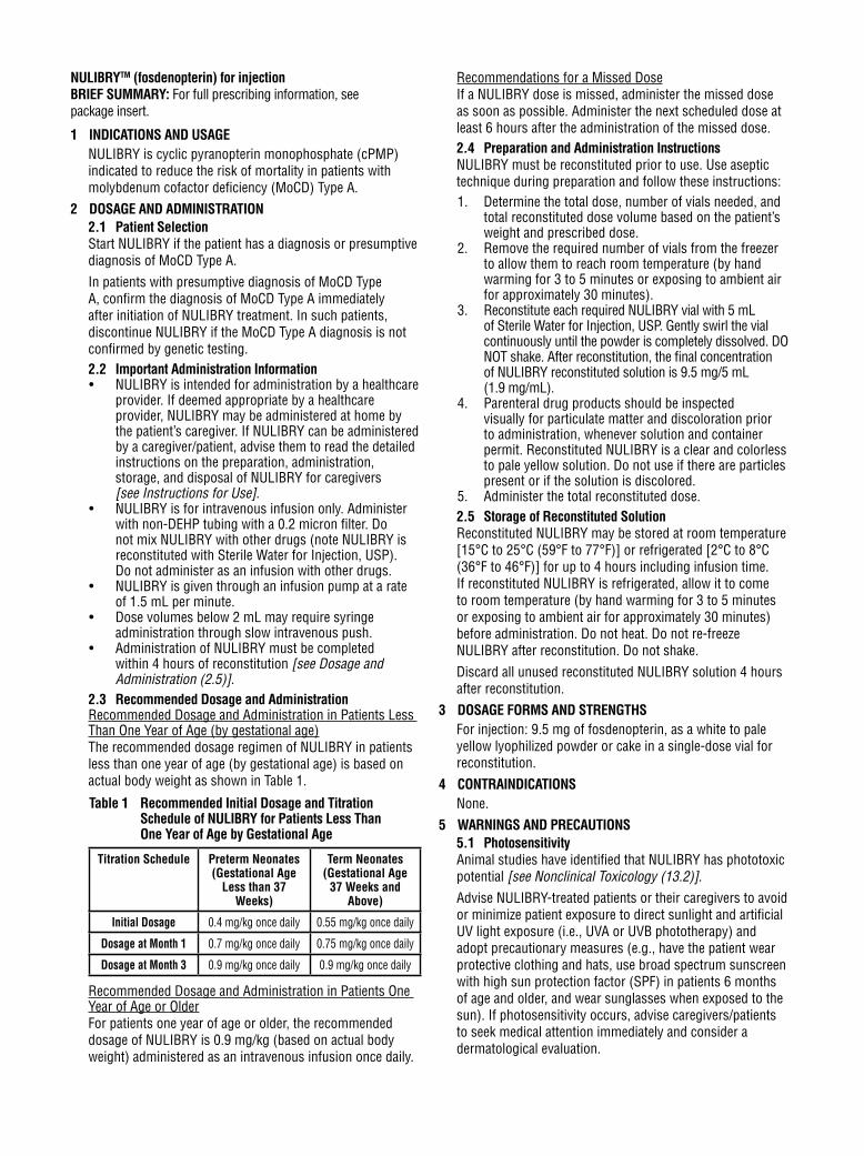
Ad Page 36
11511499 Origin_Nulibry Journal Ad M3FRDate:Client:Product:Client Code:WF Issue #Releasing as:Final Size:Finishing:Gutter:Colors:
Producer:AD:AE:QC:Production:Digital Artist:FR Spellcheck:
4-5-2021 2:29 PMBridgeBio PharmaOrigin Biosciences-NulibryUS-cPMP-20000828626038PDFx1A8.125" x 10.875"NoneNone4C process
Noble JacksonJake ArchambaultAndrea SebatianNoneCarolina Gali©YIP (NYC-FCB)None
Job info
Team
Special Instructions
NoneFonts Images
Inks
PREPARED BY
Additional Information
Additional Comments for Sizing
Please center Brief summary within the trim specsThis Ad need to be released as (2) single pages + 3 pages b&w brief summary
Single page 4C + 3 B&W Brief summary
None Black
55035 NULIBRY Brief Summary_M1.pdf (105%; 94KB)
Scale: 1" = 1"
BleedTrim/FlatLive/Safety
8.375" w x 11.125" h 8.375" w x 11.125" h8.125" w x 10.875" h 8.125" w x 10.875" h7.875" w x 10.625" h 7.875" w x 10.625" h
Path: PrePress:BridgeBio:Origin:11511499:11511499_Origin_Nulibry_Journal_Ad_M3FR.indd
PDFX1A _
NULIBRYTM (fosdenopterin) for injectionBRIEF SUMMARY: For full prescribing information, see package insert.
1 INDICATIONS AND USAGE NULIBRY is cyclic pyranopterin monophosphate (cPMP) indicated to reduce the risk of mortality in patients with molybdenum cofactor deficiency (MoCD) Type A.
2 DOSAGE AND ADMINISTRATION2.1 Patient Selection Start NULIBRY if the patient has a diagnosis or presumptive diagnosis of MoCD Type A.In patients with presumptive diagnosis of MoCD Type A, confirm the diagnosis of MoCD Type A immediately after initiation of NULIBRY treatment. In such patients, discontinue NULIBRY if the MoCD Type A diagnosis is not confirmed by genetic testing.2.2 Important Administration Information• NULIBRY is intended for administration by a healthcare
provider. If deemed appropriate by a healthcare provider, NULIBRY may be administered at home by the patient’s caregiver. If NULIBRY can be administered by a caregiver/patient, advise them to read the detailed instructions on the preparation, administration, storage, and disposal of NULIBRY for caregivers [see Instructions for Use].
• NULIBRY is for intravenous infusion only. Administer with non-DEHP tubing with a 0.2 micron filter. Do not mix NULIBRY with other drugs (note NULIBRY is reconstituted with Sterile Water for Injection, USP). Do not administer as an infusion with other drugs.
• NULIBRY is given through an infusion pump at a rate of 1.5 mL per minute.
• Dose volumes below 2 mL may require syringe administration through slow intravenous push.
• Administration of NULIBRY must be completed within 4 hours of reconstitution [see Dosage and Administration (2.5)].
2.3 Recommended Dosage and AdministrationRecommended Dosage and Administration in Patients Less Than One Year of Age (by gestational age)The recommended dosage regimen of NULIBRY in patients less than one year of age (by gestational age) is based on actual body weight as shown in Table 1.
Table 1 Recommended Initial Dosage and Titration Schedule of NULIBRY for Patients Less Than One Year of Age by Gestational Age
Titration Schedule Preterm Neonates (Gestational Age
Less than 37 Weeks)
Term Neonates (Gestational Age
37 Weeks and Above)
Initial Dosage 0.4 mg/kg once daily 0.55 mg/kg once daily
Dosage at Month 1 0.7 mg/kg once daily 0.75 mg/kg once daily
Dosage at Month 3 0.9 mg/kg once daily 0.9 mg/kg once daily
Recommended Dosage and Administration in Patients One Year of Age or OlderFor patients one year of age or older, the recommended dosage of NULIBRY is 0.9 mg/kg (based on actual body weight) administered as an intravenous infusion once daily.
Recommendations for a Missed DoseIf a NULIBRY dose is missed, administer the missed dose as soon as possible. Administer the next scheduled dose at least 6 hours after the administration of the missed dose.2.4 Preparation and Administration InstructionsNULIBRY must be reconstituted prior to use. Use aseptic technique during preparation and follow these instructions:1. Determine the total dose, number of vials needed, and
total reconstituted dose volume based on the patient’s weight and prescribed dose.
2. Remove the required number of vials from the freezer to allow them to reach room temperature (by hand warming for 3 to 5 minutes or exposing to ambient air for approximately 30 minutes).
3. Reconstitute each required NULIBRY vial with 5 mL of Sterile Water for Injection, USP. Gently swirl the vial continuously until the powder is completely dissolved. DO NOT shake. After reconstitution, the final concentration of NULIBRY reconstituted solution is 9.5 mg/5 mL (1.9 mg/mL).
4. Parenteral drug products should be inspected visually for particulate matter and discoloration prior to administration, whenever solution and container permit. Reconstituted NULIBRY is a clear and colorless to pale yellow solution. Do not use if there are particles present or if the solution is discolored.
5. Administer the total reconstituted dose. 2.5 Storage of Reconstituted SolutionReconstituted NULIBRY may be stored at room temperature [15°C to 25°C (59°F to 77°F)] or refrigerated [2°C to 8°C (36°F to 46°F)] for up to 4 hours including infusion time. If reconstituted NULIBRY is refrigerated, allow it to come to room temperature (by hand warming for 3 to 5 minutes or exposing to ambient air for approximately 30 minutes) before administration. Do not heat. Do not re-freeze NULIBRY after reconstitution. Do not shake.Discard all unused reconstituted NULIBRY solution 4 hours after reconstitution.
3 DOSAGE FORMS AND STRENGTHSFor injection: 9.5 mg of fosdenopterin, as a white to pale yellow lyophilized powder or cake in a single-dose vial for reconstitution.
4 CONTRAINDICATIONSNone.
5 WARNINGS AND PRECAUTIONS5.1 PhotosensitivityAnimal studies have identified that NULIBRY has phototoxic potential [see Nonclinical Toxicology (13.2)].Advise NULIBRY-treated patients or their caregivers to avoid or minimize patient exposure to direct sunlight and artificial UV light exposure (i.e., UVA or UVB phototherapy) and adopt precautionary measures (e.g., have the patient wear protective clothing and hats, use broad spectrum sunscreen with high sun protection factor (SPF) in patients 6 months of age and older, and wear sunglasses when exposed to the sun). If photosensitivity occurs, advise caregivers/patients to seek medical attention immediately and consider a dermatological evaluation.
S:7.875"S:10.625"
T:8.125"T:10.875"
B:8.375"B:11.125"
11511499_Origin_Nulibry_Journal_Ad_M3FR.indd 3 4/5/21 2:30 PM

Ad Page 37
11511499 Origin_Nulibry Journal Ad M3FRDate:Client:Product:Client Code:WF Issue #Releasing as:Final Size:Finishing:Gutter:Colors:
Producer:AD:AE:QC:Production:Digital Artist:FR Spellcheck:
4-5-2021 2:29 PMBridgeBio PharmaOrigin Biosciences-NulibryUS-cPMP-20000828626038PDFx1A8.125" x 10.875"NoneNone4C process
Noble JacksonJake ArchambaultAndrea SebatianNoneCarolina Gali©YIP (NYC-FCB)None
Job info
Team
Special Instructions
NoneFonts Images
Inks
PREPARED BY
Additional Information
Additional Comments for Sizing
Please center Brief summary within the trim specsThis Ad need to be released as (2) single pages + 3 pages b&w brief summary
Single page 4C + 3 B&W Brief summary
None Black
55035 NULIBRY Brief Summary_M1.pdf (105%; 94KB)
Scale: 1" = 1"
BleedTrim/FlatLive/Safety
8.375" w x 11.125" h 8.375" w x 11.125" h8.125" w x 10.875" h 8.125" w x 10.875" h7.875" w x 10.625" h 7.875" w x 10.625" h
Path: PrePress:BridgeBio:Origin:11511499:11511499_Origin_Nulibry_Journal_Ad_M3FR.indd
PDFX1A _
6 ADVERSE REACTIONS6.1 Clinical Trials ExperienceBecause clinical trials are conducted under widely varying conditions, adverse reaction rates observed in the clinical trials of a drug cannot be directly compared to rates in the clinical trials of another drug and may not reflect the rates observed in practice.Overview of Safety EvaluationThe safety of NULIBRY was assessed in 37 pediatric patients and healthy adults who received at least one intravenous infusion of NULIBRY or an E. coli derived non-salt, anhydrous form of cPMP (recombinant cPMP or rcPMP, which has the same active moiety and therefore the same biologic activity as NULIBRY). Of these 37 patients/healthy adults, 13 were pediatric patients with MoCD Type A in Studies 1, 2, and 3 [see Clinical Studies (14)], 6 were pediatric patients with presumptive MoCD Type A but who were later confirmed to not have MoCD Type A, and 18 were healthy adults (without MoCD Type A) in a Phase 1 study.Adverse ReactionsAssessment of adverse reactions for NULIBRY is based on data from two open-label, single-arm studies, Study 1 (n=8) and Study 2 (n=1), in patients with a confirmed diagnosis of MoCD Type A (8 of the 9 patients were previously treated with rcPMP). In these studies, patients received a daily intravenous infusion of NULIBRY. The median exposure to NULIBRY was 4.3 years and ranged from 8 days to 5.6 years [see Clinical Studies (14)]. In these studies, 44% of patients were males and 56% were females, 67% were White and 33% were Asian. The mean age was 14 days and ranged from 1 day to 69 days at time of first infusion.Table 2 presents the most common adverse reactions that occurred in NULIBRY-treated patients in Studies 1 and 2.
Table 2 Common Adverse Reactions Reported in Two or More NULIBRY-Treated Patients with MoCD Type A (Studies 1 and 2)
Adverse ReactionsNULIBRY-Treated Patients
(N=9) n (%)
Catheter-related complications1 8 (89%)
Pyrexia 7 (78%)
Viral infection 5 (56%)
Pneumonia 4 (44%)
Otitis Media 4 (44%)
Vomiting 4 (44%)
Cough/Sneezing 4 (44%)
Upper viral respiratory infection 3 (33%)
Gastroenteritis 3 (33%)
Diarrhea 3 (33%)
Bacteremia 3 (33%)
Abdominal pain 2 (22%)
Influenza 2 (22%)
Lower respiratory tract infection 2 (22%)
Adverse ReactionsNULIBRY-Treated Patients
(N=9) n (%)
Viral tonsillitis 2 (22%)
Oropharyngeal pain 2 (22%)
Rash maculo-papular 2 (22%)
Anemia 2 (22%)
Eye swelling 2 (22%)
Seizure 2 (22%)
Agitation 2 (22%)Abbreviations: MoCD = molybdenum cofactor deficiency1 Catheter-related complications included complication associated with device, catheter site abscess, catheter site discharge, catheter site extravasation, catheter site pain, catheter site infection, catheter site inflammation, device dislocation, device leakage, device occlusion, and vascular device infection.
Safety data are also available from 10 patients with MoCD Type A who received rcPMP in Study 3 (an observational study) [see Clinical Studies (14)]. The median time on rcPMP treatment was 1.5 years and ranged from 6 days to 4.4 years. In Study 3, the patient population was evenly distributed between males and females with a mean age of 18 days (range 1, 69) at time of first infusion, 70% were white, and 30% were Asian.In Study 3, one patient died of necrotizing enterocolitis. Adverse reactions for the rcPMP-treated patients were similar to the NULIBRY-treated patients, except for the following additional adverse reactions that were reported in more than one patient: sepsis, oral candidiasis, varicella, fungal skin infection, and eczema.
8 USE IN SPECIFIC POPULATIONS8.1 PregnancyRisk SummaryThere are no available data on NULIBRY use in pregnant women to evaluate for a drug-associated risk of major birth defects, miscarriage, or adverse maternal or fetal outcomes. Animal reproduction toxicology studies have not been conducted with NULIBRY.The estimated background risk of major birth defects and miscarriage for the indicated population is unknown. All pregnancies have a background risk of birth defect, loss, or other adverse outcomes. In the U.S. general population, the estimated background risks of major birth defects and miscarriage in clinically recognized pregnancies are 2% to 4% and 15% to 20%, respectively.8.2 LactationRisk SummaryThere are no human or animal data available to assess the presence of NULIBRY or its metabolites in human milk, the effects on the breastfed infant, or the effects on milk production for the mother.The developmental and health benefits of breastfeeding should be considered along with the mother’s clinical need for NULIBRY and any potential adverse effects on the breastfed infant from NULIBRY or from the underlying maternal condition.
S:7.875"S:10.625"
T:8.125"T:10.875"
B:8.375"B:11.125"
11511499_Origin_Nulibry_Journal_Ad_M3FR.indd 4 4/5/21 2:30 PM
Ad Page 38
11511499 Origin_Nulibry Journal Ad M3FRDate:Client:Product:Client Code:WF Issue #Releasing as:Final Size:Finishing:Gutter:Colors:
Producer:AD:AE:QC:Production:Digital Artist:FR Spellcheck:
4-5-2021 2:29 PMBridgeBio PharmaOrigin Biosciences-NulibryUS-cPMP-20000828626038PDFx1A8.125" x 10.875"NoneNone4C process
Noble JacksonJake ArchambaultAndrea SebatianNoneCarolina Gali©YIP (NYC-FCB)None
Job info
Team
Special Instructions
NoneFonts Images
Inks
PREPARED BY
Additional Information
Additional Comments for Sizing
Please center Brief summary within the trim specsThis Ad need to be released as (2) single pages + 3 pages b&w brief summary
Single page 4C + 3 B&W Brief summary
None Black
55035 NULIBRY Brief Summary_M1.pdf (105%; 94KB)
Scale: 1" = 1"
BleedTrim/FlatLive/Safety
8.375" w x 11.125" h 8.375" w x 11.125" h8.125" w x 10.875" h 8.125" w x 10.875" h7.875" w x 10.625" h 7.875" w x 10.625" h
Path: PrePress:BridgeBio:Origin:11511499:11511499_Origin_Nulibry_Journal_Ad_M3FR.indd
PDFX1A _
8.4 Pediatric UseSafety and effectiveness of NULIBRY for the treatment of MoCD Type A have been established in pediatric patients starting from birth. Use of NULIBRY for this indication is supported by evidence from two open-label studies (Studies 1 and 2) and one observational study (Study 3), in which 13 pediatric patients aged birth to 6 years of age were treated with NULIBRY or rcPMP. Pediatric use information is discussed throughout the labeling.Animal studies have identified that NULIBRY has phototoxic potential. Advise NULIBRY-treated patients or their caregivers to avoid patient exposure to direct sunlight and artificial UV light exposure (i.e., UVA or UVB phototherapy) and adopt precautionary measures [see Warnings and Precautions (5.1) and Nonclinical Toxicology (13.2)].8.5 Geriatric UseMoCD Type A is largely a disease of pediatric patients. Clinical studies of NULIBRY did not include patients 65 years of age and older.
DRUG INTERACTION STUDIESIn Vitro StudiesFosdenopterin does not inhibit CYP1A2, CYP2B6, CYP2C8, CYP2C9, CYP2C19, CYP2D6, or CYP3A4/5. Fosdenopterin does not induce CYP1A2, CYP2B6, or CYP3A4.Fosdenopterin is a weak inhibitor of MATE2-K and OAT1, but does not exhibit P-gp, BCRP, OATP1B1, OATP1B3, OCT2, OAT3, and MATE1.Fosdenopterin is a weak substrate for MATE1, but is not a substrate of P-gp, BCRP, OATP1B1, OATP1B3, OCT2, OAT1, OAT3, or MATE2-K.
13 NONCLINICAL TOXICOLOGY13.1 Carcinogenesis, Mutagenesis, Impairment of FertilityCarcinogenicity studies have not been conducted with fosdenopterin.Fosdenopterin was not genotoxic in a standard battery of in vitro (bacterial reverse mutation and human lymphocyte chromosomal aberration) and in vivo (rodent bone marrow micronucleus) assays.Fertility studies have not been conducted with fosdenopterin.
13.2 Animal Toxicology and/or PharmacologyFosdenopterin has demonstrated phototoxic potential in an animal study at doses equal to and greater than 4.5 times the maximum recommended human dose (based on human equivalent dose comparison). In this study, which was conducted in pigmented rats, intravenous (bolus) administration of fosdenopterin for three consecutive days followed by ultraviolet radiation (UVR) exposure resulted in dose-dependent cutaneous skin reactions (erythema, edema, flaking, and eschar) and ophthalmic and histopathologic changes indicative of phototoxicity [see Warnings and Precautions (5.1)].
17 PATIENT COUNSELING INFORMATIONAdvise patients/caregivers to read the FDA-approved patient labeling (Instructions for Use) and complete the treatment logs as appropriate.PhotosensitivityAdvise patients and/or caregivers of the potential for photosensitivity reactions and to ensure that the patient avoids or minimizes exposure to sunlight and artificial UV light exposure (i.e., UVA or UVB phototherapy) during use of NULIBRY, uses broad spectrum sunscreen with high sun protection factor (patients 6 months of age and older), and wears clothing, a hat, and sunglasses that protects against sun exposure. Instruct patients/caregivers to seek medical attention immediately if the patient develops a rash or if they notice symptoms of photosensitivity reactions (redness, burning sensation of the skin, blisters) [see Warnings and Precautions (5.1) and Nonclinical Toxicology (13.2)].To report SUSPECTED ADVERSE REACTIONS, contact Origin Biosciences, Inc. at 1-888-55BRIDGE (1-888-552-7434) or FDA at 1-800-FDA-1088 or www.fda.gov/medwatch.
NULIBRY.comNULIBRY is a trademark of Origin Biosciences, Inc.Origin Biosciences is a member of the BridgeBio family.©2021 Origin Biosciences, Inc. All rights reserved. US-NUL-2100003 03/21
S:7.875"S:10.625"
T:8.125"T:10.875"
B:8.375"B:11.125"
11511499_Origin_Nulibry_Journal_Ad_M3FR.indd 5 4/5/21 2:30 PM

Use of naso-/orogastric tubes in the Neonatal Intensive Care Unit (NICU) is very commonplace. However, they are not without possible complications and challenges. One that has gotten recent discussion and action is the risk of tubing misconnection. The Global Enteral Device Supplier Association (GEDSA) made the recommendation some time ago for manufacturers and end-users to transition to the standard enteral design of ENFit. This design prevents the accidental connection of intravenous (IV) or other tubing to a feeding tube, or feeding tubing to IV or other tubing.
In the process of transitioning to the new design, one issue that has arisen is the practice of giving oral medications via enteral tube, particularly low dose medications with a narrow therapeutic index. Due to the design change, the hub has a larger dead space and the syringe now has a moat around the connector as shown in Figure 1 that can serve to collect milk and medication. This results in two risks that deserve discussion: medication administration accuracy and infection.
Medication Administration AccuracyLow risk oral medications have an industry established acceptable 10% dosing variance with some high risk medications limited to a 5% dosing variance (Johnson, Lee, Spooner, et al, 2011). In a study by O’Mara and Campbell (2019), dosing variance using the ENFit design in both oral and enteral use was
evaluated. Enteral use refers to administration of medication via tubing while oral was giving the medication directly into the child’s mouth, often with use of an appropriate adapter and more often in the pediatric setting. These researchers found that dosing variance over 10% occurred twice as often with an ENFit low dose tip syringe as with an oral slip tip syringe (26.9% vs 12.9%). Additionally, they found that a greater than 5% variance happened significantly in either oral or enteral administration.
Since this is a common practice for many medications in neonatal and pediatric settings, it should be addressed from a clinical perspective to maintain medication safety/accuracy. High risk medications are not uncommon via enteral or oral method. This must be addressed before making the transition to an ENFit design system. Adapters for use with the ENFit syringe have been created and will be addressed further later in this article.
Another issue with the ENFit design when giving medications orally (directly into the mouth) is that the ENFit hub is quite large for many neonatal patients. This can result in trauma to the mouth. Therefore, an adapter or alternative method of administering medications to an infant must be considered. One may be to use the adapter and another may be to administer medications with a feeding via bottle. Oral-only tips still exist as alternatives to the ENFit design and may be an option when delivering medication orally. The use of adapters creates potential for dosing inaccuracy. The adapter has a long tip (Figure 2), making it convenient for giving medications into the mouth but also increases the dead space leading to loss of medication. This, too, may increase the dosing variance more than the acceptable 5-10%.
Some suggestions are to establish clinical guidelines in your institution between clinical areas where these may be used and pharmacy to address when and how the adapters will be used. Larger volumes of medications will result in less percentage loss,
The ENFit Enteral-Only System: Challenges and SolutionsSandra Sundquist Beauman, MSN, RNC-NIC
Sandra Sundquist Beauman has been a neonatal nurse for her entire career, spanning over 30 years. She is currently a research nurse coordinator at the University of New Mexico and an independent consultant with Medela LLC. She also provides neonatal consultation and continuing education though CNS Consulting.
Figure 2
Figure 1
neonatal INTENSIVE CARE Vol. 34 No. 4 n Fall 2021 39

the more intense method of cleaning (17 steps) resulted in the absence of visible residue and did not find any difference between use of the toothbrush or commercial brush. The concern presented by these researchers was more about the potential for adherence of the connector such that it could not be disconnected due to buildup of residue. The residue was not cultured nor were cultures taken before or after cleaning. There is no evidence that these cleaning methods decrease the risk of infection/contamination of the tube, something that is much more important in the neonatal population than any other. This study did not evaluate the frequency of cleaning that is needed, likely dependent on an individual patient and how the tube is used. In addition, taking care to avoid spillage into the moat when a syringe is connected will decrease the risk of contamination/overgrowth of bacteria in this area as well as potentially decreasing the need for cleaning. Lyman et al (2020) still recommend that cleaning take place at regular intervals, even if no visible residue is present.
Medication DeliveryTraditionally, pharmacy has used slip tip oral-only syringes. These work fine and are made such that they will not connect to an intravenous line. The use of these syringes for medications delivered in unit dose from the pharmacy is still acceptable. If the medication is to be delivered directly into the infant’s mouth or a bottle, as are most oral medications in the NICU, they work great. If, however, they need to be connected to a ENFit feeding tube for delivery, they do not connect. Some manufacturers have created a device that allows this connection. This device will create dead space so some practice changes are necessary. Nurses may attach this device to a slip tip oral syringe, connect to the feeding tube and administer the medication. This should be followed by a small flush of air or feeding to ensure the desired amount of medication is administered to the patient.
ConclusionThe change to the ENFit oral connector has been a challenging one but the purpose of preventing tubing misconnections is important in clinical practice. We know that changing practice by simply saying to remember to do something, to do better at something is less likely to bring this about. The only effective method is to make it impossible to be done incorrectly! The ENFit system accomplishes this.
However, as with any change, there are unintended consequences that must be addressed. The effect on administration of medications, particularly low dose medications as is common in the NICU, has created challenges. These challenges can be met with the available supplies but require planning and education.
References1. Johnson KB, Lee CK, Spooner SA, et al. Automated dose-
rounding recommendations for pediatric medications. Pediatrics. 2011 Aug;128(2):e422-8. DOI: 10.1542/peds.2011-0760.
2. O’Mara K, Campbell C. Dosing inaccuracy with enteral use of ENFit ® low-dose tip syringes: The risk beyond oral adapters. J Clin Pharm Ther. 2020 Apr;45(2):335-339. doi: 10.1111/jcpt.13079. Epub 2019 Nov 22. PMID: 31755574.
3. Mehall JR, Kite CA, Saltzman DA, Wallett T, Jackson RJ, Smith SD. Prospective study of the incidence and complications of bacterial contamination of enteral feeding in neonates. J Pediatr Surg. 2002 Aug;37(8):1177-82. doi:
perhaps leading to acceptable levels of dosing variance. These guidelines do not yet exist universally so must be created by individual hospitals as part of medication safety.
Infection PreventionThe bacterial growth in feeding tubes used in neonates has been examined by various researchers. Mehall and colleagues (2002) cultured 125 gastric tubes after being in place for 7 days. They found 71/125 were heavily contaminated with three different types of bacteria. Feeding intolerance occurred more often (24/32) in the group whose tubes were contaminated. There were no cases of feeding intolerance reported in those who had non-contaminated tubes. Hurrell et al (2009) examined 129 feeding tubes from four different neonatal units. They found that 76% of these tubes were colonized with Enterobacter, Serratia marcescens and Klebsiella within 48 hours of insertion. These studies were done prior to the implementation of the ENFit connector, did not report the frequency of tube disconnection/interruption nor whether enteral medications were administered via the feeding tube.
Other more recent studies have evaluated and compared bacteria found in the feeding tube and that found in the infant’s stool. Taft et all (2019) sampled portions of the feeding tube, residual fluid in the tube and infant stool from 47 infants. The purpose of this study was to evaluate differences in colonization of the feeding tube at various positions such as in the esophagus vs in the stomach. Previous studies have shown that the microbiome in the esophagus is different than in the stomach (Hunt and Yaghoobi, 2017). Furthermore, bacteria found in the stomach and stool of infants with feeding tubes in place are known to carry antimicrobial resistant genes (Ogrodzki et al, 2017). Taft et al (2019) found that organisms cultured from all levels of the feeding tube were similar and were reflected in the infant’s stool samples, demonstrating that bacteria in the tube reflects that in the gut.
Increased rates of feeding intolerance, poor weight gain, meningitis and necrotizing enterocolitis have been associated with gastric tube contamination (Mehall et al 2002a; Alkeskas et al, 2015).
Various practices can impact risk for feeding tube contamination. These might include how the tube is handled prior to and during insertion, flushing the tube after feedings to prevent milk staying in the tube for an extended period of time and maintaining a “closed system” as much as possible (Beauman & Bowles, 2018), Keeping the tube closed when not in use may aid in preventing pathogenic bacteria from entering gastrointestinal system and potentially colonizing and/or infecting the infant.
Specific to the ENFit design, the male hub of the ENFit device contains a moat around the connector. This may get contaminated with milk and medications during connections and disconnections. Industry has created various devices to use in cleaning this area while other available items are also used such as q-tips, toothbrushes, and gauze. Lyman et al (2020) evaluated two different cleaning methods to remove residue in this area of the connector. The outcome of the study was based on presence of visible residue in the connector. The cleaning methods were quite onorous with one containing 17 steps and the other 9 steps. Each method was performed using either a firm-bristled toothbrush or commercially available cleaning brush designed to clean the ENFit connector. They found that
40 neonatal INTENSIVE CARE Vol. 34 No. 4 n Fall 2021
10.1053/jpsu.2002.34467. PMID: 12149697.4. Hurrell E, Kucerova E, Loughlin M, Caubilla-Barron J,
Hilton A, Armstrong R, Smith C, Grant J, Shoo S, Forsythe S. Neonatal enteral feeding tubes as loci for colonisation by members of the Enterobacteriaceae. BMC Infect Dis. 2009 Sep 1;9:146. doi: 10.1186/1471-2334-9-146. PMID: 19723318; PMCID: PMC2749046.
5. Taft DH, Salinero LK, Vongbhavit K, Kalanetra KM, Masarweh C, Yu A, Underwood MA, Mills DA. Bacterial colonization and antimicrobial resistance genes in neonatal enteral feeding tubes. FEMS Microbiol Ecol. 2019 Apr 1;95(4):fiz039. doi: 10.1093/femsec/fiz039. PMID: 30915455; PMCID: PMC6449222.
6. Hunt RH, Yaghoobi M. The Esophageal and Gastric Microbiome in Health and Disease. Gastroenterol Clin North Am. 2017 Mar;46(1):121-141. doi: 10.1016/j.gtc.2016.09.009. PMID: 28164846.
7. Ogrodzki P, Cheung CS, Saad M et al. Rapid in situ imaging and whole genome sequencing of biofilm in neonatal feeding tubes: a clinical proof of concept. Sci Rep 2017;7:15948.
8. Mehall JR, Kite CA, Gilliam CH, Jackson RJ, Smith SD. Enteral feeding tubes are a reservoir for nosocomial antibiotic-resistant pathogens. J Pediatr Surg. 2002 Jul;37(7):1011-2. doi: 10.1053/jpsu.2002.33831. PMID: 12077760.
9. National Association of Neonatal Nurses. Policies, Procedures, and Competencies for Neonatal Nursing Care. Edited by Sandra Sundquist Beauman and Susan Bowles, Sixth ed., National Association of Neonatal Nurses, 2019.
10. Alkeskas A, Ogrodzki P, Saad M et al. The molecular characterisation of Escherichia coli K1 isolated from neonatal nasogastric feeding tubes. BMC Infect Dis 2015;15:449
11. Lyman B, Sherman AK, Rahe KL, Abdelhadi RA, Keeler D. Randomized controlled trial assessing the effectiveness of two cleaning regimens for ENFit connectors. Med Surg Nursing 2020;29(6):401-406.
neonatal INTENSIVE CARE Vol. 34 No. 4 n Fall 2021 41

Can you tell us about the recent clearances your company has received? In April of this year, we received FDA Class II 510(k) clearance for 4.1 software upgrades for the Servo-u and Servo-n ventilator systems and clearance to introduce the next generation of the Servo-u MR ventilator system into the US market.
Mechanical Ventilation — why is this important?Mechanical ventilation is a well-established life supporting treatment for the lung-compromised patient. In the United States, it is estimated that over 300,000 patients receive mechanical ventilation each year (excluding COVID-19 in 2020/21). Mechanical ventilation is a life supporting treatment delivered to patients who suffer from a wide spectrum of acute respiratory failure (ARF). The ventilator functions to allow the patient to heal and recover. Over time, we have come to understand that life supporting, mechanical ventilation over extended periods of time can have an adverse effect on the lungs. Ultimately, the goal is to assist the patient when needed and to help improve clinical care.
What are the challenges in the ICU and, in particular, mechanical ventilation?Mechanical ventilation is life supporting in patients with respiratory failure but may lead to lung injury.
Lung injury can be impacted by the settings of the mechanical ventilator resulting in potential consequences including:• Ventilatoracquiredpneumonia(VAP)• Ventilatorinducedlunginjury(VILI)including
atelectrauma, volutrauma, barotrauma, and biotrauma
• Ventilatorinduceddiaphragmaticdysfunction(VIDD)
• Myotrauma
Recent clinical studies suggest that many ventilators lack effective bedside decision support tools. It is a problem that results in protective strategies being delayed or inconsistently applied. Ultimately, this can harm the patient and worsen the outcome.1,2,3
Can you tell us about the benefits of personalized lung protection and weaning solutions? How does this help patients?Getinge is a leader when it comes to high acuity patients and mechanical ventilation.
Our Servo family is tailored to the acute care segment. We constantly strive to innovate our products and advance the markets that we serve. Servo-u, Servo-n, and Servo-u MR with system version 4.1 give clinicians many options for personalized lung protection and weaning.
Our goal is that clinicians can deliver the right protection, for each patient, at the right time with a comprehensive toolkit for clinicians to make decisions and plan of care. The main toolkit includes:• ServoCompassfor easy-to-see plateau driving pressure
or tidal volume per predicted body weight (VT/PBW) monitoring.
• Transpulmonary pressure monitoring to provide guidance on the relationship between airway and transpulmonary pressure.
• Open Lung Tool to assess lung mechanics and gas exchange. • Finally,Automatic recruitment maneuvers Auto SRM and
Auto RM. Auto SRM is an automatic workflow for Stepwise recruitment maneuvers based on the Open Lung approach and is unique to Getinge.
In terms of weaning support, most clinicians are familiar with NAVA/ Edi monitoring and its potential to activate the diaphragm and protect the lungs. In addition to PRVC and High Flow Therapy, clinicians can now have access to Heliox therapy to help reduce the work of breathing for patients with obstructed asthma.
Do the software upgrades have any benefits for the clinician?The 4.1 software integrates major respiratory therapy enhancements for all patient categories — adults, pediatrics, and neonates. We have a legacy of more than 50 years of close collaboration with intensive care clinicians worldwide, working together to develop ways to improve the software offerings of the Servo ventilators. We always ensure we are updating the base software with new features and then layering in additional purchasable upgrades like the Automatic Recruitment maneuvers, transpulmonary pressure monitoring, and Heliox therapy.
If you would like to participate in this feature, as a company or healthcare provider, please contact Steve Goldstein at [email protected]
The Benefits of Personalized Lung Protection and Weaning SolutionsIn this feature, Neonatal Intensive Care interviews clinicians and healthcare providers about the actual application of specific products and therapies. This interview is with Eric Honroth, Getinge President, North America.
Interview
42 neonatal INTENSIVE CARE Vol. 34 No. 4 n Fall 2021

Can you tell us more about the Servo-u MR system and how it might make it easier to monitor patients on ventilators?The Servo-u MR is the newest member of the Servo family. It has been designed to provide all the key features of a Servo-u with special adaptations for the MR room. Positioning the ventilator in an MRI room can be a real challenge. We have included a magnetic field indicator with visual and audible alerts to help guide the unit to a safe position. In addition, the Servo-u MR features a visible auto-lock handle. When depressed, the wheels unlock, allowing the unit to be moved. As soon as your hand leaves the handle, it will automatically lock up all four wheels, stopping the unit.
We have also enhanced4 some existing features. Our software interface, a key feature, is now housed in a large 15" screen that can be tilted and rotated for a flexible view from any angle.
Now, with Distance view with Servo Compass, the operator can see at a glance from the control room all the important parameters, and the system will alert you of any deviation from set targets.
Can you tell us more about the Servo Family Range?With the introduction of Servo-air last year and Servo-u MR to the Servo Family, you can be confident, no matter where in the hospital you are, that you have access to the trusted Servo technology. The Servo-u was designed to enhance user confidence in tailoring treatments to the individual patient’s condition. This means more patients in all phases of ventilation — controlled, supported, non-invasive, and during spontaneous breathing trials — can benefit from advanced lung protective strategies.
The Servo-n is our neonatal/pediatric ventilator created to help provide vulnerable patients with the support they need while protecting the lungs, brain, and other developing organs. Sensitive and responsive, Servo-n compensates for variable leakage in both invasive and non-invasive modes of ventilation. Servo-n can deliver tidal volumes as low as 2ml in patients as small as 0.3kg and includes optional hot-wire flow sensor technology to both trigger and measure pressure and flow at the patient interface.
The recently introduced Servo-air launched in Oct 2020 is a turbine-driven ventilator with a hot-swappable battery backup, making it perfect for intra-hospital transport without requiring wall gas or power outlets. It can be easily lifted and moved with patients within the facility. Finally, rounding out the family offerings is the NEW conditional, Servo-u MR, designed to ventilate all patient categories during MR scanning, from invasive and non-invasive ventilation to high-flow therapy.
It has been quite a year for mechanical ventilation. Yes, it really has. In addition to the introduction of the Servo-air, Servo-u MR, and the updated SW 4.1, there have also been a few very impactful studies around NAVA. We have additional information about those on our website, www.getinge.com. We are celebrating 50 years as a leader in the mechanical ventilation market globally and are proud of our legacy of driving innovation within the space. We are really looking forward to many more years of helping to shape the future of mechanical ventilation through continued collaboration with intensive care clinicians and further innovation.
One of the software options includes Automatic Stepwise Recruitment maneuver — why is this important for patients and clinicians?The Open Lung Tools includes three new and different tools: Automatic Stepwise Recruitment maneuvers (Auto SRM), Automatic Recruitment maneuvers (Auto RM), and open lung trends, a manual recruitment maneuver tool. We are very excited about the Automatic Stepwise Recruitment maneuvers, automatic lung recruitment with PEEP titration. Auto SRM was designed to create a standardized workflow that optimally divides the workload between the clinician and the ventilator. Pressures are incrementally increased to reduce the occurrence of hemodynamic compromise, and alarm limits are automatically adapted. The tool guides the clinician through recruitment, decremental PEEP titration, re-recruitment, and post-recruitment. With Auto SRM, the clinician will find the right PEEP and right driving pressure, and all of this is automatic.
The Automatic recruitment maneuver allows for quick recruitment after patient disconnection, suction, or surgery, keeps recruitment settings used in Auto SRM. It also provides an opportunity to delegate recruitment when few physicians are available (during night shifts), a post-recruitment summary with color-coded results, and a shortcut to open lung tool trends.
We see the clearance includes Heliox therapy. Can you tell us more about what Heliox therapy is and how it benefits patients?Servo-i users will be familiar with Heliox, and we are excited to offer Heliox now as part of the Servo u/n ventilators. It is yet another tool that clinicians can use to support advanced personalized ventilation. Heliox has long been used as an adjunctive therapy to overcome airflow-obstruction disorders. In combination with Servo’s integrated Aerogen nebulizer, it is often used as an adjuvant treatment while waiting for the onset of conventional pharmacological treatments. It provides a smoother airflow for patients with obstructive lung disease such as exacerbated asthma and COPD, reduces airway resistance due to laminar flow, cost-efficient due to low gas consumption, can be combined with all ventilator modes, invasive to NIV, High Flow therapy, and nebulization. It is easy to switch from Heliox to air and back.
What other kinds of lung protection functionality does the new software have?Transpulmonary pressure monitoring, including key parameters for assessment of lung stress during controlled and spontaneous ventilation, complements the lung protective toolkit, which was designed to optimally divide the cognitive workload between the clinician and the ventilator. Transpulmonary pressure monitoring validates balloon positioning and includes a diagnostic view including esophageal (Pes) and transpulmonary (PL) pressure waveforms. We wanted clinicians to have an intuitive tool that provides guidance on the relationship between airway and transpulmonary pressure. It is one additional tool for clinicians to use as part of a personalized and lung protection strategy.
What about the base software — any enhancements there?There have been several additional enhancements to the Servo base software, version 4.1, including the addition of the Stress index for lung mechanics monitoring (Servo-u & Servo-u MR only) and the inclusion of PEEP 0 (ZEEP), and several enhancements to the user interface.
neonatal INTENSIVE CARE Vol. 34 No. 4 n Fall 2021 43

References1 Ducharme-Crevier L, Du Pont-Thibodeau G, Emeriaud G.
Interest of Monitoring Diaphragmatic Electrical Activity in the Pediatric Intensive Care Unit. Crit Care Res Pract. 2013; 2013: 384210.
2 Stein H, Firestone K. Application of neurally adjusted ventilatory assist in neonates. Semin Fetal Neonatal Med. 2014 Feb;19(1):60-9.
3 Houtekie L, Moerman D, Bourleau A, et al. Feasibility Study on Neurally Adjusted Ventilatory Assist in Noninvasive Ventilation After Cardiac Surgery in Infants. Respir Care. 2015 Jul;60(7):1007-14.
4 Vs. Servo-i
44 neonatal INTENSIVE CARE Vol. 34 No. 4 n Fall 2021

Personalized lung protection
Servo ventilators offer a complete toolkit for personalized lung protection, enabling you to detect risks early and support timely and consistent implementation of protective ventilation strategies personalized for each patient. Powerful tools such as Servo Compass, Transpulmonary pressure monitoring, and Automatic Recruitments with Open Lung Tool help you utilize advanced protective strategies.
Built on over 50 years of close collaboration with intensive care clinicians around the world, Servo ventilators offer clinical innovation, a focus on patient safety, and a modular, intuitively designed package.
www.getinge.com
The right protection, for each patient at the right time.
Getinge is a registered trademark of Getinge AB, its subsidiaries, or affiliates in the United States or other countries • Servo-u and Servo Compass are trademarks of Maquet Critical Care AB • Copyright 2021 Getinge AB or its subsidiaries or affiliates • All rights reserved • CAUTION: Federal (US) law restricts this device to sale by or on the order of a physician. Refer to Instructions for Use for current indications, warnings, contraindications, and precautions. • MX-7797 Rev01 • MCV00107592 REVA
MCV00107592 REVA Neonatal Intensive Care Servo 4.1 Thumbprint ad .indd 1 7/1/21 12:26 PM

Preterm birth is the leading cause of long-term neurodevelop-mental disability, impacting approximately 500,000 infants in the US annually, at an estimated cost of $26 billion dollars per year.1 When a baby is born too soon, normal intrauterine growth and development is interrupted; thrusting the infant into a foreign and high stress environment that negatively affects the brain, growth, and development. Although advances in neonatal care for preterm infants have greatly increased the chances for survival, survival is not the only anticipated outcome. Helping pre-term and ill infants to thrive by reducing threats to long-term physical, psychological, and social well-being is equally as important.
Preterm babies are at risk for serious health problems, including birth defects, neurodevelopmental delays, and complications of care such as cranial deformities and pressure injuries. Consequently, improper or inadequate neonatal positioning can negatively affect sleep, brain and musculoskeletal development, neurobehavioral organization, neuromotor and physiologic functioning, and feeding.1 Although a number of advanced technologies aim to reduce harm, therapeutic developmental positioning remains a fundamental catalyst for optimal outcomes in the care of premature and ill neonates.
Effective positioning provides a number of benefits to the preterm and ill infant. It reduces toxic stress, facilitates security and self-control, and improves sleep. Proper positioning can also improve brain, neuromotor, and musculoskeletal development, physiologic function and stability, and thermal regulation, and preserve skin integrity.2 Infants who are contained also tend to be calmer, require less medication, sleep more, develop better, and gain weight more rapidly.2,3
Z-Flo™: The New StandardWhile most healthcare providers in the NICU understand the value of positioning to the preterm and ill infant’s well-being, process and product standardization are rare. This results in variable and often sub-optimal clinical outcomes, waste, and unnecessary costs. Standardizing positioning practices with a one product solution can optimize neurodevelopment outcomes, while saving healthcare providers and their organizations time and money.4 Mölnlycke’s unique, patented Z-Flo™ Fluidized Positioner is that solution!
Z-Flo™ Fluidized Positioners allow for customizable positioning and have been shown to meet the unique positioning needs of premature and ill infants. In addition to an infinitely customizable nest, Z-Flo allows for individualized positioning that promotes flexion, midline orientation, and containment for comfort, sleep positively affecting flexor tone, symmetrical movement, head shape, range of motion, pressure injury prevention, and overall development.5
A Solution for Every ChallengeSleep deprivation in premature infants can result in loss of brain plasticity, smaller brains, altered learning and long-term behavioral challenges.2,5 A study of conformational fluidized positioning with Z-Flo™ versus standard positioning, infants calmed more easily, showed reduced wakefulness, and were observed to sleep better. Even neonates with surgical and gastrointestinal complications showed higher sleep efficiency.5
Low birth weight and younger gestational age puts infants at increased risk for intraventricular hemorrhage. Although the cause of these brain bleeds is multifactorial, seemingly benign care activities such as head turning is a contributing factor. Evidence and expert opinion support the recommendation of midline head positioning for premature infants to reduce the risk of intraventricular hemorrhages.6 Z-Flo™ Fluidized Positioners can be molded to facilitate effective and sustained midline head positioning.7
Premature and ill infants are also at high risk for pressure injuries due to immature skin, decreased mobility, head-body disproportion, and medical devices.8,9 Hospital acquired pressure injuries increase healthcare cost and may result in litigation, reduced reimbursement, and government penalties.8,9 Z-Flo™ Fluidized Positioners offload bony prominences, including the occiput to prevent pressure injuries. They can also be molded to support and align medical tubing and lines, to minimize torque and tension that could cause pressure injuries, skin tears, and other skin injuries.10
Premature and ill patients may also experience long periods of immobility due to external monitoring, therapeutic equipment, and physiologically instability. This can lead to prolonged and unrelieved pressure on the cranial bones, resulting in positional plagiocephaly and other cranial deformities. In addition to parental anxiety, cranial deformities can negatively impact the patient’s long-term quality of life and have serious implications including developmental delays, disfigurement, and behavioral
From Surviving To Thriving: A New Standard in Developmental PositioningRhonda Sullivan DNP, PhD, MSN, MBA, CWON, LNCC, NE-BC, CSPHA and Amanda Ball RN, BSN, CWOCN, CSPHA
Rhonda Sullivan is a Clinical Director, Wound Care Marketing at Mölnlycke Health Care and Amanda Ball is a Health Economic Solutions Manager at Mölnlycke Health Care.
46 neonatal INTENSIVE CARE Vol. 34 No. 4 n Fall 2021

Ad Page 47
THIS IS HOW YOU PUT A NICU PATIENT IN A POSITION TO THRIVE.In the NICU, little things are big things. That’s why the team works around the clock to
watch over, nurture and provide comprehensive care for every infant. You’ll reposition the
baby 8 to 12 times a day because you know it promotes development—physically and
even psychologically.1 And that’s where we come in. Our Z-FloTM Fluidized Positioners
continue the nurturing when you’re not at the bedside. They allow you to custom-mold
a shape to fit the infant’s body for optimum comfort and protection. And they hold shape
so infants stay in position, feel secure and get the sleep they need.
We’re here to help. Call your Mölnlycke Health Care Representative or Regional Clinical Specialist. 1-800-843-8497 | www.molnlycke.us | 5445 Triangle Pkwy, Ste 400, Peachtree Corners, GA, 30092
1. Altimier, L., & Phillips, R. (2016). The neonatal integrative developmental care model: Advanced clinical applications of the seven core measures for neuroprotective family-centered developmental care. Newborn And Infant Nursing Reviews, 16, 230-244. doi:10.1053/j.nainr.2016.09.030
Get the complete story at www2.molnlycke.us/zfloneonatal
••Mölnlycke® Z-Flo Fluidized Positioners for Neonatal also helps offload bony prominences while maintaining neutral body alignment and reduces risk of skin tears and pressure on ultra-sensitive skin.
The Mölnlycke trademarks, names and logo types are registered globally to one or more of the Mölnlycke Health Care Group of Companies. The Z-Flo is a trademark in the United States and other countries of EdiZONE, LLC of Alpine, Utah and USA.
Distributed by Mölnlycke Health Care, US, LLC, Peachtree Corners, Georgia 30092. © 2021 Mölnlycke Health Care AB. All rights reserved. 1-800-882-4582. MHC-2018-37184

hospitalized neonate: Rates and risk factors. Scientific Reports. 4(7429), 1-6
9 Hutchinson, G., & Richard, W. (2016). From surviving to thriving: The impact of cranial deformation and pressure ulcers in the hospitalized infant. Neonatal Intensive Care. 29(2), 44-47
10 Hunter, J. (2010). Therapeutic Positioning: Neuromotor, Physiologic, and Sleep Implications. In Developmental Care of Newborns and Infants, A Guide for Health Professionals, 2nd ed pp. 285-312. Glenview: NANN.)
11 Flannery, A., Looman, W., & Kemper, K. (2011). Evidence-based care of the child with deformational plagiocephaly, part II: Management. Accessed 6.12.2
abnormalities. Cranial deformities often require costly and lengthy management and result in litigation, government penalties, and negatively affect hospital metrics.9 Prevention is key since the human cost and financial aspects of treatment for cranial orthoses alone can range from $2000 to $4000, a cost rarely covered by insurance.9 Z-Flo™ has been shown to facilitate optimal head shape and decrease cranial deformities in preterm infants.7
The True CostThe average length of stay for preterm infants is 21 days at an average cost of stay for preterm infants of $121,000 ($5,600/day) with lower gestational ages.11 Risk reduction of pressure injuries, avoidance of positional injuries, and optimized sleep can lead to reduced total cost of care for the hospitalized neonate. The added cost of corrective therapies and medical equipment for unintended consequences of care often fall on the parents following hospital discharge, creating a continued financial burden, in addition to the stress of caring for a preterm infant long after leaving the NICU.
A standardized approach to positioning the pre-term and ill infant with Z-Flo™ can have far-reaching benefits for infants, their caregivers, and the healthcare organization, as a whole. By utilizing a standardized approach to positioning with the Z-Flo™ Fluidized positioner, hospitals can meet the needs of all preterm and ill infants for containment, midline positioning, and pressure redistribution with a singular product. Standardization of practice with Z-Flo™ can help reduce the incidence of complications of care and adverse outcomes, while streamlining practice to improve clinical decision making; optimizing results and cost savings through waste reduction and the avoidance of patient harm.
References1 Berman, R. & Butler, A. (2007). Preterm birth: Causes,
consequences, and prevention. National Academies Press: Accessed 6.12.21 on http://www.nap.edu/catalog/11622.html
2 Altimier, L., & Phillips, R. (2016). The neonatal integrative developmental care model: Advanced clinical applications of the seven core measures for neuroprotective family-centered developmental care. Newborn and Infant Nursing Reviews. 16(4), 230-244
3 Visscher, M., Lucina, L., Casper, T., Dixon, M., Harmeyer, J., Haberman, B., Alberts, J., & Simakajornboon, N. (2015). Conformational positioning improves sleep in premature infants with feeding difficulties. The Journal of Pediatrics. 166, 44-8
4 Hunter, A. (2015). The use of clinical audit in successfully implementing a change in clinical practice in developmental positioning. Infant, 11(5), 1-5
5 Barr, J. (2014). The trials and tribulations of the introduction and implementation of new positioning products in the NICU. Overland Park Regional Medical Center: Poster Presentation
6 Malusky, S., & Donze, A. (2011). Neutral head positioning in premature infants for intraventricular hemorrhage prevention: an evidence-based review. Neonatal Network. 30(6):381-96. doi: 10.1891/0730-0832.30.6.381. Accessed on 6.12.21 on https://pubmed.ncbi.nlm.nih.gov/22052118/
7 Yeh, S. (2018). The effect of early fluidized positioner use on the prevention and treatment of cranial deformities in pre-term infants in the NICU. Shady Grove Medical Center: Rockville, MD. Poster presentation: NANT 2018
8 Visscher, M., & Taylor, T. (2014). Pressure ulcers in the
48 neonatal INTENSIVE CARE Vol. 34 No. 4 n Fall 2021

AbstractBackground: Indirect hyperbilirubinemia is frequently encountered during neonatal period. Although it has different causes, in some cases it can’t be explained. Previous studies have illustrated that jaundice could be a major sign of urinary tract infection (UTI) in neonates.
Aim of the work: We aimed to determine the association between UTI and significant unexplained neonatal indirect hyperbilirubinemia.
Methods: This prospective controlled study was performed on 150 neonates divided in two groups (100 as cases and 50 as controls) to investigate the incidence of UTI in neonates with significant unexplained hyperbilirubinemia. Urine sample was obtained using urine catheterization technique from neonates and full urine analysis was done and cases with pyuria had urine culture to confirm UTI. Immediate renal ultrasonography (USG) was performed for neonates with UTI.
Results: UTI incidence was 11% in cases while none of neonates in control group had UTI with statistical significance between cases and controls (P value < 0.05). The most common (36.4%) pathogen was Escherichia coli. Posterior urethral valve with mild hydronephrosis was diagnosed in 18.2% of UTI positive patients by renal ultrasonography.
Conclusion: In neonates with unexplained indirect hyperbilirubinemia, UTI should be considered as a pathological cause.
IntroductionHyperbilirubinemia is frequent in newborns. In some newborns, total serum bilirubin doesn’t reach pathological levels while in others it may exceed physiological levels and requires treatment. Many well-known causes could be detected during routine investigation of neonates with significant hyperbilirubinemia while in others it may remain unexplained.1
Neonatal jaundice is mainly physiological and 60% of neonates have it. A small number of those neonates have pathological problems like ABO or Rh incompatibility, hepatic impairment,
systemic infection or metabolic disease. In neonates with Urinary Tract Infections (UTIs), jaundice could be an early sign.2
One of the causes of prolonged jaundice is UTI. UTI investigations have been included in routine workup in neonates with prolonged jaundice. There was high incidence of UTI in neonates who developed jaundice after first week of neonatal age in study performed by Garcia and Nager. Ghaemi et al. also observed equal incidence rate of UTI in neonates with prolonged jaundice and neonates with febrile illness with matched ages. On the contrary, it was observed by Omar et al. and Bilgen et al. that asymptomatic neonates with UTI developed jaundice during the first week of neonatal age. Therefore, besides investigating UTI in prolonged jaundice, it was recommended that testing for UTI should be included in the workup of neonates who develop jaundice in early neonatal period.3
The aim of this study was to detect if UTI is a significant cause of unexplained indirect hyperbilirubinemia in jaundiced neonates who require treatment and should it be included in routine workup of those neonates.
MethodsThis analytical case-control study was conducted on 100 neonates (43 males & 57 females) at NICU in Kafr Elsheikh University Hospital with a gestational age greater than 35 weeks with significant unexplained indirect hyperbilirubinemia as cases and on 50 matched neonates (25 males and 25 females) without hyperbilirubinemia as controls. Cases were recruited during study period from January 2019 till February 2020.
Neonates with hemolytic diseases, glucose-6-phosphate dehydrogenase (G6PD) deficiency, neonatal sepsis, polycythemia, with direct hyperbilirubinemia, hypothyroidism and metabolic diseases were excluded.
All newborns were subjected to careful history taking including mode of delivery, full maternal history, maternal blood grouping for detection of incompatibility, family history of jaundice and history of G6PD in a family member. Full clinical examination was done with performing the following investigations serum total and direct bilirubin, full blood count, reticulocyte index and peripheral blood smear, coomb’s test (direct), C reactive protein (CRP), urea and creatinine, liver functions, confirmed urinary tract infection were investigated by renal ultrasonography to exclude congenital anomalies.
Incidence of Urinary Tract Infection in Neonates with Significant Indirect Hyperbilirubinemia of Unknown EtiologyAhmed Mahrous Kamal Baz1, Osama Abd El-Fattah El-Agamy2 and Ashraf Mohamed Ibrahim3
1Neonatology Unit, Pediatrics department, Kafrelsheikh Faculty of Medicine, Ibrahim Moghazy st. 21, Kafrelsheikh 33511, Egypt. 2Kafrelsheikh Faculty of Medicine, Kafrelsheikh, Egypt. 3Neonatology Unit, Pediatrics department, Tanta Faculty of Medicine, Tanta, Egypt.
neonatal INTENSIVE CARE Vol. 34 No. 4 n Fall 2021 49
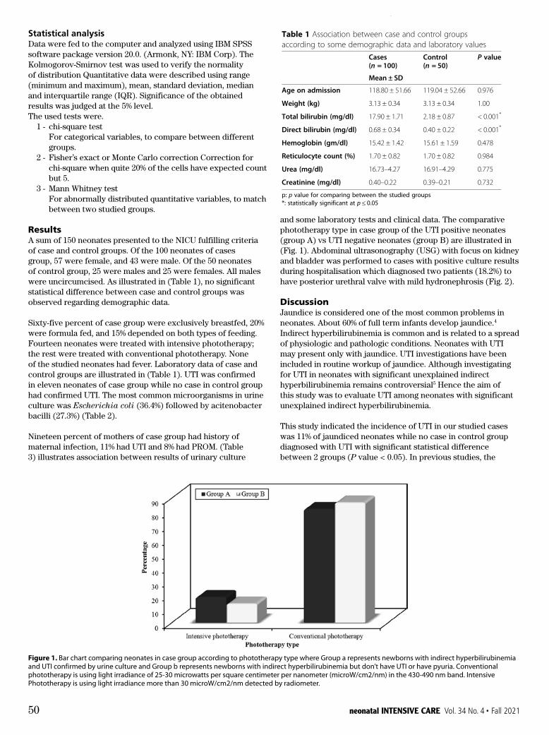
and some laboratory tests and clinical data. The comparative phototherapy type in case group of the UTI positive neonates (group A) vs UTI negative neonates (group B) are illustrated in (Fig. 1). Abdominal ultrasonography (USG) with focus on kidney and bladder was performed to cases with positive culture results during hospitalisation which diagnosed two patients (18.2%) to have posterior urethral valve with mild hydronephrosis (Fig. 2).
DiscussionJaundice is considered one of the most common problems in neonates. About 60% of full term infants develop jaundice.4 Indirect hyperbilirubinemia is common and is related to a spread of physiologic and pathologic conditions. Neonates with UTI may present only with jaundice. UTI investigations have been included in routine workup of jaundice. Although investigating for UTI in neonates with significant unexplained indirect hyperbilirubinemia remains controversial5 Hence the aim of this study was to evaluate UTI among neonates with significant unexplained indirect hyperbilirubinemia.
This study indicated the incidence of UTI in our studied cases was 11% of jaundiced neonates while no case in control group diagnosed with UTI with significant statistical difference between 2 groups (P value < 0.05). In previous studies, the
Statistical analysisData were fed to the computer and analyzed using IBM SPSS software package version 20.0. (Armonk, NY: IBM Corp). The Kolmogorov-Smirnov test was used to verify the normality of distribution Quantitative data were described using range (minimum and maximum), mean, standard deviation, median and interquartile range (IQR). Significance of the obtained results was judged at the 5% level.The used tests were.
1 - chi-square testFor categorical variables, to compare between different groups.
2 - Fisher’s exact or Monte Carlo correction Correction for chi-square when quite 20% of the cells have expected count but 5.
3 - Mann Whitney test For abnormally distributed quantitative variables, to match
between two studied groups.
ResultsA sum of 150 neonates presented to the NICU fulfilling criteria of case and control groups. Of the 100 neonates of cases group, 57 were female, and 43 were male. Of the 50 neonates of control group, 25 were males and 25 were females. All males were uncircumcised. As illustrated in (Table 1), no significant statistical difference between case and control groups was observed regarding demographic data.
Sixty-five percent of case group were exclusively breastfed, 20% were formula fed, and 15% depended on both types of feeding. Fourteen neonates were treated with intensive phototherapy; the rest were treated with conventional phototherapy. None of the studied neonates had fever. Laboratory data of case and control groups are illustrated in (Table 1). UTI was confirmed in eleven neonates of case group while no case in control group had confirmed UTI. The most common microorganisms in urine culture was Escherichia coli (36.4%) followed by acitenobacter bacilli (27.3%) (Table 2).
Nineteen percent of mothers of case group had history of maternal infection, 11% had UTI and 8% had PROM. (Table 3) illustrates association between results of urinary culture
neonates with prolonged jaundice. There was high inci-dence of UTI in neonates who developed jaundice afterfirst week of neonatal age in study performed by Garciaand Nager. Ghaemi et al. also observed equal incidencerate of UTI in neonates with prolonged jaundice and ne-onates with febrile illness with matched ages. On thecontrary, it was observed by Omar et al. and Bilgen et al.that asymptomatic neonates with UTI developedjaundice during the first week of neonatal age. There-fore, besides investigating UTI in prolonged jaundice, itwas recommended that testing for UTI should be in-cluded in the workup of neonates who develop jaundicein early neonatal period [3].The aim of this study was to detect if UTI is a signifi-
cant cause of unexplained indirect hyperbilirubinemia injaundiced neonates who require treatment and should itbe included in routine workup of those neonates.
MethodsThis analytical case-control study was conducted on 100neonates (43 males & 57 females) at NICU in KafrElsheikh University Hospital with a gestational agegreater than 35 weeks with significant unexplained indir-ect hyperbilirubinemia as cases and on 50 matchedneonates (25 males and 25 females) without hyperbiliru-binemia as controls. Cases were recruited during studyperiod from January 2019 till February 2020.Neonates with hemolytic diseases, glucose-6-
phosphate dehydrogenase (G6PD) deficiency, neonatalsepsis, polycythemia, with direct hyperbilirubinemia,hypothyroidism and metabolic diseases were excluded.All newborns were subjected to careful history taking
including mode of delivery, full maternal history, mater-nal blood grouping for detection of incompatibility, fam-ily history of jaundice and history of G6PD in a familymember. Full clinical examination was done with per-forming the following investigations serum total and dir-ect bilirubin, full blood count, reticulocyte index andperipheral blood smear, coomb’s test (direct), C reactiveprotein (CRP), urea and creatinine, liver functions,G6PD enzyme assay, thyroid function (TSH and freeT4). All study participants had a urine sample obtainedusing urinary catheterization technique under acomplete aseptic technique. The urine sample was testedusing Multistix 10 for standard urinalysis. The sampleswere examined microscopically under high power field(HPF) for pyuria. Pyuria was defined as > 5 white bloodcell (WBC) per HPF. Patients whom urine analysis re-sults showed pyuria had urine culture to confirm pres-ence of UTI and detect causing organism. Culture wasdone using CLED agar (cysteine, lactose and electrolytedeficient agar). The culture was considered positivewhen it detected the presence of at least 50,000 CFUsper mL of a single urinary pathogen. Neonates with
confirmed urinary tract infection were investigated byrenal ultrasonography to exclude congenital anomalies.
Statistical analysisData were fed to the computer and analyzed using IBMSPSS software package version 20.0. (Armonk, NY: IBMCorp). The Kolmogorov-Smirnov test was used to verifythe normality of distribution Quantitative data were de-scribed using range (minimum and maximum), mean,standard deviation, median and interquartile range(IQR). Significance of the obtained results was judged atthe 5% level.The used tests were.1 - chi-square testFor categorical variables, to compare between different
groups.2 - Fisher’s exact or Monte Carlo correctionCorrection for chi-square when quite 20% of the cells
have expected count but 5.3 - Mann Whitney testFor abnormally distributed quantitative variables, to
match between two studied groups.
ResultsA sum of 150 neonates presented to the NICU fulfilling cri-teria of case and control groups. Of the 100 neonates of casesgroup, 57 were female, and 43 were male. Of the 50 neonatesof control group, 25 were males and 25 were females. Allmales were uncircumcised. As illustrated in (Table 1), no sig-nificant statistical difference between case and control groupswas observed regarding demographic data.Sixty-five percent of case group were exclusively
breastfed, 20% were formula fed, and 15% depended onboth types of feeding. Fourteen neonates were treatedwith intensive phototherapy; the rest were treated withconventional phototherapy. None of the studied neo-nates had fever. Laboratory data of case and control
Table 1 Association between case and control groupsaccording to some demographic data and laboratory values
Cases(n = 100)
Control(n = 50)
P value
Mean ± SD
Age on admission 118.80 ± 51.66 119.04 ± 52.66 0.976
Weight (kg) 3.13 ± 0.34 3.13 ± 0.34 1.00
Total bilirubin (mg/dl) 17.90 ± 1.71 2.18 ± 0.87 < 0.001*
Direct bilirubin (mg/dl) 0.68 ± 0.34 0.40 ± 0.22 < 0.001*
Hemoglobin (gm/dl) 15.42 ± 1.42 15.61 ± 1.59 0.478
Reticulocyte count (%) 1.70 ± 0.82 1.70 ± 0.82 0.984
Urea (mg/dl) 16.73–4.27 16.91–4.29 0.775
Creatinine (mg/dl) 0.40–0.22 0.39–0.21 0.732
p: p value for comparing between the studied groups*: statistically significant at p ≤ 0.05
Baz et al. Italian Journal of Pediatrics (2021) 47:35 Page 2 of 6
Table 3 Comparison between cases confirmed to have UTI and negative cases in case group according to some clinical andlaboratory variablesVariable Urinary Tract Infection P value
Group A (Positive)(n = 11)
Group B (Negative)(n = 89)
Mean ± SD.
Age on admission (hours) 120.0 ± 41.57 118.65 ± 52.97 0.674
Weight (kg) 3.01 ± 0.20 3.14 ± 0.36 0.255
Total bilirubin (mg/dl) 18.09 ± 1.45 17.87 ± 1.74 0.468
Direct bilirubin (mg/dl) 0.67 ± 0.33 0.68 ± 0.34 0.934
Hemoglobin (gm/dl) 16.05 ± 1.23 15.34 ± 1.43 0.132
Reticulocyte count (%) 2.64 ± 1.14 2.4 ± 1 0.454
Urea (mg/dl) 17.19 ± 5.13 16.67 ± 4.18 0.639
Creatinine (mg/dl) 0.55 ± 0.28 0.39 ± 0.20 0.081
No. % No. %
Sex
Male 5 45.5 38 42.7 FEp=1.000
Female 6 54.5 51 57.3
Maternal infection
Negative 4 36.4 77 86.5
UTI 5 45.5 6 6.7 MCp=0.001*
PROM 2 18.2 6 6.7
Maternal history of chronic disease
Non 8 72.7 62 69.7
Diabetes 1 9.1 9 10.1 MCp=0.001*
Hypertension 2 18.2 18 20.2
FE Fisher Exact, MC Monte Carlop: p value for comparing between the studied groups*: Statistically significant at p ≤ 0.05Group A: Newborns with indirect hyperbilirubinemia and UTI confirmed by urine cultureGroup B: Newborns with indirect hyperbilirubinemia but don’t have UTI or have pyuria
Fig. 1 Bar chart comparing neonates in case group according to phototherapy type where Group a represents `newborns with indirecthyperbilirubinemia and UTI confirmed by urine culture and Group b represents newborns with indirect hyperbilirubinemia but don’t have UTI orhave pyuria. Conventional phototherapy is using light irradiance of 25–30 microwatts per square centimeter per nanometer (microW/cm2/nm) inthe 430–490 nm band. Intensive Phototherapy is using light irradiance more than 30 microW/cm2/nm detected by radiometer
Baz et al. Italian Journal of Pediatrics (2021) 47:35 Page 4 of 6
Figure 1. Bar chart comparing neonates in case group according to phototherapy type where Group a represents newborns with indirect hyperbilirubinemia and UTI confirmed by urine culture and Group b represents newborns with indirect hyperbilirubinemia but don’t have UTI or have pyuria. Conventional phototherapy is using light irradiance of 25-30 microwatts per square centimeter per nanometer (microW/cm2/nm) in the 430-490 nm band. Intensive Phototherapy is using light irradiance more than 30 microW/cm2/nm detected by radiometer.
50 neonatal INTENSIVE CARE Vol. 34 No. 4 n Fall 2021
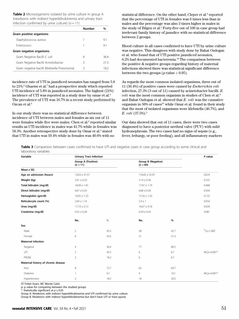
statistical difference. On the other hand, Cleper et al.9 reported that the percentage of UTI in females was 6 times less than in males and the percentage was also 3 times higher in males in the study of Bilgen et al.2 Forty-five out of 100 in case group had irrelevant family history of jaundice with no statistical difference between 2 groups.
Blood culture in all cases confirmed to have UTI by urine culture was negative. This disagrees with study done by Bahat Ozdogan et al. who found that of UTI positive jaundiced neonates, 6.2% had documented bacteremia.10 The comparison between the positive & negative groups regarding history of maternal infections showed there was statistical significant difference between the two groups (p-value < 0.05).
As regards the most common isolated organisms, three out of 11 (36.4%) of positive cases were caused by Escherichia coli infection, 27.3% (3 out of 11) caused by acinetobacter bacilli. E. coli was the most common organism in studies of Chen et al.8 and Bahat Ozdogan et al. showed that E. coli was the causative organism in 50% of cases10 while Omar et al. found in their study that the most of isolated organisms were klebsiella (46.7%), and E. coli (37.5%).3
Our data showed that out of 11 cases, there were two cases diagnosed to have a posterior urethral valve (PUV) with mild hydronephrosis. The two cases had no signs of sepsis (e.g., fever, lethargy, or poor feeding), and all inflammatory markers
incidence rate of UTI in jaundiced neonates has ranged from 5.8 to 21%.6 Ghaemi et al.7 had a prospective study which reported UTI incidence of 5.8% in jaundiced neonates. The highest (21%) incidence of UTI was reported in a study done by omae et al.3 The prevalence of UTI was 16.7% in a recent study performed by Ozcan et al.6
In our study there was no statistical difference between incidence of UTI between males and females as six out of 11 were females while five were males. Chen et al.8 reported similar results as UTI incidence in males was 41.7% while in females was 58.3%. Another retrospective study done by Omar et al.4 stated that UTI in males was 59.4% while in females was 40.6% with no
groups are illustrated in (Table 1). UTI was confirmed ineleven neonates of case group while no case in controlgroup had confirmed UTI. The most common microor-ganisms in urine culture was Escherichia coli (36.4%)followed by acitenobacter bacilli (27.3%) (Table 2).Nineteen percent of mothers of case group had history of
maternal infection, 11% had UTI and 8% had PROM.(Table 3) illustrates association between results of urinaryculture and some laboratory tests and clinical data. The com-parative phototherapy type in case group of the UTI positiveneonates (group A) vs. UTI negative neonates (group B) areillustrated in (Fig. 1). Abdominal ultrasonography (USG) withfocus on kidney and bladder was performed to cases withpositive culture results during hospitalisation which diag-nosed two patients (18.2%) to have posterior urethral valvewith mild hydronephrosis (Fig. 2).
DiscussionJaundice is considered one of the most common problemsin neonates. About 60% of full term infants develop jaun-dice [4]. Indirect hyperbilirubinemia is common and is re-lated to a spread of physiologic and pathologic conditions.Neonates with UTI may present only with jaundice. UTIinvestigations have been included in routine workup ofjaundice. Although investigating for UTI in neonates withsignificant unexplained indirect hyperbilirubinemia re-mains controversial [5]. Hence the aim of this study wasto evaluate UTI among neonates with significant unex-plained indirect hyperbilirubinemia.This study indicated the incidence of UTI in our studied
cases was 11% of jaundiced neonates while no case in controlgroup diagnosed with UTI with significant statistical differ-ence between 2 groups (P value < 0.05). In previous studies,the incidence rate of UTI in jaundiced neonates has rangedfrom 5.8 to 21% [6]. Ghaemi et al. [7] had a prospective studywhich reported UTI incidence of 5.8% in jaundiced neonates.The highest (21%) incidence of UTI was reported in a studydone by omae et al. [3]. The prevalence of UTI was 16.7% ina recent study performed by Ozcan et al. [6]
In our study there was no statistical difference betweenincidence of UTI between males and females as six outof 11 were females while five were males. Chen et al. [8]reported similar results as UTI incidence in males was41.7% while in females was 58.3%. Another retrospectivestudy done by Omar et al. [4] stated that UTI in maleswas 59.4% while in females was 40.6% with no statisticaldifference. On the other hand, Cleper et al. [9] reportedthat the percentage of UTI in females was 6 times lessthan in males and the percentage was also 3 timeshigher in males in the study of Bilgen et al. [2] Forty-fiveout of 100 in case group had irrelevant family history ofjaundice with no statistical difference between 2 groups.Blood culture in all cases confirmed to have UTI by
urine culture was negative. This disagrees with studydone by Bahat Ozdogan et al. who found that of UTIpositive jaundiced neonates, 6.2% had documentedbacteremia [10]. The comparison between the positive &negative groups regarding history of maternal infectionsshowed there was statistical significant difference be-tween the two groups (p-value< 0.05).As regards the most common isolated organisms,
three out of 11 (36.4%) of positive cases were caused byEscherichia coli infection, 27.3% (3 out of 11) caused byacinetobacter bacilli. E. coli was the most common or-ganism in studies of Chen et al. [8] and Bahat Ozdoganet al. showed that E. coli was the causative organism in50% of cases [10] while Omar et al. found in their studythat the most of isolated organisms were klebsiella(46.7%), and E. coli (37.5%) [3].Our data showed that out of 11 cases, there were two cases
diagnosed to have a posterior urethral valve (PUV) with mildhydronephrosis. The two cases had no signs of sepsis (e.g.,fever, lethargy, or poor feeding), and all inflammatorymarkers were negative; this may attribute the bilateral hydro-nephrosis to mechanical obstruction by PUV rather thanUTI. The guidelines of American Academy of Paediatrics(AAP) advocate doing US in all > 2-month-old infants withUTI accompanied by fever but there are no recommenda-tions for neonates with UTI. Our study didn’t document asignificant portion of USG abnormality neonates with UTI.However, in study done by Bahat Ozdogan et al., he foundthat there was abnormal finding in 28.1% in renal USG ofjaundiced neonates confirmed to have UTI [10].One limitation of the study is that not all the studied cases
and controls have had urine culture; we performed screeningfor all neonates included in the study using urine analysis (in-cluding performing leukocyte esterase test (LE) and nitritetest) and microscopic examination for the presence of pyuria.This limitation was due to NICU and microbiology labora-tory local policy, which restricts performing urine cultureonly to cases with sepsis (which was one of the exclusion cri-teria in the study) or cases with abnormal urine analysis orpyuria. However, obtaining urine samples using aseptic
Table 2 Microorganisms isolated by urine culture in group A(newborns with indirect hyperbilirubinemia and urinary tractinfection confirmed by urine culture) (n = 11)
Number %
Gram positive organisms
Staphylococcus aureus 1 9.1
Enterococci 1 9.1
Gram negative organisms
Gram Negative Bacilli E. coli 4 36.4
Gram Negative Bacilli Acinetobactar 3 27.3
Gram negative bacilli (Klebsiella Pneumonia) 2 18.2
Baz et al. Italian Journal of Pediatrics (2021) 47:35 Page 3 of 6
Table 3 Comparison between cases confirmed to have UTI and negative cases in case group according to some clinical andlaboratory variablesVariable Urinary Tract Infection P value
Group A (Positive)(n = 11)
Group B (Negative)(n = 89)
Mean ± SD.
Age on admission (hours) 120.0 ± 41.57 118.65 ± 52.97 0.674
Weight (kg) 3.01 ± 0.20 3.14 ± 0.36 0.255
Total bilirubin (mg/dl) 18.09 ± 1.45 17.87 ± 1.74 0.468
Direct bilirubin (mg/dl) 0.67 ± 0.33 0.68 ± 0.34 0.934
Hemoglobin (gm/dl) 16.05 ± 1.23 15.34 ± 1.43 0.132
Reticulocyte count (%) 2.64 ± 1.14 2.4 ± 1 0.454
Urea (mg/dl) 17.19 ± 5.13 16.67 ± 4.18 0.639
Creatinine (mg/dl) 0.55 ± 0.28 0.39 ± 0.20 0.081
No. % No. %
Sex
Male 5 45.5 38 42.7 FEp=1.000
Female 6 54.5 51 57.3
Maternal infection
Negative 4 36.4 77 86.5
UTI 5 45.5 6 6.7 MCp=0.001*
PROM 2 18.2 6 6.7
Maternal history of chronic disease
Non 8 72.7 62 69.7
Diabetes 1 9.1 9 10.1 MCp=0.001*
Hypertension 2 18.2 18 20.2
FE Fisher Exact, MC Monte Carlop: p value for comparing between the studied groups*: Statistically significant at p ≤ 0.05Group A: Newborns with indirect hyperbilirubinemia and UTI confirmed by urine cultureGroup B: Newborns with indirect hyperbilirubinemia but don’t have UTI or have pyuria
Fig. 1 Bar chart comparing neonates in case group according to phototherapy type where Group a represents `newborns with indirecthyperbilirubinemia and UTI confirmed by urine culture and Group b represents newborns with indirect hyperbilirubinemia but don’t have UTI orhave pyuria. Conventional phototherapy is using light irradiance of 25–30 microwatts per square centimeter per nanometer (microW/cm2/nm) inthe 430–490 nm band. Intensive Phototherapy is using light irradiance more than 30 microW/cm2/nm detected by radiometer
Baz et al. Italian Journal of Pediatrics (2021) 47:35 Page 4 of 6
neonatal INTENSIVE CARE Vol. 34 No. 4 n Fall 2021 51

the bactericidal activity of their serum as reported in study done by Cisowska et al.14
ConclusionFrom the previous discussion, we can come to the conclusion that UTI may be one of causes of neonatal unexplained indirect hyperbilirubinemia. Also, it was found that there is a positive correlation between maternal infections and UTI in jaundiced neonates. Therefore, we can suggest that UTI investigations could be included in routine workup of neonates with unexplained indirect hyperbilirubinemia and good antenatal follow up with early treatment of maternal infections could prevent UTI in newborns.
Authors’ contributionsAhmed Mahrous Baz performed the clinical examination, collected urine samples, did basic investigations and contribute in data interpretation, Osama Abdelfattah Elagamy formatted study design and contributed in results interpretation and discussion, Ashraf Mohamed Ibrahim did the statistical analysis and contributed in results interpretation and discussion. All authors have participated to drafting the manuscript, author A revised it critically. All authors read and approved the final version of the manuscript. All authors contributed equally to the manuscript and read and approved the final version of the manuscript.
FundingThe authors received no financial support for the research, authorship, and/ or publication of this article.
Availability of data and materialsAll data generated or analysed during this study are included in this published article [and its supplementary information files].
Ethics approval and consent to participateThe study followed the Principles of the Declaration of Helsinki
were negative; this may attribute the bilateral hydronephrosis to mechanical obstruction by PUV rather than UTI. The guidelines of American Academy of Paediatrics (AAP) advocate doing US in all > 2-month-old infants with UTI accompanied by fever but there are no recommendations for neonates with UTI. Our study didn’t document a significant portion of USG abnormality neonates with UTI. However, in study done by Bahat Ozdogan et al., he found that there was abnormal finding in 28.1% in renal USG of jaundiced neonates confirmed to have UTI.10
One limitation of the study is that not all the studied cases and controls have had urine culture; we performed screening for all neonates included in the study using urine analysis (including performing leukocyte esterase test (LE) and nitrite test) and microscopic examination for the presence of pyuria. This limitation was due to NICU and microbiology laboratory local policy, which restricts performing urine culture only to cases with sepsis (which was one of the exclusion criteria in the study) or cases with abnormal urine analysis or pyuria. However, obtaining urine samples using aseptic urinary catheterization technique increased the specificity of these investigations; also using aggregate urine analysis (the presence of any LE, nitrite, or pyuria > 5 WBCs/HPF) increases the sensitivity for UTI detection in infants less than 60 days of age to 99.4% as indicated in a study done by Tzimenatos L et al.11 Also, the absence of pyuria can help in differentiation true UTI from asymptomatic bacteriuria.12
UTI in neonatal period has many nonspecific symptoms like fever, lethargy, vomiting, anorexia, diarrhea, weight loss, changes in urine characters and jaundice. So, neonates with UTI could present early with jaundice.13 It remains unclear how UTI is related to jaundice in neonates but there are some explanations that need further evaluating studies. Of these explanations is hepatocellular injury that may be caused directly circulating microorganisms that caused UTI or by their circulating endotoxins. On the other hand, jaundice may be the cause of UTI through making neonates more prone to infections by decreasing
urinary catheterization technique increased the specificity ofthese investigations; also using aggregate urine analysis (thepresence of any LE, nitrite, or pyuria > 5 WBCs/HPF) in-creases the sensitivity for UTI detection in infants less than60 days of age to 99.4% as indicated in a study done by Tzi-menatos L et al. [11] Also, the absence of pyuria can help indifferentiation true UTI from asymptomatic bacteriuria [12].UTI in neonatal period has many nonspecific symptoms
like fever, lethargy, vomiting, anorexia, diarrhea, weight loss,changes in urine characters and jaundice. So, neonates withUTI could present early with jaundice [13]. It remains un-clear how UTI is related to jaundice in neonates but thereare some explanations that need further evaluating studies.Of these explanations is hepatocellular injury that may becaused directly circulating microorganisms that caused UTIor by their circulating endotoxins. On the other hand, jaun-dice may be the cause of UTI through making neonatesmore prone to infections by decreasing the bactericidal activ-ity of their serum as reported in study done by Cisowskaet al. [14].
ConclusionFrom the previous discussion, we can come to the conclu-sion that UTI may be one of causes of neonatal unexplainedindirect hyperbilirubinemia. Also, it was found that there is apositive correlation between maternal infections and UTI injaundiced neonates. Therefore, we can suggest that UTI
investigations could be included in routine workup of neo-nates with unexplained indirect hyperbilirubinemia and goodantenatal follow up with early treatment of maternal infec-tions could prevent UTI in newborns.
Supplementary InformationThe online version contains supplementary material available at https://doi.org/10.1186/s13052-021-00982-0.
Additional file 1.
AcknowledgementsNot applicable.
Authors’ contributionsAhmed Mahrous Baz performed the clinical examination, collected urine samples,did basic investigations and contribute in data interpretation, Osama AbdelfattahElagamy formatted study design and contributed in results interpretation anddiscussion, Ashraf Mohamed Ibrahim did the statistical analysis and contributed inresults interpretation and discussion. All authors have participated to drafting themanuscript, author A revised it critically. All authors read and approved the finalversion of the manuscript. All authors contributed equally to the manuscript andread and approved the final version of the manuscript.
FundingThe authors received no financial support for the research, authorship, and/or publication of this article.
Availability of data and materialsAll data generated or analysed during this study are included in thispublished article [and its supplementary information files].
Fig. 2 Pie chart of US kidney and pelvis results of neonates with UTI confirmed by urine culture (PUV = posterior urethral valve)
Baz et al. Italian Journal of Pediatrics (2021) 47:35 Page 5 of 6
Figure 2. Pie chart of US kidney and pelvis results of neonates with UTI confirmed by urine culture (PUV = posterior urethral valve)
52 neonatal INTENSIVE CARE Vol. 34 No. 4 n Fall 2021

and approved by the Ethical Review Committee of Kafrelsheikh University and informed written parental consent from all participants in the research.
References1. Pashapour N, Nikibahksh AA, Golmohammadlou S. Urinary
tract infection in term neonates with prolonged jaundice. Urol J. 2007;4:91–4 PMID: 17701928.
2. Bilgen H, Ozek E, Unver T, Biyikli N, Alpay H, Cebeci D. Urinary tract infection and hyperbilirubinemia. Turk J Pediatr. 2006;48:51–5 PMID: 16562786.
3. Omar C, Hamza S, Bassem AM, Mariam R. Urinary tract infection and indirect hyperbilirubinemia in newborns. N Am J Med Sci. 2011;3:544–7. https://doi. org/10.4297/najms.2011.3544.
4. Zafarzadeh M, Mohammadzadeh A. Should urine culture be considered in the hyperbilirubinemia work up of neonate? J Chinese Clin Med. 2009;4(3):136–8.
5. Mutlu M, Cayır Y, Aslan Y. Urinary tract infections in neonates with jaundice in their first two weeks of life. World J Pediatr. 2014;10:164e7. https://doi. org/10.1007/s12519-013-0433-1.
6. Özcan M, Sarici SÜ, Yurdugül, et al. Association Between Early Idiopathic Neonatal Jaundice and Urinary Tract Infections. Clin Med Insights Pediatr. 2017. https://doi.org/10.1177/1179556517701118.
7. Ghaemi S, Fesharaki RJ, Kelishadi R. Late onset jaundice and urinary tract infection in neonates. Indian J Pediatr. 2007;74:139–41. https://doi.org/10.1 007/s12098-007-0006-1.
8. Chen TH, Jeng JM, Soong JW, Yang FC, et al. Hyperbilirubinemia with urinary tract infection in infants younger than eight weeks old. J Chin Med Assoc. 2011;74(4):159–63. https://doi.org/10.1016/j.jcma.2011.01.036.
9. Cleper R, Krause I, Eisenstein B, Davidovits M. Prevalence of vesicoureteral reflux in neonatal urinary tract infection. Clin Pediatr (Phila). 2004;43:619–25. https://doi.org/10.1177/000992280404300706.
10. Bahat Ozdogan E, Mutlu M, Camlar SA, Bayramoglu G, Kader S, Aslan Y. Urinary tract infections in neonates with unexplained pathological indirect hyperbilirubinemia: prevalence and significance. Pediatr Neonatol. 2018; 59(3):305–9. https://doi.org/10.1016/j.pedneo.2017.10.010.
11. Tzimenatos L, Mahajan P, Dayan PS, et al. Accuracy of the urinalysis for urinary tract infections in febrile infants 60 days and younger. Pediatrics. 2018;141(2):e20173068. https://doi.org/10.1542/peds.2017-3068.
12. Chadha V., Alon U. (2014), Neonatal urinary tract infection. In: Chishti A., Alam S., Kiessling S. (eds). Kidney and urinary tract diseases in the newborn (PP.183-198). Berlin, Springer.
13. Xinias I, Demertzidou V, Mavroudi A, et al. Bilirubin levels predict renal cortical changes in jaundiced neonates with urinary tract infection. World J Pediatr. 2009;5:42–5. https://doi.org/10.1007/s12519-009-0007-4.
14. Cisowska A, Dorota T, Ek Goska I, Szozda A, Lewczykm E, et al. The bactericidal activity of complement in sera of children with infectious hyperbilirubinemia. Adv Clin Exp Med. 2007;16(5):629–34.
neonatal INTENSIVE CARE Vol. 34 No. 4 n Fall 2021 53
Ad Page 54 Improving Critical Care in the NICU
By sharing your goals, we can help you address your toughest challengesThis is what drives us: To improve critical care by offering solutions that provide clinical and financial value in the NICU. That’s why Frost & Sullivan selected Dräger for its prestigious 2020 Global Ventilator Technology Innovation Leadership Award – citing our pioneering work to enhance patient safety and expedite recovery through innovative clinical strategies and continuous improvements in ventilation technology, as well as best-in-class services/support, training and continuing education.
Visit www.draeger.com/intensivecare to learn more.
GLOBAL VENTILATOR TECHNOLOGY INNOVATION LEADERSHIP
2020
NORTH AMERICAN MEDICAL VENTILATIONPRODUCT LEADERSHIP AWARD
2017 2014
GLOBALMECHANICAL VENTILATION EQUIPMENT
GROWTH EXCELLENCE LEADERSHIP AWARD

AbstractBackground: Neonatal encephalopathy often leads to lifelong disabilities with limited treatments currently available. The brain vasculature is an important factor in many neonatal neurological disorders but there is a lack of diagnostic tools to evaluate the brain vascular dysfunction of neonates in the clinical setting. Measurement of blood-brain barrier tight-junction (TJ) proteins have shown promise as biomarkers for brain injury in the adult. Here we tested the biomarker potential of tight-junctions in the context of neonatal brain injury.
Methods: The levels of TJ-proteins (occluding, claudin-5, and zonula occludens protein 1) in both blood plasma and cerebrospinal fluid (CSF) as well as blood–brain barrier function via 14C-sucrose (342 Da) and Evans blue extravasation were measured in a hypoxia/ischemia brain-injury model in neonatal rats.
Results: Time-dependent changes of occludin and claudin-5 levels could be measured in blood and CSF after hypoxia/ischemia with males generally having higher levels than females. The levels of claudin-5 in CSF correlated with the severity of the brain injury at 24 h post-hypoxia/ischemia. Simultaneously, we detected early increase in blood–brain barrier-permeability at 6 and 24 h after hypoxia/ischemia.
Conclusions: Levels of circulating claudin-5 and occludin are increased after hypoxic/ischemic brain injuries and blood–brain barrier-impairment and have promise as early biomarkers for cerebral vascular dysfunction and as a tool for risk assessment of neonatal brain injuries.
Keywords: Biomarkers, Blood–brain barrier, Hypoxia/ischemia, Neonatal, Tight-junctions
BackgroundNeonatal encephalopathy is a syndrome characterised by neurological dysfunction presenting as e.g. seizures and respiratory difficulties.1,2 Neonates that are diagnosed with
neonatal encephalopathy (NE) are, if they survive, at high risk of developing life-long and permanent neurodevelopmental disabilities.3 Although hypothermia has been shown to be a beneficial treatment under certain circumstances, since it has to be initiated within six hours and can only be implemented in some of the neonates, with a number needed to treat of around eight, novel or adjunctive treatments are needed.4,5 A key step in the development of new treatments is diagnostic tools enabling early diagnosis and/or monitoring of injury progression. The aetiology underlying brain injury in the newborn is complex and is likely to involve many factors, making early diagnosis very difficult.6 The most commonly used early clinical tools to assess neonatal NE include cord blood gas analyses,7 Apgar and Sarnat scoring systems.8 However, these methods have a low predictive value for the subsequent brain injury.9 The resulting brain injuries are typically visualised by advanced imaging methods such as MRI and CT-scans,10 techniques that are both expensive and have a relatively low availability, in terms of both equipment and skilled personnel, in many countries.11 This is important to note as it has been estimated that as many as 96% of neonates affected by NE are born in low- and middle-income countries.12 There is a clinical need for better and affordable diagnostics of neonatal brain injuries that would enable early risk assessment and intervention as well as monitoring of injury development. Putative biomarkers for neuronal damage following NE would be a valuable diagnostic tool to predict long-term outcomes but has the drawback that they are usually measurable only after the injury has manifested.13
The brain vasculature is a central factor in many human neonatal neurological disorders, such as intracranial haemorrhages and neonatal stroke,14 and the blood-brain barrier (BBB) is disrupted early in the disease process.15-20 A biomarker that reflects the state and function of the neonatal brain vasculature could be a powerful predictor for brain injury, considering that studies in rats have shown that the dysfunction of the BBB mirrors the severity of hypoxic/ischemic (HI)-injuries.15,16 Additionally, assessment of BBB function is of importance for the success of treatments that are directed at the brain. The capillary endothelial cells of the cerebrovasculature are connected by continuous complexes of tight-junction (TJ) proteins, which restrict passage between the brain and the bloodstream, thus maintaining the integrity of the BBB and function of the brain.21 Previous studies in adults have shown that TJ-proteins are shed from the BBB and cerebral vasculature and enters the circulation in a model of adult stroke in rats,22 in humans following stroke23 as well as after intracranial haemorrhage.24 Furthermore, studies
Circulating Tight-Junction Proteins Are Potential Biomarkers For Blood-Brain Barrier FunctionE Axel Andersson, Carina Mallard and C Joakim Ek*
CJE and CM conceived and designed the study and supervised all aspects of the study. EAA and CJE performed animal experiments. EAA performed ex vivo experiments, image- and statistical-analyses. Results were interpreted by EAA, CJE, and CM. EAA wrote the initial draft of the paper with input, revisions, and approval from all authors. Correspondence to be addressed to CJE. All authors read and approved the final manuscript. This is an Open Access article distributed under the terms of the Creative Commons Attribution License.
neonatal INTENSIVE CARE Vol. 34 No. 4 n Fall 2021 55
and control animals were euthanised with a lethal overdose of pentobarbital. Cerebrospinal fluid was collected from the cisterna magna through glass capillaries as described previously39 and blood was collected by cardiac puncture with ethylenediaminetetraacetic acid (EDTA)-treated syringes. CSF was checked for blood contamination as previously described40 and samples discarded when contamination detected (detection limit about 0.2%). Blood samples were centrifuged at 2000xg for five min to separate the plasma. Samples were placed on dry ice after collection and long-term stored in –80°C freezer until analysed. Whole brains (excluding the cerebellum and brain stem) were collected and immersed in cold 6% buffered formaldehyde (Histofix; Histolab, Gothenburg, Sweden) at 4°C for 24 h before processing for paraffin embedding.
Caspase-3 activity assayThe activity of cleaved caspase-3 at 6 h (n= 7) and 24 h (n= 7) after HI was measured using a fluorometric assay based on an earlier study.41 Whole brain hemispheres where homogenised in cold RNase free phosphate-buffered saline (PBS) and sonicated in cold RNase free PBS containing 2% protease inhibitor cocktail (Sigma-Aldrich, MO, USA) and 10 mM EDTA. Aliquots were centrifuged at 10 000xg for 15 min in 4°C and some supernatant were used for bicinchoninic acid concentration measurements. For caspase-3 activity, 20 μl supernatant were incubated with 80 µl extraction buffer composed of a buffer base (50 mM Tris, 100 mM NaCl, 5 nM EDTA, 1 mM egtazic acid, pH 7.3) and 0.2% 3-(3-cholamidopropyl)dimethylammonio-1-propanesulfonate, 1% protease inhibitor cocktail, and 1 mM phenylmethylsulfonyl fluoride (PMSF) (Sigma-Aldrich) on a 96 well plate for 15 min in room temperature (RT). 100 µl assay buffer made up of buffer base plus 4 mM dithiothreitol, 1 mM PMSF and 25 µM caspase-3 substrate (Peptides International, KY, USA) were added to the wells before the plate was read for 1 h at 37°C with 2 min intervals on a SpectraMax Gemini EM microplate reader (Molecular Devices, CA, USA) set to excitation wavelength 380 nm and emission wavelength 460 nm. Endpoint readings were made before and after 10 µl of 10 µM free 7-amino-4-methylcoumarin (AMC) (Peptides International) and the Vmax was calculated from the linear part of the curve, caspase-3 activity was expressed as pmol AMC/min·mg caspase-3.
Enzyme-linked immunosorbent assay (ELISA)Plasmaand CSF-samples were analysed using precoated ELISA kits for tight-junction proteins CLDN5 (Nordic BioSite. Stockholm, Sweden), OCLN (Cusabio, Wuhan, China), and ZO-1 (Cusabio) as per the manufacturer’s instructions. Plasma was diluted 20 times and CSF 10 times. In short, standards, CSF and plasma-samples were diluted in sample diluent buffer and incubated on ELISA-plates pre-coated with the antibody. After incubations with a biotinylated secondary antibody, horseradish peroxidase (HRP)-avidin, 3,3′,5,5′-Tetramethylbenzidine substrate, and a stop-solution the optical density was determined with a Spectramax Plus microplate reader (San Jose, CA, USA) set to 450 nm with 540 nm wavelength-correction (OCLN and ZO-1) or 450 nm (CLDN5). The protein concentration was determined from the resulting standard-curve. CSF and plasma from the same animals were analysed for both CLDN5 and OCLN, n= 7–8 for all time-points. Due to some differences between ELISA-plates, all data were normalised to the median of time-matched controls analysed on the same plate.
Blood-brain barrier assessmentThe blood–brain barrier permeability was measured using
in cultured cerebral endothelial cells and in vivo adult rats have shown that the BBB is dysfunctional after hypoxia/ ischemia25-28 and that a TJ-barrier is formed early in development has been shown in a range of mammals including humans.29-32
Given that TJ-proteins have been suggested as potential biomarkers in adult brain injury models, we hypothesised that BBB related proteins could be detected in blood and cerebrospinal fluid (CSF) after neonatal HI. In this study we focused on three of the key TJ-proteins in the brain endothelium, claudin-5 (CLDN5), occludin (OCLN) and zonula occludens protein 1 (ZO-1). CLDN5 is an integral transmembrane TJ-protein expressed by brain vascular endothelial cells adhering neighbouring cells together and sealing up the paracellular space between the cells of the BBB33 with structural support from the intracellular scaffold protein ZO-134 while OCLN acts as a regulator of TJ-function and remodelling.35 In order to study the role of TJ-proteins in neonatal brain injury we used a well-established model wherein a HI brain-injury is induced in post-natal day (PND) 7 rats by ligation of the left common carotid artery combined with a period of hypoxia,36 the brains of PND 7 rats approximates near-term human infants.37 This model has been developed to approximate cerebral hypoxia-ischemia believed to be part of the aetiology of infants affected by NE.38 Circulating TJ-proteins in blood plasma and cerebrospinal fluid (CSF), brain injury and BBB function after injury were determined.
MethodsAnimalsPostnatal day 7 (PND7) Wistar rat pups were bred inhouse at the Laboratory for Experimental Biomedicine of Gothenburg University (parents were sourced from Janvier Labs, Le Genest-Saint-Isle, France) and maintained under normal housing conditions with a 12 h light/dark cycle and free access to water and standard laboratory fodder. Animals of both sexes and different litters were used for the experiments and care was taken to minimise the number of animals used and to maintain an even sex-balance in all experimental and control groups. All experiments were approved by the Gothenburg Committee of the Swedish Animal Welfare Agency (Application nos. 663/17) and performed in accordance with the ARRIVE guidelines. A total of 104 animals were used throughout the study.
Hypoxia–ischemia (HI)PND7 rats were anaesthetised with isoflurane (3.5–5%, Vetmedic, Stockholm, Sweden) in a 50/50 oxygen/nitrogen mixture, placed on their backs and a small incision was made in the neck to gain access to the carotid artery. A suture was placed permanently around the left carotid artery and the incision sealed with Vetbond tissue adhesive (3 M, MN, USA). Surgery typically lasted for 3–5 min. Following surgery, pups were allowed to recover in their home cage together with their mother for 1 h. Subsequently, operated pups were placed in a 36°C chamber. The chamber was perfused first with humidified air for 10 min followed by 8% oxygen for 1 h and then by humidified air for 10 min. After the hypoxic exposure, pups were returned to their home cages. Control animals was subjected to sham-surgery (anaesthesia and incision) but no hypoxia. During all procedures, animals were monitored for vitals (i.e. breathing and skin-colour); every animal in the study survived the surgery and hypoxia.
Sample collection and processingFor all time-points after HI (i.e. 6 h, 24 h and 5 days) injured
56 neonatal INTENSIVE CARE Vol. 34 No. 4 n Fall 2021
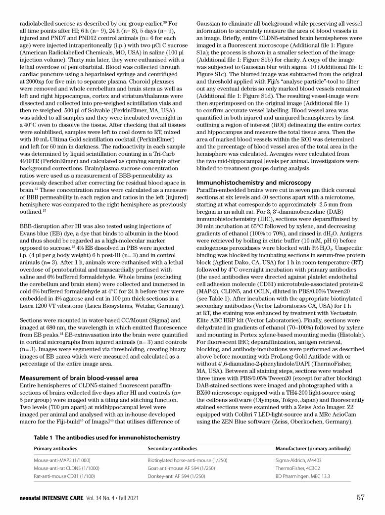
Gaussian to eliminate all background while preserving all vessel information to accurately measure the area of blood vessels in an image. Briefly, entire CLDN5-stained brain hemispheres were imaged in a fluorescent microscope (Additional file 1: Figure S1a); the process is shown in a smaller selection of the image (Additional file 1: Figure S1b) for clarity. A copy of the image was subjected to Gaussian blur with sigma=10 (Additional file 1: Figure S1c). The blurred image was subtracted from the original and threshold applied with Fiji’s “analyse particle”-tool to filter out any eventual debris so only marked blood vessels remained (Additional file 1: Figure S1d). The resulting vessel-image were then superimposed on the original image (Additional file 1) to confirm accurate vessel labelling. Blood vessel area was quantified in both injured and uninjured hemispheres by first outlining a region of interest (ROI) delineating the entire cortex and hippocampus and measure the total tissue area. Then the area of marked blood vessels within the ROI was determined and the percentage of blood vessel area of the total area in the hemisphere was calculated. Averages were calculated from the two mid-hippocampal levels per animal. Investigators were blinded to treatment groups during analysis.
Immunohistochemistry and microscopyParaffin-embedded brains were cut in seven µm thick coronal sections at six levels and 40 sections apart with a microtome, starting at what corresponds to approximately -2.5 mm from bregma in an adult rat. For 3, 3′-diaminobenzidine (DAB) immunohistochemistry (IHC), sections were deparaffinised by 30 min incubation at 65°C followed by xylene, and decreasing gradients of ethanol (100% to 70%), and rinsed in dH2O. Antigens were retrieved by boiling in citric buffer (10 mM, pH 6) before endogenous peroxidases were blocked with 3% H2O2. Unspecific binding was blocked by incubating sections in serum-free protein block (Aglient Dako, CA, USA) for 1 h in room-temperature (RT) followed by 4°C overnight incubation with primary antibodies (the used antibodies were directed against platelet endothelial cell adhesion molecule (CD31) microtubule-associated protein-2 (MAP-2), CLDN5, and OCLN, diluted in PBS/0.05% Tween20 (see Table 1). After incubation with the appropriate biotinylated secondary antibodies (Vector Laboratories CA, USA) for 1 h at RT, the staining was enhanced by treatment with Vectastain Elite ABC HRP kit (Vector Laboratories). Finally, sections were dehydrated in gradients of ethanol (70–100%) followed by xylene and mounting in Pertex xylene-based mounting media (Histolab). For fluorescent IHC; deparaffinization, antigen retrieval, blocking, and antibody-incubations were performed as described above before mounting with ProLong Gold Antifade with or without 4′,6-diamidino-2-phenylindole/DAPI (ThermoFisher, MA, USA). Between all staining steps, sections were washed three times with PBS/0.05% Tween20 (except for after blocking). DAB-stained sections were imaged and photographed with a BX60 microscope equipped with a TH4-200 light-source using the cellSens software (Olympus, Tokyo, Japan) and fluorescently stained sections were examined with a Zeiss Axio Imager. Z2 equipped with Colibri 7 LED-light-source and a MRc AcioCam using the ZEN Blue software (Zeiss, Oberkochen, Germany).
radiolabelled sucrose as described by our group earlier.39 For all time points after HI; 6 h (n= 9), 24 h (n= 8), 5 days (n= 9), injured and PND7 and PND12 control animals (n= 6 for each age) were injected intraperitoneally (i.p.) with two µCi C sucrose (American Radiolabelled Chemicals, MO, USA) in saline (100 µl injection volume). Thirty min later, they were euthanised with a lethal overdose of pentobarbital. Blood was collected through cardiac puncture using a heparinised syringe and centrifuged at 2000xg for five min to separate plasma. Choroid plexuses were removed and whole cerebellum and brain stem as well as left and right hippocampus, cortex and striatum/thalamus were dissected and collected into pre-weighed scintillation vials and then re-weighed. 500 µl of Solvable (PerkinElmer, MA, USA) was added to all samples and they were incubated overnight in a 40°C oven to dissolve the tissue. After checking that all tissues were solubilised, samples were left to cool down to RT, mixed with 10 mL Ultima Gold scintillation cocktail (PerkinElmer) and left for 60 min in darkness. The radioactivity in each sample was determined by liquid scintillation counting in a Tri-Carb 4910TR (PerkinElmer) and calculated as cpm/mg sample after background corrections. Brain/plasma sucrose concentration ratios were used as a measurement of BBB-permeability as previously described after correcting for residual blood space in brain.42 These concentration ratios were calculated as a measure of BBB permeability in each region and ratios in the left (injured) hemisphere was compared to the right hemisphere as previously outlined.15
BBB-disruption after HI was also tested using injections of Evans blue (EB) dye, a dye that binds to albumin in the blood and thus should be regarded as a high-molecular marker opposed to sucrose.43 4% EB dissolved in PBS were injected i.p. (4 µl per g body weight) 6 h post-HI (n= 3) and in control animals (n= 3). After 1 h, animals were euthanised with a lethal overdose of pentobarbital and transcardially perfused with saline and 6% buffered formaldehyde. Whole brains (excluding the cerebellum and brain stem) were collected and immersed in cold 6% buffered formaldehyde at 4°C for 24 h before they were embedded in 4% agarose and cut in 100 µm thick sections in a Leica 1200 VT vibratome (Leica Biosystems, Wetzlar, Germany).
Sections were mounted in water-based CC/Mount (Sigma) and imaged at 680 nm, the wavelength in which emitted fluorescence from EB peaks.44 EB-extravasation into the brain were quantified in cortical micrographs from injured animals (n= 3) and controls (n= 3). Images were segmented via thresholding, creating binary images of EB ±area which were measured and calculated as a percentage of the entire image area.
Measurement of brain blood-vessel areaEntire hemispheres of CLDN5-stained fluorescent paraffin-sections of brains collected five days after HI and controls (n= 5 per group) were imaged with a tiling and stitching function. Two levels (700 µm apart) at midhippocampal level were imaged per animal and analysed with an in-house developed macro for the Fiji-build45 of ImageJ46 that utilises difference of
Page 4 of 14Andersson et al. Fluids Barriers CNS (2021) 18:7
BBB-disruption after HI was also tested using injec-tions of Evans blue (EB) dye, a dye that binds to albumin in the blood and thus should be regarded as a high-molecular marker opposed to sucrose [43]. 4% EB dis-solved in PBS were injected i.p. (4 µl per g body weight) 6 h post-HI (n = 3) and in control animals (n = 3). After 1 h, animals were euthanised with a lethal overdose of pentobarbital and transcardially perfused with saline and 6% buffered formaldehyde. Whole brains (excluding the cerebellum and brain stem) were collected and immersed in cold 6% buffered formaldehyde at 4 °C for 24 h before they were embedded in 4% agarose and cut in 100 µm thick sections in a Leica 1200 VT vibratome (Leica Bio-systems, Wetzlar, Germany).
Sections were mounted in water-based CC/Mount (Sigma) and imaged at 680 nm, the wavelength in which emitted fluorescence from EB peaks [44]. EB-extrava-sation into the brain were quantified in cortical micro-graphs from injured animals (n = 3) and controls (n = 3). Images were segmented via thresholding, creating binary images of EB + -area which were measured and calcu-lated as a percentage of the entire image area.
Measurement of brain blood‑vessel areaEntire hemispheres of CLDN5-stained fluorescent par-affin-sections of brains collected five days after HI and controls (n = 5 per group) were imaged with a tiling and stitching function. Two levels (700 µm apart) at mid-hippocampal level were imaged per animal and analysed with an in-house developed macro for the Fiji-build [45] of ImageJ [46] that utilises difference of Gaussian to elim-inate all background while preserving all vessel informa-tion to accurately measure the area of blood vessels in an image. Briefly, entire CLDN5-stained brain hemispheres were imaged in a fluorescent microscope (Additional file 1: Figure S1a); the process is shown in a smaller selec-tion of the image (Additional file 1: Figure S1b) for clarity. A copy of the image was subjected to Gaussian blur with sigma = 10 (Additional file 1: Figure S1c). The blurred image was subtracted from the original and threshold applied with Fiji’s “analyse particle”-tool to filter out any eventual debris so only marked blood vessels remained (Additional file 1: Figure S1d). The resulting vessel-image were then superimposed on the original image (Addi-tional file 1) to confirm accurate vessel labelling. Blood
vessel area was quantified in both injured and uninjured hemispheres by first outlining a region of interest (ROI) delineating the entire cortex and hippocampus and meas-ure the total tissue area. Then the area of marked blood vessels within the ROI was determined and the percent-age of blood vessel area of the total area in the hemi-sphere was calculated. Averages were calculated from the two mid-hippocampal levels per animal. Investigators were blinded to treatment groups during analysis.
Immunohistochemistry and microscopyParaffin-embedded brains were cut in seven µm thick coronal sections at six levels and 40 sections apart with a microtome, starting at what corresponds to approxi-mately -2.5 mm from bregma in an adult rat. For 3, 3′-diaminobenzidine (DAB) immunohistochemistry (IHC), sections were deparaffinised by 30 min incuba-tion at 65 °C followed by xylene, and decreasing gradients of ethanol (100% to 70%), and rinsed in dH2O. Antigens were retrieved by boiling in citric buffer (10 mM, pH 6) before endogenous peroxidases were blocked with 3% H2O2. Unspecific binding was blocked by incubat-ing sections in serum-free protein block (Aglient Dako, CA, USA) for 1 h in room-temperature (RT) followed by 4 °C overnight incubation with primary antibodies (the used antibodies were directed against platelet endothe-lial cell adhesion molecule (CD31) microtubule- associ-ated protein-2 (MAP-2), CLDN5, and OCLN, diluted in PBS/0.05% Tween20 (see Table 1). After incubation with the appropriate biotinylated secondary antibodies (Vector Laboratories CA, USA) for 1 h at RT, the stain-ing was enhanced by treatment with Vectastain Elite ABC HRP kit (Vector Laboratories). Finally, sections were dehydrated in gradients of ethanol (70–100%) fol-lowed by xylene and mounting in Pertex xylene-based mounting media (Histolab). For fluorescent IHC; depar-affinization, antigen retrieval, blocking, and antibody-incubations were performed as described above before mounting with ProLong Gold Antifade with or without 4′,6-diamidino-2-phenylindole/DAPI (ThermoFisher, MA, USA). Between all staining steps, sections were washed three times with PBS/0.05% Tween20 (except for after blocking). DAB-stained sections were imaged and photographed with a BX60 microscope equipped with a TH4-200 light-source using the cellSens software
Table 1 The antibodies used for immunohistochemistry
Primary antibodies Secondary antibodies Manufacturer (primary antibody)
Mouse-anti-MAP2 (1/1000) Biotinylated horse-anti-mouse (1/250) Sigma-Aldrich, M4403
Mouse-anti-rat CLDN5 (1/1000) Goat-anti-mouse AF 594 (1/250) ThermoFisher, 4C3C2
Rat-anti-mouse CD31 (1/100) Donkey-anti AF 594 (1/250) BD Pharmingen, MEC 13.3
neonatal INTENSIVE CARE Vol. 34 No. 4 n Fall 2021 57
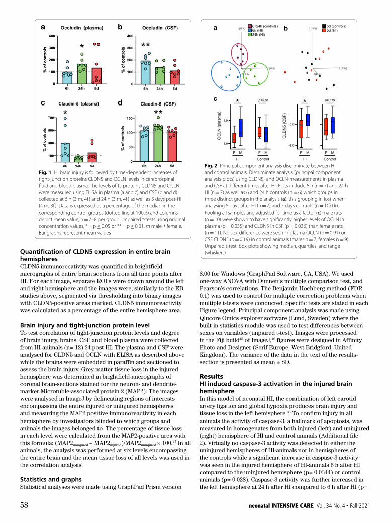
8.00 for Windows (GraphPad Software, CA, USA). We used one-way ANOVA with Dunnett’s multiple comparison test, and Pearson’s correlations. The Benjamin-Hochberg method (FDR 0.1) was used to control for multiple correction problems when multiple t-tests were conducted. Specific tests are stated in each Figure legend. Principal component analysis was made using Qlucore Omics explorer software (Lund, Sweden) where the built-in statistics module was used to test differences between sexes on variables (unpaired t-test). Images were processed in the Fiji build45 of ImageJ,46 figures were designed in Affinity Photo and Designer (Serif Europe, West Bridgford, United Kingdom). The variance of the data in the text of the results-section is presented as mean ± SD.
ResultsHI induced caspase-3 activation in the injured brain hemisphereIn this model of neonatal HI, the combination of left carotid artery ligation and global hypoxia produces brain injury and tissue loss in the left hemisphere.36 To confirm injury in all animals the activity of caspase-3, a hallmark of apoptosis, was measured in homogenates from both injured (left) and uninjured (right) hemisphere of HI and control animals (Additional file 2). Virtually no caspase-3 activity was detected in either the uninjured hemispheres of HI-animals nor in hemispheres of the controls while a significant increase in caspase-3 activity was seen in the injured hemisphere of HI-animals 6 h after HI compared to the uninjured hemisphere (p= 0.0344) or control animals (p= 0.028). Caspase-3 activity was further increased in the left hemisphere at 24 h after HI compared to 6 h after HI (p=
Quantification of CLDN5 expression in entire brain hemispheresCLDN5 immunorecativity was quantified in brightfield micrographs of entire brain sections from all time points after HI. For each image, separate ROI:s were drawn around the left and right hemisphere and the images were, similarly to the EB-studies above, segmented via thresholding into binary images with CLDN5-positive areas marked. CLDN5 immunoreactivity was calculated as a percentage of the entire hemisphere area.
Brain injury and tight-junction protein levelTo test correlation of tight-junction protein levels and degree of brain injury, brains, CSF and blood plasma were collected from HI-animals (n= 12) 24 post-HI. The plasma and CSF were analysed for CLDN5 and OCLN with ELISA as described above while the brains were embedded in paraffin and sectioned to assess the brain injury. Grey matter tissue loss in the injured hemisphere was determined in brightfield-micrographs of coronal brain-sections stained for the neuron- and dendrite-marker Microtuble-associated protein 2 (MAP2). The images were analysed in ImageJ by delineating regions of interests encompassing the entire injured or uninjured hemispheres and measuring the MAP2 positive immunoreactivity in each hemisphere by investigators blinded to which groups and animals the images belonged to. The percentage of tissue loss in each level were calculated from the MAP2-positive area with this formula: (MAP2uninjured − MAP2injured)/MAP2uninjured × 100.47 In all animals, the analysis was performed at six levels encompassing the entire brain and the mean tissue loss of all levels was used in the correlation analysis.
Statistics and graphsStatistical analyses were made using GraphPad Prism version
Page 6 of 14Andersson et al. Fluids Barriers CNS (2021) 18:7
Principal component analysis (PCA) was performed (dimension reducing) as means of discriminate analy-sis with the input of all above measured variables for OCLN and CLDN5. HI and control animals were grouped into three distinct groups at 6 h and 24 h after HI (Fig. 2a) whereas at 5 days post-HI (Fig. 2b), no clustering of control vs HI animals was apparent. To test effect of sex on levels of TJ-proteins we performed unpaired t-test between male and female animals pool-ing all samples within all injured and within all control animals, adjusting for time as a factor. Male rats (n = 10) showed significantly higher levels of OCLN in plasma (p = 0.035) and CLDN5 in CSF (p = 0.036) than female rats (n = 11), control animals (males n = 7, females n = 9) had no sex-differences in plasma OCLN (p = 0.91) or CSF CLDN5 (p = 0.19) (Fig. 2c). In HI-animals, the aver-age amount of OCLN in plasma were ~ 700 pg/ml for males and ~ 400 pg/ml for females, for CLDN5 in CSF the numbers were ~ 36 ng/ml for males and ~ 26 ng/ml for females.
Dynamic changes in BBB function following HI in neonatesTo determine changes in BBB function over time after HI we performed measurements of BBB-permeability
in different brain regions (i.e. hippocampus, cortex, and striatum/thalamus) by quantifying the permeability for 14C-labelled sucrose across the BBB. We previously showed [15] that the BBB in the uninjured hemisphere is not altered. We confirmed this (Additional file 5) for the 6 h (controls n = 5, HI n = 5) and 5-day time-points (controls n = 5, HI n = 5) corroborating with caspase-3 activation (Additional file 2). Increased BBB permeabil-ity occurred in the ipsilateral hemisphere at 6 h post-HI in the cortex (1.10 ± 0.05, p = 0.0007), hippocampus (1.07 ± 0.08, p = 0.0277), and striatum/thalamus (1.05 ± 0.05, p = 0.0111) and was also significantly higher at 24 h in both hippocampus (1.12 ± 0.12, p = 0.0486) and cortex (1.18 ± 0.28, p = 0.0474) while the striatum/thalamus appeared unaltered (1.01 ± 0.07, p = 0.7624) (Fig. 3a). Mentionable is that these concentration ratios are probably somewhat affected by the edema occur-ring in the injured hemisphere after the insult suggesting that the magnitude of BBB permeability increase is likely marginally higher than what these ratios reflect. The con-centration ratios 5d post-HI was significantly lower in
Fig. 1 HI brain injury is followed by time-dependent increases of tight-junction proteins CLDN5 and OCLN levels in cerebrospinal fluid and blood plasma. The levels of TJ-proteins CLDN5 and OCLN were measured using ELISA in plasma (a and c) and CSF (b and d) collected at 6 h (3 m, 4f ) and 24 h (3 m, 4f ) as well as 5 days post-HI (4 m, 3f ). Data is expressed as a percentage of the median in the corresponding control-groups (dotted line at 100%) and columns depict mean value, n = 7–8 per group. Unpaired t-tests using original concentration values, * = p ≤ 0.05 or ** = p ≤ 0.01. m male, f female. Bar graphs represent mean values
Fig. 2 Principal component analysis discriminate between HI and control animals. Discriminate analysis (principal component analysis-plots) using CLDN5- and OCLN-measurements in plasma and CSF at different times after HI. Plots include 6 h (n = 7) and 24 h HI (n = 7) as well as 6 and 24 h controls (n = 6) which groups in three distinct groups in the analysis (a), this grouping in lost when analysing 5 days after HI (n = 7) and 5 days controls (n = 10) (b). Pooling all samples and adjusted for time as a factor (c) male rats (n = 10) were shown to have significantly higher levels of OCLN in plasma (p = 0.035) and CLDN5 in CSF (p = 0.036) than female rats (n = 11). No sex-difference were seen in plasma OCLN (p = 0.91) or CSF CLDN5 (p = 0.19) in control animals (males n = 7, females n = 9). Unpaired t-test, box-plots showing median, quartiles, and range (whiskers)
58 neonatal INTENSIVE CARE Vol. 34 No. 4 n Fall 2021
differences were measured across groups at any time point after HI (Additional file 4).
Principal component analysis (PCA) was performed (dimension reducing) as means of discriminate analysis with the input of all above measured variables for OCLN and CLDN5. HI and control animals were grouped into three distinct groups at 6 h and 24 h after HI (Fig. 2a) whereas at 5 days post-HI (Fig. 2b), no clustering of control vs HI animals was apparent. To test effect of sex on levels of TJ-proteins we performed unpaired t-test between male and female animals pooling all samples within all injured and within all control animals, adjusting for time as a factor. Male rats (n= 10) showed significantly higher levels of OCLN in plasma (p= 0.035) and CLDN5 in CSF (p= 0.036) than female rats (n= 11), control animals (males n= 7, females n= 9) had no sex-differences in plasma OCLN (p= 0.91) or CSF CLDN5 (p= 0.19) (Fig. 2c). In HI-animals, the average amount of OCLN in plasma were~ 700 pg/ml for males and~ 400 pg/ml for females, for CLDN5 in CSF the numbers were~ 36 ng/ml for males and~ 26 ng/ml for females.
Dynamic changes in BBB function following HI in neonates To determine changes in BBB function over time after HI we performed measurements of BBB-permeability in different brain regions (i.e. hippocampus, cortex, and striatum/thalamus) by quantifying the permeability for 14C-labelled sucrose across the BBB. We previously showed15 that the BBB in the uninjured hemisphere is not altered. We confirmed this (Additional file 5) for the 6 h (controls n= 5, HI n= 5) and 5-day time-points (controls n= 5, HI n= 5) corroborating with caspase-3 activation (Additional file 2). Increased BBB permeability occurred in the ipsilateral hemisphere at 6 h post-HI in the cortex (1.10 ± 0.05, p= 0.0007), hippocampus (1.07 ± 0.08, p= 0.0277),
0.0087), controls (p= 0.0027) as well as the uninjured hemisphere (p= 0.0060). The range of caspase activity for the 6 h and 24 h post-HI groups were 3–77 and 10–950 pmol AMC/ min x mg protein, respectively.
HI-injury resulted in time-dependent increased levels of circulating tight-junction proteins CLDN5 and OCLN in cerebrospinal fluid and plasma and ZO-1 in CSF Levels of CLDN5, OCLN and ZO-1 were measured in CSF and plasma with ELISA at 6, 24 h and 5 days after HI and all time-points were compared with a control group collected and analysed at the same time and on the same ELISA-plate. CLDN5 and OCLN proteins were detected in all samples with levels ranging between ~41 to 1300 pg/ml (plasma), ~100 to 2400 pg/ml (CSF) for OCLN and ~2 to 30 ng/ml (plasma), ~30 to 53 ng/ml (CSF) for CLDN5. Elevated CLDN5-levels were detected in CSF (Fig. 1d) at 24 h post-HI (p= 0.0082), while plasma-concentrations (Fig. 1c) were higher than controls at 6 h (p= 0.0427). There were no difference at later times between HI and controls. Similarly, OCLN concentration in CSF (Fig. 1b) was raised at 6 h (p= 0.0026) after HI while the levels were higher in plasma (Fig. 1a) of HI-animals at 24 h (p= 0.0285). Measured values (mean ± SD) for CLDN5 and OCLN in blood plasma and CSF are available in Additional file 3. ZO-1 was only detectable in 47% of CSF samples. We found great variably in levels between plasma with as high as 2000 pg/mL in one animal but no
Page 7 of 14Andersson et al. Fluids Barriers CNS (2021) 18:7
Fig. 3 The BBB permeability increases in the injured hemisphere at 6 and 24 h after neonatal HI. a Regional (hippocampus, cortex, striatum/thalamus) brain 14C-sucrose concentration ratios between left (injured) and right (uninjured) hemispheres at 6 (4 m, 5f ), 24 (4 m, 4f ) hours and 5 days (4 m, 5f ) after neonatal HI. b Entire hemisphere brain 14C-sucrose concentration ratios between left (injured) and right (uninjured) hemispheres at various times between 6 h and 5 days after neonatal HI. Horizontal lines indicate mean value. * = p ≤ 0.05, ** = p ≤ 0.01, and *** = p ≤ 0.001 (paired t-test between hemispheres). c The vascular density of the brain is not altered at 5 days after hypoxia/ischemia in neonates. Vascular area defined as CLDN5+ area in brain sections (See Additional file 1 for more details). Vascular area of total area in cortex/hippocampus in both the left (injured) and right hemispheres of HI-animals (n = 5, 2 m, 4f ) was similar to control animals (n = 5, 2 m, 4f ). BBB-damage were confirmed by imaging brain sections from animals injected with the fluorescent dye Evans blue (EB). After transcardial perfusion, areas of EB-bound albumin in the brain parenchyma was present in the cortex of injured hemispheres (d) while the uninjured hemispheres only showed weak EB-fluorescence restricted to blood vessels in corresponding areas (e). Control animals injected with EB and not perfused showed strong EB-fluorescence in blood vessels and none in parenchyma (f). Extravasated EB encompassed 51.1 ± 8.4% of the total area in images from the injured hemisphere, significantly altered from the uninjured hemisphere (6.3 ± 1.9%, p = 0.0002) and controls (5.8 ± 1.6%, p = 0.0002) where the EB was restricted to blood vessels (g), cortical images were were taken from the region outlined in (h). n=3 per group. Scale-bars are 100 μm (d‑f) and two mm (h). One-way ANOVA with Dunnett’s multiple comparison test. Data presented as mean ± SD. M male, f female. Bar graphs represent mean values.
the injured cortex (0.88 ± 0.12, p = 0.0191) as well as in the entire injured hemispheres (0.91 ± 0.10, p = 0.0303) (Fig. 3b).
BBB disruption following HI were confirmed via assess-ment of EB-bound albumin extravasation into the brain parenchyma in brain sections. Animals injected with EB at 6 h post-HI showed fluorescence in the cortex of the injured hemispheres (Fig. 3d) while the uninjured hemi-spheres showed weak fluorescence restricted to blood vessels (Fig. 3e). Control animals injected with EB and not perfused showed distinct blood vessels-restricted fluorescence (Fig. 3f ) while animals not injected with EB showed no signal. Extravasated EB encompassed 51.1 ± 8.4% of the total area in images from the injured hemisphere, significantly compared to images from the uninjured hemisphere (6.3 ± 1.9%, p = 0.0002) and con-trols (5.8 ± 1.6%, p = 0.0002) where the EB was restricted to blood vessels.
The vascular density of the brain is not altered five days after HISince the cerebrovascular area, the effective surface area for exchange between blood and brain, can affect meas-urements of BBB-permeability we developed an in-house written macro for blood vessel analysis of CLDN5 (as a vascular marker) immunolabelled sections. We spe-cifically wanted to estimate cerebrovascular area at later times after injury since there is loss of brain tissue and potentially blood vessels. Sections from brains 5 days post-HI were used to calculate the area of blood ves-sels in the brain. Two levels were analysed per animal and results averaged (Fig. 3c). For this analysis, the hip-pocampus and cortex results were combined in each hemisphere. In control animals 2.01 ± 0.64% of the brain area was comprised of vessels while in HI animals vessels comprised 1.53 ± 0.37% of the uninjured hemisphere and 2.05 ± 0.61% in the injured hemisphere. No significant differences were detected between control animals and either hemisphere of injured animals (p > 0.05).
CLDN5 immunoreactivity is not altered in the cerebral blood vessels of neonatal ratsGiven that there are reports of changes in TJ-protein immunoreactivity following hypoxia/ischemia and
neonatal INTENSIVE CARE Vol. 34 No. 4 n Fall 2021 59
± 8.4% of the total area in images from the injured hemisphere, significantly compared to images from the uninjured hemisphere (6.3 ± 1.9%, p= 0.0002) and controls (5.8 ± 1.6%, p= 0.0002) where the EB was restricted to blood vessels.
The vascular density of the brain is not altered five days after HISince the cerebrovascular area, the effective surface area for exchange between blood and brain, can affect measurements of BBB-permeability we developed an in-house written macro for blood vessel analysis of CLDN5 (as a vascular marker) immunolabelled sections. We specifically wanted to estimate cerebrovascular area at later times after injury since there is loss of brain tissue and potentially blood vessels. Sections from brains 5 days post-HI were used to calculate the area of blood vessels in the brain. Two levels were analysed per animal and results averaged (Fig. 3c). For this analysis, the hippocampus and cortex results were combined in each hemisphere. In control animals 2.01 ± 0.64% of the brain area was comprised of vessels while in HI animals vessels comprised 1.53 ± 0.37% of the uninjured hemisphere and 2.05 ± 0.61% in the injured hemisphere. No significant differences were detected between
and striatum/thalamus (1.05 ± 0.05, p= 0.0111) and was also significantly higher at 24 h in both hippocampus (1.12 ± 0.12, p= 0.0486) and cortex (1.18 ± 0.28, p= 0.0474) while the striatum/ thalamus appeared unaltered (1.01 ± 0.07, p= 0.7624) (Fig. 3a). Mentionable is that these concentration ratios are probably somewhat affected by the edema occurring in the injured hemisphere after the insult suggesting that the magnitude of BBB permeability increase is likely marginally higher than what these ratios reflect. The concentration ratios 5d post-HI was significantly lower in the injured cortex (0.88 ± 0.12, p= 0.0191) as well as in the entire injured hemispheres (0.91 ± 0.10, p= 0.0303) (Fig. 3b).
BBB disruption following HI were confirmed via assessment of EB-bound albumin extravasation into the brain parenchyma in brain sections. Animals injected with EB at 6 h post-HI showed fluorescence in the cortex of the injured hemispheres (Fig. 3d) while the uninjured hemispheres showed weak fluorescence restricted to blood vessels (Fig. 3e). Control animals injected with EB and not perfused showed distinct blood vessels-restricted fluorescence (Fig. 3f) while animals not injected with EB showed no signal. Extravasated EB encompassed 51.1
Page 8 of 14Andersson et al. Fluids Barriers CNS (2021) 18:7
hypoxia alone [28, 48], we performed double immuno-fluorescent labelling of CLDN5 (Fig. 4a) together with blood vessel marker CD31 (platelet endothelial cell adhesion molecule) in paraffin brain-sections collected at all time points after HI (time chosen given BBB changes). In control animals, we found robust immuno-reactivity of TJ-proteins in vessels in all brain regions examined including the cortex, hippocampus, and striatum/thalamus, while no labelling was detectable in parenchyma of the brain. Likewise, in animals after HI we found immunolabelling of blood vessels across all brain regions including MAP2-negative regions with no apparent changes compared to control animals.
CLDN5 immunoreactivity was quantified in DAB-developed sections for CLDN5 only. No significant differences in CLDN5-coverage were seen between HI (n = 9) and control animals (n = 6) (p > 0.05), or between the injured and uninjured hemisphere in HI-animals (p > 0.05), at any time-point (n = 3 per time point) when CLDN5-immunoreactivity was quantified in entire stained brain hemispheres (Fig. 4b).
CLDN5 levels in CSF correlates with brain‑injury severity 24 h after HIThe correlation between circulating TJ-protein lev-els and brain injury severity were investigated 24 h
Fig. 4 CLDN5 immunoreactivity is not altered in the cerebral blood vessels of neonatal rats after HI. Representative double-immunoreactivity for CD31 (red, blood vessel marker) and tight-junctional proteins CLDN5 (green) in cortical brain sections collected 6 h after hypoxia/ischemia (a). Scale-bars are 100 μm in all images. No difference in CLDN5-coverage were seen (b) between controls or either hemisphere in HI-animals at 6 h (2 m, 1f ), 24 h (2 m, 1f ), 5d (2 m, 1f ) post-HI (left in b; p > 0.5) nor between the injured and uninjured hemisphere in HI animals (right in b; p > 0.05). HI, n = 3 per time point, controls (n = 2 per time-point). One-way ANOVA with Dunnett’s multiple comparison test and paired t-tests. m male, f female. Bar graphs represent mean values
60 neonatal INTENSIVE CARE Vol. 34 No. 4 n Fall 2021
vessels across all brain regions including MAP2-negative regions with no apparent changes compared to control animals.
CLDN5 immunoreactivity was quantified in DAB-developed sections for CLDN5 only. No significant differences in CLDN5-coverage were seen between HI (n = 9) and control animals (n = 6) (p > 0.05), or between the injured and uninjured hemisphere in HI-animals (p > 0.05), at any time-point (n = 3 per time point) when CLDN5-immunoreactivity was quantified in entire stained brain hemispheres (Fig. 4b).
CLDN5 levels in CSF correlates with brain-injury severity 24 h after HIThe correlation between circulating TJ-protein levels and brain injury severity were investigated 24 h after HI by analysing plasma- and CSF-levels of TJ:s and simultaneously quantifying
control animals and either hemisphere of injured animals (p> 0.05).
CLDN5 immunoreactivity is not altered in the cerebral blood vessels of neonatal ratsGiven that there are reports of changes in TJ-protein immunoreactivity following hypoxia/ischemia andhypoxia alone,28,48 we performed double immuno-fluorescent labelling of CLDN5 (Fig. 4a) together with blood vessel marker CD31 (platelet endothelial cell adhesion molecule) in paraffin brain-sections collected at all time points after HI (time chosen given BBB changes). In control animals, we found robust immuno-reactivity of TJ-proteins in vessels in all brain regions examined including the cortex, hippocampus, and striatum/thalamus, while no labelling was detectable in parenchyma of the brain. Likewise, in animals after HI we found immunolabelling of blood
Page 9 of 14Andersson et al. Fluids Barriers CNS (2021) 18:7
after HI by analysing plasma- and CSF-levels of TJ:s and simultaneously quantifying the loss of grey mat-ter in the brain of the same animal. This time-point was chosen based on the earlier ELISA-results (Fig. 1). The average brain tissue loss in the HI-group (n = 12) varied from 24.7 to 59.9% (one representative level is shown in Fig. 5b and d, respectively). By using the Pearson correlation on the levels of circulating
TJ-proteins and the tissue loss percentages (Fig. 5a), the levels of CLDN5 in CSF was found to significantly (p = 0.016) correlate with the severity of the grey mat-ter tissue loss (r = 0.702). No correlation was observed between the loss of grey matter and levels of CLDN5 in plasma (r = -− 0.001) nor OCLN in CSF (r = 0.060) or plasma (r = 0.039).
Fig. 5 CLDN5 levels in CSF correlates with brain-injury severity 24 h after HI. The grey matter tissue loss, defined as the average percentage of negative MAP2 immunoreactivity calculated from six levels throughout the whole brain, correlates with the levels of CLDN5 in the CSF of the same animal 24 h post-HI (a) with a Pearson correlation coefficient (r) of 0.702 (p = 0.016). No correlation was observed between the loss of grey matter and levels of CLDN5 in plasma (c) nor OCLN in CSF (b) or plasma (d). The tissue loss varied between 24.7 to 59.9%, three representative images of injured brain hemispheres are shown in (e), n = 12 (5 m, 7f ). Scale-bars are two mm in all images. m male, f female
neonatal INTENSIVE CARE Vol. 34 No. 4 n Fall 2021 61

in plasma increased at 24 h. Levels of CLDN5 on the other hand were elevated in plasma at 6 h post-HI, while levels in CSF were higher than normal at 24 h. Levels of CLDN5 and OCLN appeared to be normalised in both plasma and CSF five days after HI. CLDN5 was more concentrated than OCLN in both plasma and CSF. High levels of ZO-1 could only be measured in the CSF in some of the animals and was not detectable in plasma, thus it appears that ZO-1, being an intracellular protein, is not as readily released into the blood after BBB damage. Ischemia has been shown to induce matrix metalloproteinase(MMP)-mediated disruption of TJ-proteins,55 thus altering the cellular distribution of CLDN5 and OCLN in neonatal mice14 and adult rats56 and seemingly releasing some TJ-proteins into the circulation.
Pan et al.22 measured the levels of tight-junction proteins in blood up to 4.5 h after the induction of ischemic stroke via middle-cerebral artery occlusion (MCAO) in adult rats and saw a significant increase of circulating OCLN and a molecular loss of OCLN from cerebral microvessels at 4.5 h post-MCAO while reporting no differences in blood-CLDN5 levels. A follow up study yielded similar results in adult rats after MCAO as well as showing that blood OCLN was elevated in human stroke patients within 24 h and up to 3d after stroke onset, the levels of OCLN in blood was reduced after treatment with normobaric hyperoxia which inhibits MMP-9 activity.57 While we cannot directly compare our results to their studies as they have utilised adult animals in another model, it is evident that focal stroke and MCAO leads to an increase in blood OCLN levels earlier than in our neonatal model where we see a peak in blood OCLN 24 h after HI.
The raised levels of TJ-proteins in both CSF and plasma indicate that BBB dysfunction after neonatal HI injury is likely to be at least partially due to direct molecular damage to the endothelial TJ:s. Intriguingly, the raised levels of circulating OCLN and CLDN5 did not occur at the same time after HI showing that these proteins although normally intimately localised do not appear to be released into these biofluids in the same manner. The exact cause of this unclear, but it has been shown that OCLN and CLDN5 are affected differently in the early ischemic stroke stages with the former being degraded by MMP:s and the latter instead redistributed via the membrane protein caveolin-1.58 The complex linking of CLDN5 and OCLN to domains of intercellular adapter proteins is different, as reviewed by Piontek et al.,59 and may also play a role in their release from tight-junctions.
Furthermore, we tested whether levels TJ-proteins could reflect severity of injury, choosing the 24 h time-point since our results indicated particularly raised levels of CLDN5 in CSF at this time. This showed that levels of CLDN5 in CSF correlated with the severity of brain injury while OCLN levels and plasma CLDN5 showed no correlation. Human studies on circulating TJ-proteins have only been made in adults, which makes direct comparisons with our results more difficult, but there are some common observations. Kazmierski et al.23 measured TJ-proteins in serum after ischemic stroke and found that levels of circulating CLDN5 and OCLN could predict clinical deterioration as a result of haemorrhagic transformation up to 4.5 h after stroke onset, with the most sensitive measurement being the CLDN5/ ZO-1 ratio. Comparing TJ-levels in blood and CSF as well as BBB-disruption between controls and a cohort of patients with intracranial haemorrhage (ICH), Jiao et al.24 showed that CSF, but not serum, levels of TJ-proteins are sensitive predictors for BBB-damage after ICH. These studies and our results implies that the levels
the loss of grey matter in the brain of the same animal. This time-point was chosen based on the earlier ELISA-results (Fig. 1). The average brain tissue loss in the HI-group (n = 12) varied from 24.7 to 59.9% (one representative level is shown in Fig. 5b and d, respectively). By using the Pearson correlation on the levels of circulating
TJ-proteins and the tissue loss percentages (Fig. 5a), the levels of CLDN5 in CSF was found to significantly (p = 0.016) correlate with the severity of the grey matter tissue loss (r = 0.702). No correlation was observed between the loss of grey matter and levels of CLDN5 in plasma (r = –0.001) nor OCLN in CSF (r = 0.060) or plasma (r = 0.039).
Discussion and conclusionsThe brain vasculature is a central component in HI associated brain injuries that appears damaged early in the injury process with studies indicating loss of BBB function in response to neonatal HI.15,49 However, diagnostic tools to evaluate brain vascular dysfunction in neonates in an early, affordable, accessible, and reliable manner are lacking. We show elevated levels of OCLN and CLDN5 in both CSF and plasma in response to loss of blood-brain barrier function and that CLDN5 levels in CSF correlates with brain-injury severity. Our data indicate that loss of BBB function is likely, at least partially, due to molecular impairment of endothelial tight-junctions after HI injury. The data suggest that the intermembrane TJ-proteins CLDN5 and OCLN are putative blood or CSF biomarkers for cerebral vascular dysfunction and brain damage in neonates.
The cerebrovasculature is specialised in that it harbours a range of barrier and transport mechanism not found in peripheral blood vessels, which compartmentalises the brain from the rest of the body so that brain cells can function in a controlled environment and provides protection for the brain from potential harmful blood solutes. These mechanisms are normally referred to as the blood–brain barrier and an essential part are the tight-junctions localised to the luminal side of the inter-endothelial cleft forming a physical barrier between blood plasma and brain.50 The protein architecture of these junctions is complex with both inter- and intracellular elements. In this study we focused on two intracellular TJ-proteins, CLDN5, which are specific and essential for normal BBB function51 and, OCLN, which plays a role in BBB modulation.52 We made measurements of both BBB function together with measurements of TJ-proteins in both CSF and plasma in order to interpret data in an integrated manner.
We found time-dependent changes in both BBB function and TJ-proteins levels following HI. Detection of Evans blue in the injured region of HI-injured animals confirmed BBB-disruption following HI and the BBB permeability was acutely increased following HI in rat neonates in agreement with earlier rat studies16,20,53 and with a similar time line to what we have seen in mice.15 Clinical data show about five times higher albumin CSF/blood ratios of babies diagnosed with NE indicative of BBB damage also in human newborns, although such ratios should be interpreted with caution.49 Overall, the greatest changes in BBB-opening appeared to be in the cortex, the region most affected in this HI-model of rats.54 However, we also see a correlation between severity of injury and BBB opening in the different brain regions, similar to the earlier mouse study.15
OCLN in CSF was significantly increased 6 h after HI while levels
62 neonatal INTENSIVE CARE Vol. 34 No. 4 n Fall 2021

study. Brain blood flow, measured between 3 to 48 h, in the PND 7 rat HI-model have also shown that that cortical blood flow in this model is reduced not until 48 h after injury, indicating that HI could lead to alterations in the vascular physiology.67 Speculatively, the time-dependent changes in BBB permeability, increased entry rate in the acute phase (6-24 h) followed by decreased entry rate later 5 days could influence the efficacy of drug treatment in relation to their time of administration time. A limitation of the study is the large inter-animal variability in injury, inherent to this model-system of neonatal HI.68 Furthermore, brain injury is limited to the hypoxic/ischemic hemisphere, unlike human infants which often develop more generalised brain injuries.69
Impact statement• BBBdysfunctionfollowingneonatalhypoxia/ischemiais
likely in part due to the loss of tight-junction proteins from cerebral blood vessels. BBB breakdown release tight junction proteins and BBB function may be assessed by measuring these proteins in the circulation.
• Thisisthefirststudywhichinvestigatestight-junctionproteinsin the CSF and correlate to levels in circulation in a neonatal animal model of brain injury.
• Elevatedlevelsofblood–brainbarrier-derivedtight-junctionproteins Claudin-5 and Occludin can be detected in the circulation at several time-points in a rat-model for neonatal HI, signifying the proteins potential as biomarkers for the brain vascular dysfunction in neonates.
Supplementary InformationThe online version contains supplementary material available at https://doi. org/10.1186/s12987-021-00240-9.
Additional file 1: Images showing the process of delineating blood vessels from CLDN5 immunoreactivity in brain sections. Blood vessels in entire hemispheres (a) were delineated, the process is shown in a selection (b) for clarity. By utilizing difference of Gaussian (c) and Fiji’s “analyse particle”-tool, blood vessels in entire brain hemispheres were delineated (d). Accurate marking of vessels was confirmed by overlaying the processed image with the original non-processed image (e).
Additional file 2: Hypoxia/ischemia induces caspase-3 activation in the injured brain hemisphere only. Activated caspase-3 could be measured in the injured hemisphere 6h (3m, 4f ), and 24h (3m, 4f ) post-HI but not in the uninjured hemisphere nor in any hemisphere of control animals. Columns depict mean value, n 7 per group. Unpaired t-tests between groups, significant differences are marked * (p 0.05) or ** (p 0.01). m male, f female. Bar graphs represent mean values.
Additional file 3: CLDN5 and OCLN levels in blood plasma and CSF, mean ± SD.
Additional file 4: Tight-junction protein ZO-1 can be detected in CSF from some, but not all, animals after neonatal hypoxia/ischemia. The levels of ZO-1 in CSF at 6h, and 24h as well as 5d after HI was measured with ELISA. Out of 7-8 animals per group, we could detect ZO-1 in 3-5 CSF samples. Columns depict mean value, one-way ANOVA with Dunnett’s multiple comparison test, HI-groups compared to the controls, p < 0.05 for all comparisons. Bar graphs represent mean values.
Additional file 5: BBB-permeability is not altered in right
=
=
of circulating TJ-proteins indeed corresponds to the severity of vascular and tissue injury, and BBB-disruption following ischemic events in the brain. Their results also agree with our finding that circulating CLDN5 was more abundant than OCLN.
Taken together our study shows that even a moderate opening of BBB in this model results in raised levels of TJ-proteins at early time points after HI which suggests that TJ-proteins are released into the circulation in the early stages of BBB-damage and could act as biomarkers for vascular integrity and possibly also be useful as brain injury predictor. Our results also indicate a sex difference where males have higher levels of circulating OCLN in plasma and CLDN5 in CSF. This resonates well with previous studies that have shown that there is a tendency for male rats to have graver injuries than females after neonatal HI in an almost identical model to the one employed in this study60 and it is also known that male human infants have a higher risk for NE.61 Since our study was not specifically designed to investigate sex differences and analysis involved pooling samples from different time-points, further studies would be needed for confirmation of results. CLDN5 and OCLN showed different patterns of release into biofluids indicating that measuring both of them in tandem would give a better interpretation of injury to the brain vasculature after HI. We therefore performed discriminate analysis showing that HI-animals grouped together and were separate from controls up to 24 h after HI when the levels of CLDN5 and OCLN in both plasma and CSF were analysed together.
While TJ-proteins seem to have potential as standalone biomarkers for neonatal cerebral vasculature dysfuntion, many studies, as reviewed in Douglas-Escobar and Weiss,62 Chalak,63 and Lv,13 have focused on biomarkers related to inflammation and brain injury. As the reviews state, the best course of action will most likely be to develop a panel of different markers in conjunction with diagnosis and other monitoring methods of neonatal brain injuries. Promising biomarker-candidates for assessing NE brain injuries include neuronal injury markers such as Tau and neurofilament light proteins,64 brain injury marker protein S100B,65 and inflammation-related cytokines like IL-6 and IL-8.66 The levels of circulating TJ-proteins in blood from human stroke patients has been shown to positively correlate with the levels of S100B23 and we saw a correlation between white matter tissue loss and the levels of CLDN5 in CSF. We therefore believe circulating TJ-proteins have promise as a marker for vascular dysfunction, which, in combination with markers for inflammation and injury, would increase the discriminatory, and predictive power of a marker panel for NE brain injuries.
Intriguingly, our results indicated that BBB function does not normalise at later times after HI but instead there is an apparent decrease in permeability below normal levels five days after the insult. One explanation could be that there is loss of blood vessels in the brain tissue after HI, which would reduce the surface area for exchange and thus reduce flux between blood and brain. We therefore estimated the cerebrovascular area at 5 days after HI, which showed that the tissue remaining in the injured hemisphere did not differ in vascular area compared to control animals. Thus, loss of blood vessels unlikely explains the difference in measured BBB permeability. We previously demonstrated an upregulation of both CLDN5 and OCLN genes following neonatal HI injury in mice,15 which could be a response to normalise barrier function and might underlie the decrease in barrier permeability we observed in the present
neonatal INTENSIVE CARE Vol. 34 No. 4 n Fall 2021 63
encephalopathy: a review for the clinician. JAMA Pediatrics. 2015;169(4):397–403.
4. Jacobs SE, Berg M, Hunt R, TarnowMordi WO, Inder TE, Davis PG. Cooling for newborns with hypoxic ischaemic encephalopathy. Cochrane Database Systematic Rev. 2013;2013(1):CD003311.
5. Gluckman PD, Wyatt JS, Azzopardi D, Ballard R, Edwards AD, Ferriero DM, et al. Selective head cooling with mild systemic hypothermia after neonatal encephalopathy: multicentre randomised trial. Lancet. 2005;365(9460):663–70.
6. Glass HC. Hypoxic-Ischemic Encephalopathy and Other Neonatal Encephalopathies Continuum (Minneapolis, Minn). Child Neurology. 2018;24(1,):57–71.
7. Armstrong L, Stenson BJ. Use of umbilical cord blood gas analysis in the assessment of the newborn. Arch Dis Child Fetal Neonatal Ed. 2007;92(6):F430–4.
8. Goswami IR, Whyte H, Wintermark P, Mohammad K, Shivananda S, Louis D, et al. Characteristics and short-term outcomes of neonates with mild hypoxic-ischemic encephalopathy treated with hypothermia. J Perinatol . 2020;40(2):275–83.
9. Ruth VJ, Raivio KO. Perinatal brain damage: predictive value of metabolic acidosis and the Apgar score. BMJ. 1988;297(6640):24–7.
10. Natarajan N, Pardo AC. Challenges in neurologic prognostication after neonatal brain injury. Semin Perinatol. 2017;41(2):117–23.
11. Mariani G, Kasznia-Brown J, Paez D, Mikhail MN, D HS, Bhatla N, et al. Improving women’s health in low-income and middle-income countries. Part II: the needs of diagnostic imaging. Nuclear medicine communications. 2017;38(12):1024–8.
12. Lee AC, Kozuki N, Blencowe H, Vos T, Bahalim A, Darmstadt GL, et al. Intrapartum-related neonatal encephalopathy incidence and impairment at regional and global levels for 2010 with trends from 1990. Pediatric Res. 2013;74(Suppl 1):50–72.
13. Lv H, Wang Q, Wu S, Yang L, Ren P, Yang Y, et al. Neonatal hypoxic ischemic encephalopathy-related biomarkers in serum and cerebrospinal fluid. Clinica chimica acta; international journal of clinical chemistry. 2015;450:282–97.
14. Baburamani A, Ek CJ, Walker D, Castillo-Melendez M. Vulnerability of the developing brain to hypoxic-ischemic damage: contribution of the cerebral vasculature to injury and repair. Frontiers in Physiology. 2012;3(424).
15. Ek CJ, D’Angelo B, Baburamani AA, Lehner C, Leverin AL, Smith PL, et al. Brain barrier properties and cerebral blood flow in neonatal mice exposed to cerebral hypoxia-ischemia. J Cerebral Blood Flow Metabolism : . 2015;35(5):818–27.
16. Muramatsu K, Fukuda A, Togari H, Wada Y, Nishino H. Vulnerability to cerebral hypoxic-ischemic insult in neonatal but not in adult rats is in parallel with disruption of the blood-brain barrier. Stroke. 1997;28(11):2281–8.
17. Svedin P, Hagberg H, Sävman K, Zhu C, Mallard C. Matrix metalloproteinase-9 gene knock-out protects the immature brain after cerebral hypoxia-ischemia. J Neurosci. 2007;27(7):1511–8.
18. Tu YF, Tsai YS, Wang LW, Wu HC, Huang CC, Ho CJ. Overweight worsens apoptosis, neuroinflammation and blood-brain barrier damage after hypoxic ischemia in neonatal brain through JNK hyperactivation. J Neuroinflammation. 2011;8:40.
19. Yang D, Sun YY, Nemkul N, Baumann JM, Shereen A, Dunn RS, et al. Plasminogen activator inhibitor-1 mitigates
hemisphere of control animals. Blood/brain 14C-sucrose concentration ratios in the right hemispheres at 6h (3m, 2f ) and 5 days (2m, 3f ) following HI together with litter-mate controls. Horizontal lines indicate mean value, n 5 per group (mixed sexes), two-way ANOVA between regions and groups for both time-points, p<0.05 for all comparisons. . m male, f female.
AbbreviationsAMC: 7-Amino-4-methylcoumarin; BBB: Blood–brain barrier; CD31: Platelet endothelial cell adhesion molecule (CD31; CLDN5: Claudin-5; CSF: Cerebrospinal fluid; DAB: 3, 3′-Diaminobenzidine; EB: Evans blue; ELISA: Enzyme-linked immunosorbent assay; HI: Hypoxia/ischemia; HRP: Horseradish peroxidase; ICH: Intracranial haemorrhage; i.p: Intraperitoneal; IHC: Immunohistochemistry; MAP2: Microtubule Associated Protein 2; MAP-2: Microtubule-associated protein-2 (MAP-2); NE: Neonatal encephalopathy; OCLN: Occludin; PBS: Phosphate-buffered saline; PCA: Principal component analysis; PMSF: Phenylmethylsulfonyl fluoride; PND#: Post-natal day #; TJ: Tight-junction(s); ZO-1: Zonula occludens protein 1.
AcknowledgementsThe authors want to acknowledge the staff at the Centre for Cellular Imaging at the University of Gothenburg for providing assistance in fluorescent imaging and Nazanin Shahbazi for the technical assistance she provided to the Evans blue-experiments.
FundingOpen Access funding provided by University of Gothenburg. The authors disclosed receipt of the following financial support for the research, authorship, and/or publication of this article: Rune och Ulla Amlövs Stiftelse (EAA), Herbert och Karin Jacobsons Stiftelse (EAA), Swedish Research Council (VR-2017–01409 – CM), Åhlen Foundation (CM), Public Health Service at the Sahlgrenska University Hospital (ALFGBG-722491—CM), Swedish Brain Foundation (FO2019-0270—CM), Cerebral Palsy Alliance Australia (CJE), Stroke-Riksförbundet (CJE), and Wilhelm and Martina Lundgren Foundations (CJE).
Availability of data and materialsThe data behind the conclusions of this study are available from the corresponding author upon reasonable request.
Ethics approval and consent to participateAll animal experiments were approved by the Gothenburg Committee of the Swedish Animal Welfare Agency (Application nos. 663/17) and performed in accordance with the ARRIVE guidelines.
Consent for publicationNot applicable.
Competing interestsThe Authors declares that there is no conflict of interest with respect to the research, authorship, or publication of this article.
References1. Molloy EJ, Bearer C. Neonatal encephalopathy versus
Hypoxic-Ischemic Encephalopathy. Pediatric Res. 2018;84(5):574.
2. Aslam S, Strickland T, Molloy EJ. Neonatal encephalopathy: need for recognition of multiple etiologies for optimal management. Front Pediatr. 2019;7:142.
3. Douglas-Escobar M, Weiss MD. Hypoxic-ischemic
64 neonatal INTENSIVE CARE Vol. 34 No. 4 n Fall 2021
35. Van Itallie CM, Fanning AS, Holmes J, Anderson JM. Occludin is required for cytokine-induced regulation of tight junction barriers. J Cell Sci. 2010;123(16):2844.
36. Rice JE 3rd, Vannucci RC, Brierley JB. The influence of immaturity on hypoxic-ischemic brain damage in the rat. Ann Neurol. 1981;9(2):131–41.
37. Hagberg H, Ichord R, Palmer C, Yager JY, Vannucci SJ. Animal models of developmental brain injury: relevance to human disease. A summary of the panel discussion from the Third Hershey Conference on Developmental Cerebral Blood Flow and Metabolism. Dev Neurosci. 2002;24(5):364–6.
38. Hagberg H, Peebles D, Mallard C. Models of white matter injury: comparison of infectious, hypoxic-ischemic, and excitotoxic insults. Mental Retardation Developmental Disabilities Res Rev. 2002;8(1):30–8.
39. Mottahedin A, Smith PL, Hagberg H, Ek CJ, Mallard C. TLR2-mediated leukocyte trafficking to the developing brain. J Leukoc Biol. 2017;101(1):297–305.
40. Habgood MD, Knott GW, Dziegielewska KM, Saunders NR. The nature of the decrease in blood-cerebrospinal fluid barrier exchange during postnatal brain development in the rat. J Physiol. 1993;468:73–83.
41. Wang X, Karlsson JO, Zhu C, Bahr BA, Hagberg H, Blomgren K. Caspase-3 activation after neonatal rat cerebral hypoxia-ischemia. Biol Neonate. 2001;79(3–4):172–9.
42. Ek CJ, Habgood MD, Dziegielewska KM, Potter A, Saunders NR. Permeability and route of entry for lipid-insoluble molecules across brain barriers in developing Monodelphis domestica. J Physiol. 2001;536(Pt 3):841–53.
43. Ryu HW, Lim W, Jo D, Kim S, Park JT, Min JJ, et al. Low-dose evans blue dye for near-infrared fluorescence imaging in photothrombotic stroke model. Int Journal Medical Sci. 2018;15(7):696–702.
44. Saria A, Lundberg JM. Evans blue fluorescence: quantitative and morphological evaluation of vascular permeability in animal tissues. J Neurosci Methods. 1983;8(1):41–9.
45. Schindelin J, Arganda-Carreras I, Frise E, Kaynig V, Longair M, Pietzsch T, et al. Fiji: an open-source platform for biological-image analysis. Nat Methods. 2012;9(7):676–82.
46. Schneider CA, Rasband WS, Eliceiri KW. NIH Image to ImageJ: 25 years of image analysis. Nat Methods. 2012;9(7):671–5.
47. Mottahedin A, Blondel S, Ek J, Leverin AL, Svedin P, Hagberg H, et al. N-acetylcysteine inhibits bacterial lipopeptide-mediated neutrophil transmigration through the choroid plexus in the developing brain. Acta Neuropathologica Commun. 2020;8(1):4.
48. Koto T, Takubo K, Ishida S, Shinoda H, Inoue M, Tsubota K, et al. Hypoxia disrupts the barrier function of neural blood vessels through changes in the expression of claudin-5 in endothelial cells. Am J Pathol. 2007;170(4):1389–97.
49. Kumar A, Mittal R, Khanna HD, Basu S. Free radical injury and blood-brain barrier permeability in hypoxic-ischemic encephalopathy. Pediatrics. 2008;122(3):e722–7.
50. Dobrogowska DH, Vorbrodt AW. Immunogold localization of tight junctional proteins in normal and osmotically-affected rat blood-brain barrier. J Mol Histol. 2004;35(5):529–39.
51. Nitta T, Hata M, Gotoh S, Seo Y, Sasaki H, Hashimoto N, et al. Size-selective loosening of the blood-brain barrier in claudin-5-deficient mice. J Cell Biol. 2003;161(3):653–60.
52. Raleigh DR, Boe DM, Yu D, Weber CR, Marchiando AM, Bradford EM, et al. Occludin S408 phosphorylation regulates tight junction protein interactions and barrier function. J Cell Biol. 2011;193(3):565–82.
brain injury in a rat model of infection-sensitized neonatal hypoxia-ischemia. Cerebral cortex (New York, NY : 1991). 2013;23(5):1218–29.
20. Ferrari DC, Nesic OB, Perez-Polo JR. Oxygen resuscitation does not ameliorate neonatal hypoxia/ischemia-induced cerebral edema. J Neurosci Res. 2010;88(9):2056–65.
21. Berndt P, Winkler L, Cording J, Breitkreuz-Korff O, Rex A, Dithmer S, et al. Tight junction proteins at the blood-brain barrier: far more than claudin-5. Cellular Molecular life Sci . 2019;76(10):1987–2002.
22. Pan R, Yu K, Weatherwax T, Zheng H, Liu W, Liu KJ. Blood occludin level as a potential biomarker for early blood brain barrier damage following ischemic stroke. Scientific Rep. 2017;7:40331.
23. Kazmierski R, Michalak S, Wencel-Warot A, Nowinski WL. Serum tight-junction proteins predict hemorrhagic transformation in ischemic stroke patients. Neurology. 2012;79(16):1677–85.
24. Jiao X, He P, Li Y, Fan Z, Si M, Xie Q, et al. The role of circulating tight junction proteins in evaluating blood brain barrier disruption following intracranial hemorrhage. Dis Markers. 2015;2015:860120.
25. Mark KS, Davis TP. Cerebral microvascular changes in permeability and tight junctions induced by hypoxia-reoxygenation. Am J Physiol Heart Circulatory Physiol. 2002;282(4):H1485–94.
26. Yang Y, Estrada EY, Thompson JF, Liu W, Rosenberg GA. Matrix metalloproteinase-mediated disruption of tight junction proteins in cerebral vessels is reversed by synthetic matrix metalloproteinase inhibitor in focal ischemia in rat. J Cerebral Blood Flow Metab . 2007;27(4):697–709.
27. Liddelow SA, Dziegielewska KM, Ek CJ, Habgood MD, Bauer H, Bauer H-C, et al. Mechanisms that determine the internal environment of the developing brain: a transcriptomic, functional and ultrastructural approach. PLoS ONE. 2013;8(7):e65629.
28. Page S, Munsell A, Al-Ahmad AJ. Cerebral hypoxia/ischemia selectively disrupts tight junctions complexes in stem cell-derived human brain microvascular endothelial cells. Fluids Barriers CNS. 2016;13(1):16.
29. Ballabh P, Hu F, Kumarasiri M, Braun A, Nedergaard M. Development of tight junction molecules in blood vessels of germinal matrix, cerebral cortex, and white matter. Pediatr Res. 2005;58(4):791–8.
30. Bauer HC, Bauer H, Lametschwandtner A, Amberger A, Ruiz P, Steiner M. Neovascularization and the appearance of morphological characteristics of the blood-brain barrier in the embryonic mouse central nervous system. Brain Res Dev Brain Res. 1993;75(2):269–78.
31. Daneman R, Zhou L, Kebede AA, Barres BA. Pericytes are required for blood-brain barrier integrity during embryogenesis. Nature. 2010;468(7323):562–6.
32. Ek CJ, Dziegielewska KM, Stolp H, Saunders NR. Functional effectiveness of the blood-brain barrier to small water-soluble molecules in developing and adult opossum (Monodelphis domestica). J Comp Neurol. 2006;496(1):13–26.
33. Wen H, Watry DD, Marcondes MCG, Fox HS. Selective decrease in paracellular conductance of tight junctions: role of the first extracellular domain of claudin-5. Mol Cell Biol. 2004;24(19):8408–17.
34. Rodgers LS, Beam MT, Anderson JM, Fanning AS. Epithelial barrier assembly requires coordinated activity of multiple domains of the tight junction protein ZO-1. J Cell Sci. 2013;126(7):1565.
neonatal INTENSIVE CARE Vol. 34 No. 4 n Fall 2021 65

53. Tu XK, Yang WZ, Liang RS, Shi SS, Chen JP, Chen CM, et al. Effect of baicalin on matrix metalloproteinase-9 expression and blood-brain barrier permeability following focal cerebral ischemia in rats. Neurochem Res. 2011;36(11):2022–8.
54. Tang S, Xu S, Lu X, Gullapalli RP, McKenna MC, Waddell J. Neuroprotective effects of acetyl-l-carnitine on neonatal hypoxia ischemia-induced brain injury in rats. Dev Neurosci. 2016;38(5):384–96.
55. Rosenberg GA, Yang Y. Vasogenic edema due to tight junction disruption by matrix metalloproteinases in cerebral ischemia. Neurosurg Focus. 2007;22(5):E4.
56. Jiao H, Wang Z, Liu Y, Wang P, Xue Y. Specific role of tight junction proteins claudin-5, occludin, and ZO-1 of the blood-brain barrier in a focal cerebral ischemic insult. J Molecular Neurosci. 2011;44(2):130–9.
57. Shi S, Qi Z, Ma Q, Pan R, Timmins GS, Zhao Y, et al. Normobaric hyperoxia reduces blood occludin fragments in rats and patients with acute ischemic stroke. Stroke. 2017;48(10):2848–54.
58. Liu J, Jin X, Liu KJ, Liu W. Matrix metalloproteinase-2-mediated occludin degradation and caveolin-1-mediated claudin-5 redistribution contribute to blood-brain barrier damage in early ischemic stroke stage. J Neurosci. 2012;32(9):3044–57.
59. Piontek J, Krug SM, Protze J, Krause G, Fromm M. Molecular architecture and assembly of the tight junction backbone. Biochimica et Biophysica Acta (BBA) - Biomembranes. 2020;1862(7):183279.
60. Hill CA, Threlkeld SW, Fitch RH. Early testosterone modulated sex differences in behavioral outcome following neonatal hypoxia ischemia in rats. Int J Develop Neurosci . 2011;29(4):381–8.
61. Hill CA, Fitch RH. Sex differences in mechanisms and outcome of neonatal hypoxia-ischemia in rodent models: implications for sex-specific neuroprotection in clinical neonatal practice. Neurol Res Int. 2012;2012:867531.
62. Douglas-Escobar M, Weiss MD. Neonatal biomarkers of brain injury. NeoReviews. 2013;14(10):e501–12.
63. Chalak LF. Inflammatory Biomarkers of Birth Asphyxia. Clin Perinatol. 2016;43(3):501–10.
64. Toorell H, Zetterberg H, Blennow K, Sävman K, Hagberg H. Increase of neuronal injury markers Tau and neurofilament light proteins in umbilical blood after intrapartum asphyxia. J Maternal Fetal Neonatal Med . 2018;31(18):2468–72.
65. Thorngren-Jerneck K, Alling C, Herbst A, Amer-Wahlin I, Marsal K. S100 protein in serum as a prognostic marker for cerebral injury in term newborn infants with hypoxic ischemic encephalopathy. Pediatr Res. 2004;55(3):406–12.
66. Chalak LF, Sánchez PJ, Adams-Huet B, Laptook AR, Heyne RJ, Rosenfeld CR. Biomarkers for severity of neonatal hypoxic-ischemic encephalopathy and outcomes in newborns receiving hypothermia therapy. J Pediatrics. 2014;164(3):468-74.e1.
67. Gilland E, Bona E, Hagberg H. Temporal changes of regional glucose use, blood flow, and microtubule-associated protein 2 immunostaining after hypoxia-ischemia in the immature rat brain. J Cerebral Blood Flow Metab. 1998;18(2):222–8.
68. Mallard C, Vexler ZS. Modeling ischemia in the immature brain: how translational are animal models? Stroke. 2015;46(10):3006–11.
69. Khong PL, Lam BC, Tung HK, Wong V, Chan FL, Ooi GC. MRI of neonatal encephalopathy. Clin Radiol. 2003;58(11):833–44.
66 neonatal INTENSIVE CARE Vol. 34 No. 4 n Fall 2021

Ad Page 67
Are you bubbling?
© 2019 Respiralogics, GaleMed. All rights reserved.
3545 Airway Drive, Suite 104 • Reno, NV 89511 775 954 0160 • www.respiralogics.com
Babi.Plus Bubble PAP Valve
Baby Nose Bumper and RespiraGel Mustache
Babi.Plus® Pressure Relief
Manifold
Baby™ Head Band and Circuit Bumpers
Baby Band for Chin and Head
Babi.Plus is the standard of care for non-invasive ventilation, providing a gentle approach to improve ventilation and optimize neonatal outcomes.
To start optimizing ventilation
in your NICU contact us at [email protected]
Babi.Plus Nasal Prongs

Ad Page 68
©2020 Prolacta Bioscience, Inc. All rights reserved. MKT-0679 Rev-0 08/20
Healthy development of low birth weight (LBW) premature infants’ gut,1 brain,2 and lungs3 are all impaired without the necessary nutrition. With Prolacta’s products as a part of an Exclusive Human Milk Diet, you can safely fortify within the first week of life to provide the necessary nutrition.3
prolacta.com
1. Brune KD, Donn SM. Enteral feeding of the preterm infant. NeoReviews. 2018;19(11):e645-e653. doi:10.1542/neo.19-11-e645 2. Schneider J, Fischer Fumeaux CJ, Duerden EG, et al. Nutrient intake in the first two weeks of life and brain growth in preterm neonates. Pediatrics. 2018;141(3):e20172169. doi:10.1542/peds.2017-2169 3. Huston R, Lee M, Rider E, et al. Early fortification of enteral feedings for infants <1250 grams birth weight receiving a human milk diet including human milk based fortifier. J Neonatal Perinatal Med. 2020;13(2):215-221. doi:10.3233/NPM-190300 4. Estimated number of premature infants fed Prolacta’s products from January 2007 to August 2020; data on file.
Half of the nation’s level III and IV neonatal intensive care units have seen firsthand just how much our meticulously crafted human milk-based products matter. 63,000 premature infants throughout the world and counting.4
[email protected] 888.776.5228
TOGETHER,we are addressing
the risks of late, inadequate, and
inappropriate nutrition for LBW premature infants