Acute Pain Service Handbook - Institut de Cardiologie de ...Acute Pain Service Handbook A peer...
116
Acute Pain Service Handbook A peerͲreviewed, referenced resource 2010
Transcript of Acute Pain Service Handbook - Institut de Cardiologie de ...Acute Pain Service Handbook A peer...
Acute Pain Service Handbook
A peer reviewed, referenced resource
2010

Acute Pain Service Handbook
First Canadian Edition
This book belongs to:
Name:___________________________________
Phone:___________________________________
Email:___________________________________
Foreword Acute pain after injury or surgery remains poorly treated despite an armamentarium of effective treatments and the activity of acute pain services. Acute pain in itself is very distressing however adverse effects of pain on other organ systems can lead to significant complications and the generation of chronic pain. Effective and rapid treatment of pain is vital. Barriers to effective management of acute pain are currently not well defined however effective education is a key component. Currently few easily accessible resources exist for the practitioner managing acute pain. The APS Handbook by Kashin, Riazi and Sawhney will be an important resource for the many interns, residents, physicians and nursing staff dealing with common acute pain problems. The authors aim was to produce a resource that is both straightforward yet comprehensive enough to treat most acute pain problems in a timely and effective manner. Brian Kashin MD and Mona Sawhney RN are both highly knowledgeable and experienced pain practitioners who have effectively treated many patients in pain. Sheila Riazi MD is a recently graduated anesthesia resident who has very clear knowledge of the problems and barriers faced by many residents who are called to manage patients in severe pain. Together they have produced a practical and readable handbook. The APS Handbook will be an essential resource leading to better pain management for many patients. I commend the authors for producing this excellent book. Colin J.L. McCartney MBChB FRCA FRCPC Sunnybrook Health Sciences Centre University of Toronto
Preface
Although knowledge regarding the treatment of acute pain is rapidly expanding and the quality of evidence has improved, this improvement has not led to progress in patient care. There remains a gap between the advances in assessment and management of acute pain and the improvements in clinical practice Acute pain management has seen many changes in the assessment and the available therapies. Acute pain is being identified as a problem in many patient populations. Beyond postoperative, traumatic and obstetric causes of pain, patients experience acute on-chronic pain, acute cancer pain or acute pain from medical conditions. It is our hope that this handbook will provide, nurses, medical students, and physicians in training simple and practical information that would help them manage their patients�’ pain in the most effective manner. This handbook includes information regarding conventional methods of analgesia for acute pain as well as newer techniques such as patient-controlled intravenous and epidural analgesia. It also includes information on the management of medical conditions that can cause pain as well as special patient subpopulations. The purpose of this book is to be a practical handbook therefore detailed information about anatomy, and specific regional anesthesia techniques have not been included. Suggested drugs, doses and treatment regimens are guidelines only and may have to be adapted according to different patients and clinical situations. The authors of this book have used their best efforts to provide accurate information at the time of printing The authors hereby disclaim all responsibility for any loss suffered by any person in the light of future discoveries in this field, and for any omissions or errors in the text. Special thanks go out to Dr. Mark Friedlander who was the first director of the Acute Pain Service at the Toronto General Hospital and then North York General Hospital. We are fortunate to have his input and editing skills. I would also like to thank the contributions of the following physicians: Thomas Engelhardt: University of Aberdeen, Edward Mariano: University of California, San Diego, Paul Tumber: University of Toronto, Dr Basem Naser: Hospital for Sick Children and Anita Sarmah: University of Toronto Brian Kashin Sheila Riazi Mona Sawhney
Table of Contents
Chapter 1: Pain Pathways, Transmission and Modulation Chapter 2: Pain Assessment Tools and Considerations Chapter 3: Non-Opioids, Opioids and Adjuvant Agents Chapter 4: Pharmacology of Local Anesthetic Chapter 5: Post-operative Pain Management Chapter 6 : Pain Management in Patient Subpopulations Chapter 7 : Acute Pain Syndromes Appendices: Appendix A: Acute Pain Service Initial Assessment and Daily Flow Sheet Appendix B: Adjuncts Appendix C: Postoperative Nausea and Vomiting (PONV) Appendix D: Common Oral Narcotics Appendix E: NSAIDS Appendix F: Opioid Equianalgesic Table Appendix G: Pain Glossary

Chapter 1:
Pain Pathways,
transmission and
modulation
2
Chapter 1: Pain pathways, transmission and modulation
Tissue injury, such as that induced by surgical incision results in the local release of numerous chemicals that mediate or facilitate inflammation. Collectively these chemical have become known as an inflammatory soup which includes bradykinin, prostaglandin, leukotrienes, serotonin, histamine, substance P, calcitonin-gene related peptide, thromboxanes, platelet-activating factor (PAF), adenosine/ ATP, cytokines and neurotrophins (i.e. nerve growth factor). These substances may be released from tissue factors, such as lipids following injury, from nerve endings that respond to injury (nociceptors) or from immune cells. These agents are generally characterized by their ability to (1) evoke inflammation (i.e. swelling, redness or increased skin temperature) (2) directly activate and/or sensitize nociceptors. Those agents that can directly activate nociceptors, may do so directly or indirectly via inflammatory cells, which in turn release algogenic agents. For instance, mast cells are the primary source of histamine and PAF. Histamine contributes directly to inflammation by producing vasodilation and oedema, while PAF leads to serotonin release from platelets which can directly activate and sensitize nociceptors. The effect of sensitization is increased primary afferent sensitivity (Fitzgerald and Lynn 1978; Schaible and Grubb 1993; Pawlak et al., 2001; Chen et al., 2006), which decreases the threshold for afferent activation by a noxious stimulus. This results in increased sensitivity to painful stimuli (hyperalgesia) and pain to stimuli that are not normally painful (allodynia). Hyperalgesia and allodynia are the primary features of a wide range of chronic pain conditions, including postoperative pain. Primary afferents that are activated by noxious stimuli to peripheral tissues (i.e. viscera and somatic sites) are small-diameter A and C fibre nociceptive afferents. These fibres send impulses (i.e., action potentials) into the CNS to provide sensory-discriminative information about the location, quality, intensity and duration of the noxious stimulus.Nociceptive primary afferent axons terminate exclusively in the dorsal horn of the spinal cord, and it is therefore the site of the first synapse in the ascending pathways that convey (somatosensory cortex), sensory information to the brain that underlies conscious perception of pain. In addition, neuronal
3
circuits in the spinal cord generate local reflexes (dorsal root reflexes) that send retrograde impulses into the periphery that can cause the release of inflammatory mediators from nociceptive terminals, thereby prolonging inflammation (Willis and Coggeshall 2004). The dorsal horn of the spinal cord is also the site where peripheral nociceptive information is modulated by other afferent inputs and descending modulatory inputs from supraspinal structures (i.e. periaqueductal grey, raphe nuclei and locus coeruleus in the midbrain). Depending on which descending modulatory inputs are activated (i.e., serotonin, noradrenaline versus enkephalin), the transmission of nociceptive information may be either enhanced or attenuated. There are 3 general classes of nociceptors: thermal, mechanical, and polymodal. Thermal (extreme temperatures 45 °C or < 5°C) and mechanical nociceptors are thinly myelinated A fibres whereas polymodal nociceptors are both A and unmyelinated C fibres that are activated by high intensity mechanical, chemical and thermal stimuli (Basbaum and Jessell 2000; Willis and Coggeshall 2004; Willis 2005). Surgical incision is thought to predominantly activate polymodal A and C fibre primary afferents.
Neurons in the spinal cord that receive nociceptive information reside predominantly in the marginal layer (lamina I) and the substantia gelatinosa (lamina II) of the superficial dorsal horn. The majority of these neurons receive direct converging input from A and C fibres. Neurons that respond exclusively to noxious stimulation are classified as nociceptive-specific or NS neurons and project to higher brain centers, whereas some neurons in this layer, called wide-dynamic-range (WDR) neurons, respond in a graded fashion to both non-noxious and noxious stimulation. WDR and some NS neurons are also found in Lamina V and project to the brainstem and to regions of the thalamus.
4
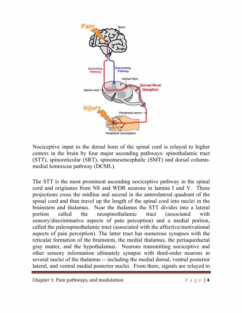
Nociceptive input to the dorsal horn of the spinal cord is relayed to higher centers in the brain by four major ascending pathways: spinothalamic tract (STT), spinoreticular (SRT), spinomesencephalic (SMT) and dorsal column-medial lemniscus pathway (DCML).
The STT is the most prominent ascending nociceptive pathway in the spinal cord and originates from NS and WDR neurons in lamina I and V. These projections cross the midline and ascend in the anterolateral quadrant of the spinal cord and than travel up the length of the spinal cord into nuclei in the brainstem and thalamus. Near the thalamus the STT divides into a lateral portion called the neospinothalamic tract (associated with sensory/discriminative aspects of pain perception) and a medial portion, called the paleospinothalamic tract (associated with the affective/motivational aspects of pain perception). The latter tract has numerous synapses with the reticular formation of the brainstem, the medial thalamus, the periaqueductal gray matter, and the hypothalamus. Neurons transmitting nociceptive and other sensory information ultimately synapse with third-order neurons in several nuclei of the thalamus -- including the medial dorsal, ventral posterior lateral, and ventral medial posterior nuclei. From there, signals are relayed to
5
the primary somatosensory cortex, which is responsible for our conscious recognition of pain. The somatosensory cortex and the thalamus directly relay nociceptive information to other brain areas such as the cingulate cortex and insular cortex, which are involved in the evaluative and affective aspects of pain perception.
The SRT plays a critical role in relaying and integrating nociceptive information contributing to the motivational, affective, and aversive response aspects of pain. The neurons of the SRT originate primarily in Laminas VII and VIII of the spinal cord. They terminate in many sites throughout the brain stem reticular formation. Neurons from the reticular formation project to many areas of the brain, including the hypothalamus, the thalamus, and both directly and indirectly to the limbic forebrain and neocortex; areas associated with the emotional aspect of pain.
The SMT neurons originate in Laminas I, IV, V and VI in the dorsal horn of the spinal cord. They terminate in several structures of the midbrain, especially the periaqueductal gray, the nucleus cuneiformis, and the superior colliculus. These connections produce affective and aversive behaviours associated with pain such as fear. They may also initiate orienting responses. The SMT input to the periaqueductal gray activates the system for descending pain modulation which produces endogenous analgesia.
The DCML pathway transmits sensory information about touch and proprioception and has been traditionally viewed as a pathway not involved in pain perception. However, there is compelling evidence that implicates the DCML pathway in relaying nociceptive information. Axons of the DCML pathway travel up the ipsilateral side of the spinal cord and synapse with second order neurons at the gracile and cuneate nuclei. Studies have shown that fibres of the dorsal column that ascend close to the midline are involved in the transmission of nociceptive information. Second order axons of the DCML pathways cross the midline and ascend to the ventral posterior lateral and medial thalamus where they join nociceptive fibres of the STT and than project to same higher brain centers involved in pain perception (i.e. somatosensory cortex).
6
Pain Modulation
The transmission of nociceptive information is part of the body's defense system that produces a rapid-warning response, instructing the body to react to damaging stimuli. However, ongoing noxious impulses conveyed from the periphery to the spinal cord and brain can result in neuroplastic changes that sensitize several sites of the pain pathway giving rise to clinical pain.
Peripheral modulation: The high threshold of nociceptors can be reduced by changes in the function or expression of ion channels, receptors or transducer proteins on peripheral nociceptor terminals. In the case of tissue damage, the release of inflammatory mediators activates nociceptors and initiate an intracellular signaling cascade that evokes such changes. The major mechanism responsible for these alterations is phosphorylation of receptor/ion channels and/or changes in the expression of channels in primary sensory and dorsal horn neurons. This modulation increases the excitability of nociceptor terminals which reduces its threshold for activation, thus producing peripheral sensitization. The clinical feature of peripheral sensitization is increased pain sensitivity at the site of damaged tissue (i.e. primary hyperalgesia). However, pain may also appear outside the area of injury (secondary hyperalgesia), spontaneously or in response to light touch (allodynia). It is also possible for pain to arise without any physical injury at all (migraine, fibromyalgia or irritable bowel syndrome). In these conditions, pain arises from central amplification of peripheral inputs, central sensitization.
Central modulation: (1) Spinal: When C-fibre nociceptors are activated, they induce changes in the CNS. Mild noxious stimuli generates fast excitatory responses in the dorsal horn of the spinal cord. These responses are mediated by the synaptic release of glutamate and activation of the N-methyl-D-aspartic glutamatergic receptor on pre and postsynaptic terminals. However, intense or sustained noxious stimuli results in the co-release of several neuromodulators (glutamate and substance P), producing slow long lasting responses in the CNS. Both types of responses result in temporal summation and the net effect is a phenomenom known as windup. Windup refers to the amplification of excitatory responses in the dorsal horn of the spinal cord and the clinical manifestation of this response is secondary
7
hyperalgesia and allodynia. This change in neuronal function is the result of activation of intracellular kinases by G-protein coupled and tyrosine kinase receptors activating protein kinase A or protein kinase C which phosphorylate and alter ion channel (i.e. primarily sodium and calcium) function, including activation threshold, rate of activation/inactivation and the magnitude of depolarization. Phosphorylation of ion channels and receptors is usually a reversible process that returns to normal when the injury heals or disease process is controlled. However, modifications involving long-lasting alterations in the expression of transmitters/receptor/ion channels or in the structure and connectivity of central neuronal circuits often leads to permanent neuroplastic changes and the development of chronic pain conditions. Another important mechanism that contributes to central sensitization is a reduction in inhibitory transmission in the dorsal horn. Inhibitory interneurons in lamina III of the dorsal horn play an important role in damping down sensory processing. After peripheral injury, there is a reduction in the action of inhibitory transmitters and loss of -aminobutyric acid (GABA) interneurons, resulting in a loss of inhibition (disinhibition) producing pain hypersensitivity.
(2) Supraspinal: Supraspinal brain areas that connect back to the spinal cord can modify nociceptive information that is coming into the brain. This is one way that the brain can reduce pain, by a mechanism known as supraspinal (descending) analgesia. It uses feedback loops that involve several different nuclei in the brainstem reticular formation. Two important areas of the brainstem that are involved in reducing pain are the periaqueductal gray (PAG) and the nucleus raphe magnus (NRM).
The PAG contains opioid-rich neurons that excite the raphe nuclei (RN) and/or loceus celereus (LC) neurons by disinhibiting GABAergic interneurons in the PAG. This allows PAG (anti-nociceptor) neurons to excite the amine-containing cells in the NRM and LC that in turn project down to the spinal cord to block pain transmission by dorsal horn cells by different mechanisms: (1) direct postsynaptic inhibition of projection cells causing hyperpolarisation of the membrane potential due to activation of G protein-linked receptors that cause the opening of potassium channels, (2) presynaptic inhibition of neurotransmitter release from primary afferent terminals. This
8
works by activating G protein-linked receptors that cause closing of calcium channels, thus reducing transmitter release.
A second descending system of serotonin-containing neurons exists. The cell bodies of these neurons are located in the NR, like the noradrenaline-containing neurons, the axons synapse on cells in lamina II. They also synapse on cells in lamina III. Stimulation of the raphe nuclei produces a powerful analgesia and it is thought that the serotonin released by this stimulation activates the inhibitory interneurons even more powerfully than the noradrenaline and thus blocks pain transmission. However, serotonin may not be specifically involved in inhibition of pain transmission. Serotonergic agonists do not have significant analgesic effects. Serotonin neurons appear to inhibit all somatosensory transmission, and may have a function in the initiation of sleep. A complicating factor is that serotonin receptors are found in many places in the dorsal horn, including on primary afferents from C fibres. Serotonin may act to presynaptically inhibit pain by blocking C fibre terminals.
Some of the interneurons of lamina II of the dorsal horn contain enkephalins. Enkephalins have bind to the same receptors as opiate drugs like morphine and heroin. Therefore, opiate drugs may act by mimicking the activity of the interneurones of lamina II. It has not yet been fully established how endogenous enkephalins work at the spinal level. They may act as �‘trophic factors�’, somehow amplifying the response of the post-synaptic dendrites to the action of GABA. Enkephalin-containing neurons have also been found in the medulla, mid-brain and hypothalamus.
Chapter 2: Pain Assessment Tools and Considerations
2
Chapter 2: Pain Assessment Tools and Considerations By using a variety of measurement approaches, it is possible to obtain an accurate picture of pain. These approaches include:
self-report (what the individual says), behavioural (how the individual behaves) and physiological indicators (how the individual�’s body reacts).
It is most desirable to obtain and rely on self-report measures of pain when possible. The exceptions to this measurement approach are with infants, preverbal children and cognitively impaired children and adults for whom behavioural observation should be the primary source for pain measurement. The main goals of pain assessments are to:
describe the nature of pain and factors that influence it assist in the diagnosis and facilitate a pain management plan evaluate the effectiveness of the pain management plan
Assessment of Pain History of Prior Pain Experiences Understanding past pain experiences and previous effective therapies will help the health care team obtain a clearer picture of the present experience. Specific Pain Types Nociceptive pain
Somatic Sharp, hot or stinging pain which is usually localized to the area of injury
Visceral Dull, cramping, or colicky pain, often poorly localized or referred over a wide area There can be associated symptoms such as nausea and sweating
Neuropathic Pain Injury or disease leading to damage to the peripheral or central
nervous system e.g. brachial plexus injury, spinal cord injury, stroke or shingles
Sensory loss, motor weakness, bowel or bladder sphincter abnormalities
Pain in an area of sensory loss but not confined to that area
3
Increased sympathetic activity (skin color, temperature, texture, sweating)
Pain that is burning, shooting, stabbing Pain that is paroxysmal Pain responds poorly to opioids Phantom pain Allodynia: sensation of pain in response to a stimulus that does not
normally produce pain (light touch) Hyperalgesia: Exaggerated response to a stimulus that is normally
painful Dysesthesias: Unpleasant abnormal sensations
History of Current Pain There are 12 key features of pain which must be elicited in the history:
1. Type of pain: e.g., acute or persistent/chronic non-cancer, cancer, and disease-related pain; nociceptive, neuropathic or mixed
2. Timing- onset/ duration: When did the pain begin? What was the person doing before the pain began? Was there any initiating injury, trauma or stressors? How long has the pain been present? (Eg: minutes, hours, days or months)
3. Location and Radiation: This can be done verbally or using a body map.
4. Intensity (at rest and with activity): Ask the patient to rate how severe their pain is using a pain scales eg: NRS(0 �– 10). For those not capable of self-report behavioral observational measures and composite measures that combine behavioral and physiologic indicators can be used
5. Quality of Pain: Ask the patient to describe their pain by using words such as sharp, dull, achy, stabbing, burning, shooting or throbbing. This helps determine whether the pain is nociceptive or neuropathic in nature or a combination of both
6. Frequency: How often is pain present? Is it continuous or intermittent?
7. Precipitating Factors: What makes the pain worse? (e.g., movement, deep breathing and coughing, stress etc.)
4
8. Relieving factors: What makes the pain better? This should include both non-pharmacological and pharmacological interventions. Side effects of interventions should be documented. The degree of pain relief or intensity of pain after a pain relieving treatment/intervention should be determined.
9. Associated Symptoms: Are there any other symptoms that go along with or occur just before or immediately after the pain, such as nausea, vomiting, light-headedness, diarrhea, or difficulty ambulating? Are there any changes in the color or temperature of the affected extremity or painful area?
10. Temporal or seasonal variations: Does the pain vary with time of day, changes in seasons or weather? Does the pain occur at certain times of the day, for example after eating or going to the washroom?
11. Impact on daily living: Does the pain effect daily activities or behaviors (e.g. sleep disturbances, decreased appetite, decreased physical activity, changes in mood, or a decrease in social interactions)?
12. Culture, ethnic, or religious background: Elicit culturally determined beliefs about pain that may influence care. Ask the patient and family if the pain has any specific meaning to them, if there is a specific word they call the pain, why they believe they have pain, and what they think will help them manage their pain.
Self-Report Measures Self-report approach to pain assessment is generally regarded as the gold standard of pain measurement. The individual�’s own report of their feelings, images or statements about the pain that they perceive are used. There are multiple self-report rating scales available, 2 which are commonly used are: Numerical Rating Scales A numerical rating scale of pain intensity consists of a range of numbers (e.g., 0 �– 10) Respondents are told that the lowest number represents �‘no pain�’ and the highest number represents an extreme level of pain (e.g., �‘worst pain imaginable�’) and are asked to indicate a number or point on this scale.

5
Faces Scales Faces pain scales present the person with drawings of facial expressions representing increasing levels of pain intensity. The individual is asked to select the picture of a face that best represents their pain intensity and their score is the number (rank order) of the expression chosen.
Multidimensional Self-Report Pain Assessment Measures
At times a more comprehensive pain assessment is necessary. Valid and reliable tools that include the quality and affective components of pain as well as how pain interferes with aspects of every day life can contribute to the evaluation and treatment of recurrent and chronic/persistent pain.
Examples of comprehensive pain assessment tools include: o The McGill Pain Questionnaire o Brief Pain Inventory o the Pain Disability Index o DN4 (neuropathic pain) o Adolescent Pediatric Pain Tool o Pediatric Pain Assessment Tool

6
Behavioural Observations Involve assessment of specific, non-verbal behaviors. Estimating
pain from observation of behaviors is the most common approach for pain assessment in infants, preverbal children and those with cognitive impairments.
This approach to pain measurement is unobtrusive, and without additional burden on the patient. Although some behaviors are more consistent than others across age groups (e.g. facial expression), the range of possible responses is wide and no particular set has been shown to be consistent with particular pain experiences.
Behavioral observations may not be unique to pain. Therefore, distinguishing between pain and distress or other phenomena such as fear, anxiety or loneliness can be difficult.
Special Pain Assessment Situations Assessment of Pain in the Non-verbal Adult Assessing pain in non-verbal adults can be a challenge because of the diversity of patients who are non-verbal and the difficulty of tailoring assessment measures to these individuals. Since these patients are most vulnerable, the interprofessional team may use a variety of standardized measures including observation of behavior. Feldt�’s Checklist of Non Verbal Pain Indicators is helpful with people with severe cognitive impairment. In addition, a history from the family or primary caregiver can provide valuable information regarding the patient�’s pain. Examples of behavioral cues include:
�• Flat affect �• Decreased Interaction �• Decreased Intake �• Altered Sleep Pattern �• Rocking �• Negative vocalizations �• Frown / grimacing �• Noisy breathing �• Irritability �• Agitation
7
Assessment of Pain in Cognitively Impaired Children Children with cognitive impairments include those with
cerebral palsy, neurodevelopmental disorders or delays, severe mental retardation or pervasive developmental disorders.
These children are at higher risk for under-treatment of pain for the following reasons:
�• multiple medical problems may cause or be a source of pain;
�• they must undergo multiple procedures that are often painful;
�• their idiosyncratic behaviors, such as moaning, may mask expression of pain;
�• many pain behaviors, such as changes in facial expression and patterns of sleep or play, are already inconsistent and difficult to interpret because of physical problems;
Examples of behavioral cues include:
facial expression, vocal cues, changes in posture and movements, physiological changes such as sweating, pallor or reddening, alterations in sleeping and eating, as well as changes in mood and
sociability. Assessment of Pain in Neonates, Infants and Children
Utilize self-report measures with children who are old enough to understand and use self-report scale (3 years of age and older), not overtly distressed, who do not have impaired cognitive or communicative abilities, and whose self-reports ratings are not considered exaggerated or minimized
Children have pain words by 18 �– 24 months of age, and by the age of 3-4 years are able to report the degree of pain.
Children greater than 4 years of age can provide detailed descriptions of pain intensity (e.g., faces scales, simple word descriptors) quality and location.
For preverbal and young pre-school children there are a variety of tools that include behavioural observational and self-report (e.g.,
8
moaning) approaches that can be used such as the FLACC (ages 2 months to 7 years) and CHEOPS (ages 1-5 years).
The FLACC is an established behavioral observation scale for acute procedure-related and post-operative pain in children (1 to 7 years of age). Each of the five categories (F) Face; (L) Legs; (A) Activity; (C) Cry; (C) Consolability is scored from 0-2, which results in a total score between zero and ten.
Pain in neonates and infants can be assessed and managed effectively using reliable, valid and clinically sensitive assessment tools such as: Neonatal Pain, Agitation & Sedation Scale (NPASS) and Premature Infant Pain Profile (PIPP).
Pain Assessment in Clinical Practice
What pain score is �‘comfortable�’?
Correlation of comfort and a specific pain score show marked interpatient variability
Analgesic regimens need to take into account a factors including the patient�’s pain score, functional ability and the level they would regard as comfortable
Side effects from analgesic drugs will affect alterations to treatment orders
Discrepancies between pain behavior and a patient�’s self report of pain may be due to coping skills, patients who are very anxious may report high pain levels and treatment for their anxiety not necessarily additional analgesics
Some patients may have pain that is NOT opioid responsive and may require treatment using another class of analgesics (neuropathic)
Pain should be assessed & reassessed:
At rest and with movement including; deep breathing and coughing Regularly and vary according to the analgesic regimen and the
response to therapy If the pain stimulus is changing, treatment interventions changing or
the patient�’s pain response is poorly controlled. A repeat pain history will determine whether the nature of the pain has changed or
9
if there is a new cause for the pain or whether a change should be made to the analgesic regimen
Assessment of Function
The ability to take a deep breath, cough, ambulate and cooperate with physiotherapy after surgery determines the effectiveness of analgesic therapy
Patient Satisfaction
Difficult to separate satisfaction with pain control from overall satisfaction with the patient�’s treatment (patient may have a high degree of satisfaction despite having moderate to severe pain)
Many factors can determine a patient�’s satisfaction including; degree of pain, expectations of pain, interference with functioning, side-effects and the relationship with medical and nursing staff ( ability to communicate well, kindness, information given)
Psychological Factors
Preoperative anxiety , depression and neuroticism may be associated with reports of higher pain intensities after surgery
Catastrophizing is an important predictor of pain and increased analgesic use
Clinical Pearls
Self reporting of pain should be used whenever appropriate, as pain is by definition a subjective experience
Scoring should incorporate different components of pain. In the postoperative patient this should include static (rest) and dynamic (sitting or coughing)
Uncontrolled or unexpected pain requires a reassessment of the diagnosis and consideration of alternative causes for the pain ( new surgical/medical diagnosis, neuropathic pain)
10
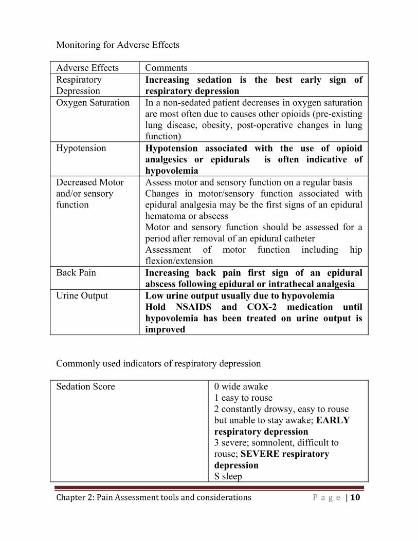
Monitoring for Adverse Effects Adverse Effects Comments Respiratory Depression
Increasing sedation is the best early sign of respiratory depression
Oxygen Saturation In a non-sedated patient decreases in oxygen saturation are most often due to causes other opioids (pre-existing lung disease, obesity, post-operative changes in lung function)
Hypotension Hypotension associated with the use of opioid analgesics or epidurals is often indicative of hypovolemia
Decreased Motor and/or sensory function
Assess motor and sensory function on a regular basis Changes in motor/sensory function associated with epidural analgesia may be the first signs of an epidural hematoma or abscess Motor and sensory function should be assessed for a period after removal of an epidural catheter Assessment of motor function including hip flexion/extension
Back Pain Increasing back pain first sign of an epidural abscess following epidural or intrathecal analgesia
Urine Output Low urine output usually due to hypovolemia Hold NSAIDS and COX-2 medication until hypovolemia has been treated on urine output is improved
Commonly used indicators of respiratory depression Sedation Score 0 wide awake
1 easy to rouse 2 constantly drowsy, easy to rouse but unable to stay awake; EARLY respiratory depression 3 severe; somnolent, difficult to rouse; SEVERE respiratory depression S sleep

11
Respiratory Rate Less than 8 breaths/min often considered to be a sign of respiratory depression but is an unreliable indicator Respiratory depression can coexist with a NORMAL respiratory rate
Oxygen Saturation May be unreliable with patient receiving oxygen
Examples of an initial pain assessment and flow sheet can be seen in the Appendix.
Chapter 3: Pharmacology of Pain Management: Non-opioids, Opioids and Adjuvant Agents

2
Chapter 3: Non-Opioid, Opioids and Adjuvant Agents Acetaminophen Use
First line analgesic for mild to moderate pain Used as part of a multi-modal analgesic regimen for moderate to
severe pain Mechanism
Weak effects on COX-1 and COX-2 CNS prostaglandin inhibition Serotonergic pathway activation Effect on substance P or nitric oxide pathways NMDA antagonism COX-3 mechanism
Dose
Given by oral or rectal route and intravenous in some countries Available in liquid or tablets Oral and rectal administration the peak effect is within one hour When administered by the rectal route, doses 30-50 % higher than
recommended oral doses are required to obtain comparable plasma levels
No universally accepted rectal dosing regimen due to inter- and intra-patient variability in drug absorption and the possibility of accumulation with use greater than 72 hours
Blood levels required for analgesia are 10-20 mg/L Acetaminophen dosing Patients 0-3 months of age
Oral: 10 mg/kg po q 4 hr prn up to 60mg/kg/day Rectal: 20 mg/kg pr q 6 hr up to 80 mg/kg/day Max 6 doses
3
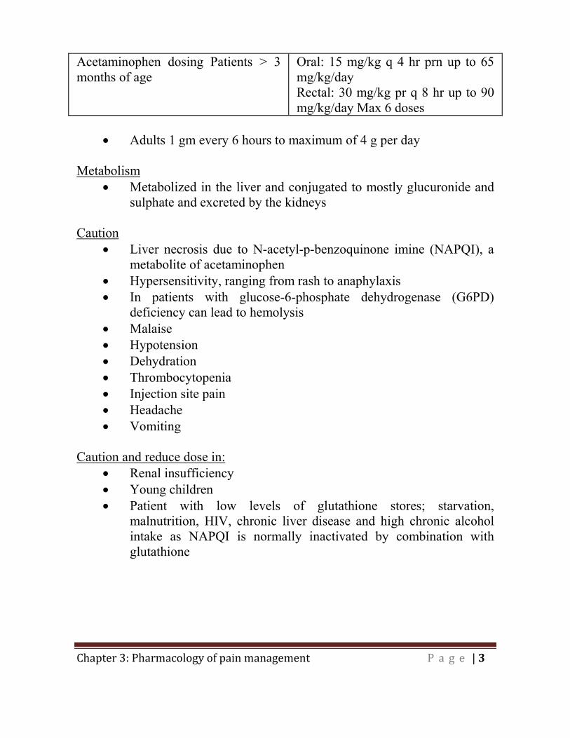
Acetaminophen dosing Patients > 3 months of age
Oral: 15 mg/kg q 4 hr prn up to 65 mg/kg/day Rectal: 30 mg/kg pr q 8 hr up to 90 mg/kg/day Max 6 doses
Adults 1 gm every 6 hours to maximum of 4 g per day
Metabolism
Metabolized in the liver and conjugated to mostly glucuronide and sulphate and excreted by the kidneys
Caution
Liver necrosis due to N-acetyl-p-benzoquinone imine (NAPQI), a metabolite of acetaminophen
Hypersensitivity, ranging from rash to anaphylaxis In patients with glucose-6-phosphate dehydrogenase (G6PD)
deficiency can lead to hemolysis Malaise Hypotension Dehydration Thrombocytopenia Injection site pain Headache Vomiting
Caution and reduce dose in:
Renal insufficiency Young children Patient with low levels of glutathione stores; starvation,
malnutrition, HIV, chronic liver disease and high chronic alcohol intake as NAPQI is normally inactivated by combination with glutathione
4
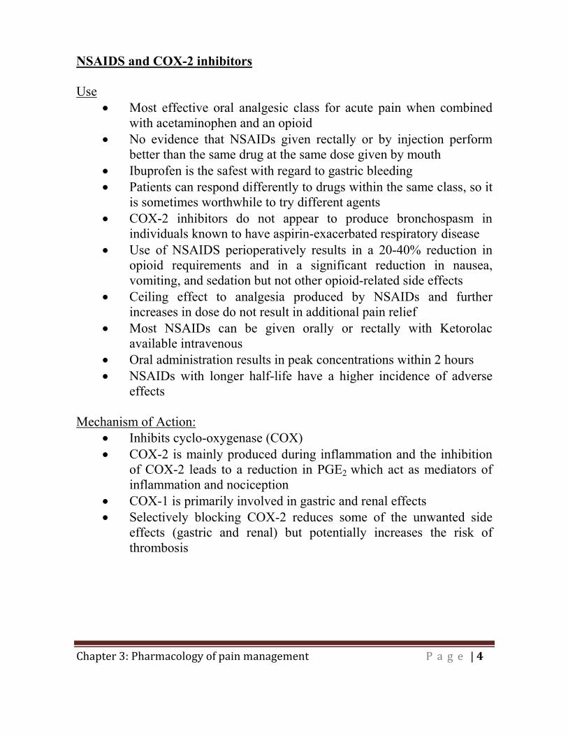
NSAIDS and COX-2 inhibitors Use
Most effective oral analgesic class for acute pain when combined with acetaminophen and an opioid
No evidence that NSAIDs given rectally or by injection perform better than the same drug at the same dose given by mouth
Ibuprofen is the safest with regard to gastric bleeding Patients can respond differently to drugs within the same class, so it
is sometimes worthwhile to try different agents COX-2 inhibitors do not appear to produce bronchospasm in
individuals known to have aspirin-exacerbated respiratory disease Use of NSAIDS perioperatively results in a 20-40% reduction in
opioid requirements and in a significant reduction in nausea, vomiting, and sedation but not other opioid-related side effects
Ceiling effect to analgesia produced by NSAIDs and further increases in dose do not result in additional pain relief
Most NSAIDs can be given orally or rectally with Ketorolac available intravenous
Oral administration results in peak concentrations within 2 hours NSAIDs with longer half-life have a higher incidence of adverse
effects Mechanism of Action:
Inhibits cyclo-oxygenase (COX) COX-2 is mainly produced during inflammation and the inhibition
of COX-2 leads to a reduction in PGE2 which act as mediators of inflammation and nociception
COX-1 is primarily involved in gastric and renal effects Selectively blocking COX-2 reduces some of the unwanted side
effects (gastric and renal) but potentially increases the risk of thrombosis
5
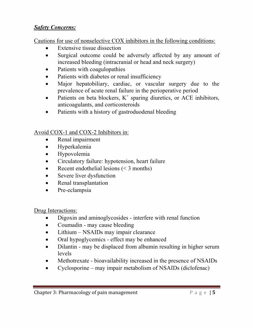
Safety Concerns: Cautions for use of nonselective COX inhibitors in the following conditions:
Extensive tissue dissection Surgical outcome could be adversely affected by any amount of
increased bleeding (intracranial or head and neck surgery) Patients with coagulopathies Patients with diabetes or renal insufficiency Major hepatobiliary, cardiac, or vascular surgery due to the
prevalence of acute renal failure in the perioperative period Patients on beta blockers, K+ sparing diuretics, or ACE inhibitors,
anticoagulants, and corticosteroids Patients with a history of gastroduodenal bleeding
Avoid COX-1 and COX-2 Inhibitors in:
Renal impairment Hyperkalemia Hypovolemia Circulatory failure: hypotension, heart failure Recent endothelial lesions (< 3 months) Severe liver dysfunction Renal transplantation Pre-eclampsia
Drug Interactions:
Digoxin and aminoglycosides - interfere with renal function Coumadin - may cause bleeding Lithium �– NSAIDs may impair clearance Oral hypoglycemics - effect may be enhanced Dilantin - may be displaced from albumin resulting in higher serum
levels Methotrexate - bioavailability increased in the presence of NSAIDs Cyclosporine �– may impair metabolism of NSAIDs (diclofenac)

6
Bleeding and NSAIDs COX-1 inhibitors increase bleeding time (30%) but usually still
within normal range Not clear if blood loss is increased in surgery Avoid ketorolac for tonsillectomy
GI Side Effects and NSAIDs
NSAID-induced GI lesions are asymptomatic in 50% of cases Risk factors include: age > 65, past GI bleeding, known peptic ulcer
disease, use of glucocorticoids, other anticoagulants, smoking and alcohol use
Ketamine
Phencyclidine derivative and is the most potent NMDA receptor
channel blocker currently available Racemic mixture , but the S enantiomer is more potent Sub-anesthetic dosing of intravenous ketamine is useful adjuvant
for balanced perioperative analgesia Use Low-dose:
Management of pain in opioid-tolerant patients Management of neuropathic pain Treatment of poorly opioid-responsive pain Prevention (reversal) of central sensitization and wind-up
High-dose:
Treatment of acute pain (fractures, dressing changes on burn patients)
Mechanism of action
NMDA antagonist Non-competitive binding at NMDA receptors in the CNS reduces
central sensitization and �“wind up�”
7
Has mu, delta and kappa opioid-like effect and therefore reduces opioid requirements
Effects GABA receptors and inhibits synaptic uptake of serotonin and noradrenaline
Acts on non-NMDA glutamate receptors, muscarinic receptors, cholinergic transmission and voltage gated Na+, K+ and Ca2+ channels
Possesses an antidepressant effect Metabolism
Metabolized in liver to norketamine and excreted by the kidneys Primary metabolite norketamine is less potent than ketamine but
also an NMDA antagonist and contributes to analgesia T½ (redistribution from central nervous system) is rapid T½ (elimination) is 2-3 hours
Dose
Usually given IV or SC, however undergoing research in nasal, transmucosal, and transdermal administration.
Subanesthetic doses: Loading dose: 0.1-0.2 mg/kg (5-15 mg) and an infusion of 0.05-0.1 mg/kg/hr (5-10 mg/hour)
Single I.V. doses in the 5-10 mg range for rescue analgesia in the PACU
High dose 10-20 mg in combination with Midazolam to reduce the incidence of adverse events and nightmares are useful for fracture reductions and other painful procedures (dressing changes)
Caution
High doses are associated with dreaming, nightmares, hallucinations, excitation, agitation, and delirium. These can be reduced with the addition of benzodiazepines
Lower doses include dizziness and a feeling of unreality or floating (Midazolam will reduce this effect)
In most cases this is less likely at doses of 0.1 mg/kg/hour in the average adult and 0.05 mg/kg/day in the elderly
Low doses usually avoid of significant cardiac or CNS side-effects

8
Clonidine Uses:
Alpha adrenergic agonist; analgesic, reduces post-operative narcotic requirements
Sedation in the ICU Control autonomic symptoms of opioid withdrawal Relieves hyperalgesia in sympathetically mediated pain Enhances local anesthetics Antihypertensive Reversed by naloxone Routes of administration: oral, intravenously or epidural,
transdermal Mechanism of Action:
Stimulates the central descending noradrenergic inhibitory system acting on the spinal dorsal horn neurons of laminae IV and V
Inhibition of substance P Central mediated effect on spinal pre- and postsynaptic alpha 2
adrenergic receptors in the dorsal horn Supraspinal effect and inhibits acetylcholinesterase
Side Effects:
Hypotension Bradycardia Sedation Anxiolysis Dizziness Dry Mouth Decreased bowel motility Diuresis
Dose:
Half-life 6-20 hours 50-150 mcg tid peak effect in 3-5 hours

9
Gabapentin Mechanism of Action:
Inhibitory action in the dorsal root ganglion and spinal cord at the voltage-gated calcium channel where it blocks the alpha 2 beta subunit
Side Effects:
Sedation, dizziness, headaches Dose:
Doses: 100 mg to 1200 mg three times per day Decrease dosage in renal impairment to twice per day When given preoperatively, will reduce postoperative pain scores
and opioid consumption in the first 24 hours after surgery Should not be discontinued in the perioperative period to avoid
central nervous system hyperexcitability
Pregabalin Mechanism of Action:
Blocks calcium channels within nerves Rapid onset and shorter duration of titration compared to
gabapentin Potentiated by oxycodone Used for epilepsy, neuropathic pain, and anxiety states Improves sleep, has anxiolytic properties, and is well tolerated
Side Effects:
Dizziness Drowsiness Water Retention and weight gain

10
Dose: Doses range from 75 mg daily up to 600 mg per day Excreted by the kidney so daily dose should not exceed 300 mg in
patients with a creatinine clearance less than 60 mL/min. Should not be discontinued in the perioperative period to avoid
central nervous system hyperexcitability Antidepressants:
Tricyclic antidepressants; amitriptyline, nortriptyline dose 10-25 mg at night, side-effects include: anticholinergic effects, sedation. Nortriptyline is less sedating.
Analgesic dosage is much lower than antidepressant dose Night sleep improved at relatively low doses and within a few days Analgesic effects takes at least three weeks of therapy Mechanism of action for pain relief include: stabilization of nerve
membranes and blocking the reuptake of serotonin and noradrenaline at presynaptic membranes in the central nervous system
Continue the usual dose of these drugs as well as SSRI medication
Anticonvulsants:
�• Mechanism of analgesia: �– Reduce membrane excitability �– Suppress abnormal discharges in pathologically-altered
neurons �– Affects sodium and voltage-gated calcium channels
�• Indications: acute and chronic neuropathic pain from peripheral
nerve syndromes �– Trigeminal neuralgia:
�• NNT 2.5 (2.0-3.4) carbamazepine �– Postherpetic neuralgia:
�• NNT 3.2 (2.4-5.0) gabapentin �– Diabetic neuropathy:
�• NNT 2.3 (1.6-3.8) carbamazepine

11
�• NNT 3.8 (2.4-8.7) gabapentin �• NNT 2.1 (1.5-3.6) phenytoin
�– Efficacy with both lancinating and burning pain
�• Carbamazepine �– Proven indications: diabetic neuropathy, post-herpetic
neuralgia, trigeminal neuralgia and other causes of central pain
�– First FDA-approved anticonvulsant for neuropathic pain �– Common adverse effects: sedation, mental clouding,
dizziness, nausea, unsteadiness �– Multiple drug interactions �– enzyme inducer �– Potential for liver damage and aplastic anemia require
regular monitoring of CBC, liver enzymes, PT/INR, and serum drug levels
�• Lamotrigine
�– Proven efficacy in neuropathic pain caused by neurotoxic anti-retroviral therapy in HIV-positive patients
�– Efficacy in patients with diabetic neuropathy and central pain
�– High incidence of skin rashes and Stevens-Johnson Syndrome
�– Start 25mg and titrate weekly to effect or 500mg max daily
�• Valproic acid
�– Evidence in migraine prophylaxis, diabetic neuropathy �– Third line for other neuropathic pain syndromes �– Common adverse effects: sedation, nausea, vomiting,
dizziness, headache, significant weight gain �– Severe adverse effects: hepatic toxicity and pancreatitis,
thrombocytopenia, hyperammonemia, androgenization, polycystic ovaries
�– Monitor CBC, liver enzymes and serum drug levels
12
Capsaicin Topical application used to reduce the pain of post-herpetic
neuralgia Provides pain relief from neuropathy scar tissue pain Produces a burning sensation when applied Absorption of capsaicin is believed to affect C fibres and deplete
them of the neurotransmitter �“substance P�” which is implicated in peripheral neuropathic pain
Opioids Definition of opioid: Substances with morphine-like activity including agonists, antagonists as well as naturally occurring and synthetic opioid peptides. Classification: Naturally occurring: Codeine, Morphine Semi synthetic: Oxycodone, Hydromorphone Synthetic: Methadone, Meperidine, Fentanyl, Sufentanil, Alfentanil Activity of Opioids: Agonist: binds to and stimulates an opioid receptor Antagonist: binds to an opioid receptor without receptor stimulation e.g. Naloxone Partial agonist: binds to an opioid receptor and stimulates the receptor to a level below the maximum level (ceiling effect) e.g. Buprenorphine Mixed agonist-antagonist: binds to many opioid subtypes to produce agonist action on one or more subtypes and antagonist action on one or more subtypes e.g. Nalbuphine
13
Opioid Receptors: Mu receptor: in the brain and spinal cord. Activation produces analgesia, euphoria, respiratory depression, bradycardia, nausea and vomiting, decreased G.I motility, tolerance and dependence. Kappa receptor: activation causes analgesia, hallucinations, dysphoria and mild respiratory depression. Delta receptor: activation in brain and preferentially in the spinal cord to produce analgesia Opioid Like receptors: structurally similar to opioid receptor with no activity Receptor Mechanisms: Opioid receptors are coupled to G-proteins. Opioids effect protein phosphorylation via second messenger system thereby altering ion channel conductance. Opioids act presynapically by inhibiting substance P and glutamate. They act postsynaptically inhibiting neurons by opening potassium channels that hyperpolarize the cell. Opioid Effects: CNS: Analgesia, euphoria, dysphoria and in high doses sedation and eventually loss of consciousness. Other side effects include; cough miosis, hypothermia and rarely convulsions. Muscle rigidity reported in doses much larger than those used in pain management. Accumulation of neurotoxic metabolite normeperidine can result in seizures. The risk of opioid induced seizures is dose related and patients with pre-existing epilepsy or taking other seizure lowering drugs may be at increased risk. Myoclonus can be associated with the accumulation of morphine-3-glucuronide.
14
Respiratory: Opioids cause dose-dependent depression of all phases of respiratory activity. Opioids decrease respiratory rate, decrease tidal volume, cause irregularities of respiratory rhythm (hypoventilation, central apnea), and intermittent partial or complete upper airway obstruction. GI: Opioids affect smooth muscle activity leading to delayed gastric emptying, inhibition of bowel motility and constipation. The etiology is due to stimulation of opioid receptors in the bowel wall and due to a central effect. Treatment involves fluids, mobilization, stool softeners, as well as peripheral opioid antagonists. Opioids also increase biliary pressure and spasm of the sphincter of Oddi. This can be treated with naloxone. Urinary retention which is caused by a similar mechanism is also reversed with naloxone. CVS: Opioids can cause hypotension by various mechanisms. Opioids reduce sympathetic tone (especially in those with high tone: elderly, poor cardiac function, hypovolemic), reduce arterial and venous tone, and release histamine. Opioids can also cause bradycardia but not usually in the doses used in patient management. Clinically if a supine patient develops hypotension after receiving opioids then they are usually hypovolemic. Other: Tolerance, physical dependence and addiction. Precautions in using Opioids: Respiratory disease: Caution in patients with limited respiratory reserve. Tolerance to respiratory depression develops quickly. The respiratory centre receives nociceptive input so pain acts as a respiratory stimulant. Opioids titrated to the level of pain results in a low incidence of respiratory depression. Risk factors for Respiratory Depression with opioids:
Opioid Naïve patients Patients at extremes of age Severe COPD and Severe Restrictive Lung disease Obstructive Sleep Apnea Morbid Obesity Kidney Failure

15
Liver Failure Neurological Disease Neuromuscular Disease
Predictors of Opioid Dose:
Best clinical predictor of opioid dose is the patient�’s age; Useful formula; average 24-hour morphine requirements (mg) for
patients over 20 years of age= 100 �– (age in years) Marked variation 8-10-fold in dose requirements in age group Metabolites can have analgesic or adverse effects Goal is to titrate opioids so the patient is comfortable, sedation
score < 2 and respiratory rate > 8/min Post-operative confusion: Opioids are frequently blamed as a cause. Other causes should be entertained including; withdrawal from alcohol or benzodiazepines, sleep deprivation, hypoxia, sepsis, increasing age, endocrine and metabolic problems, polypharmacy, drug interactions, and unrelieved severe pain. Treatment should be aimed at treating any reversible causes including hypoxemia. If pharmacological treatment is used haloperidol is the drug of choice. It should be given in titrated doses. Benzodiazepines should be avoided unless the patient is withdrawing from alcohol or benzodiazepines. Hepatic and renal disease: Reduced dosage for codeine, oxycodone, morphine and Meperidine. Head Injury: Opioids increase PCO2 from respiratory depression and lead to elevation of intracerebral pressure. Miosis, vomiting and mental clouding are important clinical signs for evaluation of head injury may be obscured. Allergic reactions: Rare and mediated by the immune system and results in rash, urticaria, bronchoconstriction, angioneurotic edema and cardiovascular disturbances. Opioids may induce histamine release, cause bronchospasm and depress the cough reflex. Pruritus: Probably due to mu receptor stimulation in the dorsal horn of the spinal cord as well as histamine release from mast cells resulting in localized
16
or generalized itching. It can be associated with flushing of the skin or along the track of a vein. It is more likely after morphine or Meperidine and more common following epidural or intrathecal administration of opioids. To prevent pruritis avoid morphine, codeine or meperidine and switch to fentanyl. Treatment is small doses of naloxone, nalbuphine, ondansetron or propofol. Drug Interactions: The sedative and respiratory depressant effects of opioids may be exaggerated by other drugs with sedative properties. These drugs include; antihistamines, anxiolytics, antiemetics. Meperidine and MAOIs can cause delirium, hyperpyrexia, and convulsions caused by central serotonergic overactivity due to blockage of neuronal uptake of serotonin by meperidine. Specific Opioids: Morphine:
Least lipid soluble of all opioids in common use Metabolized in the liver by glucuronidation and N-demethylation to
morphine-3-glucuronide and morphine-6-glucuronide M-6-G is active more potent mu receptor agonist than morphine M-3-G is has no analgesic activity M-3-G may create morphine tolerance and produce some of the
side-effects of long term morphine treatment such as myoclonus, seizures, hyperalgesia, allodynia
Some individuals produce a lot of M-6-G and very sensitive to morphine while others produce more M-3-G and are insensitive to morphine
M-6-G accumulates in poor renal function and will not be dialyzed
Codeine:
Analgesic effect mostly as a result of metabolism to morphine Other metabolites include codeine-6-glucuronide, M-3-G, M-6-G,
normorphine, and norcodeine-6-glucuronide Codeine has variable analgesic effect due to genetic polymorphism
producing variable expression of the enzyme CYP2D6
17
There are poor metabolizers (8-10% Caucasians) which convert no codeine to morphine, and extensive metabolizers which convert up to 15% to morphine
Codeine can be given IM, PO, or rectally Peak concentrations: Oral= 60 minutes, IM=30 minutes, Efficacy is low with a ceiling effect above which the side effects
increase but the analgesia does not Hydromorphone
Semisynthetic opioid 5-10 times more potent than morphine Available in oral, parenteral, suppository forms and used for
epidural analgesia No analgesic metabolites Intravenous administration creates a rapid serum rise but slow onset
of CNS effect Half-life is 2-3 hours after I.V. dose and peak in 30-60 minutes after
oral dosing 95 % of drug metabolized to hydromorphone-3-glucuronide which
has similar neurotoxic effects as M-3-G Not metabolized via the CYP system therefore it is less likely to be
involved in drug-to-drug interactions Caution and reduced dosing with renal failure as there can be an
accumulation of drug and metabolites with a half-life of up to 40 hours
Caution and reduced dosing with liver dysfunction No dose adjustment in healthy elderly patients
Fentanyl
50-100 times more potent than Morphine Rapid onset in 3-5 minutes Highly lipid soluble synthetic opioid with no histamine release Inactive metabolites and can be used in renal or hepatic failure Fentanyl patches NOT for acute pain Avoid placing warming blanket near fentanyl patches Fentanyl patches should not be cut
18
Tramadol
Weak affinity to mu opioid receptor: 10,000 times lower than morphine and 10 times less than codeine
Activity unique: At spinal cord level by indirect activation of postsynaptic alpha 2 adrenoceptor blocking impulses from reaching the brain.
Inhibition of 5-HT and noradrenaline reuptake and presynaptic stimulation of 5-HT release
Rapidly absorbed orally, 69 % bioavailability after one dose and 90-100 % after multiple doses
11 metabolites of which O-desmethyltramadol (M1) predominates with a higher affinity for the opioid receptor than tramadol
Metabolism depends on CYP2D6 and poor metabolizers show some evidence of reduce analgesic activity
90 % excreted by the kidneys, reduce dose if creatinine clearance less than 30ml/min and severe hepatic failure
Usual dose is 400 mg/day (100 mg 4 x per day) Causes less constipation, sedation, respiratory depression and
nausea and vomiting than other opioids, useful in elderly patients Caution in using with patients who have epilepsy and other drugs
that lower the seizure threshold Drugs interactions: Coumadin= increase INR, MAOI=
Hypertensive Crisis, Carbamazepine=Increased Tramadol clearance Oxycodone
Given orally and two times as potent as Morphine Major metabolites are noroxycodone which has only minimal
analgesic effect and renally excreted and oxymorphone which posses analgesic activity but present in small concentrations
Better bioavailability than Morphine and between 60-80 % Fewer side effects than morphine; sedation, nausea and vomiting

19
Partial opioid agonists and agonist-antagonists
Partial agonists have the affinity for the opioid receptor but NOT the same intrinsic activity as full agonists
Ceiling effect for BOTH analgesia and adverse effects Stimulation of one opioid receptor while acting as an antagonist at
another Can precipitate opioid withdrawal in a opioid-dependent patients
Specific Partial Agonists-Antagonists: Buprenorphine
Available in parenteral, sublingual, and transdermal formulations Good absorption sublingually due to high lipid solubility Very high affinity for the opioid receptor and dissociates slowly
from the mu receptor and hence it is highly potent and has a long duration of action
Antagonist of the kappa receptor Used for the management of opioid substance abuse disorder and
transdermal for chronic pain In the event of respiratory depression from Buprenorphine higher
than usual doses of Naloxone are required to reverse the respiratory failure and a continuous infusion may be required
Nalbuphine
Related to naloxone. Available for intravenous or intramuscular route
Used to treat side-effects of mu-agonists such as respiratory depression and pruritis
Opioid Antagonists Naloxone
Used most commonly to reverse opioid overdose

20
Short half-life of 60 minutes and hence an infusion is usually needed to reverse respiratory depression
Dose to treat respiratory depression is between 40-100 mcg, it can also be administered SC or IM in much higher doses (400 mcg)
With rapid reversal of analgesia hypertension, tachycardia, nausea and vomiting even arrhythmias and pulmonary edema
May be titrated to reduce respiratory depression and pruritis without reversing analgesia
Naltrexone
Can be used orally and has a half-life of 2-4 hours and it main metabolite 6-naltrexol, is a weaker mu antagonist with a half-life of 8 hours
Used orally or as subcutaneous implant for the treatment of opioid addiction and alcoholism
Alvimopan
Mu receptor antagonist for the prevention and treatment of opioid induced ileus and constipation
Good oral absorption, with no penetration of the blood-brain barrier Works on the receptors in the gut wall and assists in the recovery of
GI function after surgery and reduces opioid-induced bowel dysfunction in chronic pain patients
References:
Katz WA, Rothenberg R. Section 2: The importance of improving function in patients with pain. J Clin Rheumatol. 2005;11(2 Suppl):S6-9, discussion S9-10.
Moulin DE, Clark AJ, Speechley M, Morley-Forster PK. Chronic pain in Canada--prevalence, treatment, impact and the role of opioid analgesia. Pain Res Manag. 2002;7(4):179-84.
21
Rocchi A, Chung F, Forte L. Canadian survey of postsurgical pain and pain medication experiences. Can J Anaesth. 2002;49(10):1053-6.
College of Physicians & Surgeons of Alberta. Management of Chronic Non-Malignant Pain: 1993.
The College of Physicians and Surgeons of Saskatchewan. Narcotics in the Management of Chronic Non-Malignant Pain. General principles of appropriate pain management with opioids. 2006.
College of Physicians and Surgeons of New Brunswick. Guidelines for Management of Chronic Non-Malignant Pain. 1995.
Collège des Médecins Du Québec: Treating Pain: An Update On The Use Of Narcotics 1999 (2006-030e).Traitement de la douleur : Le point sur l�’utilisation des narcotiques. 1998 (2006-030f).
The College of Physicians and Surgeons of Nova Scotia. Guidelines for the Use of Controlled Substances in the Treatment of Pain. 1999, updated 2005.
The College of Physicians and Surgeons of Newfoundland & Labrador. Use of Controlled Substances for the Treatment of Pain. 2005.
College of Physicians and Surgeons of Ontario. Evidence-based recommendations for medical management of chronic non-malignant pain : reference guide for clinicians. 2000, updates 2005.
Canadian Pain Society: Position Statement on Pain Relief, 1997. Ballantyne JC. The Massachusetts General Hospital Handbook of Pain Management, 3rd Edition, Lippincott Williams and Wilkins, 2006. Jovey R. Managing Pain. Healthcare and Financial Publishing, Rogers Media, 2002 Coniam S, Mendham J, Arnold H. Principles of Pain Management for Anesthetists, 2005.

Chapter 4:
Pharmacology of
local anesthetic
2
Chapter 4: Pharmacology of Local Anesthetics
Local anesthetics are classified based on the nature of the linkage between water soluble, and lipid soluble components as amides, and esters.
Ester local anesthetics are metabolized by paeudocholinesterase. They produce para-aminobenzoic acid(PABA), which acts as a hapten. Therefore they have great potential to cause allergic reactions. Examples are cocaine, tetracaine, chloroprocaine.
Amide local anesthetics are metabolized in the liver, and rarely cause allergic reaction. Examples are lidocaine, bupivacaine, and ropivacaine.
Mechanism of action
They block the generation, and conduction of nerve impulses by blocking sodium channels in the cell membrane, and therefore preventing the influx of sodium.
They can block nerve conduction in all sensory, and motor nerves. Smaller diameter nerve fibers (i.e B and C fibers) are more easily
blocked by local anesthetics, as they have a smaller critical blocking length(The length of nerve fiber that must be exposed to the drug for blockade of conduction).
Sympathetic blockade usually occurs first, followed by block of nociception, touch, and temperature sensation. Motor block is last.
Efficacy of local anesthetic
The potency, and therefore efficacy of a local anesthetic is related to lipid solubility.
The speed of onset depends on physicochemical properties, which the most important one is pKa. A higher pKa is associated with a slower onset of action.
3
Adverse effects
Physiological effects are mainly caused by sympathetic blockade, and important after neuraxial blocks.
All local anesthetics are neurotoxic in high concentrations. Transient Neurological symptoms(TNS) is a temporary pain
affecting gluteal region, and lower extremities following spinal anesthesia, in particular when lidocaine is used. There is no neurological deficit. A few risk factors for TNS are obesity, lithotomy, and day surgery patients.
High blood concentration of local anesthetics can cause cardiorespiratory, as well as neurological symptoms. Inadvertent intravascular injection, excessive doses, or high dose in patients with severe hepatic impairment can cause systemic toxicity. Factors affecting blood concentration are
Dose of the drug Site of injection (interpleural >intercostals >caudal
>epidural >brachial plexus) Vasoconstrictor (reduces the rate of absorption, and
increases the duration)
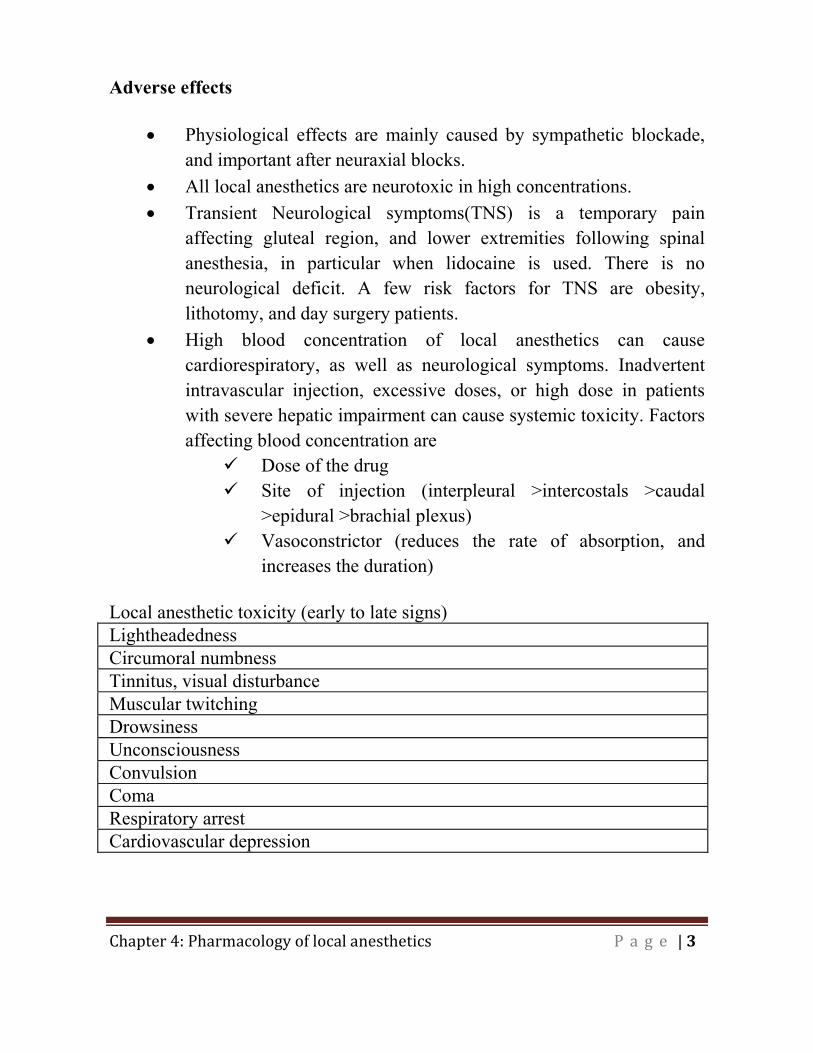
Local anesthetic toxicity (early to late signs) Lightheadedness Circumoral numbness Tinnitus, visual disturbance Muscular twitching Drowsiness Unconsciousness Convulsion Coma Respiratory arrest Cardiovascular depression
4
Hypercarbia, and acidosis reduce the convulsive threshold of the drug
Local anesthetics can affect the heart, causing alteration in contractility, conductivity, and rhythmicity. Arrhythmia varies from PVCs, to ventricular tachycardia, ventricular fibrillation, conduction delay, complete heart block, and asystole.
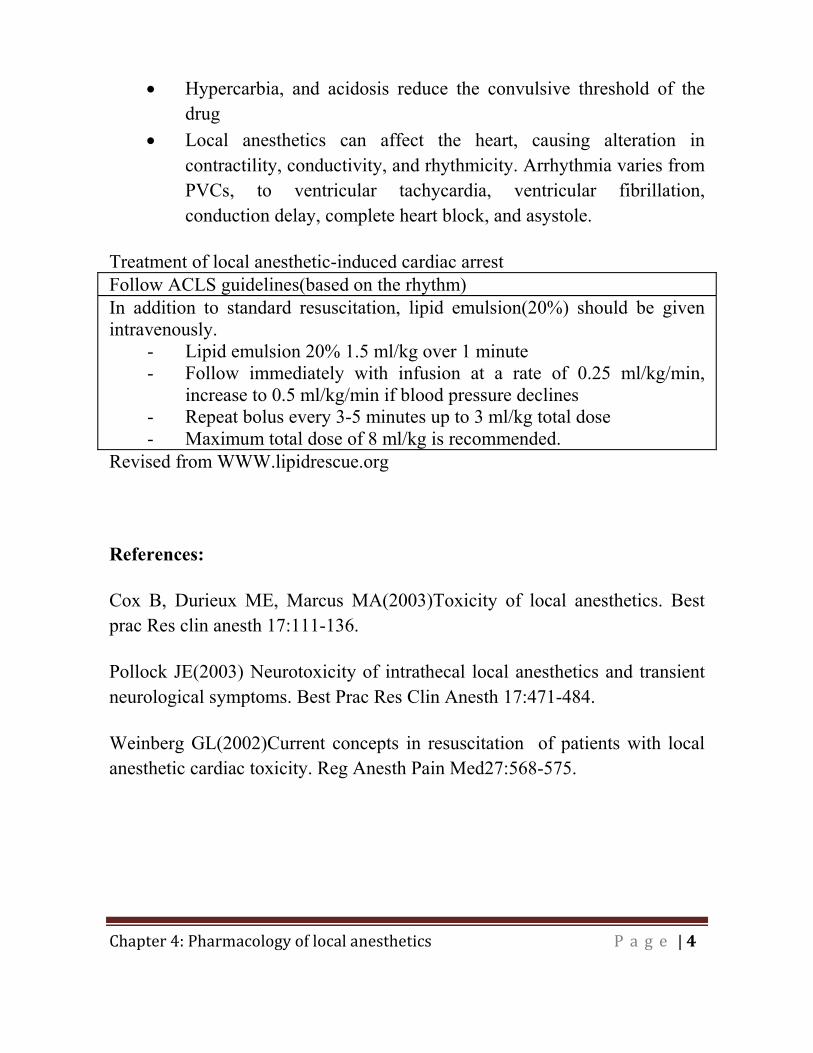
Treatment of local anesthetic-induced cardiac arrest Follow ACLS guidelines(based on the rhythm) In addition to standard resuscitation, lipid emulsion(20%) should be given intravenously.
- Lipid emulsion 20% 1.5 ml/kg over 1 minute - Follow immediately with infusion at a rate of 0.25 ml/kg/min,
increase to 0.5 ml/kg/min if blood pressure declines - Repeat bolus every 3-5 minutes up to 3 ml/kg total dose - Maximum total dose of 8 ml/kg is recommended.
Revised from WWW.lipidrescue.org
References:
Cox B, Durieux ME, Marcus MA(2003)Toxicity of local anesthetics. Best prac Res clin anesth 17:111-136.
Pollock JE(2003) Neurotoxicity of intrathecal local anesthetics and transient neurological symptoms. Best Prac Res Clin Anesth 17:471-484.
Weinberg GL(2002)Current concepts in resuscitation of patients with local anesthetic cardiac toxicity. Reg Anesth Pain Med27:568-575.
Chapter 5:
Post operative pain
management
2
Chapter 5: Post-op Pain management
Pre-emptive analgesia
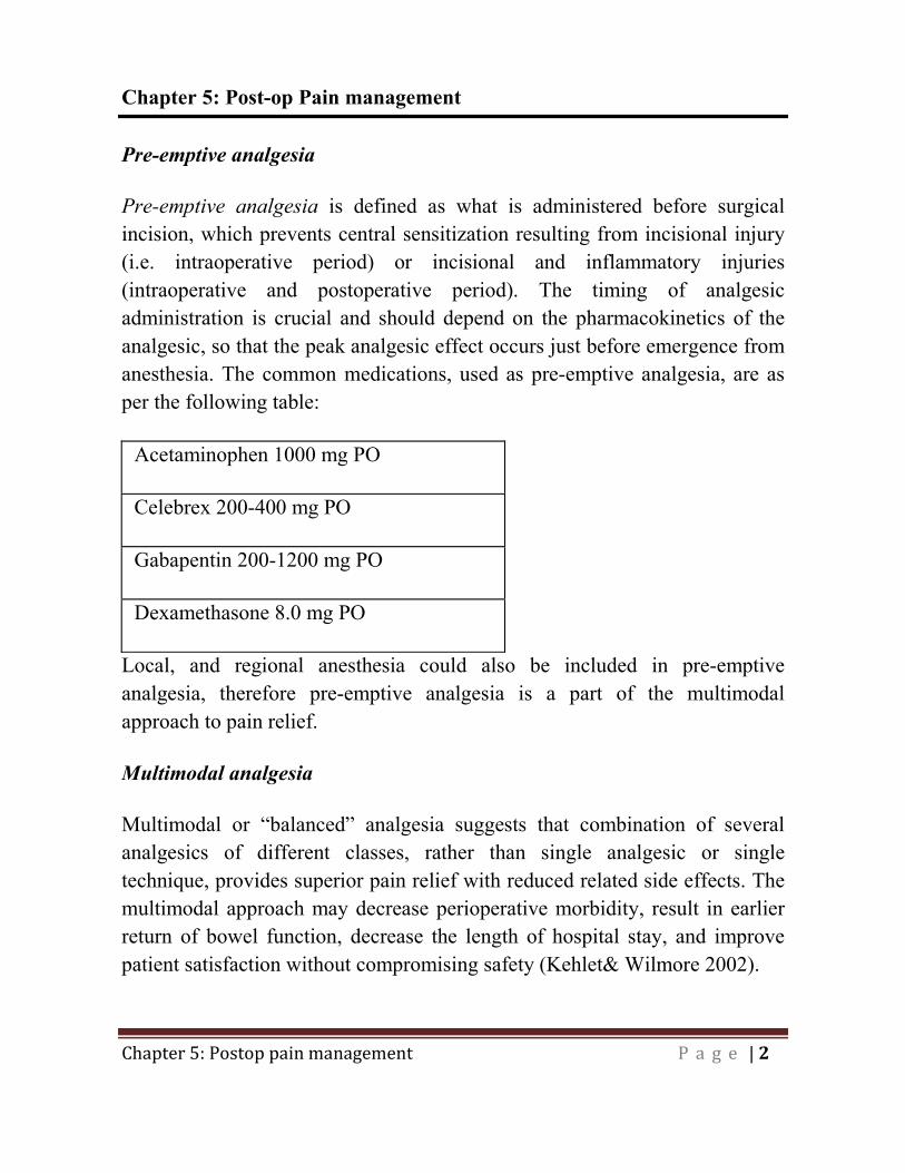
Pre-emptive analgesia is defined as what is administered before surgical incision, which prevents central sensitization resulting from incisional injury (i.e. intraoperative period) or incisional and inflammatory injuries (intraoperative and postoperative period). The timing of analgesic administration is crucial and should depend on the pharmacokinetics of the analgesic, so that the peak analgesic effect occurs just before emergence from anesthesia. The common medications, used as pre-emptive analgesia, are as per the following table:
Acetaminophen 1000 mg PO
Celebrex 200-400 mg PO
Gabapentin 200-1200 mg PO
Dexamethasone 8.0 mg PO
Local, and regional anesthesia could also be included in pre-emptive analgesia, therefore pre-emptive analgesia is a part of the multimodal approach to pain relief.
Multimodal analgesia
Multimodal or �“balanced�” analgesia suggests that combination of several analgesics of different classes, rather than single analgesic or single technique, provides superior pain relief with reduced related side effects. The multimodal approach may decrease perioperative morbidity, result in earlier return of bowel function, decrease the length of hospital stay, and improve patient satisfaction without compromising safety (Kehlet& Wilmore 2002).
3
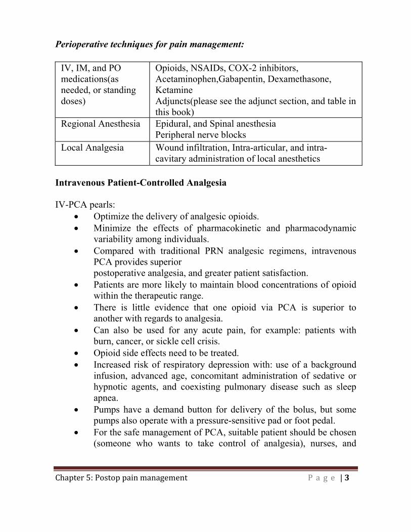
Perioperative techniques for pain management:
IV, IM, and PO medications(as needed, or standing doses)
Opioids, NSAIDs, COX-2 inhibitors, Acetaminophen,Gabapentin, Dexamethasone, Ketamine Adjuncts(please see the adjunct section, and table in this book)
Regional Anesthesia Epidural, and Spinal anesthesia Peripheral nerve blocks
Local Analgesia Wound infiltration, Intra-articular, and intra-cavitary administration of local anesthetics
Intravenous Patient-Controlled Analgesia IV-PCA pearls:
Optimize the delivery of analgesic opioids. Minimize the effects of pharmacokinetic and pharmacodynamic
variability among individuals. Compared with traditional PRN analgesic regimens, intravenous
PCA provides superior postoperative analgesia, and greater patient satisfaction.
Patients are more likely to maintain blood concentrations of opioid within the therapeutic range.
There is little evidence that one opioid via PCA is superior to another with regards to analgesia.
Can also be used for any acute pain, for example: patients with burn, cancer, or sickle cell crisis.
Opioid side effects need to be treated. Increased risk of respiratory depression with: use of a background
infusion, advanced age, concomitant administration of sedative or hypnotic agents, and coexisting pulmonary disease such as sleep apnea.
Pumps have a demand button for delivery of the bolus, but some pumps also operate with a pressure-sensitive pad or foot pedal.
For the safe management of PCA, suitable patient should be chosen (someone who wants to take control of analgesia), nurses, and
4
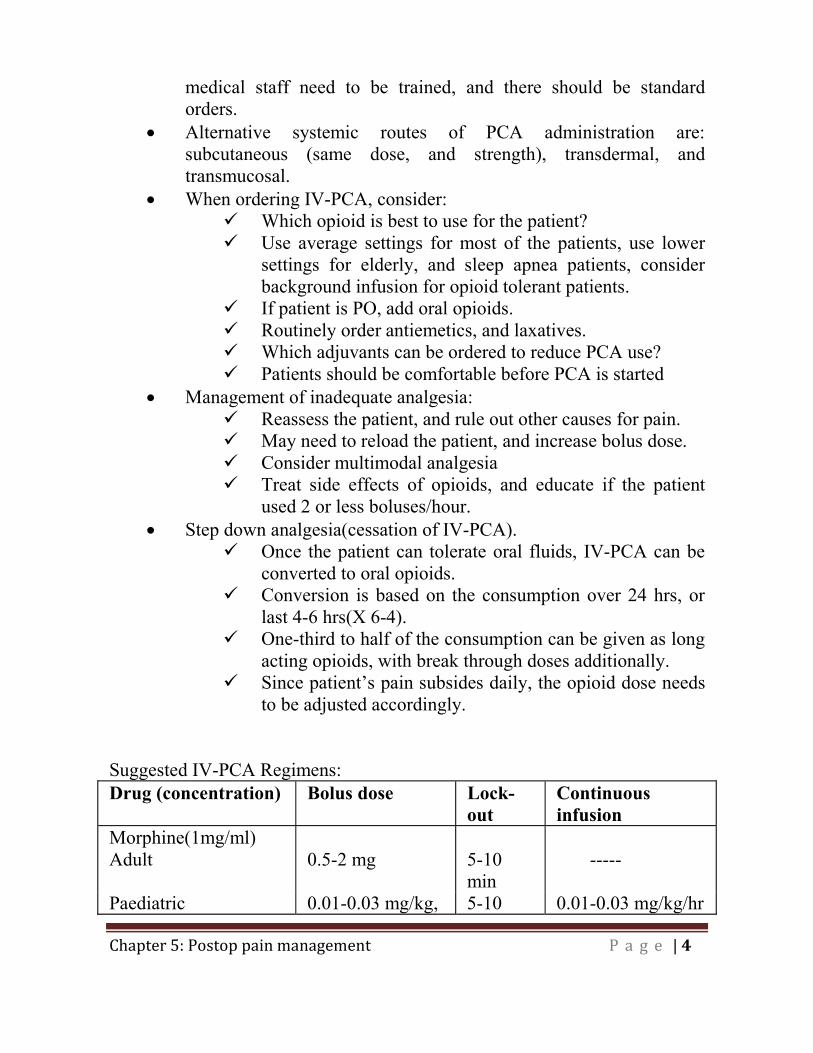
medical staff need to be trained, and there should be standard orders.
Alternative systemic routes of PCA administration are: subcutaneous (same dose, and strength), transdermal, and transmucosal.
When ordering IV-PCA, consider: Which opioid is best to use for the patient? Use average settings for most of the patients, use lower
settings for elderly, and sleep apnea patients, consider background infusion for opioid tolerant patients.
If patient is PO, add oral opioids. Routinely order antiemetics, and laxatives. Which adjuvants can be ordered to reduce PCA use? Patients should be comfortable before PCA is started
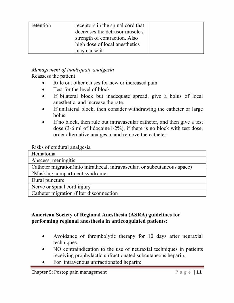
Management of inadequate analgesia: Reassess the patient, and rule out other causes for pain. May need to reload the patient, and increase bolus dose. Consider multimodal analgesia Treat side effects of opioids, and educate if the patient
used 2 or less boluses/hour. Step down analgesia(cessation of IV-PCA).
Once the patient can tolerate oral fluids, IV-PCA can be converted to oral opioids.
Conversion is based on the consumption over 24 hrs, or last 4-6 hrs(X 6-4).
One-third to half of the consumption can be given as long acting opioids, with break through doses additionally.
Since patient�’s pain subsides daily, the opioid dose needs to be adjusted accordingly.
Suggested IV-PCA Regimens: Drug (concentration) Bolus dose Lock-
out Continuous infusion
Morphine(1mg/ml) Adult 0.5-2 mg 5-10
min -----
Paediatric 0.01-0.03 mg/kg, 5-10 0.01-0.03 mg/kg/hr

5
max: 0.15 mg/kg/hr
min
Hydromorphone (0.2 mg/ml)
Adult 0.2-0.6 mg 5-10 min
-----
Paediatric 0.003-0.005 mg/kg, max: 0.02 mg/kg/hr
5-10 min
0.003-0.005 mg/kg/hr
Fentanyl (0.01 mg/ml) Adult 10-20 mcg 5-10
min -----
Paediatric 0.2-0.5 mcg/kg, max: 2 mcg/kg/hr
5-10 min
0.15-1 mcg/kg/hr
Revised from Miller: Miller's Anesthesia, 6th ed, 2005 Churchill Livingstone. Complications of IV-PCA
Narcotics side effects (see the pharmacology of pain management). Equipment malfunction Staff error Patient not suited to PCA
Neuraxial Analgesic Techniques Contraindication to neuraxial block:
Patient�’s refusal Coagulopathy Local or systemic infection/sepsis Hypovolemia/hemodynamic instability
Epidural analgesia pearls
Clinical decisions include the choice and dose of analgesic agents, location of catheter placement, and onset and duration of perioperative use.
In general, bupivacaine, ropivacaine, or levobupivacaine is used because of the differential and preferential clinical sensory blockade with minimal motor block.
6
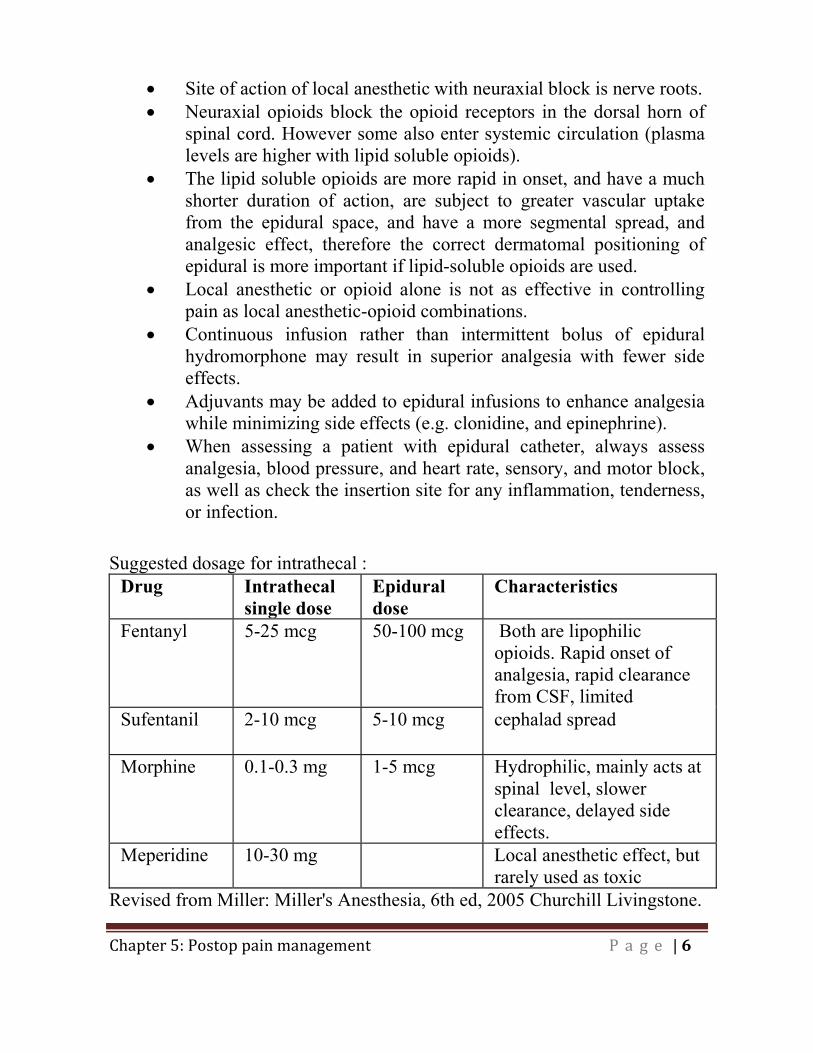
Site of action of local anesthetic with neuraxial block is nerve roots. Neuraxial opioids block the opioid receptors in the dorsal horn of
spinal cord. However some also enter systemic circulation (plasma levels are higher with lipid soluble opioids).
The lipid soluble opioids are more rapid in onset, and have a much shorter duration of action, are subject to greater vascular uptake from the epidural space, and have a more segmental spread, and analgesic effect, therefore the correct dermatomal positioning of epidural is more important if lipid-soluble opioids are used.
Local anesthetic or opioid alone is not as effective in controlling pain as local anesthetic-opioid combinations.
Continuous infusion rather than intermittent bolus of epidural hydromorphone may result in superior analgesia with fewer side effects.
Adjuvants may be added to epidural infusions to enhance analgesia while minimizing side effects (e.g. clonidine, and epinephrine).
When assessing a patient with epidural catheter, always assess analgesia, blood pressure, and heart rate, sensory, and motor block, as well as check the insertion site for any inflammation, tenderness, or infection.
Suggested dosage for intrathecal : Drug Intrathecal
single doseEpidural dose
Characteristics
Fentanyl 5-25 mcg 50-100 mcg Both are lipophilic opioids. Rapid onset of analgesia, rapid clearance from CSF, limited
Sufentanil 2-10 mcg 5-10 mcg cephalad spread
Morphine 0.1-0.3 mg 1-5 mcg Hydrophilic, mainly acts at spinal level, slower clearance, delayed side effects.
Meperidine 10-30 mg Local anesthetic effect, but rarely used as toxic
Revised from Miller: Miller's Anesthesia, 6th ed, 2005 Churchill Livingstone.
7
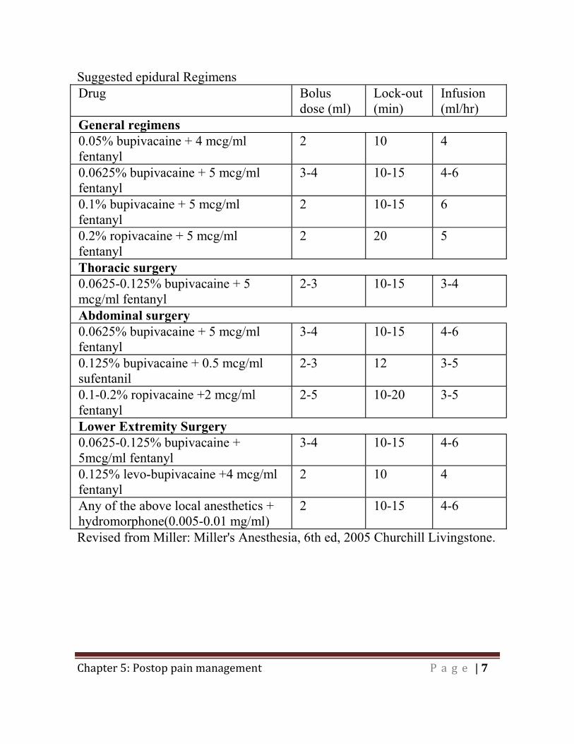
Suggested epidural Regimens Drug Bolus
dose (ml) Lock-out (min)
Infusion (ml/hr)
General regimens 0.05% bupivacaine + 4 mcg/ml fentanyl
2 10 4
0.0625% bupivacaine + 5 mcg/ml fentanyl
3-4 10-15 4-6
0.1% bupivacaine + 5 mcg/ml fentanyl
2 10-15 6
0.2% ropivacaine + 5 mcg/ml fentanyl
2 20 5
Thoracic surgery 0.0625-0.125% bupivacaine + 5 mcg/ml fentanyl
2-3 10-15 3-4
Abdominal surgery 0.0625% bupivacaine + 5 mcg/ml fentanyl
3-4 10-15 4-6
0.125% bupivacaine + 0.5 mcg/ml sufentanil
2-3 12 3-5
0.1-0.2% ropivacaine +2 mcg/ml fentanyl
2-5 10-20 3-5
Lower Extremity Surgery 0.0625-0.125% bupivacaine + 5mcg/ml fentanyl
3-4 10-15 4-6
0.125% levo-bupivacaine +4 mcg/ml fentanyl
2 10 4
Any of the above local anesthetics + hydromorphone(0.005-0.01 mg/ml)
2 10-15 4-6
Revised from Miller: Miller's Anesthesia, 6th ed, 2005 Churchill Livingstone.
8
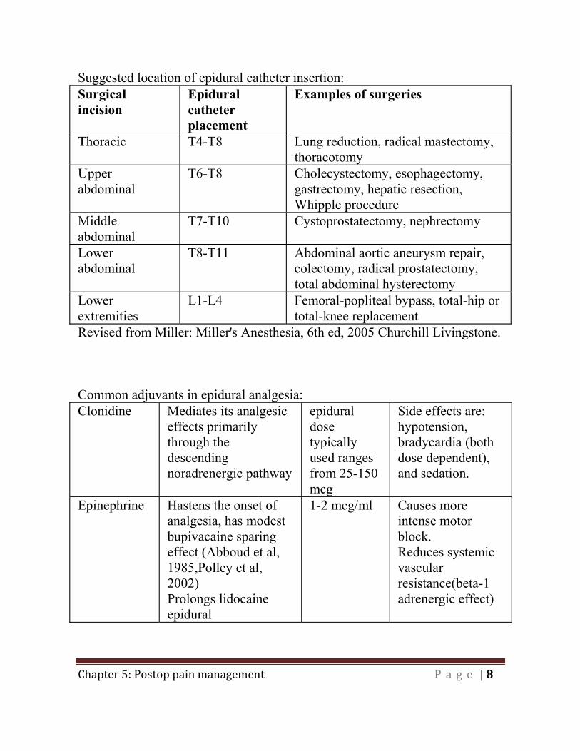
Suggested location of epidural catheter insertion: Surgical incision
Epidural catheter placement
Examples of surgeries
Thoracic T4-T8 Lung reduction, radical mastectomy, thoracotomy
Upper abdominal
T6-T8 Cholecystectomy, esophagectomy, gastrectomy, hepatic resection, Whipple procedure
Middle abdominal
T7-T10 Cystoprostatectomy, nephrectomy
Lower abdominal
T8-T11 Abdominal aortic aneurysm repair, colectomy, radical prostatectomy, total abdominal hysterectomy
Lower extremities
L1-L4 Femoral-popliteal bypass, total-hip or total-knee replacement
Revised from Miller: Miller's Anesthesia, 6th ed, 2005 Churchill Livingstone. Common adjuvants in epidural analgesia: Clonidine Mediates its analgesic
effects primarily through the descending noradrenergic pathway
epidural dose typically used ranges from 25-150 mcg
Side effects are: hypotension, bradycardia (both dose dependent), and sedation.
Epinephrine Hastens the onset of analgesia, has modest bupivacaine sparing effect (Abboud et al, 1985,Polley et al, 2002) Prolongs lidocaine epidural
1-2 mcg/ml Causes more intense motor block. Reduces systemic vascular resistance(beta-1 adrenergic effect)
9
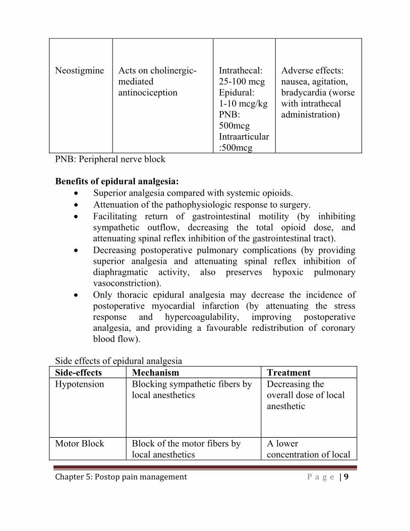
Neostigmine
Acts on cholinergic-mediated antinociception
Intrathecal: 25-100 mcg Epidural: 1-10 mcg/kg PNB: 500mcg Intraarticular:500mcg
Adverse effects: nausea, agitation, bradycardia (worse with intrathecal administration)
PNB: Peripheral nerve block Benefits of epidural analgesia:
Superior analgesia compared with systemic opioids. Attenuation of the pathophysiologic response to surgery. Facilitating return of gastrointestinal motility (by inhibiting
sympathetic outflow, decreasing the total opioid dose, and attenuating spinal reflex inhibition of the gastrointestinal tract).
Decreasing postoperative pulmonary complications (by providing superior analgesia and attenuating spinal reflex inhibition of diaphragmatic activity, also preserves hypoxic pulmonary vasoconstriction).
Only thoracic epidural analgesia may decrease the incidence of postoperative myocardial infarction (by attenuating the stress response and hypercoagulability, improving postoperative analgesia, and providing a favourable redistribution of coronary blood flow).
Side effects of epidural analgesia Side-effects Mechanism Treatment Hypotension Blocking sympathetic fibers by
local anesthetics Decreasing the overall dose of local anesthetic
Motor Block Block of the motor fibers by
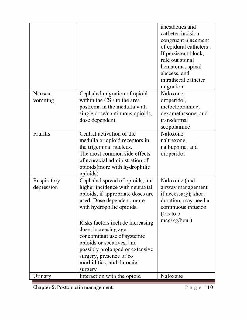
local anesthetics A lower concentration of local
10
anesthetics and catheter-incision congruent placement of epidural catheters . If persistent block, rule out spinal hematoma, spinal abscess, and intrathecal catheter migration
Nausea, vomiting
Cephalad migration of opioid within the CSF to the area postrema in the medulla with single dose/continuous opioids, dose dependent
Naloxone, droperidol, metoclopramide, dexamethasone, and transdermal scopolamine
Pruritis Central activation of the medulla or opioid receptors in the trigeminal nucleus. The most common side effects of neuraxial administration of opioids(more with hydrophilic opioids)
Naloxone, naltrexone, nalbuphine, and droperidol
Respiratory depression
Cephalad spread of opioids, not higher incidence with neuraxial opioids, if appropriate doses are used. Dose dependent, more with hydrophilic opioids.
Risks factors include increasing dose, increasing age, concomitant use of systemic opioids or sedatives, and possibly prolonged or extensive surgery, presence of co morbidities, and thoracic surgery
Naloxone (and airway management if necessary); short duration, may need a continuous infusion (0.5 to 5 mcg/kg/hour)
Urinary Interaction with the opioid Naloxane
11
retention receptors in the spinal cord that decreases the detrusor muscle's strength of contraction. Also high dose of local anesthetics may cause it.
Management of inadequate analgesia Reassess the patient
Rule out other causes for new or increased pain Test for the level of block If bilateral block but inadequate spread, give a bolus of local
anesthetic, and increase the rate. If unilateral block, then consider withdrawing the catheter or large
bolus. If no block, then rule out intravascular catheter, and then give a test
dose (3-6 ml of lidocaine1-2%), if there is no block with test dose, order alternative analgesia, and remove the catheter.
Risks of epidural analgesia Hematoma Abscess, meningitis Catheter migration(into intrathecal, intravascular, or subcutaneous space) ?Masking compartment syndrome Dural puncture Nerve or spinal cord injury Catheter migration /filter disconnection
American Society of Regional Anesthesia (ASRA) guidelines for performing regional anesthesia in anticoagulated patients:
Avoidance of thrombolytic therapy for 10 days after neuraxial techniques.
NO contraindication to the use of neuraxial techniques in patients receiving prophylactic unfractionated subcutaneous heparin.
For intravenous unfractionated heparin:
12
Delay heparinization for 1 hour after needle placement. Remove indwelling neuraxial catheter 2-4 hours after last
heparin dose, and restart heparin 1 hour after catheter removal.
Concurrent use of medications that affect other component of coagulation cascade may increase risk of bleeding complications.
Low molecular weight heparin(LMWH)
Monitoring of anti-Xa level is not recommended (not predictive of bleeding risk).
In the presence of blood during needle placement, LMWH therapy should be delayed 24hours postoperatively.
Needle placement 10-12 hours after prophylactic dose, and 24 hours after treating dose.
Postop LMWH for twice-daily dosing: first dose no earlier than 24 hours postop, regardless of anesthtic technique, and indwelling catheter should be REMOVED prior to this dosing.
Postop LMWH for single-daily dosing: First postop dose at 6-8 hours postop, and second postop dose no sooner than 24 hours after the first dose.
Stop LMWH 10-12 hours prior to catheter removal, and restart 2 hours after.
Suggested time interval between discontinuation of antiplatelet medications, and neuraxial blockade is 14 days for ticlopidine, 7 days for clopidogrel, 5 days for warfarin.
Use of NSAIDs alone doesn�’t create a risk for bleeding.
Peripheral nerve blocks
Single injection is primarily used for intraoperative analgesia/anesthesia, and the block normally regresses 10-24 hours after the injection of a long-acting
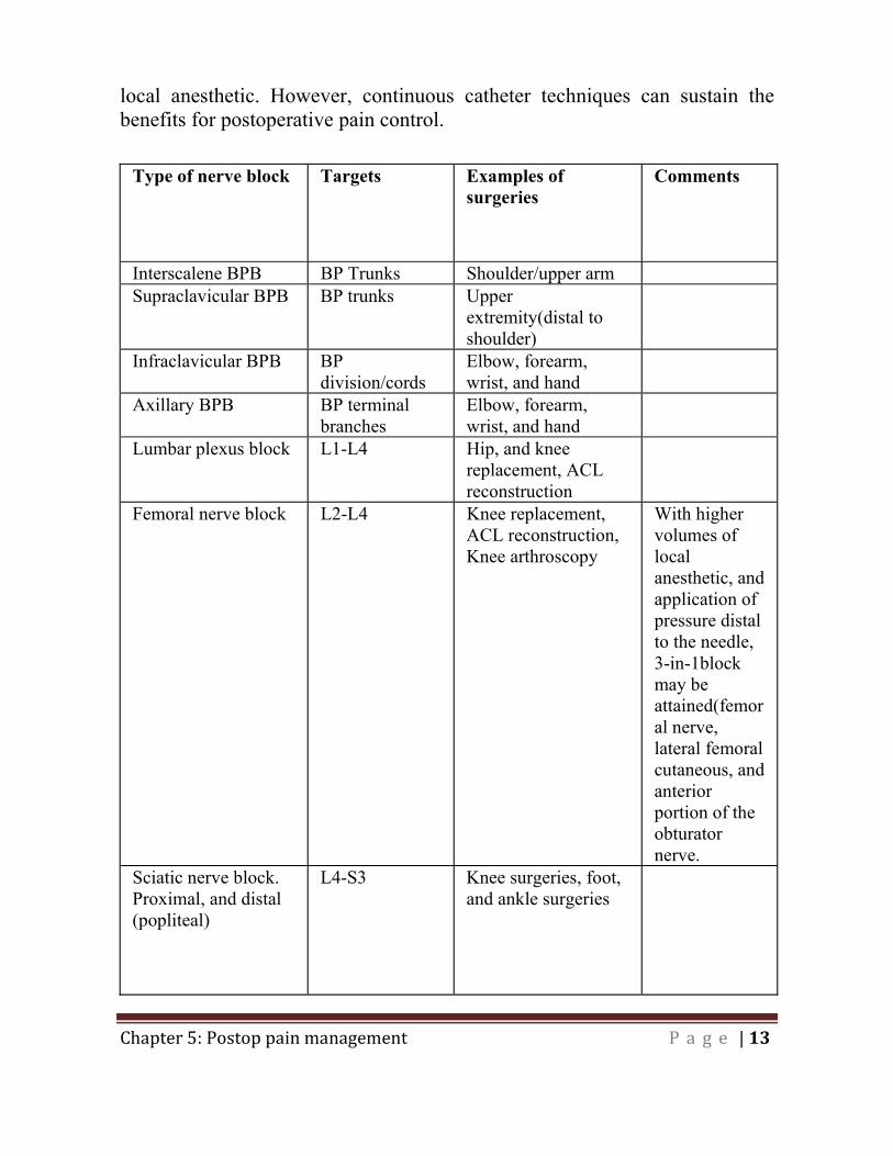
13
local anesthetic. However, continuous catheter techniques can sustain the benefits for postoperative pain control.
Type of nerve block Targets Examples of surgeries
Comments
Interscalene BPB BP Trunks Shoulder/upper arm Supraclavicular BPB BP trunks Upper
extremity(distal to shoulder)
Infraclavicular BPB BP division/cords
Elbow, forearm, wrist, and hand
Axillary BPB BP terminal branches
Elbow, forearm, wrist, and hand
Lumbar plexus block L1-L4 Hip, and knee replacement, ACL reconstruction
Femoral nerve block L2-L4 Knee replacement, ACL reconstruction, Knee arthroscopy
With higher volumes of local anesthetic, and application of pressure distal to the needle, 3-in-1block may be attained(femoral nerve, lateral femoral cutaneous, and anterior portion of the obturator nerve.
Sciatic nerve block. Proximal, and distal (popliteal)
L4-S3 Knee surgeries, foot, and ankle surgeries
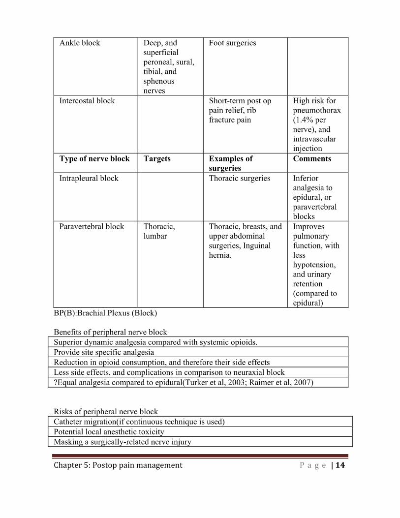
14
Ankle block Deep, and superficial peroneal, sural, tibial, and sphenous nerves
Foot surgeries
Intercostal block Short-term post op pain relief, rib fracture pain
High risk for pneumothorax(1.4% per nerve), and intravascular injection
Type of nerve block Targets Examples of surgeries
Comments
Intrapleural block Thoracic surgeries Inferior analgesia to epidural, or paravertebral blocks
Paravertebral block Thoracic, lumbar
Thoracic, breasts, and upper abdominal surgeries, Inguinal hernia.
Improves pulmonary function, with less hypotension, and urinary retention (compared to epidural)
BP(B):Brachial Plexus (Block) Benefits of peripheral nerve block Superior dynamic analgesia compared with systemic opioids. Provide site specific analgesia Reduction in opioid consumption, and therefore their side effectsLess side effects, and complications in comparison to neuraxial block ?Equal analgesia compared to epidural(Turker et al, 2003; Raimer et al, 2007) Risks of peripheral nerve block Catheter migration(if continuous technique is used) Potential local anesthetic toxicity Masking a surgically-related nerve injury
15
References: Abboud TK, Sheik-ol-Eslam A, Yanagi T, et al. Safet and efficacy of epinephrine added to bupivacaine for lumbar epidural analgesia in obstetrics. Anesth Analg 1985; 64:585-91.
Ballnatyne JC, McKenna JM, Ryder E. Epidural analgesia-experience of 5628 patients in a large teaching hospital through audit. Acute Pain 2003; 4: 89-97.
Habib AS, Gan TJ.Role of analgesic adjuncts in postoperative pain management. Anesthesiology Clin N Am 2005; 23:85-107.
Horlocker TT, Wedel DJ, Benzon H, Brown DL,Enneking FK, Heit JA, Mulroy MF, ROsenquist RW, Rowlingson J, Tryba M, Yuan CS. Regional anesthesia in the anticoagulated patient:defining the risks. Reg Anesth Pain Med 2004; 29:1-12.
Horlocker TT. Wedel DJ. Benzon H. Brown DL. Enneking FK. Heit JA. Mulroy MF. Rosenquist RW. Rowlingson J. Tryba M. Yuan CS. Regional anesthesia in the anticoagulated patient: defining the risks (the second ASRA Consensus Conference on Neuraxial Anesthesia and Anticoagulation).Regional Anesthesia & Pain Medicine 2003; 28(3):172-97.
Jin F, Chung F. Multimodal analgesia for postoperative pain control. J Clin Anesth 2001; 13:524-539.
Joshi GP.Multimodal analgesia techniques and postoperative rehabilitation. Anesthesiology Clin N Am 2005; 23:185-202.
Kehlet H, Dahl JB. The value of �“multimodal�” or �“balanced analgesia�” in postoperative pain treatment. Anesth Analg 1993; 77:1048-1056.
Kehlet H, Wilmore DW. Multimodal strategies to improve surgical outcome. Am J Surg 2002; 183:630-641.
16
Lema MJ. Monitoring epidural local anesthetic action during the postoperative period. Reg Anesth 1996; 21:94-99.
Macintyre PE. Safety and efficacy of patient-controlled analgesia. British J Anesth 2001; 87:36-46.
Miller: Miller's Anesthesia, 6th ed, 2005 Churchill Livingstone.
Polley LS, Columb MO, Naughton NN et al. Effect of epidural epinephrine on the minimum local analgesic concentration of epidural bupivacaine in labor. Anesthesiology 2002;96: 1123-8.
Raimer C, Preim K, Wiese AA, Birnbaum J, Dirkmorfeld LM, Mossner A, Matiolis G, Perka C, Volk T. Continuous psoas and sciatic block after knee arthroplasty: good effects compared to epidural analgesia or i.v. opioid analgesia: a prospective study of 63 patients. Acta Orthop 2007; 78:193-200.
Schugs A, Torrie JJ. Safelty assessment of postoperative pain management by an acute pain service. Pain 1993;55:387-391.
Turker G, Uckunkaya N, Yavascoaglu B, Yilmazlar A, Ozcelik S.Comparison of the catheter-technique psoas compartment block and the epidural block for analgesia in partial hip replacement surgery. Acta Anesthesiol Scand 2003; 47:30-36.
Chapter 6:
Pain management in
patient
subpopulations
2
Chapter 6: Pain management in patient subpopulations
Pain Management in the Opioid-Tolerant Patient:
Definitions:
Physical Dependence: Adaptation that occurs in the body with long-term opioid use. If the opioid is abruptly stopped, or is rapidly reduced, or if an opioid receptor antagonist is administered the body will experience a withdrawal reaction.
Opioid Withdrawal: Characterized by adrenergic hyperactivity; hypertension, tachycardia, chills, piloerection, diaphoresis, nausea, vomiting, diarrhea, abdominal cramps, salivation, lacrimation, rhinorrhea and yawning.
Tolerance: Adaptation that can occur after days to weeks of drug exposure, causing a reduction in drug effects. Tolerance develops to all opioid effects-analgesia and side effects, less so for miosis and constipation. Tolerance to the opioid�’s analgesic effects can be mitigated by increasing the opioid dose.
Addiction: Psychological dependence and a need to take the drug for non-pain relieving purposes. Characterized by the presence of one or more of the 4 C�’s- impaired Control over drug use, Compulsive drug use, Continued use despite harm, and Craving. It has been defined as a primary, chronic, neurobiological disease, with genetic, psychological and environmental factors influencing it development and manifestations.
Opioid-Induce Hyperalgesia
Patients receiving long term opioids for pain become paradoxically more sensitive to pain as a direct result of opioid therapy
Opioid induced hyperalgesia peaks during periods of opioid abstinence or in periods between regularly administered opioid doses
3
Chronic administration of opioids leads to compensatory neurobiological changes that facilitate nociception
Mechanism is unclear and may be genetic This phenomenon is more common in patients with a history a
opioid abuse
Notes on Opioid Tolerance:
Tolerance to one opioid will also display cross tolerance to other opioids, but it is incomplete thus when switching opioid tolerant patients from one opioid to another reduce the calculated equianalgesic dose of the new opioid by 25-50% to account for incomplete cross tolerance
Pathophysiology:
Opioid tolerance due to desensitized opioid receptors, non-opioid mechanisms, release of anti-opioids, neuroplastic changes in pain perception in the brain.
Basic Guidelines for the Treatment of acute on chronic pain:
Stress multidisciplinary communication: Surgeon, preadmission clinic personnel must identify the chronic pain patient as high risk, alert the APS and consult preoperatively with an Anesthesiologist to discuss the patient�’s concerns and the management strategy.
Analgesic history: Obtain an accurate analgesic history; ask about oral, transdermal, rectal, nasal and injectable opioid analgesics as well as their use of non-opioid prescription and over the counter analgesics. Include antidepressants, anxiolytics, antidepressants and anticonvulsants
Pre-op: Patients should take their regular opioid (oral or transdermal) on the morning of surgery. If increases in body
4
temperature are anticipated, (use of a warming blanket) the fentanyl patch should be removed pre-op and replacement of an equivalent amount of analgesic. Elevations in body temperature can increase the rate of fentanyl absorption and result in toxicity.
Optimize Multimodal pre-operative Analgesia: Use
Acetaminophen 1.0 gram, Celebrex 200-400 mg, and Gabapentin 300-600 mg 1 hour pre-operatively.
Intra and Post-operatively: There is wide interpatient variability
in intra and post-operative opioid requirements. Due to opioid tolerance and opioid induced hyperalgesia, the opioid-tolerant patients should receive intra and post-operative opioid doses that are initially 1.3-3 times higher than the usual standard doses that are used in opioid-naïve patients. Post-operatively patients must continue their regular opioid as a baseline to prevent withdrawal. Patients who are unable to take their oral opioid post-operatively need to receive an equianalgesic dose via the parenteral route. Opioid rotation should be considered in those who are unable to tolerate the increased opioid dose. Switching opioids to a dose of 50-75% of the equipotent dose as determined from equianalgesic dose tables due to incomplete cross tolerance. Patients undergoing surgical procedures that are expected to significantly reduce their level of pre-operative chronic pain should reduce their opioid by 25-50% of their baseline pre-operative dose.
Mixed Agonist-Antagonist MUST NOT BE USED as they can
displace the maintenance opioid from the opioid receptor and precipitate withdrawal in the opioid dependent patient. (examples Nalbuphine, Buprenorphine)
Use of Gabapentin or Pregabulin, Ketamine, and Clonidine may be considered (see section on pharmacology)
Regional Anesthetics, Peripheral Nerve blocks as well as epidural or intrathecal modalities are clinically indicated.
5
Remember to maintain usual dose of systemic opioids by the intravenous or oral route to prevent opioid withdrawal.
Neuraxial Opioids can be used. Suggest using very lipophilic opioids such as fentanyl and sufentanil. Sufentanil may provide better pain control because of its greater potency.
Conversion from intravenous to oral opioids postoperatively
Rough guideline is to calculate the total PCA consumption over the last 24 hours and administer one half to one third as a controlled release opioid while allowing the remainder to be maintained as a short acting opioid as needed.
Postoperative follow-up involves communication with the primary
physician regarding treatment plan and re-evaluation in the outpatient pain clinic to re-assess pain levels and medications
Special Considerations for patients on methadone maintenance therapy:
Available in Canada only in oral or liquid formulation Patients usually receiving methadone q 8 or 12 hours for chronic
neuropathic pain Patients on daily methadone to treat opioid addiction generally on a
single daily dosing schedule Methadone prescriber and pharmacy should be contacted to verify
the methadone dose and when it was last administered. Both parties should be informed of the patient�’s hospital admission and discharge date
Temporary authorization to prescribe methadone can be obtained for the physician responsible for the post-operative pain management
Patients should receive their usual a.m. dose of methadone on the day of surgery to avoid fluctuations in serum methadone concentration
Avoid abrupt discontinuation of methadone before surgery
6
Patients on methadone > 200 mg/day may develop prolonged Q-T interval which is a risk factor for the development of Torsades de pointes. A baseline electrocardiogram should be performed
Patients who are fasting should convert their methadone to an appropriate choice of opioid and an equianalgesic regimen to prevent opioid withdrawal, however methadone may be administered via a nasogastric tube or rectally
Conversion from methadone to another opioid is problematic, consultation with a methadone provider or expert in pain medicine is advised. CAMH addiction clinical consultation service is available M-F 9-4 p.m. at 1-888-720-2227
There is no uniform method of converting methadone to another opioid. Recommend a methadone to morphine ratio of 4-5:1. Therefore methadone 30 mg per day would be equivalent to 120 mg of oral morphine, factoring in oral bioavailability of 33% and a cross tolerance of 50% this would amount to 1 mg/hr of intravenous morphine.
Like other opioid tolerant patients patient controlled analgesia dose will be 1.5-3 times greater than opioid naïve patients.
Frequent monitoring of patients are required as the methadone clears from their system and the alternate opioid is loaded into the patient
References
Alford DP, Compton P, Samet JH. Acute pain management for patients receiving maintenance methadone or buprenorphine therapy. Ann Intern Med. 2006; 144(2) :127-34.
Gourlay DL, Heit HA, Almahrezi A. Universal precautions in pain medicine: a rational approach to the treatment of chronic pain. Pain Med. 2005;6(2):107-12.
Jovey RD. Managing acute pain in the opioid-dependent patient. In: Flor H, Kalso E, Dostrovsky JO (Ed). Proceedings of the 11th World Congress on Pain. IASP Press, Seattle, 2006:469-79
7
Mehta V, Langford RM. Acute pain management for opioid dependent patients. Anaesthesia. 2006 ;61(3):269-76.
Peng PW, Tumber PS, Gourlay D. Review article: perioperative pain management of patients on methadone therapy. Can J Anaesth. 2005; 52(5):513-23.
Swenson JD, Davis JJ, Johnson KB. Postoperative care of the chronic opioid-consuming patient. Anesthesiol Clin North America. 2005; 23(1):37-48.
Hadi I, Morley-Forester P, Dain S, Horrill K, Moulin, D. Brief Review: Perioperative management of the patient with chronic non-cancer pain. Can J Anesth 2006, 53: 12 pp 1190-1199.
8
Pain Management in Elderly (over 65 year old) Physiological changes in elderly relevant to pain control:
Difficulty in assessment of pain, and efficacy of treatment, as reduced cerebral blood flow, and cerebral volume result in some neurological dysfunction.
No significant alteration in pain perception or sensitivity to electrical stimulation in elderly, but there is increase in thermal and pressure pain threshold, and decrease in pain tolerance.
Decline in median effective dose requirement for agents that act within the CNS.
Prolonged clinical effects if hepatic or renal degradation is required. At greater risk for unrelieved pain, prolonged analgesic use and
impaired long-term recovery.
Pain assessment in elderly: Less likely to report pain associated with acute pathology. Visual and hearing disturbances may lead to inaccurate assessment. Use verbal descriptor (no pain, mild, moderate, and severe pain). Use functional pain scale (rates pain severity as tolerable or
intolerable by interference with activity focusing on ability to watch TV, read, and use the telephone).
Use numeric rating scale when possible.
Non pharmacologic interventions: Non pharmacologic methods may promote better orientation, and
minimize confusion. Psychological modalities: such as cognitive�–behavioural therapy,
relaxation and biofeedback training, and behaviour therapy used alone or in combination with appropriate pharmacological strategies, should be an integral part of care plans in most cases .
Physical rehabilitation like: transcutaneous electrical nerve stimulation (TENS), massage, and the application of heat and cold are also useful.
9
Pharmacologic interventions:
General pearls in pharmacologic treatment in elderly (Key Recommendations by the American Geriatric Society, 2002)
Use the lowest effective initial dose (25-50% decrease from the adult dose).
Slower escalation of drug until the maximum ceiling dose or side effects are reached (There is no maximum ceiling dose with opioids).
Frequently monitor patients for side effects of the medication used, as more sensitive to the side effects including sedation, respiratory depression, urinary retention, and cognitive impairment.
NSAIDs should be used with caution. In older patients, NSAIDs have significant side effects and are the most common cause of adverse drug reactions. If NSAIDs are used, monitoring for hematocrit, renal function, and occult blood in stool should be done frequently.
Tramadol, an opioid with intermediate potency, is a good choice for moderate pain, especially if can�’t tolerate NSAIDs. Its monoaminergic reuptake inhibition is of advantage in reversing dementia symptoms in elderly.
Greater analgesia in response to a fixed dose of opioids, as well as higher peak, and longer duration of analgesia with opioids.
Self-administering less opioid than young patients but obtain comparable pain relief using patient controlled analgesia (PCA).
Avoid continuous opioid infusion, as there are risks of accumulation, and toxicity.
Tapering 10-20% daily over 10 days can wean most patients, however slower tapering is recommended for patients with cardiovascular disease.
Hydromorphone and oxycodone, which have minimal active metabolites and relatively short half-lives (i.e., t½ is 2 to 3 hours), are more desirable than drugs such as methadone (i.e., t½ is 12 to 190 hours) or meperidine and propoxyphene with accumulation of metabolites toxic to both the kidneys and the CNS.
10
Opioids that are antagonistic to the mu receptors are less desirable, given the high prevalence of depression among elderly, and the advantage of euphoric component that occurs with mu receptor agonists.
Epidural analgesia, where applicable, is an excellent option for pain relief in elderly. However, it is suggested to use either pure local anesthetic, or local anesthetic with low dose fentanyl (2mcg/ml) for elderly to avoid central side effects of opioids.
Adjuvant Drugs
Adjuvant drug therapy is guided by the same principles used in NSAID and opioid therapy (see earlier). However, since poly pharmacy in the elderly is a frequent source of morbidity, basic principles of multimodal analgesia are safer. Adjuvant drugs for geriatric pain management span the entire spectrum of drug types and include (but are not limited to):
Muscle relaxants(carisoprodol) Corticosteroids(prednisone) Anticonvulsants(Gabapentin) Antidepressants(amitriptyline) Neuroleptics(methotrimeprazine) Antihistamines(hydroxyzine) Local anesthetics(lidocaine) Antiarrhythmics(betablockers) 2-adrenergic agonists(clonidine) Psychostimulants(dextroamphetamine) Calcitonin Capsaicin
Medications such as phenothiazines, antihistamines, and benzodiazepines should be avoided in the elderly as increase the risk of delirium.
11
In general, the clinical end-points for pharmacological interventions should not concentrate on reduced drug dose but, rather, on decreased pain, improved function, and improved mood and sleep.
References:
AGS panel on persistent pain in older persons: The management of persistent pain in older persons. J Am Geriatr Soc 2002; 50: S205-S224.
Ballantyne JC, McKenna JM, Ryder E. Epidural analgesia. Acute Pain 2003; 4: 89-97.
Burris JE. Pharmacologic approaches to geriatric pain management. Arch Phys Med Rehabil 2004; 85(3):S45-S49.
Eisenberg DM, Kessler RC, Foster C. Unconventional medicine in the United States: prevalence, costs and patterns of use. New England Journal of Medicine 1993; 328:246-252.
Freye E, Levy JV. The effects of tramadol on pain relief, fast EEG-power spectrum, and cognitive function in elderly patients with chronic osteoarthritis. Acute Pain 2006; 8: 55-61.
Gagliese L, Jackson M, Ritvo P, Wowk A, Katz J. Age is not an impediment to effective use of patient controlled analgesia by surgical patient. Anesthesiology 2000; 93(3): 601-610.
Gagliese L, Melzack R. Pain in the elderly. In Wall &Mezack�’s Textbook of Pain 2006; Churchill Livingstone.1167-1177.
Gagliese L, Weizblit N, Ellis W, Chan VW. The measurement of postoperative pain; a comparison of intensity scales in younger and older surgical patients. Pain 2005; 117(3): 412-420.
12
Gloth FM. Principles of perioperative pain management in older adults. Clin Geriatr Med. 2001; 17(3):553-573.
Lynch D. Geriatric Pain. In Raj: Practical Management of Pain 2000; Mosby. 270-290.
Nicholson NL, Blanchard EB. A controlled evaluation of behavioral treatment of chronic headache in the elderly. Behaviour Therapy 1993; 24:395-408.
Prowse M. Postoperative pain in older people. J Clin Nurs 2006; 16:84-97.
13
Pain management in Children
Children have been and remain the least medicated patients in the perioperative period. Fear of overdosing, a poor understanding of childhood physiology and pain manifestation are some of the reasons for inadequate pain treatment in this age group.
Control of postoperative pain in children is very important as the catabolic state induced by acute pain may be more damaging to infants and young children who already have higher metabolic rates, and less nutritional reserves than adults.
Pain physiology in children
The fetal brain is developed and active early in its development and that the neonatal nervous system is capable of detecting olfactory, tactile, auditory, and visual stimuli, as well as perceiving pain by 28 weeks.
All four processes of nociception (transduction, transmission, modulation, and perception) are developed in a newborn.
The neonatal nervous system may be less effective at blocking painful stimuli, as it has:
Larger receptor fields, Higher concentration of receptor sites for substance P, Less-developed descending pathway.
In the face of unsatisfactory pain control in paediatric population, American Academy of Pediatrics and American Pain Society published a statement (2001), concluded that physicians need to expand their knowledge, use appropriate assessment tools, and techniques, anticipate painful experiences, and intervene accordingly, use a multimodal approach to pain management, use a multidisciplinary approach when possible, involve families, and advocate for the use of effective pain management in children.
General pearls in pharmacologic treatment in paediatrics
14
For the list of commonly-used medication, and dosage, please refer to the medication tables.
Avoid intramuscular route in paediatric. Nasal fentanyl, although painful, results in analgesic blood level comparable to IV use, which is useful if the IV is lost
Meperidine is a poor choice just as it is for the adult population. Remifentanil is not licensed for use in children under 2 years of
age. Patient-Parent-Nurse controlled analgesia: Previously only
adolescent or older were given IV-PCA, but nowadays with children playing electronic games, they easily understand the concept, and patients as young as 4, can use this method. Morphine is the most common drug used in paediatrics. In children who can�’t tolerate morphine, hydromorphone is an alternative. Some believe adding a background infusion, and reduction of bolus by 50%, achieve better analgesia, and sleep quality, with lower opioid consumption, however careful monitoring is required.
Nurse controlled analgesia is a variation of above method for children who may not have a motor skill to use the PCA pump unaided. A low basal rate is complemented with boluses by the nurse. There have been concerns with regards to the risk of overdose, but it is a way of delivering the pain medication without leaving the bedside. Close monitoring of the patient may be needed because significant respiratory depression occurs in approximately 1.7% of these patients.
Weaning from PCA in children is similar to adults.
Tolerance to opioids may occur after 21 days of treatment, and it is most commonly seen in sickle cell disease patients. Addiction is extraordinarily rare in children.
Alternative-route drugs for children
Indication Comments Caution
EMLA(Lidocaine-Prilocaine) cream
For IV insertion, LP (Only on intact skin)
Slow onset (1 hour), In children beyond neonatal age
Methemoglobinemia
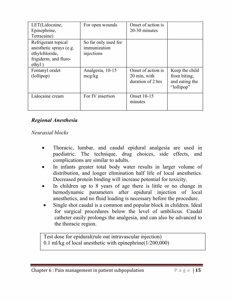
15
LET(Lidocaine, Epinephrine, Tetracaine)
For open wounds Onset of action is 20-30 minutes
Refrigerant topical anesthetic sprays (e.g. ethylchloride, frigiderm, and fluro-ethyl )
So far only used for immunization injections
Fentanyl oralet (lollipop)
Analgesia, 10-15 mcg/kg
Onset of action is 20 min, with duration of 2 hrs
Keep the child from biting, and eating the �“lollipop�”
Lidocaine cream For IV insertion Onset 10-15 minutes
Regional Anesthesia Neuraxial blocks
Thoracic, lumbar, and caudal epidural analgesia are used in paediatric. The technique, drug choices, side effects, and complications are similar to adults.
In infants greater total body water results in larger volume of distribution, and longer elimination half life of local anesthetics. Decreased protein binding will increase potential for toxicity.
In children up to 8 years of age there is little or no change in hemodynamic parameters after epidural injection of local anesthetics, and no fluid loading is necessary before the procedure.
Single shot caudal is a common and popular block in children. Ideal for surgical procedures below the level of umbilicus. Caudal catheter easily prolongs the analgesia, and can also be advanced to the thoracic region.
Test dose for epidural(rule out intravascular injection) 0.1 ml/kg of local anesthetic with epinephrine(1/200,000)
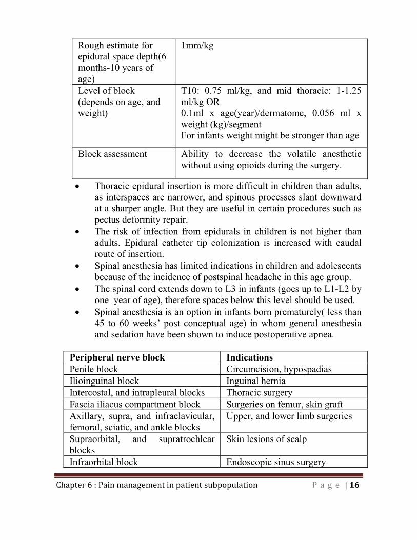
16
Rough estimate for epidural space depth(6 months-10 years of age)
1mm/kg
Level of block (depends on age, and weight)
T10: 0.75 ml/kg, and mid thoracic: 1-1.25 ml/kg OR 0.1ml x age(year)/dermatome, 0.056 ml x weight (kg)/segment For infants weight might be stronger than age
Block assessment Ability to decrease the volatile anesthetic without using opioids during the surgery.
Thoracic epidural insertion is more difficult in children than adults, as interspaces are narrower, and spinous processes slant downward at a sharper angle. But they are useful in certain procedures such as pectus deformity repair.
The risk of infection from epidurals in children is not higher than adults. Epidural catheter tip colonization is increased with caudal route of insertion.
Spinal anesthesia has limited indications in children and adolescents because of the incidence of postspinal headache in this age group.
The spinal cord extends down to L3 in infants (goes up to L1-L2 by one year of age), therefore spaces below this level should be used.
Spinal anesthesia is an option in infants born prematurely( less than 45 to 60 weeks�’ post conceptual age) in whom general anesthesia and sedation have been shown to induce postoperative apnea.
Peripheral nerve block Indications Penile block Circumcision, hypospadias Ilioinguinal block Inguinal hernia Intercostal, and intrapleural blocks Thoracic surgery Fascia iliacus compartment block Surgeries on femur, skin graft Axillary, supra, and infraclavicular, femoral, sciatic, and ankle blocks
Upper, and lower limb surgeries
Supraorbital, and supratrochlear blocks
Skin lesions of scalp
Infraorbital block Endoscopic sinus surgery
17
Regional anesthesia is avoided in supracondylar fracture, as it may interfere with postop neurological assessment.
References:
Brislin RP, Rose JB. Pediatric acute pain management. Anesthesiol Clin N Am 2005; 23:789-814.
Committee on psychosocial aspects of child and family health. The assessment and management of acute pain in infants, children and adolescents. Pediatrics 2001; 108:793-797.
Farrar MW, Lerman J. Novel concepts for analgesia in pediatric surgical patients. Anesthesiol Clin N Am 2007; 20(1): 59-82.
Franck LS, Greenburg CS, Stevens B. Pain assessment in infants and children. Ped Clin N Am 2000; 47(3): 487-512.
Kost-Byerly S. New concepts in acute and extended postoperative pain management in children. Anesthesiol Clin N Am 2002; 20(1):115-135.
Sahinler BE. A review of pediatric pain management in acute and chronic setting. Pain Prac 2002; 2(2): 137-150.
Suresh S, Wheeler M. Practical pediatric regional anesthesia. Anesthesiol Clin N Am 2002; 20: 83-113.
Verghese ST, Hannallah RS. Postoperative pain management in children. Anesthesiol Clin N Am 2005: 23, 163-184.
Yaster M, Kost-Byerly S, Maxwell LG. The management of pain in sickle cell disease. Pediatr Clin N Am 2000; 47(3): 699-710.
Zwass MS. Regional anesthesia in children. Anesthesiol Clin N Am 2005; 23: 815-835.
18
Pain Management in patients with Burn
Burn-injured patients of all ages have procedural, background, and breakthrough pain during the acute, healing, and rehabilitative phases over the long course of burn recovery.
The pain changes during different phases of burn recovery. Unlike most acute injuries, procedural burn pain may worsen unpredictably over the course of healing that adds to emotional distress in these patients.
Burn pain is a mixture of nociceptive and neuropathic pain, and requires aggressive multimodal, and multidisciplinary treatment.
Immediately after the injury, cooling, covering the burn, and immobilization of the injured limbs will help with pain relief.
Procedural pain (e.g. primary mechanical hyperalgesia) is the most intense and most likely type of burn injury pain to be undertreated.
In the initial stages after a burn injury, IV opioid is usually required. In later stages a combination of controlled-release, and immediate-release oral opioids can be used. The effects of opioids, are difficult to gauge over the course of burn recovery as the need for an opioid may change rapidly, resulting in the overmedication or undermedication of burn-injured patients.
Anxiolytic agents, such as the benzodiazepines are known to reduce anxiety in burn patients, but the response is highly variable.
Ketamine as a low dose infusion or intermittent boluses can be used as an adjunct.
Intravenous lidocaine must be considered a pharmacological agent under investigation.
Nonpharmacologic adjuncts include cognitive techniques, behavioural techniques, education and/or preparatory information (enhancing predictability of sensory and procedural components of aversive procedures, hypnotherapeutic techniques.
During the healing and rehabilitation phases, the metabolic rate of increases by 50% when burn size is greater than 20% to 30% surface area and even greater in larger burns or if wound sepsis is present. Thus, concern for changes in pharmacokinetics and pharmacodynamics after burn injury can be a barrier to pain management.
19
Tolerance to, and requirements for large doses of narcotics are common.
References
Frenay MC, Faymonville ME, Devlieger S(2001)Psychological approaches during dressing changes of burned patients. Burns 27:793-799.
Gallagher G, Rae CP, Kinsella J(2000)Treatment of pain in severe burns. Am J Clin Derm 1:329-335.
Summer GJ, Puntillo KA, Miaskowski C, Green PG, Levine JD(2007) Burn Injury Pain: The Continuing Challenge. J Pain 8:533-548.
Wasiak J, Cleland H. (2007)Lidocaine for pain relief in burn injured patients. Cochrane Database Syst Rev. 18:CD005622.
20
Pain management in patients with Obstructive Sleep Apnea
There is little good evidence to guide the �“best choice�” of analgesia in these patients.
Regional analgesic techniques, and non-opioid analgesics are recommended.
Any opioids, if given, should be titrated safely, with close monitoring of patient�’s level of sedation. Supplemental oxygen, and CPAP reduce the risk of significant hypoxemia.
Increasing sedation is the best early clinical indicator of respiratory depression, lack of appropriate monitoring to detect sedation, seems to be crucial in development of hypoxemic events.
Routine use of oxygen is recommended for all the postoperative obstructive sleep apnea patients who are on IV-PCA.
A novel application of alpha-2 agonists for perioperative anesthetic care is emerging.
References
Blake DW, Chia PH, Donnan G, Williams DL(2008) Preoperative assessment for obstructive sleep apnoea and the prediction of postoperative respiratory obstruction and hypoxaemia. Anaesth Intensive Care. 36:379-84.
Schumann R, Jones SB, Cooper B, et al(2009) Update on best practice recommendations for anesthetic perioperative care and pain management in weight loss surgery, 2004-2007. Obesity 17:889-94.
21
Pain management in Pregnant or Lactating patients
Almost all analgesic medications will cross the placenta to some degree, and will transfer in part to milk and breastfed infant. Therefore when possible non pharmacological therapies should be used.
In general, drugs prescribed during pregnancy should be reviewed according to the risk to fetus (consult with www.motherisk.org).
Acetaminophen is the analgesic of choice.
NSAIDS are associated with increased risk of miscarriage. They can also cause fetal cardiac, and renal problems, as well as impair the productin of amoniotic fluid on third trimester. Therefore they should also be avoided after 32nd weeks. NSAIDS are safe during breastfeeding(Aspirin should be avoided).
Opioids can be used, when benefits outweigh the risk. They do not cause fetal malformation, but may result in neonatal abstinence syndrome.
As there have been reports of drug toxicity of the very young and premature infants, cautious use of codeine in lactating mothers is recommended.
There is a significant inter-individual variations in response to codeine, the dose-response relationship with respect to drug toxicity, and the role of pharmacogenetics in both the mother and the infant. These host factors may combine in a particular patient to act synergistically to produce an adverse reaction.
Metoclopramide is the antiemetic of choice in pregnant patients.
22
As a general precaution, it is best to avoid breastfeeding at times of peak maternal blood concentration of any drug, and infants should be monitored for any adverse effect.
References:
Madadi P, Shirazi F, Walter FG, Koren G(2009) Establishing causality of CNS depression in breastfed infants following maternal codeine use. Paediatr Drugs.10:399-404.
Rathmell JP, Viscomi CM, Ashburn MA(1997) Management of nonobstetric pain during pregnancy, and lactation. Anesth Analg 85:1074-1087.
Wilbeck J, Schorn MN, Daley L(2008) Pharmacological management of acute pain in breastfeeding women. J Emerg Nurs. 34:340-344.
Wunsch MJ, Stenard V, Schnoll SH(2003) Treatment pain during pregnancy. Clin J Pain 19:148-155.
Chapter 7:
Acute Pain
Syndromes
2
Chapter 7: Acute pain syndromes
Pain control for fractures:
Pain from fractured bone is acute, localized, and non-inflammatory. However osteoporotic fractures occur in elderly, therefore comorbidities should be considered when prescribing analgesics.
For moderate to severe pain, opioids (parenteral in hospital, and oral as outpatient) are usually better analgesics, but multimodal analgesia should always be used to reduce narcotic requirement.
For opioid dosing titration to the effect is the best and safest way to administer.
If pain is very intense, low concentration nerve block according to the site of injury can also be performed.
Once the period of intense pain has passed, for sub acute pain phase, especially during physiotherapy, NSAIDs are usually more effective.
For fractured ribs, analgesic options vary from IV-PCA to intercostal nerve block, and thoracic epidural. Intercostal nerve blocks carry the risk of pneumothorax, bleeding, as well as short term analgesia (12 hours).
Pain control for acute cholecystitis, nephrolithiasis, colitis:
Multimodal analgesia, including PCA is useful in these conditions. Commonly, the patients are NPO, therefore IV-PCA, and IV-
NSAIDs are useful. Pain in these conditions may resolve quickly (e.g. passing a kidney
stone), so beware of acute narcotic withdrawal.
3
Pain control for Gout:
Gout, also called gouty arthritis, is a complex disorder that can affect anyone. If occurs more frequently in men, but women become increasingly susceptible to gout after menopause. Gout occurs when urate crystals accumulate around the joint, causing the inflammation and intense pain. Urate crystals can form when there are high levels of uric acid in the blood stream caused by the breakdown of purines found in foods such as meat and seafood. The signs and symptoms of gout are almost always acute, often occurring at night. Symptoms include:
Intense joint pain. o Usually affects the large joint of the big toe but it can
occur in the feet, ankles, knees, elbows, hands and wrists. o If untreated, the pain typically lasts five to 10 days and
then stops. o The discomfort subsides gradually over one to two weeks,
leaving the joint apparently normal and pain-free. Inflammation and redness. The affected joint or joints become
swollen, tender and red.
Treatment: In an acute gout attack, symptomatic pain relief and management of inflammation is the priority after which preventing future attacks may be necessary. Treatment for gout includes the use of the following medications:
NSAIDS or COX II inhibitors at maximum doses for 2 to 3 days. The most commonly used NSAID for the treatment of gout is Indomethacin.
Colchicine starting at 0.5 �– 0.6mg q 6h (do not exceed 12 tablets per attack). Use with caution in patients with renal or hepatobiliary dysfunction, active infection, age >70 years. Drug interactions with cyclosporin, statins, macrolides.
Steroids
4
o Oral prednisone 30�–60 mg/day for 2�–3 days, taper over 2 to 3 weeks.
o Methylprednisolone 80�–120 mg/day IV for 1�–2 days
Pain control for Pericarditis:
Pericarditis treatment is aimed at reduction of inflammation. NSAIDs are typically used for about four weeks. For severe pain, opioids can be added temporarily for a short period
of time. Colchicine (suggested dose of 3 mg as loading, and 1 mg/day as
maintenance), used along with NSAIDs, can be considered for patients with recurrent or continued symptoms beyond 14 days.
Corticosteroids (prednisone: 0.5 mg/kg) are used only for severe inflammation that does not respond to other treatments, but its use is controversial.
Avoid NSAIDs and corticosteroids in acute MI pericarditis because they may interfere with ventricular healing, remodelling, or both.
For frequent recurrent cases, pericardiectomy may be indicated.
Pain control for termination of pregnancy:
Commonly, IV-PCA, with supplemental acetaminophen is used. It is useful to connect PCA prior to the start of contractions. These patients may require anxiolytics.
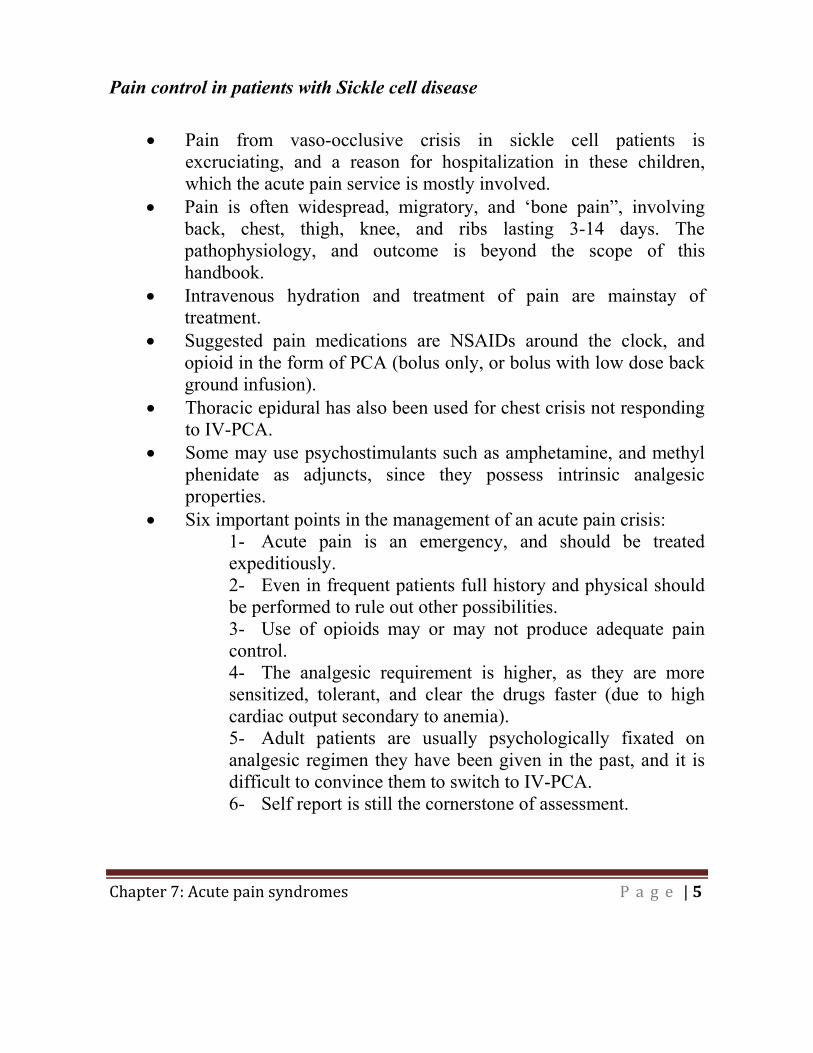
5
Pain control in patients with Sickle cell disease
Pain from vaso-occlusive crisis in sickle cell patients is excruciating, and a reason for hospitalization in these children, which the acute pain service is mostly involved.
Pain is often widespread, migratory, and �‘bone pain�”, involving back, chest, thigh, knee, and ribs lasting 3-14 days. The pathophysiology, and outcome is beyond the scope of this handbook.
Intravenous hydration and treatment of pain are mainstay of treatment.
Suggested pain medications are NSAIDs around the clock, and opioid in the form of PCA (bolus only, or bolus with low dose back ground infusion).
Thoracic epidural has also been used for chest crisis not responding to IV-PCA.
Some may use psychostimulants such as amphetamine, and methyl phenidate as adjuncts, since they possess intrinsic analgesic properties.
Six important points in the management of an acute pain crisis: 1- Acute pain is an emergency, and should be treated expeditiously. 2- Even in frequent patients full history and physical should be performed to rule out other possibilities. 3- Use of opioids may or may not produce adequate pain control. 4- The analgesic requirement is higher, as they are more sensitized, tolerant, and clear the drugs faster (due to high cardiac output secondary to anemia). 5- Adult patients are usually psychologically fixated on analgesic regimen they have been given in the past, and it is difficult to convince them to switch to IV-PCA. 6- Self report is still the cornerstone of assessment.
6
References:
Keith, M.P., Gilliland, W.R. Updates in the Management of Gout. The American Journal of Medicine, 2007; 120: 221-224
Teeg, G.G., Nair, R., Saag, K.G. Pathophysiology, Clinical Presentation and Treatment of Gout. Drugs 2006; 66 (12): 1547-1563.
Tingle LE, Molina D, Calvert CW. Acute paricarditis, Am Fam Physician 2007; 76(10):1509-1514.
Booker MJ, Blethyn KL, Wright CJ, Greenfield SM. Pain management in sickle cell disease. Chronic Illn. 2006; 2(1):39-50. Udezue E, Herrera E. Pain management in adult acute sickle cell pain crisis: a view point. West Afr J Med. 2007;26(3): 179-182. Jacob E, Mueller BU. Pain experience of children with sickle cell disease who had prolonged hospitalization for acute painful episodes. Pain Med, 2008;9(1):13-21.

Appendices

APPENDIX A: Acute pain service initial assessment & daily flow sheet
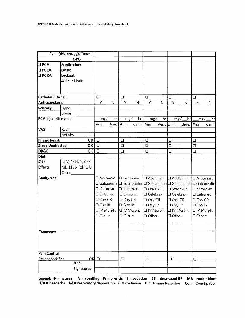
APPENDIX A: Acute pain service initial assessment & daily flow sheet
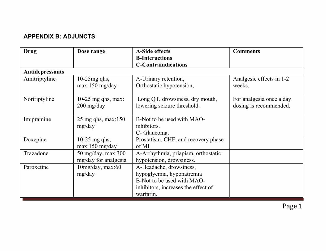
APPENDIX B: ADJUNCTS
Drug Dose range A-Side effects B-Interactions C-Contraindications
Comments
Antidepressants Amitriptyline 10-25mg qhs,
max:150 mg/day A-Urinary retention, Orthostatic hypotension,
Analgesic effects in 1-2 weeks.
Nortriptyline 10-25 mg qhs, max: 200 mg/day
Long QT, drowsiness, dry mouth, lowering seizure threshold.
For analgesia once a day dosing is recommended.
Imipramine 25 mg qhs, max:150 mg/day
B-Not to be used with MAO-inhibitors. C- Glaucoma,
Doxepine 10-25 mg qhs, max:150 mg/day
Prostatism, CHF, and recovery phase of MI
Trazadone 50 mg/day, max:300 mg/day for analgesia
A-Arrhythmia, priapism, orthostatic hypotension, drowsiness.
Paroxetine 10mg/day, max:60 mg/day
A-Headache, drowsiness, hypoglyemia, hyponatremia B-Not to be used with MAO-inhibitors, increases the effect of warfarin.
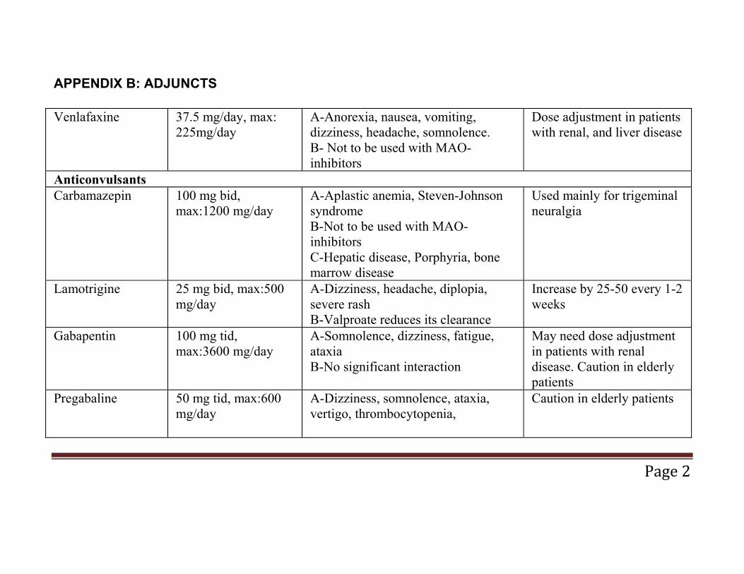
APPENDIX B: ADJUNCTS
Venlafaxine 37.5 mg/day, max: 225mg/day
A-Anorexia, nausea, vomiting, dizziness, headache, somnolence. B- Not to be used with MAO-inhibitors
Dose adjustment in patients with renal, and liver disease
Anticonvulsants Carbamazepin 100 mg bid,
max:1200 mg/day A-Aplastic anemia, Steven-Johnson syndrome B-Not to be used with MAO-inhibitors C-Hepatic disease, Porphyria, bone marrow disease
Used mainly for trigeminal neuralgia
Lamotrigine 25 mg bid, max:500 mg/day
A-Dizziness, headache, diplopia, severe rash B-Valproate reduces its clearance
Increase by 25-50 every 1-2 weeks
Gabapentin 100 mg tid, max:3600 mg/day
A-Somnolence, dizziness, fatigue, ataxia B-No significant interaction
May need dose adjustment in patients with renal disease. Caution in elderly patients
Pregabaline 50 mg tid, max:600 mg/day
A-Dizziness, somnolence, ataxia, vertigo, thrombocytopenia,
Caution in elderly patients
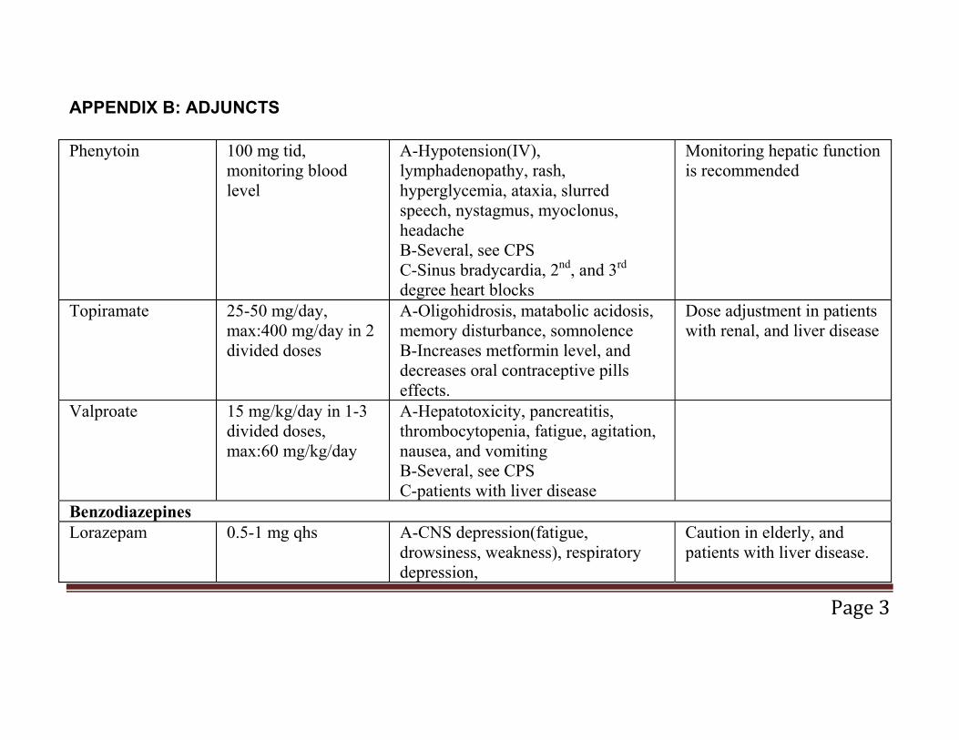
APPENDIX B: ADJUNCTS
Phenytoin 100 mg tid, monitoring blood level
A-Hypotension(IV), lymphadenopathy, rash, hyperglycemia, ataxia, slurred speech, nystagmus, myoclonus, headache B-Several, see CPS C-Sinus bradycardia, 2nd, and 3rd degree heart blocks
Monitoring hepatic function is recommended
Topiramate 25-50 mg/day, max:400 mg/day in 2 divided doses
A-Oligohidrosis, matabolic acidosis, memory disturbance, somnolence B-Increases metformin level, and decreases oral contraceptive pills effects.
Dose adjustment in patients with renal, and liver disease
Valproate 15 mg/kg/day in 1-3 divided doses, max:60 mg/kg/day
A-Hepatotoxicity, pancreatitis, thrombocytopenia, fatigue, agitation, nausea, and vomiting B-Several, see CPS C-patients with liver disease
Benzodiazepines Lorazepam 0.5-1 mg qhs A-CNS depression(fatigue,
drowsiness, weakness), respiratory depression,
Caution in elderly, and patients with liver disease.
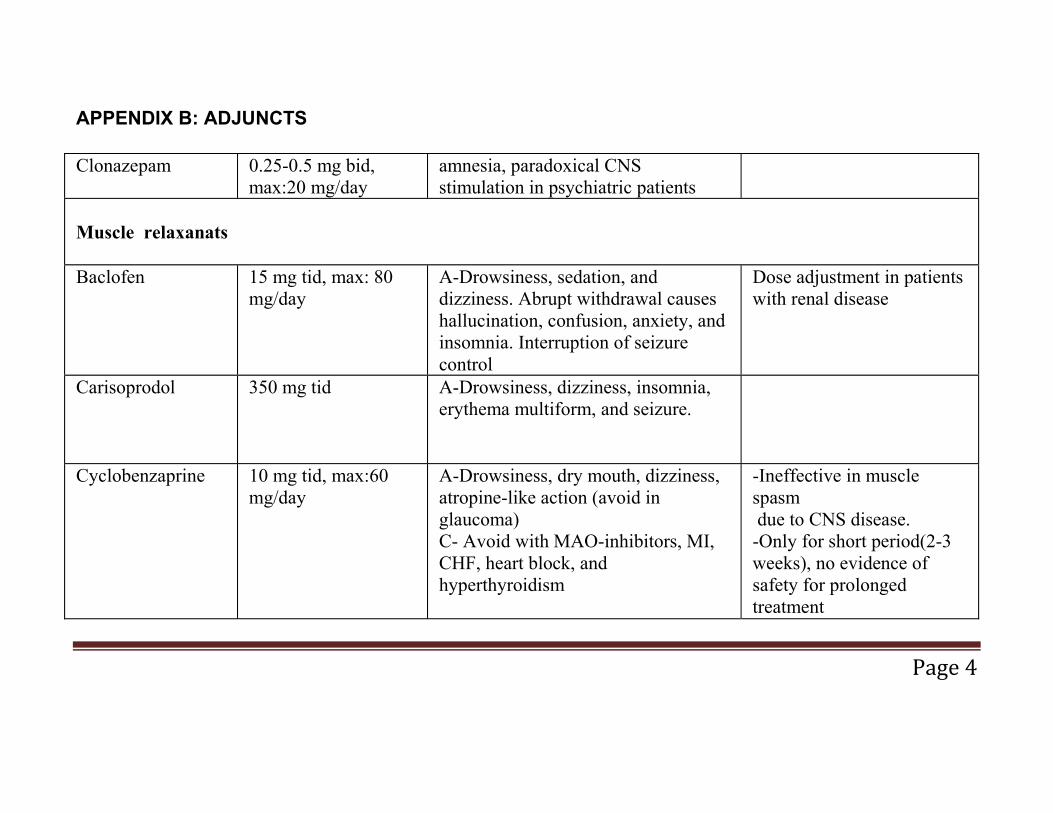
APPENDIX B: ADJUNCTS
Clonazepam 0.25-0.5 mg bid, max:20 mg/day
amnesia, paradoxical CNS stimulation in psychiatric patients
Muscle relaxanats Baclofen 15 mg tid, max: 80
mg/day A-Drowsiness, sedation, and dizziness. Abrupt withdrawal causes hallucination, confusion, anxiety, and insomnia. Interruption of seizure control
Dose adjustment in patients with renal disease
Carisoprodol 350 mg tid
A-Drowsiness, dizziness, insomnia, erythema multiform, and seizure.
Cyclobenzaprine 10 mg tid, max:60 mg/day
A-Drowsiness, dry mouth, dizziness, atropine-like action (avoid in glaucoma) C- Avoid with MAO-inhibitors, MI, CHF, heart block, and hyperthyroidism
-Ineffective in muscle spasm due to CNS disease. -Only for short period(2-3 weeks), no evidence of safety for prolonged treatment
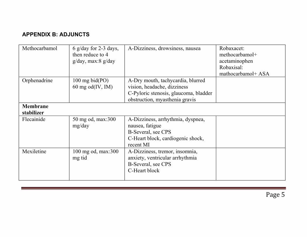
APPENDIX B: ADJUNCTS
Methocarbamol 6 g/day for 2-3 days, then reduce to 4 g/day, max:8 g/day
A-Dizziness, drowsiness, nausea Robaxacet: methocarbamol+ acetaminophen Robaxisal: mathocarbamol+ ASA
Orphenadrine 100 mg bid(PO) 60 mg od(IV, IM)
A-Dry mouth, tachycardia, blurred vision, headache, dizziness C-Pyloric stenosis, glaucoma, bladder obstruction, myasthenia gravis
Membrane stabilizer
Flecainide 50 mg od, max:300 mg/day
A-Dizziness, arrhythmia, dyspnea, nausea, fatigue B-Several, see CPS C-Heart block, cardiogenic shock, recent MI
Mexiletine 100 mg od, max:300 mg tid
A-Dizziness, tremor, insomnia, anxiety, ventricular arrhythmia B-Several, see CPS C-Heart block

APPENDIX B: ADJUNCTS
Miscellaneous
Ketamine A-Hypertension, tachycardia, dissociation, hallucination B- C-Hypertension, history of cerebrovascular accident
Intramuscular ketamine facilitates performance of pediatric procedures.
Clonidine 0.1 mg bid, max: 0.6 mg/day
A-Dry mouth, drowsiness, dizziness, hypotension, sedation C-Bradycardia, and heart block
Dextro amphetamine
5 mg/day, max: 60 mg/day
A-Hypertension, tachycardia, restlessness, B-May alter insulin requirement in diabetic patients C-Cardiovascular disease, agitation, hypertension, glaucoma, hyperthyroidism
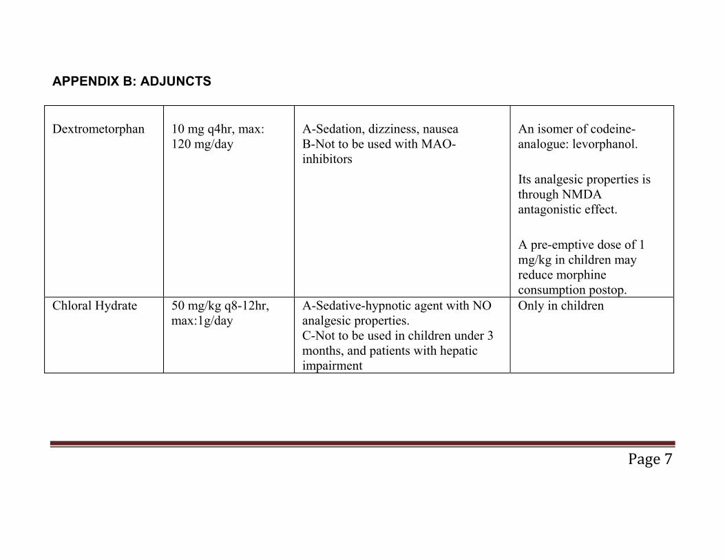
APPENDIX B: ADJUNCTS
Dextrometorphan
10 mg q4hr, max: 120 mg/day
A-Sedation, dizziness, nausea B-Not to be used with MAO-inhibitors
An isomer of codeine-analogue: levorphanol.
Its analgesic properties is through NMDA antagonistic effect.
A pre-emptive dose of 1 mg/kg in children may reduce morphine consumption postop.
Chloral Hydrate 50 mg/kg q8-12hr, max:1g/day
A-Sedative-hypnotic agent with NO analgesic properties. C-Not to be used in children under 3 months, and patients with hepatic impairment
Only in children

APPENDIX C: Postoperative Nausea and Vomiting (PONV) Postoperative nausea and vomiting nausea, vomiting (emesis), and/or wretching. PONV may take place as a single episode or multiple episodes, either immediately after surgery or up to 48 hours after surgery.
Risk factors include:
Patient related factors o Female (from puberty on) o Non-smoker o History of PONV or motion sickness o History of migraine o Low ASA score
Anesthesia and Surgery related factors
o Long duration of surgery o Type of surgery (Adult): intraabdominal, laparoscopic,
orthopedic, major gynecological, ENT, thyroid, breast, plastic surgery, neurosurgery
o Type of surgery (Children): hernia repair, adenotonsillectomy, strabismus, penile surgery.
o Anesthesia related factors: Use of volatile anesthetics o Use of nitrous oxide o Balanced inhaled versus total IV anesthesia o Large dose neostigmine (> 2.5mg) o Use of intra-operative and post-operative opioids
Risk factors for children are similar to those in adults with the following differences:
o Vomiting incidence is twice as frequent in children than in adults
o Risk increases as children age, decreasing after puberty o Gender differences are not seen before puberty

Techniques to reduce PONV
o Use of regional anesthesia o Use of propofol for induction and maintenance of
anesthesia o Use of intraoperative supplemental oxygen o adequate hydration o Avoidance of nitrous oxide o Avoidance of volatile anesthetics o Minimize of intraoperative and postoperative opioids o Minimize of neostigmine
Prophylaxis of PONV
Serotonin Receptor Antagonists (5-HT3) including ondansetron, granisetron and tropisetron. Administer at the end of surgery
Dexamethasone 8 �– 10mg IV in adults (150 mcg/kg �– 8mg in children) administered prior to induction is more effective than at the end of surgery.
Other antiemetics including dimenhydrinate, prochlorperazine, metoclopramide, and haloperidol, administered at the end of surgery.
Transdermal scopolamine applied the night prior to surgery or 4 hours prior to surgery.
Treatment of PONV
If a patient has not received prophylaxis or has only received dexamethasone as prophylaxis: small-dose 5-HT3 receptor antagonists should be initiated, treatment doses are about a quarter
of those used for prophylaxis (ondansetron 1.0 �– 4 mg , granisetron 1 mg, and tropisetron 0.5 mg)
If patient has received prophylaxis with 5-HT3 and it is less than 6 hours after surgery: Treat with other antiemetic including: dimenhydrinate, prochlorperazine, metoclopramide, and haloperidol (not scopolamine)
If patient has received prophylaxis with 5-HT3 and it is more than 6 hours after surgery: Treat with small dose of 5-HT3 receptor antagonists and other anti-emetics.
Use combination therapy or anti-emetics with different mechanisms of action together to manage moderate to severe PONV
References:
e-CPS. https://www.e-therapeutics.ca. Accessed August 4, 2009.
Gan, T.J. Risk factors for post-operative nausea and vomiting. Anesth Analg 2006; 102: 1884-1898.
Gan, T.J., Meyer, T., Apfel, C.C., Chung, F., Davis, P.J., Eubanks, S., Kovac, A., Philip, B.K., Sessler, D.I., Temo, J., Trame`r, M.R., Watcha, M. Consensus Guidelines for Managing Postoperative Nausea and Vomiting. Anesth Analg 2003;97: 62�–71
Habib, A.S., Gan, T.J. Evidence-based management of postoperative nausea and vomiting: a review Can J Anesth 2004; 51(4): 325-341.

Anti-emetic Table:
Classification Anti-emetic and Dose
Adverse effects Contraindications
Serotonin Receptor Antagonists (5-HT3)
Ondansetron 1- 4mg, granisetron 1mg, tropisetron 2mg.
Headache, elevation of AST/ALT, constipation
There is no experience in children
Corticosteroid Dexamethasone 4 �– 8 mg for prophylaxis at induction of anesthesia
None after single bolus dose
Single dose only
Anti-emetic dimenhydrinate 25-50mg q 4h prn (maximum of 400 mg in 24 hours)
Drowsiness, dizzy, dry mouth
Not recommended in patients under 1 yr of age.
Phenothiazine prochlorperazine 5-10 mg po/IV tid prn (maximum 40 mg) Children: 9-14 kg 2.5 mg po bid prn, 0.13mg/kg IV (max 7.5 mg); > 14�–18 kg 2.5 mg po/pr tid prn, 0.13mg/kg IV (max 10mg); > 18�–39 kg2.5
Drowsiness, dizziness, and headache are common. Neuroleptic malignant syndrome, seizures, confusion, insomnia. Extrapyramidal symptoms (akathisia, dystonia, pseudoparkinsonism, tardive
Coma and/or severe CNS depression, particularly when due to intoxication with CNS depressants
Do not use with Children under the age of 2 years
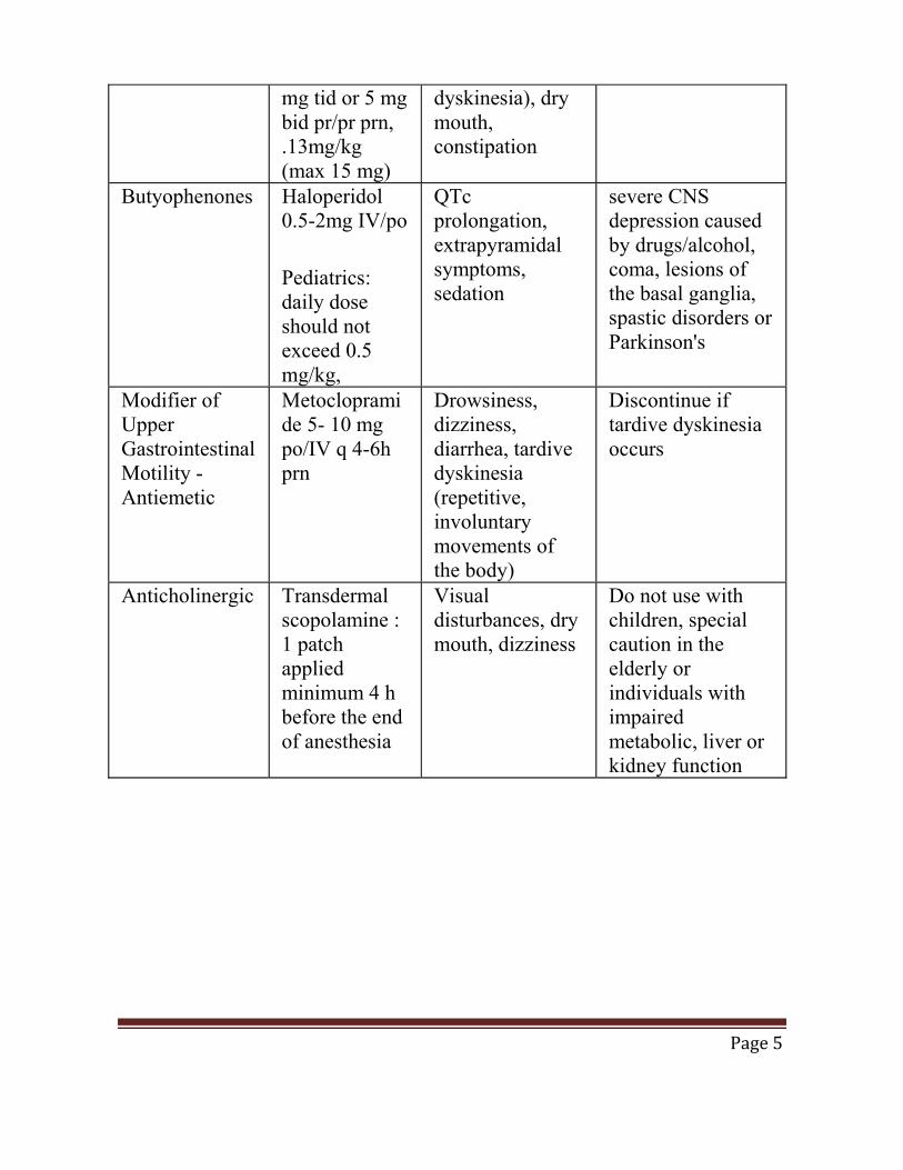
mg tid or 5 mg bid pr/pr prn, .13mg/kg (max 15 mg)
dyskinesia), dry mouth, constipation
Butyophenones Haloperidol 0.5-2mg IV/po
Pediatrics: daily dose should not exceed 0.5 mg/kg,
QTc prolongation, extrapyramidal symptoms, sedation
severe CNS depression caused by drugs/alcohol, coma, lesions of the basal ganglia, spastic disorders or Parkinson's
Modifier of Upper Gastrointestinal Motility -Antiemetic
Metoclopramide 5- 10 mg po/IV q 4-6h prn
Drowsiness, dizziness, diarrhea, tardive dyskinesia (repetitive, involuntary movements of the body)
Discontinue if tardive dyskinesia occurs
Anticholinergic
Transdermal scopolamine : 1 patch applied minimum 4 h before the end of anesthesia
Visual disturbances, dry mouth, dizziness
Do not use with children, special caution in the elderly or individuals with impaired metabolic, liver or kidney function
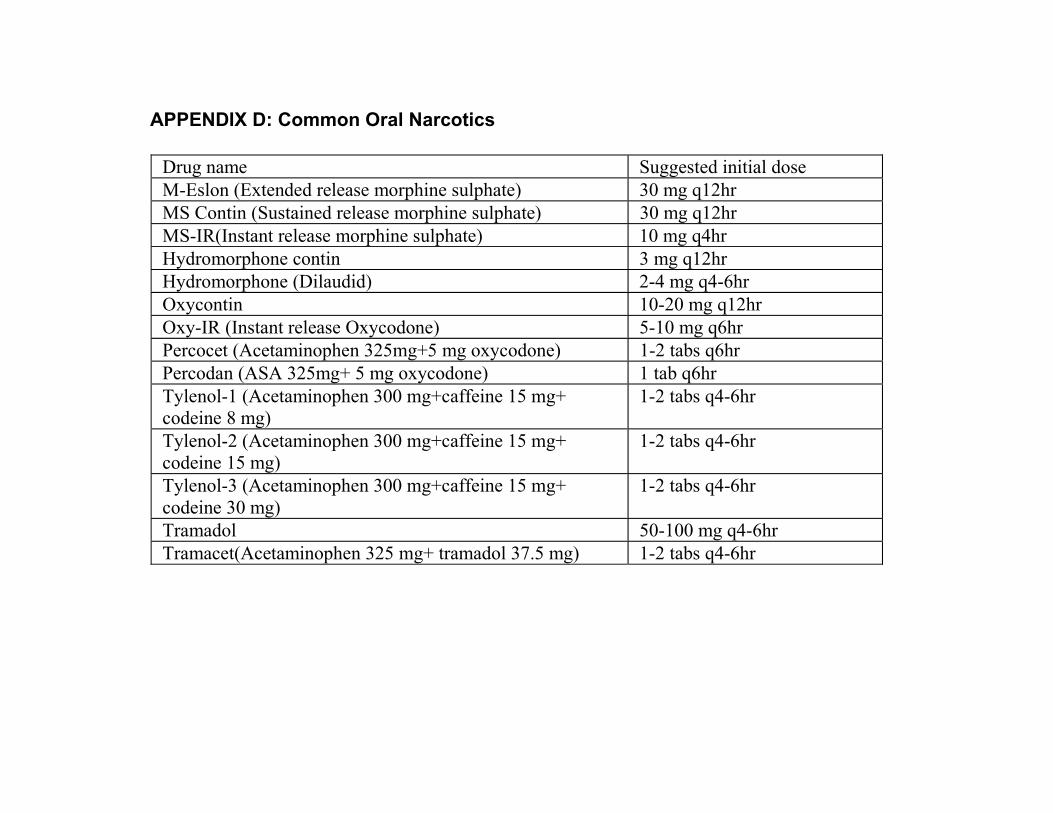
APPENDIX D: Common Oral Narcotics
Drug name Suggested initial dose M-Eslon (Extended release morphine sulphate) 30 mg q12hr MS Contin (Sustained release morphine sulphate) 30 mg q12hr MS-IR(Instant release morphine sulphate) 10 mg q4hr Hydromorphone contin 3 mg q12hr Hydromorphone (Dilaudid) 2-4 mg q4-6hr Oxycontin 10-20 mg q12hr Oxy-IR (Instant release Oxycodone) 5-10 mg q6hr Percocet (Acetaminophen 325mg+5 mg oxycodone) 1-2 tabs q6hr Percodan (ASA 325mg+ 5 mg oxycodone) 1 tab q6hr Tylenol-1 (Acetaminophen 300 mg+caffeine 15 mg+ codeine 8 mg)
1-2 tabs q4-6hr
Tylenol-2 (Acetaminophen 300 mg+caffeine 15 mg+ codeine 15 mg)
1-2 tabs q4-6hr
Tylenol-3 (Acetaminophen 300 mg+caffeine 15 mg+ codeine 30 mg)
1-2 tabs q4-6hr
Tramadol 50-100 mg q4-6hr Tramacet(Acetaminophen 325 mg+ tramadol 37.5 mg) 1-2 tabs q4-6hr
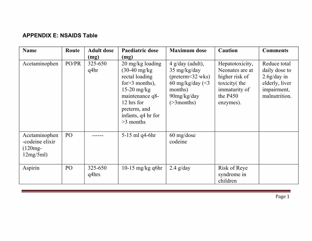
APPENDIX E: NSAIDS Table
Name Route Adult dose (mg)
Paediatric dose (mg)
Maximum dose Caution Comments
Acetaminophen PO/PR 325-650 q4hr
20 mg/kg loading (30-40 mg/kg rectal loading for>3 months), 15-20 mg/kg maintenance q8-12 hrs for preterm, and infants, q4 hr for >3 months
4 g/day (adult), 35 mg/kg/day (preterm<32 wks) 60 mg/kg/day (<3 months) 90mg/kg/day (>3months)
Hepatotoxicity, Neonates are at higher risk of toxicity( the immaturity of the P450 enzymes).
Reduce total daily dose to 2.6g/day in elderly, liver impairment, malnutrition.
Acetaminophen-codeine elixir (120mg-12mg/5ml)
PO ------ 5-15 ml q4-6hr 60 mg/dose codeine
Aspirin PO 325-650 q4hrs
10-15 mg/kg q6hr 2.4 g/day Risk of Reye syndrome in children
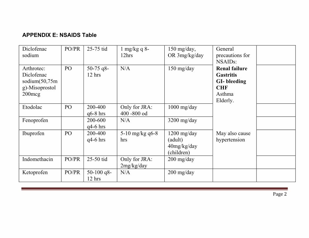
APPENDIX E: NSAIDS Table
Diclofenac sodium
PO/PR 25-75 tid 1 mg/kg q 8-12hrs
150 mg/day, OR 3mg/kg/day
General precautions for NSAIDs:
Arthrotec: Diclofenac sodium(50,75mg)-Misoprostol 200mcg
PO 50-75 q8-12 hrs
N/A 150 mg/day Renal failure Gastritis GI- bleeding CHF Asthma Elderly.
Etodolac PO 200-400 q6-8 hrs
Only for JRA: 400 -800 od
1000 mg/day
Fenoprofen 200-600 q4-6 hrs
N/A 3200 mg/day
Ibuprofen PO 200-400 q4-6 hrs
5-10 mg/kg q6-8 hrs
1200 mg/day (adult) 40mg/kg/day (children)
May also cause hypertension
Indomethacin PO/PR 25-50 tid Only for JRA: 200 mg/day 2mg/kg/day Ketoprofen PO/PR 50-100 q8-
12 hrs N/A 200 mg/day
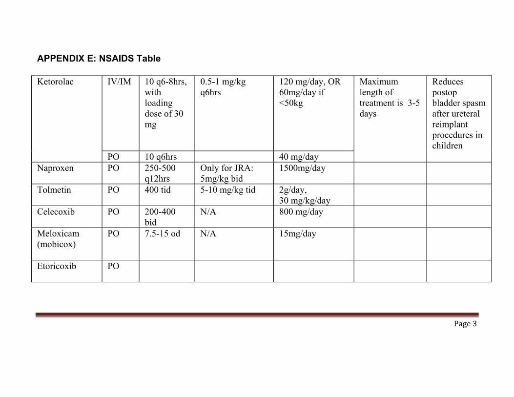
APPENDIX E: NSAIDS Table
Ketorolac IV/IM 10 q6-8hrs, with loading dose of 30 mg
0.5-1 mg/kg q6hrs
120 mg/day, OR 60mg/day if <50kg
Maximum length of treatment is 3-5 days
Reduces postop bladder spasm after ureteral reimplant procedures in children
PO 10 q6hrs 40 mg/day Naproxen PO 250-500
q12hrs Only for JRA: 5mg/kg bid
1500mg/day
Tolmetin PO 400 tid 5-10 mg/kg tid 2g/day, 30 mg/kg/day
Celecoxib PO 200-400 bid
N/A 800 mg/day
Meloxicam (mobicox)
PO 7.5-15 od N/A 15mg/day
Etoricoxib
PO
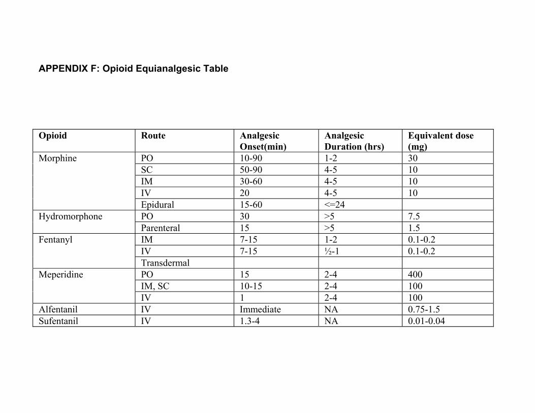
APPENDIX F: Opioid Equianalgesic Table
Opioid Route Analgesic Onset(min)
Analgesic Duration (hrs)
Equivalent dose (mg)
Morphine PO 10-90 1-2 30 SC 50-90 4-5 10 IM 30-60 4-5 10 IV 20 4-5 10 Epidural 15-60 <=24
Hydromorphone PO 30 >5 7.5 Parenteral 15 >5 1.5
Fentanyl IM 7-15 1-2 0.1-0.2 IV 7-15 ½-1 0.1-0.2 Transdermal
Meperidine PO 15 2-4 400 IM, SC 10-15 2-4 100 IV 1 2-4 100
Alfentanil IV Immediate NA 0.75-1.5 Sufentanil IV 1.3-4 NA 0.01-0.04
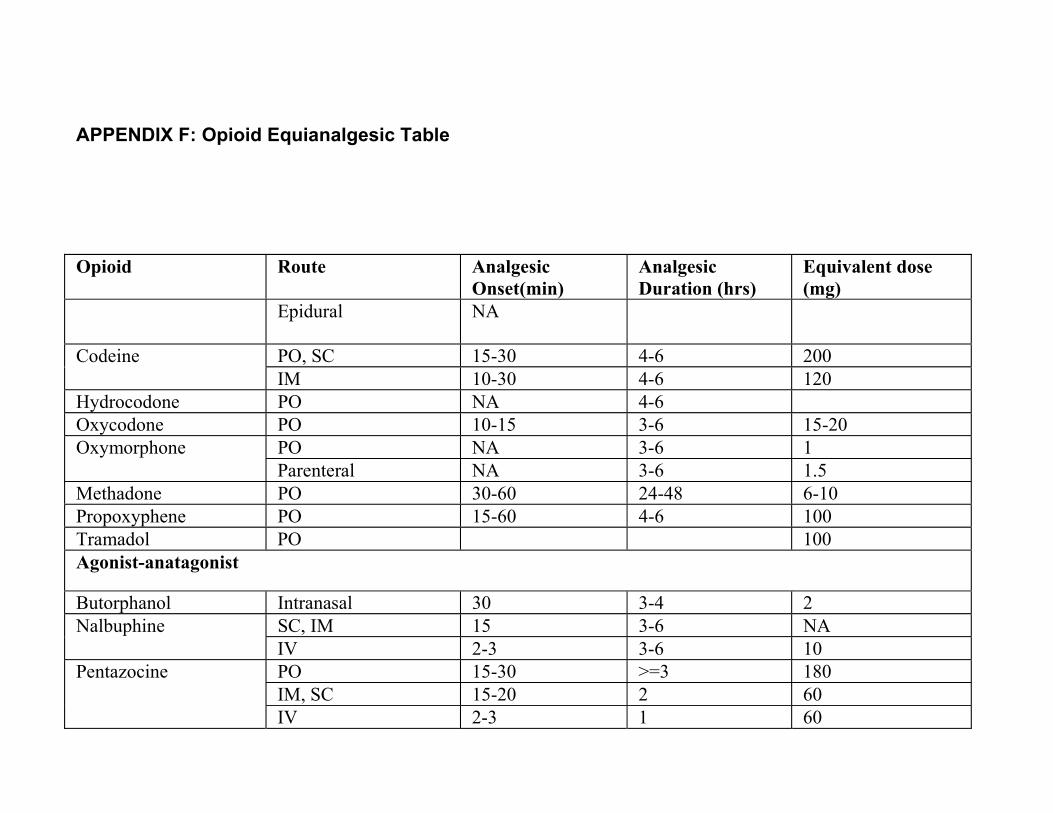
APPENDIX F: Opioid Equianalgesic Table
Opioid Route Analgesic Onset(min)
Analgesic Duration (hrs)
Equivalent dose (mg)
Epidural NA
Codeine PO, SC 15-30 4-6 200 IM 10-30 4-6 120
Hydrocodone PO NA 4-6 Oxycodone PO 10-15 3-6 15-20 Oxymorphone PO NA 3-6 1
Parenteral NA 3-6 1.5 Methadone PO 30-60 24-48 6-10 Propoxyphene PO 15-60 4-6 100 Tramadol PO 100 Agonist-anatagonist
Butorphanol Intranasal 30 3-4 2 Nalbuphine SC, IM 15 3-6 NA
IV 2-3 3-6 10 Pentazocine PO 15-30 >=3 180
IM, SC 15-20 2 60 IV 2-3 1 60
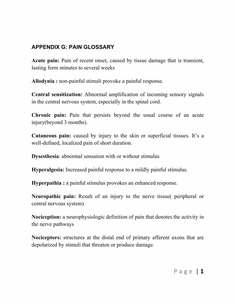
APPENDIX G: PAIN GLOSSARY
P a g e | 1
Acute pain: Pain of recent onset, caused by tissue damage that is transient, lasting form minutes to several weeks
Allodynia : non-painful stimuli provoke a painful response.
Central sensitization: Abnormal amplification of incoming sensory signals in the central nervous system, especially in the spinal cord.
Chronic pain: Pain that persists beyond the usual course of an acute injury(beyond 3 months).
Cutaneous pain: caused by injury to the skin or superficial tissues. It�’s a well-defined, localized pain of short duration.
Dysesthesia: abnormal sensation with or without stimulus
Hyperalgesia: Increased painful response to a mildly painful stimulus.
Hyperpathia : a painful stimulus provokes an enhanced response.
Neuropathic pain: Result of an injury to the nerve tissue( peripheral or central nervous system).
Nociception: a neurophysiologic definition of pain that denotes the activity in the nerve pathways
Nociceptors: structures at the distal end of primary afferent axons that are depolarized by stimuli that threaten or produce damage.
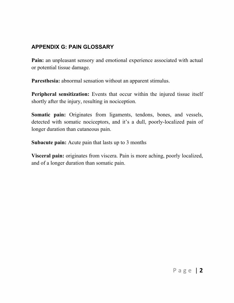
APPENDIX G: PAIN GLOSSARY
P a g e | 2
Pain: an unpleasant sensory and emotional experience associated with actual or potential tissue damage.
Paresthesia: abnormal sensation without an apparent stimulus.
Peripheral sensitization: Events that occur within the injured tissue itself shortly after the injury, resulting in nociception.
Somatic pain: Originates from ligaments, tendons, bones, and vessels, detected with somatic nociceptors, and it�’s a dull, poorly-localized pain of longer duration than cutaneous pain.
Subacute pain: Acute pain that lasts up to 3 months
Visceral pain: originates from viscera. Pain is more aching, poorly localized, and of a longer duration than somatic pain.