Haematogenous abdominal wall metastasis of differentiated, alpha

ACUTE HAEMATOGENOUS OSTEOMYELITIS
Diagnosis and Prognosis
Prof. Jacques du ToitSAPOS Instructional Course LecturesMuskuloskeletal InfectionsInkosi Albert Luthuli Hospital
Durban12-13 May 2017
Acute HaematogenousOsteomyelitis
What are we discussing?

OsteomyelitisDefinitions
• Osteomyelitis: Inflammation of bone caused by pyogenic organisms
Scheme that applies irrespective of the underlying source of the offending bacterium
• Acute Osteomyelitis: Infection diagnosed ≤ 2/52 after the onset of symptoms• Subacute Osteomyelitis: Infection diagnosed > 2/52 after the onset of
symptoms• Chronic Osteomyelitis: Infection diagnosed months after the onset of
symptoms with hallmark findings of dead and reactive bone
NO SCIENTIFIC RATIONALE FOR TIME FRAMES? Virulence properties of infective strain?
Nada S, Smeltzer M. Management of acute haematogenous osteomyelitis in childrenExpert Rev Anti Infect Ther. 2010 February; 8(2): 175-‐181

OsteomyelitisDefinitions
Scheme that applies to the underlying source of the offending bacterium
• Acute Haematogenous Osteomyelitis: Infection diagnosed ≤ 2/52 after the onset of symptoms arising from hematogenous seeding from the endostealblood supply
• Acute Osteomyelitis secondary to a contiguous focus of infection and/or vascular insufficiency
Nada S, Smeltzer M. Management of acute haematogenous osteomyelitis in childrenExpert Rev Anti Infect Ther. 2010 February; 8(2): 175-‐181

Acute HaematogenousOsteomyelitis
Diagnosis

Acute Haematogenous OsteomyelitisDiagnoses
• No definitive guidelines for diagnosis
• Recommendations in the literature based on expert opinions, case series and cohort studies.
• Difficult to standardize diagnosis
• Avoid “Cookbook” approach
Dartnell J, Ramachandran M, Katchburian M. Haematogenous acute and subacute paediatric osteomyelitis: a systematic review of the literature. J Bone Joint Surg Br 2012 May;94(5):584-‐595.

Acute Haematogenous OsteomyelitisDiagnoses
Standard Radiography
Further ImagingLaboratory Tests
Clinical Manifestations

Acute Haematogenous OsteomyelitisDiagnosis-Clinical Manifestations
Skeletal Distribution
Red FlagsRisk
Factors
Presenting Features
Nada S, Smeltzer M. Management of acute haematogenous osteomyelitis in childrenExpert Rev Anti Infect Ther. 2010 February; 8(2): 175-‐181

Acute Haematogenous OsteomyelitisPresenting Features
Most Common Presenting Features
Symptom Incidence (%)
Pain 81.1
Localized signs/symptoms
70.0
Fever 61.7
Reduced ROM 50.3
Reduced Weight-‐bearing
49.3
• Pain: Constant and Progressive
• Localized: Older > Young Children
: > Metaphysis
• Fever: Typically abrupt and > 38 oC
• Moderate reduction in ROM
• Pseudo-paralysis or Reduced Weight-bearing
• Redness, Warmth, Swelling once progression through metaphyseal cortex into subperiosteal space
• Less Common: Anorexia, Irritability, Lethargy
Dartnell J, Ramachandran M, Katchburian M. Haematogenous acute and subacute paediatric osteomyelitis: a systematic review of the
literature. J Bone Joint Surg Br 2012 May;94(5):584-‐595.
Herring J, Infections of the Musculoskeletal System, Tachdjians’sPediatric Orthopaedics. 2014
Skeletal Distribution
Red FlagsRisk
Factors
Presenting Features

Acute Haematogenous OsteomyelitisSkeletal Distribution
Pelvis
• Average of 12 days before diagnosis
• Frequently masked by A/B
• Infective DDx in and around the hip in children:
Dartnell J, Ramachandran M, Katchburian M. Haematogenous acute and subacute paediatric osteomyelitis: a systematic
review of the literature. J Bone Joint Surg Br 2012 May;94(5):584-‐595.
Skeletal Distribution
Red FlagsRisk
Factors
Presenting Features
Iliac Crest Osteitis
Psoas Abcess
Septic Arthritis

Acute Haematogenous OsteomyelitisRisk Factors
Dartnell J, Ramachandran M, Katchburian M. Haematogenous acute and subacute paediatric osteomyelitis: a systematic review of the
literature. J Bone Joint Surg Br 2012 May;94(5):584-‐595.
Skeletal Distribution
Red FlagsRisk
Factors
Presenting Features
? Who are more at RISK ?
Unknown
Developing Countries
Children < 3 Years
Blunt Trauma
Recent Systemic Infection
Sickle Cell Disease

Acute Haematogenous OsteomyelitisRed Flags
Skeletal Distribution
Red FlagsRisk
Factors
Presenting Features
Presumed Trauma or Injury
Emperic Antibiotics
Young Children
Pelvic Osteomyelitis
Concurrent Septic Arthritis
Acute Lymphoblastic Leukemia
Multiple Sites

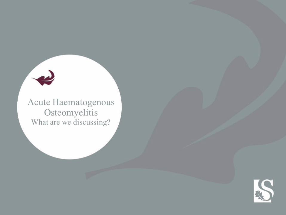
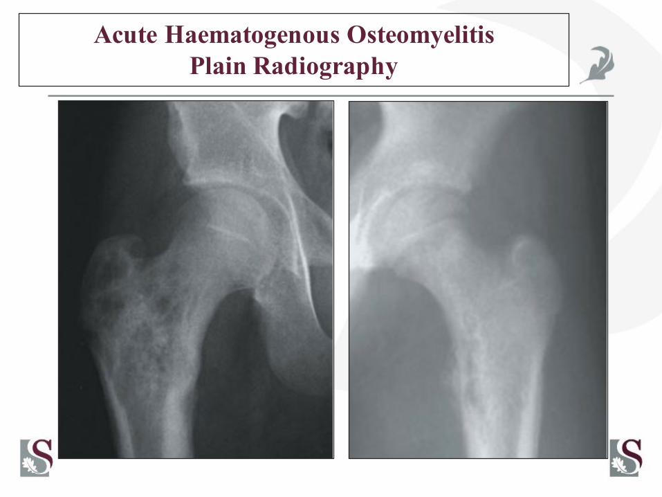
Acute Haematogenous OsteomyelitisPlain Radiography
• Obvious changes (Osteopenia & Osteolytic Lesions) may not occur until 10-14days after onset of Symptoms
• In isolation poor diagnostic sensitivity during first 10 days
• Essential to exclude other pathology

Acute Haematogenous OsteomyelitisPlain Radiography
Soft Tissue Oedema & Loss Facial Planes
Adherent Joint Effusions
Osteopenia
Osteolytic Lesions
Periosteal Elevation
Cortical disruption
Greenspan A, Orthopedic Imaging-‐A Practical Approach,5th Edition
Acute Haematogenous OsteomyelitisPlain Radiography

Acute Haematogenous OsteomyelitisPlain Radiography
Osteomyelitis Ewings Sarcoma

Acute Haematogenous OsteomyelitisLaboratory Tests
ESR
CultureCRP
FBC & Diff.
Nada S, Smeltzer M. Management of acute haematogenous osteomyelitis in childrenExpert Rev Anti Infect Ther. 2010 February; 8(2): 175-‐181

Acute Haematogenous OsteomyelitisLaboratory Tests
ESR
CultureCRP
FBC & Diff.
Complete Blood Count with Differential
• WCC only elevated in 25-35% of children with AHO
• WCC > 12000 cells/ml
• Allows for assessment of all three marrow cell lines
• Useful to assess for unusual/particularly virulent organism OR Concurrent Septic Arthritis
Herring J, Infections of the Musculoskeletal System, Tachdjians’s PediatricOrthopaedics. 2014

Acute Haematogenous OsteomyelitisLaboratory Tests
Erythrocyte Sedimentation Rate
• Significant correlation between an ESR > 55ml/h and the presence of an abscess in pelvic osteomyelitis
• No periosteal abcesses or pyomyositiswith ESR < 22ml/hr
Erythrocyte Sedimentation Rate
• Rate at which red blood cells fall through plasma as measured in ml
• 91% Abnormal on presentation
• Peak 3-5 days
• Normalise in 3-4 weeks with effective treatment
• Non-Spesific diagnostic indicator
ESR
CultureCRP
FBC & Diff.
Dartnell J, Ramachandran M, Katchburian M. Haematogenous acute and subacute paediatric osteomyelitis: a systematic review of the literature. J Bone Joint Surg Br 2012 May;94(5):584-‐595.

Acute Haematogenous OsteomyelitisLaboratory Tests
C-Reactive Protein
• CRP < 5 mg/dl effectively rules out serious bacterial infection
• CRP > 30 mg/l significantly associated with subperiosteal abcess formation and pyomyositis
• CRP > 1,5 admission levels on day 3 6.5 times more likely to have concurrent septic arthritis
• Significantly influenced by MRSA
C-Reactive Protein
• Acute-Phase reactant
• 80.5% abnormal on presentation but 100% abnormal with concurrent septic arthritis
• Increase 1000 fold within 6 hours
• Peak 36-50 hrs
• Normalise within 1week with effective treatment
• Most sensitive and reliable laboratory test for detecting acute inflammatory reactions
ESR
CultureCRP
FBC & Diff
Herring J, Infections of the Musculoskeletal System, Tachdjians’s PediatricOrthopaedics. 2014

Acute Haematogenous OsteomyelitisLaboratory Tests
Local Tissue and Blood Cultures
Isolation of the causative organism remains the diagnostic gold standard
and
It is currently the only way to establish a definitive microbiologic diagnosis
BLOOD CULTURE & NEEDLE ASPIRATION
Management of acute haematogenous osteomyelitis in childrenExpert Rev Anti Infect Ther. 2010 February; 8(2): 175-‐181
ESR
CultureCRP
FBC & Diff.

Acute Haematogenous OsteomyelitisLaboratory Tests
Needle Aspiration
• A relatively non invasive procedures in neonates and young children
Ø Aspirate subperiosteal collection
Ø Metaphyseal bone aspiration
• Older children and adolescents often require more invasive surgical techniques such as drilling or cutting into the bone
• Direct inoculation of cultured material into a blood culture bottle increases the probability of recovering a fastidious organism such as K. kingae.
• Using PCR to identify pathogens from bone specimens is also increasingly common
Krogstad P. Osteomyelitis. In: Feigin RD, Cherry JD, Demmler-‐Harrison GD, Kaplan SL, editors. Textbook of Pediatric Infectious Diseases. 6th Edition. PA, USA: Saunders Elsevier; 2009. pp.
725–742.
Blood Culture
• Should always be performed pre administration of Antibiotics
• Organism is recovered in approximately 50% of all AHO infections
ESR
CultureCRP
FBC & Diff.

Acute Haematogenous OsteomyelitisDiagnosis- Further Imaging
MRI
Bone Scan
Ultrasound
Nada S, Smeltzer M. Management of acute haematogenous osteomyelitis in childrenExpert Rev Anti Infect Ther. 2010 February; 8(2): 175-‐181

Acute Haematogenous OsteomyelitisFurther Imaging
Herring J, Infections of the Musculoskeletal System, Tachdjians’s Pediatric Orthopaedics. 2014
Unsure Diagnosis
Complex Anatomical Location
Satellite Septic Foci
Treatment Guideline
MRI
BoneScan
Ultrasound

Acute Haematogenous OsteomyelitisFurther Imaging
Ultrasound
• ? Second step in diagnostic pathway ?
• Indications:
Ø Diagnosis if X-rays normal
Ø Abcess Formation
Ø ? Concurrent Septic Arthritis
Ø Ultrasound guided aspiration for culture
MRI
BoneScan
Ultrasound
Soft Tissue Swelling
Periosteal Thickening
SubperiostealFluid
Collection
Cortical Breach

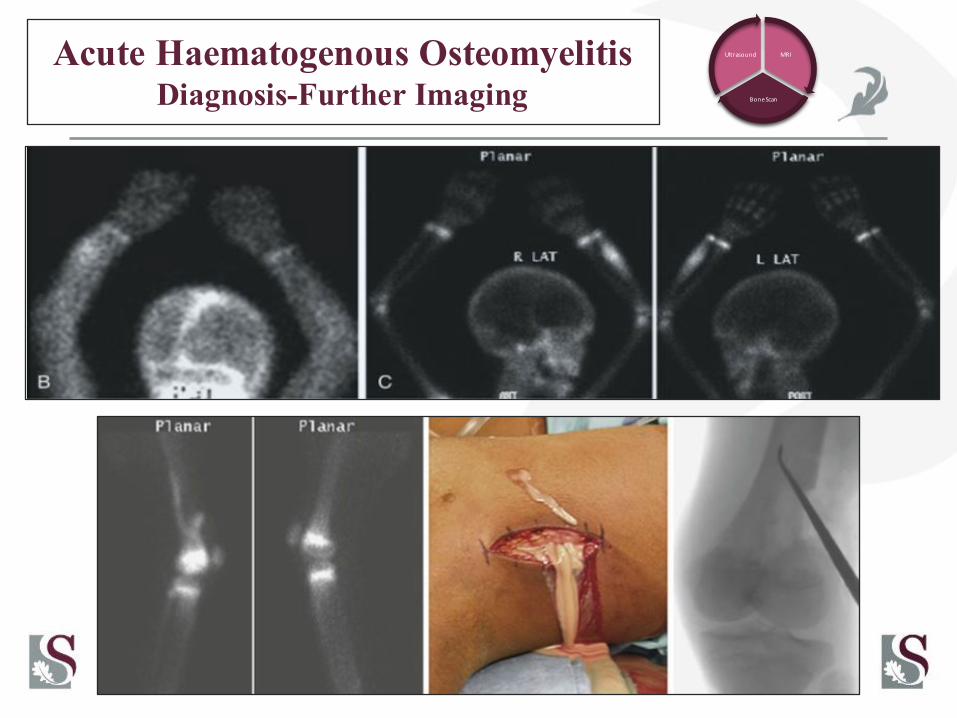
Acute Haematogenous OsteomyelitisDiagnosis-Further Imaging
Herring J, Infections of the Musculoskeletal System, Tachdjians’s Pediatric Orthopaedics. 2014
MRI
BoneScan
Ultrasound
Bone Scintigraphy
• Technetium Methylene diphosphonate
• Three Phase bone scan:
Ø Blood Flow phase
Ø Blood Pool or Soft Tissue phase
Ø Delayed or Skeletal phase
Diagnostic for Osteomyelitis
• Focally increased uptake in all three phases of the study
• “Cold” or Photopenic in the delayed images
Bone Scintigraphy
• Most common indications:
Ø Unable to localize site of infection
Ø Multifocal involvement in Neonates
• Sensitivity: 54-100 %
• Specificity: 70-90 %
• Overall Accuracy: 90%
Acute Haematogenous OsteomyelitisDiagnosis-Further Imaging
Management of acute haematogenous osteomyelitis in childrenExpert Rev Anti Infect Ther. 2010 February; 8(2): 175-‐181
MRI
BoneScan
Ultrasound

Acute Haematogenous OsteomyelitisDiagnosis-Further Imaging
Herring J, Infections of the Musculoskeletal System, Tachdjians’s Pediatric Orthopaedics. 2014
MRI
BoneScan
Ultrasound
Magnetic Resonance Imaging
• Most power technique available
• Define definitive diagnosis
• Guide treatment decisions
• Disadvantages:
Ø Cost
Ø Lack of immediate availability
Ø Sedation required in children
Magnetic Resonance Imaging
• Sensitivity: 80-100 %
• Spesificity : 70 -100 %

Acute Haematogenous OsteomyelitisDiagnosis-Further Imaging
MRI
BoneScan
Ultrasound
Decreased marrow signal intensity on T1-‐weighted images
Increased marrow signal intensity on T2-‐weighted images
Short-‐tau inversion recover images(STIR)
Marrow signal enhancement on post gadoliniumT2-‐weighted images
Herring J, Infections of the Musculoskeletal System, Tachdjians’s Pediatric Orthopaedics. 2014

Acute Haematogenous OsteomyelitisDiagnosis-What does the future hold?
Herring J, Infections of the Musculoskeletal System, Tachdjians’s Pediatric Orthopaedics. 2014
MRI
BoneScan
Ultrasound
Laboratory Investigations
• Polymerase Chain Reaction
Ø More Sensitive
Ø Quicker Diagnosis
• Serum Procalcitonin
Ø Differentiate between viral, bacterial and inflammatory process
Ø More effective than CRP. ESR and WCC to differentiate between Septic Arthritis and Osteomyelitis
Imaging
• Positron emission tomography with computed tomography (PET-CT)
Ø Superior to MRI
Ø Limited availability
Ø Radiation

Acute Haematogenous OsteomyelitisDiagnostic Flow Diagram

Acute Haematogenous OsteomyelitisDiagnostic Flow Diagram
Dartnell J, Ramachandran M, Katchburian M. Haematogenous acute and subacute paediatric osteomyelitis: a systematic review of the literature. J Bone Joint Surg Br 2012 May;94(5):584-‐595.
Acute HaematogenousOsteomyelitis:Prognosis

Acute Haematogenous OsteomyelitisPrognosis
• Pre Antibiotics ERA
Ø Serious disease with high morbidity and mortality
• Antibiotic Sensitive ERA
Ø Improved diagnostics and treatment modalities
Ø Morbidity significantly decreased
Ø Mortality negligible in the developed world
• Post Antibiotics ERA
Ø MRSA as a primary pathogen.
Ø Current approach probably not applicable and AHO should probably be treated more like chronic osteomyelitis
Gutierrez K. Bone and joint infections in children. Pediatr. Clin. N. Am 2005;52(3):779–794
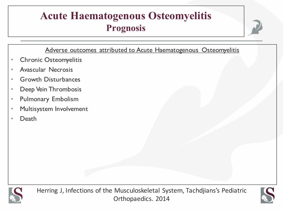
Acute Haematogenous OsteomyelitisPrognosis
Adverse outcomes attributed to Acute Haematogenous Osteomyelitis
• Chronic Osteomyelitis
• Avascular Necrosis
• Growth Disturbances
• Deep Vein Thrombosis
• Pulmonary Embolism
• Multisystem Involvement
• Death
Herring J, Infections of the Musculoskeletal System, Tachdjians’s PediatricOrthopaedics. 2014

Acute Haematogenous OsteomyelitisPrognosis
Predictors of Poor Prognosis
• Younger age: Delays in diagnosis, presentation and treatment
• Location: Hip is at the highest risk of complications (40%), ankle (33%) and knee (10%)
• Concurrent Septic Arthritis, pyomyositis and/or abcess
• Virulence of Organism: MRSA (methicillin-resistant Staph. Aureus), PVL (Panton–Valentine leukocidin-positive Staph. Aureus), S Pneumonia
• Positive culture: Kingela Kingae frequently culture negative but also less virulent
• Delay in treatment: ‘Cure rate’ fall from 92% to 25% when treatment was delayed by > 5 days
Dartnell J, Ramachandran M, Katchburian M. Haematogenous acute and subacute paediatric osteomyelitis: a systematic review of the literature. J Bone Joint Surg Br 2012 May;94(5):584-‐595.

Summarry
Orthopaedic Emergency
Diagnostic Rate and Accuracy
AND
Effective Treatment
DETERMINE
Prognosis

DankieThank youEnkosi