
HAEMATOGENOUS OSTEOMYELITIS AND SEPTIC ... BACTERIOLOGY OF HAEMATOGENOUS OSTEOMYELITIS AND SEPTIC...
25
1 HAEMATOGENOUS OSTEOMYELITIS AND SEPTIC ARTHRITIS E E G LAUTENBACH Division of Orthopaedics Faculty of Health Sciences August 2008 1. Acute 2. Chronic Copies of this presentation available at: www.boneinfection.co.za Acute Haematogenous Osteomyelitis and Septic Arthritis E E G LAUTENBACH Department of Orthopaedics Witwatersrand Medical School 1988 DEVELOPMENT OF TYPICAL LONG BONE EMBRYO TO NEW BORN CHILD TO ADULT
Transcript of HAEMATOGENOUS OSTEOMYELITIS AND SEPTIC ... BACTERIOLOGY OF HAEMATOGENOUS OSTEOMYELITIS AND SEPTIC...
1
HAEMATOGENOUS OSTEOMYELITISAND
SEPTIC ARTHRITIS
E E G LAUTENBACHDivision of OrthopaedicsFaculty of Health SciencesAugust 2008
1. Acute
2. Chronic
Copies of this presentation available at: www.boneinfection.co.za
Acute Haematogenous Osteomyelitis and Septic Arthritis
E E G LAUTENBACHDepartment of OrthopaedicsWitwatersrand Medical School1988
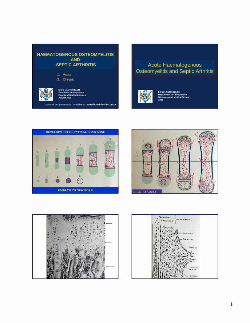
DEVELOPMENT OF TYPICAL LONG BONE
EMBRYO TO NEW BORN CHILD TO ADULT

2
BACTERIOLOGY OFHAEMATOGENOUS OSTEOMYELITIS
AND SEPTIC ARTHRITIS
Infants St. Aureus 75% B.H. Strep. 25%
2-6 yrs St. Aureus 60% H. Influenza 33%
Over 6 St. Aureus 90% other gm +ve 10%
PATHOGENISIS OF ACUTE
HAEMATOGENOUS
OSTEOMYELITIS IN CHILDHOOD
Metaphyseal side of MINOR TRAUMAphysis is weakestpart of bone
Haematoma
+Chance bacteraemia
Sluggish flow insinusoids
Lack of Seeding of infectionmacrophages
Chemotaxis
OEDEMA
OEDEMA
Rigid compartments Increased pressurebetween cartilage and thrombosiscolumns
Compression occludesCapillaries
No communication ISCHAEMIAbetween capillaries
ISCHAEMIA
Local necrosis
No access forhumoral and
Infarction cellular defence
UNHINDERED BACTERIAL PROLIFERATION
3
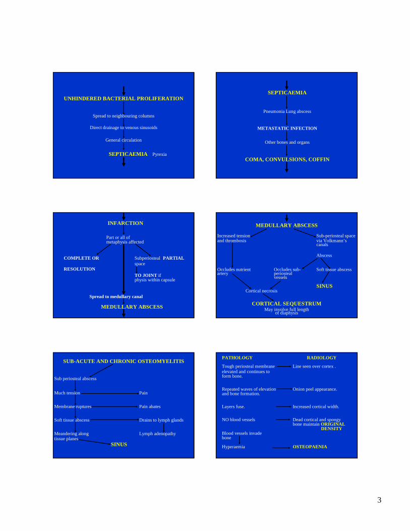
UNHINDERED BACTERIAL PROLIFERATION
Spread to neighbouring columns
Direct drainage to venous sinusoids
General circulation
SEPTICAEMIA Pyrexia
SEPTICAEMIA
Pneumonia Lung abscess
METASTATIC INFECTION
Other bones and organs
COMA, CONVULSIONS, COFFIN
INFARCTION
Part or all ofmetaphysis affected
COMPLETE OR SubperiostealPARTIALspace
RESOLUTIONTO JOINT ifphysis within capsule
Spread to medullary canal
MEDULLARY ABSCESS
MEDULLARY ABSCESS
Increased tension Sub-periosteal spaceand thrombosis via Volkmann’s
canals
Abscess
Occludes nutrient Occludes sub- Soft tissue abscessartery periosteal
vessels
SINUSCortical necrosis
CORTICAL SEQUESTRUMMay involve full length
of diaphysis
SUB-ACUTE AND CHRONIC OSTEOMYELITIS
Sub periosteal abscess
Much tension Pain
Membrane ruptures Pain abates
Soft tissue abscess Drains to lymph glands
Meandering along Lymph adenopathytissue planes
SINUS
PATHOLOGY RADIOLOGY
Tough periosteal membrane Line seen over cortex .elevated and continues to form bone.
Repeated waves of elevation Onion peel appearance.and bone formation.
Layers fuse. Increased cortical width.
NO blood vessels Dead cortical and spongybone maintainORIGINAL
DENSITYBlood vessels invadebone
Hyperaemia OSTEOPAENIA

4
PATHOLOGY RADIOLOGY
Granulation tissue or callus laidon dead and injured trabeculae INCREASED DENSITY
Patchy resorption of dead bone MOTTLED POROSIS
Areas of complete bone resorption CAVITATION
Weakened bone REFILLED WITH IRREGULAR NEW BONE
Liability to pathological fracture EVENTUALLY REMODELLED TO NORMAL ARCHITECTURE
CHRONIC OSTEOMYELITISEasy escape of pus viathick insulated sinus
Slow bacterial turnover Enclosed by veryand toxin production dense bone and scar
IF BARRIER VERY EFFICIENT
•Impervious to antibiotics and antibodies in
•And bacteria and toxins out
•Few if any systemic effects
COMPATIBLE WITH GOOD GENERAL HEALTH
Confined IschaemicLesion
IF BARRIER INEFFICIENT OR LEAKY
• Systemic effects of variable intensity.
• Antibiotics and antibodies still inefficient.
DIMINISHING DEGREES OF OSTEITIS
Continuous drainage
Dormant lesion for Intermittent abscessdecades until disturbed temporary sinus
Longer and Longer Intermittent flare-up ofperiods of remission local inflammation
Intermittent achewithout inflammation
Chronicosteitis
CHANGES OF BONE
IN
CHRONIC OSTEOMYELITIS
SUBPERIOSTEAL NEW BONE
• looks rough (fuzzy)
• widens bone
• envelopes dead cortical
bone (involucrum)
leaving gaps (cloacae)

5
DEAD CORTICAL BONE MAY -
• form sequestrumunchanged for years
• be resorbed in irregularfashion
• be discharged out of diaphysis as fragments of varying size
DEAD CANCELLOUS BONE MAY -
• persist as sequestrum.
• be resorbed and formabscess cavity (Brodies).
• become incarcerated insclerotic bone.
• Establish sinus to surface. Such lesions behave like chronic osteomyelitis
MEDULLARY CANAL
Endosteal new bone encroaches on medulla obliterating it.
One or more cavities may remain where medulla was.
When infection cured medulla reforms.
PHYSIS (Growth Plate)
Usually hyperaemia stimulates it resulting in overgrowth.
If paired bone (tibia/radius) : uneven growth, subluxed joints, bowing of shaft, stress fracture, or wedging of epiphysis.
If physis within capsule intra-articular pressure occludes blood supply arrests growth or distorts growth.
CLINICAL PICTURE
Boys 4 : Girls 1
Incidence distal femoral proximal tibia mostly.
Infants: hip shoulder
CLINICAL
Minor trauma -
after latent period
Pain, tenderness pyrexia,
Effusion much later
Cellulitis much much later -adenopathy, abscess, sinus
Confusion, coma,convulsions.
6
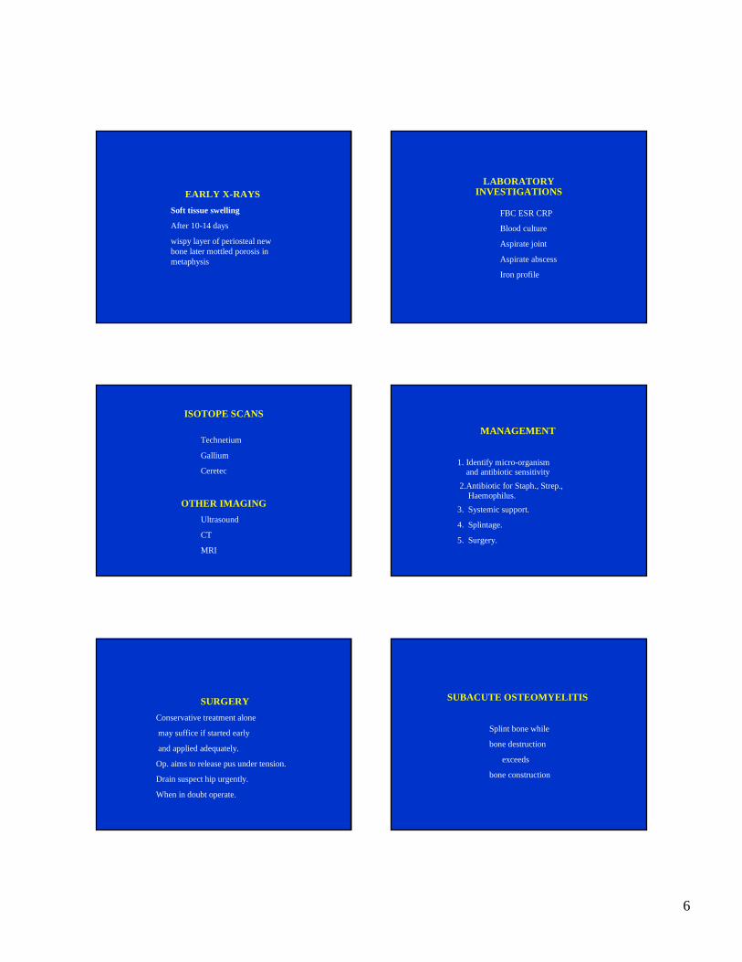
EARLY X-RAYS
Soft tissue swelling
After 10-14 days
wispy layer of periosteal new bone later mottled porosis in metaphysis
LABORATORY INVESTIGATIONS
FBC ESR CRP
Blood culture
Aspirate joint
Aspirate abscess
Iron profile
ISOTOPE SCANS
Technetium
Gallium
Ceretec
OTHER IMAGING
Ultrasound
CT
MRI
MANAGEMENT
1. Identify micro-organismand antibiotic sensitivity
2.Antibiotic for Staph., Strep., Haemophilus.
3. Systemic support.
4. Splintage.
5. Surgery.
SURGERY
Conservative treatment alone
may suffice if started early
and applied adequately.
Op. aims to release pus under tension.
Drain suspect hip urgently.
When in doubt operate.
SUBACUTE OSTEOMYELITIS
Splint bone while
bone destruction
exceeds
bone construction
7
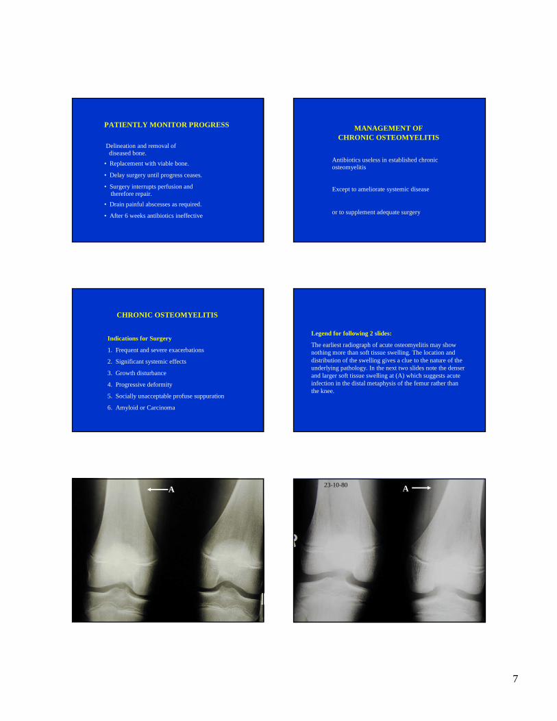
PATIENTLY MONITOR PROGRESS
Delineation and removal of diseased bone.
• Replacement with viable bone.
• Delay surgery until progress ceases.
• Surgery interrupts perfusion andtherefore repair.
• Drain painful abscesses as required.
• After 6 weeks antibiotics ineffective
MANAGEMENT OF CHRONIC OSTEOMYELITIS
Antibiotics useless in established chronicosteomyelitis
Except to ameliorate systemic disease
or to supplement adequate surgery
CHRONIC OSTEOMYELITIS
Indications for Surgery
1. Frequent and severe exacerbations
2. Significant systemic effects
3. Growth disturbance
4. Progressive deformity
5. Socially unacceptable profuse suppuration
6. Amyloid or Carcinoma
Legend for following 2 slides:
The earliest radiograph of acute osteomyelitis may show nothing more than soft tissue swelling. The location and distribution of the swelling gives a clue to the nature of the underlying pathology. In the next two slides note the denser and larger soft tissue swelling at (A) which suggests acute infection in the distal metaphysis of the femur rather than the knee.
A23-10-80 A

8
Legend for following slide:
Four months later the femur of the previous patient has sustained a pathological spiral fracture at the junction of the diaphysis and metaphysis. Notice the new bone deposited on the cortex of the distal end of the diaphysis (A). If resolution permits you may see layers (onion peel appearance) of the new bone. The bone of the metaphysis shows severe patchy osteopaenia (B).
Three months later still, in the X-Ray on the right, note the consolidation of the new bone deposited on the cortex (C). The layering is less discernable. The metaphysis looks even more ragged and osteopaenic(D). Notice the attenuated lateral cortex of the metaphysis which has been resorbed (E). As yet new bone formation is still tardy here. Notice the appearance of rapidly forming new bone between the ragged metaphysisand the physis. New bone formation continues to be formed from the physis (F) and looks relatively normal because it has not necessarily been affected by infection.
17-02-81 19-05-81
A
B
C
D
F
E
Legend for following slide:
Radiographs of the pelvis and hips of a 17 year old girl with severe pain in her right hip and a febrile illness. No bony changes are evident. Notice the increased density and swelling of the soft tissues of the right proximal thigh (A). If you trace the dark shadowpassing between the lower border of the acetabulum and the lesser trochanter you will see this line lies much closer to the neck of the femur on the left (B)). On the right this line is further from the neck of the femur because of distension of the joint.
15-07-03
A
B
Legend for following slide:
Two weeks later a Technetium Scan clearly shows the increased uptake along the acetabular margin (A) The white patch where Technetium has not been taken up by the head of the femur indicates avascularity or ischaemia of this portion of the bone (B). The neck and inter-trochantericregion shows increased uptake (C). Osteomyelitis of the proximal femoral metaphysis has gone on to septic arthritis and avascular necrosis of the head of the femur.
A
A
BC
BC
9
Legend for following slide
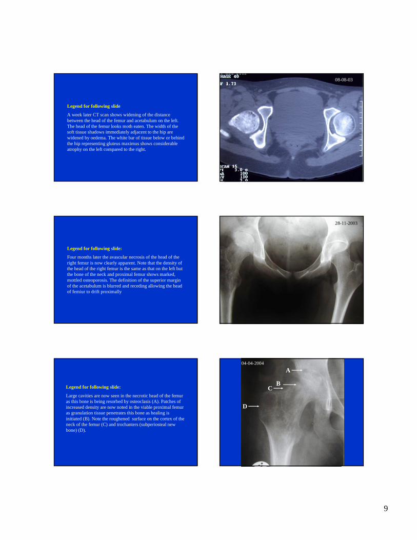
A week later CT scan shows widening of the distance between the head of the femur and acetabulum on the left. The head of the femur looks moth eaten. The width of the soft tissue shadows immediately adjacent to the hip are widened by oedema. The white bar of tissue below or behind the hip representing gluteus maximus shows considerable atrophy on the left compared to the right.
08-08-03
Legend for following slide:
Four months later the avascular necrosis of the head of the right femur is now clearly apparent. Note that the density of the head of the right femur is the same as that on the left but the bone of the neck and proximal femur shows marked, mottled osteoporosis. The definition of the superior margin of the acetabulum is blurred and receding allowing the head of femiur to drift proximally
28-11-2003
Legend for following slide:
Large cavities are now seen in the necrotic head of the femur as this bone is being resorbed by osteoclasis (A). Patches of increased density are now noted in the viable proximal femur as granulation tissue penetrates this bone as healing is initiated (B). Note the roughened surface on the cortex of the neck of the femur (C) and trochanters (subperiostealnew bone) (D).
04-04-2004
A
BC
D

10
Legend for following slide
Chronic osteomyelitis of the distal metaphysis of the femur about one year after onset.
Note: The widening or thickening of the shaft by the apposition of periosteal new bone (A). The dark cyst (Brodies’ abscess) at the distal end of the metaphysis (B). The wide band of fairly regularly trabeculated bone forming between the physis and the diseased metaphysis. Over lengthening without angulation is occurring. There is a clear cloaca in the posterior cortex of the metaphysis seen to opening into the popliteal fossa (C).
A
C
D
B
Legend for following slide
The same child had a metastatic lesion. Note the localisedwell demarcated infection in the distal radial metaphysis at the same time. This lesion is smaller and more sharply defined because the infection was probably initiated after treatment of the primary lesion in the femur had begun with antibiotics. Besides no significant trauma was involved in the production of this lesion. The antibiotics prescribed for the femur suppressed the progress of the pathology here.
Legend for following slide
Progression of osteomyelitis to resolution. Note the oval lucency in the proximal metaphysis (A) and another lucency in the distal metaphysius(B). The medullary canal in the mid shaft is narrow (C) and the distal third of the canal is obliterated (D)
3/9911/99 6/01
A
C
D
B
11
Legend for following slide
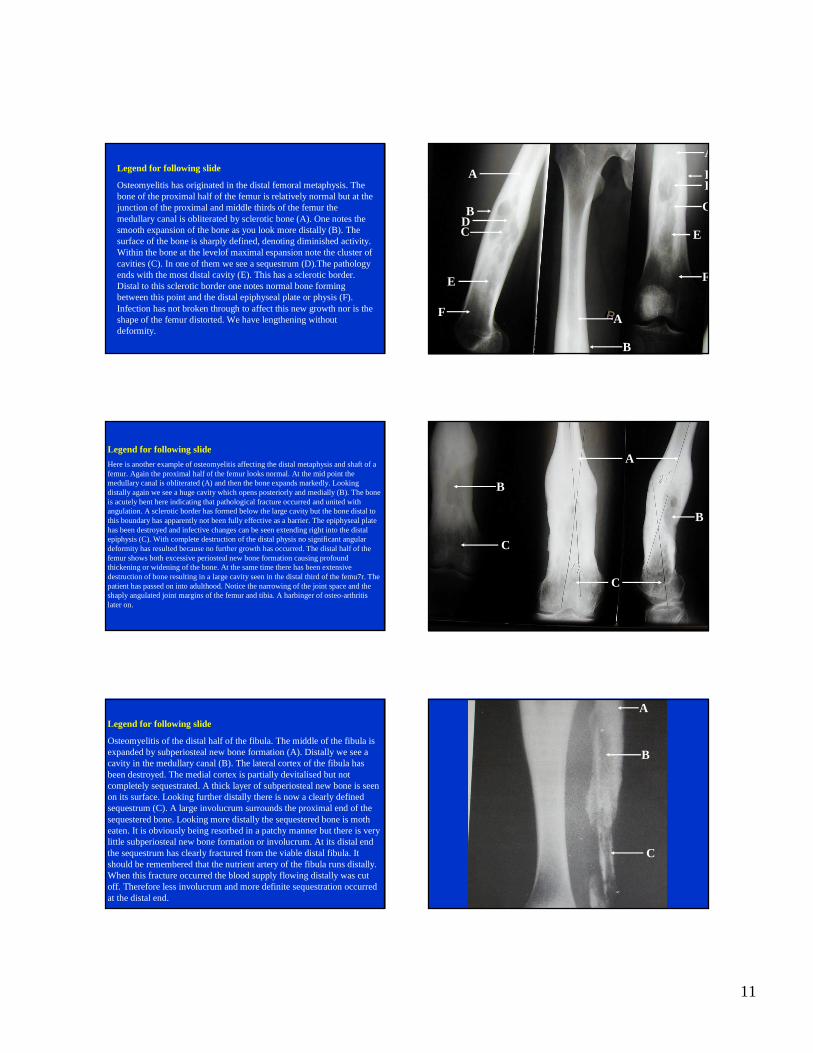
Osteomyelitis has originated in the distal femoral metaphysis. The bone of the proximal half of the femur is relatively normal but at the junction of the proximal and middle thirds of the femur the medullary canal is obliterated by sclerotic bone (A). One notes the smooth expansion of the bone as you look more distally (B). The surface of the bone is sharply defined, denoting diminished activity. Within the bone at the levelof maximal espansion note the cluster of cavities (C). In one of them we see a sequestrum (D).The pathology ends with the most distal cavity (E). This has a sclerotic border. Distal to this sclerotic border one notes normal bone forming between this point and the distal epiphyseal plate or physis (F). Infection has not broken through to affect this new growth nor is the shape of the femur distorted. We have lengthening without deformity.
A
A
A
B
B
B C
C
D
D
E
E
F
F
Legend for following slide
Here is another example of osteomyelitis affecting the distal metaphysis and shaft of a femur. Again the proximal half of the femur looks normal. At the mid point the medullary canal is obliterated (A) and then the bone expands markedly. Looking distally again we see a huge cavity which opens posteriorly and medially (B). The bone is acutely bent here indicating that pathological fracture occurred and united with angulation. A sclerotic border has formed below the large cavity but the bone distal to this boundary has apparently not been fully effective as a barrier. The epiphyseal plate has been destroyed and infective changes can be seen extending right into the distal epiphysis (C). With complete destruction of the distal physis no significant angular deformity has resulted because no further growth has occurred. The distal half of the femur shows both excessive periosteal new bone formation causing profound thickening or widening of the bone. At the same time there has been extensive destruction of bone resulting in a large cavity seen in the distal third of the femu7r. The patient has passed on into adulthood. Notice the narrowing of the joint space and the shaply angulated joint margins of the femur and tibia. A harbinger of osteo-arthritis later on.
A
B
B
C
C
Legend for following slide
Osteomyelitis of the distal half of the fibula. The middle of the fibula is expanded by subperiosteal new bone formation (A). Distally we see a cavity in the medullary canal (B). The lateral cortex of the fibula has been destroyed. The medial cortex is partially devitalised but not completely sequestrated. A thick layer of subperiosteal new bone is seen on its surface. Looking further distally there is now a clearly defined sequestrum (C). A large involucrum surrounds the proximal end of the sequestered bone. Looking more distally the sequestered bone is moth eaten. It is obviously being resorbed in a patchy manner but there is very little subperiosteal new bone formation or involucrum. At its distal end the sequestrum has clearly fractured from the viable distal fibula. It should be remembered that the nutrient artery of the fibula runs distally. When this fracture occurred the blood supply flowing distally was cut off. Therefore less involucrum and more definite sequestration occurred at the distal end.
A
B
C

12
Legend for following slide
An infective lesion is seen in the midshaft of the femur. Destruction has caused a massive cavity. Some of the original cortex of the femur can be seen as a sequestrumanteriorly and laterally (A). A cloaca opens medially (B). The bone is locally expanded by subperiosteal new bone formation (C). The surface of this bone is rough indicating ongoing activity. Below the cavity the medullary canal is obstructed by sclerotic bone (D). Distal and proximal to that again the medullary canal looks normal and the thickness of the cortex once again looks normal. Note that this lesion has occurred in the midshaft. Spontaneous haematogenous osteomyelitis affects the metaphyses of growing children. In adults the lesion occurs in the midshaft of a long bone. Invariably such an adult has immune compromise.
A
AB
CD
D
Legend for following slide
Image with lateral view of two tibiae. Here we see the growth disturbance of ongoing osteomyelitis on the right. Notice the proximal metaphysis of the tibia is larger on the right. Distal to the physis is an area of normal bone. Below that is a large cavity. The tibia is wider on the right. As we approach the centre of the bone the right tibia is much wider than the left. Now we see a bowing deformity or curvature. As we get to the distal third we see another large cavity. The width of the metaphysis is wider on the right but the difference is less marked than the difference noted in the centre. The distal tibial metaphysis is wedge shaped when compared to the flat disc seen on the right. The wedge is narrow posteriorly. Looking overall the right tibia is not only wider but longer than the left. The head of the fibula on the right is much further from the epiphysis and physis than on the left. There has been a distal subluxation here whereas distally deformity has been produced by disproportionate compression - much the same effect as would be achieved by stapling or epiphysiodesis.
Legend for following slide
An AP view of both tibiae of an adult who suffered chronic osteomyelitis as a child. All the features of osteomyelitis have resolved.Notice the osteoarthritis on right. The right tibia has a bowing or curved deformity. The bowing is convex medially. (In the lateral view it would be convex anteriorly). The head of the right fibula lies further from the knee joint on the right than on the left. The distal epiphysis is wedge shaped resulting in a sloped articular surface. The tip of the lateral malleoluson the right is situated more proximally than the medial malleolus. The right tibia is not only bowed but it is longer and wider.

13
2. Management of Chronic Haematogenous Osteomyelitis
E E G LAUTENBACHDepartment of OrthopaedicsWitwatersrand Medical School1990
After 8 Weeks
• Stop antibiotic• Simple wound toilet• Dressings merely
• to protect clothing
Monitor progress
Clinical signsGrowth
Laboratory parameters
Radiological changes
Monitor Destruction and Repair of Bone
• Splint for fracturevs
• Mobilise joints
Destruction
• Cavitation of cancellous bone• Moth eaten cortical sequestra
• Stress Fracture
Repair
• Sequestra reabsorbed
• Localisation of lesion• Obliteration of medulla
• Appositional bone• Involucrum - increased girth• Sclerosis of abscess walls
14
Reconstruction
• Moulding of outline• Re-opening of medullary canal
• Normalisation of trabecular pattern
Palliative Surgery
• Drain Abscesses• Sequestrectomy
(if not structrural)• Curettage pointless
Monitor Progress Long Term
• Bone length, deformity,• FBC, CRP, Iron profile
Undergrowth
• Vascular or mechanical damage to physis
• Failure of epiphysealcentre to ossify
Overgrowth
Acute and chronic hyperaemia stimulates and accelerates growth
Unequal Growth
• Partial damage to physis
15
Paired Bones(Forearm and Lower Leg)
• Affected bone grows faster than mate which tethers it
• Resulting in bowing, and subluxationof proximal and distal joints
• Wedging of epiphysis• Deformity at joint level, • Stress fracture of bending bone
• May need timeous fibular osteotomy
• Later judicious and timeous physeal stapling or epiphysiodesis
Indications for Surgery(All being equal)
• Await maximal involucrum formation• Haste if sequestered shaft
destroying physeal plate
• Pathological fracture uncontrolled by conservative splintage
• Most fractures usually unite
Indication for Surgery(Radical DRI)
• Chronic toxaemia
• Low haemoglobin, Iron, protein• (Amyloid myth)
• Frequent severe relapses
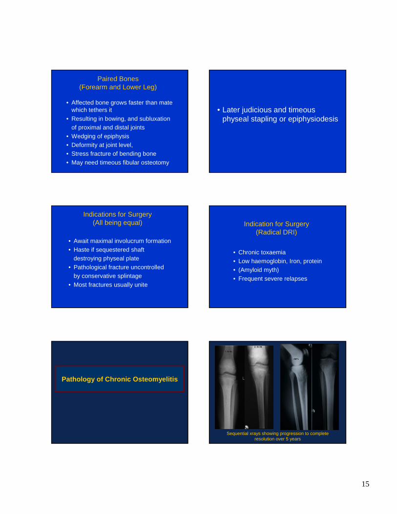
Pathology of Chronic Osteomyelitis
Sequential xrays showing progression to complete resolution over 5 years

16
Scars of old sinuses and broader left tibia. Valgus of left calcaneum. Lateral malleolus sited higher.
Anterior viewOsteomyelitis proximally, closed medulla, normal distal1/3,
anteromedial bowing, distally displaced head of fibula.
Note lateral malleolus displaced superiorly. Wedge-shaped distal epiphysis.
Chronic osteomyelitis of ulna with bowing. Proximal and distal radio-ulnar joints normally aligned.

17
Early chronic osteomyelitis subsequently pathological stress fracture
Antero-posterior view of tibiae with chronic osteomyelitis on the right. Note longer, broader right tibia.
Anterior bowing of tibia. Fibula lengthened and slight displacement of proximal and distal joints
Osteomyelitis of distal femur.
Pathological double fracture at metaphysis.
Distal fragment sequestrates, impales physis and destroys it.
Distal physis is subsequently closed while the proximal
remains open. The sequestrum has been
removed.
The fractures have united and remodeled.
Osteomyelitis of 1st metatarsal. Physis (proximal) destroyed leading to abrupt cessation of growth
18
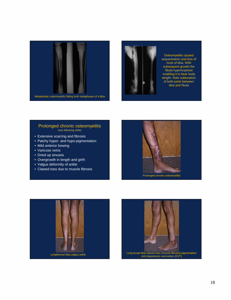
Metatastatic osteomyelitis hitting both metaphyses of a tibia
Osteomyelitis caused sequestration and loss of
most of tibia. With subsequent growth the
fibula hypertrophied enabling it to bear body weight. Note subluxation
of both joints between tibia and fibula
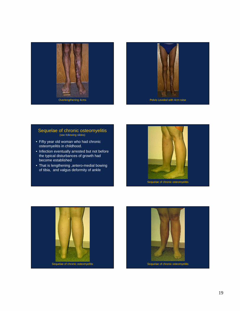
Prolonged chronic osteomyelitis(see following slide)
• Extensive scarring and fibrosis• Patchy hyper- and hypo-pigmentation• Mild anterior bowing • Varicose veins• Dried up sinuses• Overgrowth in length and girth• Valgus deformity of ankle• Clawed toes due to muscle fibrosis
Prolonged chronic osteomyelitis
Lengthened tibia,valgus ankleLong broad tibia clawed toes (muscle fibrosis) pigmentation
(old diapedesis) varicosities (DVT)
19
Overlengthening 4cms Pelvis Leveled with 4cm raise
Sequelae of chronic osteomyelitis(see following slides)
• Fifty year old woman who had chronic osteomyelitis in childhood.
• Infection eventually arrested but not before the typical disturbances of growth had become established
• That is lengthening ,antero-medial bowing of tibia, and valgus deformity of ankle
Sequelae of chronic osteomyelitis
Sequelae of chronic osteomyelitis Sequelae of chronic osteomyelitis

20
Sequelae of chronic osteomyelitis
Debridement Reaming and Irrigation of Right Humerus
Chronic om.of humerus with sequestrum & posterior cloaca Chronic om.of humerus with sequestrum & posterior cloaca
Incision along sinus and probing to bone Incision extended distally to expose long and lateral triceps

21
This interval dissected down to bone Cloaca with sequestrum located
Sequestra defined And removed
Cloaca enlarged as a gutter and curetted Lateral epicondyle with previously healed sinus
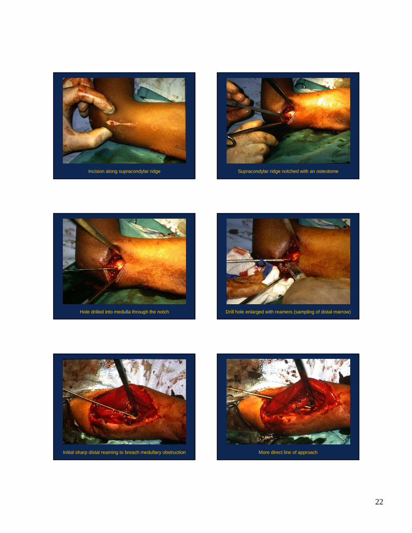
22
Incision along supracondylar ridge Supracondylar ridge notched with an osteotome
Hole drilled into medulla through the notch Drill hole enlarged with reamers (sampling of distal marrow)
Initial sharp distal reaming to breach medullary obstruction More direct line of approach

23
Follow with larger reamers Outline of proximal window
Enlargement of gutter opening proximal barrier in medulla Reaming full length of medulla proximally from below
Medullary canal flushed clean with saline Further reaming of proximal medulla
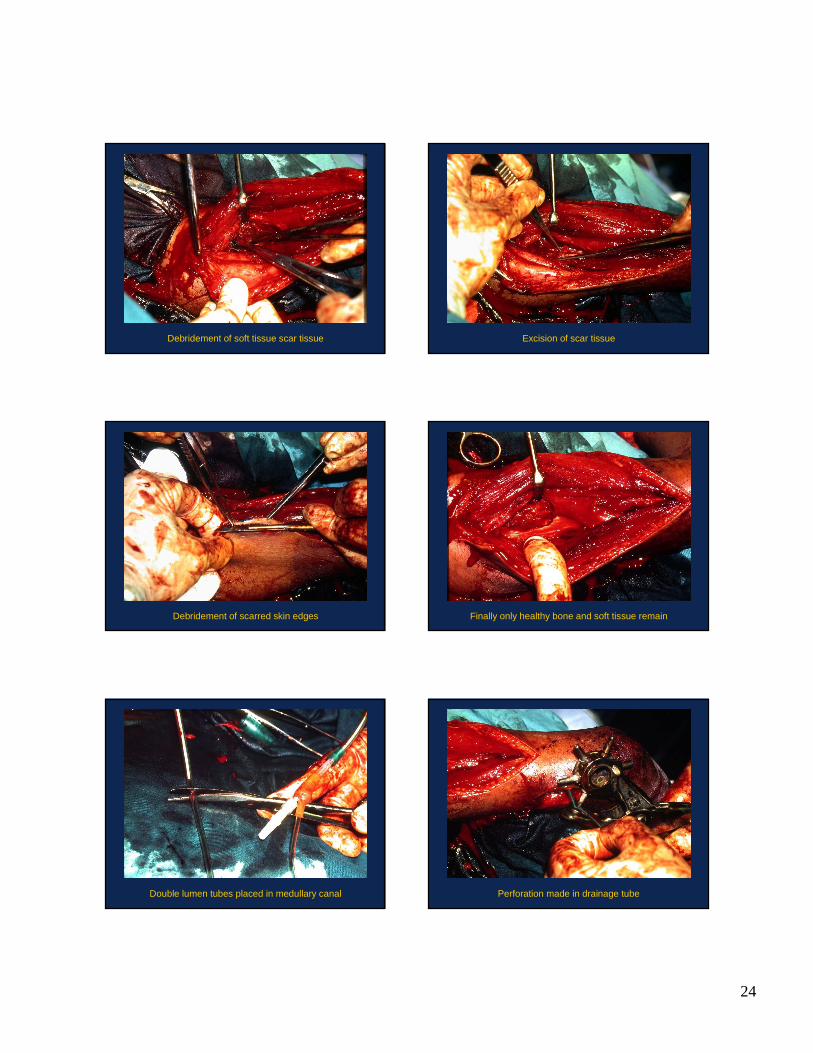
24
Debridement of soft tissue scar tissue Excision of scar tissue
Debridement of scarred skin edges Finally only healthy bone and soft tissue remain
Double lumen tubes placed in medullary canal Perforation made in drainage tube
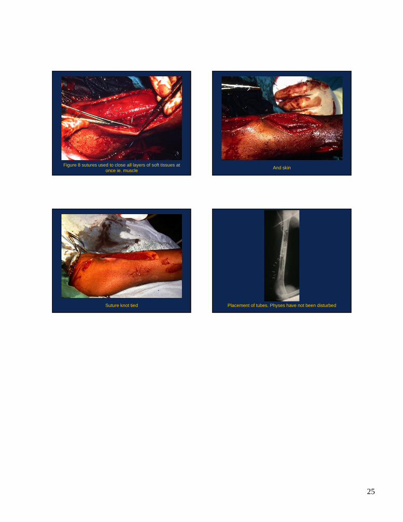
25
Figure 8 sutures used to close all layers of soft tissues at once ie. muscle
And skin
Suture knot tied Placement of tubes. Physes have not been disturbed


![RESEARCH ARTICLE Open Access Staphylococcus aureus … · osteomyelitis [9-11], that clinical osteomyelitis isolates of S. aureus are capable of forming biofilms in vitro [12-14],](https://static.fdocuments.in/doc/165x107/5e2dbd77b609da091e164f06/research-article-open-access-staphylococcus-aureus-osteomyelitis-9-11-that-clinical.jpg)


![Methacillin Resistant Staph aureus 3-11[1].pdfboil, abscess, furuncle erythema, swelling, pain, drainage Invasive infections osteomyelitis, pneumonia, blood stream infxn, CNS infxn.](https://static.fdocuments.in/doc/165x107/5e3409d39d5e6170295783f9/methacillin-resistant-staph-aureus-3-111pdf-boil-abscess-furuncle-erythema.jpg)