Acute Care Surgery Regional Management for COVID19 Alaska ...
26
Acute Care Surgery Regional Management for COVID19 Alaska Native Tribal Health Consortium Department of General Surgery ANMC Sheridan Morgan MD
Transcript of Acute Care Surgery Regional Management for COVID19 Alaska ...

Acute Care SurgeryRegional Management for COVID19
Alaska Native Tribal Health Consortium
Department of General Surgery ANMC
Sheridan Morgan MD

Disclosures
• No Relevant Disclosures

Alaska Native Health NetworkUnique Challenges
• Geography
• Concentration of Surgical Care
• Travel Limitations

Goals of Regional Management
Prevent Unnecessary COVID19 Exposures
• Increase Regional care for common non-operative surgical problems
• Avoid Unnecessary patient transports
• Continue to identify patients that require transfer to ANMC
• Continue to provide surgical care at ANMC for acute surgical disease

Conditions Covered in This Lecture
• Adhesive Small Bowel Obstruction
• Simple Pneumothorax
• Simple Cutaneous Abscess
• Appendicitis
• Benign Gallstone Diesease
• Diverticulitis
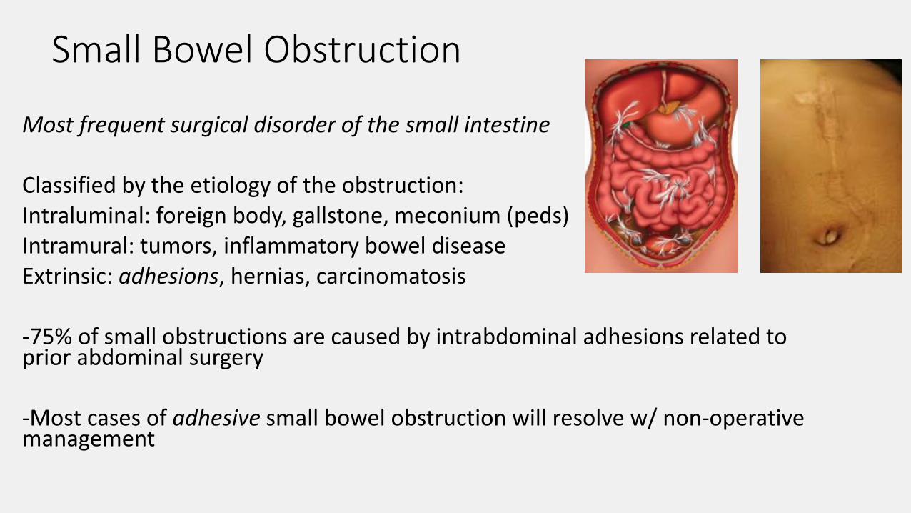
Small Bowel Obstruction
Most frequent surgical disorder of the small intestine
Classified by the etiology of the obstruction: Intraluminal: foreign body, gallstone, meconium (peds)Intramural: tumors, inflammatory bowel diseaseExtrinsic: adhesions, hernias, carcinomatosis
-75% of small obstructions are caused by intrabdominal adhesions related to prior abdominal surgery
-Most cases of adhesive small bowel obstruction will resolve w/ non-operative management

Adhesive Small Bowel Disease, Presentation
History: past abdominal surgery, colicky abdominal pain, nausea, emesis, decreased or absent bowel function
PE: abdominal distention, hypo/hyperactive bowel sounds, tenderness
Labs: elevated Cr, electrolyte abnormalities, +/-leukocytosis, +/- elevated lactate
Abd X-ray: dilated loops small bowel, air fluid levels, paucity of colonic gas
CT imaging: discrete transition from dilated to decompressed SB, decompressed colon

Adhesive Small Bowel Disease, Management• Work-Up: CBC, BMP, Lactate, Abd X-rays, CT Abd/Pelvis
• Resuscitation: fluid bolus, continuous IVF, follow UOP, replete electrolytes
• NG tube decompression: check NG position, low intermittent suction, monitoring NG output
• Follow Bowel Function: Discontinue NG tube after decreased NG output & return of bowel function
Recommend: telemedicine consult & daily review with ANMC surgical team

Adhesive Small Bowel Obstruction, Indications for Transfer
High Risk Features: -free air -free fluid -closed loop obstruction
-pneumatosis intestinalis -portal venous gas
-hemodynamic instability -persistently elevated lactic acid
Closed LoopSmall BowelObstruction
Pneumatosis Intestinalis Portal Venous Gas

Simple Pneumothorax
• Occurs when air enters the space between visceral and parietal pleura
• Can happen spontaneously or from trauma (blunt or penetrating)
Symptoms: dyspnea, pleuritic chest pain, increased O2 requirements
Signs: decreased breath sounds, hyperresonance, unequal chest rise
CXR: -absent lung markings
(look at apex & periphery of lung)
-visible pleural edge
-deep sulcus sign
(deep dark costophrenic angle)
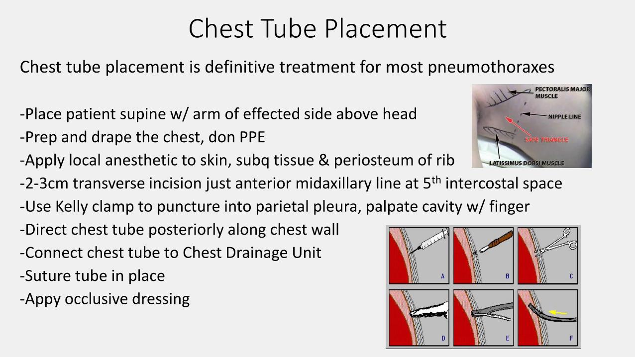
Chest Tube PlacementChest tube placement is definitive treatment for most pneumothoraxes
-Place patient supine w/ arm of effected side above head
-Prep and drape the chest, don PPE
-Apply local anesthetic to skin, subq tissue & periosteum of rib
-2-3cm transverse incision just anterior midaxillary line at 5th intercostal space
-Use Kelly clamp to puncture into parietal pleura, palpate cavity w/ finger
-Direct chest tube posteriorly along chest wall
-Connect chest tube to Chest Drainage Unit
-Suture tube in place
-Appy occlusive dressing

After Chest Tube Placement
-Obtain CXR, confirm re-expansion of lung
-Continue chest tube to suction, -20mmHg
-Check Chest Drainage Unit daily & monitor for air leak
-If no air leak, place chest tube to water seal
-After additional 12-24 hour repeat CXR,
confirm lung expansion remove chest tube
Pathology that requires transfer to ANMC:
-Hemothorax (300ml+) -Flail Chest
-Open Pneumothorax -Pulmonary Contusion
-Residual Pneumothorax after CT -Unresolving Air leak
Recommend: telemedicine consult & daily review with ANMC surgical team
Chest Drainage Unit
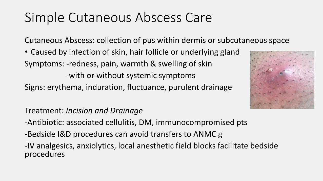
Simple Cutaneous Abscess Care
Cutaneous Abscess: collection of pus within dermis or subcutaneous space
• Caused by infection of skin, hair follicle or underlying gland
Symptoms: -redness, pain, warmth & swelling of skin
-with or without systemic symptoms
Signs: erythema, induration, fluctuance, purulent drainage
Treatment: Incision and Drainage
-Antibiotic: associated cellulitis, DM, immunocompromised pts
-Bedside I&D procedures can avoid transfers to ANMC g
-IV analgesics, anxiolytics, local anesthetic field blocks facilitate bedside procedures
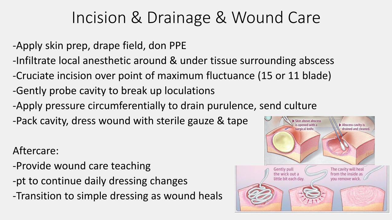
Incision & Drainage & Wound Care
-Apply skin prep, drape field, don PPE
-Infiltrate local anesthetic around & under tissue surrounding abscess
-Cruciate incision over point of maximum fluctuance (15 or 11 blade)
-Gently probe cavity to break up loculations
-Apply pressure circumferentially to drain purulence, send culture
-Pack cavity, dress wound with sterile gauze & tape
Aftercare:
-Provide wound care teaching
-pt to continue daily dressing changes
-Transition to simple dressing as wound heals

Abscesses, Special Circumstances
Breast Abscess: -Consider needle aspiration & antibiotics 1st
-Patient may require image guided drain
-Surgical I&D, reserved for treatment failures after above measures
Perirectal Abscesses: Arise from infections of anal glands
-categorized by the space involved:
perianal, ischiorectal, intersphincteric, supralevator
-superficial perianal & small Ischiorectal abscesses bedside I&D
-incision close to anus, avoid aggressive probing & packing
Hands, Feet & Digit Abscesses: -May require specialized surgical drainage
-Consider early orthopedic involvement
Signs of Necrotizing Infection: blistering, bulla, skin streaking,
pain out of proportion to exam, gas formation imaging
Urgent Surgical Involvement

Appendicitis• Blockage of appendiceal lumen inflammation of appendiceal wall
-increased intralumenal pressure bacterial overgrowth & deceased blood flow to wall of appendix
- If left untreated: necrosis of appendix appendicular wall perforation sepsis
• One of the most common reasons for hospitalization among children & young adults worldwide
Risk Factors: age 5 to 40, male sex, rural living
Symptoms: abdominal pain (periumbilical RLQ), nausea, vomiting, fever
Signs: Tenderness to palpation at McBurney’s Point, Dunphys’ sign, Rovsing’s sign, Psoas sign etc.
Dx: elevated WBC, elevated CRP, CT Abd/Pelvis, MRI, Abd Ultrasound
Surgical removal of the appendix is standard treatment for acute appendicitis in the United States
Non-operative treatment with antibiotics is safe and effective for many patients with uncomplicated appendicitis, commonly done outside of the United States
Non-operative regional treatment should be considered for select patients in our network during COVID epidemic

Appendicitis, Non-operative Management
Benefits: quicker return to normal activity, decreased pain & narcotic requirements, avoidance of transfer to surgical hospital, lower morbidity
Risks: 10% failure rate, only effective for uncomplicated appendicitis, 30% of patients require appendectomy within 4-7 months
Inclusion: uncomplicated appendicitis, age 6+, no fecalith, appendiceal diameter ≤ 11mm
-no perforation -no phlegmon -no free air
-non-pregnant -symptoms less than 48 hours duration
Exclusion Criteria: hemodynamic instability, diffuse peritonitis, abscess 3cm+
High Risk patients: immunocompromised, age > 70 yrs, medical comorbidities
Appendicitis, Antibiotic Treatment Regimen
Low Risk Patients: Pip-tazo 3.375g q6 h or Ertapenem 1g q day for 24 to 48 hrs
PO Levaquin & Flagyl or Keflex & Flagyl or Augmentin for total of 10 days
High Risk Patients: Pip-tazo 4.5g IV q 8 h or Meropenum 1g q 8 h for 24 to 48 hrs
PO Levaquin & Flagyl or Keflex & Flagyl or Augmentin for a total of 10 days
Supportive Care: IV fluids PRN, Anti-emetics PRN, Pain medications PRN
Recommend hospital admission for 24-48 hrs w/ IV Abx, monitoring & serial labs
Inflammatory marks improve, tolerating PO transitioned to PO antibiotics for 10-day total course
Failure: Lack of improvement after 24-48 hrs IV Abx, persistently elevated inflammatory markers, hemodynamic instability transfer to ANMC for surgical intervention
Recommend: telemedicine consult & daily review with ANMC surgical team

Symptomatic Gallstones & Gallstone Related Disease
• Gallstone and benign biliary disease, very common
• Prevalence of gallstones 11 to 36%, general US population
• Most patients are asymptomatic from gallstones
• Some patients will develop symptoms & complication from gallstones: symptomatic cholelithiasis, cholecystitis, choledocholithiasis, cholangitis, gallstone pancreatitis etc.
• Treatment for these conditions are highly variable but most gallstone disease can be treated acutely without surgery
For cases of symptomatic cholelithiasis and mild to moderate cholecystitis nonoperative management at regional centers should be considered while travel is limited from the COVID Pandemic

Symptomatic Cholelithiasis / CholecystitisSymptomatic Cholelithiasis: intermittent obstruction of the cystic duct gallbladder irritation
Episodic / intermittent RUQ pain, associated w/ fatty foods, lasts 1-5 hrs. No fever. No WBC count.
US/CT: stones in the gallbladder w/o wall thickening
TX: -Initial treatment involves supportive measures, pain control, antiemetics, avoid aggravating foods
-Surgery performed on elective basis, may be delayed unless pt has debilitating & recurrent symptoms
Acute Cholecystitis: obstruction of cystic duct gallbladder inflammation & bacterial overgrowth
Unremitting RUQ pain, lasts 6hrs+. Decreased apatite. Fever. Elevated WBC count
PE- acutely tender to palpation RUQ. + Murphy’s sign.
US/CT: Thickening of the Gallbladder wall, pericholecystic fluid
Initial TX: -IVF, analgesia, Antibiotics: Pip-tazo 1st line, alternatives: Ertapenem, Ciprofloxacin, Levaquin
-Consider Regional Treatment with antibiotics for 24-48 hrs, mild to moderate disease
Transfer: hemodynamic instability, end organ dysfunction, Symptoms > 72 hrs, failure to improve after 48 hrs
Recommend: telemedicine consult & daily review with ANMC surgical team

Cholecystitis, Grading Criteria & TreatmentMild & Moderate Disease: Consider Regional Treatment w/ IV antibiotics for 48 hrs
Severe Disease or Failure to Improve after 48 hrs of IV antibiotics Transfer to ANMC
IV Antibiotics: Pip-tazo 1st line Alternatives: Ertapenem, Ciprofloxacin, Levaquin

High Risk Gallstone Disease Choledocholithiasis: presence of stones common bile duct
-most commonly from stones that have migrated from gallbladder
RUQ pain, nausea, jaundice. Elevated LFTs
US: stones in gallbladder, dilated CBD (8mm+)
Tx: requires clearance of CBD & cholecystectomy Transfer to ANMC
Cholangitis: ascending bacterial infection from obstructed bile duct
-most commonly from gallstone obstructing the CBD
Fever, RUQ/epigastric pain, jaundice. Elevated LFTs. Elevated WBC.
Tx: fluid resuscitation, IV antibiotics, biliary drainage Transfer to ANMC
Gallstone Pancreatitis: pancreatitis caused by biliary stones
Epigastric/back pain, nausea, emesis. Elevated LFTs, amylase, lipase
US/CT: biliary stones, pancreatitis
TX: conservative medical management for pancreatitis, interval cholecystectomy
Semi urgent transfer to ANMC for definitive biliary clearance & cholecystectomy
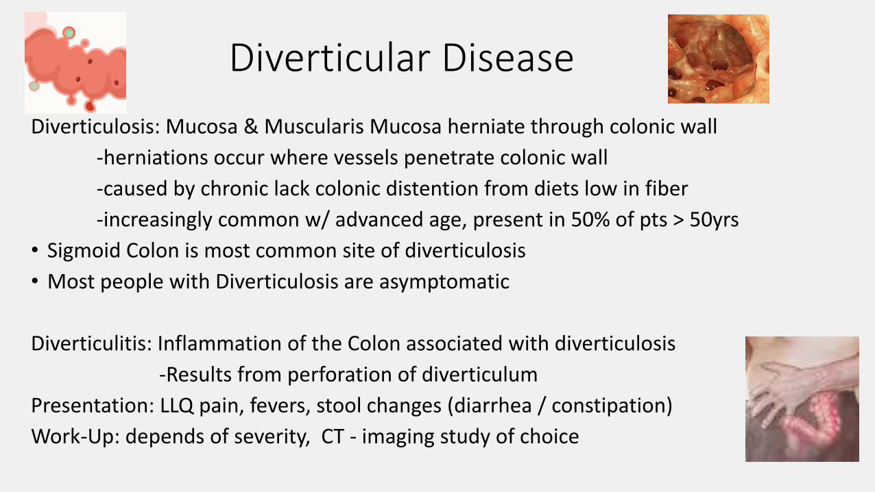
Diverticular Disease
Diverticulosis: Mucosa & Muscularis Mucosa herniate through colonic wall
-herniations occur where vessels penetrate colonic wall
-caused by chronic lack colonic distention from diets low in fiber
-increasingly common w/ advanced age, present in 50% of pts > 50yrs
• Sigmoid Colon is most common site of diverticulosis
• Most people with Diverticulosis are asymptomatic
Diverticulitis: Inflammation of the Colon associated with diverticulosis
-Results from perforation of diverticulum
Presentation: LLQ pain, fevers, stool changes (diarrhea / constipation)
Work-Up: depends of severity, CT - imaging study of choice

Treatment of DiverticulitisUncomplicated Diverticulitis: local colonic inflammation w/ colonic wall thickening or phlegmonTx: Low residue diet & antibiotics, Consider Regional Management
Complicated Diverticulitis: Abscess, Obstruction, Perforation or FistulaHinchey Staging System
-Stage I: Diverticulitis w/ Pericolonic Abscess-Stage II: Diverticulitis w/ Retroperitoneal or Pelvic Abscess -Stage III/IV: Diverticulitis w/ Peritonitis
TX: Diverticulitis w/ small abscesses < 3cm Consider Regional Management w/ IV antibioticsDiverticulitis w/ large abscess > 3cm Transfer to ANMC for percutaneous drainageDiverticulitis w/ peritonitis or free air on CT imaging Transfer to ANMC for Urgent Surgical Management
Special Cases:Colovesicle Fistula, mildly symptomatic not requiring suppressive Abx Regional Management Elective SurgeryColovesicle Fistula, symptomatic requiring suppressive Abx Consultation with ANMC Surgical TeamColonic Obstruction from Diverticular Stricture Transfer to ANMC for Urgent Surgical Management
For patients with new diagnosis of diverticulitis-Colonoscopy should be performed in 6-12 weeks elective basis, after COVID travel restrictions have been lifted

Conclusions
• Unique challenges in Alaska Native Healthcare Network during the COVID19 pandemic: Geography, Travel, Surgical Access
• Focus on providing appropriate care, limiting unnecessary travel
• Some common surgical diseases can be safely treated at regional centers: Simple Adhesive SBO, Simple Abscesses, Uncomplicated Appendicitis, Symptomatic Cholelithiasis, Cholecystitis, Uncomplicated Diverticulitis
• For Regional Management of SBO, Appendicitis & Cholecystitis we recommend daily consultation with surgical team at ANMC
• Always feel free to discuss any cases w/ General Surgery team at ANMC to help guide management
Thank You!

References
American College of Surgeons. Professional Society Guidelines. March 25, 2020 Covid-19 Guidelines for Triage of Emergency General Surgery Patients. https://www.facs.org/covid-19/clinical-guidance/elective-case/emergency-surgery.
Brunicardi C, et al. Schwartz’s Principles of Surgery, Tenth Edition. 2015
Lee S, et al. Expanding the inclusion criteria for non-operative management of uncomplicated appendicitis: Outcomes and cost. J PedSurg. 2017;53(1):42-47.
Steele S, et al. The ASCRS Textbook of Colon and Rectal Surgery, Third Edition. 2016.
Talan D, et al. Methods of conservative antibiotic treatment of acute uncomplicated appendicitis: A systematic review. J Trauma Acute Care Surg. 2019;86(4):722-736.
Yokoe M, et al. TG13 diagnostic criteria and severity grading of acute cholecystitis (with videos). J Hepatobiliary Pancreat Sci. 2013 Jan;20(1):35-46.