Activation of c-Jun NH2-terminal kinase 1 increases cellular responsiveness to BMP-2 and decreases...
11
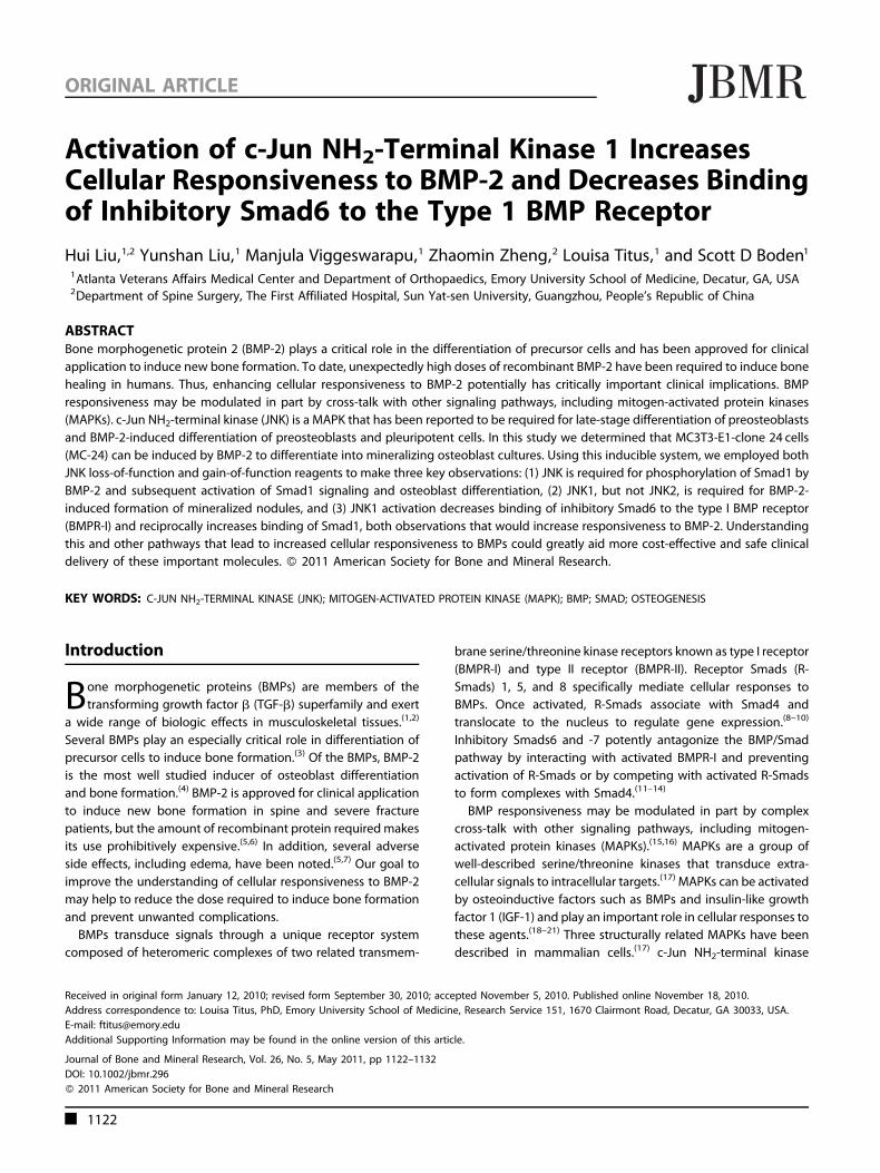
Activation of c-Jun NH 2 -Terminal Kinase 1 Increases Cellular Responsiveness to BMP-2 and Decreases Binding of Inhibitory Smad6 to the Type 1 BMP Receptor Hui Liu, 1,2 Yunshan Liu, 1 Manjula Viggeswarapu, 1 Zhaomin Zheng, 2 Louisa Titus, 1 and Scott D Boden 1 1 Atlanta Veterans Affairs Medical Center and Department of Orthopaedics, Emory University School of Medicine, Decatur, GA, USA 2 Department of Spine Surgery, The First Affiliated Hospital, Sun Yat-sen University, Guangzhou, People’s Republic of China ABSTRACT Bone morphogenetic protein 2 (BMP-2) plays a critical role in the differentiation of precursor cells and has been approved for clinical application to induce new bone formation. To date, unexpectedly high doses of recombinant BMP-2 have been required to induce bone healing in humans. Thus, enhancing cellular responsiveness to BMP-2 potentially has critically important clinical implications. BMP responsiveness may be modulated in part by cross-talk with other signaling pathways, including mitogen-activated protein kinases (MAPKs). c-Jun NH 2 -terminal kinase (JNK) is a MAPK that has been reported to be required for late-stage differentiation of preosteoblasts and BMP-2-induced differentiation of preosteoblasts and pleuripotent cells. In this study we determined that MC3T3-E1-clone 24 cells (MC-24) can be induced by BMP-2 to differentiate into mineralizing osteoblast cultures. Using this inducible system, we employed both JNK loss-of-function and gain-of-function reagents to make three key observations: (1) JNK is required for phosphorylation of Smad1 by BMP-2 and subsequent activation of Smad1 signaling and osteoblast differentiation, (2) JNK1, but not JNK2, is required for BMP-2- induced formation of mineralized nodules, and (3) JNK1 activation decreases binding of inhibitory Smad6 to the type I BMP receptor (BMPR-I) and reciprocally increases binding of Smad1, both observations that would increase responsiveness to BMP-2. Understanding this and other pathways that lead to increased cellular responsiveness to BMPs could greatly aid more cost-effective and safe clinical delivery of these important molecules. ß 2011 American Society for Bone and Mineral Research. KEY WORDS: C-JUN NH 2 -TERMINAL KINASE (JNK); MITOGEN-ACTIVATED PROTEIN KINASE (MAPK); BMP; SMAD; OSTEOGENESIS Introduction B one morphogenetic proteins (BMPs) are members of the transforming growth factor b (TGF-b) superfamily and exert a wide range of biologic effects in musculoskeletal tissues. (1,2) Several BMPs play an especially critical role in differentiation of precursor cells to induce bone formation. (3) Of the BMPs, BMP-2 is the most well studied inducer of osteoblast differentiation and bone formation. (4) BMP-2 is approved for clinical application to induce new bone formation in spine and severe fracture patients, but the amount of recombinant protein required makes its use prohibitively expensive. (5,6) In addition, several adverse side effects, including edema, have been noted. (5,7) Our goal to improve the understanding of cellular responsiveness to BMP-2 may help to reduce the dose required to induce bone formation and prevent unwanted complications. BMPs transduce signals through a unique receptor system composed of heteromeric complexes of two related transmem- brane serine/threonine kinase receptors known as type I receptor (BMPR-I) and type II receptor (BMPR-II). Receptor Smads (R- Smads) 1, 5, and 8 specifically mediate cellular responses to BMPs. Once activated, R-Smads associate with Smad4 and translocate to the nucleus to regulate gene expression. (8–10) Inhibitory Smads6 and -7 potently antagonize the BMP/Smad pathway by interacting with activated BMPR-I and preventing activation of R-Smads or by competing with activated R-Smads to form complexes with Smad4. (11–14) BMP responsiveness may be modulated in part by complex cross-talk with other signaling pathways, including mitogen- activated protein kinases (MAPKs). (15,16) MAPKs are a group of well-described serine/threonine kinases that transduce extra- cellular signals to intracellular targets. (17) MAPKs can be activated by osteoinductive factors such as BMPs and insulin-like growth factor 1 (IGF-1) and play an important role in cellular responses to these agents. (18–21) Three structurally related MAPKs have been described in mammalian cells. (17) c-Jun NH 2 -terminal kinase ORIGINAL ARTICLE J JBMR Received in original form January 12, 2010; revised form September 30, 2010; accepted November 5, 2010. Published online November 18, 2010. Address correspondence to: Louisa Titus, PhD, Emory University School of Medicine, Research Service 151, 1670 Clairmont Road, Decatur, GA 30033, USA. E-mail: [email protected] Additional Supporting Information may be found in the online version of this article. Journal of Bone and Mineral Research, Vol. 26, No. 5, May 2011, pp 1122–1132 DOI: 10.1002/jbmr.296 ß 2011 American Society for Bone and Mineral Research 1122
Transcript of Activation of c-Jun NH2-terminal kinase 1 increases cellular responsiveness to BMP-2 and decreases...
Activation of c-Jun NH2-Terminal Kinase 1 IncreasesCellular Responsiveness to BMP-2 and Decreases Bindingof Inhibitory Smad6 to the Type 1 BMP Receptor
Hui Liu,1,2 Yunshan Liu,1 Manjula Viggeswarapu,1 Zhaomin Zheng,2 Louisa Titus,1 and Scott D Boden1
1Atlanta Veterans Affairs Medical Center and Department of Orthopaedics, Emory University School of Medicine, Decatur, GA, USA2Department of Spine Surgery, The First Affiliated Hospital, Sun Yat-sen University, Guangzhou, People’s Republic of China
ABSTRACTBone morphogenetic protein 2 (BMP-2) plays a critical role in the differentiation of precursor cells and has been approved for clinical
application to induce new bone formation. To date, unexpectedly high doses of recombinant BMP-2 have been required to induce bone
healing in humans. Thus, enhancing cellular responsiveness to BMP-2 potentially has critically important clinical implications. BMP
responsiveness may be modulated in part by cross-talk with other signaling pathways, including mitogen-activated protein kinases
(MAPKs). c-Jun NH2-terminal kinase (JNK) is a MAPK that has been reported to be required for late-stage differentiation of preosteoblasts
and BMP-2-induced differentiation of preosteoblasts and pleuripotent cells. In this study we determined that MC3T3-E1-clone 24 cells
(MC-24) can be induced by BMP-2 to differentiate into mineralizing osteoblast cultures. Using this inducible system, we employed both
JNK loss-of-function and gain-of-function reagents to make three key observations: (1) JNK is required for phosphorylation of Smad1 by
BMP-2 and subsequent activation of Smad1 signaling and osteoblast differentiation, (2) JNK1, but not JNK2, is required for BMP-2-
induced formation of mineralized nodules, and (3) JNK1 activation decreases binding of inhibitory Smad6 to the type I BMP receptor
(BMPR-I) and reciprocally increases binding of Smad1, both observations that would increase responsiveness to BMP-2. Understanding
this and other pathways that lead to increased cellular responsiveness to BMPs could greatly aid more cost-effective and safe clinical
delivery of these important molecules. � 2011 American Society for Bone and Mineral Research.
KEY WORDS: C-JUN NH2-TERMINAL KINASE (JNK); MITOGEN-ACTIVATED PROTEIN KINASE (MAPK); BMP; SMAD; OSTEOGENESIS
Introduction
Bone morphogenetic proteins (BMPs) are members of the
transforming growth factor b (TGF-b) superfamily and exert
a wide range of biologic effects in musculoskeletal tissues.(1,2)
Several BMPs play an especially critical role in differentiation of
precursor cells to induce bone formation.(3) Of the BMPs, BMP-2
is the most well studied inducer of osteoblast differentiation
and bone formation.(4) BMP-2 is approved for clinical application
to induce new bone formation in spine and severe fracture
patients, but the amount of recombinant protein requiredmakes
its use prohibitively expensive.(5,6) In addition, several adverse
side effects, including edema, have been noted.(5,7) Our goal to
improve the understanding of cellular responsiveness to BMP-2
may help to reduce the dose required to induce bone formation
and prevent unwanted complications.
BMPs transduce signals through a unique receptor system
composed of heteromeric complexes of two related transmem-
brane serine/threonine kinase receptors known as type I receptor
(BMPR-I) and type II receptor (BMPR-II). Receptor Smads (R-
Smads) 1, 5, and 8 specifically mediate cellular responses to
BMPs. Once activated, R-Smads associate with Smad4 and
translocate to the nucleus to regulate gene expression.(8–10)
Inhibitory Smads6 and -7 potently antagonize the BMP/Smad
pathway by interacting with activated BMPR-I and preventing
activation of R-Smads or by competing with activated R-Smads
to form complexes with Smad4.(11–14)
BMP responsiveness may be modulated in part by complex
cross-talk with other signaling pathways, including mitogen-
activated protein kinases (MAPKs).(15,16) MAPKs are a group of
well-described serine/threonine kinases that transduce extra-
cellular signals to intracellular targets.(17) MAPKs can be activated
by osteoinductive factors such as BMPs and insulin-like growth
factor 1 (IGF-1) and play an important role in cellular responses to
these agents.(18–21) Three structurally related MAPKs have been
described in mammalian cells.(17) c-Jun NH2-terminal kinase
ORIGINAL ARTICLE JJBMR
Received in original form January 12, 2010; revised form September 30, 2010; accepted November 5, 2010. Published online November 18, 2010.
Address correspondence to: Louisa Titus, PhD, Emory University School of Medicine, Research Service 151, 1670 Clairmont Road, Decatur, GA 30033, USA.
E-mail: [email protected]
Additional Supporting Information may be found in the online version of this article.
Journal of Bone and Mineral Research, Vol. 26, No. 5, May 2011, pp 1122–1132
DOI: 10.1002/jbmr.296
� 2011 American Society for Bone and Mineral Research
1122

(JNK) is one of these MAPKs, and it contributes to multiple
cellular events such as proliferation, differentiation, and survival
in response to extracellular stimuli.(22) JNK has been reported
to be required for late-stage differentiation of preosteoblasts
and BMP-2-induced differentiation of preosteoblasts and
pleuripotent cells.(18,20,23,24) JNK is considered a critical regulator
of activating transcription factor 4, osteocalcin, and bone
sialoprotein expression in spontaneously differentiating osteo-
blasts.(21,23) These studies suggest that JNK plays an important
role in the induction of osteogenesis by BMP-2. However, the
exact mechanism of this interaction is still unclear.
In this study, we describe a novel role of JNK in Smad1
C-terminal phosphorylation and consequent osteogenic differ-
entiation induced by BMP-2 in MC3T3-E1 subclone 24
preosteoblasts that do not differentiate spontaneously.(25)
Specifically, activation of JNK suppresses binding of Smad6 to
BMPR-I and enhances binding and consequent phosphorylation/
activation of Smad1.
Materials and Methods
MC3T3-E1 subclone 24 cell culture
MC3T3-E1 subclone 24 (MC-24) cells, a mouse preosteoblast cell
line that can be induced to differentiate into osteoblasts and
produce mineralized nodules in vitro, was obtained from the
American Type Culture Collection (ATCC, Manassas, VA, USA) and
maintained in a-modified essential medium (a-MEM, Invitrogen/
Gibco, Grand Island, NY, USA) with 10% non-heat-inactivated
fetal bovine serum (FBS; Atlanta Biologicals, Norcross, GA, USA) at
378C in 5% CO2 humidified air. Differentiation of MC-24 cells was
induced by BMP-2 and the addition of 50mg/mL of ascorbic acid
(AA) and 5mM b-glycerophosphate (b-GP) in the culture
medium.
Fetal rat calvarial osteoblast cultures
Following approval by the Institutional Animal Care and Use
Committee, fetal Sprague-Dawley rats were removed at 20 days’
gestation, decapitated, and calvarial preosteoblasts were
obtained as described previously.(26) Cells were seeded in T-75
flasks (Corning, Inc., Corning, NY, USA) at 1� 106 cells/flask in
MEM supplemented with L-glutamine (Gibco/Invitrogen) plus
heat-inactivated 10% FBS (Hyclone, Logan, UT, USA) and ex-
panded after 1 week of culture. Seven days later, cells were
plated in 6-well plates at 5� 104 cells/well and grown to
confluence prior to initiation of differentiation with the addition
of 50mg/mL of AA with or without BMP-2 (100 ng/mL, reapplied
after 4 days) in the culture medium. During the second week of
differentiation, cultures were grown in Biggers, Gwathin, and
Judah (BGJb) medium, Fitton Jackson modification (Invitrogen/
Gibco), in the presence of 5mM b-GP.
RNA interference of Smad1 or JNK1/2
Using lipofectamine RNAiMAX transfection reagent (Invitrogen,
Carlsbad, CA, USA) according to the manufacturer’s instructions,
MC-24 cells were transfected with small interfering RNA (siRNA)
duplexes specific for mouse Smad1, JNK1, JNK2, or control siRNA
duplexes predesigned and purchased from Applied Biosystems/
Ambion (Austin, TX, USA) Briefly, cells were seeded at 5� 104/
cm2 in 6-well plates and incubated for 16 hours. Different
amounts of 20mM siRNA duplexes (to achieve final concentra-
tions of 10 or 50 pmol/mL) were mixed with 5mL/well of
transfection reagent and Opti MEM Reduced Serum Medium
(Invitrogen) to a total volume of 500mL and incubated for
20minutes at room temperature. The mixture then was applied
to cells and incubated for 16 hours at 378C in 5% CO2. Duplex
siRNA used were as follows: Smad1 sense: 5’-GGC AGU UGC UUA
CGA GGA Att-3’; Smad1 antisense: 5’-UUC CUC GUA AGC AAC
UGC Ctg-3’; JNK1 sense: 5’-CAA AGA UCC CGG ACA AGC Att-3’;
JNK1 antisense: 5’-UGC UUG UCC GGG AUC UUU Ggt-3’; and JNK2
sense: 5’-CCU AUG UGG UAA CUC GAU Att-3’; JNK2 antisense:
5’-UAU CGA GUU ACC ACA UAG Gga-3’. Control sense: 5’-UAA
CGA CGC GAC GAC GUA Att-3’; Control Antisense: 5’-UUA CGU
CGU CGC GUC GUU Att-3’.
Rat calvarial osteoblasts were treated 24 hours after plating
with 25 pmol/mL of siRNA duplexes for rat JNK1 or JNK2 (Sigma-
Aldrich, St Louis, MO, USA) using the same protocol as for MC-
24 cells. Duplex sequences were as follows: JNK1 sense: 5’-GAU
GCU UAU UCU UGA AAG AAtt-3’; JNK1 antisense: 5’-UUC UUU
CAA GAA UAG CAU Ctt-3’; and JNK2 sense: 5’-GCU AAC UUA UGU
CAG GUU Att-3’; JNK2 antisense: 5’-UAA CCU GAC AUA AGU UAG
Ctt-3’. The transfection reagent was removed after 16 hours, and
fresh medium was applied. Total RNA was harvested 48 hours
after transfection to establish the efficacy of each siRNA by real-
time reverse-transcriptase polymerase chain reaction (RT-PCR).
RNA isolation and real-time RT-PCR
Total RNA was isolated using the RNeasy Mini Kit (Qiagen,
Valencia, CA, USA) according to the manufacturer’s protocol.
After isolation, 2mg of total RNAwas reverse transcribed in a 100-
mL total volume containing 50mM KCl, 10mM Tris-HCl, pH 8.3,
5.5mM MgCl2, 0.5mM dNTPs, 0.125mM random hexamer, 40
units of RNase inhibitor, and 125 units of MultiScribe (Applied
Biosystems, Foster City, CA, USA). In control samples, the RNase
inhibitor and MultiScribe were omitted. The samples were
incubated for 10minutes at 258C, 30minutes at 488C, and
5minutes at 958C to inactivate the enzyme. Real-time PCR then
was performed on 5mL of the resulting cDNA in a total volume of
25mL containing 12.5mL of 2� SYBR Green PCR Master Mix
(Applied Biosystems) and 0.8mM of each primer. Real-time PCR
was performed using the 7500 Real-Time PCR System from
Applied Biosystems. Real-time PCR conditions were as follows:
508C for 2minutes, 958C for 10minutes, 958C for 15 seconds, and
628C for 1 minunte for 32 cycles. Data were normalized to
endogenous 18S rRNA levels using the Applied Biosystems DDCtmethod. The Smad1, alkaline phosphatase (Alp), osteocalcin (Ocn),
and 18S rRNA primer sets were designed and constructed by
Applied Biosystems. Primer sequences were as follows: mouse
Smad1: forward: 5’-ACC CTG TCT GAG GAG CGT GTA-3’; reverse:
5’-ACC AAA GCG TCC ACA GCT TT-3’; mouse Alp: forward: 5’-CGG
CCC TGA GTC TGA CAA AG-3’; reverse: 5’-CTC GTC ACA AGC AGG
GTCAA-3’; mouse osteocalcin: forward: 5’-TGA GTC TGA CAA AGC
CTT C-3’; reverse: 5’-CTG CTG TGA CAT CCA TAC TTG-3’; rat JNK1:
forward: 5’-CGG AAC ACC TTG TCC TGA AT-3’; reverse: 5’-TCG CCT
GAC TGG CTT TAA GT-3’; rat JNK2: forward: 5’-ATC ACA AAG CAC
JNK1 INCREASES CELLULAR RESPONSIVENESS TO BMP-2 Journal of Bone and Mineral Research 1123
CCC ATC TC-3’; reverse: 5’- TGG TGG CTT CTG TCA GTG -3’. The
18S rRNA primer sequence (Applied Biosystems) is proprietary.
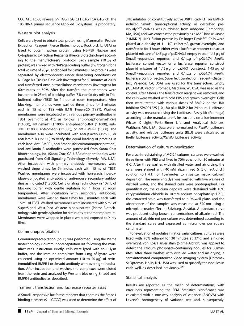
Western blot analysis
Cells were lysed to obtain total protein using Mammalian Protein
Extraction Reagent (Pierce Biotechnology, Rockford, IL, USA) or
lysed to obtain nuclear protein using NE-PER Nuclear and
Cytoplasmic Extraction Reagents (Pierce Biotechnology) accord-
ing to the manufacturer’s protocol. Each sample (10mg of
protein) was mixed with NuPage loading buffer (Invitrogen) for a
total volume of 20mL and boiled for 5minutes. The proteins were
separated by electrophoresis under denaturing conditions on
NuPage Bis-Tris Pre-Cast Gels (Invitrogen) for 60minutes at 200 V
and transferred onto nitrocellulose membranes (Invitrogen) for
60minutes at 30 V. After the transfer, the membranes were
incubated in 25mL of blocking buffer [5% nonfat dry milk in Tris-
buffered saline (TBS)] for 1 hour at room temperature. After
blocking, membranes were washed three times for 5minutes
each in 15mL of TBS with 0.1% Tween-20 (TBST). Washed
membranes were incubated with various primary antibodies in
TBST overnight at 48C as follows: anti-phospho-Smad1/5/8
(1:1000), anti-Smad1 (1:1000), anti-phospho-JNK (1:1000), anti-
JNK (1:1000), anti-Smad6 (1:1000), or anti-BMPR-I (1:500). The
membranes also were incubated with anti-b-actin (1:2500) or
anti-lamin B (1:2000) to verify the equal loading of proteins in
each lane. Anti-BMPR-I, anti-Smad6 (for coimmunoprecipitation),
and anti-lamin B antibodies were purchased from Santa Cruz
Biotechnology, Inc. (Santa Cruz, CA, USA); other antibodies were
purchased from Cell Signaling Technology (Beverly, MA, USA).
After incubation with primary antibody, membranes were
washed three times for 5minutes each with 15mL of TBST.
Washed membranes were incubated with horseradish perox-
idase–conjugated anti-rabbit or anti-mouse secondary antibo-
dies as indicated (1:2000; Cell Signaling Technology in 10mL of
blocking buffer with gentle agitation for 1 hour at room
temperature. After incubation with secondary antibodies,
membranes were washed three times for 5minutes each with
15mL of TBST. Washed membranes were incubated with 5mL of
SuperSignal West Pico Western blot substrate (Pierce Biotech-
nology) with gentle agitation for 4minutes at room temperature.
Membranes were wrapped in plastic wrap and exposed to X-ray
films.
Coimmunoprecipitation
Coimmunoprecepitation (co-IP) was performed using the Pierce
Biotechnology Co-immunoprecepitation Kit following the man-
ufacturer’s instruction. Briefly, cells were lysed with co-IP lysis
buffer, and the immune complexes from 1mg of lysate were
collected using an optimized amount (10 to 20mg) of resin-
immobilized BMPR-I or Smad6 antibody with overnight incuba-
tion. After incubation and washes, the complexes were eluted
from the resin and analyzed by Western blot using Smad6 and
BMPR-I antibodies as described.
Transient transfection and luciferase reporter assay
A Smad1-responsive luciferase reporter that contains the Smad1
binding element (9�GCCG) was used to determine the effect of
JNK inhibitor or constitutively active JNK1 (caJNK1) on BMP-2-
induced Smad1 transcriptional activity, as described pre-
viously.(27) caJNK1 was purchased from Addgene (Cambridge,
MA, USA) and was constructed previously as a MAP kinase kinase
7 (MKK-7)–JNK1 fusion protein by Dr Roger Davis.(28) Cells were
plated at a density of 1� 104 cells/cm2, grown overnight, and
transfected for 4 hours either with a luciferase reporter construct
plasmid mixture of 1.45mg of pcDNA3.1 empty vector, 1.45mg of
Smad1-responsive reporter, and 0.1mg of pGL4.74 Renilla
luciferase control vector or a luciferase reporter construct
plasmid mixture of 1.45mg of caJNK1 construct, 1.45mg of
Smad1-responsive reporter, and 0.1mg of pGL4.74 Renilla
luciferase control vector. Superfect tranfection reagent (Qiagen,
Inc., Valencia, CA, USA) was used for the transfection. Empty
pGL3-BASIC vector (Promega, Madison, WI, USA) was used as the
control. After 4 hours, the transfection reagent was removed, and
the cells were washed with cold PBS and grown overnight. Cells
then were treated with various doses of BMP-2 or the JNK
inhibitor SP6001225 (10mM) plus BMP-2 for 24 hours. Luciferase
activity was measured using the Luciferase Assay Kit (Promega)
according to the manufacturer’s instructions on a luminometer
(Victor X Light; PerkinElmer Life and Analytical Sciences,
Waltham, MA, USA). Data were normalized to Renilla luciferase
activity, and relative luciferase units (RLU) were calculated as
firefly luciferase activity/Renilla luciferase activity.
Determination of culture mineralization
For alizarin red staining of MC-24 cultures, cultures were washed
three times with PBS and fixed in 70% ethanol for 30minutes at
48C. After three washes with distilled water and air drying, the
cells were stained with 40mM alizarin red S (Sigma-Aldrich)
solution (pH 4.1) for 10minutes to visualize matrix calcium
deposition. The remaining dye was washed with five washes of
distilled water, and the stained cells were photographed. For
quantification, the calcium deposits were destained with 10%
cetylpyridinium chloride in 10mM sodium phosphate (pH 7.0),
the extracted stain was transferred to a 96-well plate, and the
absorbance of the samples was measured at 570 nm using a
microplate reader (Tecan, Salzburg, Austria). A standard curve
was produced using known concentrations of alizarin red. The
amount of alazirin red per culture was determined according to
the standard curve and expressed as micromoles per square
centimeter.
For evaluation of nodules in rat calvarial cultures, cultures were
fixed with 70% ethanol for 30minutes at 378C and air dried
overnight. von Kossa silver stain (Sigma-Aldrich) was applied to
detect the calcium phosphate–containing nodules for 30min-
utes. After three washes with distilled water and air drying, a
semiautomated computerized video imaging system (Optomax
5; Optomas, Hollis, NH, USA) was used to quantify the nodules in
each well, as described previously.(26)
Statistical analysis
Results are reported as the mean of determinations, with
error bars representing the SEM. Statistical significance was
calculated with a one-way analysis of variance (ANOVA) with
Levene’s homogeneity of variance test and, subsequently,
1124 Journal of Bone and Mineral Research LIU ET AL.

Bonferroni’s post-hoc test or Dunnett’s T3 post-hoc test based
on the comparison to be made and the statistical indication of
each test. Analysis (including correction of p values when
Bonferroni was used) was performed using Statistical Products
for Social Sciences, Version 13.0 for Windows (SPSS, Inc, Chicago,
IL, USA). Statistical probability of p< .05 was considered
significant.
Results
BMP-2 induces preosteoblast differentiation andmineralization in a dose-dependent manner
To first establish the cell model of BMP-2-induced osteoblast
differentiation, MC-24 cells were treated with various doses of
BMP-2. Cells were fixed and stained to detect calcium deposition,
as measured by alizarin red staining on days 3, 11, and 17. RNA
was isolated to detect changes in the levels of mRNA for
osteogenic markers with real-time RT-PCR on days 1, 3, 5, and 7.
BMP-2 induced nodule formation in a dose- and time-dependent
manner (Fig. 1A). Quantification indicated that BMP-2 at 100 ng/
mL increased alizarin red deposition to 14mmol/cm2 on day 3,
337mmol/cm2 on day 11, and 540mmol/cm2 on day 17. In
addition, BMP-2 at 200 ng/mL increased alizarin red deposition
to 25mmol/cm2 on day 3, 528mmol/cm2 on day 11, and
819mmol/cm2 on day 17 (Fig. 1B). On both days 11 and 17,
200 ng/mL of BMP-2 induced more alizarin red deposition than
BMP-2 at 100 ng/mL (p< 0.05). Thus, in subsequent experiments,
day 11 was selected as the time point to detect mineralization.
Increased Alp and OcnmRNA levels also were induced by BMP-2
in a dose- and time-dependent manner. Alp was increased by
100 ng/mL of BMP-2 4-fold, 83-fold, 216-fold, and 116-fold on
days 1, 3, 5, and 7, respectively, and also was increased by
200 ng/mL of BMP-2 13-fold, 439-fold, 503-fold, and 322-fold on
days 1, 3, 5, and 7, respectively (Fig. 1C). Ocn was increased by
100 ng/mL of BMP-2 9-fold, 59-fold, 450-fold, and 2945-fold on
days 1, 3, 5, and 7, respectively, and was further increased by
200 ng/mL of BMP-2 9-fold, 183-fold, 2135-fold, and 8603-fold on
days 1, 3, 5 and 7, respectively (Fig. 1D). On day 3, both Alp and
Ocn had significantly different mRNA levels induced by BMP-2 at
both doses tested (p< .05; Fig. 1E, F). Thus, in subsequent
experiments, day 3 was selected as the time point for detecting
changes in mRNA levels.
Smad1 is essential for BMP-2-induced preosteoblastdifferentiation and mineralization
To clarify that the Smad pathway is essential for BMP-2-induced
osteogenic differentiation in MC-24 cells and that JNK does not
regulate BMP-2-induced osteogenic differentiation in a Smad-
independent manner, RNA interference of Smad1 was per-
formed. Application of Smad1 siRNA and successful knockdown
of Smad1 mRNA (p< .05; Fig. 2C) rendered BMP-2 unable to
inducemineralization on day 11 (Fig. 2A, B). Furthermore, Alp and
OcnmRNA levels did not increase with BMP-2 treatment (Fig. 2D,
E). These data suggest that Smad1 is essential for BMP-2 induced
osteogenic differentiation in this cell line.
JNK activation is required for BMP-2 inducedpreosteoblast differentiation and mineralization
To determine whether JNK activation is required for BMP-2-
induced osteoblast differentiation and mineralization, JNK-
specific inhibitor (SP600125) and Jnk1 and Jnk2 siRNA were
applied. JNK inhibitor suppressed BMP-2-induced mineralization
in a dose-dependent manner (Fig. 3A). SP600125 at 5, 10, and
25mM decreased the BMP-2-induced alizarin red from 369 to
345, 131 (p< .05), and 72mmol/cm2 (p< .05), respectively
(Fig. 3B). The Alp increase induced by BMP-2 was suppressed
from about 360-fold to 301-fold, 102-fold (p< .05), and 71-fold
(p< .05) by SP600125 at 5, 10, and 25mM, respectively. Also, the
BMP-2-induced increase in Ocn mRNA levels was suppressed
from 91-fold to 80-fold, 32-fold (p< .05), and 10-fold (p< .05) by
SP600125 at 5, 10, and 25mM, respectively (Fig. 3C, D). We
confirmed the importance of JNK in BMP-2-induced MC-24 cell
differentiation and extended these observations by applying
specific siRNA for Jnk1 or Jnk2. siRNA application (10 and
50 pmol/mL) specifically and dose-dependently reduced Jnk1 or
Jnk2 mRNA levels 85% to 90% compared with the level in cells
treated with control siRNA (Fig. 3E). BMP-2-induced alizarin red
deposition was suppressed significantly by Jnk1 siRNA compared
with the BMP-2-induced deposition in the presence of control
siRNA (Fig. 3F, G). However, with Jnk2 siRNA application, BMP-2-
induced alizarin red staining was not reduced significantly.
To confirm the requirement for JNK1 in BMP-2-induced
differentiation of nontransformed, freshly isolated preosteo-
blasts, we applied siRNA for Jnk1 or Jnk2 to rat calvarial
preosteoblast cultures treated with BMP-2. The siRNA duplexes
specifically reduced the targeted Jnk mRNA by more than 75%
but did not significantly affect the nontargeted Jnk mRNA (data
not shown). As with the MC-24 cell line, JNK1 was required for
BMP-2 stimulation of nodule formation, whereas JNK2 was not
required (Supplemental Fig. S1A, B). BMP-2 stimulated miner-
alized nodules from 60� 1 to 158� 4/well (p< .05). Nodule
number in the cultures treated with Jnk1 siRNA prior to BMP-2
treatment was reduced to 78� 1/well (p< .05), whereas nodule
number in the cultures treated with Jnk2 siRNA was slightly
increased. These data suggest that JNK, especially JNK1, is
required for BMP-2-induced osteoblast differentiation and
mineralization.
Constitutively active JNK1 increases BMP-2-inducedpreosteoblast differentiation and mineralization
To determine whether JNK1 activation enhances BMP-2-induced
osteoblast differentiation and mineralization, a constitutively
active JNK1 (caJNK1) construct was transfected into MC-24 cells
prior to treating the cells with or without BMP-2. Constitutively
active JNK1 alone did not increase mineralization, but it
significantly increased the response of MC-24 cells to BMP-2
(Fig. 4A). With caJNK1 overexpression, alizarin red induced by
BMP-2 was increased from 419 to 1083mmol/cm2 (p< .05)
(Fig. 4B). The increase in AlpmRNA levels induced by BMP-2 was
elevated from 170-fold to 405-fold (p< .05); the increase in Ocn
mRNA levels induced by BMP-2 was elevated from 60-fold to
186-fold (p< .05; Fig. 4C, D). These data suggest that JNK1
JNK1 INCREASES CELLULAR RESPONSIVENESS TO BMP-2 Journal of Bone and Mineral Research 1125

constitutive activation increases BMP-2-induced osteoblast
differentiation and mineralization.
Inhibition of JNK suppresses but activation of JNK1increases pSmad1 nuclear accumulation induced byBMP-2
To explore the role of JNK in BMP-2-induced osteogenesis, we
determine whether JNK regulated BMP-2-induced osteogenesis
by interacting with the Smad pathway. MC-24 cells were
transfected with empty pcDNA3.1 control plasmid or caJNK1
plasmid. After transfection, the cells were pretreated with DMSO
(control) or JNK inhibitor (SP600125, 25mM) for 60minutes and
then treated with BMP-2 (100 ng/mL) for 30minutes. Nuclear
protein was isolated, and Western blots were used to determine
pSmad1 nuclear accumulation induced by BMP-2. JNK inhibitor
or caJNK1 alone had no effect on Smad1 nuclear accumulation;
however, JNK inhibitor suppressed but caJNK1 increased
Fig. 1. BMP-2 induces osteoblast differentiation and mineralization in a dose-dependent manner. (A) Nodule formation by MC-24 cells induced by BMP-2
at 100 or 200 ng/mL. Cells were stained with alizarin red on days 3, 11, or 17. The image presented is from one representative experiment out of three
independent experiments performed in duplicate. (B) Alizarin red was quantified as described under ‘‘Materials and Methods.’’ Data are presented as
mean� SEM from three independent experiments performed in duplicate. (C, D) Time course of increased Alp and OcnmRNA levels in response to BMP-2
treatment. Cells were treated with the indicated doses of BMP-2 for 1, 3, 5, or 7 days. The mRNA levels of Alp and Ocnwere detected and expressed as the
fold change over untreated cells on day 1. Data are presented as mean� SEM from three independent experiments performed in duplicate. (E, F) BMP-2
induced Alp andOcnmRNA levels on day 3. Cells were treated with the indicated doses of BMP-2 for 3 days. ThemRNA levels of Alp andOcnwere detected
and expressed as fold change over untreated cells on day 3. Data are presented as mean� SEM from three independent experiments performed in
duplicate. ap< .05.
1126 Journal of Bone and Mineral Research LIU ET AL.

pSmad1 nuclear accumulation (Fig. 5A). A luciferase reporter
assay was used to detect Smad1 transcriptional activity, as
described under ‘‘Materials and Methods.’’ The Smad1 transcrip-
tional activity was increased by BMP-2 in a dose-dependent
manner (Fig. 5B). BMP-2 at 2.5 ng/mL was selected for
subsequent experiments. JNK inhibitor or caJNK1 alone did
not have any effect on Smad1 transcriptional activity. However,
JNK inhibitor significantly suppressed and caJNK1 dramatically
increased BMP-2-induced Smad1 transcriptional activity. These
data suggest that JNK affects BMP-2-induced osteogenesis by
interaction with the Smad pathway.
Inhibition of JNK suppresses but activation of JNK1increases Smad1 C-terminal phosphorylation
To further explore regulation of the Smad pathway by JNK, we
determined whether JNK has any effect on BMP-2-induced
Smad1 C-terminal phosphorylation, which is a direct down-
stream effect of BMP-2 stimulation. First, we determined the
profile of Smad1 C-terminal phosphorylation induced by BMP-2.
MC-24 cells were treated with BMP-2 for 15, 30, 60, 90, and
120minutes. Western blots were used to detect Smad1 C-
terminal phosphorylation as well as JNK phosphorylation. BMP-2
induces both Smad1 C-terminal phosphorylation and JNK
phosphorylation in a time-dependent manner (Fig. 6A). Based
on these results, 15, 30, and 90minutes were the time points
selected for subsequent experiments. MC-24 cells were trans-
fected with empty pcDNA3.1 control plasmid or caJNK1 plasmid
for 16 hours. After transfection, the cells were pretreated with
DMSO (control) or JNK inhibitor (SP600125, 25mM) and then
treated with BMP-2 for 15, 30, or 90minutes. Western blots were
used to detect Smad1 C-terminal phosphorylation as well as JNK
phosphorylation. BMP-2-induced Smad1 C-terminal phosphor-
ylation was suppressed by JNK inhibitor but enhanced by caJNK1
significantly at 15, 30, and 90minutes. These data suggest that
JNK affects BMP-2-induced osteogenesis by modulating Smad1
C-terminal phosphorylation.
Inhibition of JNK increases but activation of JNK1suppresses Smad6 competition with Smad1 binding toBMPR-I
To further explore themechanism by which JNK affects Smad1 C-
terminal phosphorylation, the direct interaction of Smad1 and its
upstream kinase, BMPR-I, was compared with the interaction of
BMPR-I with inhibitory Smad6, the endogenous inhibitor of the
BMP-2 pathway. Cells were transfected with empty pcDNA3.1
control plasmid or caJNK1 plasmid as described under ‘‘Materials
and Methods.’’ After transfection, cells were pretreated with
DMSO or JNK inhibitor for 1 hour and then treated with BMP-2
(100 ng/mL) for 30minutes. After BMP-2 treatment, total protein
was isolated, and BMPR-I antibody or Smad6 antibody was used
to immunoprecipitate BMPR-I or Smad6, respectively. Interaction
of Smad1 or Smad6with BMPR-I was determined byWestern blot
of the proteins coimmuoprecipitating with BMPR-I. Inhibition of
JNK decreased Smad1 binding to BMPR-I, whereas constitutive
activation of JNK1 suppressed Smad1 binding to BMPR-I (Fig. 7A).
Similarly, inhibition of JNK increased but activation of JNK1
suppressed binding of BMPR-I with Smad6 (Fig. 7B). These data
Fig. 2. Smad1 is essential for BMP-2-induced preosteoblast differentia-
tion and mineralization. (A) BMP-2-induced nodule formation by MC-
24 cells with or without pretreatment with Smad1 interference RNA. Cells
were transfected with control or Smad1 siRNA (10pmol/mL) as described
under ‘‘Materials and Methods,’’ treated with BMP-2 (100 ng/mL), and
stained with alizarin red on day 11. The image presented is from one
representative experiment out of three independent experiments per-
formed in duplicate. (B) Alizarin red was quantified as described under
‘‘Materials and Methods.’’ Data are presented as mean� SEM from three
independent experiments performed in duplicate. (C) Interference RNA
for Smad1. Cells were transfected with control or Smad1 siRNA (10 pmol/
mL) as described under ‘‘Materials and Methods’’ and treated with BMP-2
(100 ng/mL) for 3 days. The mRNA level of Smad1 was detected and
expressed as the fold change from the level in cells treated with control
siRNA. Data are presented as mean� SEM from three independent
experiments performed in duplicate. (D, E) BMP-2-induced Alp and
Ocn mRNA levels on day 3 in cells treated with control or Smad1 siRNA.
Cells were transfected with control or Smad1 siRNA (10 pmol/mL) as
described under ‘‘Materials and Methods’’ and then treated with BMP-2
(100 ng/mL) for 3 days. The mRNA levels of Alp and Ocn were detected
and expressed as the fold change from the level in cells treated with
control siRNA. Data are presented as mean� SEM from three indepen-
dent experiments performed in duplicate. ap< .05.
JNK1 INCREASES CELLULAR RESPONSIVENESS TO BMP-2 Journal of Bone and Mineral Research 1127

Fig. 3. JNK activation is required for BMP-2-induced osteoblast differentiation andmineralization. (A) Nodule formation by MC-24 cells induced with BMP-
2 in the presence or absence of JNK inhibitor. Cells were pretreated with DMSO or the indicated doses of JNK inhibitor, SP600125, for 1 hour, treated with
BMP-2 (100 ng/mL) for 3 days, and stained with alizarin red on day 11. The image presented is from one representative experiment out of three
independent experiments performed in duplicate. (B) Alizarin red deposition was quantified as described under ‘‘Materials and Methods.’’ Data are
presented asmean� SEM from three independent experiments performed in duplicate. (C,D) BMP-2-induced Alp andOcnmRNA levels on day 3 from cells
treated with or without the inhibitor of JNK. Cells were pretreated with DMSO or the indicated doses of the JNK inhibitor, SP600125, for 1 hour and then
treated with BMP-2 (100 ng/mL) for 3 days. ThemRNA levels of Alp andOcnwere detected and expressed as the fold change from cells treated with control
siRNA. Data are presented as mean� SEM from three independent experiments performed in duplicate. (E) siRNA reduced Jnk1 or Jnk2 mRNA levels
specifically and dose dependently. Cells were transfected with control (cont), Jnk1, or Jnk2 siRNA (10 or 50 pmol/mL) as described under ‘‘Materials and
Methods.’’ The mRNA level of Jnk1 or Jnk2 was detected and expressed as the fold change from the level in cells treated with control siRNA. (F, G) Nodule
formation and alizarin red deposition were significantly reduced using Jnk1 siRNA but not using Jnk2 siRNA. The image and analysis presented are from
one representative experiment performed three times in duplicate. ap< .05.
1128 Journal of Bone and Mineral Research LIU ET AL.

suggest that JNK1 regulates Smad1 phosphorylation and BMP-2
induction of osteogenesis by regulating Smad6 competition with
Smad1 binding to BMPR-I.
Discussion
Enhancing cellular responsiveness to BMP-2 potentially has
critically important clinical implications. To date, unexpectedly
high doses (concentrations) of recombinant BMP-2 have been
required to induce bone healing in humans compared with that
required in rodents and cell culture.(5,6,29) BMPs bind specific
receptors, resulting in the formation of heteromeric receptor
complexes containing type I (BMPR-I) and type II (BMPR-II)
receptors. The type I receptor then phosphorylates receptor
Smad1, -5, and -8 (R-Smads) as the next step in transmitting the
BMP-2 signal. Phosphorylated R-Smads bind Smad4 and
Fig. 4. Constitutively active JNK1 increases BMP-2-induced osteoblast
differentiation and mineralization. (A) BMP-2-induced nodule formation
by MC-24 cells transfected with empty plasmid (cont) or constitutively
active JNK1 (caJNK1) plasmid. Cells were transfected with plasmids for
4 hours, grown overnight in fresh medium, and treated with BMP-2
(100 ng/mL) for 3 days. Cells were fixed and stained with alizarin red
on day 11. The image presented is from one representative experiment
out of three independent experiments in duplicate. (B) Alizarin red was
quantified as described under ‘‘Materials and Methods.’’ Data are pre-
sented as mean� SEM from three independent experiments performed
in duplicate. (C, D) BMP-2-induced Alp and Ocn mRNA levels 3 days after
transfection with control or caJNK1 plasmid. Cells were transfected with
plasmids for 4 hours, grown overnight in fresh medium, and treated with
BMP-2 (100 ng/mL) for 3 days. Alp and Ocn mRNA were detected and
expressed as fold change over cells treatedwith noncoding plasmid. Data
are presented as mean� SEM from three independent experiments
performed in duplicate. ap< .05.
Fig. 5. Inhibition of JNK suppresses but activation of JNK1 increases
BMP-2-induced pSmad1 nuclear accumulation and Smad1 transcrip-
tional activity. (A) Cells were transfected with empty vector (cont) or
caJNK1 plasmid for 4 hours and grown overnight in fresh medium. Then
the cells were pretreated with DMSO or JNK inhibitor for 1 hour, followed
by BMP-2 (100 ng/mL) treatment for 30minutes. Nuclear protein was
extracted and the protein level of pSmad1 in nuclear extracts was
determined by Western blot. Lamin B antibody was used to show equal
protein loading. The image presented is from one representative experi-
ment out of three independent experiments. (B) BMP-2-induced Smad1-
responsive reporter activity. A Luciferase reporter assay was used to
assess Smad1 transcriptional activity. The reporter construct was trans-
fected into cells as described under ‘‘Materials andMethods.’’ Transfected
cells then were treated with the indicated doses of BMP-2 for 24 hours.
Luciferase activity was determined as described under ‘‘Materials and
Methods’’ and normalized to Renilla luciferase activity as relative lucifer-
ase units (RLUs). Data from two independent experiments performed in
triplicate are presented as mean� SEM of the fold change in RLUs
compared with the untreated control. (C) BMP-2-induced Smad1-respon-
sive reporter activity change in cells with inhibition or activation of JNK.
Cells were cotransfected with the Smad1-responsive reporter and the
noncoding or caJNK1 plasmid as described under ‘‘Materials and Meth-
ods.’’ Then cells were pretreated with DMSO or JNK inhibitor for 1 hour.
After pretreatment, cells were treated with BMP-2 (100 ng/mL) for
24 hours. Luciferase activity was normalized to Renilla luciferase activity
as relative luciferase units (RLUs). Data from two independent experi-
ments performed in triplicate are presented as mean� SEM of the fold
change in RLU compared with the control cells treated with DMSO and
the control plasmid. ap< .05.
JNK1 INCREASES CELLULAR RESPONSIVENESS TO BMP-2 Journal of Bone and Mineral Research 1129

translocate to the nucleus, where they regulate gene expres-
sion.(8,9)
The experimental study of BMP-2 signaling and BMP
responsiveness is challenging and is complicated by at least
two issues: (1) cross-talk with numerous other pathways(15,16)
and (2) lack of synchronization of cellular differentiation in
spontaneously differentiating heterogeneous preosteoblast
cultures.(25) Several studies have demonstrated that MAPKs
are required for BMP-2-induction of the osteoblastic phenotype
and mineral deposition in vitro.(18,21,30) In particular, the MAPK
JNK is reported to be required for both late-stage spontaneous
differentiation of osteoblasts and BMP-2-induced differentiation
of preosteoblasts and pleuripotent cells in vitro.(20,21,23) However,
in those previous studies, the points of intersection of the JNK
and BMP-2 pathways were not investigated.
In addition to cross-talk, study of BMP signaling is further
complicated by virtue of the fact that most osteoblast cell culture
systems result in spontaneous differentiation of preosteoblasts
that are at varying stages of lineage commitment.(31,32) Since
BMP signaling and cross-talk are likely to be time-depen-
dent,(32,33) we felt that it might be helpful to study BMP
responsiveness in a model where preosteoblasts were more
synchronized in their differentiation toward the mature
osteoblast phenotype. To this end, we used subclone 24 of
MC3T3-E1 cells (MC-24), which were characterized originally by
Franceschi as a nondifferentiating subclone of preosteoblasts.(25)
While not undergoing spontaneous differentiation, like the more
commonly used subclone 14 of MC3T3-E1 cells,(25) we show for
the first time that this MC-24 subclone can be induced by BMP-2
to differentiate into osteoblasts that produce mineralized
nodules.
We used the unique property that the MC-24 preosteoblasts
required a stimulus to initiate osteoblast differentiation to
facilitate the measurement of BMP responsiveness in the
presence of various inhibitors of cross-talk signaling pathways.
A major limitation of these transient gain- or loss-of-function
studies arises whenmeasuring amore physiologic endpoint such
as mineralized nodules, which requires 11 to 17 days in culture.
There is often uncertainty as to whether and when to reapply an
inhibitory agent if the positive BMP-2 stimulus requires repeated
Fig. 6. Inhibition of JNK suppresses but activation of JNK1 increases
Smad1 C-terminal phosphorylation. (A) Time course of BMP-2-induced
phosphorylation of Smad1, -5, and -8. Cells were treated with BMP-2
(100 ng/mL), and protein levels of pSmad1, -5, and -8, Smad1, pJNK, and
JNK were determined by Western blot. The image presented is from one
representative experiment out of three independent experiments. (B)
Inhibition of JNK suppresses but activation of JNK1 increases Smad1 C-
terminal phosphorylation. Cells were transfected with noncoding or
caJNK1 plasmids for 4 hours and grown overnight in fresh medium.
Then cells were pretreated with DMSO or JNK inhibitor for 1 hour,
followed by treatment with BMP-2 (100 ng/mL) for 15, 30, or 90minutes.
Total protein was extracted, and the protein levels of pSmad1, -5, and -8,
Smad1, and pJNK were determined by Western blot. b-Actin antibody
was used to show equal protein loading. The image presented is from
one representative experiment out of three independent experiments.
Fig. 7. JNK modulates binding of Smad1 to BMPR-I by regulating recep-
tor binding of the inhibitor Smad6. Cells were transfected with noncod-
ing or caJNK1 plasmids and grown overnight in fresh medium. Then cells
were pretreated with DMSO or JNK inhibitor for 1 hour, followed by
treatment with BMP-2 (100 ng/mL) for 30minutes. Total protein was
isolated, and BMPR-I (A) or Smad6 (B) antibodies were used for coim-
munoprecipitation studies. Interaction of Smad6 or Smad1 with BMPR-I
was determined by Western blot with specific antibody for each protein.
The image presented is from one representative experiment out of three
independent experiments.
1130 Journal of Bone and Mineral Research LIU ET AL.

applications every few days, which is frequently recommended
in other systems. We addressed this potential confounder and
ensured more synchronized differentiation by optimizing the
protocol to require only a single administration of BMP-2 protein
rather than multiple reapplications. This single BMP-2 admin-
istration protocol allowed us to test inhibitors of pathways
known to interact with the BMP-2 signaling pathway without
concern over the need or timing of reapplication of the inhibitor.
Having validated an inducible osteoblast culture system, we
made three key observations that provide new insight into
modulation of BMP-2 signaling by JNK. First, we made the novel
observation that JNK is required for phosphorylation of Smad1,
the first intracellular event in the canonical BMP-2 signaling
cascade that can be observed within 15minutes of treatment.
Second, we found that JNK1, but not JNK2, was required for BMP-
2-induced formation of mineralized nodules. This important
observation was confirmed in freshly isolated rat calvarial
osteoblast cultures, implying that it is not an artifact of
transformed cell lines. Third, we found that JNK1 activation
results in decreased binding of the inhibitory Smad6 to BMPR-I
and, reciprocally, increased binding of Smad1, both observations
that would increase BMP responsiveness.
To understand the relative importance of isoforms of JNK
expressed in skeletal tissues, we used specific siRNA and found
that JNK1, but not JNK2, was required for BMP-2-induced nodule
formation. This observation is contrary to at least one previously
published work stating that JNK2, but not JNK1, is required for
late-stage osteoblast differentiation of spontaneously differen-
tiating MC3T3 cells.(23) We interpret this discrepancy as suggest-
ing that JNK1 is required for the initial stages of differentiation by
preosteoblasts but less important when cells are more
committed to the osteoblast lineage.
The observation that JNK1 activation suppresses Smad6
association with BMPR-I and enhances Smad1 association was
further confirmed by the observation that inhibition of JNK1/2
enhances binding of Smad6 to BMPR-I, whereas it decreases
Smad1 binding. These early-event findings are consistent with
our observations of downstream endpoints that JNK1 activation
enhances BMP-2-induced Smad1 activation, osteoblast differ-
entiation, and nodule formation in vitro, whereas inhibition of
JNK1/2 has the opposite effect.We hypothesize two possible
ways in which JNK might modulate Smad6 or Smad1 binding to
BMPR-I. Either JNK directs posttranslational modification of
Smad1 or Smad6 or JNK modifies a Smad1 or Smad6 binding
partner to alter Smad availability to bind the receptor.
Phosphorylation within the linker region of Smad1 by MAPKs
has emerged as an important mechanism by which growth
factors desensitize cells to BMP-2.(34,35) Intense study of linker-
region phosphorylation has led to the realization that many
kinases, in addition to MAPKs, contribute to linker-region
phosphorylation. Further, the E-3 ligase Smurf1 has been shown
to bind preferentially to Smad1 that is phosphorylated in the
linker region, targeting it for degradation.(36) Recently, an
additional level of complexity in understanding the importance
of Smad1 linker-region phosphorylation has been added with
the identification of R-Smad linker phosphatases that have site
specificity.(37) Less is known about linker-region phosphorylation
of Smad6. These complex modifications are of considerable
interest when considering the mechanism by which JNK
activation enhances Smad1 binding to the BMPR-I or reduces
the binding of Smad 6 to the receptor. However, to date, no
evidence has emerged indicating that MAPK-induced linker-
region phosphorylation would affect the relative association of
these molecules with the BMPR-I. Future studies will be needed
to address specifically what cellular changes JNK elicits to
modulate Smad6 and Smad1 binding to BMPR-I.
One potential limitation of this study is its dependence on the
specificity of the pJNK and pSmad1, -5, and -8 antibodies to
detect only the phosphorylated molecule without cross-reacting
with unphosphorylated forms. Further, we cannot evaluate the
relative contribution of pSmad1, pSmad5, or pSmad8 in the
detected species. However, these antibodies have been used by
other investigators in many studies to evaluate pSmad1, -5, and -
8 levels. More specific antibodies have not been developed
because of the sequence homology in the region. Also, our
conclusion regarding Smad1 or Smad6 binding to BMPR-I
assumes complete immunoprecipation of Smad6 and its bound
proteins. The consistency of our findings in three independent
experiments and the fact that our coimmunoprecipitation
studies are consistent with the physiologic outcomes measured
independently suggest that our conclusions accurately reflect
cellular events.
In summary, we have made the novel observation that JNK1 is
required for BMP-2 activity and moreover can modulate cellular
responsiveness to BMP-2 in an osteoblastic cell line and in freshly
isolated calvarial osteoblasts. JNK1 activation results in
decreased binding of inhibitory Smad6 and increased binding
of Smad1 to the BMP receptor, resulting in increased cellular
response to BMP-2, as measured within 30minutes by increased
levels of phosphorylated Smad1 and at later points by expression
of the mature osteoblast phenotype, including increased
formation of mineralized nodules. Understanding this and other
pathways that lead to increased cellular responsiveness to BMPs
could greatly aid more cost-effective and safe clinical delivery of
these important molecules.
Disclosures
SDB has received compensation in the past as a consultant for
Medtronic Sofamor Danek and for intellectual property. The
terms of this arrangement were reviewed and approved by
Emory University in accordance with its conflict-of-interest
policies. All the other authors state that they have no conflicts of
interest.
Acknowledgments
This work was supported in part by a VAMerit Award to SDB. This
work was performed in facilities at the Atlanta Veterans Affairs
Medical Center (Decatur, GA, USA). HL is partially supported by
the International Program of Project 985, Sun Yat-sen University.
We would like to thank Dr Sreedhara Sangadala for helpful
discussions and Mesfin Teklemariam for real-time RT-PCR, plas-
mid purification, and Western blot assistance.
JNK1 INCREASES CELLULAR RESPONSIVENESS TO BMP-2 Journal of Bone and Mineral Research 1131

References
1. Kingsley DM. The TGF-b superfamily: new members, new receptors,
and new genetic tests of function in different organisms. Genes Dev.
1994;8:133–146.
2. Xiao YT, Xiang LX, Shao JZ. Bone morphogenetic protein. Biochem
Biophys Res Commun. 2007;362:550–553.
3. Luu HH, Song WX, Luo X, et al. Distinct roles of bone morphogenetic
proteins in osteogenic differentiation of mesenchymal stem cells.J Orthop Res. 2007;25:665–677.
4. Riley EH, Lane JM, Urist MR, Lyons KM, Lieberman JR. Bone morpho-
genetic protein 2: biology and applications. Clin Orthop Relat Res.1996;324:39–46.
5. Boden SD, Kang J, Sandhu H, Heller JG. Use of recombinant human
bone morphogenetic protein 2 to achieve posterolateral lumbar
spine fusion in humans: a prospective, randomized clinical pilot trial[2002 Volvo Award in clinical studies]. Spine (Phila). 2002;27:2662–
2673.
6. Rihn JA, Gates C, Glassman SD, Phillips FM, Schwender JD, Albert TJ.
The use of bone morphogenetic protein in lumbar spine surgery.J Bone Joint Surg Am. 2008;90:2014–2025.
7. Smoljanovic T, Bojanic I, Delimar D. Adverse effects of posterior
lumbar interbody fusion using rhBMP-2. Eur Spine J. 2009;18:920–
923 author reply 924.
8. Miyazono K. Signal transduction by bone morphogenetic protein
receptors: functional roles of Smad proteins. Bone. 1999;25:
91–93.
9. Gilboa L, Nohe A, Geissendorfer T, Sebald W, Henis YI, Knaus P. Bone
morphogenetic protein receptor complexes on the surface of live
cells: a new oligomerization mode for serine/threonine kinase recep-
tors. Mol Biol Cell. 2000;11:1023–1035.
10. Ross S, Hill CS. How the Smads regulate transcription. Int J Biochem
Cell Biol. 2008;40:383–408.
11. Hayashi H, Abdollah S, Qiu Y, et al. The MAD-related protein Smad7
associates with the TGFbeta receptor and functions as an antagonistof TGFbeta signaling. Cell. 1997;89:1165–1173.
12. Imamura T, Takase M, Nishihara A, et al. Smad6 inhibits signalling by
the TGF-beta superfamily. Nature. 1997;389:622–626.
13. Hata A, Lagna G, Massague J, Hemmati-Brivanlou A. Smad6 inhibits
BMP/Smad1 signaling by specifically competing with the Smad4
tumor suppressor. Genes Dev. 1998;12:186–197.
14. Goto K, Kamiya Y, Imamura T, Miyazono K, Miyazawa K. Selectiveinhibitory effects of Smad6 on bone morphogenetic protein type I
receptors. J Biol Chem. 2007;282:20603–20611.
15. Wu X, Shi W, Cao X. Multiplicity of BMP signaling in skeletal devel-
opment. Ann N Y Acad Sci. 2007;1116:29–49.
16. Guo X, Wang XF. Signaling cross-talk between TGF-beta/BMP and
other pathways. Cell Res. 2009;19:71–88.
17. Chang L, Karin M. Mammalian MAP kinase signalling cascades.Nature. 2001;410:37–40.
18. Gallea S, Lallemand F, Atfi A, et al. Activation of mitogen-activated
protein kinase cascades is involved in regulation of bone morpho-
genetic protein-2-induced osteoblast differentiation in pluripotentC2C12cells. Bone. 2001;28:491–498.
19. Vinals F, Lopez-Rovira T, Rosa JL, Ventura F. Inhibition of PI3K/p70 S6K
and p38 MAPK cascades increases osteoblastic differentiation
induced by BMP-2. FEBS Lett. 2002;510:99–104.
20. Celil AB, Campbell PG. BMP-2 and insulin-like growth factor-I mediate
Osterix (Osx) expression in human mesenchymal stem cells via the
MAPK and protein kinase D signaling pathways. J Biol Chem.
2005;280:31353–31359.
21. Guicheux J, Lemonnier J, Ghayor C, Suzuki A, Palmer G, Caverzasio J.Activation of p38 mitogen-activated protein kinase and c-Jun-NH2-
terminal kinase by BMP-2 and their implication in the stimulation of
osteoblastic cell differentiation. J Bone Miner Res. 2003;18:2060–
2068.
22. Davis RJ. Signal transduction by the JNK group of MAP kinases. Cell.
2000;103:239–252.
23. Matsuguchi T, Chiba N, Bandow K, Kakimoto K, Masuda A, Ohnishi T.JNK activity is essential for Atf4 expression and late-stage osteoblast
differentiation. J Bone Miner Res. 2009;24:398–410.
24. Lemonnier J, Ghayor C, Guicheux J, Caverzasio J. Protein kinase C-
independent activation of protein kinase D is involved in BMP-2-induced activation of stress mitogen-activated protein kinases JNK
and p38 and osteoblastic cell differentiation. J Biol Chem. 2004;279:
259–264.
25. Wang D, Christensen K, Chawla K, Xiao G, Krebsbach PH, FranceschiRT. Isolation and characterization of MC3T3-E1 preosteoblast sub-
clones with distinct in vitro and in vivo differentiation/mineralization
potential. J Bone Miner Res. 1999;14:893–903.
26. Boden SD, McCuaig K, Hair G, et al. Differential effects and gluco-corticoid potentiation of bone morphogenetic protein action during
rat osteoblast differentiation in vitro. Endocrinology. 1996;137:3401–
3407.
27. Okada M, Sangadala S, Liu Y, et al. Development and optimization of
a cell-based assay for the selection of synthetic compounds that
potentiate bone morphogenetic protein-2 activity. Cell Biochem
Funct. 2009;27:526–534.
28. Lei K, Nimnual A, Zong WX, et al. The Bax subfamily of Bcl2-related
proteins is essential for apoptotic signal transduction by c-Jun NH(2)-
terminal kinase. Mol Cell Biol. 2002;22:4929–4942.
29. Zanella JM, Oliver C, Peckham SM, McKay B, Toth JM, Boden SD. Effecton bone induction of using contrast media to reconstitute recombi-
nant human bone morphogenetic protein-2 in an ectopic model in
rats. J Neurosurg Spine. 2006;5:434–439.
30. Xiao G, Gopalakrishnan R, Jiang D, Reith E, BensonMD, Franceschi RT.
Bone morphogenetic proteins, extracellular matrix, and mitogen-
activated protein kinase signaling pathways are required for osteo-
blast-specific gene expression and differentiation in MC3T3-E1cells.J Bone Miner Res. 2002;17:101–110.
31. Maeda T, Matsunuma A, Kurahashi I, Yanagawa T, Yoshida H, Horiuchi
N. Induction of osteoblast differentiation indices by statins in MC3T3-
E1cells. J Cell Biochem. 2004;92:458–471.
32. Schindeler A, Little DG. Ras-MAPK signaling in osteogenic differen-
tiation: friend or foe? J Bone Miner Res. 2006;21:1331–1338.
33. Javelaud D, Mauviel A. Crosstalk mechanisms between the mitogen-
activated protein kinase pathways and Smad signaling downstreamof TGF-beta: implications for carcinogenesis. Oncogene. 2005;24:
5742–5750.
34. Kretzschmar M, Doody J, Massague J. Opposing BMP and EGFsignalling pathways converge on the TGF-beta family mediator
Smad1. Nature. 1997;389:618–622.
35. Wrighton KH, Feng XH. To (TGF)beta or not to (TGF)beta: fine-tuning
of Smad signaling via post-translational modifications. Cell Signal.
2008;20:1579–1591.
36. Sapkota G, Alarcon C, Spagnoli FM, Brivanlou AH, Massague J.
Balancing BMP signaling through integrated inputs into the Smad1
linker. Mol Cell. 2007;25:441–454.
37. Sapkota G, Knockaert M, Alarcon C, Montalvo E, Brivanlou AH,
Massague J. Dephosphorylation of the linker regions of Smad1and Smad2/3 by small C-terminal domain phosphatases has distinct
outcomes for bonemorphogenetic protein and transforming growth
factor-beta pathways. J Biol Chem. 2006;281:40412–40419.
1132 Journal of Bone and Mineral Research LIU ET AL.